ORTHOPAEDIC RESEARCH DAY May 4, 2022 | 8:00 AM – 3:50 PM PST Chair: Dr. Henry Broekhuyse Review Panel: Dr. Mohit Bhandari, Distinguished University Professor, Chair, Department of Surgery, McMaster University; Senior Tier Canada Research Chair; Editor-in-Chief, OrthoEvidence Dr. Raphaële Charest-Morin, Clinical Assistant Professor, UBC Department of Orthopaedics Dr. Dena Shahriari, Assistant Professor, UBC Department of Orthopedics and the School of Biomedical Engineering Note: All presentations are strictly limited to 6 minutes, followed by a 4 minute discussion period with the review panel 0800 - 0805: Welcome and Opening remarks – Dr . K. Mulpuri 0805 - 0815: Mathieu Laflamme (Clinical Fellow) Surgical adverse events for primary tumors of the spine and their impact on prognosis and outcomes: A PTRON Study (Co-Authors - N Dea and the AOSpine Knowledge Forum Tumor) 0815 - 0825: Lukas Grassner (Clinical Fellow) Serum neurofilament light (NF-L) and glial fibrillary acidic protein (GFAP) biomarkers and their association with MRI findings in acute human traumatic spinal cord injury (Co-Authors - S Stukas, I Leister, J Cooper, J Gill, L Belanger, L Ritchie, A Tsang, K Dong, F Streijger, JT Street, S Paquette, T Ailon, N Dea, R Charest-Morin, CG Fisher, MF Dvorak, C Wellington, BK Kwon) 0825 - 0835: Bryn Zomar (Post Doctoral Fellow) A systematic review to assess multicentre collaboration in the orthopaedic surgery literature (Co-Authors – N South, K Jackson, H Arneja, A Chehil, M Potluri, K Mulpuri, EK Schaeffer)

Welcome message from author
This document is posted to help you gain knowledge. Please leave a comment to let me know what you think about it! Share it to your friends and learn new things together.
Transcript
ORTHOPAEDIC RESEARCH DAY May 4, 2022 | 8:00 AM – 3:50 PM PST
Chair: Dr. Henry Broekhuyse
Review Panel:
Dr. Mohit Bhandari, Distinguished University Professor, Chair, Department of Surgery, McMaster University;
Senior Tier Canada Research Chair; Editor-in-Chief, OrthoEvidence
Dr. Raphaële Charest-Morin, Clinical Assistant Professor, UBC Department of Orthopaedics
Dr. Dena Shahriari, Assistant Professor, UBC Department of Orthopedics and the School of Biomedical
Engineering
Note: All presentations are strictly limited to 6 minutes, followed by a 4 minute discussion period with
the review panel
0800 - 0805:
Welcome and Opening remarks – Dr . K. Mulpuri
0805 - 0815:
Mathieu Laflamme (Clinical Fellow)
Surgical adverse events for primary tumors of the spine and their impact on prognosis and outcomes: A
PTRON Study
(Co-Authors - N Dea and the AOSpine Knowledge Forum Tumor)
0815 - 0825:
Lukas Grassner (Clinical Fellow)
Serum neurofilament light (NF-L) and glial fibrillary acidic protein (GFAP) biomarkers and their association
with MRI findings in acute human traumatic spinal cord injury
(Co-Authors - S Stukas, I Leister, J Cooper, J Gill, L Belanger, L Ritchie, A Tsang, K Dong, F Streijger, JT
Street, S Paquette, T Ailon, N Dea, R Charest-Morin, CG Fisher, MF Dvorak, C Wellington, BK Kwon)
0825 - 0835:
Bryn Zomar (Post Doctoral Fellow)
A systematic review to assess multicentre collaboration in the orthopaedic surgery literature
(Co-Authors – N South, K Jackson, H Arneja, A Chehil, M Potluri, K Mulpuri, EK Schaeffer)
0835 - 0845:
Mikaela Peters (PGY4)
How the Gender Gap “Presents”: Results of Gender Disparity between Orthopaedic Subspecialty
Presentations at a National Conference
(Co-Authors - GA Sheridan, ME Neufeld, LC Howard)
0845 - 0855:
Taylor Crown (PGY2)
Quality Improvement survey of the University of British Columbia Orthopedic residency program
(Co-Authors – F Leung, HM Broekhuyse)
0855 - 0905:
Daniella Crocker (PGY3, Graduate Studies)
Self-Leadership in Medicine: A Systematic Review
(Co-Author – M Maier, University of Alberta, M Peters)
0905 - 0915:
Mary Sun (PGY3)
Design and Implementation of a Comprehensive Perioperative Complex Spine Communication Tool
(Co-Authors – J Street, C McRae)
0915 - 0925:
Gabby Levesque (PGY1)
Incidence of Congenital Limb Reduction Defects in Canada 2010-2019
(Co-Author – A Cooper)
0925 - 0935:
Luke Johnson (Graduate Studies)
Advanced MR imaging of Legg-Calvé-Perthes disease: a pilot study
(Co-Authors – D Wilson, K Mulpuri)
0935 - 0945:
Hanny Chen (PGY2)
Patient outcomes and cost-effectiveness of a physiotherapy led rapid access shoulder screening clinic
(Co-Author – A Huang)
0945 - 0955:
Abdulmohsen Almeshari (PGY3)
Accuracy of Ultrasound Reported Distal Biceps Musculotendinous Junction Ruptures in Comparison to
Intra-Operative Findings
(Co-Authors – T Okamoto, T Goetz)
0955 – 1030: Break (DHCC 4115)
1030 - 1040:
Sebastian Drago (Clinical Fellow)
Patient reported outcomes in isolated peroneus brevis to longus tendon transfer and gastrocnemius
recession in the management of symptomatic progressive collapsing foot deformity improves: a series of
43 consecutive feet
(Co-Authors – J. Britton, McQuail P, A. Wang, A. Younger, K. Wing, M. Penner, A. Veljkovic)
1040 - 1050:
Taro Okamoto (Clinical Fellow)
Prospective study comparing pre-operative and post-operative patient reported outcome measures in
Proximal Row Carpectomy and Four Corner Fusion
(Co-Author – T Goetz)
1050 - 1100:
Mohamed Al-Amoodi (PGY2)
The Effect Of Sarcopenia On Early Mortality And Adverse Events After Emergent Surgery For Spinal
Fractures In Patients With Ankylosing Spondylitis
(Co-Author – JT Street)
1100 - 1110:
Eryck Moskven (PGY2)
The role of frailty and sarcopenia in predicting major adverse events, length of stay, reoperation and
mortality following en bloc resection of primary bone tumours and isolated metastases of the spine
(Co-Authors - O Lasry, S Singh, AM Flexman, JT Street, N Dea, CG Fisher, T Ailon, MF Dvorak, BK
Kwon, S Paquette, R Charest-Morin)
1110 - 1120:
Shahriar Shalileh (Graduate Studies)
Fully implantable, flexible optical probes for neuromodulation of the spinal cord
(Co-Author – D Shahriari)
1120 - 1130:
Otis Shirley (Clinical Fellow)
Influence of Baseline Blood Pressure on Neuromonitoring Alerts in Adolescent Idiopathic Scoliosis
(Co-Authors - S Kirk, P Rushton, F Miyanji, A Ghag)
1130 - 1140:
Helen Crofts (PGY2)
Sex differences in outcomes after hip arthroscopy: a systematic review and meta-analysis
(Co-Authors –C Proceviat, J Leith, M McConkey, OR Ayeni, P Lodhia)
1140 - 1150:
Emily Bliven (Graduate Studies) presented by Jade Levine
Prophylactic augmentation to prevent age-related hip fracture: preliminary biomechanical results
(Co-Authors - A Fung, J Levine, I Fleps, B Helgason, P Cripton, P Guy)
1150 - 1200:
Charles Bouchard (Clinical Fellow)
Retrospective review of distal femoral fractures treated at RCH
(Co-Author – D Viskontas)
1200 – 1300: Lunch (DHCC 4115)
1300 - 1310:
Yasir AlShehri (PGY1)
The Association Between Increased Posterior Tibial Slope and Native Anterior Cruciate Ligament Injury
(Co-Author – A Veljkovic)
1310 - 1320:
Tanya MacDonell (PGY3)
Highly crosslinked polyethylene liner thickness in THA does not influence long-term survival: A
retrospective cohort study with minimum 11 years follow-up
(Co-Authors - BL Fransen, LC Howard, F Bengoa, DS Garbuz, G Sheridan, ME Neufeld)
1320 - 1330:
Bas Fransen (Clinical Fellow)
Analysis of thin highly cross-linked polyethylene liners combined with big femoral heads in primary total
hip arthroplasty shows excellent survival and low wear rates at a mean follow-up of 10 years
(Co-Authors – F Bengoa, ME Neufeld, G Sheridan, DS Garbuz, LC Howard)
1330 - 1340:
Erden Ali (Clinical Fellow)
Anatomic Variation in Osteoblast Function and its Implications for Joint Arthroplasty
(Co-Authors - R Brooks, University of Cambridge)
1340 - 1350:
Gerard Sheridan (Clinical Fellow)
Identification of Protective and Risky HLA Genotypes for the Development of Pseudotumours Around Hip
Resurfacing – A Case-Control Study
(Co-Authors – M Hanlon, A Welch-Phillips, K Spratt, R Hagan, J O’Byrne, P Kenny, A Kurmis, C Hurson,
BA Masri, DS Garbuz)
1350 - 1400:
Carly Jones (Graduate Studies)
dGEMRIC T1 is Reduced in Cartilage Overlying Bone Marrow Lesions in the Hip
(Co-Author – D Wilson)
1400 - 1410:
Alice Wang (PGY1)
Cost Analysis Comparing Staged vs Non-staged Pes Planovalgus Foot Reconstruction
(Co-Authors – J Steyn, SD Perez, M Penner, K Wing, A Younger, A Veljkovic)
1410 - 1420:
Aly Alsaifan (PGY4)
Are SPECT CTs accurate in identifying pain in patients requiring surgery for flat foot deformity
(Co-Author – A Veljkovic)
1420 - 1430:
Dana Mohammad (PGY1)
The Association Between Pain Catastrophization and Functional Outcome In Post-Progressive Collapsing
Foot Deformity Reconstruction Patients
(Co-Author – A Veljkovic)
14:30 – 14:50: Break (DHCC 4115)
1450 - 1500:
Arashdeep Sidhu (PGY1)
Tourniquet Use in Total Knee Arthroplasty – A Survey of Current Practice and Preferences Amongst
Orthopaedic Surgeons of the Canadian Arthroplasty Society
(Co-Authors – J Kerr, L Howard, B Masri, JA McEwen, ME Neufeld)
1500 - 1510:
Jessica Kupper (Post Doctoral Fellow)
Design of a Pneumatic Cartilage Loading Rig for Magnetic Resonance Imaging
(Co-Authors – E Sullivan, R Coope, D Wilson)
1510 - 1520:
Adam Tucker (Clinical Fellow)
Long term recovery trajectory of patient reported outcomes following acetabular fractures
(Co-Authors – HM Broekhuyse, P Guy, JM Potter, DM Roffey, KS Lefaivre)
1520 - 1530:
Ali Eren (Clinical Fellow) Major Complications Following Anterior Vertebral Body Tethering Surgery
(Co-Authors – A Eren, BS Lonner, PJ Cahill, L Haber, A Alanay, SA Shah, S Parent, L. Blakemore,
D Hoershenmeyer, K Neil, PO Newton, F Miyanji)
1530 - 1540:
Anna Stock (Clinical Fellow)
SPECT-CT To Evaluate Anterior Ankle Impingement and Its Association with Postoperative Clinical and
Functional Outcomes
(Diogo Vieira Cardoso, Anna Stock , Peter Salat , Alaistair Younger , Kevin Wing , Murray Penner, Andrea Veljkovic) 1540 - 1550:
Review Team Closing Comments
1550: Adjourn
Surgical adverse events for primary tumors of the spine and their
impact on prognosis and outcomes: A PTRON Study
Mathieu Laflamme MD FRCSC1, Nicolas Dea MD MSc FRCSC1
On behalf of AOSpine Knowledge Forum Tumor
Affiliations:
1. Combined Neurosurgical and Orthopedic Spine Program, Department of Orthopedic
Surgery, University of British Columbia, Vancouver, BC, Canada
Introduction:
Best available evidence supports highly invasive en bloc resection for primary tumors of
the spine to decrease local recurrence. These operations are however challenging and
associated with a high rate of adverse events (AE). The impact of adverse events on patient
reported outcomes is unknown and is critical to the shared decision making process and to
quality improvement initiatives. Our objectives were to assess the rate of surgical adverse
events from a large multicenter registry and their impact on patient-reported outcomes and
prognosis.
Materials and Methods:
The Primary Tumors Research and Outcomes Network (PTRON) is a multicenter
international prospective registry. We selected adult patients with documented surgical
treatment and available follow-up data at 3 months. Our primary outcome was the risk of
adverse events (total, intra-operative and post-operative). The secondary outcomes were
patient-reported quality of life (measured with SOSGOQ, SF-36 and EQ-5D), readmission,
reoperation and mortality at 3 and 12 months post-op. We performed a descriptive synthesis
of our results as well as a multivariate logistic regression model to assess the impact of
adverse events on outcomes.
Results:
From the 944 patients enrolled in PTRON, 362 met inclusion criteria (211 males/ 151
females). The mean age of the cohort (SD) was 48.2 years (17.0). The most frequent
histology was chordoma (33.7%) followed by MPNST (15.2%) and chondrosarcoma
(10.5%). The thoracolumbar spine (T3-L5) was the most frequent location (54.7%). Sixty-
two percent of the patients did not have prior surgical treatment. Sixty-nine patients
(19.1%) experienced at least one intra-operative adverse event and 116 patients (32.0%)
had at least one post-operative adverse event within 3 months. Overall, 157 patients
(43.4%) experienced AE. After univariate analysis, results showed that the risk of
readmission was significantly higher in patients who experienced adverse events (23.1 vs
6.1% at 12 months; p= <0.001). Health related quality of life measured with EQ-5D showed
a significantly smaller change from baseline to 12 months in patients with AE (0.0 vs 0.1;
p= 0.015). Risk of reoperation and mortality were similar regardless of AE status.
Conclusion:
The rate of surgical adverse events is considerable in this patient population, but lower than
what was previously reported in previous single center studies. Surgical adverse events
seem to be associated with a higher risk of readmission and a smaller improvement of
patient-reported health status, but don’t seem to result in higher risk of reoperation or
mortality.
Serum neurofilament light (NF-L) and glial fibrillary acidic protein
(GFAP) biomarkers and their association with MRI findings in acute
human traumatic spinal cord injury
Lukas Grassner, Sophie Stukas, Iris Leister, Jennifer Cooper, Jasmine Gill, Lise Belanger, Leanna
Ritchie, Angela Tsang, Kevin Dong, Femke Streijger, John Street, Scott Paquette, Tamir Ailon, Nicolas
Dea, Raphaele Charest-Morin, Charles G. Fisher, Marcel F. Dvorak, Cheryl Wellington, Brian
K. Kwon
Introduction:
Injury severity after traumatic spinal cord injury (SCI) in the acute setting is assessed by a
standardized neurological examination that is subjective, poorly predictive of outcome and
often impossible or severely confounded by pharmacological sedation, or concomitant injuries.
Therefore, biomarkers that objectively characterize severity and are able to predict outcome are
urgently needed for clinical decision-making, communication with patients and their families
as well as study design. Preliminary data shows that serological values of NF-L and GFAP are
interesting candidates for objective diagnostic and prognostic biomarkers. Previous studies also
tried to assess magnetic resonance imaging (MRI) findings as potential surrogate markers for
SCI. The following study aims to combine certain MRI findings with serum values of NF-L
and GFAP as potential markers to characterize injury severity, predict outcome and eventually
monitor treatment response in future clinical trials.
Material and Methods:
This is a retrospective analysis of prospective collected data. Patients with acute traumatic SCI
were included in an observational trial in which serial serum samples were collected in the first
5 days after injury. The concentrations of NF-L as well as GFAP were analyzed using SimoaTM
technology. ASIA impairment Scale (AIS) grade and motor scores were obtained according to
the International Standards for Neurological Classification of Spinal Cord Injury at presentation
and at 6-months post-injury. As MRI parameters we assessed the Maximal Canal Compromise
(MCC), Maximal Spinal Cord Compromise (MSCC), intramedullary lesion length,
intramedullary hemorrhage, AO Spine fracture classification, BASIC score and sagittal grade.
We included all patients from our institution that were enrolled in the prospective trial and had
preoperative MRI available.
Results:
74 patients met inclusion criteria and had a preoperative MRI available in our institution. GFAP
is rapidly released into the blood stream in a severity dependent manner, whereas NF- L
increases within the first 5 days after injury. Both, GFAP and NF-L, are associated with lower
extremity motor scores and total motor scores. Further, they are associated with multiple MRI
finding. Unbiased recursive partitioning revealed BASIC Score, intramedullary lesion length as
well as GFAP levels after 24 hours as the most important predictors for injury severity.

Conclusion:
Preliminary data indicates that NF-L and GFAP levels in the serum qualify as potential
biomarkers after acute human SCI to stratify patients according to the severity. We are currently
evaluating the potential prognostic value of adding several MRI parameters with biomarkers to
predict outcome.
A systematic review to assess multicentre collaboration in the
orthopaedic surgery literature
Bryn O Zomar, Natalie South, Kendra Jackson, Hari Arneja, Arjun Chehil, Mansi Potluri,
Kishore Mulpuri, Emily K Schaeffer
Purpose:
The aim of our systematic review was to assess collaboration between centres and countries in
orthopaedic publications over the past 20 years.
Methods:
We performed a systematic review of clinical research studies published in two prominent
orthopaedic journals. We included only clinical research studies (randomized trials, cohort
studies, retrospective chart review) published in the Journal of Bone and Joint Surgery American
(JBJS) and the Bone and Joint Journal (BJJ) (referred to as JBJS Br prior to 2013) between 1996-
2000 and 2016-2020. We excluded non-clinical studies (such as commentaries, letters to the
editor, case reports, systematic reviews, etc), as well as studies which did not involve live
humans (such as animal, cadaver or simulation studies). We collected all bibliographic data for
the relevant articles including the name and number of cities and countries involved (taken from
author affiliations), the country of origin (country of the corresponding author), study type, year
and journal name. For the analysis, we calculated summary statistics for the above-mentioned
data points.
Results:
A total of 2713 papers were included across both journals. Seventy-one different countries were
involved across all papers, representing all six inhabited continents. Just less than half of all
papers involved more than one institution/city (48.3%), while 16.6% involved more than one
country and 12.9% involved more than one continent. Most papers were published in either
North America (40.5%) or Europe (39.1%) with centres from the United States (US) involved in
42.6% and the United Kingdom (UK) involved in 19.1% of all published studies. Broken down
by year, 1222 papers were published between 1996-2000 and 1491 between 2016-2020. More
total countries were involved in 2016-2020 compared to 1996-2000 (58 vs 48). More papers
involved multiple cities (51.5% vs 44.4%), countries (19.1% vs 13.4%) and continents (14.6% vs
9.7%) in 2016-2020 compared to 1996-2000. Both the US and UK remained the most involved
countries during both timeframes.
Conclusion:
Our study found that while multi-institution/city collaboration is common in the orthopaedic
literature, relatively few published studies involve multiple countries or continents. Though the
proportion of multicentred studies has improved over the past 20 years, progress has been slow.

The orthopaedic surgery specialty has much room for improvement in clinical research given the
importance of including diverse patient populations for creating impactful and relevant clinical
research. We should aim to be more inclusive of centres in less commonly involved countries
and continents such as those in Africa and Central/South America to ensure the results of clinical
research are relevant to populations in these areas.
How the Gender Gap “Presents”: Results of Gender Disparity between
Orthopaedic Subspecialty Presentations at a National Conference
MJ Peters, GA Sheridan, ME Neufeld
Supervisor: LC Howard
Purpose:
The Canadian Orthopaedic Society (COA) has a mandate to improve gender diversity in
Canadian orthopaedics as outlined in their 2019 COA Gender Diversity Strategic Plan. Although
only 11.2% of practicing Canadian orthopaedic surgeons were women in 2018, the number of
females in leadership roles and on the podium at the COA annual meeting was consistent with
the gender diversity of the association’s membership. The purpose of this study is to identify
differences in gender disparity in poster and podium presentations between orthopaedic sub-
specialties at the COA annual meetings over the last five years.
Method:
Accepted abstracts were reviewed for poster and podium classification at the COA over the last
five years (2017-2021). Projects were categorized into one of 11 sub-specialty categories:
arthroplasty, basic science, education and quality improvement, foot and ankle, hand and wrist,
pediatrics, shoulder and elbow, spine, sports, trauma, tumor, and other. Analysis of the overall
and subgroup data was performed using chi-squared tests of independence.
Results:
From 2017-2021, there were 763 podium presentations (44.9%) and 936 posters (55.1%). There
were 443 (26.1%) female presenters. The proportion of female and male presenters did not
significantly differ between study years, or between poster and podium presentations. Females
made up 26.0% (N=243) of poster presenters and 26.8% (N=216) of podium presenters. A
significantly higher proportion of females (4.9%) presented on hand and wrist topics compared
to males (2.5%, p=0.013). Of the 63 hand and wrist presentations, 30 were by female presenters
(47.6%). There was a significantly lower proportion of females (3.6%) presenting on spine topics
compared to males (6.2%, p=0.04). Of the 91 spine presentations, only 19 were by female
presenters (20.9%). Although not statistically significant, there was a trend towards a lower
proportion of female presenters on arthroplasty (19.3%), shoulder and elbow (21.5%), and sports
(22.3%) topics. Pediatrics (38.9%), foot and ankle (30.0%), education and quality improvement
(29.5%), trauma (28.1%), and basic science (27.3%) topics all had higher than average
proportion of female presenters, although still lower than their male counterparts. There were
965 attendees in 2021; 275 (28.5%) were female.

Conclusion:
Progress has been made in gender diversity at the COA annual conference in recent years.
Although the proportion of female presenters was similar to the overall female conference
attendees, gender disparity still exists overall, and most subspecialties have this disparity but
with differing severity. A continued focus on promoting female diversity at national meetings
should be maintained to equalize representation while most subspecialties should focus on
promoting gender diversity in their own centers as well as at the national level.
Quality Improvement survey of the University of British Columbia
Orthopedic residency program
Crown T
Supervisors: Leung F, Broekhuyse H
Introduction:
Surgical training programs such as residency and fellowship typically include long hours,
minimal sleep and a fixed salary regardless of hours worked. Discrimination, harassment and
racism have all been shown to be present in these training environments, contributing to high
rates of burnout (1,2). These negative experiences are reported more frequently in surgical
specialties when compared to their medical counterparts (3).
Purpose:
The University of British Columbia (UBC) Orthopaedics Residency Program has taken it upon
themselves to further evaluate their program and identify areas requiring improvement as well as
highlight any negative experiences reported by residents to further ameliorate the program and
training experience.
Methods:
A qualitative survey evaluating the residency program as a whole was distributed through the
one45 platform to all orthopaedic surgery residents of the 2020-2021 academic year. The survey
comprised of nine sections: basic information, future plans, curriculum, assessments and
evaluations, research and scholarship, learning environment, resident wellness, program
evaluation and parental leave.
Results:
15 of 27 orthopaedic surgery residents completed the survey, 33% identifying as male and 60%
as female. An equal distribution of junior and senior residents participated in the survey. 89% of
residents want to practice in Canada, with 54% wanting to remain in British Columbia. Most
intend to work in a community with a population of 50,000 or greater. 60% of residents plan to
do 2 or more fellowships with the most popular subspecialties being trauma and arthroplasty.
Evaluations were felt to be easily attainable with the exception of Entrustable Professional
Activities and mid-rotation evaluations. 76% of residents felt well supported with their research,
however 43% felt that the allotted 1 day a month of protected time was not enough to fulfill their
research requirements. All residents felt at minimum adequately prepared for practice, with 40%
feeling well and very well prepared. A large percentage of residents reported sometimes having
experiences of shaming from orthopaedic preceptors, non-orthopaedic preceptors and staff other
than preceptors at 47, 36 and 27% respectively. Discrimination was sometimes and often
reported from patients in 40 and 20% respectively. Comments regarding discrimination based on
gender, race and parental leave were also highlighted by participants. Overall, resident wellness

was impacted by residency training, with the most affected elements being sleep hygiene,
physical fitness and nutrition.
Conclusion:
Orthopaedic surgery residents at UBC had overall positive experiences during their training.
Shaming and discriminatory behaviour towards residents continues to occur. Attention should be
focused on these particular issues in order to eliminate this behaviour and ameliorate resident
learning experiences.
References:
1. Dyrbye LN, Burke SE, Hardeman RR, et al. Association of clinical specialty with
symptoms of burnout and career choice regret among US resident physicians. JAMA
2018; 320: 1114-30.
2. Hu, Y.-Y. et al. Discrimination, Abuse, Harassment, and Burnout in Surgical Residency
Training. New Engl J Med 381, 1741–1752 (2019).
3. Fnais, N. et al. Harassment and Discrimination in Medical Training. Acad Med 89, 817–
827 (2014).
Self-Leadership in Medicine: A Systematic Review
Daniella Crocker MD, Mikaela Peters MD, Michael Maier CPA, PhD
Supervisor: Michael Maier CPA, PhD, University of Alberta- Associate Dean of Master’s and
Professional Programs
Background:
Self-leadership is an organizational behaviour concept that describes how an individual
intentionally influence themself to achieve their objectives. The basis that forms self-leadership
is having deep knowledge of one’s values, attitudes, beliefs, and expectations as well as a
willingness to question and revise them as needed. Further, self-leadership is the beginning of
effective leadership in teams, organizations, and society. Self-leadership has been shown to be
particularly useful in work environments prone to rapid change and unpredictability requiring
impeccable control over ones-self. Within the corporate world, it has been demonstrated to
improve job satisfaction as well as adaptive performance even during periods of uncertainty. For
this reason, self leadership is likely to be a particularly useful tool for medical doctors. This
systematic review seeks to discover the extent to which self-leadership has been studied within
the medical community.
Purpose:
To collect and summarize studies of self-leadership in the medical field and inform future
directions for research.
Methods:
This systematic review was conducted in accordance with the Preferred Reporting Items for
Systematic Review and Meta-Analysis (PRISMA) Guidelines and included searches of PubMed,
MEDLINE, Academic Search Complete, and PsychINFO for studies with the keywords targeting
self-leadership and healthcare workers, doctors, residents, interns, or nurses. A two-person
review process was conducted first for abstracts and then full text reviews.
Results:
50 studies were identified by data base searches, 26 of which were excluded after abstract
review, and an additional 13 were excluded after full text review. 12 Studies were included in the
systematic review. Eleven of those studies were cross-sectional and each of them demonstrated
positive attributes associated with self-leadership. One study was an RCT and it showed
increased work engagement and job performance after a self-leadership training program.

Conclusion:
Self-leadership shows promise in helping those who work in health care increase creativity,
individual work performance, organizational performance, job engagement and decrease stress.
More studies need to be completed to determine the impact of Self-leadership training on
Physicians specifically.
Design and Implementation of a Comprehensive Perioperative Complex Spine
Communication Tool
Mary Sun
Supervisor: Dr. Street, Dr.McRae
Objectives:
Existing care pathways for complex spine surgery include mitigation of modifiable patient
factors, multidisciplinary team determination of appropriateness, and two attending surgeries.
Opportunities for, and benefit of, effective communication and team dynamics are areas which
have received less attention, despite demonstrated effectiveness in other surgical specialties. We
report our early experience in developing, initiating, and utilizing a comprehensive perioperative
communication tool for complex spine surgeries.
Methods:
A comprehensive spine specific communication tool was developed combining existing local
tools with published and validated care pathways using a Delphi process that included operating
and recovery room technicians and nursing staff, anesthesiology, RTs, radiographers,
neuromonitoring and cell-salvage technicians, residents, fellows, and spine surgeons. Multiple
PDSA cycles resulted in the current tool, consisting of a standardised surgical time-out in
attestation format, a whiteboard to facilitate intraoperative communication and personnel
handovers, and a post-operative recovery room handover tool. Pre, peri, and post-implementation
surveys were distributed to all operating room personnel after each PDSA cycle. Once
established, intra-operative and post-operative adverse events (using SAVES), length of stay and
PROMS will be compared to a matched historical cohort to examine the effectiveness of the tool.
Results:
Prior to development of the tool, poor communication, role uncertainty and lack of continuity
were highlighted concerns in multiple staff surveys. Our initial pilot study consisted of 14
complex deformity surgeries, of which three were two-day cases. Each case served as a separate
PDSA cycle, allowing adaptation and refinement of the communication tool. Improvement in
teamwork was reported by 94%, and a decrease of unnecessary interruptions and duplications
were reported at a rate of 89% and 78%, respectively. Improvement in communication at
handover and in overall OR team dynamics, as well as role identification were cited as primary
benefits of the communication tool in qualitative feedback.
Conclusions:
With engagement of a multi-disciplinary team, a comprehensive complex spine care
communication tool can be successfully implemented at a tertiary care facility, improving peri-
operative communication, handover efficiency, role assumption and team dynamics.
Incidence of Congenital Limb Reduction Defects in Canada 2010-2019
Gabrielle Levesque
Supervisor: Dr. Anthony Cooper
Purpose:
Congenital limb reduction defects (CLRD) are physical malformations that occur in utero, often
secondary to teratogens, vascular disruption or as part of a syndrome from chromosomal
abnormalities. CLRDs cause substantial impact on the health-related quality of life (HRQL) of
children. Monitoring the incidence of CLRDs will help identify the resource requirements from
the healthcare system. The goal of this papers is to describe the incidence of CLRDs in Canada
for the years 2010-2019, excluding Quebec. We hypothesize that there has been no significant
change in the incidence of CLRDs in Canada for this time.
Methods:
Retrospective population-based analysis of CLRDs ICD-10 codes Q71 (Reduction defects of
upper limbs), Q72 (Reduction defects of lower limbs), Q73 (Reduction defects of unspecified
limb) and Q798 (Other congenital malformations of the musculoskeletal system) reported in the
Canadian Institution of Health Information (CIHI) database for the years 2010-2019, excluding
Quebec. Incidence rates were calculated using the number of CLRDs recorded in livebirths
divided by the total number of livebirths per province. Birth data was gathered from Statistics
Canada and reported as an incidence per 10,000 births. Statistical analysis was performed using
R statistical software version 4.0.3. A p-value <0.05 was considered statistically significant.
Results:
There were a total of 2,919,498 livebirths in Canada for the years 2010-2019, excluding Quebec.
The total incidence of CLRD calculated for livebirths in Canada was 7.93/10,000. With the
exclusion of Q798, the incidence was 6.14/10,000. The average yearly incidence for Q71, Q72,
Q73, and Q798 was 2.07, 4.07, 0.00 and 1.79, respectively. Reduction defects of the lower limb
had the highest incidence for this period, specifically congenital shortening of lower limbs,
longitudinal reduction defects of femur, congenital absence of foot and toes, and longitudinal
reduction defect of the tibia. Overall, there was a statistically significant decrease in incidence
rate for Q71, and Q72 for the years 2010-2019 with an incidence rate ratio of 0.97 (p=0.04) and
0.97 (p=0.01).
Conclusion:
There is a paucity of population level data in Canada regarding the incidence of CLRDs, yet
CLRDs, specifically of the lower limb, require significant Orthopaedic surgical intervention. The

results from this study can be used to compare Canada’s incidence of CLRDs compared with
other countries and highlights the significance of CLRDs in the Canadian healthcare system.
This data can help promote appropriate funding and resource allocation for children’s future
care.
Advanced MR imaging of Legg-Calvé-Perthes disease: a pilot study
Author: Luke Johnson
Supervisor: David Wilson
Purpose:
Legg-Calvé-Perthes disease (LCPD) is a pediatric hip disorder that often results in permanent
residual deformity of the femoral head, associated with reduced range of motion (ROM),
cartilage degradation, and early-onset arthritis. Our understanding of how deformity leads to
these outcomes is limited, making effective management of residual deformity difficult. Previous
research has been limited by a lack of methods for imaging the hip across the whole ROM in 3D
and difficulty assessing cartilage health in young patients.
The aim of this pilot study is to develop and validate protocols for MR imaging of hip ROM and
cartilage health in adolescent and young adult participants.
Methods:
We recruited four participants (18-24 years old, 1 female 3 male) with residual LCPD deformity
(Stulberg 2-4, all unilateral) from a BC Children’s hospital (BCCH) research database. We
scanned the affected hip of each participant at an upright open MRI in four physiological
postures: supine; supine with flexion, adduction and internal rotation (FADIR); seated; and
seated FADIR. We measured the beta angle to describe joint clearance in each posture. We also
scanned both hips of each participant in the BCCH MRI Research Facility using a T1ρ sequence.
We measured the mean overall and regional T1ρ relaxation time, a validated marker of cartilage
degeneration.
Results:
Preliminary results indicate that larger deformity reduces hip clearance in all postures. One
participant with large deformity reported difficulty attaining the FADIR postures, and
pronounced hip impingement was visible on the corresponding scans. Elevated overall mean T1ρ,
indicating cartilage degradation, was present in the affected side of two participants. However,
the opposite was observed in one participant. It is not appropriate to conduct a statistical analysis
at this stage.
Conclusions:
The pilot study has shown that our methods are feasible in this population. We have observed
promising results so far, especially in assessing hip clearance in different postures. One clear
limitation is that the upright open MRI protocol is limited to four discrete postures, which is
difficult to extrapolate to the whole ROM. We will expand this protocol to a full study of 40
participants and 20 controls in the near future, and utilize ROM modelling to explore how
residual LCPD deformity can impact activities in daily life.
Patient outcomes and cost-effectiveness of a physiotherapy led
rapid access shoulder screening clinic
Hanny Chen
Supervisor: Adrian Huang
Purpose:
The purpose of this prospective pilot study is to examine the feasibility of a physiotherapist led
rapid access shoulder screening clinic (RASC). The goal of this study is to assess for
improvements in patient access to care, patient reported outcome measures, patient reported
experience measures, and cost outcomes using time driven activity based costing methods.
Methods:
Patient recruitment began in January 2021. Consultation requests from general practitioners and
emergency rooms are analyzed and triaged through a central system. One half of patients
awaiting consultation were triaged to the traditional route used at our center while the other half
were triaged to be assessed at the RASC. Outcome measures consisting of the Simple Shoulder
Test and SF-12 were recorded at the initial consultation and at follow up appointments. Cost
benefit analysis was conducted using time driven activity based costing methods (TD-ABC).
Results:
From January to August of 2021, 123 new patients were triaged for RASC assessment. On
average, the RASC gets 10 new referrals per month. As of September 2021, there are 65 patients
still on waitlist for RASC assessment with 58 having been assessed. Of the 58, 11% were
discharged through the RASC, 48% pursued private physiotherapy, 14% had injections, 19%
proceeded on for surgical consultation, and 8% did not show. Over time same time period,
approximately 15 new patients were seen in consultation by the surgeon’s office.
Thirty-five responses were obtained from RASC patients during their initial intake assessment.
The average age of respondents was 54.7 with 21 females and 14 males. Median SF-12 scores in
the physical dimension (PCS-12) for RASC patients were 36.82 and mental (MCS-12) 49.38927.
Median Simple Shoulder Test scores measured 6. Of the patients who responded to the follow up
questionnaires after completing physiotherapy at the RASC, both the SF-12 and Simple Shoulder
Test scoring improved. Median PCS-12 measured 47.08, MCS-12 of 55.87, and Simple Shoulder
Test measured 8.
RASC assessments by PT based solely on consulting fee schedules saved $172.91 per hour for
consultation and $157.97 per hour for patient follow ups.
Conclusion:
Utilization of a physiotherapy led rapid access shoulder clinic resulted in improvements in
patient outcomes as measured by the SF-12 and Simple Shoulder Test as well as significant
direct cost savings. Proper triage protocols to identify which patients would be suitable for
RASC assessment, buy-in from physiotherapists, and timely assessment of patients for early
initiation of rehabilitation for shoulder pain is paramount to the success of a RASC system at our
centre. Future research direction would be geared to analyzing a larger dataset as it becomes
available.
Notes:
Cost effectiveness and patient satisfaction
- Utility of RASC
o Cost effectiveness
o Patient outcomes
o Patient satisfaction
o Patient wait times
- Time driven activity based costing
New PT has this info and I can connect with them
- Dataset
Grade 2 PT (Karen) 46.25
Grade 3 PT (Kelly) 47.88
1 hour booking for new and 30 min for follow ups. Patient’s were assessed, given
recommendations, and taught home exercises in that time. 15 min to chart per patient after
Need costs for surgeon assessment/rate
Need outcome measures from patients
1. JJ Luime, BW Koes, IJM Hendriksen, A Burdorf, AP Verhagen, HS Miedema & JAN
Verhaar (2004) Prevalence and incidence of shoulder pain in the general population; a systematic review, Scandinavian Journal of Rheumatology, 33:2, 73- 81, DOI: 10.1080/03009740310004667
2. Barua, B, Moir, M. Waiting your Turn – wait times for health care in Canada, 2020 Report. Fraser Institute.
3. Lowry V, Bass A, Lavigne P, et al. Physiotherapists’ ability to diagnose and manage
shoulder disorders in an outpatient orthopedic clinic: results from a concordance study. J Shoulder Elb Surg. 2020;29(8):1564-1572. doi:10.1016/j.jse.2019.11.030
Accuracy of Ultrasound Reported Distal Biceps Musculotendinous Junction
Ruptures in Comparison to Intra-Operative Findings
Abdulmohsen Almeshari, Taro Okamoto, Thomas Goetz
Purpose:
Distal biceps tendon rupture injuries have been reported to be relatively rare injuries in the past.
More recent studies show an increased incidence, especially amongst the young, active male
population. Many of these individuals get referred to orthopedic surgeons with a diagnostic
ultrasound indicating a rupture of the distal biceps tendon. Traditionally ruptures at the
musculotendinous junction have been managed non-operatively. The goal of our study is to
assess the accuracy of these ultrasound findings, specifically ruptures being reported to occur at
the musculotendinous junction.
Methods:
This is a retrospective cohort study. The current data was collected from a single center database;
with the goal to include two more centers in the region. A total of (TBD) adult patients who
suffered a distal biceps rupture over the past 10 years, who had a diagnostic US, and who
underwent surgical repair/reconstruction, were included in our study. Demographics, US report
findings and intra-operative findings were tabulated into a data set. Consistency between the US
reports and intra-operative findings were compared as a dichotomous outcome.
Hypothesis:
We hypothesize there is variability/inconsistency between the US reports of biceps tendon
musculotendinous ruptures in comparison to the intra-operative findings. In this paper, we aim to
assess the consistency between US reports describing distal biceps rupture in the
musculotendinous junction as compared to intra-operative findings.
Conclusion:
Pending
Patient reported outcomes in isolated peroneus brevis to longus tendon
transfer and gastrocnemius recession in the management of
symptomatic progressive collapsing foot deformity improves: a series
of 43 consecutive feet
S. Drago, J. Britton, McQuail P, A. Wang, A. Younger, K. Wing, M. Penner, A. Veljkovic
Introduction:
Progressive collapsing foot deformity (PCFD) is a complex foot deformity with varying degrees
foot deformity. The role of peroneus brevis to peroneus longus tendon transfer in cases of
PCFD has not been reported. This study evaluates patient reported outcomes including pain
scores and any associated surgical complications for patients with PCFD undergoing isolated
peroneus brevis to longus tendon transfer and gastrocnemius recession.
Methods:
Patients with symptomatic PCFD who had failed non-operative treatment, and underwent
isolated soft tissue correction with peroneus brevis to longus tendon transfer and gastrocnemius
recession were included. Procedures were performed by a single surgeon at a large University
affiliated teaching hospital between January 1 2016 to March 31 2021. Patients younger than 18
years old, or undergoing surgical correction for PCFD which included osseous correction were
excluded. Patient demographics, medical comorbidities, procedures performed, and pre and
post-operative patient related outcomes were collected via medical chart review and using the
appropriate questionnaires. Outcomes assessed included Visual Analogue Scale (VAS) for foot
and ankle pain as well as sinus tarsi pain (0-10), patient reported outcomes on EQ-5D, and
documented complications. Statistical analysis was utilized to report change in VAS and EQ-
5D outcomes using a paired t-test. Statistical significance was noted with p<0.05.
Results:
We analysed 43 feet in 39 adults who fulfilled the inclusion criteria. Mean age was 55.4 ± 14.5
years old. Mean pre and post-operative foot and ankle pain VAS was 6.73, and 3.13
respectively with a mean difference of 3.6 (p<0.001, 95% CI 2.6, 4.6). Mean pre and post-
operative sinus tarsi pain VAS was 6.03 and 3.88, respectively with a mean difference of 2.1
(p<0.001, 95% CI 0.9, 3.4). Mean pre and post-operative EQ-5D Pain scores were 2.19 and
1.83, respectively with a mean difference of 0.4 (p=0.008, 95% CI 0.1, 0.6). Mean follow up
time was 18.8 ± 18.4 months.
Conclusion:
Peroneus brevis to longus tendon transfer and gastrocnemius recession in the management of
symptomatic progressive collapsing foot deformity significantly improved sinus tarsi and
overall foot and ankle pain.
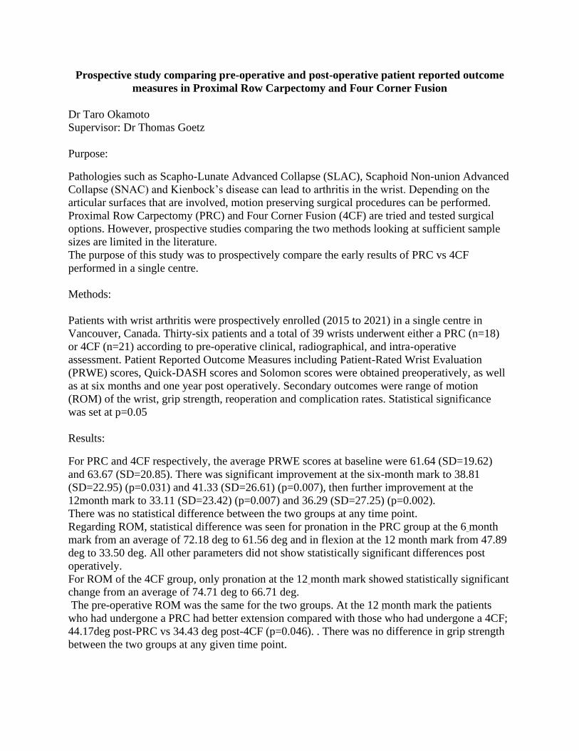
Prospective study comparing pre-operative and post-operative patient reported outcome
measures in Proximal Row Carpectomy and Four Corner Fusion
Dr Taro Okamoto
Supervisor: Dr Thomas Goetz
Purpose:
Pathologies such as Scapho-Lunate Advanced Collapse (SLAC), Scaphoid Non-union Advanced
Collapse (SNAC) and Kienbock’s disease can lead to arthritis in the wrist. Depending on the
articular surfaces that are involved, motion preserving surgical procedures can be performed.
Proximal Row Carpectomy (PRC) and Four Corner Fusion (4CF) are tried and tested surgical
options. However, prospective studies comparing the two methods looking at sufficient sample
sizes are limited in the literature.
The purpose of this study was to prospectively compare the early results of PRC vs 4CF
performed in a single centre.
Methods:
Patients with wrist arthritis were prospectively enrolled (2015 to 2021) in a single centre in
Vancouver, Canada. Thirty-six patients and a total of 39 wrists underwent either a PRC (n=18)
or 4CF (n=21) according to pre-operative clinical, radiographical, and intra-operative
assessment. Patient Reported Outcome Measures including Patient-Rated Wrist Evaluation
(PRWE) scores, Quick-DASH scores and Solomon scores were obtained preoperatively, as well
as at six months and one year post operatively. Secondary outcomes were range of motion
(ROM) of the wrist, grip strength, reoperation and complication rates. Statistical significance
was set at p=0.05
Results:
For PRC and 4CF respectively, the average PRWE scores at baseline were 61.64 (SD=19.62)
and 63.67 (SD=20.85). There was significant improvement at the six-month mark to 38.81
(SD=22.95) (p=0.031) and 41.33 (SD=26.61) (p=0.007), then further improvement at the
12month mark to 33.11 (SD=23.42) (p=0.007) and 36.29 (SD=27.25) (p=0.002).
There was no statistical difference between the two groups at any time point.
Regarding ROM, statistical difference was seen for pronation in the PRC group at the 6 month
mark from an average of 72.18 deg to 61.56 deg and in flexion at the 12 month mark from 47.89
deg to 33.50 deg. All other parameters did not show statistically significant differences post
operatively.
For ROM of the 4CF group, only pronation at the 12 month mark showed statistically significant
change from an average of 74.71 deg to 66.71 deg.
The pre-operative ROM was the same for the two groups. At the 12 month mark the patients
who had undergone a PRC had better extension compared with those who had undergone a 4CF;
44.17deg post-PRC vs 34.43 deg post-4CF (p=0.046). . There was no difference in grip strength
between the two groups at any given time point.
One patient in the 4CF group required a revision for delayed union, and three patients experienced
ulnar sided wrist pain.
Conclusion:
This analysis of outcomes for surgical interventions for arthritis of the wrist demonstrated
several clinically significant results that impact surgical planning and patient counselling. ROM
analysis showed that patients that underwent PRC had a mild reduction in wrist flexion and
patients who underwent 4CF had a reduction in pronation at 12 months post-operatively.
However, other ROM parameters were unchanged. Patients undergoing PRC and 4CF showed
significant improvement in post operative PRWE scores, consistent with the existing literature.
Regarding 4CF, care must be taken to minimize surgical and peri-operative factors that may
contribute to ulnar sided wrist pain given the increased rate of this complication seen in these
patients.
The Effect Of Sarcopenia On Early Mortality And Adverse Events After
Emergent Surgery For Spinal Fractures In Patients With Ankylosing
Spondylitis
Mohamed Al-Amoodi
Supervisor: Dr John Street
Purpose:
Frailty is considered to be a state of decreased resistance and resilience to internal and external
stressors and can occur independent to chronological age. Patients with chronic disease, such as
ankylosing spondylitis, have been found to be at a higher risk of deconditioning, loss of muscle
mass and frailty. Prior studies have attempted to evaluate sarcopaenia in a practical fashion by
using axial computed tomography (CT) scanning to measure the total area of the psoas muscle.
In order to quantify frailty, there have been multiple scoring systems developed, most notably the
modified Frailty Index, which have been shown to predict mortality and complications post
spinal surgery. While frailty is shown to be a predictor of complications and adverse outcomes in
patients undergoing spinal surgery, and recent work has demonstrated sarcopaenia to be a
predictor of mortality rates in patients with spinal metastases undergoing surgery, the use of
frailty and sarcopaenia to predict complications and mortality in patients with ankylosing
spondylitis undergoing surgery for spinal fractures remains unknown.
Hypothesis:
1. Internal validation study, which aims to show that sarcopaenia is a good measure of
frailty in patients with ankylosing spondylitis and that the disorder in itself is not a
confounder
2. Demonstration of a significant association between frailty, sarcopaenia and adverse
outcomes
Method:
Descriptive analysis will be conducted and the prevalence of sarcopaenia will be estimated with
95% confidence intervals. Linear regression modelling will be used for mortality. Logistic
regression will be used for dichotomous outcomes. Confounders (e.g. complexity of surgery, age
and sex) will be adjusted for in statistical analysis.
Cohort
Multicentre, retrospective. 1) Mater Misericordiae University Hospital in Ireland, 2) UBC
at VGH, and 3) U of T at Sunnybrook. Duration: 2009-2019. At UBC: 108 patients.
Inclusion: all patients who underwent emergency surgery with ankylosing spondylitis.
Exclusion: metastatic disease.
Update:
1. CREB approval granted
2. VCHRI approval granted

3. Data sharing agreement granted
4. We are in the same phase as the Irish site in data gathering phase. Presently, we plan to
extract from a database of spine patients from VGH: two groups of patients, those with
sarcopaenia and those without, after filtering out of a list of 1000s data those that don’t
have CT lumbar as it is essential for L3 psoas measurements. Dr Hong Ling can later use
the provided images to produce the sarcopaenia scores, this has been previously
established to be reliable interrater.
The role of frailty and sarcopenia in predicting major adverse events, length
of stay, reoperation and mortality following en bloc resection of primary bone
tumours and isolated metastases of the spine
Eryck Moskven, Oliver Lasry, Supriya Singh, Alana M. Flexman, John T. Street, Nicolas Dea,
Charles G. Fisher, Tamir Ailon, Marcel F. Dvorak, Brian K. Kwon, Scott Paquette, Raphaële
Charest-Morin
Supervisor: Dr. R. Charest-Morin
Introduction:
En bloc resection for primary bone tumours and isolated metastasis are complex surgeries
associated with a high rate of adverse events (AEs).
Objectives:
The primary objective of this study was to explore the relationship between frailty/sarcopenia
and major perioperative AEs following en bloc resection of primary bone tumours or metastases
of the spine. Secondary objectives were to report the prevalence and distribution of frailty and
sarcopenia; and determine the relationship between these factors and length of stay (LOS),
unplanned reoperation, and 1-year postoperative mortality in this population.
Methods:
This is a retrospective study of prospective data from a single quaternary care referral center
(January 1st, 2009, to February 28th, 2020) consisting of patients undergoing en bloc resection for
a primary bone tumour or an isolated spinal metastasis. Frailty was calculated with the modified
frailty index (mFI) and spine tumour frailty index (STFI). Sarcopenia, determined by the total
psoas area (TPA) / vertebral body area (VB) ratio (TPA/VB), was measured at L3 and L4. We
used multivariable regression analysis to quantify the association between frailty/sarcopenia and
major perioperative AEs, LOS, unplanned reoperation and 1-year postoperative mortality.
Results:
112 patients met the inclusion criteria. Using the mFI, 5 patients (5%) were frail (mFI 0.21),
while the STFI identified 21 patients (19%) as frail (STFI 2). The mean CT TPA/VB ratios
were 1.45 (SD 0.05) and 1.81 (SD 0.06) at L3 and L4, respectively. Unadjusted analysis
demonstrated that sarcopenia (defined by the CT L3 TPA/VB and CT L4 TPA/VB ratios) and
frailty (mFI and STFI) were not significant predictors of major perioperative AEs, increased
LOS or unplanned reoperation. Sarcopenia significantly predicted 1-year mortality (HR of 0.32
per unit increase, 95% CI 0.11-0.93, p=0.04 vs. HR of 0.28 per unit increase, 95% CI 0.11-0.69,
p=0.01). Frailty defined by an STFI score ≥ 2 predicted 1-year postoperative mortality (OR of
2.10, 95% CI 1.02-4.30, p=0.04).

Conclusions:
The mFI was not predictive of any clinical outcome in patients undergoing en bloc resection for
primary bone tumours of isolated metastases of the spine. Sarcopenia defined by the CT L3
TPA/VB and L4 TPA/VB and frailty assessed with the STFI predicted 1-year postoperative
mortality on unadjusted analysis but not major perioperative AEs, LOS or reoperation. Further
investigation is needed to characterize the relationship between frailty, sarcopenia and
perioperative outcomes in this spine surgery population.
Fully implantable, flexible optical probes for
neuromodulation of the spinal cord
Author: Shahriar Shalileh
Supervisor: Dena Shahriari
Purpose:
Optogenetics has proven to be a revolutionary tool in neural circuit interrogation and behavioural
neuroscience. Although it has been widely employed in brain circuitry studies, optogenetics has
not been broadly implemented in studying the spinal cord circuitry, due to hardware technical
impediments. Here, we present a flexible neural probe connected to a fully implantable,
autonomous optoelectronic system, to deliver light with controllable timing and lighting
parameters. The device eliminates conventional tethered optical fibers and wires and leaves
minimum interference with animals’ natural movement. Being rechargeable with off-the-shelf
components, the optoelectronic device is employed as a standalone system without the need for
the complex, expensive RF transmitters presented in the current battery-less manipulation methods
for the spinal cord. Moreover, our ultra-low-power device enables automatic awakening of the
device for chronic light stimulation studies without secondary surgeries on animal models. The
device, therefore, offers neuroscientists the freedom for time intervals, periods, frequency of
stimulation throughout the study.
Methods:
The final device comprises a four-layer rigid-flex printed circuit board (PCB) connected to a
flexible polyamide shank serving as a flexible optical probe. Four micro light-emitting diodes
(µLED) are arrayed in series at the end tip of the flexible probe. The optoelectronic system is
controlled by an ultra-low-power microcontroller. An external crystal resonator is provided in the
design running the calendar peripheral of the microcontroller to generate precise time steps. A
wireless power receiver system is designed to charge the battery. The whole system is insulated
via Paralyne-C coating and then a medical-grade epoxy.
Results:
Our optoelectronic device enables light emission from all the four µLEDs embedded on the flexible
neural probe for 30 minutes (45ms ON/5ms OFF) per day for 28 days with a single battery
implantable in rodent animal models. The battery is wirelessly recharged within 20 minutes.
Conclusion:
We have developed a flexible neural probe connected to a fully-implantable optoelectronic device
for chronic light delivery and stimulation of the spinal cord in freely moving animal models. Since
we developed our device from commercially available components, provided our circuit design
and algorithm, it can be readily fabricated and implemented by non-engineering scientists in spinal
cord circuitry and behavioural neuroscience to study neural relays. Our device is therefore
extending the optogenetics toolkit from the widely used brain studies to spinal cord research.
Influence of Baseline Blood Pressure on Neuromonitoring Alerts
in Adolescent Idiopathic Scoliosis
Sam Kirk, Otis Shirley, Paul Rushton, Firoz Miyanji, Arvindera Ghag
Introduction:
Spinal cord injury is the most feared of all complications in paediatric spinal surgery. Secondary to
this Multimodal Neuromonitoring (NM) combining motor evoked potentials (MEPs) and
somatosensory evokes potentials (SSEPs) has become the standard of care. This can be considered
an ‘early warning system’, allowing pathological changes in NM to be responded to in a timely
manner, and lasting neurological injury avoided. Neuromonitoring alerts are reported when there is
a reduction in amplitude of the MEPs/SSEPS to <50% when compared to the baseline readings.
While there is an understanding that there is a relationship between intraoperative blood pressure
and alerts; the relationship between the blood pressure at baseline and the frequency of alerts has
not been studied.
Method:
A retrospective chart review of 76 adolescent idiopathic scoliosis cases was performed. Data was
collected from the medical notes, neuromonitoring data set and radiographs reviewed. Inclusion
criteria included a single surgeon, single stage posterior correction, use of traction and adequate
data. Exclusion criteria included non-idiopathic scoliosis, anterior or staged surgery and vertebral
column resection. Patients were placed into three groups; no alerts, 1-2 alerts or 3+ alerts and their
relationship to both the baseline blood pressure and the difference in the baseline blood pressure to
their pre-op blood pressure was evaluated. Adjustment due to possible confounding due to the
magnitude of the major curve, the percentage of deformity correction, surgical time, instrumented
levels and blood loss was performed.
Results:
A 5 unit increase in MAP gives an adjusted odds ratio of being in a higher alert group of 1.29 (1.03,
1.66) and a 5 unit decrease in the difference between the admission day MAP and the baseline map
also gives an adjusted odds ratio of being in a higher alert group of 1.29 (1.03, 1.66)
Conclusion:
This paper demonstrates an association between higher baseline MAP and the likelihood of
increased alerts during a posterior instrumented fusion in adolescent idiopathic scoliosis. A
prospective study looking at the affect of controlling MAP prior to setting baselines could confirm a
causal relationship and help to reduce the frequency of unhelpful alerts during surgery.
Sex differences in outcomes after hip arthroscopy:
a systematic review and meta-analysis
Helen Crofts
Supervisor: Dr. Parth Lodhia
Purpose:
To assess differences in outcomes between males and females following hip arthroscopy.
Methods:
A systematic review was performed following the Preferred Reporting Items for Systematic
Reviews and Meta-Analysis (PRISMA) guidelines. Medline, Embase, Cochrane and PubMed
databases were searched in December of 2021. Keywords included “hip,” “arthroscopy,”
“outcome,” “gender difference,” “sex difference,” “gender,” and “patient reported outcome.”
Studies were included that reported sex specific analysis of outcomes following primary hip
arthroscopy. Methodological Index for Non-Randomized Studies (MINORS) criteria was applied
to each study. Data collected included patient reported outcome measures (PROMs),
complications, rates of revision arthroscopy (RA) and conversion to total hip arthroplasty (THA).
A meta-analysis was performed for the most frequently reported PROMs, RA and THA rates.
Results:
48 studies met the inclusion criteria. There were 58544 (54% female) hips included. Males had
significantly higher postoperative Hip Outcome Score- Sport Specific Subscale (HOS-SSS)
scores than females on meta-analysis (mean difference (MD) 4.93, CI [ 0.07; 9.79]). Males had
lower odds of reaching the minimal clinically important difference (MCID) for the Modified
Harris Hip Score (mHHS) (OR 0.69, CI [0.53; 0.89]) but not the patient acceptable symptom
state (PASS) (OR 1.36, CI [0.94; 1.96]). Males were at lower odds of reaching both the MCID
(OR 0.53, CI [0.32; 0.87]) and PASS (OR 0.55, CI [0.35; 0.84]) for the HOS-SSS. Females were
more likely to undergo RA (OR 0.51, CI [0.36; 0.72]) and there was no observed difference
between sexes for conversion to THA (OR 1.10, CI [0.79; 1.52]). There were no differences
between sexes in complication rates.
Conclusion:
There was no significant difference between sexes for post-operative PROM scores, except for
the HOS-SSS where females scored significantly lower. Males were less likely to reach the
MCID for the mHHS and HOS-SSS than females but there was no clear trend for PASS rates.
Females were more likely to require RA. There was no difference between sexes in conversion to
THA. Complication rates post hip arthroscopy were low in both sexes.
Prophylactic augmentation to prevent age-related hip fracture:
preliminary biomechanical results
EK Bliven, A Fung, J Levine, I Fleps, B Helgason, P Cripton, P Guy
Introduction:
Hip fracture is a devastating injury with increasing prevalence and high mortality rates. Many
elderly individuals are especially fragile and susceptible to secondary hip fracture, which bears
even higher consequences in the form of death and disability. Prophylactic augmentation with
orthopaedic implants has been used clinically to prevent skeletal injury from conditions like
slipped capital epiphysis or metastatic lesions. Here we propose implanting an intramedullary
nail and lag screw in the femur to protect against age-related hip fracture in vulnerable
candidates. This work investigates the efficacy of this approach in a biomechanical test set-up
that simulates a sideways fall from standing height, a common scenario for hip fracture.
Methods:
We use a previously developed dual experimental-computational method that consists of an
inverse pendulum impactor and a corresponding finite element model (Fleps et al., 2019). A
fellowship-trained orthopaedic surgeon (PG) augmented the impacting (left) femur of each
specimen with a titanium intramedullary nail and lag screw. Per the experimental protocol, each
cadaveric pelvis-femur specimens is moulded in a subject-specific gelatin soft tissue shape and
guided in an inertia-driven fall onto a force plate. The validated computational model uses CT
scan data of the native specimen to predict bony areas with high strain indicating fracture
location, which we then compare to the experimental fracture outcome of the augmented
specimen. PG inspected each specimen and classified fracture severity after the fall impact.
Results:
Our preliminary results to date include two specimens. For each specimen, the computational
model simulated an impact with the native femur and the experiment was conducted with an
augmented femur. The experimental outcome of specimen 1 (female, 81 y.o.) aligned with the
computational prediction: a lateral compression fracture of the pelvis on the impacted side (left
pelvic ramus and ischium). Specimen 2 (female, 63 y.o.) did not show signs of fracture after
testing, whereas the computational model predicted a femoral neck fracture.
Conclusion:
When exposed to a realistic sideways fall impact, the augmented specimens did not show signs
of hip fracture in the experiment, despite this outcome being predicted by the simulations in one
of the cases. An outcome of pelvic fracture after a sideways fall (as in Specimen 1) may be seen
as a “preferred” alternative to hip fracture clinically, due to the less invasive treatment and lower

associated complication rates. These preliminary biomechanical results suggest that augmenting
the proximal femur with an intramedullary nail and lag screw may show promise in mitigating
hip fracture.
Retrospective review of distal femoral fractures treated at RCH
Charles Bouchard
Supervisor: Dr. Darius Viskontas
Purpose:
The purpose of this study was to compare the re-operation rate and union rates of various
constructs used in the surgical fixation of native and periprosthetic distal femoral fractures.
Methods:
Retrospective review for patients treated with operative fixation of the distal femur between
2016 and 2022 at the Royal Columbian Hospital were identified using population level
administrative data. Radiographic screening and chart review of these patients was carried out to
identify those treated with their initial construct: locking or nonlocking lateral plating, dual
lateral/medial plating, intramedullary nails, plate/nail combinations, and far cortical locking
plates. Radiographic review of fracture patterns was also characterized. Chart reviews followed
to determine the indications for re-operation and other post-operative complications. Main
outcome measures were re-operation rate and their indications.
Results:
From 2016-2022, 115 patients with 117 distal femoral fractures were operated on with one of the
above initial constructs: 83 lateral plating (70.9%), 9 dual plates (7.7%), 13 intramedullary nail
(11.1%), 2 plate-nail combination (1.7%), 7 far cortical locking plating (6.0%). Chart review
determined 16 patients had re-operations demonstrating a 13.7% re-operation rate. The dual plate
group had the highest re-operation rate (44.4%). In the lateral plating group, 6 patients (7.2%)
went on to develop non-union. Radiographic review of the fractures demonstrates that those who
went on to develop non-unions tended to have evidence of medial comminution on injury films.
Complete results pending.
Conclusion:
Patients requiring re-operation for distal femoral fractures can depend on several factors.
Radiographically, insight may be drawn as to which patients may require supplementation or
augmentation based on initial injury films.
Complete conclusion pending.
The Association Between Increased Posterior Tibial Slope and
Native Anterior Cruciate Ligament Injury.
Yasir AlShehri
Supervisor: Dr. Andrea Veljkovic
Abstract:
The posterior inclination of the tibia plateau relative to the longitudinal axis of the tibia is
referred to as the Posterior Tibial Slope (PTS). It is well known that increased posterior tibial
slope is associated with higher anterior cruciate ligament reconstruction (ACLR) failure rate. A
less commonly studied association is that between increased PTS and primary ACL injury. The
purpose of this study is to examine and summarize that association, and to determine whether or
not there is a defined "at risk" posterior tibial slope. In this systematic review, we reviewed
publications in PubMed to identify all studies reporting an incidence of increased tibial plateau
slope in ACL-injured groups. Although an increased risk of primary ACL injury with increased
posterior tibial slope has been reported in some studies, there is a large disagreement regarding
the actual values of the posterior tibial slope that would be considered ‘‘at risk.’’ While trends in
the current literature indicate a possible relationship between ACL injury and posterior tibial
slope, standardized techniques and more consistent and repeatable data are required to
definitively link the two.
Highly crosslinked polyethylene liner thickness in THA does not influence
long-term survival: A retrospective cohort study with minimum 11 years
follow-up
BL Fransen, LC Howard, T MacDonell, F Bengoa, DS Garbuz, G Sheridan
Supervisor: ME Neufeld
Introduction:
Increased femoral head size reduces the rate of dislocation after total hip arthroplasty (THA).
With the introduction of highly crosslinked polyethylene (HXLPE) liners in THA there has been
a trend towards using larger size femoral heads in relatively smaller cup sizes, theoretically
increasing the risk of liner fracture, wear, or aseptic loosening. Short to medium follow-up
studies have not demonstrated a negative effect of using thinner HXLPE liners. However, there
is concern that these thinner liners may prematurely fail in the long-term. The aim of this study
was to evaluate the long-term survival and revision rates of HXLPE liners in primary THA, as
well as the effect of liner thickness on these outcomes. We hypothesized that there would be no
significant differences between the different liner thicknesses.
Method:
We performed a retrospective database analysis from a single center of all primary total hip
replacements using HXLPE liners from 2010 and earlier. All procedures were performed by
fellowship trained arthroplasty surgeons. Patient characteristics, implant details including liner
thickness, death, and revisions were recorded. Patients were grouped for analysis for each
millimeter of PE thickness. Kaplan-Meier survival estimates were estimated with all-cause and
aseptic revisions as endpoints.
Results:
In total, 2354 patients (2584 hips; 47.6% female) were included (mean age 64.3 years). Mean
follow-up was 13.2 years (range 11.0-18.8). Liner thickness varied from 4.9 to 12.7 mm. Seven
patients had a liner thickness <5.0mm and 859 had a liner thickness of <6.0mm. Head sizes were
28mm (n=85, 3.3%), 32mm (n=1214, 47.0%), 36mm (n=1176, 45.5%), and 40mm (n=109,
4.2%), and 98.4% were metal heads. There were 101 revisions, and in 78 of these cases the liner
was revised. Reason for revision was instability/dislocation (n=34), pseudotumor/aseptic
lymphocyte-dominant vasculitis associated lesion (n=18), fracture (n=17), early loosening
(n=11), infection (n=7), aseptic loosening (n=4), and other (n=10). When grouped by liner
thickness, there were no significant differences between the groups when looking at all-cause
revision (p=0.112) or aseptic revision (p=0.116).
Conclusion:
In our cohort, there were no significant differences in all-cause or aseptic revisions between any
of the liner thickness groups at long-term follow-up. Our results indicate that using thinner
HXPE liners to maximize femoral head size in THA does not lead to increased complications or

liner failures at medium to long term follow-up. As such, orthopedic surgeons can consider the
use of larger heads at the cost of liner thickness a safe practice to reduce the risk of dislocation
after THA when using HXLPE liners.
Analysis of thin highly cross-linked polyethylene liners combined with big
femoral heads in primary total hip arthroplasty shows excellent survival and
low wear rates at a mean follow-up of 10 years
BL Fransen, F Bengoa, ME Neufeld, G Sheridan, DS Garbuz, LC Howard
Purpose:
With the introduction of highly crosslinked polyethylene (HXLPE) in total hip arthroplasty
(THA), orthopaedic surgeons have moved towards using larger femoral heads at the cost of
thinner liners to decrease the risk of instability. Several short and mid-term studies have shown
minimal liner wear with the use of HXLPE liners, but the safety of using thinner HXLPE liners
to maximize femoral head size remains uncertain and concerns that this may lead to premature
failure exist.
Method:
Between 2000 and 2010, we retrospectively identified 55 patients that underwent a primary THA
performed in a high-volume single tertiary referral center using HXLPE liners with 36-mm or
larger heads in cups with an outer diameter of or 52-mm or smaller. Patient characteristics,
implant details including liner thickness, death, complications, and all cause revisions were
recorded. Patients that had a minimum radiographic follow-up of seven years were assessed
radiographically for linear and volumetric wear. Wear was calculated using a validated open-
source software by two independent researchers.
Results:
A total of 55 patients were identified and included, with a mean age of 74.8 (range 38.67 - 95.9)
years and a mean BMI of 28.98 (range 18.87 - 63-68). Fifty-one (94.4%) of patients were
female. Three patients were revised, none for liner wear, fracture or dissociation. Twenty-two
patients had a
radiographic follow-up of minimum seven years (mean 9.9 years, min-max 7.5 – 13.7) and were
included in the long-term radiographic analysis. Mean linear liner wear was 0.085 mm/year and
mean volumetric wear was 11.097 mm3/year.
Conclusion:
Using HXLPE liners with 36-mm heads or bigger in 52-mm cups or smaller is safe, with low
rates of linear and volumetric wear in the mid to long-term follow-up. Patients did not require
revision surgery for liner complications, including liner fracture, dissociation, or wear. Our
results suggest that the advantages of using larger heads should outweigh the potential risks of
using thin HXLPE liners.
Anatomic Variation in Osteoblast Function and its Implications for Joint
Arthroplasty
Erden Ali
Supervisor: Dr. R. Brooks. University of Cambridge
Hypothesis:
Accumulated evidence indicates that local cell origins may ingrain differences in the phenotypic
activity of human osteoblasts. These differences may also exist in osteoblasts harvested from the
same bone type at periarticular sites, including those adjacent to the fixation sites for total joint
implant components.
Methods:
Human osteoblasts were obtained from the acetabulum and femoral neck of seven patients
undergoing total hip arthroplasty and from the femoral and tibial cuts of six patients undergoing
total knee arthroplasty. Osteoblasts were extracted from the usually discarded bone via enzyme
digestion, characterized by flow cytometry, and cultured to passage three before measurement of
metabolic activity, collagen production, alkaline phosphatase (ALP) expression, and
mineralization.
Results:
Osteoblasts from the acetabulum showed lower proliferation (p = 0.034), cumulative collagen
release (p < 0.001), and ALP expression (p = 0.009), and produced less mineral (p = 0.006) than
those from the femoral neck. Osteoblasts from the tibia produced significantly less collagen (p =
0.021) and showed lower ALP expression than those from the distal femur.
Conclusion:
The results have demonstrated for the first time an anatomical regional variation in the biological
behaviours of osteoblasts on either side of the hip and knee joint. The lower osteoblast
proliferation, matrix production, and mineralization from the acetabulum compared to those from
the proximal femur may be reflected in differences in bone formation and implant fixation at
these sites.
Identification of Protective and Risky HLA Genotypes for the Development
of Pseudotumours Around Hip Resurfacing – A Case-Control Study
All Authors:
Gerard Sheridan, University of British Columbia (Primary Presenter)
Michael Hanlon
Adanna Welch-Phillips
Karen Spratt, National Orthopaedic Hospital Cappagh, Dublin, Ireland.
Richard Hagan
John M O’Byrne, National Orthopaedic Hospital Dublin
Paddy Kenny
Andrew Kurmis
Conor Hurson, St. Vincent's University Hospital
Bassam A. Masri, Department of Orthopaedics, UBC
Donald Garbuz, Department of Orthopaedics, UBC
Purpose:
Hip resurfacing may be a useful surgical procedure when patient selection is correct and only
implants with superior performance are used. In order to establish a body of evidence in
relation to hip resurfacing, pseudotumour formation and its genetic predisposition, we
performed a case-control study investigating the role of HLA genotype in the development of
pseudotumour around MoM hip resurfacings.
Method:
All metal-on-metal (MoM) hip resurfacings performed in the history of the institution were
assessed. A total of 392 hip resurfacings were performed by 12 surgeons between February
1st 2005 and October 31st 2007. In all cases, pseudotumour was confirmed in the
preoperative setting on Metal Artefact Reduction Sequencing (MARS) MRI. Controls were
matched by implant (ASR or BHR) and absence of pseudotumour was confirmed on MRI.
Blood samples from all cases and controls underwent genetic analysis using Next Generation
Sequencing (NGS) assessing for the following alleles of 11 HLA loci (A, B, C, DRB1,
DRB3/4/5, DQA1, DQB1, DPB1, DPA1). Statistical significance was determined using a
Fisher’s exact test or Chi-Squared test given the small sample size to quantify the clinical
association between HLA genotype and the need for revision surgery due to pseudotumour.
Results:
Both groups were matched for implant type (55% ASR, 45% BHR in both the case and
control groups). According to the ALVAL histological classification described by Kurmis et
al., the majority of cases (63%, n=10) were found to have group 2 histological findings. Four
cases (25%) had group 3 histological findings and 2 (12%) patients had group 4 findings. Of
the 11 HLA loci analysed, 2 were significantly associated with a higher risk of pseudotumour
formation (DQB1*05:03:01
and DRB1*14:54:01) and 4 were noted to be protective against pseudotumour formation
(DQA1*03:01:01, DRB1*04:04:01, C*01:02:01, B*27:05:02).

Conclusion:
These findings further develop the knowledge base around specific HLA genotypes and their
role in the development of pseudotumour formation in MoM hip resurfacing. Specifically, the
two alleles at higher risk of pseudotumour formation (DQB1*05:03:01 and DRB1*14:54:01)
in MoM hip resurfacing should be noted, particularly as patient-specific genotype-dependent
surgical treatments continue to develop in the future.
dGEMRIC T1 is Reduced in Cartilage Overlying
Bone Marrow Lesions in the Hip
Presenter: Carly Jones
Supervisor: Dr. David Wilson
Purpose:
Bone marrow lesions (BML) are associated with painful and progressive OA. Quantitative MRI
has found evidence of early cartilage degeneration overlying BMLs in knees, but similar work
has not been done in the hip. The purpose of this study is to determine if there is evidence of
cartilage degeneration in BML overlying cartilage in hips.
Method:
MRI study participants (n=128) were recruited from a cross-sectional population-based study of
adults aged 20-49 years. Delayed Gadolinium Enhanced MRI of Cartilage (dGEMRIC) and
proton-density weighted fat-suppressed (PDw-FS) MRI scans were acquired of one hip for each
participant. dGEMRIC is a well-validated method of quantifying cartilage proteoglycan content
using MRI. BMLs were identified from PDw-FS scans by a MSK radiologist. BMLs were
segmented semi-automatically from the PDw-FS scans, and acetabular and femoral cartilage
were segmented manually from the dGEMRIC scans. The PDw-FS and dGEMRIC images were
rigidly registered using landmark points to align the cartilage with the BML locations. The
outlines of femoral and acetabular BMLs were projected onto their respective cartilage surfaces
to define the BML overlying cartilage (OC) and surrounding cartilage (SC). Age and sex
matched controls were identified for each BML subject to control for natural variations in
dGEMRIC. Mean dGEMRIC was calculated for the OC and SC for each BML subject and
matched control. We applied a linear mixed-effects model to compare the change score
(difference in mean dGEMRIC between OC and SC) between the BML and control group. The
model was adjusted for BMI, physical activity (MET), FAI, hip pain, and location of
measurement (acetabular/femoral).
Results:
Thirty-two of the 128 participants had at least one BML. There was no significant difference
between the BML subjects and the age and sex-matched controls for age (p=0.95), BMI
(p=0.91), MET (p=0.36), and hip pain (p=0.80). The BML group had significantly higher rates
of FAI (65.6% for BML subjects, 37.5% for matched controls, p=0.02).
The change score between mean dGEMRIC in OC and SC was significantly larger in the BML
subjects compared to the matched controls (change score of BML subjects minus the change
score of matched controls): -80.11ms (95% CI: -157.88, -2.33), p=0.04.
Conclusion:
Our result that the change score for BML subjects is significantly larger than the change score
for the matched controls suggests that cartilage overlying BMLs is relatively less healthy than
cartilage not overlying BMLs. This result is consisted with previous research of knees with
BMLs using quantitative MRI.
We found that BMLs in the hip in this cross-sectional population are associated with reduced
cartilage health in the OC when compared to the SC. This work further supports that BMLs are
an important factor in osteoarthritis progression.
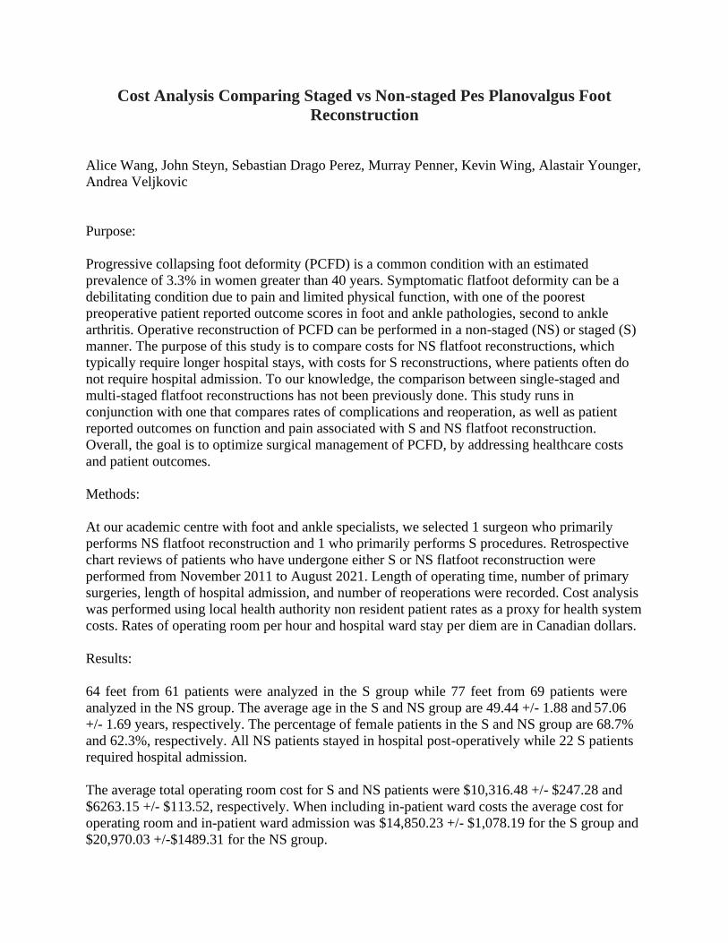
Cost Analysis Comparing Staged vs Non-staged Pes Planovalgus Foot
Reconstruction
Alice Wang, John Steyn, Sebastian Drago Perez, Murray Penner, Kevin Wing, Alastair Younger,
Andrea Veljkovic
Purpose:
Progressive collapsing foot deformity (PCFD) is a common condition with an estimated
prevalence of 3.3% in women greater than 40 years. Symptomatic flatfoot deformity can be a
debilitating condition due to pain and limited physical function, with one of the poorest
preoperative patient reported outcome scores in foot and ankle pathologies, second to ankle
arthritis. Operative reconstruction of PCFD can be performed in a non-staged (NS) or staged (S)
manner. The purpose of this study is to compare costs for NS flatfoot reconstructions, which
typically require longer hospital stays, with costs for S reconstructions, where patients often do
not require hospital admission. To our knowledge, the comparison between single-staged and
multi-staged flatfoot reconstructions has not been previously done. This study runs in
conjunction with one that compares rates of complications and reoperation, as well as patient
reported outcomes on function and pain associated with S and NS flatfoot reconstruction.
Overall, the goal is to optimize surgical management of PCFD, by addressing healthcare costs
and patient outcomes.
Methods:
At our academic centre with foot and ankle specialists, we selected 1 surgeon who primarily
performs NS flatfoot reconstruction and 1 who primarily performs S procedures. Retrospective
chart reviews of patients who have undergone either S or NS flatfoot reconstruction were
performed from November 2011 to August 2021. Length of operating time, number of primary
surgeries, length of hospital admission, and number of reoperations were recorded. Cost analysis
was performed using local health authority non resident patient rates as a proxy for health system
costs. Rates of operating room per hour and hospital ward stay per diem are in Canadian dollars.
Results:
64 feet from 61 patients were analyzed in the S group while 77 feet from 69 patients were
analyzed in the NS group. The average age in the S and NS group are 49.44 +/- 1.88 and 57.06
+/- 1.69 years, respectively. The percentage of female patients in the S and NS group are 68.7%
and 62.3%, respectively. All NS patients stayed in hospital post-operatively while 22 S patients
required hospital admission.
The average total operating room cost for S and NS patients were $10,316.48 +/- $247.28 and
$6263.15 +/- $113.52, respectively. When including in-patient ward costs the average cost for
operating room and in-patient ward admission was $14,850.23 +/- $1,078.19 for the S group and
$20,970.03 +/-$1489.31 for the NS group.

Conclusion:
Preliminary cost analysis suggests that multi-staged flatfoot reconstruction costs less than single-
staged flatfoot reconstruction. Once full assessment is complete with statistical analysis,
correlation with patient reported outcomes and complication rate can guide future PCFD surgical
management.
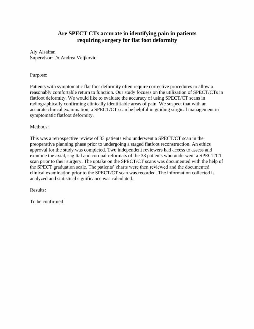
Are SPECT CTs accurate in identifying pain in patients
requiring surgery for flat foot deformity
Aly Alsaifan
Supervisor: Dr Andrea Veljkovic
Purpose:
Patients with symptomatic flat foot deformity often require corrective procedures to allow a
reasonably comfortable return to function. Our study focuses on the utilization of SPECT/CTs in
flatfoot deformity. We would like to evaluate the accuracy of using SPECT/CT scans in
radiographically confirming clinically identifiable areas of pain. We suspect that with an
accurate clinical examination, a SPECT/CT scan be helpful in guiding surgical management in
symptomatic flatfoot deformity.
Methods:
This was a retrospective review of 33 patients who underwent a SPECT/CT scan in the
preoperative planning phase prior to undergoing a staged flatfoot reconstruction. An ethics
approval for the study was completed. Two independent reviewers had access to assess and
examine the axial, sagittal and coronal reformats of the 33 patients who underwent a SPECT/CT
scan prior to their surgery. The uptake on the SPECT/CT scans was documented with the help of
the SPECT graduation scale. The patients’ charts were then reviewed and the documented
clinical examination prior to the SPECT/CT scan was recorded. The information collected is
analyzed and statistical significance was calculated.
Results:
To be confirmed
Dana Mohammad (PGY1)
The Association Between Pain Catastrophization and Functional Outcome In Post-Progressive Collapsing Foot Deformity Reconstruction Patients
(Co-Author – A Veljkovic)
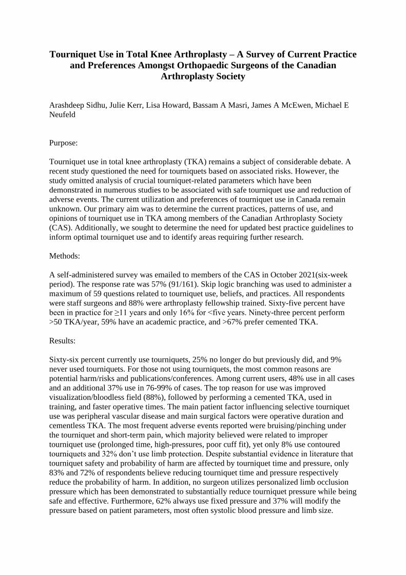
Tourniquet Use in Total Knee Arthroplasty – A Survey of Current Practice
and Preferences Amongst Orthopaedic Surgeons of the Canadian
Arthroplasty Society
Arashdeep Sidhu, Julie Kerr, Lisa Howard, Bassam A Masri, James A McEwen, Michael E
Neufeld
Purpose:
Tourniquet use in total knee arthroplasty (TKA) remains a subject of considerable debate. A
recent study questioned the need for tourniquets based on associated risks. However, the
study omitted analysis of crucial tourniquet-related parameters which have been
demonstrated in numerous studies to be associated with safe tourniquet use and reduction of
adverse events. The current utilization and preferences of tourniquet use in Canada remain
unknown. Our primary aim was to determine the current practices, patterns of use, and
opinions of tourniquet use in TKA among members of the Canadian Arthroplasty Society
(CAS). Additionally, we sought to determine the need for updated best practice guidelines to
inform optimal tourniquet use and to identify areas requiring further research.
Methods:
A self-administered survey was emailed to members of the CAS in October 2021(six-week
period). The response rate was 57% (91/161). Skip logic branching was used to administer a
maximum of 59 questions related to tourniquet use, beliefs, and practices. All respondents
were staff surgeons and 88% were arthroplasty fellowship trained. Sixty-five percent have
been in practice for ≥11 years and only 16% for <five years. Ninety-three percent perform
>50 TKA/year, 59% have an academic practice, and >67% prefer cemented TKA.
Results:
Sixty-six percent currently use tourniquets, 25% no longer do but previously did, and 9%
never used tourniquets. For those not using tourniquets, the most common reasons are
potential harm/risks and publications/conferences. Among current users, 48% use in all cases
and an additional 37% use in 76-99% of cases. The top reason for use was improved
visualization/bloodless field (88%), followed by performing a cemented TKA, used in
training, and faster operative times. The main patient factor influencing selective tourniquet
use was peripheral vascular disease and main surgical factors were operative duration and
cementless TKA. The most frequent adverse events reported were bruising/pinching under
the tourniquet and short-term pain, which majority believed were related to improper
tourniquet use (prolonged time, high-pressures, poor cuff fit), yet only 8% use contoured
tourniquets and 32% don’t use limb protection. Despite substantial evidence in literature that
tourniquet safety and probability of harm are affected by tourniquet time and pressure, only
83% and 72% of respondents believe reducing tourniquet time and pressure respectively
reduce the probability of harm. In addition, no surgeon utilizes personalized limb occlusion
pressure which has been demonstrated to substantially reduce tourniquet pressure while being
safe and effective. Furthermore, 62% always use fixed pressure and 37% will modify the
pressure based on patient parameters, most often systolic blood pressure and limb size.

Almost all (88%) were interested in new evidence-based guidelines regarding these
parameters.
Conclusion:
Tourniquet use in TKA remains prevalent among arthroplasty surgeons in the CAS; however
tremendous practice variability regarding several key parameters required for optimal use
exists. Current best practices of tourniquet use regarding personalized pressures, time, and
type are not being utilized across Canada. There is considerable interest and need for further
research and updated guidelines regarding key parameters of safe tourniquet usage to
optimize tourniquet use in TKA.
Design of a Pneumatic Cartilage Loading Rig for
Magnetic Resonance Imaging
Jessica C. Kupper, Emily Sullivan, Robin Coope
Supervisor: David Wilson
Introduction:
In spite of the prevalence of osteoarthritis (OA) and the general acceptance that OA initiates due
to mechanical factors (Andriacchi and Mündermann, 2006), it is unclear what mechanical
changes are important. One variable of interest is cartilage strain, which is potentially relevant
to OA cartilage damage (Wilson et al., 2009) and a potential imaging biomarker (Neu, 2014).
To further investigate strain, magnetic resonance (MR) imaging is one non-invasive option.
Loading via a compression device is applied to tissue samples and the resulting strain and
change in the imaging parameter of interest can be recorded (e.g., T2 relaxometry).
Compression devices that are designed specifically for MR applications must be MR compatible
(i.e., non-ferromagnetic) and validated for force and displacement capabilities.
Purpose:
To design and validate an MR-compatible loading rig for measurement of increasing levels of
strain (0.1-0.3) in cartilage-on-cartilage samples.
Methods:
A cartilage compression actuator (CCA) was designed to allow for set displacements [Fiber
Bragg Grating (FBG) sensor] under pneumatic control. A 1110 N load cell (Sensotec Model 75,
Honeywell, Charlotte, NC, USA) was attached rigidly to a Delrin rod 8.5 cm long, which was
placed against the CCA distal actuator. Pressure was applied from 0 to 550 kPa incrementally,
and force values were collected by a controller (8800, Instron, Norwood, MA, USA). This was
completed three times to assess repeatability [coefficient of variation (COV)], and at a set
pressure level for 30 minutes to record force values over time (175 kPa, 345 kPa, and 515 kPa,
three times). Calibration of the FBG sensor against displacement was completed using calipers at
the distal actuator end under increasing pressure levels.
Results:
The force vs. pressure relationship was best fit with a natural logarithmic fit (R2=0.99). A
change in 1 kPa of pressure resulted in an output force of 2.0 N at lower pressures and 0.6 N at
higher pressures. The FBG wavelength vs. displacement relationship demonstrated a highly
linear fit (R2=0.99), with a change in 0.01 mm of displacement resulting in a wavelength change
of -0.044 nm. Over 30 minutes the FBG wavelength changed by 0.12-0.17 nm, equivalent to
strain changes of 0.005-0.006. The COVs for force and FBG wavelength were 1.63%, and
0.01%, respectively.
Conclusions:
With sufficiently repeatable displacement feedback and remote operation capabilities, the MR-
compatible CCA enables detailed investigation into strain and cartilage health relationships.
These relationships could inform future in vivo research for understanding the biomechanics that
lead to cartilage degeneration in OA, leading to possible improvements in prevention and
treatment.
Acknowledgments:
Scott Young and Jugoslav Kitanovic (BC Cancer Medical Physics), NSERC, MSFHR, The
Arthritis Society
Long term recovery trajectory of patient reported outcomes
following acetabular fractures
Adam Tucker, Henry M Broekhuyse, Pierre Guy, Jeffrey M Potter, Darren M Roffey, Kelly
A Lefaivre
Purpose:
Acetabular fractures are associated with long term morbidity. This prospective cohort study
seeks to understand the recovery trajectory over 5 years.
Methods:
Longitudinal follow-up of surgical acetabular fractures occurred between 2004-2019. The
36-Item Short-Form Health Survey (SF-36) physical component score (PCS) was recorded at
6,12,24 and 60 months post-operatively. Comparative analyses were performed for
elementary and associated fracture patterns. The minimal clinically important difference
(MCID) and patients achieving this change was determined. The rate of, and time to,
conversion to total hip arthroplasty (THA) is reported.
Results:
We identified 253 fractures (251 patients), with a 4:1 male preponderance and mean age of
46.1±16.4 years. Associated fracture patterns accounted for 143/253 (56.5%). Trajectory
analysis showed all time points had significant disability versus baseline (p<0.001; Figure 1).
Elementary fractures had higher SF-36 PCS at 6 (p=0.023) and 12 (p=0.007) months
compared to associated subtypes, but not at 2 (p=0.135) or 5 years (p=0.631). A total of
37.3% of patients improved at least 1 MCID between 6 months and 1 year, 26.9% between 1
and 2 years, and 23.2% between 2 and 5 years. A significant proportion failed to return
within 1 MCID of baseline at 2 (70%) and 5 years (65%). Conversion to THA occurred in
13.1% patients: 11/110 elementary and 22/143 associated fractures. Approximately two
thirds (63.6%) were performed within 2 years of index surgery.
Conclusion:
Acetabular fractures significantly impact physical function and recovery trajectory is long –
often beyond 1 year. There were 65% of patients who had persistent clinically relevant
disability long term.
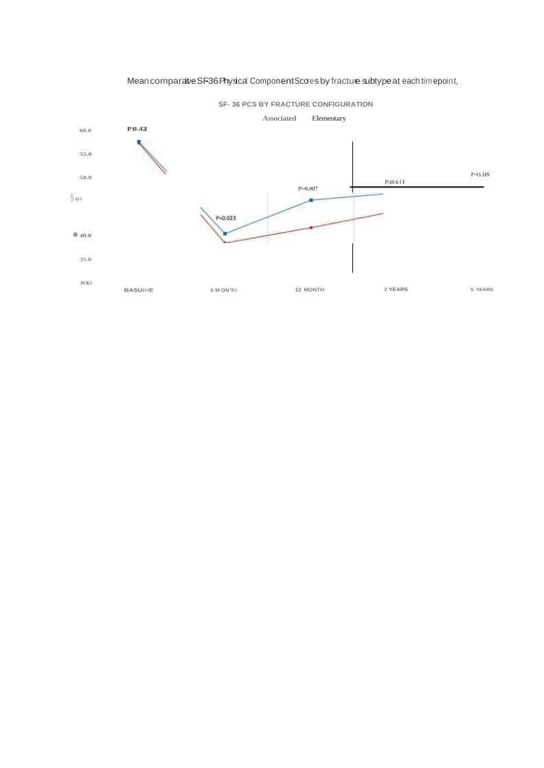
Mean comparaivt eSF-36Physical ComponentScores by fracture subtypeat each tim epoint,
60.0 P:0.421
55.0
50.0
§ 45 0
SF- 36 PCS BY FRACTURE CONFIGURATION
Associated Elementary
• '
40.0
35.0
JOO.
BASUIHE
6 M ON 'fH 12 MONTH
2 YEARS S YEARS
P:i0.6 l l
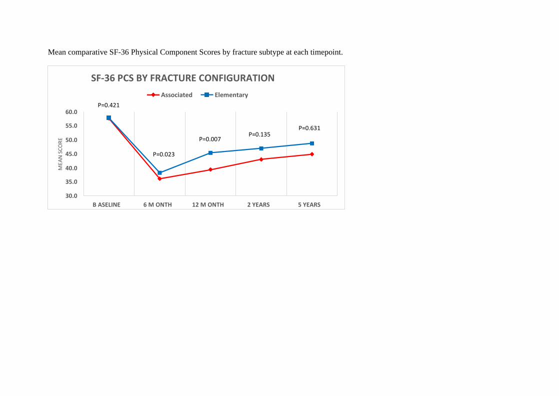
Mean comparative SF-36 Physical Component Scores by fracture subtype at each timepoint.
SF-36 PCS BY FRACTURE CONFIGURATION
Associated Elementary
P=0.421 60.0
55.0 P=0.631 P=0.135
50.0 P=0.007
45.0 P=0.023
40.0
35.0
30.0
B ASELINE 6 M ONTH 12 M ONTH 2 YEARS 5 YEARS
MEA
N S
CO
RE
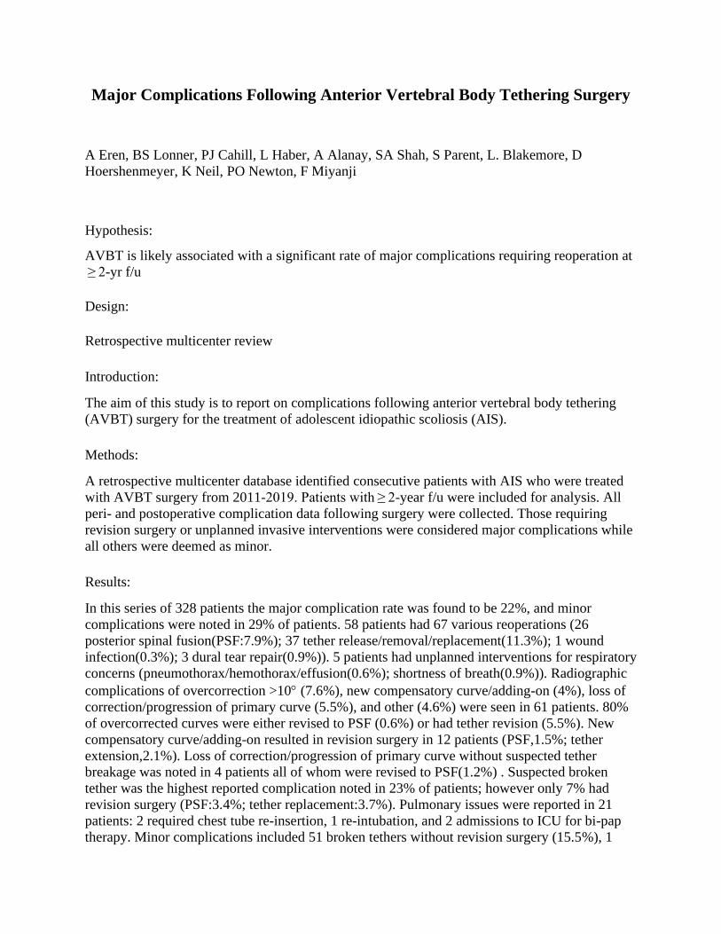
Major Complications Following Anterior Vertebral Body Tethering Surgery
A Eren, BS Lonner, PJ Cahill, L Haber, A Alanay, SA Shah, S Parent, L. Blakemore, D
Hoershenmeyer, K Neil, PO Newton, F Miyanji
Hypothesis:
AVBT is likely associated with a significant rate of major complications requiring reoperation at
≥ 2-yr f/u
Design:
Retrospective multicenter review
Introduction:
The aim of this study is to report on complications following anterior vertebral body tethering
(AVBT) surgery for the treatment of adolescent idiopathic scoliosis (AIS).
Methods:
A retrospective multicenter database identified consecutive patients with AIS who were treated
with AVBT surgery from 2011-2019. Patients with ≥ 2-year f/u were included for analysis. All
peri- and postoperative complication data following surgery were collected. Those requiring
revision surgery or unplanned invasive interventions were considered major complications while
all others were deemed as minor.
Results:
In this series of 328 patients the major complication rate was found to be 22%, and minor
complications were noted in 29% of patients. 58 patients had 67 various reoperations (26
posterior spinal fusion(PSF:7.9%); 37 tether release/removal/replacement(11.3%); 1 wound
infection(0.3%); 3 dural tear repair(0.9%)). 5 patients had unplanned interventions for respiratory
concerns (pneumothorax/hemothorax/effusion(0.6%); shortness of breath(0.9%)). Radiographic
complications of overcorrection >10 (7.6%), new compensatory curve/adding-on (4%), loss of
correction/progression of primary curve (5.5%), and other (4.6%) were seen in 61 patients. 80%
of overcorrected curves were either revised to PSF (0.6%) or had tether revision (5.5%). New
compensatory curve/adding-on resulted in revision surgery in 12 patients (PSF,1.5%; tether
extension,2.1%). Loss of correction/progression of primary curve without suspected tether
breakage was noted in 4 patients all of whom were revised to PSF(1.2%) . Suspected broken
tether was the highest reported complication noted in 23% of patients; however only 7% had
revision surgery (PSF:3.4%; tether replacement:3.7%). Pulmonary issues were reported in 21
patients: 2 required chest tube re-insertion, 1 re-intubation, and 2 admissions to ICU for bi-pap
therapy. Minor complications included 51 broken tethers without revision surgery (15.5%), 1
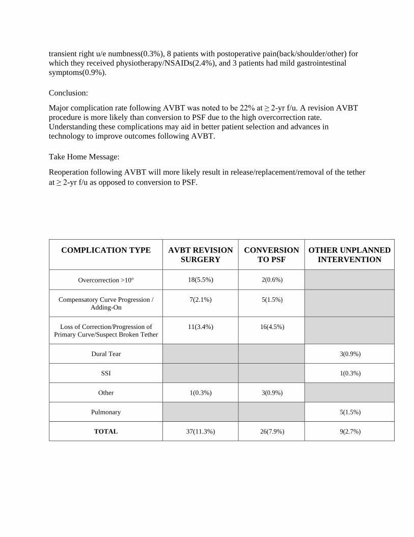
transient right u/e numbness(0.3%), 8 patients with postoperative pain(back/shoulder/other) for
which they received physiotherapy/NSAIDs(2.4%), and 3 patients had mild gastrointestinal
symptoms(0.9%).
Conclusion:
Major complication rate following AVBT was noted to be 22% at ≥ 2-yr f/u. A revision AVBT
procedure is more likely than conversion to PSF due to the high overcorrection rate.
Understanding these complications may aid in better patient selection and advances in
technology to improve outcomes following AVBT.
Take Home Message:
Reoperation following AVBT will more likely result in release/replacement/removal of the tether
at ≥ 2-yr f/u as opposed to conversion to PSF.
COMPLICATION TYPE AVBT REVISION
SURGERY
CONVERSION
TO PSF
OTHER UNPLANNED
INTERVENTION
Overcorrection >10 18(5.5%) 2(0.6%)
Compensatory Curve Progression /
Adding-On
7(2.1%) 5(1.5%)
Loss of Correction/Progression of
Primary Curve/Suspect Broken Tether
11(3.4%) 16(4.5%)
Dural Tear
3(0.9%)
SSI
1(0.3%)
Other 1(0.3%) 3(0.9%)
Pulmonary
5(1.5%)
TOTAL 37(11.3%) 26(7.9%) 9(2.7%)
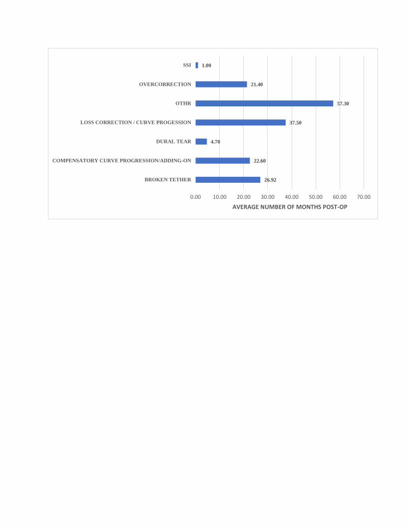
SSI 1.00
OVERCORRECTION 21.40
OTHR 57.30
LOSS CORRECTION / CURVE PROGESSION 37.50
DURAL TEAR 4.70
COMPENSATORY CURVE PROGRESSION/ADDING-ON 22.60
BROKEN TETHER 26.92
0.00 10.00 20.00 30.00 40.00 50.00 60.00 70.00
AVERAGE NUMBER OF MONTHS POST-OP
SPECT-CT To Evaluate Anterior Ankle Impingement and Its Association with
Postoperative Clinical and Functional Outcomes
Diogo Vieira Cardoso, Anna Stock, Peter Salat, Alaistair Younger, Kevin Wing, Murray Penner, Andrea
Veljkovic
Introduction:
Single-photon emission computed tomography and CT (SPECT-CT) has proven helpful in
diagnosing ankle impingement. However, the role of the SPECT-CT evaluating postoperative
ankle pain is still unclear. Therefore, our study aimed to assess the SPECT-CT activity in the
ankle before and after anterior ankle impingement surgery and determine whether there is a
correlation between clinical pain and function scores.
Methods:
Sixty-eight patients with preoperative SPECT-CT and surgically treated anterior ankle
impingement between January 2015 to December 2020 were reviewed. Pain severity and location
were identified pre- and postoperatively. Of the 68 patients, 47 had a postoperative SPECT-CT
done at 29.5 ± 15.8 months after surgery. Patients were grouped according to the time interval
between surgery and postoperative SPECT-CT. The Manchester Oxford Foot and ankle
questionnaire (MOXFQ) was completed by 59% of patients at 36 ± 15.9 months after surgery.
Results:
There was a significant correlation between preoperative SPECT-CT uptake grade and
preoperative pain severity and location (P<.001). Pain scores significantly improved from
preoperative (6.9 ± 1.8 vs. 3.4 ± 2.2, P<.001) for all time intervals, but this was not correlated with
SPECT-CT uptake improvement. Significant differences between pre and postoperative SPECT-
CT were not seen before 36 months after surgery (50 ± 11 months, P<.03). With medium to large
effect sizes, patients with SPECT-CT improvement had significantly lower pain scores and better
MOXFQ scores (P<.05).
Conclusion:
Although postoperative pain improved significantly from preoperative levels, this was not
correlated with SPECT-CT uptake improvement initially. Furthermore, significant differences
between pre- and postoperative SPECT-CT were not seen 36 months after surgery. Therefore,
SPECT-CT done in early postoperative phases is inefficient to evaluate ongoing ankle pain.
Nevertheless, postoperative SPECT-CT improvement has important clinical perceptions and is
strongly associated with clinical and functional outcomes improvement.
Related Documents


