ARTICLE Orthodontic tooth separation activates the hypothalamic area in the human brain Yoshiko Ariji 1 , Hisataka Kondo 2 , Ken Miyazawa 3 , Masako Tabuchi 3 , Syuji Koyama 4 , Yoshitaka Kise 1 , Akifumi Togari 2 , Shigemi Gotoh 3 and Eiichiro Ariji 1 OBJECTIVES: An animal experiment clarified that insertion of an orthodontic apparatus activated the trigeminal neurons of the medulla oblongata. Orthodontic tooth movement is known to be associated with the sympathetic nervous system and controlled by the nucleus of the hypothalamus. However, the transmission of both has not been demonstrated in humans. The purpose of this study were to examine the activated cerebral areas using brain functional magnetic resonance imaging (MRI), when orthodontic tooth separators were inserted, and to confirm the possibility of the transmission route from the medulla oblongata to the hypothalamus. METHODS: Two types of alternative orthodontic tooth separators (brass contact gauge and floss) were inserted into the right upper premolars of 10 healthy volunteers. Brain functional T2*-weighted images and anatomical T1-weighted images were taken. RESULTS: The blood oxygenation level dependent (BOLD) signals following insertion of a brass contact gauge and floss significantly increased in the somatosensory association cortex and hypothalamic area. CONCLUSION: Our findings suggest the possibility of a transmission route from the medulla oblongata to the hypothalamus. International Journal of Oral Science (2018)10:8 https://doi.org/10.1038/s41368-017-0001-y INTRODUCTION Tooth separation is frequently used in orthodontic tooth move- ment, and it usually causes uncomfortable sensations, including pain around the tooth. In animal experiments, the mechanical pressure caused by insertion of orthodontic elastics elevates neuronal activity in trigeminal spinal subnucleus caudalis (Vc) neurons of the medulla oblongata. 1–2 Activation of Vc neurons may be transmitted to the cerebral somatosensory cortex, resulting in recognition of pain around the tooth. The activation of the somatosensory cortex by other painful stimuli has been confirmed in human beings also. 3 Regarding tooth movement force, however, it has been confirmed solely by a recent in vivo study with rats. 4 Meanwhile, pain as an afferent stimulus is considered to play a role in tooth movement through activation of the central nervous system. 2–5 Tooth movements are performed by both bone resorption and formation through the sympathetic nervous system, which may be regulated by the hypothalamus. 6–7 Indeed, the intervention of the central nervous system is verified indirectly by the fact that administration of nonsteroidal anti-inflammatory drugs (NSAIDs) for pain relief impairs the tooth movement process in orthodontic treatment. 5 Although the activation of the medulla oblongata is verified in tooth separation. 1–2 , the route of transmission signals to the hypotha- lamus from the medulla oblongata has not been verified in human beings. Although differences in afferent stimuli may cause different degrees of activation in the somatosensory cortex and hypotha- lamus, even activations themselves in these areas have not been confirmed in orthodontic tooth separation for human beings due to the lack of appropriate procedures. Recently, functional magnetic resonance imaging (fMRI) was introduced as an effective tool for non-invasively identifying the anatomical brain areas that are activated by afferent signals. 8–9 Taken together, if fMRI would indicate the activation in the hypothalamus area, the possibility of a transmission pathway to the hypothalamus from the medulla oblongata could be verified. The purpose of this study was to identify the activated cerebral areas using brain fMRI with a special emphasis on the hypothalamic area together with the somatosensory cortex. In this regard, two types of orthodontic tooth separators were inserted between the upper premolars of healthy volunteers. RESULTS Table 1 shows the cerebral regions with significant increases in blood oxygenation level dependent (BOLD) signals during the insertion of the orthodontic appliances compared with the baseline (at rest). During the insertion of both appliances, BOLD signals increased in the left parietal association area, frontal association area, temporal association area, insula, and cerebel- lum. BOLD signals during insertion of the floss increased in these areas and in the left hippocampus and amygdala (Fig. 1a). BOLD signals during the insertion of the brass contact gauge increased in these areas and in the right thalamus, hippocampus, calcarine sulcus, left putamen, and lingual gyrus (Fig. 1b). Figure 2 shows the results of group analysis based on individual activated sites and magnitude. BOLD signals were significantly Accepted: 31 October 2017 1 Department of Oral and Maxillofacial Radiology, Aichi-Gakuin University School of Dentistry, Nagoya, Japan; 2 Department of Pharmacology, Aichi-Gakuin University School of Dentistry, Nagoya, Japan; 3 Department of Orthodontics, Aichi-Gakuin University School of Dentistry, Nagoya, Japan and 4 Brain and Mind Research Center, Nagoya University, Nagoya, Japan Correspondence: Yoshiko Ariji ([email protected]) www.nature.com/ijos International Journal of Oral Science

Welcome message from author
This document is posted to help you gain knowledge. Please leave a comment to let me know what you think about it! Share it to your friends and learn new things together.
Transcript

ARTICLE
Orthodontic tooth separation activates the hypothalamic areain the human brainYoshiko Ariji1, Hisataka Kondo2, Ken Miyazawa3, Masako Tabuchi3, Syuji Koyama4, Yoshitaka Kise1, Akifumi Togari2, Shigemi Gotoh3 andEiichiro Ariji1
OBJECTIVES: An animal experiment clarified that insertion of an orthodontic apparatus activated the trigeminal neurons of themedulla oblongata. Orthodontic tooth movement is known to be associated with the sympathetic nervous system and controlledby the nucleus of the hypothalamus. However, the transmission of both has not been demonstrated in humans. The purpose of thisstudy were to examine the activated cerebral areas using brain functional magnetic resonance imaging (MRI), when orthodontictooth separators were inserted, and to confirm the possibility of the transmission route from the medulla oblongata to thehypothalamus.METHODS: Two types of alternative orthodontic tooth separators (brass contact gauge and floss) were inserted into the right upperpremolars of 10 healthy volunteers. Brain functional T2*-weighted images and anatomical T1-weighted images were taken.RESULTS: The blood oxygenation level dependent (BOLD) signals following insertion of a brass contact gauge and flosssignificantly increased in the somatosensory association cortex and hypothalamic area.CONCLUSION: Our findings suggest the possibility of a transmission route from the medulla oblongata to the hypothalamus.
International Journal of Oral Science (2018) 10:8 https://doi.org/10.1038/s41368-017-0001-y
INTRODUCTIONTooth separation is frequently used in orthodontic tooth move-ment, and it usually causes uncomfortable sensations, includingpain around the tooth. In animal experiments, the mechanicalpressure caused by insertion of orthodontic elastics elevatesneuronal activity in trigeminal spinal subnucleus caudalis (Vc)neurons of the medulla oblongata.1–2 Activation of Vc neuronsmay be transmitted to the cerebral somatosensory cortex,resulting in recognition of pain around the tooth. The activationof the somatosensory cortex by other painful stimuli has beenconfirmed in human beings also.3 Regarding tooth movementforce, however, it has been confirmed solely by a recent in vivostudy with rats.4
Meanwhile, pain as an afferent stimulus is considered toplay a role in tooth movement through activation of the centralnervous system.2–5 Tooth movements are performed byboth bone resorption and formation through the sympatheticnervous system, which may be regulated by the hypothalamus.6–7
Indeed, the intervention of the central nervous system isverified indirectly by the fact that administration of nonsteroidalanti-inflammatory drugs (NSAIDs) for pain relief impairs thetooth movement process in orthodontic treatment.5 Althoughthe activation of the medulla oblongata is verified in toothseparation.1–2, the route of transmission signals to the hypotha-lamus from the medulla oblongata has not been verified in humanbeings.Although differences in afferent stimuli may cause different
degrees of activation in the somatosensory cortex and hypotha-lamus, even activations themselves in these areas have not been
confirmed in orthodontic tooth separation for human beings dueto the lack of appropriate procedures. Recently, functionalmagnetic resonance imaging (fMRI) was introduced as an effectivetool for non-invasively identifying the anatomical brain areas thatare activated by afferent signals.8–9
Taken together, if fMRI would indicate the activation in thehypothalamus area, the possibility of a transmission pathway tothe hypothalamus from the medulla oblongata could be verified.The purpose of this study was to identify the activated cerebralareas using brain fMRI with a special emphasis on thehypothalamic area together with the somatosensory cortex. Inthis regard, two types of orthodontic tooth separators wereinserted between the upper premolars of healthy volunteers.
RESULTSTable 1 shows the cerebral regions with significant increases inblood oxygenation level dependent (BOLD) signals during theinsertion of the orthodontic appliances compared with thebaseline (at rest). During the insertion of both appliances, BOLDsignals increased in the left parietal association area, frontalassociation area, temporal association area, insula, and cerebel-lum. BOLD signals during insertion of the floss increased in theseareas and in the left hippocampus and amygdala (Fig. 1a). BOLDsignals during the insertion of the brass contact gauge increasedin these areas and in the right thalamus, hippocampus, calcarinesulcus, left putamen, and lingual gyrus (Fig. 1b).Figure 2 shows the results of group analysis based on individual
activated sites and magnitude. BOLD signals were significantly
Accepted: 31 October 2017
1Department of Oral and Maxillofacial Radiology, Aichi-Gakuin University School of Dentistry, Nagoya, Japan; 2Department of Pharmacology, Aichi-Gakuin University School ofDentistry, Nagoya, Japan; 3Department of Orthodontics, Aichi-Gakuin University School of Dentistry, Nagoya, Japan and 4Brain and Mind Research Center, Nagoya University,Nagoya, JapanCorrespondence: Yoshiko Ariji ([email protected])
www.nature.com/ijosInternational Journal of Oral Science

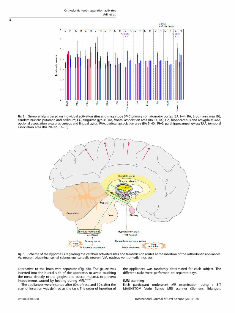
higher in the left thalamus and cerebellum during the insertionof the brass contact gauge compared with the floss (P = 0.032 andP = 0.046, respectively, Wilcoxon rank sum test). In other areas,no differences were observed between them.Visual analog scale (VAS) values of pain during the task were
51.8 ± 24.2 for the brass contact gauge and 3.3 ± 5.0 for the floss.There was a significant difference (P = 0.005, Wilcoxon rank sumtest). VAS values of the residual discomfort after magneticresonance (MR) examination were 24.7 ± 25.6 and 2.0 ± 2.7,respectively. There was a significant difference (P = 0.008,Wilcoxon rank sum test). There were no relationships betweenthe VAS and the maximal t-values.
DISCUSSIONIn the early stage of tooth separation, patients often complain ofpain or discomfort.10 Pain itself as an afferent stimulus isconsidered to be related with tooth movement through thesympathetic nervous control.2–4 This is symbolically expressed asNSAIDs administration for the pain disturbing the tooth move-ment.5 To elucidate this mechanism, fMRI study of brain activitycould provide a great contribution. The known facts are firstsummarized.Insertion of the orthodontic appliance first activates the
trigeminal spinal subnucleus caudalis (Vc) neurons of the medullaoblongata (Fig. 3). In an immunohistochemical study with rats, themechanical pressure for tooth movement elevates the Vc neuronof the medulla oblongata.1–2 The stimuli activating the medullaoblongata are transmitted to the somatosensory cortex. A recentin vivo study with rats verified that the stimuli by orthodontic
tooth movement could evoke the neural excitation of the cerebralsomatosensory cortex.4 As a result, the patient feels pain ordiscomfort.11 Regarding the tooth movements, some animalexperiments suggest that bone turnover is related to thesympathetic nervous system.6–7 In an experiment with orthodon-tic appliances in mice, the sympathetic neuromarkers, such astyrosine hydroxylase, was increased in the periodontal ligament,and the number of osteoclasts was markedly increased on thebone surface.12 Periosteal tyrosine hydroxylase expression wasdecreased along with preosteoclasts and osteoclasts by experi-mentally destroying the sympathetic nervous system.13–14 Further-more, hypothalamo-pituitary disconnection in ewes resulted inlow bone turnover with depression of osteoblast and osteoclastcellular activity.15–16
Based on these facts, the following hypotheses were designed(Fig. 3). Appliance of the orthodontic separator would activate theVc neurons of the medulla oblongata and the cerebral somato-sensory cortex. Simultaneously, the stimuli would be transmittedfrom the medulla oblongata to the nucleus ventromedial nucleus(VM) neurons of the hypothalamus and could activate thesympathetic nervous system. As a result, bone resorption andtooth movement would occur. No verifications have beenreported for the activation in the somatosensory cortex or thepossibility of the route from the medulla oblongata to thehypothalamus in tooth separation in human beings. Although thebrain fMRI could not depict the medulla oblongata, the results ofthis study have verified the somatosensory cortex activation andthe possibility of the transmission route from the medullaoblongata to the hypothalamus. Therefore, our results provide apotential new therapeutic method, such as local administration of
Fig. 1 BOLD signal increases during insertion of apparatuses. a During insertion of floss; b during insertion of the brass contact gauge. Arrowsshow the thalamus
Orthodontic tooth separation activatesAriji et al.
2
International Journal of Oral Science (2018) 10:8
1234567890();,:

a sympathomimetic agent, which may allow efficient toothmovement while blocking pain.In this experiment, two types of orthodontic tooth separators
were considered: a brass wire separator and an elastomericseparator. Nalbantgil et al. investigated pain during the insertionof two types of separators in 87 healthy volunteers anddetermined that the brass wire caused the greatest pain anddiscomfort immediately after insertion, whereas the elastomericseparator caused the greatest pain during the first 2 days afterinsertion.10 The brass wire is non-magnetic and can therefore bebrought into the MR room.10 However, the orthodontic plier,which is used for the insertion of the orthodontic separators,is magnetic and cannot be brought into the MR room.Therefore, non-magnetic alternative appliances that can beinserted by hand were used in this experiment. As an alternativeto the brass wire separator, the self-made brass contact gaugewas used. As an alternative to the elastomeric separator,commercially available floss was used. In this study, the flosscaused only a small amount of pain, and doubt remainedregarding whether the floss was a sufficient alternative to theelastomeric separator.
A possible problem during MR examination is the generation ofheat by the metal. In experiments examining the effects oforthodontic appliances on radiofrequency heating during 3-T MRI,the greatest change in temperature was 3.04° by nickel-titaniumarch wire and stainless steel ligature wire.17 Therefore, thetemperature change by orthodontic appliances was consideredto be within acceptable ranges clinically.17–18 The brass wire usedin this study is non-magnetic and is expected to show smallerchanges than nickel-titanium. To prevent mucosal damage bydirect contact, the gauze was inserted into the buccal side of theappliance.This experiment showed that insertion of the brass contact
gauge increased the BOLD signals in the parietal association areas(somatosensory association cortex, BA 5, 40), thalamus, andhippocampus. Namely, activation of the somatosensory areaassociated with pain was observed, and the possibility of thetransmission system including the hypothalamic area wassuggested. The other activated areas (frontal association area,temporal association area, putamen, insula, and cerebellum) wereassociated with cognition and judgment. In contrast, the insertionof the floss also activated the parietal association area (somato-sensory association cortex, BA 40) and the hypothalamic area(hippocampus, amygdala).BOLD signals following insertion of the brass contact gauge
were significantly higher in the left thalamus and cerebellum thanwith the floss. This result may be associated with the fact that thecontact gauge caused a greater pain response during the task andthe greatest discomfort after examination.This study did not confirm whether the tooth was moved by the
orthodontic separators. Davidovitch et al. reported that theinsertion of the elastomeric separator for 1, 4, 12, and 24 h in24-healthy volunteers produced a space of 0.087, 0.123, 0.184, and0.198mm, respectively.19 Furthermore, recovery to baseline afterseparator removal was achieved after 24 h.19 The amount of toothmovement by the brass wire separator was smaller than that bythe elastomeric separator.10 With respect to these reports, anexperiment involving inserting the appliances for less than 10minduring MR examination would be considered to produce a smallamount of tooth movement. Recovery after appliance removalwould be achieved quickly, so this experiment would result inminimal damage to the participants.
MATERIALS AND METHODSSubjectsThis study was performed with approval from the authors’university ethics committee (No. 420). This study was plannedand performed in accordance with the code of ethics of the WorldMedical Association (Declaration of Helsinki).We recruited volunteers to cooperate in the research. All
participants were informed of the study aims and providedconsent before participating. None of the participants had anyabnormalities in skeletal or occlusal status. None of the volunteershad received orthodontic treatment. Ten healthy volunteers(six men and four women; age range 26–40 years; mean age(30.5 ± 5.9) years) participated in this study.
TaskTwo types of alternative orthodontic tooth separators wereinserted between the upper right first and second premolars ofhealthy volunteers. The orthodontic plier used to insert theorthodontic separators was magnetic and could not be broughtinto the MR room. Therefore, non-magnetic alternative appliancesthat could be inserted by hand were used in this experiment. Onewas commercially available floss (GUM expanding dental flosswith wax, Sunstar, Inc., Tokyo, Japan), as an alternative to theelastomeric separator (Fig. 4a). Another was a self-made brasscontact gauge with 0.15 mm or 0.20 mm diameter, as an
Table 1. Significant increase in fMRI signal during insertion of theapparatuses minus baseline
Region of activation BA Side MNI Maximalt value
x y z
Brass contact gauge
Parietal association area 5 L 12 −50 20 4.02
40 L −26 −50 46 3.31
Frontal association area 44 L −36 18 8 3.95
Temporal association area 20 L −46 −50 −22 3.73
21 L −54 −50 20 3.42
22 L −48 −46 18 3.66
37 L −38 −62 −18 3.60
Thalamus R 22 −24 4 3.44
Hippocampus R 22 −8 −12 3.63
Putamen L −26 −16 8 3.96
Lingual gyrus L −10 −74 −10 4.00
Calcarine sulcus R 10 −84 10 3.39
Insula 13 L −40 −2 18 3.33
Cerebellum L −18 −56 −14 3.68
Floss
Parietal association area 40 L −48 −40 26 4.31
Frontal association cortex 11 L −28 28 −8 4.90
44 L −46 −28 20 3.59
Temporal association area 20 L −46 −52 −10 3.77
21 L −52 2 −24 3.58
22 L −42 −20 0 3.45
37 L −42 −54 −18 4.14
38 L −48 8 −20 3.20
Hippocampus L −32 −8 −22 3.31
Amygdala L −30 −2 −18 3.36
Insula 13 L −34 12 −8 3.84
Cerebellum L −20 −46 −20 3.62
Only significant clusters of activation corrected for multiple comparisons(P< 0.05) were listed. The maximal t value indicated the most significantpeak activations in each cluster. BA, Brodmann area; fMRI, functionalmagnetic resonance imaging; L, left; MNI, Montreal Neurological Institute;R, right
Orthodontic tooth separation activatesAriji et al.
3
International Journal of Oral Science (2018) 10:8
alternative to the brass wire separator (Fig. 4b). The gauze wasinserted into the buccal side of the apparatus to avoid touchingthe metal directly to the gingiva and buccal mucosa, to preventimpediments caused by heating during MRI.14–15
The appliances were inserted after 60 s of rest, and 30 s after thestart of insertion was defined as the task. The order of insertion of
the appliances was randomly determined for each subject. Thedifferent tasks were performed on separate days.
fMRI scanningEach participant underwent MR examination using a 3-TMAGNETOM Verio Syngo MRI scanner (Siemens, Erlangen,
Fig. 2 Group analysis based on individual activation sites and magnitude SMC primary somatomotor cortex (BA 1–4). BA, Brodmann area; BG,caudate nucleus putamen and pallidum; CG, cingulate gyrus; FAA, frontal association area (BA 11, 44); HA, hippocampus and amygdala; OAA,occipital association area plus cuneus and lingual gyrus; PAA, parietal association area (BA 5, 40); PHG, parahippocampal gyrus; TAA, temporalassociation area (BA 20–22, 37–38)
Fig. 3 Schema of the hypothesis regarding the cerebral activated sites and transmission routes at the insertion of the orthodontic appliances.Vc, neuron trigeminal spinal subnucleus candalis neuron; VM, nucleus ventromedial nucleus
Orthodontic tooth separation activatesAriji et al.
4
International Journal of Oral Science (2018) 10:8

Germany) to allow functional T2*-weighted and anatomical T1-weighted image acquisition. Each patient’s head was fixed by tapeon the forehead and a sponge on both sides of the face to preventmovement during MR examination. Whole-brain functionalimages were obtained using gradient echo-planar imagingsequencing, with a 3000-ms repetition time (TR); 30-ms time ofecho (TE); 90° flip angle; 216-mm field of view (FOV); and 3mm×3mm× 3-mm voxel resolution. Sampling consisted of 36 slices, 3mm thick, with no gap, parallel to the anteroposterior commissureline. For each task series, 110 consecutive image volumes wereacquired, including a 60-s rest, 30-s insertion (task), and 240-srecovery period.Before functional data were acquired, anatomical images were
obtained using a T1-weighted magnetization prepared rapidacquisition gradient echo (MPRAGE) sequence: (TR/TE/TI=2 500ms/2.48 ms/900 ms, flip angle = 8°, FOV = 256 × 256mm2, voxelsize = 1mm× 1mm× 1mm, 192 slices, no gap).Each participant was asked to evaluate the maximal pain during
the task and the degree of residual discomfort after MRexamination using the VAS.
fMRI-data processingPreprocessing was performed with SPM 8 (http://www.fil.ion.ucl.ac.uk/spm/). The functional images were realigned to removemovement-related artefacts, normalized to the MontrealNeurological Institute (MNI) template, and spatially smoothedwith a Gaussian kernel with a full width at half maximum of 8 mm.Statistical analysis was performed with the general linear model
using SPM8. For each subject, a design matrix was created using acanonical hemodynamic response function to model the responseto each task. For group analysis, random effect analysis (pairedt-test) was applied to statistically contrast images of all subjects.A value of P < 0.05 was considered statistically significant.For each region of the brain, BOLD signal changes during
insertion of the brass contact gauge were compared with floss(Wilcoxon rank sum test). Statistical significance was established atP < 0.05.Comparisons of VAS values were performed using the Wilcoxon
rank sum test. A P value < 0.05 was considered statisticallysignificant.
CONCLUSIONCerebral activation following orthodontic separation was con-firmed. BOLD signals at insertion of the brass contact gauge andfloss significantly increased in the somatosensory associationcortex and hypothalamic area. The possibility of a transmissionroute from the medulla oblongata to the hypothalamus wasindirectly proved.
ACKNOWLEDGEMENTSThe authors thank all of the volunteers for their participation in this study. This studywas partially supported by a Grant-in-Aid for Scientific Research (C) (26462862) fromthe Japan Society for the Promotion of Science.
AUTHOR CONTRIBUTIONSAll of the authors made substantial contributions to the conception and design of thestudy, acquisition of data, and analysis and interpretation of data. All participated indrafting the article or revising it critically for important intellectual content. Allauthors have approved this version of the manuscript.
ADDITIONAL INFORMATIONConflict of interest: The authors declare that they have no conflict of interest.
Ehical approval The current study was performed with the approval of the UniversityEthics Committee (No. 420). This study was planned and performed in accordancewith the code of ethics of the World Medical Association (Declaration of Helsinki).
REFERENCES1. Hasegawa, M. et al. ERK is involved in tooth-pressure-induced Fos expression in
Vc neurons. J. Dent. Res. 91, 1141–1146 (2012).2. Gao, Y. & Duan, Y. Z. Increased COX2 in the trigeminal nucleus caudalis is
involved in orofacial pain induced by experimental tooth movement. Anat. Rec.293, 485–491 (2010).
3. Suenaga, S. et al. The usefulness of diagnostic imaging for the assessment of painsymtoms in temporomandibular disorders. Jpn Dent. Sci. Rev. 52, 93–106 (2016).
4. Horinuki, E. et al. Orthodontic force facilitates cortical responses to periodontalstimulation. J. Dent. Res.. 94, 1158–1166 (2015).
5. Walker, J. B. & Buring, S. M. NSAID impairment of orthodontic tooth movement.Ann. Pharmacother. 35, 113–115 (2001).
6. Araújo, A. S. et al. New methodology for evaluating osteoclastic activity inducedby orthodontic load. J. Appl. Oral. Sci. 23, 19–25 (2015).
7. Shen, T. et al. Cyclic tension promotes osteogenic differentiation in humanperiodontal ligament stem cells. Int. J. Clin. Exp. Pathol. 7, 7872–7880 (2014).
8. Ariji, Y. et al. Regional brain activity during jaw clenching with natural teeth andwith occlusal splints: a preliminary functional MRI study. Cranio 34, 188–194(2016).
9. Yılmaz, S. To see bruxism: a functional MRI study. Dentomaxillofac. Radiol. 44,20150019 (2015).
10. Nalbantgil, D. et al. Perception of pain and discomfort during tooth separation.Aust. Orthod. J. 25, 110–115 (2009).
11. Moisset, X. et al. Functional brain imaging of trigeminal neuralgia. Eur. J. Pain. 15,124–131 (2011).
12. Kondo, M. et al. Experimental tooth movement-induced osteoclast activation isregulated by sympathetic signaling. Bone 52, 39–47 (2013).
13. Cao, H. et al. Force-induced Adrb2 in periodontal ligament cells promotes toothmovement. J. Dent. Res. 93, 1163–1169 (2014).
14. Cherruau, M. et al. Chemical sympathectomy -induced changes in TH-, VIP-, andCGRP-immunoreactive fibers in the rat mandible periosteum: influence on boneresorption. J. Cell. Physiol. 194, 341–348 (2003).
Fig. 4 Orthodontic apparatuses used in this experiment. a GUM expanding dental floss with wax; b self-made brass contact gauge
Orthodontic tooth separation activatesAriji et al.
5
International Journal of Oral Science (2018) 10:8
15. Oheim, R. et al. Sheep model for osteoporosis: sustainability and biomechanicalrelevance of low turnover osteoporosis induced by hypothalamic-pituitary dis-connection. J. Orthop. Res. 31, 1067–1074 (2013).
16. Beil, F. T. et al. Low turnover osteoporosis in sheep induced byhypothalamic-pituitary disconnection. J. Orthop. Res. 30, 1254–1262(2012).
17. Görgülü, S. et al. Effect of orthodontic brackets and different wires on radio-frequency heating and magnetic field interactions during 3-T MRI. Dentomax-illofac. Radiol. 43, 20130356 (2014).
18. Hasegawa, M. et al. Radiofrequency heating of metallic dental devices during 3.0T MRI. Dentomaxillofac. Radiol. 42, 20120234 (2013).
19. Davidovitch, M. et al. Duration of elastomeric separation and effect on inter-proximal contact point characteristics. Am. J. Orthod. Dentofac. Orthop. 133,414–422 (2008).
Open Access This article is licensed under a Creative CommonsAttribution 4.0 International License, which permits use, sharing,
adaptation, distribution and reproduction in anymedium or format, as long as you giveappropriate credit to the original author(s) and the source, provide a link to the CreativeCommons license, and indicate if changes were made. The images or other third partymaterial in this article are included in the article’s Creative Commons license, unlessindicated otherwise in a credit line to the material. If material is not included in thearticle’s Creative Commons license and your intended use is not permitted by statutoryregulation or exceeds the permitted use, you will need to obtain permission directlyfrom the copyright holder. To view a copy of this license, visit http://creativecommons.org/licenses/by/4.0/.
© The Author(s) 2018
Orthodontic tooth separation activatesAriji et al.
6
International Journal of Oral Science (2018) 10:8
Related Documents