ORIGINAL RESEARCH ADULT BRAIN Evaluation of Thick-Slab Overlapping MIP Images of Contrast- Enhanced 3D T1-Weighted CUBE for Detection of Intracranial Metastases: A Pilot Study for Comparison of Lesion Detection, Interpretation Time, and Sensitivity with Nonoverlapping CUBE MIP, CUBE, and Inversion-Recovery-Prepared Fast-Spoiled Gradient Recalled Brain Volume X B.C. Yoon, X A.F. Saad, X P. Rezaii, X M. Wintermark, X G. Zaharchuk, and X M. Iv ABSTRACT BACKGROUND AND PURPOSE: Early and accurate identification of cerebral metastases is important for prognostication and treatment planning although this process is often time consuming and labor intensive, especially with the hundreds of images associated with 3D volumetric imaging. This study aimed to evaluate the benefits of thick-slab overlapping MIPs constructed from contrast-enhanced T1-weighted CUBE (overlapping CUBE MIP) for the detection of brain metastases in comparison with traditional CUBE and inversion- recovery prepared fast-spoiled gradient recalled brain volume (IR-FSPGR-BRAVO) and nonoverlapping CUBE MIP. MATERIALS AND METHODS: A retrospective review of 48 patients with cerebral metastases was performed at our institution from June 2016 to October 2017. Brain MRIs, which were acquired on multiple 3T scanners, included gadolinium-enhanced T1-weighted IR-FSPGR- BRAVO and CUBE, with subsequent generation of nonoverlapping CUBE MIP and overlapping CUBE MIP. Two blinded radiologists identified the total number and location of metastases on each image type. The Cohen was used to determine interrater agreement. Sensitivity, interpretation time, and lesion contrast-to-noise ratio were assessed. RESULTS: Interrater agreement for identification of metastases was fair-to-moderate for all image types ( 0.222– 0.598). The total number of metastases identified was not significantly different across the image types. Interpretation time for CUBE MIPs was significantly shorter than for CUBE and IR-FSPGR-BRAVO, saving at least 50 seconds per case on average (P .001). The mean lesion contrast-to-noise ratio for both CUBE MIPs was higher than for IR-FSPGR-BRAVO. The mean contrast-to-noise ratio for small lesions (4 mm) was lower for nonoverlapping CUBE MIP (1.55) than for overlapping CUBE MIP (2.35). For both readers, the sensitivity for lesion detection was high for all image types but highest for overlapping CUBE MIP and CUBE (0.93– 0.97). CONCLUSIONS: This study suggests that the use of overlapping CUBE MIP or nonoverlapping CUBE MIP for the detection of brain metastases can reduce interpretation time without sacrificing sensitivity, though the contrast-to-noise ratio of lesions is highest for overlapping CUBE MIP. ABBREVIATIONS: CNR contrast-to-noise ratio; IR-FSPGR-BRAVO inversion-recovery-prepared fast-spoiled gradient recalled brain volume; nC-MIP non- overlapping CUBE MIP; oC-MIP overlapping CUBE MIP; SRS stereotactic radiosurgery; XR cross-reference T he early and accurate identification of brain metastases in patients with systemic cancers has important implications for patient prognosis and treatment strategy because a greater num- ber of lesions at presentation correlates with decreased survival. 1 In patients with a small number of metastases, surgical resection or stereotactic radiosurgery (SRS) may be pursued, while whole- brain radiation therapy is generally recommended for those with an extensive lesion burden. 2,3 Previous studies have shown that SRS alone is an effective treatment and provides good local tumor control in patients with up to 10 brain metastases. 4,5 Additionally, the rate of local control with SRS is greater for small lesions, which further stresses the importance of early detection. The choice of SRS or whole-brain radiation therapy for the treatment of brain metastases in individuals with a specific number of lesions is im- portant because the risk of radiation-induced dementia and neu- rocognitive decline associated with whole-brain radiation therapy can be potentially avoided with SRS. Counting multiple small metastases is often laborious and time-consuming, especially with the hundreds of images associ- ated with 3D volumetric imaging. Multiple investigators have demonstrated improved lesion detection using 3D T1-weighted volumetric fast spin-echo sequences (CUBE, GE Healthcare, Mil- Received September 23, 2017; accepted after revision June 16, 2018. From the Department of Radiology, Division of Neuroimaging and Neurointerven- tion, Stanford University, Stanford, California. Paper previously presented as an electronic exhibit at: Annual Meeting of the American Society of Neuroradiology and the Foundation of the ASNR Symposium, April 22–27, 2017; Long Beach, California (#eP-50). Please address correspondence to Michael Iv, MD, Department of Radiology, Divi- sion of Neuroimaging and Neurointervention, Stanford University, 300 Pasteur Dr, Grant Building, Room S031E, Stanford, CA 94305; e-mail: [email protected], @Michael_Iv_MD http://dx.doi.org/10.3174/ajnr.A5747 AJNR Am J Neuroradiol 39:1635– 42 Sep 2018 www.ajnr.org 1635

Welcome message from author
This document is posted to help you gain knowledge. Please leave a comment to let me know what you think about it! Share it to your friends and learn new things together.
Transcript

ORIGINAL RESEARCHADULT BRAIN
Evaluation of Thick-Slab Overlapping MIP Images of Contrast-Enhanced 3D T1-Weighted CUBE for Detection of Intracranial
Metastases: A Pilot Study for Comparison of Lesion Detection,Interpretation Time, and Sensitivity with Nonoverlapping
CUBE MIP, CUBE, and Inversion-Recovery-PreparedFast-Spoiled Gradient Recalled Brain Volume
X B.C. Yoon, X A.F. Saad, X P. Rezaii, X M. Wintermark, X G. Zaharchuk, and X M. Iv
ABSTRACT
BACKGROUND AND PURPOSE: Early and accurate identification of cerebral metastases is important for prognostication and treatmentplanning although this process is often time consuming and labor intensive, especially with the hundreds of images associated with 3Dvolumetric imaging. This study aimed to evaluate the benefits of thick-slab overlapping MIPs constructed from contrast-enhancedT1-weighted CUBE (overlapping CUBE MIP) for the detection of brain metastases in comparison with traditional CUBE and inversion-recovery prepared fast-spoiled gradient recalled brain volume (IR-FSPGR-BRAVO) and nonoverlapping CUBE MIP.
MATERIALS AND METHODS: A retrospective review of 48 patients with cerebral metastases was performed at our institution from June2016 to October 2017. Brain MRIs, which were acquired on multiple 3T scanners, included gadolinium-enhanced T1-weighted IR-FSPGR-BRAVO and CUBE, with subsequent generation of nonoverlapping CUBE MIP and overlapping CUBE MIP. Two blinded radiologistsidentified the total number and location of metastases on each image type. The Cohen � was used to determine interrater agreement.Sensitivity, interpretation time, and lesion contrast-to-noise ratio were assessed.
RESULTS: Interrater agreement for identification of metastases was fair-to-moderate for all image types (� � 0.222– 0.598). The totalnumber of metastases identified was not significantly different across the image types. Interpretation time for CUBE MIPs was significantlyshorter than for CUBE and IR-FSPGR-BRAVO, saving at least 50 seconds per case on average (P � .001). The mean lesion contrast-to-noiseratio for both CUBE MIPs was higher than for IR-FSPGR-BRAVO. The mean contrast-to-noise ratio for small lesions (�4 mm) was lower fornonoverlapping CUBE MIP (1.55) than for overlapping CUBE MIP (2.35). For both readers, the sensitivity for lesion detection was high for allimage types but highest for overlapping CUBE MIP and CUBE (0.93– 0.97).
CONCLUSIONS: This study suggests that the use of overlapping CUBE MIP or nonoverlapping CUBE MIP for the detection of brain metastases canreduce interpretation time without sacrificing sensitivity, though the contrast-to-noise ratio of lesions is highest for overlapping CUBE MIP.
ABBREVIATIONS: CNR � contrast-to-noise ratio; IR-FSPGR-BRAVO � inversion-recovery-prepared fast-spoiled gradient recalled brain volume; nC-MIP � non-overlapping CUBE MIP; oC-MIP � overlapping CUBE MIP; SRS � stereotactic radiosurgery; XR � cross-reference
The early and accurate identification of brain metastases in
patients with systemic cancers has important implications for
patient prognosis and treatment strategy because a greater num-
ber of lesions at presentation correlates with decreased survival.1
In patients with a small number of metastases, surgical resection
or stereotactic radiosurgery (SRS) may be pursued, while whole-
brain radiation therapy is generally recommended for those with
an extensive lesion burden.2,3 Previous studies have shown that
SRS alone is an effective treatment and provides good local tumor
control in patients with up to 10 brain metastases.4,5 Additionally,
the rate of local control with SRS is greater for small lesions, which
further stresses the importance of early detection. The choice of
SRS or whole-brain radiation therapy for the treatment of brain
metastases in individuals with a specific number of lesions is im-
portant because the risk of radiation-induced dementia and neu-
rocognitive decline associated with whole-brain radiation therapy
can be potentially avoided with SRS.
Counting multiple small metastases is often laborious and
time-consuming, especially with the hundreds of images associ-
ated with 3D volumetric imaging. Multiple investigators have
demonstrated improved lesion detection using 3D T1-weighted
volumetric fast spin-echo sequences (CUBE, GE Healthcare, Mil-
Received September 23, 2017; accepted after revision June 16, 2018.
From the Department of Radiology, Division of Neuroimaging and Neurointerven-tion, Stanford University, Stanford, California.
Paper previously presented as an electronic exhibit at: Annual Meeting of theAmerican Society of Neuroradiology and the Foundation of the ASNR Symposium,April 22–27, 2017; Long Beach, California (#eP-50).
Please address correspondence to Michael Iv, MD, Department of Radiology, Divi-sion of Neuroimaging and Neurointervention, Stanford University, 300 Pasteur Dr,Grant Building, Room S031E, Stanford, CA 94305; e-mail: [email protected],@Michael_Iv_MD
http://dx.doi.org/10.3174/ajnr.A5747
AJNR Am J Neuroradiol 39:1635– 42 Sep 2018 www.ajnr.org 1635

waukee, Wisconsin; sampling perfection with application opti-
mized contrasts by using different flip angle evolution [SPACE],
Siemens, Erlangen, Germany; volume isotropic turbo spin-echo
[VISTA], Philips Healthcare, Best, the Netherlands).6-13 CUBE
(and similar sequences) uses a variable flip angle technique and
higher echo-train length to acquire gap-free volumetric images
with reduced acquisition time and specific absorption rate.7 Fur-
thermore, the black-blood properties offered by CUBE allow good
background vascular suppression and provide a higher contrast-to-
noise ratio (CNR) than 3D T1-weighted gradient-echo sequences
(inversion-recovery-prepared fast spoiled gradient recalled brain
volume [IR-FSPGR-BRAVO], GE Healthcare; MPRAGE, Siemens;
3D TFE, Phillips Healthcare).7
The use of maximum-intensity-projection images has become
standard in chest imaging when evaluating pulmonary nodules
because MIPs have been shown to enhance lesion detection and
reduce the total amount of time the radiologist spends searching
for small nodules.14-17 In neuroimaging, 3D volumetric fast spin-
echo imaging is ideally suited to MIP reconstruction because of its
black-blood and increased CNR properties. However, the use of
MIP images in the detection of brain metastases has been lim-
ited.18,19 Recently, Bae et al18 have shown that nonoverlapping
5-mm-thick MIPs of 3D T1-weighted turbo spin-echo signifi-
cantly reduced interpretation time without sacrificing diagnostic
accuracy but with an increased false-positive rate, compared with
the source 1-mm images. Thick-section MIPs reconstructed with
slice overlapping can potentially reduce artifacts from partial
volume averaging and improve visualization of lesions. There-
fore, in this study, we examined whether the use of thick-slab
overlapping MIPs constructed from gadolinium-enhanced
3D T1-weighted CUBE (overlapping CUBE MIP [oC-MIP])
would allow improved visualization and quicker and more
sensitive detection of brain metastases compared with non-
overlapping CUBE MIP (nC-MIP) as well as source 3D T1-
weighted CUBE and IR-FSPGR-BRAVO.
MATERIALS AND METHODSWe performed an institutional review board–approved retrospec-
tive study of patients with brain metastases who had MRIs from
June 2016 to October 2017 at a single academic institution. Pa-
tients with a diagnosis of metastatic disease from any primary
cancer who had brain MR imaging during the study period were
identified through our PACS imaging data base by keyword
search criteria. The specific keywords used were “metastasis” and
“metastases.” On the basis of finalized reports associated with the
MRIs, patients with at least 2 intra-axial brain metastases were
included in the study.
MR Imaging ProtocolAll brain MRIs were performed on multiple 3T scanners (Discov-
ery 750; GE Healthcare) using our institutional brain metastasis
protocol, which consisted of precontrast and postcontrast images,
the latter acquired following the intravenous administration of
0.1 mmol/kg of gadobenate dimeglumine (MultiHance; Bracco
Diagnostics, Princeton, New Jersey). Gadobenate dimeglumine is
the specific contrast agent used at our institution because of
its high relaxivity, which allows improved lesion conspicuity.
Postgadolinium sequences were obtained in the following order:
sagittal 3D T1-weighted CUBE (TR � 600 ms, TE � 13 ms, slice
thickness � 1 mm, echo-train length � 28, flip angle � 90°, ma-
trix � 256 � 256 mm, FOV � 250 mm, in-plane resolution �
0.977 mm, bandwidth � 244 Hz, number of averages � 1, imag-
ing time � 2 minutes 15 seconds) with 1-mm axial and coronal
reformats and axial 3D T1-weighted IR-FSPGR-BRAVO (TR �
9.2 ms, TE � 3.7 ms, TI � 400 ms, slice thickness � 1 mm,
echo-train length � 1, flip angle � 13°, matrix � 256 � 256 mm,
FOV � 240 mm, in-plane resolution � 0.938 mm, bandwidth �
195 Hz, number of averages � 1, imaging time � 3 minutes 30
seconds) with 1-mm sagittal and coronal reformats. IR-FSPGR-
BRAVO was the last sequence performed in our protocol because
increased scan time delay after contrast administration allows im-
proved lesion detection.
Axial MIP images (oC-MIP: 10-mm sections reconstructed at
4-mm intervals; nC-MIP: 5-mm sections at 5-mm intervals) were
then constructed from the contrast-enhanced T1-weighted CUBE
images. Image parameters for the nC-MIPs were chosen on the
basis of results from a recent prior study showing their benefits for
the detection of brain metastases.18 Image parameters for the oC-
MIPs were chosen on the basis of our clinical experience of using
thick-slabbed MIPs for detection of enhancing brain lesions be-
cause no prior studies, to our knowledge, have reported optimal
values for overlapping MIPs. Axial MIPs of IR-FSPGR-BRAVO
(10-mm sections reconstructed at 4-mm intervals) were also gen-
erated solely for comparison of CNR with CUBE MIPs. These
specific MIPS were not used for detection of metastases in this
study because we and others have found them to be of little
clinical utility, in large part due to the difficulty in distinguish-
ing true enhancing metastases from excessive background vas-
cular enhancement.18
Determination of Ground Truth for Brain MetastasesA neuroradiologist with �10 years of experience who did not
participate in counting of the metastases as part of the study care-
fully reviewed all sequences from the initial MR imaging, includ-
ing precontrast (diffusion-weighted imaging, gradient recalled,
T2-weighted fast spin-echo, 3D T1 CUBE) and postcontrast (3D
T1 CUBE, IR-FSPGR-BRAVO, 3D CUBE FLAIR) images as well
as any follow-up MRIs that were acquired before any intervening
treatment. A brain metastasis was positively identified and re-
corded (image slice number of the lesion and its anatomic loca-
tion) if the following criteria were present: a nonvascular enhanc-
ing lesion with or without associated FLAIR or diffusion signal
intensity, surrounding edema, or hemorrhage that was unchanged or
increased in size on follow-up MR imaging. For this study, any post-
surgical enhancement (defined as enhancement contiguous with a
remote surgical resection cavity) and leptomeningeal enhancement
were excluded.
Image AnalysisA neuroradiologist with 10 years of experience (reader 1) and a
radiology resident with 2 years of experience (reader 2) counted
the number of brain metastases for each patient during 4 sessions
separated by 1 week between each session. Each reader indepen-
dently evaluated CUBE in 1 session, nC-MIP alone in the second
1636 Yoon Sep 2018 www.ajnr.org

session, oC-MIP alone in the third session, and IR-FSPGR-
BRAVO in the last session. In additional sessions separated by
1 week, each reader also independently evaluated nC-MIP and
oC-MIP with the option to cross-reference a lesion with source
images to confirm the authenticity of a questionable lesion (nC-
MIP� cross-reference [XR] and oC-MIP�XR, respectively); this
was performed to determine whether the simultaneous use of
cross-referencing affected interpretation time and detection sen-
sitivity. The order of patients during each session was random-
ized. The number of metastases identified and the time for each
reader to identify and count all lesions (time for interpretation)
were recorded. During the counting process, readers also re-
corded the image slice number and anatomic location of each
lesion on each sequence using speech recognition software (Nu-
ance PowerScribe 360 Reporting; Nuance Communications, Bur-
lington, Massachusetts).
The contrast-to-noise ratio was calculated using the smallest
(�4 mm) and largest metastatic lesions identified on CUBE, both
CUBE MIPs, IR-FSPGR-BRAVO, and IR-FSPGR-BRAVO MIPs
with the following formula: Mean Signal Intensity of Lesion �
Mean Signal Intensity of Normal White Matter / SD of Lesion
Signal Intensity.8
Statistical AnalysisDue to the lack of histopathologic data, our study did not have an
absolute standard of truth. Our ground truth was based on the
evaluation of all sequences on the initial MR imaging and com-
parison of lesions with any follow-up MR imaging in which there
was no intervening treatment. Therefore, we aimed to compare
the sensitivities and discrepancy rates of all image types rather
than to determine diagnostic accuracy; this method has been sim-
ilarly used in previous analyses of pulmonary nodules.20
Interrater agreement for identification of metastases was cal-
culated using the Cohen � coefficient with the following interpre-
tation model of �: 0-.20, slight; 0.21– 0.40, fair; 0.41– 0.60, mod-
erate; 0.61– 0.80, substantial; and 0.81–1, almost perfect.21
Specifically, of the total pool of ground truth metastases, agree-
ment for the presence of each individual lesion was assessed as
follows: 1) both readers agreed that a lesion was present, 2) reader
1 identified a lesion but reader 2 did not, 3) reader 2 identified a
lesion but reader 1 did not, and 4) both readers agreed that a lesion
was not present. The total number of lesions detected on each
image type and time for interpretation were compared between
readers using 1-way analysis of variance with a Bonferroni ad-
justment. A P � .013 (calculated by .05/4) was considered
statistically significant in comparing the 4 image types (CUBE,
nC-MIP, oC-MIP, IR-FSPGR-BRAVO), as determined by the
Bonferroni adjustment.22 The CNR of the smallest and largest
lesions on each series was reported as mean � SD. In a sub-
group analysis, the time for interpretation was compared be-
tween all CUBE MIPs (nC-MIP, oC-MIP, nC-MIP�XR, oC-
MIP�XR) and source CUBE, using 1-way analysis of variance
with a Bonferroni adjustment. A P � .01 (calculated by .05/5)
was considered statistically significant in comparing these 5
image types, as determined by the Bonferroni adjustment.22
Lesion-detection sensitivity, mean false-negative, mean false-
positive, and mean discrepancy (total number of false-posi-
tives and false-negatives) per case were assessed for all image
analysis types.
RESULTSA total of 308 metastases were identified in 48 patients, consisting
of 37 women and 11 men (mean age: 62.4 � 13 years; age range,
29 – 81 years). Twenty-six patients had primary non-small cell
lung cancer, 1 had small cell lung cancer, 13 had breast cancer, 2
had melanoma, 2 had renal cell carcinoma, 1 had esophageal can-
cer, and 2 had ovarian cancer. One patient had a history of Li-
Fraumeni syndrome and had multiple primary cancers, including
colon, lung, breast, uterine, and hepatocellular carcinoma. Of the
48 patients, 27 had undergone prior treatment for brain metasta-
ses, including surgical resection, whole-brain radiation therapy,
and/or SRS.
Interrater agreement for the presence of individual metastatic
lesions was fair-to-moderate across all image types (� � 0.222–
0.598) but highest for oC-MIP (� � 0.598) (Table 1). No signifi-
cant difference was found among the total number of metastases
on any of the 4 main image types (P � .062): CUBE (319 for
reader one, 336 for reader 2), nC-MIP (325 for reader one, 313 for
reader 2), oC-MIP (327 for reader one, 333 for reader 2), IR-
FSPGR-BRAVO (289 for reader one, 303 for reader 2) (Fig 1).
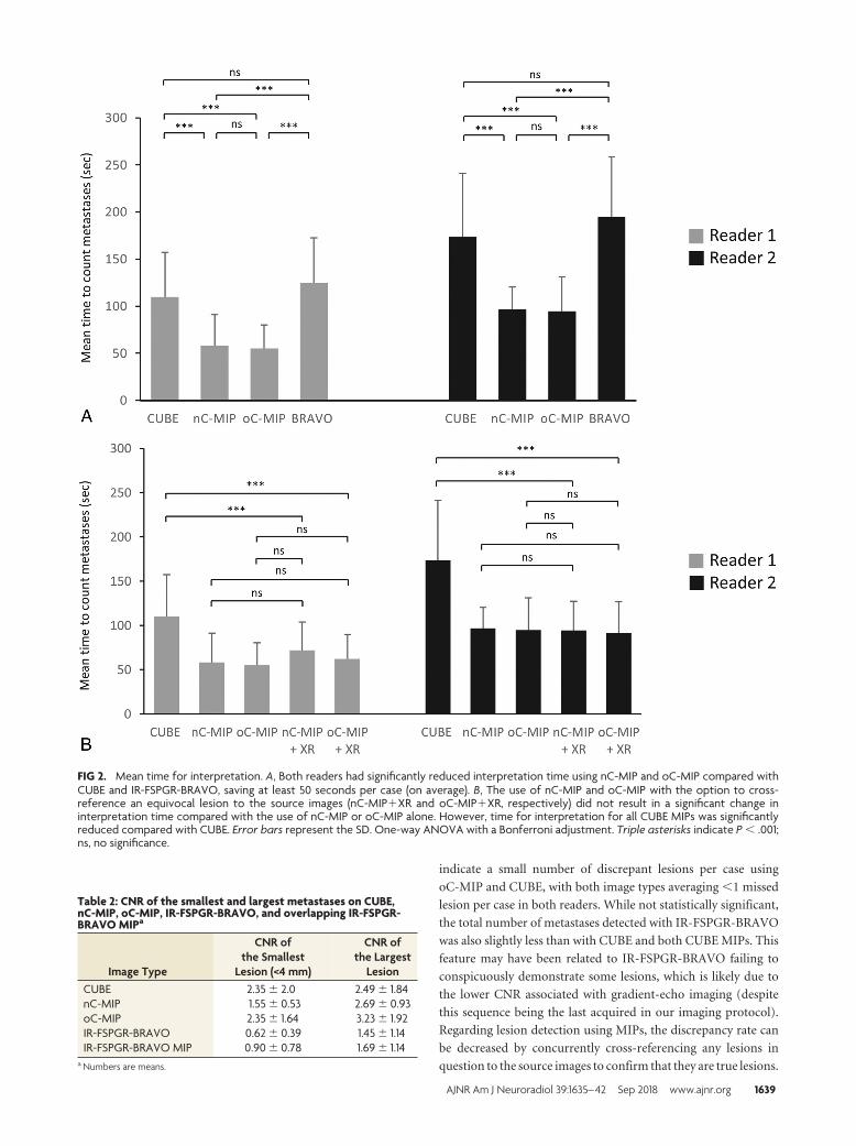
A significant difference in the time for interpretation among
the image types was found for both readers (P � .001) (Fig 2A).
The time for interpretation using oC-MIP (mean: 55.2 � 25.1
seconds for reader one, 94.7 � 36.5 seconds for reader 2) and
nC-MIP (58.2 � 32.9 seconds for reader one, 97.0 � 23.4 seconds
for reader 2) was significantly reduced compared with CUBE
(109.8 � 47.5 seconds for reader one, P � .001; 173.5 � 67.7
seconds for reader 2, P � .001) and IR-FSPGR-BRAVO (124.6 �
48 seconds for reader one, P � .001; 195 � 64.8 seconds for reader
2, P � .001), with a savings of at least 50 seconds per case (on
average). In a subgroup analysis of all CUBE image types, time for
interpretation using oC-MIP�XR (61.9 � 27.7 seconds for
reader one, 91.7 � 35.0 seconds for reader 2) and nC-MIP�XR
(71.7 � 32.0 seconds for reader one, 94.3 � 33.0 seconds for
reader 2) was significantly reduced compared with CUBE (P �
.001) (Fig 2B). However, time for interpretation was not signifi-
cantly different among oC-MIP, nC-MIP, oC-MIP�XR, and
nC-MIP�XR.
The conspicuity of the lesions as indicated by CNR on the 4
image types also differed for both small (�4 mm) and large le-
sions (Table 2 and Fig 3). For both lesion groups, the mean CNR
was the highest for oC-MIP and higher with CUBE and both
CUBE MIPs than with IR-FSPGR-BRAVO and IR-FSPGR-
BRAVO MIP. The mean CNR for small lesions was lower with
nC-MIP (1.55 � 0.3) than with CUBE (2.35 � 2.0) or oC-MIP
(2.35 � 1.64).
Table 1: Interrater agreement using the Cohen � coefficient forT1-weighted CUBE, nC-MIP, oC-MIP, and IR-FSPGR-BRAVO
Image Type �
95%Confidence
Interval AgreementCUBE 0.235 0.024–0.447 FairnC-MIP 0.222 0.069–0.374 FairoC-MIP 0.598 0.371–0.825 ModerateIR-FSPGR-BRAVO 0.445 0.290–0.599 Moderate
AJNR Am J Neuroradiol 39:1635– 42 Sep 2018 www.ajnr.org 1637

For both readers, the sensitivity for lesion detection was high
for all image types. Of CUBE, nC-MIP, oC-MIP, and IR-FSPGR-
BRAVO, sensitivity was highest for oC-MIP (0.96 for both read-
ers) and CUBE (0.97 for reader one, 0.93 for reader 2) (Table 3).
Sensitivity was slightly lower for nC-MIP (0.95 for reader one,
0.90 for reader 2) and even lower for IR-FSPGR-BRAVO (0.92 for
reader one, 0.89 for reader 2). On average, there was �1 false-
negative and 1 false-positive case per patient across all image types
(Table 3). The mean discrepancy rate (total number of false-neg-
ative and false-positive lesions per case) was also �1 lesion per
patient on all image types except with nC-MIP and IR-FSPGR-
BRAVO for reader 2. Mean false-positives per case were reduced
with oC-MIP�XR (0.21 for reader one, 0.23 for reader 2) com-
pared with oC-MIP (0.54 for reader one, 0.33 for reader 2) and
with nC-MIP�XR (0.25 for reader one, 0.29 for reader 2) com-
pared with nC-MIP (0.58 for reader one, 0.71 for reader 2).
DISCUSSIONIn this study, we found that the use of oC-MIP or nC-MIP re-
duced interpretation time without sacrificing lesion detection
sensitivity compared with traditional CUBE and IR-FSPGR-
BRAVO. The coupling of CUBE MIPs with the option to cross-
reference a questionable lesion to source images further reduced
false-positives without significantly changing the time for inter-
pretation compared with MIPs alone. The CNR of brain metasta-
ses was higher with CUBE and both CUBE MIPs than with IR-
FSPGR-BRAVO and IR-FSPGR-BRAVO MIP, though the CNR
of small and large metastases was highest with thick-slab oC-MIP.
Interrater agreement for the detection of brain metastases was
fair-to-moderate across all image types but highest with oC-MIP.
The findings in our study are consistent with those of other inves-
tigators regarding enhanced lesion detection and increased CNR
using 3D fast spin-echo imaging compared with gradient-echo
imaging.7,8 CUBE images are ideally suited for MIP reconstruction
because there is an inherent reduction in the amount of background
vascular enhancement, which further reduces background image
noise and increases lesion conspicuity.
An issue of clinical importance for practicing radiologists that
has received less research attention is interpretation time, a par-
ticularly relevant issue with the increasing use of 3D volumetric
sequences. Tasks such as counting individual millimetric metas-
tases is of high importance in patient management, but this
remains time-consuming and laborious. A recent study demon-
strated that the use of nonoverlapping 5-mm-thick MIP reforma-
tions of contrast-enhanced 3D T1-weighted turbo spin-echo im-
aging yielded a shorter time for interpretation with sensitivity
comparable with that of the 1-mm source images.18 Similarly, we
found that the use of CUBE MIPs resulted in significantly reduced
interpretation times compared with both source CUBE and IR-
FSPGR-BRAVO, without a reduction in detection sensitivity.
When we compared the 5-mm-thick nC-MIP (used in the study
of Bae et al18) and 10-mm-thick oC-MIP, the sensitivity for de-
tection of cerebral metastases was slightly higher with the latter.
Moreover, while the mean discrepancy (number of missed le-
sions) was �1 lesion per case between the 2 MIPs for reader 1, the
mean discrepancy and the SD were slightly higher for reader 2, the
radiology resident. This is not surprising because the CNR is depen-
dent on the SNR, which increases with MIP technique and slice thick-
ness. There is also less effect of partial volume averaging with over-
lapping than nonoverlapping MIPs, which contributes to increased
lesion conspicuity and detection. As an example, Fig 4 demonstrates
a small lesion that is visible over a greater number of slices on oC-MIP
than on nC-MIP. There is also easier tracking of vessels on oC-MIP
because vessels are seen continuously over multiple slices. Thus, the
use of thick-slab oC-MIP can be a useful tool for less experienced
radiologists, such as trainees, to detect cerebral metastases.
The absolute number of brain metastases identified on MR
imaging is important in determining treatment planning (eg, se-
lection of SRS or whole-brain radiation therapy). Our results
FIG 1. Total number of brain metastases detected. No significant difference was found among the total number of metastases detected usingCUBE, nC-MIP, oC-MIP, and IR-FSPGR-BRAVO (P � .062) using 1-way ANOVA with a Bonferroni adjustment. The orange line denotes the totalnumber of ground truth lesions (n � 308).
1638 Yoon Sep 2018 www.ajnr.org
indicate a small number of discrepant lesions per case using
oC-MIP and CUBE, with both image types averaging �1 missed
lesion per case in both readers. While not statistically significant,
the total number of metastases detected with IR-FSPGR-BRAVO
was also slightly less than with CUBE and both CUBE MIPs. This
feature may have been related to IR-FSPGR-BRAVO failing to
conspicuously demonstrate some lesions, which is likely due to
the lower CNR associated with gradient-echo imaging (despite
this sequence being the last acquired in our imaging protocol).
Regarding lesion detection using MIPs, the discrepancy rate can
be decreased by concurrently cross-referencing any lesions in
question to the source images to confirm that they are true lesions.
Table 2: CNR of the smallest and largest metastases on CUBE,nC-MIP, oC-MIP, IR-FSPGR-BRAVO, and overlapping IR-FSPGR-BRAVO MIPa
Image Type
CNR ofthe Smallest
Lesion (<4 mm)
CNR ofthe Largest
LesionCUBE 2.35 � 2.0 2.49 � 1.84nC-MIP 1.55 � 0.53 2.69 � 0.93oC-MIP 2.35 � 1.64 3.23 � 1.92IR-FSPGR-BRAVO 0.62 � 0.39 1.45 � 1.14IR-FSPGR-BRAVO MIP 0.90 � 0.78 1.69 � 1.14
a Numbers are means.
FIG 2. Mean time for interpretation. A, Both readers had significantly reduced interpretation time using nC-MIP and oC-MIP compared withCUBE and IR-FSPGR-BRAVO, saving at least 50 seconds per case (on average). B, The use of nC-MIP and oC-MIP with the option to cross-reference an equivocal lesion to the source images (nC-MIP�XR and oC-MIP�XR, respectively) did not result in a significant change ininterpretation time compared with the use of nC-MIP or oC-MIP alone. However, time for interpretation for all CUBE MIPs was significantlyreduced compared with CUBE. Error bars represent the SD. One-way ANOVA with a Bonferroni adjustment. Triple asterisks indicate P � .001;ns, no significance.
AJNR Am J Neuroradiol 39:1635– 42 Sep 2018 www.ajnr.org 1639

While missing a few metastatic lesions may not mean much in an
individual with innumerable lesions, it may potentially change
management in patients with fewer lesions who are being consid-
ered for SRS because certain institutions may only offer SRS to
patients with up to a specific number of lesions.
In chest imaging, MIPs have been shown to enhance the de-
tection of small lesions with increased sensitivity and decreased
interpretation time; however, increased false-positive rates have
been found.14-17 Many authors have found that MIPs are of great-
est benefit when used for the detection of smaller lesions (�4
mm) because larger lesions were detected at an equivalent rate
when using thin-section source images. Axial source images yield
a lower false-positive rate, consistent with the results in our study,
and thus a higher positive predictive value. Thus, given the im-
portance of lesion-detection accuracy, we believe that the use of
CUBE MIPs for the evaluation of brain metastases should play a
FIG 3. Enhancing cerebral metastases in a 74-year-old male with metastatic tongue squamous cell carcinoma. Postcontrast T1-weighted CUBE(A), nonoverlapping CUBE MIP (nC-MIP) (B), overlapping CUBE MIP (oC-MIP) (C), and IR-FSPGR-BRAVO (D) images demonstrate enhancingmetastatic lesions. The lesions appear most conspicuous with CUBE MIPs (B and C). The contrast-to-noise ratio of lesions was highest foroC-MIP (C).
Table 3: Sensitivity, number of false-negatives, number of false-positives, and number of discrepant lesions (FN � FP) per case forCUBE, nC-MIP, oC-MIP, IR-FSPGR-BRAVO, and non-overlapping and overlapping CUBE MIPsa
Reader 1 Reader 2
%Sensitivity Mean FN Mean FP
MeanDiscrepancy
%Sensitivity Mean FN Mean FP
MeanDiscrepancy
CUBE 97.1 � 14.8 0.08 � 0.28 0.31 � 0.63 0.40 � 0.65 93.0 � 16.8 0.38 � 0.70 0.38 � 0.82 0.75 � 0.93nC-MIP 94.7 � 20.4 0.08 � 0.28 0.58 � 0.98 0.65 � 0.96 90.2 � 16.8 0.60 � 0.96 0.71 � 1.98 1.31 � 2.00oC-MIP 95.8 � 15.4 0.19 � 0.49 0.54 � 0.92 0.73 � 0.94 95.8 � 15.4 0.19 � 0.50 0.33 � 0.69 0.75 � 0.93BRAVO 91.5 � 17.0 0.58 � 0.99 0.21 � 0.50 0.79 � 0.99 89.0 � 19.5 0.85 � 1.49 0.31 � 0.59 1.17 � 1.42nC-MIP � XR 95.5 � 9.8 0.33 � 0.66 0.25 � 0.70 0.58 � 0.87 91.2 � 12.3 0.67 � 0.93 0.29 � 0.74 0.95 � 1.01oC-MIP � XR 96.6 � 7.3 0.29 � 0.62 0.21 � 0.46 0.68 � 0.96 94.8 � 12.3 0.38 � 0.79 0.23 � 0.47 0.60 � 0.82
Note:—FN indicates false-negative; FP, false-positive.a With the option to cross-reference a lesion to the source images (nC-MIP�XR and oC-MIP�XR, respectively) for both readers. Numbers are means.
FIG 4. Comparison between nonoverlapping and overlapping CUBE MIP. A small 3-mm metastasis in a 57-year-old woman with non-small celllung cancer is seen across 4 different slices with oC-MIP but is only identified on 2 slices with nC-MIP.
1640 Yoon Sep 2018 www.ajnr.org

role similar to the use of MIPs in chest imaging; MIPs can help
provide a global overview of the presence of lesions, which can
subsequently help focus the reader on a particular area in the
brain for a more targeted assessment. Specifically, an equivocal
lesion that is identified on MIPs can be cross-referenced to source
images to confirm its authenticity as a true metastasis. Our study
suggests that this can be achieved without significantly increasing
the interpretation time.
A limitation of our study, which was also encountered by
Kato et al,8 is the identification of false-positive lesions on
CUBE images. Despite the predominant black-blood contrast
of CUBE, scattered regions of short-segment vascular en-
hancement persist. Thus, small vessels may be difficult to dif-
ferentiate from punctate enhancing metastases, and often in
clinical practice, concurrent review of IR-FSPGR-BRAVO is
necessary to confirm the nature of these enhancing foci by
showing their continuity with vascular structures. This issue
persists on MIP and may even be more problematic given the
reduced ability to trace the origin of a given focus of enhance-
ment due to thicker slabs and fewer images. Future investiga-
tion using improved blood flow suppression techniques with
CUBE may help to address this dilemma. The use of MIPs in
patients with innumerable (�20) lesions also poses a challenge
because superimposition of lesions may occur with thick-slab
MIPs, thereby hampering differentiation of separate-but-adja-
cent metastases. Finally, the generalizability of our study re-
sults is somewhat limited because this study was performed at
a single institution on 3T MR imaging scanners of a single
vendor type using a specific gadolinium-based contrast agent
(gadobenate dimeglumine). Additional studies that include a
larger sample size, more scanner types and of different magnet
strength, more raters with varying levels of experience, and
different contrast agents including macrocyclic agents (espe-
cially given the issue of intracranial gadolinium deposition) are
needed to further validate our results. Future studies can also
explore the potential use of MIPs for characterization of me-
tastases beyond the total number of lesions, including defining
tumor extent, intratumoral features, and effects of radiation
treatment.
CONCLUSIONSMIPs have been established in chest imaging for pulmonary nod-
ule assessment and can be successfully extrapolated to brain met-
astatic disease. The use of oC-MIP or nC-MIP for the detection of
multiple brain metastases yields reduced reading time without
sacrificing diagnostic sensitivity compared with source CUBE and
IR-FSPGR-BRAVO. However, the use of thick-slab oC-MIP pro-
vides higher lesion conspicuity, which can aid in overall lesion
detection, especially of smaller lesions. While MIPs may not en-
tirely replace the use of thin source images, given the limitations
described in this study, they may serve as a complementary tool to
enhance visualization of lesions.
Disclosures: Max Wintermark—UNRELATED: Board Membership: GE NFL AdvisoryBoard. Greg Zaharchuk—UNRELATED: Board Membership: GE Healthcare, Com-ments: Research funding*. *Money paid to the institution.
REFERENCES1. Lam TC, Sahgal A, Chang EL, et al. Stereotactic radiosurgery for
multiple brain metastases. Expert Rev Anticancer Ther 2014;14:1153–72 CrossRef Medline
2. Tsao MN, Lloyd NS, Wong RKS, et al; Supportive Care GuidelinesGroup of Cancer Care Ontario’s Program in Evidence-Based Care.Radiotherapeutic management of brain metastases: a systematicreview and meta-analysis. Cancer Treat Rev 2005;31:256 –73CrossRef Medline
3. Sills AK. Current treatment approaches to surgery for brain metas-tases. Neurosurgery 2005;57(5 Suppl):S24 –32; discusssion S1–S4Medline
4. Yamamoto M, Serizawa T, Shuto T, et al. Stereotactic radiosurgeryfor patients with multiple brain metastases (JLGK0901): a multi-institutional prospective observational study. Lancet Oncol 2014;15:387–95 CrossRef Medline
5. Serizawa T, Hirai T, Nagano O, et al. Gamma knife surgery for 1–10brain metastases without prophylactic whole-brain radiationtherapy: analysis of cases meeting the Japanese prospective multi-institute study (JLGK0901) inclusion criteria. J Neurooncol 2010;98:163– 67 CrossRef Medline
6. Park J, Kim J, Yoo E, et al. Detection of small metastatic braintumors: comparison of 3D contrast-enhanced whole-brain black-blood imaging and MP-RAGE imaging. Invest Radiol 2012;47:136 – 41 CrossRef Medline
7. Majigsuren M, Abe T, Kageji T, et al. Comparison of brain tumorcontrast-enhancement on T1-CUBE and 3D-SPGR Images. MagnReson Med Sci 2016;15:34 – 40 CrossRef Medline
8. Kato Y, Higano S, Tamura H, et al. Usefulness of contrast-enhancedT1-weighted sampling perfection with application-optimized con-trasts by using different flip angle evolutions in detection of smallbrain metastasis at 3T MR imaging: comparison with magnetiza-tion-prepared rapid acquisition of gradient echo imaging. AJNRAm J Neuroradiol 2009;30:923–29 CrossRef Medline
9. Fukuoka H, Hirai T, Okuda T, et al. Comparison of the added valueof contrast-enhanced 3D fluid-attenuated inversion recovery andmagnetization-prepared rapid acquisition of gradient echo se-quences in relation to conventional postcontrast T1-weighted im-ages for the evaluation of leptomeningeal diseases at 3T. AJNR Am JNeuroradiol 2010;31:868 –73 CrossRef Medline
10. Komada T, Naganawa S, Ogawa H, et al. Contrast-enhanced MRimaging of metastatic brain tumor at 3 Tesla: utility of T(1)-weighted SPACE compared with 2D spin echo and 3D gradient echosequence. Magn Reson Med Sci 2008;7:13–21 CrossRef Medline
11. Takeda T, Takeda A, Nagaoka T, et al. Gadolinium-enhanced three-dimensional magnetization-prepared rapid gradient-echo (3DMP-RAGE) imaging is superior to spin-echo imaging in delineatingbrain metastases. Acta Radiol 2008;49:1167–73 CrossRef Medline
12. Yoshida A, Tha KK, Fujima N, et al. Detection of brain metastases by3-dimensional magnetic resonance imaging at 3 T: comparison be-tween T1-weighted volume isotropic turbo spin echo acquisitionand 3-dimensional T1-weighted fluid-attenuated inversion recov-ery imaging. J Comput Assist Tomogr 2013;37:84 –90 CrossRefMedline
13. Kwak HS, Hwang S, Chung GH, et al. Detection of small brain me-tastases at 3 T: comparing the diagnostic performances of contrast-enhanced T1-weighted SPACE, MPRAGE, and 2D FLASH imaging.Clin Imaging 2015;39:571–75 CrossRef Medline
14. Valencia R, Denecke T, Lehmkuhl L, et al. Value of axial and coronalmaximum intensity projection (MIP) images in the detection of pul-monary nodules by multislice spiral CT: comparison with axial 1-mmand 5-mm slices. Eur Radiol 2006;16:325–32 CrossRef Medline
15. Kilburn-Toppin F, Arthurs OJ, Tasker AD, et al. Detection of pulmo-nary nodules at paediatric CT: maximum intensity projections andaxial source images are complementary. Pediatr Radiol 2013;43:820 –26 CrossRef Medline
16. Jankowski A, Martinelli T, Timsit JF, et al. Pulmonary nodule detec-tion on MDCT images: evaluation of diagnostic performance using
AJNR Am J Neuroradiol 39:1635– 42 Sep 2018 www.ajnr.org 1641

thin axial images, maximum intensity projections, and computer-assisted detection. Eur Radiol 2007;17:3148 –56 CrossRef Medline
17. Diederich S, Lentschig MG, Overbeck TR, et al. Detection of pulmo-nary nodules at spiral CT: comparison of maximum intensity pro-jection sliding slabs and single-image reporting. Eur Radiol 2001;11:1345–50 CrossRef Medline
18. Bae YJ, Choi BS, Lee KM, et al. Efficacy of maximum intensity pro-jection of contrast-enhanced 3D turbo-spin echo imaging with im-proved motion-sensitized driven-equilibrium preparation in thedetection of brain metastases. Korean J Radiol 2017;18:699 –709CrossRef Medline
19. Sepulveda F, Yanez P, Carnevale MD, et al. MIP improves detectionof brain metastases. J Comput Assist Tomogr 2016;40:997–1000CrossRef Medline
20. Peloschek P, Sailer J, Weber M, et al. Pulmonary nodules: sensitivityof maximum intensity projection versus that of volume renderingof 3D multidetector CT data. Radiology 2007;243:561– 69 CrossRefMedline
21. Landis JR, Koch GG. The measurement of observer agreement forcategorical data. Biometrics 1977;33:159 –74 CrossRef Medline
22. Bland JM, Altman DG. Multiple significance tests: the Bonferronimethod. BMJ 1995;310:170 CrossRef Medline
1642 Yoon Sep 2018 www.ajnr.org
Related Documents


