Int J Clin Exp Med 2013;6(4):259-268 www.ijcem.com /ISSN:1940-5901/IJCEM1303007 Original Article Sodium alginate/heparin composites on PVC surfaces inhibit the thrombosis and platelet adhesion: applications in cardiac surgery Wenqing Gao 1,2* , Tingting Lin 1* , Tong Li 2 , Meili Yu 3 , Xiaomin Hu 2 , Dawei Duan 2 1 Tianjin Medical University, Tianjin, China; 2 Department of Heart Center, The Third Central Hospital, Tianjin, China; 3 Key Laboratory of Artificial Cells, The Third Central Hospital, Tianjin, China. * Contributed equally to the work. Received March 8, 2013; Accepted March 28, 2013; Epub April 12, 2013; Published April 30, 2013 Abstract: Thrombosis and hemocyte damage are the main problems of applied non-coated biomaterials to cardiac surgery that remain unsolved. The present study is aimed at the chemical modification of polyvinyl chloride (PVC) for applications in cardiac surgery and the biological property assessment of modified PVC. Sodium alginate (SA)/ heparin (HEP) composites were covalently immobilized onto the surface of the PVC pipeline. The surface graft- ing density and protein adsorption were determined by ultraviolet spectrophotometry. The surface contact angles were evaluated by contact-angle measurement, whereas the surface characteristics were evaluated by Fourier- transform infrared spectroscopy. Blood coagulation time and platelet adhesion were measured using an automated blood coagulation analyzer and a hemocytometer, respectively. Surface morphologies of the thrombus and platelets were evaluated by scanning electron microscopy. The immobilization of SA/HEP reduced the contact angles of the coated surface. Protein adsorption was reduced by the immobilization of SA. The activated partial thrombin time and thrombin time of the coated PVC were significantly prolonged as compared with the non-coated PVC. Platelet adhesion and thrombus formation were all reduced by the immobilization of HEP. The results revealed that the SA/ HEP coating can improve the antithrombogenicity of the PVC pipeline, as well as improve its biocompatibility and hemocompatibility, which are essential for cardiac pulmonary bypass surgery. Keywords: Polyvinyl chloride (PVC), sodium alginate (SA), heparin (HEP), hemocompatibility, biocompatibility Introduction Cardiac pulmonary bypass (CPB) is an impor- tant clinical procedure in heart surgery and heart assistance. The central component of the CPB machine is a polyvinyl chloride (PVC) pipe- line, which allows for blood transport. When the PVC surfaces are exposed to blood, the plasma proteins are adsorbed, which is followed by the activation of clotting factors and the adhesion of platelets [1-3], prior to the formation of non- soluble fibrin network [4]. Several host defense mechanisms are subsequently activated, the most evident being the hemostatic mechanism that causes thrombus formation, occlusion of medical devices, and embolization. Systemic anticoagulation, usually via the intravenous administration of heparin (HEP), counteracts the complications related to coagulation. Unfortunately, HEP may induce side effects such as hemorrhage, HEP-induced thrombocy- topenia (HIT) [5], difficulty in breathing, closure of the throat, and swelling of the lips as well as less serious symptoms such as mild pain, increased body temperature, and hair loss. Meanwhile, the anticoagulant response to HEP is unpredictable because the HEP-antithrombin complex is unable to inhibit fibrin-bound throm- bin [6]. Both HEP and sodium alginate (SA) are polysac- charides. HEP catalytically increases the forma- tion rate of antithrombin III (AT III) as well as inhibits thrombin and other coagulating prote- ases, thereby proving its effectiveness in cur- tailing thrombosis when it is immobilized onto polymer surfaces [7, 8]. SA contains mannuron- ic (M) and glucuronic (G) groups [14], with pro- thrombic G-groups and anticoagulant M-groups [15]. SA has been used to develop skin substi-

Welcome message from author
This document is posted to help you gain knowledge. Please leave a comment to let me know what you think about it! Share it to your friends and learn new things together.
Transcript

Int J Clin Exp Med 2013;6(4):259-268www.ijcem.com /ISSN:1940-5901/IJCEM1303007
Original ArticleSodium alginate/heparin composites on PVC surfaces inhibit the thrombosis and platelet adhesion: applications in cardiac surgery
Wenqing Gao1,2*, Tingting Lin1*, Tong Li2, Meili Yu3, Xiaomin Hu2, Dawei Duan2
1Tianjin Medical University, Tianjin, China; 2Department of Heart Center, The Third Central Hospital, Tianjin, China; 3Key Laboratory of Artificial Cells, The Third Central Hospital, Tianjin, China. *Contributed equally to the work.
Received March 8, 2013; Accepted March 28, 2013; Epub April 12, 2013; Published April 30, 2013
Abstract: Thrombosis and hemocyte damage are the main problems of applied non-coated biomaterials to cardiac surgery that remain unsolved. The present study is aimed at the chemical modification of polyvinyl chloride (PVC) for applications in cardiac surgery and the biological property assessment of modified PVC. Sodium alginate (SA)/heparin (HEP) composites were covalently immobilized onto the surface of the PVC pipeline. The surface graft-ing density and protein adsorption were determined by ultraviolet spectrophotometry. The surface contact angles were evaluated by contact-angle measurement, whereas the surface characteristics were evaluated by Fourier-transform infrared spectroscopy. Blood coagulation time and platelet adhesion were measured using an automated blood coagulation analyzer and a hemocytometer, respectively. Surface morphologies of the thrombus and platelets were evaluated by scanning electron microscopy. The immobilization of SA/HEP reduced the contact angles of the coated surface. Protein adsorption was reduced by the immobilization of SA. The activated partial thrombin time and thrombin time of the coated PVC were significantly prolonged as compared with the non-coated PVC. Platelet adhesion and thrombus formation were all reduced by the immobilization of HEP. The results revealed that the SA/HEP coating can improve the antithrombogenicity of the PVC pipeline, as well as improve its biocompatibility and hemocompatibility, which are essential for cardiac pulmonary bypass surgery.
Keywords: Polyvinyl chloride (PVC), sodium alginate (SA), heparin (HEP), hemocompatibility, biocompatibility
Introduction
Cardiac pulmonary bypass (CPB) is an impor-tant clinical procedure in heart surgery and heart assistance. The central component of the CPB machine is a polyvinyl chloride (PVC) pipe-line, which allows for blood transport. When the PVC surfaces are exposed to blood, the plasma proteins are adsorbed, which is followed by the activation of clotting factors and the adhesion of platelets [1-3], prior to the formation of non-soluble fibrin network [4]. Several host defense mechanisms are subsequently activated, the most evident being the hemostatic mechanism that causes thrombus formation, occlusion of medical devices, and embolization. Systemic anticoagulation, usually via the intravenous administration of heparin (HEP), counteracts the complications related to coagulation. Unfortunately, HEP may induce side effects
such as hemorrhage, HEP-induced thrombocy-topenia (HIT) [5], difficulty in breathing, closure of the throat, and swelling of the lips as well as less serious symptoms such as mild pain, increased body temperature, and hair loss. Meanwhile, the anticoagulant response to HEP is unpredictable because the HEP-antithrombin complex is unable to inhibit fibrin-bound throm-bin [6].
Both HEP and sodium alginate (SA) are polysac-charides. HEP catalytically increases the forma-tion rate of antithrombin III (AT III) as well as inhibits thrombin and other coagulating prote-ases, thereby proving its effectiveness in cur-tailing thrombosis when it is immobilized onto polymer surfaces [7, 8]. SA contains mannuron-ic (M) and glucuronic (G) groups [14], with pro-thrombic G-groups and anticoagulant M-groups [15]. SA has been used to develop skin substi-
Applications of sodium alginate and heparin composites in cardiac surgery
260 Int J Clin Exp Med 2013;6(4):259-268
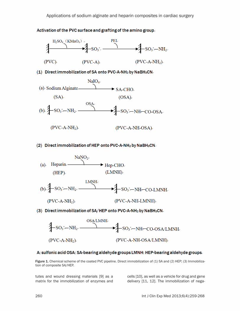
Figure 1. Chemical scheme of the coated PVC pipeline. Direct immobilization of (1) SA and (2) HEP; (3) Immobiliza-tion of composite SA/HEP.
tutes and wound dressing materials [9] as a matrix for the immobilization of enzymes and
cells [10], as well as a vehicle for drug and gene delivery [11, 12]. The immobilization of nega-
Applications of sodium alginate and heparin composites in cardiac surgery
261 Int J Clin Exp Med 2013;6(4):259-268
tively charged sulfated polysaccharides allows surfaces to become more biocompatible because of the electrostatic repulsion of nega-tively charged components of blood. Furthermore, the increased wettability caused by the presence of negatively charged groups may diminish protein adsorption or make it more reversible [13]. Thus, sulfated polysac-charides should be investigated as attractive alternatives for HEP to decrease the thrombo-genic activity and protein adsorption of artificial surfaces.
In this study, we modified a PVC pipeline to increase its biocompatibility and antithrombot-ic activity. The surface of the PVC pipeline was immobilized with a SA/HEP composite. The bio-compatibility and anticoagulation activity of the SA/HEP-coated surface was compared with HEP-coated and SA-coated surfaces. The study revealed that chemical modification provides PVCs with important clinical value.
Materials and methods
Reagents
PVC pipelines (10 cm2) were obtained from KE-Wei (Dong guan, China). SA, polyethylenei-mine (PEI), and sodium periodate were pro-cured from Sigma (St. Louis, USA). HEP (155 IU/mg) was supplied by Jin-xing Bio. (An-hui, China). Human serum albumin (HAS) was obtained from CSL Behring GmbH (Marburg, Germany), whereas human fibrinogen (HPF) was obtained from EMD Chemicals (San Diego, Germany). All other reagents were of analytical grade.
Surface modification
The PVC pipelines were cut into 10 cm2 pieces and acidized with concentrated sulfuric acid (with 2 g/l potassium permanganate) to form new carboxyl groups on the PVC surface. The acidized PVC samples were rinsed with double-distilled water three times to remove the unre-acted sulfuric acid. The carboxyl-bearing PVCs were immersed in PEI with multi-amino groups. The process of direct HEP coating is described in the chemical scheme in Figure 1-(2). The pro-cess of direct SA coating is described in the chemical scheme in Figure 1-(1). Using a differ-ent method, the SA/HEP-immobilized PVC sur-faces were prepared according to the scheme in Figure 1-(3). Amino-bearing PVC samples
were immersed in SA/HEP solution (pH 3.5) containing NaBH3CN at 40 °C for 3 h. NaBH3CN was used to couple the polysaccharides with the amino-bearing PVC surfaces.
Determination of surface grafting density
The surface density of the SA-bearing aldehyde groups (OSA) and the HEP-bearing aldehyde groups (LMNH) were measured using the phe-nol sulfuric acid procedure and the toluidine blue O method, respectively. The dye concen-tration was determined using an ultraviolet spectrometer (UV-2800; Hitachi, Japan).
Modified PVC characterization
The functional groups on modified PVC pipe-lines were analyzed using Fourier-transform infrared spectroscopy (FTIR; NICOLET 6700; Thermo, USA). The SA and HEP standards were determined using the potassium bromide tab-let mode (detector: DTGs KBr; beam splitter: KBr; wavelength range: 650 nm-4000 nm).
Protein adsorption measurements
The protein adsorption was measured for HAS and HPF using the bicinchoninic acid assay, as described by Ishihara et al. [16].
Blood coagulation time
Human whole blood from a healthy volunteer was collected and centrifuged at 800×g for 10 min at 4 °C to separate the blood corpuscles. The resulting platelet-rich plasma (PRP) was used for the platelet adhesion experiment. Subsequently, half PRP was centrifuged at 3000×g for 10 min at 4 °C to obtain the plate-let-poor plasma (PPP) for the test of human plasma protein adsorption. A sample PVC pipe-line (10 cm2) was incubated in 0.5 ml of PPP at 37 °C for 1 h. The activated partial thrombin time (APTT), thrombin time (TT), prothrombin time (PT), and fibrinogen time (FT) of PPP were then determined using an automated blood coagulation analyzer (STA-R Evolution®; Diagnostica Stago, France).
Evaluation of platelet adhesion
The concentration of human PRP before adhe-sion (PLT1) was determined with a hemocytom-eter (ADVIA2120; Siemens, Germany). The PVC pipeline samples (0.5 cm × 0.5 cm; 10 cm2) were washed three times with double-distilled

Applications of sodium alginate and heparin composites in cardiac surgery
262 Int J Clin Exp Med 2013;6(4):259-268
Figure 2. Qualitative analysis of immobilized HEP and immobilized SA. FTIR was used to detect the characteristic peak of uncoated PVC, HEP-coated, and SA-coated PVC, as well as the KBr tablets of HEP and SA. A. A strong and wide O-H adsorption peak appeared at 3352 cm-1 in the FITC of HEP and HEP-coated PVC, whereas the non-uncoat-ed surface had no characteristic peaks. B. A characteristic absorption peak appeared at 3450 cm-1 in the FITC of SA and SA-coated surface.
Figure 3. Platelet adhesion on different PVC surfaces: A. uncoated, (B) SA-coated, (C) HEP-coated, and (D) SA/HEP-coated. SEM (×5000) was used to detect the morphology of the PVC surface after platelet adhesion. Numer-ous platelets adhered to the uncoated surface (Figure A). Slightly less platelets adhered to the SA-coated and HEP-coated surfaces (Figure B and C, respectively). Much fewer platelets adhered to the SA/HEP-coated surface. SA and HEP improved the hemocompatibility of the PVC surface. SA/HEP coating can significantly inhibit platelet adhesion.
Applications of sodium alginate and heparin composites in cardiac surgery
263 Int J Clin Exp Med 2013;6(4):259-268
water and placed into 24-well culture plates incubated with human PRP (750 μl/well) in 5% CO2 at 37 °C for 60 min. After which, the con-centration of human PRP after adhesion (PLT2) was obtained.
Adhesion quantity = PLT1 - PLT2
Adhesion rate = (PLT1 - PLT2) / PLT1
The PVC pipeline samples were washed three times after incubation with PRP, and then fixed with 3% (w/v) GA solution in PBS for 2 h. The fixed samples were dehydrated with graded ethanol (50%, 60%, 70%, 80%, 90%, v/v; abso-lute alcohol) and dried using the critical-point procedure with CO2. The platelet adhesion mor-phology was observed using SEM after metal spraying.
Thrombus formation
The PVC pipeline samples were washed thrice with double-distilled water and placed into 24-well culture plates with 1.5 ml human whole blood per well. After which, the samples were incubated in 5% CO2 at 37 °C for 60 min, and then washed three times with PBS. The formed thrombus was fixed with 3% (w/v) GA solution in PBS for 1 h. The samples were dehydrated with graded ethanol and dried by the critical-point procedure with CO2. The degree of thrombosis
(DT) of the PVC pipeline at a given time was defined as follows:
DT = (W2 - W1) / W1,
where W1 and W2 are the weights of the dry PVC and blood coagula-tion samples, respecti- vely.
Statistical methods
Statistical analysis was performed using SPSS (version 17.0). All quanti-tative data were expre- ssed as the mean ± stan-dard deviation. Compari- sons among groups were performed using ANOVA (analysis of variance), whereas pairwise com-
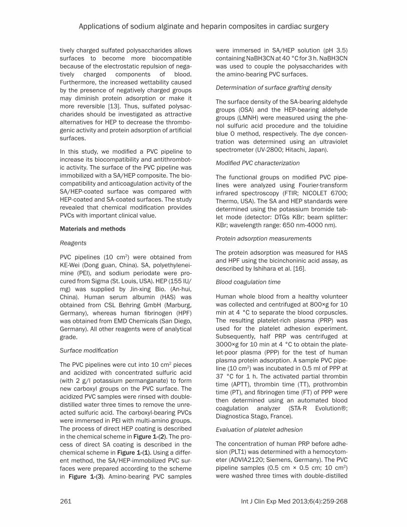
Figure 4. Comparison of anticoagulation time of different surfaces: (C) uncoated, (SA) SA-coated, (HEP) HEP-coated, and (SA/HEP) coated with the SA/HEP compos-ite. The coagulation time of APTT, TT, PT, and FT (n = 6) were evaluated by an auto-mated blood coagulation analyzer. APTT and TT of HEP-coated surface were signifi-cantly longer than those of non-coated surface. The SA-coated and SA/HEP-coated surface significantly prolonged the coagulation time of APTT and TT. Both HEP and SA demonstrated their anticoagulant properties by prolonging APTT and TT.
parisons used the Student-Newman-Kuels (SNK) q test. Differences with p < 0.05 were considered statistically significant.
Results
Surface characterization
SA/HEP-coated PVC surface is provided with a characteristic peak. A strong and wide O–H adsorption peak appeared at 3352 cm-1 in the fluorescein isothiocyanate (FITC) labeling of the HEP tablet and HEP-coated surface (Figure 2A). Characteristic absorption peaks appeared at 3450 cm-1 in FITC labeling of the SA tablet and SA-coated surface (Figure 2B).
Surface grafting density
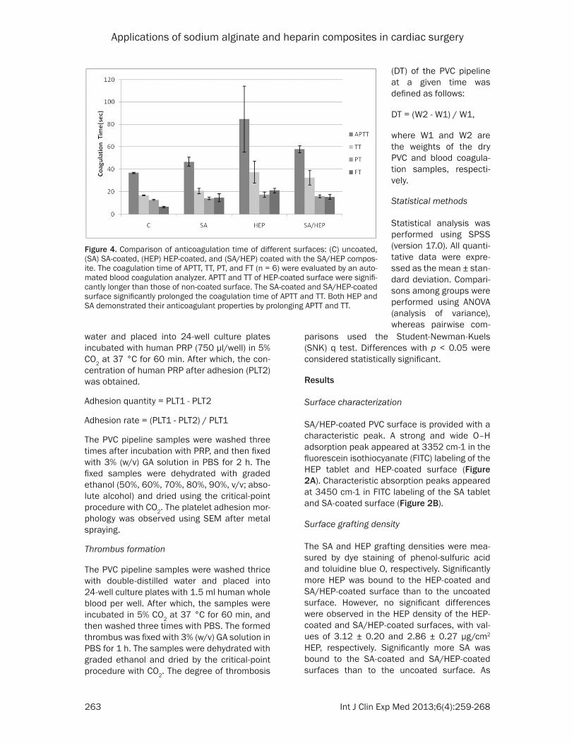
The SA and HEP grafting densities were mea-sured by dye staining of phenol-sulfuric acid and toluidine blue O, respectively. Significantly more HEP was bound to the HEP-coated and SA/HEP-coated surface than to the uncoated surface. However, no significant differences were observed in the HEP density of the HEP-coated and SA/HEP-coated surfaces, with val-ues of 3.12 ± 0.20 and 2.86 ± 0.27 μg/cm2 HEP, respectively. Significantly more SA was bound to the SA-coated and SA/HEP-coated surfaces than to the uncoated surface. As
Applications of sodium alginate and heparin composites in cardiac surgery
264 Int J Clin Exp Med 2013;6(4):259-268
shown in Table 1, slightly more SA was bound to the SA-coated surface than to the SA/HEP-coated surface.
Protein adsorption
Protein adsorption was measured by ultraviolet spectrometry from solutions containing various concentrations of purified HAS and HPF. The amount of protein adsorbed by the uncoated, SA-coated, HEP-coated, and SA/HEP-coated surfaces was depicted in Table 2, as a function of their concentration in the free solution. The two proteins that were adsorbed by the SA-coated surface were lower (HAS 3.49 ± 0.86, HPF 9.24 ± 1.71) than those adsorbed by
the non-coated surface (HAS 15.11 ± 0.64, HPF 23.66 ± 0.43) with a significant differ-ence. Slightly more proteins were adsorbed by the SA/HEP-coated surface than by the SA-coated surface. The HEP-coated surface adsorbed higher amounts of HAS and HPF than the SA-coated surface.
Platelet adhesion
The equilibrium platelet adhe-sion and activation on the sur-face was related to the pro-tein adhesion. The maximum number of adhering platelets
Table 1. Grafting density on different surfaces (mean ± SD)Surface type n Sodium alginate1 Heparin2
μg μg/cm2 μg μg/cm2
C 6 2.22 ± 0.15 0.45 ± 0.03 1.01 ± 0.60 0.50 ± 0.30SA 6 52.76 ± 0.62* 10.55 ± 0.12* _ _HEP 6 _ _ 6.24 ± 0.41* 3.12 ± 0.20*
SA/HEP 6 38.18 ± 0.58* 7.64 ± 0.12* 5.72 ± 0.54* 2.86 ± 0.27*
F1 = 10213.17, F2 = 110.19, *p < 0.05.
Table 2. Plasma protein adsorption onto the PVC surfaces (mean ± SD)
PVC pipeline type n HAS adsorption1 (μg/cm2) HPF adsorption2 (μg/cm2)C 6 15.11 ± 0.64 23.66 ± 0.43SA 6 3.49 ± 0.86* 9.24 ± 1.71*
HEP 6 13.88 ± 1.52 24.06 ± 0.42SA/HEP 6 5.33 ± 0.94* 8.02 ± 2.93*
F1 = 87.29, F2 = 85.38, *p < 0.05.
Table 3. Platelet adhesion onto different PVC surfaces (mean ± SD)
Surface type n Adhesion1 Adhesion rate (%)2
C 6 49.67 ± 11.33 18.31 ± 4.18SA 6 12.83 ± 2.83 4.73 ± 1.04*
HEP 6 1.17 ± 1.22* 0.43 ± 0.45*
SA/HEP 6 4.33 ± 2.44* 1.60 ± 0.90*
F1 = 45.94, F2 = 45.90, *p < 0.05.
Table 4. Thrombin formation of different surfaces (mean ± SD)
Group type n Adhesion weight (mg)1 DT (%)2
C 5 67.00 ± 3.2 7.08 ± 0.34SA 5 48.94 ± 1.27* 5.17 ± 0.13*
HEP 5 45.32 ± 0.45* 4.79 ± 0.05*
SA/HEP 5 46.86 ± 1.63* 4.95 ± 0.17*
F1 = 67.20, F2 = 39.80, *p < 0.05.
on the PVC surfaces after the 2 h incubation appeared on the uncoated surface (Figure 3A). The platelet adhesion on the HEP-coated and SA-coated surfaces slightly changed after the 2 h incubation, as shown in Table 3 and Figure 3.
Coagulation time
The anticoagulant effects of HEP and SA can be observed by comparing the APTT, TT, PT, and FT of the uncoated surface with those of the HEP-coated, SA-coated, and SA/HEP-coated surfac-es. Results showed the different clotting times of different surface types. The clotting times of the uncoated surface were nearly the same as those of the human plasma, whereas those of

Applications of sodium alginate and heparin composites in cardiac surgery
265 Int J Clin Exp Med 2013;6(4):259-268
the SA/HEP-coated surfaces were longer than those of the uncoated surfaces. The clotting times of HEP-coated surface were 50, 21, 5, 14 s (for APTT, TT, PT and FT, respectively), which were longer than those of the uncoated sur-face. The SA/HEP-coated surface prolonged the APTT, TT, PT and TT, such that the APTT and TT of the HEP-coated surface were almost twice that of the uncoated surface, as shown in Figure 4.
Thrombin formation
Thrombin formation is undesirable during CPB. Antithrombogenic materials have been of great interest in the development of cardiac surgery. The effect of surface modification on thrombin formation is shown in Table 4 and Figure 5. The HEP-coated surface can reduce thrombin for-mation by about two-thirds as compared with
the uncoated surface. The SA-coated surface can likewise significantly decrease thrombus formation. The SA/HEP-coated surface has slightly less thrombin formation than the SA-coated surface. Furthermore, thrombus for-mation increased with incubation time, which agreed with the results of Kang et al. [3].
Discussion
Thrombosis and hemocyte damage are the main problems of uncoated biomaterials that are applied to cardiac surgery. HEP, as an anti-coagulant, has been used to prevent thrombo-sis during CPB. However, the systemic intrave-nous administration of HEP can cause serious side effects, including hemorrhage and HIT. Biomedical materials are broadly used in extra-corporeal circulation, hemodialysis, and hemo-filtration. Biocompatibility and hemocompatibil-
Figure 5. Thrombin formation of different surfaces: A. uncoated, (B) SA-coated, (C) HEP-coated, and (D) SA/HEP-coated. SEM (×5000) was used to detect the morphology of PVC surfaces after 2 hour incubation in fresh whole blood cells. The thrombin complex appeared on uncoated PVC (Figure A). Slightly less thrombin appeared on the SA-coated surface (Figure B). Much less thrombin appeared on the HEP-coated and SA/HEP-coated surfaces (Fig-ure C and D, respectively). SA and HEP are both antithrombogenic, with the same trends as those of the platelet adhesion test.
Applications of sodium alginate and heparin composites in cardiac surgery
266 Int J Clin Exp Med 2013;6(4):259-268
ity are the main problems during the application of synthetic biomedical materials [17], which may trigger the host defense mechanism when exposed to blood, thereby causing a cascade of thrombosis and thromboembolism. Coating techniques [18] can improve the biocompatibil-ity and hemocompatibility of biomedical materi-als via the modification of the materials’ sur-face. The anticoagulant properties of biomedical materials can be obtained by sup-pressing or preventing four pathways, including coagulation factor activation, platelet adhesion and aggregation, erythrocyte adhesion, and complement system activation. Several princi-ples are used to design the coating materials, including protein adhesion resistance, decreased platelet adhesion and aggregation, inhibition of intrinsic coagulation factor activa-tion, and inhibition of thrombosis. HEP, as an acid mucopolysaccharide, exerts its anticoagu-lant property by accelerating or increasing the activity of coagulation inhibiting factors, espe-cially AT-III [19, 20]. The clinical application of HEP is mainly via intravenous injection, which is coupled with side effects [21] such as hemor-rhage and thrombocytopenia. Moreover, intra-venous HEP cannot facilitate continuous anti-coagulation. A previous study indicated that HEP coating has poor biocompatibility and could not decrease protein adhesion [22]. SA, as a polyanionic macromolecule, is natural and non-toxic, with high biocompatibility and antico-agulant effects [20, 23, 24]. A composite coat-ing with HEP and SA may improve the biocom-patibility and hemocompatibility of biomedical materials.
The results in this study revealed that SA and HEP were constantly immobilized on the PVC surface by surface modification. The partial degradation of HP [25, 26] and SA [27, 28] with nitrous acid and sodium periodate gives rise to the fraction of molecules with highly reactive aldehyde terminal groups. The end-point bond is established between the aldehyde terminal groups and primary amino functions by reduc-tive amination [29-31]. The surface density and stability of functional molecules are important because the enzyme inhibiting capacity may be associated with the surface density. The quan-titative experiment showed that the coating density of SA or HEP has no significant influ-ence on the single coating and composite coat-ing. The surface contact angles are significantly decreased in the SA-coated and SA/HEP-
coated surfaces as compared with the HEP-coated surface, thereby revealing that the SA coating can improve the hydrophilicity of the PVC surface. The nature of the adsorbed pro-tein is believed to determine all adverse events that impair the use of biomaterials in medical devices: thrombus formation, platelet activa-tion, initiation of coagulation [32-34], and acti-vation of the complement system, which, in turn, causes leukocyte adhesion and activation [35]. The SA-coated surface can significantly decrease the level of protein adhesion. The SA/HEP-coated surface has slightly less protein adhesion than the HEP-coated surface. The protein adhesion study showed that the appli-cation of SA can improve the biocompatibility of the single HEP-coated surface and decrease the adverse effects.
Hemocompatibility is an important property of modified PVC. The partial degradation and covalent binding of HEP and SA could inevitably lead to the complete loss of antithrombin-bind-ing. The ideal concept of SA and HEP binding includes an inherently stable covalent end-point bond of the molecules, thereby leaving the other parts of the molecule intact to react with the blood constituents [36]. The blood coagulation cascade includes intrinsic, extrin-sic, and common pathways. APTT and PT are used to examine the intrinsic and common pathways, whereas FT is used to measure the time for transferring fibrinogen into fibrin [37, 38]. Results showed that the HEP coating can significantly prolong the coagulation time of APTT and TT by inhibiting the intrinsic coagula-tion pathway. The SA coating produced a slight anticoagulation effect by prolonging the APTT. The anticoagulation effect of SA/HEP is inferior to that of the single HEP coating, but the APTT and TT of SA/HEP are likewise significantly pro-longed. The results revealed that composite coating demonstrated its anticoagulant prop-erty by inhibiting the intrinsic coagulation path-way. Furthermore, the fragmentation of SA and HEP did not destroy their functional molecules, and the binding procedure did not shield the anticoagulant molecules. The SA/HEP compos-ite coating can decrease platelet adhesion and thrombosis, with the same trend as that of the coagulation time.
The immobilization of SA/HEP on PVC surfaces to incorporate endothelium-like antithrombo-genic properties appears to be based on sound
Applications of sodium alginate and heparin composites in cardiac surgery
267 Int J Clin Exp Med 2013;6(4):259-268
rationale. The ideal density and stability of the SA/HEP coating may mimic the function of nat-ural endothelial cells and express continuous anticoagulant properties.
Conclusion
The immobilized SA/HEP composite can improve the hemocompatibility of the PVC sur-face. The SA coating combined with the HEP coating could increase the hemocompatibility, and enhance the biocompatibility of the PVC surfaces. Thus, the process for immobilizing SA/HEP is applicable for the PVC pipeline in CPB. The results suggested that PVC pipelines immobilized with SA/HEP can significantly reduce the administration of HEP in cardiac surgery, and may even lead to HEP-free thera-py. The SA/HEP coatings represent the begin-ning of PVCs with improved hemocompatibility and biocompatibility in the presence of human blood or tissues. Intelligent CPB materials with near-complete physiological surfaces will be available for surgical use in the near future.
Acknowledgment
This work was partly supported by research grants from Tianjin Key items of Scientific Supporting Project awarded to Tong Li (11ZCGYSY02000).
Address correspondence to: Dr. Tong Li, Department of Heart Center, The Third Central Hospital, 83 Jintang Road, Hedong District, Tianjin, China. Phone: 86-22-8411-2006; Fax: 22-2431-5132; E-mail: [email protected]
References
[1] Vroman L, Adams AL. Identification of rapid changes at plasmasolid interfaces. J Biomed Mater Res 1969; 3: 43-67.
[2] Zhang M, Desai T, Ferrari M. Proteins and cells on PEG immobilized silicon surfaces. Biomate-rials 1998; 19: 953-960.
[3] Tanaka M, Motomura T, Kawada M, Anzai T, Kasori Y, Shiroya T, Shimura K, Onishi M, Mo-chizuki A. Blood compatible aspects of poly (2-methoxyethylacrylate) (PMEA)-relationship between protein adsorption and platelet adhe-sion on PMEA surface. Biomaterials 2000; 21: 1471-1481.
[4] Christensen K, Larsson R, Emanuelsson H, El-gue G, Larsson A. Coagulation and comple-
ment activation. Biomaterials 2001; 22: 349-355.
[5] Rollason G, Sefton MV. Inactivation of throm-bin in heparin-PVA coated tubes. J Biometer Sci Polym Ed 1989; 1: 31-41.
[6] Byun Y, Jacobs HA, Kim SW. Binding kinetics of thrombin and antithrombin III with mmobilized heparin using a spacer. ASAIO Journal 1992; 38: M649-653.
[7] Nowak G. Anticoagulation with r-hirudin in reg-ular hemodialysis with heparin-induced throm-bocytopenia (HIT II). Wien Klin Wochenschr 1997; 109: 343-345.
[8] Liaw PCY, Becker DL, Stafford AR, Fredenburgh JC, Weitz JI. Molecular basis for he susceptibil-ity of fibrin-bound thrombin to inactivation by heparin cofactor II in he presence of dermatan sulfate but not heparin. J Biol Chem 2001; 276: 20959-20965.
[9] Murakami K, Aoki H, Nakamura S, Takikawa M, Hanzawa M, Kishimoto S, Hattori H, Tanaka Y, Kiyosawa T, Sato Y, Ishihara M. Hydrogel blends of chitin/chitosan, ucoidan and algi-nate as healing-impaired wound dressings. Biomaterials 2010; 31: 83-90.
[10] Leonard M, De Boisseson MR, Hubert P, Dalen-con F, Dellacherie E. Hydrophobically modified alginate hydrogels as protein carriers with spe-cific controlled release properties. J Control Release 2004; 98: 395-405.
[11] Del Gaudio P, Russo P, Rosaria Lauro M, Co-lombo P, Aquino RP. Encapsulation of ketopro-fen and ketoprofen lysinate by prilling for con-trolled drug release. AAPS PharmSciTech 2009; 10: 1178-1185.
[12] Khanna O, Moya ML, Opara EC, Brey EM. Syn-thesis of multilayered alginate microcapsules for the sustained release of fibroblast growth factor-1. J Biomed Mater Res A 2010; 95: 632-640.
[13] Lens JP, Terlingen JG, Engbers GH, Feijen J. Preparation of heparin-like surfaces by intro-ducing sulfate and carboxylate groups on poly(ethylene) using an argon plasma treat-ment. J Biomater Sci Polym Ed 1998; 9: 357-372.
[14] Rehm BH, Valla S. Bacterial alginates: biosyn-thesis and applications. Appl Microbiol Bio-technol 1997; 48: 281-288.
[15] Segal HC, Hunt BJ, Gilding K. The effects of al-ginate and nonalginate wound dressings on blood coagulation and platelet activation. J Biomater Appl 1998; 12: 249-257.
[16] Ishihara K, Fukumoto K, Iwasaki Y, Nakabayas-hi N. Modification of polysulfone with phospho-lipid polymer for improvement of the blood compatibility. Part 1. Surface characterization. Biomaterials 1999; 20: 1545-1551.
Applications of sodium alginate and heparin composites in cardiac surgery
268 Int J Clin Exp Med 2013;6(4):259-268
[17] Gunaydin S. Clinical significance of coated ex-tracorporeal circuits: a review of novel tech-nologies. Perfusion 2004; 19: S33-41.
[18] Murugesan S, Xie J, Linhardt RJ. Immobiliza-tion of Heparin: Approaches and Applications. Curr Top Med Chem 2008; 8: 80-100.
[19] Petitou M, Barzu T, Herault JP, Herbert JM. A unique trisaccharide sequence in heparin me-diates the early step of antithrombin IIIactiva-tion. Glycobiology 1997; 7: 323-327.
[20] Bouhadir KH, Lee KY, Alsberg E, Damm KL, An-dereson KW, Mooney DJ. Degradation of par-tially oxidized alginate and its potential appli-cation for tissue engineering. Biotechnol Prog 2001; 17: 945-995.
[21] Santerre JP, Woodhouse K, Laroche G, Labow RS. Understanding the biodegradation of poly-urethanes: from classical implants to tissue engineering materials. Biomaterials 2005; 26: 7457-7470.
[22] Keuren JF, Wielders SJ, Willems GM, Morra M, Cahalan L, Cahalan P, Lindhout T. Thromboge-nicity of polysaccharide-coated surfaces. Bio-materials 2003; 24: 1917-1924.
[23] Nishino T, Yokoyama G, Dobashi K, Fujihara M, Nagumo T. Isolation, purification, and charac-terization of fucose-containing sulfated poly-saccharides from the brown seaweed Ecklonia kurome and their blood-anticoagulant activi-ties. Carbohydr Res 1989; 186: 119-148.
[24] Manju S, Muraleedharan CV, Rajeev A, Jay-akrishnan A, Joseph R. Evaluation of alginate dialdehyde cross-linked gelatin hydrogel as a biodegradable sealant for polyester vascular graft. J Biomed Mater Res B Appl Biomater 2011; 98: 139-188.
[25] Barnett WE. Improved anticoagulant sub-stance. WO 81/03276. 1981.
[26] Hovanessian HC. New-generation anticoagu-lants: the low molecular weight heparins. Ann Emerg Med 1999; 34: 768-779.
[27] Lee KY, Mooney DJ. Hydrogels for tissue engi-neering. Chem Rev 2001; 101: 1869-1879.
[28] Bouhadir KH, Lee KY, Alsberg E, Damm KL, An-derson KW, Mooney DJ. Degradation of par-tially oxidized alginate and its potential appli-
cation for tissue engineering. Biotechnol Prog 2001; 17: 945-950.
[29] Larm O, Larsson R, Olsson P. A new non-throm-bogenic surface prepared by selective cova-lent binding of heparin via a modified reducing terminal residue. Biomater Med Devices Artif Organs 1983; 11: 161-173.
[30] Larsson R, Larm O, Olsson P. The search for thromboresistance using mmobilized heparin. Ann NY Acad Sci 1987; 516: 102-115.
[31] Hoffman J, Larm O, Scholander E. A new meth-od for covalent coupling of heparin and other glycosaminoglycans to substances containing primary amino groups. Carbohydrate Res 1983; 117: 328-331.
[32] Vroman L, Adams AL. Identification of rapid changes at plasmasolid interfaces. J Biomed Mater Res 1969; 3: 43-67.
[33] Zhang M, Desai T, Ferrari M. Proteins and cells on PEG immobilized silicon surfaces. Biomate-rials 1998; 19: 953-960.
[34] Tanaka M, Motomura T, Kawada M, Anzai T, Kasori Y, Shiroya T, Shimura K, Onishi M, Mo-chizuki A. Blood compatible aspects of poly (2-methoxyethylacrylate) (PMEA)-relationship between protein adsorption and platelet adhe-sion on PMEA surface. Biomaterials 2000; 21: 1471-1481.
[35] Christensen K, Larsson R, Emanuelsson H, El-gue G, Larsson A. Improved blood compatibility of a stent graft by combining heparin coating and abciximab. Thromb Res 2005; 115: 245-53.
[36] Islam T, Butler M, Sikkander SA, Toida T, Lin-hardt RJ. Further evidence that periodate cleavage of heparin occurs primarily through the antithrombin binding site. Carbohydr Res 2002; 337: 2239-2243.
[37] Favaloro EJ, Bonar R, Sioufi J, Wheeler M, Low J, Aboud M, LIoyd J, Street A, Marsden K. An international survey of current practice in the laboratory assessment of anticoagulant thera-py with heparin. Pathology 2005; 37: 234-238.
[38] Bonar RA, Favaloro EJ, Marsden K. External quality assurance for heparin monitoring. Se-min Thromb Hemost 2012; 38: 632-639.
Related Documents


