Genetica 118: 233–244, 2003. © 2003 Kluwer Academic Publishers. Printed in the Netherlands. 233 Origin and evolution of a new gene expressed in the Drosophila sperm axoneme Jos´ e Mar´ ıa Ranz 1 , Ana Rita Ponce 1 , Daniel L. Hartl 1,∗ & Dmitry Nurminsky 2 1 Department of Organismic and Evolutionary Biology, Harvard University, Cambridge, MA 33143, USA; 2 Department of Anatomy and Cell Biology, Tufts University School of Medicine, Boston, MA 02111, USA; ∗ Author for correspondence: (Phone: +1-617-496-3917; Fax: +1-617-496-5854; E-mail: [email protected]) Key words: axoneme, dynein intermediate chain, exon shuffle, gene fusion, spermatogenesis Abstract Sdic is a new gene that evolved recently in the lineage of Drosophila melanogaster. It was formed from a duplica- tion and fusion of the gene AnnX, which encodes annexin X, and Cdic, which encodes the intermediate polypeptide chain of the cytoplasmic dynein. The fusion joins AnnX exon 4 with Cdic intron 3, which brings together three putative promoter elements for testes- specific expression of Sdic: the distal conserved element (DCE) and testes- specific element (TSE) are derived from AnnX, and the proximal conserved element (PCE) from Cdic intron 3. Sdic transcription initiates within the PCE, and translation is initiated within the sequence derived from Cdic intron 3, continuing through a 10 base pair insertion that creates a new splice donor site that enables the new coding sequence derived from intron 3 to be joined with the coding sequence of Cdic exon 4. A novel protein is created lacking 100 residues at the amino end that contain sequence motifs essential for the function of cytoplasmic dynein intermediate chains. Instead, the amino end is a hydrophobic region of 16 residues that resembles the amino end of axonemal dynein intermediate chains from other organisms. The downstream portion of Sdic features large deletions eliminating Cdic exons v2 and v3, as well as multiple frameshift deletions or insertions. The new protein becomes incorporated into the tail of the mature sperm and may function as an axonemal dynein intermediate chain. The new Sdic gene is present in about 10 tandem repeats between the wildtype Cdic and AnnX genes located near the base of the X chromosome. The implications of these findings are discussed relative to the origin of new gene functions and the process of speciation. Abbreviations: dynein IC – dynein intermediate polypeptide chain; DCE – distal conserved element; PCE – proximal conserved element; TSE – testes-specific element. Introduction The evolution of novel gene functions is thought to occur primarily by one of two mechanisms, either by duplication and divergence or by exon shuffling. Both mechanisms are known to occur. There are many examples of evolution by gene duplication in Drosophila. These are usually recognized by similar- ities between paralogous genes sometimes, but not al- ways, found in small gene clusters, such as the maltase gene cluster (Snyder & Davidson, 1983), the chorion The authors Jos´ e Mar´ ıa Ranz and Ana Rita Ponce contributed equally to this work. protein gene cluster (Martinez-Cruzado et al., 1988), the larval cuticle protein gene cluster (Steinemann & Steinemann, 1990), the alcohol dehydrogenase and al- cohol dehydrogenase-related genes (Jeffs, Holmes & Ashburner, 1994), and the alpha-esterase gene cluster (Robin et al., 1996). These examples are by no means exhaustive, and many more can be found in FlyBase (http://flybase.bio.indiana.edu). The other primary mechanism for creating new gene functions is exon shuffling (Gilbert, 1978; Long, Rosenberg & Gilbert, 1995; Long, 2001). There are also examples of exon shuffling in Drosophila. Per- haps the most dramatic is that of jingwei, a newly

Welcome message from author
This document is posted to help you gain knowledge. Please leave a comment to let me know what you think about it! Share it to your friends and learn new things together.
Transcript
Genetica 118: 233–244, 2003.© 2003Kluwer Academic Publishers. Printed in the Netherlands.
233
Origin and evolution of a new gene expressed in the Drosophilasperm axoneme�
Jose Marıa Ranz1, Ana Rita Ponce1, Daniel L. Hartl1,∗ & Dmitry Nurminsky2
1Department of Organismic and Evolutionary Biology, Harvard University, Cambridge, MA 33143, USA;2Department of Anatomy and Cell Biology, Tufts University School of Medicine, Boston, MA 02111, USA; ∗Authorfor correspondence: (Phone: +1-617-496-3917; Fax: +1-617-496-5854; E-mail: [email protected])
Key words: axoneme, dynein intermediate chain, exon shuffle, gene fusion, spermatogenesis
Abstract
Sdic is a new gene that evolved recently in the lineage ofDrosophila melanogaster. It was formed from a duplica-tion and fusion of the geneAnnX, which encodes annexin X, andCdic, which encodes the intermediate polypeptidechain of the cytoplasmic dynein. The fusion joinsAnnX exon 4 withCdic intron 3, which brings together threeputative promoter elements for testes- specific expression ofSdic: the distal conserved element (DCE) and testes-specific element (TSE) are derived fromAnnX, and the proximal conserved element (PCE) fromCdic intron 3.Sdic transcription initiates within the PCE, and translation is initiated within the sequence derived fromCdic intron3, continuing through a 10 base pair insertion that creates a new splice donor site that enables the new codingsequence derived from intron 3 to be joined with the coding sequence ofCdic exon 4. A novel protein is createdlacking 100 residues at the amino end that contain sequence motifs essential for the function of cytoplasmic dyneinintermediate chains. Instead, the amino end is a hydrophobic region of 16 residues that resembles the amino endof axonemal dynein intermediate chains from other organisms. The downstream portion ofSdic features largedeletions eliminatingCdic exons v2 and v3, as well as multiple frameshift deletions or insertions. The new proteinbecomes incorporated into the tail of the mature sperm and may function as an axonemal dynein intermediatechain. The newSdic gene is present in about 10 tandem repeats between the wildtypeCdic andAnnX genes locatednear the base of the X chromosome. The implications of these findings are discussed relative to the origin of newgene functions and the process of speciation.
Abbreviations: dynein IC – dynein intermediate polypeptide chain; DCE – distal conserved element; PCE –proximal conserved element; TSE – testes-specific element.
Introduction
The evolution of novel gene functions is thought tooccur primarily by one of two mechanisms, eitherby duplication and divergence or by exon shuffling.Both mechanisms are known to occur. There aremany examples of evolution by gene duplication inDrosophila. These are usually recognized by similar-ities between paralogous genes sometimes, but not al-ways, found in small gene clusters, such as the maltasegene cluster (Snyder & Davidson, 1983), the chorion
� The authors Jose Marıa Ranz and Ana Rita Ponce contributedequally to this work.
protein gene cluster (Martinez-Cruzado et al., 1988),the larval cuticle protein gene cluster (Steinemann &Steinemann, 1990), the alcohol dehydrogenase and al-cohol dehydrogenase-related genes (Jeffs, Holmes &Ashburner, 1994), and the alpha-esterase gene cluster(Robin et al., 1996). These examples are by no meansexhaustive, and many more can be found in FlyBase(http://flybase.bio.indiana.edu).
The other primary mechanism for creating newgene functions is exon shuffling (Gilbert, 1978; Long,Rosenberg & Gilbert, 1995; Long, 2001). There arealso examples of exon shuffling inDrosophila. Per-haps the most dramatic is that ofjingwei, a newly
234
evolved gene of unknown function in the lineage lead-ing to D. teissieri and D. yakuba (Long & Langley,1993; Wang et al., 2000). The chimericjingwei genewas created by the insertion of part of a reversetranscript of the alcohol dehydrogenase (Adh) codingsequence into the third intron of a different gene, de-noted yande. The jingwei coding sequence therebyincludes the initialyande exon to which is appendedthe ‘shuffled’ and already splicedAdh exons.
This paper examines a novel gene that originated asa duplication and gene fusion accompanied by recruit-ment of new promoter elements and the formation of anew exon encoding the amino end of the polypeptidechain. These events fused exon 4 of the geneAnnX,which encodes an annexin protein, with intron 3 ofthe geneCdic, which encodes the intermediate poly-peptide chain for the cytoplasmic dyneins. The newgene, calledSdic (for sperm-specific dynein interme-diate chain), is expressed primarily if not exclusivelyin testes, and it encodes a protein that features a re-fashioned amino end to which is appended much ofthe carboxyl end of what originally encoded a cyto-plasmic dynein intermediate chain. The Sdic proteinlocalizes to the sperm tail and may function as anaxonemal dynein intermediate chain. TheSdic genefeatures some unprecedented ‘fudging’ of the geneticfunctions: exon 4 sequences in the wildtypeAnnXgene have become a part of the Sdic promoter that isnot transcribed, and intron 3 sequences in the wildtypeCdic gene that have no preexisting coding functionnow, through multiple mutations, including a 10-bpinsertion, encode the amino end of the Sdic protein.Remarkably, the sperm-specific promoter element wasformed by the gene fusion itself. In addition, theSdicgene has become tandemly duplicated and is presentin about 10 copies in the base of the X chromosomeof D. melanogaster. The origin and evolution of thisgene and its tandem duplicates is recent, since it is notfound in closely related species that diverged withinthe last 1–3 million years.
Origin of the chimeric Sdic gene
The Sdic gene was discovered through an anomalouscDNA recovered in a study of alternative splicingof cytoplasmic intermediate-chain dynein transcripts(Nurminsky et al., 1998a). Cytoplasmic dynein is amultisubunit complex composed of two heavy chains,three intermediate chains (ICs), several light interme-diate chains (LICs), and one light chain (LC) (Paschal
et al., 1992; King et al., 1996). It acts as a minusend-directed microtubule motor, participating in anumber of events, including slow axonal transport(Dillman, Dabney & Pfister, 1996), anterograde or-ganelle movement (Schroer, Steuer & Sheetz, 1989;Corthesy-Theulaz, Pauloin & Rfeffer, 1992; Anientoet al., 1993), mitosis (Vaisberg, Koonce & McIntosh,1993), and nuclear migration (Xiang, Beckwith &Morris, 1994).
Although the heavy chain comprises the catalyticdynein subunit and by itself can bring about an ATP-dependent force on the microtubules (Mazumdar et al.,1996), the presence of other subunits is apparently re-quired for dynein functionin vivo. The roles of theseso-called accessory subunits still remain unclear. Atleast two accessory subunits of cytoplasmic dynein,the intermediate-chain and light-chain subunits, sharesignificant homology with the corresponding subunitsof the axonemal dyneins, suggesting similarity of theirfunctions in these two complexes. In the case of theICs, this suggestion lead to the hypothesis that theIC subunits are responsible for linking the cytoplas-mic dynein to the intracellular targets (Paschal et al.,1992), because in the axonemal dyneins the ICs havebeen localized in the base of complex, binding di-rectly to the A-microtubule. The ICs of cytoplasmicdyneins possess a large carboxyl-terminal portion con-taining a series of WD-40 repeats, which is presentin the axonemal ICs as well (Wilkerson et al., 1995).The amino-terminal part shows no homology with theaxonemal ICs.
In Drosophila, the multiple forms of the dynein ICsare created by alternative splicing of the transcript of asingle-copy gene, denotedCdic, located in region 19Enear the base of the X chromosome (Nurminsky et al.,1998a). The anomalous IC cDNA was unusual in thatthe apparent amino end of the coding sequence wasmissing both the coiled-coil domain and the serine-rich domain necessary for the interaction betweendynein intermediate chains and the p150/Glued pro-tein that participates in attaching the dynein complexto its cytoplasmic targets. Instead, the amino-terminalend of the protein had a novel sequence that was veryhydrophobic (Nurminsky et al., 1998a).
The region of the genome encoding this anoma-lous cDNA was also in the base of the X chromosomenear Cdic. Immediately upstream of the transcrip-tion start site was a sequence derived from the genefor Annexin X, denotedAnnX. The annexins are alarge family of proteins that bind to phospholipids ina calcium-dependent manner. They appear to have a
235
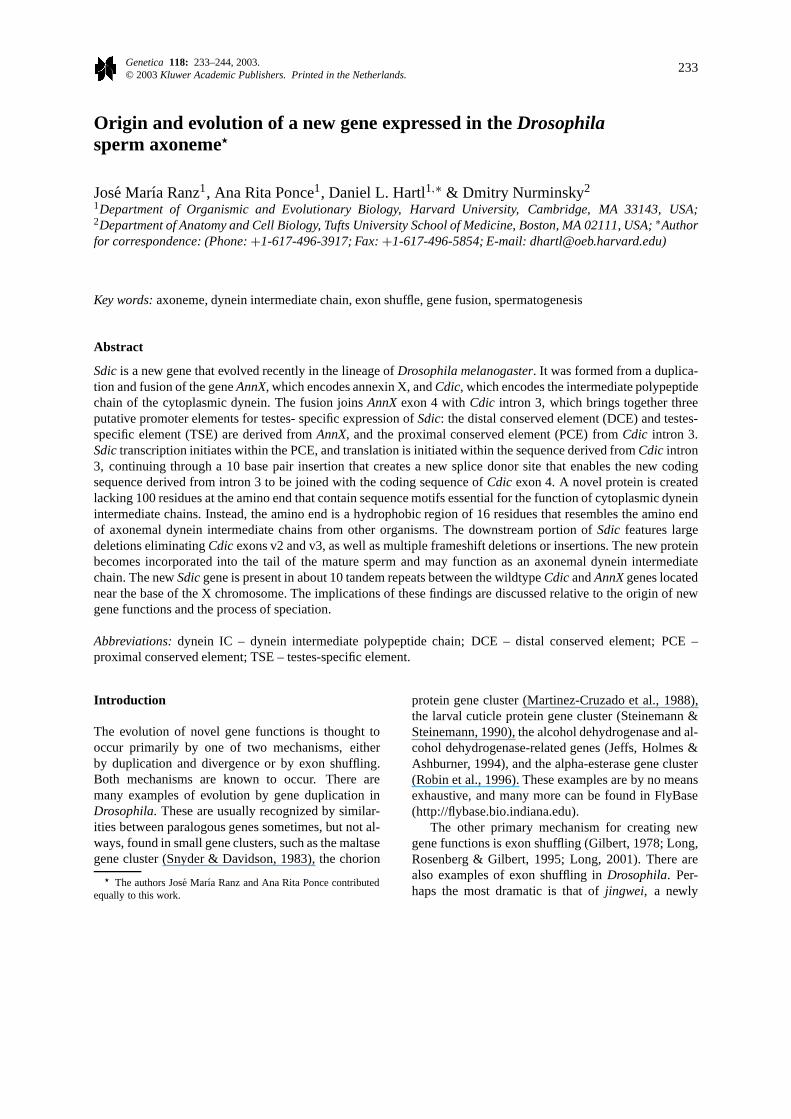
Figure 1. Origin and structure ofSdic. (A) A duplication of the region containingCdic (C) andAnnX (A) took place, followed (or accompanied)by several large deletions that created a gene fusion betweenAnnX andCdic [AC], which was the progenitor of the geneSdic (S). In present-daypopulations ofD. melanogaster, Sdic is present in about 10 nonidentical tandem repeats. (B) TheSdic promoter elements were formed by thefusion ofAnnX exon 4 (DCE and TSE) andCdic intron 3 (PCE). The amino end of the new Sdic protein derives from sequences inCdic intron3 and a 10-bp insertion that creates a new spice donor site that is spliced to the normal 3′ splice acceptor site ofCdic intron 3. The exons aredesignated as in Nurminsky et al. (1998a).
wide variety of functions and have been implicatedin cytoskeletal interactions, phospholipase inhibition,intracellular signalling, anticoagulation, membranefusion, and apoptosis (Barton et al., 1991; Geisow,1991).
A curious observation was that, in the region tran-scribed to yield the anomalous cDNA, the sequencesimilar to AnnX was upstream of the 5′ end of theCdic-like cDNA, whereas in the genome the positionof these genes is reversed. The inferred explanationfor this orientation is shown in Figure 1(A). Priorto the origin of Sdic, the Cdic (C) and AnnX (A)genes were both single-copy genes orientedC A, as
shown at the left. A duplication of this region ledto the configurationC A C A, and a series of atleast three large deletions fused the 3′ end of AnnXwith the 5′ end of Cdic, producing the configura-tion C [AC] A, where the square brackets denote thegene fusion. There is at present no way of know-ing when these deletions occurred, nor the order inwhich they occurred. One possibility is that they alltook place simultaneously with the formation of theduplication, another possibility is that they occurredsequentially after the duplication was already in place,and there are also other scenarios. Whatever the pro-cess, the [AC] fusion created the framework ofSdic
236
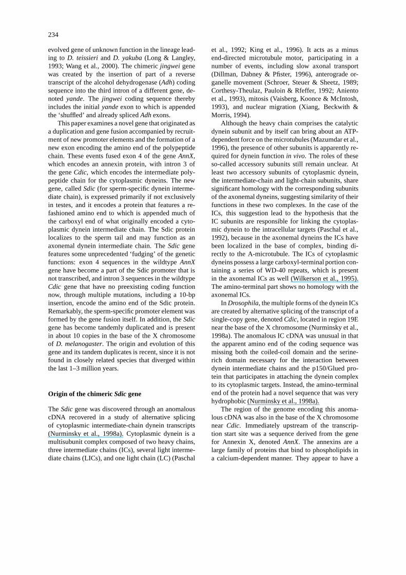
Figure 2. Sequence of theSdic promoter and the wildtype sequences ofAnnX exon 4 andCdic intron 3. Single underline, distal conservedelement (DCE); wavy underline, testes-specific element (TSE); double underline, proximal conserved element (PCE); dashed underline ATG,initial codon of Sdic protein.
(S in Figure 1(A)), which has also undergone abouta tenfold amplification yielding the present config-uration C S S · · · S S A (Nurminsky et al., 1998b).Evidence that the gene is newly evolved is that itis present in all wildtype strains ofD. melanogasterso far examined, but neither the novel gene nor anyevidence of a tandem repeat is found in wildtypestrains ofD. simulans nor in any other member of theD. melanogaster species subgroup (Nurminsky et al.,1998b).
Molecular structure of the Sdic gene
The molecular structure of anSdic repeating unit isshown in Figure 1(B). Within theSdic cluster, how-ever, there may be variation in sequence or structurefrom one unit to the next. The numbers above eachbox or line segment give the number of nucleotidespresent in the region.
As shown at the left, the promoter region ofSdicis formed from a fusion between the exon for the 3′untranslated region ofAnnX, joined to intron 3 ofCdic.Upstream ofCdic intron 3, one noncoding exon andtwo coding exons with open reading frames of 155 and147 nucleotides are deleted, which accounts for themissing amino end of Cdic in the Sdic protein encodedin the cDNA originally discovered.
As indicated in Figure 1(B), the promoter regionconsists of three discrete elements, called the distalconserved element (DCE), the proximal conservedelement (PCE), and a testes-specific element (TSE).As we shall see in a moment, the DCE and the PCE aresomewhat similar to corresponding promoter elementsin the wildtypeCdic gene, although theSdic elementshave a completely different origin.
Transcription of Sdic begins in the PCE(Figure 1(B)), and 140 nucleotides downstream trans-lation begins with an initiation codon that encodes thenovel amino end of theSdic protein. An insertion of10 bp creates a novel splice site, which serves as a
237
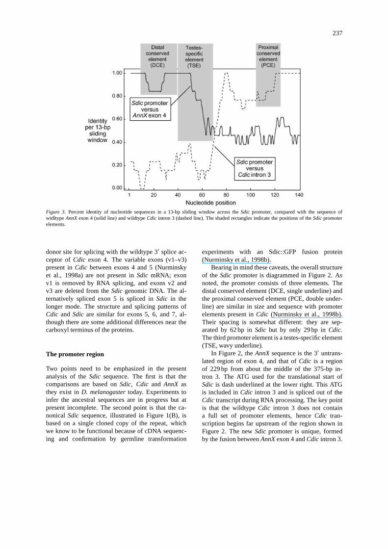
Figure 3. Percent identity of nucleotide sequences in a 13-bp sliding window across theSdic promoter, compared with the sequence ofwidltype AnnX exon 4 (solid line) and wildtypeCdic intron 3 (dashed line). The shaded rectangles indicate the positions of theSdic promoterelements.
donor site for splicing with the wildtype 3′ splice ac-ceptor of Cdic exon 4. The variable exons (v1–v3)present inCdic between exons 4 and 5 (Nurminskyet al., 1998a) are not present inSdic mRNA; exonv1 is removed by RNA splicing, and exons v2 andv3 are deleted from theSdic genomic DNA. The al-ternatively spliced exon 5 is spliced inSdic in thelonger mode. The structure and splicing patterns ofCdic and Sdic are similar for exons 5, 6, and 7, al-though there are some additional differences near thecarboxyl terminus of the proteins.
The promoter region
Two points need to be emphasized in the presentanalysis of theSdic sequence. The first is that thecomparisons are based onSdic, Cdic and AnnX asthey exist inD. melanogaster today. Experiments toinfer the ancestral sequences are in progress but atpresent incomplete. The second point is that the ca-nonical Sdic sequence, illustrated in Figure 1(B), isbased on a single cloned copy of the repeat, whichwe know to be functional because of cDNA sequenc-ing and confirmation by germline transformation
experiments with an Sdic::GFP fusion protein(Nurminsky et al., 1998b).
Bearing in mind these caveats, the overall structureof the Sdic promoter is diagrammed in Figure 2. Asnoted, the promoter consists of three elements. Thedistal conserved element (DCE, single underline) andthe proximal conserved element (PCE, double under-line) are similar in size and sequence with promoterelements present inCdic (Nurminsky et al., 1998b).Their spacing is somewhat different: they are sep-arated by 62 bp inSdic but by only 29 bp inCdic.The third promoter element is a testes-specific element(TSE, wavy underline).
In Figure 2, theAnnX sequence is the 3′ untrans-lated region of exon 4, and that ofCdic is a regionof 229 bp from about the middle of the 375-bp in-tron 3. The ATG used for the translational start ofSdic is dash underlined at the lower right. This ATGis included inCdic intron 3 and is spliced out of theCdic transcript during RNA processing. The key pointis that the wildtypeCdic intron 3 does not containa full set of promoter elements, henceCdic tran-scription begins far upstream of the region shown inFigure 2. The newSdic promoter is unique, formedby the fusion betweenAnnX exon 4 andCdic intron 3.
238
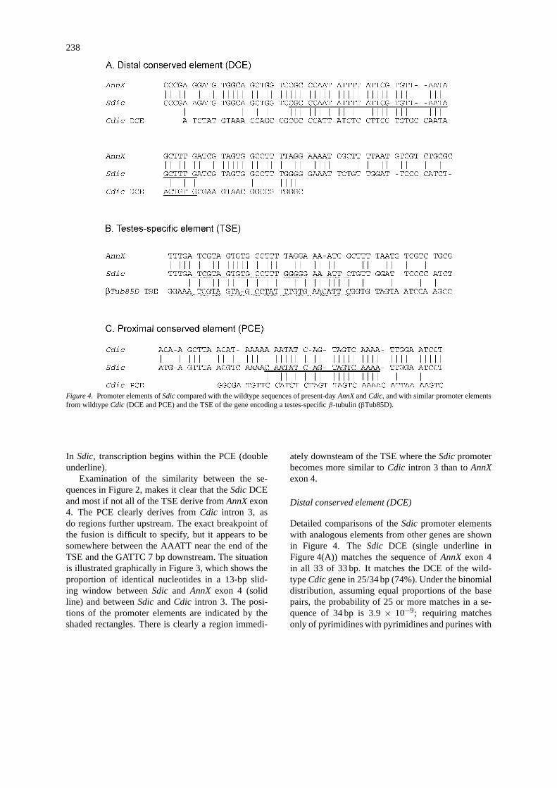
Figure 4. Promoter elements ofSdic compared with the wildtype sequences of present-dayAnnX andCdic, and with similar promoter elementsfrom wildtypeCdic (DCE and PCE) and the TSE of the gene encoding a testes-specificβ-tubulin (βTub85D).
In Sdic, transcription begins within the PCE (doubleunderline).
Examination of the similarity between the se-quences in Figure 2, makes it clear that theSdic DCEand most if not all of the TSE derive fromAnnX exon4. The PCE clearly derives fromCdic intron 3, asdo regions further upstream. The exact breakpoint ofthe fusion is difficult to specify, but it appears to besomewhere between the AAATT near the end of theTSE and the GATTC 7 bp downstream. The situationis illustrated graphically in Figure 3, which shows theproportion of identical nucleotides in a 13-bp slid-ing window betweenSdic and AnnX exon 4 (solidline) and betweenSdic andCdic intron 3. The posi-tions of the promoter elements are indicated by theshaded rectangles. There is clearly a region immedi-
ately downsteam of the TSE where theSdic promoterbecomes more similar toCdic intron 3 than toAnnXexon 4.
Distal conserved element (DCE)
Detailed comparisons of theSdic promoter elementswith analogous elements from other genes are shownin Figure 4. TheSdic DCE (single underline inFigure 4(A)) matches the sequence ofAnnX exon 4in all 33 of 33 bp. It matches the DCE of the wild-typeCdic gene in 25/34 bp (74%). Under the binomialdistribution, assuming equal proportions of the basepairs, the probability of 25 or more matches in a se-quence of 34 bp is 3.9 × 10⁻⁹; requiring matchesonly of pyrimidines with pyrimidines and purines with
239
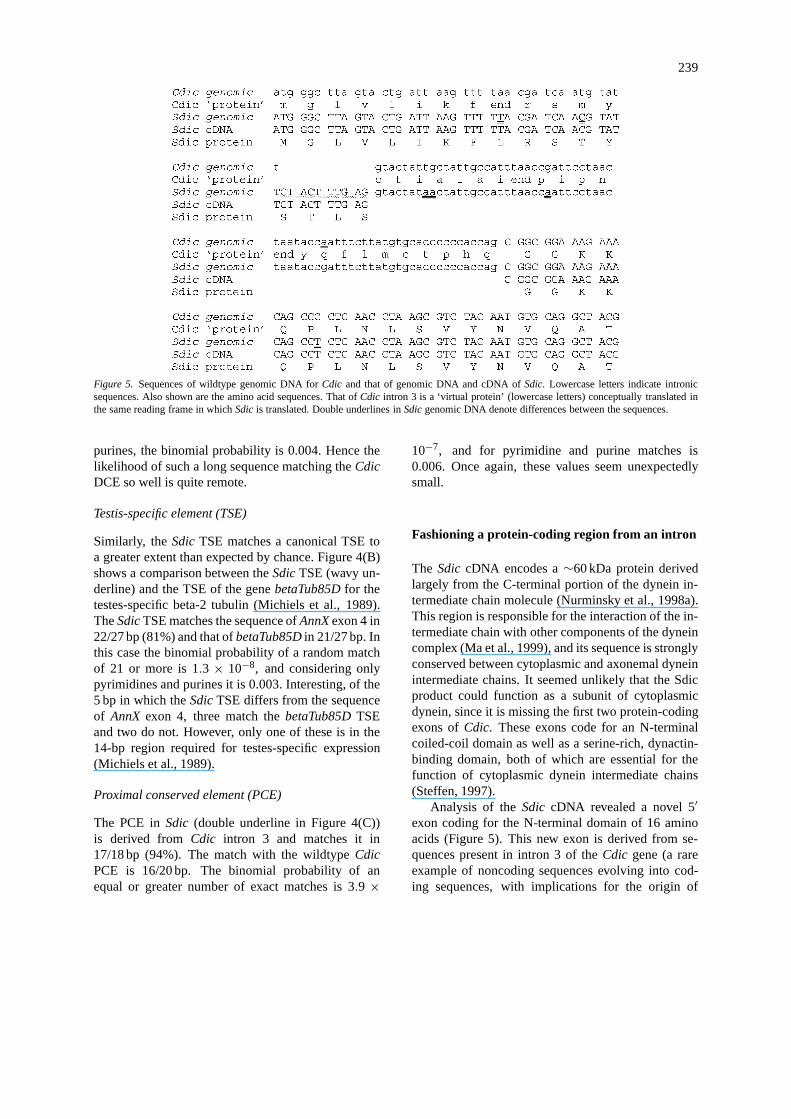
Figure 5. Sequences of wildtype genomic DNA forCdic and that of genomic DNA and cDNA ofSdic. Lowercase letters indicate intronicsequences. Also shown are the amino acid sequences. That ofCdic intron 3 is a ‘virtual protein’ (lowercase letters) conceptually translated inthe same reading frame in whichSdic is translated. Double underlines inSdic genomic DNA denote differences between the sequences.
purines, the binomial probability is 0.004. Hence thelikelihood of such a long sequence matching theCdicDCE so well is quite remote.
Testis-specific element (TSE)
Similarly, theSdic TSE matches a canonical TSE toa greater extent than expected by chance. Figure 4(B)shows a comparison between theSdic TSE (wavy un-derline) and the TSE of the genebetaTub85D for thetestes-specific beta-2 tubulin (Michiels et al., 1989).TheSdic TSE matches the sequence ofAnnX exon 4 in22/27 bp (81%) and that ofbetaTub85D in 21/27 bp. Inthis case the binomial probability of a random matchof 21 or more is 1.3 × 10⁻⁸, and considering onlypyrimidines and purines it is 0.003. Interesting, of the5 bp in which theSdic TSE differs from the sequenceof AnnX exon 4, three match thebetaTub85D TSEand two do not. However, only one of these is in the14-bp region required for testes-specific expression(Michiels et al., 1989).
Proximal conserved element (PCE)
The PCE inSdic (double underline in Figure 4(C))is derived from Cdic intron 3 and matches it in17/18 bp (94%). The match with the wildtypeCdicPCE is 16/20 bp. The binomial probability of anequal or greater number of exact matches is 3.9 ×
10−7, and for pyrimidine and purine matches is0.006. Once again, these values seem unexpectedlysmall.
Fashioning a protein-coding region from an intron
The Sdic cDNA encodes a∼60 kDa protein derivedlargely from the C-terminal portion of the dynein in-termediate chain molecule (Nurminsky et al., 1998a).This region is responsible for the interaction of the in-termediate chain with other components of the dyneincomplex (Ma et al., 1999), and its sequence is stronglyconserved between cytoplasmic and axonemal dyneinintermediate chains. It seemed unlikely that the Sdicproduct could function as a subunit of cytoplasmicdynein, since it is missing the first two protein-codingexons ofCdic. These exons code for an N-terminalcoiled-coil domain as well as a serine-rich, dynactin-binding domain, both of which are essential for thefunction of cytoplasmic dynein intermediate chains(Steffen, 1997).
Analysis of theSdic cDNA revealed a novel 5′exon coding for the N-terminal domain of 16 aminoacids (Figure 5). This new exon is derived from se-quences present in intron 3 of theCdic gene (a rareexample of noncoding sequences evolving into cod-ing sequences, with implications for the origin of
240
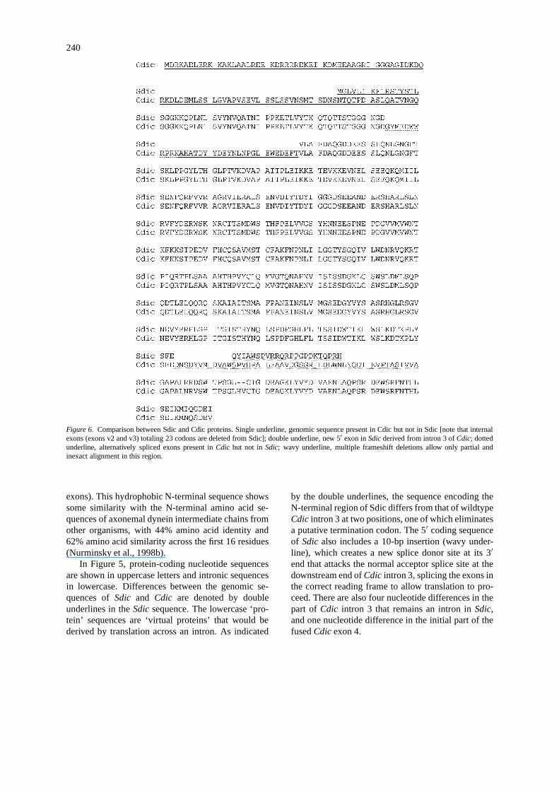
Figure 6. Comparison between Sdic and Cdic proteins. Single underline, genomic sequence present in Cdic but not in Sdic [note that internalexons (exons v2 and v3) totaling 23 codons are deleted from Sdic]; double underline, new 5′ exon inSdic derived from intron 3 ofCdic; dottedunderline, alternatively spliced exons present inCdic but not in Sdic; wavy underline, multiple frameshift deletions allow only partial andinexact alignment in this region.
exons). This hydrophobic N-terminal sequence showssome similarity with the N-terminal amino acid se-quences of axonemal dynein intermediate chains fromother organisms, with 44% amino acid identity and62% amino acid similarity across the first 16 residues(Nurminsky et al., 1998b).
In Figure 5, protein-coding nucleotide sequencesare shown in uppercase letters and intronic sequencesin lowercase. Differences between the genomic se-quences ofSdic and Cdic are denoted by doubleunderlines in theSdic sequence. The lowercase ‘pro-tein’ sequences are ‘virtual proteins’ that would bederived by translation across an intron. As indicated
by the double underlines, the sequence encoding theN-terminal region of Sdic differs from that of wildtypeCdic intron 3 at two positions, one of which eliminatesa putative termination codon. The 5′ coding sequenceof Sdic also includes a 10-bp insertion (wavy under-line), which creates a new splice donor site at its 3′end that attacks the normal acceptor splice site at thedownstream end ofCdic intron 3, splicing the exons inthe correct reading frame to allow translation to pro-ceed. There are also four nucleotide differences in thepart of Cdic intron 3 that remains an intron inSdic,and one nucleotide difference in the initial part of thefusedCdic exon 4.
241
The Sdic protein may function as an axonemaldynein intermediate chain
The protein comparisons suggested that, ifSdic isfunctional, its product might well be an axonemaldynein IC. InDrosophila, the axoneme constitutes amajor part of the sperm tail. Amplification of reverse-transcribed cDNA indicated thatSdic transcription isnot only abundant in testes, but is also testes-specific(Nurminsky et al., 1998b). To follow the fate of theSdic protein in more detail, we created an Sdic::GFPreporter cassette coding for the Sdic polypeptide fusedto GFP (green fluorescent protein) at the carboxyl end,under the conserved of theSdic promoter. Transgenicflies carrying this cassette exhibited green fluorescentSdic::GFP fusion protein only in the testes and seminalvesicles. Sdic::GFP fusion protein is not present in thestem cells or proliferating spermatocytes, but first ap-pears in the growing spermatocytes. The fluorescentlabel is especially abundant in bundles of maturingspermatocytes, and it is very strong all along the fulllength of the tails of mature sperm. The cytologicalpreparations supporting these patterns of fluorescenceare not shown because they require color, but theyare dramatic, completely unambiguous, and highlyreproducible (Nurminsky et al., 1998b).
Comparison of the Sdic and Cdic proteins
Earlier we mentioned that the ICs of axonemal dyneinspossess a large carboxyl-terminal portion that is sim-ilar to those of cytoplasmic dyneins (Wilkerson et al.,1995). Figure 6, which compares the complete se-quences of the Cdic and Sdic proteins, shows theextensive similarity across a large part of the carboxylregion. In Figure 6, the double underline denoted thenovel amino end of Sdic, the single underlines denoteresidues in Cdic derived from exons that are present inCdic cDNA but not inSdic cDNA, and the dotted un-derline denotes residues present in some alternativelyspliced versions of Cdic but not in Sdic. The wavy linetoward the carboxyl end denotes a region in whichmultiple frameshift deletions prevent only partial aninexact alignment of the genomic sequences.
In spite of regions in which there are major differ-ences between Sdic and Cdic due to the novel aminoend of Sdic and to insertions or deletions, a total of 474residues can be aligned without ambiguity. Amongthese only five are different, and all are concentratednear the extreme carboxyl end of the protein (in the
last two rows in Figure 6, which encompass Sdiccodons 459–517).
Silent and replacement substitutions
As there appear to be few nonsynonymous (replace-ment) nucleotide substitutions that distinguishSdicfrom Cdic (Figure 6), so too there are few synonym-ous substitutions. Among the 474 codons inCdic andSdic that can be unambiguously aligned, there are atotal of five nonsynonymous substitutions and sevensynonymous substitutions. The nonsynonymous sub-stitutions are inSdic codons 464, 466, 512, 514, and517 (Figure 6). The synonymous substitutions are inSdic codons 23, 343, 428, 467, 469, 472, and 515. It isodd that the majority of the synonymous substitutions(4/7) occur in the same carboxyl 10% of the codingsequence as all of the nonsynonymous substitutions.
Discussion
Sdic is a novel gene that has only recently been cre-ated and is apparently still in the process of evolving,‘caught in the act’ as it were (Nurminsky et al., 2001).The reason why newly evolved genes warrant detailedanalysis is that they give us rare, first-hand examplesof the early stages of gene creation and evolution,from which we may hope to generalize the findingsto other genes whose origin and functional elaborationcannot be observed directly. Most genetic functions,such as those involved in basic cellular and metabolicprocesses, are ancient. They came into existence solong ago that it is difficult to imagine the mecha-nisms of their origin and functional divergence. Yetwe may hope that insights into such early evolutionaryprocesses may be gained by studying the handful ofrecently evolved gene functions that happen to havebeen identified. At the very least we shall learn hownew genes are created and evolve in contemporaryorganisms.
The fact thatSdic is male-specific in its functionfits into a wider picture. One hypothesis toaccount for Haldane’s rule (‘when hybrid steril-ity occurs in only one sex, it is likely to bethe heterogametic sex’) is that genes governing re-productive functions evolve faster in the hetero-gametic sex (Wu & Davis, 1993; Wu, Johnson& Palopoli, 1996; Laurie, 1997). In support ofthis hypothesis, among sequences for homologous
242
genes in closely related species present in GenBank,classified as to function of their protein product,there is a significantly high ratio of nonsynonymousto synonymous substitutions in the coding regionsfor ‘sex- related’ genes (defined as those affect-ing mating behavior, fertility, spermatogenesis, orsex determination), which is more pronouncedbetween closely related species than more distantlyrelated species, and which is due to an elevatedproportion of nonsynonymous changes (Civetta &Singh, 1995). Consistent with this finding, two-dimensional electrophoresis ofDrosophila proteinshas revealed a surprising number of examples,anonymous as to function, that differ between closelyrelated species, many of which are male spe-cific (Thomas & Singh, 1992; Civetta & Singh,1995; Coulthart & Singh, 1988). There are alsosome knownDrosophila genes that fall into thiscategory:
• The segregation distorter system, a spermato-genesis-specific meiotic drive found inD. melano-gaster but not inD. simulans, which involves theinteraction of two distinct genetic elements (Wuet al., 1988; McClean et al., 1994).
• A system of maleX-chromosomal meiotic drivefound in D. simulans but not inD. melanogaster,which also involves multiple genetic elements(Atlan et al., 1997).
• Mst40, a repeated locus coding for a male-specific transcript of unknown function found inD. melanogaster but not inD. simulans (Russell &Kaiser, 1994).
• In D. melanogaster only, a genetic interac-tion between theY -linked Suppressor of Stellate[Su(Ste)] locus (Balakireva et al., 1992; Mckee &Satter, 1996) and theX-linked Stellate elements(Livak, 1990), mediated by short, double-strandedRNA (Aravin et al., 2001), in which the combina-tion of Stellate elements with a deletion ofSu(Ste)causes meiotic abnormalities in spermatogenesis,gamete-genotype dependent failure of sperm de-velopment, and deposition of protein crystals inspermatocytes (Palumbo et al., 1994; Bozzettiet al., 1995).
• The jingwei gene in theteissieri/yakuba lineage,which is expressed specifically in the testes (Wanget al., 2000).
• The homeobox geneOdysseus, a putative male-sterility gene in themelanogaster/simulans lin-eage, a homolog of theC. elegans neurogenesis
geneunc-4, which was recruited for testes expres-sion inD. simulans but notD. melanogaster (Tinget al., 1998; Ting, Tsaur & Wu, 2000).
• The Sdic repeats, the subject of this paper, whichencode a novel sperm-specific dynein found onlyin D. melanogaster (Nurminsky et al., 1998b).
TheSdic system also suggests a new model for thelong-term fate of some gene duplications which, toour knowledge, has not been observed previously, butmay be quite important. It relates to the fact thatSdicitself is duplicated about tenfold in tandem repeats, butDNA sequencing as well as recovery of cDNAs sug-gests that at least some of the copies may be defectiveor transcriptionally silent (unpublished observations).One scenario to explain this unexpected contrast isthat, in the early stages of gene evolution, when therate of transcription may be limiting to fitness, perhapsthe ‘easiest’ kind of favorable mutation to arise is aduplication leading to a tandem repeat or to multiplecopies. Duplications are quite common, for example,of theAdh region (Jeffs, Holmes & Ashburner, 1994;Nurminsky et al., 1995; Begun, 1997; Luque, Marfany& Gonzàlez-Duarte, 1997). As time goes on, one ofthe duplicated copies may undergo point mutations(or other rearrangement) that increases the promoterefficiency, making the other duplicated copy (or cop-ies) superfluous. Over additional time the superfluousduplicated copy (or copies) would be expected to un-dergo mutational degeneration and, given the high rateof DNA loss in Drosophila (Petrov, Lozovskaya &Hartl, 1996; Petrov & Hartl, 1997; Petrov & Hartl,1998), eventually complete elimination. This suggestsa more general principle that, except in special casesgoverned by different constraints (e.g., rDNA repeatsor histone repeats), duplications may persist over longstretches of evolutionary time only if they diverge infunction enough to be wholly or partially noncomple-menting. Rapid acquisition and loss of duplicationsmay help to explain why, for example,D. virilis andD. melanogaster both have a maltase gene cluster, butthe origin of each cluster from the ancestral maltaseis completely different (Vieira, Vieira & Hartl, 1997),and why both major phylads of theD. virilis speciesgroup haveAdh duplications, but the duplications areof completely independent origin (Nurminsky et al.,1995). We recognize that it is impossible to gen-eralize from any single example. But on the otherhand, so few genes are caught in the act of evolvingthat every example contributes potentially importantinsights.
243
Acknowledgements
This work was supported by NIH grants 60035 (DH)and GM61549 (DN), and by fellowships from theNational Research Council of Spain to JMR and theFoundation for Science and Technology of Portugal toARP.
References
Aniento, F., N. Emans, G. Griffiths & J. Gruenberg, 1993. Cyto-plasmic dynein-dependent vesicular transport from early to lateendosomes. J. Cell Biol. 123: 1373–1387.
Aravin, A.A., N.M. Naumova, A.V. Tulin, V.V. Vagin, Y.M.Rozovsky & V.A. Gvozdev, 2001. Double-stranded RNA-mediated silencing of genomic tandem repeats and transpos-able elements in theD. melanogaster germline. Curr. Biol. 11:1017–1027.
Atlan, A., H. Mercot, C. Landre & C. Montchampmoreau, 1997.The sex-ratio trait inDrosophila simulans: geographical distri-bution of distortion and resistance. Evolution 51: 1886–1895.
Balakireva, M.D., Y.Y. Shevelyov, D.I. Nurminsky, K.J. Livak &V.A. Gvozdev, 1992. Structural organization and diversificationof Y -linked sequences comprisingSu(Ste) genes inDrosophilamelanogaster. Nucl. Acids Res. 20: 3731–3736.
Barton, G.J., R.H. Newman, P.S. Freemont & M.J. Crumpton, 1991.Amino acid sequence analysis of the annexin super-gene familyof proteins. Eur. J. Biochem. 198: 749–760.
Begun, D.J., 1997. Origin and evolution of a new gene descen-ded fromalcohol dehydrogenase in Drosophila. Genetics 145:375–382.
Bozzetti, M.P., S. Massari, P. Finelli, F. Meggio, L.A. Pinna, B.Boldyreff, O.G. Issinger, G. Palumbo, C. Ciriaco, S. Bonaccorsi& S. Pimpinelli, 1995. TheSte locus, a component of theparasiticcry-ste system ofDrosophila melanogaster, encodes aprotein that forms crystals in primary spermatocytes and mimicsproperties of the beta subunit of casein kinase. Proc. Natl. Acad.Sci. USA 92: 6067–6071.
Civetta, A. & R.S. Singh, 1995. High divergence of reproductivetract proteins and their association with postzygotic reproduct-ive isolation inDrosophila melanogaster andDrosophila virilisgroup species. J. Mol. Evol. 41: 1085–1095.
Corthesy-Theulaz, I., A. Pauloin & S.R. Rfeffer, 1992. Cytoplasmicdynein participates in the centrosomal localization of the Golgicomplex. J. Cell. Biol. 118: 1333–1345.
Coulthart, M.B. & R.S. Singh, 1988. High level of divergenceof male-reproductive-tract proteins betweenDrosophila melano-gaster and its sibling species,D. simulans. Mol. Biol. Evol. 5:182–191.
Dillman, J.F., L.P. Dabney & K.K. Pfister, 1996. Cytoplasmicdynein is associated with slow axonal transport. Proc. Natl. Acad.Sci. USA 93: 141–144.
Geisow, M.J., 1991. Annexins: forms without function but notwithout fun. Trends Biotechnol. 9: 180–181.
Gilbert, W., 1978. Why genes in pieces? Nature 271: 501.Jeffs, P.S., E.C. Holmes & M. Ashburner, 1994. The
molecular evolution of the alcohol dehydrogenase andalcohol dehydrogenase-related genes in the Drosophilamelanogaster species subgroup. Mol. Biol. Evol. 11: 287–304.
King, S.M., E. Barbarese, J.F. Dillman, R.S. Patel-King, J.H. Carson& K.K. Pfister, 1996. Brain cytoplasmic and flagellar outer armdyneins share a highly conserved Mr 8,000 light chain. J. Biol.Chem. 271: 19358–19366.
Laurie, C.C., 1997. The weaker sex is heterogamatic: 75 years ofHaldane’s rule. Genetics 147: 937–951.
Livak, K.J., 1990. Detailed structure of theDrosophila melano-gaster Stellate genes and their transcripts. Genetics 124:303–316.
Long, M., 2001. Evolution of novel genes. Curr. Opin. Genet. Dev.11: 673–680.
Long, M. & C.H. Langley, 1993. Natural selection and the originof jingwei, a chimeric processed functional gene inDrosophila.Science 260: 91–95.
Long, M., C. Rosenberg & W. Gilbert, 1995. Intron phase correla-tions and the evolution of the intron/exon structure of genes.Proc. Natl. Acad. Sci. USA 92.
Luque, T., G. Marfany & R. Gonzàlez-Duarte, 1997. Character-ization and molecular analysis ofAdh retrosequences in spe-cies of theDrosophila obscura group. Mol. Biol. Evol. 14:1316–1325.
Ma, S., L. Trivinos-Lagos, R. Graf & R.L. Chisholm, 1999. Dyneinintermediate chain mediated dynein–dynactin interaction is re-quired for interphase microtubule organization and centrosomereplication and separation in Dictyostelium. J. Cell Biol. 147:1261–1273.
Martinez-Cruzado, J.C., C. Swimmer, M.G. Fenerjian & F.C.Kafatos, 1988. Evolution of the autosomal chorion locus inDrosophila. I. General organization of the locus and sequencecomparisons of geness15 ands19 in evolutionary distant species.Genetics 199: 663–677.
Mazumdar, M., A. Mikami, M.A. Gee & R.B. Vallee, 1996.In vitromotility from recombinant dynein heavy chain. Proc. Natl. Acad.Sci. USA 93: 6552–6556.
McClean, J.R., C.J. Merrill, P.A. Powers & B. Ganetzky, 1994.Functional identification of thesegregation distorter locus ofDrosophila melanogaster by germline transformation. Genetics137: 201–209.
Mckee, B.D. & M.T. Satter, 1996. Structure of theY chro-mosomalSu(Ste) locus in Drosophila melanogaster and evid-ence for localized recombination among repeats. Genetics 142:149–161.
Michiels, F., A. Gasch, B. Kaltschmidt & R. Renkawitz-Pohl, 1989.A 14 bp promoter element directs the testis specificity of theDrosophila beta 2 tubulin gene. EMBO J. 8: 1559–1565.
Nurminsky, D.I., E.N. Moriyama, E.R. Lozovskaya & D.L. Hartl,1995. Molecular phylogeny and genome evolution in theDro-sophila virilis group: duplications of thealcohol dehydrogenasegene. Mol. Biol. Evol. 13: 132–149.
Nurminsky, D.I., E.V. Benevolenskaya, M.V. Nurminskaya, Y.Y.Shevelyov, D.L. Hartl & V.A. Gvozdev, 1998a. Cytoplasmicdynein intermediate chain isoforms with different targeting prop-erties created by tissue-specific alternative splicing. Mol. Cell.Biol. 18: 6816–6825.
Nurminsky, D.I., M.V. Nurminskaya, D. De Aguiar & D.L. Hartl,1998b. Selective sweep of a newly evolved sperm-specific genein Drosophila. Nature 396: 572–575.
Nurminsky, D., D. De Aguiar, C.D. Bustamante & D.L. Hartl, 2001.Chromosomal effects of rapid gene evolution inDrosophilamelanogaster. Science 291: 128–130.
Palumbo, G., S. Bonaccorsi, L.G. Robbins & S. Pimpinelli, 1994.Genetic analysis ofstellate elements ofDrosophila melano-gaster. Genetics 138: 1181–1197.
244
Paschal, B.M., A. Mikami, K.K. Pfister & R.B. Vallee, 1992. Ho-mology of the 74-kD cytoplasmic dynein subunit with a flagellardynein polypeptide suggests an intracellular targeting function.J. Cell Biol. 118: 1133–1143.
Petrov, D.A. & D.L. Hartl, 1997. Trash DNA is what gets thrownaway: high rate of DNA loss inDrosophila. Gene 205: 279–289.
Petrov, D.A. & D.L. Hartl, 1998. High rate of DNA loss in theD.melanogaster andD. virilis species groups. Mol. Biol. Evol. 15:293–302.
Petrov, D.A., E.R. Lozovskaya & D.L. Hartl, 1996. High intrinsicrate of DNA loss inDrosophila. Nature 384: 346–349.
Robin, C., R.J. Russell, K.M. Medveczky & J.G. Oakeshott, 1996.Duplication and divergence of the genes of theα-esterase clusterof D. melanogaster. J. Mol. Evol. 43: 241–252.
Russell, S.R.H. & K. Kaiser, 1994. ADrosophila melanogasterchromosome-2L repeat is expressed in the male germ line.Chromosoma 103: 63–72.
Schroer, T.A., E.R. Steuer & M.P. Sheetz, 1989. Cytoplasmic dyneinis a minus end-directed motor for membranous organelles. Cell7: 331–343.
Snyder, M. & N. Davidson, 1983. Two gene families clustered ina small region of theDrosophila genome. J. Mol. Biol. 166:101–118.
Steffen, W., S. Karki, K.T. Vaughan, R.B. Vallee, E.L.F. Holzbaur,D.G. Weiss & S.A. Kuznetsov, 1997. The involvement of theintermediate chain of cytoplasmic dynein in binding the motorcomplex to membranous organelles ofXenopus oocytes. Mol.Biol. Cell 8: 2077–2088.
Steinemann, M. & S. Steinemann, 1990. Evolutionary changesin the organization of the majorLcp gene cluster during sexchromosomal differentiation in the sibling speciesDrosophilapersimilis, D. pseudoobscura andD. miranda. Chromosoma 99:424–431.
Thomas, S. & R.S. Singh, 1992. A comprehensive studyof genetic variation in natural population ofDrosophilamelanogaster. VII. Varying rates of genic divergence as revealed
by two-dimensional electrophoresis. Mol. Biol. Evol. 9:507–525.
Ting, C.T., S.C. Tsaur & C.I. Wu, 2000. The phylogeny of closelyrelated species as revealed by the genealogy of a speciation gene,Odysseus. Proc. Natl. Acad. Sci. USA 97: 5313–5316.
Ting, C.T., S.C. Tsaur, M.L. Wu & C.I. Wu, 1998. A rapidlyevolving homeobox at the site of a hybrid sterility gene. Science282: 1501–1504.
Vaisberg, E.A., M.P. Koonce & J.R. McIntosh, 1993. Cytoplasmicdynein plays a role in mammalian mitotic spindle formation. J.Cell Biol. 123: 849–858.
Vieira, C.P., J. Vieira & D.L. Hartl, 1997. The evolution of smallgene clusters: evidence for an independent origin of the maltasegene cluster inD. virilis andD. melanogaster. Mol. Biol. Evol.14: 985–993.
Wang, W., J.M. Zhang, C. Alvarez, A. Llopart & M. Long,2000. The origin of theJingwei gene and the complex modu-lar structure of its parental gene,yellow emperor, in Drosophilamelanogaster. Mol. Biol. Evol. 17: 1294–1301.
Wilkerson, C.G., S.M. King, A. Koutoulis, G.J. Pazour &G.B. Witman, 1995. The 78,000 M(r) intermediate chain ofChlamydomonas outer arm dynein is a WD-repeat protein re-quired for arm assembly. J. Cell Biol. 129: 169–178.
Wu, C.-I. & A.W. Davis, 1993. Evolution of postmating repro-ductive isolation: the composite nature of Haldane’s rule and itsgenetic bases. Am. Nat. 142: 187–212.
Wu, C.-I., N.A. Johnson & M.F. Palopoli, 1996. Haldane’s rule andits legacy: why are there so many sterile males? Trends Ecol.Evol. 11: 281–284.
Wu, C.-I., T.W. Lyttle, M.-L. Wu & G.-F. Lin, 1988. Asso-ciation between a satellite DNA sequence and theRespon-der of Segregation Distorter in D. melanogaster. Cell 54:179–189.
Xiang, X., S.M. Beckwith & N.R. Morris, 1994. Cytoplasmicdynein is involved in nuclear migration inAspergillus nidulans.Proc. Natl. Acad. Sci. USA 91: 2100–2104.
Related Documents