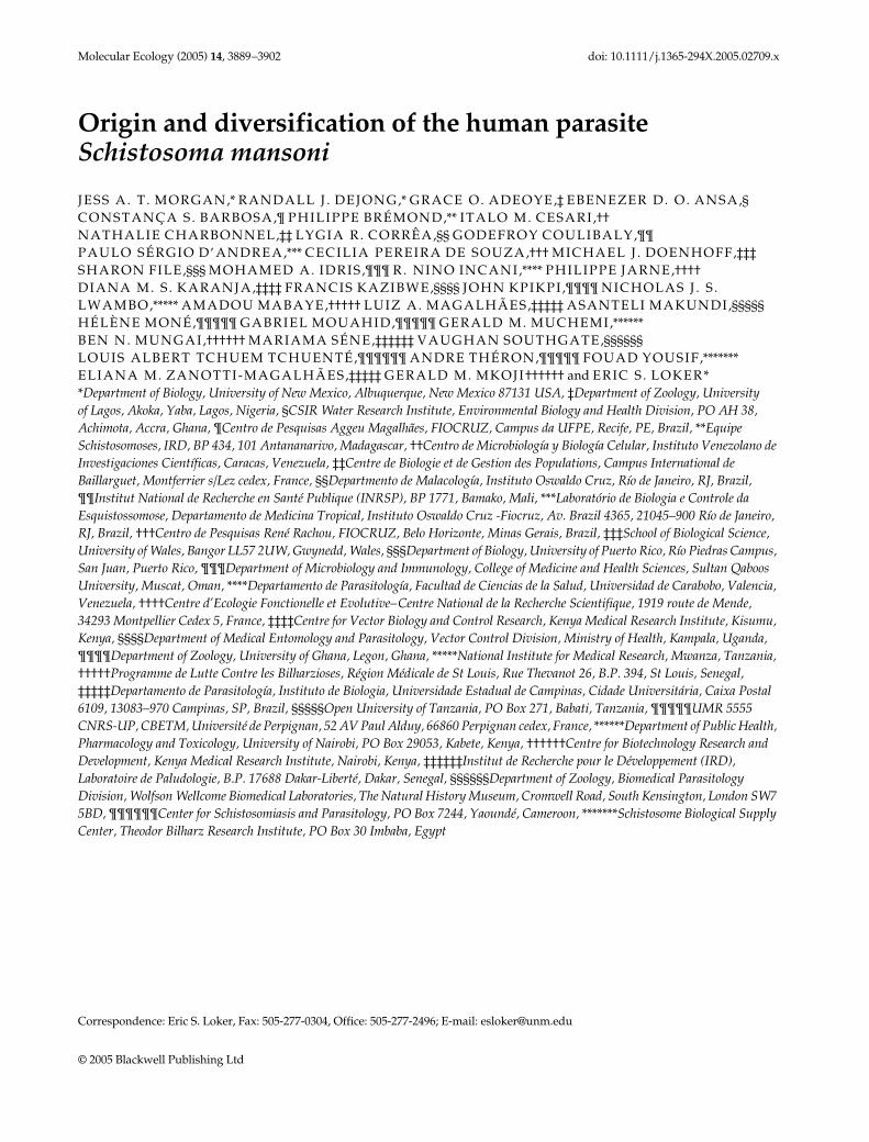
Molecular Ecology (2005) 14, 3889–3902 doi: 10.1111/j.1365-294X.2005.02709.x © 2005 Blackwell Publishing Ltd Blackwell Publishing, Ltd. Origin and diversification of the human parasite Schistosoma mansoni JESS A. T. MORGAN,* RANDALL J. DEJONG,* GRACE O. ADEOYE,‡ EBENEZER D. O. ANSA,§ CONSTANÇA S. BARBOSA, ¶ PHILIPPE BRÉMOND, ** ITALO M. CESARI, †† NATHALIE CHARBONNEL, ‡‡ LYGIA R. CORRÊA, §§ GODEFROY COULIBALY, ¶¶ PAULO SÉRGIO D’ANDREA, *** CECILIA PEREIRA DE SOUZA, ††† MICHAEL J. DOENHOFF, ‡‡‡ SHARON FILE, §§§ MOHAMED A. IDRIS, ¶¶¶ R. NINO INCANI, **** PHILIPPE JARNE, †††† DIANA M. S. KARANJA, ‡‡‡‡ FRANCIS KAZIBWE, §§§§ JOHN KPIKPI, ¶¶¶¶ NICHOLAS J. S. LWAMBO, ***** AMADOU MABAYE, ††††† LUIZ A. MAGALHÃES, ‡‡‡‡‡ ASANTELI MAKUNDI, §§§§§ HÉLÈNE MONÉ, ¶¶¶¶¶ GABRIEL MOUAHID, ¶¶¶¶¶ GERALD M. MUCHEMI, ****** BEN N. MUNGAI, †††††† MARIAMA SÉNE, ‡‡‡‡‡‡ VAUGHAN SOUTHGATE, §§§§§§ LOUIS ALBERT TCHUEM TCHUENTÉ, ¶¶¶¶¶¶ ANDRE THÉRON, ¶¶¶¶¶ FOUAD YOUSIF, ******* ELIANA M. ZANOTTI-MAGALHÃES, ‡‡‡‡‡ GERALD M. MKOJI †††††† and ERIC S. LOKER * * Department of Biology, University of New Mexico, Albuquerque, New Mexico 87131 USA, ‡ Department of Zoology, University of Lagos, Akoka, Yaba, Lagos, Nigeria, § CSIR Water Research Institute, Environmental Biology and Health Division, PO AH 38, Achimota, Accra, Ghana, ¶ Centro de Pesquisas Aggeu Magalhães, FIOCRUZ, Campus da UFPE, Recife, PE, Brazil, ** Equipe Schistosomoses, IRD, BP 434, 101 Antananarivo, Madagascar, †† Centro de Microbiología y Biología Celular, Instituto Venezolano de Investigaciones Científicas, Caracas, Venezuela, ‡‡ Centre de Biologie et de Gestion des Populations, Campus International de Baillarguet, Montferrier s/Lez cedex, France, §§ Departmento de Malacología, Instituto Oswaldo Cruz, Río de Janeiro, RJ, Brazil, ¶¶ Institut National de Recherche en Santé Publique (INRSP), BP 1771, Bamako, Mali, *** Laboratório de Biologia e Controle da Esquistossomose, Departamento de Medicina Tropical, Instituto Oswaldo Cruz -Fiocruz, Av. Brazil 4365, 21045–900 Río de Janeiro, RJ, Brazil, ††† Centro de Pesquisas René Rachou, FIOCRUZ, Belo Horizonte, Minas Gerais, Brazil, ‡‡‡ School of Biological Science, University of Wales, Bangor LL57 2UW, Gwynedd, Wales, §§§ Department of Biology, University of Puerto Rico, Río Piedras Campus, San Juan, Puerto Rico, ¶¶¶ Department of Microbiology and Immunology, College of Medicine and Health Sciences, Sultan Qaboos University, Muscat, Oman, **** Departamento de Parasitología, Facultad de Ciencias de la Salud, Universidad de Carabobo, Valencia, Venezuela, †††† Centre d’Ecologie Fonctionelle et Evolutive– Centre National de la Recherche Scientifique, 1919 route de Mende, 34293 Montpellier Cedex 5, France, ‡‡‡‡ Centre for Vector Biology and Control Research, Kenya Medical Research Institute, Kisumu, Kenya, §§§§ Department of Medical Entomology and Parasitology, Vector Control Division, Ministry of Health, Kampala, Uganda, ¶¶¶¶ Department of Zoology, University of Ghana, Legon, Ghana, ***** National Institute for Medical Research, Mwanza, Tanzania, ††††† Programme de Lutte Contre les Bilharzioses, Région Médicale de St Louis, Rue Thevanot 26, B.P. 394, St Louis, Senegal, ‡‡‡‡‡ Departamento de Parasitología, Instituto de Biologia, Universidade Estadual de Campinas, Cidade Universitária, Caixa Postal 6109, 13083–970 Campinas, SP, Brazil, §§§§§ Open University of Tanzania, PO Box 271, Babati, Tanzania, ¶¶¶¶¶ UMR 5555 CNRS-UP, CBETM, Université de Perpignan, 52 AV Paul Alduy, 66860 Perpignan cedex, France, ****** Department of Public Health, Pharmacology and Toxicology, University of Nairobi, PO Box 29053, Kabete, Kenya, †††††† Centre for Biotechnology Research and Development, Kenya Medical Research Institute, Nairobi, Kenya, ‡‡‡‡‡‡ Institut de Recherche pour le Développement (IRD), Laboratoire de Paludologie, B.P. 17688 Dakar-Liberté, Dakar, Senegal, §§§§§§ Department of Zoology, Biomedical Parasitology Division, Wolfson Wellcome Biomedical Laboratories, The Natural History Museum, Cromwell Road, South Kensington, London SW7 5BD, ¶¶¶¶¶¶ Center for Schistosomiasis and Parasitology, PO Box 7244, Yaoundé, Cameroon, ******* Schistosome Biological Supply Center, Theodor Bilharz Research Institute, PO Box 30 Imbaba, Egypt Correspondence: Eric S. Loker, Fax: 505-277-0304, Office: 505-277-2496; E-mail: [email protected]

Welcome message from author
This document is posted to help you gain knowledge. Please leave a comment to let me know what you think about it! Share it to your friends and learn new things together.
Transcript
Molecular Ecology (2005)
14
, 3889–3902 doi: 10.1111/j.1365-294X.2005.02709.x
© 2005 Blackwell Publishing Ltd
Blackwell Publishing, Ltd.
Origin and diversification of the human parasite
Schistosoma mansoni
JESS A. T . MORGAN,
*
RANDALL J . DEJONG,
*
GRACE O. ADEOYE,
‡
EBENEZER D. O. ANSA,
§
CONSTANÇA S . BARBOSA,
¶
PHILIPPE BRÉMOND,
**
ITALO M. CESARI ,
††
NATHALIE CHARBONNEL,
‡‡
LYGIA R. CORRÊA,
§§
GODEFROY COULIBALY,
¶¶
PAULO SÉRGIO D’ANDREA,
***
CECILIA PEREIRA DE SOUZA,
†††
MICHAEL J . DOENHOFF,
‡‡‡
SHARON FILE,
§§§
MOHAMED A. IDRIS ,
¶¶¶
R . NINO INCANI,
****
PHILIPPE JARNE,
††††
DIANA M. S . KARANJA,
‡‡‡‡
FRANCIS KAZIBWE,
§§§§
JOHN KPIKPI ,
¶¶¶¶
NICHOLAS J . S . LWAMBO,
*****
AMADOU MABAYE,
†††††
LUIZ A. MAGALHÃES,
‡‡‡‡‡
ASANTELI MAKUNDI,
§§§§§
HÉLÈNE MONÉ,
¶¶¶¶¶
GABRIEL MOUAHID,
¶¶¶¶¶
GERALD M. MUCHEMI,
******
BEN N. MUNGAI,
††††††
MARIAMA SÉNE,
‡‡‡‡‡‡
VAUGHAN SOUTHGATE,
§§§§§§
LOUIS ALBERT TCHUEM TCHUENTÉ,
¶¶¶¶¶¶
ANDRE THÉRON,
¶¶¶¶¶
FOUAD YOUSIF ,
*******
ELIANA M. ZANOTTI-MAGALHÃES,
‡‡‡‡‡
GERALD M. MKOJI
††††††
and ERIC S . LOKER
*
*
Department of Biology, University of New Mexico, Albuquerque, New Mexico 87131 USA,
‡
Department of Zoology, University of Lagos, Akoka, Yaba, Lagos, Nigeria,
§
CSIR Water Research Institute, Environmental Biology and Health Division, PO AH 38, Achimota, Accra, Ghana,
¶
Centro de Pesquisas Aggeu Magalhães, FIOCRUZ, Campus da UFPE, Recife, PE, Brazil,
**
Equipe Schistosomoses, IRD, BP 434, 101 Antananarivo, Madagascar,
††
Centro de Microbiología y Biología Celular, Instituto Venezolano de Investigaciones Científicas, Caracas, Venezuela,
‡‡
Centre de Biologie et de Gestion des Populations, Campus International de Baillarguet, Montferrier s/Lez cedex, France,
§§
Departmento de Malacología, Instituto Oswaldo Cruz, Río de Janeiro, RJ, Brazil,
¶¶
Institut National de Recherche en Santé Publique (INRSP), BP 1771, Bamako, Mali,
***
Laboratório de Biologia e Controle da Esquistossomose, Departamento de Medicina Tropical, Instituto Oswaldo Cruz -Fiocruz, Av. Brazil 4365, 21045–900 Río de Janeiro, RJ, Brazil,
†††
Centro de Pesquisas René Rachou, FIOCRUZ, Belo Horizonte, Minas Gerais, Brazil,
‡‡‡
School of Biological Science, University of Wales, Bangor LL57 2UW, Gwynedd, Wales,
§§§
Department of Biology, University of Puerto Rico, Río Piedras Campus, San Juan, Puerto Rico,
¶¶¶
Department of Microbiology and Immunology, College of Medicine and Health Sciences, Sultan Qaboos University, Muscat, Oman,
****
Departamento de Parasitología, Facultad de Ciencias de la Salud, Universidad de Carabobo, Valencia, Venezuela,
††††
Centre d’Ecologie Fonctionelle et Evolutive–Centre National de la Recherche Scientifique, 1919 route de Mende, 34293 Montpellier Cedex 5, France,
‡‡‡‡
Centre for Vector Biology and Control Research, Kenya Medical Research Institute, Kisumu, Kenya,
§§§§
Department of Medical Entomology and Parasitology, Vector Control Division, Ministry of Health, Kampala, Uganda,
¶¶¶¶
Department of Zoology, University of Ghana, Legon, Ghana,
*****
National Institute for Medical Research, Mwanza, Tanzania,
†††††
Programme de Lutte Contre les Bilharzioses, Région Médicale de St Louis, Rue Thevanot 26, B.P. 394, St Louis, Senegal,
‡‡‡‡‡
Departamento de Parasitología, Instituto de Biologia, Universidade Estadual de Campinas, Cidade Universitária, Caixa Postal 6109, 13083–970 Campinas, SP, Brazil,
§§§§§
Open University of Tanzania, PO Box 271, Babati, Tanzania,
¶¶¶¶¶
UMR 5555CNRS-UP, CBETM, Université de Perpignan, 52 AV Paul Alduy, 66860 Perpignan cedex, France,
******
Department of Public Health, Pharmacology and Toxicology, University of Nairobi, PO Box 29053, Kabete, Kenya,
††††††
Centre for Biotechnology Research and Development, Kenya Medical Research Institute, Nairobi, Kenya,
‡‡‡‡‡‡
Institut de Recherche pour le Développement (IRD), Laboratoire de Paludologie, B.P. 17688 Dakar-Liberté, Dakar, Senegal,
§§§§§§
Department of Zoology, Biomedical Parasitology Division, Wolfson Wellcome Biomedical Laboratories, The Natural History Museum, Cromwell Road, South Kensington, London SW7 5BD,
¶¶¶¶¶¶
Center for Schistosomiasis and Parasitology, PO Box 7244, Yaoundé, Cameroon,
*******
Schistosome Biological Supply Center, Theodor Bilharz Research Institute, PO Box 30 Imbaba, Egypt
Correspondence: Eric S. Loker, Fax: 505-277-0304, Office: 505-277-2496; E-mail: [email protected]
3890
J . A . T . M O R G A N
E T A L .
© 2005 Blackwell Publishing Ltd,
Molecular Ecology
, 14, 3889–3902
Abstract
Schistosoma mansoni
is the most widespread of the human-infecting schistosomes, presentin 54 countries, predominantly in Africa, but also in Madagascar, the Arabian Peninsula,and the Neotropics. Adult-stage parasites that infect humans are also occasionally recoveredfrom baboons, rodents, and other mammals. Larval stages of the parasite are dependent uponcertain species of freshwater snails in the genus
Biomphalaria
, which largely determine theparasite’s geographical range. How
S
.
mansoni
genetic diversity is distributed geographicallyand among isolates using different hosts has never been examined with DNA sequencedata. Here we describe the global phylogeography of
S
.
mansoni
using more than 2500 bpof mitochondrial DNA (mtDNA) from 143 parasites collected in 53 geographically wide-spread localities. Considerable within-species mtDNA diversity was found, with 85 uniquehaplotypes grouping into five distinct lineages. Geographical separation, and not host use,appears to be the most important factor in the diversification of the parasite. East Africanspecimens showed a remarkable amount of variation, comprising three clades and basalmembers of a fourth, strongly suggesting an East African origin for the parasite 0.30–0.43million years ago, a time frame that follows the arrival of its snail host. Less but still sub-stantial variation was found in the rest of Africa. A recent colonization of the New Worldis supported by finding only seven closely related New World haplotypes which have WestAfrican affinities. All Brazilian isolates have nearly identical mtDNA haplotypes, suggestinga founder effect from the establishment and spread of the parasite in this large country.
Keywords
: Africa,
Biomphalaria
, Brazil, phylogeography, schistosomiasis, slave trade
Received 20 May 2005; revision accepted 25 July 2005
Introduction
Schistosomiasis is one of the world’s great neglected dis-eases, with six species of
Schistosoma
still infecting 200million people (Crompton 1999; Chitsulo
et al
. 2000). One ofthe most common is
Schistosoma mansoni
, which infects morethan 83 million humans in 54 countries, mostly in tropicalAfrica (Crompton 1999), causing intestinal schistosomiasis,which results in pathology, morbidity, and even death(Lambertucci
et al
. 2000). Adult worms living in veins sur-rounding the intestine produce eggs that either pass withthe faeces to continue the life cycle, or become trapped inthe tissues, provoking granulomatous reactions, fibro-obstructive disease in the liver and other organs and portalhypertension (Lambertucci
et al
. 2000).The evolution of the genus
Schistosoma
has long fascin-ated parasitologists and evolutionary biologists (Davis 1980,1992), and has garnered much attention by recent molecularstudies (Desprès
et al
. 1992; Snyder & Loker 2000; Lockyer
et al
. 2003; Morgan
et al
. 2003a), but at least as many questionshave been raised as have been answered. It is suspectedthat the genus has an Asian origin, as the most basal cladein the genus is Asian (Snyder & Loker 2000; Lockyer
et al
.2003; Morgan
et al
. 2003a), but three other groups withinthe genus are entirely African.
S
.
mansoni
, along with itssister taxon
Schistosoma rodhaini
, form one of these groups.Both species utilize
Biomphalaria
snails as intermediate
hosts
,
but the adult stages of
S
.
rodhaini
infect rodents, dogsor serval cats instead of humans and this species is con-fined to tropical Africa. Other
Schistosoma
taxa, from thehippopotamus, that were previously thought to be part ofthe
S
.
mansoni
and
S
.
rodhaini
group, have been shown bymolecular analysis to comprise their own monophyleticlineage within the genus (Morgan
et al
. 2003a). It has beenhypothesized that the separation of
S
.
rodhaini
and
S
.
mansoni
began 5–7 million years ago (Ma) with the emergence ofhominins (Desprès
et al
. 1992), but this study was limited toa small amount of sequence data from a limited numberof specimens. Also,
S
.
mansoni
and
S
.
rodhaini
have beenshown to hybridize in the laboratory, and recently a natur-ally occurring hybrid was identified (Morgan
et al
. 2003b).How diverse
S
.
mansoni
really is, with such a large geo-graphical range, and the ability to use several differentmammalian and snail hosts, has not been well assessed. Anaccurate measure of within-species diversity should beinformative for understanding the evolution of the species.
Additionally, the presence of
S
.
mansoni
in the NewWorld has been hypothesized to be the result of intro-duction through the Atlantic slave trade within the last500 years and data from snail–parasite strain compatibilitystudies (Files 1951), allozymes (Fletcher
et al
. 1981), andmitochondrial restriction enzyme sites (Desprès
et al
.1993) are consistent with this hypothesis, but a final con-clusion cannot be drawn from these studies. For example,
P H Y L O G E O G R A P H Y O F
S C H I S T O S O M A M A N S O N I
3891
© 2005 Blackwell Publishing Ltd,
Molecular Ecology
, 14, 3889–3902
if
S
.
mansoni
originated in Africa several million years agoand colonized the New World with the slave trade, thenmuch more diversity should be seen in Africa than in theNew World. Desprès
et al
. (1993), found that the low levelof genetic diversity (measured by restriction enzyme sites)amongst five New World samples was similar to thatbetween New World samples and a single African sample,but because only one African sample was included, a com-parison of diversity on each continent could not be made.Other evidence for or against this hypothesis is indirect.Colonization of the New World by
S
.
mansoni
is favouredby recent phylogenies of
Biomphalaria which show thatBiomphalaria glabrata, a New World species, is closelyrelated to African Biomphalaria and thus may have beenquite susceptible to S. mansoni coming from Africa duringthe slave trade (Campbell et al. 2000; DeJong et al. 2001, 2003).Yet, variability in host use, morphology, and behaviourhave been described in the New World (Théron et al. 1997;D’Andrea et al. 2002; Freire et al. 2002), suggestive of geneticdiversity. The genealogical information from DNA sequencesfrom an extensive collection of S. mansoni specimens shouldbe able to definitively confirm or deny a recent coloniza-tion event.
Control of schistosomes depends primarily on treat-ment with one drug, praziquantel, and a global initiativeto reduce worm burden in endemic areas with this drughas begun (Crompton et al. 2003; see also www.who.int/wormcontrol/en/). Unfortunately, some isolates of S. mansonihave shown worrisome signs in recent years of praziquantelresistance (in Egypt and Senegal; Doenhoff et al. 2002), andit has been difficult to predict how and when resistancemay develop, though one possibility is that an area of highgenetic diversity is more likely to contain alleles that couldcontribute to drug resistance.
To address these issues, we examined DNA sequencevariation for S. mansoni collected from geographicallywidespread localities, and from different intermediate anddefinitive hosts, and from two collections of S. rodhaini. Weconstructed phylogenies of observed haplotypes and testedwhether host use or geographical separation has beenimportant in the diversification of the parasite. Also, bycollecting parasites from several infected snails, at a fewlocalities where it was possible, we aimed to obtain aglimpse of within-locality diversity.
Materials and methods
Parasite specimens
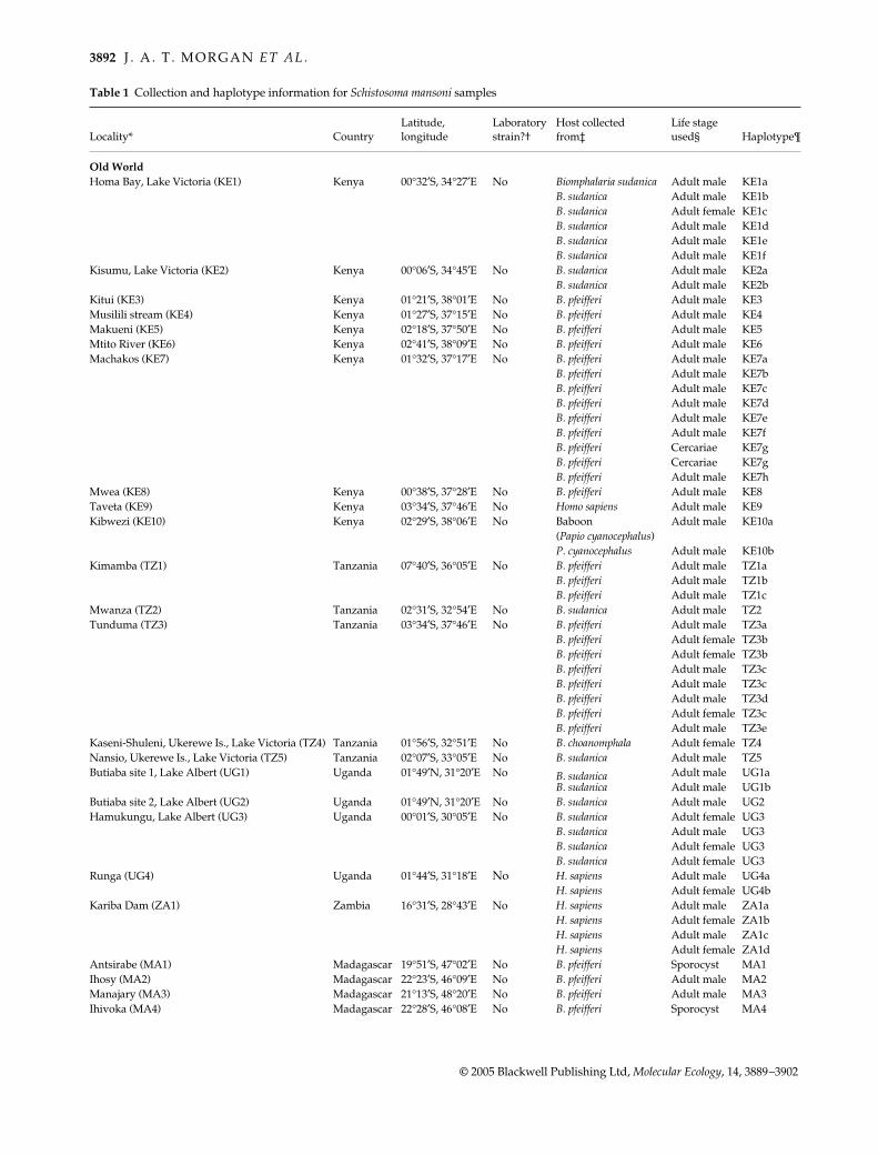
We collected 143 specimens of Schistosoma mansoni from 53geographically widespread localities in Africa, the ArabianPeninsula, Madagascar, South America, and the Caribbeanregion (Table 1). The number and geographical samplingof S. mansoni in this study is comparable to that of recent
global genetic studies of human parasites (Joy et al. 2003;Leclerc et al. 2004). Most collections were from the field, buta few collections were made from laboratory stocks forwhich the original collection locality was known. Field-derived specimens of S. mansoni were obtained either frominfected snails collected in the field, from anonymoushuman faecal samples provided by local health authorities,from baboon faecal samples collected in the field, or fromthe trapping, dissection, and perfusion of wild rodents.Though the percentage of field-collected snails found to beinfected is often quite low (< 1%), this method is logist-ically simple and was the predominate method in ourstudy (Table 1). To generate adult worms, the best sourceof DNA, field-collected snails were exposed to light tostimulate emergence of larval stages (cercariae) that werethen used to infect laboratory mice. At 7 weeks post infec-tion, adult worms were retrieved by dissection or perfusionfrom mice and preserved in ethanol. If specimens wereobtained from faecal samples of humans or baboons, theprocess was similar, but instead began with hatching thelarval stages (miracidia) from eggs in the faeces, and usingthese to infect laboratory-reared snails. Cercariae fromthese snails were then used to infect mice and adult wormswere retrieved 7 weeks later. From a few localities, adultworms could not be obtained at all, so DNA was extractedfrom cercariae or sporocysts from individual snails.
An aspect of S. mansoni biology relevant to our samplingis that individual field-infected snails usually are infectedby a single S. mansoni genotype (Eppert et al. 2002). Allworms derived from such a snail are clones and cannot betreated as individual samples. Thus we attempted to findmultiple infected snails within a locality, which enabled usto sample multiple worms within a few localities. Within-locality sampling and clone avoidance were also made pos-sible for some locations when male and female wormswere obtained, indicating multiple genotypes. Overall,within-locality sampling was very modest, and was notintended to thoroughly describe the within-locality vari-ation in all parts of S. mansoni’s range. The number of wormsanalysed from a site varied from 1 to 13. Two specimensof Schistosoma rodhaini from separate localities in Kenya(Homa Bay and Kisumu) were also obtained.
Molecular data
DNA was extracted from whole worms as describedpreviously (Morgan et al. 2003b). The primary data set forthis study was obtained by amplifying and sequencingfour partial regions of the mitochondrial genome to findpoint mutations to distinguish among globally collectedS. mansoni: (COI, 617 bp; 16S-12S rDNA, 660–663 bp; cyt b-ND4L-ND4, 637–639 bp; ND1, 620 bp; total 2532–2535 bp).Polymerase chain reaction (PCR) and sequencing proto-cols have been described previously (Morgan et al. 2003b),
3892 J . A . T . M O R G A N E T A L .
© 2005 Blackwell Publishing Ltd, Molecular Ecology, 14, 3889–3902
Table 1 Collection and haplotype information for Schistosoma mansoni samples
Locality* CountryLatitude, longitude
Laboratory strain?†
Host collected from‡
Life stage used§ Haplotype¶
Old WorldHoma Bay, Lake Victoria (KE1) Kenya 00°32′S, 34°27′E No Biomphalaria sudanica Adult male KE1a
B. sudanica Adult male KE1bB. sudanica Adult female KE1cB. sudanica Adult male KE1dB. sudanica Adult male KE1eB. sudanica Adult male KE1f
Kisumu, Lake Victoria (KE2) Kenya 00°06′S, 34°45′E No B. sudanica Adult male KE2aB. sudanica Adult male KE2b
Kitui (KE3) Kenya 01°21′S, 38°01′E No B. pfeifferi Adult male KE3Musilili stream (KE4) Kenya 01°27′S, 37°15′E No B. pfeifferi Adult male KE4Makueni (KE5) Kenya 02°18′S, 37°50′E No B. pfeifferi Adult male KE5Mtito River (KE6) Kenya 02°41′S, 38°09′E No B. pfeifferi Adult male KE6Machakos (KE7) Kenya 01°32′S, 37°17′E No B. pfeifferi Adult male KE7a
B. pfeifferi Adult male KE7bB. pfeifferi Adult male KE7cB. pfeifferi Adult male KE7dB. pfeifferi Adult male KE7eB. pfeifferi Adult male KE7fB. pfeifferi Cercariae KE7gB. pfeifferi Cercariae KE7gB. pfeifferi Adult male KE7h
Mwea (KE8) Kenya 00°38′S, 37°28′E No B. pfeifferi Adult male KE8Taveta (KE9) Kenya 03°34′S, 37°46′E No Homo sapiens Adult male KE9Kibwezi (KE10) Kenya 02°29′S, 38°06′E No Baboon Adult male KE10a
(Papio cyanocephalus)P. cyanocephalus Adult male KE10b
Kimamba (TZ1) Tanzania 07°40′S, 36°05′E No B. pfeifferi Adult male TZ1aB. pfeifferi Adult male TZ1bB. pfeifferi Adult male TZ1c
Mwanza (TZ2) Tanzania 02°31′S, 32°54′E No B. sudanica Adult male TZ2Tunduma (TZ3) Tanzania 03°34′S, 37°46′E No B. pfeifferi Adult male TZ3a
B. pfeifferi Adult female TZ3bB. pfeifferi Adult female TZ3bB. pfeifferi Adult male TZ3cB. pfeifferi Adult male TZ3cB. pfeifferi Adult male TZ3dB. pfeifferi Adult female TZ3cB. pfeifferi Adult male TZ3e
Kaseni-Shuleni, Ukerewe Is., Lake Victoria (TZ4) Tanzania 01°56′S, 32°51′E No B. choanomphala Adult female TZ4Nansio, Ukerewe Is., Lake Victoria (TZ5) Tanzania 02°07′S, 33°05′E No B. sudanica Adult male TZ5Butiaba site 1, Lake Albert (UG1) Uganda 01°49′N, 31°20′E No B. sudanica Adult male UG1a
B. sudanica Adult male UG1bButiaba site 2, Lake Albert (UG2) Uganda 01°49′N, 31°20′E No B. sudanica Adult male UG2Hamukungu, Lake Albert (UG3) Uganda 00°01′S, 30°05′E No B. sudanica Adult female UG3
B. sudanica Adult male UG3B. sudanica Adult female UG3B. sudanica Adult female UG3
Runga (UG4) Uganda 01°44′S, 31°18′E No H. sapiens Adult male UG4aH. sapiens Adult female UG4b
Kariba Dam (ZA1) Zambia 16°31′S, 28°43′E No H. sapiens Adult male ZA1aH. sapiens Adult female ZA1bH. sapiens Adult male ZA1cH. sapiens Adult female ZA1d
Antsirabe (MA1) Madagascar 19°51′S, 47°02′E No B. pfeifferi Sporocyst MA1Ihosy (MA2) Madagascar 22°23′S, 46°09′E No B. pfeifferi Adult male MA2Manajary (MA3) Madagascar 21°13′S, 48°20′E No B. pfeifferi Adult male MA3Ihivoka (MA4) Madagascar 22°28′S, 46°08′E No B. pfeifferi Sporocyst MA4
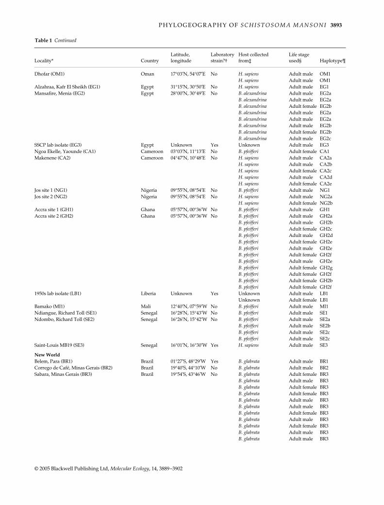
P H Y L O G E O G R A P H Y O F S C H I S T O S O M A M A N S O N I 3893
© 2005 Blackwell Publishing Ltd, Molecular Ecology, 14, 3889–3902
Dhofar (OM1) Oman 17°03′N, 54°07′E No H. sapiens Adult male OM1H. sapiens Adult male OM1
Alzahraa, Kafr El Sheikh (EG1) Egypt 31°15′N, 30°50′E No H. sapiens Adult male EG1Mansafire, Menia (EG2) Egypt 28°00′N, 30°49′E No B. alexandrina Adult male EG2a
B. alexandrina Adult male EG2aB. alexandrina Adult female EG2bB. alexandrina Adult male EG2aB. alexandrina Adult male EG2aB. alexandrina Adult male EG2bB. alexandrina Adult female EG2bB. alexandrina Adult male EG2c
SSCP lab isolate (EG3) Egypt Unknown Yes Unknown Adult male EG3Ngoa Ekelle, Yaounde (CA1) Cameroon 03°03′N, 11°13′E No B. pfeifferi Adult female CA1Makenene (CA2) Cameroon 04°47′N, 10°48′E No H. sapiens Adult male CA2a
H. sapiens Adult male CA2bH. sapiens Adult female CA2cH. sapiens Adult male CA2dH. sapiens Adult female CA2e
Jos site 1 (NG1) Nigeria 09°55′N, 08°54′E No B. pfeifferi Adult male NG1Jos site 2 (NG2) Nigeria 09°55′N, 08°54′E No H. sapiens Adult male NG2a
H. sapiens Adult female NG2bAccra site 1 (GH1) Ghana 05°57′N, 00°36′W No B. pfeifferi Adult male GH1Accra site 2 (GH2) Ghana 05°57′N, 00°36′W No B. pfeifferi Adult male GH2a
B. pfeifferi Adult male GH2bB. pfeifferi Adult female GH2cB. pfeifferi Adult male GH2dB. pfeifferi Adult female GH2eB. pfeifferi Adult male GH2eB. pfeifferi Adult female GH2fB. pfeifferi Adult male GH2eB. pfeifferi Adult female GH2gB. pfeifferi Adult female GH2fB. pfeifferi Adult female GH2bB. pfeifferi Adult female GH2f
1950s lab isolate (LB1) Liberia Unknown Yes Unknown Adult male LB1Unknown Adult female LB1
Bamako (MI1) Mali 12°40′N, 07°59′W No B. pfeifferi Adult male MI1Ndiangue, Richard Toll (SE1) Senegal 16°28′N, 15°43′W No B. pfeifferi Adult male SE1Ndombo, Richard Toll (SE2) Senegal 16°26′N, 15°42′W No B. pfeifferi Adult male SE2a
B. pfeifferi Adult male SE2bB. pfeifferi Adult male SE2cB. pfeifferi Adult male SE2c
Saint-Louis MB19 (SE3) Senegal 16°01′N, 16°30′W Yes H. sapiens Adult male SE3
New WorldBelem, Para (BR1) Brazil 01°27′S, 48°29′W Yes B. glabrata Adult male BR1Corrego de Café, Minas Gerais (BR2) Brazil 19°40′S, 44°10′W No B. glabrata Adult male BR2Sabara, Minas Gerais (BR3) Brazil 19°54′S, 43°46′W No B. glabrata Adult female BR3
B. glabrata Adult male BR3B. glabrata Adult female BR3B. glabrata Adult female BR3B. glabrata Adult male BR3B. glabrata Adult male BR3B. glabrata Adult female BR3B. glabrata Adult male BR3B. glabrata Adult female BR3B. glabrata Adult male BR3B. glabrata Adult male BR3
Locality* CountryLatitude, longitude
Laboratory strain?†
Host collected from‡
Life stage used§ Haplotype¶
Table 1 Continued
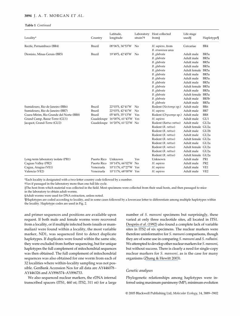
3894 J . A . T . M O R G A N E T A L .
© 2005 Blackwell Publishing Ltd, Molecular Ecology, 14, 3889–3902
Recife, Pernambuco (BR4) Brazil 08°06′S, 34°53′W No H. sapiens, from Cercariae BR4B. straminea area
Dionisio, Minas Gerais (BR5) Brazil 19°49′S, 42°45′W No B. glabrata Adult male BR5aB. glabrata Adult male BR5aB. glabrata Adult male BR5aB. glabrata Adult male BR5aB. glabrata Adult female BR5aB. glabrata Adult male BR5aB. glabrata Adult male BR5aB. glabrata Adult male BR5aB. glabrata Adult female BR5aB. glabrata Adult male BR5aB. glabrata Adult female BR5aB. glabrata Adult male BR5bB. glabrata Adult male BR5a
Sumidouro, Rio de Janeiro (BR6) Brazil 22°03′S, 42°41′W No Rodent (Nectomys sp.) Adult male BR6Sumidouro, Rio de Janeiro (BR7) Brazil 22°03′S, 42°41′W No H. sapiens Adult male BR7Ceara-Mirim, Rio Grande del Norte (BR8) Brazil 05°40′S, 35°13′W Yes Rodent (Oryzomys sp.) Adult male BR8Grand Camp, Basse-Terre (GU1) Guadeloupe 16°00′N, 61°43′W Yes H. sapiens Adult male GU1Jacquot, Grand-Terre (GU2) Guadeloupe 16°20′N, 61°22′W No Rodent (Rattus rattus) Adult male GU2a
Rodent (R. rattus) Adult female GU2aRodent (R. rattus) Adult male GU2bRodent (R. rattus) Adult male GU2aRodent (R. rattus) Adult female GU2aRodent (R. rattus) Adult male GU2aRodent (R. rattus) Adult female GU2aRodent (R. rattus) Adult male GU2aRodent (R. rattus) Adult female GU2a
Long-term laboratory isolate (PR1) Puerto Rico Unknown Yes Unknown Adult male PR1Caguas Valley (PR2) Puerto Rico 18°14′N, 66°02′W No H. sapiens Adult male PR2Cagua, Aragua (VE1) Venezuela 10°11′N, 67°27′W Yes H. sapiens Adult male VE1Valencia (VE2) Venezuela 10°11′N, 68°00′W Yes H. sapiens Adult male VE2
*Each locality is designated with a two-letter country code followed by a number.†Yes if passaged in the laboratory more than one full life cycle.‡The host from which material was collected in the field. Most specimens were collected from their snail hosts, and then passaged to mice in the laboratory to obtain adult worms.§Adult worms were used for DNA extraction, unless noted.¶Haplotypes are coded according to locality, and in some cases followed by a lowercase letter to differentiate among multiple haplotypes within the locality. Haplotype codes are used in Fig. 2.
Locality* CountryLatitude, longitude
Laboratory strain?†
Host collected from‡
Life stage used§ Haplotype¶
Table 1 Continued
and primer sequences and positions are available uponrequest. If both male and female worms were recoveredfrom a locality, or if multiple infected hosts (snails or mam-malian) were found within a locality, the most variablemarker, ND1, was sequenced first to detect duplicatehaplotypes. If duplicates were found within the same site,they were excluded from further sequencing, but for uniquehaplotypes the full complement of mitochondrial sequenceswas then obtained. The full complement of mitochondrialsequences was also obtained for one worm from each of32 localities where within-locality sampling was not pos-sible. GenBank Accession Nos for all data are AY446078–AY446326 and AY896574–AY896733.
We also sequenced nuclear markers, the rDNA internaltranscribed spacers (ITS1, 460 nt; ITS2, 311 nt) for a large
number of S. mansoni specimens but surprisingly, thesevaried at only three nucleotide sites, all located in ITS1.Desprès et al. (1992) also found a complete lack of variablesites in ITS2 of six specimens. The nuclear markers weretherefore uninformative for S. mansoni comparisons, thoughthey are of some use in comparing S. mansoni and S. rodhaini.We attempted to develop other nuclear markers for S. mansoni,but without success. There is clearly a need for single-copynuclear markers for S. mansoni, as is the case for manyorganisms (Zhang & Hewitt 2003).
Genetic analyses
Phylogenetic relationships among haplotypes were in-ferred using maximum parsimony (MP), minimum evolution
P H Y L O G E O G R A P H Y O F S C H I S T O S O M A M A N S O N I 3895
© 2005 Blackwell Publishing Ltd, Molecular Ecology, 14, 3889–3902
(ME), and maximum likelihood (ML) as implemented inpaup*4.0b10 (Swofford 2004), with 1000 bootstrap re-plicates performed for maximum parsimony and minimumevolution, and 100 replicates for maximum likelihood.A bestfit ML model was chosen (GTR + I + Γ) using theAkaike criterion in modeltest 3.4 (Posada & Crandall1998). Likelihood-ratio tests (Felsenstein 1981) wereused to test the hypothesis that the data collected wereconsistent with a molecular clock. The ML phylogeny wasestimated using the best-fit model, and then the likelihoodof this phylogeny was recalculated while constrainingthe estimate to fit the molecular clock model. Addition-ally, we employed Bayesian phylogenetic inference usingmrbayes 3.0 (Ronquist & Huelsenbeck 2003), which canallow for multiple models of evolution for each DNAregion (each model chosen using modeltest). Eachindividual mtDNA region was also analysed separatelyand no significant conflicts with the concatenated data setwere found. Nested clade analysis (Templeton 1998) wasnot completed on our data set because minimum-spanningnetworks could not be constructed due to the high diversityof the sequences, even when each mitochondrial regionwas considered separately. There were simply too manymissing theoretical haplotypes, as might be predicted fromthe long branch lengths leading to each lineage in the tree(Fig. 1).
Haplotype diversity (h) was calculated using dnasp 4.00(Rozas et al. 2003). Measures of nucleotide diversity weremade using the most complex substitution model conveni-ently available: nucleotide diversity (π) was calculated withJukes–Cantor corrections using dnasp 4.00 and net nucleotide
divergence (Dxy) between lineages was calculated withthe Tamura–Nei gamma correction model using mega 3(Kumar et al. 2004).
Tests of neutrality
We conducted two tests to determine whether selectionappeared strong in our mitochondrial data. For protein-coding regions, the number of synonymous substitutionsper synonymous site (KS) was compared with the numberof nonsynonymous substitutions per nonsynonymoussite (KN) and did not deviate from neutral expectations(KS > KN; P < 0.0001). Also, the McDonald–Kreitman test(McDonald & Kreitman 1991) for the two continuousprotein-coding sequences revealed no differences in theratios of nonsynonymous to synonymous changes within(RP) S. mansoni and between (RF) S. mansoni and S. rodhaini(Fisher’s exact test: COI, P = 0.37; nd1, P = 0.15), indicatingthat strong selection is not acting on these genes.
Tests of host use
Shimodaira–Hasegawa (SH) tests were conducted in paup*to evaluate alternative trees with or without host special-ization (Shimodaira & Hasegawa 1999). Trees constrainedto monophyly of parasites using the same host were gener-ated and used in the SH test compared to the unconstrainedtree. For the test of snail host use, only samples obtainedfrom snails were kept in the trees. For the definitive hosttests, there were unfortunately few specimens obtaineddirectly from the definitive hosts, rendering the test without
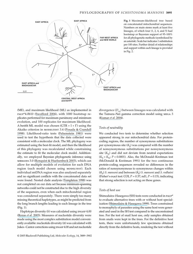
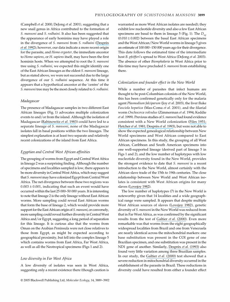
Fig. 1 Maximum-likelihood tree basedon concatenated mitochondrial sequences.Numbers on main stems mark at least fivelineages, of which four (1, 2, 4, and 5) hadbootstrap or Bayesian support of 95–100%for all phylogenetic methods (symbolized byan asterisk). Scale bar indicates 1 substitutionper 100 sites. Further detail of relationshipsand support within each lineage is providedin Fig. 2.
3896 J . A . T . M O R G A N E T A L .
© 2005 Blackwell Publishing Ltd, Molecular Ecology, 14, 3889–3902
meaning. However, most specimens obtained in the fieldfrom snails, unless they were obtained from areas with fewhuman hosts, can reasonably be assumed to be human-infecting. The definitive hosts test was conducted usingthis assumption.
Tests for founder effects
We also tested for bottleneck or founder effects whereappropriate using the method of Galtier et al. (2000). Thistest assumes that historical events like bottlenecks, colon-izations, and selective sweeps, through a quick increase ofthe coalescence rate, modify the shape of the genealogy ofhaplotypes, usually generating starlike genealogies. Anadvantage of the test is that the time (in generations) of theevent is estimated using a maximum-likelihood approach,and a statistical inference is made. The test requires that nomore than two states should be observed at any site, andthat sequence and nucleotide sites give similar information(Galtier et al. 2000).
Results
Diversity and suitability of mtDNA
Mitochondrial diversity within Schistosoma mansoni, incontrast with nuclear ITS1 and ITS2, was substantial: thecombined mitochondrial data set contains 376 variable sites(289 parsimony informative), and a total of 85 mitochondrialhaplotypes. Measures of haplotype and nucleotide diver-sity for different regions and for within localities arepresented in Tables 2 and 3, respectively. We tested forstrong selection in protein-coding regions, and found nodeviation from neutral expectations (methods describedabove). Likelihood-ratio tests suggested that in three indi-vidual mtDNA regions there has been a relatively constantrate of evolution (COI, P = 0.263; 12S-16S, P = 0.652; cyt b-ND4L-ND4, P = 0.097), although this was less true for theND1 region (P = 0.026). These findings suggest that themtDNA is suitable for tracing the evolutionary historyof the parasite. A general 2–4% mitochondrial clock isoften used to estimate divergence times and is appropriatehere because the mtDNA regions we used are distributedthroughout the mitochondrial genome and evolve at differ-ent rates. However, because three of the four regions used(COI, cyt b-ND4L-ND4, and ND1) evolve more quicklythan the 12S-16S (Palumbi 1996), the 4% rate is used in thisstudy (estimates based on a 2% rate are easily obtained bysimply doubling the times given).
Phylogeographic structure in S. mansoni
The 85 mitochondrial haplotypes found in S. mansoniseparated into five lineages, four of which were well sup-
ported by all phylogenetic methods (Fig. 1). These lineageswere also present when each mitochondrial region wasconsidered separately (not shown). Detail of each lineage isshown in Fig. 2. Lineages 1, 3 and 4 are comprised exclusively
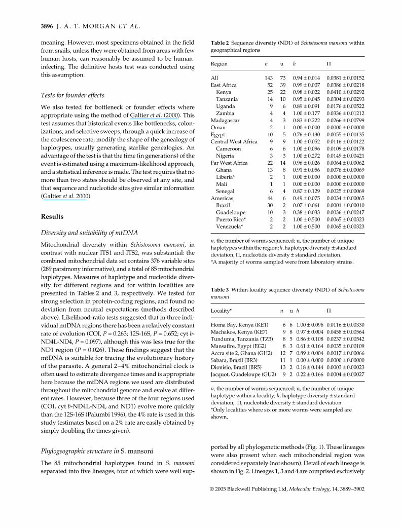
Table 2 Sequence diversity (ND1) of Schistosoma mansoni withingeographical regions
Region n u h Π
All 143 73 0.94 ± 0.014 0.0381 ± 0.00152East Africa 52 39 0.99 ± 0.007 0.0386 ± 0.00218
Kenya 25 22 0.98 ± 0.022 0.0410 ± 0.00292Tanzania 14 10 0.95 ± 0.045 0.0304 ± 0.00293Uganda 9 6 0.89 ± 0.091 0.0176 ± 0.00522Zambia 4 4 1.00 ± 0.177 0.0336 ± 0.01212
Madagascar 4 3 0.83 ± 0.222 0.0266 ± 0.00799Oman 2 1 0.00 ± 0.000 0.0000 ± 0.00000Egypt 10 5 0.76 ± 0.130 0.0055 ± 0.00135Central West Africa 9 9 1.00 ± 0.052 0.0116 ± 0.00122
Cameroon 6 6 1.00 ± 0.096 0.0109 ± 0.00178Nigeria 3 3 1.00 ± 0.272 0.0149 ± 0.00421
Far West Africa 22 14 0.96 ± 0.026 0.0064 ± 0.00062Ghana 13 8 0.91 ± 0.056 0.0076 ± 0.00069Liberia* 2 1 0.00 ± 0.000 0.0000 ± 0.00000Mali 1 1 0.00 ± 0.000 0.0000 ± 0.00000Senegal 6 4 0.87 ± 0.129 0.0025 ± 0.00069
Americas 44 6 0.49 ± 0.075 0.0034 ± 0.00065Brazil 30 2 0.07 ± 0.061 0.0001 ± 0.00010Guadeloupe 10 3 0.38 ± 0.033 0.0036 ± 0.00247Puerto Rico* 2 2 1.00 ± 0.500 0.0065 ± 0.00323Venezuela* 2 2 1.00 ± 0.500 0.0065 ± 0.00323
n, the number of worms sequenced; u, the number of unique haplotypes within the region; h, haplotype diversity ± standard deviation; Π, nucleotide diversity ± standard deviation.*A majority of worms sampled were from laboratory strains.
Table 3 Within-locality sequence diversity (ND1) of Schistosomamansoni
Locality* n u h Π
Homa Bay, Kenya (KE1) 6 6 1.00 ± 0.096 0.0116 ± 0.00330Machakos, Kenya (KE7) 9 8 0.97 ± 0.004 0.0458 ± 0.00564Tunduma, Tanzania (TZ3) 8 5 0.86 ± 0.108 0.0237 ± 0.00542Mansafire, Egypt (EG2) 8 3 0.61 ± 0.164 0.0035 ± 0.00109Accra site 2, Ghana (GH2) 12 7 0.89 ± 0.004 0.0017 ± 0.00066Sabara, Brazil (BR3) 11 1 0.00 ± 0.000 0.0000 ± 0.00000Dionisio, Brazil (BR5) 13 2 0.18 ± 0.144 0.0003 ± 0.00023Jacquot, Guadeloupe (GU2) 9 2 0.22 ± 0.166 0.0004 ± 0.00027
n, the number of worms sequenced; u, the number of unique haplotype within a locality; h, haplotype diversity ± standard deviation; Π, nucleotide diversity ± standard deviation*Only localities where six or more worms were sampled are shown.
P H Y L O G E O G R A P H Y O F S C H I S T O S O M A M A N S O N I 3897
© 2005 Blackwell Publishing Ltd, Molecular Ecology, 14, 3889–3902
of specimens from East Africa and Madagascar. Lineage 2contains specimens from Central West Africa (Cameroonand Nigeria) and Egypt. The fifth and final lineage containsall of the specimens from South America, the Caribbeanregion, and Far West Africa (countries west of Nigeria),and at the base of this lineage lie a few East Africansamples. The presence of East African samples in four ofthe five clades is a revealing indicator of their diversity,which can also be seen in other measures (Table 2). Allother areas were less diverse, with Far West Africa and theNew World harbouring the least diversity. Within-localitysampling, where available, was consistent with this trend.The within-locality sampling is poor, but it is significantthat the small amount of within-locality sampling easily un-covered diversity within East African localities, and almostnone in New World localities (Table 3).
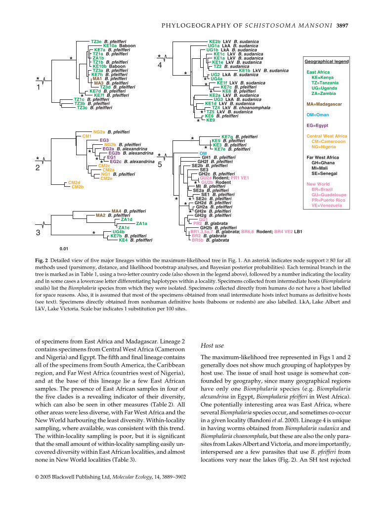
Host use
The maximum-likelihood tree represented in Figs 1 and 2generally does not show much grouping of haplotypes byhost use. The issue of snail host usage is somewhat con-founded by geography, since many geographical regionshave only one Biomphalaria species (e.g. Biomphalariaalexandrina in Egypt, Biomphalaria pfeifferi in West Africa).One potentially interesting area was East Africa, whereseveral Biomphalaria species occur, and sometimes co-occurin a given locality (Bandoni et al. 2000). Lineage 4 is uniquein having worms obtained from Biomphalaria sudanica andBiomphalaria choanomphala, but these are also the only para-sites from Lakes Albert and Victoria, and more importantly,interspersed are a few parasites that use B. pfeifferi fromlocations very near the lakes (Fig. 2). An SH test rejected
Fig. 2 Detailed view of five major lineages within the maximum-likelihood tree in Fig. 1. An asterisk indicates node support ≥ 80 for allmethods used (parsimony, distance, and likelihood bootstrap analyses, and Bayesian posterior probabilities). Each terminal branch in thetree is marked as in Table 1, using a two-letter country code (also shown in the legend above), followed by a number indicating the localityand in some cases a lowercase letter differentiating haplotypes within a locality. Specimens collected from intermediate hosts (Biomphalariasnails) list the Biomphalaria species from which they were isolated. Specimens collected directly from humans do not have a host labelledfor space reasons. Also, it is assumed that most of the specimens obtained from snail intermediate hosts infect humans as definitive hosts(see text). Specimens directly obtained from nonhuman definitive hosts (baboons or rodents) are also labelled. LkA, Lake Albert andLkV, Lake Victoria. Scale bar indicates 1 substitution per 100 sites.
3898 J . A . T . M O R G A N E T A L .
© 2005 Blackwell Publishing Ltd, Molecular Ecology, 14, 3889–3902
the separation of lineage 4 haplotypes according to snailhost use (P < 0.001). Shimodaira–Hasegawa tests rejectedtrees which were constrained to monophyly of baboonisolates (P < 0.05) or rodent isolates (P < 0.001).
Bottleneck tests
Due to the requirements of the test of Galtier et al. (2000),we were able to compute the test only for the followingdata sets: for Brazil, and for the New World (includingBrazil). In both instances, a strong and highly significantreduction in diversity was inferred to have occurred veryrecently (estimates were 0 generations ago and P < 0.01 inboth cases). We could not run the test to differentiatebetween demographic bottlenecks and selective sweepssince we essentially had only one marker (mtDNA).
Relationship to Schistosoma rodhaini
The two isolates of S. rodhaini we obtained had identicalhaplotypes, despite having been collected at different local-ities. We attempted to determine the root for the S. mansonitree by using S. rodhaini as an outgroup, but the large geneticdistances between S. mansoni and S. rodhaini caused differentphylogenetic methods to determine different S. mansoni roots,even when third codon positions were removed (not shown).The molecular clock estimate for the divergence between S.mansoni and S. rodhaini is 2.8 ± 0.19 Ma (Dxy = 0.224 ± 0.015),although having only a single haplotype from S. rodhainioverestimates this divergence (Edwards & Beerli 2000).A more recent divergence is supported by only six fixednuclear ITS sequence differences between the two species.
Discussion
Schistosoma mansoni exhibits high mitochondrial diversity
The DNA sequences presented here, the first from anextensive and globally widespread set of S. mansoni speci-mens, show high mitochondrial diversity, both in haplotypeand nucleotide diversity (Table 2). There were 73 haplo-types when only ND1 was considered (Table 2); 85 whenall mitochondrial sequences were combined (Fig. 1).
Geographical structure vs. host usage
There is considerable geographical structure, at global andregional scales, in the haplotypes obtained in our study(Fig. 1), and we discuss these patterns below. In fact, itwould appear that geographical separation over time hasplayed a prominent role in the diversification of S. mansoni,far more important a role than host usage. We did not findany strong evidence that major or minor lineages within
S. mansoni have specialized to different mammalian or snailhosts. The two baboon isolates in lineage 2 are close relatives,but several other haplotypes infecting humans are alsoclosely related to these (Fig. 2). Also, an SH test rejected theseparation of these haplotypes into monophyletic baboon-and human-infecting lineages (P < 0.05). Several rodentisolates from the New World are present in lineage 5, butalso do not group together, and an SH test rejected rodentisolates as a monophyletic assemblage (P < 0.001). Theseresults show that use of unusual mammalian hosts is notunique to a certain lineage, and that morphological or beha-vioural adaptations observed (Théron et al. 1997; D’Andreaet al. 2002; Freire et al. 2002) are likely recent and local. Inthe only plausible test for snail host use (see Results), anSH test rejected the separation of lineage 4 haplotypesaccording to snail host use (P < 0.001). A likely explanationfor this pattern is that lineage 4 arose by geographicalseparation in the area of Lakes Victoria and Albert, andalthough Biomphalaria sudanica and its relatives such asBiomphalaria choanomphala (DeJong et al. 2001) are commonthere, the parasite lineage has retained the ability to alsoinfect Biomphalaria pfeifferi. Snail, like mammalian, host usedoes not seem to comprise the barriers that have led to thediversification of S. mansoni seen in our data. Adaptation tolocal hosts probably does occur to some degree (e.g. theparasite may become more infective to local hosts aftercolonizing a new geographical area), but this adaptationdoes not preclude infection of a different host later in time.This has probably been particularly true in Africa becauseBiomphalaria has only been there for as little as 1.1 millionyears, and the African species of Biomphalaria are all quiteclosely related (DeJong et al. 2001).
East African origin
The conspicuous amount of diversity in East Africa, andthe presence of East African haplotypes in four of the fivelineages suggests an East African origin for S. mansoni(Figs 1 and 2). Although sampling is somewhat biasedtowards East Africa, the nucleotide diversity in East Africais several times higher (3–10×) than all regions exceptMadagascar (Table 2). Diversity within even single sites inEast Africa (Homa Bay 0.0116, Machakos 0.0458, andTunduma 0.0237; Table 3) was 2–9× that in South America,the Caribbean region, and Far West Africa combined(0.00485 ± 0.00052; not shown in either table). It should alsobe noted that there is likely much East African (and perhapsMadagascaran) diversity yet unsampled, as evidenced bylineage 3 which contains some distantly related haplotypesand may represent two or more lineages (Fig. 2).
The estimate for the time of origin in East Africa is rela-tively recent, approximately 0.30–0.43 Ma (average Dxybetween the five lineages 0.029 ± 0.005). These dates followthe arrival in Africa of Biomphalaria between 1.1 and 4.5 Ma
P H Y L O G E O G R A P H Y O F S C H I S T O S O M A M A N S O N I 3899
© 2005 Blackwell Publishing Ltd, Molecular Ecology, 14, 3889–3902
(Campbell et al. 2000; DeJong et al. 2001), suggesting that anew snail genus in Africa contributed to the formation ofS. mansoni and S. rodhaini. It also has been suggested thatthe appearance of early hominins may have played a rolein the divergence of S. mansoni from S. rodhaini (Desprèset al. 1992); however, our data indicate a more recent originfor the parasite, and Homo ergaster, the immediate ancestorto Homo sapiens, or H. sapiens itself, may have been the firsthominin hosts. When we attempted to root the S. mansonitree using S. rodhaini, we expected this might identify oneof the East African lineages as the oldest S. mansoni lineage,but as stated above, we were not successful due to the largedivergence of our S. rodhaini sequence. At this time itappears that a hypothetical ancestor at the ‘centre’ of theS. mansoni tree may be the most closely related to S. rodhaini.
Madagascar
The presence of Madagascar samples in two different EastAfrican lineages (Fig. 1) advocates multiple colonizationevents to and/or from the island. Although the isolation ofMadagascar (Rabinowitz et al. 1983) could have led to aseparate lineage of S. mansoni, none of the Madagascarisolates fall in basal positions within the two lineages. Thesimplest explanation is at least two separate and relativelyrecent colonizations of the island from East Africa.
Egyptian and Central West African affinities
The grouping of worms from Egypt and Central West Africain lineage 2 was a surprising finding. Although the numberof specimens and localities sampled is low, there appears tobe more diversity in Central West Africa, which may suggestthat S. mansoni may have colonized Egypt from Central WestAfrica. The net divergence between these two regions was0.003 ± 0.001, indicating that such an event would haveoccurred within the last 25 000–50 000 years. It is interestingto note that lineage 2 is the only lineage without East Africanworms. More sampling could reveal East African wormsthat form the base of lineage 2, which would provide moresupport for the East African origin of S. mansoni, or conversely,more sampling could reveal further diversity in Central WestAfrica and/or Egypt, suggesting a long period of separationfor this lineage. It is curious also that the worms fromOman on the Arabian Peninsula were not close relatives tothose from Egypt, as might be expected according togeographical proximity, but fell into the complex lineage 5which contains worms from East Africa, Far West Africa,as well as all the Neotropical specimens (Figs 1 and 2).
Low diversity in Far West Africa
A low diversity of isolates was seen in West Africa,suggesting only a recent existence there (though caution is
warranted as more West African isolates are needed); theyexhibit low nucleotide diversity and also a few East Africanspecimens are basal to them in lineage 5 (Fig. 1). The Dxy(0.010 ± 0.002) between the basal East African specimensand the West African/New World worms in lineage 5 givesan estimate of 100 000–150 000 years ago for their divergence.This date follows the estimated time of the intermediatehost B. pfeifferi’s spread to West Africa (DeJong et al. 2003).The absence of other Biomphalaria in West Africa prior tothis time may have precluded S. mansoni from establishingthere.
Colonization and founder effect in the New World
While a number of parasites that infect humans arethought to be post-Columbian colonists of the New World,this has been confirmed genetically only for the malariaagent Plasmodium falciparum (Joy et al. 2003), the liver flukeFasciola hepatica (Mas-Coma et al. 2001), and the filarialworm Onchocerca volvulus (Zimmerman et al. 1994; Keddieet al. 1999). Previous studies of S. mansoni had found evidenceconsistent with a New World colonization (Files 1951;Fletcher et al. 1981; Desprès et al. 1993), but were not able toshow the expected genealogical relationship between NewWorld specimens and West African compared to EastAfrican specimens. In this study, the grouping of all WestAfrican, Caribbean and South American specimens intoone well-supported lineage (derived part of lineage 5 inFigs 1 and 2), and the low number of haplotypes with lownucleotide diversity found in the New World, providesthe strongest evidence to date that S. mansoni is a recentintroduction to the New World, almost certainly with theAfrican slave trade of the 15th to 19th centuries. The closerelationship between New World and West African iso-lates is consistent with West African origins for manyslaves (Lovejoy 1982).
The low number of haplotypes (7) in the New World isnoteworthy given that 14 localities and a wide geograph-ical range were sampled. It appears that despite multipleWest African sources of slaves (Lovejoy 1982), geneticdiversity of S. mansoni in the New World was reduced fromthat in Far West Africa, as was confirmed by the significantresults from the test of Galtier et al. (2000). Even moreremarkable was that worms from the eight geographicallywidespread localities from Brazil and one from Venezuelaare nearly identical across the mitochondrial markers: onebase substitution was present in the COI gene of oneBrazilian specimen, and one substitution was present in theND1 gene of another. Similarly, Desprès et al. (1993) alsofound very little variation among three Brazilian samples.In our study, the Galtier et al. (2000) test showed that asevere reduction in mitochondrial diversity occurred in theestablishment of the parasite in Brazil. These reductions indiversity could have resulted from either a founder effect
3900 J . A . T . M O R G A N E T A L .
© 2005 Blackwell Publishing Ltd, Molecular Ecology, 14, 3889–3902
(reduced number of parasites surviving the journey whichslaves were forced to make) or from a selective sweep,imposed by a new environment and a new intermediatehost, or both demographics and selection. We were unableto test for a selective sweep due to the lack of a secondgenetic marker, and consider the founder effect a plausibleand simple explanation given the current data.
Few mutations would be expected in 500 years since thebeginning of the slave trade, so the presence of multipleNew World haplotypes suggests introductions from mul-tiple West African sources, and one would expect to be ableto find New World haplotypes in their West African regionof origin. We found one such instance of a shared haplo-type: Liberia, which had an identical haplotype to the mostcommon Brazilian haplotype. However, we urge cautionin interpreting this result because the Liberian isolate avail-able to us was from a strain that has been maintained in thelaboratory for more than 50 years (Gonnert 1955). We sam-pled from seven West African localities, but more robustWest African sampling, especially from more regions whichwere sources of slaves and from which parasite materialhas been difficult to obtain, might yield haplotypes thatmatch those in the New World.
Implications
Schistosoma mansoni shows high levels of mitochondrialdifferentiation among regions in Africa, and in fact, in EastAfrica, there is high genetic diversity within single local-ities. Though ITS sequences are conserved, there is probablysome variation in the nuclear genome that correlates withmitochondrial variation, such that places with high mito-chondrial diversity also have the highest nuclear diversity.Worms of different genetic backgrounds could responddifferently to control measures such as drugs and vaccines.It would be prudent for researchers involved in thedevelopment and implementation of these tools againstthe disease to test them against S. mansoni strains frommultiple geographical locations, including representativesfrom each of the lineages uncovered in this study. Also, asthe complete genome sequence of S. mansoni is imminentlycompleted (Chitsulo et al. 2004), it should be rememberedthat it is representative of a diverse parasite.
As a massive control effort is now underway, in whichpraziquantel, the current drug of choice to cure schistosomi-asis, is being provided to the populations of whole countries(Crompton et al. 2003; www.who.int/wormcontrol/en/),signs of drug resistance need to be monitored. It is possiblethat high genetic diversity in worms, particularly in EastAfrica, might make development of drug resistancemore likely, though the putative examples of praziquantelresistance thus far have been from areas (Egypt, Senegal;Doenhoff et al. 2002) that seem to have relatively lowerdiversity. It should be noted as well that worms from
these areas are members of two different mtDNA line-ages (lineages 2 and 5, respectively), so resistance appearsnot to have been confined to any one lineage.
It is interesting to consider that in East African localitieswhere different lineages are present, worms from theselineages almost certainly are present together within indi-vidual human hosts. Do these genetic lineages mate witheach other? Do they compete? Does the presence of multiplegenetic lineages affect pathogenicity in the human host?A recent study (Gower & Webster 2005) found evidencefor competition between S. mansoni strains of differingvirulence co-infecting the intermediate host. These types ofquestions have not been addressed for human hosts, andthe diversity within individual human hosts has only beenassessed with one study (Curtis et al. 2002), in Brazil.Moderate microsatellite diversity was found within patientsand genetic subdivision was found among them. Similarstudies in areas with the other extreme of S. mansoni diver-sity, such as East Africa, may yield much different results,and it may be instructive to compare such findings. Indeed,Schistosoma mansoni research should include a substantialfocus on East Africa, where the parasite originated andwhere it continues to thrive today.
Acknowledgements
We are grateful to Joseph Kinuthia, Geoffrey Maina, TimothyKamau, Julius Andove of the Kenya Medical Research Institute(KEMRI), Jimmy Kihara and Francis Mungai of the Division ofVector-Borne Diseases, Ministry of Health, Kenya, for logisticalsupport and assistance in the field. The Director of KEMRI isthanked for publication approval. We thank Sarah Tishkoff, AlainFroment, and Bernard Wood for feedback providing an anthropo-logical perspective. We acknowledge technical support from theUniversity of New Mexico’s Molecular Biology Facility which issupported by NIH Grant number 1P20RR18754 from the InstituteDevelopment Award (IDeA) Program of the National Center forResearch Resources. All authors collected S. mansoni isolates,and thank L. Mubila for providing isolates from Zambia, withthe assistance of M. Shehata. M. Tanner and C. Grevelding arethanked for providing the Liberian isolates from each of theirlaboratories. Funding was provided by U.S. National Institutes ofHealth Grant AI44913 to G.M.M. and E.S.L.
References
Bandoni SM, Mulvey M, Loker ES (2000) Population structure andtaxonomic discrimination among three species of BiomphalariaPreston, 1910 (Gastropoda: Planorbidae) from Kenya. ZoologicalJournal of the Linnean Society, 129, 387–401.
Campbell G, Jones CS, Lockyer AE et al. (2000) Molecular evidencesupports an African affinity of the Neotropical freshwater gas-tropod, Biomphalaria glabrata, Say 1818, an intermediate host forSchistosoma mansoni. Proceedings of the Royal Society of London.Series B, Biological Sciences, 267, 2351–2358.
Chitsulo L, Engels D, Montresor A, Savioli L (2000) The global statusof schistosomiasis and its control. Acta Tropica, 77, 41–51.
P H Y L O G E O G R A P H Y O F S C H I S T O S O M A M A N S O N I 3901
© 2005 Blackwell Publishing Ltd, Molecular Ecology, 14, 3889–3902
Chitsulo L, Loverde R, Engels D et al. (2004) Schistosomiasis.Nature Reviews Microbiology, 2, 12–13.
Crompton DWT (1999) How much human helminthiasis is therein the world? Journal of Parasitology, 85, 397–403.
Crompton DWT, Engels D, Montresor A, Neira MP, Savioli L (2003)Action starts now to control disease due to schistosomiasis andsoil-transmitted helminthiasis. Acta Tropica, 86, 121–124.
Curtis J, Sorensen RE, Minchella DJ (2002) Schistosome geneticdiversity: the implications of population structure as detectedby microsatellite markers. Parasitology, 125, S51–S59.
D’Andrea PS, Fernandes FA, Cerqueira R, Rey L (2002) Experi-mental evidence and ecological perspectives for the adaptationof Schistosoma mansoni Sambon, 1907 (Digenea: Schistosomatidae)to a wild host, the water-rat, Nectomys squamipes Brants, 1827(Rodentia: Sigmodontinae). Memorias Do Instituto Oswaldo Cruz,97, 11–14.
Davis GM (1980) Snail hosts of Asian Schistosoma infectingman: evolution and coevolution. Malcological Review, Suppl. 2:195–238.
Davis GM (1992) Evolution of prosobranch snails transmitting AsianSchistosoma; coevolution with Schistosoma: a review. Progress inClinical Parasitology, 3, 145–204.
DeJong RJ, Morgan JAT, Paraense WL et al. (2001) Evolutionaryrelationships and biogeography of Biomphalaria (Gastropoda:Planorbidae) with implications regarding its role as host of thehuman bloodfluke, Schistosoma mansoni. Molecular Biology andEvolution, 18, 2225–2239.
DeJong RJ, Morgan JAT, Wilson WD et al. (2003) Phylogeographyof Biomphalaria glabrata and B. pfeifferi, important intermediatehosts of Schistosoma mansoni in the New and Old World tropics.Molecular Ecology, 12, 3041–3056.
Desprès L, Imbert-Establet D, Combes C, Bonhomme F (1992)Molecular evidence linking hominid evolution to recent radi-ation of schistosomes (Platyhelminthes: Trematoda). MolecularPhylogenetics and Evolution, 1, 295–304.
Desprès L, Imbert-Establet D, Monnerot M (1993) Molecular char-acterization of mitochondrial-DNA provides evidence for therecent introduction of Schistosoma mansoni into America. Molecu-lar and Biochemical Parasitology, 60, 221–230.
Doenhoff MJ, Kusel JR, Coles GC, Cioli D (2002) Resistance ofSchistosoma mansoni to praziquantel: is there a problem? Trans-actions of the Royal Society of Tropical Medicine and Hygiene, 96,465–469.
Edwards SV, Beerli P (2000) Perspective: gene divergence,population divergence, and the variance in coalescence time inphylogeographic studies. Evolution, 54, 1839–1854.
Eppert A, Lewis FA, Grzywacz C, Coura P, Caldas I, Minchella DJ(2002) Distribution of schistosome infections in molluscan hostsat different levels of parasite prevalence. Journal of Parasitology,88, 232–236.
Felsenstein J (1981) Evolutionary trees from DNA sequences:a maximum likelihood approach. Journal of Molecular Evolution,17, 368–376.
Files VS (1951) A study of the vector-parasite relationships inSchistosoma mansoni. Parasitology, 41, 264–269.
Fletcher M, Loverde PT, Woodruff DS (1981) Genetic variation inSchistosoma mansoni — enzyme polymorphisms in poulationsfrom Africa, southwest Asia, South America, and the WestIndies. American Journal of Tropical Medicine and Hygiene, 30,406–421.
Freire N, Machado-Silva JR, Rodrigues-Silva R, Rey L (2002)Cercarial chaetotaxy and sex differentiation of Schistosoma
mansoni deriving from humans and Nectomys squamipes (Muridae:Sigmondontinae) in Brazil. Memorias Do Instituto Oswaldo Cruz,97, 127–132.
Galtier N, Depaulis F, Barton NH (2000) Detecting bottlenecks andselective sweeps from DNA sequence polymorphism. Genetics,155, 981–987.
Gonnert R (1955) Schiztosomiasis studies. I. Contributions to theanatomy and histology of Schistosoma mansoni. Zeitschrift furTropenmedizine und Parasitologie, 6, 18–33.
Gower CM, Webster JP (2005) Intraspecific competition and theevolution of virulence in a parasitic trematode. Evolution, 59,544–553.
Joy DA, Feng XR, Mu JB et al. (2003) Early origin and recent expan-sion of Plasmodium falciparum. Science, 300, 318–321.
Keddie EM, Higazi T, Boakye D et al. (1999) Onchocerca volvulus:limited heterogeneity in the nuclear and mitochondrial genomes.Experimental Parasitology, 93, 198–206.
Kumar S, Tamura K, Nei M (2004) mega 3: Integrated software formolecular evolutionary genetics analysis and sequence align-ment. Briefings in Bioinformatics, 5, 150–163.
Lambertucci JR, Serufo JC, Gerspacher-Lara R et al. (2000) Schisto-soma mansoni: assessment of morbidity before and after control.Acta Tropica, 77, 101–109.
Leclerc MC, Durand P, Gauthier C et al. (2004) Meager geneticvariability of the human malaria agent Plasmodium vivax. Pro-ceedings of the National Academy of Sciences, USA, 14455–14460.
Lockyer AE, Olson PD, Ostergaard P et al. (2003) The phylogenyof the Schistosomatidae based on three genes with emphasis onthe interrelationships of Schistosoma Weinland, 1858. Parasitology,126, 203–224.
Lovejoy PE (1982) The volume of the the Atlantic slave trade — asynthesis. Journal of African History, 23, 473–501.
Mas-Coma S, Funatsu IR, Bargues MD (2001) Fasciola hepatica andlymnaeid snails occurring at very high altitude in South America.Parasitology, 123, S115–S127.
McDonald JH, Kreitman M (1991) Adaptive protein evolution atthe Adh locus in Drosophila. Nature, 351, 652–654.
Morgan JAT, DeJong RJ, Kazibwe F, Mkoji GM, Loker ES (2003a)A newly-identified lineage of Schistosoma. International Journalfor Parasitology, 33, 977–985.
Morgan JAT, DeJong RJ, Lwambo NJS et al. (2003b) First reportof a natural hybrid between Schistosoma mansoni and S. rodhaini.Journal of Parasitology, 89, 416–418.
Palumbi SR (1996) Nucleic acids II: the polymerase chain reaction.In: Molecular Systematics (eds Hillis DM, Moritz C Mable BK),pp. 205–248. Sinauer Associates, Sunderland, Massahusetts.
Posada D, Crandall KA (1998) modeltest: testing the model ofDNA substitution. Bioinformatics, 14, 817–818.
Rabinowitz PD, Coffin MF, Falvey D (1983) The separation ofMadagascar and Africa. Science, 220, 67–69.
Ronquist F, Huelsenbeck JP (2003) mrbayes 3: Bayesian phylo-genetic inference under mixed models. Bioinformatics, 19, 1572–1574.
Rozas J, Sanchez-DeI, Barrio JC, Messeguer X, Rozas R (2003)dnasp, DNA polymorphism analyses by the coalescent andother methods. Bioinformatics, 19, 2496–2497.
Shimodaira H, Hasegawa M (1999) Multiple comparisons oflog-likelihoods with applications to phylogenetic inference.Molecular Biology and Evolution, 16, 1114–1116.
Snyder SD, Loker ES (2000) Evolutionary relationships among theSchistosomatidae (Platyhelminthes: Digenea) and an Asian originfor Schistosoma. Journal of Parasitology, 86, 283–288.
3902 J . A . T . M O R G A N E T A L .
© 2005 Blackwell Publishing Ltd, Molecular Ecology, 14, 3889–3902
Swofford DL (2004) PAUP*: Phylogenetic Analysis Using Parsimony(*and Other Methods). Sinauer Associates, Sunderland, Massachusetts.
Templeton AR (1998) Nested clade analyses of phylogeographicdata: testing hypotheses about gene flow and population history.Molecular Ecology, 7, 381–397.
Théron A, Mouahid G, Moné H (1997) Schistosoma mansoni: cercarialshedding patterns from a mixed infection of Biomphalaria glabratawith two (early and late) chronobiological variants. ParasitologyResearch, 83, 356–358.
Zhang DX, Hewitt GM (2003) Nuclear DNA analyses in geneticstudies of populations: practice, problems and prospects. Molec-ular Ecology, 12, 563–584.
Zimmerman PA, Katholi CR, Wooten MC, Langunnasch N,Unnasch TR (1994) Recent evolutionary history of American
Onchocerca volvulus, based on analysis of a tandemly repeatedDNA-sequence family. Molecular Biology and Evolution, 11, 384–392.
This study is a key part of a project to assess the global diversityand the evolutionary history of Schistosoma mansoni and its snailhosts. Jess Morgan and Randall DeJong completed the molecularwork, data analysis, and manuscript preparation and continue usingmolecular tools to investigate the interactions of parasitic organismsand their hosts. Gerald Mkoji and Eric (Sam) Loker continue tocollaborate on projects on the interactions between genetic diversityand epidemiology of Schistosoma mansoni in Kenya.
Related Documents