Organotin(IV) complexes of carboxylate derivative as potential chemotherapeutic agents against Leishmania Qurrat-ul-Ain H. Qureshi a , Akhtar Nadhman a , Muhammad Sirajuddin b,⇑ , Gul Shahnaz c , Saqib Ali b,⇑ , Afzal Shah b , Muhammad Masoom Yasinzai a a Department of Biotechnology, Quaid-i-Azam University, Islamabad, Pakistan b Department of Chemistry, Quaid-i-Azam University, Islamabad, Pakistan c Department of Pharmacy, Quaid-i-Azam University, Islamabad, Pakistan article info Article history: Received 12 May 2014 Received in revised form 12 June 2014 Accepted 13 June 2014 Available online 24 June 2014 Keywords: Organotin(IV) carboxylate Leishmania DNA interaction Docking Apoptosis Drug targeting dose–response abstract The aim of the present research is to search for new antileishmanial drugs as potential chemotherapeutic agents. We have some newly synthesized organotin(IV) based peptide derivatives of carboxylate ligand as potential chemotherapeutic agents against Leishmania. Antileishmanial activity was carried out against Leishmania tropica KWH23 strain, and the compounds showed significant activity except MS 20 , main ligand with no tin. Biocompatibility assays showed that the tested compounds have LD 50 values ranging from 60.296 to 224.759 lg/mL with the exception of MS 20 Me 3 (596.91 lg/mL) and MS 20 Oct 2 (405.72 lg/mL). They were found highly biocompatible. From the in-silico studies, all the synthetic deriv- atives except MS 20 Me 3 (1,10-Ph) and MS 20 were bound to leishmanolysin (GP63) receptor. This binding probably resulted in a receptor mediated inlet of these synthetic derivatives inside the Leishmania cells, where they were further bound to the DNA (confirmed by DNA interaction study). The interaction caused a redox reaction between the DNA and the compounds, which lead to the degradation of DNA and caused apoptotic death of L. tropica. The results suggested that the organotin(IV) derivatives have good potential as chemotherapeutic agents and can be a better replacement for the currently available drugs. Ó 2014 Elsevier B.V. All rights reserved. 1. Introduction Leishmaniasis is a vector-borne disease caused by obligate pro- tozoan parasites of genus Leishmania. The Leishmania parasites infect several mammals, including human beings. The insect vector of the disease is phlebotomine sand fly [1]. Leishmaniasis is specif- ically endemic in 88 countries [2]. Drugs are the most important tool in order to control differ- ent clinical manifestations of leishmaniasis. Several drugs (e.g., Miltefosine, Amphotericin B, Glucantime and Pentamidine) have been reported which can be used as antileishmanial agents but none of these is the ultimate choice in terms of efficacy and potential [3,4]. Pentavalent antimonials are considered to be effective for the treatment of leishmaniasis [5] but the discovery of pathogenic strains is showing resistance against antimony salt [6]. Furthermore, there are several diverse side effects associated with the antimony derivatives, which caused several deaths, secondary to fulminant hepatotoxicity [7]. Oral Miltefosine, a phosphocholine analogue, also appeared to be effective in the disease treatment [8]. It has showed severe side effects including gastrointestinal problems, teratogenic effects and further, cannot be used in the pregnant women [9–11]. In order to get new and beneficent drugs for effective treatment, scientists have always tried to come up with something new and discovered of new biologically active compounds [12]. Con- sidering different problems related to the treatment options, peptide derivatives have been considered as an alternative option for therapeutic intervention of cutaneous leishmaniasis. Peptide derivatives have been testified to have antimicrobial functions. They have direct or individual effect on microorgan- isms or they act synergistically by curbing the functions of immune cells [13,14]. Synthetic peptide derivatives have shown significant effects in vertebrates and invertebrates considering their effects in different clinical manifestations (as well as infec- tions, autoimmune diseases and cancer) and their efficacy have opened up the horizon for their application as a therapeutic tool in case of leishmaniasis [15]. Tin (Sn) played a major role in advancements of organometallic chemistry, which started in 1949 and it was considered to be effec- tive agent in chemistry by showing various applications [16]. http://dx.doi.org/10.1016/j.ica.2014.06.011 0020-1693/Ó 2014 Elsevier B.V. All rights reserved. ⇑ Corresponding authors. Tel.: +92 51 90642130; fax: +92 51 90642241. E-mail addresses: [email protected], [email protected] (M. Sirajuddin), [email protected] (S. Ali). Inorganica Chimica Acta 423 (2014) 220–228 Contents lists available at ScienceDirect Inorganica Chimica Acta journal homepage: www.elsevier.com/locate/ica

Welcome message from author
This document is posted to help you gain knowledge. Please leave a comment to let me know what you think about it! Share it to your friends and learn new things together.
Transcript
Inorganica Chimica Acta 423 (2014) 220–228
Contents lists available at ScienceDirect
Inorganica Chimica Acta
journal homepage: www.elsevier .com/locate / ica
Organotin(IV) complexes of carboxylate derivative as potentialchemotherapeutic agents against Leishmania
http://dx.doi.org/10.1016/j.ica.2014.06.0110020-1693/� 2014 Elsevier B.V. All rights reserved.
⇑ Corresponding authors. Tel.: +92 51 90642130; fax: +92 51 90642241.E-mail addresses: [email protected], [email protected] (M. Sirajuddin),
[email protected] (S. Ali).
Qurrat-ul-Ain H. Qureshi a, Akhtar Nadhman a, Muhammad Sirajuddin b,⇑, Gul Shahnaz c,Saqib Ali b,⇑, Afzal Shah b, Muhammad Masoom Yasinzai a
a Department of Biotechnology, Quaid-i-Azam University, Islamabad, Pakistanb Department of Chemistry, Quaid-i-Azam University, Islamabad, Pakistanc Department of Pharmacy, Quaid-i-Azam University, Islamabad, Pakistan
a r t i c l e i n f o
Article history:Received 12 May 2014Received in revised form 12 June 2014Accepted 13 June 2014Available online 24 June 2014
Keywords:Organotin(IV) carboxylateLeishmaniaDNA interactionDockingApoptosisDrug targeting dose–response
a b s t r a c t
The aim of the present research is to search for new antileishmanial drugs as potential chemotherapeuticagents. We have some newly synthesized organotin(IV) based peptide derivatives of carboxylate ligand aspotential chemotherapeutic agents against Leishmania. Antileishmanial activity was carried out againstLeishmania tropica KWH23 strain, and the compounds showed significant activity except MS20, mainligand with no tin. Biocompatibility assays showed that the tested compounds have LD50 values rangingfrom 60.296 to 224.759 lg/mL with the exception of MS20Me3 (596.91 lg/mL) and MS20Oct2
(405.72 lg/mL). They were found highly biocompatible. From the in-silico studies, all the synthetic deriv-atives except MS20Me3 (1,10-Ph) and MS20 were bound to leishmanolysin (GP63) receptor. This bindingprobably resulted in a receptor mediated inlet of these synthetic derivatives inside the Leishmania cells,where they were further bound to the DNA (confirmed by DNA interaction study). The interaction causeda redox reaction between the DNA and the compounds, which lead to the degradation of DNA and causedapoptotic death of L. tropica. The results suggested that the organotin(IV) derivatives have good potentialas chemotherapeutic agents and can be a better replacement for the currently available drugs.
� 2014 Elsevier B.V. All rights reserved.
1. Introduction
Leishmaniasis is a vector-borne disease caused by obligate pro-tozoan parasites of genus Leishmania. The Leishmania parasitesinfect several mammals, including human beings. The insect vectorof the disease is phlebotomine sand fly [1]. Leishmaniasis is specif-ically endemic in 88 countries [2].
Drugs are the most important tool in order to control differ-ent clinical manifestations of leishmaniasis. Several drugs (e.g.,Miltefosine, Amphotericin B, Glucantime and Pentamidine) havebeen reported which can be used as antileishmanial agents butnone of these is the ultimate choice in terms of efficacy andpotential [3,4]. Pentavalent antimonials are considered to beeffective for the treatment of leishmaniasis [5] but the discoveryof pathogenic strains is showing resistance against antimonysalt [6]. Furthermore, there are several diverse side effectsassociated with the antimony derivatives, which causedseveral deaths, secondary to fulminant hepatotoxicity [7]. Oral
Miltefosine, a phosphocholine analogue, also appeared to beeffective in the disease treatment [8]. It has showed severe sideeffects including gastrointestinal problems, teratogenic effectsand further, cannot be used in the pregnant women [9–11]. Inorder to get new and beneficent drugs for effective treatment,scientists have always tried to come up with something newand discovered of new biologically active compounds [12]. Con-sidering different problems related to the treatment options,peptide derivatives have been considered as an alternativeoption for therapeutic intervention of cutaneous leishmaniasis.Peptide derivatives have been testified to have antimicrobialfunctions. They have direct or individual effect on microorgan-isms or they act synergistically by curbing the functions ofimmune cells [13,14]. Synthetic peptide derivatives have shownsignificant effects in vertebrates and invertebrates consideringtheir effects in different clinical manifestations (as well as infec-tions, autoimmune diseases and cancer) and their efficacy haveopened up the horizon for their application as a therapeutic toolin case of leishmaniasis [15].
Tin (Sn) played a major role in advancements of organometallicchemistry, which started in 1949 and it was considered to be effec-tive agent in chemistry by showing various applications [16].
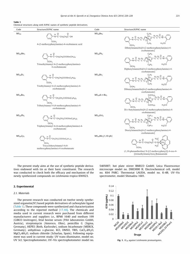
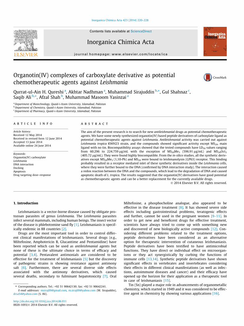
Table 1Chemical structures along with IUPAC names of synthetic peptide derivatives.
Code Structure/IUPAC name Code Structure/IUPAC name
MS20 HN
OCH3
COCH2CH2C
OOH
4-(2-methoxyphenylamino)-4-oxobutanoic acid
MS20Bu2
HN CO H2
CH2C C
O
OSnOC
O
H2CH2C COHN
C4H9
C4H9OCH3
H3CO
Dibutylstannicbis[4-(2-methoxyphenylamino)-4-oxobutanoate]
MS20Me3 HN
OCH3
COCH2CH2COOSn(CH3)3
Trimethylstannyl 4-(2-methoxyphenylamino)-4-oxobutanoate
MS20Ph2
HN CO H2
CH2C C
O
OSnOC
O
H2CH2C CO HN
C6H5
C6H5OCH3
H3CO
Diphenylstannicbis[4-(2-methoxyphenylamino)-4-oxobutanoate]
MS20Et3 HN
OCH3
COCH2CH2COOSn(C2H5)3
Triethylstannyl 4-(2-methoxyphenylamino)-4-oxobutanoate
MS20Bz2
HN CO H2
CH2C C
O
OSnOC
O
H2CH2C CO
HN
C7H7
C7H7OCH3
H3CO
Dibenzylstannicbis[4-(2-methoxyphenylamino)-4-oxobutanoate]
MS20Bu3 HN
OCH3
CO
CH2CH2COOSn(C4H9)3
Tributylstannyl 4-(4-methoxyphenylamino)-4-oxobutanoate
MS20di-t-Bu2
HN CO H2
CH2C C
O
OSnOC
O
H2CH2C CO HN
C(CH3)3
C(CH3)3OCH3
H3CO
Di-t-butylstannicbis[4-(2-methoxyphenylamino)-4-oxobutanoate]
MS20Ph3 HN
OCH3
CO
CH2CH2COOSn(C6H5)3
Triphenylstannyl 4-(4-methoxyphenylamino)-4-oxobutanoate
MS20Oct2
HN CO H2
CH2C C
O
OSnOC
O
H2CH2C CO HN
C8H17
C8H17OCH3
H3CO
Dioctylstannicbis[4-(2-methoxyphenylamino)-4-oxobutanoate]
MS20Cy3 HN
OCH3
CO
CH2CH2COOSn(C6H11)3
Tricyclohexylstannyl 4-(4-methoxyphenylamino)-4-oxobutanoate
MS20Me3(1,10-ph)
HN
OCH3
COCH2CH2C
OO Sn
H3C
H3CCH3N
N
(1,10-phenanthroline) N-[(2-methoxyphenyl)]-4-oxo-4-[(trimethylstannyl)oxy]butanamide
Qurrat-ul-Ain H. Qureshi et al. / Inorganica Chimica Acta 423 (2014) 220–228 221
The present study aims at the use of synthetic peptide deriva-tives endowed with tin as their basic constituent. The researchwas conducted to check both the efficacy and mechanism of thenewly synthesized compounds on Leishmania tropica KWH23.
Fig. 1. IC50 against Leishmania promastigotes.
2. Experimental
2.1. Materials
The present research was conducted on twelve newly synthe-sized organotin(IV) based peptide derivatives of carboxylate ligand(Table 1). These compounds were synthesized and characterizationaccording to the reported method [17,18]. The chemicals andmedia used in current research were purchased from differentmanufacturers and suppliers i.e., RPMI 1640 and medium 199(GIBCO Invitrogen), fetal bovine serum (PAA laboratories GmbH,Austria), streptomycin (Amresco, Ohio), penicillin G (Sigma,Germany), HEPES (Roth, Karlsruhe), sodium bicarbonate (MERCK,Germany), anhydrous D-glucose, KCl, DMSO, TRIS, CaCl2�2H2O,MgCl2�6H2O, sodium chloride (Scharlau, Spain). Following equip-ment was used in current study: UV trans-illuminator, model no.UV 3cl; Spectrophotometer, UV–Vis spectrophotometer model no.
5405007; hot plate stirrer RMECO GmbH; Leica Fluorescencemicroscope model no. DMI3000 B; Electrochemical cell, modelno. K64 PARC; Thermostat LAUDA, model no. K-4R; UV–Visspectrometer, model Shimadzu 1601.
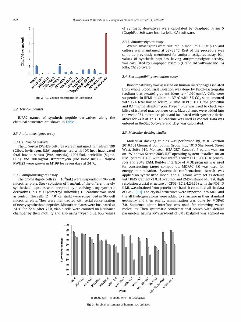
Fig. 2. IC50 against amastigotes of Leishmania.
222 Qurrat-ul-Ain H. Qureshi et al. / Inorganica Chimica Acta 423 (2014) 220–228
2.2. Test compounds
IUPAC names of synthetic peptide derivatives along thechemical structures are shown in Table 1.
2.3. Antipromastigotes assay
2.3.1. L. tropica culturesThe L. tropica KWH23 cultures were maintained in medium 199
(Gibco, Invitrogen, USA) supplemented with 10% heat-inactivatedfetal bovine serum (PAA, Austria), 100 U/mL penicillin (Sigma,USA), and 100 mg/mL streptomycin (Bio Basic Inc.). L. tropicaKWH23 were grown in M199 for seven days at 24 �C.
2.3.2. Antipromastigotes assayThe promastigote cells (2 � 106/mL) were suspended in 96-well
microtiter plate. Stock solutions of 1 mg/mL of the different newlysynthesized peptides were prepared by dissolving 1 mg syntheticderivatives in DMSO (dimethyl sulfoxide). Glucantime was usedas control. The cells (2 � 106 cells/mL) were suspended in 96-wellmicrotiter plate. They were then treated with serial concentrationof newly synthesized peptides. Microtiter plates were incubated at24 �C for 72 h. After 72 h, viable cells were counted on Neubauerchamber by their motility and also using trypan blue. IC50 values
Fig. 3. Survival percentage o
of synthetic derivatives were calculated by Graphpad Prism 5(GraphPad Software Inc., La Jolla, CA) software.
2.3.3. Antiamastigotes assayAxenic amastigotes were cultured in medium 199 at pH 5 and
culture was maintained at 32–33 �C. Rest of the procedure wassame as previously mentioned for antipromastigotes assay. IC50
values of synthetic peptides having antipromastigote activity,was calculated by Graphpad Prism 5 (GraphPad Software Inc., LaJolla, CA) software.
2.4. Biocompatibility evaluation assay
Biocompatibility was assessed on human macrophages isolatedfrom whole blood. First isolation was done by Ficoll-gastrografin(sodium diatrozoate) gradient (density = 1.070 g/mL). Cells weresuspended in RPMI medium at 37 �C with 5% CO2 supplementedwith 12% fetal bovine serum, 25 mM HEPES, 100 U/mL penicillinand 0.1 mg/mL streptomycin. Trypan blue was used to check via-bility of isolated macrophages cells. Macrophages were added intothe well of 24-microtiter plate and incubated with synthetic deriv-atives for 24 h at 37 �C. Glucantime was used as control. Data wasentered in BioStat Software and LD50 was calculated.
2.5. Molecular docking studies
Molecular docking studies was performed by, MOE (version2010.10) Chemical Computing Group Inc., 1010 Sherbrook StreetWest, Suite 910, Montreal, H3A 2R7, Canada). Program was runon ‘‘Windows Server 2003 R2’’ operating system installed on anIBM System 93400 with four Intel� Xeon™ CPU 3.00 GHz proces-sors and 2048 RAM. Builder interface of MOE program was usedfor constructing target compounds. MOPAC 7.0 was used forenergy minimization. Systematic conformational search wasapplied on synthesized model and all atoms were set as defaultwith RMS gradient of 0.01 kcal/mol and RMS distance of 0.1 Å. Highresolution crystal structure of GP63 (EC 3.4.24.36) with the PDB IDILML was obtained from protein data bank. It contained all the dataof GP63 [19]. The crystal structures were imported into MOE andthe all hydrogen atoms were added to structure in their standardgeometry and then energy minimization was done by MOPAC7.0. Sequence editor interface was used for removing watermolecules. Then systematic conformational search with defaultparameters having RMS gradient of 0.01 kcal/mol was applied on
f human macrophages.
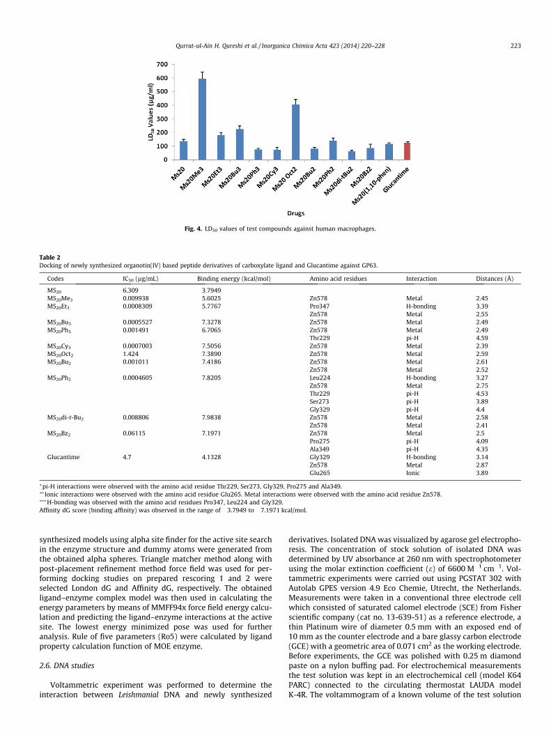
Fig. 4. LD50 values of test compounds against human macrophages.
Table 2Docking of newly synthesized organotin(IV) based peptide derivatives of carboxylate ligand and Glucantime against GP63.
Codes IC50 (lg/mL) Binding energy (kcal/mol) Amino acid residues Interaction Distances (Å)
MS20 6.309 �3.7949MS20Me3 0.009938 �5.6025 Zn578 Metal 2.45MS20Et3 0.0008309 �5.7767 Pro347 H-bonding 3.39
Zn578 Metal 2.55MS20Bu3 0.0005527 �7.3278 Zn578 Metal 2.49MS20Ph3 0.001491 �6.7065 Zn578 Metal 2.49
Thr229 pi-H 4.59MS20Cy3 0.0007003 �7.5056 Zn578 Metal 2.39MS20Oct2 1.424 �7.3890 Zn578 Metal 2.59MS20Bu2 0.001011 �7.4186 Zn578 Metal 2.61
Zn578 Metal 2.52MS20Ph2 0.0004605 �7.8205 Leu224 H-bonding 3.27
Zn578 Metal 2.75Thr229 pi-H 4.53Ser273 pi-H 3.89Gly329 pi-H 4.4
MS20di-t-Bu2 0.008806 �7.9838 Zn578 Metal 2.58Zn578 Metal 2.41
MS20Bz2 0.06115 �7.1971 Zn578 Metal 2.5Pro275 pi-H 4.09Ala349 pi-H 4.35
Glucantime 4.7 �4.1328 Gly329 H-bonding 3.14Zn578 Metal 2.87Glu265 Ionic 3.89
⁄pi-H interactions were observed with the amino acid residue Thr229, Ser273, Gly329, Pro275 and Ala349.⁄⁄ Ionic interactions were observed with the amino acid residue Glu265. Metal interactions were observed with the amino acid residue Zn578.⁄⁄⁄H-bonding was observed with the amino acid residues Pro347, Leu224 and Gly329.Affinity dG score (binding affinity) was observed in the range of �3.7949 to �7.1971 kcal/mol.
Qurrat-ul-Ain H. Qureshi et al. / Inorganica Chimica Acta 423 (2014) 220–228 223
synthesized models using alpha site finder for the active site searchin the enzyme structure and dummy atoms were generated fromthe obtained alpha spheres. Triangle matcher method along withpost-placement refinement method force field was used for per-forming docking studies on prepared rescoring 1 and 2 wereselected London dG and Affinity dG, respectively. The obtainedligand–enzyme complex model was then used in calculating theenergy parameters by means of MMFF94x force field energy calcu-lation and predicting the ligand–enzyme interactions at the activesite. The lowest energy minimized pose was used for furtheranalysis. Rule of five parameters (Ro5) were calculated by ligandproperty calculation function of MOE enzyme.
2.6. DNA studies
Voltammetric experiment was performed to determine theinteraction between Leishmanial DNA and newly synthesized
derivatives. Isolated DNA was visualized by agarose gel electropho-resis. The concentration of stock solution of isolated DNA wasdetermined by UV absorbance at 260 nm with spectrophotometerusing the molar extinction coefficient (e) of 6600 M�1 cm�1. Vol-tammetric experiments were carried out using PGSTAT 302 withAutolab GPES version 4.9 Eco Chemie, Utrecht, the Netherlands.Measurements were taken in a conventional three electrode cellwhich consisted of saturated calomel electrode (SCE) from Fisherscientific company (cat no. 13-639-51) as a reference electrode, athin Platinum wire of diameter 0.5 mm with an exposed end of10 mm as the counter electrode and a bare glassy carbon electrode(GCE) with a geometric area of 0.071 cm2 as the working electrode.Before experiments, the GCE was polished with 0.25 m diamondpaste on a nylon buffing pad. For electrochemical measurementsthe test solution was kept in an electrochemical cell (model K64PARC) connected to the circulating thermostat LAUDA modelK-4R. The voltammogram of a known volume of the test solution
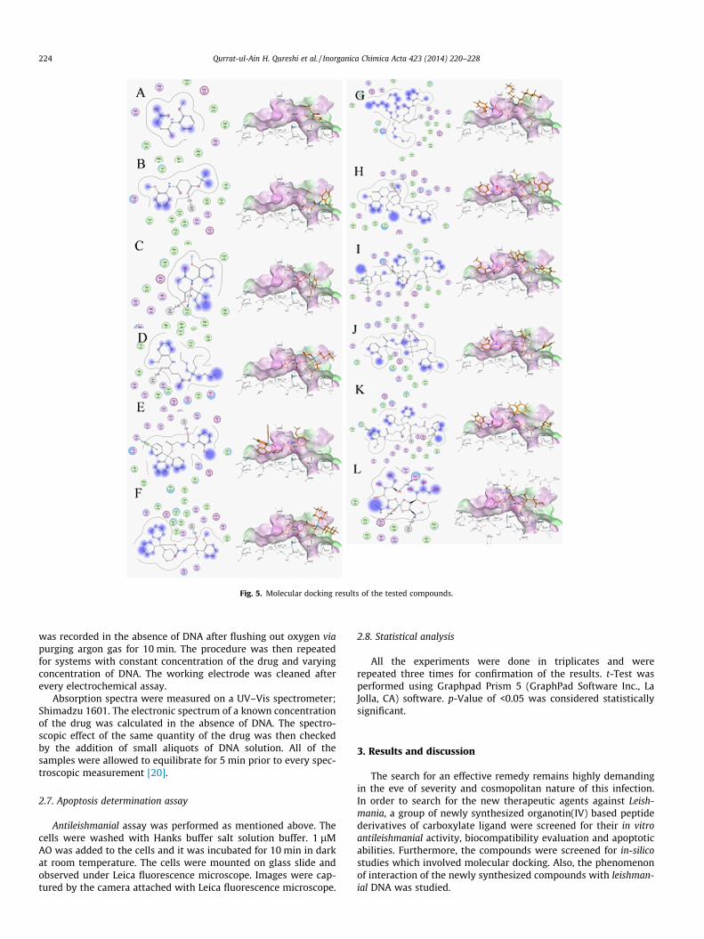
Fig. 5. Molecular docking results of the tested compounds.
224 Qurrat-ul-Ain H. Qureshi et al. / Inorganica Chimica Acta 423 (2014) 220–228
was recorded in the absence of DNA after flushing out oxygen viapurging argon gas for 10 min. The procedure was then repeatedfor systems with constant concentration of the drug and varyingconcentration of DNA. The working electrode was cleaned afterevery electrochemical assay.
Absorption spectra were measured on a UV–Vis spectrometer;Shimadzu 1601. The electronic spectrum of a known concentrationof the drug was calculated in the absence of DNA. The spectro-scopic effect of the same quantity of the drug was then checkedby the addition of small aliquots of DNA solution. All of thesamples were allowed to equilibrate for 5 min prior to every spec-troscopic measurement [20].
2.7. Apoptosis determination assay
Antileishmanial assay was performed as mentioned above. Thecells were washed with Hanks buffer salt solution buffer. 1 lMAO was added to the cells and it was incubated for 10 min in darkat room temperature. The cells were mounted on glass slide andobserved under Leica fluorescence microscope. Images were cap-tured by the camera attached with Leica fluorescence microscope.
2.8. Statistical analysis
All the experiments were done in triplicates and wererepeated three times for confirmation of the results. t-Test wasperformed using Graphpad Prism 5 (GraphPad Software Inc., LaJolla, CA) software. p-Value of <0.05 was considered statisticallysignificant.
3. Results and discussion
The search for an effective remedy remains highly demandingin the eve of severity and cosmopolitan nature of this infection.In order to search for the new therapeutic agents against Leish-mania, a group of newly synthesized organotin(IV) based peptidederivatives of carboxylate ligand were screened for their in vitroantileishmanial activity, biocompatibility evaluation and apoptoticabilities. Furthermore, the compounds were screened for in-silicostudies which involved molecular docking. Also, the phenomenonof interaction of the newly synthesized compounds with leishman-ial DNA was studied.
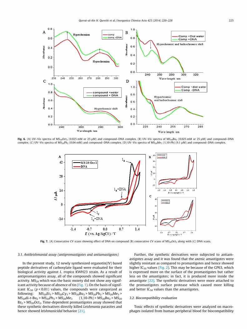
Fig. 6. (A) UV–Vis spectra of MS20Oct2 (0.025 mM or 25 lM) and compound–DNA complex. (B) UV–Vis spectra of MS20Bu2 (0.025 mM or 25 lM) and compound–DNAcomplex. (C) UV–Vis spectra of MS20Ph2 (0.04 mM) and compound–DNA complex. (D) UV–Vis spectra of MS20Me3 (1,10-Ph) (9.1 lM) and compound–DNA complex.
Fig. 7. (A) Consecutive CV scans showing effect of DNA on compound (B) consecutive CV scans of MS20Oct2 along with (C) DNA scans.
Qurrat-ul-Ain H. Qureshi et al. / Inorganica Chimica Acta 423 (2014) 220–228 225
3.1. Antileishmanial assay (antipromastigotes and antiamastigotes)
In the present study, 12 newly synthesized organotin(IV) basedpeptide derivatives of carboxylate ligand were evaluated for theirbiological activity against L. tropica KWH23 strain. As a result ofantipromastigotes assay, all of the compounds showed significantactivity. MS20 which was the basic moiety did not show any signif-icant activity because of absence of tin (Fig. 1). On the basis of signif-icant IC50 (p < 0.01) values, the compounds were categorized asfollowing: MS20Et3 > MS20Cy3 > MS20Bu3 > MS20Ph2 > MS20Me3 >MS20di-t-Bu2 > MS20Ph3 > MS20Me3 (1,10-Ph) > MS20Bu2 > MS20
Bz2 > MS20Oct2. Time-dependent promastigotes assay showed thatthese synthetic derivatives directly killed Leishmania parasites andhence showed leishmanicidal behavior [21].
Further, the synthetic derivatives were subjected to antiam-astigotes assay and it was found that the axenic amastigotes wereslightly resistant as compared to promastigotes and hence showedhigher IC50 values (Fig. 2). This may be because of the GP63, whichis expressed more on the surface of the promastigotes but ratherless on the amastigotes; in fact, it is produced more inside theamastigote [22]. The synthetic derivatives were more attached tothe promastigotes surface protease which caused more killingand better IC50 values than the amastigotes.
3.2. Biocompatibility evaluation
Toxic effects of synthetic derivatives were analyzed on macro-phages isolated from human peripheral blood for biocompatibility
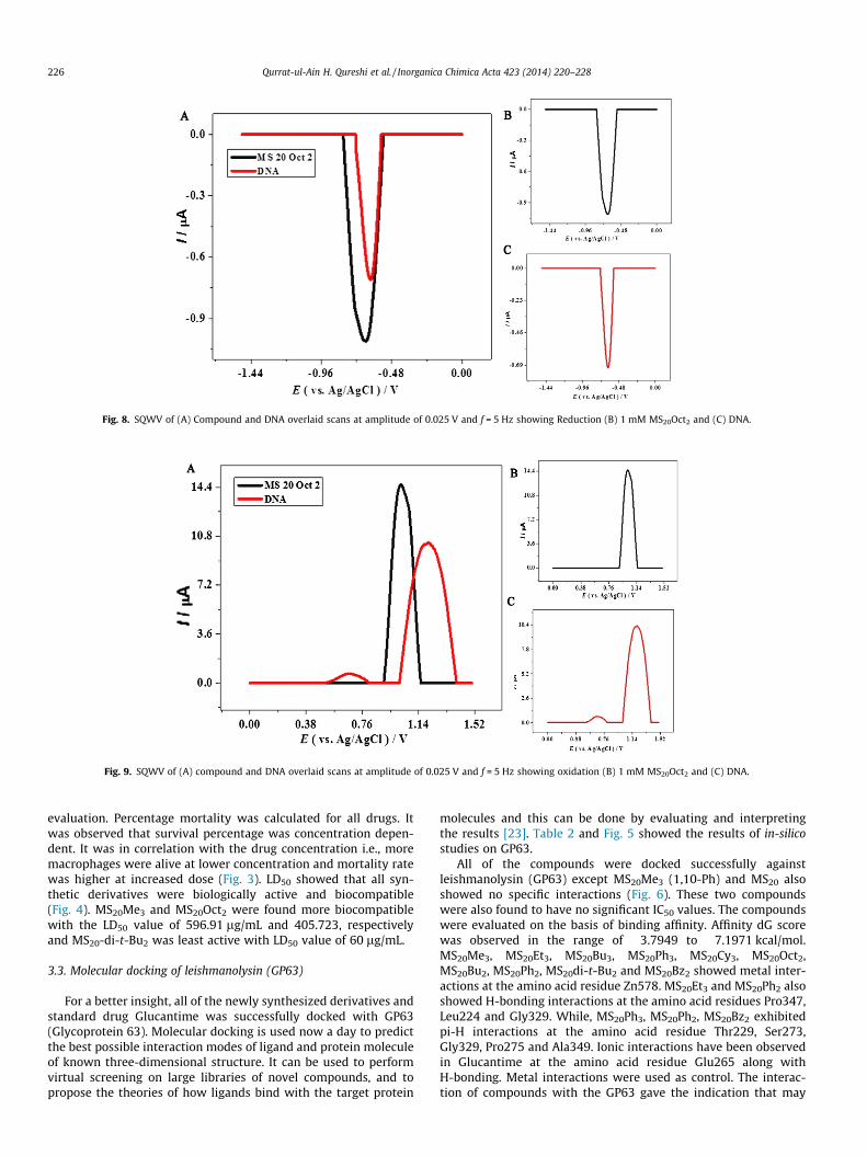
Fig. 8. SQWV of (A) Compound and DNA overlaid scans at amplitude of 0.025 V and f = 5 Hz showing Reduction (B) 1 mM MS20Oct2 and (C) DNA.
Fig. 9. SQWV of (A) compound and DNA overlaid scans at amplitude of 0.025 V and f = 5 Hz showing oxidation (B) 1 mM MS20Oct2 and (C) DNA.
226 Qurrat-ul-Ain H. Qureshi et al. / Inorganica Chimica Acta 423 (2014) 220–228
evaluation. Percentage mortality was calculated for all drugs. Itwas observed that survival percentage was concentration depen-dent. It was in correlation with the drug concentration i.e., moremacrophages were alive at lower concentration and mortality ratewas higher at increased dose (Fig. 3). LD50 showed that all syn-thetic derivatives were biologically active and biocompatible(Fig. 4). MS20Me3 and MS20Oct2 were found more biocompatiblewith the LD50 value of 596.91 lg/mL and 405.723, respectivelyand MS20-di-t-Bu2 was least active with LD50 value of 60 lg/mL.
3.3. Molecular docking of leishmanolysin (GP63)
For a better insight, all of the newly synthesized derivatives andstandard drug Glucantime was successfully docked with GP63(Glycoprotein 63). Molecular docking is used now a day to predictthe best possible interaction modes of ligand and protein moleculeof known three-dimensional structure. It can be used to performvirtual screening on large libraries of novel compounds, and topropose the theories of how ligands bind with the target protein
molecules and this can be done by evaluating and interpretingthe results [23]. Table 2 and Fig. 5 showed the results of in-silicostudies on GP63.
All of the compounds were docked successfully againstleishmanolysin (GP63) except MS20Me3 (1,10-Ph) and MS20 alsoshowed no specific interactions (Fig. 6). These two compoundswere also found to have no significant IC50 values. The compoundswere evaluated on the basis of binding affinity. Affinity dG scorewas observed in the range of �3.7949 to �7.1971 kcal/mol.MS20Me3, MS20Et3, MS20Bu3, MS20Ph3, MS20Cy3, MS20Oct2,MS20Bu2, MS20Ph2, MS20di-t-Bu2 and MS20Bz2 showed metal inter-actions at the amino acid residue Zn578. MS20Et3 and MS20Ph2 alsoshowed H-bonding interactions at the amino acid residues Pro347,Leu224 and Gly329. While, MS20Ph3, MS20Ph2, MS20Bz2 exhibitedpi-H interactions at the amino acid residue Thr229, Ser273,Gly329, Pro275 and Ala349. Ionic interactions have been observedin Glucantime at the amino acid residue Glu265 along withH-bonding. Metal interactions were used as control. The interac-tion of compounds with the GP63 gave the indication that may
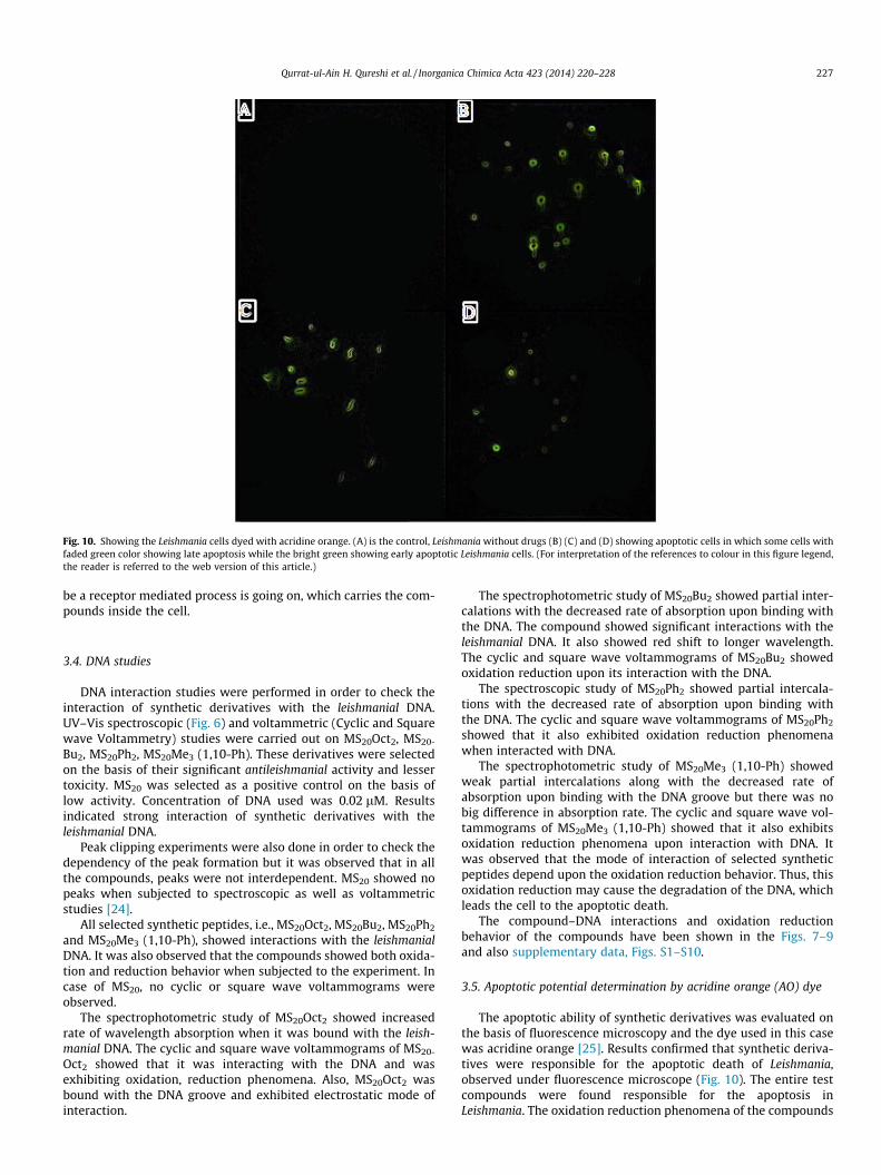
Fig. 10. Showing the Leishmania cells dyed with acridine orange. (A) is the control, Leishmania without drugs (B) (C) and (D) showing apoptotic cells in which some cells withfaded green color showing late apoptosis while the bright green showing early apoptotic Leishmania cells. (For interpretation of the references to colour in this figure legend,the reader is referred to the web version of this article.)
Qurrat-ul-Ain H. Qureshi et al. / Inorganica Chimica Acta 423 (2014) 220–228 227
be a receptor mediated process is going on, which carries the com-pounds inside the cell.
3.4. DNA studies
DNA interaction studies were performed in order to check theinteraction of synthetic derivatives with the leishmanial DNA.UV–Vis spectroscopic (Fig. 6) and voltammetric (Cyclic and Squarewave Voltammetry) studies were carried out on MS20Oct2, MS20-
Bu2, MS20Ph2, MS20Me3 (1,10-Ph). These derivatives were selectedon the basis of their significant antileishmanial activity and lessertoxicity. MS20 was selected as a positive control on the basis oflow activity. Concentration of DNA used was 0.02 lM. Resultsindicated strong interaction of synthetic derivatives with theleishmanial DNA.
Peak clipping experiments were also done in order to check thedependency of the peak formation but it was observed that in allthe compounds, peaks were not interdependent. MS20 showed nopeaks when subjected to spectroscopic as well as voltammetricstudies [24].
All selected synthetic peptides, i.e., MS20Oct2, MS20Bu2, MS20Ph2
and MS20Me3 (1,10-Ph), showed interactions with the leishmanialDNA. It was also observed that the compounds showed both oxida-tion and reduction behavior when subjected to the experiment. Incase of MS20, no cyclic or square wave voltammograms wereobserved.
The spectrophotometric study of MS20Oct2 showed increasedrate of wavelength absorption when it was bound with the leish-manial DNA. The cyclic and square wave voltammograms of MS20-
Oct2 showed that it was interacting with the DNA and wasexhibiting oxidation, reduction phenomena. Also, MS20Oct2 wasbound with the DNA groove and exhibited electrostatic mode ofinteraction.
The spectrophotometric study of MS20Bu2 showed partial inter-calations with the decreased rate of absorption upon binding withthe DNA. The compound showed significant interactions with theleishmanial DNA. It also showed red shift to longer wavelength.The cyclic and square wave voltammograms of MS20Bu2 showedoxidation reduction upon its interaction with the DNA.
The spectroscopic study of MS20Ph2 showed partial intercala-tions with the decreased rate of absorption upon binding withthe DNA. The cyclic and square wave voltammograms of MS20Ph2
showed that it also exhibited oxidation reduction phenomenawhen interacted with DNA.
The spectrophotometric study of MS20Me3 (1,10-Ph) showedweak partial intercalations along with the decreased rate ofabsorption upon binding with the DNA groove but there was nobig difference in absorption rate. The cyclic and square wave vol-tammograms of MS20Me3 (1,10-Ph) showed that it also exhibitsoxidation reduction phenomena upon interaction with DNA. Itwas observed that the mode of interaction of selected syntheticpeptides depend upon the oxidation reduction behavior. Thus, thisoxidation reduction may cause the degradation of the DNA, whichleads the cell to the apoptotic death.
The compound–DNA interactions and oxidation reductionbehavior of the compounds have been shown in the Figs. 7–9and also supplementary data, Figs. S1–S10.
3.5. Apoptotic potential determination by acridine orange (AO) dye
The apoptotic ability of synthetic derivatives was evaluated onthe basis of fluorescence microscopy and the dye used in this casewas acridine orange [25]. Results confirmed that synthetic deriva-tives were responsible for the apoptotic death of Leishmania,observed under fluorescence microscope (Fig. 10). The entire testcompounds were found responsible for the apoptosis inLeishmania. The oxidation reduction phenomena of the compounds
228 Qurrat-ul-Ain H. Qureshi et al. / Inorganica Chimica Acta 423 (2014) 220–228
might be causing the degradation of the DNA, which further leadsto the disappearance of the chromatin material (apoptosis) andcausing death of the cell.
4. Conclusion
The aim of this study deals with the effort to push the scientificapproach a leap further into developing a cost effective, harmless,potent and efficacious drug against Leishmania. The investigatedcompounds have the ability of apoptotic in Leishmania. The inter-action of the compounds with Leishmania DNA was also confirmedby cyclic voltammetry and molecular docking. The interestingthing is comparison in the results of cyclic voltammetry andmolecular docking that support each other. Time-dependent prom-astigotes assay results showed that these compounds directly killthe Leishmania parasites and hence show leishmanicidal behavior.
Acknowledgments
This work was financially supported by Higher Education Com-mission (HEC) of Pakistan and Pakistan Science Foundation (PSF)and their support is gratefully acknowledged.
Appendix A. Supplementary material
Supplementary data associated with this article can be found, inthe online version, at http://dx.doi.org/10.1016/j.ica.2014.06.011.
References
[1] A.L. Banuls, M. Hide, F. Prugnolle, Adv. Parasitol. 64 (2007) 1.[2] J.C. Dujardin, L. Campino, C. Cañavate, J.P. Dedet, L. Gradoni, K. Soteriadou, A.
Mazeris, Y. Ozbel, M. Boelaert, Emerg. Infect. Dis. 14 (7) (2008) 1013.
[3] H.W. Murray, Antimicrob. Agents Chemother. 45 (8) (2001) 2185.[4] E. Rosenthal, P. Marty, Expert Opin. Pharmacother. 3 (8) (2002) 1101.[5] B.L. Herwaldt, J.D. Berman, Am. J. Trop. Med. Hyg. 46 (3) (1992) 296.[6] S. Sundar, Trop. Med. Int. Health 6 (11) (2001) 849.[7] S.M. Robledo, J.A. Puerta, D.L. Muñoz, M. Guardo, I.D. Vélez, Biomedica: revista
del Instituto Nacional de Salud 26 (2006) 188.[8] S.L. Croft, G.H. Coombs, Trends Parasitol. 19 (11) (2003) 502.[9] H. Sangraula, K. Sharma, S. Rijal, S. Dwivedi, S. Koirala, J. Assoc. Physicians India
51 (2003) 686.[10] R. Prasad, R. Kumar, B. Jaiswal, U.K. Singh, Ind. J. Pediatr. 71 (2) (2004) 143.[11] J. Soto, J.T. Toledo, D. Dean, B. Atik, T. Thanh, V. Luong, S. Lagree, S. Gregson, G.
Garnett, C. Nyamukapa, Lancet Infect. Dis. 7 (1) (2007) 7.[12] M.L. Cohen, Science 257 (5073) (1992) 1050.[13] K. Kandler, R. Shaykhiev, P. Kleemann, F. Klescz, M. Lohoff, C. Vogelmeier, R.
Bals, Int. Immunol. 18 (12) (2006) 1729.[14] E.M. Molhoek, A.L. den Hertog, A.-M.B. de Vries, K. Nazmi, E.C. Veerman, F.C.
Hartgers, M. Yazdanbakhsh, F.J. Bikker, D. van der Kleij, Biol. Chem. 390 (4)(2009) 295.
[15] J.R. Luque-Ortega, W. Van’t Hof, E.C. Veerman, J.M. Saugar, L. Rivas, FASEB J. 22(6) (2008) 1817.
[16] G. Wilkinson, F.G.A. Stone, E.W. Abel, Comprehensive OrganometallicChemistry, 1982.
[17] F.A. Shah, M. Sirajuddin, S. Ali, S.M. Abbas, M.N. Tahir, C. Rizzoli, Inorg. Chim.Acta 400 (2013) 159.
[18] M. Sirajuddin, S. Ali, V. McKee, M. Sohail, H. Pasha, Eur. J. Med. Chem. (2014).[19] E. Schlagenhauf, R. Etges, P. Metcalf, Structure 6 (8) (1998) 1035.[20] A. Shah, R. Qureshi, A.M. Khan, F.L. Ansari, S. Ahmad, Bull. Chem. Soc. Jpn. 82
(4) (2009) 453.[21] J.J. Pérez-Cordero, J.M. Lozano, J. Cortés, G. Delgado, Peptides 32 (4) (2011) 683.[22] C.H. Hsiao, C. Yao, P. Storlie, J.E. Donelson, M.E. Wilson, Mol. Biochem.
Parasitol. 157 (2) (2008) 148.[23] S.S. Mpoke, J. Wolfe, J. Histochem. Cytochem. 45 (5) (1997) 675.[24] G.M. Morris, M. Lim-Wilby, in: Molecular Modeling of Proteins, Springer, 2008,
p. 365.[25] B.L. Roth, M. Poot, S.T. Yue, P.J. Millard, Appl. Environ. Microbiol. 63 (6) (1997)
2421.
Related Documents