Organic Molecules: Proteins

Welcome message from author
This document is posted to help you gain knowledge. Please leave a comment to let me know what you think about it! Share it to your friends and learn new things together.
Transcript

Organic Molecules: Proteins

Proteins • Most structurally & functionally diverse group
• Function: involved in almost everything – enzymes (pepsin, DNA polymerase)– structure (keratin, collagen)– carriers & transport (hemoglobin, aquaporin)– cell communication
• signals (insulin & other hormones) • receptors
– defense (antibodies) – movement (actin & myosin)– storage (bean seed proteins)

Proteins• Structure– monomer = amino acids
• 20 different amino acids
– polymer = polypeptide• protein can be one or more polypeptide chains folded
& bonded together
• large & complex molecules
• complex 3-D shape
Rubisco
hemoglobin
growthhormones
H2O

Amino acids
• Structure– central carbon
– amino group
– carboxyl group (acid)
– R group (side chain)• variable group
• different for each amino acid
• confers unique chemical properties to each amino acid- like 20 different letters of an alphabet
- can make many words (proteins)
Oh, I get it!amino = NH2 acid = COOH

Effect of different R groups:Polar amino acids
▪ polar or charged & hydrophilic
Why are these polar & hydrophillic?
H+ acceptorsH+ donors

Effect of different R groups:Nonpolar amino acids
Why are these nonpolar & hydrophobic?
▪ nonpolar & hydrophobic

Sulfur containing amino acids• Form disulfide bridges S-S
– covalent cross links betweens sulfhydryls – stabilizes 3-D structure
H-S – S-H

AP Biology
Building proteins• Peptide bonds– covalent bond between NH
2 (amine) of one
amino acid & COOH (carboxyl) of another
– C–N bond
peptidebond
dehydration synthesisH2O

Building proteins• Polypeptide chains have direction– N-terminus = NH
2 end
– C-terminus = COOH end– repeated sequence (N-C-C) is the
polypeptide backbone• can only grow in one direction
http://www2.nl.edu/jste/proteins.htm

Protein structure & function• Function depends on structure– 3-D structure
• twisted, folded, coiled into unique shape
hemoglobin
collagen
pepsin

Protein Structure• Protein types include globular proteins which
are usually enzymes and Fibrous proteins which usually serve for structure (eg. Hair)
• Proteins Exhibit 4 levels of structure.

Primary (1°) structure• Order of amino acids in chain
– amino acid sequence determined by gene (DNA)l; dictates all further levels of protein structure
– slight change in amino acid sequence can affect protein’s structure & its function
• even just one amino acid change can make all the difference!

Sickle cell anemia
I’mhydrophilic!
But I’mhydrophobic!
Just 1out of 146amino acids!

15
Fibers of abnormalhemoglobin deform cell into sickle shape.
Primary structure
Secondaryand tertiarystructures
Quaternary structure
Function
Red bloodcell shape
Hemoglobin A
Molecules donot associatewith oneanother, eachcarries oxygen.
Normal cells arefull of individualhemoglobinmolecules, eachcarrying oxygen
α
β
β
α
10 μm 10 μm
α
β
β
α
Primary structure
Secondaryand tertiarystructures
Quaternary structure
Function
Red bloodcell shape
Hemoglobin S
Molecules interact with one another tocrystallize into a fiber, capacity to carry oxygen is greatly reduced.
β subunit β subunit
1 2 3 4 5 6 7 3 4 5 6 721
Normal hemoglobin Sickle-cell hemoglobin. . .. . .
Figure 5.21
Exposed hydrophobic
region
Val ThrHis Leu Pro Glul Glu Val His Leu Thr Pro Val Glu

Secondary (2°) structure• “Local folding”– folding along short sections of polypeptide
– interactions between adjacent amino acids
– H bond: weak bonds between R groups
– forms sections of 3-D structure
•α-helix
•β-pleated sheet


Tertiary (3°) structure
• “Whole molecule folding”– interactions between distant amino acids
• hydrophobic interactions
– cytoplasm is water-based
– nonpolar amino acids cluster away from water
• Covalent, H bonds & ionic bonds
• disulfide bridges – covalent bonds between
sulfurs in sulfhydryls (S–H)
– anchors 3-D shape

Quaternary (4°) structure• More than one polypeptide chain bonded together
– only then does polypeptide become functional protein• pH, changes or heat can disrupth bonds perm. denaturing
the protein
collagen = skin & tendons hemoglobin
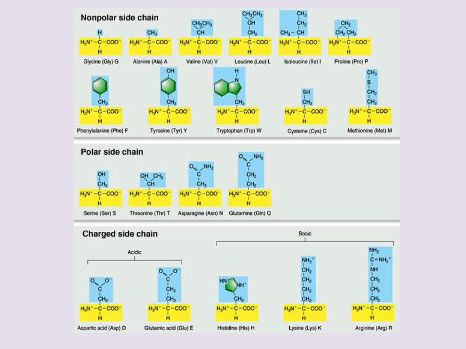
Protein structure (review)
amino acid sequencepeptide bonds
1°
determinedby DNA R groups
H bonds
R groupshydrophobic interactionsdisulfide bridges(H & ionic bonds)3°
multiple polypeptideshydrophobic interactions4
°2°

Protein denaturation• Unfolding a protein– conditions that disrupt H bonds, ionic bonds,
disulfide bridges• temperature
• pH
• salinity
– alter 2° & 3° structure• alter 3-D shape
– destroys functionality• some proteins can return to their functional shape after
denaturation, many cannot
http://highered.mcgraw-hill.com/sites/0072943696/student_view0/chapter2/animation__protein_denaturation.html

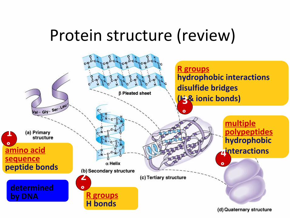
• Chaperonins– Are protein molecules that assist in the proper
folding of other proteins
23
Hollowcylinder
Cap
Chaperonin(fully assembled)
Steps of ChaperoninAction: An unfolded poly- peptide enters the cylinder from one end.
The cap attaches, causing the cylinder to change shape insuch a way that it creates a hydrophilic environment for the folding of the polypeptide.
The cap comesoff, and the properlyfolded protein is released.
Correctlyfoldedprotein
Polypeptide
2
1
3
Figure 5.23

Review Questions

A. What happens when a protein denatures? *1. It loses its primary structure.
2. It loses its secondary and tertiary structure.
3. It becomes irreversibly insoluble and precipitates.
4. It hydrolyzes into component amino acids.
5. Its hydrogen bonds, ionic bonds, and peptide bonds are disrupted.
B. The R group or side chain of the amino acid serine is –CH
2 –OH. The R group or side chain of the amino
acid alanine is –CH3. Where would you expect to find
these amino acids in globular protein in aqueous solution?1. Serine would be in the interior, and alanine would be on
the exterior of the globular protein.2. Alanine would be in the interior, and serine would be on
the exterior of the globular protein.3. Both serine and alanine would be in the interior of the
globular protein.4. Both serine and alanine would be on the exterior of the
globular protein.5. Both serine and alanine would be in the interior and on
the exterior of the globular protein.
Related Documents