3,5-Diamino-1-phenyl-1,2,4-triazolium bromide V. M. Chernyshev, a * A. V. Astakhov, a V. V. Ivanov a and Z. A. Starikova b a South-Russia State Technical University, 346428 Novocherkassk, Russian Federation, and b A. N. Nesmeyanov Institute of Organoelement Compounds, 119991 Moscow, Russian Federation Correspondence e-mail: [email protected] Received 27 May 2010; accepted 3 June 2010 Key indicators: single-crystal X-ray study; T = 100 K; mean (C–C) = 0.003 A ˚ ; R factor = 0.027; wR factor = 0.071; data-to-parameter ratio = 17.1. The title salt, C 8 H 10 N 5 + Br , crystallizes with two independent structural units in the asymmetric unit. The two independent cations have different conformations, the triazole and phenyl rings forming dihedral angles of 32.57 (6) and 52.27 (7) . In both cations, the amino groups are planar (the sum of the angles at the N atom of each amino group is 360 ) and conjugated with the triazole ring. Intermolecular N—HN and N—HBr hydrogen bonds consolidate the crystal packing. Related literature For the crystal structures of protonated C-amino-1,2,4-tria- zoles, see: Reck et al. (1982); Lynch et al. (1998, 1999); Baouab et al. (2000); Bichay et al. (2006); Guerfel et al. (2007); Matulkova ´ et al. (2007). For the crystal structure of 3,5- diamino-1,2,4-triazole, see: Starova et al. (1980). For the theoretical investigation of the protonation of C-amino-1,2,4- triazoles, see: Anders et al. (1997). For the reactions of 1- substituted 3,5-diamino-1,2,4-triazoles with electrophilic reagents, see: Steck et al. (1958); Chernyshev et al. (2005, 2008). For the use of 1-substituted 3,5-diamino-1,2,4-triazoles as building blocks in the synthesis of various derivatives of 1,2,4-triazole and fused heterocyclic systems, see: Dunstan et al. (1998); Chernyshev et al. (2006, 2009, 2010). For a description of the Cambridge Structural Database, see: Allen (2002). Experimental Crystal data C 8 H 10 N 5 + Br M r = 256.12 Monoclinic, P2 1 =n a = 13.752 (2) A ˚ b = 7.1172 (13) A ˚ c = 20.394 (4) A ˚ = 95.519 (3) V = 1986.7 (6) A ˚ 3 Z =8 Mo K radiation = 4.11 mm 1 T = 100 K 0.55 0.40 0.30 mm Data collection Bruker APEXII CCD area-detector diffractometer Absorption correction: multi-scan (SADABS; Bruker, 2004) T min = 0.211, T max = 0.372 19484 measured reflections 4314 independent reflections 3808 reflections with I >2(I) R int = 0.033 Refinement R[F 2 >2(F 2 )] = 0.027 wR(F 2 ) = 0.071 S = 1.00 4314 reflections 253 parameters H-atom parameters constrained max = 0.63 e A ˚ 3 min = 0.52 e A ˚ 3 Table 1 Hydrogen-bond geometry (A ˚ , ). D—HA D—H HA DA D—HA N3—H3AN2 0 i 0.86 2.20 3.037 (3) 164 N3—H3BBr1 0.86 2.56 3.387 (2) 163 N3 0 —H3 0 AN2 ii 0.86 2.34 3.046 (3) 140 N3 0 —H3 0 BBr2 0.86 2.65 3.404 (3) 147 N4—H4Br2 0.86 2.74 3.417 (3) 137 N4 0 —H4 0 Br2 0.86 2.51 3.254 (3) 145 N5—H5ABr1 iii 0.86 2.69 3.369 (3) 137 N5—H5BBr2 0.86 2.49 3.281 (3) 153 N5 0 —H5 0 ABr1 iv 0.86 2.84 3.489 (3) 133 N5 0 —H5 0 BBr1 0.86 2.43 3.278 (3) 167 Symmetry codes: (i) x þ 1 2 ; y þ 1 2 ; z þ 1 2 ; (ii) x 1 2 ; y þ 1 2 ; z 1 2 ; (iii) x þ 1 2 ; y þ 1 2 ; z þ 3 2 ; (iv) x þ 1; y; z þ 1. Data collection: APEX2 (Bruker, 2004); cell refinement: SAINT (Bruker, 2004); data reduction: SAINT and XPREP (Bruker, 2005); program(s) used to solve structure: SHELXS97 (Sheldrick, 2008); program(s) used to refine structure: SHELXL97 (Sheldrick, 2008); molecular graphics: Mercury (Macrae et al. , 2006); software used to prepare material for publication: SHELXTL (Sheldrick, 2008), publCIF (Westrip, 2010) and PLATON (Spek, 2009). The authors thank the Federal Agency for Education of Russia for financial support of this work through the Federal Target Program "Research and Educational Personnel of Innovative Russia at 2009–2013 Years", State contract P302, project NK-109P/2. Supplementary data and figures for this paper are available from the IUCr electronic archives (Reference: CV2726). References Allen, F. H. (2002). Acta Cryst. B58, 380–388. Anders, E., Wermann, K., Wiedel, B. & VandenEynde, J.-J. (1997). Lieb. Ann. Recueil, 745–752. Baouab, L., Guerfel, T., Soussi, M. & Jouini, A. (2000). J. Chem. Crystallogr. 30, 805–809. organic compounds o1644 Chernyshev et al. doi:10.1107/S1600536810021318 Acta Cryst. (2010). E66, o1644–o1645 Acta Crystallographica Section E Structure Reports Online ISSN 1600-5368

Welcome message from author
This document is posted to help you gain knowledge. Please leave a comment to let me know what you think about it! Share it to your friends and learn new things together.
Transcript
3,5-Diamino-1-phenyl-1,2,4-triazoliumbromide
V. M. Chernyshev,a* A. V. Astakhov,a V. V. Ivanova and
Z. A. Starikovab
aSouth-Russia State Technical University, 346428 Novocherkassk, Russian
Federation, and bA. N. Nesmeyanov Institute of Organoelement Compounds,
119991 Moscow, Russian Federation
Correspondence e-mail: [email protected]
Received 27 May 2010; accepted 3 June 2010
Key indicators: single-crystal X-ray study; T = 100 K; mean �(C–C) = 0.003 A;
R factor = 0.027; wR factor = 0.071; data-to-parameter ratio = 17.1.
The title salt, C8H10N5+�Br�, crystallizes with two independent
structural units in the asymmetric unit. The two independent
cations have different conformations, the triazole and phenyl
rings forming dihedral angles of 32.57 (6) and 52.27 (7)�. In
both cations, the amino groups are planar (the sum of the
angles at the N atom of each amino group is 360�) and
conjugated with the triazole ring. Intermolecular N—H� � �N
and N—H� � �Br hydrogen bonds consolidate the crystal
packing.
Related literature
For the crystal structures of protonated C-amino-1,2,4-tria-
zoles, see: Reck et al. (1982); Lynch et al. (1998, 1999); Baouab
et al. (2000); Bichay et al. (2006); Guerfel et al. (2007);
Matulkova et al. (2007). For the crystal structure of 3,5-
diamino-1,2,4-triazole, see: Starova et al. (1980). For the
theoretical investigation of the protonation of C-amino-1,2,4-
triazoles, see: Anders et al. (1997). For the reactions of 1-
substituted 3,5-diamino-1,2,4-triazoles with electrophilic
reagents, see: Steck et al. (1958); Chernyshev et al. (2005,
2008). For the use of 1-substituted 3,5-diamino-1,2,4-triazoles
as building blocks in the synthesis of various derivatives of
1,2,4-triazole and fused heterocyclic systems, see: Dunstan et
al. (1998); Chernyshev et al. (2006, 2009, 2010). For a
description of the Cambridge Structural Database, see: Allen
(2002).
Experimental
Crystal data
C8H10N5+�Br�
Mr = 256.12Monoclinic, P21=na = 13.752 (2) Ab = 7.1172 (13) Ac = 20.394 (4) A� = 95.519 (3)�
V = 1986.7 (6) A3
Z = 8Mo K� radiation� = 4.11 mm�1
T = 100 K0.55 � 0.40 � 0.30 mm
Data collection
Bruker APEXII CCD area-detectordiffractometer
Absorption correction: multi-scan(SADABS; Bruker, 2004)Tmin = 0.211, Tmax = 0.372
19484 measured reflections4314 independent reflections3808 reflections with I > 2�(I)Rint = 0.033
Refinement
R[F 2 > 2�(F 2)] = 0.027wR(F 2) = 0.071S = 1.004314 reflections
253 parametersH-atom parameters constrained��max = 0.63 e A�3
��min = �0.52 e A�3
Table 1Hydrogen-bond geometry (A, �).
D—H� � �A D—H H� � �A D� � �A D—H� � �A
N3—H3A� � �N20 i 0.86 2.20 3.037 (3) 164N3—H3B� � �Br1 0.86 2.56 3.387 (2) 163N30—H30A� � �N2ii 0.86 2.34 3.046 (3) 140N30—H30B� � �Br2 0.86 2.65 3.404 (3) 147N4—H4� � �Br2 0.86 2.74 3.417 (3) 137N40—H40� � �Br2 0.86 2.51 3.254 (3) 145N5—H5A� � �Br1iii 0.86 2.69 3.369 (3) 137N5—H5B� � �Br2 0.86 2.49 3.281 (3) 153N50—H50A� � �Br1iv 0.86 2.84 3.489 (3) 133N50—H50B� � �Br1 0.86 2.43 3.278 (3) 167
Symmetry codes: (i) xþ 12;�yþ 1
2; z þ 12; (ii) x� 1
2;�yþ 12; z� 1
2; (iii)�x þ 1
2; yþ 12;�zþ 3
2; (iv) �xþ 1;�y;�zþ 1.
Data collection: APEX2 (Bruker, 2004); cell refinement: SAINT
(Bruker, 2004); data reduction: SAINT and XPREP (Bruker, 2005);
program(s) used to solve structure: SHELXS97 (Sheldrick, 2008);
program(s) used to refine structure: SHELXL97 (Sheldrick, 2008);
molecular graphics: Mercury (Macrae et al., 2006); software used to
prepare material for publication: SHELXTL (Sheldrick, 2008),
publCIF (Westrip, 2010) and PLATON (Spek, 2009).
The authors thank the Federal Agency for Education of
Russia for financial support of this work through the Federal
Target Program "Research and Educational Personnel of
Innovative Russia at 2009–2013 Years", State contract P302,
project NK-109P/2.
Supplementary data and figures for this paper are available from theIUCr electronic archives (Reference: CV2726).
References
Allen, F. H. (2002). Acta Cryst. B58, 380–388.Anders, E., Wermann, K., Wiedel, B. & Vanden Eynde, J.-J. (1997). Lieb. Ann.
Recueil, 745–752.Baouab, L., Guerfel, T., Soussi, M. & Jouini, A. (2000). J. Chem. Crystallogr.
30, 805–809.
organic compounds
o1644 Chernyshev et al. doi:10.1107/S1600536810021318 Acta Cryst. (2010). E66, o1644–o1645
Acta Crystallographica Section E
Structure ReportsOnline
ISSN 1600-5368
Bichay, M., Fronabarger, J. W., Gilardi, R., Butcher, R. J., Sanborn, W. B.,Sitzmann, M. E. & Williams, M. D. (2006). Tetrahedron Lett. 47, 6663–6666.
Bruker (2004). APEX2, SADABS and SAINT. Bruker AXS Inc., Madison,Wisconsin, USA.
Bruker (2005). XPREP. Bruker AXS Inc., Madison, Wisconsin, USA.Chernyshev, V. M., Astakhov, A. V. & Starikova, Z. A. (2010). Tetrahedron, 66,
3301–3313.Chernyshev, V. M., Rakitov, V. A., Astakhov, A. V., Sokolov, A. N.,
Zemlyakov, N. D. & Taranushich, V. A. (2006). Russ. J. Appl. Chem. 79,624–630.
Chernyshev, V. M., Rakitov, V. A., Blinov, V. V., Taranushich, V. A. &Starikova, Z. A. (2009). Chem. Heterocycl. Compd, 45, 436–444.
Chernyshev, V. M., Rakitov, V. A., Taranushich, V. A. & Blinov, V. V. (2005).Chem. Heterocycl. Compd, 41, 1139–1146.
Chernyshev, V. M., Sokolov, A. N., Khoroshkin, D. A. & Taranushich, V. A.(2008). Russ. J. Org. Chem. 44, 715–722.
Dunstan, A. R., Weber, H.-P., Rihs, G., Widmer, H. & Dziadulewicz, E. K.(1998). Tetrahedron. Lett. 39, 7983–7986.
Guerfel, T., Guelmami, L. & Jouini, A. (2007). J. Soc. Alger. Chim. 17,27–35.
Lynch, D. E., Dougall, T., Smith, G., Byriel, K. A. & Kennard, C. H. L. (1999).J. Chem. Crystallogr. 29, 67–73.
Lynch, D. E., Latif, T., Smith, G., Byriel, K. A., Kennard, C. H. L. & Parsons, S.(1998). Aust. J. Chem. 51, 403–408.
Macrae, C. F., Edgington, P. R., McCabe, P., Pidcock, E., Shields, G. P., Taylor,R., Towler, M. & van de Streek, J. (2006). J. Appl. Cryst. 39, 453–457.
Matulkova, I., Nemec, I., Cısarova, I., Nemec, P. & Micka, Z. (2007). J. Mol.Struct. 834, 328–335.
Reck, G. & Just, M. (1982). Cryst. Struct. Commun. 11, 1857–1861.Sheldrick, G. M. (2008). Acta Cryst. A64, 112–122.Spek, A. L. (2009). Acta Cryst. D65, 148–155.Starova, G. L., Frank-Kamenetskaya, O. V., Makarskii, V. V. & Lopirev, V. A.
(1980). Kristallografiya, 25, 1292–1295.Steck, E. A., Brundage, R. P. & Fletcher, L. T. (1958). J. Am. Chem. Soc. 80,
3929–3931.Westrip, S. P. (2010). J. Appl. Cryst. 43. Submitted.
organic compounds
Acta Cryst. (2010). E66, o1644–o1645 Chernyshev et al. � C8H10N5+�Br� o1645
supporting information
sup-1Acta Cryst. (2010). E66, o1644–o1645
supporting information
Acta Cryst. (2010). E66, o1644–o1645 [doi:10.1107/S1600536810021318]
3,5-Diamino-1-phenyl-1,2,4-triazolium bromide
V. M. Chernyshev, A. V. Astakhov, V. V. Ivanov and Z. A. Starikova
S1. Comment
1-Substituted 3,5-diamino-1,2,4-triazoles are employed as convenient models for investigating the reactions of C-
amino-1,2,4-triazoles with electrophiles so far as their molecules contain two amino groups having greatly varied
nucleophilicity in positions 3 and 5 of triazole cycle (Chernyshev et al., 2005, 2008, 2010). Relatively high reactivity
toward electrophiles allows to use 1-substituted 3,5-diamino-1,2,4-triazoles as starting materials for the selective
synthesis of 1,2,4-triazole derivatives and annulated heterocycles (Dunstan et al., 1998; Chernyshev et al., 2006, 2009,
2010). Some contradictions concerning the direction of several reactions of these compounds with electrophiles are
present in the literature. For example, it was reported that quaternization of 1-substituted 3,5-diamino-1,2,4-triazoles by
alkyl halides (Steck et al., 1958) resulted in formation of 1,2-disubstituted 3,5-diamino-1,2,4-triazolium salts (Fig. 1).
However these data are in contrast with quantum chemical calculations and synthetic experiments according to which the
quaternization of 1-substituted 3-amino-1,2,4-triazoles as well as 1-substituted 5-amino-1,2,4-triazoles occurs at the atom
N4 of triazole cycle (Anders et al.; 1997). While studying the quaternization of 2-amino-4,5,6,7-tetra-
hydro-[1,2,4]triazolo[1,5-a]pyrimidines (Chernyshev et al., 2008), which are analogous to 1-substituted 3,5-di-
amino-1,2,4-triazoles in most of reactions with electrophiles, we found that alkylation takes place at the atom N4 of
triazole cycle (a prevailing product) and at the C3-amino group (a minor product). The quantum chemical calculations
predict that the direction of protonation and quaternization of 1-substituted C-amino-1,2,4-triazoles should be identical
(Anders et al., 1997). Therefore protonation can be used as model reaction for investigation of quaternization of 1-
substituted 3,5-diamino-1,2,4-triazoles by alkyl halides. It should be noted that the data concerning the crystal structure
of salts of 1-substituted C-amino-1,2,4-triazoles with proton acids were absent in the Cambridge Structural Database so
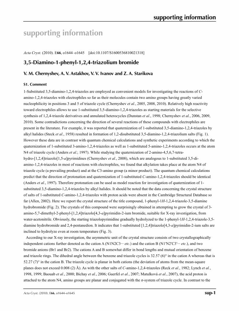
far (Allen, 2002). Here we report the crystal structure of the title compound, 1-phenyl-1H-1,2,4-triazole-3,5-diamine
hydrobromide (Fig. 2). The crystals of this compound were surprisingly obtained in attempting to grow the crystal of 3-
amino-5,7-dimethyl-2-phenyl-[1,2,4]triazolo[4,3-a]pyrimidin-2-ium bromide, suitable for X-ray investigation, from
water-acetonitrile. Obviously, the starting triazolopyrimidine gradually hydrolyzed to the 1-phenyl-1H-1,2,4-triazole-3,5-
diamine hydrobromide and 2,4-pentanedion. It indicates that 1-substituted [1,2,4]triazolo[4,3-a]pyrimidin-2-ium salts are
inclined to hydrolyze even at room temperature (Fig. 3).
According to our X-ray investigation, the asymmetric unit of the crystal structure consists of two crystallographically
independent cations further denoted as the cation A (N1N2C3··· etc.) and the cation B (N1′N2′C3′··· etc.), and two
bromide anions (Br1 and Br2). The cations A and B somewhat differ in bond lengths and mutual orientation of benzene
and triazole rings. The dihedral angle between the benzene and triazole cycles is 32.57 (6)° in the cation A whereas that is
52.27 (7)° in the cation B. The triazole cycle is planar in both cations (the deviation of atoms from the mean-square
planes does not exceed 0.008 (2) Å). As with the other salts of C-amino-1,2,4-triazoles (Reck et al., 1982; Lynch et al.,
1998, 1999; Baouab et al., 2000; Bichay et al., 2006; Guerfel et al., 2007; Matulková et al., 2007), the acid proton is
attached to the atom N4, amino groups are planar and conjugated with the π-system of triazole cycle. In contrast to the
supporting information
sup-2Acta Cryst. (2010). E66, o1644–o1645
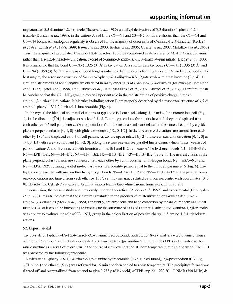
unprotonated 3,5-diamino-1,2,4-triazole (Starova et al., 1980) and alkyl derivatives of 3,5-diamino-1-phenyl-1,2,4-
triazole (Dunstan et al., 1998), in the cations A and B the C5—N1 and C3—N2 bonds are shorter than the C3—N4 and
C5—N4 bonds. An analogous regularity is observed for the majority of other salts of C-amino-1,2,4-triazoles (Reck et
al., 1982; Lynch et al., 1998, 1999; Baouab et al., 2000; Bichay et al., 2006; Guerfel et al., 2007; Matulková et al., 2007).
Thus, the majority of protonated C-amino-1,2,4-triazoles should be considered as derivatives of 4H-1,2,4-triazol-1-ium
rather than 1H-1,2,4-triazol-4-ium cation, except of 5-amino-3-azido-1H-1,2,4-triazol-4-ium nitrate (Bichay et al., 2006).
It is remarkable that the bond C5—N5 (1.325 (3) Å) in the cation A is shorter than the bonds C5—N1 (1.335 (3) Å) and
C5—N4 (1.358 (3) Å). The analysis of bond lengths indicates that molecules forming by cation A can be described in the
best way by the resonance structure of 5-amino-2-phenyl-2,4-dihydro-3H-1,2,4-triazol-3-iminium bromide (Fig. 4). A
similar distributions of bond lengths are observed in many other salts of C-amino-1,2,4-triazoles (for example, see: Reck
et al., 1982; Lynch et al., 1998, 1999; Bichay et al., 2006; Matulková et al., 2007; Guerfel et al., 2007). Therefore, it can
be concluded that the C5—NH2 group plays an important role in the redistribution of positive charge in the C-
amino-1,2,4-triazolium cations. Molecules including cation B are properly described by the resonance structure of 3,5-di-
amino-1-phenyl-4H-1,2,4-triazol-1-ium bromide (Fig. 4).
In the crystal the identical and parallel cations of type A or B form stacks along the b axis of the monoclinic cell (Fig.
5). In the direction [101] the adjacent stacks of the different-type cations form pairs in which they are displaced from
each other on 0.5 cell parameter b. One-type cations from the nearest stacks are related in the same direction by a glide
plane n perpendicular to [0, 1, 0] with glide component [1/2, 0, 1/2]. In the direction c the cations are turned from each
other by 180° and displaced on 0.5 of cell parameter, i.e. are space related by 2-fold screw axis with direction [0, 1, 0] at
1/4, y, 1/4 with screw component [0, 1/2, 0]. Along the c axis one can see parallel linear chains which "links" consist of
pairs of cations A and B connected with bromide anions Br1 and Br2 by means of the hydrogen bonds N3—H3B···Br1,
N5′—H5′B···Br1, N4—H4···Br2, N4′—H4′···Br2, N5—H5B···Br2, N3′—H3′B···Br2 (Table 1). The nearest chains in the
plane perpendicular to b axis are connected with each other by continuous net of hydrogen bonds N3—H3A···N2′i and
N3′—H3′A···N2ii, forming parallel molecular layers with identity period equal to the unit-cell parameter b (Fig. 6). The
layers are connected with one another by hydrogen bonds N5—H5A···Br1iii and N5′—H5′A···Br1iv. In the parallel layers
one-type cations are turned from each other by 180°, i.e. they are space related by inversion centre with coordinates [0, 0,
0]. Thereby, the C8H10N5+ cations and bromide anions form a three-dimensional framework in the crystal.
In conclusion, the present study and previously reported theoretical (Anders et al., 1997) and experimental (Chernyshev
et al., 2008) results indicate that the structures attributed to the products of quaternization of 1-substituted 3,5-di-
amino-1,2,4-triazoles (Steck et al., 1958), apparently, are erroneous and need correction by means of modern analytical
methods. Also it would be interesting to investigate the structure of salts of another 1-substituted 3-amino-1,2,4-triazoles
with a view to evaluate the role of C3—NH2 group in the delocalization of positive charge in 3-amino-1,2,4-triazolium
cations.
S2. Experimental
The crystals of 1-phenyl-1H-1,2,4-triazole-3,5-diamine hydrobromide suitable for X-ray analysis were obtained from a
solution of 3-amino-5,7-dimethyl-2-phenyl-[1,2,4]triazolo[4,3-a]pyrimidin-2-ium bromide (TPB) in 1:9 water: aceto-
nitrile mixture as a result of hydrolysis in the course of slow evaporation at room temperature during one week. The TPB
was prepared by the following procedure.
A mixture of 1-phenyl-1H-1,2,4-triazole-3,5-diamine hydrobromide (0.73 g, 2.85 mmol), 2,4-pentanedion (0.371 g,
3.71 mmol) and ethanol (5 ml) was refluxed for 15 min and then cooled to room temperature. The precipitate formed was
filtered off and recrystallized from ethanol to give 0.757 g (83% yield) of TPB, mp 221–223 °C. 1H NMR (300 MHz) δ:
supporting information
sup-3Acta Cryst. (2010). E66, o1644–o1645
1.95 (s, 3H, CH3), 2.95 (s, 3H, CH3), 7.22 (s, 1H, CH), 7.67–7.83 (m, 5H, Ph), 8.37 (s, 2H, NH2). 13C NMR (150 MHz) δ:
17.66, 24.37, 113.62, 130.13, 130.30, 132.16, 132.63, 147.86, 153.85, 159.74, 170.43. LCMS: 240.29 [C13H14N5+]. Anal.
Calcd for C13H14BrN5: C, 48.76; H, 4.41; N, 21.87. Found: C, 48.81; H, 4.21; N, 21.98.
Starting 1-phenyl-1H-1,2,4-triazole-3,5-diamine hydrobromide used for the preparation of TPB was obtained by
addition of equimolar amount of 48% hydrobromic acid to an ethanol solution of 3,5-diamino-1-phenyl-1,2,4-triazole.
The latter compound was synthesized by known method (Steck et al., 1958).
S3. Refinement
C-bound H atoms were positioned geometrically (C—H 0.93 Å), while the rest H atoms were located on difference map
and further placed in idealized positions (N—H 0.86 Å). All H atoms were refined as riding on their parent atoms, with
Uiso(H) = 1.2 Ueq(parent atom).
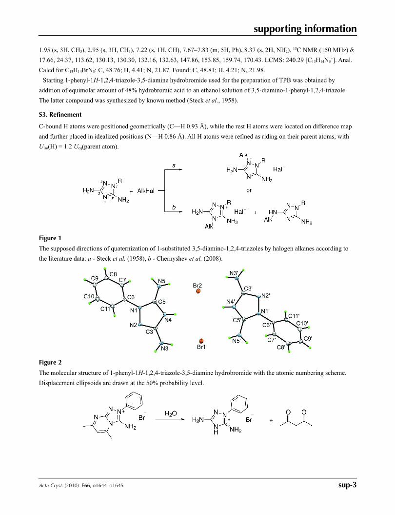
Figure 1
The supposed directions of quaternization of 1-substituted 3,5-diamino-1,2,4-triazoles by halogen alkanes according to
the literature data: a - Steck et al. (1958), b - Chernyshev et al. (2008).
Figure 2
The molecular structure of 1-phenyl-1H-1,2,4-triazole-3,5-diamine hydrobromide with the atomic numbering scheme.
Displacement ellipsoids are drawn at the 50% probability level.
supporting information
sup-4Acta Cryst. (2010). E66, o1644–o1645
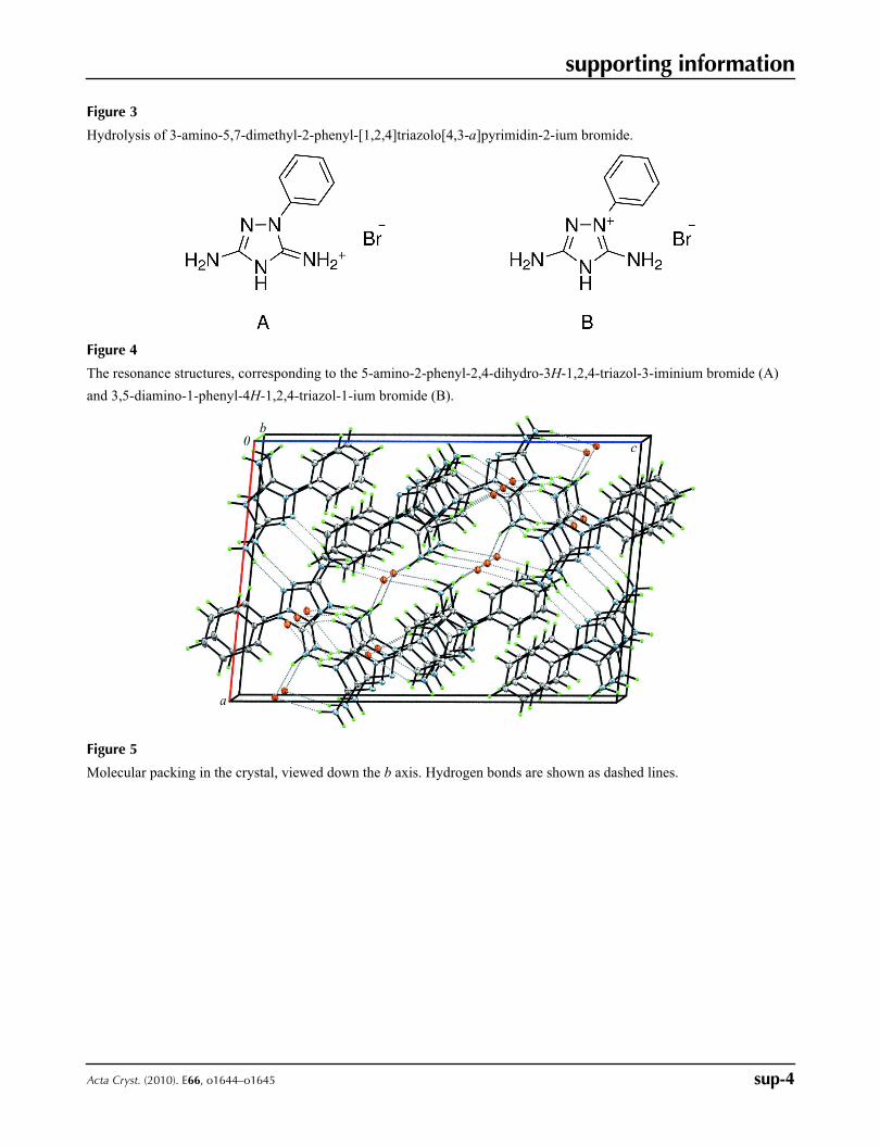
Figure 3
Hydrolysis of 3-amino-5,7-dimethyl-2-phenyl-[1,2,4]triazolo[4,3-a]pyrimidin-2-ium bromide.
Figure 4
The resonance structures, corresponding to the 5-amino-2-phenyl-2,4-dihydro-3H-1,2,4-triazol-3-iminium bromide (A)
and 3,5-diamino-1-phenyl-4H-1,2,4-triazol-1-ium bromide (B).
Figure 5
Molecular packing in the crystal, viewed down the b axis. Hydrogen bonds are shown as dashed lines.
supporting information
sup-5Acta Cryst. (2010). E66, o1644–o1645
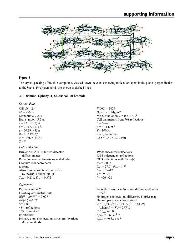
Figure 6
The crystal packing of the title compound, viewed down the a axis showing molecular layers in the planes perpendicular
to the b axis. Hydrogen bonds are shown as dashed lines.
3,5-Diamino-1-phenyl-1,2,4-triazolium bromide
Crystal data
C8H10N5+·Br−
Mr = 256.12Monoclinic, P21/nHall symbol: -P 2yna = 13.752 (2) Åb = 7.1172 (13) Åc = 20.394 (4) Åβ = 95.519 (3)°V = 1986.7 (6) Å3
Z = 8
F(000) = 1024Dx = 1.713 Mg m−3
Mo Kα radiation, λ = 0.71073 ÅCell parameters from 568 reflectionsθ = 3–26°µ = 4.11 mm−1
T = 100 KPlate, colourless0.55 × 0.40 × 0.30 mm
Data collection
Bruker APEXII CCD area-detector diffractometer
Radiation source: fine-focus sealed tubeGraphite monochromatorω scansAbsorption correction: multi-scan
(SADABS; Bruker, 2004)Tmin = 0.211, Tmax = 0.372
19484 measured reflections4314 independent reflections3808 reflections with I > 2σ(I)Rint = 0.033θmax = 27.0°, θmin = 1.7°h = −17→17k = −9→9l = −26→26
Refinement
Refinement on F2
Least-squares matrix: fullR[F2 > 2σ(F2)] = 0.027wR(F2) = 0.071S = 1.004314 reflections253 parameters0 restraintsPrimary atom site location: structure-invariant
direct methods
Secondary atom site location: difference Fourier map
Hydrogen site location: difference Fourier mapH-atom parameters constrainedw = 1/[σ2(Fo
2) + (0.0375P)2 + 2.843P] where P = (Fo
2 + 2Fc2)/3
(Δ/σ)max = 0.001Δρmax = 0.63 e Å−3
Δρmin = −0.52 e Å−3
supporting information
sup-6Acta Cryst. (2010). E66, o1644–o1645
Special details
Geometry. All e.s.d.'s (except the e.s.d. in the dihedral angle between two l.s. planes) are estimated using the full covariance matrix. The cell e.s.d.'s are taken into account individually in the estimation of e.s.d.'s in distances, angles and torsion angles; correlations between e.s.d.'s in cell parameters are only used when they are defined by crystal symmetry. An approximate (isotropic) treatment of cell e.s.d.'s is used for estimating e.s.d.'s involving l.s. planes.Refinement. Refinement of F2 against ALL reflections. The weighted R-factor wR and goodness of fit S are based on F2, conventional R-factors R are based on F, with F set to zero for negative F2. The threshold expression of F2 > σ(F2) is used only for calculating R-factors(gt) etc. and is not relevant to the choice of reflections for refinement. R-factors based on F2 are statistically about twice as large as those based on F, and R- factors based on ALL data will be even larger.
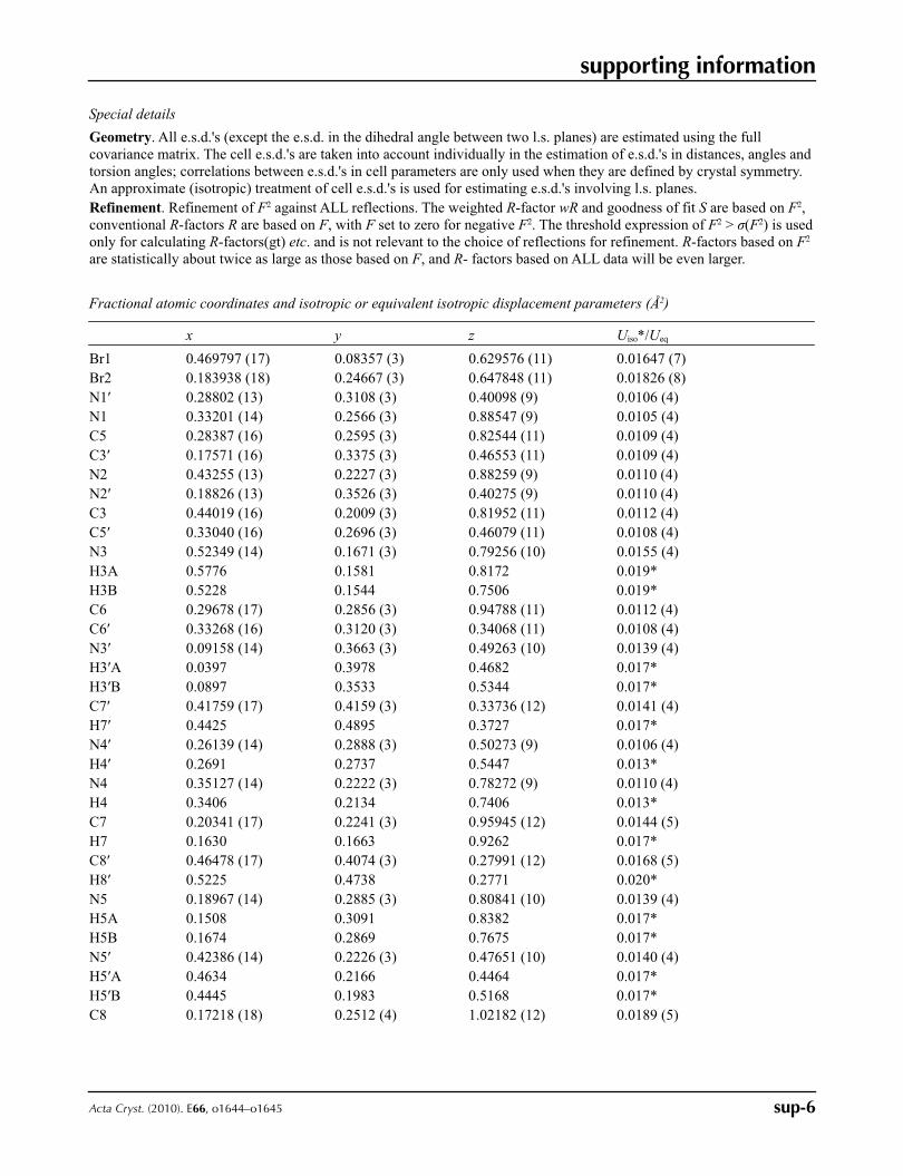
Fractional atomic coordinates and isotropic or equivalent isotropic displacement parameters (Å2)
x y z Uiso*/Ueq
Br1 0.469797 (17) 0.08357 (3) 0.629576 (11) 0.01647 (7)Br2 0.183938 (18) 0.24667 (3) 0.647848 (11) 0.01826 (8)N1′ 0.28802 (13) 0.3108 (3) 0.40098 (9) 0.0106 (4)N1 0.33201 (14) 0.2566 (3) 0.88547 (9) 0.0105 (4)C5 0.28387 (16) 0.2595 (3) 0.82544 (11) 0.0109 (4)C3′ 0.17571 (16) 0.3375 (3) 0.46553 (11) 0.0109 (4)N2 0.43255 (13) 0.2227 (3) 0.88259 (9) 0.0110 (4)N2′ 0.18826 (13) 0.3526 (3) 0.40275 (9) 0.0110 (4)C3 0.44019 (16) 0.2009 (3) 0.81952 (11) 0.0112 (4)C5′ 0.33040 (16) 0.2696 (3) 0.46079 (11) 0.0108 (4)N3 0.52349 (14) 0.1671 (3) 0.79256 (10) 0.0155 (4)H3A 0.5776 0.1581 0.8172 0.019*H3B 0.5228 0.1544 0.7506 0.019*C6 0.29678 (17) 0.2856 (3) 0.94788 (11) 0.0112 (4)C6′ 0.33268 (16) 0.3120 (3) 0.34068 (11) 0.0108 (4)N3′ 0.09158 (14) 0.3663 (3) 0.49263 (10) 0.0139 (4)H3′A 0.0397 0.3978 0.4682 0.017*H3′B 0.0897 0.3533 0.5344 0.017*C7′ 0.41759 (17) 0.4159 (3) 0.33736 (12) 0.0141 (4)H7′ 0.4425 0.4895 0.3727 0.017*N4′ 0.26139 (14) 0.2888 (3) 0.50273 (9) 0.0106 (4)H4′ 0.2691 0.2737 0.5447 0.013*N4 0.35127 (14) 0.2222 (3) 0.78272 (9) 0.0110 (4)H4 0.3406 0.2134 0.7406 0.013*C7 0.20341 (17) 0.2241 (3) 0.95945 (12) 0.0144 (5)H7 0.1630 0.1663 0.9262 0.017*C8′ 0.46478 (17) 0.4074 (3) 0.27991 (12) 0.0168 (5)H8′ 0.5225 0.4738 0.2771 0.020*N5 0.18967 (14) 0.2885 (3) 0.80841 (10) 0.0139 (4)H5A 0.1508 0.3091 0.8382 0.017*H5B 0.1674 0.2869 0.7675 0.017*N5′ 0.42386 (14) 0.2226 (3) 0.47651 (10) 0.0140 (4)H5′A 0.4634 0.2166 0.4464 0.017*H5′B 0.4445 0.1983 0.5168 0.017*C8 0.17218 (18) 0.2512 (4) 1.02182 (12) 0.0189 (5)
supporting information
sup-7Acta Cryst. (2010). E66, o1644–o1645
H8 0.1098 0.2134 1.0300 0.023*C9′ 0.42605 (18) 0.3005 (4) 0.22696 (12) 0.0184 (5)H9′ 0.4585 0.2932 0.1891 0.022*C9 0.2331 (2) 0.3340 (4) 1.07174 (12) 0.0203 (5)H9 0.2116 0.3514 1.1132 0.024*C10′ 0.33847 (19) 0.2038 (3) 0.23038 (12) 0.0177 (5)H10′ 0.3116 0.1362 0.1940 0.021*C10 0.32641 (19) 0.3909 (3) 1.05977 (12) 0.0184 (5)H10 0.3677 0.4445 1.0935 0.022*C11 0.35831 (17) 0.3681 (3) 0.99777 (11) 0.0142 (5)H11 0.4205 0.4078 0.9897 0.017*C11′ 0.29117 (17) 0.2074 (3) 0.28729 (11) 0.0135 (4)H11′ 0.2332 0.1418 0.2899 0.016*
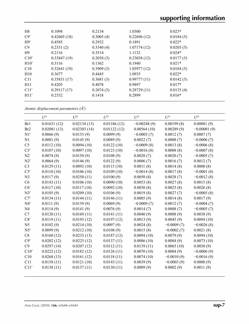
Atomic displacement parameters (Å2)
U11 U22 U33 U12 U13 U23
Br1 0.01631 (12) 0.02134 (13) 0.01186 (12) −0.00248 (9) 0.00199 (8) 0.00081 (9)Br2 0.02081 (13) 0.02303 (14) 0.01122 (12) 0.00564 (10) 0.00289 (9) −0.00001 (9)N1′ 0.0066 (9) 0.0153 (9) 0.0099 (9) −0.0003 (7) 0.0012 (7) 0.0007 (7)N1 0.0081 (9) 0.0145 (9) 0.0089 (9) 0.0022 (7) 0.0008 (7) −0.0006 (7)C5 0.0112 (10) 0.0094 (10) 0.0122 (10) −0.0009 (8) 0.0013 (8) −0.0006 (8)C3′ 0.0107 (10) 0.0097 (10) 0.0123 (10) −0.0016 (8) 0.0008 (8) −0.0007 (8)N2 0.0074 (9) 0.0150 (9) 0.0108 (9) 0.0020 (7) 0.0020 (7) −0.0005 (7)N2′ 0.0064 (9) 0.0146 (9) 0.0122 (9) 0.0006 (7) 0.0016 (7) 0.0012 (7)C3 0.0129 (11) 0.0092 (10) 0.0115 (10) 0.0011 (8) 0.0014 (8) 0.0000 (8)C5′ 0.0110 (10) 0.0106 (10) 0.0109 (10) −0.0014 (8) 0.0017 (8) −0.0001 (8)N3 0.0117 (9) 0.0250 (11) 0.0100 (9) 0.0050 (8) 0.0028 (7) −0.0012 (8)C6 0.0143 (11) 0.0106 (10) 0.0090 (10) 0.0053 (8) 0.0027 (8) 0.0015 (8)C6′ 0.0117 (10) 0.0117 (10) 0.0092 (10) 0.0030 (8) 0.0023 (8) 0.0026 (8)N3′ 0.0105 (9) 0.0209 (10) 0.0106 (9) 0.0019 (8) 0.0027 (7) −0.0005 (8)C7′ 0.0134 (11) 0.0144 (11) 0.0146 (11) 0.0005 (9) 0.0014 (8) 0.0017 (9)N4′ 0.0111 (9) 0.0139 (9) 0.0069 (9) −0.0009 (7) 0.0012 (7) −0.0004 (7)N4 0.0114 (9) 0.0141 (9) 0.0076 (9) 0.0014 (7) 0.0008 (7) −0.0005 (7)C7 0.0120 (11) 0.0169 (11) 0.0141 (11) 0.0040 (9) 0.0008 (9) 0.0038 (9)C8′ 0.0119 (11) 0.0193 (12) 0.0197 (12) 0.0013 (9) 0.0045 (9) 0.0094 (10)N5 0.0102 (9) 0.0214 (10) 0.0097 (9) 0.0024 (8) −0.0009 (7) −0.0026 (8)N5′ 0.0099 (9) 0.0212 (10) 0.0108 (9) 0.0015 (8) −0.0002 (7) 0.0021 (8)C8 0.0160 (12) 0.0233 (13) 0.0187 (12) 0.0094 (10) 0.0079 (9) 0.0094 (10)C9′ 0.0202 (12) 0.0225 (12) 0.0137 (11) 0.0086 (10) 0.0084 (9) 0.0073 (10)C9 0.0297 (14) 0.0207 (12) 0.0112 (11) 0.0139 (11) 0.0065 (10) 0.0030 (9)C10′ 0.0222 (12) 0.0182 (12) 0.0126 (11) 0.0070 (10) 0.0004 (9) −0.0006 (9)C10 0.0268 (13) 0.0161 (12) 0.0118 (11) 0.0074 (10) −0.0010 (9) −0.0016 (9)C11 0.0158 (11) 0.0121 (10) 0.0143 (11) 0.0039 (9) −0.0003 (9) 0.0000 (9)C11′ 0.0138 (11) 0.0137 (11) 0.0130 (11) 0.0009 (9) 0.0002 (9) 0.0011 (9)
supporting information
sup-8Acta Cryst. (2010). E66, o1644–o1645
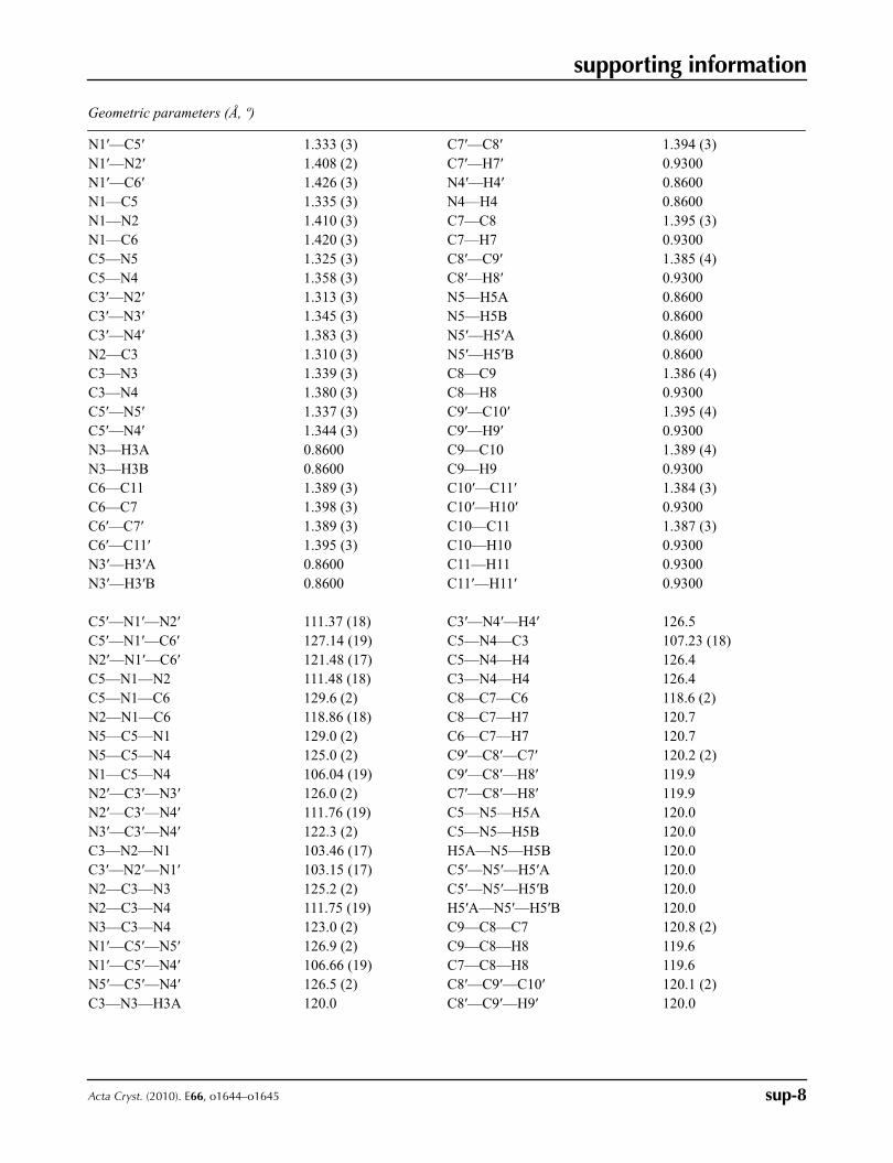
Geometric parameters (Å, º)
N1′—C5′ 1.333 (3) C7′—C8′ 1.394 (3)N1′—N2′ 1.408 (2) C7′—H7′ 0.9300N1′—C6′ 1.426 (3) N4′—H4′ 0.8600N1—C5 1.335 (3) N4—H4 0.8600N1—N2 1.410 (3) C7—C8 1.395 (3)N1—C6 1.420 (3) C7—H7 0.9300C5—N5 1.325 (3) C8′—C9′ 1.385 (4)C5—N4 1.358 (3) C8′—H8′ 0.9300C3′—N2′ 1.313 (3) N5—H5A 0.8600C3′—N3′ 1.345 (3) N5—H5B 0.8600C3′—N4′ 1.383 (3) N5′—H5′A 0.8600N2—C3 1.310 (3) N5′—H5′B 0.8600C3—N3 1.339 (3) C8—C9 1.386 (4)C3—N4 1.380 (3) C8—H8 0.9300C5′—N5′ 1.337 (3) C9′—C10′ 1.395 (4)C5′—N4′ 1.344 (3) C9′—H9′ 0.9300N3—H3A 0.8600 C9—C10 1.389 (4)N3—H3B 0.8600 C9—H9 0.9300C6—C11 1.389 (3) C10′—C11′ 1.384 (3)C6—C7 1.398 (3) C10′—H10′ 0.9300C6′—C7′ 1.389 (3) C10—C11 1.387 (3)C6′—C11′ 1.395 (3) C10—H10 0.9300N3′—H3′A 0.8600 C11—H11 0.9300N3′—H3′B 0.8600 C11′—H11′ 0.9300
C5′—N1′—N2′ 111.37 (18) C3′—N4′—H4′ 126.5C5′—N1′—C6′ 127.14 (19) C5—N4—C3 107.23 (18)N2′—N1′—C6′ 121.48 (17) C5—N4—H4 126.4C5—N1—N2 111.48 (18) C3—N4—H4 126.4C5—N1—C6 129.6 (2) C8—C7—C6 118.6 (2)N2—N1—C6 118.86 (18) C8—C7—H7 120.7N5—C5—N1 129.0 (2) C6—C7—H7 120.7N5—C5—N4 125.0 (2) C9′—C8′—C7′ 120.2 (2)N1—C5—N4 106.04 (19) C9′—C8′—H8′ 119.9N2′—C3′—N3′ 126.0 (2) C7′—C8′—H8′ 119.9N2′—C3′—N4′ 111.76 (19) C5—N5—H5A 120.0N3′—C3′—N4′ 122.3 (2) C5—N5—H5B 120.0C3—N2—N1 103.46 (17) H5A—N5—H5B 120.0C3′—N2′—N1′ 103.15 (17) C5′—N5′—H5′A 120.0N2—C3—N3 125.2 (2) C5′—N5′—H5′B 120.0N2—C3—N4 111.75 (19) H5′A—N5′—H5′B 120.0N3—C3—N4 123.0 (2) C9—C8—C7 120.8 (2)N1′—C5′—N5′ 126.9 (2) C9—C8—H8 119.6N1′—C5′—N4′ 106.66 (19) C7—C8—H8 119.6N5′—C5′—N4′ 126.5 (2) C8′—C9′—C10′ 120.1 (2)C3—N3—H3A 120.0 C8′—C9′—H9′ 120.0
supporting information
sup-9Acta Cryst. (2010). E66, o1644–o1645
C3—N3—H3B 120.0 C10′—C9′—H9′ 120.0H3A—N3—H3B 120.0 C8—C9—C10 119.9 (2)C11—C6—C7 120.9 (2) C8—C9—H9 120.1C11—C6—N1 118.8 (2) C10—C9—H9 120.1C7—C6—N1 120.3 (2) C11′—C10′—C9′ 120.7 (2)C7′—C6′—C11′ 121.9 (2) C11′—C10′—H10′ 119.7C7′—C6′—N1′ 118.6 (2) C9′—C10′—H10′ 119.7C11′—C6′—N1′ 119.5 (2) C11—C10—C9 120.3 (2)C3′—N3′—H3′A 120.0 C11—C10—H10 119.8C3′—N3′—H3′B 120.0 C9—C10—H10 119.8H3′A—N3′—H3′B 120.0 C10—C11—C6 119.6 (2)C6′—C7′—C8′ 118.7 (2) C10—C11—H11 120.2C6′—C7′—H7′ 120.7 C6—C11—H11 120.2C8′—C7′—H7′ 120.7 C10′—C11′—C6′ 118.3 (2)C5′—N4′—C3′ 107.03 (18) C10′—C11′—H11′ 120.8C5′—N4′—H4′ 126.5 C6′—C11′—H11′ 120.8
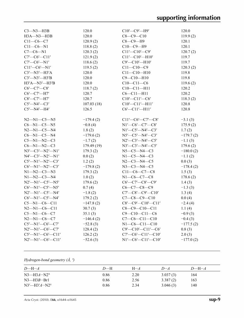
N2—N1—C5—N5 −179.4 (2) C11′—C6′—C7′—C8′ −3.1 (3)C6—N1—C5—N5 −0.8 (4) N1′—C6′—C7′—C8′ 175.9 (2)N2—N1—C5—N4 1.8 (2) N1′—C5′—N4′—C3′ 1.7 (2)C6—N1—C5—N4 −179.6 (2) N5′—C5′—N4′—C3′ −179.7 (2)C5—N1—N2—C3 −1.7 (2) N2′—C3′—N4′—C5′ −1.1 (3)C6—N1—N2—C3 179.49 (19) N3′—C3′—N4′—C5′ 179.6 (2)N3′—C3′—N2′—N1′ 179.3 (2) N5—C5—N4—C3 −180.0 (2)N4′—C3′—N2′—N1′ 0.0 (2) N1—C5—N4—C3 −1.1 (2)C5′—N1′—N2′—C3′ 1.2 (2) N2—C3—N4—C5 0.0 (3)C6′—N1′—N2′—C3′ −179.8 (2) N3—C3—N4—C5 −178.4 (2)N1—N2—C3—N3 179.3 (2) C11—C6—C7—C8 1.5 (3)N1—N2—C3—N4 1.0 (2) N1—C6—C7—C8 178.6 (2)N2′—N1′—C5′—N5′ 179.6 (2) C6′—C7′—C8′—C9′ 1.4 (3)C6′—N1′—C5′—N5′ 0.7 (4) C6—C7—C8—C9 −1.3 (3)N2′—N1′—C5′—N4′ −1.8 (2) C7′—C8′—C9′—C10′ 1.3 (4)C6′—N1′—C5′—N4′ 179.2 (2) C7—C8—C9—C10 0.0 (4)C5—N1—C6—C11 −147.8 (2) C8′—C9′—C10′—C11′ −2.4 (4)N2—N1—C6—C11 30.7 (3) C8—C9—C10—C11 1.1 (4)C5—N1—C6—C7 35.1 (3) C9—C10—C11—C6 −0.9 (3)N2—N1—C6—C7 −146.4 (2) C7—C6—C11—C10 −0.4 (3)C5′—N1′—C6′—C7′ −52.8 (3) N1—C6—C11—C10 −177.5 (2)N2′—N1′—C6′—C7′ 128.4 (2) C9′—C10′—C11′—C6′ 0.8 (3)C5′—N1′—C6′—C11′ 126.2 (2) C7′—C6′—C11′—C10′ 2.0 (3)N2′—N1′—C6′—C11′ −52.6 (3) N1′—C6′—C11′—C10′ −177.0 (2)
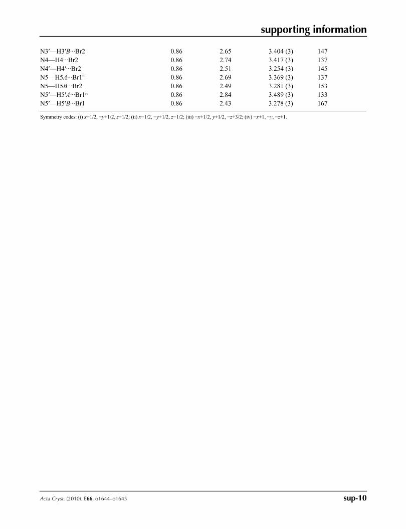
Hydrogen-bond geometry (Å, º)
D—H···A D—H H···A D···A D—H···A
N3—H3A···N2′i 0.86 2.20 3.037 (3) 164N3—H3B···Br1 0.86 2.56 3.387 (2) 163N3′—H3′A···N2ii 0.86 2.34 3.046 (3) 140
supporting information
sup-10Acta Cryst. (2010). E66, o1644–o1645
N3′—H3′B···Br2 0.86 2.65 3.404 (3) 147N4—H4···Br2 0.86 2.74 3.417 (3) 137N4′—H4′···Br2 0.86 2.51 3.254 (3) 145N5—H5A···Br1iii 0.86 2.69 3.369 (3) 137N5—H5B···Br2 0.86 2.49 3.281 (3) 153N5′—H5′A···Br1iv 0.86 2.84 3.489 (3) 133N5′—H5′B···Br1 0.86 2.43 3.278 (3) 167
Symmetry codes: (i) x+1/2, −y+1/2, z+1/2; (ii) x−1/2, −y+1/2, z−1/2; (iii) −x+1/2, y+1/2, −z+3/2; (iv) −x+1, −y, −z+1.
Related Documents