Organic & Biomolecular Chemistry Dynamic Article Links Cite this: DOI: 10.1039/c2ob26393a www.rsc.org/obc PAPER Synthesis, antibacterial activity and mode of action of novel linoleic acid–dipeptide–spermidine conjugates† Seema Joshi,‡ a,b Rikeshwer P. Dewangan,‡ a Shruti Yadav, a Diwan S. Rawat b and Santosh Pasha* a Received 18th July 2012, Accepted 4th September 2012 DOI: 10.1039/c2ob26393a Towards therapeutically viable mimics of host defense cationic peptides (HDCPs) here we report the design and synthesis of a small library, based on a novel hydrophobic–dipeptide–spermidine template. Lipidated sequences 11, 14, 15, 16, 18 and 19 exhibited potent activity against susceptible as well as drug resistant Gram-positive and Gram-negative bacterial strains. Structure–activity relationships of the template revealed a hydrophobicity window of 50–70% with minimum +2 charges to be crucial for activity and cell selectivity. Active sequences 14, 15 and 16 exhibited different modes of action based on dipeptide composition as revealed by studies on model membranes, intact bacterial cells and DNA. Further, severe damage to surface morphology of methicillin resistant S. aureus caused by 14, 15 and 16 at 10 × MIC was observed. The present study provides us two active sequences (14 and 16) with a membrane perturbing mode of action, cell selectivity to hRBCs and keratinocytes along with potent activity against clinically relevant pathogen MRSA. The designed template thus may prove to be a suitable probe to optimize sequences for better selectivity and potential to combat a wide range of drug resistant strains in further research. Introduction The emergence of multiple drug resistant bacterial strains causes millions of deaths worldwide. 1,2 The rise in morbidity and mor- tality related to microbial infections has led to tremendous pressure on health care systems. 3 Host defense cationic peptides (HDCPs) are known for their wide range of activity against microorganisms including bacteria, fungi, viruses and even cancer- ous cells. With multifaceted roles in innate immunity and a direct cell lytic mode of action, it is difficult for a microorganism to endure resistance against HDCPs. 4,5 Therefore HDCPs are being explored as promising alternatives to conventional drugs. 6,7 However in spite of all these advantages only a handful of antimicrobial peptides are under clinical trials due to their high manufacturing costs, poor pharmacokinetic properties and associated toxicity issues. 8 To address these issues, efforts are being made to explore the characteristic features of HDCPs such as net positive charge at physiological pH and hydrophobic bulk 9 to develop more economic membrane active antimicrobial peptidomimetics. Many classes of compounds such as ceragenins, 10,11 oligoacyllysines, 12,13 arylamides, 14 peptoid based scaffolds 15 and lipopeptides 16,17 that mimic HDCPs are being developed as alternative antimicrobial agents with low sus- ceptibility for development of resistance. Cationic polyamines spermine, spermidine and putrescine are ubiquitous components of eukaryotic and prokaryotic cells with multiple roles in modulating functions of DNA, RNA and pro- teins inside the cells. 18 A number of modified/conjugated polya- mines have been reported with various biological activities such as LPS sequestration, 19 anti-parasitic activity, 20,21 anticancer activity, 22,23 as well as nucleic acid carriers for DNA transfec- tion. 24 Considering the medicinal potential of polyamines we anticipated that incorporation of spermidine as a positively charged moiety to hydrophobic–dipeptides may lead to small peptidomimetics with better antibacterial activity. Long-chain free fatty acids (FFAs) are known to be present at skin surfaces, maintaining an acidic pH which helps to prevent colonization of various microorganisms including methicillin- resistant S. aureus and H. pylori. 25–27 The mode of bacterial killing for FFAs has not been unambiguously determined however, the prime target is believed to be microbial membranes where direct lysis, perturbation of the electron transport chain, oxidative phosphorylation and inhibition of fatty acid meta- bolism have been reported as some of the probable causes leading to bacterial cell death. 28,29 Therefore with multiple non- specific modes of action, a broad range of activity and minimum toxicity long chain FFAs were chosen to be incorporated into designed peptidomimetics as the hydrophobic moiety. † Electronic supplementary information (ESI) available: HPLC traces, mass spectra and 1 H NMR data of representative designed sequences. See DOI: 10.1039/c2ob26393a ‡ These authors have contributed equally to the work. a Institute of Genomics and Integrative Biology, Mall Road, Delhi, India. E-mail: [email protected]; Fax: +91 11 27667471; Tel: +9111 27666156 b Department of Chemistry, University of Delhi, Delhi, India This journal is © The Royal Society of Chemistry 2012 Org. Biomol. Chem. Downloaded by Institute of Genomics and Integrative Biology (IGIB) on 18 September 2012 Published on 05 September 2012 on http://pubs.rsc.org | doi:10.1039/C2OB26393A View Online / Journal Homepage

Welcome message from author
This document is posted to help you gain knowledge. Please leave a comment to let me know what you think about it! Share it to your friends and learn new things together.
Transcript

Organic &BiomolecularChemistry
Dynamic Article Links
Cite this: DOI: 10.1039/c2ob26393a
www.rsc.org/obc PAPER
Synthesis, antibacterial activity and mode of action of novel linoleicacid–dipeptide–spermidine conjugates†
Seema Joshi,‡a,b Rikeshwer P. Dewangan,‡a Shruti Yadav,a Diwan S. Rawatb and Santosh Pasha*a
Received 18th July 2012, Accepted 4th September 2012DOI: 10.1039/c2ob26393a
Towards therapeutically viable mimics of host defense cationic peptides (HDCPs) here we report thedesign and synthesis of a small library, based on a novel hydrophobic–dipeptide–spermidine template.Lipidated sequences 11, 14, 15, 16, 18 and 19 exhibited potent activity against susceptible as well as drugresistant Gram-positive and Gram-negative bacterial strains. Structure–activity relationships of thetemplate revealed a hydrophobicity window of 50–70% with minimum +2 charges to be crucial foractivity and cell selectivity. Active sequences 14, 15 and 16 exhibited different modes of action based ondipeptide composition as revealed by studies on model membranes, intact bacterial cells and DNA.Further, severe damage to surface morphology of methicillin resistant S. aureus caused by 14, 15 and 16at 10 × MIC was observed. The present study provides us two active sequences (14 and 16) with amembrane perturbing mode of action, cell selectivity to hRBCs and keratinocytes along with potentactivity against clinically relevant pathogen MRSA. The designed template thus may prove to be asuitable probe to optimize sequences for better selectivity and potential to combat a wide range of drugresistant strains in further research.
Introduction
The emergence of multiple drug resistant bacterial strains causesmillions of deaths worldwide.1,2 The rise in morbidity and mor-tality related to microbial infections has led to tremendouspressure on health care systems.3 Host defense cationic peptides(HDCPs) are known for their wide range of activity againstmicroorganisms including bacteria, fungi, viruses and even cancer-ous cells. With multifaceted roles in innate immunity and a directcell lytic mode of action, it is difficult for a microorganism toendure resistance against HDCPs.4,5 Therefore HDCPs are beingexplored as promising alternatives to conventional drugs.6,7
However in spite of all these advantages only a handful ofantimicrobial peptides are under clinical trials due to their highmanufacturing costs, poor pharmacokinetic properties andassociated toxicity issues.8 To address these issues, efforts arebeing made to explore the characteristic features of HDCPs suchas net positive charge at physiological pH and hydrophobicbulk9 to develop more economic membrane active antimicrobialpeptidomimetics. Many classes of compounds such as
ceragenins,10,11 oligoacyllysines,12,13 arylamides,14 peptoidbased scaffolds15 and lipopeptides16,17 that mimic HDCPs arebeing developed as alternative antimicrobial agents with low sus-ceptibility for development of resistance.
Cationic polyamines spermine, spermidine and putrescine areubiquitous components of eukaryotic and prokaryotic cells withmultiple roles in modulating functions of DNA, RNA and pro-teins inside the cells.18 A number of modified/conjugated polya-mines have been reported with various biological activities suchas LPS sequestration,19 anti-parasitic activity,20,21 anticanceractivity,22,23 as well as nucleic acid carriers for DNA transfec-tion.24 Considering the medicinal potential of polyamines weanticipated that incorporation of spermidine as a positivelycharged moiety to hydrophobic–dipeptides may lead to smallpeptidomimetics with better antibacterial activity.
Long-chain free fatty acids (FFAs) are known to be present atskin surfaces, maintaining an acidic pH which helps to preventcolonization of various microorganisms including methicillin-resistant S. aureus and H. pylori.25–27 The mode of bacterialkilling for FFAs has not been unambiguously determinedhowever, the prime target is believed to be microbial membraneswhere direct lysis, perturbation of the electron transport chain,oxidative phosphorylation and inhibition of fatty acid meta-bolism have been reported as some of the probable causesleading to bacterial cell death.28,29 Therefore with multiple non-specific modes of action, a broad range of activity and minimumtoxicity long chain FFAs were chosen to be incorporated intodesigned peptidomimetics as the hydrophobic moiety.
†Electronic supplementary information (ESI) available: HPLC traces,mass spectra and 1H NMR data of representative designed sequences.See DOI: 10.1039/c2ob26393a‡These authors have contributed equally to the work.
aInstitute of Genomics and Integrative Biology, Mall Road, Delhi, India.E-mail: [email protected]; Fax: +91 11 27667471;Tel: +91 11 27666156bDepartment of Chemistry, University of Delhi, Delhi, India
This journal is © The Royal Society of Chemistry 2012 Org. Biomol. Chem.
Dow
nloa
ded
by I
nstit
ute
of G
enom
ics
and
Inte
grat
ive
Bio
logy
(IG
IB)
on 1
8 Se
ptem
ber
2012
Publ
ishe
d on
05
Sept
embe
r 20
12 o
n ht
tp://
pubs
.rsc
.org
| do
i:10.
1039
/C2O
B26
393A
View Online / Journal Homepage

Here we report the design and synthesis of a small library ofhydrophobic–dipeptide–spermidine template based sequencesconsisting of cationic polyamine spermidine and FFAs (linoleicacid and stearic acid). We dissected the template into two partsto evaluate the role of the hydrophobic moiety–dipeptide or cat-ionic–dipeptide part of the template in imparting activity and/orselectivity to these sequences. We further evaluated interactionof the active sequences with model membranes, intact bacterialcells and DNA to probe into the molecular mode of action.
Results and discussion
1. Design and synthesis
Clinically approved lipopeptide antibiotics such as polymyxin B,daptomycin and echinocandins have spurred the research fornovel analogues of these classes of lipopeptides with clinicalrelevance.30–32 A large number of membrane active lipopeptideshave been designed to achieve net positive charge (at least +2)and hydrophobic bulk making use of varied chemicalmoieties.15–17 However, cationic lipopeptides with saturated fattyacids are difficult to optimize for cell selectivity.33 Realizing theimportance of charge and amphiphilicity for membrane activeantimicrobial peptidomimetics, we designed a novel short tem-plate that comprised a hydrophobic moiety attached at the N-terminus to dipeptide–spermidine. The sequences presented inthe current study are unique where inherently active unsaturatedfatty acids were coupled to dipeptides which were further taggedwith organic polyamine spermidine, prompting membraneactivity as well as cellular translocation to bind DNA. For thedipeptide portion we made use of three different combinations oftryptophan (Trp) and ornithine (Orn) amino acids i.e. Trp–Trp,Trp–Orn and Orn–Orn. Hydrophobic Trp amino acid was chosenbecause of its well documented membrane anchoring property.34
Cationic residue Orn was used to impart protease stability to
designed sequences due to its non-ribosomal origin. Based onvarious biological activities associated with spermidine conju-gates as outlined above, we incorporated spermidine at the C-ter-minus of the dipeptides to give rise to sequences 1–3 (Table 1).N-Terminal hydrophobic tagging of dipeptide sequences wasdone to evaluate the role of FFAs such as linoleic acid andstearic acid in imparting activity leading to sequences 4–11. Toevaluate the role of lipidation as a control an aromatic moiety3-(4-hydroxyphenyl)-propionic acid (HPPA) was also conju-gated. The HPPA was used based on our observations thatcovalent hybridization of HPPA to a tetra-peptide template led tothe discovery of some potent tetra-peptidomimetics.35 To havean appropriate balance between charge and hydrophobicity, thedesigned complete template consisting of a hydrophobic tag atthe N-terminal with cationic spermidine at the C-terminus result-ing in sequences 12–19 was also synthesized. With at least +2charge and varied degrees of hydrophobicity these conjugateswere further evaluated for antibacterial activity and mode ofaction studies.
The sequences were synthesized using a combination of solidphase and solution phase strategy as presented in Scheme 1.Structures of representative sequences are shown in Fig. 1.
2. Antibacterial activity
Antibacterial activity of designed sequences against a range ofGram-positive and Gram-negative bacterial strains was deter-mined using the serial broth dilution method (Table 2). Out ofthe sequences 1–3, sequence 1 exhibited MIC at 113.6 μg mL−1
against S. aureus. Out of sequences 4–11, 4, 5 and 9 weredevoid of antibacterial activity, whereas sequences 6 and 7showed MIC at 113.6 μg mL−1 against S. aureus and B. subtilis.Sequence 8 with +1 charge showed moderate activity againsttested strains with MIC in the range of 28.4–113.6 μg mL−1.Sequence 11 with +1 charge and stearic acid tagging showed
Table 1 Sequences, molecular mass, charge and % of acetonitrile at RP-HPLC elution
Sequences Composition
Mass [M]+
Charge % acetonitrile at RP-HPLC elutionCalc. Obser.
1 NH2-WW-spermidine 518.3238 518.3228 +3 262 NH2-WO-spermidine 446.3238 446.3226 +4 223 NH2-OO-spermidine 374.3238 374.3235 +5 194 HPPA-WW-COOH 539.2289 539.2277 −1 615 HPPA-WO-COOH 467.2289 467.2278 0 466 LIN-WW-COOH 653.4061 653.62a −1 847 LIN-WO-COOH 581.4061 581.4063 0 798 LIN-OO-COOH 509.4061 509.4054 +1 599 STER-WW-COOH 657.4374 657.68a −1 8810 STER-WO-COOH 585.4374 585.4366 0 8911 STER-OO-COOH 513.4374 513.4368 +1 7012 HPPA-WW-spermidine 666.3762 666.3762 +2 5213 HPPA-WO-spermidine 594.3762 594.3760 +3 2714 LIN-WW-spermidine 780.5535 780.5537 +2 7015 LIN-WO-spermidine 708.5535 708.5544 +3 6216 LIN-OO-spermidine 636.5535 636.5540 +4 5117 STER-WW-spermidine 784.5848 784.5858 +2 7618 STERWO-spermidine 712.5848 712.5847 +3 6819 STER-OO-spermidine 640.5848 640.5853 +4 63
HPPA: 3-(4-hydroxy phenyl)-propionic acid; LIN: linoleic acid; STER: stearic acid.a ESI-MS data.
Org. Biomol. Chem. This journal is © The Royal Society of Chemistry 2012
Dow
nloa
ded
by I
nstit
ute
of G
enom
ics
and
Inte
grat
ive
Bio
logy
(IG
IB)
on 1
8 Se
ptem
ber
2012
Publ
ishe
d on
05
Sept
embe
r 20
12 o
n ht
tp://
pubs
.rsc
.org
| do
i:10.
1039
/C2O
B26
393A
View Online

good antibacterial activity against Gram-positive bacterial strainswith MIC in the range of 7.1–14.2 μg mL−1. Sequence 12 withHPPA tagging showed moderate activities against almost all thetested strains with MIC in the range of 56.8–113.6 μg mL−1.Sequence 13 showed activity against S. aureus and B. subtiliswith MIC at 113.6 μg mL−1. Percentage of acetonitrile fromRP-HPLC elution profile data as an indicator for hydrophobicityalong with MIC data showed that the designed sequences with athreshold of acetonitrile (50%) with a single positive charge (8and 11) showed good antibacterial activity (Table 2). Thereforesimilar to the previously reported concept of a hydrophobicitywindow for HDCPs,36 here we propose a window of 50% to70% acetonitrile at RP-HPLC elution (Table 1), below or abovethis threshold the sequences were either less active or inactive.MIC data on these sequences thus ensured that lipidation with asingle positive charge may lead to activity (8 and 11) however;charge alone (1–3) without lipidation is not sufficient to impartappreciable activity.
Sequences 14–19 showed a broad range of activity againstGram-positive as well as Gram-negative bacterial strains withMIC values in the range of 0.88–28.4 μg mL−1. Against E. coliand P. aeruginosa, MIC values of these sequences were found to
be in the range of 3.5–28.4 μg mL−1 and 7.1–14.2 μg mL−1
respectively (except sequence 17). Against the four Gram-posi-tive strains tested in the study, all the sequences showed MICvalues below 15 μg mL−1. Interestingly MIC of these sequencesagainst MRSA was found to be comparable or even better ascompared to S. aureus. Sequence 17 with 76% acetonitrile atRP-HPLC elution was above the hydrophobicity threshold of70%, above which the sequences were found to be less activeagainst tested strains. It is important to note that in comparisonto standard antibiotics tetracycline and polymyxin B, sequences14–19 showed better MIC against MRSA (except 17), where 14and 15 were very promising with MIC at 0.88 μg mL−1.
3. Toxicity evaluation of designed sequences
The efficacy of designed sequences as safe antibacterial agentswas established based on their interaction with enucleatedhRBCs and human keratinocytes (HaCaT cells). Most of thesequences (1–7, 9, 12, 13, 16 and 17) were found to be non-hemolytic up to 62.5 μg mL−1. Sequences 8 and 11 were foundto be lytic to hRBCs, however on conjugation of spermidine,
Scheme 1 Reagents and conditions: (a) Fmoc-NH-X1-COOH, DIPEA, DCM : DMF (50 : 50), 3 h, MeOH for 30 min, (b) 20% piperidine in DMF,(c) Fmoc-NH-X2-COOH, HOBt, DIPCDI, DCM : DMF (50 : 50), 1.5 h, (d) R-COOH, HOBt, DIPCDI, DCM : DMF (50 : 50), 14 h, (e) 50% TFA–DCM, (f ) TFE : acetic acid : DCM (1 : 1 : 8), 1.5 h, (g) N1,N4-bis(Boc)-spermidine, DIPCDI, HOBt, THF, 0 °C for 30 min, rt 18 h, (h) (Boc)2O,DIPEA–DCM, 1.5 h.
This journal is © The Royal Society of Chemistry 2012 Org. Biomol. Chem.
Dow
nloa
ded
by I
nstit
ute
of G
enom
ics
and
Inte
grat
ive
Bio
logy
(IG
IB)
on 1
8 Se
ptem
ber
2012
Publ
ishe
d on
05
Sept
embe
r 20
12 o
n ht
tp://
pubs
.rsc
.org
| do
i:10.
1039
/C2O
B26
393A
View Online
reduction in hemolysis was observed for N-terminal taggeddipeptides (Table 2). Sequences 14 and 16 showed negligiblehemolytic activity whereas sequence 15 caused 40% hemolysisat concentrations corresponding to 18–78 times its MIC valuesagainst Gram-positive bacterial strains tested in the study.Sequences with stearic acid tagging (18 and 19) were found tobe more lytic to hRBC than the linoleic acid counter sequences(15 and 16). Similar to our results, experimental evidence ispresent in the literature that reports cationic lipopeptides withsaturated fatty acid chain length to be more lytic as compared totheir unsaturated counterparts.33
Since HDCP mimics are considered more suitable as topicalagents, we evaluated the effects of designed sequences onhuman keratinocytes. Results of the MTT assay on HaCaT cellsshowed 80% and 100% cell survival for sequences 14 and 16 at62.5 μg mL−1 respectively (Fig. 2). Even up to a high concen-tration of 250 μg mL−1 65% and 78% cell survival was observedfor sequences 14 and 16 respectively. These data thus confirm nodetrimental effects of these sequences on human keratinocyteseven upon incubation for 18 h.
4. Bactericidal kinetics
HDCPs with a predominant membrane active mode of action areendowed with the potential to kill bacterial cells within minutesat concentrations higher than MIC. In order to monitor the rapid-ity of mode of action of designed sequences, we incubatedsequences 14, 15 and 16 with log-phase MRSA at 37 °C andmonitored the course of change in optical density (OD600) atdifferent time intervals. The results showed inhibition of bac-terial growth after 2 h at MIC for sequences 15 and 16 (Fig. 3).
Fig. 1 Representative structures of designed antibacterial sequences.
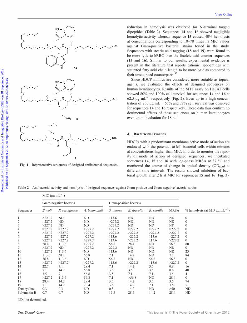
Table 2 Antibacterial activity and hemolysis of designed sequences against Gram-positive and Gram-negative bacterial strains
Sequences
MIC (μg mL−1)
% hemolysis (at 62.5 μg mL−1)
Gram-negative bacteria Gram-positive bacteria
E. coli P. aeruginosa A. baumannii S. aureus E. faecalis B. subtilis MRSA
1 >227.2 ND ND 113.6 ND ND ND 02 >227.2 ND ND >227.2 ND ND ND 03 >227.2 ND ND >227.2 ND ND ND 04 >227.2 >227.2 >227.2 >227.2 >227.2 >227.2 >227.2 05 >227.2 >227.2 >227.2 >227.2 >227.2 >227.2 >227.2 06 >227.2 >227.2 >227.2 113.6 >227.2 113.6 >227.2 07 >227.2 >227.2 >227.2 113.6 >227.2 113.6 >227.2 08 28.4 113.6 >227.2 56.8 28.4 ND 56.8 809 >227.2 ND >227.2 227.2 ND ND ND 010 >227.2 113.6 ND 113.6 ND ND ND 2311 113.6 ND 56.8 7.1 14.2 ND 7.1 8412 56.8 113.6 ND 56.8 ND 56.8 56.8 013 >227.2 >227.2 >227.2 113.6 >227.2 113.6 >227.2 014 22.7 7.1 28.4 7.1 14.2 3.5 0.8 1615 7.1 14.2 56.8 3.5 3.5 3.5 0.8 4016 3.5 7.1 56.8 3.5 7.1 7.1 3.5 417 >227.2 113.6 56.8 7.1 >56.8 ND 28.4 018 28.4 14.2 28.4 3.5 14.2 3.5 3.5 7419 7.1 14.2 28.4 3.5 14.2 7.1 3.5 51Tetracycline 0.3 0.3 ND 0.3 14.2 ND >50 NDPolymyxin B 0.7 0.7 ND 15.5 28.4 14.2 28.4 ND
ND: not determined.
Org. Biomol. Chem. This journal is © The Royal Society of Chemistry 2012
Dow
nloa
ded
by I
nstit
ute
of G
enom
ics
and
Inte
grat
ive
Bio
logy
(IG
IB)
on 1
8 Se
ptem
ber
2012
Publ
ishe
d on
05
Sept
embe
r 20
12 o
n ht
tp://
pubs
.rsc
.org
| do
i:10.
1039
/C2O
B26
393A
View Online
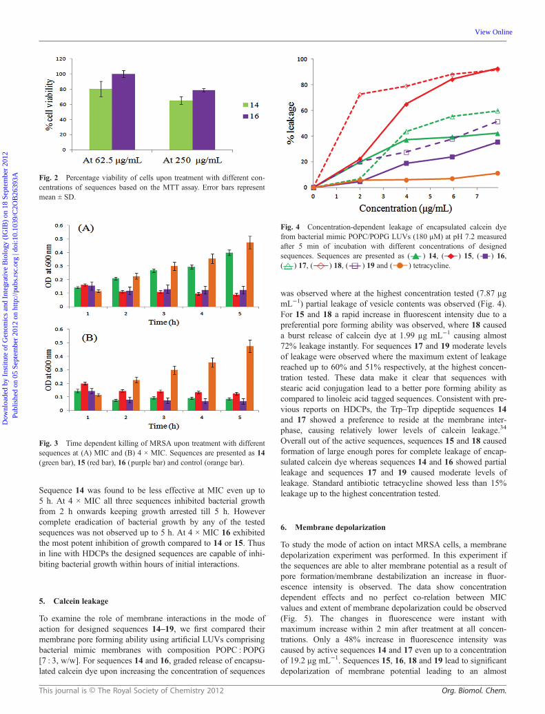
Sequence 14 was found to be less effective at MIC even up to5 h. At 4 × MIC all three sequences inhibited bacterial growthfrom 2 h onwards keeping growth arrested till 5 h. Howevercomplete eradication of bacterial growth by any of the testedsequences was not observed up to 5 h. At 4 × MIC 16 exhibitedthe most potent inhibition of growth compared to 14 or 15. Thusin line with HDCPs the designed sequences are capable of inhi-biting bacterial growth within hours of initial interactions.
5. Calcein leakage
To examine the role of membrane interactions in the mode ofaction for designed sequences 14–19, we first compared theirmembrane pore forming ability using artificial LUVs comprisingbacterial mimic membranes with composition POPC : POPG[7 : 3, w/w]. For sequences 14 and 16, graded release of encapsu-lated calcein dye upon increasing the concentration of sequences
was observed where at the highest concentration tested (7.87 μgmL−1) partial leakage of vesicle contents was observed (Fig. 4).For 15 and 18 a rapid increase in fluorescent intensity due to apreferential pore forming ability was observed, where 18 causeda burst release of calcein dye at 1.99 μg mL−1 causing almost72% leakage instantly. For sequences 17 and 19 moderate levelsof leakage were observed where the maximum extent of leakagereached up to 60% and 51% respectively, at the highest concen-tration tested. These data make it clear that sequences withstearic acid conjugation lead to a better pore forming ability ascompared to linoleic acid tagged sequences. Consistent with pre-vious reports on HDCPs, the Trp–Trp dipeptide sequences 14and 17 showed a preference to reside at the membrane inter-phase, causing relatively lower levels of calcein leakage.34
Overall out of the active sequences, sequences 15 and 18 causedformation of large enough pores for complete leakage of encap-sulated calcein dye whereas sequences 14 and 16 showed partialleakage and sequences 17 and 19 caused moderate levels ofleakage. Standard antibiotic tetracycline showed less than 15%leakage up to the highest concentration tested.
6. Membrane depolarization
To study the mode of action on intact MRSA cells, a membranedepolarization experiment was performed. In this experiment ifthe sequences are able to alter membrane potential as a result ofpore formation/membrane destabilization an increase in fluor-escence intensity is observed. The data show concentrationdependent effects and no perfect co-relation between MICvalues and extent of membrane depolarization could be observed(Fig. 5). The changes in fluorescence were instant withmaximum increase within 2 min after treatment at all concen-trations. Only a 48% increase in fluorescence intensity wascaused by active sequences 14 and 17 even up to a concentrationof 19.2 μg mL−1. Sequences 15, 16, 18 and 19 lead to significantdepolarization of membrane potential leading to an almost
Fig. 3 Time dependent killing of MRSA upon treatment with differentsequences at (A) MIC and (B) 4 × MIC. Sequences are presented as 14(green bar), 15 (red bar), 16 (purple bar) and control (orange bar).
Fig. 2 Percentage viability of cells upon treatment with different con-centrations of sequences based on the MTT assay. Error bars representmean ± SD.
Fig. 4 Concentration-dependent leakage of encapsulated calcein dyefrom bacterial mimic POPC/POPG LUVs (180 μM) at pH 7.2 measuredafter 5 min of incubation with different concentrations of designedsequences. Sequences are presented as ( ) 14, ( ) 15, ( ) 16,( ) 17, ( ) 18, ( ) 19 and ( ) tetracycline.
This journal is © The Royal Society of Chemistry 2012 Org. Biomol. Chem.
Dow
nloa
ded
by I
nstit
ute
of G
enom
ics
and
Inte
grat
ive
Bio
logy
(IG
IB)
on 1
8 Se
ptem
ber
2012
Publ
ishe
d on
05
Sept
embe
r 20
12 o
n ht
tp://
pubs
.rsc
.org
| do
i:10.
1039
/C2O
B26
393A
View Online
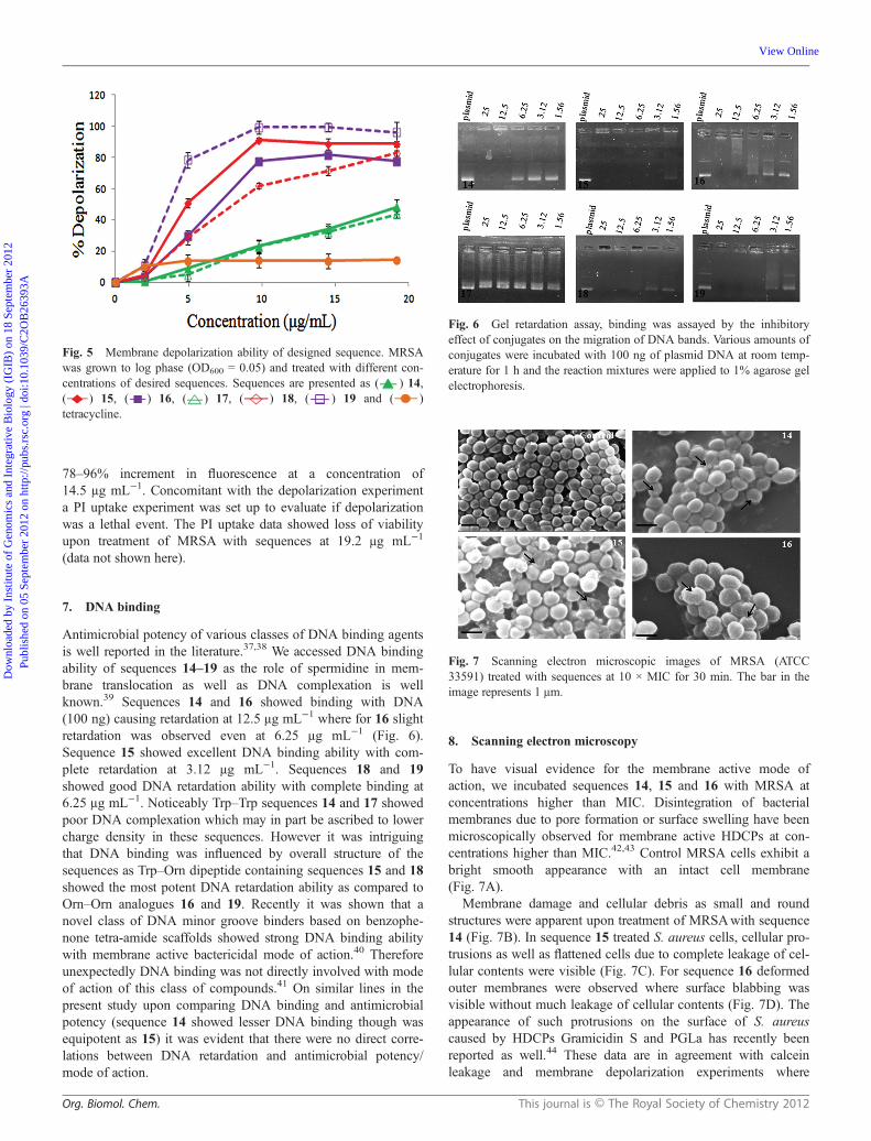
78–96% increment in fluorescence at a concentration of14.5 μg mL−1. Concomitant with the depolarization experimenta PI uptake experiment was set up to evaluate if depolarizationwas a lethal event. The PI uptake data showed loss of viabilityupon treatment of MRSA with sequences at 19.2 μg mL−1
(data not shown here).
7. DNA binding
Antimicrobial potency of various classes of DNA binding agentsis well reported in the literature.37,38 We accessed DNA bindingability of sequences 14–19 as the role of spermidine in mem-brane translocation as well as DNA complexation is wellknown.39 Sequences 14 and 16 showed binding with DNA(100 ng) causing retardation at 12.5 μg mL−1 where for 16 slightretardation was observed even at 6.25 μg mL−1 (Fig. 6).Sequence 15 showed excellent DNA binding ability with com-plete retardation at 3.12 μg mL−1. Sequences 18 and 19showed good DNA retardation ability with complete binding at6.25 μg mL−1. Noticeably Trp–Trp sequences 14 and 17 showedpoor DNA complexation which may in part be ascribed to lowercharge density in these sequences. However it was intriguingthat DNA binding was influenced by overall structure of thesequences as Trp–Orn dipeptide containing sequences 15 and 18showed the most potent DNA retardation ability as compared toOrn–Orn analogues 16 and 19. Recently it was shown that anovel class of DNA minor groove binders based on benzophe-none tetra-amide scaffolds showed strong DNA binding abilitywith membrane active bactericidal mode of action.40 Thereforeunexpectedly DNA binding was not directly involved with modeof action of this class of compounds.41 On similar lines in thepresent study upon comparing DNA binding and antimicrobialpotency (sequence 14 showed lesser DNA binding though wasequipotent as 15) it was evident that there were no direct corre-lations between DNA retardation and antimicrobial potency/mode of action.
8. Scanning electron microscopy
To have visual evidence for the membrane active mode ofaction, we incubated sequences 14, 15 and 16 with MRSA atconcentrations higher than MIC. Disintegration of bacterialmembranes due to pore formation or surface swelling have beenmicroscopically observed for membrane active HDCPs at con-centrations higher than MIC.42,43 Control MRSA cells exhibit abright smooth appearance with an intact cell membrane(Fig. 7A).
Membrane damage and cellular debris as small and roundstructures were apparent upon treatment of MRSAwith sequence14 (Fig. 7B). In sequence 15 treated S. aureus cells, cellular pro-trusions as well as flattened cells due to complete leakage of cel-lular contents were visible (Fig. 7C). For sequence 16 deformedouter membranes were observed where surface blabbing wasvisible without much leakage of cellular contents (Fig. 7D). Theappearance of such protrusions on the surface of S. aureuscaused by HDCPs Gramicidin S and PGLa has recently beenreported as well.44 These data are in agreement with calceinleakage and membrane depolarization experiments where
Fig. 5 Membrane depolarization ability of designed sequence. MRSAwas grown to log phase (OD600 = 0.05) and treated with different con-centrations of desired sequences. Sequences are presented as ( ) 14,( ) 15, ( ) 16, ( ) 17, ( ) 18, ( ) 19 and ( )tetracycline.
Fig. 7 Scanning electron microscopic images of MRSA (ATCC33591) treated with sequences at 10 × MIC for 30 min. The bar in theimage represents 1 μm.
Fig. 6 Gel retardation assay, binding was assayed by the inhibitoryeffect of conjugates on the migration of DNA bands. Various amounts ofconjugates were incubated with 100 ng of plasmid DNA at room temp-erature for 1 h and the reaction mixtures were applied to 1% agarose gelelectrophoresis.
Org. Biomol. Chem. This journal is © The Royal Society of Chemistry 2012
Dow
nloa
ded
by I
nstit
ute
of G
enom
ics
and
Inte
grat
ive
Bio
logy
(IG
IB)
on 1
8 Se
ptem
ber
2012
Publ
ishe
d on
05
Sept
embe
r 20
12 o
n ht
tp://
pubs
.rsc
.org
| do
i:10.
1039
/C2O
B26
393A
View Online

sequence 15 caused cellular damage in the form of membranedisruption whereas 14 and 16 were found to show lesser leakageof encapsulated dye.
Overall the present study affords us sequences 14–19 withpotent activity against a broad range of bacterial strains includingMRSA. Upon dissecting the designed template to identifyfeatures responsible for activity and selectivity, we found that thecationic charge imparted to dipeptides by spermidine insequences 1–3 was not sufficient per se to show bactericidalproperties. Lipidation alone with neutral or negative charge insequences 6, 7, 9 and 10 also led to low activity. With unit posi-tive charge, lipidated sequences 8 and 11 exhibited improvedactivity though with compromised cell selectivity (Table 2).However conjugation of spermidine in sequences 14–19improved potency as well as cell selectivity. With minimum +2charges, lipidated sequences 14–19 showed a broad range ofantibacterial activity.
Since the sequences show potent activity against MRSA, tohave better insights into the mode of action of designedsequences we characterized their interactions with S. aureusmimic artificial membranes, intact MRSA and DNA.
Mode of action studies revealed a predominant role of thedipeptide sequence in initial binding, bactericidal kinetics andmembrane disrupting abilities of designed sequences. Forsequence 14, low leakage causing ability, lower levels of mem-brane depolarization as well as reduced DNA binding abilitymake membrane destabilization a less probable mode of action atMIC. A slower bactericidal kinetics of 14 at MIC might as wellbe due to different modes of action operative at low concen-trations. Slower bactericidal kinetics has previously beenreported for HDCP mimics interfering with vital functions inbacterial cells other than membrane disruption.45 However inSEM studies at concentrations 10 × MIC cellular debris and dif-fused outer bacterial membranes were evident for 14 potentiatinga membrane disruptive mode of action (Fig. 7B). Sequence 15showed faster leakage of calcein along with membrane depolar-ization, rapid killing kinetics and excellent DNA binding ability.Therefore this sequence showed clean membrane perturbingmode of action at MIC as well as higher concentrations as wasevidenced in SEM images of the treated MRSA (Fig. 7C).Sequence 16 with rapid bactericidal kinetics, good DNA bindingability caused appreciable damage to membrane potential inMRSA at the tested concentrations, however moderate levels ofleakage causing ability showed that either transient pores wereformed or the pores were not large enough to cause leakage ofcalcein which is evident by the surface blabbing observed inSEM studies. A low leakage causing ability in spite of potentmembrane depolarization has previously been reported for ana-logues of HDCP indolicidin.46
Conclusion
In summary, using simple chemistry and economically viablebuilding blocks FFAs (linoleic acid/stearic acid), Trp, Orn andspermidine, we obtained 6 active sequences with a broad rangeof activity against Gram-positive as well as Gram-negative bac-terial strains including clinically relevant pathogen MRSA.Sequences 14 and 16 showed excellent cell selectivity and
membrane perturbing mode of action at concentrations higherthan MIC. These sequences were also able to alter the electro-phoretic mobility of DNA which although was not directlyrelated to activity may as well be responsible for furtherenhanced potency of these sequences due to intracellular mode ofaction. The structure–activity work in this study paves the way forthe design of optimized FFAs based peptidomimetics with spermi-dine/spermine conjugated with different combinations of aminoacids which is currently in progress in our laboratory.
Experimental section
Materials
Fmoc-protected amino acids and resins were purchased fromNovabiochem. Dimethylamino pyridine (DMAP), N,N-diisopro-pylcarbodiimide (DIPCDI), N-hydroxybenzotrizole (HOBt), di-isopropyl ethylamine (DIPEA), N-methyl pyrrolidinone (NMP),piperidine, trifluoroethanol (TFE), trifluoroacetic acid (TFA), tri-isopropyl silane (TIS), calcein and 3,3′-dipropylthiadicarbocya-nine iodide (DiSC35) were obtained from Sigma Chemical Co.1-Palmitoyl-2-oleoyl-sn-glycero-3-phosphocholine (POPC) and1-palmitoyl-2-oleoyl-sn-glycero-3-phospho-(1′-rac-glycerol)(sodium salt) (POPG) were purchased from Avanti Polar Lipids.Dulbecco’s modified Eagles’ medium-high glucose (DMEM),antibiotic/antimycotic solutions, heat inactivated fetal bovineserum (FBS), trypsin from porcine pancreas and 3-(4,5-dimethyl-2-thiazolyl)-2,5-diphenyl-2H-tetrazolium bromide(MTT) were obtained from Sigma Aldrich Chemical Company.All solvents used for the purification were of HPLC grade andobtained from Merck, Germany. Dimethylformamide (DMF) anddichloromethane (DCM) were obtained from Merck India. DMFwas double distilled prior to use.
Synthesis and purification of sequences
The dipeptides were synthesized on 2-chlorotrityl chloride resinas a solid support using Fmoc chemistry as reported pre-viously.47 The terminal amino group of dipeptides was Boc pro-tected on a solid support before cleavage from the resin undermild conditions (TFE : CH3COOH : CH2Cl2 cocktail, 1 : 1 : 8) toretain Boc groups. The cleaved dipeptides were coupled withN1,N4-bis(boc)spermidine (SIGMA) using HOBt and DIPCDI indry tetrahydrofuran under a N2 atmosphere at 0 °C for 30 minfollowed by 18 h at rt as reported previously.48 The obtainedproduct was dissolved in CHCl3 (15 mL) and washed with 1%aqueous NaHCO3 (50 mL), 1% aqueous HCl (50 mL), and brine(50 mL). The organic layer was dried over anhydrous Na2SO4
and concentrated under reduced pressure to give crude dipeptidespermidine conjugates. Boc groups were removed from the con-jugates using 50% TFA in CH2Cl2 to give sequences 1–3. On asolid support N-terminal end tagging of dipeptides was achievedby coupling 4 equiv. of 3-(4-hydroxyphenyl)-propionic acid(HPPA)/linoleic acid/stearic acid overnight with HOBt andDIPCDI. The Kaiser test was performed to check completion ofreactions on a solid support.49 The N-terminal tagged di-peptido-mimetics were cleaved from a solid support under two differentconditions. For synthesis of 4–11, cleavage was effectuatedusing 50% TFA in DCM. For synthesis of 12–19, cleavage was
This journal is © The Royal Society of Chemistry 2012 Org. Biomol. Chem.
Dow
nloa
ded
by I
nstit
ute
of G
enom
ics
and
Inte
grat
ive
Bio
logy
(IG
IB)
on 1
8 Se
ptem
ber
2012
Publ
ishe
d on
05
Sept
embe
r 20
12 o
n ht
tp://
pubs
.rsc
.org
| do
i:10.
1039
/C2O
B26
393A
View Online

performed under mild conditions (TFE : CH3COOH : CH2Cl2cocktail 1 : 1 : 8). Further Boc protected N-terminal tagged con-jugates were coupled with N1,N4-bis(boc) spermidine asdescribed for dipeptides earlier in the text. Finally Boc groupswere removed with 50% TFA resulting in sequences 12–19. Syn-thesized sequences were purified using an RP-HPLC column(7.8 × 300 mm, 125 Å, 10 μm particle size) with either gradientsof 10 to 90% buffer 2 where buffer 1 was water (0.05% TFA)and buffer 2 was acetonitrile (0.05% TFA) over 45 min or 30 to100% buffer 2 gradients were run over 45 min where buffer 1was water (0.1% TFA) and buffer 2 was acetonitrile (0.1% TFA).The correct sequences after purification were confirmed byLC-MS/MS (Quattro micro API, Waters), LC-ESI-HRMS onUHPLC (Dionex, Germany) and LTQ Orbitrap XL (ThermoFisher Scientific, USA) mass determination and 1H NMR. Massspectra, analytical HPLC traces and 1H NMR data of representa-tive sequences are provided in supplementary files.
Antibacterial activity
Antibacterial activity of designed sequences was evaluated usinga modification of the serial broth dilution method as reportedpreviously.50 Bacterial strains used in this study were as follows,E. coli (ATCC 11775), P. aeruginosa (ATCC 25668), A. bau-mannii (ATCC 19606), S. aureus (ATCC 29213), E. faecalis(ATCC 7080), B. subtilis (ATCC 6633) and methicillin resistantS. aureus (ATCC 33591). The inoculums were prepared frommid-log phase bacterial cultures. Each well of the first 11columns of a 96-well polypropylene micro titre plate (SIGMA)was inoculated with 100 μL of approximately 105 CFU mL−1 ofbacterial suspension per mL of Mueller-Hinton broth (MHB,DIFCO). Then 11 μL of serially diluted test sequences in0.001% acetic acid and 0.2% bovine serum albumin (SIGMA)over the desired concentration range was added to the wells ofmicro titre plates. The micro titre plates were incubated overnightwith agitation (200 rpm) at 37 °C. After 18 h absorbance wasread at 630 nm. Cultures (approximately 105 CFU mL−1)without test sequences were used as a positive control. Un-inocu-lated MHB was used as a negative control. Tests were carried outin duplicate on three different days. Minimum inhibitory concen-tration (MIC) is defined as the lowest concentration of testsequences that completely inhibits growth. For comparisonpeptide antibiotic polymyxin B and tetracycline were alsoassayed under identical conditions.
Hemolytic activity
Hemolytic activity assay was done as described previously.51
Briefly, 100 μL of fresh hRBC suspension 4% v/v in PBS(35 mM phosphate buffer, 150 mM NaCl) was placed in a 96-well plate. After incubation of the test sequences (100 μL) in theerythrocyte solution for 1 h at 37 °C, the plates were centrifugedand the supernatant (100 μL) was transferred to fresh 96-wellplates. Absorbance was read at 540 nm using an ELISA platereader (Molecular Devices). Percent hemolysis was calculatedusing the following formula:
% hemolysis ¼ 100½ðA� A0Þ=ðAt � A0Þ�
where A represents absorbance of sample wells at 540 nm andA0 and At represent zero percent and 100% hemolysis determinedin PBS and 1% Triton X-100, respectively.
Cytotoxicity
To assess cell viability, the MTT assay was performed asdescribed previously.52 HaCaT keratinocytes, 3000 cells perwell, were seeded in 96-well plates in DMEM HAMSF12 media supplemented with 10% serum (FBS) to grow over-night. The next day media were aspirated and fresh incompletemedia were added (50 μL per well). To the wells serial two-folddilutions of different test sequences (50 μL) were added and theplates were incubated at 37 °C with 5% CO2 for 18 h. After 18 hthe media were aspirated and 100 μL of MTT solution wasadded to each well. The plates were further incubated for 4 h inCO2 at 37 °C. After 4 h the MTT-containing medium wasremoved by aspiration. The blue formazan product generatedwas dissolved by the addition of 100 μL of 100% DMSO perwell. The plates were then gently swirled for 2–3 min at roomtemperature to dissolve the precipitate. The absorbance wasmonitored at 540 nm. Percentage viability was calculated basedon the following formula:
% cell viability ¼ ðA=AcontrolÞ � 100
where A represents sample absorbance at a given concentrationand Acontrol represents untreated cells. The experiment wasrepeated thrice and results are given as mean ± SD.
Bactericidal kinetics
Overnight cultures of methicillin resistant S. aureus (ATCC33591) were grown in fresh MHB up to log phase. For determin-ing the time course of killing activity 100 μL of fresh MHB wasadded to all wells of the 96 well-plate. Then 90 μL of approxi-mately 105 CFU mL−1 were added to the wells of the 96-wellplate (4 wells for a single concentration). Then 10 μL of appro-priate concentrations of test sequences corresponding to MICand 4 × MIC were added to the wells. The plates were incubatedat 37 °C at 200 rpm. Absorbance of the plates was read at600 nm at various time points at 0, 1, 2, 3, 4 and 5 h. The exper-iment was repeated on three different days and values are plottedas mean ± SD.
Calcein leakage
The ability of designed sequences 14–19 to cause leakage fromartificial LUVs composed of bacterial mimic membrane (POPC/POPG) was accessed as described previously.53,54 Briefly,desired mixtures of the lipids POPC/POPG (7 : 3, w/w) were dis-solved in a 2 mL chloroform–methanol mixture in a 150 mLround bottom flask. The solvent was removed under a streamof nitrogen and the lipid film obtained was lyophilizedovernight to remove any traces of organic solvent. The drylipid films were rehydrated with 10 mM Tris-HCl [70 mMcalcein, 150 mM NaCl, 0.1 mM EDTA]. The liposomesuspension obtained after rehydration was freeze thawed forfive cycles and extruded 16 times through two stacked
Org. Biomol. Chem. This journal is © The Royal Society of Chemistry 2012
Dow
nloa
ded
by I
nstit
ute
of G
enom
ics
and
Inte
grat
ive
Bio
logy
(IG
IB)
on 1
8 Se
ptem
ber
2012
Publ
ishe
d on
05
Sept
embe
r 20
12 o
n ht
tp://
pubs
.rsc
.org
| do
i:10.
1039
/C2O
B26
393A
View Online

polycarbonate filters (Mini extruder, Avanti Polar Lipids).Free calcein was removed by passing the liposome suspensionthrough a Sephadex G-50 column at 23 °C and eluting with abuffer containing 10 mM Tris-HCl [150 mM NaCl, 0.1 mMEDTA]. After passing the liposome through a Sephadex G-50,liposome diameter was measured by dynamic light scatteringusing a Zetasizer Nano ZS (Malvern Instruments). The averagediameter of LUVs was found to be in the range of 90–110 nm.Different concentrations of test sequences were incubatedwith POPC/POPG LUVs for 5 min before exciting thesamples. Leakage was monitored by measuring the fluorescenceintensity at an emission wavelength of 520 nm upon excitationat 490 nm on a model Fluorolog (Jobin Yuvon, Horiba) spec-trofluorimeter. A slit width of 3 nm was used for both excitationand emission. Percentage dye leakage was calculated using theformula
% dye leakage ¼ 100½ðF � F0Þ=ðF t � F0Þ�
where F is the fluorescence intensity achieved by addition ofdifferent concentrations of sequences. F0 and Ft are fluorescenceintensities in buffer and with Triton X-100 (20 μL of 10%solution) respectively. All measurements were made in duplicateand less than 4% deviation was obtained in the data points. Aphosphate assay was performed to determine the concentrationof lipids for the leakage experiment.55
Membrane depolarization
For the evaluation of membrane depolarization a previouslydefined method was used.56 Briefly, overnight grown MRSAwassubcultured into MHB for 2–3 h at 37 °C to obtain midlog phasecultures. The cells were centrifuged at 4000 rpm for 10 min at25 °C, washed, and re-suspended in respiration buffer (5 mMHEPES, 20 mM glucose, pH 7.4) to obtain a dilutedsuspension of OD600 ≈ 0.05. A membrane potential-sensitivedye, 3,3′-dipropylthiadicarbocyanine iodide (DiSC35), 0.18 μM(prepared in DMSO) was added to a 500 μL aliquot of there-suspended cells and allowed to stabilize for 1 h. Baselinefluorescence was acquired using a Fluorolog (Jobin Yuvon,Horiba) spectrofluorometer by excitation at 622 nm andemission at 670 nm. A bandwidth of 5 nm was employedfor excitation and emission. Subsequently, increasingconcentrations of test sequences between 2 and 19.2 μg mL−1
were added to the stabilized cells and the increase offluorescence on account of the dequenching of DiSC35 dyewas measured after every 2 min to obtain the maximaldepolarization. Percent depolarization was calculated by usingthe formula
% depolarization ¼ ðF � F0Þ=ðFm � F0Þ � 100
where F is the fluorescence intensity 2 min after addition ofsequences, F0 is the initial basal fluorescence intensity, and Fm isthe maximum fluorescence intensity obtained after addition of10 μg mL−1 gramicidin. Percent depolarization mean ± SD oftwo independent experiments was plotted versus increasing con-centrations of different sequences.
DNA binding assay
Gel retardation experiments were performed as described pre-viously.54,57 Briefly, 100 ng of plasmid DNA (pBluescript IISK+) was mixed with increasing amounts of test sequences14–19 in 20 μL of binding buffer (5% glycerol, 10 mM Tris-HCl, pH 8.0, 1 mM EDTA, 1 mM dithiothreitol, 20 mM KCl,and 50 μg mL−1 bovine serum albumin). Reaction mixtures wereincubated at room temperature for 1 h. Subsequently, 4 μL ofnative loading buffer was added (10% Ficoll 400, 10 mM Tris-HCl, pH 7.5, 50 mM EDTA, 0.25% bromophenol blue, and0.25% xylene cyanol), and a 20 μL aliquot subjected to 1%agarose gel electrophoresis in 0.5× Tris borate–EDTA buffer(45 mM Tris-borate and 1 mM EDTA, pH 8.0). The gels wererun for 1.5 h at 80 V and visualized with ethidium bromide. Theplasmid DNA was purchased from Stratagene and was used assuch without further purification.
Scanning electron microscopy
For electron microscopy samples were prepared as describedpreviously.58 Briefly, freshly inoculated methicillin resistantS. aureus (ATCC 33591) was grown on MHB up to an OD600 of0.5 (corresponding to 108 CFU mL−1). Bacterial cells were thenspun down at 4000 rpm for 15 min, washed thrice in PBS(20 mM, 150 mM NaCl) and re-suspended in an equal volumeof PBS. The cultures were then incubated with test sequences14, 15 or 16 at 10 × MIC for 30 min. Controls were run in theabsence of sequences. After 30 min, the cells were spun downand washed with PBS thrice. For cell fixation the washed bac-terial pallet was re-suspended in 1 mL of 2.5% glutaraldehyde inPBS and was incubated at 4 °C for 4 h. After fixation, cells werespun down and washed with PBS twice. Further the sampleswere dehydrated in series of graded ethanol solutions (30% to100%), and finally dried in desiccators under a vacuum. An auto-matic sputter coater (Polaron OM-SC7640) was used for coatingthe specimens with 20 nm gold particles. Then samples wereviewed via a scanning electron microscope (EVO 40, Carl Zeiss,Germany).
Acknowledgements
This work was financially supported by a Council of Scientificand Industrial Research (CSIR) network project NWP-013. Wethank Dr Souvik Maiti and Dr Kausik Chakraborty for usefuldiscussions and instrumentation facility. We acknowledge theAnalytical Instrumental Research Facility at Jawahar Lal NehruUniversity, Delhi, India for providing us scanning electronmicroscopic facility. Dr V. Sabareesh and Richa Guleria areacknowleged for help in ESI-HR-MS data acquisition. Theauthor S.J. is thankful to CSIR, New Delhi, India, for the awardof SRF.
References
1 S. B. Levy and B. Marshall, Nat. Med., 2004, 10, 122.2 J. Davies and D. Davies, Microbiol. Mol. Biol. Rev., 2010, 74, 417.3 A. Opar, Nat. Rev. Drug Discovery, 2007, 6, 943.4 R. E. W. Hancock and H. G. Sahl, Nat. Biotechnol., 2006, 24, 1551.
This journal is © The Royal Society of Chemistry 2012 Org. Biomol. Chem.
Dow
nloa
ded
by I
nstit
ute
of G
enom
ics
and
Inte
grat
ive
Bio
logy
(IG
IB)
on 1
8 Se
ptem
ber
2012
Publ
ishe
d on
05
Sept
embe
r 20
12 o
n ht
tp://
pubs
.rsc
.org
| do
i:10.
1039
/C2O
B26
393A
View Online

5 M. Zasloff, Nature, 2002, 415, 389.6 J. He, D. K. Yarbrough, J. Kreth, M. H. Anderson, W. Shi and R. Eckert,Antimicrob. Agents Chemother., 2010, 54, 2143.
7 S. E. Blondelle and K. Lohner, Curr. Pharm. Des., 2010, 28, 3204.8 P. W. Latham, Nat. Biotechnol., 1999, 17, 755.9 M. B. Strom, B. E. Haug, M. L. Skar, W. Stensen, T. Stiberg andJ. S. Svendsen, J. Med. Chem., 2003, 46, 1567.
10 B. Ding, N. Yin, Y. Liu, J. C. Garcia, R. Evanson, T. Orsak, M. Fan,G. Turin and P. B. Savage, J. Am. Chem. Soc., 2004, 126, 13642.
11 J. N. Chin, M. J. Rybak, C. M. Cheung and P. B. Savage, Antimicrob.Agents Chemother., 2007, 51, 1268.
12 I. S. Radzishevsky, S. Rotem, D. Bourdetsky, S. Navon-Venezia,Y. Carmeli and A. Mor, Nat. Biotechnol., 2007, 25, 657.
13 F. Zaknoon, H. Sarig, S. Rotem, L. Livne, A. Ivankin, D. Gidalevitz andA. Mor, Antimicrob. Agents Chemother., 2009, 53, 3422.
14 N. Srinivas, K. Moehle, K. A. Hadeed, D. Obrecht and J. A. Robinson,Org. Biomol. Chem., 2007, 5, 3100.
15 N. P. Chongsiriwatana, T. M. Miller, M. Wetzler, S. Vakulenko,A. J. Karlsson, S. P. Palacek, S. Mobashery and A. E. Barron, Antimicrob.Agents Chemother., 2011, 55, 417.
16 A. Makovitzki, D. Avrahami and Y. Shai, Proc. Natl. Acad. Sci. U. S. A.,2006, 103, 15997.
17 W. Kamysz, C. Silvestri, O. Cirioni, A. Giacometti, A. Licci, A. DellaVittoria, M. Okroj and G. Scalise, Antimicrob. Agents Chemother., 2007,51, 354.
18 K. Igarashi and K. Kashiwagi, Biochem. Biophys. Res. Commun., 2000,271, 559.
19 A. Shrestha, D. Sil, S. S. Malladi, H. J. Warshakoon and S. A. David,Bioorg. Med. Chem. Lett., 2009, 19, 2478.
20 X. Bi, C. Lopez, C. J. Bacchi, D. Rattendi and P. M. Woster, Bioorg.Med. Chem. Lett., 2006, 16, 3229.
21 B. K. Verlinden, J. Niemand, J. Snyman, S. K. Sharma, R. J. Beattie,P. M. Woster and L. M. Birkholtz, J. Med. Chem., 2011, 54, 6624.
22 T. Yildirim, K. Bilgin, G. Y. Ciftci, E. T. Ecik, E. Senkuytu, Y. Uluda,L. Tomak and A. Kilic, Eur. J. Med. Chem., 2012, 52, 213.
23 R. A. Casero Jr. and P. M. Woster, J. Med. Chem., 2009, 52, 4551.24 A. Liberska, A. Lilienkampf, A. U. Broceta and M. Bradley, Chem.
Commun., 2011, 47, 12774.25 D. R. Drake, K. A. Brogden, D. V. Dawson and P. W. Wertz, J. Lipid
Res., 2008, 49, 4.26 M. Farrington, N. Brenwald, D. Haines and E. Walpole, J. Med. Micro-
biol., 1992, 36, 56.27 C. Q. Sun, C. J. O’Connor and A. M. Roberton, FEMS Immunol. Med.
Microbiol., 2003, 36, 9.28 P. A. Desbois and V. J. Smith, Appl. Microbiol. Biotechnol., 2010, 85,
1629.29 C. J. Zheng, J. S. Yoo, T. G. Lee, H. Y. Choc, Y. H. Kim and W. G. Kim,
FEBS Lett., 2005, 579, 5157.30 M. Vaara, J. Fox, G. Loidl, O. Siikanen, J. Apajalahti, F. Hansen,
N. Frimodt-Moller, J. Nagai, M. Takano and T. Vaara, Antimicrob. AgentsChemother., 2008, 52, 3229.
31 J. N. Steenbergen, J. Alder, G. M. Thorne and F. P. Tally, J. Antimicrob.Chemother., 2005, 55, 283.
32 M. P. C. Mulder, J. A. W. Kruijtzer, E. J. Breukink, J. Kemmink,R. J. Pieters and R. M. J. Liskamp, Bioorg. Med. Chem., 2011, 19, 6505.
33 H. Sarig, S. Rotem, L. Ziserman, D. Danino and A. Mor, Antimicrob.Agents Chemother., 2008, 52, 4308.
34 D. I. Chan, E. J. Prenner and H. J. Vogel, Biochim. Biophys. Acta, 2006,1758, 1184.
35 G. S. Bisht, D. S. Rawat, A. Kumar, R. Kumar and S. Pasha, Bioorg.Med. Chem. Lett., 2007, 17, 4343.
36 Y. Chen, M. T. Guarnieri, A. I. Vasil, M. L. Vasil, C. T. Mant andR. S. Hodges, Antimicrob. Agents Chemother., 2007, 51, 1398.
37 J. A. Kaizerman, M. I. Gross, Y. Ge, S. White, W. Hu, J. X. Duan,E. E. Baird, K. W. Johnson, R. D. Tanaka, H. E. Moser and R. W. Burli,J. Med. Chem., 2003, 46, 3914.
38 A. I. Khalaf, A. H. Ebrahimabadi, A. J. Drummond, N. G. Anthony,S. P. Mackay, C. J. Suckling and R. D. Waigh, Org. Biomol. Chem.,2004, 2, 3119.
39 J. G. Delcros, S. Tomasi, S. Duhieu, M. Foucault, B. Martin, M. Roch,V. Eifler-Lima, J. Renault and P. Uriac, J. Med. Chem., 2006, 49,232.
40 S. K. Vooturi, C. M. Cheung, M. J. Rybak and S. M. Firestine, J. Med.Chem., 2009, 52, 5020.
41 S. K. Vooturi, M. B. Dewal and S. M. Firestine, Org. Biomol. Chem.,2011, 9, 6367.
42 I. L. Exposito, L. Amigo and I. Recio, Biochim. Biophys. Acta, 2008,1778, 2444.
43 A. J. Hyde, J. Parisot, A. McNichol and B. B. Bonev, Proc. Natl. Acad.Sci. U. S. A., 2006, 103, 19896.
44 M. Hartmann, M. Berditsch, J. Hawecker, M. F. Ardakani, D. Gerthsenand A. S. Ulrich, Antimicrob. Agents Chemother., 2010, 54, 3132.
45 S. Rotem, I. S. Radzishevsky, D. Bourdetsky, S. N. Venezia, Y. Carmeliand A. Mor, FASEB J., 2008, 22, 2652.
46 Y. H. Nan, J. K. Bang and S. Y. Shin, Peptides, 2009, 30, 832.47 K. Barlos, O. Chatzi, D. Gatos and G. Stavropoulos, Int. J. Pept. Protein
Res., 1991, 37, 513.48 W. H. Chen, X. B. Shao, R. Moellering, C. Wennersten and S. L. Regen,
Bioconjugate Chem., 2006, 17, 1582.49 E. Kaiser, R. L. Colescott, C. D. Bossinger and P. I. Cook, Anal.
Biochem., 1970, 34, 595.50 M. A. Wikler, D. E. Low, F. R. Cockerill, D. J. Sheehan, W. A. Craig,
F. C. Tenover and M. L. Dudley, Methods for dilution antimicrobial sus-ceptibility tests for bacteria that grow aerobically: approved standard-seventh edition (2006) CLSI (formerly NCCLS) M7-A7.
51 M. Sharma, P. Joshi, N. Kumar, S. Joshi, R. K. Rohilla, N. Roy andD. S. Rawat, Eur. J. Med. Chem., 2011, 46, 480.
52 L. Schmidtchen, G. Ringstad, H. Kasetty, M. W. Mizuno, M. Rutland andM. Malmsten, Biochim. Biophys. Acta, 2011, 1808, 1081.
53 L. Zhang, A. Rozek and R. E. W. Hancock, J. Biol. Chem., 2001, 276,35714.
54 S. Joshi, G. S. Bisht, D. S. Rawat, A. Kumar, R. Kumar, S. Maiti andS. Pasha, Biochim. Biophys. Acta, 2010, 1798, 1864.
55 G. R. Bartlett, J. Biol. Chem., 1959, 234, 466.56 P. N. Domadia, A. Bhunia, A. Ramamoorthy and S. Bhattacharjya, J. Am.
Chem. Soc., 2010, 132, 18417.57 S. T. Yang, J. Y. Lee, H. J. Kim, Y. J. Eu, S. Y. Shin, K. S. Hahm and
J. I. Kim, FEBS J., 2006, 273, 4040.58 M. Singh and K. Mukhopadhyay, Antimicrob. Agents Chemother., 2011,
55, 1920.
Org. Biomol. Chem. This journal is © The Royal Society of Chemistry 2012
Dow
nloa
ded
by I
nstit
ute
of G
enom
ics
and
Inte
grat
ive
Bio
logy
(IG
IB)
on 1
8 Se
ptem
ber
2012
Publ
ishe
d on
05
Sept
embe
r 20
12 o
n ht
tp://
pubs
.rsc
.org
| do
i:10.
1039
/C2O
B26
393A
View Online
Related Documents