Oral mucosa

Welcome message from author
This document is posted to help you gain knowledge. Please leave a comment to let me know what you think about it! Share it to your friends and learn new things together.
Transcript

Oral mucosa
Oral mucosa
• Mucous membrane is the moist lining of the intestinal tract, nasal passages, and other body cavities that communicate with the exterior. In oral cavity this lining is called the oral mucous membrane or oral mucosa.
• Oral mucosa is anatomically situated between the intestinal mucosa and skin.
• Skin , oral mucosa and intestinal all consist of two separate tissue components i.e epithelium and connective tissue.
Functions of oral mucosa
• Protection The deeper tissue is protected from hard food,
biting and chewing barriers against the normal microorganisms
of the oral cavity. protection from stretching, shearing and
abrasion
Sensation
• Perceive stimuli like temperature, pain, and touch
• Taste sensation• Signaling the satisfaction of thirst• Helps in reflexes like swallowing, gagging,
retching, and salivation.

Secretion
• Major and minor salivary glands contribute to the moist environment.

Thermal regulation
• In dogs only , no obvious role in human.

Difference between skin and oral mucosa
• Appearance melanin Keratinization vascular dilatation
• Skin contains hair follicles, sebaceous glands and sweat glands while in O M the glandular tissue is minor and major salivary glands
• O M is more smoother and have fewer folds and wrinkles
• O M has epithelium and connective tissue and skin has epidermis and dermis

Organization of oral mucosa
• Masticatory mucosa• Lining mucosa• Lining mocosa


Oral epithelium
• Primary barrier between the oral environment and deeper tissues
• Stratified Squamous epithelium consisting of cells tightly attached to each other and arranged in a number of distinct layers or strata.

Two functional populations
• Progenitor population To divide and provide new cells• Maturation population whose cells are continuously undergoing a
process of differentiation or maturation to form a protective layer

Epithelial proliferation
• Progenitor cells are in basal layer in the floor and in 2-3 layers in thick epithelium

Progenitor cells( 2 subpopulations)
• Stem cells cycles very slowly, to produce basal cells and
retain the proliferative potential of the tissue• Amplifying cells To increase the number of cells available for
subsequent maturation Dicophase


Turnover rate
• 52 to 75 days skin• 4 to 14 days in gut• 41 to 57 days in gingiva• 2 days in cheek

Role of cytokines
• Soluble glycoprotein, secreted by living cells, that acts to regulate functions including cell division , growth and differentiation. E.g EGF, IL-1, KGF etc

Epithelial maturation
• While moving towards the surface during maturation
• Keratinization hard palate, gingiva and dorsum of the tongue
are inflexible, resistant to abrasion and tough• Non-keratinization

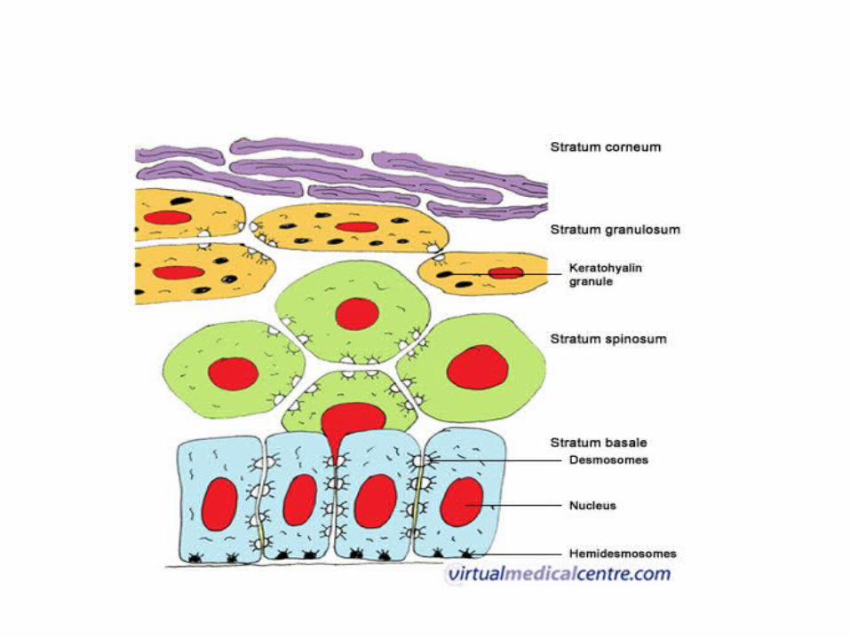
Layers of keratinized epithelium
• Stratum Basale Columnar or cuboidal cells• Stratum Spinosum or prickle cell layer elliptical or spherical cells • Stratum Granulosam granular cells containg keratinohyaline granules• Stratum Cornium Flat cells or squames


• Ortho keratinization and• Parakeratinization

Keratinized epithelium
• Contains Keratinohyalin granules • Rapid desequamation of the epithelial cells
within hours • 20 layers of squames
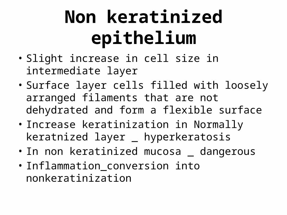
Non keratinized epithelium
• Slight increase in cell size in intermediate layer• Surface layer cells filled with loosely arranged
filaments that are not dehydrated and form a flexible surface
• Increase keratinization in Normally keratnized layer _ hyperkeratosis
• In non keratinized mucosa _ dangerous• Inflammation_conversion into nonkeratinization

Permeability and absorption
• Unlike intestinal mucosa the oral epithelium does not have absorptive capacity
• Floor of the mouth is the thinnest so medications are given sublingually
Non keratinocytes
Non keratinocytes
• Melanocytes ( lacks desmosomes) endogenous_ melanocytes in the basal layer
produce melanin as melanosomes and inject it into keratinocytes through their dendritic processes
• freckle_ increase in melanin production• nevus_ increase melanocyte proiferation• Melanophages in c.tissue

• Exogenous pigmentation lead and bismith poisoning leads to Burton’s line

Langerhans cells
• Move in and out of epithelium and their source is bone marrow
• Recognizing and processing antigens and presenting it to T lymphocytes
• High level clear cells because of presence in the subrabasal layers
• Lacks desmosomal junctions

Merkel’s cells
• Contains desmosomes• Situated in basal layer of epithelium• Synapse like junction between Merkel’s cell
and nerve fibre , trigger the impulse

Inflamatory cells
• Polymorphonuclear leukocytes • Mast cells • T lymphocytes

Interaction between non-keratinocytes and keratinocytes

Junction of the epithelium and lamina propria
• Folded because they increase the surface area for the metabolic exchange, better attachment and better force distribution


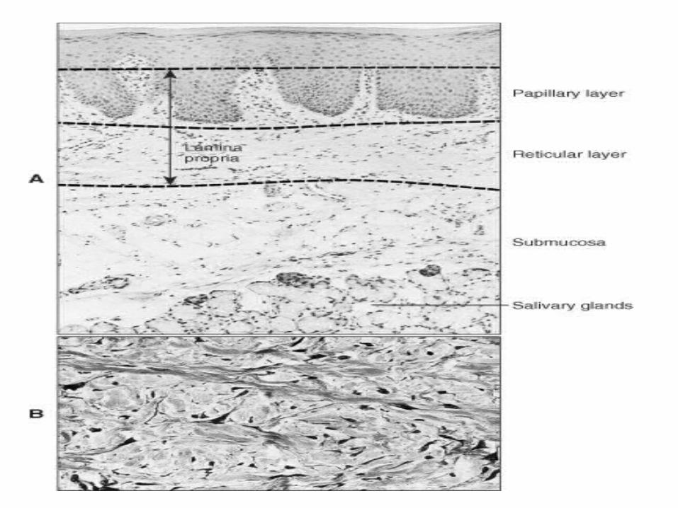
Lamina propria
• The connective tissue supporting the oral epithelium is termed lamina propria
• Superficial papillary layer Associated with the epithelial ridges thin and loosely arranged collagen fibers• Reticular lies between the papillary layer and the
underlying structures

Cell types in lamina propria
• Fibroblasts • Histiocyte • Macrophage • Monocyte • Neutrophil • Lymphocytes • Plasma cell• Endothelial cell

Fibroblasts
• Responsible for elaboration and turnover of both fiber and ground substance
• Contractile and participate in wound contraction
• Gingival overgrowth sometimes seen with drugs like Phenytoin, calcium channel blockers and cyclosporine.
Macrophages ( histiocyte)
• Ingest damaged tissue or foreign material in phagocytic vacuoles that fuse, intracytoplasmically, with the lysosomes and initiate breakdown of these materials.

Mast cells
• Mast cells having granules containing heparin and histamine which play a role in maintaining normal tissue stability and vascular homoestasis
Fibers and ground substance
• Collagen type 1 and type 3 in lamina propria and 4 , 7 in
basal lamina• Elastic fibers • Ground substance Proteoglycans and Glycoproteins

Blood supply
• Derived from the arteries in the submucosa or in deeper part of reticular layer when submucosa is absent.
• Forms an extensive capillaries network in papillary layer, capillary loops into the connective tissue papillae.
• Regional variation in different parts of oral cavity
Nerve supply
• Serves to initiate and maintain a wide variety of voluntary and reflexive activities
• Free nerve endings are found in lamina propria and epithelium associated with merkel’s cells and intraepithelial nerves
• Groups of coiled fibers surrounded by connective tissue capsule , meissner’s, krause’s bulb, ruffini’s corpuscles , mucocutanous end organs.

Types of mucosa
• Lining mucosa noted for its softer surface texture, moist
surface, and ability to stretch and be compressed , acting as a cushion for the underlying structures
• Masticatory mucosa noted for its rubbery surface texture,and
resiliancy.
Regional differences in oral mucosa
• Lining mucosa labial and buccal mucosa- thick non
keratinized epithelium, irregular and blunt CT papillae, some elastic fibers, extensive vascular supply, submucosa contains adipose and minor salivary glands with firm attachment to muscle

• Alveolar mucosa, floor of the mouth and soft palate have thin non keratinized epithelium, submucosa present with minor and major salivary glands. numerous C.T papillae except in alveolar mucosa.
Masticatory mucosa
• Attached gingiva and hard palate thick orthokeratinized epithelium, tall narrow
C.T papillae, submucosa is absent except in the lateral part of the hard palate

Specialized mucosa
• Dorsum of the tongue, covered by a functionally masticatory mucosa and also has a highly extensible lining and has different types of lingual papillae.
• Mechanical as well as a sensory function (taste buds)

Lingual papillae
• Fungiform papillae (fungus like)• Filiform papillae ( hair like)• Foliate papillae ( leaf like)• Circumvallate papillae ( walled)


Taste bud
Age changes in oral mucosa
• Epithelium thinner, smoothening of epithelium C.tissue
interface, flattening of ridges, decrease in filiform papillae, decrease in langerhan’s cells
• Connective tissue decrease cellularity, more collagen, decrease in
minor salivary glands, increase in number of sebaceous glands.
“Caviar” tongue
Related Documents