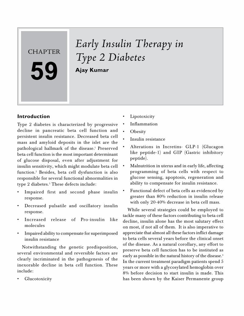
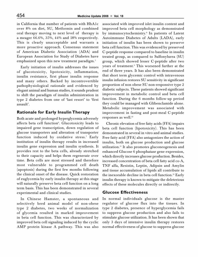
CHAPTER 58 Oral Hypoglycemic Agents : Where Do We Stand Today? V. K. Gujral Introduction Type 2 diabetes has a complex patho physiology. After a long period of controversy and debate regarding the primacy of the contribution of defects in insulin sensitivity versus insulin secretion, beta cell dysfunction is now accepted as the hallmark of type 2 diabetes (Figure 1). While beta cells become poorly responsive to glucose, they remain capable of being stimulated by sulfonylureas and other insulin secretagogues (Figure 2). Type 2 diabetes needs to be handled carefully, aggressively and comprehensively. Primary prevention (to prevent or delay type 2 diabetes), secondary prevention (to prevent, delay or minimise the long term complications) and tertiary prevention (to prevent or limit incapacitation) are well recognized and accepted ways to tackle type 2 diabetes. Primary prevention suffers due to difficulties in early detection of impaired fasting glucose and impaired glucose tolerance, inadequate lifestyle modification, lack of totally safe yet effective drugs for diabetes prevention and ever-elusive consensus regarding use of these medications. The secondary and tertiary prevention must involve tight glycemic control, and control of Figure 2 : Pharmacological Approaches to the Major Metabolic Defects of Type 2 Diabetes Mellitus Figure 1 : Schematic representation of a pancreatic β-cell, showing the pathway of the insulinotropic effect of sulfonylureas. Binding of a sulfonylurea to the sulfonylurea receptor (SUR 1 ) results in closure of the K ATP channels, stimulating the secretion of insulin. Pancreatic β-cell Insulin release Ca 2+ channel Ca 2+ Ca 2+ concentration ↑ channel ATP K K + SUR 1 Sulfonylurea Depolarization Glucose ↓ Insulin Target Tissues Thiazolidinediones (TZDs) Increase Glucose Uptake TZDs ? Biguanides Decrease Hepatic Glucose Production TZDs Decrease Lipolysis Sulfonylureas and Nonsulfonylurea Secretagogues Increase insulin Secretion Pancreatic Beta Cells Decreased Insulin Secretion DEFECTIVE INSULIN SECRETION HYPERGLYCEMIA Glucotoxicity Small Intestine Carbohydrate Absorption α-Glucosidase Inhibitors Delay Intestinal Carbohydrate Absorption INSULIN RESISTANCE Lipotoxicity Lipotoxicity Skeletal Muscle Decreased Glucose Uptake Liver Increased Glucose Production Adipose Tissue Increased Lipolysis Increased Free Fatty Acids

Welcome message from author
This document is posted to help you gain knowledge. Please leave a comment to let me know what you think about it! Share it to your friends and learn new things together.
Transcript

CHAPTER
58oral Hypoglycemic Agents : Where Do We Stand Today?V. K. Gujral
IntroductionType 2 diabetes has a complex patho physiology. After a long period of controversy and debate regarding the primacy of the contribution of defects in insulin sensitivity versus insulin secretion, beta cell dysfunction is now accepted as the hallmark of type 2 diabetes (Figure 1). While beta cells become poorly responsive to glucose, they remain capable of being stimulated by sulfonylureas and other insulin secretagogues (Figure 2).
Type 2 diabetes needs to be handled carefully, aggressively and comprehensively. Primary prevention (to prevent or delay type 2 diabetes), secondary prevention (to prevent, delay or minimise the long term complications) and tertiary prevention (to prevent or limit incapacitation) are well recognized and accepted ways to tackle type 2 diabetes. Primary prevention suffers due to difficulties in early detection of impaired fasting glucose and impaired glucose tolerance, inadequate lifestyle modification, lack of totally safe yet effective drugs for diabetes prevention and ever-elusive consensus regarding use of these medications. The secondary and tertiary prevention must involve tight glycemic control, and control of
Figure 2 : Pharmacological Approaches to the Major Metabolic Defects of Type 2 Diabetes Mellitus
Figure 1 : Schematic representation of a pancreatic β-cell, showing the pathway of the insulinotropic effect of sulfonylureas. Binding of a sulfonylurea to the sulfonylurea receptor (SUR
1) results in closure of the
KATP
channels, stimulating the secretion of insulin.
Pancreatic β-cell
Insulin release
Ca2+ channel
Ca2+Ca2+
concentration ↑
channelATPK
K+
SUR1
Sulfonylurea
DepolarizationGlucose ↓
Insulin Target Tissues
Thiazolidinediones(TZDs)
Increase GlucoseUptake
TZDs?
BiguanidesDecrease
Hepatic GlucoseProduction
TZDsDecreaseLipolysis
Sulfonylureasand
NonsulfonylureaSecretagogues
Increase insulinSecretion
Pancreatic Beta CellsDecreased Insulin
Secretion
DEFECTIVE INSULINSECRETION
HYPERGLYCEMIA
Glucotoxicity
Small IntestineCarbohydrateAbsorption
α-GlucosidaseInhibitors
Delay IntestinalCarbohydrateAbsorption
INSULIN RESISTANCE
Lipotoxicity Lipotoxicity
Skeletal MuscleDecreased
Glucose Uptake
LiverIncreased Glucose
Production
Adipose TissueIncreased Lipolysis
IncreasedFree Fatty
Acids

442 Medicine Update 2008 Vol. 18
other factors, hence done only as per physician’s ability and patient’s compliance.
Difficulties in controlling T2DM Challenges
• Latediagnosisandinitiationoftherapy
• Therapeuticinertia
• Lackofeffectivelifestyleintervention
• Secondaryfailure
• Adverse events associated with antihyperglycemictherapies
• Complexityofcare
• Roleofpostprandialglucoseinfailure
What recent Literature says?“Increased mortality is evident at OGTT levels approximately 90 mg% which is well below current definitions of type 2 diabetes.”
“Therapy targeted at PPG has been shown to improve glucose control & to reduce progression of atherosclerosis and CV events; therefore,physicians should consider monitoring and targeting Post Prandial plasma Glucose as well as HbA1c and Fasting Plasma Glucose 1.”
In a large trial conducted in Europe (Italy) with 500 diabetes clinics
Post Prandial Glucose value > 160 mg% was recorded at least once in 84% patients and 81% had at least one post meal glucose excursion value > 40 mg%Concludingthathighpostprandialglucoseisvery frequent phenomenon in patients with type2 diabetes & can occur even when metabolic control is apparently good.
Currently there are wide range of oralhypoglycemic agents available according to their mode of action (Table 1).
Sulfonylureas
Sulfonylureas stimulate insulin secretion by interacting with the potassium ATP channels of beta cell. These drugs are most effective individuals with relatively recent onset (< 5 years) of type 2 diabetes hence have residual endogenous insulin production. They reduce both fasting and post prandial blood glucose and should be initiated in a smaller
dose and increased at one to two week interval according to self monitoring of blood glucose values. Sulfonylureas tend to cause weight gain and can cause severe & prolonged hypoglycemia. Most sulfonylureas are metabolized in liver and converted into compounds that are then cleared by kidney. Thus their use in patients with significant hepatic or renal insufficiency is not advisable.
The concerns regarding increased cardiovascular risk are still debatable and may be different with Glibenclamide, Glipizide, Gliclazide and Glimepiride .
Concern 1: Supposed Atherogenicity of Insulin
In vitro, insulin has been shown to have several potentially pro-atherogenic effects, including stimulation of cellular cholesterol accumulation and stimulation of vascular smooth muscle cell
proliferation. In vivo, hyperinsulinemia is associated with increased VLDL cholesterol levels, decreased HDL cholesterol levels, decreased LDL cholesterol particle size (so-called “small, dense LDL”), and hypertension. Insulin can also stimulate arterial
smooth muscle cell proliferation. However, recent clinical trials suggest that raising circulating insulin levels with either sulfonylureas or intensive insulin therapy actually decreases, rather than increases, cardiovascular risk in patients with Type2 DM.
Concern 2: Insulin Secretagogues May Have Unwanted Cardiovascular Effects
Insulin secretagogues, including glucose, sulfonylureas, and meglitinides, stimulate insulin secretion by elevating the intracellular ratio of adenosine tri phosphate (ATP) to adenosine di phosphate (ADP) in the pancreatic ß-cell. This causes closure of ATP-sensitive potassium (K
ATP) channels,
which results in membrane depolarization and influxofcalcium(Ca2+) into the ß-cell. This increase inintracellularCa2+ causes release of insulin from ß-cell secretory granules. In cardiomyocytes, it has
been shown that KATP
channels mediate ischemic preconditioning. Ischemic preconditioning is the condition in which exposure of cardiomyocytes to episodes of ischemia induces cellular adaptations

443oral Hypoglycemic Agents : Where Do We Stand Today?
Table 1 : Oral anti hyperglycemic agents
Mode of action Anticipated reduction in HbA1c %
Advantages Disadvantages / Contraindications
(C/I )
Sulfonylureas
Glibenclamide
Stimulate Insulin Secretion
1 – 2% Lowers F & Post Meal Glucose
Weight gain , Hypo.
?Cardiovascular
C/I:Liver&RenalDisease,Ac.Coronary
Glipizide Same Same Same same
Glimepiride , Gliclazide
Stimulate Insulin Secretion through different receptors
1 – 2% Effective Glucose lowering , Safer in Ischemic Heart,
Hypoglycemia, less than other SUs.
C/I:Liver&RenalDisease
Non Sulfonylurea secretagogues
Repaglinide
Nateglinide
Glucose dependent insulin secretion ,
1 – 2% Short acting ,
Effective post prandial control,
Least likely to cause Hypo. Safe for cardiovascular & in renal insufficiency(repaglinide)
C/I:LiverDisease(repaglinide)
Renal Disease(Nateglinide)
Bigunides
Metformin
Reduced hepatic glucose production, Increased glucose utilization, Reduced insulin resistance
1 – 2% No Hypo.
Weight Reduction
Improved Lipids
Nausea, Diarrhea,? Lactic acidosis
C/I:Serumcreatinine>1.5(men), > 1.4(women),
Radiographic contrast studies, seriously ill patient, Hypoxia,
acidosis
Alfa Glucosidase inhibitors
Acarbose
Reduced glucose absorption
0.5–1.0% No Hypo. GI. Flatulence, raised transaminases
C/I:Renal/LiverDisease
Miglitol Same as acarbose - -
Voglibose Same as acarbose - - Much less Flatulence
C/I:Renal/LiverDisease
Thiozolidinediones
Rosiglitazone
Pioglitazone
Reduced Insulin Resistance
Increased Glucose utilization
1 – 2% Reduced SU and Insulin requirement
Reduced Triglycerides
Heart Failure
Edema
Anemia
Higher risk of Fractures
C/I:SignificantLVdysfunction, Liver disease, Anemia
444 Medicine Update 2008 Vol. 18
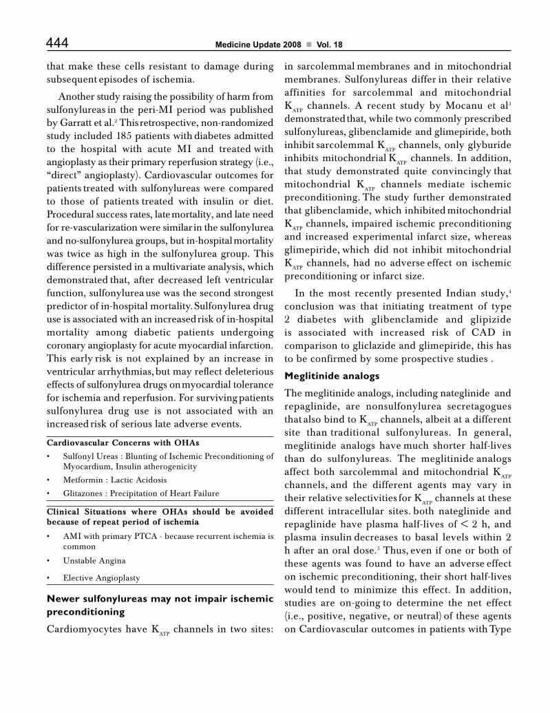
that make these cells resistant to damage during subsequent episodes of ischemia.
Another study raising the possibility of harm from sulfonylureas in the peri-MI period was published by Garratt et al.2 This retrospective, non-randomized study included 185 patients with diabetes admitted to the hospital with acute MI and treated with angioplasty as their primary reperfusion strategy (i.e.,
“direct”angioplasty).Cardiovascularoutcomesforpatients treated with sulfonylureas were compared to those of patients treated with insulin or diet. Procedural success rates, late mortality, and late need for re-vascularization were similar in the sulfonylurea and no-sulfonylurea groups, but in-hospital mortality was twice as high in the sulfonylurea group. This
difference persisted in a multivariate analysis, which demonstrated that, after decreased left ventricular function, sulfonylurea use was the second strongest predictor of in-hospital mortality. Sulfonylurea drug use is associated with an increased risk of in-hospital mortality among diabetic patients undergoing
coronary angioplasty for acute myocardial infarction. This early risk is not explained by an increase in ventricular arrhythmias, but may reflect deleterious effects of sulfonylurea drugs on myocardial tolerance for ischemia and reperfusion. For surviving patients sulfonylurea drug use is not associated with an increased risk of serious late adverse events.
Cardiovascular Concerns with OHAs
• Sulfonyl Ureas : Blunting of Ischemic Preconditioning of Myocardium, Insulin atherogenicity
• Metformin:LacticAcidosis
• Glitazones:PrecipitationofHeartFailure
Clinical Situations where OHAs should be avoided because of repeat period of ischemia
• AMIwithprimaryPTCA-becauserecurrentischemiaiscommon
• UnstableAngina
• ElectiveAngioplasty
Newer sulfonylureas may not impair ischemic preconditioning
CardiomyocyteshaveKATP
channels in two sites:
in sarcolemmal membranes and in mitochondrial membranes. Sulfonylureas differ in their relative affinities for sarcolemmal and mitochondrial
KATP
channels. A recent study by Mocanu et al3
demonstrated that, while two commonly prescribed sulfonylureas, glibenclamide and glimepiride, both inhibit sarcolemmal K
ATP channels, only glyburide
inhibits mitochondrial KATP
channels. In addition, that study demonstrated quite convincingly that mitochondrial K
ATP channels mediate ischemic
preconditioning. The study further demonstrated that glibenclamide, which inhibited mitochondrial K
ATP channels, impaired ischemic preconditioning
and increased experimental infarct size, whereas glimepiride, which did not inhibit mitochondrial K
ATP channels, had no adverse effect on ischemic
preconditioning or infarct size.
In the most recently presented Indian study,4 conclusion was that initiating treatment of type 2 diabetes with glibenclamide and glipizide is associated with increased risk of CAD incomparison to gliclazide and glimepiride, this has to be confirmed by some prospective studies .
Meglitinide analogs
The meglitinide analogs, including nateglinide and
repaglinide, are nonsulfonylurea secretagogues that also bind to K
ATP channels, albeit at a different
site than traditional sulfonylureas. In general, meglitinide analogs have much shorter half-lives than do sulfonylureas. The meglitinide analogs affect both sarcolemmal and mitochondrial K
ATP
channels, and the different agents may vary in their relative selectivities for K
ATP channels at these
different intracellular sites. both nateglinide and
repaglinide have plasma half-lives of < 2 h, and plasma insulin decreases to basal levels within 2 h after an oral dose.5 Thus, even if one or both of these agents was found to have an adverse effect on ischemic preconditioning, their short half-lives would tend to minimize this effect. In addition, studies are on-going to determine the net effect (i.e., positive, negative, or neutral) of these agents onCardiovascularoutcomesinpatientswith Type

445oral Hypoglycemic Agents : Where Do We Stand Today?
2 diabetes. NON SU Secretagogues (Repaglinide, Nateglinide)are safer, there is very little if at all, effect on Ischemic Preconditioning because of shorter duration of action & more selective action onBetacell&betterControlofPPHyperglycemiahencefavorableforCADprevention.
Repaglinide and Nateglinide – Amino acid derivatives, stimulate pancreas & reduce blood glucose levels by increasing first phase insulin secretion. These rapid acting secretagogues are specially used as prandial insulin releasers. Extent of insulin release is glucose dependent and diminishes at low glucose level .They restore first phase insulin secretion which is diminished in early in natural history of type 2 diabetes. They also improve beta cell function – possibly delay occurrence of diabetes in IGT patients. For Best Results, glitinides should be given 30 minutes prior to meal.
Impact of Therapies on A1C LevelsTherapy A1C ReductionDiet and Exercise 0.5 - 2.0%Sulfonylureas and Glitinides 1.0 - 2.0%Metformin 1.0 - 2.0%α-Glycosidase Inhibitors 0.5 - 1.0 %Thiazolidinedione 0.5- 1.0%Insulin >5.0% Nathan, D. Oct 2002. N Engl J Med, Vol. 347, No.17
Glitinides –Place in Therapy• Monotherapy - in patients with type 2 diabetes whose
hyperglycemia is not adequately controlled by diet and exercise.
• Inearlycasesoftype2diabeteswhoarenotadequatelycontrolled by Metformin.
• Type2diabetespatientnotonotherSulfonylureas.
Alfa Glucosidase Inhibitors
It is the sole drug class not targeted at a specific pathophysiological defect of type-2 DM. Acarbose by inhibiting the enzyme alfa glucosidase, delays intestinal absorption of carbohydrates. It specifically blunts post prandial hyperglycemia which has been directly linked with cardiovascular mortality. It decreases fibrinogen levels, prevents platelet activation, reduces vascular inflammation and improves endothelial function. Acarbose has been shown to decrease the macrovascular events in patients with impaired glucose tolerance (IGT). In the Stop NIDDM trial, 1368 patients with IGT were randomized to receive acarbose or placebo. At the end of 3½ years, there was a significant relative risk reduction in the development of cardiovascular events (particularly myocardial infarction) in the acarbose group (HR 0.09; 95% CI,0.01 - 0.072;p<0.02)However there isnolong term data on cardiovascular safety in patients with type-2 DM. The favorable and adverse cardio vascular effects of oral hypoglycemic agents are shown in Table 2.
STOP-NIDDM TRIAL Revealed – Acarbose, an alfa glucosidase inhibitor improved post prandial hyperglycemia and subsequently reduced risk of development of type 2 diabetes. Significantly reduces BMI, waist circumference over 3 years. ReducesincidenceofCardioVasculardiseasesandnewly diagnosed hypertensions in subjects with IGT.7
Table 2 : Effect of Acarbose on the Development of Cardiovascular Disease 6
Acarbose(n=682) Placebo(n=686) Hazard Ratio p-value
CoronaryArteryDisease:M.I. 1 12 0.09 .02
Angina 5 12 0.45 .13
Revascularisation procedures 11 20 0.61 .18
CardiovascularDeath 01 02 0.55 .63
CHF 0 02 - -
CVA 02 04 0.56 .51
Peripheral Vasc. Disease 01 01 1.14 .93
AnyCardiovascularEvent 15 32 0.51 .03

446 Medicine Update 2008 Vol. 18
Inhibition of postprandial hyperglycemia by Acarbose is a promising therapeutic strategy for the treatment of patients with metabolic syndrome.
Acarbose, Voglibose
• FavorableforCADpreventionduetogoodeffectonPPHyperglycemia
• Decreases Fibrinogen , Platelet activation & VascularInflammation
• CardioProtectiveinIGTcases
Metformin
It reduces hepatic glucose production through an undefined mechanism and improves peripheral glucose utilization. Metformin reduces fasting and post prandial blood glucose as well as serum insulin levels, improves lipid profile and induces modest weight loss. The initial dose of 500 mg twice a day can be increased to 1000 mg twice a day, after a period of 2-3 weeks. The only major toxicity, the lactic acidosis, can be avoided by careful patient selection. Metformin should not be used in presence of significant renal insufficiency (Serum creatinine more than 1.5 mg% in men & 1.4 mg% in women.). Metformin is also contraindicated in any form of acidosis, congestive heart failure, liver disease and severe hypoxia. Metformin should be discontinued in all patients who are seriously ill, can not take orally, and in those receiving radiographic contrast material. The gastrointestinal side effects can be minimized by gradual increment in doses.
Metformin is usually avoided in heart failure because of fear of aggravating tissue hypoxia and
acidosis but there is a scarcity of data supporting this. On the contrary the real world scenario is quite different. In a retrospective cohort study48 of 15,000 patients with diabetes and heart failure, they found that those patients who were on metformin had a significant reduction in mortality than those who were not on any insulin sensitizer (24% vs. 36%, p < 0.001). Not only mortality, there was a significant reduction in readmission for all cause and congestive heart failure. Similarly in an another recent study, 64 metformin was found to improve survival and clinical outcomes in patients with diabetes and heart failure. Till date, the evidence suggests that metformin can be safely used in diabetic patient with stable heart failure.8
Metformin and Risk of Lactic Acidosis
Lactic Acidosis risk was 8.1 per 100,000 patient years inspite of being used in 44% patients with Renal Insufficiency & 96% patients having one or the other contraindication of Metformin(Meta analysis of 194 prospective trials).9
Pathogenesis of edema with Glitazone use
It is still not fully understood but is multi factorial.The increase in plasma volume may result from a reduction in renal excretion of sodium and an increase in sodium and free water retention. Glitazones may act synergistically with insulin to arterial vasodilatation resulting in sodium reabsorption with a subsequent increase in extracellular volume & hence pedal oedema.12
Pathogenesis of Congestive Heart Failure with Glitazone use
An increase in plasma volume either alone or in combination with preexisting systolic or diastolic heart dysfunction seems most plausible mechanism.13
Clinical Practice Experience with Glitazones
Various studies have shown relationship of ThiozolidinedioneswithCHFrisk.14 Delea and co workers,15 in a retrospective observational study of health insurance claims, determined the risk of
Table 3 : Benefits of metforminLipids Reduction in triglycerides, total
cholesterol and LDL cholesterol and increase in HDL cholesterol
Weight Weight loss
Hypoglycemia Low incidence of hypoglycemia
Blood Pressure Lowering of blood pressure
Atherosclerotic Increased fibrinolytic activity (reduced PAI-1 level), reduced platelet aggregation, reduced fibrinogen level
Endothelial Function
Improvement in vascular relaxation

447oral Hypoglycemic Agents : Where Do We Stand Today?
Table 4 : Favorable and adverse cardiovascular effects of various oral hypoglycemic agents.10 , 11
Oral Hypoglycemic Agents Favorable Cardiovascular Effect Adverse Cardiovascular Effects
SUs - Impairment of ischemic preconditioning (IP)
Metformin Decreases macrovascular events Risk of lactic acidosis
Preserves beta cell function
Prevents new onset diabetes
Thiazolidinediones Decreases macrovascular events Precipitate heart failure, edema and weight gain
Glucosidase Inhibitors Blunts post prandial hyperglycemia No long term data on cardiovascular safety
Decreases MI in patients with impaired glucose tolerance
Non SU Benzoic Acid Secretagogues Blunts post prandial hyperglycemia No long term data on cardiovascular safety
No effect on IP
heart failure among diabetic patients prescribed TZDs over 5 years, with a mean follow up period of 8.5 months. The risk of heart failure was 4.5% in the group treated with TZDs , and 2.6% in those not treated with them. This increased risk (hazard ratio 0f 1.6, p < 0.001 ) persisted after adjustment for potential confounders, including age, history of complications of diabetes, risk factors for CHF and use of other anti hyperglycemic medicines.
The PRO active (PROspective pioglitazone Clinical Trial)
The PRO active (PROspective pioglitazone Clinical Trial)16,17 demonstrated no significant effects of pioglitazone compared with placebo on the primary Cardio Vascular Disease outcome (composite of all cause mortality, non fatal and silent myocardial infarction, stroke, major leg amputations, acute coronary syndrome, coronary artery bypass surgery or percutaneous coronary interventions) after 3 years of follow up, but a 16% reduction in death, myocardial infarction, and stroke, a secondary end point, was reported with marginal statistical significance. But the occurrence of heart failure was much more with pioglitazone than with placebo. There were 115 more heart failure endpoints and 221 more edema in the pioglitazone group (p value significant for both). The heart failure induced
by glitazones is not malignant as other types of heart failure because heart failure with glitazone is not caused because of any adverse change in myocardial structure or function but is due to their tendency to cause fluid overload and edema and it resolves with discontinuation of therapy and use of diuretics.
The American Heart Association and American Diabetes Association46 have made a consensus statement regarding TZDs use in patients with heart failure.
The DREAM Trial18 found that 8 mg daily of Rosiglitazone given to 2365 non diabetic persons resulted in 306 cases of diabetes or death (11.6%) compared with 686 cases(26.0%) out of 2634 persons given a placebo, a difference that was statistically significant at p < .0001. In terms of reaching normal glycemic levels, 1330 persons (50.5%) in the Rosiglitazone group and 798 (30.3%) in placebo group achieved normal glycemic goals (p < .0001). Subjects in the trial were followed for a median of 3 years. The two groups were similar in terms of their rates of cardiovascular events. But the incidence of congestive heart failure (CHF) was much greater in Rosiglitazone treated persons (0.5% vs 0.1%), p = .01. The relative risk reduction for diabetes & cardiovascular disease with Rosiglitazone was same

448 Medicine Update 2008 Vol. 18
as obtained by lifestyle interventions. The risk is of course likely to return to untreated levels after the drug is withdrawn, an observation that is not observed with lifestyle interventions.19 Moreover the greater benefits in higher risk individuals would have to be balanced against the increased risk of heart failure.20
The ADOPT Trial,21 a five year follow up, showed 14.1% incidence of edema with Rosiglitazone. Recent further analysis revealed lower rate of fractures reported as adverse effects in women taking glibenclamide or metformin versus rosiglitazone (3.5%, 5.1% & 9.3% respectively).
Scattered reports indicate that TZDs induced pulmonary edema may occur even with normal left ventricular systolic function.22
The management of patients with AHA stage A heart failure; ( presence of asymptomatic LV diastolic dysfunction with normal LV systolic function); includes avoiding medications that are capable of aggravating the heart failure. Since majority of long standing type 2 diabetes patients have LV Diastolic dysfunction, they may run the risk of onset or aggravationofCHFwithThiozolidinediones.
The American Heart Association guidelines for TZDs use
• GlitazonesshouldnotbeusedinpatientswithCHF(NYHAIII&IV)
• CautioususeoflowdoseswithCHF(NYHAI & II)
• InpatientswithLVEF<40%butno signs&symptoms of CHF, TZDs should be used inlower doses
On July 30, 2007, the Endocrine and Metabolic Drugs Advisory Committee and the Drug SafetyandRiskManagementAdvisoryCommittee of the Food and Drug Administration (FDA) convened to discuss the myocardial ischemic risk associated with rosiglitazone treatment in patients with type 2 diabetes mellitus, as a follow up of the meta analysis published by Nissen et al.23Committeeconcluded
that the use of rosiglitazone for the treatment of type 2 diabetes was associated with a greater
risk of myocardial ischemic events than placebo, metformin, or sulfonylureas. That conclusion was based primarily on three independently conducted
meta-analyses demonstrating an increase in the relative risk of myocardial infarction, angina, or sudden death among patients taking rosiglitazone
RECORD24 (Rosiglitazone Evaluated for Cardiovascular Outcomes)
An interim findings from this ongoing study were
inconclusive regarding the effect of rosiglitazone on the overall risk of hospitalization or death from cardiovascular causes. There was no evidence of any increase in death from either cardiovascular
causes or all causes. Rosiglitazone was associated with an increased risk of heart failure. The data were insufficient to determine whether the drug was associated with an increase in the risk of myocardial infarction.
Combination Therapy with Glucose Lowering AgentsA number of combination therapies are successful in type2 diabetes and the doses used are same as usedinmonotherapy.Commonlyusedcombinationregimens are
• Insulin secretagogue with metformin orglitazones
• Sulfonylureawithalfaglucosidaseinhibitor
• Sulfonylureawithmetforminandglitazones
• Insulinwithmetforminorglitazone
In a recent study,25 of the 4282 patients who met the criteria, 1050 (25%) received one oral agent, 486 (11%) received two oral agents 56 (1%) got three or more oral agents, 84(2%) received insulin exclusively within 90 days after index date. Among the 1075 patients receiving oral agents, 39% had optimal glycemic control. Optimum control was most frequent among those receiving 1 oral agent (47%), and least among those on 3 or more agents (13%). The conclusion was that the vast majority of

449oral Hypoglycemic Agents : Where Do We Stand Today?
patients treated with multiple oral antihyperglycemic drugs had suboptimal glycemic control, suggesting a need for intensified efforts to treat these patients, preferably by Insulin.
Need for Combination Therapy
Because Type 2 DM is a Multifactorial Complex Cardiovascular - Dysmetabolic- Disorder, The Therapy Must Include the Remedy for each Malady, hence the combination of LIFESTYLE MODIFICATION, Oral Antidiabetic Agents, Insulin, Anti platelet Agents, Lipid Regulators, Antihypertensive Agents.
Combination Therapy in Type 2 Diabetes Decision Considerations
HbA1c efficacy
Reductions from baseline
Reaching target
Synergy of mechanisms of action
Side effects and toxicity profile
Frequency and severity of hypoglycemia
Effect on weight gain
Avoiding polypharmacy and complex regimens
Compliance and convenience
Cost
Advantages of Combination Therapy
Toxicity and side effects of individual drug is reduced.
Drug acting at different level provides better glucose control. E.g. Sulfonylurea acts on Beta cells
and Metformin acts on peripheral tissue at muscle level. One is insulin secretogogue and metformin breaks the insulin resistance at the peripheral tissue.
OHA may increase the receptors sensitivity to insulin if patient on insulin has insulin resistance before changing the insulin, one can try Metformin.
Contraindications for Combination Therapy
• If patient has complicated diabetes withNephropathy & Retinopathy - ideal treatment is to give small dose of insulin, multiple dose. Combinationtherapyisavoided
• DMwithSepsis
• DMwithTissueHypoxicstatesandSystemicBP < 90 mmHg - Risk of lactic acidosis increased.
• Type1DM
• Ifbothtype1ortype2DMareinD.K.A.
• DMwithPregnancy
• AutoImmuneDiabetes
Benefits of treating Post Prandial Hyperglycemia
• Reduction of cardiovascular disease &mortality
• Decreased risk of neonatal complications inGDM.
• Inadvanced,highriskcasesconrollingppbgalone may not be enough & controlling PPTG, LDL, HTN, smoking cessation & homocystein lowering may also be combined
How to Control Post Prandial Hyperglycemia• Smallmultiplemeals
• Complex carbs. Low fat. Adequate mufa &fiber
• Repaglinide, nateglenide, alfa glucosidaseinhibitors, metformin
Diagram 1 : The Management of Hyperglycemia in Cardiac Patients
Before Acute Cardiac Event
Tight glycemic control (6.5 – 6.9% HbA1c) SU/+metformin/+TZDs/+ insulin; tight hypertension control; aggressive lipid statin, fibrates; niacin; anti platelet; ??Antioxidants
During Acute Cardiac Event
Glucose insulin infusion in AMI; avoid metformin, SUs.duringMI,PTCA,CABG;statin/fibratesto all
After Acute Cardiac Event
Insulintherapyatleast3-6monthsfollowingPTCA/CABG.Tightglycemiccontrol.avoidglitazonesinCHFclassIIandmore, continue statin, antiplatelet
450 Medicine Update 2008 Vol. 18
• Regularhumaninsulin/analogs
• Regularmonitoringtokeepmeetingtargets: ppbg < 140 mg%, fbg < 110 mg%, hb a1c < 6.5%
Emerging Oral Antihyperglycemic TherapiesThe DPP 4 Inhibitors
In an attempt to expand the focus of therapeutics in type 2 diabetes, attention is being paid to Glucagon, Glucagon like peptide-1( GLP-1). The Dipeptidyl Peptidase 4 is an enzyme that breaks down the GLP-1. By delaying the breakdown of GLP-1, the drug is able to extend the action of insulin, while also suppressing the release of glucagon, leading to reduction in hyperglycemia. In a recent study,26 Hba1c decreased progressively from baseline to week 12 and then remained stable until the 24 week trial ended (Vildagliptin vs. placebo). The post prandial blood glucose also showed significant improvement. DPP4 inhibitor Vildagliptin was
found to be weight neutral, Sitagliptin is another such drug that has shown similar results.27
Rimonabant
This drug initially and essentially developed as anti obesity agent that works through modulation of Endo Cannabinoid Receptors, has shown afavourable effect on blood glucose disproportionate to its weight reducing effect. It is yet to be cleared by US FDA.
Future Oral TherapiesOral Insulin
While insulin replacement/supplement is the traditional way to treat diabetes by controlling fluctuations in blood sugar levels, injected insulin has drawbacks. It does not mimic normal delivery into the portal circulation. In a healthy person, the liver has a 2-4 fold higher level of insulin compared to the periphery (i.e. the arms and legs). In people with diabetes, this concentration difference is impossible to mimic
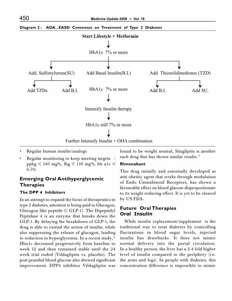
Diagram 2 : ADA , EASD Consensus on Treatment of Type 2 Diabetes
Start Lifestyle + Metformin
HbA1c 7% or more
Add. Sulfonylureas(SU) Add Basal Insulin(B.I.) Add Thiozolidinediones (TZD)
HbA1c 7% or moreAdd TZDs
Intensify Insulin therapy
Add B.I. Add SU.Add B.I.
HbA1c still 7% or more
Further Intensify Insulin + OHA combination

451oral Hypoglycemic Agents : Where Do We Stand Today?
by insulin injection. Instead of high levels of insulin in the liver, the injection delivers a large dose of insulin throughout the body, with no gradation from the periphery to the internal organs. In contrast, oral insulin is believed to be capable of mimicking the natural distribution of insulin throughout body. Oral drug delivery via the gastro-intestinal route delivers the drug almost direct to the liver – and in the case of insulin delivery, this is an advantage that allows for a more effective, efficient and safe method of treatment. Insulin microspheres for oral delivery are a reality.28
Oral Insulin Spray 29(Oral-lyn) for Type 1 and 2 Diabetes is approved for use in Ecuador. On May 3, the Ecuadorian Ministry of Public Health approved the first oral spray formulation of insulin (Oral-lyn,madebyGenerexBiotechnologyCorp.) forthe treatment of type 1 and 2 diabetes mellitus. The approval is expected to pave the way for similar approvals worldwide.
A comparison of the daily nine-point glucose profile of each patient between the Oral-lyn(TM) regimen and injected regular human insulin indicated that Oral-lyn(TM), administered as a pre-meal insulin in a divided dose schedule, produced glucodynamic profiles comparable to that produced by injected regular human insulin. Fructosamine levels displayed a tendency to lower values with the Oral-lyn(TM) therapy at the end of the trial indicating the better control attained during the study period.
The Thiruvananthapuram-based Sree ChitraTirunal Institute of Medical Sciences and Technology has successfully demonstrated the possibility of developing an oral insulin preparation in experiments on mice. It is now getting ready to conduct clinical trials with human subjects to establish its efficacy and viability. The Centerfor Bio-Medical Engineering (CBME) of IndianInstitute of Technology, Delhi (IIT-D) has recently found positive results in a research that aims to develop an ‘oral insulin delivery system’.
IN-105 is a novel analog of insulin, proprietary to Biocon. The product has special properties to deliver in tablet form at room temperature Biocon Ltd, India’s leading biotechnology firm, has submitted clinical data of phase-one studies on its oral insulin product to the European Association for Study of Diabetes at Amsterdam.
References 1. Postprandial glucose regulation: New data and new
implications. Clin Ther. 2005;27 Suppl 2:S42-56
2. Garratt KN, Brady PA, Hassinger NL, Grill DE, Terzic A, Holmes DR Jr: Sulfonylurea drugs increase early mortality in patients with diabetes mellitus after direct angioplasty for acute myocardial infarction. J Am Coll Cardiol 33:119–124, 1999
3. Mocanu MM, Maddock HL, Baxter GF, Lawrence CL,Standen NB, Yellon DM: Glimepiride, a novel sulfonylurea, does not abolish myocardial protection afforded by either ischemic preconditioning or diazoxide. Circulation 103:3111–3116, 2001
4. sadikot SM . Risk of coronary artery disease associated with initial sulfonylurea treatment of type 2 diabetes: a matched case – control study , abstract presented at EASD 2007,19th September.
5. Dornhorst A: Insulinotropic meglitinide analogues. Lancet 358:1709–1716, 2001
6. ChaissonJL.Etal,JAMA.2003;290:486-494
7. Hypotheses.2005;65(1):252-4.Epub 2005 Jan 28
8. Eurich DT, Majumdar SR, McAlister FA, et al. Improved ClinicalOutcomesAssociatedwithMetformin inpatientsWith Diabetes and Heart Failure. Diabetes Care 2005; 28: 2345–51
9. Misbin RI, et al. The Phantom of Lactic Acidosis due to Metformin in Patients with Diabetes, Diabetes Care 2004; 27(7): 1791 – 93.
10. Bonnie Kimmel, Silvio E. Inzucchi SE. Oral agents for type-2 diabetes, an update. Clinical Diabetes 2005; 23: 64–76.
11. Mark A Deeg. Basic approach to managing Hyperglycemia for the Non endocrinologist. Am J Cardiol 2005; 96 (4A): 37E–40E.
12. Niemeyer NV, Janney LM.Thiozolidinedione induced oedema. Pharmacotherapy, 2002 ; 22: 924-929
13. Congestive heart failure and cardiovascular death inpatients with prediabetes and type 2 diabetes given thiazolidinediones: a meta-analysis of randomised clinical trials; The Lancet 2007; 370:1129-1136,DOI:10.1016/S0140-6736(07)61514-1
14. Masoudi FA, Wang Y, Inzucchi SE, et al. Thiazolidinedione, metformin and outcomes in older patients with diabetes and heart failure: An observational study. Circulation 2005; 111: 583–90.
452 Medicine Update 2008 Vol. 18
15. Delea TE , Edelsberg JS , Hagiwara M , et al . Use of Thiozolidinediones and risk of heart failure in people with type 2 diabetes : Diabetes Care 2002 : 25: 2058-2064
16. Dormandy JA, Charmonnel B, Eckland DJA. Secondaryprevention of macrovascular events in patients with type-2 diabetes in the PROactive Study (Prospective Pioglitazones Clinical Trial in Macrovascular Events): a randomizedcontrolled trial. Lancet 2005; 366; 1279–289
17. Hannele YK Jarvinen. PROactive study: some answers, many questions Editorial. Lancet 2005; 366; 1241–242.
18. GersteinHC,YusufS,BoschJ,etal.Effectofrosiglitazoneon the frequency of diabetes in patients with impaired glucose tolerance or impaired fasting glucose: a randomised controlled trial. Lancet 2006;368:1096-1105. [Erratum, Lancet 2006;368:1770.]
19. Montori VM, Isley WL, Guyatt GH. Waking up from the DREAM of preventing diabetes with drugs. BMJ 2007;334:882-884.
20. Nathan DM. Rosiglitazone and cardiotoxicity: weighing the evidence. N Engl J Med 2007;357. DOI: 10.1056/NEJMe078117.
21. Kahn SE et al New England Journal of Medicine 2006, 355.2427-2443
22. Shah M , Kolandaivelu A,Fearon WF,:Pioglitazone induced heart failure despite normal LV function. Am J Med.2004 Dec15 ; 117(12):973-74
23. Nissen SE, Wolski K. Effect of rosiglitazone on the risk of myocardial infarction and death from cardiovascular causes. N Engl J Med 2007;356:2457-2471. [Erratum, N Engl J Med 2007;357:100.]
24. Home PD, Pocock SJ, Beck-Nielsen H, et al. Rosiglitazone Evaluated for Cardiac Outcomes and Regulation ofGlycemia inDiabetes (RECORD)Study: interim findingson cardovascular hospitalizations and deaths. N Engl J Med 2007;357. DOI: 10.1056/NEJMoa073394.
25. CynthiaJWilley,SusanE,CohenJ,FullerC,GurwitzJH,Am J Manage Care.2006; 12: 435-440
26. Pi-Sunyer FX , SchweiserA , Mills D , Dejager S : efficacy and safety of vildagliptin monotherapy in drug naïve patients with type 2 diabetes. Diabetes Res Clin Pract76:132-138, 2007
27. Barnett A. et al : DPP4 inhibitors and their use in management of type 2 diabetes, Int J Clin Pract 2006 Nov; 60(11):1454-1470
28. Jain D, Panda A K, Majumdar D K; Eudragit S100 entrapped insulin microspheres for oral delivery. AAPS Pharm Sci Tech. 2005; 06(01): E100–E107. DOI.
29. Cerena, Simona MD 1; Kidron , Miriam Phd 1 ;Wohlgelernter, Jay MD 1; Modi Pankaj MD ; Raz Itamar MD 1: dose response relationship of oral insulin spray in healthy subjects. Diabetes Care. 28(6):1353-1357, June 2005.
CHAPTER
59Early Insulin Therapy in Type 2 DiabetesAjay Kumar
IntroductionType 2 diabetes is characterized by progressive decline in pancreatic beta cell function and persistent insulin resistance. Decreased beta cell mass and amyloid deposits in the islet are the pathological hallmark of the disease.1 Preserved beta cell function is the most important determinant of glucose disposal, even after adjustment for insulin sensitivity, which might modulate beta cell function.2 Besides, beta cell dysfunction is also responsible for several functional abnormalities in type 2 diabetes.3 These defects include:
• Impaired first and second phase insulinresponse.
• Decreased pulsatile and oscillatory insulinresponse.
• Increased release of Pro-insulin likemolecules
• Impairedabilitytocompensateforsuperimposedinsulin resistance
Notwithstanding the genetic predisposition, several environmental and reversible factors are clearly incriminated in the pathogenesis of the inexorable decline in beta cell function. These include:
• Glucotoxicity
• Lipotoxicity
• Inflammation
• Obesity
• Insulinresistance
• Alterations in Incretins- GLP-1 (Glucagonlike peptide-1) and GIP (Gastric inhibitory peptide).
• Malnutritioninuterusandinearlylife,affectingprogramming of beta cells with respect to glucose sensing, apoptosis, regeneration and ability to compensate for insulin resistance.
• Functionaldefectofbetacellsasevidencedbygreater than 80% reduction in insulin release with only 20-40% decrease in beta cell mass.
While several strategies could be employed to tackle many of these factors contributing to beta cell decline, insulin alone has the most salutary effect on most, if not all of them. It is also imperative to appreciate that almost all these factors inflict damage to beta cells several years before the clinical onset of the disease. As a natural corollary, any effort to preserve beta cell function has to be instituted as early as possible in the natural history of the disease.4 In the current treatment paradigm patients spend 5 years or more with a glycosylated hemoglobin over 8% before decision to start insulin is made. This has been shown by the Kaiser Permanente group
454 Medicine Update 2008 Vol. 18
inCaliforniathatnumberofpatientswithHbA1cover 8% on diet, SU, Metformin and combined oral therapy moving to next level of therapy is a meager 66.6%, 35%, 44% and 18% respectively. This is clearly unacceptable and warrants a more proactive approach. Consensus statementof American Diabetic Association (ADA) and European Association for Study of Diabetes have emphasized upon this new treatment paradigm.5
Early initiation of insulin addresses the issues of glucotoxicity, lipotoxicity, inflammation, insulin resistance, first phase insulin response and many others. Backed by incontrovertible pathophysiological rationale and evidenced by elegant animal and human studies, it sounds prudent to shift the paradigm of insulin administration in type 2 diabetes from one of ‘last resort’ to ‘first assault’.
Rationale for Early Insulin TherapyBoth acute and prolonged hyperglycemia adversely affects beta cell function6. Glucotoxicity leads to impaired gene transcription, down regulation of glucose transporters and alteration of transporter function induced by oxidative stress.7 Early institution of insulin therapy results in increased insulin gene expression and insulin synthesis. It provides rest to the beta cells, already stretched to their capacity and helps them regenerate over time. Beta cells are most stressed and therefore most vulnerable to programmed cell death (apoptosis) during the first few months following the clinical onset of the disease. Quick restoration of euglycemia by early insulin therapy at this stage will naturally preserve beta cell function on a long term basis. This has been demonstrated in several experimental and clinical studies.
In Chinese Hamster, a spontaneous andselectively bred animal model of non-obese type 2 diabetes, two weeks of normalization of glycemia resulted in marked improvement in beta cell function. This was characterized by improved beta cell signaling induced by the cyclic AMP protein kinase A pathway. This was also
associated with improved islet insulin content and improved beta cell morphology as demonstrated by immunocytochemistry.8 In patients of Latent Autoimmune Diabetes of Adults (LADA), early initiation of insulin has been shown to preserve beta cell function. This was evidenced by preserved C-peptideresponsecomparedtobaselineininsulintreated group, as compared to Sulfonylurea (SU) group, which showed lesser C-peptide after twoyears of treatment.9 This worsened further at the end of three years. It has also been demonstrated that short term glycemic control with intravenous insulin infusion restores SU sensitivity in significant proportion of non obese SU non-responsive type 2 diabetic subjects. These patients showed significant improvement in metabolic control and beta cell function. During the 6 months follow-up period they could be managed with Glibenclamide alone. Metabolic improvement was associated with improvement in fastingandpost-mealC-peptideresponses as well.10
Chronicelevationoffreefattyacids(FFA)impairsbeta cell function (lipotoxicity). This has been demonstrated in several in vitro and animal studies. Free fatty acid (FFA) also antagonizes the action of insulin, both on glucose production and glucose utilization.11 It also promotes gluconeogenesis and enhanced Glucose 6 phosphatase gene expression, which directly increases glucose production. Besides, increased concentration of beta cell fatty acid co-A, TNF alfa, Resistin, Leptin, Adipsin and Amylin and tissue accumulation of lipids all contribute to the inexorable decline in beta cell function.12 Early insulin therapy is known to mitigate the deleterious effects of these molecules directly or indirectly.
Glucose EffectivenessIn normal individuals glucose is the master regulator of glucose flux into the tissues. In type 2 diabetes, presence of hyperglycemia fails to suppress glucose production and also fails to stimulate glucose utilization. It has been shown that only 3 days of intensive insulin therapy restores normal effectiveness of glucose to suppress glucose
455Early Insulin Therapy in Type 2 Diabetes
production and stimulate glucose utilization in response to hyperglycemia. During this study it was concluded that the mechanism involved in restoration of glucose effectiveness was improved glycogen synthesis and decreased level of circulating free fatty acids.13
InflammationInflammation has been identified as one of the major determinants of beta cell dysfunction. Several pro-inflammatory transcription factors have been identified which inflict damage to beta cells through liberation of large number of inflammatory cytokines.14 It has now been established that our daily macronutrient intake is largely pro-inflammatory. It leads to oxidative stress, generation of reactive oxygen species (ROS) and expression of pro-inflammatory transcription factor NFkB. Resultant liberation of cytokines like,Intercellularadhesionmolecule-1(ICAM-1),Vascular cell adhesion molecule-1 (VCAM-1),p-selectin and others initiate and perpetuate the inflammation induced damage to beta cells. In the context of macronutrient intake, prompt and adequate insulin response counteracts the expression of NFkB and subsequent inflammatory cascade. This inhibits any inflammation induced damage to beta cells. In this context insulin can be viewed as a natural anti-inflammatory molecule. Elegant studies have demonstrated remarkable reductioninlevelsofNFkB,ICAM-1,P-47,ROSetc by insulin administration.15
First Phase Insulin Response (FPIR)Loss of first phase insulin response has emerged as one of the most important factors in the pathogenesis of type 2 diabetes. Its magnitude correlates with degree of beta cell dysfunction.16 Its consequences include:
• Inadequatesuppressionofendogenousglucoseproduction
• Rise innon-esterified fattyacids (NEFA)dueto inadequate anti lipolytic action of insulin
• Inadequateprimingofinsulinsensitivetissuesleading to decreased utilization of glucose.
• Alteredsignalingcapacityofhormonesleadingto insulin resistance
• Enhanced stimulatoryactionofGlucagononneoglucogenesis
• Enhancedpostprandialhyperglycemia
• Increased risk of micro and macro vascularcomplications
It is also important to understand the correlation of degree of glycemia and loss of first phase insulin response.
• FPIR is mostly absent when fasting plasmaglucose is > 109 mg/dl
• Whenfastingplasmaglucoseismorethan140mg/dl, 75% of beta cell function is lost17
• Whenfastingplasmaglucoseismorethan180mg/dl, there is complete loss of FPIR
• When2hrsPGvaluesaremorethan200mg/dl, there is marked reduction in FPIR18
• Even in subjects with IGT, there is markedreduction in FPIR
Consideringthesefactsitseemsprudentthatallefforts be made to restore the FPIR. This would logically correct or mitigate all the deleterious consequences mentioned above. Additional benefits will include adequate beta cell rest, reduced hyperinsulinemia of the late phase after ingestion of meal, reduced production of islet amyloid peptide and improved insulin secretion overtime. Excessive accumulation of amyloid deposits between islet cells and capillaries lead to destruction of islet endocrine cells and progressive worseningofbetacellfunction.Currentparadigmof using SU in majority of type 2 diabetics for pronged period leads to increased deposition of amyloid and faster decline in beta cell function.19 However insulin – sparing SU, Glimepiride and non-sulfonylurea insulin secretagogues, Repaglinide and Nateglinide may not have this deleterious effect.

456 Medicine Update 2008 Vol. 18
Marked improvement in glucose tolerance by restoration of FPIR by intravenous infusion of insulin during the first 30 minutes of OGTT has been elegantly demonstrated by Bruttomesso et al. It was clearly demonstrated in this study that neither continuous infusion of insulin nor delaying the infusion beyond 30 minutes achieved similar results.20 This implies the importance of timing of insulin administration. Unfortunately intravenous insulin infusion cannot be recommended as a therapeutic option for obvious reasons. However rapid acting insulin analogs, Lispro, Aspart and Glulisine have similar pharmacokinetic profile and can mimic intravenous insulin infusion demonstrating similar benefits. Several studies have demonstrated these effects thus assuring translation of the benefit of restoration of FPIR in clinical practice. As compared to regular insulin rapid acting analogs peak earlier (60 versus 120 minutes) and lead to 46% lower glucose area under the curve. These differences could be attributed to rapid and complete suppression of endogenous glucose production as rates of appearance of ingested glucose remains identical. These studies have shown that restoration of FPIR by intensive insulin treatment leads to improved insulin secretion and long term glycemic control.21 This may pave way to withdrawal of insulin for several years.
Pulmonary delivery of insulin has added another dimension to insulin administration particularly with respect to rapid onset of action. This could have a salutary effect on restoration of FPIR along with ease of administration. Their onset of action is similar to rapid acting analogs while the duration of action is closer to that of regular insulin.22 Thus their therapeutic effect can be positioned somewhere between the rapid acting analogs and regular insulin. Unfortunately the only FDA approved brand Exubera has been withdrawn from the market due to reasons other than safety.
ConclusionInsulin possesses the unique ability to correct majority of the reversible factors contributing to
the inexorable decline in beta cell function in the natural history of type 2 diabetes. Initiation of insulin early after clinical onset of the disease provides adequate rest to beta cells and helps restore FPIR. Appropriate timing of insulin initiation and prudent selection of rapid acting insulin analogs and inhaled insulins are extremely vital in preservation of beta cell function, long term glycemic control and prevention of micro and macro vascular complications.
Increasing availability of Incretin mimetic, GLP-1 analogs and DPP IV inhibitors have added exciting dimensions and require better understanding of positioning these molecules in the treatment paradigm for type 2 diabetes. They increase insulin secretion in a glucose dependent manner. It has been demonstrated that they improve insulin responses during 30 minutes immediately after the ingestion of a standard test meal resulting in 58% decrease in the glucose area under the curve.23 They have several ancillary advantages including weight loss, glucagon suppression and beta cell preservation.24 In patients with adequate beta cell reserve they could be better option than rapid acting insulin analogs. However these conjectures need to be proven by well designed clinical trials. A very recent report issued on behalf of FDA in October 2007 has linked GLP-1 mimetic Exanetide with episodes of pancreatitis. This may be an area of concern.
High economic burden associated with these newer molecules and strategies will need to be addressed by the health care providers across the globe. In the meantime clinicians must shed the inhibition of starting insulin early, when it is most required. In clinical practice insulin initiation when fasting plasma glucose is more than 140 mg/dl is justifiably warranted as 75% of beta cell function is lost at this juncture. Whipping 25% of tired beta cells to achieve normoglycemia by using drugs like sulfonylureas is unphysiological and does not address the underlying pathogenesis.16 Similarly decision to start basal or combined basal prandial insulin regimens can be made on the basis of HbA1c

457Early Insulin Therapy in Type 2 Diabetes
levels between > 7% – < 10% or > 10%.
There are several advantages of using this strategy including quick restoration of normoglycemia, restoration of FPIR, beta cell protection, re-establishment of diet responsiveness etc to name a few. All these benefits accrue with transient intensive treatment and small total daily dose of insulin (0.6 units / kg) which is less than the endogenous insulin production in non diabetics.21 Patient’s inhibitions regarding insulin injections should not deter clinicians from prescribing appropriate drug at the right time for the right indication.
SummaryThe current paradigm of management of type 2diabetes is one of sequential addition of treatment modalities starting from medical nutrition therapy, exercise, single or combination oral hypoglycemic agents (OHAs) and finally insulin administration with or without OHAs. This strategy has miserably failed in achieving recommended glycemic goals to prevent micro vascular as well as macro vascular complications. Besides it does not address the fundamental issues of progressive beta cell dysfunction and several other pathogenetic mechanisms including first phase insulin response, inflammation, glucotoxicity, lipotoxicity, inflammation etc. Patients continue to have glycosylated hemoglobin over 8% for more than 5 years before appropriate treatment decisions are made.
Insulin administration is uniquely suitable to address most of these issues, provided it is started early in the natural history of type 2 diabetes. Short-term intensive insulin administration quickly restores normoglycemia, provides rest to the stressed beta cells, allowing them to regenerate and helps in maintaining long term glycemic control with diet, exercise and insulin sensitizers. It also prevents deposition of islet amyloid which is inherent with sulfonylureas administration, the most prescribed drug in the treatment of type 2 diabetes.
Rapid acting insulin analogs are uniquely positioned to address the issues of first phase insulin response and post prandial hyperglycemia. Nasal insulin, GLP-1 analogs and DPP IV inhibitors are potential agents to compete with these drugs. However all of them have one or the other problems of cost, availability, safety and lack of long term data. Notwithstanding patient’s and clinician’s reluctance to start insulin at the appropriate time, scientific evidence is loaded in favor of using insulin as first assault rather than last resort.
References1. HoppenerJWM,AhrenB,LipsCJM.Isletamyloidandtype
2 diabetes mellitus. N Eng J Med 2000; 343: 411-419.
2. JensenCC,CnopM,HullRLetalfortheAmericanDiabeticAssociation GENNID study group. Diabetes 2002; 51: 2170-2178.
3. Jerich JE, Smith TS. Beta cell defects and pancreatic abnormalities in type 2 diabetes mellitus. In: Pickup JC, Williams G Eds Textbook of diabetes, Third edition,Massachusetts,USABlackwellPublishingCompany2003:23.1-23.11.
4. Elder R, Stern E, Milicevic Z, Raz I. Early use of insulin in type 2 diabetes. Diabetes Res Clin Pract 2005; 68 (Suppl 1): 530-535.
5. Nathan D M, Buse J B, Davidson M B et al. Management of HyperglycemiainType2Diabetes:AConsensusalgorithmfortheinitiationandAdjustmentofTherapy.AConsensusstatement from the American diabetic association and the European association for the study of Diabetes. Diabetes Care 2006; 29: 1963-1972.
6. Meyer J, Sturis J, Katshchinski M et al. Acute hyperglycemia alters the ability of normal beta cell to sense and respond to glucose. American J physiol Endocrinol Metabol 2002; 282: E 917-922.
7. Yki Jarvinon H. Glucose toxicity. Endocrine Rev 1992, 13: 415-431.
8. Kohnert KD, Hehmke B, Kloting I et al. Insulin treatment improvesisletfunctionintype2diabeticChinesehamsters.Exp Clinical Endocrinol Diabetes 2001; 109: 196-202.
9. Kobayash T, Nakanishi K, Murase T et al. Small doses of subcutaneous insulin as a strategy for preventing slowly progressive beta cell failure in islet cell antibody positive patients with clinical features of NIDDM. Diabetes 1996, 45; 622-626.
10. SinagraD,GrecoD,amatoMCetal.A12hourintravenousinsulin infusion restores the beta cell response to sulfonylureas in patients affected by type 2 diabetes. Diabetes Care 2000; 23: 1857-1858.

458 Medicine Update 2008 Vol. 18
11. Poitout V, Roberson R. Mini review: Secondary beta cell failure in type 2 diabetes - a convergence of glucotoxicity and lipotoxicity. Endocrinology 2002; 143: 339-342.
12. Ahima R, Flier J. Adipose tissue as an endocrine organ. Trends Endocrinol Metab 2000; 11: 327-332.
13. Hawkins M, Gabriely I, Wozniac R et al. Glycemic control determines hepatic and peripheral glucose effectiveness in type 2 diabetic subjects. Diabetes 2002; 51: 2179-2189.
14. Dandona P, Alijada A, Mohanty P. The anti-inflammatory and potential anti-atherogenic effects of insulin: a new paradigm. Diabetologia 2002; 45: 924-930.
15. Alijada a, Ghanim H, Dandona P et al. Insulin inhibits NFkB andMCP-1expressioninhumanaorticandendothelialcells.The J of Clinical Endocrinol and Metab 2001; 86(1): 450-453.
16. Seshiah V, Balaji V. Early insulin therapy in type 2 diabetes. Int J of Diabetes. Dev. Countries 2003; 23: 90-93.
17. Porte D Jr. Banting Lecture 1990. Beta cells in type 2 diabetes mellitus. Diabetes 1991 ; 40 : 165-180.
18. Defronzo F. The Triumvirate: Beta cell, muscle and liver; a collusion responsible for NIDDM. Diabetes 1998. 347: 667-687.
19. RachmanJ,LevyJC,BarrowBAetal.ChangesinAmylinand Amylin like peptide concentration and beta cell function
in response to sulfonylurea or insulin therapy in NIDDM. Diabetes Care 1998; 21: 810-816.
20. Bruttomesso D, Pianta A, Mari A et al. Restoration of early rise in plasma insulin levels improves glucose tolerance in type 2 diabetic patients. Diabetes 1999; 48: 99-105.
21. Iikova H, Benjamin G, Ayidin T et al. Induction of long term glycemic control in newly diagnosed type 2 diabetic patients by transient intensive insulin treatment. Diabetes Care 1997; 20: 1353-1356.
22. Priscilla A, Hollander P, Blonde L et al for the Exubera Phase III study Group. Efficacy and safety of Inhaled Insulin (Exubera)ComparedwithSubcutaneousInsulinTherapyinPatients with Type 2 Diabetes. Diabetes Care 2004; 27: 2356-2362.
23. Kjems LL, Holst JJ, Volund A et al. The influence of GLP-1 on glucose stimulated insulin secretion: effects on beta cell sensitivity in type 2 and non-diabetic subjects. Diabetes 2003; 52: 380-386.
24. RatnerRE,DeFronzoR,PratleyRE.ProsandConsofGLPagonistsversusDPPIVinhibitors.Currentissuessymposium.Program and abstracts of ADA, 67th Scientific sessions, June 22-26,2007.Chicago,Illinois.

CHAPTER
60Glucagon Like Peptide 1 – Related AnalogsV. Seshiah, V. Balaji
Insulin release from the beta cells is influenced by nutrients (both carbohydrates and non-carbohydrates), hormonal factors including gut hormones and neural factors. Of all these insulin secretagogues, glucose availability is the major physiological determinant of insulin secretion. The insulin secretory response is greater after oral administration of glucose than after intravenous glucose administration (Fig – 1), an indication that absorption of glucose by way of gastro-intestinal tract stimulates the release of hormones and other mechanism that ultimately enhance the sensitivity of the beta cell to glucose. This phenomenon is known as ‘Incretin effect’ and is facilitated by gut hormones.
Glucagon is secreted from α cells and functions predominantly during the fasting state to maintain
blood glucose levels by the mobilization of glucose from glycogen stores in peripheral tissues such as muscle and liver. Excessive production of glucagon contributes to hyperglycemia and thus approaches that antagonize glucagon action in subjects with diabetes are being actively investigated. Two aspects of the gut have interested the diabetologists over the past 30 years. They are the incretin effect and the occurrence of glucagon-producing L-cells in the gut. The incretin effect is the amplification of nutrient-induced insulin secretion by hormones from the gut particularly GIP (gastric inhibitory peptide) and GLP 1 (glucagon like peptide 1)1 [Fig – 2]. Of these two peptides, GLP 1 is the most potent and efficacious insulinotrophic hormone1. The
Figure 1 : Incretin Effect
Adapted from Joslin’s Textbook of Diabetes Mellitus (14th edition).
Figure 2 : Nutrients and Gut Hormones
460 Medicine Update 2008 Vol. 18
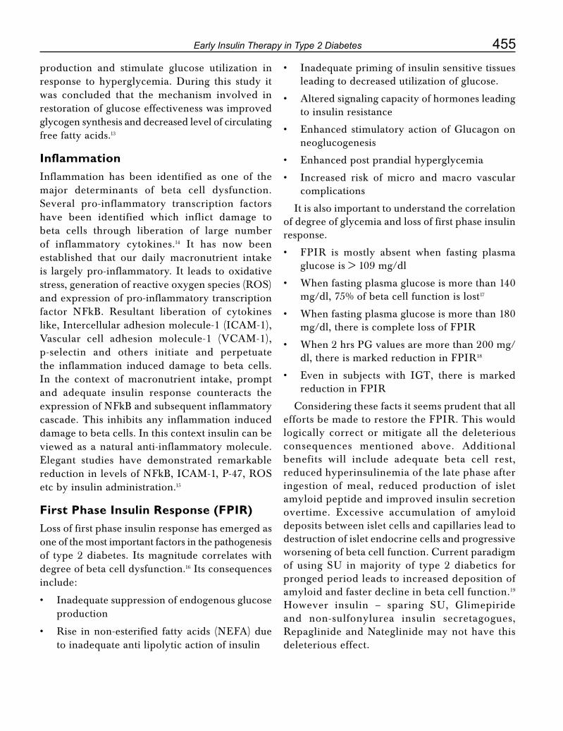
insulinotrophic actions of GLP 1 appear to be due to an increase in the insulinogenic index, such that the same degree of insulin secretion is produced at lower glucose levels. The incretin effect is markedly impaired or absent in patients with type 2 diabetes because of decreased secretion of GLP 1.
The glucagon like peptides are secreted in response to feeding. Glucagon like peptide 1 (GLP 1) and gastric inhibitory polypeptide (GIP) comprise the intestinal ‘incretin’ hormones released from the intestine in response to feeding and augment glucose stimulated insulin secretion from the β cells2, 3. GLP 1 also enhances insulin stimulated glucose uptake in peripheral tissues (muscle, fat, liver), suppresses glucagon secretion, induces satiety, and promotes the growth and differentiation of new b cells in the pancreas. These antidiabetic properties of GLP 1 have prompted considerable interest in the therapeutic potential of GLP 1 for the treatment of diabetes.
The regulation of secretion of GLP-1 from the L-cells in the gut is complex and appears to involve combination of nutrient, hormonal and neural stimuli. There are atleast three potential sites where insulin secretion can be modulated by peptides. Firstly GLP-1 binds to receptors on pancreatic β-cells and thus affects the ion channels that regulate the membrane potential by activation of
adenylate cyclase, thereby stimulating cyclic AMP production and calcium influx. Secondly they may influence the mobilization of intracellular calcium stores notably the endoplasmic reticulum and thus cytosolic calcium concentration. Thirdly they may modify the calcium sensitivity of the contractile protein interactions that lead to the release of insulinsecretorygranules.CyclicAMPandcalciumstimulate rapid release of insulin from the cells and induce transcription of the insulin gene, thereby replenishing insulin stores. GLP-1 also activates receptors in neurons located in the hypothalamus, resulting in a reduction in food intake; thus, GLP-1 has an important role in controlling energy balance. GLP-1 receptors are also located on neurons in brain regions, such as the hippocampus, that are involved in learning and memory.
GLP 1 and diabetes treatment GLP-1 powerfully inhibited glucagon secretion4. Furthermore, GLP-1 not only stimulated glucose-induced insulin secretion, but also all steps of insulin biosynthesis and insulin gene expression5. GLP- 1 also turned out to have powerful effects on gastrointestinal secretion and motility6,7, and it was shown that inhibition of gastric emptying had strong effects on postprandial glucose excursions8 in healthy subjects and patients with type 2 diabetes9. In addition, GLP-1 was shown to inhibit appetite and food intake, both in healthy individuals10, and in patients with type 2 diabetes11. These gastrointestinal ‘ileal brake’ effects of GLP-1 may in fact be the most important actions of the hormone under physiological conditions12.
GLP-1 had dramatic effects on insulin secretion and blood glucose in patients with type 2 diabetes and was capable of completely normalizing fasting blood glucose levels, even in patients with long-standing type 2 diabetes and HbA1c levels of 11%13-16.
GLP 1 PreparationMetabolic control in Type 2 DM can be restored or greatly improved by administration of exogenous GLP 1. Initial preparation of GLP 1 was ineffective
Figure 3 : Nor GLP 1 Action (Short Life) Enzyme resistant GLP 1 Analog

461Glucagon Like Peptide 1 – Related Analogs
when injected subcutaneously or intravenously as its effect on insulin and blood glucose was both transient and weak17,18. The explanation for this was that the molecule is broken down extremely rapidly after both subcutaneous and intravenous administration19-21. Mechanism involved in the degradation of GLP 1 is the ubiquitous enzyme, dipeptidyl peptidase IV (DPP-IV)19.
The degradation is truly extensive, which means that the peptide has a plasma half-life of 1 to 2 min and a clearance of 5 to 10 l/min. (Fig 3). For practical diabetes treatment, there were now three possibilities: (a) to provide GLP-1 continuously; (b) to develop stable analogs of GLP-1 or agonists of the GLP-1 receptors; and (c) to try to inhibit the enzyme, DPP-IV.
Enzyme resistant GLP 1 AnalogExenatide (Byetta®)
GLP-1 peptide is almost immediately degraded by dipeptidyl peptidase IV (DPP IV) and therefore has little clinical value. DPP IV resistant analogs (incretin mimetics) have been identified. Exendin-4 is a GLP-1 receptor agonist originally isolated from the venom of the Gila monster and is resistant to DPP-IV degradation and survives longer in circulation (Fig 4).
Exenatide (Synthetic Exendin-4, Byetta®) is a 39 amino acid peptide incretin mimetic agent that exhibits its glucoregulatory activities similar to the mammalian incretin hormone GLP 1. These
actions include glucose dependent enhancement of insulin secretion, restoration of first phase insulin response, suppression of inappropriately high glucagon secretion, slowing of gastric emptying, and reduction of food intake. Exenatide has acute effects on pancreatic β cell responsiveness to glucose and leads to insulin release only in the presence of elevated glucose concentrations. This insulin secretion subsides as blood glucose concentrations decrease and approach euglycemia.22 Exenatide’s glucose dependent enhancement of insulin secretion may be mediated by exenatide binding to the pancreatic GLP 1 receptor.23
The collective glucoregulatory effects of Exenatide (Byetta®) complement the actions of existing therapies, making it an excellent treatment option for combination therapy. The effects of Exenatide (Byetta®) on the² cell to enhance glucose dependent insulin secretion and restore first phase insulin secretion are unique. It is also apparently cleared in the kidneys only by glomerular filtration24.
Exenatide (Byetta®) is found to be more stable and when given twice daily subcutaneously in type 2 diabetic patients reduces blood glucose. Exenatide is initiated as 5 mcg twice a day and up-titrated to 10 mcg twice a day. Following subcutaneous administration to patients with type 2 DM, exenatide reaches median peak plasma concentration in 2.1 hours.
Long term use of Exenatide (Byetta®) in combination with metformin, sulfonylurea, or both, reduced both fasting and postprandial plasma glucose concentrations in a statistically significant, dose dependent manner through week 30.25,26,27 Patients with type 2 diabetes receiving Exenatide (Byetta®) 10 mcg BID experienced placebo corrected A1c changes of –0.9 to -1.0%, with 34% to 46%ofpatientsachievingA1ctargetlevelsofd•7%bysignificantly reducing both fasting and postprandial plasma glucose concentrations25,26,27. Improvements in glycemic control with Exenatide (Byetta®) were achieved with the added benefit of reduction in
Figure 4 : Analog-Toprolong GLP 1 Action

462 Medicine Update 2008 Vol. 18
body weight in most patients25,26,27. Adverse effects were mild and generally gastrointestinal.
Treatment with Exenatide (Byetta®) for 1 year resulted in sustained reductions in A1c and progressive reductions in body weight. Patients in the 5 mcg BID Exenatide (Byetta®) treatment arm showed changes from baseline to week 30 of -0.8% in A1c and -1.6kg in body weight. Upon shifting to 10 mcg BID during the extension, changes from baseline to week 52 were -1.0% in A1c and -3.1 kg in body weight. Patients in the 10mcg BID Exenatide (Byetta®) treatment arm showed changes from baseline to week 30 of -0.9% in A1c and -2.1 kg in body weight, and changes from baseline to week 52 of -1.1% in A1c and -3.2 kg in body weight28.
Exenatide thus represents an efficacious supplement to failing conventional oral antihyperglycemic agents, and the sustained effect observed in the extension studies and its continued weight-lowering effects must be considered.
Liraglutide
Other analogs currently in clinical development include slightly modified versions of the GLP 1 molecule that attach to albumin, thereby acquiring the pharmacokinetic profile of albumin.
Liraglutide is a potent, long-acting GLP-1 analog. The peptide is based on the structure of native GLP-1. The modifications include an amino acid substitution (replacement of lysine with arginine at position 34) and an attachment of a C16 acyl chain via a glutamoyl spacer tolysine at position 26. Liraglutide is administered as an isotonic solution for injection by the subcutaneous route; it is slowly absorbed with a time to maximum concentration (Tmax) of ~ 10 – 14 h and half-life (t½) of ~ 11 – 13 h29, making it suitable for once-daily injection. The long half-life of liraglutide is believed to be based on albumin binding and an ability to form micellar-like aggregates in the subcutis, resulting in prolonged absorption and elimination as well as DDP IV stability. It has been proven that liraglutide provides 24-h glycemic control30. No
effect of gender or age has been seen with respect to the pharmacokinetics of liraglutide31.
The glucoregulatory mode of action of liraglutide in patients with Type 2 diabetes mellitus includes a glucose-dependent enhancement of insulin secretion and suppression of glucagons secretion together with a slowing of gastric emptying after both single and multiple injections. In addition, liraglutide has been shown to promote increased β-cell mass in animal models of Type 2 diabetes mellitus.32,33 In Phase II studies in patients with Type 2 diabetes mellitus on diet or oral antidiabetic treatment as monotherapy, liraglutide injected once daily significantly lowered fasting plasma glucose (FPG) concentrations, improved β-cell function and reduced body weight with a very low risk of hypoglycemia.34,35 The risk of hypoglycemia during GLP-1 treatment is very low. Liraglutide induced hypoglycemia do not impair the glucagon response or the general hypoglycemic counter-regulatory responses and liraglutide has not been proven to be insulinotropic at hypoglycemic plasma glucose concentrations.36 Preclinical studies have shown that liraglutide lowers blood glucose, body weight and food intake in a broad selection of animal models.32, 37-39
Recent data from a randomised, double-blind, parallel- group trial including 165 patients with Type 2 diabetes mellitus administered higher doses of liraglutide (0.65, 1.25 or 1.9 mg) for 14 weeks and demonstrated that liraglutide is capable of decreasing FPG levels between 2.7 mM (0.65 mg) and 3.4 mM (1.25 and 1.9 mg) on average when compared with placebo.35 All three doses of liraglutide lowered both pre and postprandial self-monitored blood glucose levels. Interestingly, in the same study, a decrease in levels of HbA1c of d•1.7percentagepointswasnotedand~50%ofthe patients with Type 2 diabetes mellitus managed to reach the goal level of < 7% in HbA1c set by the American Diabetes Association (ADA)40 when receiving the 2 highest doses of liraglutide (1.25 and 1.9 mg) compared with only 5% in the placebo group.35 In the highest liraglutide dose group (1.9
463Glucagon Like Peptide 1 – Related Analogs
mg), change from baseline in body weight was -2.99 and -1.21 kg compared with placebo35. The most frequently reported side effects involve the gastrointestinal system during liraglutide treatment. Gradual dose escalation of liraglutide successfully reduced the proportion of subjects experiencing dose-limiting nausea.
Liraglutide as an add-on therapy to metformin was evaluated by Nauck et al. Following 5 weeks of treatment, HbA1c was significantly reduced relative to baseline in all of the groups except the group receiving metformin as a monotherapy. Furthermore, combination therapy with liraglutide plus metformin resulted in significantly greater reductions in HbA1c than liraglutide or metformin monotherapy41. Liraglutide in combination with metformin induced a clinically and statistically significant weight loss (2.9 kg) compared with metformin plus glimepiride.
DPP IV InhibitorsInhibitors of DPP IV have also proved effective in protecting endogenous GLP 1 from degradation.
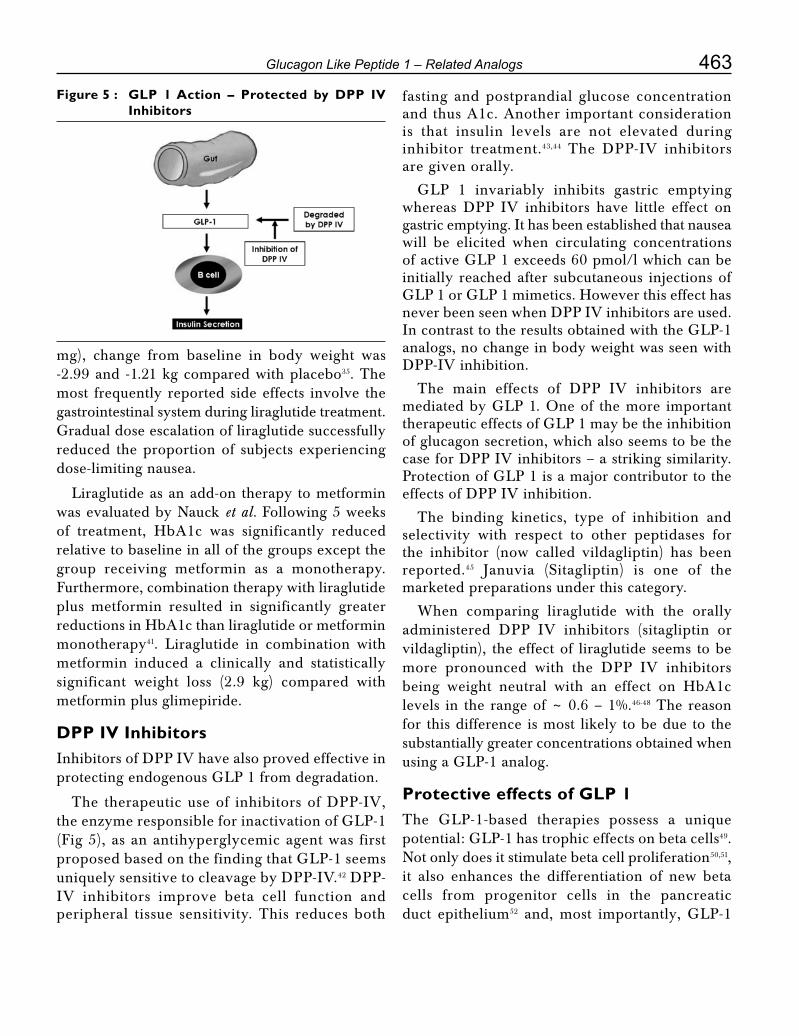
The therapeutic use of inhibitors of DPP-IV, the enzyme responsible for inactivation of GLP-1 (Fig 5), as an antihyperglycemic agent was first proposed based on the finding that GLP-1 seems uniquely sensitive to cleavage by DPP-IV.42 DPP-IV inhibitors improve beta cell function and peripheral tissue sensitivity. This reduces both
fasting and postprandial glucose concentration and thus A1c. Another important consideration is that insulin levels are not elevated during inhibitor treatment.43,44 The DPP-IV inhibitors are given orally.
GLP 1 invariably inhibits gastric emptying whereas DPP IV inhibitors have little effect on gastric emptying. It has been established that nausea will be elicited when circulating concentrations of active GLP 1 exceeds 60 pmol/l which can be initially reached after subcutaneous injections of GLP 1 or GLP 1 mimetics. However this effect has never been seen when DPP IV inhibitors are used. In contrast to the results obtained with the GLP-1 analogs, no change in body weight was seen with DPP-IV inhibition.
The main effects of DPP IV inhibitors are mediated by GLP 1. One of the more important therapeutic effects of GLP 1 may be the inhibition of glucagon secretion, which also seems to be the case for DPP IV inhibitors – a striking similarity. Protection of GLP 1 is a major contributor to the effects of DPP IV inhibition.
The binding kinetics, type of inhibition and selectivity with respect to other peptidases for the inhibitor (now called vildagliptin) has been reported.45 Januvia (Sitagliptin) is one of the marketed preparations under this category.
When comparing liraglutide with the orally administered DPP IV inhibitors (sitagliptin or vildagliptin), the effect of liraglutide seems to be more pronounced with the DPP IV inhibitors being weight neutral with an effect on HbA1c levels in the range of ~ 0.6 – 1%.46-48 The reason for this difference is most likely to be due to the substantially greater concentrations obtained when using a GLP-1 analog.
Protective effects of GLP 1The GLP-1-based therapies possess a unique potential: GLP-1 has trophic effects on beta cells49. Not only does it stimulate beta cell proliferation50,51, it also enhances the differentiation of new beta cells from progenitor cells in the pancreatic duct epithelium52 and, most importantly, GLP-1
Figure 5 : GLP 1 Action – Protected by DPP IV Inhibitors
464 Medicine Update 2008 Vol. 18
is capable of inhibiting apoptosis of beta cells including human beta cells.53
GLP-1 improves postprandial lipidemia, presumably as a result of delayed gastric emptying and insulin-mediated inhibition of lipolysis. Thus, by lowering both glucose and lipid concentrations, GLP-1 administration may reduce the cardiovascular risk in patients with type 2 diabetes.54
GLP 1 based therapy should be started as early in the clinical course as possible, before beta cell function has deteriorated to unacceptable levels.
References1. Vilsboll T, Holst JJ. Incretins, insulin secretion and Type 2
diabetes mellitus. Diabetologia 2004; 47: 357–366.
2. Brown JC. Gastric inhibitory polypeptide. Monogr Endocrinol 1982; 24:1–88.
3. Dupre J, Ross SA, Watson D, et al. Stimulation of insulin secretion by gastric inhibitory polypeptide in man. J Clin Endocrinol Metab. 1973; 37:826–828.
4. OrskovC,HolstJJ,NielsenOV.Effectoftruncatedglucagon-likepeptide-1 [proglucagon-(78-107) amide] on endocrine secretion from pig pancreas, antrum, and nonantral stomach. Endocrinology 1988; 123:2009–2013.
5. FehmannHC,HabenerJF.Insulinotropichormoneglucagon-like peptide-I(7-37) stimulation of proinsulin gene expression and proinsulin biosynthesis in insulinoma beta TC-1 cells.Endocrinology 1992; 130:159–166.
6. Schjoldager BT, Mortensen PE, Christiansen J, et al. GLP-1(glucagon-like peptide 1) and truncated GLP-1, fragments of human proglucagon, inhibit gastric acid secretion in humans. Dig Dis Sci 1989; 34:703–708.
7. Wettergren A, Schjoldager B, Mortensen PE, et al. Truncated GLP-1 (proglucagon 78-107-amide) inhibits gastric and pancreatic functions in man. Dig Dis Sci 1993; 38:665–673.
8. Nauck MA, Niedereichholz U, Ettler R et al. Glucagon-like peptide 1 inhibition of gastric emptying outweighs its insulinotropic effects in healthy humans. Am J Physiol 1997; 273:E981–E988.
9. Willms B, Werner J, Holst JJ, et al. Gastric emptying, glucose responses and insulin secretion after a liquid test meal: effects of exogenous glucagon-like peptide-1 (GLP-1)-(7-36) amide in type 2 (non insulin-dependent) diabetic patients. J Clin Endocrinol Metab 1996; 81:327–332
10. Flint A, Raben A, Astrup A, et al. Glucagon-like peptide 1 promotes satiety and suppresses energy intake in humans. J Clin Invest 1998; 101:515–520.
11. Gutzwiller JP, Drewe J, Goke B et al. Glucagon-like peptide-1 promotes satiety and reduces food intake in patients with diabetes mellitus type 2. Am J Physiol 1999; 276:R1541-R1544.
12. Holst JJ. Glucagon-like peptide 1(GLP-1): an intestinal hormone signaling nutritional abundance, with an unusual therapeutic
potential. Trends Endocrinol Metab 1999; 10:229–234.
13. GutniakM,OrskovC,HolstJJ,etal.Antidiabetogeniceffectof glucagon-like peptide-1 (7-36) amide in normal subjects and patients with diabetes mellitus. N Engl J Med 1992; 326:1316-1322.
14. Nathan DM, Schreiber E, Fogel H, et al. Insulinotropic action of glucagon like peptide-I-(7-37) in diabetic and nondiabetic subjects. Diabetes Care 1992; 15:270-276.
15) NauckMA,KleineN,OrskovC,etal.Normalizationoffastinghyperglycaemia by exogenous glucagon-like peptide 1 (7-36 amide) in type 2 (non-insulindependent) diabetic patients. Diabetologia 1993; 36:741–744.
16. NauckMA,HeimesaatMM,OrskovC,etal.Preservedincretinactivity of glucagon-like peptide 1 [7-36 amide] but not of synthetic human gastric inhibitory polypeptide in patients with type-2 diabetes mellitus. J Clin Invest 1993; 91:301–307.
17. Nauck MA, Wollschlager D, Werner J et al. Effects of subcutaneous glucagon-like peptide 1(GLP-1 [7-36 amide]) in patients with NIDDM. Diabetologia 1996; 39:1546–1553.
18. Juntti-Berggren L, Pigon J, Karpe F et al. The antidiabetogenic effect of GLP-1 is maintained during a 7-day treatment period and improves diabetic dyslipoproteinemia in NIDDM patients. Diabetes Care 1996; 19:1200–1206.
19. DeaconCF, JohnsenAH,Holst JJ.Degradationof glucagon-like peptide-1 by human plasma in vitro yields an N-terminally truncated peptide that is a major endogenous metabolite in vivo. J Clin Endocrinol Metab 1995; 80:952–957.
20. DeaconCF,NauckMA,Toft-NielsenM,etal.Bothsubcutaneouslyand intravenously administered glucagon-like peptide I are rapidly degraded from the NH2-terminus in type II diabetic patients and in healthy subjects. Diabetes 1995; 44:1126–1131.
21. KiefferTJ,McIntoshCH,PedersonRA.Degradationofglucose-dependent insulinotropic polypeptide and truncated glucagon-like peptide 1 in vitro and in vivo by dipeptidyl peptidase IV. Endocrinology 1995; 136:3585–3596.
22. Kolterman OG, Buse JB, Fineman MS et al. Synthetic exendin 4 (AC2993)significantlyreducespostprandialandfastingplasmaglucose in subjects with type 2 diabetes. J Clin Endrinol Metab 2003; 88: 3082 – 3089.
23. Egan JM,ClocquetAR,ElahiD.The insulinotrophic effectof acute exendin 4 administered to humans: comparison of nondiabetic state to type 2 diabetes. J Clin Endocrinol Metab. 2002; 87: 1282-1290.
24. Edwards CM, Stanley SA, Davis R et al. Exendin-4 reducesfasting and postprandial glucose and decreases energy intake in healthy volunteers. Am J Physiol Endocrinol Metab 2001; 281:E155–E161.
25. DeFronzo RA, Ratner RE, Han J et al. Effects of exenatide (exendin 4) on glycemic control and weight over 30 weeks in Metformin treated patients with type 2 DM. Diabetes Care 2005; 28: 1092 – 1100.
26. Buse JB, Henry RR, Han J et al. Effects of exenatide (exendin 4) on glycemic control over 30 weeks in sulfonylurea treated patients with type 2 diabetes. Diabetes Care 2004; 27: 2628 -2635.
465Glucagon Like Peptide 1 – Related Analogs
27. KendallDM,RiddleMC,RosenstockJetal.Effectsofexenatide(exendin 4) on glycemic control over 30 weeks in patients with type 2 DM treated with Metformin and a sulfonylurea. Diabetes Care 2005; 28: 1083 – 1091.
28. Data on file. Amylin Pharmaceuticals, Inc.
29. Agerso H, Jensen LB, Elbrond B, et al. The pharmacokinetics, pharmacodynamics, safety and tolerability of NN2211, a new long-acting GLP-1 derivative, in healthy men. Diabetologia 2002; 45(2):195-202.
30. DegnKB, JuhlCB,Sturis Jetal.Oneweek’s treatmentwiththe long-acting glucagon-like peptide 1 derivative liraglutide (NN2211) markedly improves 24-h glycemia and ±- and ²-cell function and reduces endogenous glucose release in patients with Type 2 diabetes. Diabetes 2004; 53(5):1187-1194.
31. Damholt B, Golor G, Wierich W, et al. An open-label, parallel group study investigating the effects of age and gender on the pharmacokinetics of the once-daily glucagon-like peptide-1 analogue liraglutide. J Clin Pharmacol 2006; 46(6):635-641.
32. Rolin B, Larsen MO, Gotfredsen CF, et al. The long-actingGLP-1 derivative NN2211 ameliorates glycemia and increases ²-cell mass in diabetic mice. Am. J. Physiol. Endocrinol. Metab. 2002; 283(4):E745-E752.
33. Sturis J, Gotfredsen CF, Romer J, et al. GLP-1 derivativeliraglutide in rats with ²-cell deficiencies: influence of metabolic state on ²-cell mass dynamics. Br. J. Pharmacol. 2003; 140(1):123-132.
34. Madsbad S, Schmitz O, Ranstam J, et al. Improved glycemic control with no weight increase in patients with Type 2 diabetes after once-daily treatment with the long-acting glucagon-like peptide 1 analog liraglutide (NN2211): a 12-week, double-blind, randomized, controlled trial. Diabetes Care 2004; 27(6):1335-1342.
35. Vilsboll T, Zdravkovic M, Le-Thi T et al. Liraglutide significantly improves glycemic control, and lowers body weight without risk of either major or minor hypoglycemic episodes in subjects with Type 2 diabetes. Diabetes 2006; A115.
36. Nauck MA, El-Ouaghlidi A, Hompesch M, et al. No impairment of hypoglycemia counterregulation via glucagon with the long-acting GLP-1 derivative, NN2211, in subjects with Type 2 diabetes. Diabetologia 2003; A285.
37. Bock T, Pakkenberg B, Buschard K: The endocrine pancreas in non-diabetic rats after short-term and long-term treatment with the long-acting GLP-1 derivative NN2211. APMIS 2003; 111(12):1117-1124.
38. LarsenPJ,FledeliusC,KnudsenLB,etal.Systemicadministrationof the long-acting GLP-1 derivative NN2211 induces lasting and reversible weight loss in both normal and obese rats. Diabetes 2001; 50(11): 2530-2539.
39. Ribel U, Larsen MO, Rolin B et al. NN2211: a long-acting glucagon-like peptide-1 derivative with anti-diabetic effects in glucose-intolerant pigs. Eur. J. Pharmacol. 2002; 451(2):217-225.
40. American Diabetes Association: Standards of medical care in diabetes. Diabetes Care 2005; 28 (Suppl 1):S4-S36.
41. Nauck MA, Hompesch M, Filipczak R, et al. Five weeks of treatment with the GLP-1 analogue liraglutide improves glycaemic control and lowers body weight in subjects with Type 2 diabetes.Exp.Clin.Endocrinol.Diabetes 2006; 114(8):417-423.
42. DeaconCF,NauckMA,Toft-NielsenM,etal.Bothsubcutaneouslyand intravenously administered glucagon-like peptide I are rapidly degraded from the NH2-terminus in type II diabetic patients and in healthy subjects. Diabetes 1995; 44:1126–1131.
43. Ahren B, Simonsson E, Larsson H et al. Inhibition of dipeptidyl peptidase IV improves metabolic control over a 4- week study period in type 2 diabetes. Diabetes Care 2002; 25:869–875.
44. Ahren B, Landin-Olsson M, Jansson PA, et al. Inhibition of dipeptidyl peptidase-4 reduces glycemia, sustains insulin levels, and reduces glucagon levels in type 2 diabetes. J Clin Endocrinol Metab 2004; 89:2078–2084.
45. Brandt I, Joossens J, Chen X et al. Inhibition of dipeptidyl-peptidase IV catalyzed peptide truncation by Vildagliptin ((2S)-{[(3-hydroxyadamantan-1-yl)amino]acetyl}-pyrrolidine-2-carbonitrile). Biochem Pharmacol 2005; 70 :134–143.
46. Aschner P, Kipnes M, Lunceford J et al.: Sitagliptin monotherapy improved glycaemic control in patients with Type 2 diabetes. Diabetologia (2006):0005 (Abstract).
47. Deacon CF, Holst JJ: Dipeptidyl peptidase IV inhibitors: a promising new therapeutic approach for the management of Type 2 diabetes. Int. J. Biochem. Cell Biol. 2006; 38(5-6):831-844.
48. Pratley RE, Jauffret-Kamel S, Galbreath E, et al. Twelve-week monotherapy with the DPP-4 inhibitor vildagliptin improves glycemic control in subjects with Type 2 diabetes. Horm. Metab. Res. 2006; 38(6):423-428.
49. Egan JM, Bulotta A, Hui H, et al. GLP-1 receptor agonists are growth and differentiation factors for pancreatic islet beta cells. Diabetes Metab Res Rev 2003; 19:115–123.
50. Xu G, Stoffers DA, Habener JF et al. Exendin-4 stimulates both beta-cell replication and neogenesis, resulting in increased beta-cell mass and improved glucose tolerance in diabetic rats. Diabetes 1999; 48:2270–2276.
51. Stoffers DA, Kieffer TJ, Hussain MA et al. Insulinotropic glucagon-like peptide 1 agonists stimulate expression of homeodomain protein IDX-1 and increase islet size in mouse pancreas. Diabetes 2000; 49:741–748.
52. Zhou J, Wang X, Pineyro MA, et al. Glucagon-like peptide 1 and exendin-4 convert pancreatic AR42J cells into glucagon- and insulin-producing cells. Diabetes 1999; 48:2358–2366.
53. ButeauJ,El-AssaadW,RhodesCJ,etal.Glucagon-likepeptide-1prevents beta cell glucolipotoxicity. Diabetologia 2004; 47:806–815.
54. Meier J. J, Gethmann A, Götze O et al. Glucagon-like peptide 1 abolishes the postprandial rise in triglyceride concentrations and lowers levels of non-esterified fatty acids in humans. Diabetologia 2006; 49: 452–458.

CHAPTER
61Tight Disease Control in Rheumatoid Arthritis – What, Why and How?R. Handa
IntroductionRheumatoid Arthritis (RA) is the commonest inflammatory joint disease seen in clinical practice. It is an autoimmune disorder that shortens life expectancy. Passive treatment has now given way to active intervention. Much of this stems from aggressive use of disease modifying anti-rheumatic drugs (DMARDs), often in combination, to achieve tight disease control. Also, better understanding of the disease pathobiology has led to the development of several biologic agents. These agents are being employed not only for refractory, difficult to treat disease but also, increasingly, for early RA. The present article focuses on ‘disease control’ in RA- what is disease activity, how is it measured, the need for tight disease control and the ways to achieve this goal.
Disease Activity In RA- What is it and How is it Measured?The advent of effective treatments like biologics has stimulated interest in quantitative measurements in Rheumatology- the science of ‘Metrology’. The concept of measurement in Rheumatology is more esoteric because, more often than not, the instruments used are questionnaires to be filled in by the patient. This is in contradistinction to quantification possible in other areas of medicine e.g. proteinuria or GFR in kidney diseases, ejection
fraction or valve areas in cardiac illnesses, where numerical values are available to aid comparison and decision making. To complicate matters, most of these patients reported questionnaires are not available or validated in Hindi or other Indian languages. Notwithstanding this drawback, there is enough data to show that patient questionnaires are a scientifically validated way to assess disease activity in Rheumatology.
In general, 2 types of measures are employed in Rheumatology: Status Measures and Response Measures. The former assess disease activity at a specific point in time and are more applicable to individuals and in the clinic. Response measures assess how disease activity changes over time, for example, response to medication.1 Response measures by their very nature require longitudinal observation and are more useful in clinical trials to study groups of patients. Disease activity score (DAS) and remission are considered to be status measures. In contrast, the American College ofRheumatology-ACR20/50/70criteriaareresponsemeasures. DAS score is a continuous numerical index while the ACR criteria are a categoricalmean. It must be pointed out that DAS can also be used as a response measure (as part of the so called EULAR-European League against Rheumatism-response criteria), although it is most often used as a status measure. The EULAR response criteria

467Tight Disease Control in Rheumatoid Arthritis – What, Why and How?
classify individual patients as non-, moderate, or good responders, dependent on the extent of change in DAS and the level of disease activity reached.
Disease activity in RA is assessed by several parameters which include duration of morning stiffness, tender joint count, swollen joint count, observer global assessment, patient global assessment, visual analog scale (VAS) for pain, health assessment questionnaire for activities of daily living, ESR, NSAID pill count etc.2 Scores which integrate several parameters are now frequently employed e.g. DAS score. DAS 28 is one of the simplified versions of original DAS in common usage. DAS 28 requires four simple inputs: 28tenderjointcount(TJC),28swollenjointcount(SJC),ESRandgeneralhealth(GH)assessmentbythe patient on a VAS from 0 to 100. The 28 joints assessed for swelling and tenderness include the 10 PIP joints, 10 MP joints, 2 wrists, 2 elbows, 2 shoulders and the 2 knees.
One of the drawbacks of DAS 28 is the requirement of a DAS calculator which is available online also on the DAS web site (www.das-score.nl). Two composite indices that are derived from the
DAS but do not require a calculator or computer have been constructed- simplified disease activity index(SDAI)andclinicalDAI(CDAI).TheSDAIindexincludesfivecomponents:SJC(28joints),TJC(28joints),C-reactiveprotein(CRP)inmg/dL(witha range of 0.1-10), patient’s global disease activity on a 10-cm VAS, and physician’s global assessment on a 10-cm VAS. The index constitutes a simple numerical summation of the values of the individual components of SDAI, and ranges from 0.1 to 86. Four of these componentsareincludedinCDAI,whichexcludestheCRP.CDAIscoresmayrangefrom0to76.CDAIis the only composite index constructed to measure clinical remission in RA that does not include a laboratory test.3,4
It also needs to be emphasized here that RA is a multidimensional disease and disease activity is one of the domains that can be measured (Figure 1). Other domains that are measured include disability (commonly measured by health assessment questionnaire-HAQ) and disease damage (measured on hand radiographs by scoring methods like Sharp score and its modifications). An all encompassing facet is quality of life, for which generic and disease specific measures like WHO-QoL Bref, RA-QoL etc are available.5 A detailed discussion on these is beyond the scope of this write up.
What is tight disease control?Tight disease control aims to keep disease activity at low levels preferably in remission. The traditionally usedACRcriteria for remission6 mandate that 5 or more of the following requirements must be fulfilled for at least 2 consecutive months:
• Durationofmorningstiffnessnotexceeding15minutes
• Nofatigue
• Nojointpain(byhistory)
• Nojointtendernessorpainonmotion
• No soft tissue swelling in joints or tendonsheaths
Figure 1 : Domains in RA and their measurement tools
DAS= Disease Activity Score; HAQ=Health Assessment Questionnaire; QoL=Quality of life
468 Medicine Update 2008 Vol. 18
• ESR (Westergren method) < 30 mm/h for afemale or 20 mm/h for a male
Using DAS 28, RA activity is classified as mild when the DAS 28 is 2.6-3.2, moderate when the score is 3.2-.5.1 or high when it is > 5.1. Remission is defined as a DAS 28 < 2.6. A change of 1.2 in DAS 28 is considered a meaningful change.
It has become increasingly apparent that complete remissionasdefinedby theACRcriteria isnotcommon in RA. Instead, the concept of minimal disease activity (MDA) may be more realistic. The original name for this state was low disease activity state (LDAS). Over the course of time, it became apparent that the name LDAS gave the impression that this was referring to a state of low activity and excluded remission. The change of the name to MDA was, in part, to address this misconception. MDA is between high disease activity and remission and any patient in remission is also in MDA.7 For DAS 28, the remission cut off is a score < 2.6. It is important to be aware of the fact that patients who meet the DAS28 remission cut point of < 2.6 may have a few tender and/or swollen joints unlike theACRcriteria.TheDAS28definitionplacesthepatient in MDA when DAS28 < The cut points forremissionforSDAIandCDAIare3.3and2.8respectively.8
Why tight disease control in RA?There is abundant data to show that apart from morbidity, the mortality is also increased in RA with an average shortening of life span by 10 years. The saving grace is that despite being a disease with unfavorable prognosis, suppression of disease activity does correlate with reduction in radiological joint damage.9 In the landmarkTICORAstudy,patients were assigned to 2 groups, intensive treatment and conventional care. The intensive treatment group developed less radiographic damage than the control group after 18 months of follow up, suggesting an association between remission (or low disease activity) and further joint destruction.10 Similarly, Dutch investigators have shown that control of disease activity has a salubrious influence
on radiological progression, after adjustment for time effects and baseline predictors of radiological destruction and their interactions with time.11 Similar data has been obtained from the recent PREMIER, ASPIRE and the TEMPO trials which reveals that higher remission rates are associated with arrest of radiographic progression and better physical function.12-14 It needs to be emphasized that maintenance of durable remission/MDA is as important as achieving remission/MDA.
Also, time is of essence in RA. Intervention should be early since there is irrefutable evidence to show that irreversible damage occurs within the first 2 years of the disease. The rate of progression in the first year of disease is significantly higher than that in later years, indicating the need for early intervention. Apart from the clinical and radiological benefits, early DMARD therapy also favorably influences mortality, which has been shown to be lower in patients who present early compared to those who present late.15 The concept of ‘window period’ is now firmly entrenched in RA akin to the concept in myocardial infarction. This window of opportunity in the treatment of RA describes a period of time early in course of RA when the disease is more responsive to therapy. The window period is a moving target and some authorities reckon that this may be as little as 3-4 months from the onset of symptoms.16 The duration of the disease too has a bearing on responsiveness to treatment. Patients with a longer duration of disease do not respond as well to treatment compared with patients with early disease. Trials of TNF blockers in RA too provide proof of concept that intensive intervention early in the course of RA can have a bearing on long term radiographic progression. In patients with a disease duration of less than three years, the use of a TNF blocking drug (adalimumab, etanercept, or infliximab) in combination with methotrexate revealed an increased rate of clinical remission and slowing of radiographic progression compared with methotrexate monotherapy.12,13,17 The available evidence, thus, overwhelmingly supports the case for early intervention in RA.

469Tight Disease Control in Rheumatoid Arthritis – What, Why and How?
Despite the evidence that early treatment fetches the best dividends in RA, it is also pertinent to point out that patients in India present late and it is never too late to start treatment, though earlier is better.
How to achieve control in RA?This is perhaps the most contentious area in this field. There is unanimity of opinion on the need to objectively measure disease activity, and control disease effectively. However, opinion on the tools to achieve this is divided.
There are robust data to show that compared with existing DMARDs, the biologic agents are capable of higher response rates, greater remission rates, slower radiographic damage over time, and fewer cardiovascular deaths, particularly when initiated early in the disease course.12,13,17,18 However, resource constraints make it unlikely that these agents would be used as the first line agents in India. I would, therefore, in this article, focus on the conventional DMARDs and how they can be used to achieve tight control. DMARDs can be instituted in various ways:
Step-up approach
Therapy is started with a single DMARD, other agents are added one by one till response is achieved.
Step-down approach
Several DMARDs are started together till remission; one agent is then continued and others withdrawn.
Saw-tooth approach
Therapy is started with a single DMARD which is substituted by another agent in case of toxicity or when it ceases to be effective.
Parallel approach
Several DMARDs are started simultaneously and continued.
Three landmark trials need mention: COBRA, TICORA and BeST. The COBRA
(CombinatietherapieBijReumatoideArtritis)trialwas a randomized, double-blind, multicenter trial representing step down DMARD use.19 In this trial 155 patients with early RA were treated with either sulfasalazine (SSZ) monotherapy or combination therapy, comprising SSZ (2 g/day), methotrexate (MTX; 7.5 mg/week) and prednisolone (initially 60 mg/day, tapered in 6-weekly steps to 7.5 mg/day. TheCOBRAcombinationwasfoundtobesuperiorto SSZ monotherapy in suppressing disease activity and radiological progression of early RA. After a 5-year follow-up, it was seen that the initial 6-month cycleofintensivecombinationtreatment(COBRAtherapy) resulted in a sustained reduction in the rate of radiological progression, independent of subsequent antirheumatic therapy.20 To put things in perspective, it needs to be mentioned that despite impressive results most rheumatologists are reluctanttoembracetheCOBRAprotocolduetoreasons like high dose of steroids (prednisolone ~60 mg/day initially), pill burden, complexity of regimen etc.21
It is perhaps the TICORA study that is mostapplicable to theIndiansetting.In theTICORAstudy, patients with recent onset RA were randomly assigned to receive routine DMARD treatment at the discretion of the treating rheumatologist, or intensive treatment with monthly assessment of clinical disease activity.10 The DMARDs employed were SSZ, MTX, hydroxychloroquine singly to begin with. The investigators in addition to frequent, objective assessment of patients made intensive use of intraarticular steroid injections if needed; and the application of a structured protocol for the escalation of treatment (step up) in face ofactivedisease.Combinationswereemployedinpatients where disease activity was not controlled with single drugs, with prednisolone added to the treatment if response was suboptimal despite triple drug therapy. The people in the intensive group had greater improvements in physical function and substantially enhanced quality of life. Reduced progression of erosive disease and total radiographic damage was recorded, but not in joint-
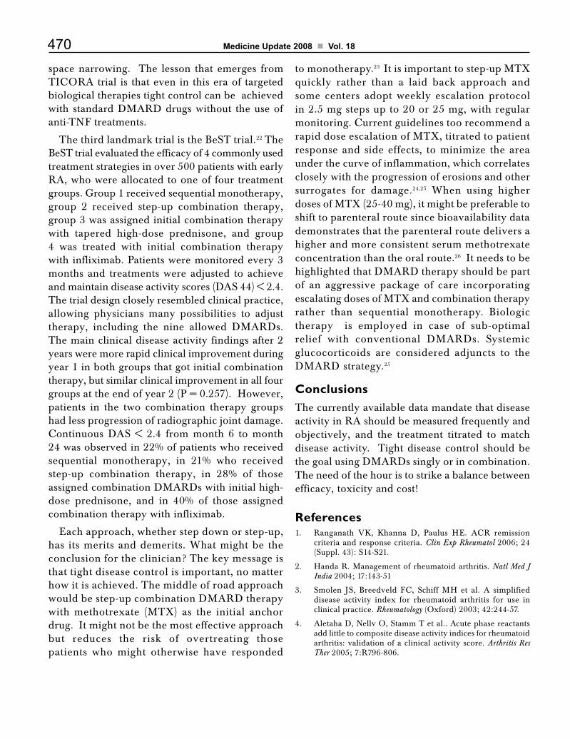
470 Medicine Update 2008 Vol. 18
space narrowing. The lesson that emerges from TICORAtrial is thateveninthiseraof targetedbiological therapies tight control can be achieved with standard DMARD drugs without the use of anti-TNF treatments.
The third landmark trial is the BeST trial.22 The BeST trial evaluated the efficacy of 4 commonly used treatment strategies in over 500 patients with early RA, who were allocated to one of four treatment groups. Group 1 received sequential monotherapy, group 2 received step-up combination therapy, group 3 was assigned initial combination therapy with tapered high-dose prednisone, and group 4 was treated with initial combination therapy with infliximab. Patients were monitored every 3 months and treatments were adjusted to achieve and maintain disease activity scores (DAS 44) < 2.4. The trial design closely resembled clinical practice, allowing physicians many possibilities to adjust therapy, including the nine allowed DMARDs. The main clinical disease activity findings after 2 years were more rapid clinical improvement during year 1 in both groups that got initial combination therapy, but similar clinical improvement in all four groups at the end of year 2 (P = 0.257). However, patients in the two combination therapy groups had less progression of radiographic joint damage. Continuous DAS < 2.4 from month 6 to month24 was observed in 22% of patients who received sequential monotherapy, in 21% who received step-up combination therapy, in 28% of those assigned combination DMARDs with initial high-dose prednisone, and in 40% of those assigned combination therapy with infliximab.
Each approach, whether step down or step-up, has its merits and demerits. What might be the conclusion for the clinician? The key message is that tight disease control is important, no matter how it is achieved. The middle of road approach would be step-up combination DMARD therapy with methotrexate (MTX) as the initial anchor drug. It might not be the most effective approach but reduces the risk of overtreating those patients who might otherwise have responded
to monotherapy.23 It is important to step-up MTX quickly rather than a laid back approach and some centers adopt weekly escalation protocol in 2.5 mg steps up to 20 or 25 mg, with regular monitoring.Currentguidelinestoorecommendarapid dose escalation of MTX, titrated to patient response and side effects, to minimize the area under the curve of inflammation, which correlates closely with the progression of erosions and other surrogates for damage.24,25 When using higher doses of MTX (25-40 mg), it might be preferable to shift to parenteral route since bioavailability data demonstrates that the parenteral route delivers a higher and more consistent serum methotrexate concentration than the oral route.26 It needs to be highlighted that DMARD therapy should be part of an aggressive package of care incorporating escalating doses of MTX and combination therapy rather than sequential monotherapy. Biologic therapy is employed in case of sub-optimal relief with conventional DMARDs. Systemic glucocorticoids are considered adjuncts to the DMARD strategy.25
ConclusionsThe currently available data mandate that disease activity in RA should be measured frequently and objectively, and the treatment titrated to match disease activity. Tight disease control should be the goal using DMARDs singly or in combination. The need of the hour is to strike a balance between efficacy, toxicity and cost!
References1. Ranganath VK, Khanna D, Paulus HE. ACR remission
criteria and response criteria. Clin Exp Rheumatol 2006; 24 (Suppl. 43): S14-S21.
2. Handa R. Management of rheumatoid arthritis. Natl Med J India 2004; 17:143-51
3. Smolen JS, Breedveld FC, Schiff MH et al. A simplifieddisease activity index for rheumatoid arthritis for use in clinical practice. Rheumatology (Oxford) 2003; 42:244-57.
4. Aletaha D, Nellv O, Stamm T et al.. Acute phase reactants add little to composite disease activity indices for rheumatoid arthritis: validation of a clinical activity score. Arthritis Res Ther 2005; 7:R796-806.
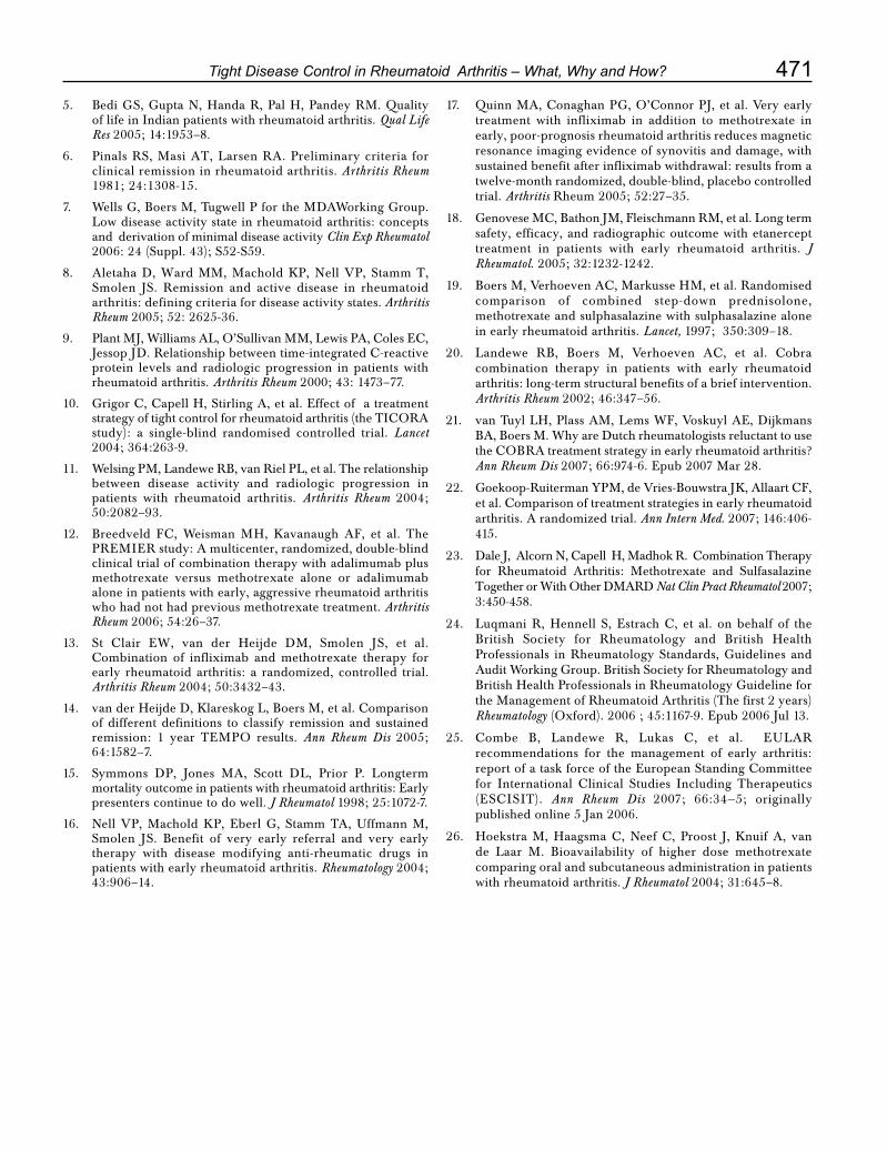
471Tight Disease Control in Rheumatoid Arthritis – What, Why and How?
5. Bedi GS, Gupta N, Handa R, Pal H, Pandey RM. Quality of life in Indian patients with rheumatoid arthritis. Qual Life Res 2005; 14:1953–8.
6. Pinals RS, Masi AT, Larsen RA. Preliminary criteria for clinical remission in rheumatoid arthritis. Arthritis Rheum 1981; 24:1308-15.
7. Wells G, Boers M, Tugwell P for the MDAWorking Group. Low disease activity state in rheumatoid arthritis: concepts and derivation of minimal disease activity Clin Exp Rheumatol 2006: 24 (Suppl. 43); S52-S59.
8. Aletaha D, Ward MM, Machold KP, Nell VP, Stamm T, Smolen JS. Remission and active disease in rheumatoid arthritis: defining criteria for disease activity states. Arthritis Rheum 2005; 52: 2625-36.
9. PlantMJ,WilliamsAL,O’SullivanMM,LewisPA,ColesEC,JessopJD.Relationshipbetweentime-integratedC-reactiveprotein levels and radiologic progression in patients with rheumatoid arthritis. Arthritis Rheum 2000; 43: 1473–77.
10. GrigorC,CapellH,StirlingA,etal.Effectofatreatmentstrategyoftightcontrolforrheumatoidarthritis(theTICORAstudy): a single-blind randomised controlled trial. Lancet 2004; 364:263-9.
11. Welsing PM, Landewe RB, van Riel PL, et al. The relationship between disease activity and radiologic progression in patients with rheumatoid arthritis. Arthritis Rheum 2004; 50:2082–93.
12. Breedveld FC, Weisman MH, Kavanaugh AF, et al. ThePREMIER study: A multicenter, randomized, double-blind clinical trial of combination therapy with adalimumab plus methotrexate versus methotrexate alone or adalimumab alone in patients with early, aggressive rheumatoid arthritis who had not had previous methotrexate treatment. Arthritis Rheum 2006; 54:26–37.
13. St Clair EW, van der Heijde DM, Smolen JS, et al.Combination of infliximab and methotrexate therapy forearly rheumatoid arthritis: a randomized, controlled trial. Arthritis Rheum 2004; 50:3432–43.
14. vanderHeijdeD,KlareskogL,BoersM,etal.Comparisonof different definitions to classify remission and sustained remission: 1 year TEMPO results. Ann Rheum Dis 2005; 64:1582–7.
15. Symmons DP, Jones MA, Scott DL, Prior P. Longterm mortality outcome in patients with rheumatoid arthritis: Early presenters continue to do well. J Rheumatol 1998; 25:1072-7.
16. Nell VP, Machold KP, Eberl G, Stamm TA, Uffmann M, Smolen JS. Benefit of very early referral and very early therapy with disease modifying anti-rheumatic drugs in patients with early rheumatoid arthritis. Rheumatology 2004; 43:906–14.
17. QuinnMA,ConaghanPG,O’ConnorPJ,etal.Veryearlytreatment with infliximab in addition to methotrexate in early, poor-prognosis rheumatoid arthritis reduces magnetic resonance imaging evidence of synovitis and damage, with sustained benefit after infliximab withdrawal: results from a twelve-month randomized, double-blind, placebo controlled trial. Arthritis Rheum 2005; 52:27–35.
18. GenoveseMC,BathonJM,FleischmannRM,etal.Longtermsafety, efficacy, and radiographic outcome with etanercept treatment in patients with early rheumatoid arthritis. J Rheumatol. 2005; 32:1232-1242.
19. BoersM,VerhoevenAC,MarkusseHM,etal.Randomisedcomparison of combined step-down prednisolone, methotrexate and sulphasalazine with sulphasalazine alone in early rheumatoid arthritis. Lancet, 1997; 350:309–18.
20. Landewe RB, Boers M, Verhoeven AC, et al. Cobracombination therapy in patients with early rheumatoid arthritis: long-term structural benefits of a brief intervention. Arthritis Rheum 2002; 46:347–56.
21. van Tuyl LH, Plass AM, Lems WF, Voskuyl AE, Dijkmans BA, Boers M. Why are Dutch rheumatologists reluctant to use theCOBRAtreatmentstrategyinearlyrheumatoidarthritis?Ann Rheum Dis 2007; 66:974-6. Epub 2007 Mar 28.
22. Goekoop-RuitermanYPM,deVries-BouwstraJK,AllaartCF,etal.Comparisonoftreatment strategies in early rheumatoid arthritis. A randomized trial. Ann Intern Med. 2007; 146:406-415.
23. DaleJ,AlcornN,CapellH,MadhokR.CombinationTherapyfor Rheumatoid Arthritis: Methotrexate and Sulfasalazine Together or With Other DMARD Nat Clin Pract Rheumatol 2007; 3:450-458.
24. LuqmaniR,Hennell S,EstrachC, et al. onbehalf of theBritish Society for Rheumatology and British Health Professionals in Rheumatology Standards, Guidelines and Audit Working Group. British Society for Rheumatology and British Health Professionals in Rheumatology Guideline for the Management of Rheumatoid Arthritis (The first 2 years) Rheumatology (Oxford). 2006 ; 45:1167-9. Epub 2006 Jul 13.
25. Combe B, Landewe R, Lukas C, et al. EULARrecommendations for the management of early arthritis: reportofataskforceoftheEuropeanStandingCommitteefor International Clinical Studies Including Therapeutics(ESCISIT). Ann Rheum Dis 2007; 66:34—5; originallypublished online 5 Jan 2006.
26. Hoekstra M,Haagsma C,NeefC,Proost J,Knuif A, vande Laar M. Bioavailability of higher dose methotrexate comparing oral and subcutaneous administration in patients with rheumatoid arthritis. J Rheumatol 2004; 31:645–8.

CHAPTER
62Management of Ankylosing SpondylitisG. Narsimulu, K. Suresh
IntroductionAnkylosing spondylitis (AS) is a human leukocyte antigen (HLA)-B27-associated chronic, inflammatory rheumatic disease characterised by sacroiliitis and spondylitis with formation of syndesmophytes leading to ankylosis. The disease can be accompanied by extraskeletal manifestations, such as acute anterior uveitis, aortic incompetence, cardiac conduction defects, fibrosis of the upper lobes of the lungs, cauda equina syndrome or renal amyloidosis.
Diagnosis AS usually manifests in late adolescence or early adulthood and only rarely starts after age 40.1 The diagnosis of AS at an early stage of disease is difficult and depends primarily on a careful history and physical examination. The presence of inflammatory low back pain is important for the diagnosis.Clinicalfeaturesofankylosingspondylitisinclude modified NewYork criteria,2 which helps in the diagnosis of Ankylosing spondylitis. It should be stressed that classification criteria are usually not well suited for early diagnosis of disease because radiological proof of sacroiliitis is a late feature of
the disease.3
It usually takes many years before definite radiographic sacroiliac abnormalities first appear.
Table 1 : Extraskeletal Manifestation of Ankylosing Spondilitis
Inflammatory spinal pain
- Onset before age 40
- Insidious onset
- Persistence for at least 3 months
- Morning stiffness > 30 min
- Improvement with exercise
Alternate buttock pain
Acute anterior uveitis
Synovitis (predominantly of lower limbs, asymmetric)
Enthesitis (heel, plantar)
Positive family history for
Ankylosing spondylitis
Chronicinflammatoryboweldisease
Psoriasis
Table 2 : Clinical Features of Ankylosing Spondilitis (Modified New York Criteria, 19842)1. Low back pain of at least 3 months’ duration improved
by exercise and not relieved by rest
2. Limitation of lumbar spine in sagittal and frontal planes
3.Chestexpansiondecreasedrelativetonormalvaluesforage and sex
4. Bilateral sacroiliitis grade 2 to 4
5. Unilateral sacroiliitis grade 3 or 4
Definite ankylosing spondylitis: unilateral grade 3 or 4, or bilateral grade 2 to 4 sacroiliitis and atleast one clinical criterion
473Management of Ankylosing Spondylitis
The New York grading system for sacroiliac joint status is as follows: grade I=suspicious; grade II=evidence of erosion and sclerosis; grade III=erosions, sclerosis, and early ankylosis; and grade IV=total ankylosis.
CTandMRIcandetectASlesionsearlierandwithgreater consistency than plain radiography.4
Assessment of Clinical Outcomes in Patients with AS in PracticeThe most widely used measure of the inflammatory activity of AS is the Bath Ankylosing Spondylitis Disease Activity Index (BASDAI).5 This simple instrument is patient-completed, sensitive to change over 3 weeks, and has been validated.6 BASDAI was developed as a composite index, consisting of an evaluation on a visual analogue scale (0–10) of fatigue, axial pain, peripheral pain, stiffness and enthesopathy. Scores of 4 or greater suggest suboptimal control of disease, and patients with scores of 4 or greater are usually good candidates for either a change in their medical therapy or for enrolment in clinical trials evaluating new drug therapies directed at Ankylosing Spondylitis.
ManagementFor most patients, AS is a relatively mild disease with a good functional prognosis. The objectives
for treatment of AS are to relieve pain, stiffness, and fatigue and to maintain good posture and good physical and psychosocial functioning.Treatment of AS should be tailored according to:• Current manifestations of the disease (axial,
peripheral, entheseal, extra-articular symptoms and signs)
• Level of current symptoms, clinical findings, and prognostic indicators
• Diseaseactivity/inflammation
• Pain
• Function,disability, handicap
• Structuraldamage, hip involvement, spinal deformities
• Wishesandexpectations of the patient.
• General clinical status (age, sex, comorbidity, concomitant drugs).7
The principles of management are summarized in Table 3.
A complete explanation of the disease is essential toachievegoodpatientcompliance.TheCurrentevidence suggests that none of the conventional disease-modifying antirheumatic drugs (DMARDs), including sulfasalazine and methotrexate (MTX) alter or inhibit the inflammation seen in the spine and entheses in AS.
Physical therapy
Physiotherapy remains the mainstay of management of AS. There is evidence that exercise alone can produce adequate symptom relief in patients with AS. Group exercises are better than home exercises and improve pain, stiffness, movement in the spine.(9) Preferably, they should be started after a hot shower or a hot bath. Swimming and extension-promoting exercises are appropriate. These activities counteract the kyphotic effects of pain and fatigue on posture and reduce stiffness.
Table 3 : Principles of Management of Ankylosing Spondylitis8
•Nocure,butmostpatientscanbewellmanaged
• Patient education to increase compliance
• Appropriate use of antirheumatic drugs, primarily nonsteroidal, anti-inflammatory drugs (NSAIDs)
• Daily exercises very important (e.g., swimming)
• Sleep on firm mattress
• Sports and recreation
• Supportive measures and counseling
• Avoid smoking
• Avoid trauma
• Patient support groups
• Family counseling
474 Medicine Update 2008 Vol. 18
NSAIDs and coxibs
NSAIDs and coxibs are useful for spinal pain and physical function. NSAIDs are the first line drug treatment for patients with AS with pain and stiffness. NSAIDs improve spinal pain, peripheral joint pain, and function over a short period of time (6 weeks).10 The studies of different NSAIDs have not shown one drug to be better than the others. A recent randomised controlled trial comparing the efficacy of continuous celecoxib treatment for AS with intermittent “on demand” use suggests
that continuous treatment retards radiographic disease progression at 2 years.11 This is the first study to show a possible disease modifying effect of continuous treatment.
Coxibsortheaddition of GI protectors (misoprostol, double doses of H
2 blockers, or PPIs) to conventional
NSAIDs can significantly reduce GI bleeding.
Apart from nephrotoxicity, there is increased cardiovascular toxicity with NSAIDs and more so with coxibs.12
In general, the choice of NSAID or coxibs should be based on risk factors for cardiovascular disease, renal and GI symptoms. Analgesics, such as paracetamol and opioids, may be used for pain control in patients in whom NSAIDs are contraindicated or poorly tolerated.
Corticosteroids
Local Corticosteroid injections of the involvedjoints are effective, especially for sacroiliac joints.13 The long term use of systemic corticosteroids for axial disease has a little role to play. However, intravenous methylprednisolone has been found useful in resistant cases of active AS.14
Disease Modifying Antirheumatic Drugs (DMARDs)
There is no evidence for the efficacy of disease modifying antirheumatic drugs (DMARDs) for the treatment of axial disease. However sulfasalazine may be considered in patients with peripheral arthritis. Sulfasalazine (SSZ) has demonstrated some benefit in reducing ESR, severity and
duration of morning stiffness. But it has shown no benefit in spinal pain and mobility, enthesitis, patient and physician global assessment. However patients at early and active disease stage with higher level of ESR and those with peripheral arthritis benefit from SSZ.15 Toxicity with sulfasalazine is common but usually mild which includes GI symptoms, hepatic enzyme abnormalities, mucocutaneous manifestations and hematological abnormalities.
There is no significant effect of methotrexate on
spinal pain or function. Methotrexate might benefit patients with peripheral joint involvement. There is not enough evidence to support the use of other DMARDs in AS.
Pamidronate
There is some evidence for a beneficial effect of intravenous pamidronate (60 mg IV monthly for 6 months) on both axial pain and function.16 There is no study to assess its effect on peripheral joint disease. Side effects include transient post-infusional arthralgias and myalgias and an acute phase response with lymphopenia and raised Creactive protein.
Thalidomide17
Open trials suggest a beneficial effect for thalidomide
on spinal disease, but toxicity is substantial which includes drowsiness, severe birth defects and irreversible peripheral neuropathies.
Anti-TNF treatment
Anti-TNF treatment should be given to patients with persistently high disease activity despite conventional treatments (NSAIDs and DMARDs) according to the Assessment in Ankylosing spondylitis group (ASAS) recommendations.(18) There is no evidence to support the obligatory use of DMARDs before, or concomitant with, anti-TNF treatment in patients with axial disease.7 The present evidence supports the use of the TNF inhibitors
etanercept, infliximab and adalimumab for spinal pain, function, and peripheral joint disease.

475Management of Ankylosing Spondylitis
The onset of clinical effect with TNF blockers is rapid, and therapeutic effect persists for up to 3 years with continuing treatment.7 Stopping treatment results in a high rate of clinical relapse. Adding methotrexate to infliximab treatment reduces side effects without any additional benefit in AS.
Toxicity with anti-TNF treatment includes injection site reactions with subcutaneous injections (etanercept and adalimumab), increased risk of infections in particular, tuberculosis. Screening for Mycobacterium tuberculosis is now a standard prerequisite for anti-TNF treatment. Demyelinating disease, lupus-like syndromes, and worsening of pre-existing congestive heart failure have also been reported.
There is insufficient evidence available at present on the role of interleukin 1 antagonists in AS.
Surgery
Total hip arthroplasty is indicated in patients with refractory pain or disability and radiographic evidence of structural damage, irrespective of age.19 Spinal surgeries like corrective osteotomy and stabilization procedures are useful in selected patients.20
Assessment in Ankylosing spondylitis (ASAS) consensus for anti-TNF therapy.18 (Modified)Specification (Definition of the terms)
Patient selection
Diagnosis
• PatientsnormallyfulfillingmodifiedNewYorkCriteriafordefinitiveAS
Active disease
• BASDAI ≥ 4 (0–10) and an expert opinion that anti-TNF treatment should be started ≥ 4 weeks
Treatment failure
• Allpatientsmusthavehadadequatetherapeutictrials of at least 2 NSAIDs. An adequate therapeutic trial is defined as:
• Treatment for ≥ 3 months at maximal recommended or tolerated anti-inflammatory dose unless contraindicated
• Treatmentfor<3monthswheretreatmentwas withdrawn because of intolerance, toxicity, or contraindications.
• Patientswithsymptomaticperipheralarthritis(normally having a lack of response to a local steroid injection for those with oligoarticular involvement) must have had adequate therapeutic trial of both NSAIDs and sulfasalazine.
• Patientswithsymptomaticenthesitismusthavehad an adequate therapeutic trial of at least two local steroid injections unless contraindicated
Contraindication for anti TNF therapy
• Womenwhoarepregnantorbreastfeeding;
• Activeinfection
• Patientsathighriskofinfectionincluding:
• Chroniclegulcer
• Previoustuberculosis
• Septicarthritisofanativejointwithinthepast 12 months
• Sepsisofaprosthetic jointwithinthepast12 months, or indefinitely if the prosthesis remains in situ
• Persistentorrecurrentchestinfections
• Indwellingurinarycatheter
• Historyoflupusormultiplesclerosis
• Malignancy or premalignancy statesexcluding:
• Basalcellcarcinoma
• Malignancies diagnosed and treatedmore than 10 years previously (where the probability of total cure is very high)

476 Medicine Update 2008 Vol. 18
Assessment in Ankylosing Spondylitis International Working Group (ASAS) Improvement Criteria (ASAS-20) and ASAS Partial Remission Criteria ASAS-20 Improvement Criteria
At least 20-per cent improvement AND 10 units improvement in three out of the four following domains, without worsening of 20 per cent or more AND 10 units in the remaining domain:
- Bath Ankylosing Spondylitis Functional Index (BASFI)
- Morning stiffness
- Patient global assessment
- Pain
ASAS Partial Remission Criteria
A value below 20 units in all four domains
Assessment of response
• Responder criteria:BASDAI: 50% relativechange or absolute change of 2 (scale 0–10) and expert opinion.
• Timeofevaluation:Between6and12weeks.
References1. Khan MA. Update on spondyloarthropathies. Ann Intern Med
2002;135:896–907
2. van der Linden S,ValkenburgHA,CatsA.Evaluationofdiagnostic criteria for ankylosing spondylitis. A proposal for modification of the New York criteria. Arthritis Rheum 1984;27:361–8
3. Mau W, Zeidler, Mau R, Majewski A, Freyschmidt J, Stangel W, et al. Clinical features and prognosis of patients withpossible ankylosing spondylitis. Results of a 10-year followup. J Rheumatol 1988;15:1109–14.
4. Braun J, Bollow M, Eggens U, Konig H, Distler A, Sieper J. Use of dynamic magnetic resonance imaging with fast imaging in the detection of early and advanced sacroiliitis in spondylarthropathy patients. Arthritis Rheum 1994;37:1039–45.
5. http://www.basdai.com/
6. Garrett S, Jenknson T, Kennedy LG, Whitelock H, Gaisford P,CalinA.Anewapproach todefining disease status inankylosing spondylitis: the Bath Ankylosing Spondylitis Disease Activity Index. J Rheumatol 1994:21;2286–91.
7. Zochling J et al. ASAS/EULAR recommendations for the management of ankylosing spondylitis. Ann Rheum Dis, Apr 2006; 65: 442 - 452.
8. S van der Linden, D van der Heijde, , J Braun : Ankylosing spondylitis. Harris DE et al (Ed). Kelleys text book of rheumatology.1125-41.
9. Dagfinrud H, Hagen KB, Kvien TK. Physiotherapy interventions for ankylosing spondylitis. Cochrane Database of Systematic Reviews2004,Issue4.:CD002822
10. Zochling J, van der Heijde D, Dougados M, and Braun J:Current evidence for the management of ankylosingspondylitis: a systematic literature review for the ASAS/EULAR management recommendations in ankylosing spondylitis. Ann Rheum Dis. Apr 2006; 65: 423 - 432.
11. Wanders A, van der Heijde D, Landewé R, Béhier J - M, CalinA,OlivieriI,et al. Nonsteroidal antiinflammatory drugs reduce radiographic progression in patients with ankylosing spondylitis. Arthritis Rheum 2005;52:1756–65.
12. Juni P, Nartey L, Reichenbach S, Sterchi R, Dieppe PA, Egger M. Risk of cardiovascular events and rofecoxib: cumulative meta-analysis. Lancet 2004;364:2021–9
13. Maugars Y, Mathis C, Berthelot JM, Charlier C, ProstA. Assessment of the efficacy of sacroiliac corticosteroid injections in spondylarthropathies: a double-blind study. Br J Rheumatol 1996;35:767–70.
14. Mintz G, Enriquez RD, Mercado U, Robles EJ, Jimenez FJ, Gutierrez G. Intravenous methylprednisolone pulse therapy in severe ankylosing spondylitis. Arthritis Rheum 1981;24:734–6.
15. Chen J, Liu C. Sulfasalazine for ankylosing spondylitis.Cochrane Database Syst Rev 2005; (2) :CD00800.
16. Maksymowych WP et al. A six-month randomized, controlled, double-blind, dose-response comparison of intravenous pamidronate (60 mg versus 10 mg) in the treatment of nonsteroidal antiinflammatory drug-refractory ankylosing spondylitis. Arthritis Rheum 2002;46:766–73
17. Huang F, Gu J, Zhao W, Zhu J, Zhang J, Yu DTY. One-year open-label trial of thalidomide in ankylosing spondylitis. Arthritis Care Res 2002;47:15.
18. Braun J, Davis J, Dougados M, Sieper J, van der Linden S, van der Heijde D, et al. First update of the international ASAS consensus statement for the use of anti-TNF agents in patients with ankylosing spondylitis. Ann Rheum Dis 2006;65:316–20
19. Sochart DH, Porter ML. Long-term results of total hip replacement in young patients who had ankylosing spondylitis. Eighteen to thirty-year results with survivorship analysis. J Bone Joint Surg Am 1997;79:1181–9.
20. Van Royen BJ, De Gast A. Lumbar osteotomy for correction of thoracolumbar kyphotic deformity in ankylosing spondylitis. A structured review of three methods of treatment. Ann Rheum Dis 1999;58:399–406
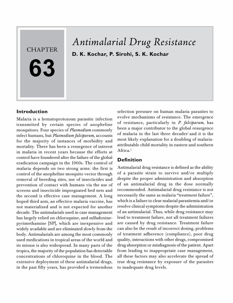
CHAPTER
63Antimalarial Drug ResistanceD. K. Kochar, P. Sirohi, S. K. Kochar
IntroductionMalaria is a hematoprotozoan parasitic infection transmitted by certain species of anopheline mosquitoes. Four species of Plasmodium commonly infect humans, but Plasmodium falciparum, accounts for the majority of instances of morbidity and mortality. There has been a resurgence of interest in malaria in recent years because the efforts at control have foundered after the failure of the global eradication campaign in the 1960s. The control of malaria depends on two strong arms: the first is control of the anopheline mosquito vector through removal of breeding sites, use of insecticides and prevention of contact with humans via the use of screens and insecticide impregnated bed nets and the second is effective case management. A long hoped third arm, an effective malaria vaccine, has not materialized and is not expected for another decade. The antimalarials used in case management has largely relied on chloroquine, and sulfadoxine-pyrimethamine [SP], which are inexpensive and widely available and are eliminated slowly from the body. Antimalarials are among the most commonly used medications in tropical areas of the world and its misuse is also widespread. In many parts of the tropics, the majority of the population has detectable concentrations of chloroquine in the blood. The extensive deployment of these antimalarial drugs, in the past fifty years, has provided a tremendous
selection pressure on human malaria parasites to evolve mechanisms of resistance. The emergence of resistance, particularly in P. falciparum, has been a major contributor to the global resurgence of malaria in the last three decades1 and it is the most likely explanation for a doubling of malaria-attributable child mortality in eastern and southern Africa.2
DefinitionAntimalarial drug resistance is defined as the ability of a parasite strain to survive and/or multiply despite the proper administration and absorption of an antimalarial drug in the dose normally recommended. Antimalarial drug resistance is not necessarily the same as malaria “treatment failure”, which is a failure to clear malarial parasitemia and/or resolve clinical symptoms despite the administration of an antimalarial. Thus, while drug resistance may lead to treatment failure, not all treatment failures are caused by drug resistance. Treatment failure can also be the result of incorrect dosing, problems of treatment adherence (compliance), poor drug quality, interactions with other drugs, compromised drug absorption or misdiagnosis of the patient. Apart from leading to inappropriate case management, all these factors may also accelerate the spread of true drug resistance by exposure of the parasites to inadequate drug levels.
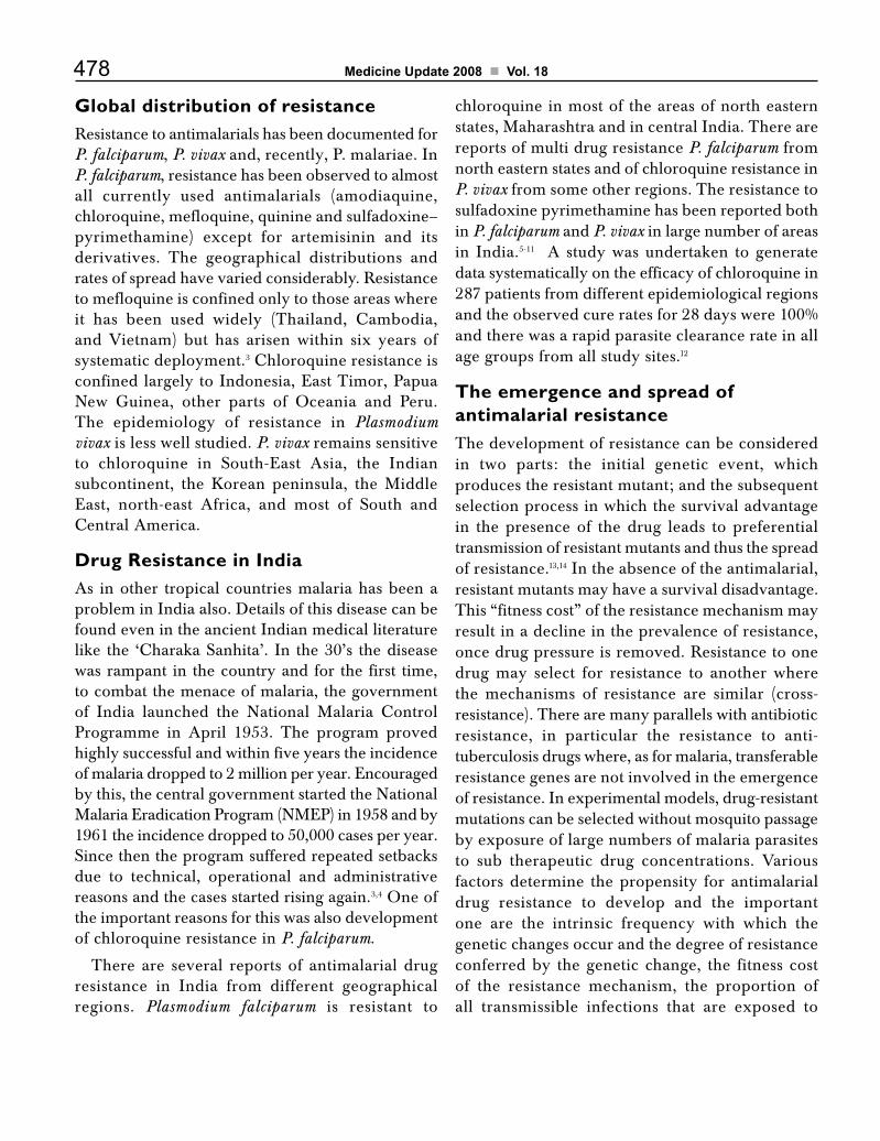
478 Medicine Update 2008 Vol. 18
Global distribution of resistanceResistance to antimalarials has been documented for P. falciparum, P. vivax and, recently, P. malariae. In P. falciparum, resistance has been observed to almost all currently used antimalarials (amodiaquine, chloroquine, mefloquine, quinine and sulfadoxine–pyrimethamine) except for artemisinin and its derivatives. The geographical distributions and rates of spread have varied considerably. Resistance to mefloquine is confined only to those areas where it has been used widely (Thailand, Cambodia,and Vietnam) but has arisen within six years of systematic deployment.3Chloroquineresistanceisconfined largely to Indonesia, East Timor, Papua New Guinea, other parts of Oceania and Peru. The epidemiology of resistance in Plasmodium vivax is less well studied. P. vivax remains sensitive to chloroquine in South-East Asia, the Indian subcontinent, the Korean peninsula, the Middle East, north-east Africa, and most of South and CentralAmerica.
Drug Resistance in IndiaAs in other tropical countries malaria has been a problem in India also. Details of this disease can be found even in the ancient Indian medical literature like the ‘CharakaSanhita’. In the30’s thediseasewas rampant in the country and for the first time, to combat the menace of malaria, the government of India launched the National Malaria ControlProgramme in April 1953. The program proved highly successful and within five years the incidence of malaria dropped to 2 million per year. Encouraged by this, the central government started the National Malaria Eradication Program (NMEP) in 1958 and by 1961 the incidence dropped to 50,000 cases per year. Since then the program suffered repeated setbacks due to technical, operational and administrative reasons and the cases started rising again.3,4 One of the important reasons for this was also development of chloroquine resistance in P. falciparum.
There are several reports of antimalarial drug resistance in India from different geographical regions. Plasmodium falciparum is resistant to
chloroquine in most of the areas of north eastern states, Maharashtra and in central India. There are reports of multi drug resistance P. falciparum from north eastern states and of chloroquine resistance in P. vivax from some other regions. The resistance to sulfadoxine pyrimethamine has been reported both in P. falciparum and P. vivax in large number of areas in India.5-11 A study was undertaken to generate data systematically on the efficacy of chloroquine in 287 patients from different epidemiological regions and the observed cure rates for 28 days were 100% and there was a rapid parasite clearance rate in all age groups from all study sites.12
The emergence and spread of antimalarial resistanceThe development of resistance can be considered in two parts: the initial genetic event, which produces the resistant mutant; and the subsequent selection process in which the survival advantage in the presence of the drug leads to preferential transmission of resistant mutants and thus the spread of resistance.13,14 In the absence of the antimalarial, resistant mutants may have a survival disadvantage. This “fitness cost” of the resistance mechanism may result in a decline in the prevalence of resistance, once drug pressure is removed. Resistance to one drug may select for resistance to another where the mechanisms of resistance are similar (cross-resistance). There are many parallels with antibiotic resistance, in particular the resistance to anti-tuberculosis drugs where, as for malaria, transferable resistance genes are not involved in the emergence of resistance. In experimental models, drug-resistant mutations can be selected without mosquito passage by exposure of large numbers of malaria parasites to sub therapeutic drug concentrations. Various factors determine the propensity for antimalarial drug resistance to develop and the important one are the intrinsic frequency with which the genetic changes occur and the degree of resistance conferred by the genetic change, the fitness cost of the resistance mechanism, the proportion of all transmissible infections that are exposed to

479Antimalarial Drug Resistance
the drug (the selection pressure), the number of parasites exposed to the drug, the concentrations of drug to which these parasites are exposed, the pharmacokinetic and pharmacodynamic properties of the antimalarial, individual (dosing, duration, adherence) and community (quality, availability, distribution) patterns of drug use, the immunity profile of the community and the individual and the simultaneous presence of other antimalarials or substances in the blood to which the parasite is not resistant.
Selection and spread of resistanceThe emergence of resistance is the product of the probabilities of its de novo emergence (a rare event) and subsequent spread. Resistant parasites, if present, will be selected when parasites are exposed to “selective” (subtherapeutic) drug concentrations. “Selective” in this context means a concentration of drug that will eradicate the sensitive parasites but still allow growth of the resistant parasite population such that it eventually transmits to another person. De novo resistance arises randomly among malaria parasites and the non-immune patients infected with large numbers of parasites who receive inadequate treatment (either because of poor drug quality, poor adherence, vomiting of an oral treatment, etc.) are the potent source of de novo resistance. This emphasizes the importance of correct prescribing, and good adherence to prescribed drug regimens, and also provision of treatment regimens that are still highly effective in hyperparasitaemic patients. The recrudescence and subsequent transmission of an infection that has generated a de novo resistant malaria parasite is essential for propagation of resistance.15
Step 1: de novo selection of resistance
In order to assess the factors determining the emergence and spread of resistance, we need to consider the numbers of malaria parasites likely to be exposed to the drugs, both within an individual and in the entire human population. Fortunately this estimate of parasite numbers is much more precise than for almost any other human pathogen.
Malaria parasites are eukaryotes and meiosis occurs after a female anopheline mosquito has taken viable gametocytes in its blood meal. All the other 108–1013 cell divisions in the life cycle are mitotic and nearly all these divisions take place in the bloodstream of the human host. Usually, less than ten sporozoite parasites are inoculated by an infected mosquito in order to establish malaria infection.16,17 These rapidly find their way to the liver. During P. falciparum infection, each infected hepatocyte liberates approximately 30,000 merozoites after 5–6 days of pre-erythrocytic schizogony. Thus approximately 100,000–300,000 merozoites are liberated into the bloodstream to begin the 48-hour asexual reproduction cycle. This is an important number, as it is the number of parasites that would encounter residual drug levels from a previous antimalarial treatment or drug levels during chemoprophylaxis.8 The density of parasites in the blood at which symptoms and fever occur (the pyrogenic density), and thus the stage at which appropriate antimalarial treatment could be given, vary considerably.17,18,19 In nonimmune people, nonspecific symptoms often occur a day or two before parasites are detectable on the blood smear (about 50 parasites per microliter of blood). This density corresponds to a total of between 108 and 109 asexual parasites in an adult with a red cell volume of about 2 litre. In areas of moderate- or high-intensity transmission, parasitemias considerably higher than this level may be tolerated without symptoms, although densities over 10,000 per microliter (between 1010 and 1011 parasites in the body of an adult, and correspondingly less in children) are usually symptomatic, even in very high-transmission settings.20 Median or geometric mean parasite counts in malariometric surveys are usually below this value (i.e., most people with detectable parasitemias in these endemic areas are not obviously ill). It is estimated that approximately 300 million people in the world now have malaria parasites in their blood. Using current epidemiological data we have estimated that there must be less than 3 x 10¹⁶ malaria parasites in the world’s asymptomatic carriers.21

480 Medicine Update 2008 Vol. 18
Step 2: the spread of resistance
Resistance to one drug may be selected for by another drug in which the mechanism of resistance is similar (a phenomenon known as cross-resistance). Antimalarial drug resistance in malaria parasites spreads because it confers a survival advantage in the presence of the antimalarial drugs and therefore results in a greater probability of transmission for resistant than for sensitive parasites. Resistant infections are more likely to recrudesce, and eventually as resistance worsens, the infections with resistant parasites respond more slowly to treatment. Both increased rates of recrudescence and slow initial responses to treatment increase the likelihood of generating sufficient gametocyte densities to transmit the infection, as compared to drug-sensitive infections. Mathematically, it is this ratio of transmission probabilities in drug-resistant compared with drug-sensitive infections that drives the spread of resistance. The recrudescence and subsequent transmission of an infection that generated resistant malaria parasites de novo are essential for propagation of resistance.22 If resistance is low grade or a highly effective combination treatment is given, then resistance may confer only a little increase in the treatment failure rate, and a correspondingly slow rate of spread. As resistance worsens, the failure rates rise and the rate of spread accelerates. In the rare but important infection in which resistance arises de novo, killing of the transmissible sexual stages (gametocytes) during the primary infection does not affect the spread of resistance because these gametocytes are derived from the drug-sensitive parasites. Gametocytes carrying the resistance genes will not reach transmissible densities until the resistant biomass has expanded to a population size close to that which is necessary to produce illness (> 107 parasites).23 Thus, to prevent the spread of resistance, gametocyte production from the subsequent recrudescent-resistant infection must be prevented.
ChloroquineresistanceinP. falciparum may be multigenic and is initially conferred by mutations
in a gene encoding a transporter (PfCRT).24 In thepresenceofPfCRTmutations, themutationsin a second transporter (PfMDR1) modulate the level of resistance in vitro. However the role of PfMDR1 mutations in determining the therapeutic response following chloroquine treatment remains unclear.25 At least one other as-yet unidentified gene is also thought to be involved in this process. Resistance to chloroquine in P. falciparum has arisen spontaneously less than ten times in the past fifty years.26 This suggests that the per-parasite probability of developing resistance de novo is on the order of 1 in 1020 parasite multiplications. The single point mutations in the gene encoding cytochrome b (cytB) which confer atovaquone resistance or in the gene encoding dihydrofolate reductase (dhfr) which confer pyrimethamine resistance, have a per-parasite probability of arising de novo of approximately 1 in 1012 parasite multiplications22. To put this in context, an adult with approximately 2% parasitemia has 1012 parasites in his or her body but in the laboratory much higher mutation rates than 1 in every 1012 are recorded.27
Mutations may be associated with fitness disadvantages (i.e., in the absence of the drug they are less fit and multiply less well than their drug-sensitive counterparts). Another factor that may explain the discrepancy between in vitro and much lower apparent in vivo rates of spontaneous mutation is host immunity. Even a previously nonimmune individual develops a specific immune response to a malaria infection. This response is systematically evaded by the parasite population through programmed antigenic variation of the main red cell surface–expressed epitopes. In falciparum malaria, P. falciparum erythrocyte membrane protein 1 (PfEMP1), which is encoded by the var multigene family, changes its behavior in 2–3% of parasites in each asexual cycle.28 The untreated infection is characterized by successive waves of parasites, each comprising largely one antigenically distinct surface phenotype. It is likely that this specific immune response directed against the immunodominant surface antigens will reduce
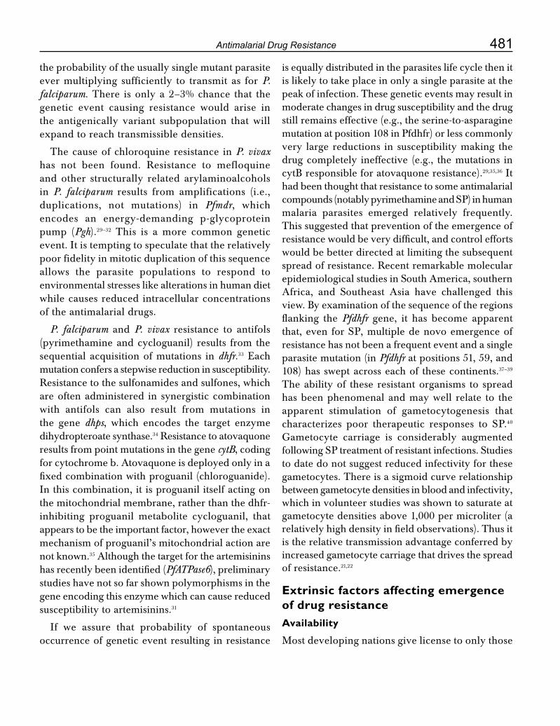
481Antimalarial Drug Resistance
the probability of the usually single mutant parasite ever multiplying sufficiently to transmit as for P. falciparum. There is only a 2–3% chance that the genetic event causing resistance would arise in the antigenically variant subpopulation that will expand to reach transmissible densities.
The cause of chloroquine resistance in P. vivax has not been found. Resistance to mefloquine and other structurally related arylaminoalcohols in P. falciparum results from amplifications (i.e., duplications, not mutations) in Pfmdr, which encodes an energy-demanding p-glycoprotein pump (Pgh).29–32 This is a more common genetic event. It is tempting to speculate that the relatively poor fidelity in mitotic duplication of this sequence allows the parasite populations to respond to environmental stresses like alterations in human diet while causes reduced intracellular concentrations of the antimalarial drugs.
P. falciparum and P. vivax resistance to antifols (pyrimethamine and cycloguanil) results from the sequential acquisition of mutations in dhfr.33 Each mutation confers a stepwise reduction in susceptibility. Resistance to the sulfonamides and sulfones, which are often administered in synergistic combination with antifols can also result from mutations in the gene dhps, which encodes the target enzyme dihydropteroate synthase.34 Resistance to atovaquone results from point mutations in the gene cytB, coding for cytochrome b. Atovaquone is deployed only in a fixed combination with proguanil (chloroguanide). In this combination, it is proguanil itself acting on the mitochondrial membrane, rather than the dhfr-inhibiting proguanil metabolite cycloguanil, that appears to be the important factor, however the exact mechanism of proguanil’s mitochondrial action are not known.35 Although the target for the artemisinins has recently been identified (PfATPase6), preliminary studies have not so far shown polymorphisms in the gene encoding this enzyme which can cause reduced susceptibility to artemisinins.31
If we assure that probability of spontaneous occurrence of genetic event resulting in resistance
is equally distributed in the parasites life cycle then it is likely to take place in only a single parasite at the peak of infection. These genetic events may result in moderate changes in drug susceptibility and the drug still remains effective (e.g., the serine-to-asparagine mutation at position 108 in Pfdhfr) or less commonly very large reductions in susceptibility making the drug completely ineffective (e.g., the mutations in cytB responsible for atovaquone resistance).29,35,36 It had been thought that resistance to some antimalarial compounds (notably pyrimethamine and SP) in human malaria parasites emerged relatively frequently. This suggested that prevention of the emergence of resistance would be very difficult, and control efforts would be better directed at limiting the subsequent spread of resistance. Recent remarkable molecular epidemiological studies in South America, southern Africa, and Southeast Asia have challenged this view. By examination of the sequence of the regions flanking the Pfdhfr gene, it has become apparent that, even for SP, multiple de novo emergence of resistance has not been a frequent event and a single parasite mutation (in Pfdhfr at positions 51, 59, and 108) has swept across each of these continents.37–39 The ability of these resistant organisms to spread has been phenomenal and may well relate to the apparent stimulation of gametocytogenesis that characterizes poor therapeutic responses to SP.40 Gametocyte carriage is considerably augmented following SP treatment of resistant infections. Studies to date do not suggest reduced infectivity for these gametocytes. There is a sigmoid curve relationship between gametocyte densities in blood and infectivity, which in volunteer studies was shown to saturate at gametocyte densities above 1,000 per microliter (a relatively high density in field observations). Thus it is the relative transmission advantage conferred by increased gametocyte carriage that drives the spread of resistance.21,22
Extrinsic factors affecting emergence of drug resistanceAvailability
Most developing nations give license to only those

482 Medicine Update 2008 Vol. 18
antimalarial drugs, which are provided through their national health programs.41 This approach often excludes relatively expensive or risky therapies, even for patients who may be able to afford a given drug and have access to medical supervision. The mainfactoraffectingavailabilityiseconomic—theability or inability to purchase a drug for broad distribution and the potential reluctance to use the drug because of an inability either to screen users or to monitor its quality.
Adherence
Most people taking antimalarial drugs live in rural regions of the developing world and are not supervised by health professionals. A study conducted among 1640 febrile patients with malaria in Burkina Faso42 showed that 69 per cent were self-treated, and in a study in Ethiopia, among 630 febrile patients with malaria, 67 per cent were self-treated.43 Complex, inconvenient or poorlytolerated antimalarial regimens carry a substantial risk of inadequate adherence.44 Among 414 Brazilian patients, the risk of recurrent malaria correlated with self-reported poor adherence,45 whereas among 632 Nigerian children strict adherence correlated with clinical recovery.46 Convenientand easily understood packaging and education of the patients alleviate poor adherence. Owing to lengthy and complex regimens, currently used therapies such as quinine and primaquine and new combined therapeutic strategies challenge the ease of adherence.47
Counterfeit and Substandard Drugs
Counterfeitantimalarialdrugsposeaseriousthreatin regions where the trade in pharmaceuticals is not rigorously regulated. A survey conducted in Cameroonfoundinsufficientorinactiveingredientsin 38 per cent of preparations labeled chloroquine, 78 per cent of those labeled quinine, and 12 per cent of tablets labeled as an antifolate agent.48 A survey in Southeast Asia involving 104 purchases of artesunate tablets found that 38 per cent of the tablets contained no drug.49 In some countries the trade in counterfeit drugs undoubtedly results in many
deaths.50 The inadvertent marketing of substandard pharmaceuticals poses another threat. In a survey of eight authorized wholesalers in Tanzania selling combined sulfadoxine–pyrimethamine tablets, 11 per cent of the tablets failed industry standards for content, and 44 per cent failed dissolution testing.51
Intrinsic factors affecting emergence of drug resistancePlasmodia pass through distinct stages of form, function, location, clinical consequence, and susceptibility to antimalarial drugs. Drug activity ranges from narrow (e.g., the activity of quinine against asexual blood stages) to broad (e.g., the activity of primaquine against sexual and asexual forms in the blood and liver). Stage specific susceptibility differs among species of plasmodia – for example, chloroquine kills the gametocytes of P. vivax but exerts no effect against those of P. falciparum. These intrinsic properties define the recommended uses of antimalarial drugs. Species specific innate resistance (e.g., asexual blood stages of P. falciparum lack susceptibility to primaquine, whereas those of P. vivax appear to be sensitive to it);52 strain-specific innate resistance (e.g., that of asexual liver stages of P. vivax from the island of New Guinea against primaquine)53 and acquired resistance which is most important, because failure may occur even in the presence of complete adherence to the recommended therapies.
Parasite Burden
Most patients with malaria carry a burden of 108 to 1013 parasites.54 Effective chemotherapy induces a constant fractional decline with each asexual cycle,55 at a rate that varies according to the susceptibility of the parasite to a given drug. For example, artemisinin derivatives induce reductions of 104, whereas tetracycline achieves a reduction by only a factor of 10 with each cycle. The duration of exposure to a drug that is needed to eliminate infection hinges on the intrinsic rate of decline and on initial parasite burden.55 High levels of parasitemia, as compared with a low burden,

483Antimalarial Drug Resistance
require longer exposure to effective drug levels and have a relatively higher risk of treatment failure.56
Prevention of resistance by antimalarial combination therapy The theory underlying combination drug treatment of tuberculosis, leprosy, and HIV infection is well known and is now generally accepted for malaria.21,22,57–60 If two drugs are used with different modes of action, and therefore different resistance mechanisms, then the per-parasite probability of developing resistance to both drugs is the product of their individual per-parasite probabilities. This is particularly powerful in malaria, because there are only about 1017 malaria parasites in the entire world. For example, if the per-parasite probabilities of developing resistance to drug A and drug B are both 1 in 1012, then a simultaneously resistant mutant will arise spontaneously every 1 in 1024 parasites. As there is a cumulative total of less than 1020 malaria parasites in existence in one year, then such a simultaneously resistant parasite would arise spontaneouslyroughlyonceevery10,000years—provided the drugs always confronted the parasites in combination. Thus the lower the de novo per-parasite probability of developing resistance, the greater the delay in the emergence of resistance.
Stable and therapeutically significant resistance to the artemisinin derivatives has not yet been identified and cannot be induced yet in the laboratory, which suggests that it may be a very rare event. But it would be unwise to bank on its not happening, and should it arise, it would be a global disaster. For mutual protection against the emergence of drug resistance, these drugs should be used only in combination with other antimalarials.
Artemisinin derivatives are particularly effective in combinations because of their very high killing rates (parasite reduction ratios 10,000-fold per cycle), lack of adverse effects, and absence of significant resistance.61 The ideal pharmacokinetic properties for an antimalarial drug have been greatly debated. From a resistance-prevention perspective, the combination partners should have similar
pharmacokinetic properties. Rapid elimination ensures that the residual concentrations do not provide a selective filter for resistant parasites, but these drugs (if used alone) must be given for 7 days, and adherence to 7-day regimens is poor. Even 7-day regimens of artemisinin derivatives are associated with approximately 10% failure rates. In order to be highly effective in a 3-day regimen, terminal elimination half-lives of at least one drug componentneedtoexceed24hours.Combinationsof artemisinin derivatives (which are eliminated very rapidly) given for 3 days, with a slowly eliminated drug such as mefloquine (artemisinin combination treatment) provide complete protection for the artemisinin derivatives from selection of a de novo resistant mutant if adherence is good (i.e., no parasite is exposed to artemisinin during one asexual cycle without mefloquine being present). But this does leave the slowly eliminated “tail” of mefloquine unprotected by the artemisinin derivative. The residual number of parasites exposed to mefloquine alone, following two asexual cycles, is a tiny fraction (less than 0.00001%) of those present at the peak of the acute symptomatic infection. Furthermore, these residual parasites are exposed to relatively high levels of mefloquine, and, even if susceptibility is reduced, these levels are usually sufficient to eradicate infection. This strategy would be expected to be effective at preventing the de novo emergence of resistance at higher levels of transmission, where high-biomass infections still constitute the major source of de novo resistance. Various combination therapy used presently are Artemether + Lumefantrine, Artesunate + Amodiaquine, Artesunate + Mefloquine and Artesunate + Sulfadoxine Pyrimethamine.
The national drug policy for malaria in high-risk areas is advocating the full dose of chloroquine in chloroquine sensitive areas and a combination of artesunate and sulfadoxine–pyrimethamine in chloroquine resistant areas in uncomplicated malaria, whereas all patients of severe malaria irrespective of chloroquine sensitivity of the region should be treated by I.V. quinine or artesunate.

484 Medicine Update 2008 Vol. 18
Since malaria is changing its facet from time to time; all the knowledge of this phenomenon is highly essential for every level healthcare provider for administering effective management to the suffering humanity.
Reference 1. Marsh, K. 1998. Malaria disaster in Africa. Lancet. 352:924-
925.
2. Korenromp,E.L.,Williams,B.G.,Gouws,E.,Dye,C.,andSnow, R.W. 2003. Measurement of trends in childhood malaria mortality in Africa: an assessment of progress toward targets based on verbal autopsy. Lancet Infect. Dis. 3:349-358.
3. PandaM,MohapatraA.MalariaControl–AnoverviewinIndia. J Hum Ecol 2004; 15(2) : 101 – 104.
4. NAMP, DGHS Govt. of India : Malaria and its control in India–CountryScenario1999.
5. Schwobel B, Alifrangis M, Salanti A, Jelinek T. Different mutation patterns of atovaquone resistance to Plasmodium falciparum in vitro and in vivo: rapid detection of codon 268 polymorphism in the cytochrome b as potential in vivo resistance marker. Malar J 2003;2:5.
6. Assessment and monitoring of antimalarial drug efficacy for the treatment of uncomplicated falciparum malaria. Geneva: World Health Organization, 2003.
7. Rathod,P.K.,McErlean,T.,andLee,P.C.1997.Variationsin frequencies of drug resistance in Plasmodium falciparum. Proc. Natl. Acad. Sci. U. S. A. 94:9389-9393.
8. Plowe,C.V.2003.Monitoringantimalarialdrugresistance:making the most of the tools at hand. J. Exp. Biol. 206:3745-3752.
9. Su, X., Kirkman, L.A., Fujioka, H., and Wellems, T.E. Complex polymorphisms in an approximately 330 kDaprotein are linked to chloroquine-resistant P. falciparum in SoutheastAsiaandAfrica.Cell1997.91:593-603.
10. Kyes,S.,Horrocks,P.,andNewbold,C.Antigenicvariationat the infected red cell surface in malaria. Annu. Rev. Microbiol 2001. 55:673-707.
11. Cowman, A.F., Galatis, D., and Thompson, J.K. Selectionfor mefloquine resistance in Plasmodium falciparum is linked to amplification of the pfmdr1 gene and cross-resistance to halofantrine and quinine. Proc. Natl. Acad. Sci. U. S. A. 1994. 91:1143-1147.
12. Valecha N, Joshi H, Eapen A, Ravinderan J, Kumar A, Prajapati SK, Ringwald P. Therapeutic efficacy of chloroquine in Plasmodium vivax from areas with different epidemiological patterns in India and their Pvdhfr gene mutation pattern. Trans R Soc Trop Med Hyg. 2006; 100(9):831-7.
13. Reed, M.B., Saliba, K.J., Caruana, S.R., Kirk, K., andCowman,A.F.Pgh1modulatessensitivityandresistancetomultiple antimalarials in Plasmodium falciparum. Nature 2000. 403:906-909.
14. Austin, D.J., and Anderson, R.M. Studies of antibiotic resistance within the patient, hospitals and the community using simple mathematical models. Philos. Trans. R. Soc. Lond. B Biol. Sci. 1999. 354:721-738.
15. Lipsitch, M., and Levin, B.R. The population dynamics of antimicrobial chemotherapy. Antimicrob. Agents Chemother 1997. 41:363-373.
16. White, N.J. Antimalarial drug resistance and combination chemotherapy. Philos. Trans. R. Soc. Lond. B Biol. Sci. 1999. 354:739-749.
17. Peters,W.Chemotherapyanddrugresistanceinmalaria.2ndedition. Academic Press. London, United Kingdom. 1987. 1091 pp.
18. Rosenburg, R., and Wirtz, R.A. An estimation of the number of sporozoites ejected by a feeding mosquito. Trans. R. Soc. Trop. Med. Hyg. 1990. 84:209-212.
19. Fairley, N.H. Sidelights on malaria in man obtained by subinoculation experiments. Trans. R. Soc. Trop. Med. Hyg. 1947. 40:521-676.
20. White, N.J., and Pongtavornpinyo, W. The de-novo selection of drug resistance in malaria parasites. Philos. Trans. R. Soc. Lond. B Biol. Sci. 2003. 270:545-554.
21. James, S.P., Nichol, W.D., and Shute, P.G. A study of induced malignant tertian malaria. Proc. R. Soc. Med. 1932. 25:1153-1186.
22. Kitchen, S.F. Symptomatology: general considerations and falciparum malaria. In Malariology. Volume 2. M.F. Boyd, editor.W.B.SaundersCo.Philadelphia,Pennsylvania,USA.1949. 996–1017.
23. White, N.J. Antimalarial drug resistance and combination chemotherapy. Philos. Trans. R. Soc. Lond. B Biol. Sci. 1999. 354:739-749.
24. Jeffery, G.M., and Eyles, D.E. Infectivity to mosquitoes of Plasmodium falciparum as related to gametocyte density and duration of infection. Am. J. Trop. Med. Hyg. 1955. 4:781-789.
25. Plowe,C.V.Monitoringantimalarialdrugresistance:makingthe most of the tools at hand. J. Exp. Biol. 2003. 206:3745-3752.
26. Su, X., Kirkman, L.A., Fujioka, H., and Wellems, T.E. Complex polymorphisms in an approximately 330 kDaprotein are linked to chloroquine-resistant P. falciparum in SoutheastAsiaandAfrica.Cell.1997.91:593-603.
27. Rathod, P.K., McErlean, T., and Lee, P.C. Variations infrequencies of drug resistance in Plasmodium falciparum. Proc. Natl. Acad. Sci. U. S. A. 1997. 94:9389-9393.
28. Kyes,S.,Horrocks,P.,andNewbold,C.Antigenicvariationat the infected red cell surface in malaria. Annu. Rev. Microbiol. 2001. 55:673-707.
29. Cowman, A.F., Galatis, D., and Thompson, J.K. Selectionfor mefloquine resistance in Plasmodium falciparum is linked to amplification of the pfmdr1 gene and cross-resistance to halofantrine and quinine. Proc. Natl. Acad. Sci. U. S. A. 1994. 91:1143-1147.
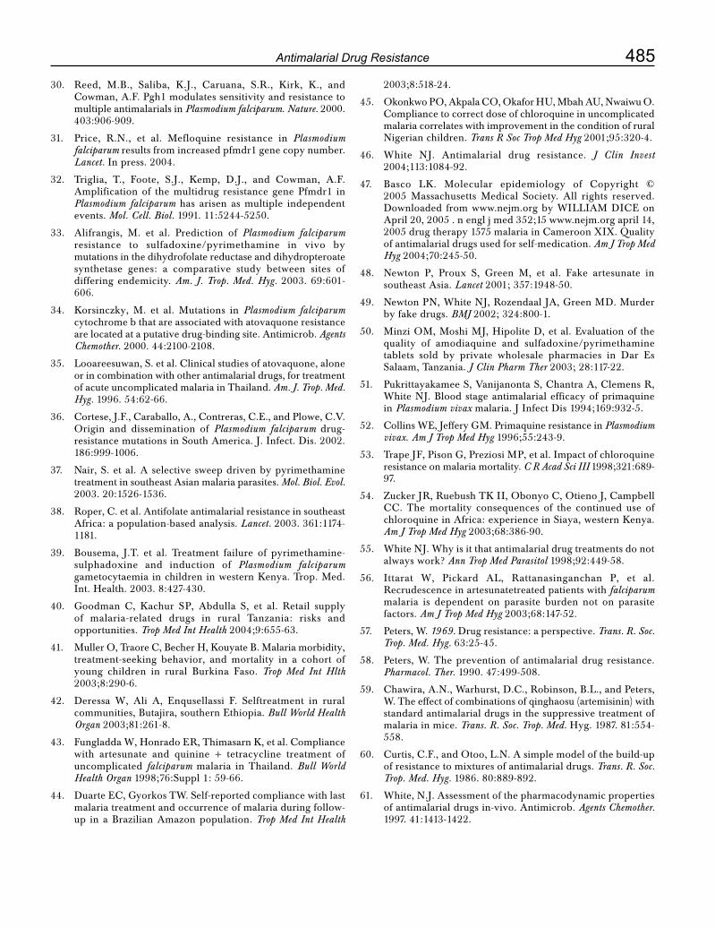
485Antimalarial Drug Resistance
30. Reed, M.B., Saliba, K.J., Caruana, S.R., Kirk, K., andCowman,A.F.Pgh1modulatessensitivityandresistancetomultiple antimalarials in Plasmodium falciparum. Nature. 2000. 403:906-909.
31. Price, R.N., et al. Mefloquine resistance in Plasmodium falciparum results from increased pfmdr1 gene copy number. Lancet. In press. 2004.
32. Triglia, T., Foote, S.J., Kemp, D.J., and Cowman, A.F.Amplification of the multidrug resistance gene Pfmdr1 in Plasmodium falciparum has arisen as multiple independent events. Mol. Cell. Biol. 1991. 11:5244-5250.
33. Alifrangis, M. et al. Prediction of Plasmodium falciparum resistance to sulfadoxine/pyrimethamine in vivo by mutations in the dihydrofolate reductase and dihydropteroate synthetase genes: a comparative study between sites of differing endemicity. Am. J. Trop. Med. Hyg. 2003. 69:601-606.
34. Korsinczky, M. et al. Mutations in Plasmodium falciparum cytochrome b that are associated with atovaquone resistance are located at a putative drug-binding site. Antimicrob. Agents Chemother. 2000. 44:2100-2108.
35. Looareesuwan,S.etal.Clinicalstudiesofatovaquone,aloneor in combination with other antimalarial drugs, for treatment of acute uncomplicated malaria in Thailand. Am. J. Trop. Med. Hyg. 1996. 54:62-66.
36. Cortese,J.F.,Caraballo,A.,Contreras,C.E.,andPlowe,C.V.Origin and dissemination of Plasmodium falciparum drug-resistance mutations in South America. J. Infect. Dis. 2002. 186:999-1006.
37. Nair, S. et al. A selective sweep driven by pyrimethamine treatment in southeast Asian malaria parasites. Mol. Biol. Evol. 2003. 20:1526-1536.
38. Roper,C.etal.AntifolateantimalarialresistanceinsoutheastAfrica: a population-based analysis. Lancet. 2003. 361:1174-1181.
39. Bousema, J.T. et al. Treatment failure of pyrimethamine-sulphadoxine and induction of Plasmodium falciparum gametocytaemia in children in western Kenya. Trop. Med. Int. Health. 2003. 8:427-430.
40. Goodman C, Kachur SP, Abdulla S, et al. Retail supplyof malaria-related drugs in rural Tanzania: risks and opportunities. Trop Med Int Health 2004;9:655-63.
41. MullerO,TraoreC,BecherH,KouyateB.Malariamorbidity,treatment-seeking behavior, and mortality in a cohort of young children in rural Burkina Faso. Trop Med Int Hlth 2003;8:290-6.
42. Deressa W, Ali A, Enqusellassi F. Selftreatment in rural communities, Butajira, southern Ethiopia. Bull World Health Organ 2003;81:261-8.
43. FungladdaW,HonradoER,ThimasarnK,etal.Compliancewith artesunate and quinine + tetracycline treatment of uncomplicated falciparum malaria in Thailand. Bull World Health Organ 1998;76:Suppl 1: 59-66.
44. DuarteEC,GyorkosTW.Self-reportedcompliancewithlastmalaria treatment and occurrence of malaria during follow-up in a Brazilian Amazon population. Trop Med Int Health
2003;8:518-24.
45. OkonkwoPO,AkpalaCO,OkaforHU,MbahAU,NwaiwuO.Compliancetocorrectdoseofchloroquineinuncomplicatedmalaria correlates with improvement in the condition of rural Nigerian children. Trans R Soc Trop Med Hyg 2001;95:320-4.
46. White NJ. Antimalarial drug resistance. J Clin Invest 2004;113:1084-92.
47. Basco LK. Molecular epidemiology of Copyright ©2005 Massachusetts Medical Society. All rights reserved. Downloaded fromwww.nejm.orgbyWILLIAMDICEonApril 20, 2005 . n engl j med 352;15 www.nejm.org april 14, 2005drugtherapy1575malariainCameroonXIX.Qualityof antimalarial drugs used for self-medication. Am J Trop Med Hyg 2004;70:245-50.
48. Newton P, Proux S, Green M, et al. Fake artesunate in southeast Asia. Lancet 2001; 357:1948-50.
49. Newton PN, White NJ, Rozendaal JA, Green MD. Murder by fake drugs. BMJ 2002; 324:800-1.
50. Minzi OM, Moshi MJ, Hipolite D, et al. Evaluation of the quality of amodiaquine and sulfadoxine/pyrimethamine tablets sold by private wholesale pharmacies in Dar Es Salaam, Tanzania. J Clin Pharm Ther 2003; 28:117-22.
51. PukrittayakameeS,VanijanontaS,ChantraA,ClemensR,White NJ. Blood stage antimalarial efficacy of primaquine in Plasmodium vivax malaria. J Infect Dis 1994;169:932-5.
52. CollinsWE,JefferyGM.PrimaquineresistanceinPlasmodium vivax. Am J Trop Med Hyg 1996;55:243-9.
53. Trape JF, Pison G, Preziosi MP, et al. Impact of chloroquine resistance on malaria mortality. C R Acad Sci III 1998;321:689-97.
54. ZuckerJR,RuebushTKII,ObonyoC,OtienoJ,CampbellCC. The mortality consequences of the continued use ofchloroquine in Africa: experience in Siaya, western Kenya. Am J Trop Med Hyg 2003;68:386-90.
55. White NJ. Why is it that antimalarial drug treatments do not always work? Ann Trop Med Parasitol 1998;92:449-58.
56. Ittarat W, Pickard AL, Rattanasinganchan P, et al. Recrudescence in artesunatetreated patients with falciparum malaria is dependent on parasite burden not on parasite factors. Am J Trop Med Hyg 2003;68:147-52.
57. Peters, W. 1969. Drug resistance: a perspective. Trans. R. Soc. Trop. Med. Hyg. 63:25-45.
58. Peters, W. The prevention of antimalarial drug resistance. Pharmacol. Ther. 1990. 47:499-508.
59. Chawira,A.N.,Warhurst,D.C.,Robinson,B.L.,andPeters,W. The effect of combinations of qinghaosu (artemisinin) with standard antimalarial drugs in the suppressive treatment of malaria in mice. Trans. R. Soc. Trop. Med. Hyg. 1987. 81:554-558.
60. Curtis,C.F.,andOtoo,L.N.Asimplemodelofthebuild-upof resistance to mixtures of antimalarial drugs. Trans. R. Soc. Trop. Med. Hyg. 1986. 80:889-892.
61. White, N.J. Assessment of the pharmacodynamic properties of antimalarial drugs in-vivo. Antimicrob. Agents Chemother. 1997. 41:1413-1422.

CHAPTER
64Antileishmanial Resistance – Reasons and RemediesJ. Chakravarty, V. K. Rai, M. Rai, Shyam Sundar
IntroductionVisceral leishmaniasis (VL) or Kala-azar is the most severe form of leishmaniasis and is uniformly fatal, if untreated. An estimated 500,000 new cases occur per year, 90% of which occur in the endemic areas of India, Bangladesh, Sudan Nepal and Brazil.1 More than 100,000 cases of VL occur in India alone every year and the state of Bihar accounts for more than 90% of these.2 Over the years new therapies have developed for VL e.g. liposomal amphotericin B, oral miltefosine, and paramomycin. Although a number of drugs have now become available for the treatment of leishmaniasis; each have limitation of either toxicity, long course of treatment, need for hospitalization and close monitoring or parenteral administration (except miltefosine). At the same time as these new therapies are becoming available the standard pentavalent antimonials (Sbv) are being threatened by development of resistance. It is therefore imperative to look into the reasons behind the development of resistance and prevent future resistance.
History of Antimony resistanceEver since the discovery of pentavalent antimonials about 60 years ago as a therapeutic agent for VL, they have remained the first line of treatment all over the world. The first indication of drug resistance came from North Bihar in early 80’s of about 30%
patients not responding to the prevailing regimen of Sbv.3 Then a two 10-day courses with a 10-day interval therapy with sodium antimony gluconate was recommended by an expert committee.With this regimen, only 1% patients were refractory to Sbv therapy in hyperendemic areas. However, in1984, it was seen that with 20 mg/kg (maximum 600 mg) for 20 days, 86% of patients were cured and cure rate with 10mg/kg was quite low.4 In the same year, the WHO expert committee recommended that pentavalent antimony be used in doses of 20 mg/kg up to a maximum of 850 mg for 20 days, and a repetition of similar regimen for 20 days in cases of treatment failures. The efficacy of the 20 day regimen continued to fall over the years and duration of treatment was increased. In 1997, 156 patients were randomized in three arms for treatment either with (a) Sbv alone for 30 days, or (b) Sbv plus interferon-γ (IFN-γ) for 15 days or (c) Sbv plus IFN-γ for 30 days. 36% patients i.e. only 1/3rd were cured with Sbv alone, and addition of IFN-γ could improve the cure rate to 42 and 49% in groups b and c, respectively.5 During the same period only 2% of patients from neighboring UP failed Sbv treatment.6 These studies confirmed that a high level of antimony resistance existed in Bihar whereas it was still effective in surrounding areas and the end result was that Sbv no longer remained the drug of choice in North Bihar.
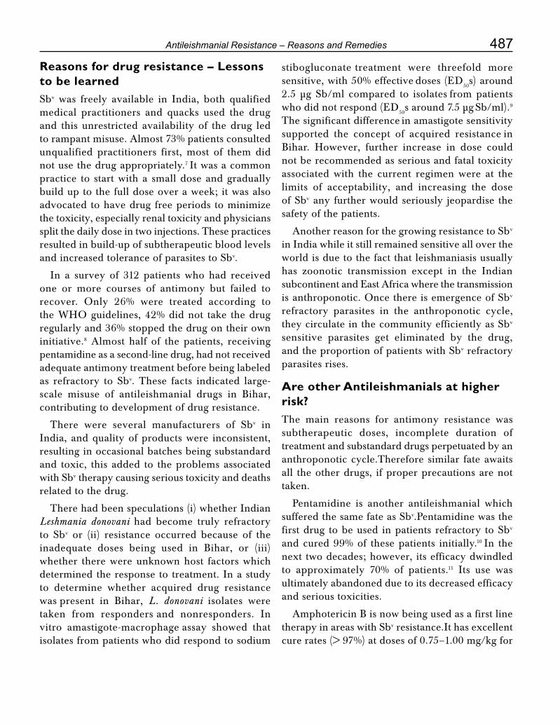
487Antileishmanial Resistance – Reasons and Remedies
Reasons for drug resistance – Lessons to be learnedSbv was freely available in India, both qualified medical practitioners and quacks used the drug and this unrestricted availability of the drug led to rampant misuse. Almost 73% patients consulted unqualified practitioners first, most of them did not use the drug appropriately.7 It was a common practice to start with a small dose and gradually build up to the full dose over a week; it was also advocated to have drug free periods to minimize the toxicity, especially renal toxicity and physicians split the daily dose in two injections. These practices resulted in build-up of subtherapeutic blood levels and increased tolerance of parasites to Sbv.
In a survey of 312 patients who had received one or more courses of antimony but failed to recover. Only 26% were treated according to the WHO guidelines, 42% did not take the drug regularly and 36% stopped the drug on their own initiative.8 Almost half of the patients, receiving pentamidine as a second-line drug, had not received adequate antimony treatment before being labeled as refractory to Sbv. These facts indicated large-scale misuse of antileishmanial drugs in Bihar, contributing to development of drug resistance.
There were several manufacturers of Sbv in India, and quality of products were inconsistent, resulting in occasional batches being substandard and toxic, this added to the problems associated with Sbv therapy causing serious toxicity and deaths related to the drug.
There had been speculations (i) whether Indian Leshmania donovani had become truly refractory to Sbv or (ii) resistance occurred because of the inadequate doses being used in Bihar, or (iii) whether there were unknown host factors which determined the response to treatment. In a study to determine whether acquired drug resistance was present in Bihar, L. donovani isolates were taken from responders and nonresponders. In vitro amastigote-macrophage assay showed that isolates from patients who did respond to sodium
stibogluconate treatment were threefold more sensitive, with 50% effective doses (ED
50s) around
2.5 µg Sb/ml compared to isolates from patients who did not respond (ED
50s around 7.5 µg Sb/ml).9
The significant difference in amastigote sensitivity supported the concept of acquired resistance in Bihar. However, further increase in dose could not be recommended as serious and fatal toxicity associated with the current regimen were at the limits of acceptability, and increasing the dose of Sbv any further would seriously jeopardise the safety of the patients.
Another reason for the growing resistance to Sbv in India while it still remained sensitive all over the world is due to the fact that leishmaniasis usually has zoonotic transmission except in the Indian subcontinent and East Africa where the transmission is anthroponotic. Once there is emergence of Sbv refractory parasites in the anthroponotic cycle, they circulate in the community efficiently as Sbv sensitive parasites get eliminated by the drug, and the proportion of patients with Sbv refractory parasites rises.
Are other Antileishmanials at higher risk?The main reasons for antimony resistance was subtherapeutic doses, incomplete duration of treatment and substandard drugs perpetuated by an anthroponotic cycle.Therefore similar fate awaits all the other drugs, if proper precautions are not taken.
Pentamidine is another antileishmanial which suffered the same fate as Sbv.Pentamidine was the first drug to be used in patients refractory to Sbv and cured 99% of these patients initially.10 In the next two decades; however, its efficacy dwindled to approximately 70% of patients.11 Its use was ultimately abandoned due to its decreased efficacy and serious toxicities.
Amphotericin B is now being used as a first line therapy in areas with Sbv resistance.It has excellent cure rates (> 97%) at doses of 0.75–1.00 mg/kg for

488 Medicine Update 2008 Vol. 18
15 infusions on alternate days. 12 It has been used extensively in Bihar with uniformly good results. The high cost, need for prolonged hospitalization, intravenous administration and occasional severe adverse reaction like hypokalemia, thrombocytopenia, myocarditis and death are some of the drawbacks of this excellent drug. Special amphotericin B treatment centers with trained personnel and free supply of drugs need to establish in these areas to promote its proper use.
Lipid-associated amphotericin (L-AB) preparations (AmBisome and Abelcet) are as effective as conventional amphotericin B, and have negligible adverse reactions.13 It is possible to administer high doses of L-AB over a short period with high cure rates; however, their high cost makes these compounds unaffordable in the endemic areas.
Miltefosine an alkyl phospholipid is the first oral agent approved for the treatment of leishmaniasis. At the recommended doses (100 mg daily for patients weighing ≥ 25 kg and 50 mg daily for those weighing < 25 kg for 4 weeks) cure rates were > 95%.14 As it is effective in Sbv resistant cases it can be used as a first line drug in areas with >10% Sbv unresponsiveness. Being an oral agent it offers an advantage of improved compliance, self administration and reduced costs of admission. Miltefosine with its excellent efficacy, oral administration and good tolerance can be an important tool in containing the epidemic. However the easy availability of the drug over the counter, high cost of the total therapy could lead to the intake of inadequate dose of the drug for shorter duration. As the drug has a long half-life (approximately 150 h), this could lead to subtherapeutic drug level for a prolonged period and ultimately widespread resistance.
Paromomycin, an aminoglycoside antibiotic was approved by the Indian government in August 2006 for the treatment of patients with visceral leishmaniasis. It is administered intramuscularly at a dose of 11 mg per kilogram daily for 21 days and has shown overall cure rate of 95%. The cure
rate among those whose disease had not responded to previous treatment with sodium stibogluconate or miltefosine or who had had a relapse was high (98%).15 Since paromomycin has not yet been used extensively, resistance is not a problem in the field, nevertheless, monitoring of resistance needs to be done.
Policies to prevent the appearance and spread of antileishmanial resistanceFree distribution of drugs
The high cost of the antileishmanial drugs coupled with easy, over the counter availability often leads to under dosing and incomplete treatment. This has been the major factor for antimony resistance and could lead to resistance to other drugs as well especially the novel oral agent miltefosine. Consideringthatmajorityofthepopulationcannotafford to purchase and complete a full course of treatment it is recommended that antileishmanials should be made available free of cost to be distributed through public and/ or private health care providers like Antitubercular and Antiretroviral drugs.
Directly observed Therapy
The directly observed treatment strategy for tuberculosis has been a big success and a parallel system could be evolved for leishmaniasis especially with oral drugs. This will lead to better compliance, completion of the treatment course and ultimately prevent resistance.
Combination therapy
Growing resistance of the parasite to antileishmanial drugs suggests that the currently used monotherapy needs to be reviewed. Multidrug combination therapy has been used successfully in tuberculosis, leprosy and malaria. The rationale behind combination therapy are (i) increased activity through use of compounds with synergistic or additive activity, (ii) preventing the emergence of drug resistance, (iii) lower dose requirement thereby reducing chances of toxic side effects and cost, and (iv) increased spectrum of activity. Studies to identify such combination in leishmaniasis need

489Antileishmanial Resistance – Reasons and Remedies
to be undertaken, as it will shorten the duration of therapy, improve compliance and decrease the development of resistance. In India amphotericin B and its lipid formulations, miltefosine and paromomycin are some of the drugs which can be combined.
Monitoring drug resistance
Ideally, parasite resistance should be monitored, rather than patient relapse rates. It will also permit the identification of key intracellular targets and parasite defense mechanisms, which can then be exploited to rationally develop analogs of existing drugs that would not get affected by the most common defenses. Analysis of genetic markers that determine high antileishmanial resistance, performed systematically for every parasite isolate that shows low antileishmanial sensitivity would facilitate the tracking of the level of resistance in affected populations. At present no molecular markers of resistance are available for the currently used antileishmanial drugs and the only reliable method for monitoring resistance of isolates is the technically demanding in vitro amastigote-macrophage model. Development of drug resistance markers and tools easy to use in the field should be encouraged.
New targets, new drugs
There are few better ways to avoid drug resistance than to have an adequate armory of drugs with different targets and no cross-resistance.
Training and Health education programs
One of the major reasons for antimony resistance was lack of awareness among the affected population and health care providers, about the need for effective treatment and control of kala-azar. There is a considerable need and scope for orientation programs to educate those at risk, doctors and the government agencies responsible for controlling and preventing kala-azar in India.
Management of HIV/VL co-infection
Another potential source for the emergence of drug resistance are the HIV/VL coinfected patients.
These patients have high parasite burden, a weak immune response, respond poorly to treatment and have a high relapse rate.Therefore they are the ideal candidates to harbor drug resistant parasites. With the growing burden of HIV in India, HIV/VL coinfection could become a major problem. Experience from Southern Europe shows that initial response to Sbv and conventional amphotericin B is low (~40-65%) in severely immunocompromised persons and severe adverse events are frequent. The best results are observed with AmBisome.It has also been observed that initiation of HAART dramatically decreases the incidence of VL coinfection. Therefore, HAART in combination with antileishmanials should be advocated strictly in these patients.
ConclusionFew drugs are available for treating Leishmania infections and the emergence of drug resistance is further complicating the control of leishmaniasis. A better understanding of resistance mechanisms and mechanism of action of drugs may point the waytomorerationalusesofdrugs.Combinationchemotherapy is rapidly emerging as the norm for treating several parasitic infections and is strongly advocated for kala-azar. Directly observed therapy given free, in treatment centers manned by trained personnel will go a long way in controlling the disease as well as drug resistance.
References1. Desjeux P. Leishmaniasis: current situation and new
perspectives. Comp ImmunolMicrobiol Infect Dis 2004; 27:305-18
2. Bora D. Epidemiology of visceral leishmaniasis in India.National Medical Journal of India 1999; 12, 62-68
3. Peters W The treatment of kala-azar. New approach to an old problem. Indian Journal of Medical Research 73 1981 Suppl.),1-18.
4. ThakurCP,KumarM,SinghSKetal.(1984)Comparisonofregimens of treatment with sodium stibogluconate in kala-azar. British Medical Journal 1981; 288, 895-897.
5. Sundar S, Singh VP, Sharma S, Makharia MK & Murray HW Response to interferon-γ plus pentavalent antimony in Indian visceral leishmaniasis. Journal of Infectious Diseases 1997; 176,1117-1119
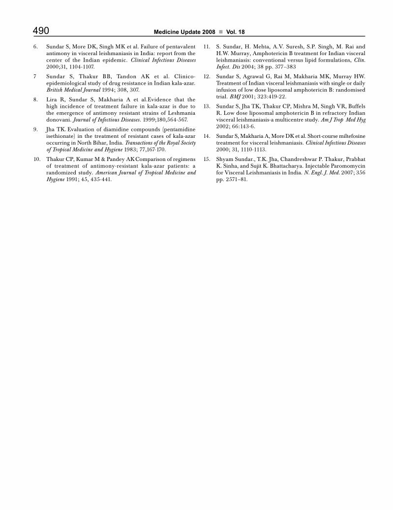
490 Medicine Update 2008 Vol. 18
6. Sundar S, More DK, Singh MK et al. Failure of pentavalent antimony in visceral leishmaniasis in India: report from the center of the Indian epidemic. Clinical Infectious Diseases 2000;31, 1104-1107.
7 Sundar S, Thakur BB, Tandon AK et al. Clinico-epidemiological study of drug resistance in Indian kala-azar. British Medical Journal 1994; 308, 307.
8. Lira R, Sundar S, Makharia A et al.Evidence that the high incidence of treatment failure in kala-azar is due to the emergence of antimony resistant strains of Leshmania donovani. Journal of Infectious Diseases. 1999;180,564-567.
9. Jha TK. Evaluation of diamidine compounds (pentamidine isethionate) in the treatment of resistant cases of kala-azar occurring in North Bihar, India. Transactions of the Royal Society of Tropical Medicine and Hygiene 1983; 77,167-170.
10. ThakurCP,KumarM&PandeyAK.Comparisonofregimensof treatment of antimony-resistant kala-azar patients: a randomized study. American Journal of Tropical Medicine and Hygiene 1991; 45, 435-441.
11. S. Sundar, H. Mehta, A.V. Suresh, S.P. Singh, M. Rai and H.W. Murray, Amphotericin B treatment for Indian visceral leishmaniasis: conventional versus lipid formulations, Clin. Infect. Dis 2004; 38 pp. 377–383
12. Sundar S, Agrawal G, Rai M, Makharia MK, Murray HW. Treatment of Indian visceral leishmaniasis with single or daily infusion of low dose liposomal amphotericin B: randomised trial. BMJ 2001; 323:419-22.
13. SundarS,JhaTK,ThakurCP,MishraM,SinghVR,BuffelsR. Low dose liposomal amphotericin B in refractory Indian visceral leishmaniasis-a multicentre study. Am J Trop Med Hyg 2002; 66:143-6.
14. Sundar S, Makharia A, More DK et al. Short-course miltefosine treatment for visceral leishmaniasis. Clinical Infectious Diseases 2000; 31, 1110-1113.
15. ShyamSundar.,T.K.Jha,ChandreshwarP.Thakur,PrabhatK. Sinha, and Sujit K. Bhattacharya. Injectable Paromomycin for Visceral Leishmaniasis in India. N. Engl. J. Med. 2007; 356 pp. 2571–81.

CHAPTER
65Statins in Critical CarePankaj Manoria, P. C. Manoria
IntroductionThe role of statin drugs in the reduction of serum lipids has been well documented.1-3 More recently we have evidences which suggest that statins may positively impact many organ systems and disease states independent of lipid reduction. These have added a wide scope of potential targets for statin therapy ranging from plaque stabilization in acute coronary syndrome (ACS) to decreasing loss ofrenal function; lowering mortality in patients with diastolic heart failure; to prevention and treatment of stroke. This review summarizes the evidence in favor of role of statins in intensive care unit and briefly discuss the role of statins in prevention and treatment of sepsis as a potential future application of statins in critical care (see Table 1).
Statins in Acute coronary syndromesWhile the benefit of statin therapy in patients with stable coronary artery disease is clearly
recognized,1-3 the positive impact of the initiation of statin therapy immediately following ACSoccurrence has emerged only recently. Both STEMI and NSTEMI frequently require intensive-care treatment and these patients are at high risk for recurrent coronary events, sudden death and all-cause mortality. The stabilization of vulnerable lesions is a critical aspect in preventing these events following ACS. Despite significant advances inantiplatelet and antithrombotic therapy, these therapeutic options alone do not appear to suffice in treating the unstable plaque stabilization. Through their cholesterol lowering and pleiotropic effects, statins are viewed as important contributors to plaque stabilization.4 Besides this stains have several other benefits in patients of ACS whichare highlighted in Table 2.
A number of retrospective and observational studies have suggested that initiating statin therapy immediately after an ACS is associated with
Table 1 : Potential use of statins in critical careAcute coronary syndrome
Percutaneous coronary intervention
Septicemia
Cerebro-vascularaccident
Heart failure
Post organ transplant
Perioperative in non cardiac surgery
Table 2 : Rationale for use of statins in ACSStatins cause stabilization of vulnerable plaque
DecreasemortalityinACSpatients
DecreaseMACEinpatientsundergoingPCI
Antiarrhythmic action
In hospital administration improves compliance
Sudden withdrawal of statins during an ACS can behazardous
492 Medicine Update 2008 Vol. 18
significantly reduced rates of recurrent coronary events and death.5-11 These have been followed up on by large-scale, randomized, controlled trials. The Myocardial Ischemia Reduction with Aggressive CholesterolLowering(MIRACL)12 trial was the first to demonstrate a reduced rate of recurrent cardiac events by statin therapy. In this study 3086 patients with unstable angina or non-Q-wave infarction were randomized within 24– 96 h after hospital admission to receive either 80 mg of atorvastatin or placebo in addition to state-of-the-art therapy for 4monthsafterACS.Theprimaryendpointofthetrial – death, cardiac arrest, myocardial infarction or worsening unstable angina requiring emergency hospitalization at 16 weeks – showed a relative risk reduction of 16% [95% confidence interval (CI),0–30; P ¼ 0.048; absolute risk reduction, 2.6%].
These findings were supported and extended by the Pravastatin or Atorvastatin Evaluation and Infection Therapy trial (PROVE IT)13 This trial randomized4162patientshospitalizedforanACS( NSTEMI, and STEMI). Patients were included within 10 days of their index event, and were randomized to either pravastatin (40 mg daily), or atorvastatin (80 mg daily). Of note, this study also included a significant number of patients (69%) post percutaneous revascularization. Results showed a strong trend toward benefits in the primary endpoint (death from any cause or a major cardiovascular event) in the high-dose atorvastatin group within 30 days. A statistically significant decrease in the primary endpoint arose at 180 days (relative risk reduction of 16% (P value=0.005; absolute risk reduction, 3.9%). The secondary endpoints (revascularization, unstable angina requiring hospitalization, and the combined endpoint of MI, revascularization, or death from coronary heart disease) also showed a significant decrease during the overall 2-year assessment period. The benefit, derived from intensive versus conventional lipid-lowering therapy arouse on top of background evidence-basedACStherapy(includingantiplatelettherapy, β-blockers and angiotensin-converting- enzyme inhibitors in a large majority of patients).
In contrast, the Aggrastat to Zocor (A to Z)14 trial did not demonstrate superiority for the intensive statin regimen (p = 0.14). A statin trial failing to achieve a statistical significance was a major set back. But when this trial was critically analyzed it wasfoundthattheCRPlevelreductionwasonly17% in this trial as compared to 34% and 38% in the MIRACL and PROVE-IT trial. More thanthe lipid level it is the anti-inflammatory action of statinswhichismoreimportantinACS.Secondlythe benefit was not seen in the first three months in the trial but seen thereafter. This could be due to the fact that statin was titrated to its maximum dose only after 3 months.
Based on the findings from these three large randomized trials it has been speculated that the early benefits of statin therapy may be caused largely by anti-inflammatory effects, whereas the delayed benefits are more likely to be lipid-modulated.
In hospital administration of statin improves long term compliance.
It has also been seen that in hospital administration of statin improves long term compliance. Patients will be more likely to understand the importance of lipid lowering therapy if statin is started during hospitalizationforACSandthenheislesslikelytodiscontinuethetherapy.TheCHAMPprogrammeaddressed this issue in the pre-discharge cardiac patient.15 They found that the 1 year compliance rate increase from 10% to 91% when statins were prescribed during hospitalization for ACS thanwhen it is prescribed on out patient basis.
Taken together, the evidence suggests that, in the absence of contraindications or intolerance, statin therapy should be initiated within 24-96 h after an ACSregardlessofpretreatmentcholesterollevels.In addition, experimental and clinical findings support pleiotropic statin effects contributing to plaque stabilization and improved endothelial function. Early initiation of high-dose statin therapy will likely maximize clinical benefit derived from both aggressive LDL-cholesterol lowering and pleiotropic effects. Until the efficacy and safety

493Statins in Critical Care
of other statins have been proven in this context, atorvastatin 80 mg should be used as the most firmly established statin for this purpose. Table 3 summarizes the randomized trials of statin in critical care.
Statin withdrawal syndromeThe possibility of a detrimental rebound effect from statin withdrawal has been proposed by Heeschen et al.16 Among acute coronary syndrome subjects, in the Platelet Receptor Inhibition in Ischemic Syndrome Management (PRISM) study, statin therapy was associated with a reduced event rate at 30-day follow-up compared with patients without statins (adjusted RR 0.49, P = 0.004). However, if statins were discontinued after admission, cardiac risk tended to be higher compared to those who never received statins (RR 1.69, P = 0.15). Continuation of statin therapy was one ofthe independent predictors of patient outcomes. ThereforestoppingstatinsinanACSpatientwhowas already on statins could be dangerous.
Statins and percutaneous coronary intervention in ACSThe pleiotropic effects of statins are also being studied for their effects following PCI in acutecoronary syndrome. The Atorvastatin for Reduction of Myocardial Damage During Angioplasty-Acute Coronary Syndromes (ARMYDA-ACS) trial
demonstrated that short-term pretreatment with atorvastatin 80 mg reduces the incidence of cardiac events inACSpatientsundergoing earlyPCI, abenefit largely driven by a significant reduction in post procedural MI.17 This trial randomized 171patientswithnon-ST-segment-elevationACSto pretreatment with atorvastatin (80 mg 12 hours before,and40mgimmediatelypriortoPCI)ortoplacebo. All patients were given a 600-mg loading dose of clopidogrel and long-term atorvastatin treatment (40 mg/day). The primary endpoint, a composite of death, MI, and TVR at 30 days, was significantlyloweramongACSpatientspretreatedwith atorvastatin compared with those treated with placebo (5% vs. 17% respectively, P = .01). TheincidenceofMACEwasprimarilydrivenbysignificant differences in post procedural MI, with reductionsincreatinekinase-MB(CK-MB)and
troponin levels in patients pretreated with atorvastatin. In multivariate analysis, pretreatment with atorvastatin was associated with an 88% reductionintherelativeriskofMACEat30days.If confirmed by larger randomized trials, this study may support the indication for “upstream” administration of high-dose statins in patients withACSundergoinganearly invasive strategy.Future studies in larger populations will need to determine if these peri-procedural reductions in MI translate into clinically meaningful reductions in hard events.
Table 3 : Randomized trials of statins in critical care
S No Trial n Subsets Statin Follow
up
Result
1 MIRACL12 3086 ACS
(NSTEMI)
Atorva-Statin
(80mg)
16 Weeks
16% RR in composite end point of death MI & TVR (P=0.48)
2 PROVE IT-TIMI 2213
4162 ACS(NSTEMI
& STEMI)
Atorva-Statin
(80mg)
2 Years 16% RR in all cause mortality (p= 0.005)
3 A TO Z14 4497 ACS
(NSTEMI)
Simva-Statin
(40mg BD)
2 Year Non significant trend (p-0.14) in favour of high dose simvastatin
4 ARMYDA-
ACS17
191 PCIfor
NSTEMI
Atorva-Statin
(80mg)
30 Days Significant reduction of composite end point of death MI & TVR (P=0.01)
5 SPARCL42 4731 Recent stroke/ TIA
Atorva-Statin
(80mg)
4.9 Year Decrease recurrent stroke by 16% (p=0.03)
494 Medicine Update 2008 Vol. 18
Statins and SepsisBackground
Currentprescribingguidelinesrecommendthatstatin therapy be discontinued in patients with an acute illness such as severe infection and septicemia. This is based on the assumptions that serum lipoproteins may protect against the lethal effects of endotoxinemia by binding and inactivating endotoxin.18-21 Furthermore, a low serum cholesterol concentration is an independent predictor of infectious complications and mortality in hospitalized patients.22,23 But recently, enormous data supporting the favorable role of statins in both prevention and as adjuvant therapy in the treatment of septicemia has piled up. This would challenge the current prescribing guidelines to with-hold statins in acutely ill patients and warrants further prospective investigation.
Mechanism of benefit of statins in septicemia
Possible mechanisms include interference with leukocyte–endothelium interaction, prevention of toxin-induced cellular damage and modulation of endothelial function.
Leukocyte–endothelial interaction is a critical event that precedes the trans-migration of leukocytes from the vasculature to tissue.24 Statins interfere with many of the steps involved in this process by increasing the level of endothelial NO, inhibiting the adhesion of monocytes to the endothelium and
by inhibiting the release of polymorphonuclear (PMN) cell chemoattractants.
Statins also exert activity against a toxin released by Staphylococcus aureus in septic patients. Elevation of HDL by statins play a role in neutralisation of endotoxins in sepsis.
Statins deplete isoprenoids, which are important non-sterol cholesterol precursors. These precursors are essential for the farnesylation and geranylation of membranal G proteins, which play an important role in the signal transduction pathways that determines cellular migration and proliferation.25
Improvement in Vascular Function in sepsis is also reported by statin use.26 Endothelial cells play an important role in the control of vascular tone, permeability, blood flow, coagulation, thrombolysis, inflammation, tissue repair, and growth. Endothelial activation, dysfunction, and apoptosis play a crucial role in the pathogenesis of sepsis and subsequent multiple organ dysfunction. It has been shown that statins increase expression and enhance activity of endothelial NOS, upregulate prostacyclin and tissue – type plasminogen activator , and downregulate tissue factor, endothelin – 1, and plasminogen activator or inhibitor – 1 (PAI – 1) thus improving endothelial functions.
Evidences from Observational data
No data from randomised trials of statins and sepsis are available, but observational studies lend
Table 4 : Summary of human observation studies in sepsis
Study design Setting (n) Study effect
Retrospective cohort27
Elderly patients with cardio-vascular disease hospitalized for sepsis
69168 Decrease sepsis & mortality
Prospective cohort28 Patients admitted to hospital with suspected or confirmed bacterial infection
361 Decrease sepsis & need for intensive care admission
Retrospective cohort29
Patients admitted to hospital with bacteremia
388 Decrease mortality in statin treated patients
Retrospective cohort30
Patients with discharge diagnosis of pneumonia
787 Decrease 30 day mortality
Retrospective cohort31
Intensive care patients ventilated more than 96 hours
438 Increase mortality
Retrospective cohort32
Patients requiring hospital admission for bacteremia
438 Decrease mortality with pre-treatment and when treatment continued after diagnosis

495Statins in Critical Care
support to a potentially important preventive and/or treatment effect (Table 4). The largest study to date is a population-based cohort study involving the linked administrative databases in Ontario, Canada,andincludedamatchedcohortof69168patients. The incidence of sepsis was substantially lower among patients receiving statins (hazard ratio[HR]0·81;95%CI0·72–0·91).Theprotectiveassociation between statins and sepsis persisted in high-risk subgroups including patients with diabetes mellitus, malignancy, and those receiving oral steroids. Significant reduction in severe sepsis (HR0·83;95%CI0·70–0·97)andfatalsepsis(HR0·75;95%CI0·61–0·93)werealsoobserved.
Due to their observational nature, the studies presented above may suffer from selection bias and hidden confounding, and we should interpret these results with caution. Taken together, however, these early data suggest that statins may, in human beings, contribute to preventing sepsis and have a role in the treatment of sepsis.
Thus there is growing interest among clinicians in the role that statins may play in preventing and treating serious infections. If such an effect of statins can be supported by randomized controlled clinical trials, then the implications could be far reaching. The stage is now set for randomized clinical trials that will determine the precise role, if any, that statins may have in preventing and treating sepsis.
Statins and StrokeStatin therapy has been shown to significantly lower the risk of stroke in several studies in patients with coronary heart disease.1,33,34 Whether statins prevent strokes in patients without heart disease is currently under evaluation in several trials. Because cholesterol is often not elevated in stroke patients35 the benefit of statins in stroke patients may be an effect independent of cholesterol lowering such as stabilization of pre-cerebral atheroma in the aorta and carotid arteries36 and the inhibition of platelet reactivity.37 Furthermore, some of the beneficialCNSeffectsofstatinsmaybeduetotheir
augmentation of NO production (NO may improve CNS collateral blood flow), enhance cerebralvasodilator responses and prevent apoptosis.38,39 For example, in a murine model of ischemic stroke, both atorvastatin and simvastatin increased cerebral blood flow and decreased infarct size in wild-type but not in an eNOS-knockout mice.40 Pravastatin was compared to placebo in patients with subarachnoid hemorrhage and it was found that those randomized to pravastatin had a 61% reduction in the incidence of ipsilateral vasospasm and an 82% reduction in the incidence of delayed ischemic deficits.41
Therecentclinical trial,SPARCLwasthefirstrandomized study which showed that statins prevent recurrent stroke and transient ischemic attack in patients who have already had 1 of these events.42 We already knew they prevented strokes in patients with coronary disease, but this is the first time it has been looked at in patients with cerebrovascular disease. Therefore, statins should get the attention of neurologists for use in these patients.
Statins in Heart FailureIt was traditionally believed that statins may be detrimental in the treatment of heart failure because they lower the lipid pool. It was thought that a large lipid pool absorbed the cytokines which are harmful in heart failure. Data from trials in the last two years show that this concept is changing. In fact statins improve microvascular circulation and endothelial function by stimulating angiogenesis and modulating the synthesis and activity of endothelial nitric oxide synthase and endothelin-1. In animal models, statins impact the process of cardiac remodeling by reducing ventricular hypertrophy in response to angiotensin II, and through down regulation of angiotensin I receptor expression and reduction in the secretion of matrix metalloproteases. In HPS study there were hardly any heart failure related deaths in patients on simvastatin. In a study of elderly population with heart failure, Roy et al showed survival benefit with
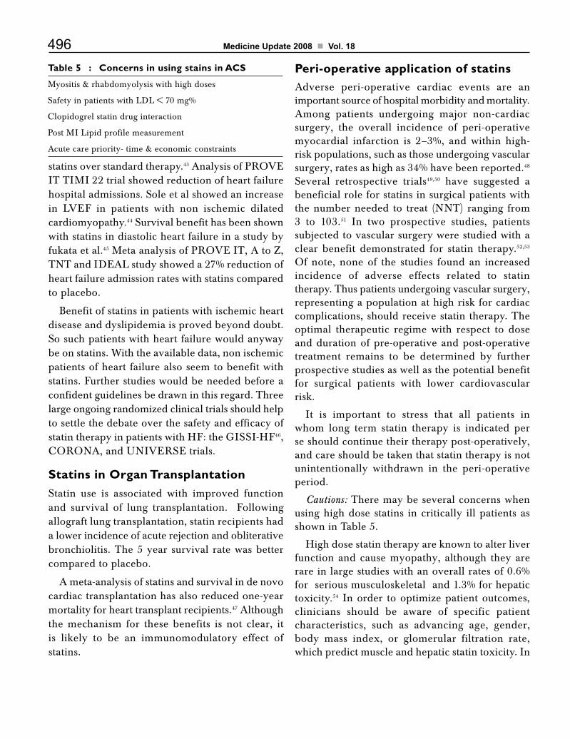
496 Medicine Update 2008 Vol. 18
statins over standard therapy.43 Analysis of PROVE IT TIMI 22 trial showed reduction of heart failure hospital admissions. Sole et al showed an increase in LVEF in patients with non ischemic dilated cardiomyopathy.44 Survival benefit has been shown with statins in diastolic heart failure in a study by fukata et al.45 Meta analysis of PROVE IT, A to Z, TNT and IDEAL study showed a 27% reduction of heart failure admission rates with statins compared to placebo.
Benefit of statins in patients with ischemic heart disease and dyslipidemia is proved beyond doubt. So such patients with heart failure would anyway be on statins. With the available data, non ischemic patients of heart failure also seem to benefit with statins. Further studies would be needed before a confident guidelines be drawn in this regard. Three large ongoing randomized clinical trials should help to settle the debate over the safety and efficacy of statin therapy in patients with HF: the GISSI-HF46, CORONA,andUNIVERSEtrials.
Statins in Organ TransplantationStatin use is associated with improved function and survival of lung transplantation. Following allograft lung transplantation, statin recipients had a lower incidence of acute rejection and obliterative bronchiolitis. The 5 year survival rate was better compared to placebo.
A meta-analysis of statins and survival in de novo cardiac transplantation has also reduced one-year mortality for heart transplant recipients.47 Although the mechanism for these benefits is not clear, it is likely to be an immunomodulatory effect of statins.
Peri-operative application of statinsAdverse peri-operative cardiac events are an important source of hospital morbidity and mortality. Among patients undergoing major non-cardiac surgery, the overall incidence of peri-operative myocardial infarction is 2–3%, and within high-risk populations, such as those undergoing vascular surgery, rates as high as 34% have been reported.48 Several retrospective trials49,50 have suggested a beneficial role for statins in surgical patients with the number needed to treat (NNT) ranging from 3 to 103.51 In two prospective studies, patients subjected to vascular surgery were studied with a clear benefit demonstrated for statin therapy.52,53 Of note, none of the studies found an increased incidence of adverse effects related to statin therapy. Thus patients undergoing vascular surgery, representing a population at high risk for cardiac complications, should receive statin therapy. The optimal therapeutic regime with respect to dose and duration of pre-operative and post-operative treatment remains to be determined by further prospective studies as well as the potential benefit for surgical patients with lower cardiovascular risk.
It is important to stress that all patients in whom long term statin therapy is indicated per se should continue their therapy post-operatively, and care should be taken that statin therapy is not unintentionally withdrawn in the peri-operative period.
Cautions: There may be several concerns when using high dose statins in critically ill patients as shown in Table 5.
High dose statin therapy are known to alter liver function and cause myopathy, although they are rare in large studies with an overall rates of 0.6% for serious musculoskeletal and 1.3% for hepatic toxicity.54 In order to optimize patient outcomes, clinicians should be aware of specific patient characteristics, such as advancing age, gender, body mass index, or glomerular filtration rate, which predict muscle and hepatic statin toxicity. In
Table 5 : Concerns in using stains in ACS
Myositis & rhabdomyolysis with high doses
Safety in patients with LDL < 70 mg%
Clopidogrelstatindruginteraction
Post MI Lipid profile measurement
Acute care priority- time & economic constraints
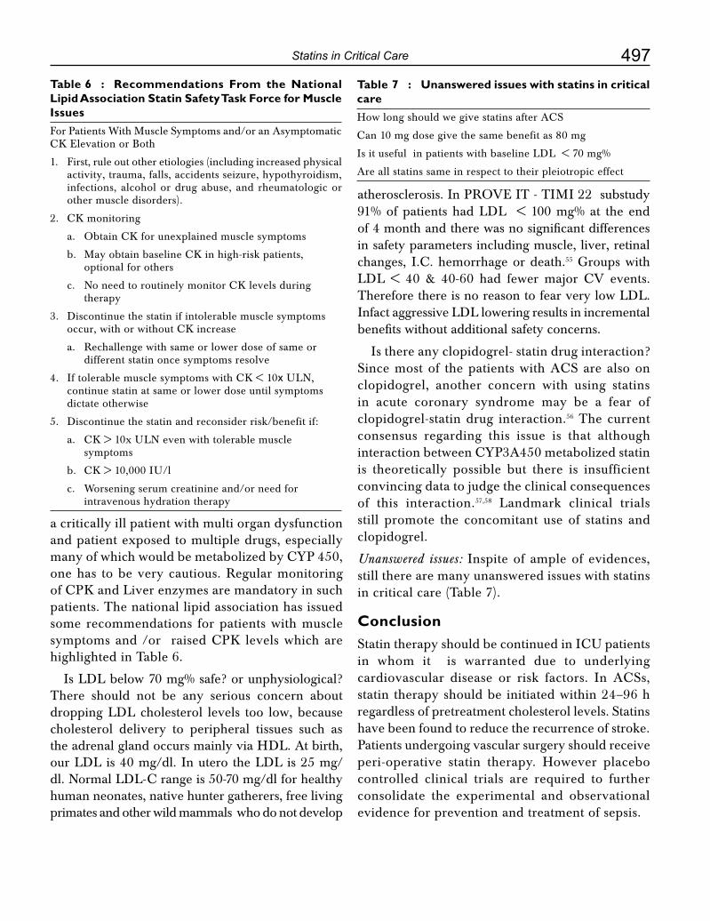
497Statins in Critical Care
a critically ill patient with multi organ dysfunction and patient exposed to multiple drugs, especially manyofwhichwouldbemetabolizedbyCYP450,one has to be very cautious. Regular monitoring ofCPKandLiverenzymesaremandatoryinsuchpatients. The national lipid association has issued some recommendations for patients with muscle symptomsand/or raisedCPK levelswhicharehighlighted in Table 6.
Is LDL below 70 mg% safe? or unphysiological? There should not be any serious concern about dropping LDL cholesterol levels too low, because cholesterol delivery to peripheral tissues such as the adrenal gland occurs mainly via HDL. At birth, our LDL is 40 mg/dl. In utero the LDL is 25 mg/dl.NormalLDL-Crangeis50-70mg/dlforhealthyhuman neonates, native hunter gatherers, free living primates and other wild mammals who do not develop
atherosclerosis. In PROVE IT - TIMI 22 substudy 91% of patients had LDL < 100 mg% at the end of 4 month and there was no significant differences in safety parameters including muscle, liver, retinal changes, I.C.hemorrhageordeath.55 Groups with LDL < 40 & 40-60 had fewer major CV events.Therefore there is no reason to fear very low LDL. Infact aggressive LDL lowering results in incremental benefits without additional safety concerns.
Is there any clopidogrel- statin drug interaction? Sincemostof thepatientswithACSarealsoonclopidogrel, another concern with using statins in acute coronary syndrome may be a fear of clopidogrel-statin drug interaction.56 The current consensus regarding this issue is that although interactionbetweenCYP3A450metabolizedstatinis theoretically possible but there is insufficient convincing data to judge the clinical consequences of this interaction.57,58 Landmark clinical trials still promote the concomitant use of statins and clopidogrel.
Unanswered issues: Inspite of ample of evidences, still there are many unanswered issues with statins in critical care (Table 7).
ConclusionStatintherapyshouldbecontinuedinICUpatientsin whom it is warranted due to underlying cardiovascular disease or risk factors. In ACSs,statin therapy should be initiated within 24–96 h regardless of pretreatment cholesterol levels. Statins have been found to reduce the recurrence of stroke. Patients undergoing vascular surgery should receive peri-operative statin therapy. However placebo controlled clinical trials are required to further consolidate the experimental and observational evidence for prevention and treatment of sepsis.
Table 6 : Recommendations From the National Lipid Association Statin Safety Task Force for Muscle Issues For Patients With Muscle Symptoms and/or an Asymptomatic CKElevationorBoth
1. First, rule out other etiologies (including increased physical activity, trauma, falls, accidents seizure, hypothyroidism, infections, alcohol or drug abuse, and rheumatologic or other muscle disorders).
2. CKmonitoring
a. ObtainCKforunexplainedmusclesymptoms
b. MayobtainbaselineCKinhigh-riskpatients,optional for others
c. NoneedtoroutinelymonitorCKlevelsduringtherapy
3. Discontinue the statin if intolerable muscle symptoms occur,withorwithoutCKincrease
a. Rechallenge with same or lower dose of same or different statin once symptoms resolve
4. IftolerablemusclesymptomswithCK<10x ULN, continue statin at same or lower dose until symptoms dictate otherwise
5. Discontinue the statin and reconsider risk/benefit if:
a. CK>10xULNevenwithtolerablemusclesymptoms
b. CK>10,000IU/l
c. Worsening serum creatinine and/or need for intravenous hydration therapy
Table 7 : Unanswered issues with statins in critical careHowlongshouldwegivestatinsafterACS
Can10mgdosegivethesamebenefitas80mg
Is it useful in patients with baseline LDL < 70 mg%
Are all statins same in respect to their pleiotropic effect
498 Medicine Update 2008 Vol. 18
References1. Scandinavian Simvastatin Survival Study Group.Randomised
trial of cholesterol lowering in 4444 patients with coronary heart disease: the Scandinavian Simvastatin Survival Study (4S). Lancet. 1994;344:1383-1389.
2. Heart Protection Study Collaborative Group. MRC/BHF Heart Protection Study of cholesterol lowering with simvastatin in 20,536 high-risk individuals; a randomized placebo-controlled trial. Lancet. 2002;360(9326):7–22. 27.
3. Anglo-ScandinavianCardiacOutcomesTrial-LipidLoweringArm(ASCOT-LLA); a multicentre randomized controlledtrial. Lancet. 2003;361:1149–1158.
4. Libby P. Coronary artery injury and the biology ofatherosclerosis: inflammation, thrombosis, and stabilization. Am J Cardiol 2000; 86:3–8.
5. Aronow HD, Topol EJ, Roe MT, et al. Effect of lipid-lowering therapy on early mortality after acute coronary syndromes: an observational study. Lancet 2001; 357:1063–1068.
6. Stenestrand UWL. Swedish Register of Cardiac IntensiveCare (RIKS-HIA). Early statin treatment following acutemyocardial infarction and 1-year survival. JAMA 2001; 285:430–436
7. Wright RS, Murphy JG, Bybee KA, et al. Statin lipid-lowering therapy for acute myocardial infarction and unstable angina: efficacy and mechanism of benefit. Mayo Clin Proc 2002; 77:1085–1092.
8. Paradiso-HardyFL,GordonWL,JackeviciusCA,etal.Theimportance of in hospital statin therapy for patients with acute coronary syndromes. Pharmacotherapy 2003; 23:506–513.
9. Bybee KA, Wright RS, Williams BA, et al. Effect of concomitant or very early statin administration on in-hospital mortality and reinfarction in patients with acute myocardial infarction. Am J Cardiol 2001; 87:771–774.
10. Newby LK, Kristinsson A, Bhapkar MV, et al. Early statin initiation and outcomes in patients with acute coronary syndromes. JAMA 2002; 287:3087–3095.
11. Spencer FA, Allegrone J, Goldberg RJ, et al. Association of statin therapy with outcomes of acute coronary syndromes: theGRACEStudy.Ann Intern Med 2004; 140:857–866.
12. Schwartz GG, Olsson AG, Ezekowitz MD, et al. Myocardial IschemiaReductionwithAggressiveCholesterolLowering(MIRACL)Study Investigators. Effects of atorvastatin onearly recurrent ischemic events in acute coronary syndromes: theMIRACL study: a randomized controlled trial. JAMA 2001; 285:1711–1718.
13. Cannon CP, Braunwald E, McCabe CH, et al. Intensiveversus moderate lipid lowering with statins after acute coronary syndromes. N Engl J Med 2004; 350:1495–1504.
14. Lemos JAM, Blazing MAM, Wiviott SDM, et al. Early intensive vs. a delayed conservative simvastatin strategy in patients with acute coronary syndromes: phase Z of the A to Z Trial. JAMA 2004; 292:1307–1316.
15. Fonarow GC, Gawlinski A. Rationale and design of theCardiac Hospitalization Atherosclerosis ManagementProgramat theUniversityofCaliforniaLosAngeles.Am J Cardiol 2000.85: 10A-17A.
16. Heeschen C, Hamm CW, Laufs, et al. Withdrawal ofstatins increases event rates in patients with acute coronary syndromes. Circulation 2002; 105: 1446-1452.
17. ARMYDA-ACS(AtorvastatinforReductionofMyocardialDamageduringAngioplasty-AcuteCoronarySyndromes)trial presented in the 56th Annual Scientific session of AmericanCollegeofcardiology,NewOrleans24thMarch2007.
18. Almog Y. Statins, inflammation, and sepsis: hypothesis. Chest 2003; 124: 740–43.
19. Feingold KR, Funk JL, Moser AH, Shigenaga JK, Rapp JH, GrunfeldC.Roleforcirculatinglipoproteins inprotectionfrom endotoxin toxicity. Infect Immun 1995; 63: 2041–46.
20. Harris HW, Gosnell JE, Kumwenda ZL. The lipemia of sepsis: triglyceride-rich lipoproteins as agents of innate immunity. J Endotoxin Res 2000; 6: 421–30.
21. HarrisHW,GrunfeldC,FeingoldKR,Rapp JH.Humanvery low density lipoproteins and chylomicrons can protect against endotoxin-induced death in mice. J Clin Invest 1990; 86: 696–702.
22. Bonville DA, Parker TS, Levine DM, et al. The relationships of hypocholesterolemia to cytokine concentrations and mortality in critically ill patients with systemic inflammatory response syndrome. Surg Infect (Larchmt) 2004; 5: 39–49.
23. CanturkNZ,CanturkZ,OkayE,YirmibesogluO,EraldemirB. Risk of nosocomial infections and effects of total cholesterol, HDL cholesterol in surgical patients. Clin Nutr 2002; 21: 431–36.
24. Pruefer D, Makowski J, Schnell M, et al. Simvastatin inhibits inflammatory properties of Staphylococcus aureus alpha-toxin. Circulation 2002;106:2104– 10.
25. Frenette PS Locking a leukocyte integrin with statins. N.Eng J Med 2001; 345:1419-1-1421
26. Pruefer D, Makowski J,Schnell M,et al (2002) Simvastatin Inhibits inflammatory properties of staphylococcus aureus alpha- toxin. Circulation 2002;106:2104-2110.
27. Hackam D, Mamdani M, Li P, Redelmeier D. Statins and sepsis in patients with cardiovascular disease: a population-based cohort analysis. Lancet 2006; 367: 413–18.
28. Almog Y, Shefer A, Novack V, et al. Prior statin therapy is associated with a decreased rate of severe sepsis. Circulation 2004; 110: 880–85.
29. LiappisAP,KanVL,RochesterCG,SimonGL.Theeffectof statins on mortality in patients with bacteremia. Clin Infect Dis 2001; 33: 1352–57.
30. Mortensen EM, Restrepo MI, Anzueto A, Pugh J. The effect of prior statin use on 30-day mortality for patients hospitalized with community-acquired pneumonia. Respir Res 2005; 6: 82.
31. Fernandez R, De Pedro VJ, Artigas A. Statin therapy prior toICUadmission:protectionagainstinfectionoraseverity
499Statins in Critical Care
marker? Intensive Care Med 2006; 32: 160–64.
32. Kruger P, Fitzsimmons K, Cook D, Jones M, Nimmo G.Statin therapy is associated with fewer deaths in patients with bacteremia. Intensive Care Med 2006; 32: 75–79.
33. Ko DT, Mamdani M . Alter DA Lipid – lowering therapy with statins in high – risk elderly patients : the treatment – risk paradoxl. JAMA 2004 291: 1864-1870.
34. HessDC,DemchukAM,BrassLM,YatsuFMHMG-CoAreductase inhibitors (statins): a promising approach to stroke prevention. Neurology 2000 54:790-796
35. ProspectivestudiescollaborationCholesterol,diastolicbloodpressure, and stroke: 13000 strokes in 4,5,00,00 people in 45 prospective cohorts. Lancet 1995 346:1647 – 1653.
36. CrisbyM,Nordin-FredrikssonG,ShahPK,YanoJ,ZhuJ,Nilsson J. Pravastatin treatment increases collagen content and decreases lipid content, inflammation, metalloproteinases, and cell death in human carotid plaques: implications for plaque stabilization. Circulation 2001;103:926– 33.
37. Del Zoppo GJ. The role of platelets in ischemic stroke. Neurology 1998;51 (Suppl.3):S9– 14.
38. Laufs U, Endres M, Stagliano N, et al. Neuroprotection mediated by changes in the endothelial actin cytoskeleton. J Clin Invest 2000; 106:15– 24.
39. KureishiY,LuoZ,ShiojimaI,etal.TheHMG-CoAreductaseinhibitor simvastatin activates the protein kinase Akt and promotes angiogenesis in normocholesterolemic animals. Nat Med 2000; 6:1004– 10.
40. Laufs U, Gertz K, Huang P, et al. Atorvastatin upregulates type III nitric oxide synthase in thrombocytes, decreases platelet activation, and protects from cerebral ischemia in normocholesterolemic mice. Stroke 2000;31:2442– 9.
41. Tseng M, Kirkpatrick PJ. Statins ameliorate cerebral vasospasm, maintain cerebral autoregulation, and reduce the incidence of ischemic deficits after aneurysmal subarachnoid hemorrhage: a double-blind, placebo-controlled trial. Stroke 2005;36:435.
42. WeichMetal.TheSPARCL(StrokePreventionbyAggressiveReduction in Cholesterol Levels) presented in the 15thEuropean Stroke Conference, May 16-19, 2006, Brussels,Belgium.
43. Ray JG, Gong Y, Sykora K, et al. Statin use and survival outcomes in elderly patients with heart failure. Arch Intern Med 2005;165:62 -7.
44. Sola S, Mir M, Rajagopalan S, et al. Statin therapy improves mortality, cardiovascular outcomes, and levels of inflammatory markers in patients with heart failure. Circulation S 2004; 110(2591 Suppl):556.
45. FukutaH,SaneDC,BrucksS,etal.Statintherapymaybeassociated with lower mortality in patients with diastolic heart failure: a preliminary report. Circulation 2005;112:357 - 63.
46. Tavazzi L, Tognoni G, Franzosi MG, et al. Rationale and design of the GISSI heart failure trial: a large trial to assess the effects of n-3 polyunsaturated fatty acids and rosuvastatin in symptomatic congestive heart failure. Eur J Heart Fail 2004;6:635 - 41.
47. Raval Ny, Mehra MR. Metaanalysis of statins and survival in de novo cardiac transplantation. Transplant proc 2004; 36: 1539- 1541.
48. LeeTH,MarcantonioER,MangioneCM,etal.Derivationand prospective validation of a simple index for prediction of cardiac risk of major Noncardiac surgery. Circulation 1999; 100:1043–1049.
49. Poldermans D, Bax JJ, Kertai MD, et al. Statins are associated with a reduced incidence of perioperative mortality in patients undergoing major Noncardiac vascular surgery. Circulation 2003; 107:1848–1851.
50. Lindenauer PK, Pekow P, Wang K, et al. Lipid-lowering therapy and in-hospital mortality following major noncardiac surgery. JAMA 2004; 291:2092–2099.
51. Biccard BM, Sear JW, Foex P. Statin therapy: a potentially useful perioperative intervention in patients with cardiovascular disease. Anaesthesia 2005; 60:1106–1114.
52. Durazzo AES, Machado FS, Ikeoka DT, et al. Reduction in cardiovascular events after vascular surgery with atorvastatin: a randomized trial. J Vasc Surg 2004; 39:967–975.
53. Schouten O, Kertai MD, Bax JJ, et al. Safety of perioperative statin use in high risk patients undergoing major vascular surgery. Am J Cardiol 2005; 95:658– 660.
54. Michael H. Davidson, and Jennifer G. Robinson. Safety of Aggressive Lipid Management. J Am Coll Cardiol. 2007;49;1753-1762.
55. Stephen D. Wiviott, Christopher P. Cannon, David A.Morrow, Kausik K. Ray, Marc A. Pfeffer, Eugene Braunwald forthePROVEIT-TIMI22InvestigatorsCanLow-DensityLipoprotein Be Too Low? The Safety and Efficacy of Achieving Very Low Low-Density Lipoprotein With Intensive Statin Therapy: A PROVE IT-TIMI 22 Substudy J Am Coll Cardiol 2005 46: 1411-1416.
56. LauWC,WaskeliLA,WatkinsPB,etal.Atorvastatinreducesthe ability of clopidogrel to inhibit platelet aggregation: a new drug- drug interaction. Circulation 2003; 107 : 32-37.
57. Saw J, Steinhubl SR, Berger PB, et al. Lack of adverse clopidogrel-atorvastatin clinical interaction from secondary analysis of a randomized, placebo- controlled clopiodogrel trial. Circulation 2003; 108 : 921-924.
58. GurbelPA,CummingsCC,BellCr,etal.Onsetandextentof platelet inhibition by clopidogrel loading in patients undergoing elective coronary stenting: the Plavix Reduction of New Thrombus Occurrence (PRONTO) trials AM Heart J. 2003 : 145 : 239 –247.

CHAPTER
66Metabolic Abnormalities in Critically Ill PatientsA. M. Bhagwati
IntroductionCriticallyillpatientshaveauniquesetofproblems,ranging from metabolic, endocrine, nutritional, respiratory and hemodynamic complications. Addressing these problems is like playing a game of chess, checkmating each disturbed parameter and restoring them to normalcy. Unfortunately these abnormalities are interlinked. To focus unilaterally on a abnormality in a critically ill patient is unfair. However it would be worthwhile to discuss at present the common metabolic problems in critically ill patients.
The common metabolic abnormalities one faces while treating critically ill patients are :
a. GlycemicControl.
b. Acid-Base Abnormalities.
c. Electrolyte imbalance.
Glycemic ControlGlycemic control is a challenge in critically ill patients. Until 2001, it was thought that maintenance of blood sugar values upto 220 mg/dl would benefit critically ill patients, since hyperglycemia due to stress occurs and higher values were also required to support the energy needs of glucose-dependent organs. However current opinion based on evidences supports a different view point.
Hyperglycemia commonly occurs in situations of stress such as trauma, burns, major surgery, sepsis, and in patients receiving dextrose infusions, particularly as part of parenteral nutrition. It is associated with complications such as fluid and electrolyte disturbances and increased risk of infections. Studies have shown that impairment of host defences occurs with a decrease in polymorphonuclear leukocyte mobilization, chemotaxis and phagocytic activity related to hyperglycemia.
Stress induced hyperglycemia also induces increased sympathomimetic activity, with increased release of counterregulatory hormones and proinflammatory cytokines. Counterregulatoryhormones enhance glycogenolysis and gluconeogenesis to increase glucose production. Growth hormone inhibits peripheral uptake of glucose and stimulates gluconeogenesis. Proinflammatory cytokines also contribute to hyperglycemia by stimulating gluconeogenesis and glycogenolysis. Release of counter regulatory hormones namely glucagon and cortisol are also stimulated by proinflammatory cytokines. Proinflammatory cytokines also contribute to the development of insulin resistance by inhibiting insulin release. These mechanisms interfere in maintaining euglycemic state in critically ill patients. The problem gets more compounded if the patient is a diabetic.

501Metabolic Abnormalities in Critically Ill Patients
Evidence through several studies between 2001 till date have shown that tight glycemic control decrease morbidity and mortality in critically ill patients. Current evidence points towardsmaintaining blood sugar between 80 -120 mg/dl.
In patients receiving nutritional support, strict glycemic control is essential. Hyperglycemia in these group of patients can lead to higher rates of infection, already prone to infectious complications in view of the central venous catheters or existing premorbid conditions requiring parenteral nutritional support.
In these group of patients prevention of hyperglycemia should be the first step. The first step is to eliminate all other dextrose sources and start with low dextrose load and advance slowly. A starting dextrose infusion rate of 2 mg/kg/min should be advanced to 4 mg/kg/min or less. Supplemental insulin administration using a sliding scale for 1-2 days, then the average of 24 – hour insulin required is calculated. 70% of that is added in the parenteral nutrition bag (not less than 10 units), while HGT monitoring is continued at regular intervals. However use of sliding scale may not be very effective in these group of patients. To maintain tight glycemic control ideally one should use continuous insulin infusion with dose titration to achieve the goal of 80 – 120 mg/dl.
Acid-Base AbnormalitiesThe maintenance of normal acid –base equilibrium in the blood is essential for the normal body function. Acid-base abnormalities are a common problem in intensive care settings. Commonlyencountered is metabolic acidosis, lactic acidosis and to a certain extent metabolic alkalosis. Studies have shown that metabolic acidosis adversely affect the outcome in critically ill patients irrespective of the etiology. Experimental evidence also suggests that acidosis itself can influence hemodynamics and innate immunity. Studies also suggest that different acids are associated with different responses. In comparison with chloride, acidosis due to lactate or other anions was associated with much higher
mortality in hospital. It has been a widely accepted fact that lactic acidosis is associated with a high mortality when compared to metabolic acidosis due to other causes. It is therefore imperative to measure serum lactate levels in critically ill patients to prognosticate the outcomes.
The diagnosis of metabolic acidosis rests on a clinical perception of the presence of the problem. It should be suspected in patients with uncontrolled diabetes mellitus, renal failure both acute or chronic, toxic ingestion with acids, drugs like aspirin, alcohol and in patients with dehydration from any cause. Metabolic acidosis can also occur following hypoxia, hypoperfusion, shock or cardiac arrest. In general determination of type of metabolic acidosis can be made by calculating the anion gap. 2 group of metabolic acidosis occur, one with a normal anion gap and the other with a high anion gap.
The group with normal anion metabolic acidosis is characterized by loss of bicarbonates. To compensate for this loss and to maintain electrical neutrality, replacement of chloride ions occur causing hypochloremic metabolic acidosis.
Common causes of normal anion gap metabolic acidosis seen in critically ill patients are:
• Acutegastro-enteritis.
• Renaltubularacidosis.
• Compensationforrespiratoryalkalosis.
• Intestinalorpancreaticfistula,and
• Fluid infusion having a high chloridecontent.
The second type is characterized by addition of a fixed acid eg, lactic acid leading to high anion gap metabolic acidosis. Studies have pointed to that fact that acidosis resulting from lactate were associated with a higher mortality rate.
Common causes of high anion gap metabolic acidosis include
• Lacticacidosis
• Ketoacidosis(DiabeticsandAlcoholics).
502 Medicine Update 2008 Vol. 18
• Renalfailure,and
• Poisonings. (salicylates, acids, ethylene andmethyl alcohol).
Lactic acidosis deserves a special mention as it is associated with high mortality rates if undiagnosed or untreated.
Common causes of lactic acidosis in critically ill patients,
• Shockofanyetiology.
• Severeanemia(Hb<5gms).
• Hypoxiaparticularlywhenassociatedwith alow cardiac output.
• Hepaticfailure.
• Severerespiratoryormetabolicalkalosis.
• Thiaminedeficiency.
• Drugse.g.,epinephrineornitroprusside.
Correctionofmetabolicacidosisisveryimportant.Treatment should be directed towards rectifying the etiology of metabolic acidosis while administering sodium bicarbonate to restore the pH. The amount of sodium bicarbonate given is calculated using one of the 2 underlying formulae;
• NaHCO3 to be given = body wt (kg) x 0.3 x
base deficit.
• NaHCO3 to be given = 0.5 x body wt (kg) x
(desiredHCO3–serumHCO
3)
The usual practice is to give half the dose immediately, balance over the next 4-6 hours. However the best guideline for correction is to maintain an arterial pH above 7.3.
Metabolic Alkalosis
Metabolic alkalosis is also commonly encountered in critically ill patients. Focus on metabolic acidosis invariably causes the clinician to overlook metabolic alkalosis.
Commonly encountered situations causingmetabolic alkalosis are vomiting, ryles tube aspiration, diuretics and hypokalemia. It is worth noting that metabolic alkalosis can cause shift in
oxygen dissociation curve to the left, which can cause a further fall in PaO
2, which might lead to
adverse effects in critically ill patients. In almost all ICU patients, metabolic alkalosis is chlorideresponsive, and the chloride should be replaced with normal saline. Management is also directed towards correction of the etiological factor. Associated hypokalemia should also be corrected. ConcurrentMg+ deficiency will impair K+ correction. Hence correction of Mg+ deficiency must be dealt with, which will allow easier correction of K+ deficiency.
The volume of normal saline to be infused should depend on the precipitating etiology, clinical condition of the patient and the degree of electrolyte disturbance. Recommended formula for correction of chloride deficit is,
Chloridedeficit=0.27xwt(kg)x(100–presentchloride in mEq/L)
Volume of saline to be administered (L) = Cldeficit/154*
*(theClpresent in1LitreofNormalSaline=154 mEq).
Electrolyte AbnormalitiesElectrolyte abnormalities are a common occurrence in critically ill patients. Though a myriad of electrolyte abnormalities occur, we will restrict to mentioning about common abnormalities viz sodium and potassium.
Sodium AbnormalitiesDisorders of plasma sodium concentration i.e. hypernatremia and hyponatremia are the most common clinically observed problems in critically ill patients. The approach to management needs a delicate balance between correction of the imbalance and risk of treatment of the etiology.
Hypernatremia
Hypernatremia is a common clinical problem in approximately 15% of patients admitted in intensive care unit. The maintenance of normal
503Metabolic Abnormalities in Critically Ill Patients
serum sodium concentration (135 to 145 mEq/L) is dependent on the balance between water intake and water excretion. Hypernatremia occurs when concentration of serum sodium is > 145 mEq/L. It may present as;
Etiology
Hypervolemic Hypernatremia
• Diet
• Hypertonicsaline
• ExcessiveNaHCO3 administration
• Cushing’ssyndrome,Conn’ssyndrome
Isovolemic Hypernatremia
• Fluidlossthroughsweat/lung
• Diabetes Ins ipidus . (Central andNephrogenic)
Hypovolemic Hypernatremia
• Diabetes insipidus in setting of impaired thirst or unavailability of water.
• Increased Renal Water Loss, e.g.Diuretics, Osmotic Diuresis, Salt – Wasting Nephropathy,
• IncreasedNonRenalWaterLoss,e.g.GILosseslike vomiting,
Diarrhea, Biliary drainage, Fistula, Fluid loss.
• Cutaneous losses, e.g. Insensible water loss,Burns injury, Perspiration.
• Respiratorylosses.
Treatment of Hypernatremia
Once diagnosis of Hypernatremia is made prompt treatment is necessary. During correction of hypernatremia caution should be exercised in following a set guideline, rather than rapid correction. If hypernatremia is corrected rapidly than likelihood of cerebral edema can develop. Monitoring of serum sodium should be done, with carefulassessmentofongoingfluidloss.Correctionof hypernatremia therefore depends on the following guidelines.
Hypovolemic Hypernatremia
Correct volume def icit.
• Administerisotonicsaline,untilimprovementof orthostasis, tachycardia occurs.
• Treatetiologyoflosses
Correction of water def icit
• Calculatewaterdeficit
• Administer0.45%saline,replacingdeficitandongoing losses
Euvolemic Hypernatremia
Correctionofwaterdeficit
• Calculatewaterdeficit
• Administer0.45%saline,replacingdeficitandongoing losses
• Follow serum Na+ carefully to avoid water intoxication
Long Term Therapy
CentralDiabetesInsipidus
• DDAVP(CompleteCentralDI)
• Clofibrate, Carbamazepine, Chlorpropamideand Acqueous vasopressin. (Partial CentralDI)
Nephrogenic Diabetes Insipidus
• CorrectionofK+andCa+
• Removalofoffendingdrug
• Lowsodiumdiet
• Drugs;Thiazidediuretics,Ameloride
Hypervolemic Hypernatremia
Removal of sodium
• Discontinueoffendingdrug
• Furosemide
• Hemodialysis as required for renalinsufficiency

504 Medicine Update 2008 Vol. 18
Hyponatremia
Incidence of hyponatremia is approximately 30% in intensive care units. Hyponatremia occurs when serum Na+ values are < 135 mEq/L. Studies have shown that mortality with acute hyponatremia is as high as 50%, whereas with chronic hyponatremia is 10% to 20%. The problem in hyponatremia is a water problem and not a sodium problem. There is excess of water relative to sodium when hyponatremia is present.
Etiology
Hyponatremia may present as,
Hypoosmolar Hyponatremia
IncreasedECF(Hypervolemia)
• CCF
• CirrhosisofLiver
• Renalfailure
• Normal ECF volume and no edema.(Euvolemia).
• SIADH
• Hypothyroidism
• PsychogenicPolydipsia
• GlucocorticoidDeficiency
• DecreasedECFVolume(Hypovolemia).
(Estimate Urinary Na+ Levels)
• Salt/waterlossesreplacedwithhypotonicfluids.
• Diuretics,AdrenalInsufficiency,BowelObstruction, Renal Injury.
Normosmolar Hyponatremia (Pseudohyponatremia).
• Hyperlipidemia
• Hyperproteinemia
Hyperosmolar Hyponatremia
• Mannitol
• Hyperglycemia
Management of Hyponatermia
Management of hyponatremia would be reflected by the clinical presentation of the patient and the underlying etiology
Acute Symptomatic Hyponatremia
• 3%hypertonicsalinewithloopdiuretic
• Correctatratenotmorethan2mEq/L/h
• Correctnotmorethan12mEq/Loverfirst24hours.
Chronic Symptomatic Hyponatremia (> 48 hours, or unknown duration)
• 3%hypertonicsalinewithloopdiuretic
• Correctatratenotmorethan1-5mEq/L/h.
• Correctnotmorethan12mEq/Loverfirst24hours.
• Correct till patient asymptomatic or 10%correction of serum sodium.
• Closemonitoringofelectrolytesandneurologicstatus.
Asymptomatic Hyponatremia
Euvolemia
• Treatunderlyingcause
• Restrictwaterintake
• Rarelyhypertonicsalineindicated
Hypovolemia
• Treatunderlyingcauseofhypovolemia
• Normalsaline
Hypervolemia
• Treatunderlyingcauseofdecreasedeffectivecirculating volume
• Saltandwaterrestriction
• Loopdiureticsforsomepatients
Potassium AbnormalitiesPotassium abnormalities occur due to a wide range of causes in sick patients. Unfortunately the exact quantification of extent of the abnormality

505Metabolic Abnormalities in Critically Ill Patients
in critically ill patients is a trifle difficult, which goes to show the relatively high incidence. Potassium abnormalities occur as hypokalemia and hyperkalemia. Prompt correction of either of the two situations is essential to minimize morbidity and mortality.
Hyperkalemia
Hyperkalemia occurs when serum K+ values are above the prescribed upper limit of the normal range of the given laboratory values, i.e. > 5.4 mEq/L. Hyperkalemia is less frequent than hypokalemia, but is more likely to cause serious complications in critically ill patients due to serious cardiovascular sideeffects.Correctionthereforeshouldberapid.
Etiology
Increased intake (usually coupled with decreased excretion)
• Unusual diet (low Na+, K+ supplement, salt substitute)
• Excessiveparenteraladministration
Decreased Excretion
• Renalfailure
• Decreasedmineralocorticoideffect
• Addison’sdisease
• RTA–TypeIV
• Urinaryobstruction
• Drugs e.g. ACE inhibitors, K+ sparing diuretics, NSAIDs
Extracellular shift
• Hormonal
• Glucagon
• β-Blockade
• α-Adrenergics
• Insulindeficiency
• Physical
• Acidemia
• Miscellaneous
• Hyperkalemicperiodicparalysis
• DigoxinToxicity
Management of Hyperkalemia
Correction of hyperkalemia is very vital asmentioned above. Approach to correction depends on the severity of hyperkalemia and the underlying etiology.
Mild Hyperkalemia
• Searchcauseofhyperkalemia
• Discontinue offending drugs, (K. salts ofpenicillin,ACEinhibitors,Ksparingdiuretics),iv infusions, diet
• Optimize Renal excretion of K + by use of diuretics e.g. furosemide
• Administration of small doses of exchangeresins orally or rectally e.g. sodium polystyrene sulfonate (Kayexalate)
• MonitorserumK+ values at regular intervals
Severe Hyperkalemia
• Emergency
• CardiacProtection–Calgluconate10ml10%iv slow bolus
• 2approaches
A. Enhance K+ movement into the cells.
• NaHCO3 IV
• GlucoseInsulindrip
• Albuterol(bynebulization)
B. Remove K+ from body
• Furosemideoranotherloopdiureticiv
• Per Rectal administration of ExchangeResin
Refractory Hyperkalemia
• Dialysis
Hyperkalemia associated with Digoxin Toxicity
• Donotadministercalcium
506 Medicine Update 2008 Vol. 18
• Give MgSO4 (e.g. 2 gm iv) if no contra-
indication.
• Consider digoxin-specific antibody fragmenttreatment
Hypokalemia
Hypokalemia is said to occur when serum K+ values are < 3.5 mEq/L. It is a more serious complication in critically ill patients, as severe hypokalemia can lead to significant complications. In critically ill patients it should be realised that increased losses are more commonly responsible for K+ depletion than inadequate ingestion. Losses are commonly due to use of diuretics, though several other causes are also responsible for hypokalemia. In general hypokalemia occurs when serum K+ falls below 3.6 mEq/L.
Etiology
Decreased Intake
• Unusualdiet(e.g.teaandtoast)
• ParenteralfluidsdeficientinK+
Increased Excretion
Renal - Increased Mineralocorticoid Effect
• PrimaryHyperaldosteronism
• SecondaryHyperaldosteronism
Volume depletion, Vomiting, CCF, Cirrhosis,Mineralocorticoid administration.
• OsmoticDiuresis
• Tubulardefects
• Hypomagnesemia
GastroIntestinal
• Diarrhea
Intracellular Shift
Hormonal
• Insulin
• β-Adrenergics
• Aldosterone
Physical
Alkalemia
Miscellaneous
• HypokalemicPeriodicParalysis
• ThyrotoxicPeriodicParalysis
Management of Hypokalemia
The immediate goal is to correct cardiac arrhythmias and neuromuscular disturbances. Reduction of 1 mmol/L in plasma K+ conc (from 4.0 to 3.0 mmol/L) may represent a total body deficit of 200 – 400 mmol. Plasma levels under 3 mmol/L often require in excess of 600 mmol of K+ to correct the deficit.
Non life threatening Hypokalemia
• Oralreplacementtherapyideal.
• I f o ra l cannot be to le ra ted . IVTherapy:KClinfusionrateshouldnotexceed20 mmol/ hr unless paralysis or malignant ventricular arrhythmias are present.
Severe Hypokalemia
• IVTherapy:
upto 40 mmol / liter via peripheral vein.
upto 60 mmol / liter via central vein.
KClinfusionrateshouldnotexceed20mmol/hr unless paralysis or malignant ventricular arrhythmias are present. Use saline containing drips initially to avoid glucose induced insulin mediated K+ movement into cells causing increase in Hypokalemia. KCl preparation ofchoice will promote more rapid correction of hypokalemia and metabolic alkalosis. KHCO
3
and Citrate (metabolized to HC03¯) appropriatefor Hypokalemia tend to alkalinize the patient and would be more appropriate to treat hypokalemia due to chronic diarrhea or RTA. Rapid iv administration of K+ should be used judiciously and requirescloseclinicalobservationandECGmonitoring and serial monitoring of K+ values. High concentration of potassium infusion should be given through a central line with controlled rates and cardiac monitoring. If magnesium levels are low,

507Metabolic Abnormalities in Critically Ill Patients
they should be corrected, because hypomagnesemia promotes renal loss of K+, making correction of hypokalemia more difficult. Post correction of hypokalemia it is mandatory that, prevention of further losses should be prevented by continuous supplementation of K+, with monitoring of serum K values. Use of potassium sparing diuretics can be also used according to clinical situation, but caution should be exercised , because of development of hyperkalemia can occur causing adverse consequences. It is therefore imperative to monitor; ECG,serumK+ and serum Mg+ values.
ConclusionMetabolic abnormalities constitute a major chunk of problems in critical care units. Setting the parameters right can influence in a positive way, the outcomes in a critically ill patient. The success
lies in recognition of the problem, and employing prompt measures in correcting the problems in a scientific manner, thereby preventing morbidity and mortality.
References1. TextbookofCriticalCare;Vth edition,by Mitchel Fink, Edward
Abraham,JLVincent.Chapters12,13,14and127,128,129.
2. PrinciplesofCriticalCare;2ndedition,FEUdwadia,Chapters10, 11.
3. Lactate versus non-lactate metabolic acidosis: A retrospective outcome evaluation of critically ill patients. By Kyle J Gunnerson, Melissa Saul, Shui He et all; Critical care2006.
4. Validation of an insulin infusion nomogram for Intensive GlucoseControlinCriticallyillPatients.ByClarenceChant,Gail Wilson et all. Pharmacotherapy, 2005;25(3); 352-359.
5. RelationbetweenhyperglycemiaandInfectionsinCriticallyill Patients. By Simone O. Butler, Imad F.Btaiche et all. Pharmacotherapy. 2005, 25(7). 963 -976.
CHAPTER
67Targeted Therapies in Solid Tumors : Current ConceptsV. Talwar
IntroductionMolecularly targeted therapies for the treatment of patients with solid tumors continues to evolve. Two cell signal transduction pathways regulate the development, proliferation, and metastasis of solid tumors: the human epidermal growth factor (HER) receptor pathway and the vascular endothelial growth factor (VEGF) receptor pathway. Pharmacologic agents with distinct indications and methods of administration target the HER and VEGF molecular pathways. Each of the pharmacologic agents that target these pathways has unique indications and means of administration.
Molecular Pathways Human Epidermal Growth Factor Receptors
The HER family of receptors consists of four structurally related transmembrane receptors: HER 1 (epidermal growth factor receptor [EGFR] or cerb81), HER2 (cerb82 or HER2/neu), HER3 (cerb83), and HER4 (cerb84). HER receptor tyrosine kinases (TKs) have an extracellular ligand-binding domain, a transmembrane domain and an intracellular tyrosine kinase (TK) domain.1 HER family dysregulation is associated with atypical cell behavior and current investigations are focused
specifically on the role that EGFR signaling pathways play in carcinogenesis.
EGFR is expressed in healthy cells of germ cell derivation, espically those of epithelial origin, EGFR overexpression is associated with cancers of the colon, head and neck, pancreas, lung (non-small cell), breast, Kidney, bladder and Gliomas. Alterations in EGFR activity correlate with disease progression, poor prognosis and the development of resistance to cytotoxic agents.2
EGFR activation begins when an extracellular ligand binds to an EGFR monomer (inactive protein). Several stimulatory ligands bind with EGFR, including the epidermal growth factor (EGF) and transforming growth factor-alpha (TGF-α). The ligand-bound receptor dimerizes or pairs with other monomers on the cell surface. EGFR can pair with another EGFR (homodimerization) or another member of the HER family (heterodimerization). Dimerization promotes transmembrane signal transduction, resulting in intracellular TK activity and phosphorylation.2 A phosphate group from adenosine triphosphate (ATP) is transferred to the tyrosine residues on the signal transduction molecules. The phosphorylated TK residue becomes a binding site for key signal transducers that activate multiple downstream signaling pathways. Significant downstream pathways include Ras-Raf-

509Targeted Therapies in Solid Tumors : Current Concepts
Mek-MAPK, which regulates gene transcription and proliferation, and the P13K/Akt signaling pathway, which governs cell survival.2 The specific binding ligand and the coreceptor to which EGFR is dimerized determine the signaling pathways that EGFR activates. Multiple factors contribute to upregulation of EGFR signaling, including overproduction of ligands by the tumor cell, overexpression of EGFRs on the cell surface, and mutations that initiate EGFR activity independently of ligand binding.2
HER2, like EGFR, is a TK receptor that is expressed on a variety of normal cells. The HER2 receptor has no known ligand and participates in signal transduction by forming heterodimers with other HER family receptors. HER2-containing heterodimers exhibit strong ligand binding, which enhances downstream signaling and delivery of proliferative signals to the nuc1eus. Overexpression of HER2 results in the formation of HER2 homodimers that are also extremely active.3 Gene amplification (generation of more than the normal two gene copies) and overexpression of HER2 occur in approximately 25% of breast cancers and are associated with aggressive tumor behavior and decreased overall survival.3
Activation of HER2 and EGFR receptors triggers multiple signaling pathways that play a critical role in cellular growth and proliferation. Tumor cells express VEGF, a protein responsible for the development of new blood vessels (angiogenesis), as a result of EGFR signaling.
Vascular Endothelial Growth Factor
Like normal cells, cancer cells depend on an adequate blood supply to provide oxygen, nutrients, and other elements essential for survival and growth. Solid tumors can absorb sufficient nutrients and oxygen by diffusion until they measure 2 to 3 mm; further growth requires the formation of new blood vessels or angiogenesis.4
Angiogenesis is a normal physiologic response during wound healing, menstruation, and embryonic development. It is a dynamic, complex process
regulated by a number of factors. VEGF, a member of the platelet-derived growth factor family, has a well-documented role in tumor angiogenesis. A number of solid tumors express VEGF; among them are glioblastomas and colon, gastric, breast, lung, brain, hepatocellular, and bladder cancers.
Numerous stimuli increase VEGF expression: genetic events, hypoxia, nitric oxide, and growth factors such as platelet-derived growth factor (PDGF), epidermal growth factor (EGF), and insulin-like growth factor (IGF-1). The primary source of VEGF is the tumor itself, but associated stromal and vascular endothelium cells also express VEGF, especially in the presence of Hypoxia.4
Angiogenesis is a multistep process that begins with VEGF binding to VEGFR1 (FLT1) and VEGFR2 (KDR or Flk-1), which are located on endothelial cells found in blood vessels. Receptor activation leads to TK phosphorylation, inducing multiple downstream pathways and production of proteins that promote angiogenesis. VEGF signaling increases the permeability of surrounding vasculature, proliferation of endothelial cells, and degradation of the extracellular matrix, which promote endothelial cell migration. Finally, VEGF inhibits endothelial cell apoptosis by stimulating the expression of antiapoptotic factors Bcl-2 and BcI-1. The resulting unstable vasculature is tortuous, dilated, and leaky. Despite the development of new vasculature, the tumor remains hypoxic, and angiogenesis is further stimulated. The unstable characteristic of the tumor vasculature may contribute to ineffective delivery of cytotoxic agents, resulting in poor response.4
Novel Strategies for Molecular TargetingSignaling pathways present multiple opportunities for intervention. In the extracellular domain, altering the ligand or the receptor would prevent dimerization and associated signaling. Disruption of TK activity or the activity of secondary cytoplasmic messengers would inhibit intracellular
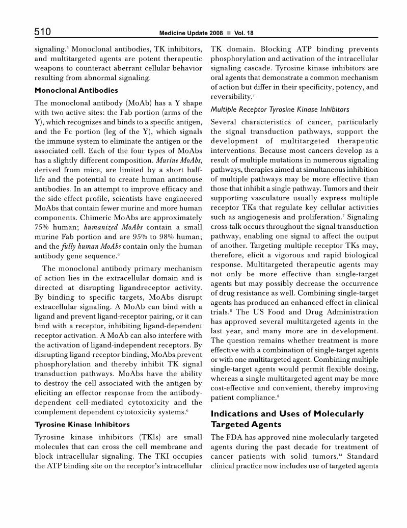
510 Medicine Update 2008 Vol. 18
signaling.5 Monoclonal antibodies, TK inhibitors, and multitargeted agents are potent therapeutic weapons to counteract aberrant cellular behavior resulting from abnormal signaling.
Monoclonal Antibodies
The monoclonal antibody (MoAb) has a Y shape with two active sites: the Fab portion (arms of the Y), which recognizes and binds to a specific antigen, and the Fc portion (leg of the Y), which signals the immune system to eliminate the antigen or the associated cell. Each of the four types of MoAbs has a slightly different composition. Murine MoAbs, derived from mice, are limited by a short half-life and the potential to create human antimouse antibodies. In an attempt to improve efficacy and the side-effect profile, scientists have engineered MoAbs that contain fewer murine and more human components.ChimericMoAbsareapproximately75% human; humanized MoAbs contain a small murine Fab portion and are 95% to 98% human; and the fully human MoAbs contain only the human antibody gene sequence.6
The monoclonal antibody primary mechanism of action lies in the extracellular domain and is directed at disrupting ligandreceptor activity. By binding to specific targets, MoAbs disrupt extracellular signaling. A MoAb can bind with a ligand and prevent ligand-receptor pairing, or it can bind with a receptor, inhibiting ligand-dependent receptor activation. A MoAb can also interfere with the activation of ligand-independent receptors. By disrupting ligand-receptor binding, MoAbs prevent phosphorylation and thereby inhibit TK signal transduction pathways. MoAbs have the ability to destroy the cell associated with the antigen by eliciting an effector response from the antibody-dependent cell-mediated cytotoxicity and the complement dependent cytotoxicity systems.6
Tyrosine Kinase Inhibitors
Tyrosine kinase inhibitors (TKls) are small molecules that can cross the cell membrane and block intracellular signaling. The TKI occupies the ATP binding site on the receptor’s intracellular
TK domain. Blocking ATP binding prevents phosphorylation and activation of the intracellular signaling cascade. Tyrosine kinase inhibitors are oral agents that demonstrate a common mechanism of action but differ in their specificity, potency, and reversibility.7
Multiple Receptor Tyrosine Kinase Inhibitors
Several characteristics of cancer, particularly the signal transduction pathways, support the development of multitargeted therapeutic interventions. Because most cancers develop as a result of multiple mutations in numerous signaling pathways, therapies aimed at simultaneous inhibition of multiple pathways may be more effective than those that inhibit a single pathway. Tumors and their supporting vasculature usually express multiple receptor TKs that regulate key cellular activities such as angiogenesis and proliferation.7 Signaling cross-talk occurs throughout the signal transduction pathway, enabling one signal to affect the output of another. Targeting multiple receptor TKs may, therefore, elicit a vigorous and rapid biological response. Multitargeted therapeutic agents may not only be more effective than single-target agents but may possibly decrease the occurrence ofdrugresistanceaswell.Combiningsingle-targetagents has produced an enhanced effect in clinical trials.8 The US Food and Drug Administration has approved several multitargeted agents in the last year, and many more are in development. The question remains whether treatment is more effective with a combination of single-target agents orwithonemultitargetedagent.Combiningmultiplesingle-target agents would permit flexible dosing, whereas a single multitargeted agent may be more cost-effective and convenient, thereby improving patient compliance.8
Indications and Uses of Molecularly Targeted AgentsThe FDA has approved nine molecularly targeted agents during the past decade for treatment of cancer patients with solid tumors.14 Standard clinical practice now includes use of targeted agents
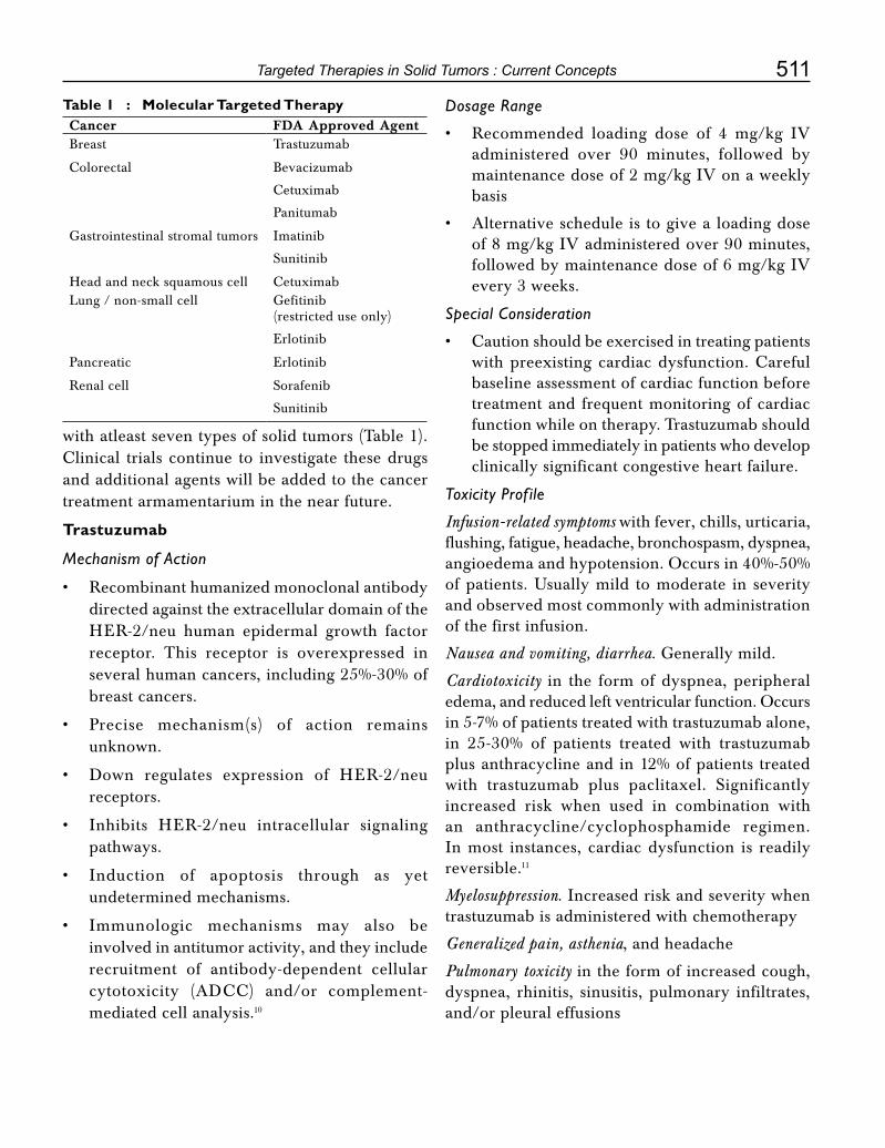
511Targeted Therapies in Solid Tumors : Current Concepts
with atleast seven types of solid tumors (Table 1). Clinical trials continue to investigate thesedrugsand additional agents will be added to the cancer treatment armamentarium in the near future.
Trastuzumab
Mechanism of Action
• Recombinanthumanizedmonoclonalantibodydirected against the extracellular domain of the HER-2/neu human epidermal growth factor receptor. This receptor is overexpressed in several human cancers, including 25%-30% of breast cancers.
• Precise mechanism(s) of action remainsunknown.
• Down regulates expression of HER-2/neureceptors.
• Inhibits HER-2/neu intracellular signalingpathways.
• Induction of apoptosis through as yetundetermined mechanisms.
• Immunologic mechanisms may also beinvolved in antitumor activity, and they include recruitment of antibody-dependent cellular cytotoxicity (ADCC) and/or complement-mediated cell analysis.10
Dosage Range
• Recommended loading dose of 4 mg/kg IVadministered over 90 minutes, followed by maintenance dose of 2 mg/kg IV on a weekly basis
• Alternativescheduleistogivealoadingdoseof 8 mg/kg IV administered over 90 minutes, followed by maintenance dose of 6 mg/kg IV every 3 weeks.
Special Consideration
• Cautionshouldbeexercisedintreatingpatientswithpreexistingcardiacdysfunction.Carefulbaseline assessment of cardiac function before treatment and frequent monitoring of cardiac function while on therapy. Trastuzumab should be stopped immediately in patients who develop clinically significant congestive heart failure.
Toxicity Prof ile
Infusion-related symptoms with fever, chills, urticaria, flushing, fatigue, headache, bronchospasm, dyspnea, angioedema and hypotension. Occurs in 40%-50% of patients. Usually mild to moderate in severity and observed most commonly with administration of the first infusion.
Nausea and vomiting, diarrhea. Generally mild.
Cardiotoxicity in the form of dyspnea, peripheral edema, and reduced left ventricular function. Occurs in 5-7% of patients treated with trastuzumab alone, in 25-30% of patients treated with trastuzumab plus anthracycline and in 12% of patients treated with trastuzumab plus paclitaxel. Significantly increased risk when used in combination with an anthracycline/cyclophosphamide regimen. In most instances, cardiac dysfunction is readily reversible.11
Myelosuppression. Increased risk and severity when trastuzumab is administered with chemotherapy
Generalized pain, asthenia, and headache
Pulmonary toxicity in the form of increased cough, dyspnea, rhinitis, sinusitis, pulmonary infiltrates, and/or pleural effusions
Table 1 : Molecular Targeted Therapy Cancer FDA Approved Agent Breast Trastuzumab
Colorectal Bevacizumab
Cetuximab
Panitumab
Gastrointestinal stromal tumors Imatinib
Sunitinib
Head and neck squamous cell CetuximabLung / non-small cell Gefitinib
(restricted use only)
Erlotinib
Pancreatic Erlotinib
Renal cell Sorafenib
Sunitinib
512 Medicine Update 2008 Vol. 18
Indications
As a single agent for the treatment of patients with metastatic breast cancer whose tumors over express the HER2 protein and who have received 1 or more chemotherapy regimens for their metastatic disease. In combination with paclitaxel for treatment of patients with metastatic breast cancer whose tumors overexpress the HER2 protein and who have not received chemotherapy for their metastatic disease. Neoadjuvant, adjuvant and metastatic breast cancer. In combination with cytotoxic agents other than anthracyclines.12
Gefitinib
Mechanism of Action
• Potentandselectivesmallmoleculeinhibitorofthe epidermal growth factor receptor (EGFR) tyrosine kinase, resulting in inhibition of EGFR autophosphorylation and inhibition of EGFR signaling.
• Inhibition of the EGFR tyrosine kinaseresults in inhibition of critical mitogenic and antiapoptotic signals involved in proliferation, growth, metastasis, angiogenesis, and response to chemotherapy and/or radiation therapy.13
Metabolism
MetabolismintheliverprimarilybytheCYP3A4microsomal enzyme. Other cytochrome P450 enzymes play a minor role in its metabolism. Elimination is mainly hepatic with excretion in the feces, and renal elimination of parent drug and its metabolites account for less than 4% of an administered dose. The terminal half-life of the parent drug is 48 hours.13
Indications
• Third line therapy (restricted use) in patientwith advanced NSCLC after failure of bothplatinum and docetaxel based chemotherapy. After 9/15/05 no new patients are allowed access to gefitinib unless they are enrolled in a clinical trial that was approved by an IRB prior to 6/17/05, or they are post to a clinical study
being conducted under an investigational new drug application. To be used only in patients who are bending or have benefited from gefitinib.14
Dosage Range
• Recommendeddoseis250mg/dayPO.
Drug Interaction
• Dilantin and other drugs that stimulate thelivermicrosomalCYP3A4enzyme, includingcarbamazepine, rifampicin, phenobarbital, and St. John’s wort - These drugs increase the rate of metabolism of gefitinib resulting in its inactivation.14
• DrugsthatinhibitthelivermicrosomalCYP3A4enzyme, including ketoconazole, itraconazole, erythromycin, and clarithromycin - These drugs decrease the rate of metabolism of gefitinib, resulting in increased drug levels and potentially increased toxicity.14
• Warfarin-Patients receivingcoumarin-derivedanticoagulants should be closely monitored for alterations in their clotting parameters (PT and INR) and/or bleeding as gefitinib inhibits the metabolism of warfarin in the liver P450 system. Dose of warfarin may require careful adjustment in the presence of gefitinib therapy.14
Special Considerations14
• Clinicalresponsesmaybeobservedwithinthefirst week of initiation of therapy.
• Patients with bronchoalveolar non-small celllung cancer may be more sensitive to gefitinib therapy than other histologic subtypes. Females and non-smokers also show increased sensitivity to gefitinib therapy.
• In patients who develop a skin rash, topicalantibiotics and oral minocycline may help.
Toxicity15
• Elevationsinbloodpressure,especiallyinthosewith underlying hypertension
513Targeted Therapies in Solid Tumors : Current Concepts
• Pruritus, dry skin with mainly a pustular,acneiform skin rash
• Mild to moderate elevations in serumtransaminases. Usually transient and clinically asymptomatic
• Astheniaandanorexia
• Mildnausea/vomitingandmucositis
• Conjunctivitis,blepharitisandcornealerosionwith abnormal eyelash growth
• Rare episodes of hemoptysis and GIhemorrhage.
Erlotinib
Mechanism of Action
• Potentandselectivesmallmoleculeinhibitorofthe epidermal growth factor receptor (EGFR) tyrosine kinase, resulting in inhibition of EGFR autophosphorylation and inhibition of EGFR signaling.
• Inhibition of the EGFR tyrosine kinaseresults in inhibition of critical mitogenic and antiapoptotic signals involved in proliferation, growth, metastasis, angiogenesis, and response to chemotherapy and/or radiation therapy.16
Metabolism
MetabolismintheliverprimarilybytheCYP3A4microsomal enzymeandbyCYP1A2 to a lesserextent. Elimination is mainly hepatic with excretion in the feces, and renal elimination of parent drug and its metabolites account for about 8% of an administered dose. The terminal half-life of the parent drug is 36 hours.
Dosage Range
• Recommendeddoseis150mg/dayP.O.
Special Consideration
• In patients with hepatic impairment, dosereduction or interruption should be considered.
• Non-smokers and patients with EGFR-positive tumors are more sensitive to erlotinib therapy.
• Coagulation parameters PT/INR should beclosely monitored when patients are receiving both erlotinib and coumadin, as erlotinib may inhibit the metabolism of coumadin by the liver P450 system.17, 18
• In patients who develop a skin rash, topicalantibiotics and oral minocyc1ine may help.
Indications
• Monotherapyisindicatedforthetreatmentofpatients with locally advanced or metastatic NSCLC after failure of at least 1 priorchemotherapy regimen. Indicated in combination with gemcitabine for first line treatment of patients with locally advanced, Unresectable or metastatic pancreatic cancer.17,18
Cetuximab
Classif ication
• Monoclonalantibody
Mechanism of Action
• Recombinanthumanizedmonoclonalantibodydirected against the epidermal growth factor receptor (EGFR). EGFR is overexpressed in a broad range of human solid tumors, including colorectal cancer, head and neck cancer, non-small cell lung cancer, pancreatic cancer, and breast cancer.19
• Precise mechanism(s) of action remainsunknown.
• Bindswithnearly10-foldhigheraffinitytoEGFRthan normal ligands EGF and TGF-a, which then results in inhibition of EGFR. Prevents both homodimerization and heterodimerization of the EGFR which then leads to inhibition of autophosphorylation and inhibition of EGFR signaling.20
• Inhibition of the EGFR signaling pathwayresults in inhibition of critical mitogenic and anti-apoptotic signals involved in proliferation, growth, invasion/metastasis, angiogenesis,
• InhibitionoftheEGFRpathwayenhancesthe

514 Medicine Update 2008 Vol. 18
response to chemotherapy and/or radiation therapy.
• Immunologic mechanisms may also beinvolved in antitumor activity, and they include recruitment of antibody-dependent cellular cytotoxicity (ADCC) and/or complement-mediated cell lysis.20
Indications
• Treatmentofpatientswithmetastaticcolorectalcancer whose tumor expresses the EGFR protein. Treatment is given either in combination with irinotecon for patients refractory to irinotecon, or as a single agent for those unable to tolerate chemotherapy with irinotecan.21
• Indicatedincombinationwithradiotherapyforthe treatment of locally or regionally advanced squamouscellheadandneckcancer.Cetuximabalone is indicated for the treatment of the patients with squamous head and neck cancer whose tumor has progressed or metastatic after treatment with platinum based therapy.22,23
• Unresectable loco-regional recurrence orsecond primary in patients with squamous cell head and neck cancers who have received prior radiotherapy.24,25
Metabolism
• Metabolism of cetuximab has not beenextensively characterized. Half life is on the order of 5-7 days with minimal clearance by the liver or kidneys.
Dosage Range
• Loadingdoseof400mg/m2 IV administered over 90 minutes, followed by maintenance dose of 250 mg/m2 IV given on a weekly basis.
Special Considerations
• Cetuximab should be used with caution inpatients with known hypersensitivity to murine proteins and/ or any individual components.
• There is no scientific or clinical evidence tosuggest that the level EGFR expression can
accurately predict for cetuximab clinical activity.TheclinicalactivityofCetuximab isthe same in EGFR-positive and EGFR-negative colorectal cancer.
• Development of skin toxicity appears to bea surrogate marker for Cetuximab clinicalactivity.
• Inpatientswhodevelopaskinrash,antibioticsuch as oral minocycline may help. Topical steroids and/or oral steroid treatment do not appear to be helpful in this setting. Patients should be warned to avoid sunlight exposure.
Toxicity20
• Infusion-related symptoms with fever,chills, urticaria, flushing, fatigue, headache, bronchospasm, dyspnea, angioedema, and hypotension. Although severe reactions occur in less than 1%. Usually mild to moderate in severity and observed most commonly with administration of the first infusion.
• Pruritus, dry skin with mainly a pustular,acneiform skin rash. Presents mainly on the face and upper trunk. Improves with continued treatment and resolves upon cessation of therapy.
• Pulmonary toxicity in the formof interstitiallung disease (ILD) manifested by increased cough, dyspnea, and pulmonary infiltrates. Observed in less than 1% of patients and more frequent in patients with underlying pulmonary disease
• Hypomagnesemia.
• Astheniaandgeneralizedmalaiseobservedinnearly 50% of patients.
• Paronychialinflammationwithswellingofthelateral nail folds of the toes and fingers.
Sunitinib Malate
Mechanism of Action
Sunitinib is an oral, multitargeted RTK inhibitor of vascular endothelial growth factor receptor

515Targeted Therapies in Solid Tumors : Current Concepts
(VEGFR)-l, -2 and -3, platelet-derived growth factor receptor (PDGFR) -α and -β, as well as other RTKs including stem cell factor receptor (KIT), glial cell line-derived neurotrophic factor and FMS-like receptor tyrosine kinase (FLT3).26
Indications
Sunitinib was approved in 2006 by the European Medicines Agency (EMEA) for use in advanced and/or metastatic RCC and by the United States Food and Drug Administration (FDA) for the treatment of advanced RCC. At the same time, sunitinib was approved by the EMEA for use in unresectable and/or metastatic malignant gastrointestinal stromal tumour (GIST) after failure of imatinib treatment due to resistance or intolerance and by USA FDA for the treatment of GIST after disease progression on or intolerance to imatinib mesylate treatment. The efficacy of sunitinib has been demonstrated in two independent, single-arm, multi center, phase II studies (trial 1: n = 63; trial 2: n = 106) in patients with cytokine-refractory metastasis RCC.27,28 On the basis of the favourable phase II efficacy and safety data, a multi center, randomized phase III study compared sunitinib with IFN-Iα as first-line therapy in metastatic RCC.27 Sunitinib has recently received European Union approval as first-line treatment for advanced and/or metastatic RCC.
Dosage
Sunitinib was administered at a dose of 50 mg/day, on a 4 weeks on then followed by 2 weeks off schedule. Patients continued on sunitinib unless there was disease progression or treatment intolerance.
Toxicity
Treatment with sunitinib was generally well tolerated. The tolerability profile of sunitinib was similar across the trials. The majority of adverse events reported were manageable with temporary delays, dose reduction and/or standard medical interventions. Dose reductions were required by approximately one-third and one-quarter of
patients in trials 1 and 2, respectively. The most commonly reported grade 3, nonhematological treatment-related adverse events included fatigue (11% in both trials) and hand-foot syndrome (7% in trial 2). No non-hematological adverse events were experienced at grade 4 severity.26,27,28
Sorafenib Tosylate
Mechanism of Action
Sorafenib is an oral, multi targeted kinase inhibitor. The molecular targets of sorafenib include the tyrosine kinases VEGFR-2 and-3, PDGFR-β, FLT3, KIT and RET, and the serine threonine Raf kinases, B-Raf and Raf-l/C-Raf. Sorafenib received approval from EMEA in July 2006 for the treatment of advanced RCC after failure of prior cytokine therapy or for patients who would be unsuitable for such therapy. FDA approval was granted in December 2005 for the treatment of patients with advanced RCC.29
Indications
The efficacy of sorafenib in the second-line setting was initially seen in a phase II randomized discontinuation trial.30 Results from the randomized discontinuation study then led to an international phase III randomized controlled study of sorafenib versus placebo in patients with previously treated RCC.Inthis‘TreatmentApproachesinRenalcellcancer Global Evaluation Trial’ (TARGET trial), patients with unresectable or metastatic RCC were randomized to sorafenib 400 mg twice daily or placebo.31 After the first interim analysis of OS, which showed that sorafenib reduced the risk of death as compared with placebo patients were allowed to crossover from placebo to sorafenib. Of 451 patients receiving sorafenib and who were assessable for investigator-assessed response, 10% achieved a PR, and 74% had SD compared with 2% and 53%, respectively, in the placebo-treated arm (n = 452).31 Adverse events were similar in phase II and III studies.
Side Effects
The most commonly reported grade 3/4 adverse

516 Medicine Update 2008 Vol. 18
events included fatigue and hypertension. Diarrhea (43% Vs 13%); rash or desquamation (40% Vs 16%); fatigue (37% Vs 28%) and hand-foot skin reactions (30% Vs 7%).31
Special Considerations
These results confirm that targeted inhibition of these single and multiple tumor targets is a feasible approach to treatment and provides a more positive outlook for the future management ofmetastaticRCC.Giventheclinicalexperiencewith these agents in the metastatic setting, their role as adjuvant treatment is now being explored in a number of large-scale randomized trials in patients at risk of relapse following surgery. In addition to multitargeted RTK inhibitors, other targets being investigated include hypoxia-inducible factor and intracellular signal transduction targets involved in proliferation, survival and hypoxia stimulation.32
Bevacizumab
Mechanism of Action
• Recombinanthumanizedmonoclonalantibodydirected against the vascular endothelial growth factor (VEGF). Binds to all isoforms at VEGF-A. VEGF is a pro-angiogenic growth factor that is overexpressed in a wide range of solid human cancers, including colorectal cancer.
• Precise mechanism(s) of action remainsunknown.
• Binding of VEGF prevents its subsequentinteraction with VEGFR receptors on the surface of endothelial cells and tumors, and in so doing, results in inhibition of VEGFR-signaling.
• Inhibits formation of new blood vessels inprimary tumor and metastatic tumors.
• Restores antitumor response by enhancingdendritic cell function.
• Immunologic mechanisms may also beinvolved in antitumor activity, and they include recruitment of antibody-dependent cellular cytotoxicity (ADCC) and/or complement-mediated cell lysis.33
Metabolism
• Metabolism of bevacizumab has not beenextensively characterized. Half-life is on the order of 17-21 days with minimal clearance by the liver or kidneys, as has been observed for other monoclonal antibodies and peptides used in the clinic.
Dosage Range
• Recommended dose for the treatment ofcolorectal cancer is 5 mg/kg IV on an every two week schedule.
• Can also be administered at 7.5mg/kg IVevery 3 weeks when used in combination with capecitabine-based regimens.
Special Considerations
• Patientsshouldbewarnedoftheincreasedriskof arterial thromboembolic events, including myocardial infarction and stroke. Risk factors are age > 65 years and history of angina, stroke, and prior arterial thromboembolic events.
• In some cases, fatal hemorrhage resultingfrom hemoptysis in patients with non-small cell lung cancer. These events have been mainly observed in patients with a central, cavitary, and/ or necrotic lesion involving the pulmonary vasculature.
• Bevacizumab treatment can result in thedevelopment of gastrointestinal perforations and wound dehiscence, which in some cases, has resulted in death.
• Usewithcautioninpatientswithuncontrolledhypertension as bevacizumab can result in grade 3 hypertension in about 10% of patients.
Toxicity
• Gastrointestinalperforationsandwoundhealingcomplications.
• Bleeding complications with epistaxis beingmost commonly observed.
• Increased risk of arterial thromboembolic

517Targeted Therapies in Solid Tumors : Current Concepts
events, including myocardial infarction, angina, and stroke.
• Hypertensionoccursinupto20-30%ofpatientswith grade 3 hypertension observed in 10-15% of patients. Usually well-controlled with oral anti-hypertensive medication.
• Proteinuriawithnephroticsyndromedevelopingin up to 5% of patients.
• Infusion-related symptoms with fever,chills, urticaria, flushing, fatigue, headache, bronchospasm, dyspnea, angioedema, and hypotension. Relatively uncommon event occurring in less than 5% of patients.33
Indications
• In combination with IV 5-FU-basedchemotherapy is indicated for first-line treatment of patients with metastatic carcinoma of the colon or rectum.34
• In combination with IV 5-FU-basedchemotherapy for second-line treatment of metastatic cancer or colon or rectum.
• Recommendations (NCCN 2006 Guidelines)availableforadvancedNSCLC,loco-regionaland metastatic breast cancer, renal cell carcinoma, Neoadjuvant and adjuvant colorectal carcinoma, recurrent ovarian cancer.34,35,36
Investigational Agents Lapatinib
Targeting the HER family of TKs remains an intriguing prospect for the treatment of solid tumors. Preclinical and clinical investigations of lapatinib, are under way. Lapatinib is a HER-family targeted therapy. Lapatinib is an oral agent that acts as a dual TKI in the intracellular domain. While all EGFR-inhibiting agents have been designed for single targets, lapatinib has the novel mechanism of targeting both HER1 and HER2. Lapatinib is available for use only in clinical trials involving patients with a variety of solid tumors. Early results for the treatment of advanced breast cancer are encouraging.37
ConclusionMolecularly targeted therapies are becoming a mainstay of treatment for cancer patients. By learning the science and applications of these therapies, oncologists can improve their own knowledge base and enhance the treatment response outcomes in patients at a significantly reduced morbidity and mortality, even in advanced stages of disease. This translates into greater survival with a much better quality of life quotient in the patient population.
References 1. Perez-Soler R: HER1/EGFR targeting: Refining the strategy.
Oncologist 2004. 9:58-67.
2. Lenz HJ: Anti-EGFR mechanism of action: Antitumor effect and underlying cause of adverse events. Oncology 2006. 20:5-12.
3. Yarden Y, Baselga J, Miles 0: Molecular approach to breast cancer treatment. Semin Oncol 2004. 31:6-13.
4. Farrara N: Vascular endothelial growth factor as a target for anticancer therapy. Oncologist 2004. 9(suppl1):2-10.
5. Adjei A, Hidalgo M: Intracellular si9nal transduction pathway proteins as targets for cancer therapy. J Clin Oncol 2005. 23:5326-5403.
6. CheifetzA,MayerL:Monoclonalantibodies,immunogenicity,and associated infusion reactions. Mt Sinai J Med 2005. 72:250-259.
7. Patyna S, Laird AD, Mendel DB, et al: SU 14813: A novel multiple receptor tyrosine kinase inhibitor with potent antiangiogenic and antitumor activity. Mol Cancer Ther 2006. 5:1774 -1782.
8. SandlerA,HerbstR:Combiningtargetedagents:Blockingthe epidermal growth factor and vascular endothelial growth factor pathways. Clin Cancer Res 2006. 12:4421s-4425s.
9. McNeilC:Twotargets,onedrugfornewEGFRinhibitors.NJCI 2006. 98: 1103-1104.
10. Herceptin (trastuzumab) package insert. Genentech, Inc, San Francisco,CA:February2005.
11. JackischC:HER2-positivemetastaticbreastcanceroptimizingtrastuzumab-based therapy. Oncologist 2006. 11(suppl1): 34-41.
12. Baselga J, Perez E, Pienkowski T, et al: Adjuvant trastuzumab: A milestone in the treatment of HER2-positive early breast cancer. Oncologist 2006. 11(suppl1):4-12.
13. Iressa (gefitlnib) tablets, package inserts. AstraZeneca Pharmaceuticals, Wilmington, DE: June 2005.
14. USFoodandDrugAdministration:Catalogofapprovedcancer
drugs. Available at http://www.accessdata.fda.gov/scripts/cder/drugsatfda/index.cfm?fuseaction = Search. Label Approval History # apphist Iressa. Accessed
518 Medicine Update 2008 Vol. 18
August 15. 2006.
15. US Food and Drug Administration: Safety alerts for drugs, biologics, medical devices, and dietary supplements. Available at http://www.fda.gov/cder/drug/infopage/ gefitinib/default.htm 2005 Iressa. Accessed September 9, 2006.
16. Tarceva (erlotinib) tablets, package insert, Genentech, San Francisco,CA.andOSIOncology,Melville,NY:November;2005.
17. ShepardFA,PereiraJR,CiuleanuT,etal,fortheNationalCancer Institute of Canada Trials Group: Erlotinib inpreviously treated non-small-cell lung cancer. N Engl J Med 2005. 353:123-132.
18. TsaoMS,SakuradaA,CutzJC,etal:Erlotinibinlungcancer-Molecular and clinical predictors of outcome. N Engl J Med 2005. 353’133-144.
19. VokesEE,ChuE:Anti-EGFRtherapies:Clinicalexperiencein colorectal, lung and head and neck cancer. Oncology 2006. 20(suppl 2): 17-25.
20. Erbitux (cetuximab)package insert. ImCloneSystemsInc,Branchberg,NJ, andBristolMyersSquibb’Co,Princeton,NJ: March 2006.
21. Cunningham D, Humblet Y, Siena S. et al: Cetuximabmonotherapy and cextuximab plus irinotecan in irinotecan-refractory metastatic colorectal cancer. N Engl J Med 2004. 351:337-345.
22. Bonner JA, Harari PM, Giralt J, et al: Radiotherapy plus cetuximab for squamous-cell carcinoma of the head and neck. N Engl J Med 2006. 354:567 -578.
23. TrigoJ,HitR,KoralewskiP,etal:Cetuximabmonotherapyis active in patients (pts) with platinum refractory recurrent/metastatic squamous cell carcinoma of the head and neck (SCCHN):ResultsofaphaseIIstudy(abstract5502)[andslidepresentation].ProcAmSocClinOncol200423:487.
24. Baselga J, Trigo JM, Bourhis J, et al: Phase II multicenter study of the anti-epidermal growth factor receptor monoclonal antibody cetuximab in combination with platinum-based chemotherapy in patients with platinum-refractory metastatic and/or recurrent squamous cell carcinoma of the head and neck.JClinOncol2005.23:5578-5587.
25. Herbst RS, Shin OM, Dicke K, et al: Phase II multicenter study of the epidermal growth factor receptor antibody cetuximab and cisplatin for recurrent and refractory head and neck. J Clin Oncol 2005. 23:5568-5577.
26. Sutent (sunitinib malate) package insert. Pfizer Lbs, New York, February 2006.
27. Mortez RJ, Michaelson MD, Redman BG et al. Activity of SU11248, a multitrageted inhibitors of vascular endothelial growth factor receptor and platelet-derived growth factor receptor, in patients with metastatic renal cell carcinoma. J. Clin. Oncol 2006; 24: 16-24
28. Mortez RJ, Rini BL, Bukowski RM et al. Sunitinib in patients with metastatic renal cell carcinoma. JAMA 2006; 2516 – 2524
29. Nexavar (sorafenib) package insert. Bayer Pharmaceuticals Corp,WestHaven,CT:August2006.
30. ratain MJ, Eisen T, Stadler WM et al. Phase II placebo-controlled randomized discontinuation trial of sorafenib in patients with Metastatic renal cell carcinoma. J Clin Oncol 2006; 24: 2505 – 2512
31. Escudier B, Eisen T, Stadler WM et al. Sorafenib in advanced clear cell renal cell carcinoma. N Engl J Med 2007; 356: 125 – 134
32. SchoffskiP,DumezH,ClementPet al.Emerging roleoftyrosine kinase inhibitors in the treatment of advanced renal cell cancer : a review. Ann Oncol 2006; 17: 1185 – 1196
33. Avastin (bevaclzumab) package Insert. Genentech, San Francisco,CA,June2006.
34. Benson AB, Catalano PJ, Meropol J, et al: Bevacizumab(anti-VEGF) plus FOLFOX 4 in previously treated advanced colorectal cancer (advCRC):An interim toxicityanalysisof the Eastern Cooperative Group (ECOG) study E3200(abstract975).ProcAmSocClinOncol2003.22:243.
35. Miller K, Wang M, Gralow J, et al: E2100, a randomized phase III trial of paclitaxel vs. paclitaxel plus bevacizumab as first-line therapy for locally recurrent or metastatic breast cancer.[Presentation].AmericanSocietyofClinicalOncology41st annual meeting, Orlando, Florida, Z005.
36. NCCNClinicalPracticeGuidelinesinOncology:Availableat www.nccn.org. Accessed September 19, 2006.
37. GeyerCE,ForsterJK,CameronOS:PhaseIIIrandomized,open-label, international study comparing lapatinib and capecitabine versus capecitabine in women with refractory advarlced or metastatic breast cancer. American Society of clinical Oncology 42nd annual meeting, Atlanta, 2006.

CHAPTER
68Thrombolysis in Ischemic StrokeS. Kaul
IntroductionThe December of 1995 was unlike any other month for stroke research ; and not without reason. National Institute of Neurological Disorders and Stroke (NINDS) in United States of America had just reported its success in significantly improving the outcome of ischemic stroke by using intravenous recombinant tissue-type plasminogen activator (rtPA), if administered within 3 hours.1 Twelve years down the line, rtPA is the most effective treatment in a subset of ischemic stroke patients both in academic centers and community hospitals. However, there are still some barriers in delivering thrombolytic therapy to ischemic stroke patients. This article will review the major studies bringing rtPA into routine practice of stroke and will address the associated issues with special reference to India.
What triggered the thrombolysis movement? Although stroke thrombolysis research has been going on since the last three decades, it was the publication of NINDS rtPA trial, which made stroke thrombolysis seem possible in real practice. The NINDS trial randomized 624 patients (312 each placebo and intravenous rtPA) within a time window of 3 h after stroke symptom onset. Half of the patients were treated within 0-90 min and the other half within 91-180 min. A good outcome was
defined as a National Institute of Health Stroke Scale (NIHSS) Score < 1, Glasgow Outcome Score (GOS) = 1, Barthel Index (BI) > 95, and modified Rankin Score (mRS) < 1. The median baseline NIHSS Score was 14 (rtPA group) versus 15 (placebo group). There was no significant difference between the drug treatment and placebo group in the percentages patients with neurological improvement at 24 h (rtPA 47% vs placebo 57%; relative risk (RR) 1.2, p = 0.21), although a post hoc analysis comparing the median NIHSS at 24 h showed a median of 8 in the rtPA treated group versus 12 in the placebo group (p < 0.02). Furthermore , the proportion of rtPA patients achieving a favorable outcome (minimal or no disability) at 3 months on each of the four scales was 1.7 times greater than patients in the placebo group. The long-term clinical benefit of rtPA was confirmed in all single scores as well as in the global test:BI(50%vs38%;OR1.6(95%C.I1.1-2.5),p=0.026);mRS(39%vs26%;OR1.7(95%CI1.1-2.5),p=0.019);GOS(44%vs32%;OR1.6(95%CI1.0-2.8), p = 0.033); and combined end point (OR 1.7 (95%CI1.0-2.8),p=0.008).Thus, forevery100 patients treated with recombinant plasminogen activator (rtPA) according to the NINDS protocol, at least 11 more patients are expected to achieve a favorable outcome. More recently, NINDS rtPA Study Group reported that the 1-year follow up of
520 Medicine Update 2008 Vol. 18
patients closely matched the results reported at 3 months. Thus evidence exists of a sustained benefit from rtPA at 1 year.4 Furthermore, the outcome did not vary by stroke subtype at baseline, meaning that patients with small vessel disease benefited as well as patients with, for instance, cardioembolic stroke. Tissue plasminogen Activator got a prompt approval by FDA within a record time of 6 months. Following close on heels, the American Academy of Neurology and American Stroke Association also issued the guidelines for rtPA administration to patients with acute ischemic stroke,2,3 which are basically based on the protocol followed in the NINDS study (Table 1). Some people felt that it was premature for FDA to approve rtPA based on just one positive study, but others held the view that the evidence of efficacy was so compelling, that one could not deny the benefit to the eligible patients.
Predictors of hemorrhageThrombolytic therapy with rtPA is not without risk.Symptomaticintracranialhemorrhage(ICH)occurred in 6.4% of patients treated with rtPA in the study sponsored by the NINDS. Those suffering a symptomaticICHhadahighmeanNIHSSscoreof 20. Overall those with the most severe strokes (NIHSS score greater than 20) had a 17% rate of ICH.Inshort,casesofsevereischemiacausedbyvolume of infarct or duration of infarct), have a high likelihood of hemorrhagic transformation by thrombolysis.
Predictors of efficacyOverall, no difference in mortality occurred despite a higher risk of symptomatic ICH (6.4%) forpatients treated with rtPA. Following the original NINDS report, several additional analyses of the cohort were examined. In an attempt to identify patients who would be most or least likely to respond to rtPA, an analysis of baseline variables did not find any pretreatment characteristic that might influence a patient’s response to rtPA.5
If a 3-hour window of treatment can be met,
thrombolytic therapy with intravenous rtPA can be beneficial for each of the major categories of ischemic stroke: atherothrombotic, cardioembolic, and lacunar stroke. However, follow-up analysis found a relationship between the chance of a favorable outcome and the time from symptom onset to initiation of treatment. The chance of a favorable outcome diminishes the closer one gets to the 3-hour time point. This highlights the fact that ultra-rapid treatment is critical for therapeutic success.6
Cost – effectivenessA post-hoc analysis of cost-effectiveness in the NINDS rtPA Study Group found that treatment with rtPA significantly reduced the cost of care, accomplished by reducing hospital length of stay and increasing the percentage of patients discharged home versus to nursing home or a rehabilitation setting.7
Are NINDS results applicable to routine practice ?Since the FDA approval of rtPA for acute ischemic stroke, several groups have reported the results of treatment in the clinical setting. Tanne and colleagues (1999) published a retrospective series of 189 consecutively treated patients in 13 hospitals who were not involved in the original NINDS study.8 TherateofsymptomaticICHwas5.8%. Thus the risk of ICH did not exceed thatfound in the NINDS study. Even more important was the finding that deviating from the treatment and management guidelines of NINDS trial increased the risk of ICH to 11% versus 4% forpatients whose treatment was within the guideline parameters. The most common deviations noted were initiation of treatment beyond 3 hours from symptom onset or use of heparin or aspirin in the first 24 hours. The first large prospective study after the FDA approval of rtPA for acute ischemic stroke was the Standard Treatment with Alteplase to Reverse Stroke (STARS) study.9 Three hundred eighty-nine patients were enrolled in 57 medical
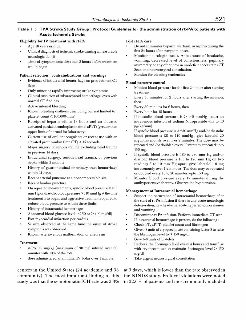
521Thrombolysis in Ischemic Stroke
Table 1 : TPA Stroke Study Group : Protocol Guidelines for the administration of rt-PA to patients with Acute Ischemic Stroke
Eligibility for IV treatment with rt-PA• Age18yearsorolder• Clinicaldiagnosisofischemicstrokecausingameasurable
neurologic deficit• Timeofsymptomonsetlessthan3hoursbeforetreatment
would begin
Patient selection : contraindications and warnings• EvidenceofintracranialhemorrhageonpretreatmentCT
Scan• Onlyminororrapidlyimprovingstrokesymptoms• Clinicalsuspicionofsubarachnoidhemorrhage,evenwith
normalCTfindings• Activeinternalbleeding• Knownbleedingdiathesis,includingbutnotlimitedto:
platelet count < 100,000/mm3
• Receipt of heparin within 48 hours and an elevatedactivated partial thromboplastin time( aPTT) (greater than upper limit of normal for laboratory)
• Currentuseoforalanticoagulantsorrecentusewithanelevated prothrombin time (PT) > 15 seconds
• Majorsurgeryorserioustraumaexcludingheadtraumain previous 14 days
• Intracranial surgery, serious head trauma, or previousstroke within 3 months
• History of gastrointestinal or urinary tract hemorrhagewithin 21 days
• Recentarterialpunctureatanoncompressiblesite• Recentlumbarpuncture• Onrepeatedmeasurements,systolicbloodpressure>185
mm Hg or diastolic blood pressure > 110 mmHg at the time treatment is to begin, and aggressive treatment required to reduce blood pressure to within these limits
• Historyofintracranialhemorrhage• Abnormalbloodglucoselevel(<50or>400mg/dl)• Post-myocardialinfarctionpericarditis• Seizure observed at the same time the onset of stroke
symptoms was observed• Knownarteriovenousmalformationoraneurysm
Treatment• rt-PA0.9 mg/kg (maximum of 90 mg) infused over 60
minutes with 10% of the total • doseadministeredasaninitialIVbolusover1minute.
Post rt-PA care• Do not administer heparin, warfarin, or aspirin during the
first 24 hours after symptom onset.• Monitor neurologic status. Appearance of headache,
vomiting, decreased level of consciousness, pupillary asymmetryoranyothernewneurodeficitnecessitatesCTScan and neurosurgical consultation.
• Monitor for bleeding tendencies
Blood pressure control• Monitor blood pressure for the first 24 hours after starting
treatment:• Every 15 minutes for 2 hours after starting the infusion,
then • Every 30 minutes for 6 hours, then• Every hour for 18 hours• If diastolic blood pressure is > 140 mmHg , start an
intravenous infusion of sodium Nitroprusside (0.5 to 10 µg/kg/min)
• If systolic blood pressure is > 230 mmHg and/or diastolic blood pressure is 121 to 140 mmHg , give labetalol 20 mg intravenously over 1 or 2 minutes. The dose may be repeated and /or doubled every 10 minutes, repeated upto 150 mg.
• If systolic blood pressure is 180 to 230 mm Hg and/or diastolic blood pressure is 105 to 120 mm Hg on two readings 5 to 10 mm Hg apart, give labetalol 10 mg intravenously over 1-2 minutes. The dose may be repeated or doubled every 10 to 20 minutes, upto 150 mg.
• Monitor blood pressure every 15 minutes during the antihypertensive therapy. Observe the hypotension.
Management of Intracranial hemorrhage• Suspect the occurrence of intracranial hemorrhage after
the start of rt-PA infusion if there is any acute neurologic deterioration, new headache, acute hypertension, or nausea and vomiting.
• Discontinuert-PAinfusion.PerformimmediateCTscan• If intracranial hemorrhage is present, do the following :• CheckPT,aPTT,plateletcountandfibrinogen• Give 6-8 units of cryoprecipitate containing factor 8 to raise
the fibrinogen level to > 150 mg/dl• Give 6-8 units of platelets• Recheck the fibrinogen level every 4 hours and transfuse
with cryoprecipitate to maintain fibrinogen level > 150 mg/dl
• Take urgent neurosurgical consultation
centers in the United States (24 academic and 33 community). The most important finding of this studywasthatthesymptomaticICHratewas3.3%
at 3 days, which is lower than the rate observed in the NINDS study. Protocol violations were noted in 32.6 % of patients and most commonly included
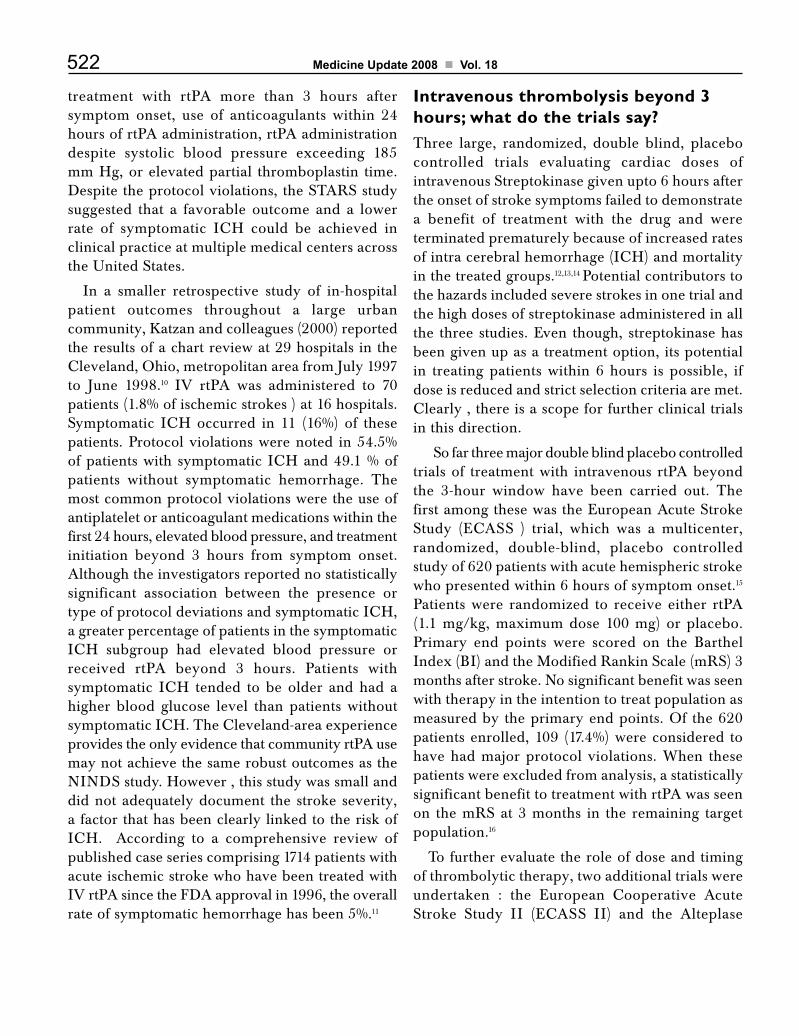
522 Medicine Update 2008 Vol. 18
treatment with rtPA more than 3 hours after symptom onset, use of anticoagulants within 24 hours of rtPA administration, rtPA administration despite systolic blood pressure exceeding 185 mm Hg, or elevated partial thromboplastin time. Despite the protocol violations, the STARS study suggested that a favorable outcome and a lower rate of symptomatic ICH could be achieved inclinical practice at multiple medical centers across the United States.
In a smaller retrospective study of in-hospital patient outcomes throughout a large urban community, Katzan and colleagues (2000) reported the results of a chart review at 29 hospitals in the Cleveland,Ohio,metropolitanareafromJuly1997to June 1998.10 IV rtPA was administered to 70 patients (1.8% of ischemic strokes ) at 16 hospitals. Symptomatic ICHoccurred in 11 (16%)of thesepatients. Protocol violations were noted in 54.5% ofpatientswith symptomatic ICHand49.1%ofpatients without symptomatic hemorrhage. The most common protocol violations were the use of antiplatelet or anticoagulant medications within the first 24 hours, elevated blood pressure, and treatment initiation beyond 3 hours from symptom onset. Although the investigators reported no statistically significant association between the presence or typeofprotocoldeviationsandsymptomaticICH,a greater percentage of patients in the symptomatic ICH subgroup had elevated blood pressure orreceived rtPA beyond 3 hours. Patients with symptomatic ICH tended to be older and had ahigher blood glucose level than patients without symptomaticICH.TheCleveland-areaexperienceprovides the only evidence that community rtPA use may not achieve the same robust outcomes as the NINDS study. However , this study was small and did not adequately document the stroke severity, a factor that has been clearly linked to the risk of ICH. According to a comprehensive review ofpublished case series comprising 1714 patients with acute ischemic stroke who have been treated with IV rtPA since the FDA approval in 1996, the overall rate of symptomatic hemorrhage has been 5%.11
Intravenous thrombolysis beyond 3 hours; what do the trials say?Three large, randomized, double blind, placebo controlled trials evaluating cardiac doses of intravenous Streptokinase given upto 6 hours after the onset of stroke symptoms failed to demonstrate a benefit of treatment with the drug and were terminated prematurely because of increased rates ofintracerebralhemorrhage(ICH)andmortalityin the treated groups.12,13,14 Potential contributors to the hazards included severe strokes in one trial and the high doses of streptokinase administered in all the three studies. Even though, streptokinase has been given up as a treatment option, its potential in treating patients within 6 hours is possible, if dose is reduced and strict selection criteria are met. Clearly,thereisascopeforfurtherclinicaltrialsin this direction.
So far three major double blind placebo controlled trials of treatment with intravenous rtPA beyond the 3-hour window have been carried out. The first among these was the European Acute Stroke Study (ECASS ) trial, which was a multicenter,randomized, double-blind, placebo controlled study of 620 patients with acute hemispheric stroke who presented within 6 hours of symptom onset.15
Patients were randomized to receive either rtPA (1.1 mg/kg, maximum dose 100 mg) or placebo. Primary end points were scored on the Barthel Index (BI) and the Modified Rankin Scale (mRS) 3 months after stroke. No significant benefit was seen with therapy in the intention to treat population as measured by the primary end points. Of the 620 patients enrolled, 109 (17.4%) were considered to have had major protocol violations. When these patients were excluded from analysis, a statistically significant benefit to treatment with rtPA was seen on the mRS at 3 months in the remaining target population.16
To further evaluate the role of dose and timing of thrombolytic therapy, two additional trials were undertaken : the European Cooperative AcuteStroke Study II (ECASS II) and the Alteplase
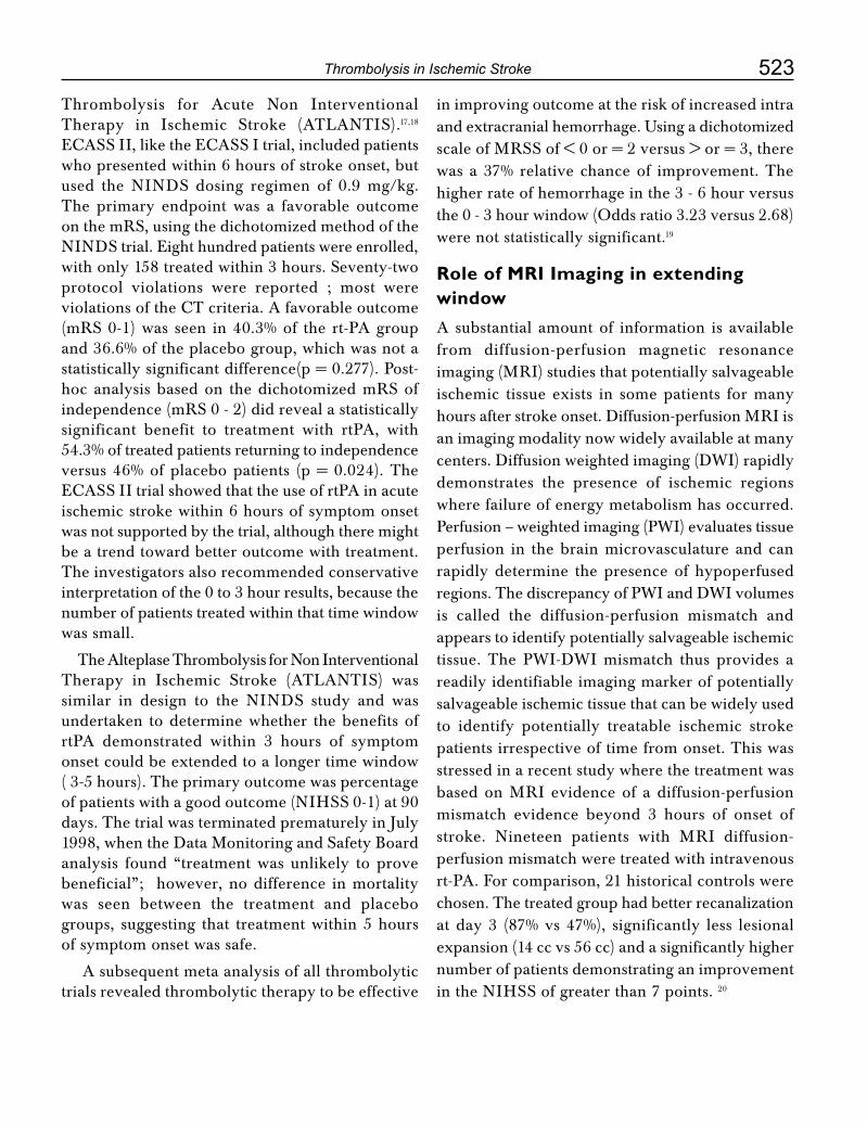
523Thrombolysis in Ischemic Stroke
Thrombolysis for Acute Non Interventional Therapy in Ischemic Stroke (ATLANTIS).17,18 ECASSII,liketheECASSItrial,includedpatientswho presented within 6 hours of stroke onset, but used the NINDS dosing regimen of 0.9 mg/kg. The primary endpoint was a favorable outcome on the mRS, using the dichotomized method of the NINDS trial. Eight hundred patients were enrolled, with only 158 treated within 3 hours. Seventy-two protocol violations were reported ; most were violationsoftheCTcriteria.Afavorableoutcome(mRS 0-1) was seen in 40.3% of the rt-PA group and 36.6% of the placebo group, which was not a statistically significant difference(p = 0.277). Post-hoc analysis based on the dichotomized mRS of independence (mRS 0 - 2) did reveal a statistically significant benefit to treatment with rtPA, with 54.3% of treated patients returning to independence versus 46% of placebo patients (p = 0.024). The ECASSIItrialshowedthattheuseofrtPAinacuteischemic stroke within 6 hours of symptom onset was not supported by the trial, although there might be a trend toward better outcome with treatment. The investigators also recommended conservative interpretation of the 0 to 3 hour results, because the number of patients treated within that time window was small.
The Alteplase Thrombolysis for Non Interventional Therapy in Ischemic Stroke (ATLANTIS) was similar in design to the NINDS study and was undertaken to determine whether the benefits of rtPA demonstrated within 3 hours of symptom onset could be extended to a longer time window ( 3-5 hours). The primary outcome was percentage of patients with a good outcome (NIHSS 0-1) at 90 days. The trial was terminated prematurely in July 1998, when the Data Monitoring and Safety Board analysis found “treatment was unlikely to prove beneficial”; however, no difference in mortality was seen between the treatment and placebo groups, suggesting that treatment within 5 hours of symptom onset was safe.
A subsequent meta analysis of all thrombolytic trials revealed thrombolytic therapy to be effective
in improving outcome at the risk of increased intra and extracranial hemorrhage. Using a dichotomized scale of MRSS of < 0 or = 2 versus > or = 3, there was a 37% relative chance of improvement. The higher rate of hemorrhage in the 3 - 6 hour versus the 0 - 3 hour window (Odds ratio 3.23 versus 2.68) were not statistically significant.19
Role of MRI Imaging in extending windowA substantial amount of information is available from diffusion-perfusion magnetic resonance imaging (MRI) studies that potentially salvageable ischemic tissue exists in some patients for many hours after stroke onset. Diffusion-perfusion MRI is an imaging modality now widely available at many centers. Diffusion weighted imaging (DWI) rapidly demonstrates the presence of ischemic regions where failure of energy metabolism has occurred. Perfusion – weighted imaging (PWI) evaluates tissue perfusion in the brain microvasculature and can rapidly determine the presence of hypoperfused regions. The discrepancy of PWI and DWI volumes is called the diffusion-perfusion mismatch and appears to identify potentially salvageable ischemic tissue. The PWI-DWI mismatch thus provides a readily identifiable imaging marker of potentially salvageable ischemic tissue that can be widely used to identify potentially treatable ischemic stroke patients irrespective of time from onset. This was stressed in a recent study where the treatment was based on MRI evidence of a diffusion-perfusion mismatch evidence beyond 3 hours of onset of stroke. Nineteen patients with MRI diffusion-perfusion mismatch were treated with intravenous rt-PA. For comparison, 21 historical controls were chosen. The treated group had better recanalization at day 3 (87% vs 47%), significantly less lesional expansion (14 cc vs 56 cc) and a significantly higher number of patients demonstrating an improvement in the NIHSS of greater than 7 points. 20

524 Medicine Update 2008 Vol. 18
Meta analysisWith growing experience and better training of emergency medical personnel, internists, and neurologists throughout all stroke services, the efficacy of intravenous thrombolytic therapy with rtPA may even improve and the time window may be routinely extended to 6 h after symptom onset. While rtPA is approved for thrombolytic therapy in the 3h time window , there is level I (positive meta-analyses from large methodologically flawless randomized controlled trials), level II (positive secondary endpoints of large randomized controlledtrials,i.e.ECASSIandII),andamplelevel III and IV evidence that thrombolysis works in the 4.5 - 6 h time window. Accordingly, with the recommendationsoftheCochraneCollaboration,21 and the European Stroke Initiative,22 intravenous thrombolysis is safe and effective up to 6 h in selected patients , and likely best within 4.5 h after stroke onset. The ideal selection tool may be stroke MRI.23 The fact that an individual therapy based on advanced knowledge is offered that does not meet the criteria of drug approval institutions and therefore may be associated with a higher risk of hazardous if not fatal side-effects must be stressed when informed consent is obtained. Patients and their relatives should be informed not only about the hazards of thrombolytic therapy within or outside the 3 h time window but also about its potential benefit and the risk of not being treated.
Intra-Arterial ThrombolysisAnterior Circulation
For ischemic stroke patients who are not candidates for intravenous rtPA , intraarterial (IA) thrombolysis may be a treatment option for carefully selected patients who present up to 6 hours after symptom onset.ThePROACTIIStudywasamulticentereffort to determine whether administering an intra-arterial thrombolytic agent- in this case 2-hour infusion of 9 mg of IA recombinant pro-urokinase (pro-UK)- could improve 3-month functional outcomes in patients with acute middle cerebral artery(MCA)territorystroke.24 One hundred eighty
patients were randomized to receive intra-arterial pro-UK or placebo in a 2:1 randomization scheme. The study design included patients with less than 6 hours of acute ischemic stroke symptoms who were suspectedof havingMCAocclusion.AfteraCTscanexcludedhemorrhageand showednoevidence of acute hypodensity or sulcal effacement inmore thanone thirdof theMCAterritory,allpatients underwent angiography to identify the site of thrombosis. If occlusion of M1 or M2 segment was found, the patient was randomized to either pro-urokinase or a control group. All patients received a periprocedural IV heparin drip for 4 hours and repeat angiogram at 1 and 2 hours to assess the status of thrombolysis. The results showed that 40% of the treatment group versus 25% of the control group achieved the primary outcome of slight disability or better at 3 months (p = .043). This difference represents a 15% absolute benefit and 60% relative benefit for treatment with IA pro-urokinase. Thus, for every six patients treated one additional patient will achieve slight disability or better outcome at 3 months (number needed to treat = 6). The benefit was seen despite a 10% riskofsymptomaticICH.Severalothercaseserieshave also suggested a benefit of IA thrombolysis in severe posterior circulation stroke even when therapy is delayed out to 24 hours.25 Intra-arterial thrombolytic therapy is not approved by FDA and is considered experimental. Patients and their attendants must be apprised of this.
Posterior circulation
Intra-arterial thrombolysis for acute posterior circulation ischemia differs substantially from intra-arterial thrombolysis of anterior circulation vessels. This includes differences in the proportions of underlying stroke mechanisms, differences in the morbidity and mortality and above all differences in the time window during which ischemia may be successfully reversed.25 Unlike the striatum and cerebral cortex, the brainstem is relatively resistant to ischemia. The practical consequence of this in regard to intra-arterial thrombolysis is that successful recanalization might reverse clinical

525Thrombolysis in Ischemic Stroke
deficits as far out as 24 hours from stroke symptom onset in some instances.25
Indian experienceIntravenous Thrombolysis
Thrombolytic therapy made a slow but sure entry into Indian neurological practice, for obvious reasons. These include lack of awareness among public and, among the referring physicians about the existence and usefulness of rtPA treatment. However, as per a survey carried out in various cities in India, it was found that 8% to 25 % of stroke patients arrived in the hospital within 3 hours.26 All of them do not receive rtPA as many do not qualify as per the NINDS criteria. Others find the cost very high. But the most important reason even in the big cities, as anywhere else in world, which discourages the neurologists to use rtPA is the uncertainty of response and potential for fatal brain hemorrhage. Approximately 15 stroke units in India use tPA for acute stroke.27 In a study from a private sector tertiary referral hospital in Northwest India, 489 stroke patients were screened between September 2001 and November 2003.28 Seventy-two of them (14.7%) presented within a 3-h window. Thirty-eight of the 72 patients had had an ischemic stroke. Sixteen out of 38 ischemic stroke patients did not meet the inclusion criteria for thrombolysis therapy. Only five out of the 22 eligible patients received tPA. The remaining 17 eligible patients could not afford the drug.28 In another study from the rural catchment area in India, only 20 (31%) patients out of 64 evaluated reached the hospital within 3 h.29 Seven patients were found eligible for thrombolysis but none of them received the drug. The cost of the drug was a major hinderance, as most of the patients belonged to a lower socioeconomic strata.29
Several Government run institutes in India have reported good outcome with use of rtPA. All India Institute of Medical Sciences, New Delhi, published their experience of 40 thrombolysed patients within a 3h onset between between March 2002 and June 2005.30 The mean age was 66 years (range 32 - 82 years, male : female ratio = 3:2). The mean time to
reach the emergency department was 27 min (25 - 45 min). The NIHSS score at admission ranged from 11 to 22 (mean 14.5 minutes). Twenty –six patients (65%) significantly improved on NIHSS at 48 h ( > 4 points) (mean change = 10; range 40 - 17). At one month, 32 (79%) improved on Barthel Index (mean change = 45).30
In a private sector hospital from Amritsar, 34 patients were given tPA over 3 years.31 In half of the group, MRI DWI/PWI sequences were used to select patients for thrombolysis. Patients in the DWI/PWI group had a better outcome than the patients who received rtPA based on CT scan.31 In another private hospital in Chennai, sono-thrombolysis was used along with iv rtPA in 42 patients with good recovery.32 Thirty seven patients were treated with rtPA in a Nizam Institute of Medical Sciences, Hyderabad over 53 months. Twenty –nine (78%) patients had a good outcome at 1 year.33 Intra-arterial thrombolysis therapy is being used in approximately 10 centers in India. In a tertiary referral center from Kerala, South India, intra-arterial Urokinase (IA UK) was given in 5 patients. The mean age was 41.2 years ( range 30 - 65 years, all were men). Digital Subtraction Angiography (DSA) showed distal internal carotid artery occlusion in one patient and occlusion in one patient and occlusion of the middle cerebral artery or its branches in the others. The UK dose ranged from 120 000 to 500 000 U. In two patients, there was complete recanalization with excellent recovery. In the remaining three patients, the recanalization rate varied from 0% to 50%, with partial recovery in two and no recovery in one patient.34
A private sector hospital in Guntur, South India, has successfully implemented IA thrombolysis therapybasedon thePROACT-IIprotocol.24 A neuro-interventional team is available 24 h and patient selection is based on CT scan and four-vessel angiogram. IA thrombolysis was used in 40 patients with ischemic stroke in both anterior and posterior circulation. UK was used in all patients (100,000- 750,000 U), except in one patient , where

526 Medicine Update 2008 Vol. 18
tPA (20 mg) was used. The baseline NIHSS ranged from 8 to 25 (median 17) at admission. Twenty-five (62%) patients had good outcome (mRS 0-2) at three months.
These preliminary data from India show that hyperacute thrombolysis in acute ischemic stroke is feasible in urban private and public sector tertiary hospitals. It could be widely used if a greater number of dedicated stroke teams /stroke units become available, and the cost of the drug is reduced.
Barriers to thrombolysis in IndiaInfrastructure
Eighty per cent of population from India lives in rural areas where the health care infrastructure is poor. Many patients having acute ischemic stroke find it difficult to travel in a reasonable time to the centers where there are resources to facilitate tPA. These facilities, which are primarily located in urban areas, become difficult to access mainly due to poor road infrastructure. There are hardly any ambulance services in rural areas.
Socio-cultural factors
People living in India have very little access to information about stroke. A study in northwest India documents that only a third of subjects interviewed, correctly identified the brain as the affected organ in a stroke.35 Low threat perception of stroke was an independent factor for the late arrival of patients at the emergency department in a study from north India.36 Most rural patients having stroke attending a university hospital in south India were not aware of the importance of the time window in stroke management.29
Economics
Most governments in developing countries are not in a position to provide thrombolysis therapy in government hospitals to patients in need. Health insurance systems are limited to a minute section of the community. Patients must cover the costs using their own personal savings or not receive treatment. In a study from south India, all seven
patients eligible for thrombolysis therapy belonged to lower socioeconomic group from rural India and could not afford the therapy.29 In an urban hospital in northwest India of the 23 eligible patients, only five actually received the drug; the remaining patients were unable to afford the high cost of the treatment.28
Low-cost thrombolytic agents
Urokinase is a cheaper alternative to rtPA, and thepreliminaryresultsofaChinesei.v.Urokinasetrial are promising.37 The effectiveness of this drug should be evaluated in a multicentered study across other countries. The Asian population may respond to low-dose rt-PA (0-.6 mg/kg) based on a study from Japan.38 The reproducibility of the results of the former study should be explored in other Asian countries. If found to be beneficial, it will increase the utilization of rtPA in India
Although written consent is not necessary before administration of rtPA for treatment of rtPA for treatment of stroke, a full discussion of the potential risks and benefits of treatment with rtPA with the family and the patient is recommended.
To conclude, stroke thrombolysis is a reality and stroke thrombolysis works. Unquestionably there is a certain risk involved in terms of intracranial hemorrhage, but considering the devastation and disability a stroke causes, the risk taken is worth it, provided the neurologist is well versed in the thrombolytic therapy. Moreover, with proper selection and strict adherence to NINDS protocol, the chances of intracerebral hemorrhage can be minimized.
Current Recommendations of American Stroke Association for thrombolysis in Adults with Ischemic Stroke Class I Recommendations
• Intravenous rtPA (0.9 mg/kg, maximumdose 90 mg) is recommended for selected patients who may be treated within 3 hours of
527Thrombolysis in Ischemic Stroke
ischemicstroke(ClassI,LevelofEvidenceA).Physicians should review the criteria outlined in Table 1 (which are modeled on those used in NINDS trial) to determine the eligibility of the patient.
• Besides bleeding complications, physiciansshould be aware of the potential side effect of angioedema that may cause partial airway obstruction(ClassI,LevelofEvidenceC).
Class II Recommendations
• Apatientwhosebloodpressurecanbeloweredsafely with antihypertensive agents may be eligible for treatment, and the physician should assess the stability of blood pressure before startingrtPA(ClassIIa,LevelofEvidenceB).
• Apatientwith a seizure at the timeof onsetof stroke may be eligible for treatment as long as the physician is convinced that residual impairments are secondary to stroke and not apostictalphenomenon. (Class IIa,LevelofEvidenceC).
Class III Recommendations
• Theintravenous administration of streptokinase for treatment of stroke is not recommended (ClassIII,LevelofEvidenceA).
• The intravenous administration of ancrod, tenecteplase, reteplase, desmoteplase, urokinase, or other thrombolytic agents outside the setting ofaclinicaltrialisnotrecommended(ClassIII,Level of EvidenceC).
References1. Tissue plasminogen activator for acute ischemic stroke. The
National Institute of Neurological Disorders and Stroke rtPA Study Group. N Engl J Med 1995; 333: 1581-1587.
2. Adams HP Jr , Brott TG, Furlan AJ, et al. Guidelines for thrombolytic therapy for acute stroke: a supplement to the guidelines for the management of patients with acute ischemic stroke. A statement for healthcare professional from aSpecialWritingGroupof theStrokeCouncil,AmericanHeart Association. Circulation 1996; 94: 1167-1174.
3. Practice advisory: thrombolytic therapy for acute ischemic stroke-summary statement. Report of the Quality Standards Subcommittee of the American Academy of Neurology. Neurology 1996; 47: 835-839.
4. Kwiatkowski TG, Libman RB, Frankel MR, et al. Effects of tissue plasminogen activator for acute ischemic stroke at one year. N Engl J Med 1999;340: 1781-1787.
5. Generalized efficacy of t-PA for acute stroke: subgroup analysis of the NINDS t-PA Stroke Trial. Stroke 1997;28: 2119-2125.
6. MarlerJR,TilleyBC,LuM,etal.Earlystroketreatmentassociated with better outcome: the NINDS rt-PA stroke study. Neurology 2000;55: 1649-1655.
7. FaganSC,MorgensternLB,PetitaA,etal.Cost-effectivenessof tissue plasminogen activator for acute ischemic stroke. Neurology 1998;50: 883-890.
8. Tanne D, Bates VE, Verro P, et al. Initial clinical experience with IV tissue plasminogen activator for acute ischemic stroke : a multicentre survey. Neurology 1999; 53: 424-427.
9. AlbersGW,BatesVE,ClarkWM,etal.Intravenoustissue-type plasminogen activator for treatment of acute stroke: the Standard Treatment with Alteplase to Reverse Stroke (STARS) study. JAMA 2000a;283:1189-1191.
10. Katzan IL, Furlan AJ, Lloyd LE, et al. Use of tissue-type plasminogen activator for acute ischemic stroke: the Clevelandareaexperience. JAMA 2000;283: 1151-1158.
11. Tanne D, Demchuk AM, Kasner SE, et al. Intravenous thrombolysis for acute ischemic stroke: the phase IV data. TPA for Acute Stroke: perspectives after 5 years of use. Semin Cerebrovasc Dis 2001; 1 : 130-140.
12. DonnanGA,DavisSM,ChambersBR,etal.Streptokinasefor acute ischemic stroke with relationship to time of administration. JAMA 1988; 276; 961-966.
13. The multicentre Acute Stroke Trial – Europe Study Group: Thrombolytic therapy with streptokinase in acute ischemic stroke. N Engl J Med 1996; 335; 145-150.
14. Multicentre Acute Stroke Trial – Italy (MAST-1) Group: Randomized controlled trial of streptokinase, aspirin, and combination of both in treatment of acute ischemic stroke. Lancet 1995; 346; 1509-1514.
15. TheEuropeanCooperativeAcuteStrokeStudy (ECASS) :Intravenous thrombolysis with recombinant tissue plasminogen activator for acute hemispheric stroke. JAMA 1995; 274: 1017-1025.
16. Hacke W, Bluhmki E, Steiner T , et al. Dichotomized efficacy end points and global end point analysis applied to theECASSintentiontotreatdataset:Post-hocanalysisofECASSI.Stroke 1998; 29: 2073-2075.
17. HackeW,KasteM,FieshciC,etal.Randomized,double-blind, placebo-controlled trial of thrombolytic therapy with intravenous alteplase in acute ischemic stroke. (ECASSII) Second European – Australian Acute Stroke Study Investigators. Lancet 1998; 352: 1245-1251.
18. ClarkWM,AlbersGW, for theATLANTISStrokeStudyInvestigators. The ATLANTIS rt-PA (Alteplase) acute stroke trial : Final results. Stroke 1999; 30: 234(abstr).
19. RinglebPA,SchellingerPD,SchranzCetal.Thrombolytictherapy within 3 to 6 hours after onset of ischemic stroke :
528 Medicine Update 2008 Vol. 18
Useful or harmful ? Stroke 2002 ; 33: 493- 496.
20. ParsonsMW,BarberPA,ChalkJetal.Diffusionandperfusionweighted MRI response to thrombolysis in stroke. Ann Neurol 2002; 51: 28-37.
21. Wardlaw JM, del Zoppo G, Yamaguchi T. Thrombolysis for acuteischemicstroke.In:TheCochraneLibrary,Issue1,2002 . Oxford : Update Software , 2002.
22. The European Stroke Initiative . Recommendations for Stroke Management. Cerebrovasc Dis 2000; 10: 1-34.
23. Schellinger PD, Fiebach JB, Hacke W. Imaging-based decision making in thrombolytic therapy for ischemic stroke : present status. Stroke 2003;34:575-583.
24. delZoppoGJ,HigashidaRT,FurlanAJ,etal.PROACT:a phase II randomized trial of recombinant pro-urokinase by direct arterial delivery in acute middle cerebral artery stroke. Stroke 1998;29: 4-11.
25. EganR,ClarkW,LutsepH,etal.Efficacyofintra-arterialthrombolysis of basilar artery stroke. J Stroke Cerebrovasc Dis 1999; 8: 22-27.
26. IndianCollaberativeAcuteStrokeStudy(ICASS).ProceedingsofAnnualConferenceofAndhraPradeshNeuroscientistsAssociation. Tirupati 2003.
27. Pandian JD, Padma V, Vijaya P, Sylaja PN, Murthy JMK. Stroke and thrombolysis in developing countries. International Journal of Stroke 2007; 2: 17-26.
28. Pandian JD , Sethi V, Dhillon R et al. Is intravenous thrombolysis feasible in a developing country? Cerebrovasc Dis 2005; 20:134-136.
29. Nandigam K, Narayan SK, Elangovan S, Dutta TK, Sethuraman KR, Das AK. Feasibility of acute thrombolytic therapy for stroke. Neurol India 2003 ;51: 470-473.
30. Padma V, Soni D, Bhatia R, Srivastava A, Singh M. Thrombolysis in ischemic stroke: experience from a tertiary care hospital in India. J Neurol Sci 2005; 238: S428.
31. Uppal A, Kapoor A. Experiences of thrombolytic therapy in acute ischemic stroke in acute ischemic stroke using NINDS criteria and imaging (NINDS plus) – an improved approach. Ann Indian Acad Neurol 2005; 8: 174.
32. Arjundas D, Sanjeev S, Sivakumar MR. Transcranial monitoring and sonothrombolysis in acute ischemic stroke. Ann Indian Acad Neurol 2005;8:210 ( Abstract)
33. Prasad V, Kaul S, Meena A et al. Intravenous thrombolytic therapy in acute ischemic stroke in a University Hospital of South India. J Neurol Sci 2005; 238 ( Suppl 1): S449(Abstract)
34. Sylaja PN, Kuruttukulam G, Joseph S, Gupta AK , Radhakrishnan K. Selective intra-arterial thrombolysis in acute carotid territory stroke. Neurol India 2001; 49: 153-157.
35. Pandian JD, Kalra G, Jaison A et al. Knowledge of stroke among stroke patients and their relatives in Northwest India. Neurol India 2006;54:152-156. (35).
36. Srivastava AK, Prasad K. A study of factors delaying hospital arrival of patients with acute stroke. Neurology India
37. ChenOT.Intravenousthrombolysiswithurokinaseforacutecerebral infarctions. HKMJ 2001;7:7.
38. Yamaguchi T, Mori E, Minematsu K et al. Alteplase at 0.6mg/kg for acute ischemic stroke within 3 hours of onset. Japan aletplaseclinicaltrial( J-ACT).Stroke 2006;37:1810-1815
39. Adams HP et al. Guidelines for the early management of adults with ischemic stroke. Stroke 2007;38:1655-1711.
CHAPTER
69Pulmonary Embolism – Newer Concepts and Role of ThrombolysisJ. P. S. Sawhney, A. Bakhshi, B. Kandpal
IntroductionPulmonary embolism (PE) is a common cardiovascular and cardiopulmonary illness with an incidence in the United States that exceeds 1 per 1000 and a mortality rate > 15% in the first 3 months after diagnosis.1 Evidence of leg DVT is found in about 70% of patients who have sustained a pulmonary embolism.2Conversely,pulmonaryembolism occurs in up to 50% of patients with proximal DVT of the legs (in the popliteal and/or more proximal veins), and is less likely when the thrombus is confined to the calf veins.2
Venous thromboembolism (VTE) is often overlooked as a major public health problem and viewed as a complication of hospitalization for another illness rather than as a specific disease entity. The potential public health benefit of preventing VTE is substantial: Data from randomized trials involving general surgical patients suggest that adequate prophylaxis in high-risk patients can prevent VTE in 1 of 10 patients and save the life of ~1 of 200 patients.3
Clinical PresentationAs both the extent and duration of embolic obstruction vary widely, pulmonary embolism can produce widely differing clinical pictures. From the point of view of treatment strategy and clinical
outcome pulmonary embolism can be broadly divided in three categories.2
Acute minor pulmonary embolism
A small embolus often produces no symptoms. At the most patient may have dyspnea on exertion. Sometimes, patients present with complication of pulmonary infarction like chest pain, hemoptysis and fever.
Acute massive pulmonary embolism
When > 50% of the pulmonary circulation is suddenly obstructed, there is a substantial increase in right ventricular afterload and, it may result in high pulmonary artery pressure, right ventricular dysfunction, right heart failure and systemic hypotension.
Subacute massive pulmonary embolism
This is caused by multiple small or moderately sized emboli that accumulate over several weeks. The rises in the right ventricular end diastolic and right atrial pressures are of a lesser extent than in acute massive pulmonary embolism since there is time for adaptation to occur and the degree of right ventricular failure is less for a given degree of pulmonary artery obstruction. The main symptoms are increasing dyspnea and falling exercise tolerance. Patient usually remains hemodynamically stable. In advanced cases, cardiac output falls and frank

530 Medicine Update 2008 Vol. 18
right heart failure develops. A further pulmonary embolus may change the picture to that resembling acute massive pulmonary embolism.4
Diagnostic ProceduresElectrocardiography
The main value of electrocardiography is in excluding other potential diagnoses, such as myocardial infarction or pericarditis. In minor pulmonary embolism there is no real hemodynamic stress and thus the only finding is sinus tachycardia. In massive pulmonary embolism, evidence of right heart strain may be seen (rightward shift of the QRS axis, transient right bundle branch block, Qr pattern in V1, T-wave inversion in leads V1–3, SI QIII TIII pattern, P pulmonale), but these signs are non-specific.5
Echocardiography
In massive pulmonary embolism the right ventricle is dilated and hypokinetic, with abnormal motion of the interventricular septum. However, because the right ventricle may show no dysfunction even in patients with massive pulmonary embolism, echocardiography should be considered an ancillary rather than a principal diagnostic test for pulmonary embolism.4,6 Similar features of right ventricular dysfunction could be seen in other condition also i.e.COPD,cardiomyopathy.
Arterial blood gases
The characteristic changes are a reduced PaO2, and
anarterial carbondioxidepressure (PaCO2) that
is normal or reduced because of hyperventilation. The PaO
2 is almost never normal in the patients
with massive pulmonary embolism but can be normal in minor pulmonary embolism, mainly due to hyperventilation. In such cases the widening of the alveolo-arterial PO
2 gradient (> 20 mm Hg)
may be more sensitive than PaO2 alone.7
Lung scintigraphy
The lung scan is an indirect method of diagnosis since it does not detect the embolus itself but only its consequence, the perfusion abnormality. Pulmonary
embolism usually produces a defect of perfusion but not ventilation (“mismatch”) while most of the other conditions produce a ventilation defect in the same area as the perfusion defect (matched defects). The probability that perfusion defects are due to pulmonary embolism can be assessed as high, intermediate, or low depending on the type of scan abnormality.8
A normal perfusion scan essentially excludes the diagnosis of a recent pulmonary embolism because occlusive pulmonary embolism of all types produces a defect of perfusion. It may be useful as a first line imaging investigation only in patients with a normal chest radiograph and with no concurrent cardiopulmonary disease.8
Spiral computed tomography
Computed tomography pulmonary angiography,(CTPA) has emerged as a valuable method fordiagnosing pulmonary embolism and because of its availability; it is becoming the first choice method. The technique is faster, less complex, and less operator dependent than conventional pulmonary angiography, and has about the same frequency of technically insufficient examinations (about 5%). The thorax can be scanned during a single breath hold. There is better interobserver agreement in the interpretationofCTPAthan for scintigraphy.AnotheradvantageofCTPAoverscintigraphyisthatby imaging the lung parenchyma and great vessels, an alternative diagnosis (for example, pulmonary mass, pneumonia, emphysema, pleural effusion, mediastinal adenopathy) can be made if pulmonary embolism is absent.9ThisadvantageofCTPAalsopertainstoconventional pulmonary angiography, which images only the arteries.Computed tomographycanalsodetect right ventricular dilatation, thus indicating severe, potentially fatal pulmonary embolism.9
Magnetic resonance imaging (MRI)
MRI offers both morphological and functional information on lung perfusion and right heart function, but its image quality still needs improvement to be comparable with computed tomography. Attractive advantage is the avoidance
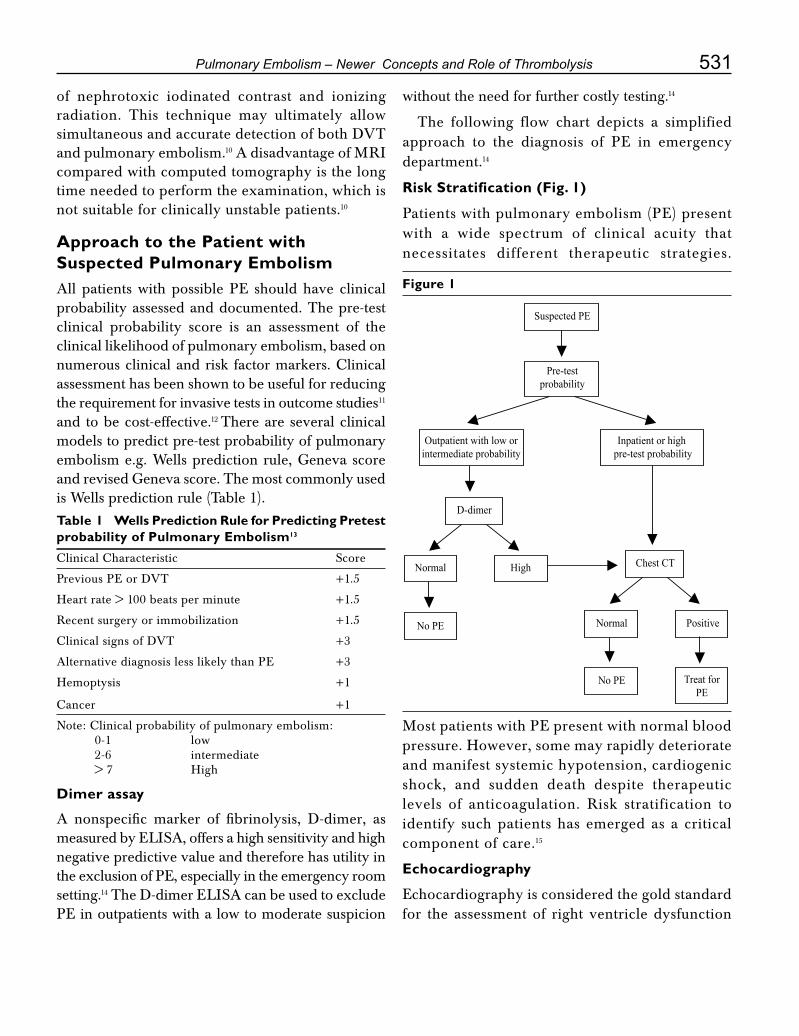
531Pulmonary Embolism – Newer Concepts and Role of Thrombolysis
of nephrotoxic iodinated contrast and ionizing radiation. This technique may ultimately allow simultaneous and accurate detection of both DVT and pulmonary embolism.10 A disadvantage of MRI compared with computed tomography is the long time needed to perform the examination, which is not suitable for clinically unstable patients.10
Approach to the Patient with Suspected Pulmonary EmbolismAll patients with possible PE should have clinical probability assessed and documented. The pre-test clinical probability score is an assessment of the clinical likelihood of pulmonary embolism, based on numerous clinical and risk factor markers. Clinicalassessment has been shown to be useful for reducing the requirement for invasive tests in outcome studies11 and to be cost-effective.12 There are several clinical models to predict pre-test probability of pulmonary embolism e.g. Wells prediction rule, Geneva score and revised Geneva score. The most commonly used is Wells prediction rule (Table 1).
Table 1 Wells Prediction Rule for Predicting Pretest probability of Pulmonary Embolism13 ClinicalCharacteristic Score
Previous PE or DVT +1.5
Heart rate > 100 beats per minute +1.5
Recent surgery or immobilization +1.5
ClinicalsignsofDVT +3
Alternative diagnosis less likely than PE +3
Hemoptysis +1
Cancer +1
Note:Clinicalprobabilityofpulmonaryembolism: 0-1 low 2-6 intermediate > 7 High
Dimer assay
A nonspecific marker of fibrinolysis, D-dimer, as measured by ELISA, offers a high sensitivity and high negative predictive value and therefore has utility in the exclusion of PE, especially in the emergency room setting.14 The D-dimer ELISA can be used to exclude PE in outpatients with a low to moderate suspicion
without the need for further costly testing.14
The following flow chart depicts a simplified approach to the diagnosis of PE in emergency department.14
Risk Stratification (Fig. 1)
Patients with pulmonary embolism (PE) present with a wide spectrum of clinical acuity that necessitates different therapeutic strategies.
Most patients with PE present with normal blood pressure. However, some may rapidly deteriorate and manifest systemic hypotension, cardiogenic shock, and sudden death despite therapeutic levels of anticoagulation. Risk stratification to identify such patients has emerged as a critical component of care.15
Echocardiography
Echocardiography is considered the gold standard for the assessment of right ventricle dysfunction
Suspected PE
Pre-testprobability
Outpatient with low orintermediate probability
D-dimer
Normal
No PE Normal
No PE Treat forPE
Positive
High Chest CT
Inpatient or high pre-test probability
Figure 1
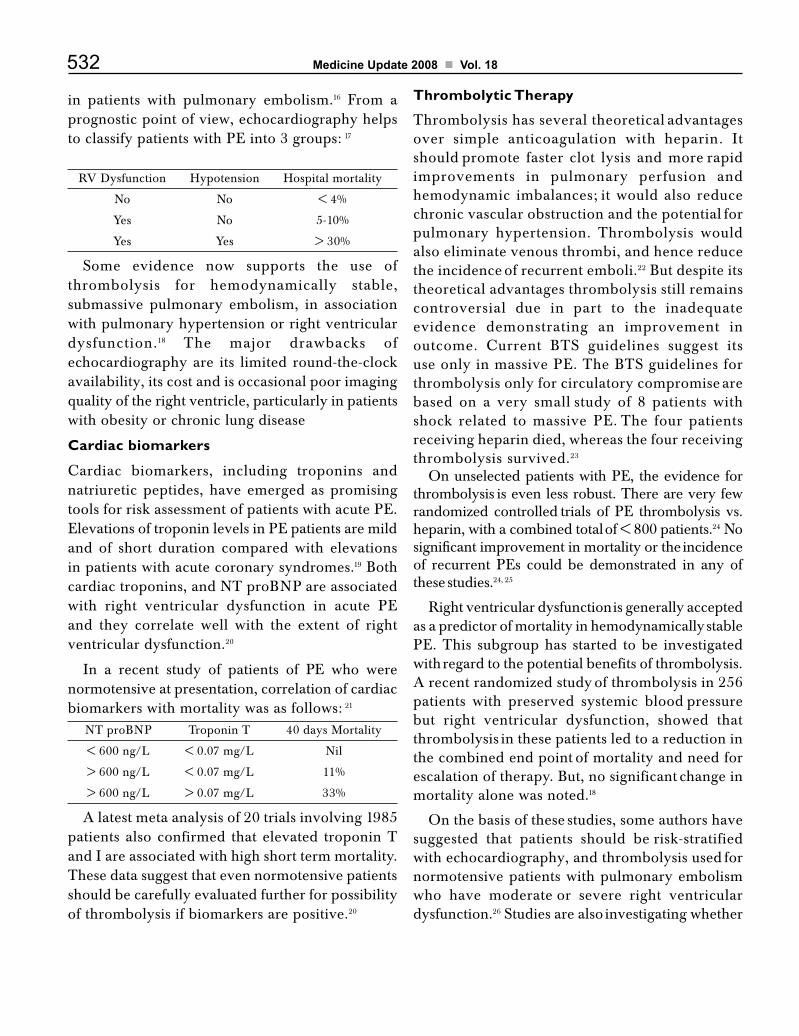
532 Medicine Update 2008 Vol. 18
in patients with pulmonary embolism.16 From a prognostic point of view, echocardiography helps to classify patients with PE into 3 groups: 17
RV Dysfunction Hypotension Hospital mortality
No No < 4%
Yes No 5-10%
Yes Yes > 30%
Some evidence now supports the use of thrombolysis for hemodynamically stable, submassive pulmonary embolism, in association with pulmonary hypertension or right ventricular dysfunction.18 The major drawbacks of echocardiography are its limited round-the-clock availability, its cost and is occasional poor imaging quality of the right ventricle, particularly in patients with obesity or chronic lung disease
Cardiac biomarkers
Cardiac biomarkers, including troponins andnatriuretic peptides, have emerged as promising tools for risk assessment of patients with acute PE. Elevations of troponin levels in PE patients are mild and of short duration compared with elevations in patients with acute coronary syndromes.19 Both cardiac troponins, and NT proBNP are associated with right ventricular dysfunction in acute PE and they correlate well with the extent of right ventricular dysfunction.20
In a recent study of patients of PE who were normotensive at presentation, correlation of cardiac biomarkers with mortality was as follows: 21
NT proBNP Troponin T 40 days Mortality
< 600 ng/L < 0.07 mg/L Nil
> 600 ng/L < 0.07 mg/L 11%
> 600 ng/L > 0.07 mg/L 33%
A latest meta analysis of 20 trials involving 1985 patients also confirmed that elevated troponin T and I are associated with high short term mortality. These data suggest that even normotensive patients should be carefully evaluated further for possibility of thrombolysis if biomarkers are positive.20
Thrombolytic Therapy
Thrombolysis has several theoretical advantages over simple anticoagulation with heparin. It should promote faster clot lysis and more rapid improvements in pulmonary perfusion and hemodynamic imbalances; it would also reduce chronic vascular obstruction and the potential for pulmonary hypertension. Thrombolysis would also eliminate venous thrombi, and hence reduce the incidence of recurrent emboli.22 But despite its theoretical advantages thrombolysis still remains controversial due in part to the inadequate evidence demonstrating an improvement in outcome. Current BTS guidelines suggest itsuse only in massive PE. The BTS guidelines for thrombolysis only for circulatory compromise are based on a very small study of 8 patients with shock related to massive PE. The four patients receiving heparin died, whereas the four receiving
thrombolysis survived.23
On unselected patients with PE, the evidence for thrombolysis is even less robust. There are very few randomized controlled trials of PE thrombolysis vs. heparin, with a combined total of < 800 patients.24 No significant improvement in mortality or the incidence of recurrent PEs could be demonstrated in any of these studies.24, 25
Right ventricular dysfunction is generally accepted as a predictor of mortality in hemodynamically stable PE. This subgroup has started to be investigated with regard to the potential benefits of thrombolysis. A recent randomized study of thrombolysis in 256 patients with preserved systemic blood pressure but right ventricular dysfunction, showed that thrombolysis in these patients led to a reduction in the combined end point of mortality and need for escalation of therapy. But, no significant change in mortality alone was noted.18
On the basis of these studies, some authors have suggested that patients should be risk-stratified with echocardiography, and thrombolysis used for normotensive patients with pulmonary embolism who have moderate or severe right ventricular dysfunction.26 Studies are also investigating whether
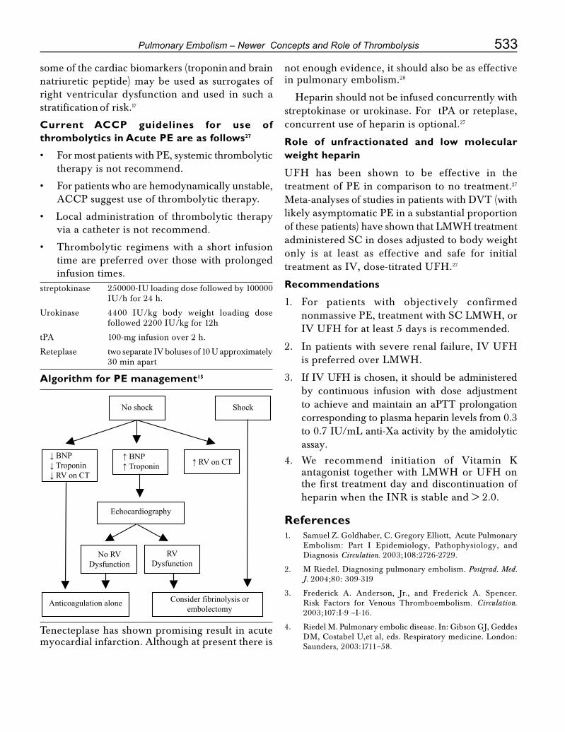
533Pulmonary Embolism – Newer Concepts and Role of Thrombolysis
some of the cardiac biomarkers (troponin and brain natriuretic peptide) may be used as surrogates of
right ventricular dysfunction and used in such a stratification of risk.17
Current ACCP guidelines for use of thrombolytics in Acute PE are as follows27
• FormostpatientswithPE,systemicthrombolytictherapy is not recommend.
• Forpatientswhoarehemodynamicallyunstable,ACCPsuggestuseofthrombolytictherapy.
• Localadministrationof thrombolytic therapyvia a catheter is not recommend.
• Thrombolytic regimenswitha short infusiontime are preferred over those with prolonged infusion times.
streptokinase 250000-IU loading dose followed by 100000 IU/h for 24 h.
Urokinase 4400 IU/kg body weight loading dose followed 2200 IU/kg for 12h
tPA 100-mg infusion over 2 h.
Reteplase two separate IV boluses of 10 U approximately 30 min apart
Algorithm for PE management15
not enough evidence, it should also be as effective in pulmonary embolism.28
Heparin should not be infused concurrently with streptokinase or urokinase. For tPA or reteplase, concurrent use of heparin is optional.27
Role of unfractionated and low molecular weight heparin
UFH has been shown to be effective in the treatment of PE in comparison to no treatment.27 Meta-analyses of studies in patients with DVT (with likely asymptomatic PE in a substantial proportion of these patients) have shown that LMWH treatment administeredSCindosesadjustedtobodyweightonly is at least as effective and safe for initial treatment as IV, dose-titrated UFH.27
Recommendations
1. For patients with objectively confirmed nonmassivePE,treatmentwithSCLMWH,orIV UFH for at least 5 days is recommended.
2. In patients with severe renal failure, IV UFH is preferred over LMWH.
3. If IV UFH is chosen, it should be administered by continuous infusion with dose adjustment to achieve and maintain an aPTT prolongation corresponding to plasma heparin levels from 0.3 to 0.7 IU/mL anti-Xa activity by the amidolytic assay.
4. We recommend initiation of Vitamin K antagonist together with LMWH or UFH on the first treatment day and discontinuation of heparin when the INR is stable and > 2.0.
References1. SamuelZ.Goldhaber,C.GregoryElliott,AcutePulmonary
Embolism: Part I Epidemiology, Pathophysiology, and Diagnosis Circulation. 2003;108:2726-2729.
2. M Riedel. Diagnosing pulmonary embolism. Postgrad. Med. J. 2004;80: 309-319
3. Frederick A. Anderson, Jr., and Frederick A. Spencer. Risk Factors for Venous Thromboembolism. Circulation. 2003;107:I-9 –I-16.
4. Riedel M. Pulmonary embolic disease. In: Gibson GJ, Geddes DM,CostabelU,etal,eds.Respiratorymedicine.London:Saunders, 2003:1711–58.
No shock Shock
↓ BNP↓ Troponin↓ RV on CT
↑ BNP↑ Troponin ↑ RV on CT
Echocardiography
No RVDysfunction
Anticoagulation alone
RVDysfunction
Consider fibrinolysis orembolectomy
Tenecteplase has shown promising result in acute myocardial infarction. Although at present there is
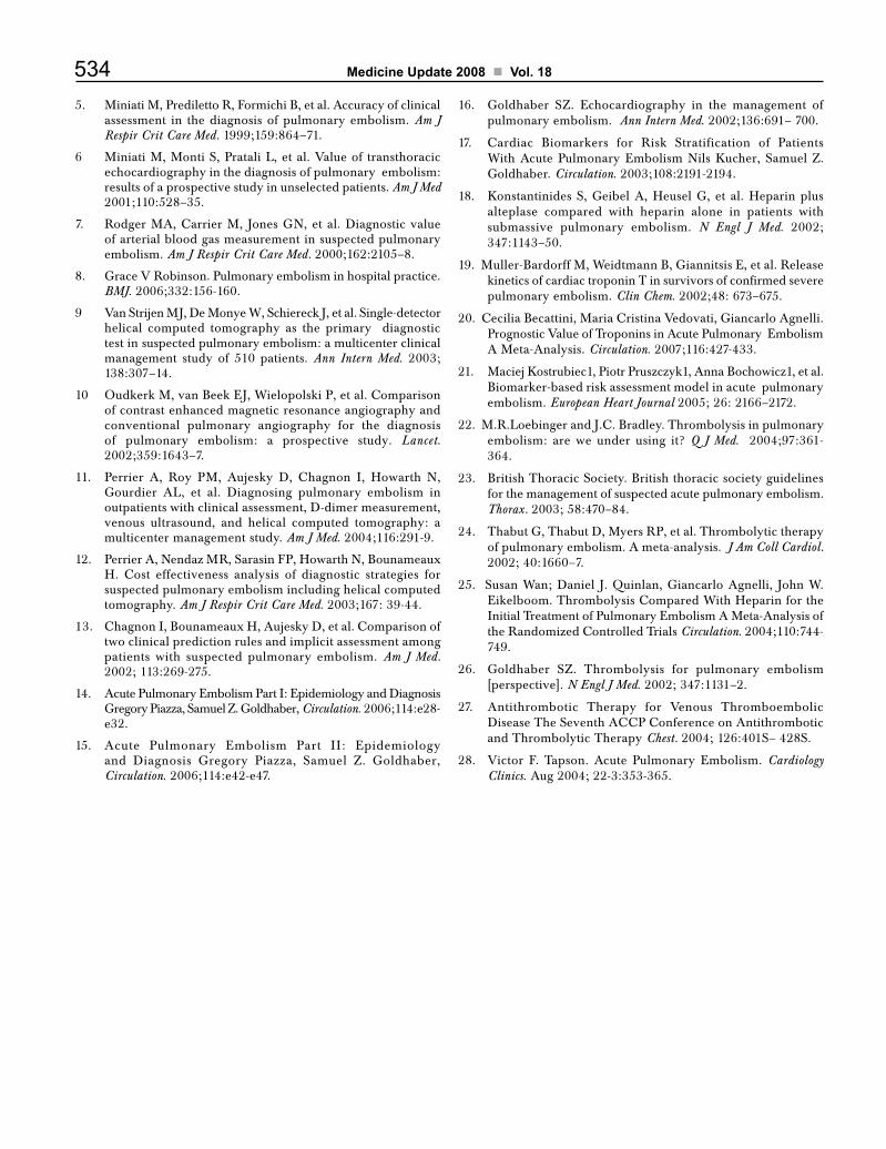
534 Medicine Update 2008 Vol. 18
5. Miniati M, Prediletto R, Formichi B, et al. Accuracy of clinical assessment in the diagnosis of pulmonary embolism. Am J Respir Crit Care Med. 1999;159:864–71.
6 Miniati M, Monti S, Pratali L, et al. Value of transthoracic echocardiography in the diagnosis of pulmonary embolism: results of a prospective study in unselected patients. Am J Med 2001;110:528–35.
7. RodgerMA,CarrierM,JonesGN,etal.Diagnosticvalueof arterial blood gas measurement in suspected pulmonary embolism. Am J Respir Crit Care Med. 2000;162:2105–8.
8. Grace V Robinson. Pulmonary embolism in hospital practice. BMJ. 2006;332:156-160.
9 Van Strijen MJ, De Monye W, Schiereck J, et al. Single-detector helical computed tomography as the primary diagnostic test in suspected pulmonary embolism: a multicenter clinical management study of 510 patients. Ann Intern Med. 2003; 138:307–14.
10 OudkerkM,vanBeekEJ,WielopolskiP,etal.Comparisonof contrast enhanced magnetic resonance angiography and conventional pulmonary angiography for the diagnosis of pulmonary embolism: a prospective study. Lancet. 2002;359:1643–7.
11. Perrier A, Roy PM, Aujesky D, Chagnon I, Howarth N,Gourdier AL, et al. Diagnosing pulmonary embolism in outpatients with clinical assessment, D-dimer measurement, venous ultrasound, and helical computed tomography: a multicenter management study. Am J Med. 2004;116:291-9.
12. Perrier A, Nendaz MR, Sarasin FP, Howarth N, Bounameaux H. Cost effectiveness analysis of diagnostic strategies forsuspected pulmonary embolism including helical computed tomography. Am J Respir Crit Care Med. 2003;167: 39-44.
13.ChagnonI,BounameauxH,AujeskyD,etal.Comparisonoftwo clinical prediction rules and implicit assessment among patients with suspected pulmonary embolism. Am J Med. 2002; 113:269-275.
14. Acute Pulmonary Embolism Part I: Epidemiology and Diagnosis Gregory Piazza, Samuel Z. Goldhaber, Circulation. 2006;114:e28-e32.
15. Acute Pulmonary Embolism Part II: Epidemiology and Diagnosis Gregory Piazza, Samuel Z. Goldhaber, Circulation. 2006;114:e42-e47.
16. Goldhaber SZ. Echocardiography in the management of pulmonary embolism. Ann Intern Med. 2002;136:691– 700.
17. Cardiac Biomarkers for Risk Stratification of PatientsWith Acute Pulmonary Embolism Nils Kucher, Samuel Z. Goldhaber. Circulation. 2003;108:2191-2194.
18. Konstantinides S, Geibel A, Heusel G, et al. Heparin plus alteplase compared with heparin alone in patients with submassive pulmonary embolism. N Engl J Med. 2002; 347:1143–50.
19. Muller-Bardorff M, Weidtmann B, Giannitsis E, et al. Release kinetics of cardiac troponin T in survivors of confirmed severe pulmonary embolism. Clin Chem. 2002;48: 673–675.
20.CeciliaBecattini,MariaCristinaVedovati,GiancarloAgnelli.Prognostic Value of Troponins in Acute Pulmonary Embolism A Meta-Analysis. Circulation. 2007;116:427-433.
21. Maciej Kostrubiec1, Piotr Pruszczyk1, Anna Bochowicz1, et al. Biomarker-based risk assessment model in acute pulmonary embolism. European Heart Journal 2005; 26: 2166–2172.
22.M.R.LoebingerandJ.C.Bradley.Thrombolysisinpulmonaryembolism: are we under using it? Q J Med. 2004;97:361-364.
23. British Thoracic Society. British thoracic society guidelines for the management of suspected acute pulmonary embolism. Thorax. 2003; 58:470–84.
24. Thabut G, Thabut D, Myers RP, et al. Thrombolytic therapy of pulmonary embolism. A meta-analysis. J Am Coll Cardiol. 2002; 40:1660–7.
25. Susan Wan; Daniel J. Quinlan, Giancarlo Agnelli, John W. Eikelboom.ThrombolysisComparedWithHeparinfortheInitial Treatment of Pulmonary Embolism A Meta-Analysis of theRandomizedControlledTrialsCirculation. 2004;110:744-749.
26. Goldhaber SZ. Thrombolysis for pulmonary embolism [perspective]. N Engl J Med. 2002; 347:1131–2.
27. Antithrombotic Therapy for Venous Thromboembolic DiseaseTheSeventhACCPConferenceonAntithromboticand Thrombolytic Therapy Chest. 2004; 126:401S– 428S.
28. Victor F. Tapson. Acute Pulmonary Embolism. Cardiology Clinics. Aug 2004; 22-3:353-365.
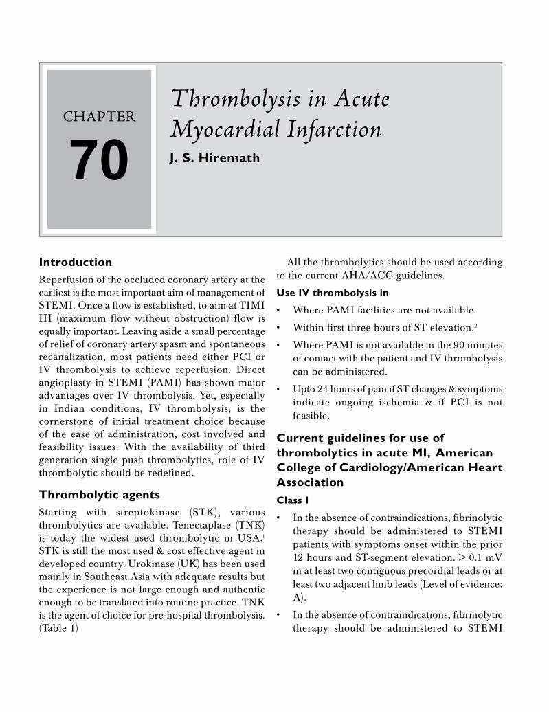
CHAPTER
70Thrombolysis in Acute Myocardial InfarctionJ. S. Hiremath
IntroductionReperfusion of the occluded coronary artery at the earliest is the most important aim of management of STEMI. Once a flow is established, to aim at TIMI III (maximum flow without obstruction) flow is equally important. Leaving aside a small percentage of relief of coronary artery spasm and spontaneous recanalization,mostpatientsneedeitherPCIorIV thrombolysis to achieve reperfusion. Direct angioplasty in STEMI (PAMI) has shown major advantages over IV thrombolysis. Yet, especially in Indian conditions, IV thrombolysis, is the cornerstone of initial treatment choice because of the ease of administration, cost involved and feasibility issues. With the availability of third generation single push thrombolytics, role of IV thrombolytic should be redefined.
Thrombolytic agentsStarting with streptokinase (STK), various thrombolytics are available. Tenectaplase (TNK) is today the widest used thrombolytic in USA.1 STK is still the most used & cost effective agent in developed country. Urokinase (UK) has been used mainly in Southeast Asia with adequate results but the experience is not large enough and authentic enough to be translated into routine practice. TNK is the agent of choice for pre-hospital thrombolysis. (Table 1)
All the thrombolytics should be used according tothecurrentAHA/ACCguidelines.
Use IV thrombolysis in
• WherePAMIfacilitiesarenotavailable.
• WithinfirstthreehoursofSTelevation.2
• WherePAMIisnotavailableinthe90minutesof contact with the patient and IV thrombolysis can be administered.
• Upto24hoursofpainifSTchanges&symptomsindicate ongoing ischemia & if PCI is notfeasible.
Current guidelines for use of thrombolytics in acute MI, American College of Cardiology/American Heart AssociationClass I
• Intheabsenceofcontraindications,fibrinolytictherapy should be administered to STEMI patients with symptoms onset within the prior 12 hours and ST-segment elevation. > 0.1 mV in at least two contiguous precordial leads or at least two adjacent limb leads (Level of evidence: A).
• Intheabsenceofcontraindications,fibrinolytictherapy should be administered to STEMI
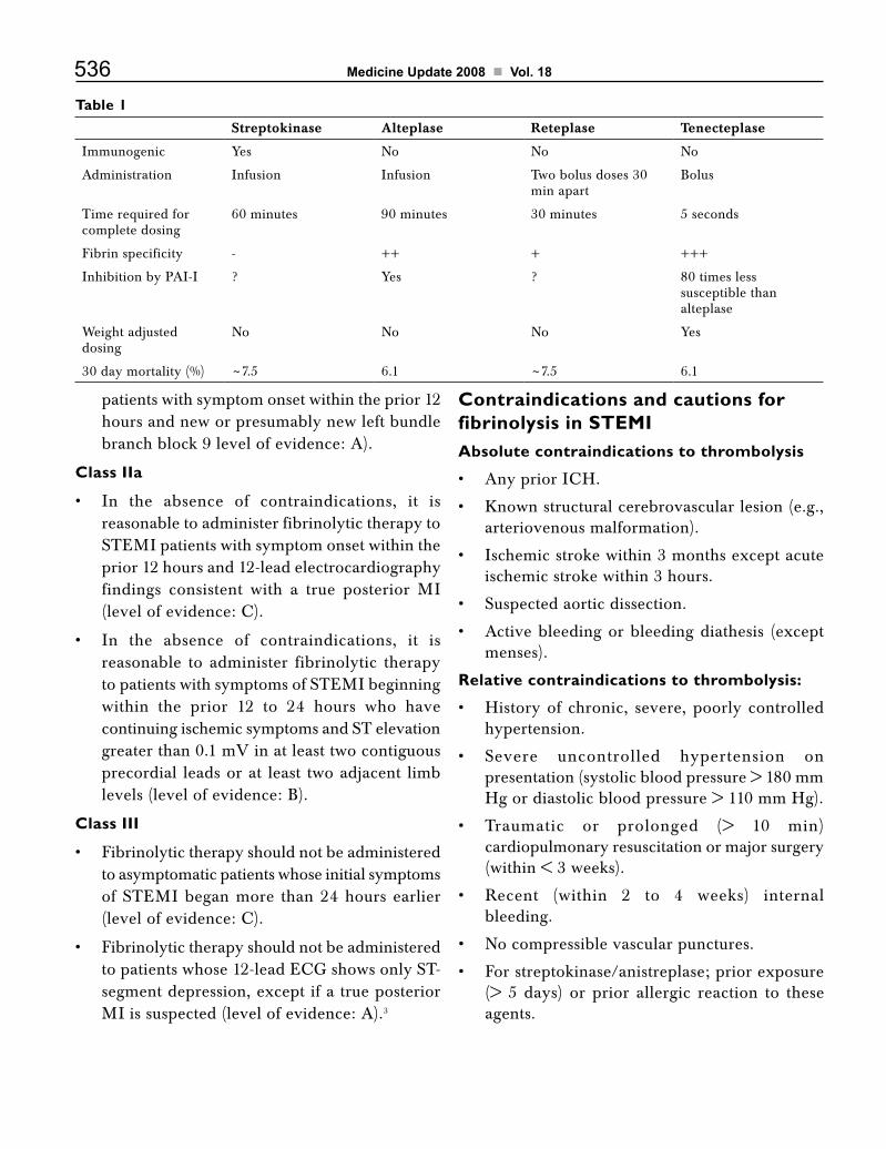
536 Medicine Update 2008 Vol. 18
patients with symptom onset within the prior 12 hours and new or presumably new left bundle branch block 9 level of evidence: A).
Class IIa
• In the absence of contraindications, it isreasonable to administer fibrinolytic therapy to STEMI patients with symptom onset within the prior 12 hours and 12-lead electrocardiography findings consistent with a true posterior MI (levelofevidence:C).
• In the absence of contraindications, it isreasonable to administer fibrinolytic therapy to patients with symptoms of STEMI beginning within the prior 12 to 24 hours who have continuing ischemic symptoms and ST elevation greater than 0.1 mV in at least two contiguous precordial leads or at least two adjacent limb levels (level of evidence: B).
Class III
• Fibrinolytictherapyshouldnotbeadministeredto asymptomatic patients whose initial symptoms of STEMI began more than 24 hours earlier (levelofevidence:C).
• Fibrinolytictherapyshouldnotbeadministeredtopatientswhose12-leadECGshowsonlyST-segment depression, except if a true posterior MI is suspected (level of evidence: A).3
Contraindications and cautions for fibrinolysis in STEMIAbsolute contraindications to thrombolysis
• AnypriorICH.
• Knownstructuralcerebrovascularlesion(e.g.,arteriovenous malformation).
• Ischemicstrokewithin3monthsexceptacuteischemic stroke within 3 hours.
• Suspectedaorticdissection.
• Activebleedingorbleedingdiathesis (exceptmenses).
Relative contraindications to thrombolysis:
• Historyof chronic, severe,poorly controlledhypertension.
• Severe uncontrolled hypertension onpresentation (systolic blood pressure > 180 mm Hg or diastolic blood pressure > 110 mm Hg).
• Traumatic or prolonged (> 10 min)cardiopulmonary resuscitation or major surgery (within < 3 weeks).
• Recent (within 2 to 4 weeks) internalbleeding.
• Nocompressiblevascularpunctures.
• Forstreptokinase/anistreplase;priorexposure(> 5 days) or prior allergic reaction to these agents.
Table 1Streptokinase Alteplase Reteplase Tenecteplase
Immunogenic Yes No No No
Administration Infusion Infusion Two bolus doses 30 min apart
Bolus
Time required for complete dosing
60 minutes 90 minutes 30 minutes 5 seconds
Fibrin specificity - ++ + +++
Inhibition by PAI-I ? Yes ? 80 times less susceptible than alteplase
Weight adjusted dosing
No No No Yes
30 day mortality (%) ~7.5 6.1 ~7.5 6.1

537Thrombolysis in Acute Myocardial Infarction
• Pregnancy.
• Activepepticulcer.
History of prior ischemic stroke (> 3 months), dementia, or known intracranial pathology not coveredinabsolutecontraindications.Currentuseof anticoagulants: the higher the INR, the higher the risk of bleeding.3
PAMI verses IV thrombolysisGiven a choice, PAMI is superior to IV thrombolysis especially after 3 hours of ST elevation. Because of various feasibility issues, if the door to balloon time is more than 90 minutes, it is worthwhile thrombolysing the patient. In the first 3 hours, reperfusion for salvage of myocardium is the aim therefore the therapy that can be offered earlier should be selected. Delay in therapy in this “steep” portion, can harm the patient (Fig1).Shift fromDtoC/B/Aisharmful.Shift toAtoBisneutral.ShiftfromAtoC/Disuseful.Inshort, if PAMI is going to be delayed by 90 minutes, choose IV thrombolysis.
Effects of IV thrombolysisWithin 12 hours, successful recanalization with STK has 18% relative risk reduction at 30 days.4 All trials with successful recanalization with various agents show reduction in mortality. The success rate
with all thrombolytics is upto 70%. 50% patients achieved TIMI III flow & 20% achieved TIMI II flow. Allergic reactions, fever, hypotension are common with STK but the newer agents hardly show such effects. The rate of major bleed requiring transfusion is about 1-2% in all trials.5
Adjuvant therapy for IV thrombolysis on admission• Aspirin:150mgormoreofsolubleaspirin.
• Clopidogrel : 75 mg daily without a loadingdose.6
• LMWH : 30 mg IV Enoxaparine if TNK isused.1
• UFH:5000IVbolus followedby infusion ifTPA is used.
After thrombolysis is over, aspirin, clopidogrel need to continue. Enoxaparine/Raviparine should be used subcutaneously for 7 days.
• Directthrombininhibitors(Hirudin,Bivalirudin)has shown less re-infarction at 30 days but at increased risk of adverse bleedings.7
• NSAIDarecontraindicated.
Use of Glycoprotein IIb/IIIa inhibitors in STEMIUse of abciximab with thrombolytics led to increased TIMI III flow, did not address mortality benefit and had higher major bleeding & complications.7,8 Half dose lytics & abciximab reduced ischemic events but without mortality benefits.2 Incidence of bleeding increased. Use of tirofiban & eptifibatide is too small to be quoted but trials were given up for increased bleeding. In a small pilot study of eptifibatide, it was used for failed thrombolysis but no proven literature is available. Many Indian clinicians use tirofiban for STEMI as “transfer” treatment but this practice is observational and not backed by evidence. The bottom line is GP IIb-IIIa inhibitors are out of bounds for use in STEMI either as “facilitation” or an adjuvant to or as post IV thrombolysis treatment.
Figure 1 : Thrombolysis in acute myocardial infarction
Time from symptom onsest to reperfusion therapy, h
Critical time-dependent periodGoal: myocardial salvage
Time-independentGoal: open infarct-related artery
Shifts in potential outcomes withdifferent treatment strategies
A to B No benefitA to C BenefitB to C BenefitD to B HarmD to C Harm
0 4 8 12 16 20 24
100
80
60
40
20
0
Mor
talit
y re
duct
ion,
%
Extent ofmyocardial salvage
D
C
B A
538 Medicine Update 2008 Vol. 18
Failed thrombolysisSuccess rate of IV thrombolysis is close to 70%. Therefore strategies for failed thrombolysis or reocclusion/reinfarction also need to be planned inadvance.Ongoingpain,non-resolutionofECG,hemodynamic or electrical instability indicate failure of recanalization. Repeat thrombolysis with the same or other agent is not to be practiced. A rescuePTCAmustbeencouragedeventhoughtheoutcomesofrescuePTCAarenotgood.IfrescuePTCAisnotavailablesmallmoleculeGPIIb-IIIainhibitors, tirofiban, eptifibatide can be used as a 24 to 48 hour infusion as a last resort.
Pre-hospital thrombolysisImportance of “early” thrombolysis cannot be over emphasized. Transfer times to tertiary hospital even in cardiac ambulances can be long. Thrombolytics like STK, TPA need continuous infusions & monitoring. Considering all of theabove, TNK single dose push in pre-hospital setting like general practitioners clinic, casualties of corporate hospitals, home & ambulances need to be considered. Large mortality & morbidity benefits have been shown in pre-hospital thrombolysis.10 Telemedicine for E-transfer of ECG could be a vital aspect of this treatment.Medical insurance to all, removing the obstacle of cost of thrombolysis will be an additional help. In general, by using modern amenities, modern thrombolytics and scientific advances an all out effort to increase pre-hospital thrombolysis should be made.11
Future of IV thrombolysisPre-hospital thrombolysis after making a quick decision will be the order of the day. Such a thrombolysed patient may then be transferred to a tertiary centre with an acceptable delay. Lanoteplase, Saruplase (Rescupace), Staphylokinase are recombinant products of existing thrombolytics.12 These products are in trial phase & may or may not prove to be improvements on the latest player – tenectaplase. Oral thrombolytic is a dream, which
is unlikely to replace IV thrombolysis in the near future.
IV thrombolysis in reference to Indian settingsIt is now widely accepted that early IV thrombolysis (pre-hospital included) can be highly effective treatment.12 In India where primary angioplasty can be offered to a small portion of patients within few hours of chest pain due to various feasibility issues, IV thrombolysis is even more important. Small hospitals, rural centers and other areas where patients reach first should be considered “pre-hospital” environments while a transfer is organized. A growing number of “young” AMI patients is a population yet not presented in evidence. This population of under forty, first MI, contain mainly thrombus in the occluded coronary artery. Subjecting these for primary stenting would mean a foreign body in a young person for no reason. These individuals respond dramatically to early and effective thrombolysis.
The common errors made in practice of IV thrombolysis should be avoided and following points need to be pondered.
• Aimasearliestthrombolysisunlessitisalreadysix hours old.13
• UsefulldoseSTKin30minutesinfusion(Pleasecheck expiry date and standard of the STK company).
• Avoid use of Urokinase as thrombolytic forAMI.
• Shift towardsuseof tPAandTNKshouldbemade.
• “Pre-hospital”useofTNKshouldbetheorderof the day.
References1. Assessment of the Safety and Efficacy of a New Thrombolytic
Regimen (ASSENT)-3 Investigators. Efficacy and safety of tenecteplase in combination with enoxaparin, abciximab or unfractionated heparin: the ASSENT-3 randomized trial in acute myocardial infarction. Lancet 2001;358(9282):605-13.
2. Maria Sejersten, Rasmus Ripa, timing of ischemic onset estimated from the electrocardiogram is better than historical
539Thrombolysis in Acute Myocardial Infarction
timing for predicting outcome after reperfusion therapy for acute anterior myocardial infarction : A DANish trial in Acute Myocardial Infarction2 (DANAMI-2) sub study : Am Heart J 2007; 154:61-68.
3. Antman EM, Anbe D Metal ACC/AHA guidelines formanagement of patients with ST elevation myocardial infarction. Executive summary, Jm Coll Cardiol 2005:45(8):1376.
4. ArmstrongPW,CollenD.Fibrinolysisforacutemyocardialinfarction: current status & new horizons for pharmacological reperfusion, part 2. Circulation 2001;103(24):2987-92.
5. Assessment of the Safety and Efficacy of a new Thrombolytic Investigators. Single-bolus tenecteplase compared with front loaded alteplase in acute myocardial infarction: the ASSENT-2 double-blinded randomized trial. Lancet 1999;354(9180):716-22.
6. Commit collaborativegroup :AdditionofClopidogrel toAspirin in 45852 patients with acute myocardial infarction : randomized placebo controlled trial : Lancet 2005; 366:1607-1621.
7. White H, Hirulog and Early Reperfusion or Occlusion (HERO)-2 trial Investigators. Thrombin-specific anticoagulation with bivalirudin versus heparin in patients
receiving fibrinolytic therapy for acute myocardial infarction: the HERO-2 randomized trial. Lancet 2001; 358(9296):1855-63.
8. AntmanEM,GiuglianoRP,GibsonCM,et al. theTIMI14 Investigators. Abciximab facilitates the rate and extent of thrombolysis: results of the thrombolysis in myocardial infarction (TIMI) 14 trial. Circulation 1999;99(21):2720-32.
9. Strategies for Patency Enhancement in the Emergency Department (SPEED) Group. Trial of abciximab with and without low-dose reteplase for acute myocardial infarction. Circulation 2000;101(24):2788-94.
10. MorrisonLJ,VerbeekPR,McDonaldAC,etal.Mortalityandpre-hospital thrombolysis for acute myocardial infarction: a meta-analysis. JAMA 2000;283(20):2686-92.
11. Bassand JP, Dankhin N ena : Implementation of reperfusion therapy in acute myocardial infarction. A policy statement fromEurSocietyofCardiology:Eur Heart J 2005:26:2733-2741.
12. Matuso O. An ideal thrombolytic and antithrombotic agent? J Thromb Haemost 2005;3(10):2154-5.
13. Nicolas Danchin: Should intravenous thrombolysis keep a place in the treatment of acute ST-elevation myocardial infarction? Eur Heart J (2006) 27, 1131-1133.

CHAPTER
71Women and obesity : one Decade of Experience with SibutramineM. V. Muraleedharan
Obesity is a common disorder. Overweight persons (BMI 25-29) form 60% and obesity (BMI >30) is noted 42% of American population. The situation is not far different in other parts of the world. With changing economic scenario, India also host a substantial percentage of obese and overweight population. Medical profession, health care providers and the population at large still do not consider overweight and obesity as a disease entity.Consequently,itisnotasubjectofintenseresearch and therapeutic intervention. Until the subject reaches morbid obesity with its associated mechanical, metabolic and psychosocial problem, medical assistance is rarely requested for.
However, in the past two decades, there is better awareness about obesity among the medical profession, healthcare providers and population. It is considered as a disease entity and early intervention is sought. Substantial interest is seen in study of the disease entity in the form of epidemiological studies, evaluation of pathophysiology and relation between major clinical diseases like diabetes, coronary artery disease, cancers and obesity.
Both men and women are afflicted with the disease but incidence of obesity in women is higher than in men (NHANES data). Even though there are common problems affecting both sexes when obese, some of the problems of obesity are gender
dependant and seen in women only. Women who have abdominal obesity have a metabolic risk profile similar to that of men1,2 Women have more fat as a per cent of body weight than men from puberty onward, and tend to gain more fat during adult life than men. In addition, women experience modest but adverse increases in body weight and fat distribution after a first pregnancy that appear to be persist.3 The trend for increased incidence of obesity is first obserseved during adolescence and persist into adulthood. This is attributed to the change in body composition between two sexes. When body fat decreases in boys during this period, it increases in girls. However, it is difficult to predict which obese child would become obese adult.
Pregnancy is an important milestone in the weight gain history of women. Most of the women gain 2 to 3 Kg in postpartum period. Weight gain with oral contraceptives is not supported by scientific evidence. However, most of the women gain weight during menopause. Decrease in estrogen and progesterone with menopause cause central fat deposition and enhanced cardiovascular risk. Estrogen therapy does not prevent weight gain but minimize fat distribution in the central abdomen.
Obesity in women can be classified as those associated with polycystic ovary disease and those which are independanat. Pathogenesis, clinical

541Women and obesity : one Decade of Experience with Sibutramine
course and management are different in polycystic ovaries when compared to women with simple obesity. There are some similarities between the two group however.
Obesity (Not associated with polycystic ovaries)Fertility
Decreased fertility in obese women is supported by available data. This often result from anovulation with or without polycystic ovary syndrome. In many obese women, weight reduction is associated with return of ovulation and conception. It is also seen that higher doses of fertility inducing treatment is needed in obese women compared to lean counterparts. It is also reported that there is increased occurrence of early miscarriages in obese women. This may be attributed to Polycystic ovarian disease or insulin resistance in some but not in all.
Weight gain in pregnant women especially when they are obese. This is important as these women fail to lose the weight gained during pregnancy. At present, permitted weight gain is 7 to 11.5 kg in overweight women (BMI 26-29) AND 6.8 for obese women (BMI > 29) (Institute of Medicine recommendation)
During pregnancy in obese person, there is increased incidence of Gestational Diabetes Mellitus. This is in parallel to the increased risk of type 2 diabetes with increasing BMI. Similarly, the risk of Pregnancy associated hypertension is increased. This is attributed to insulin resistance, dyslipidemia and low grade inflammatory state persisting in the obese pregnant patient. There is also higher incidence of preterm birth, post maturity, multifetal pregnancy, urinary infection and obstructive sleep apnea.
Problems during delivery
The duration of labor is increased in obese women. Myometrium from obese women removed during caesarian contracted less well than normal.3 Induction of labor is often needed with increased
rate of caesarian delivery and shoulder dystocia due to fetal macrosomia. Post partum hemorrhage and infection are more common in obese parturient. Duration of hospital stay is prolonged. Initiation of lactation, duration of lactation and quantity of milk are adversely influenced by obesity.
Post partum issues
Increased incidence of neural tube defect is noted in these subjects. Perinatal mortality is increased. Fetal macrosomia occur with higher frequency in the offspring of obese patient.
Cardiovascular risk in obese women
Available data does not suggest any increase in cardiovascular events in women with obesity compared to their male counterparts.
Management
Obesity is a lifestyle disease and major step in its management would be therapeutic lifestyle changes. The age at which obesity start has shifted to earlier years with affliction of children and adolescents.
Behavioral strategies
Behavior modification for obesity management goes back to 1967 with progressive increase in emphasis and length of aggressive technic over the years. 7 to 10% weight loss could be expected, most of this weight loss occur in first 6 months in motivated subjects. However, maintenance of lost weight is an important issue. Duration of behavior therapy training vary from 20 to 40 weeks. In this treatment strategy, it is presumed that obese individual has maladaptive diet and exercise habits and attempts are made to change these by behavioral inputs.
Behavior modification include the following elements:
Regular physical activity
At least 150 minutes per week – promote weight loss. This can be done as individual or as group with similar success.
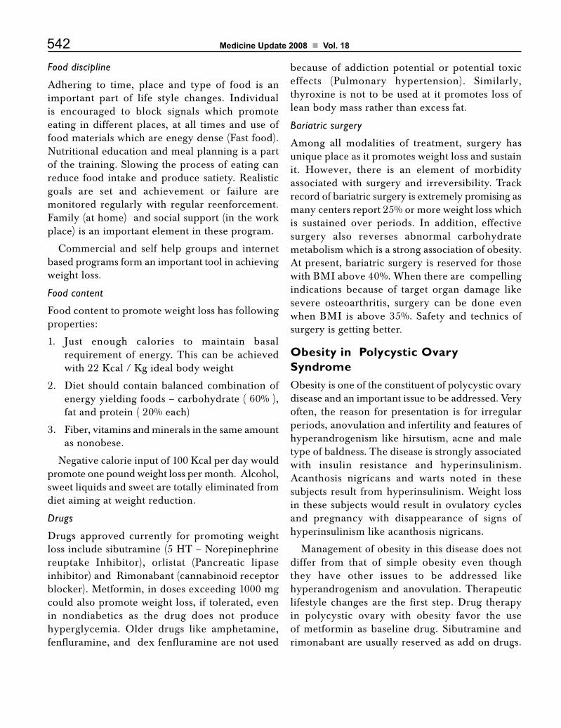
542 Medicine Update 2008 Vol. 18
Food discipline
Adhering to time, place and type of food is an important part of life style changes. Individual is encouraged to block signals which promote eating in different places, at all times and use of food materials which are enegy dense (Fast food). Nutritional education and meal planning is a part of the training. Slowing the process of eating can reduce food intake and produce satiety. Realistic goals are set and achievement or failure are monitored regularly with regular reenforcement. Family (at home) and social support (in the work place) is an important element in these program.
Commercial and selfhelpgroupsand internetbased programs form an important tool in achieving weight loss.
Food content
Food content to promote weight loss has following properties:
1. Just enough calories to maintain basal requirement of energy. This can be achieved with 22 Kcal / Kg ideal body weight
2. Diet should contain balanced combination of energy yielding foods – carbohydrate ( 60% ), fat and protein ( 20% each)
3. Fiber, vitamins and minerals in the same amount as nonobese.
Negative calorie input of 100 Kcal per day would promote one pound weight loss per month. Alcohol, sweet liquids and sweet are totally eliminated from diet aiming at weight reduction.
Drugs
Drugs approved currently for promoting weight loss include sibutramine (5 HT – Norepinephrine reuptake Inhibitor), orlistat (Pancreatic lipase inhibitor) and Rimonabant (cannabinoid receptor blocker). Metformin, in doses exceeding 1000 mg could also promote weight loss, if tolerated, even in nondiabetics as the drug does not produce hyperglycemia. Older drugs like amphetamine, fenfluramine, and dex fenfluramine are not used
because of addiction potential or potential toxic effects (Pulmonary hypertension). Similarly, thyroxine is not to be used at it promotes loss of lean body mass rather than excess fat.
Bariatric surgery
Among all modalities of treatment, surgery has unique place as it promotes weight loss and sustain it. However, there is an element of morbidity associated with surgery and irreversibility. Track record of bariatric surgery is extremely promising as many centers report 25% or more weight loss which is sustained over periods. In addition, effective surgery also reverses abnormal carbohydrate metabolism which is a strong association of obesity. At present, bariatric surgery is reserved for those with BMI above 40%. When there are compelling indications because of target organ damage like severe osteoarthritis, surgery can be done even when BMI is above 35%. Safety and technics of surgery is getting better.
Obesity in Polycystic Ovary Syndrome Obesity is one of the constituent of polycystic ovary disease and an important issue to be addressed. Very often, the reason for presentation is for irregular periods, anovulation and infertility and features of hyperandrogenism like hirsutism, acne and male type of baldness. The disease is strongly associated with insulin resistance and hyperinsulinism. Acanthosis nigricans and warts noted in these subjects result from hyperinsulinism. Weight loss in these subjects would result in ovulatory cycles and pregnancy with disappearance of signs of hyperinsulinism like acanthosis nigricans.
Management of obesity in this disease does not differ from that of simple obesity even though they have other issues to be addressed like hyperandrogenism and anovulation. Therapeutic lifestyle changes are the first step. Drug therapy in polycystic ovary with obesity favor the use of metformin as baseline drug. Sibutramine and rimonabant are usually reserved as add on drugs.
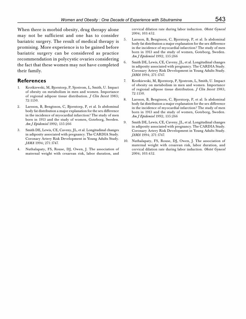
543Women and obesity : one Decade of Experience with Sibutramine
When there is morbid obesity, drug therapy alone may not be sufficient and one has to consider bariatric surgery. The result of medical therapy is promising. More experience is to be gained before bariatric surgery can be considered as practice recommendation in polycystic ovaries considering the fact that these women may not have completed their family.
References1. Krotkiewski, M, Bjorntorp, P, Sjostrom, L, Smith, U. Impact
of obesity on metabolism in men and women. Importance of regional adipose tissue distribution. J Clin Invest 1983; 72:1150.
2. Larsson,B,Bengtsson,C,Bjorntorp,P,etal.Isabdominalbody fat distribution a major explanation for the sex difference in the incidence of myocardial infarction? The study of men born in 1913 and the study of women, Goteborg, Sweden. Am J Epidemiol 1992; 135:266
3. SmithDE,Lewis,CE,Caveny,JL,etal.Longitudinalchangesinadiposityassociatedwithpregnancy.TheCARDIAStudy.CoronaryArteryRiskDevelopmentinYoungAdultsStudy.JAMA 1994; 271:1747.
4. Nuthalapaty, FS, Rouse, DJ, Owen, J. The association of maternal weight with cesarean risk, labor duration, and
cervical dilation rate during labor induction. Obstet Gynecol 2004; 103:452.
5. Larsson,B,Bengtsson,C,Bjorntorp,P,etal.Isabdominalbody fat distribution a major explanation for the sex difference in the incidence of myocardial infarction? The study of men born in 1913 and the study of women, Goteborg, Sweden. Am J Epidemiol 1992; 135:266
6. SmithDE,Lewis,CE,Caveny,JL,etal.Longitudinalchangesinadiposityassociatedwithpregnancy.TheCARDIAStudy.CoronaryArteryRiskDevelopmentinYoungAdultsStudy.JAMA 1994; 271:1747.
7. Krotkiewski, M, Bjorntorp, P, Sjostrom, L, Smith, U. Impact of obesity on metabolism in men and women. Importance of regional adipose tissue distribution. J Clin Invest 1983; 72:1150.
8. Larsson,B,Bengtsson,C,Bjorntorp,P,etal.Isabdominalbody fat distribution a major explanation for the sex difference in the incidence of myocardial infarction? The study of men born in 1913 and the study of women, Goteborg, Sweden. Am J Epidemiol 1992; 135:266
9. SmithDE,Lewis,CE,Caveny,JL,etal.Longitudinalchangesinadiposityassociatedwithpregnancy.TheCARDIAStudy.CoronaryArteryRiskDevelopmentinYoungAdultsStudy.JAMA 1994; 271:1747.
10. Nuthalapaty, FS, Rouse, DJ, Owen, J. The association of maternal weight with cesarean risk, labor duration, and cervical dilation rate during labor induction. Obstet Gynecol 2004; 103:452.

CHAPTER
72Management of obesity: Results of SCoUT TrialA. Misra, J. S. Wasir
IntroductionObesity is increasing at an alarming rate in developed industrialized countries as well as in developing countries which are undergoing rapid nutrition and lifestyle transition. Substantial changes in lifestyle (greater consumption of energy dense foods, inactive lifestyle etc.) are the predominant reasons for increase in prevalence of obesity and related disorders.1 Obesity is associated with increase in risk of diseases like type 2 diabetes mellitus (T2DM),
coronary heart disease (CHD), hypertension,
dyslipidemia, the metabolic syndrome and certain cancers significantly increases the risk of mortality at any given age. Obese subjects have a two-fold increased risk of cardiovascular disease-related mortality, and a body mass index (BMI) greater than 35 / m2 has shown a seven-fold increase in the mortalityriskinpatientswithCHD.
Secular trends of obesity in India indicate an increasing prevalence of obesity, diabetes and related cardiovascular risk factors not only in adults but also in the younger population.1,2 The urban prevalence of obesity has increased alarmingly; almost 50% of adult urban Indians in Delhi fulfill criteria for either obesity or abdominal obesity (Table 1, Misra A, unpublished data 2006). The prevalence of overweight/obesity in children has increased from 16% in 2002-2004, to 29% in 2006.3 Interestingly as compared to migrant Indians, native Indians have similar prevalence of generalized obesity (BMI) but greater abdominal obesity as estimated by waist circumference and waist-to-hip-ratio. The rural populations in India still has lower prevalence of both generalized obesity and truncal obesity when compared to the urban population (Table 1, Misra A, unpublished data 2006).
Clusteringofriskfactorsknownasthemetabolicsyndrome is often associated with obesity and abdominal adiposity, and it is particularly prevalent
Table 1 : Comparative Statistics of Obesity between Asian Indians in US and IndiaVariables Asian
Indians (US)*
Asian Indians
(Urban)**
Asian Indians
(Rural)***
Mean BMI 25.4 24.8 21.9
BMI > 30 11% 13% 2%
BMI > 23 73.3% 65.4% 31.8%
HighWC(cm)
22.5% 38.6% 7.7%
Mean WHR 0.89 0.94 0.84
WHR > 0.90 43.8% 65.7% 23%
All BMI values in kg/m2;WC,Waistcircumference;WHR,Waist-to-hip-circumference ratio
6 cities in USA, **, New Delhi; ***, Gandhigram, South India
Ref:MisraR,MisraA,etal.AAPI-CDCProject,2004-2006(Unpublished data)

545Management of obesity: Results of SCoUT Trial
The key management strategies including diet, exercise, behavior modifications, drug treatment and surgical treatment are summarized in Table 2. The details of these management strategies have been discussed in our previous publications. Briefly, the therapy in terms of using a particular modality depends on the assessment for detecting presence of various cardiovascular and other co-morbid factors (Tables 3 and 4). These guidelines give a general idea about when to apply a particular treatment modality.
In India, currently three drugs (orlistat, sibutramine and rimonabant) have been licensed as anti-obesity drugs.
Table 2 : Management of Obesity: Key StrategiesBehavioral strategies
Using strategies like self-monitoring, social support, and stress management, etc.
Dietary intake Reducing caloric intake by 500 to 1,000 kcal per day to produce weight loss.
Physical activity Obese patients to start with moderate levels of physical activity (e.g., brisk walking or jogging) for 30 to 45 minutes, three to five days per week, and then on all days of week.
Adjunctive pharmacotherapy
Drug treatment to be considered for patients with a BMI ≥ 30*, or with a BMI ≥ 27* in combination with other medical co-morbidities.
Surgery Surgery to be considered as the last choice when all other modalities fail, and patients with BMI ≥ 40 or between 35 and 40 along with co-morbidities require surgical intervention.*
All BMI values in kg/m2
* The BMI limits have been assigned in general to all ethnic groups; however, these are lower in Asian populations.
in Asian Indians and south Asians.4 Insulin resistance and the metabolic syndrome are also becoming more prevalent in children in urban India.5 We are also witnessing obesity-related morbidities in children and adolescents (polycystic ovarian syndrome, dyslipidemia etc.) If appropriate prevention and treatment approaches are not implemented, we shall witness further increase in twin epidemics of diabetes and cardiovascular disease.
The aim of this brief review is to give overview of the management of obesity and focus on recent data on sibutramine.
ManagementVarious therapeutic approaches are available to manage obesity. Non-pharmacological lifestyle management is the first and perhaps, the most important step. Pharmacological management becomes necessary if the condition is not satisfactorily treated through lifestyle management. Finally, if weight loss is not achieved by pharmacotherapy,
occasionally, surgical management is exercised in morbidly obese patients.
Table 3 : Management Recommendations According to BMIBMI ≤ 27 with or without co-morbidities*
Lifestyle management with diet, physical activity and behavioral modifications. Pharmacological management not used.
BMI > 27 and ≤ 30, without co-morbidities*
Lifestyle management with diet, physical activity and behavioral modifications. Pharmacological management not advised, but can be considered if patient does not respond, or is non-compliant with lifestyle management.
BMI > 27 and ≤ 30, with co-morbidities*
Pharmacological management recommended along with lifestyle changes
BMI > 30 and ≤ 35 with or without co-morbidities*
Pharmacological management recommended along with lifestyle changes
BMI > 35 and ≤ 40 without co-morbidities*
Pharmacological management recommended along with lifestyle changes
BMI > 35 and ≤ 40 with co-morbidities*
Considersurgicalmanagementalongwith pharmacological and non-pharmacological management.
BMI > 40 Considersurgicalmanagementalongwith pharmacological and non-pharmacological management.
All BMI values in Kg/m2.
* The BMI limits have been assigned in general to all ethnic groups; however, these are likely to be lower in Asian populations.

546 Medicine Update 2008 Vol. 18
Table 4 : Pharmacological Management of ObesityMedication Mechanism of action Side effects Dosage
Sibutramine Reuptake inhibitor of serotonin, norepinephrine and dopamine. Blocks NE and 5-HT uptake
Elevated blood pressure, tachycardia, headache, insomnia, constipation, dry mouth
10 mg daily initially; can increase to 15 mg daily after 4 weeks in non-responders
Orlistat Reversible lipase inhibitor. Blocks gastric and pancreatic lipases and causes fat malabsorption
Fecal incontinence, oily spotting, flatulence, vitamin malabsorption
120 mg three times daily just before meals
Rimonabant Endocannabinoid CB1 receptorantagonist. Causes decreasedappetite and has effects of adipocytes
Mild and transient,
Nausea, dizziness,
Anxiety, depression
20 mg per day
5 HT - 5 hydroxytryptamine; NE - Norepinephrine
Sibutramine
Sibutramine has been used as a weight loss therapy successfully. It inhibits the neuronal reuptake of serotonin, norepinephrine and dopamine. Sibutramine does not stimulate secretion of serotonin, and seems to produce weight loss by its anorectic effect and, possibly, by stimulating thermogenesis (i.e., increasing metabolic rate). It has also been approved by the Food and Drug Administration, USA for the long-term treatment of obesity.
Randomized controlled trials have shown that sibutramine produces a dose-related weight loss when given in the range 5–30 mg/day, with an optimal dose of 10–15 mg/day. Studies have shown that active weight loss occurs for the first six months of sibutramine use and can be maintained for up to one year with continued treatment. A one year trial of sibutramine showed that sibutramine dosages of 10 mg per day, 15 mg per day, and placebo resulted in weight loss of 4.8 kg, 6.1 kg, and 1.8 kg, respectively, and led to significant reductions in waist-to-hip ratio compared to patients receiving placebo.6 Another study demonstrated significant, dose-dependent weight loss over 24 weeks with sibutramine; however, like other studies, it showed that weight gain occurs after discontinuation of the drug.7 A long-term two year study found that even though there was a tendency of weight gain in both the sibutramine and placebo groups during the second year of follow up, weight losses were significantly greater among those who received sibutramine for the full two years of the study.8
Treatment with sibutramine has been also shown to improve many obesity-related co-morbidities.9 In a 12-week study, patients with T2DM who received sibutramine showed moderate but significant weight loss as well as improvements in HbA
1c
levels, compared with patients in the placebo group. Sibutramine-induced weight loss produces favorable reduction in plasma triglycerides, total cholesterol, low-density lipoprotein cholesterol and HbA
1c levels.6
The potential long-term treatment benefits of sibutramine in weight management is currently being assessed in the landmark Sibutramine CardiovascularOutcomestudy (SCOUT),whichis the first prospective study to examine the role of obesity management in relation to cardiovascular disease.
Sibutramine Cardiovascular Outcome Trial
The question of whether the use of Sibutramine can prevent cardiovascular morbidity and mortality has notbeenstudiedsofar.TheSCOUTwasdesignedto determine whether weight reduction with sibutramine would be associated with reduction of cardiovascular endpoints in high-risk overweight and obese patients.
Study Design
Study Population
Age: 55 years or older. BMI: ≥ 27 kg/m2 and < 45 kg/m2 or ≥ 25 kg/m2 and < 27 kg/m2 with waist circumference ≥ 102 cm (males) or ≥ 88 cm (females)

547Management of obesity: Results of SCoUT Trial
cardiovascular disease.
1. Clinically relevantandstatistically significantmedian decrease in weight (2.2 kg) and waist circumference (2 cm)
2. Small significant decrease in median systolic and diastolic blood pressure (-3.0 & -1.0 mmHg) and a small significant increase in median pulse rate (+1.5 bpm)
3. Number of serious adverse events (SAEs) and discontinuations for adverse events (AEs) was lower than might be anticipated for these high-risk patients and in general, were similar to those reported previously with sibutramine therapy and reflect its mode of action.
References1. Misra A, Vikram NK. Insulin resistance syndrome (metabolic
syndrome) and obesity in Asian Indians: evidence and implications. Nutrition 2004; 20:482-91.
2. Gupta R, Misra A. Type 2 diabetes in India: regional disparities. Br J Diabetes Vasc Dis 2007;7: 12-16 & 2007;7:12-16.
3. Misra A, Vikram NK, Arya S, Pandey RM, Dhingra V, ChatterjeeA,etal.Highprevalenceofinsulinresistanceinpostpubertal Asian Indian children is associated with adverse truncal body fat patterning, abdominal adiposity and excess body fat. Int J Obes Relat Metab Disord 2004;28:1217-26.
4. Misra A, Misra R, Wijesuriya M, Banerjee D. The metabolic syndrome in South Asians: continuing escalation & possible solutions. Indian J Med Res 2007;125:345-54.
5. Misra A, Khurana L, Vikram NK, Goel A, Wasir JS. Metabolic syndrome in children: current issues and South Asian perspective. Nutrition 2007;23:895-910.
6. Lean, M.E., Sibutramine--a review of clinical efficacy. Int J Obes Relat Metab Disord, 1997. 21 Suppl 1: p. S30-6; discussion 37-9.
7. Bray, G.A., et al., A double-blind randomized placebo-controlled trial of sibutramine. Obes Res, 1996. 4.
8. James, W.P., et al., Effect of sibutramine on weight maintenance after weight loss: a randomized trial. STORM Study Group. Sibutramine Trial of Obesity Reduction and Maintenance. Lancet, 2000. 356(9248): 2119-25.
9. Van Gaal, L.F., M.A. Wauters, and I.H. De Leeuw, Anti-obesity drugs: what does sibutramine offer? An analysis of its potential contribution to obesity treatment. Exp Clin Endocrinol Diabetes, 1998. 106 4: p. 35-40.
10. Philip W JT. The SCOUT study: risk-benefit profile ofsibutramine in overweight high-risk cardiovascular patients. European Heart Journal 2007;7 (Suppl L):L44-L48.
Classificationofriskgroupcategories:
1. T2DM only (24%): subjects with T2DM and another risk factor, excluding cardiovascular disease
2. Cardiovasculardiseaseonly(16%):subjectswithcardiovascular disease, excluding T2DM and another risk factor
3. Cardiovasculardisease+T2DM(60%):subjectswith cardiovascular disease and T2DM and another risk factor
The initial data for the lead in period of 6 weeks, which has been recently published.10 The following is the summary of the data.
• Despitebeinglighter,withlowerpredictedleanbody mass and higher proportion with diabetes, women lost at least as much weight as men.
• Changes during the 6-week period for bodyweight, BMI and waist circumference were statistically significant (P < 0.001).
• Overall,6-weektreatmentperiodwithSibutramine,with 10 mg dose and weight management resulted in clinically important reduction in body weight and waist circumference in women.
• Treatment with Sibutramine for 6 weeks innormotensive high-risk patients resulted in small median increases in BP consistent with changes seen in the labeled population.
• Inpatientsclassifiedashypertensive,Sibutraminereduced BP even in those patients already receiving ≥ 2 classes of medications for BP control.
SCOUT trial: ConclusionWeight management with sibutramine 10 mg is well tolerated by a broad range of high-risk subjects with
Week-8
Week-6
Week-4
Week-2
Studyday 1
Studyday -1
Month1
Month2
Month3
Visit each3 Months
and annually
Month6
2-weeksingle-blind
lead-in period
Baseline/randomization
Randomization phase
Teament period
Sibutramine 10-15 mg dailyor placebo
Follow-up periodfor study drugdiscontinuation
Final visit
Sibutramine10 mg daily2-week
screeningperiod

CHAPTER
73The Challenges of Hypertension in obese Subjects - Indian PerspectiveS. N. Arya
The relationship between obesity and hypertension is said to be a two way street.1 Obesity is being recognized as one of the most important risk factors for the development of hypertension.1 Several epidemiological studies show that the age-adjusted prevalence of hypertension increases directly with body-mass-index (BMI).1 The link between BMI and Blood Pressure (BP) appears to be stronger for systolic than diastolic BP. Central obesity, so common in Indians ismuch more clearly related to cardiovascular (C.V.) and metabolic risk factors than lowerbody obesity. Risk of developing hypertension is strongly related to both waist circumstances and waist hip ratio. The incidence of hypertension is higher in people with high waist and small hip circumference.1 Significant proportion of USA citizens with abdominal obesity (21%-27% in males and 37-57% in females) have hypertension and 85% of hypertension occurs in subjects with BMI ≥ 25 kg/m2 in Finland.1 In our country also many hypertensives are either overweight or obese. The Nurses Health Study2 had shown that women who lost 5 kg had significantly lower risk of developing hypertension than those who did not. Women who gained more than 25 kg after follow-up of 18 years had a five fold increase in risk of hypertension than those whose weight remained stable. Framinghum study3 and other4
studies have shown that future weight gain is significantly greater in hypertensive subjects than normotensives. Thus even a normal weight hypertensive is more prone to develop obesity than normotensive. This may be attributed to individual susceptibility for both hypertension and obesity or common environmental factors.1
Inspite of the above facts, current guidelines for hypertension-management do not provide specific recommendations for managing obese hypertensives- hence this review article.
The Indian Hypertension guideline 2007 (IHG-2007)5theJNC-7(2006)6 and British Hypertension Society guidelines (BHS IV-2004)7 have all considered obesity (BMI > 30 or increased waist hip ratio) as an independent CV-risk factor inhypertensives, besides dyslipidemia, IGT, insulin- resistance and hyperinsulinemia6
Management of obesity related hypertension should therefore address not only central obesity (waist circumferences ≥ 90 cm in Indian males and ≥ 80 cm in Indian females or BMI ≥ 25 kg/m2)8 and the level of hypertension but also smoking, physical inactivity, dyslipidemia, diabetes mellitus, microalbuminuria, estimated GFR less than 60 ml/min, age above 55 years in males and 65 yrs in females, and family history of coronary artery disease or stroke (in men < 55 years and in females < 65 years)6.
549The Challenges of Hypertension in obese Subjects - Indian Perspective
IHG 20075andJNC-7(2003)6, and BHS-IV. 20047 have accorded the first priority to weight reduction in the list of life style modification. Reducing body weight by 10 kg, lowers the systolic BP by 5-20 mm Hg5 or 5-10 m Hg6. It has been calculated that maintaining ideal B.M.I (20-25 kg/m2) alone can reduce systolic BP by 8-14 mm Hg.5,6,7
BP- measurement in obese hypertensiveThis poses special problem. BP recorded by standard adult cuff with bladder size (12 x 26 cm) will give a spuriously high figure of BP (Pseudohypertension). BHS-IV recommends that large adult cuff with bladder size 12 x 40 cm should be used for correct recording of BP in obese hypertensives. IHG-20075 recommends that the bladder of the cuff should encircle and cover two thirds of the girth and length of the arm-respectively.
How does obese hypertensive differ from lean hypertensive ?An obese hypertensive has greater risk than lean hypertensive, of developing athero-thrombotic and proinflammatoryabnormalitieslikeCAD,insulinresistance, hyperinsulinemia, glucose intolerance, increase in small dense LDL, low HDL, left ventricular hypertrophy, obesity-cardiomyopathy, raised plasminogen activator inihibitor-1 (PAI-1), high plasma fibrinogen level, reduced plasma teststerone in males, chronic inflammatory state [raisedIL-6,raisedC-ReactiveProtein(CRP)]andendothelial dysfunction and high mortalit.8,9,10,11,12,13,14,15,16
,17,18,19,27
An obese hypertensive has greater propensity than a non obese hypertensive to develop non-metabolic consequences.10,11,16,17,20,26,27 These are osteoarthritis, gout, reflux esophagitis, sleep apnea syndrome, gall stone, cnacer of endometrium, breast, ovaries and biliary tract in females and cancer of prostate, colon and rectum in males. Obese hypertensives are more liable to stress-incontinence. Das21 has suggested that hypertension is a low grade inflammatory condition.21 Elevated
plasmaIL-6andCRPinhypertensivessupportsthis hypothesis.21 A direct relationship between plasmaCRPlevelsandBMI,systolicBP,HDL,smoking and hormone replacement therapy has been reported.21,22,23 These observations suggested that the low-grade systemic inflammation occurs in people with high BMI. So in obese hypertensives, the chronic inflammatory processes are hyperactive than in lean hypertensives. Adiponectin24 expressed exclusively in adipose tissue25 is an anti-inflammatory, anti-diabetic and anti-athero-genic hormone. Serum adiponectin is low in young obese persons, hence their intrinsic anti-inflammatory mechanisms are at low ebb and pro-inflammatory processes are hyperactive25.
Patho-physiological mechanisms linking obesity to hypertensionThere are growing evidences that adipose tissue may be directly involved in the pathogenesis of hypertension in obese people1,2,44.
Several mechanisms are implicated in development of hypertensives associated with obesity. These are:
• Overactivationofrenin-angiotensinsystem.
• Sympatheticnervoussystemoveractivity.
• Insulin resistance leading to volumeexpansion, sodium retention and sympathetic overactivity.
• Leptin-resistance.
• Alteredcoagulationfactors.
• Inflammationandendothelialdysfunction.
Obesity might lead to hypertension by increasing renal sodium reabsorption, impairing pressure natriuresis and increased volume expansion. Obesity has been shown to cause focal segmental glomerulosclerosis and functional nephron loss contributing to hypertension, which in turn leads to further renal injury, thereby setting off a vicious circle44. Causes of sympathetic over activity inobese is not well understood but stimulation of hypothalamic-pro opiomelanocortin1 pathway by

550 Medicine Update 2008 Vol. 18
Table 1 : Life Style Modification (LSM)5,6,7,26,27,30,31,32,42
l Diet:
• Salt6Gm(1teaspoonfulcommonsalt)
• Restrictcalories(Reduce500-1000Kcalfrompreviousdiet)
• Restrictionoftotalfatandsaturatedfat
• Lowfatdairyproducts,plentyofgreenvegetables,freshfruitsandfish
2. Aerobic exercise (for one hour daily or at least 5 days a week)
• Briskwalking,cyclingorswimming
3. Stop or moderate alcohol consumption (3 ounces Whiskey or 10 ounces of Wine or 24 ounces of Beer in males and half of these in females).
4. Behaviour modification:
• Makeavowtoavoidsugar,sweets,honeyetc
• Replacesnacksbetweenmainmealsbylemon-water
• Usestairs(notelevator),parkvehicleawayfromworkplaceorshoppingcomplex
• Attendtotelephoneinstandingposture.
5. Stop smoking (remember cessation of smoking may lead to excess food intake, hence stricter compliance of 1 to 4 above)
• Nicotinereplacement(NicotineSLmicrotab,Nicotinechewing,gums,Nicotinepatches)
• Bupropion(forhighlymotivatedperson)
6. Yoga: (Pranayams, Sawashan)29 leads to :
• ReductionofsystolicanddiastolicBP
• Reductionofweightandbodyfatpercentage
• IncreaseinleanbodymassandHDL
• ReductioninfreefattyacidsandLDL
• ReductionofCVrisks
hyperleptinemia may be a possible reason. Also hyperinsulinemia, insulin resistance, activation of renal afferent nerves and renal mechanoreceptors, high plasma free fatty acid and angiotensin II have been implicated.
Co-existingobstructivesleepapnoea26,27 in obese patients cause resistant hypertension1. Insulin resistance in obese hypertensives is thought to increaseCVrisksthroughincreasedactivityofRenin-Angiotensin-Aldosterone System(RAAS).1,2,44
Obese hypertensives have usually high cholesterol, high LDL, high triglyceride and low HDL21andtheriskofCADandstrokearethereforemore in obese hypertensives. Obese hypertensives have increased risk of developing type 2 Diabetes, coronary artery disease, dyslipidaemia and of all cause mortality28.
Management of the obese hypertensiveLife style modification (LSM) should be advised for all obese hypertensives and anti-obesity (Table 1) and anti-hypertensive drugs only for selected patient who qualify for them. According to IHG-2007andJNC-72003,weightreductionbyDietaryApproach to Stop Hypertension (DASH) with diet rich in fruits, vegetables and low fat dairy products and reduced content of saturated and total fat, sodium restriction, increased physical activty and moderation of alcohol consumption will reduce systolic BP (SBP) by 5-20 mm Hg, 8-14 mm Hg, 2-8 mm Hg, 4-9 mm Hg and 2-4 mm Hg respectively. Thus LSM alone if followed strictly will reduce SBP by 21-55 mm Hg which no drug can achieve. Out of these items, weight reduction achieves the largest reduction in BP
551The Challenges of Hypertension in obese Subjects - Indian Perspective
Table 2 : Risk Stratification5
Risk Factor Stage-1 HTN
( 14 0 - 15 9 / 9 0 - 9 9 mmHg)
Stage-2 HTN
(160-179/100-109 mmHg)
Stage-3 HTN
(> 180/110 mmHg)
1. Obesity + one more risk factor Medium risk Medium risk Very high risk
2. Obesity + 2 or more risk factor or TOD or diabetes mellitus
High risk High risk Very high risk
3.AssociatedclinicalCCD+obesity Very high risk Very high risk Very high risk
Note:TOD=Targetorgandamage,CCD=ClinicalCardio-vascularDisease
and hence benefits the obese hypertensives the most.
Drug Therapy
Current guidelines do not provide specificrecommendations for obese hypertensives3. However Nerkaiewicz from Poland1 and J Scholze et al3 from Germany in their Hypertension-Obesity-Sibutramine Study (HOS) have offered certain suggestions and so have Aneza A et al from USA.44 For Indian physicians institution of drug-therapy should be based on recommendations of IHG 2007.5 BHS-IV Guidelines 20047 have been incorporated in IHG5. In the risk-stratification by IHG, obesity has been explicitly recognized as a major risk factor (Table 2). The aforesaid suggestions from Germany, USA and Poland may be relevant even in Indian-perspective.
When to start anti-hypertensive drug-therapy for obese hypertensives?
In medium risk patients5 intensive LSM should be instituted. If goal BP is not achieved in 2-3 months, drug therapy may be started. In high risk and very high risk patient,5 drug-treatment for hypertension, obesity and associated comorbidities should be immediately initiated.
I find it more convenient to follow the BHS-IV guideline and start antihypertensive drug for all obese hypertensives whose BP by appropriate cuff size is more than 160/100 mmHg on repeated recording two weeks apart. For therapy with anti-obesity drug of obese hypertensives, the suggestions of British National Formulary -51 March 200641
includingNationalInstituteofClinicalExcellence41, Narkaiwicz1 et al, Scholze et al3 Aneza et al, are worth consideration even for Indian patients.
Goal-level of BP in obese hypertensives
The IHG5 recommends the goal level of BP lowering to < 120-130/80-85 mm Hg in young and middle aged and to < 140/90 mm Hg for the elderly obese hypertensives. In obesity related hypertensives with diabetes mellitus and stroke, the optimal level have been fixed as < 130/80 mmHg and < 130/85 mmHg respectively by IHG5. In obese patients of Type-2 diabetes or non-diabetic chronic kidney disease (CKD)andheartfailure,BPshouldbeloweredto130/80 mm Hg or below.5,6,7 Further benefits may accrue if BP is brought down to 125/75 mm Hg in CKD-subjectswithproteinuriaof1gm/24hrsormore7.
Choice of Anti-hypertensive drugs for obese -hypertensives
Ideal drug should reduce weight as well as BP. The holistic approach should address associated comorbidities like diabetes mellitus, dyslipidemia, coronary artery disease, gout, asthma, renal failure, enlarged prostate etc. Let us first consider correct anti-hypertensive drug for obese patients. Initially IHG-2007 and BHS-IV 2004, had advocated the ABCD algorithm.5,7,43ASCOT-BPL- trial31 and VALUE-trial32 Aneza et al, have sounded a warning about new onset diabetes mellitus in patients treated with β-Blockers. Taking this warning seriously BHS-IVandIHG-2007havenowmodifiedtheA(B)CDalgorithm (Table 3).

552 Medicine Update 2008 Vol. 18
My belief about unsuitability of calcium channelblockers(CCB)forobesehypertensiveshas been potentiated by Aneza et al,44 as these drugs may produce edema and increase the body weight. β-blockers can also increase body weight42,44 produce dyslipidemia and impair glucose tolerance44. However studies of Scholze et al3 have for the first time, suggested that combination of ACE-inhibitors and CCB hasadvantages compared to β-blockers - diuretic basedregime.Thiscombination(ACE-inhibitor+ CCB) supports the weight reducing actionsand concomitant metabolic changes induced by Sibutramine in obese hypertensives. Given the side effects of rise of BP and pulse rate by Sibutramine, this finding of Scholze needs further studies. Many studies have shown superiority of ACE-inhibitorsandangiotensin-receptorblocker(ARB), as they improve insulin-sensitivity and
decrease sympathetic activity compared to thiazide and β-blockers, despite similar reduction of BP.1 But Anezia et al,44 recommended thiazides also for obese hypertensive. LIFE-trial demonstrated greater benefit of losartan based therapy in hypertensives with LVH than atenolol. However, use of β-blockers is mandatory in obese hypertensives with angina or myocardial infarction. These conflicting observations cry for multicenteric randomized controlled trialsofARB,ACE-inhibitorsandCCBs ‘vis-avis diuretic and β-blockers in Indian obese hypertensives. Till that time, it will be prudent touseACE-inhibitorsandARBsinyoungobesehypertensives (high renin group) and diuretics and s-amlodipine in elderly obese hypertensives (low-renin group).
Choice of anti-obesity drugs
Only those anti-obesity drugs will be discussed
Table 3 : Modified ABCD algorithm
Combinning Antihypertensive Drugs5
Step 1
Step 2
Step 3
Step 4ResistantHypertension
A: ACE Inhibitor or Angiotensin Receptor Blocker B: β-blocker
D: Diuretic (thiazides or IndapamideEplerenone (43)
C: Calcium Channel BlockerIncluding lercanitipine Hcl (43)
*Combination therapy involving B and D may induce more new onset diabetes compared with other combination therapies.
Add: either α-blocker or spironolactone or other diuretic
Younger Older
A (or B*) C or D
A (or B*) + C or D
A (or B*) + C + D

553The Challenges of Hypertension in obese Subjects - Indian Perspective
Table 4 : Classification of anti-obesity drugs26, 27
Drugs acting on Gastro-intestinal System
a. Orlistat by neutralizing lipase
b. RimonabantbyblockingConnabinoidB1(CB
1) receptor
in G.I.T.
Drugs acting on CNS
a. Acting via serotoninergic & nor-adrenergic pathways
• Sibutramine
• Fluoxetine
b. Acting via serotoninergic, nor adrenegric and dopaminergic pathways
• Bupropion
c. Acting only via serotoninergic pathway
• Dexfenfluramine,Fenfluramine
d. Acting via noradrenergic pathway
• Phentermine
• Diethylpropion } no longer used due to side effects
• Phendimetrazine
modulating neurotransmitters
• Selective serotonin re-uptake inhibitor
• Fluoxetine,sertraline
• CB1 - receptor antagonist
• Rimonabant
Anti-epileptics & anti-diabetic
• Topiramate
• Mazindol
• Zonisamide44
• Metformin
Recombinant human leptin
• DietarySupplement:Chitosun,Ephedrine+Caffeine
Statins : (author’s experience is that they reduce some weight in obese dyslipidemic patients)
which are available in India and have been permitted for human use. Experimental drugs or drugs which have been discarded due to their side effects do not come under the purview of this article. But a classification of anti-obesity drugs may be possibly referred here26 (Table 4). It must be emphasised that anti-obesity drugs are near adjuncts to LSM. These drugs should be started in those obese hypertensives who fail to
achieve a realistic weight loss inspite of 12 weeks of supervised LSM41, and whose BMI is > 27 kg/m2. Drugs should be discarded if weight-loss is less than 5% in first 12 weeks or weight regain occurs41.
Combinationof anti obesitydrugs are contra-indicated.Continuationofantiobesitydrugsbeyond2 years should be under strict medical supervision as most studies on these drugs have been carried out for 2 years only.
Out of the above drugs (Table 4) Orlistat, Rimonabant, Sibutramine, Topiramate and Zonisamide44 can be used in obese hypertensives44. A randomized controlled trial conducted with zonisamide (an anti epileptic with dose dependent biphasic dopaminergic and serotoninergic activity) resulted in significantly greater weight loss and reduction of BP compared to LSM alone44. This drug is not available in India.
Sibutramine
It raises pulse rate and BP and many clinicians including Aneza et al and author of this article object to its use in obese hypertensives. But Scholze et al3 in HOS-study3 used Sibutramine in combination withACE-inhibitorsandCCBtoreduceweightofobese hypertensive with good result.Rimonabant
It may prove to be the drug of first choice for treatment of obesity in hypertensive patients as it addresses the underlying mechanisms of both obesity hypertension and cardio metabolic risks.
Mode of action: Endogenous cannabinoid Anandamide stimulates the cannabinoid-1 receptors (CB
1)33,34,35 which are present in brain,
adipose tissue, muscles, liver and gastro intestinal tract. Anandamide thereby stimulates appetite and increases visceral fat, insulin resistance and lipogenesis33 Rimonabant, by blocking these actions of anandamide on CB
1-receptors, produces loss
of weight and other beneficial effects shown in Table-5. Three multinational trials viz RIO-Europe34, RIO-Lipid39 RIO-North America40, besides other
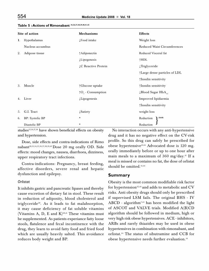
554 Medicine Update 2008 Vol. 18
Table 5 : Actions of Rimonabant 35,36,37,38,39,40,41,42
Site of action Mechanisms Effects
1. Hypothalamus ↓Foodintake Weight loss
Nucleusaccumbus ReducedWaistCircumferences
2. Adipose tissue ↑Adiponectin Reduced Visceral fat
↓Lipogenesis ↑HDL
↓C Reactive Protein ↓Triglyceride
↑Large dense particles of LDL
↑Insulin sensitivity
3. Muscle ↑Glucose uptake ↑Insulin sensitivity
↑O2-Consumption ↓Blood Sugar HbA
1C
4. Liver ↓Lipogenesis Improved lipidaemia
↑Insulin sensitivity
5. G.I. Tract ↓Satiety weight loss
6. BP: Systolic BP * Reduction }(34,40)
Diastolic BP * Reduction
studies35,36,37,38 have shown beneficial effects on obesity and hypertension.
Dose, side effects and contra-indications of Rimonabant28,33,34,35,36,37,38,39,40 Dose 20 mg orally OD. Side effects: mood changes, nausea, diarrhoea, dizziness, upper respiratory tract infections.
Contra-indications: Pregnancy, breast feeding, affective disorders, severe renal and hepatic dysfunction and epilepsy.
Orlistat
It inhibits gastric and pancreatic lipases and thereby cause excretion of dietary fat in stool. These result in reduction of adiposity, blood cholesterol and triglyceride28. As it leads to fat malabsorption, it may cause deficiency of fat soluble vitamins (Vitamins A, D, E and K)28,41 These vitamins must be supplemented. As patients experiance fatty loose stools, flatulence and fecal incontinence with the drug, they learn to avoid fatty food and fried food which are usually heavily salted. This avoidance reduces body weight and BP.
No interaction occurs with any anti-hypertensive drugandithasnonegativeeffectontheCV-riskprofile. So this drug can safely be prescribed for obese hypertensive41,44 Advocated dose is 120 mg. orally immediately before or up to one hour after main meals to a maximum of 360 mg/day.41 If a meal is missed or contains no fat, the dose of orlistat should be omitted.41,44
SummaryObesity is the most common modifiable risk factor for hypertension42,44andaddstometabolicandCVrisks. Anti obesity drugs should only be prescribed if supervised LSM fails. The original BHS - IV ABCD-algorithm7,43 has been modified the light ofASCOTandVALVE trials.ModifiedA(B)CDalgorithm should be followed in medium, high or veryhighriskobesehypertensives.ACE-inhibitors,ARBs and rarely thiazides may be used in obese hypertensives in combination with rimonabant, and orlistat.44The statusof sibutramineandCCB forobese hypertensive needs further evaluation.44
555The Challenges of Hypertension in obese Subjects - Indian Perspective
References1. Narkaiewicz K. Diagnosis and Management of hypertension
in obesity. The International Association for the study of obesity. Obesity reviews 2006; 7: 155-62
2. HuangZ,WilletWC,MansonJE,RosnerBetal.Bodyweightchange and risk of hypertension in women. Ann Intern Med 1998; 128: 81-88
3. Kannel WB, Brand N, Skinner JJ Jr., Dawber TR. The relation of adiposity to blood-pressure and development of hypertension. The Framingham Study. Ann Intern Med 1967; 67: 48-59
4. Scholze J, Grimm E, Herrmann D, Unger T, Kintscher U. Optimal Treatment of Obesity - Related Hypertension. The Hypertension - Obesity - Sibutramine (HOS) Study. Circulation2007;115:1991-98
5. Shah S, Anand P, Maiya M, Mukherjee S, Munjal YP, Wander GS, Kamath S. Indian Hypertension Guideline 2007. Postgraduate Medicine (Recent Advances in Medicine) YP Munjal (ed), The Association of Physicians of India and IndianCollegeofPhysicians,AjantaOffsetandPackagingLtd., New Delhi 2007; 21: 315-25
6. Chobanian AV, Bakris GL, Black HR, Cushman WC etal. Seventh Report of the Joint National Committee onPrevention, Detection, Evaluation and Treatment of High BloodPressure.TheJNC-7Report.JAMA2003;289:2560-71
7. Williams B, Poulter NR, Brown MJ, Davis M et al. British Hypertension Society Guidelines. Guidelines for management for hypertension: report of the fourth working party of the British Hypertension Society, 2004 - BHS-IV. Journal of Human Hypertension 2004; 18: 139-185
8. Arya SN. Emerging trends in diagnosis of overweight and obesity in “A treatise on Obesity and Related Disorders”, Dr. SN Arya (ed), Screen House 104, Ayodhya Apartment, Off Frazer Road, Patna 800001, 2005: 1-6
9. Kissebah AH, Freedman DS, Peiris AN. Health risk of Obesity.MedClinNorthAm1989;73:111-38
10. Baron RB. Obesity in chapter on Nutrition. Tierney Jr. LM,McPheeSJ,PapadakisMA(eds)CURRENTMedicalDiagnosis and Treatment 2006, 45th International Edition, Lange Medical Books, McGraw-Hill Medical Publishing Division, New Delhi 2006: 1267-70
11. FitzgeraldPA. inCommonpresentation inendocrinology:ObesityinCurrentMedicalDiagnosisandTreatment2001,Tierney LM, McPhee SJ, Papadakis MA. Lange Medical Books/McGraw-Hill, Medical Publishing Division, New Delhi 2001: 1088, and 1233-37
12. Rimm EB, Stampfer MJ, Giovannucci E et al. Body size and fat distribution as predictors of coronary heart disease among middle aged and older US men.
13. Flier JS, Maratos-Fleir. Obesity. Braunwald E, Fauci AS, Kasper DL, Hauser SL, Longo DL, Jameson JL (eds) Harrison’s Principles Of Internal Medicine 16th Edition, McGraw-Hill, Medical Publishing Division, New Delhi 2006; 1: 422-29.
14. PouliotMC,Despres’JP,NadeauA,MoorjaniS,Prud’hommeD, Lupiene PJ et al. Visceral obesity in men. Association with glucose tolerance, plasma insulin and lipoprotein levels. Diabetes 1992; 41: 826-34.
15. Despres’ JP, Moorjani S, Lupien PJ, Tremblay A, Nadeau A,BrouchardC.Regionaldistributionofbodyfat,plasmalipoproteins and cardiovascular disease. Arteriosclerosis 1990; 10: 497-511.
16. CastelliWP.EpidemiologyofCoronaryHeartDisease:TheFramingham Study. Am J Med 1984; 76: 4-12.
17. Hakim S. Medical complications of obesity. Medicine Update 2002, SB Gupta (ed) on behalf of Association of Physicians of India Mumbai-400002; 12: 480-82.
18. Allisan DB, Fontaine KR, Manson JE, Stevens J, Vanitallie TB. Annual deaths attributed to obesity in United States. JAMA 1999; 282: 1530-38.
19. Mohan V, Deepa M, Farooq S, Dutta M, Deepa R. Prevention AwarenessandControlofHypertension inChennai.TheChennaiUrbanRuralEpidemiologyStudy (CURES-52) JAssoc Physician India 2007; 55: 326-32.
20. Summerton C, Shetty P, Sandle LN, Watts S. Obesity inChapterinDAVIDSON’SPrinciplesandpracticeofMedicine19thEdition,HaslettC,ChilversER,BoonNA,ColledgeNR,Hunter JAA (eds)ChurchilLivingstone India, 2002: 301-03.
21. Das UN. Hypertension as a Low-grade Systemic Inflammatory Condition. That has its Origins in the Perinatal Period. JAssoc Physicians India 2006; 54: 133-42.
22. Das UN. Is Metabolic Syndrome-X an Inflammatory Condition?ExpBiolMed2002;227:989-97.
23. Fernandez-RealJM,VayredaM,RichardCetal.Circulatinginterleukin-6 levels, blood pressure, and insulin sensitivity in apparentlyhealthymenandwomen.JClinicalEndocrinolMetab 2001; 86: 1154-59.
24. ChaeCU,LeeRT,RifaiN,RidkerPM.Bloodpressureandinflammation in apparently healthy men. Hypertension 2001; 38: 399-403.
25. Bermudez EA, Rifai N, Buring J, Manson JE et al. Inter relationshipsamongcirculatinginterleukin-6,C-ReactiveProtein and traditional cardiovascular risk factors in women. Arterioscler Thromb Vase Biol 2002; 22: 1688-73.
26. Arya SN. Obesity - an update in A Treatise on Obesity and Related Disorders, Dr. SN Arya (Ed) 2005 Screen House, 104 Ayodhya Apartment, Off Fraser Road, Patna- 800001. 2005: 7-38.
27. AryaSN.ObesityJournalofIndianAssociationofClinicalMedicine( JIACM)2004;5:166-81.
28. Mishra A, Khurana L. Management of Obesity. Postgraduate Medicines (Recent Advances in Medicine) 2007, YP Munjal (ed),AssociationofPhysiciansofIndiaandIndianCollegeof Physicians 2007; 21: 403-11.
29. Sahay BK. Yoga in Diabetes. J Assoc Physicians India 2007; 55: 121-26.

556 Medicine Update 2008 Vol. 18
30. Joint British Societies Guideline on Prevention of CardiovasculardiseaseinClinicalPractice2005;91:22-28(www.heartjnl.com).
31. Dahlof B, Sever PS, Poulter NR, Wedel H et al. Prevention of cardiovascular events with an anti-hypertensive regimen of amlodipine adding perindopril as required in the Anglo-Scandinavian Cardiac Outcome Trial - Blood PressureLoweringArm(ASCOT-BPLA):amulti-centerrandomizedcontrolled trial. The Lancet 2005; 366: 895-906 (www.thelancet.com)
32. Julius S, Kjeldsen, Weber M, Brunner HR. Outcomes in hypertensive patients at high cardiovascular risk treated with regimens based on Valsartan or Amlodipine: the VALUE - randomized trial. The Lancet 2004; 363: 2022-31.
33. WoodsSC.RoleoftheEndocannabinoidSysteminRegulatingCardiovascular andMetabolic risk factors.TheAmericanJournal of Medicine 2007; 120 (3A): 519-25.
34. Van-Gaal LF, Rissanen AM, Scheen AJ, Zieglar, Rossner S. Effects of the cannabinoid-1-receptor-blocker rimonabant on weight reduction and cardiovascular risk factor in overweight patient: 1 year experience the RIO - Europe Study. Lancet 2005; 365:1389-97.
35. Gelfand EV, Cannon PC. Rimonabant: A cannabinoidReceptor Type-1 Blocker for Management of Multiple CardiometabolicRiskFactors.JournalofAmericanCollegeofCardiology2006;47(10):1919-26
36. Heness S, Robinson DM and Lyseng - Williamson. Rimonabant. Drugs 2006; 66 (16)2109-19
37. Padwal RS, Majumdar SR. Drug treatment for Obesity: Orlistat, Sibutramine and Rimonabant. Lancet 2007; 369: 71-77
38. Scheen AJ, Finer N, Hollander P, Jensen MD et al. Efficacy and tolerability of rimonabant in overweight and obese patient with Type-2 diabetes: a randomized controlled study. Lancet 2006; 368: 1660-72
39. Despres’ JP, Goley A, Sjostrom L: for Rimonabant in Obesity Study Group. Effects of Rimonabant on Metabolic Risk Factors in Overweight Patients with Dyslipidaemia. N Engl J Med 2005; 353 (No.2): 2121-34
40. Pi-Sunyer FX, Arrone LJ, Heshmati HM, Devin J et al. Effect of Rimonabant, a Cannabinoid - 1 Receptor Blocker, onWeight and Cardiometabolic Risk Factors in Overweightand Obese Patients. RIO-North America: A Randomized ControlledTrial.JAMAFeb15,2006;295(7):761-75
41. MehtaDK,MartinJ,CostelloI,JordanBetal(eds).Orlistat- anti obesity drug acting on gastrointestinal tract. British National Formulary 51, March 2006 published by BMJ-publishing group, Lambeth High Street, London SE1 7JN, UK. March 2006; 51: 208-09
42. SainaniGS.Non-DrugTherapyinPreventionandControlofHypertension. J Assoc Physicians India 2003; 51: 1001-06
43 AryaSN.TherapeuticChallengeofResistantHypertension-IndianPerspective.MEDICINEUPDATE2007R.K.Singal,D.G. Jain (eds), Association of Physicians of India, Mumbai, Jaypee Brothers Medical Publishers PVT. LTD. New Delhi 2007; 17: 565-73 44.Aneza A, El-Atat F, McFarlane SI, Sowers JR. Hypertension in Obesity, rpr. endojournals. Org at India: Endo Jnls Sponsored on May 18, 2007
CHAPTER
74Breaking Bad News to the Patient and RelativesS. G. Godbole
IntroductionThe word “Doctor” is derived from the Latin “DOCERE”whichmeans‘toteach’.Adoctorin his capacity as a ‘healer’ should be able to teach his patient about health. Teaching skills will involve collecting information and efficiently communicating it to the patient. Sadly our medical curriculum lacks this aspect of ‘communication skills’. It is only when a medical graduate starts his clinical practice, then he realizes the lack of communication skills.
During the last 50 years, the subject of communication in health care has become a serious matter of study. Evidence is that healthcare fails without a conscious, informed effort of communication which is the professional responsibility of all concerned including nursing staff and doctors, pharmacist, health administrators etc.1 It is the aim and mission of modern healthcare professional to provide care that is evidence-based and unconditionally patient centered. Communication styles that are patient centeredprovide a more complete clinical picture upon which diagnosis and treatment can be based and lead to improvement in health outcomes.
Breaking bad news to patients is one of the most difficult tasks in the practice of medicine. The usual problems encountered are:
i. What constitutes a bad news?
ii. Whether a patient should be informed about the news?
iii. Whether any relative should be informed ?
iv. What impact will the news have on the patient?
v. What should be the way to carry out this unpleasant task?
What constitutes a bad news ?Bad news can be defined2 as “any news that drastically and negatively alters the patient’s view of his or her future.” Bad news is usually associated with a terminal diagnosis of ‘cancer’ but a physician may encounter various situations that involve imparting bad news e.g. news of intrauterine fetal death to a pregnant mother, a diagnosis of multiple sclerosis etc. A diagnosis of a chronic illness such as Diabetes Mellitus or disability or loss of function of a limb or a part of limb can constitute a bad news. A diagnosis of AIDS to be conveyed to the patient, can be a difficult task. To me conveying a diagnosis of ‘pregnancy’ to the parents of a young unwed girl can also be difficult. A treatment plan that is painful, lengthy and costly can also have a negative impact. Thus bad news may be related to different diagnosis apart from cancer.

558 Medicine Update 2008 Vol. 18
Whether the patient should be informed?In Decorum, Hippocrates advised3 “Reveal nothing to the patient of his future or present condition for this has caused many patients to take a turn for the worse”. Unfortunately this tradition of silence persisted for centuries. In the past few decades, this has changed. A review of studies on patient preferences regarding disclosure of a terminal diagnosis found that 50-90% of patients desired full disclosure.4
Relatives may ask the physician to withhold the bad news from the patient. But, I feel the information belongs to the patient and not to the relatives.
Moreover it is observed that invariably, patients; (1) Know more about their illness than anyone’s guess or (2) May imagine things to be worse than they are (3) Welcome clear information about disease.
Thus it is clear that the bad news should be conveyed to the patient.
Whether any relative should be informed?The negative impact, the bad news can have, on the patient demands the presence of near and dear ones with the patient. But the ethical issues of the confidentiality and secrecy of patient information demands that this be discussed with the patient and his views need to be sought. Ask the patient who, if anyone, he would like to have with him. This need not be the official ‘next of kin’, but a same
sex friend, a confidant or a specific member of the health care team. But there may be exceptions to this general rule. If the patient is a child under 16 yrs., the information about the disease, prognosis and treatment belongs to the parents. If the patient has cognitive impairment or has impaired hearing faculty the presence of a relative is necessary. In case of a language barrier a suitable interpreter needs to be present. If the diagnosis has ethical issues attached, e.g. a diagnosis of ‘AIDS’, the doctor will have a social responsibility of discussing the diagnosis with the spouse. Thus the presence of a relative though not mandatory is advisable, but only with patient’s permission.
What impact will the news have on the patient?Rabow and Mcphee keenly5 described the end resultofcommunication“Cliniciansfocusoftenon relieving patient’s bodily pain, less often on their emotional distress and seldom on their suffering.” A physician should be aware of the possible negative impact of the bad news on a given patient. A reaction could consist of denial, blame, disbelief rather than acceptance or submission. This may result in meek surrender to the diagnosis and apathy towards further treatment or may result in hostility towards the doctors, relations or the hospital. A physician should be aware of such extreme reactions and be prepared for the same.
What should be the way to carry out this unpleasant task?Buckman2 suggested an organized and effective procedure for communicating bad news. He outlined seven steps which goes by the acronym P-SPIKES.1
Table- 2 provides a summary of these steps along with suggested phrases.
Rabow and Mcphee5 developed a practical and comprehensive model, from synthesized multiple sources,thatusesacronym‘ABCDE’.
Table 1 • Prepare for the discussion.
• Set up a suitable environment.
• Beginthediscussionbyfindingoutwhatthepatient and or family understand.
• Determine how they will comprehend newinformation.
• Providenewknowledge accordingly.
• Share plans for the next steps.

559Breaking Bad News to the Patient and Relatives
Table 2 : Elements of Communicating Bad News-the P-SPIKES ApproachAcronym Steps Aim of the Interaction Preparations, Questions, or Phrases
P Preparation Mentally prepare for the interaction with the patient
Review what information needs to be communicated. Plan how you will provide emotional support. Rehearse key steps and phrases in the interaction.
S Setting of the interaction
Ensure the appropriate setting for a serious and emotionally charged discussion
Ensure patient, family and appropriate social supports are present.
Devote sufficient time-do not squeeze in a discussion.
Ensure privacy and prevent interruption by people or beeper.
Bring a box of tissues.
P Patient’s perception and preparation
Begin the discussion by establishing the baseline and whether the patient and family can grasp the information.
Ease tension by having the patient and family contribute.
Start with open-ended questions to encourage participation.
Possible phrases to use:
What do you understand about your illness?
When you first had symptom X, what did you think it might be?
What did Dr. X tell you when he sent you here?
What do you think is going to happen?
I Invitation and information needs
Discover what information needs the patient and/or family have and what limits they want regarding the bad information
Possible phrases to use:
If this condition turns out to be something serious, do you want to know?
Would you like me to tell you the full details of your condition? If not, then who would you like me to talk to?
K Knowledge of the condition
Provide the bad news or other information to the patient and/or family sensitively.
Do not just dump the information on the patient and family.
Interrupt and check that the patient and family are understanding.
Possible phrases to use:
I feel badly to have to tell you this, but……..
Unfortunately, the tests showed……
I’m afraid the news is not good…
E Empathy and exploration
Identify the cause of the emotions-e.g., poor prognosis.
Empathize with the patient and/or family’s feeling.
Explore by asking open-ended questions.
Strong feelings in reaction to bad news are normal.
Acknowledge what the patient and family are feeling.
Remind them such feelings are normal, even if frightening.
Give them time to respond.
Remind patient and family you won’t abandon them.
Possible phrases to use:
I imagine this is very hard for you.
You look very upset. Tell me how you are feeling.
I wish the news were different.
I’ll do whatever I can to help you.
S Summary and strategic planning
Delineate for the patient and the family the next steps, including additional tests or interventions.
It is the unknown and uncertain that increase anxiety.
Recommend a schedule with goals and landmarks.
Provide your rationale for the patient and/or family to accept (or reject).
If the patient and/or family are not ready to discuss the next steps, schedule a follow-up visit.

560 Medicine Update 2008 Vol. 18
Though both these models are based on similar principles, these guidelines need to be modified to suit our health care set up. To provide an environment in a municipal or a government hospital may not be possible. The help of a counsellor may not always be available. Our country with its rich and varied heritage and traditions demands a different approach. Language barrier, cultural gap and existing customs do dictate the mode of breaking a bad news. The reactions of the In laws of a female patient may disrupt her social status. The stigmata attached even to a diagnosis of ‘Tuberculosis’ demands a more direct approach.
Sharman ApproachI suggest a different approach ‘SHARMAN’ which in Sanskrit means ‘protection’ in relation to the “Sharir” (body). SHARMAN is a new concept in breaking the bad news to the patient.
A. Setting up
The doctor has to prepare himself for the interview. He has to ascertain the facts and confirm the diagnosis. A verbally conveyed report should not be shared with the patient. Documentary evidence is necessary before the diagnosis is revealed to the patient. Western literature talks about ‘dress rehearsal’ for the doctor. I feel this is far fetched and not necessary.
A proper environment setup is required. It can be the doctor’s cabin or a patient’s bed side. A separate interview room is not possible in our hospital set up and is not necessary as well. Make every effort to avoid interruptions. Usually in hospitals or clinics, the patient and relatives are advised to switch off their mobiles. But during such meetings, it is advisable that
the doctor also does not receive his mobile/landline phone calls.
Ensure that the persons present in the room are the ones which the patient desires.
B. Knowledge
Most patients will have some idea about their illness. Ascertain what he knows? by framing certain questions like.
• Have you any idea of what might bewrong?
• Howwouldyoudescribeyourillness?
• Whattestshaveyouhad?
Such questions prepare the patient mentally to accept a diagnosis of the illness. It brings his thinking process in the right direction. It starts involving the patient himself in the further discussions. Moreover it gives an idea to the physician, the extent to which he can reveal the diagnosis.
C. Active Disclosure
Now is the time to divulge the information to the patient. The doctor has to understand what information can the patient accept at that point of time. To give information at the patient’s pace may mean accurate absorption of the message in manageable chunks. During the talk, the doctor has to take pauses to ascertain how the patient is accepting the information. The silence adds to the necessary creation of an environment. Phrases such as “Am I clear” or ‘Are you understanding’? can help the doctor to check for patient comprehension.
Table 31. Advance Preparation.
2. Build a therapeutic environment.
3.CommunicateWell.
4. Deal with patient and family reactions.
5. Encourage reliable emotions.
Table 4•Settingup
•Hisknowledge
•Activedisclosure
•Reaction
•Modulate
•Attitude
•NextStep
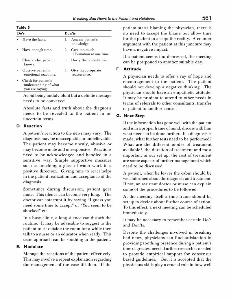
561Breaking Bad News to the Patient and Relatives
Avoid being unduly blunt but a definite message needs to be conveyed.
Absolute facts and truth about the diagnosis needs to be revealed to the patient in no uncertain terms.
D. Reaction
A patient’s reaction to the news may vary. The diagnosis may be unacceptable or unbelievable. The patient may become unruly, abusive or may become mute and unresponsive. Reactions need to be acknowledged and handled in a sensitive way. Simple supportive measure such as touching, a glass of water work in a positive direction. Giving time to react helps in the patient realization and acceptance of the diagnosis.
Sometimes during discussion, patient goes mute. This silence can become very long. The doctor can interrupt it by saying “I guess you need some time to accept” or “You seem to be shocked” etc.
In a busy clinic, a long silence can disturb the routine. It may be advisable to suggest to the patient to sit outside the room for a while then talk to a nurse or an educator when ready. This team approach can be soothing to the patient.
E. Modulate
Manage the reactions of the patient effectively. This may involve a repeat explanation regarding the management of the case till then. If the
patient starts blaming the physician, there is no need to accept the blame but allow time for the patient to accept the reality. A counter argument with the patient at this juncture may have a negative impact.
If a patient seems too depressed, the meeting can be postponed to another suitable day.
F. Attitude
A physician needs to offer a ray of hope and encouragement to the patient. The patient should not develop a negative thinking. The physician should have an empathetic attitude. It may be prudent to attend to other needs in terms of referrals to other consultants, transfer of patient to another centre.
G. Next Step
If the information has gone well with the patient and is in a proper frame of mind, discuss with him what needs to be done further. If a diagnosis is made, what further tests need to be performed? What are the different modes of treatment available?, the duration of treatment and most important in our set up, the cost of treatment are some aspects of further management which need to be discussed.
A patient, when he leaves the cabin should be well informed about the diagnosis and treatment. If not, an assistant doctor or nurse can explain some of the procedures to be followed.
At the meeting itself a time frame should be set up to decide about further course of action. To this effect, a next meeting can be scheduled immediately.
It may be necessary to remember certain Do’s and Don’ts.
Despite the challenges involved in breaking bad news, physicians can find satisfaction in providing soothing presence during a patient’s time of greatest need. Further research is needed to provide empirical support for consensus based guidelines. But it is accepted that the physicians skills play a crucial role in how well
Table 5Do’s Don’ts
•Havethefacts. 1. Assume patient's knowledge
• Haveenoughtime. 2. Give too much information at one time.
•Clarifywhatpatientknows.
3. Hurry the consultation.
•Observepatient’semotional reactions.
4. Give inappropriate reassurance.
• Checkforpatient’sunderstanding of what you are saying.
562 Medicine Update 2008 Vol. 18
patient cope with bad news and that patients and physicians will benefit, if physicians are better trained for this difficult task. The fact is that medicine does not offer a cure for every disease situations. Hence these are the situations where professionalism most acutely calls the physician to provide hope and healing for the patient.
References1. C. Mallinson. Clinical Medicine; Kumar and Clerk
(2005) 6th Ed . Elsevier Saunders , Chapter 1; Ethics andCommunication,8-18
2. Buckman R. Breaking bad news; Why is it still so difficult? Br Med J 1984; 288 (6430) : 1597-9.
3. Paul S. Nueller. Breaking bad news to patients. Postgraduate Medicine, 2002 : 40/112 (3).
4. Gregg K. Vanderkiett. Breaking Bad News. American Family Physician, 2001 : 64, (12) : 1975-8.
5. Rabow M.W., McPhee S. J. Beyond breaking bad news; how to help patients who suffer. West J Med 1999, 171: 260-3.
6. Ezekiel J. Emanuel, Linda L. Emanuel. Harrison’s Principles of Internal Medicine (2005) 16th Ed. Mc Graw Hill Part 1-9; PalliativeandEnd-of-LifeCare,53-66.
7. FallowfieldL,Jenkins,Communicatingsad,badanddifficultnews in medicine. Lancet 2004, 363:312.

CHAPTER
75Diagnostic Reasoning: Approach to Clinical Diagnosis Based on Bayes’ TheoremA. Mohan, K. Srihasam, S.K. Sharma
IntroductionDoctors caring for patients in their everyday clinical practice are faced with decisions that are sometimes routine and simple, but may be complicated on other occasions. Sometimes, the decisions may be regarding the choice of investigation or intervention, on other occasions, the decisions may have to be taken regarding a therapeutic option. Either way, clinical decision making is challenging because, these decisions are not only unavoidable but also must be made under uncertain conditions. In this review, an attempt is being made to provide an overview regarding the applications of Bayes’ theorem and clinical decision analysis in arriving at a diagnosis.
Basic MathematicsIn order to understand diagnostic reasoning, it is necessary to understand the basic mathematical language of probability and Bayes’ theorem as applied to clinical medicine. Bayes’ theorem, ascribed to Rev. Thomas Bayes (1701-1761) is a mathematical rule explaining how one should change existing beliefs in light of new evidence.1 Bayes’ paper on ‘An essay towards solving a problem in the doctrine of chances’2 posthumously, due to the efforts of his friend Richard Price.3
Probability
Probability as applied to clinical diagnostic reasoning may be regarded as a measure of one’s strength of belief that an event will occur and range from 0.0 to 1.0.4 In statistical notation, probability of an event A is written as P[A].
Summation principle
The summation principle states that the sum of probabilities of all possible outcomes of a chance event equals 1.0.4 If there are four possible outcomes A,B,C,andD,then
P[A]+P[B]+P[C]+P[D]=1
Joint probability
The concomitant occurrence of any number of events is defined as joint probability of those events.4 In statistical notation, the joint probability of two events A and B is written as
P[A,B].
Conditional probability
The probability that an event A occurs, given that the event B is known to occur is defined as the conditional probability of event A given event B or P[A½B].4
The relationship between joint and conditional probabilities is given by the formula
P[A,B] = P[A½B] = P[B]
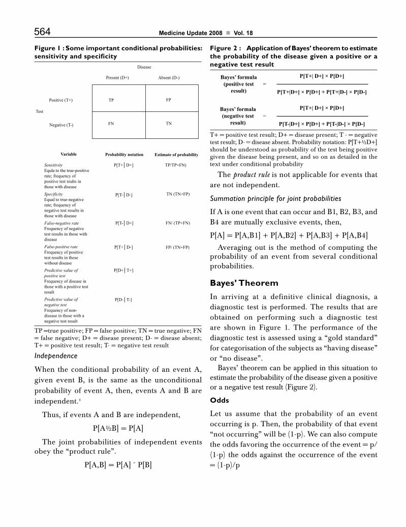
564 Medicine Update 2008 Vol. 18
Figure 1 : Some important conditional probabilities: sensitivity and specificity
TP =true positive; FP = false positive; TN = true negative; FN = false negative; D+ = disease present; D- = disease absent; T+ = positive test result; T- = negative test result
Figure 2 : Application of Bayes’ theorem to estimate the probability of the disease given a positive or a negative test result
T+ = positive test result; D+ = disease present; T - = negative test result; D- = disease absent. Probability notation: P[T+½D+] should be understood as probability of the test being positive given the disease being present, and so on as detailed in the text under conditional probability
Independence
When the conditional probability of an event A, given event B, is the same as the unconditional probability of event A, then, events A and B are independent.4
Thus, if events A and B are independent,
P[A½B] = P[A]
The joint probabilities of independent events obey the “product rule”.
P[A,B] = P[A] ´ P[B]
The product rule is not applicable for events that are not independent.
Summation principle for joint probabilities
If A is one event that can occur and B1, B2, B3, and B4 are mutually exclusive events, then,
P[A] = P[A,B1] + P[A,B2] + P[A,B3] + P[A,B4]
Averaging out is the method of computing the probability of an event from several conditional probabilities.
Bayes’ TheoremIn arriving at a definitive clinical diagnosis, a diagnostic test is performed. The results that are obtained on performing such a diagnostic test are shown in Figure 1. The performance of the diagnostic test is assessed using a “gold standard” for categorisation of the subjects as “having disease” or “no disease”.
Bayes’ theorem can be applied in this situation to estimate the probability of the disease given a positive or a negative test result (Figure 2).
Odds
Let us assume that the probability of an event occurring is p. Then, the probability of that event “not occurring” will be (1-p). We can also compute the odds favoring the occurrence of the event = p/(1-p) the odds against the occurrence of the event = (1-p)/p
Disease
Present (D+) Absent (D-)
FP
TNFN
TPPositive (T+)
Negative (T-)
Test
Variable Probability notation
P[T-│D-]
P[T+│D-]
P[D-│T-]
P[T-│D+]
Estimate of probability
TN (TN+FP)
FN/ (TP+FN)
FP/ (TN+FP)
SensitivityEqula to the true-positiverate; frequency ofpositive test reults inthose with diseaseSpecificityEqual to true-negativerate; frequency ofnegative test results inthose with diseaseFalse-negative rateFrequency of negativetest results in those withdiseaseFalse-positive rateFrequency of positivetest results in thosewithout diseasePredictive value ofpositive testFrequency of disease inthose with a positive testresultPredictive value ofnegative testFrequency of non-disease in those with anegative test result
P[T+│D+]
P[D+│T+]
TP/TP+FN)
Bayes’ formula(positive test
result)
=
=P[T+| D+] × P[D+]
P[T+|D+] × P[D+] + P[T+|D-] × P[D-]
P[T+| D+] × P[D+]
P[T-|D+] × P[D+] + P[T-|D-] × P[D-]
Bayes’ formala(negative test
result)
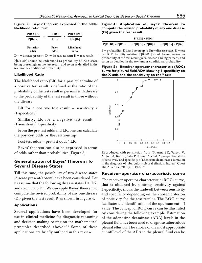
565Diagnostic Reasoning: Approach to Clinical Diagnosis Based on Bayes’ Theorem
Figure 3 : Bayes’ theorem expressed in the odds-likelihood ratio form
D+ = disease present; D- = disease absent; R = test result
P[D+½R] should be understood as probability of the disease being present given the test result, and so on as detailed in the text under conditional probability
Figure 4 : Application of Bayes’ theorem to compute the revised probability of any one disease (Di) given the test result.
P = probability; D1, and so on up to Dn = disease states. R = test result. Probability notation: P[R½D1] should be understood as probability of the test result given disease 1 being present, and so on as detailed in the text under conditional probability
Likelihood Ratio
The likelihood ratio (LR) for a particular value of a positive test result is defined as the ratio of the probability of the test result in persons with disease to the probability of the test result in those without the disease.
LR for a positive test result = sensitivity / (1-specificity)
Similarly, LR for a negative test result = (1-sensitivity) /specificity
From the pre-test odds and LR, one can calculate the post-test odds by the relationship
Post-test odds = pre-test odds ´ LR
Bayes’ theorem can also be expressed in terms of odds rather than probabilities (Figure 3).
Generalisation of Bayes’ Theorem To Several Disease StatesTill this time, the possibility of two disease states (disease present/absent) have been considered. Let us assume that the following disease states D1, D2, and so on up to Dn. We can apply Bayes’ theorem to compute the revised probability of any one disease (Di) given the test result R as shown in Figure 4.
Applications
Several applications have been developed for use in clinical medicine for diagnostic reasoning and decision making basing on the mathematical principles described above.3,5,6 Some of these applications are briefly outlined in this review.
Receiver-operator characteristic curveThereceiver-operatorcharacteristic(ROC)curve,that is obtained by plotting sensitivity against 1-specificity, shows the trade-off between sensitivity and specificity depending on the chosen criterion ofpositivity for the test result.4TheROCcurvefacilitates the identification of the optimum cut-off value.TheconceptofROCcurvecanbeillustratedby considering the following example. Estimation of the adenosine deaminase (ADA) levels in the pleural fluid has been used to diagnose tuberculous pleural effusion. The choice of the most appropriate cut-off level of the ADA in the pleural fluid can be
P[D + | R]
P[D- |R]
P[R + |D+]
P[R |D-]
P [D ]
P[D-]
Posteriorodds
Priorodds
Likelihoodratio
= ×P[R|Di] × P[Di]
P[R | D1] × P[D1]+.......+ P[R| Di] × P[Di] +.......+ P[R| Dn] × P[Dn]
Figure 5 : Receiver-operator characteristic (ROC) curve for pleural fluid ADA showing 1-specificity on the X-axis and the sensitivity on the Y-axis
Reproduced with permission from “Sharma SK, Suresh V, Mohan A, Kaur P, Saha P, Kumar A, et al. A prospective study of sensitivity and specificity of adenosine deaminase estimation inthediagnosisoftuberculosispleuraleffusion.IndianJChestDis Allied Sci 2001;43:149-55”7
1
0.8
0.6
0.4
0.2
00 0.1 0.2 0.3 0.4 0.5 0.6 0.7 0.8 0.9 1
1-Specificity
Sens
itivi
ty
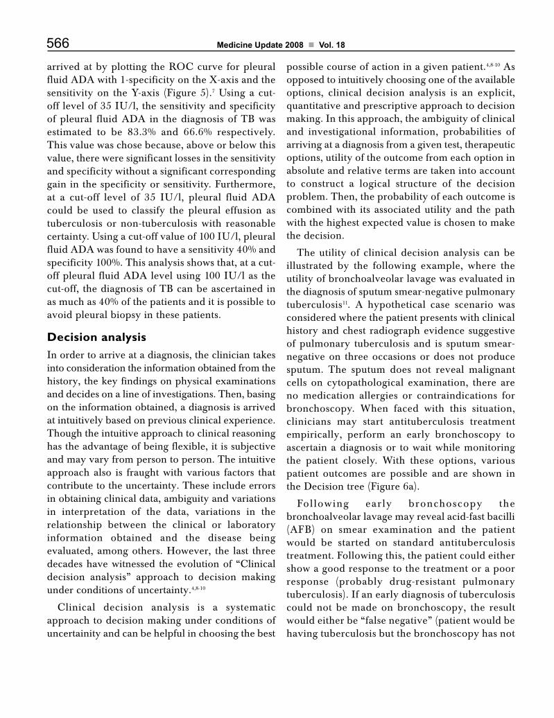
566 Medicine Update 2008 Vol. 18
arrivedatbyplotting theROCcurve forpleuralfluid ADA with 1-specificity on the X-axis and the sensitivity on the Y-axis (Figure 5).7 Using a cut-off level of 35 IU/l, the sensitivity and specificity of pleural fluid ADA in the diagnosis of TB was estimated to be 83.3% and 66.6% respectively. This value was chose because, above or below this value, there were significant losses in the sensitivity and specificity without a significant corresponding gain in the specificity or sensitivity. Furthermore, at a cut-off level of 35 IU/l, pleural fluid ADA could be used to classify the pleural effusion as tuberculosis or non-tuberculosis with reasonable certainty. Using a cut-off value of 100 IU/l, pleural fluid ADA was found to have a sensitivity 40% and specificity 100%. This analysis shows that, at a cut-off pleural fluid ADA level using 100 IU/l as the cut-off, the diagnosis of TB can be ascertained in as much as 40% of the patients and it is possible to avoid pleural biopsy in these patients.
Decision analysisIn order to arrive at a diagnosis, the clinician takes into consideration the information obtained from the history, the key findings on physical examinations and decides on a line of investigations. Then, basing on the information obtained, a diagnosis is arrived at intuitively based on previous clinical experience. Though the intuitive approach to clinical reasoning has the advantage of being flexible, it is subjective and may vary from person to person. The intuitive approach also is fraught with various factors that contribute to the uncertainty. These include errors in obtaining clinical data, ambiguity and variations in interpretation of the data, variations in the relationship between the clinical or laboratory information obtained and the disease being evaluated, among others. However, the last three decadeshavewitnessed theevolutionof“Clinicaldecision analysis” approach to decision making under conditions of uncertainty.4,8-10
Clinical decision analysis is a systematic approach to decision making under conditions of uncertainity and can be helpful in choosing the best
possible course of action in a given patient.4,8-10 As opposed to intuitively choosing one of the available options, clinical decision analysis is an explicit, quantitative and prescriptive approach to decision making. In this approach, the ambiguity of clinical and investigational information, probabilities of arriving at a diagnosis from a given test, therapeutic options, utility of the outcome from each option in absolute and relative terms are taken into account to construct a logical structure of the decision problem. Then, the probability of each outcome is combined with its associated utility and the path with the highest expected value is chosen to make the decision.
The utility of clinical decision analysis can be illustrated by the following example, where the utility of bronchoalveolar lavage was evaluated in the diagnosis of sputum smear-negative pulmonary tuberculosis11. A hypothetical case scenario was considered where the patient presents with clinical history and chest radiograph evidence suggestive of pulmonary tuberculosis and is sputum smear-negative on three occasions or does not produce sputum. The sputum does not reveal malignant cells on cytopathological examination, there are no medication allergies or contraindications for bronchoscopy. When faced with this situation, clinicians may start antituberculosis treatment empirically, perform an early bronchoscopy to ascertain a diagnosis or to wait while monitoring the patient closely. With these options, various patient outcomes are possible and are shown in the Decision tree (Figure 6a).
Fol lowing ear ly bronchoscopy the bronchoalveolar lavage may reveal acid-fast bacilli (AFB) on smear examination and the patient would be started on standard antituberculosis treatment. Following this, the patient could either show a good response to the treatment or a poor response (probably drug-resistant pulmonary tuberculosis). If an early diagnosis of tuberculosis could not be made on bronchoscopy, the result would either be “false negative” (patient would be having tuberculosis but the bronchoscopy has not

567Diagnostic Reasoning: Approach to Clinical Diagnosis Based on Bayes’ Theorem
picked up the diagnosis) or the patient would be suffering from another disease (“true negative”). The further evaluation of such a patient was not included in the decision tree in order to simplify the analysis.
Fol lowing empir ical t reatment with antituberculosis drugs, the patient could either improve (good response) or ; remain stable or deteriorate further (poor-response). A patient who does not respond, could either have drug resistant tuberculosis or another diagnosis. The further
evaluation of such a patient is not incorporated in the decision tree model in order to simplify the analysis. In waiting branch, the patient would finally develop tuberculosis or another diagnosis would finally be possible.
Each path was folded back to the starting point (fold back analysis). At a decision node the tree was folded back along the single best choice where as, at a chance node the probabilities were averaged out on all the branches emanating from that node and the best alternative course of action chosen.
Figure 6a : Decision analysis tree for the evaluation of a patient with sputum smear negative pulmonary tuberculosis. Small squares indicate a decision node. Small circle indicates a chance node. Probability bindings are represented below each event. Utility bindings are depicted at the end of the path. Numbers in balloons indicate the averaged-out outcomes connected to each node. Reproduced with permission from “Mohan A, Pande JN, Sharma SK, Rattan A, Guleria R, Khilnani GC. Bronchoalveolar lavage in pulmonary tuberculosis: a decision analysis approach. QJM 1995;88:269-76” (reference 11)
Good responsePgr Ugr 1
Udr 1
Ufn
Udox 1
Udox
Udr
Uftb
Ufodx
Ptb
[Ptb x (1-Pgr)]
[1-{Ptb x Pgr)]
Finally tuberculosis
Other diagnosis
0.6620Wait
Empirical ATT
0.8614
Choose
1-(SE x Ptb)Not tuberculosis
0.7230 [Ptb x (1-SE)]
Other diagnosis
Other diagnosis
1-[Ptb x (1-SE)]
Ugr
0.7105
Drugresistant tuberculosis
Good responsePtb x Pgr
Poor response
Early bronchoscopy0.7926
SE x PtbATTTuberculosis
0.9220
(1-Pgr)Poor response
False response
1-[Ptb x (1-Pgr)]
[1-Ptb]
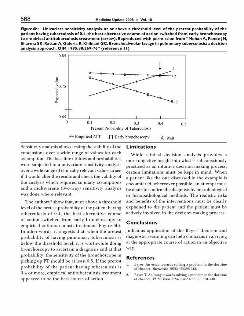
568 Medicine Update 2008 Vol. 18
Sensitivity analysis allows testing the stability of the conclusions over a wide range of values for each assumption. The baseline utilities and probabilities were subjected to a univariate sensitivity analysis over a wide range of clinically relevant values to see if it would alter the results and check the validity of the analysis which required so many assumptions and a multivariate (two-way) sensitivity analysis was done where relevant.
The authors11 show that, at or above a threshold level of the pretest probability of the patient having tuberculosis of 0.4, the best alternative course of action switched from early bronchoscopy to empirical antituberculosis treatment (Figure 6b). In other words, it suggests that, when the pretest probability of having pulmonary tuberculosis is below the threshold level, it is worthwhile doing bronchoscopy to ascertain a diagnosis and at that probability, the sensitivity of the bronchoscope in picking up PT should be at least 0.5. If the pretest probability of the patient having tuberculosis is 0.4 or more, empirical antituberculosis treatment appeared to be the best course of action.
LimitationsWhile clinical decision analysis provides a
more objective insight into what is subconsciously practiced as an intuitive decision making process, certain limitations must be kept in mind. When a patient like the one discussed in the example is encountered, whereever possible, an attempt must be made to confirm the diagnosis by microbiological or histopathological methods. The realistic risks and benefits of the interventions must be clearly explained to the patient and the patient must be actively involved in the decision making process.
ConclusionsJudicious application of the Bayes’ theorem and diagnostic reasoning can help clinicians in arriving at the appropriate course of action in an objective way.
References1. Bayes. An essay towards solving a problem in the doctrine
of chances. Biometrika 1958; 45:296-315.
2. Bayes T. An essay towards solving a problem in the doctrine of chances. Philos Trans R Soc Lond 1763; 53:370–418.
Figure 6b : Univariate sensitivity analysis. at or above a threshold level of the pretest probability of the patient having tuberculosis of 0.4, the best alternative course of action switched from early bronchoscopy to empirical antituberculosis treatment (arrow). Reproduced with permission from “Mohan A, Pande JN, Sharma SK, Rattan A, Guleria R, Khilnani GC. Bronchoalveolar lavage in pulmonary tuberculosis: a decision analysis approach. QJM 1995;88:269-76” (reference 11)
Present Probability of Tuberculosis
++ Empirical ATT Early bronchoscopy Wait
0 0.1 0.2 0.3 0.4 0.5
3
2
1
Expe
cted
val
ue0.85
0.75
0.65
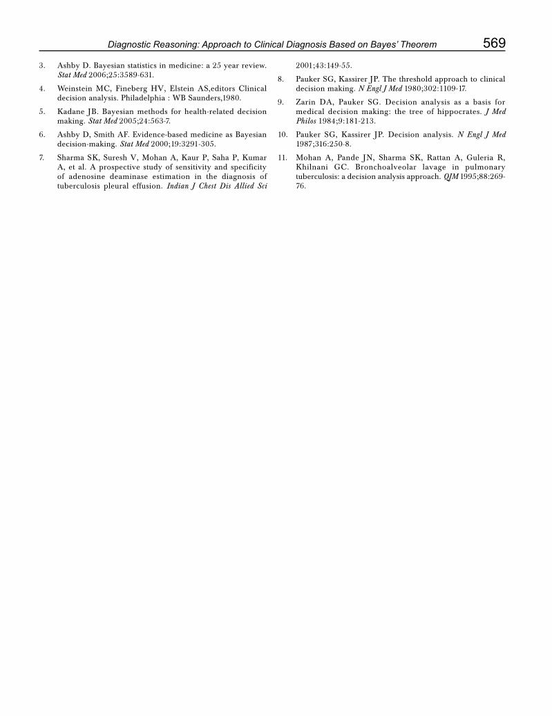
569Diagnostic Reasoning: Approach to Clinical Diagnosis Based on Bayes’ Theorem
3. Ashby D. Bayesian statistics in medicine: a 25 year review. Stat Med 2006;25:3589-631.
4. Weinstein MC, Fineberg HV, Elstein AS,editors Clinicaldecision analysis. Philadelphia : WB Saunders,1980.
5. Kadane JB. Bayesian methods for health-related decision making. Stat Med 2005;24:563-7.
6. Ashby D, Smith AF. Evidence-based medicine as Bayesian decision-making. Stat Med 2000;19:3291-305.
7. Sharma SK, Suresh V, Mohan A, Kaur P, Saha P, Kumar A, et al. A prospective study of sensitivity and specificity of adenosine deaminase estimation in the diagnosis of tuberculosis pleural effusion. Indian J Chest Dis Allied Sci
2001;43:149-55.
8. Pauker SG, Kassirer JP. The threshold approach to clinical decision making. N Engl J Med 1980;302:1109-17.
9. Zarin DA, Pauker SG. Decision analysis as a basis for medical decision making: the tree of hippocrates. J Med Philos 1984;9:181-213.
10. Pauker SG, Kassirer JP. Decision analysis. N Engl J Med 1987;316:250-8.
11. Mohan A, Pande JN, Sharma SK, Rattan A, Guleria R, Khilnani GC. Bronchoalveolar lavage in pulmonarytuberculosis: a decision analysis approach. QJM 1995;88:269-76.

CHAPTER
76Young Hypertensive : How and How much to Investigate?S. A. Kamath
IntroductionMajority of young (< 40 years) patients with high blood pressure have essential hypertension. But many also have secondary hypertension, which can be cured. Hence it is very important to diagnose these conditions and reverse the high blood pressure in order to avert target organ damage. Many of the investigations for secondary hypertension are time-consuming, tedious and expensive. Then why perform them when it has been shown that in most situations a final diagnosis of essential hypertensive will be arrived at. The following article gives us an insight into who should be investigated and to what extent.
Causes of Hypertension in the YoungEssential Hypertension
Secondary Hypertension
• RenalParenchymalHypertension
• Drugs
• ObstructiveSleepApneaSyndrome
• COPD
• Lifestyle–Diet/Nutrition
• Hypothyroidism
• Hyperthyroidism
• RenovascularHypertension
• CoarctationoftheAorta
• Cushing’sSyndrome
• Aldosteronism
• Pheochromocytoma
Practical pointsAccurate measurement of blood pressure is very important. Thorough medical history and physical examination is very valuable, and will help to arrive at conclusion very often and eliminate many unnecessary investigations that may be time-consuming, expensive, and ultimately lead nowhere.
Why should hypertension be investigated?• Detection of target organ disease (e.g., renal
damage, congestive heart failure)
• Identification of other risk factors forcardiovascular disorders (e.g., diabetes mellitus, hyperlipidemia); and
• Detection of secondary causes ofhypertension
The routine investigations to be done in any patient with hypertension are shown in Table 1.
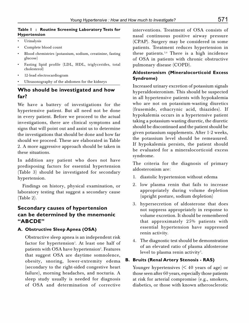
571Young Hypertensive : How and How much to Investigate?
Who should be investigated and how far?We have a battery of investigations for the hypertensive patient. But all need not be done in every patient. Before we proceed to the actual investigations, there are clinical symptoms and signs that will point out and assist us to determine the investigations that should be done and how far should we proceed. These are elaborated in Table 2. A more aggressive approach should be taken in these situations.
In addition any patient who does not have predisposing factors for essential hypertension (Table 3) should be investigated for secondary hypertension.
Findings on history, physical examination, or laboratory testing that suggest a secondary cause (Table 2).
Secondary causes of hypertension can be determined by the mnemonic “ABCDE”A. Obstructive Sleep Apnea (OSA)
Obstructive sleep apnea is an independent risk factor for hypertension1. At least one half of patients with OSA have hypertension2. Features that suggest OSA are daytime somnolence, obesity, snoring, lower-extremity edema (secondary to the right-sided congestive heart failure), morning headaches, and nocturia. A sleep study usually is needed for diagnosis of OSA and determination of corrective
interventions. Treatment of OSA consists of nasal continuous positive airway pressure (CPAP).Surgerymaybeconsidered in somepatients. Treatment reduces hypertension in these patients.3,4 There is a high incidence of OSA in patients with chronic obstructive pulmonarydisease(COPD).
Aldosteronism (Mineralocorticoid Excess Syndrome)
Increased urinary excretion of potassium signals hyperaldosteronism. This should be suspected in all hypertensive patients with hypokalemia
who are not on potassium-wasting diuretics (frusemide, ethacrynic acid, thiazides). If hypokalemia occurs in a hypertensive patient taking a potassium-wasting diuretic, the diuretic should be discontinued and the patient should be given potassium supplements. After 1-2 weeks, the potassium level should be remeasured. If hypokalemia persists, the patient should be evaluated for a mineralocorticoid excess syndrome.
The criteria for the diagnosis of primary aldosteronism are:
1. diastolic hypertension without edema
2. low plasma renin that fails to increase appropriately during volume depletion (upright posture, sodium depletion)
3. hypersecretion of aldosterone that does not suppress appropriately in response to volume excretion. It should be remembered that approximately 25% patients with essential hypertension have suppressed renin activity.
4. The diagnostic test should be demonstration of an elevated ratio of plasma aldosterone level to plasma renin activity5.
B. Bruits (Renal Artery Stenosis - RAS)
Younger hypertensives (< 40 years of age) or those seen after 60 years, especially those patients at risk for arterial compromise (e.g., smokers, diabetics, or those with known atherosclerotic
Table 1 : Routine Screening Laboratory Tests for Hypertension • Urinalysis
• Completebloodcount
• Bloodchemistries(potassium,sodium,creatinine,fastingglucose)
• Fasting lipid profile (LDL, HDL, triglycerides, totalcholesterol)
• 12-leadelectrocardiogram
• Ultrasonographyoftheabdomenforthekidneys

572 Medicine Update 2008 Vol. 18
Table 2Clinical features Preliminary Tests Disease suspected Additional Diagnostic
Studies• Edema,sallowskin,
breathlessnessOliguria + elevated BUN and
creatinine levels, proteinuria
Renal parenchymal disease
Creatinineclearance,renalultrasonography kidney biopsy
• Systolic/diastolicabdominal bruit
Renovascular hypertension
Magnetic resonance angiography,Captopril-augmented radioisotopic renography, Renal arteriography
• Inequalityofpulsationsin both upper extremities
Aorto-arteritis Aortogram with angiogram of upper extremity
Decreased or delayed femoral pulses, abnormal chest radiograph
Coarctationofaorta DopplerorCTimagingofaorta
• Useofsympathomimetics, Perioperative setting, Acute stress, Tachycardia
Excessive catecholamines
Confirmpatientisnormotensive in absence of catecholamine excess
• Snoring,Daytimesomnolence, Obesity (esp truncal)
OSA Sleep study
• Diet:highsalt,excessivealcohol,
Diet side effects Lifestyle modifications
• Centralobesity Hyperglycemia, hyperinsulinemia, hypercholesterolemia, hypertriglyceridemia
Dysmetabolic syndrome, insulin resistance
Lifestyle modifications
• UseofdruginTable4 Drug side effect Take off drug• Weightgain,fatigue,
weakness, hirsutism, amenorrhea, moon facies, dorsal hump, purple striae, truncal obesity, hypokalemia
Cushing’sSyndrome 8 AM serum cortisol, Dexamethasone suppression Test
• Paroxysmalhypertension, headaches, diaphoresis, palpitations, tachycardia
Pheochromocytoma Urinary catecholamine metabolites (VMA, metanephrines, normetanephrines) Plasma free metanephrines
MIBG scan• Fatigue,weightloss,
hair loss, diastolic hypertension, muscle weakness
Hypothyroidiosm Thyroid function tests
• Heatintolerance,weight loss, palpitations, systolic hypertension, exophthalmos tremor, tachycardia
Hyperthyroidism Thyroid function tests
• Kidneystones,osteoporosis, depression, lethargy, muscle weakness
Hyperparathyroidism Serum calcium, parathyroid hormone levels
• Headaches,fatigue,visual problems, enlargement of hands, feet, tongue
Acromegaly X-ray skull, hands,
CTbrain/MRI
Growth hormone levels
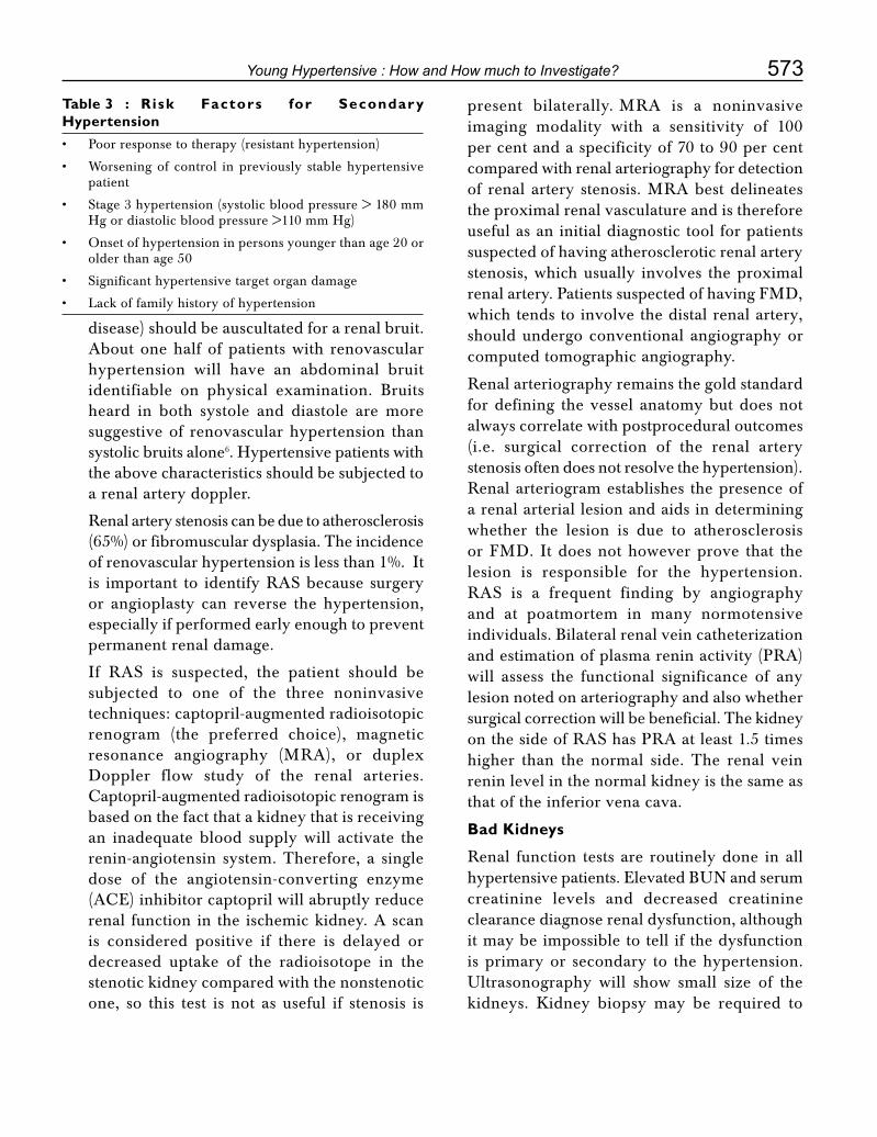
573Young Hypertensive : How and How much to Investigate?
disease) should be auscultated for a renal bruit. About one half of patients with renovascular hypertension will have an abdominal bruit identifiable on physical examination. Bruits heard in both systole and diastole are more suggestive of renovascular hypertension than systolic bruits alone6. Hypertensive patients with the above characteristics should be subjected to a renal artery doppler.
Renal artery stenosis can be due to atherosclerosis (65%) or fibromuscular dysplasia. The incidence of renovascular hypertension is less than 1%. It is important to identify RAS because surgery or angioplasty can reverse the hypertension, especially if performed early enough to prevent permanent renal damage.
If RAS is suspected, the patient should be subjected to one of the three noninvasive techniques: captopril-augmented radioisotopic renogram (the preferred choice), magnetic resonance angiography (MRA), or duplex Doppler flow study of the renal arteries. Captopril-augmentedradioisotopicrenogramisbased on the fact that a kidney that is receiving an inadequate blood supply will activate the renin-angiotensin system. Therefore, a single dose of the angiotensin-converting enzyme (ACE)inhibitorcaptoprilwillabruptlyreducerenal function in the ischemic kidney. A scan is considered positive if there is delayed or decreased uptake of the radioisotope in the stenotic kidney compared with the nonstenotic one, so this test is not as useful if stenosis is
present bilaterally. MRA is a noninvasive imaging modality with a sensitivity of 100 per cent and a specificity of 70 to 90 per cent compared with renal arteriography for detection of renal artery stenosis. MRA best delineates the proximal renal vasculature and is therefore useful as an initial diagnostic tool for patients suspected of having atherosclerotic renal artery stenosis, which usually involves the proximal renal artery. Patients suspected of having FMD, which tends to involve the distal renal artery, should undergo conventional angiography or computed tomographic angiography.
Renal arteriography remains the gold standard for defining the vessel anatomy but does not always correlate with postprocedural outcomes (i.e. surgical correction of the renal artery stenosis often does not resolve the hypertension).
Renal arteriogram establishes the presence of a renal arterial lesion and aids in determining whether the lesion is due to atherosclerosis or FMD. It does not however prove that the lesion is responsible for the hypertension. RAS is a frequent finding by angiography and at poatmortem in many normotensive individuals. Bilateral renal vein catheterization and estimation of plasma renin activity (PRA) will assess the functional significance of any lesion noted on arteriography and also whether surgical correction will be beneficial. The kidney on the side of RAS has PRA at least 1.5 times higher than the normal side. The renal vein renin level in the normal kidney is the same as that of the inferior vena cava.
Bad Kidneys
Renal function tests are routinely done in all hypertensive patients. Elevated BUN and serum creatinine levels and decreased creatinine clearance diagnose renal dysfunction, although it may be impossible to tell if the dysfunction is primary or secondary to the hypertension. Ultrasonography will show small size of the kidneys. Kidney biopsy may be required to
Table 3 : Risk Factors for Secondar y Hypertension • Poorresponsetotherapy(resistanthypertension)
• Worseningof control inpreviously stablehypertensivepatient
• Stage3hypertension(systolicbloodpressure>180mmHg or diastolic blood pressure >110 mm Hg)
• Onsetofhypertensioninpersonsyoungerthanage20orolder than age 50
• Significanthypertensivetargetorgandamage
• Lackoffamilyhistoryofhypertension

574 Medicine Update 2008 Vol. 18
determine the cause of renal failure, and for further management.
C. Catecholamines, Coarctation, Cushing’s Syndrome
Catecholamines
Patients having sweating, tachycardia, palpitations, and tremors in addition to a raised BP usually have elevated catecholamine levels. Elevated catecholamines play a role in causing white-coat hypertension and hypertension in pheochromocytoma, OSA, and other diseases discussed in this article. Acute stress induces catecholamine release and often contributes to preoperative or postoperative hypertension. Over-the-counter or prescription decongestants can have sympathomimetic effects, as do nonprescription weight-loss preparations containing ephedra (ma huang).7,8Cocaineandamphetamines also have hypertensive effects because of stimulation of the sympathetic nervous system. Hence the value of a thorough history and physical examination in a hypertensive patient should not be undermined.
Coarction of the Aorta
Coarctationoftheaortaisacongenitalnarrowingof the aortic lumen, most often occurring just distal to the origin of the left subclavian artery. Patients with less severe forms of the disorder may not be diagnosed until young adulthood but have a high incidence of premature death.9
Decreased lower-extremity (femoral) pulses with upper-extremity hypertension suggest Coarctation of the Aorta. Hence it is veryimportant to examine all the peripheral pulsations and take BP in all four extremities. Patientmayhavedyspneaonexertion.Chestradiographic findings of notched ribs (from dilated collateral vessels) and dilation of the aorta above and below the constriction (the “3” sign) are highly suggestive.9
Other diagnostic tests that should be done include ECG and Echocardiography for
hypertrophy of the heart chambers and their function.CT/MRIofthechestandaortographymay be useful to delineate anatomic narrowing. Doppler ultrasound and cardiac catheterization can be used to see if there are any differences in blood pressure in different areas of the aorta. This is very important prior to surgery and to determine post surgical prognosis.
Surgery is usually recommended. The narrowed part of the aorta will be removed. In some cases, balloon angioplasty may be done instead of surgery.
Cushing’s Syndrome
Cushing’s syndrome can cause hypertensionvia the mineralocorticoid effects of excess glucocorticoids. Weight gain, fatigue, weakness, hirsutism, amenorrhea, moon facies, buffalo hump, purple striae, truncal obesity suggest Cushing’ssyndrome.Serumpotassiummaybelow.
For initial screening of Cushing’s syndrome,8.00 a.m. serum cortisol or the overnight dexamethasone suppression test is recommended. In difficult case (obese or patients with depression), measurement of a 24-hour urine free cortisol can also be good screening test. A level > 140 nmol/d (50 µg is suggestive of Cushing’s syndrome). Thedefinitive diagnosis is then established by failure of urinary cortisol to fall to < 25 nmol/d (10 µg/d) or plasma cortisol to fall to < 140 nmol/L (5 µg/dL) after a standard low-dose dexamethasone suppression test (0.5 mg every 6 h for 48 hrs). Once the diagnosis is established further testing should be done to determine the etiology.10
D. Drugs, Diet
Drugs : Many prescription and nonprescription drugs can cause or exacerbate hypertension (Table 4).
575Young Hypertensive : How and How much to Investigate?
Diet
Dietary factors that can cause hypertension are excess consumption of salt (sodium); while low intake of potassium, calcium, and magnesium can have a similar but less pronounced effect. The lower limit of “excess salt” has not been determined. An average typical Indian diet contains at least 17-20 g of salt. Blacks, elderly, patients, those with diabetes, and patients with essential hypertension appear to be particularly sensitive to dietary sodium intake. High calorie, low fiber diet and dietary patterns that cause obesity also can cause hypertension. Sustained weight reduction lowers blood pressure--often to normal levels--in at least one half of obese patients. A loss of 5 to 10 per cent of body weight can significantly reduce blood pressure.
E. Endocrine Disorders, Erythropoietin
Hypothyroidism causes decreased cardiac output with a compensatory increase in vascular tone, resulting in a more prominent rise in diastolic blood pressure than in systolic blood pressure. Features of hypothyroidism are fatigue, cold intolerance, weight gain, non-pitting
edema, hair loss, diastolic hypertension, muscle weakness. Measurement of TSH is a screening test for hypothyroidism. If TSH is elevated, free T
4 level should be done to confirm the presence
of clinical hypothyroidism. T3 measurements
are not indicated because free T3 levels may be
normal in about 25% of hypothyroid patients.
Hyperthyroidism induces increased cardiac output and compensatory decreased vascular tone, causing a greater increase in systolic blood pressure. Heat intolerance, weight loss, palpitations, systolic hypertension, exophthalmos, tremors, tachycardia suggest hyperthyroidism. The TSH level is suppressed, while total and free T
3 and
T
4 levels are
increased.
Hyperparathyroidism (primary or secondary to chronic renal insufficiency) is a potentially reversible cause of hypertension. Its incidence in hypertensive patients is about 1%, compared with a 0.1% incidence in the general population. However, only 30 to 40 per cent of patients with hyperparathyroidism have hypertension,
Kidney stones, osteoporosis, depression, lethargy, muscle weakness are features of hyperparathyroidism. Serum calcium and parathormone levels will determine the diagnosis. It is important to distinguish between primary hyperparathyroidism and secondary hyperparathyroidism due to renal failure. It should be remembered that in primary hyperparathyroidism, parathyroidectomy may not reliably resolve hypertension.
Pheochromocytoma is another endocrine cause of hypertension. The classic symptoms include headache, diaphoresis, palpitations, and paroxysmal hypertension. The syndrome can vary depending on the types of catecholamines being produced, the amount and frequency of their release into the circulation, and other factors. The usual screening test has been urinary measurement of catecholamine metabolites (vanillylmandelic acid, metanephrines,
Table 4 : Drugs That Can Raise Blood Pressure Drug class Drug examples
Immunosuppressive agents Cyclosporine,Tacrolimus,corticosteroids
Nonsteroidal anti-inflammatory drugs
Ibuprofen, naproxen, piroxicam
COX-2inhibitors Celecoxib,rofecoxib,valdecoxib
Estrogens 30- to 35-mcg estrogen oral contraceptives
Weight-loss agents Sibutramine, phentermine, ma huang
Stimulants Nicotine, amphetamines
Mineralocorticoids Fludrocortisone
Antiparkinsonian Bromocriptine
Monoamine oxidase inhibitors
Phenelzine
Anabolic steroids Testosterone
Sympathomimetics Pseudoephedrine

576 Medicine Update 2008 Vol. 18
Figure 1 : General strategy for diagnosing a secondary cause of hypertension.Evaluation for Secondary Causes of Hypertension
normetanephrines).11 Determination of plasma free metanephrines might be the test of first choice for diagnosis of this tumor, although availability of this test at hospital and reference laboratories is limited. Pheochromocytoma is very rare, and routine screening in hypertensive patients is not recommended. MIBG scan is one more useful diagnostic modality,
Acromegaly (elevated growth hormone -GH) is a rare endocrine cause of hypertension. There is coarsening of features, prognathism, diastema
(widely spaced teeth), increased ring and shoe sizes; hands become enlarged, moist and soft with tufting of distal phalanges. Generalized thickening of the skin with increased sweating and oiliness, hypertrichosis, acanthosis nigricans and acne are also seen.
When acromegaly is clinically suspected, IGF-I estimation is a useful screening test and estimation of serum GH is confirmatory.
IGF-1 measurement (normal ranges vary in different laboratories) is an indirect measurement
Suspected hypertension
Test accury of reading (check cuff size, repeatoffice readings, out-of-readings)
NormotensiveWhite-coat hypertension
Confirmed hypertension
Screening historyScreening physical examinaton
Screening laboratory tests (Table 4)
Risk factors for secondaryhypertension present
(Table 3)?
Yes
Screening results do notsuggest a specific cause.
Screening results suggest aspecific cause (Table 1).
Consider more aggressive evaluation forsecondary hypertension (see “futher
diagnostic studies” in Table 1)
Identify and treat suspectedcause and assess response
Treat hypertension andassess response
No

577Young Hypertensive : How and How much to Investigate?
of GH. Since IGF-1 levels are much more stable over a day, they are often more practical and reliable than the measurements of GH levels. Another advantage of this test is showing activity of the disease. IGF-I level is a useful laboratory screening measure when clinical features raise the possibility of acromegaly.
The normal level of serum GH is 3 to 5 ng/mL. GH level greater than 10 ng/mL is found in 90% of patients with acromegaly. A single measurement is not entirely reliable because GH is secreted by the pituitary in spurts and its concentration can vary widely. At a given moment, an acromegalic may have normal GH levels, whereas a GH level in a healthy person may be 5 times higher, especially in conditions such as stress, sleeping time, exercise. Because of this, more accurate diagnosis can be done when GH is measured under conditions in which GH secretion is normally suppressed. Oral Glucose Tolerance Test (OGTT) is often used for this. 100 g of Glucose is administered after an overnight fast. The results are interpreted as follows: Normal GH < is 2 µg/L. The diagnosis of acromegaly is confirmed by demonstrating the failure of GH suppression to < 1 µg/L within 1-2 hours of the oral glucose load. About 20% of patients exhibit high levels of GH (called “paradoxal increase”).
Erythropoietin. High erythropoietin levels can elevate blood pressure either via a polycythemia/hyperviscosity mechanism or by direct pressor effects.7 Elevated erythropoietin levels can be endogenous (as in response to thechronichypoxiaofCOPD)orexogenous(administered to alleviate the anemia seen in
chronic renal failure).
In conclusion, the value of accurate measurement of BP, thorough medical history and clinical examination should not be underestimated. Doing so would screen for most of the secondary causes of hypertension discussed in this article, along with signs of target organ disease and comorbid factors.
References1. Silverberg DS, Oksenberg A. Essential and secondary
hypertension and sleep-disordered breathing: a unifying hypothesis. J Hum Hypertens 1996;10:353-63.
2. Kaplan NM. Other forms of secondary hypertension. In: KaplanNM,LiebermanE,eds.Clinicalhypertension.7thed. Baltimore: Williams & Wilkins, 1998:395-406.
3. Stradling JR, Partlett J, Davies RJ, Siegwart D, Tarassenko L. Effect of short term graded withdrawal of nasal continuous positive airway pressure on systemic blood pressure in patients with obstructive sleep apnea. Blood Press 1996;5:234-40.
4. Victor LD. Obstructive sleep apnea. Am Fam Physician 1999;60:2279-86.
5. Gordon RD, Stowasser M, Tunny TJ, Klemm SA, Rutherford JC.Highincidenceofprimaryaldosteronismin199patientsreferred with hypertension. Clin Exp Pharmacol Physiol1994;21:315-8.
6. Pohl M. Renal artery stenosis, renal vascular hypertension, andischemicnephropathy.In:SchrierRW,GottschalkCW,eds. Diseases of the kidney. 6th ed. Boston: Little, Brown, 1997:1367-423.
7. Phenylpropanolamine and other OTC alpha-adrenergicagonists. Med Lett Drugs Ther 2000;42:11
8. Morelli V, Zoorob RJ. Alternative therapies: part I. Depression, diabetes, obesity. Am Fam Physician 2000;62:1051-60.
9. Steiner RM. Radiology of the heart. In: Braunwald E, ed. Heart disease: a textbook of cardiovascular medicine. 5th ed. Philadelphia: Saunders, 1997: 204-39.
10. Dluhy RG, Williams RH. Endocrine hypertension. In: Williams RH, Wilson JD, eds. Williams Textbook of endocrinology. 9th ed. Philadelphia: Saunders, 1998:729-49.
11. Williams GH. Approach to the patient with hypertension. In: Braunwald E, ed. Harrison’s Principles of internal medicine. 15th ed. New York: McGraw-Hill, 2001:211-4.

CHAPTER
77Refractory Epilepsy- How Much to Investigate ?Manjari Tripathi
Historical AspectsSystematic localization of the epileptic zone and procedures for surgical resection are not recent developments in the treatment of epilepsy. based on the work of Hughlings Jackson. Between 1890 and 1910, Horsley1 reported series of patients who received craniotomy for the treatment of symptomatic epilepsy from a known cause, often traumatic. Wilder Penfield and Herbert Jasper,2,3 and many others followed with important advances in the development of evaluative and surgical techniques for refractory epilepsy. Percival Bailey and Frederick Gibbs were among the first teams to resect the epileptogenic region based on EEG localization and semiology without identified structural brain abnormalities.4
Which patients to investigate and when ?Defining Surgical Candidacy and Medical Intractability
Anyone with recurrent seizures who is interested in surgery as a possible treatment is a good candidate for a detailed discussion of the potential risks and benefits of surgical intervention. For most patients this discussion can only take place after video/EEG confirmation of the classification of the epilepsy and localization of the epileptogenic region. Another
reasonable criteria is anyone who is disabled due to their epilepsy, regardless of seizure rate, should be offered the option of presurgical evaluation. Patients considered for epilepsy surgery should meet two criteria: 1) disabling seizures that have not been controlled by adequate trials of antiepileptic drugs without adverse side effects, 2) clinical, neuroimaging, or EEG evidence of an epileptogenic brain region that may be safely resected.5,6 The specific aspects of these criteria, however, are not simple and are subject to numerous variations. Some patients without a specific localizable epileptogenic region should be evaluated for palliative procedures. There is no clear consensus on what the definition of intractable epilepsy is.7,8 Most conventional definitions include the failure of two first-line antiepileptic drugs over a period of at least two years. Recent large cohort studies,9 however, indicate that most patients who will remain refractory to medications can be accurately identified within one year after diagnosis, based on response to the initial medication, classification of the epilepsy, and the presence of a structural cerebral abnormality. Other important factors to weigh in the decision for surgical candidacy include the risk of increased morbidity and mortality from continued seizures. An important consideration is the increased mortality in patients with recurrent seizures, if surgery is not offered as a treatment.

579Refractory Epilepsy- How Much to Investigate
recent investigations of medically resistant epilepsy suggest that the overall mortality rate in this population is between 0.5 and 1.5% per year. Sperling et al10 reported that none of the 199 patients who were seizure-free after surgery died, whereas 11 of the 194 with seizure recurrence after surgery died. The only death in the recent randomized trial of surgery for temporal lobe epilepsy occurred in the medical treatment arm.11 Multiple subsequent studies have replicated similar findings,12-14 but not all studies have concurred.15,16 Head trauma was reported by 24% and burns by 16% of patients with active epilepsy in a recent survey.17 The risk of major injuries or death must be seriously considered before delaying the presurgical evaluation or committing to additional trials of antiepileptic drugs in patients at increased risk for pharmacoresistant epilepsy.18
The patient’s perspective, especially regarding the importance of controlling the seizures compared to the surgical risk of injury to a functionally eloquent brain region, must be emphasized in the decision regarding surgical options.19,20 Like patients undergoing evaluation for surgical treatment of other disorders, patients with medically resistant epilepsy also experience anxiety over the option of an invasive surgical procedure. Many epilepsy patients may not be receiving appropriate education regarding their surgical options and risk of continued seizures.21 Treatment with the vagal nerve stimulator does not appear to improve mortality rates in refractory epilepsy.22 Mortality for epilepsy surgery procedures in published series is less than 0.2%.39
Hence patients should be assured and preferably be referred to centers having an epilepsy surgery team with adequate experience with availability of all possible non invasive methods of investigation as mentioned below if these seizures are not controlled with two or more drugs in appropriate choice and dosage as early as possible this will ensure the right for equal life opportunities in persons with refractory epilepsy.
What Lesions can be operated upon?As a prerequisite to this it is mandatory that a systematic approach to the identification of the lesion or cause is carried out.
Identifying the epileptogenic region
The clinical history and physical examination
As with most neurological disorders, the history and physical examination can contribute critically important information for localization of the relevant pathology. For example, an initial simple partial seizure, or aura, of an unusual epigastric sensation is reported by nearly one half of patients with temporal lobe epilepsy, but is uncommon in seizures arising from other regions. Similarly, déjà vu or an olfactory sensation at seizure onset usually indicates a temporal lobe seizure.24 These symptoms, however, are not highly accurate for
Figure 1 : Evaluation of Potential Epilepsy Surgery Candidates
PATIENT WITH REFRACTORY EPILEPSY
3 INTERICTAL EEG’S
MRI BRAIN
EPILEPSY PROTOCOL
OPD BASIS
DISCORDANT WITH MRI OR NORMAL MRI
SPECIAL MRI SEQUENCESPET ICTALSPECT/SISCOS/SISCOM
SURGERY
INCONCLUSIVE
PHASE II INVASIVE EEG WITH HYPOTHESIS
AT LEAST TWO CONCORDANT
SURGERY
VEEG IN PATIENTCONCORDANT TO MRI

580 Medicine Update 2008 Vol. 18
lateralization of the temporal lobe of seizure onset. Ictal behavior, such as unilateral automatisms, often occurs ipsilateral to the temporal lobe of onset, while dystonic posturing of the hand may occur contralateral to side of onset.25-26 Prolonged postictal dysphasia suggests a dominant temporal lobe seizure.27 Postictal nose wiping also strongly lateralizes in temporal lobe epilepsy to the side of the involved upper extremity.28 Brief, hypermotor, frantic, bizzare behavior with minimal postictal confusion is associated with inferior or anterior frontal seizures, while posturing and abduction of the upper extremities is often present with mesial frontal (Supplementary Sensory Motor Area-SSMA) onset.29-33 Primary somatosensory symptoms are less reliably localizing, and may indicate an epileptogenic region in the SSMA or extrasensory area (i.e., not necessarily parietal primary sensory area onset). Asymmetric facial movement during spontaneous smiling on examination is highly specific and moderately sensitive for contralateral mesial temporal sclerosis in temporal lobe epilepsy.34
Neuroimaging (Fig. 2-12)Advances in MRI technology have improved the sensitivity and specificity of the clinical evaluation for identification of potentially epileptogenic cerebral pathology. Mesial temporal or hippocampal
sclerosis is the best characterized indicator of the epileptogenic region identifiable by MRI.35-37 The predictive value of MRI identified unilateral mesial temporal sclerosis, however, ranges from 61% to 96% in published series.38-42
Focal MRI abnormalities in extratemporal lobe epilepsy also provide targets to guide intracranial EEG monitoring and allow better surgical outcomes compared to patients with nonlesional extratemporal epilepsy.43 Specific MRI protocols including FLAIR and inversion recovery highlight detailed anatomy and signal abnormalities that provide increased sensitivity for localization of potentially epileptogenic pathology.44 MR spectroscopy may improve localization and advance our understanding of epileptogenesis by identification of metabolic and neurotransmitter changes in specific brain regions.45-47 Limitations include, however, the labor-intensive requirements of a well-trained team in the setting of an inpatient video/EEG monitoring unit. PET imaging, on the other hand, can provide data on metabolic dysfunction,48 but its predictive value in extratemporal epilepsy is not clearly defined.
Video/EEG Monitoring
In addition to video confirmation of features of seizure semiology discussed above, video/EEG monitoring provides ictal EEG49 and extended sampling of interictal EEG abnormalities that have high predictive value for localization of the epileptogenic region. Interictal sharp waves or spikes appear to be the strongest indicator of regional cerebral hyperexcitability, but temporal intermittent rhythmic delta activity (TIRDA) may also be highly accurate for unilateral temporal lobe epilepsy. Temporal lobe polymorphic delta activity is less specific for temporal lobe epilepsy. In one study of 90 consecutive patients with medically refractory epilepsy considered for surgery, 61% had unilateral temporal interictal abnormalities that were concordant with mesial temporal sclerosis identified by MRI. However, bitemporal interictal EEG abnormalities are not a contraindication for surgery, as up to 50% of such patients may become
Table 1 : Techniques used to localize the primary epileptogenic zone.•NeurologicalHistoryandExamination
•RoutineEEG
•Video/EEGMonitoring
•HighResolutionMRI
•InterictalPET
•IctalSPECT
•NeuropsychologicalTesting
•SodiumAmobarbitalTest(WADA)
•MRSpectroscopy
•EEGSpikeTriggeredMRI
•Magnetoelectroencephalography
•IntracranialEEGandCorticalStimulation
581Refractory Epilepsy- How Much to Investigate
seizure free after temporal lobe resection.51 Mapping of the maximal fields of interictal and initial ictal epileptogenic abnormalities is a critical component of the presurgical localization process.
Surgical ProceduresAlthough many nonpharmacologic therapies have been reported for seizure control, including yoga and herbal treatments, only vagus nerve stimulation (VNS)52 has undergone controlled clinical trials adequate to support a Food and Drug Administration approval. Randomized, double-blind clinical trials comparing two intensities of intermittent stimulation of the left vagus nerve demonstrated a significant reduction in seizures in the high intensity stimulation group.53 Although no patients remained seizure-free during these studies, the 25% to 30% reduction in seizure rate compared to baseline was adequate to allow an American Academy of Neurology Subcommittee to conclude that “VNS is indicated for adults and adolescents over 12 years of age with medically intractable partial seizures who are not candidates for potentially curative surgical resection The committee believes that patients should undergo a thorough evaluation of the epilepsy to rule out nonepileptic conditions or treatable symptomatic epilepsies before implantation of a vagus nerve stimulator.”54 Long term outcomes based on open-label studies suggest that seizure control may gradually improve during the first one to two years of treatment.55. Adverse effects of VNS reported
by more than 5% of patients include hoarseness/voicechange,throatpain,andcoughing.Cardiacasystole has been reported in five cases of initial VNS testing at time of implantation. In a study of 24 children, 12 had unexpected adverse events; the severity of the adverse events led to surgical removal of the device in two of the patients.56
After implantation during surgery under general anesthesia, the VNS device requires gradual increase in stimulation intensity until optimal seizure control with tolerable adverse effects is achieved. The stimulation period is usually one minute, occurring every five minutes, although these parameters may be altered. A magnet controlled by the patient or assistant may also be used to initiate stimulation. The increase in stimulation intensity is performed through a simple computer-assisted procedure using a “wand” placed over the patient’s skin at site of the device. The interval between stimulation setting changes is dependent on seizure frequency (i.e., enough time to determine change in seizure frequency), patient tolerance of the initial discomfort after stimulation increase, and convenience for the patient. The typical higher end of the stimulation range is 3 mA, achieved by incremental increase of 0.5 mA. The expected battery life prior to surgical replacement is estimated to be five years.
Why Consider Surgery?
The most compelling reason to consider epilepsy surgery from the patients’ perspective is the possibility to live an independent and autonomous, and lead a normal social and vocational life. As alluded to above however, minimizing mortality and morbidity risk is a major consideration as well. Other potential benefits for many individuals include the reduction of mood dysfunction, arresting cognitive decline, and reducing medication burden and toxicity.
Epilepsy Surgery Outcomes
Although only one randomized trial comparing epilepsy surgery to optimal medical management has been completed, consistent findings in numerous observational studies in patients with
Table 2 : Surgical procedures used to treat medically refractory epilepsy•AnteriorTemporalLobectomy
•Amygdalohippocampectomy
•FocalorRegionalCorticalResection
•CorpusCallosotomy
•Hemispherectomy
•SubpialTransection
•VagalNerveStimulation
•CorticalStimulation
•VagusNerveStimulation

582 Medicine Update 2008 Vol. 18
Figure 2 : MRI-T1-Right Mesial temporal sclerosis
Figure 4 : MRI-FLAIR-Bilateral MTS
Figure 6 : MRI-DNET Rt temporal
Figure 3 : MRI-Flair-Hemispheric cortical dysplasia-Rt
Figure 5 : MRI-FLAIR-Rt parietal cavernous angioma
Figure 7 : CT-Brain- Sturge Weber syndorme presenting with refractory epilepsy
583Refractory Epilepsy- How Much to Investigate
Figure 8 : MRI-FLAIR -Rasmussens encephalitis Lt Hemispheric atrophy and hyperintensity posterior left parieto-occipital
Figure 10 : MRI-Post op Hemispherotomy
Figure 9 : FMRI- for evaluating limb motor function prior to surgery the lesion is away from motor area
Figure 11 : Magnetic source imaging MSI-Shows origin from posterior superior border of lesion which is a postoperative scar. Photo courtesy Dr Gary Mathern Head pediatric epilepsy surgery UCLA.
Figure 12 : Simultaneous FMRI and EEG showing focus in right temporal
PhotocourtesyDrJohnStern,UCLA
medically refractory epilepsy indicate that anterior temporal lobectomy or amygdalohypocampectomy, and extratemporal lesional epilepsy surgery, are highly effective for controlling seizures. Reported success rates range from 60% to 95% depending on selection criteria, procedure, and definition of good outcome. Overall, 60-78% of patients eventually achieve seizure remission after temporal lobectomy or lesional extratemporal resection.
Nonlesional extratemporal epilepsy surgery has less favorable outcome, but may be warranted depending on the clinical situation.57
ConclusionAlthough epilepsy surgery carries a modest risk of morbidity, the consistently observed improvements in health outcomes, mood status, medication dependence and toxicity, and probable
584 Medicine Update 2008 Vol. 18
reduction in overall mortality support its utility in pharmacoresistant epilepsy. It is a clinicians responsibility to identify patients with recurrent seizures despite adequate medical therapy and offer them a referral to an epilepsy surgical center for evaluation. Few of such centers with established epilepsy surgery programs in India are All India InstituteofMedicalSciences,SriChitraInstituteofMedicalSciences,NIMHANS,PGIChandigarhetc. There exsists not only a wide medical treatment gap of epilepsy in our country but also a wide surgical treatment gap.
References1. Horsley V. Brain surgery. Br. Med J. 1886;2:670-675.
2. Penfield W, Jasper H. Epilepsy and the functional anatomy of the human brain. Boston: Little, Brown,1954.
3. Penfield W, Paine K. Results of surgical therapy for epileptic seizures. Canadian Medical J 1955;73:515- 530.
4. Bailey P, Gibbs FA. The surgical treatment of psychomotor epilepsy. J Am Med Assoc. 1951 Feb 10;145(6):365-70 1951;145:365-370.
5. Engel J, Jr. Surgery for seizures. N Engl J Med 1996;334:647-652.
6. Engel J, Jr., Wiebe S, French J, et al. Practice parameter: Temporal lobe and localized neocortical resections for epilepsy: Report of the Quality Standards Subcommittee of the American Academy of Neurology, in Association with the American Epilepsy Society and the American Association of Neurological Surgeons. Neurology 2003;60:538-547.
7. Engel J, Jr. The timing of surgical intervention for mesial temporal lobe epilepsy: a plan for a randomized clinical trial. Arch Neurol 1999;56:1338-1341.
8. Berg AT, Langfitt J, Shinnar S, et al. How long does it take for partial epilepsy to become intractable? Neurology 2003;60:186-190.
9. Sillanpaa M, Jalava M, Kaleva O, Shinnar S. Long-term prognosis of seizures with onset in childhood [see comments]. N Engl J Med 1998;338:1715-1722.
21. Kwan P, Brodie MJ. Early identification of refractory epilepsy. N Engl J Med 2000;342:314-319.
10. SperlingMR,FeldmanH,KinmanJ,LiporaceJD,O’ConnorMJ. Seizure control and mortality in epilepsy. Ann Neurol 1999;46:45-50.
11. Wiebe S, Blume WT, Girvin JP, Eliasziw M. A randomized, controlled trial of surgery for temporal-lobe epilepsy. N Engl J Med 2001;345:311-318.
12. HennessyMJ,LanganY,ElwesRD,BinnieCD,PolkeyCE,Nashef L. A study of mortality after temporal lobe epilepsy surgery. Neurology 1999;53:1276-1283.
13. Salanova V, Markand O, Worth R. Temporal Lobe Epilepsy Surgery:Outcome,Complications,andLateMortalityRatein 215 Patients. Epilepsia 2002;43:170-174.
14. Kelley K, Theodore WH. Prognosis 30 years after temporal lobectomy. Neurology 2005;64:1974-1976.
15. Ryvlin P, Montavont A, Kahane P. The impact of epilepsy surgery on mortality. Epileptic Disorders 2005;Suppl 1:39-46.
16. Stavem K, Guldvog B. Long-term survival after epilepsy surgery compared with matched epilepsy controls and the general population. Epilepsy Research 2005;63:67-75.
17. Buck D, Baker GA, Jacoby A, Smith DF, Chadwick DW.Patients’ experiences of injury as a result of epilepsy. Epilepsia 1997;38:439-444.
18. HennessyMJ,LanganY,ElwesRDC,BinnieCD,PolkeyCE,Nashef L. A study of mortality after temporal lobe epilepsy surgery. Neurology 1999;53:1276-.
19. Wilson SJ, Saling MM, Kincade P, Bladin PF. Patient expectations of temporal lobe surgery. Epilepsia 1998;39:167-174.
20. GilliamF,KuznieckyR,FaughtE,BlackL,CarpenterG,Schrodt R. Patient-validated content of epilepsyspecific quality-of-life measurement. Epilepsia 1997;38:233-236.
21. Gilliam F. Optimizing Health Outcomes in Active Epilepsy. Neurology 2002;58:S9-S19.
22. AnnegersJF,CoanSP,HauserWA,LeestmaJ,DuffellW,Tarver B. Epilepsy, vagal nerve stimulation by the NCPsystem, mortality, and sudden, unexpected, unexplained death. Epilepsia 1998;39:206-212.
23. Behrens E, Schramm J, Zentner J, Konig R. Surgical and neurological complications in a series of 708 epilepsy surgery procedures. Neurosurgery 1997;41:1-9; discussion 9-10.
40. Gotman J. Automatic seizure detection: improvements and evaluation. Electroencephalogr Clin Neurophysiol 1990;76:317-324.
24. Bancaud J, Brunet-Bourgin F, Chauvel P, Halgren E.Anatomical origin of deja vu and vivid ’memories’ in human temporal lobe epilepsy. Brain 1994;117:71-90.
25. Kotagal P, Luders HO, Williams G, Nichols TR, McPherson J. Psychomotor seizures of temporal lobe onset: analysis of symptom clusters and sequences. Epilepsy Res 1995;20:49-67.
26. SteinhoffBJ,SchindlerM,HerrendorfG,KurthC,BittermannHJ, Paulus W. The lateralizing value of ictal clinical symptoms in uniregional temporal lobe epilepsy. Eur Neurol 1998;39:72-79.
27. Dupont S, Semah F, Boon P, et al. Association of ipsilateral motor automatisms and contralateral dystonic posturing: a clinical feature differentiating medial from neocortical temporal lobe epilepsy [see comments]. Arch Neurol 1999;56:927-932.
28. Hirsch LJ, Lain AH, Walczak TS. Postictal Nosewiping Lateralizes and Localizes to the Ipsilateral Temporal Lobe. Epilepsia 1998;39:991-997.

585Refractory Epilepsy- How Much to Investigate
29. ChangCN,OjemannLM,OjemannGA,LettichE.Seizuresof fronto-orbital origin: a proven case. Epilepsia 1991;32:487-491.
30. Kotagal P, Arunkumar GS. Lateral frontal lobe seizures. Epilepsia 1998;39 Suppl 4:S62-68.
31. Salanova V, Morris HH, Van Ness P, Kotagal P, Wyllie E, Luders H. Frontal lobe seizures: electroclinicalsyndromes. Epilepsia 1995;36:16-24.
32. So NK. Mesial frontal epilepsy. Epilepsia 1998;39 Suppl 4:S49-61.
33. Morris HHd, Dinner DS, Luders H, Wyllie E, Kramer R. Supplementary motor seizures: clinical and electroencephalographic findings. Neurology 1988;38:1075-1082.
34. Cascino GD, Luckstein RR, Sharbrough FW, Jack CR.Facial asymmetry, hippocampal pathology, and remote symptomatic seizures: a temporal lobe epileptic syndrome. Neurology 1993;43:725-727.
35. CascinoGD,JackCR,Jr.,ParisiJE,etal.Magneticresonanceimaging-based volume studies in temporal lobe epilepsy: pathological correlations. Ann Neurol 1991;30:31-36.
36. Kuzniecky R, de la Sayette V, Ethier R, et al. Magnetic resonance imaging in temporal lobe epilepsy: pathological correlations. Ann Neurol 1987;22:341-347.
37. CendesF,AndermannF,GloorP, et al.MRIvolumetricmeasurement of amygdala and hippocampus in temporal lobe epilepsy. Neurology 1993;43:719-725.
38. Berkovic SF, McIntosh AM, Kalnins RM, et al. Preoperative MRI predicts outcome of temporal lobectomy: an actuarial analysis [see comments]. Neurology 1995;45:1358-1363.
39. JackCR,Jr.,SharbroughFW,CascinoGD,HirschornKA,O’BrienPC,MarshWR.Magneticresonanceimage-basedhippocampal volumetry: correlation with outcome after temporal lobectomy. Ann Neurol 1992;31:138-146.
40. Garcia PA, Laxer KD, Barbaro NM, Dillon WP. Prognostic value of qualitative magnetic resonance imaging hippocampal abnormalities in patients undergoing temporal lobectomy for medically refractory seizures. Epilepsia 1994;35:520-524.
41. Gilliam F, Bowling S, Bilir E, et al. Association of combined MRI, interictal EEG, and ictal EEG results with outcome and pathology after temporal lobectomy. Epilepsia 1997;38:1315-1320.
42. Gilliam F, Faught E, Martin R, et al. Predictive value of MRI-identified mesial temporal sclerosis for surgical outcome in temporal lobe epilepsy: an intent-to-treat analysis. Epilepsia 2000;41:963-966.
43. Mosewich RK, So EL, O’Bien TJ, et al. Factors predictive of the outcome of frontal lobe epilepsy surgery. Epilepsia 2000;41:843-849.
44. Kuzniecky RI, Bilir E, Gilliam F, et al. Multimodality MRI in mesial temporal sclerosis: relative sensitivity and specificity. Neurology 1997;49:774-778.
55. CendesF,CaramanosZ,AndermannF,DubeauF,ArnoldDL. Proton magnetic resonance spectroscopic imaging and magnetic resonance imaging volumetry in the lateralization of temporal lobe epilepsy: a series of 100 patients. Ann Neurol 1997;42:737-746.
46. Kuzniecky R, Hugg JW, Hetherington H, et al. Relative utility of 1H spectroscopic imaging and hippocampal volumetry in the lateralization of mesial temporal lobe epilepsy. Neurology 1998;51:66-71.
47. Petroff OA, Rothman DL, Behar KL, Mattson RH. Low brain GABA level is associated with poor seizure control. Ann Neurol 1996;40:908-911.
48. HenryTR,BabbTL,EngelJ,Jr.,MazziottaJC,PhelpsME,Crandall PH. Hippocampal neuronal loss and regionalhypometabolism in temporal lobe epilepsy. Ann Neurol 1994;36:925-927.
49. RisingerMW,EngelJ,Jr.,VanNessPC,HenryTR,CrandallPH. Ictal localization of temporal lobe seizures with scalp/sphenoidal recordings. Neurology 1989;39:1288-1293.
50. Cascino GD, Trenerry MR, So EL, et al. Routine EEGand temporal lobe epilepsy: relation to long-term EEG monitoring, quantitative MRI, and operative outcome. Epilepsia 1996;37:651-656.
51. So N, Gloor P, Quesney LF, Jones-Gotman M, Olivier A, Andermann F. Depth electrode investigations in patients with bitemporal epileptiform abnormalities. Ann Neurol 1989;25:423-431.
52. McLachlan RS. Vagus nerve stimulation for intractable epilepsy: a review. J Clin Neurophysiol 1997;14:358-368.
53. HandforthA,DeGiorgioCM,SchachterSC,etal.Vagusnervestimulation therapy for partial-onset seizures: a randomized active-control trial. Neurology 1998;51:48-55.
54. Fisher RS, Handforth A. Reassessment: vagus nerve stimulation for epilepsy: a report of the Therapeutics and Technology Assessment Subcommittee of the American Academy of Neurology. Neurology 1999;53:666-669.
55. DeGiorgioCM,SchachterSC,HandforthA,etal.ProspectiveLong-Term Study of Vagus Nerve Stimulation for the Treatment of Refractory Seizures. Epilepsia 2000;41:1195-1200.
56. MurphyJV,HornigGW,SchallertGS,TiltonCL.Adverseevents in children receiving intermittent left vagal nerve stimulation. Pediatr Neurol 1998;19:42-44.
57. Engel J, Van Ness PC, Reasmussen TB, Ojemann LM.Outcome with respect of Epileptic Seizures. In:Engel J, ed. Surgical Treatment of the Epilepsies., Second ed. New York: Raven Press, 1993: 609-621.
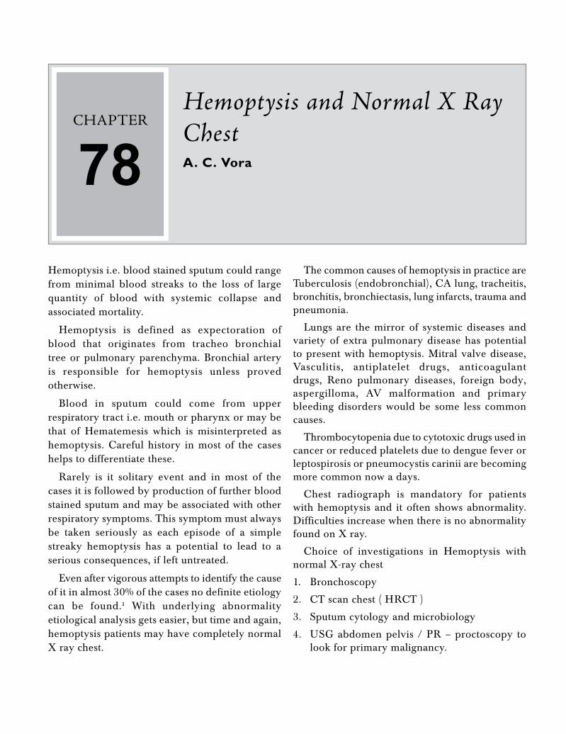
CHAPTER
78Hemoptysis and Normal X Ray ChestA. C. Vora
Hemoptysis i.e. blood stained sputum could range from minimal blood streaks to the loss of large quantity of blood with systemic collapse and associated mortality.
Hemoptysis is defined as expectoration of blood that originates from tracheo bronchial tree or pulmonary parenchyma. Bronchial artery is responsible for hemoptysis unless proved otherwise.
Blood in sputum could come from upper respiratory tract i.e. mouth or pharynx or may be that of Hematemesis which is misinterpreted as hemoptysis.Carefulhistory inmostof thecaseshelps to differentiate these.
Rarely is it solitary event and in most of the cases it is followed by production of further blood stained sputum and may be associated with other respiratory symptoms. This symptom must always be taken seriously as each episode of a simple streaky hemoptysis has a potential to lead to a serious consequences, if left untreated.
Even after vigorous attempts to identify the cause of it in almost 30% of the cases no definite etiology can be found.1 With underlying abnormality etiological analysis gets easier, but time and again, hemoptysis patients may have completely normal X ray chest.
The common causes of hemoptysis in practice are Tuberculosis(endobronchial),CAlung,tracheitis,bronchitis, bronchiectasis, lung infarcts, trauma and pneumonia.
Lungs are the mirror of systemic diseases and variety of extra pulmonary disease has potential to present with hemoptysis. Mitral valve disease, Vasculitis, antiplatelet drugs, anticoagulant drugs, Reno pulmonary diseases, foreign body, aspergilloma, AV malformation and primary bleeding disorders would be some less common causes.
Thrombocytopenia due to cytotoxic drugs used in cancer or reduced platelets due to dengue fever or leptospirosis or pneumocystis carinii are becoming more common now a days.
Chest radiograph is mandatory for patientswith hemoptysis and it often shows abnormality. Difficulties increase when there is no abnormality found on X ray.
Choice of investigations in Hemoptysis withnormal X-ray chest
1. Bronchoscopy
2. CTscanchest(HRCT)
3. Sputum cytology and microbiology
4. USG abdomen pelvis / PR – proctoscopy to look for primary malignancy.
587Hemoptysis and Normal X Ray Chest
5. Bleeding and coagulation profile.
Supportive treatmentMild or blood tinged sputum cases are managed with only observation, sedation and cough suppressants. Whereas more severe forms require more aggressive approach. These patients are kept NBM as deglutition stimulates cough reflex. They are given adequate IV fluids to maintain hydration and blood replacements if situation demands. Along with centrally acting or peripherally acting cough suppressants and sedations, anxiolytic therapy also may be required.
There are three main groups of drugs available to cease bleeding i.e.
• Local coagulants : snake venom derivatives
• Peripheral vasoconstrictors : ethamsylate
• Hemostatic: ferracrylum and others.
A small proportion of patients 2 to 3 % in moderate to severe variety of bleeding (departmental figures) require more intense approach like bronchial artery remobilization. And hence this technique must be available with teaching hospitals at least. Nonsurgical interventions for hemoptysis may be used as an interim solution before surgery or may constitute definitive therapy in a patient who is not a candidate for surgery. In over 90% of cases of hemoptysis requiring intervention with arterial embolization or surgery, the bronchial arteries are responsible for the bleeding. Failure to recognize the presence of a nonbronchial systemic arterial supply in patients with massive hemoptysis may result in recurrent bleeding after successful bronchial artery embolization.
For patients who fail to bronchial artery embolization or who are not suitable for bronchial artery embolization or where facilities are not available are subjected to resectional surgery like lobectomy or pneumonectomy.
ImagingThe imaging modalities pertinent to the evaluation
of hemoptysis include chest radiograph, CT,multidetectorCT(MDCT),andthoracicaortography–bronchial artery embolization. There is uniform recognition of the efficacy of chest radiograph in the initial stages of evaluation. Radiography can help lateralize the bleeding with a high degree of certainty and can often help detect underlying parenchymal and pleural abnormalities.
Conditions such as bronchiectasis, lungmalignancy, tuberculosis, and chronic fungal infection, some of the most common underlying causes of hemoptysis, are easily detected with CT.
MDCT angiography permits noninvasive,rapid, and accurate assessment of the cause and consequences of hemorrhage into the airways and helps guide subsequent management. Contrast-enhanced MDCT can demonstrate the site ofbleeding as accurately as bronchoscopy and detect underlying disease with high sensitivity. MDCTprovides for high-resolution angiographic studies of the thoracic and upper abdominal vasculature, which are useful prior to anticipated bronchial artery embolization or surgical intervention.
Bronchoscopy versus CT scan chestThere is inadequate data to support the choice of investigation in presence of hemoptysis with normal X ray chest, when further investigations are needed. ThecontroversyregardinguseofCTscanofchestv/s bronchoscopy is further complicated by lack of consistent clinical approach in evaluating this patients.2
Bronchoscopy with the use of either rigid or flexible endoscope is useful in identifying a specific site of bleeding, diagnosing active hemorrhage and controlling the airways in patients with catastrophic hemorrhage. Bronchoscopy does not have any majoradvantageoverCTscaninlocalizingthesiteof bleeding and it is often less useful in detecting an underlying disease process in presence of active bleeding, as evaluation of distal airways may be difficult in such situations.

588 Medicine Update 2008 Vol. 18
Many cases of hemoptysis with normal X ray chest and negative bronchoscopy were proved to be malignancyorbronchiectasisonCTscan.Whereasendobronchial diseases were better evaluated and dealt with bronchoscopy, especially rigid.
Several articles have addressed the need for further evaluation when chest X ray is inconclusive in cases of hemoptysis. Though overall diagnostic yield is low, there could be 3 to 10 % incidence of malignancy in this population. One study reported that almost one quarter of patients presenting with acute hemoptysis secondary to malignancy had normal chest X ray.
A review of 119 cases of hemoptysis with negative chest radiographs recommended that patients younger than 40 years old who had negative radiographs be managed with observation only as possibility of malignancy in this group of patients is negligible. The authors recommended to reserve bronchoscopy for persistent hemoptysis, development of focal chest radiograph findings or those at risk for malignancy.2
Another study with 196 patients with negative chest radiographs and subsequent bronchoscopy recomanded three predictors of malignancy, sex (male), age 50 years or older, and > 40 pack year smoking history , meaning may be younger patients with less smoking carries much smaller risk of malignancy.
Smokers with hemoptysis of unknown origin who are > 40 years of age, approximately 6% of them will have a lung cancer that manifests within 3 years and additional follow-up testing in patients presenting with hemoptysis in which the underlying cause was not detected at initial radiography, is worth while. It may be useful or even necessary toperformfollow-upCTseveralmonthsaftertheepisode of hemoptysis to study the evolution of underlying parenchymal lung abnormalities or to exclude the possibility that a small malignancy may havebeenmissedatinitialCT.
Frequency of lung cancer in women is also on rise (the chance that a man will develop lung cancer is 1 in 13 and for a woman, it is 1 in 17).
Diseases like bronchiectasis and tuberculosis or malignancy are better picked up on radioimaging than bronchoscopy as FOB can not detect peripheral airway disease or mediastinal lesions whereas endobronchial pathologies are better dealt with, with scopy thanCT. scopyalsohas theabsoluteadvantage of clearing airways of its secretions and collecting material for biopsy and cyto - microbiological purposes and also to deal with active bleeding site with procedures like cryo or instilation of local coagulants. Balloon catheter may be left behind in the affected segment to prevent aspiration of blood into other parts of the lung and hence to prevent asphyxiation. There can not be any debate between the choice of investigation as both have individual role to play, both tests are complimentary to each other and choice between which test to perform first depends on the availbility of the facility and general condition of the patients.
At the department of chest and TB at K. J. Somaiya medical college, we use local coagulant ferracrylum. We used it through the bronchoscope to cease bleeding and the results are really encouraging. The study of nebulised delivery of the same molecule is also ongoing and it appears promising.
We use following diagnostic protocol1. Each case of hemoptysis is admitted for
observation for atleast 24 hrs.
2. Along with clinical evaluation and routine blood biochemistry, all patients are subjected to X ray chest and sputum evaluation of TB.
3. Patients with active bleeding and hemodynamic instability are subjected to scopy first for both diagnostic and therapeutic purposes. If it is inconclusivethenCTscanchestisadvised.Ithas the absolute advantage of being bed side procedure.
4. Hemodynamicaaly stable patients are subjected toCTscanwithcontrastandthenaretakenupfor bronchoscopy later for collecting specimen and to plan therapy.

589Hemoptysis and Normal X Ray Chest
5. bleeding or coagulation profile, 2 D echo, USG abdomen, sputum malignant cell cytology in specific group of patients.
6. patientswithnegative scopyandnormalCTscan, if has higher risks for malignancy, are subjected to bronchsocpy and CT scan onfollow up evaluation after 3 to 6 months. High risk cases are : Elderly patients with significant smokinghabitsandpatientswithCA lung inpast,orpatientswithfamilyhistoryofCAlungor patients with occupations known to be having higher risks of lung.
With the advances in bronchoscopy it would be possible to pick up very early stages of malignancy maybeatcarcinomaatsitustage,evenbeforeCTscan pick it up. Autofluorescent endoscopy will be soon available at our department and managing hemoptysis with electrocautery, cryo probe or even with laser would be much easier. Bronchoscopy
thanwillsurelyreplaceCTscaninmajorityofcasestill thenwe strongly endorseperformingHRCTand FOB together as first investigations of choice in patients with hemoptysis and normal chest radiograph.
Guide lines differ from center to center and from department to department and hence each center from cottage hospital to urban five star hospital need to develop their own protocol under global universal guidance.
Reference1. Jackson CV, Savage PJ, Euinn DL. Role of fiberoptic
bronchoscopy in patients with hemoptysis and normal chest radiograph. Chest 1985: 97: 192-4
2. National guideline clearinghouse; Winer-Muram HT, Kahn A, Aquino SL, Batra PV, Gurney JW, Haramati LB, MacMahon H, Mohammed TL, Rozenshtein A, Vydareny KH, Washington L, Woodard PK, Kaiser L, Raoof S, Expert Panel on Thoracic Imaging. Hemoptysis. [online publication]. Reston (VA): American College of Radiology(ACR);2006.6p.[18 references]
CHAPTER
79Chronic Heart Failure – Newer Diagnostic ModalitiesS. R. Mittal
Chronicheart failure is theend resultof cardiacinjury from varied etiology. Early diagnosis and proper management can reduce morbidity. Several newer diagnostic modalities have evolved in this field. These modalities are discussed according to their clinical application.
Evaluation of adverse remodelingNewer Non invasive, imaging modalities1
• Sphericityindex= LVEDV
Volume of a sphere having a diameter
equal to the ventricular long axis dimension
It is an earlier and reliable predictor of adverse remodeling
• Leftventricular remodeling index = LV mass
LV End diastolic volume
It is extremely useful in differentiating dilated but normally functioning ventricle of “athelete’s heart” from dilated cardiomyopathy.2 Normal values are 1.05 ± 0.15
Values are decreased in di lated cardiomyopathy.
Histopathologic markers of adverse remodeling1
Imaging of f ibrosis
• Late Gadolinium enhancement on cardiacMRI. Delayed enhancement is caused by slower washout of gadolinium from regions of myocardial fibrosis.
• Perfusable tissue index measured using PETratio of perfusable tissue fraction versus anatomic tissue fraction.
Imaging of apoptosis
Phosphatidylserine is a phospholipid that is normally confined to the inner layer of cell membrane. It is externalized during apoptosis. Phospholipid binding protein annexin V is bound to the externalized phosphatidylserine. Labelled annexin V is used as a targeted probe.
Label Method of detection
Technetium Tc 99m SPECT/PET
Iron oxide nanoparticles MRI
Imaging of matrix metalloproteinases
Tracers Mode of detection
99 m Tc labeled SPECT
Fluorochrome cy 5.5 Charged couple devicecamera

591Chronic Heart Failure – Newer Diagnostic Modalities
Assessment of myocardial disability 3,4
Dobutamine Stress Echocardiography
Sustained contractile improvement suggests adequate flow reserve as is seen in the presence of stunning. A biphasic response (initial improvement in contractility followed by reduced contractility) suggests ischemic but viable myocardium (myocardial hibernation).
Nuclear probes for assessment of myocardial viability in heart failure 3,4
Nuclear probes for myocardial perfusion and cell membrane integrity
Perfusion present at rest
→ Viable
No perfusionAt rest
→ Perfusion after 24 hours
→ Viable
No perfusionDuring stress
→ No perfusionat rest
→ Perfusion after reinjection
→ Viable
No perfusionduring stress
→ No perfusionat rest
→ No perfusion after reinjection
→ Non viable
Thallium – 201: Viable myocardium concentrates more thallium over time as opposed to scarred myocardium that does not. Enhanced regional concentration of thallium in delayed (24 hours) images as compared to early images reflects viability. Negative predictive value is not high as redistribution may not take place in some patients. Reinjection of Thallium at rest after stress redistribution (stress- redistribution – reinjection protocol)improves sensitivity and has excellent positive and negative predictive values.
Technetium 99 m labeled sestamibi and tetrofosmin advantages over thallium
• Shorterhalflife
• Lessradiationexposure
Table 1 : Advantages and limitations of newer
non invasive modalities for evaluation of adverse
remodeling
Advantages Limitations
• GatedSPECT
• Nogeometricassumptions
• Imageacquisitionindependent of patient characteristics and operator expertise
• Measurementof EF highly Reproducible
• Nogeometricassumptions and controlled heart rate
• Exposureto ionizing radiation
• Quantitative
Gated SPECT
Using a software
• -Automaticallydetects endocardial and epicardial borders
• CardiacMRI
• Nogeometricassumptions
• Imageacquisitionindependent of patient characteristics and operator expertise
• Usefulincharacterization of LV geometry-decision regarding reconstructive surgery
• Requiresregular rhythm
• Notfeasibleinpatients with claustrophobia or implanted metal devices (e.g. pacemakers, AICDs)
• Steadystate free precesion cardiac MRI
• Shortenedacquisition time
• Improvedblood–myocardialContrast
• 2-DTTEwith Harmonic imaging and intravenous ultrasonic contrast
• Inexpensiveandportable
• Independentof patient characteristics
• Useofgeometric assumptions
• Operatordependent
• Realtime3-D TEE
• Inexpensiveandportable
• Independentof patient characteristics
• Nogeometricassumptions Required
• Limitedavailability
• Requiresoperator expertise
• Requiresregular heart rhythm

592 Medicine Update 2008 Vol. 18
• Improvedqualityofgatedimages
Nitrate : enhanced rest imaging and low dose dobutaminegatedSPECTimproveaccuracyofassessment of myocardial viability
Nuclear probes for assessment of myocardial metabolism
Hypoperfused but viable tissue is metabolically active whereas scarred tissue is metabolically inactive
• 18F-2Fluoro-2deoxyglucose(FDG)
It is a glucose analog. Myocardial uptake of this tracer parallels glucose uptake. This test is, therefore, performed in a ‘fed’ or ‘glucose loaded’ state. Uptake is strongly influenced by plasma levels of insulin and free fatty acids. Insulin stimulates uptake and free fatty acids inhibit uptake. Therefore, image quality is poor in patients with impaired glucose tolerance or overt diabetes. Hyperinsulinemic euglycemic clamping can overcome this problem but it is time consuming and laborious. Use of nicotinic acid derivatives can also improve image quality in these cases.
• β Methyl-(p123 I)- iodophenyl – pentadecanoic acid
It is a fatty acid analog and is used for fatty acid imaging. Reduced uptake relative to perfusion is due to delayed recovery of myocardial metabolism after ischemic injury
Nuclear probes for imaging the tissue angiotensin–converting enzyme receptor system
Radiolabeled captopril or lisinopril
Cardiovascular magnetic resonance assessment5
Differentiation of ischemic and non-ischemic cardiomyopathies:-
• DelayedcontrastenhancementafterGadoliniumadministration is very effective in tissue characterization. This modality can identify microscars that can not be detected by other imaging techniques. Gadolinium is an inert substance and is not nephrotoxic.
• “Ischemic type” hyperenhancement may betransmural but always involves subendocardium and correlates with area of vascular distribution.
• “Nonischemictype”hyperenhancement
• Dilated cardiomyopathy either shows nohyperenhancement or shows patchy or linear striae of hyperenhancement limited to mid myocardium.
• In myocarditis, enhancement is nodular ordiffuse and located primarily subepicardially. It does not correlate with vascular territories.
Ear ly d iagnos i s o f i n f i l t ra t i ve cardiomyopathies:
• Sarcoidosis–Cardiacmagneticresonancecandetect early and small lesions before they are detectableonECG,EchoorPET.
• Hemosiderosis–MRIcanidentifyiron-ladentissue through the loss of signals created by the presence of iron.
• Arrhythmogenic RV cardiomyopathy –Areas of fibropathy infiltration show delayed hyperenhancement after gadolinium.
Diagnosis of hypertrophic cardiomyopathy:
• CardiacMRIissuperior toechoindiagnosis
Table 2Perfusion FDG
uptakeInterpretation
Normal Present Normal wall •motion
Regional •wall motion Abnormality
• Normal
• Stunning–viable
Reduced Present Mismatch pattern • Hibernation– viable
Reduced Absent Matched pattern • Scar

593Chronic Heart Failure – Newer Diagnostic Modalities
of apical hypertrophic cardiomyopathy
• Thismodality is alsouseful indifferentiatinghypertrophic cardiomyopathy from Amyloidosis. Hypertrophic cardiomyopathy does not show subendocardial enhancement. Amyloidosis shows subendocardial and global involvement that does not follow any specific coronary artery territory.
Assessment of myocardial viability
• Cardiac MRI is more accurate thanradionuclide scintigraphy and dobutamine stress echocardiography in predicting viable myocardium in patients with severe LV dysfunction. Gadolinium enhanced MRI studies do not require administration of agents to provoke ischemia. This approach is, therefore, safer in patients who have severe coronary artery disease and active myocardial ischemia or angina. Reliably detects thickening of pericardium
Assessment of diastolic function 6
Pulsed Doppler indices of left ventricular filling are influenced by several variables such as relaxation, compliance, heart rate, arrhythmia, age and filling pressure.
Color M-mode Doppler echocardiography
Slope of the color wave front (Vp)correlates with the velocity at which the blood flow propagates within the ventricle. Vp in superior to conventional pulse doppler as it is not influenced by alterations in preload. In patients who have impaired relaxation but elevated preload, E wave of pulse Doppler is prominent (pseudonormal)but Vp is reduced.
Pulmonary venous inflow Doppler pattern
Pulmonary venous inflow atrial reversal duration exceeding 30 milliseconds above mitral inflow A wave duration also identifies pseudonormal pattern and LV end diastolic pressure of more than 15 mmHg.
Systolic flow is more than diastolic flow in normal adults. Reversal of flow pattern (diastolic more than
systolic) is seen in restrictive pattern of diastolic dysfunction.
Hepatic vein flow demonstrates similar changes. In advanced stages reversed flow during atrial or ventricular contraction are increased during inspiration.
Tissue Doppler echocardiography
It evaluates velocity of myocardial movement during systole and diastole. Ventricular systole produces a positive wave. During diastole, there are two negative waves – first corresponding to early ventricular filling and second corresponding to atrial systole. Early ventricular filling velocity correlates inversely with left ventricular relaxation. This parameter is also not affected by preload. Tissue Doppler velocities are also helpful in differentiating pericardial constriction from restrictive cardiomyopathy. Early diastolic velocity is normal (> 8 cm/sec.)in pericardial constriction whereas it is reduced in restrictive cardiomyopathy.
Combining various parameters allows betterevaluation of diastolic function.
Evaluation of myocardial dyssynchrony7
• Timedelaybetweenonsetofflowinrightandleft ventricular outflow tracts by pulse wave doppler. Delay of more than 45 seconds denotes significant dyssynchrony.
• Delay between posterior and septal inwardmotion on m-mode echocardiography delay of more than 130 milliseconds is considered significant.
• TissueDopplerechocardiography–timedelaybetween systolic peaks at septal and lateral walls. Delay of more than 65 milliseconds is significant.
• Strain-encoded MRI – Provides threedimensional information an regional mechanics and may emerge as an important technique.
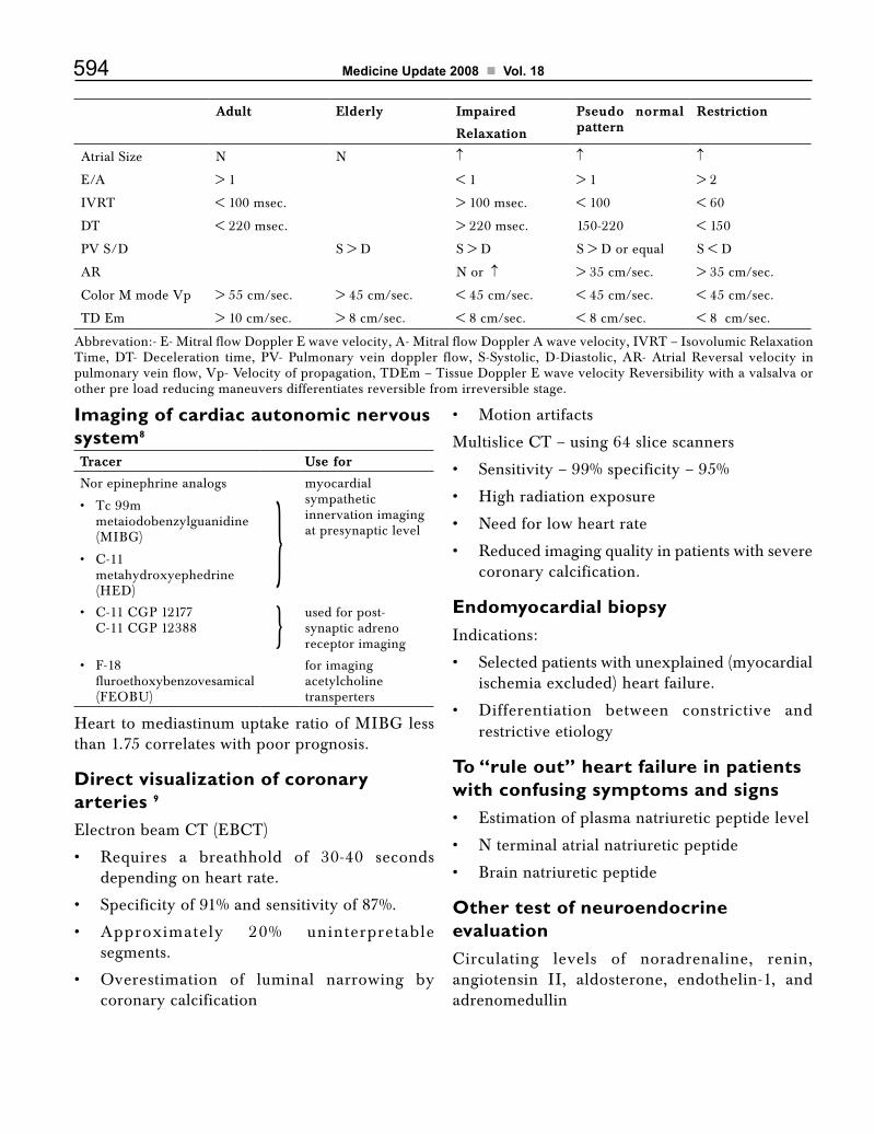
594 Medicine Update 2008 Vol. 18
Imaging of cardiac autonomic nervous system8
Tracer Use for
Nor epinephrine analogs
• Tc99mmetaiodobenzylguanidine (MIBG)
• C-11metahydroxyephedrine (HED)
}myocardial sympathetic innervation imaging at presynaptic level
C-11CGP12177 •C-11CGP12388 } used for post-
synaptic adreno receptor imaging
F-18 •fluroethoxybenzovesamical (FEOBU)
for imaging acetylcholine transperters
Heart to mediastinum uptake ratio of MIBG less than 1.75 correlates with poor prognosis.
Direct visualization of coronary arteries 9
ElectronbeamCT(EBCT)
• Requires a breathhold of 30-40 secondsdepending on heart rate.
• Specificityof91%andsensitivityof87%.
• Approximately 20% uninterpretablesegments.
• Overestimation of luminal narrowing bycoronary calcification
• Motionartifacts
MultisliceCT–using64slicescanners
• Sensitivity–99%specificity–95%
• Highradiationexposure
• Needforlowheartrate
• Reducedimagingqualityinpatientswithseverecoronary calcification.
Endomyocardial biopsyIndications:
• Selectedpatientswithunexplained(myocardialischemia excluded) heart failure.
• Differentiation between constrictive andrestrictive etiology
To “rule out” heart failure in patients with confusing symptoms and signs• Estimationofplasmanatriureticpeptidelevel
• Nterminalatrialnatriureticpeptide
• Brainnatriureticpeptide
Other test of neuroendocrine evaluationCirculating levels of noradrenaline, renin,angiotensin II, aldosterone, endothelin-1, and adrenomedullin
Adult Elderly Impaired
Relaxation
Pseudo normal pattern
Restriction
Atrial Size N N ↑ ↑ ↑
E/A > 1 < 1 > 1 > 2
IVRT < 100 msec. > 100 msec. < 100 < 60
DT < 220 msec. > 220 msec. 150-220 < 150
PV S/D S > D S > D S > D or equal S < D
AR N or ↑ > 35 cm/sec. > 35 cm/sec.
ColorMmodeVp > 55 cm/sec. > 45 cm/sec. < 45 cm/sec. < 45 cm/sec. < 45 cm/sec.
TD Em > 10 cm/sec. > 8 cm/sec. < 8 cm/sec. < 8 cm/sec. < 8 cm/sec.
Abbrevation:- E- Mitral flow Doppler E wave velocity, A- Mitral flow Doppler A wave velocity, IVRT – Isovolumic Relaxation Time, DT- Deceleration time, PV- Pulmonary vein doppler flow, S-Systolic, D-Diastolic, AR- Atrial Reversal velocity in pulmonary vein flow, Vp- Velocity of propagation, TDEm – Tissue Doppler E wave velocity Reversibility with a valsalva or other pre load reducing maneuvers differentiates reversible from irreversible stage.

595Chronic Heart Failure – Newer Diagnostic Modalities
Limitations : Inaccurate and difficult to interpret in individual patient. Diuretics, vasodilators, ACEinhibitorsandbetablockersaffecttheplasmaconcentration of endocrine substances.
• Plasmalevelsofnoradrenalinerisewithageandhealthy subjects over the age of 75 yrs may have plasma concentration in heart failure range.
ConclusionAlthough several advances have occurred in evaluation of chronic heart failure, they have not made any significant impact on mortality. Modalities that can detect subclinical myocardial damage and its precise etiology need to be evolved. Technical advances in the field of nuclear probes and cardiac MRI may solve the problem.
References1. Thorn EM, Mehra MR. Imaging markers of adverse
remodeling : from organ to organelle. Heart Failure Clin 2006; 2:117-127.
2. De Castro S, Caselli S, Maron M et al. Left ventricularremodeling index (LVRI) in various pathophysiological conditions: a real time three dimensional echocardiographic study. Heart 2007;93:205-209.
3. Arrighi JA, Dilsizian V. Nuclear probes for assessing myocardial viability in heart failure. Heart Failure Clin 2006; 2:129-143.
4. Sehinkel AFL, Poldermans D, Elhendy A, Bax JJ. Assessment of myocardial viability in patients with heart failure. J. Nuclear Medicine 2007;48:1135-1146.
5. Raney AR, Bello D. Cardiovascular magnetic resonanceassessment of ischemia and nonischemic cardiomyopathies. Heart Failure Clin 2006;2:145-161.
6. GarciaMJ.Comprehensiveechocardiographicassessmentofdiastolicfunction.HeartFailureClin2006;2:163-178.
7. AbrahamTP,LardoAC,KassDA.Myocardialdyssynchronyand resynchronization. Heart Failure Clin 2006;2:179-192.
8. Higuchi T, Schwaiger M. Non invasive imaging of heart failure: Neuronal dysfunction and risk stratification. Heart Failure Clin 2006;2:193-204.
9. Bax JJ, Poldermans D, Schuijf JD. Saholte AJHA, Elhendy A, Van der Wall EE. Imaging to differentiate between ischemic and non ischemia cardiomyopathy Heart Failure Clin 2006;2:205-214.
CHAPTER
80Advanced Therapy for Refractory Heart failure – Devices and SurgeryS. Ramakrishnan, S. Seth, V. K. Bahl
Heart failure (HF) is a complex syndrome that can result from any structural or functional cardiac disorder that impairs the ability of the heart to function as a pump to support a physiological circulation.1 Medical therapy remains the mainstay of treatment for majority of patients
with heart failure. Although medical therapy can improve the quality of life and the longevity of patients across the spectrum of heart failure symptoms, such therapy alone is insufficient in patients with advanced heart failure.1–3 Advanced heart failure may be defined as stage of heart failure, characterized by advanced structural heart disease and marked symptoms of heart failure at rest despite dietary modification, salt restriction and maximal medical therapy includingACEinhibitors,angiotensinIIreceptorblockers, digitalis, diuretics and beta blockers. These patients require frequent hospitalizations and the overall prognosis is poor.1–3
Various devices have been used in heart failure patients who remain severely symptomatic despite adequate medical therapy including cardiac resynchronisation therapy (CRT), implantablecardioverterdefibrillator(ICD),Combodevice,ultrafiltration and continuous positive airway pressure (CPAP) ventilation (Table 1). Cardiacsupport or replacement with left ventricular assist devices (LVAD) and/or cardiac transplantation are often the only therapeutic alternatives in patients with advanced/end-stage heart failure. This review focuses on the recent advances in device and surgical therapy for advanced heart failure (Table 2).
Table 1 : Approaches in Refractory Heart FailureApproach Modalities
• OptimizeCompromisedHeart Function
Optimal medical therapy, Cardiac Resynchronizationtherapy,ICD
• Reverseremodeling Drugs, CRT, Surgical orinterventional mitral valve repair / annuloplasty, acorn device
• RegeneratetheMyocytes Stem cells, myoblasts, stimulation of endogenous stem cells, gene therapy
• ReplacetheHeart Assist devices as destination t h e r a p y , C a r d i a ctransplantation
• Treatmentofco-morbidities
Antidepressants, Erythropoietin, antianorecticagents,CPAP
• TreatmentofConsequences
Pulmonary hypertension with sildenafil, Volume overload with ultra filtration, Cachexia
• Betterdeliveryofcare Telemedic ine e f for t s , Individualized patient care
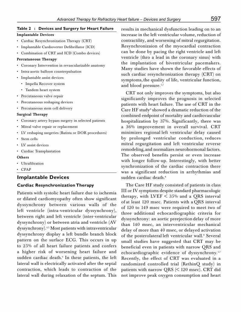
597Advanced Therapy for Refractory Heart failure – Devices and Surgery
Implantable DevicesCardiac Resynchronization Therapy
Patients with systolic heart failure due to ischemia or dilated cardiomyopathy often show significant dyssynchrony between various walls of the left ventricle (intra-ventricular dyssynchrony), between right and left ventricle (inter-ventricular dyssynchrony) or between atria and ventricle (AV dyssynchrony).4,5 Most patients with intraventricular dyssynchrony display a left bundle branch block pattern on the surface ECG. This occurs in upto 25% of all heart failure patients and confers a higher risk of worsening heart failure and sudden cardiac death.6 In these patients, the left lateral wall is electrically activated after the septal contraction, which leads to contraction of the lateral wall during relaxation of the septum. This
results in mechanical dysfunction leading on to an increase in the left ventricular volume, reduction of contractility, and worsening of mitral regurgitation. Resynchronizaion of the myocardial contraction can be done by pacing the right ventricle and left ventricle (thro a lead in the coronary sinus) with the implantation of biventricular pacemakers. Many studies have shown the favorable effects of suchcardiacresynchronizationtherapy(CRT)onsymptoms, the quality of life, ventricular function, and blood pressure.5,7
CRTnotonlyimprovesthesymptoms,butalsosignificantly improves the prognosis in selected patientswithheartfailure.TheuseofCRTintheCareHFstudy8 showed a dramatic reduction of the combined endpoint of mortality and cardiovascular hospitalization by 37%. Significantly, there was a 36% improvement in overall survival. CRTminimizes regional left ventricular delay caused by prolonged ventricular conduction, reduces mitral regurgitation and left ventricular reverse remodeling, and normalizes neurohormonal factors. The observed benefits persist or even increase with longer follow-up. Interestingly, with better synchronization of the cardiac contraction there was a significant reduction in arrhythmias and sudden cardiac death.8
TheCareHFstudyconsistedofpatientsinclassIII or IV symptoms despite standard pharmacologic
therapy, with LVEF < 35% and a QRS interval of at least 120 msec. Patients with a QRS interval of 120 to 149 msec were required to meet two of
three additional echocardiographic criteria for dyssynchrony: an aortic preejection delay of more than 140 msec, an interventricular mechanical
delay of more than 40 msec, or delayed activation of the posterolateral left ventricular wall.8 Several small studies have suggested that CRT may bebeneficial even in patients with narrow QRS and echocardiographic evidence of dyssynchrony.5,7 Recently, the effect of CRT was evaluated in arandomized controlled trial (RethinQ study) in patientswithnarrowQRS(<120msec).CRTdidnot improve peak oxygen consumption and heart
Table 2 : Devices and Surgery for Heart FailureImplantable Devices
• CardiacResynchronizationTherapy(CRT)
• ImplantableCardioverterDefibrillator(ICD)
• CombinationofCRTandICD(Combodevices)
Percutaneous Therapy
• CoronaryInterventioninrevascularizableanatomy
• Intra-aorticballooncounterpulsation
• Implantableassistdevices
• ImpellaRecoversystem
• Tandemheartsystem
• Percutaneousvalverepair
• Percutaneousreshapingdevices
• Percutaneousstemcelldelivery
Surgical Therapy
• Coronaryarterybypasssurgeryinselectedpatients
• Mitralvalverepairorreplacement
• LVreshapingsurgeries(BatistaorDORprocedures)
• Stemcells
• LVassistdevices
• CardiacTransplantation
Others
• Ultrafiltration
• CPAP

598 Medicine Update 2008 Vol. 18
failure worsenings, thereby providing evidence that patients with heart failure and narrow QRS intervals may not benefit from CRT.9 Currentguidelines supporttheuseofCRTinpatientswithan ejection fraction of 35% or less, moderate or severe heart failure (New York Heart Association [NYHA] class III or IV), and a prolonged QRS interval (≥ 120 msec).1-3
Implantable Defibrillator (ICD)
The most common cause of death in patients with advanced heart failure is progressive pump failure and the proportion of sudden cardiac death is less. Hence,ICDsaremoreeffectiveinlessadvancedheart failure, because sudden cardiac death is the main cause of death in less severe heart failure. Even after an appropriate shock, patients with advanced heart failure may die from electromechanical dissociation.10 Such theoretical considerations were proven in the large SCD-HEFT trial.11 Among patients with NYHA class II heart failure, there was a 46 per cent relative reduction in the risk of death withICDtherapyascomparedtoamiodarone.Theabsolute reduction in mortality among patients in NYHA class II was 11.9 per cent at five years. However, in patients with advanced heart failure there was no apparent reduction in the risk of death withICDtherapy.11
Although ICDs are less effective in end-stageHF,CRTandICDmaybecombinedasCRTmayimprove function status, making patients eligible also for ICD therapy. In the COMPANIONtrial,12eitherCRTaloneorCRTwithICD(combodevice) reduced the rate of death from any cause or hospitalization for any cause by approximately 20 per cent as compared with the group that received optimal pharmacologic therapy alone. The addition ofadefibrillatortoCRTdidnotappreciablyaffectthe combined outcomes of death or hospitalization for any cause. However, there was a 36% reduction in the mortality. Hence, whether to institute only CRTorCombodevice shouldbe individualizedand guided by cost, likely survival, and sickness status.10
Percutaneous and Surgical InterventionsAmong the percutaneous and surgical therapies available for advanced heart failure, heart transplantation remains the most effective and proven therapy. The other interventions aim to either repair or reshape the heart, or replace the heart function.
Coronary Revascularization Procedures
Coronary arterydisease is common inpatientswith advanced heart failure, with some studies suggesting a prevalence of 50%-70%.13Coronaryrevascularization with coronary artery bypass surgery or percutaneous coronary intervention as appropriate should be considered in patients with heart failure and suitable coronary anatomy presenting with significant angina, or acute coronary syndrome.1–3 However, this approach has not yet been prospectively tested. Revascularization is also indicated in patients who show evidence of myocardial viability or the presence of inducible ischemia in areas of significant obstructive coronary disease. There are a variety of imaging technics to detect non-contractile but viable myocardium including nuclear imaging, stress echocardiography and magnetic resonance imaging. A few ongoing clinical trials (including STICH trial) areprospectively evaluating the benefit of routine coronary revascularization in patients with heart failure and obstructive coronary artery disease.
Stem Cell Therapy
Myocardial regeneration with either percutaneously or surgically delivered stem cell is promising. Both surgical and non surgical intracoronary stem cell injection is undergoing evaluation at AIIMS and other centers, and the initial results are promising. Improvement in ventricular function and symptoms are shown with autologous bone marrow stem cell injection. Mesenchymal cell injections have also been found to be beneficial. Experimental studies using embryonal cells have shown ability to grow into sacs or rings, which develop the properties of

599Advanced Therapy for Refractory Heart failure – Devices and Surgery
cardiac muscle.14,15
Mitral Valve Interventions
In patients with heart failure, mitral regurgitation occurs commonly due to annular dilation with incomplete coaptation of the mitral leaflets and apical displacement of one or both papillary muscles causing restricted leaflet motion.16 Mitral valve annuloplasty in dilated and ischemic cardiomyopathy is shown to be safe with low mortality (2%) and morbidity.17 Small studies have shown improvement in symptoms, ejection fraction, quality of life and reduction in hospitalizations.16 However, there is no clear survival advantage when compared with propensity-matched patients not undergoing mitral valve annuloplasty.18 Considering the high recurrence rate with ringannuloplasty, some centers advocate mitral valve replacement rather than repair in functional and ischemic cardiomyopathy. However, the impact of mitral valve repair/replacement on quality of life and clinical outcomes has also not clearly been demonstrated.16,19
Percutaneous mitral and/or tricuspid valve repair may provide some benefit in suitable patients with advanced heart failure and the various devices are in early stages of development.19 The devices aim to reproduce the various technics that are used during surgery. The coronary sinus is anatomically very near the mitral annulus. By placing a series of progressively stiffer rods or ‘cinching’ devices in the coronary sinus can move the posterior mitral apparatus forward, thereby reducing the mitral annulus and regurgitation. The other devices aim to remodel the posterior mitral annulus by a transventricular or transatrial approach while still others intend to decrease the septal lateral diameter by either a transventricular or transatrial bridge and tether system.
As per current guidelines isolated mitral valve repair or replacement for severe mitral regurgitation secondary to ventricular dilatation in the presence of severe LV systolic dysfunction is not generally recommended.1–3
Cardiac Reshaping Surgeries
In patients with dilated cardiomyopathy, partial left ventriculectomy (Batista procedure) was a very popular technic some years ago. Despite a sound theoretical basis, Batista procedure is no longer used since the long term results are disappointing.20 In patients with ischemic heart disease with dyskinetic regions of left ventricle, such ventricle reshaping procedures may be of benefit. Aneurysmectomy and endoventricular circular patch plasty (Dor procedure) is a promising technique.21-23 The AssessmentofaCardiacSupportDeviceinPatientswith Heart Failure (ACORN) trial evaluated aninnovative passive cardiac restraint device in patients with end-stage HF that suggested modest improvement in ventricular remodeling but no benefit in mortality.24
LV Assist Devices
LV assist devices (LVADs) improve survival and quality of life in patients ineligible for a heart transplant. LVADs also serve as a “bridge” to transplant and ventricular recovery. Recently LVADs are being used more as end-stage or “destination-therapy”.23,25 In a prospective, multicenter study, 129 end-stage HF patients, ineligible for heart transplantation, were randomized to receive either an LVAD or optimal medical therapy. After 1 year, a 48% reduction in death and improved quality of life were shown with LVAD group as compared to medical therapy group.26 Several new ventricular assist devices are currently undergoing Phase III trials and are eagerly awaited.
CurrentindicationsforLVADsincludepatientsawaiting heart transplantation who have become refractory to all means of medical circulatory support as a bridge to transplant. Permanent mechanical assistance using an implantable assist device may be considered in highly selected patients with severe HF refractory to conventional therapy who are not candidates for heart transplantation, particularly those who cannot be weaned from intravenous inotropic support at an experienced HF center.1–3
Percutaneous implantable devices are useful for
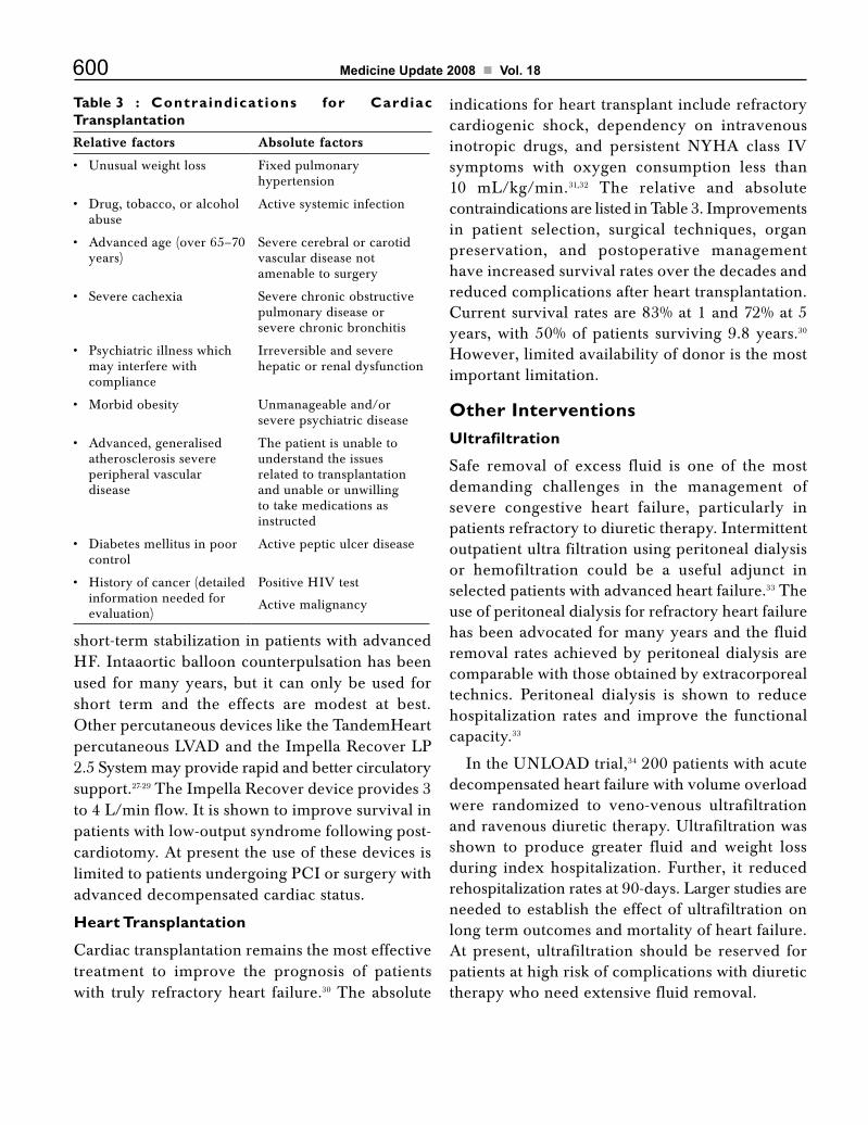
600 Medicine Update 2008 Vol. 18
short-term stabilization in patients with advanced HF. Intaaortic balloon counterpulsation has been used for many years, but it can only be used for short term and the effects are modest at best. Other percutaneous devices like the TandemHeart percutaneous LVAD and the Impella Recover LP 2.5 System may provide rapid and better circulatory support.27-29 The Impella Recover device provides 3 to 4 L/min flow. It is shown to improve survival in patients with low-output syndrome following post-cardiotomy. At present the use of these devices is limitedtopatientsundergoingPCIorsurgerywithadvanced decompensated cardiac status.
Heart Transplantation
Cardiactransplantationremainsthemosteffectivetreatment to improve the prognosis of patients with truly refractory heart failure.30 The absolute
indications for heart transplant include refractory cardiogenic shock, dependency on intravenous inotropic drugs, and persistent NYHA class IV symptoms with oxygen consumption less than 10 mL/kg/min.31,32 The relative and absolute contraindications are listed in Table 3. Improvements in patient selection, surgical techniques, organ preservation, and postoperative management have increased survival rates over the decades and reduced complications after heart transplantation. Currentsurvivalratesare83%at1and72%at5years, with 50% of patients surviving 9.8 years.30 However, limited availability of donor is the most important limitation.
Other InterventionsUltrafiltration
Safe removal of excess fluid is one of the most demanding challenges in the management of severe congestive heart failure, particularly in patients refractory to diuretic therapy. Intermittent outpatient ultra filtration using peritoneal dialysis or hemofiltration could be a useful adjunct in selected patients with advanced heart failure.33 The use of peritoneal dialysis for refractory heart failure has been advocated for many years and the fluid removal rates achieved by peritoneal dialysis are comparable with those obtained by extracorporeal technics. Peritoneal dialysis is shown to reduce hospitalization rates and improve the functional capacity.33
In the UNLOAD trial,34 200 patients with acute decompensated heart failure with volume overload were randomized to veno-venous ultrafiltration and ravenous diuretic therapy. Ultrafiltration was shown to produce greater fluid and weight loss during index hospitalization. Further, it reduced rehospitalization rates at 90-days. Larger studies are needed to establish the effect of ultrafiltration on long term outcomes and mortality of heart failure. At present, ultrafiltration should be reserved for patients at high risk of complications with diuretic therapy who need extensive fluid removal.
Table 3 : Contraindications for Cardiac TransplantationRelative factors Absolute factors
• Unusualweightloss Fixed pulmonary hypertension
• Drug,tobacco,oralcoholabuse
Active systemic infection
• Advancedage(over65–70years)
Severe cerebral or carotid vascular disease not amenable to surgery
• Severecachexia Severe chronic obstructive pulmonary disease or severe chronic bronchitis
• Psychiatricillnesswhichmay interfere with compliance
Irreversible and severe hepatic or renal dysfunction
• Morbidobesity Unmanageable and/or severe psychiatric disease
• Advanced,generalisedatherosclerosis severe peripheral vascular disease
The patient is unable to understand the issues related to transplantation and unable or unwilling to take medications as instructed
• Diabetesmellitusinpoorcontrol
Active peptic ulcer disease
• Historyofcancer(detailedinformation needed for evaluation)
Positive HIV test
Active malignancy

601Advanced Therapy for Refractory Heart failure – Devices and Surgery
CPAP
A significant number of patients with advanced heart failure have obstructive sleep apnea. Continuouspositiveairwaypressure(CPAP)isaneffectivetreatmentforsleepapnea.Hence,CPAPhas been evaluated as a therapy in advanced HF patients with sleep apnea. Small prospective controlledtrialshaveshownthatCPAPimprovesLV EF, reduce urinary norepinephrine levels, and improve cardiac output. In a recent trial, a 3 month treatmentwithCPAPis shownto increaseLVEFwhen compared to sham-CPAP. However, thebeneficial effect was not marked in patients with LVEF < 30% and in patients with predominantly Cheyne-Stokesevents.35, 36
References1. National Institute for Clinical Excellence. Chronic heart
failure. Management of chronic heart failure in adults in primaryandsecondarycare.Clinicalguideline5.London:NICE,2003.
2. HuntSA,AbrahamWT,ChinMH,etal.ACC/AHA2005Guideline Update for the Diagnosis and Management of ChronicHeartFailureintheAdult:areportoftheAmericanCollege of Cardiology/American Heart Association TaskForce on Practice Guidelines. Circulation 2005; 112:e154-235.
3. SwedbergK,ClelandJ,DargieH,etal.Guidelinesforthediagnosis and treatment of chronic heart failure: executive summary: The Task Force for the Diagnosis and Treatment of ChronicHeartFailureoftheEuropeanSocietyofCardiology.Eur Heart J 2005; 26:1115–40.
4. Bader H, Garrigue S, Lafitte S, et al. Intra-left ventricular electromechanical asynchrony. A new independent predictor of severe cardiac events in heart failure patients. J Am Coll Cardiol 2004; 43:248–56.
5. SticherlingC,SchaerB,CoenenM,KühneM,AmmannP,Osswald S. Cardiac resynchronization therapy in chronicheart failure. Swiss Med Wkly 2006; 136:611-17.
6. Baldasseroni S, Opasich C, Gorini M, et al. Left bundle-branch block is associated with increased 1-year sudden and total mortality rate in 5517 outpatients with congestive heart failure: a report from the Italian network on congestive heart failure. Am Heart J 2002; 143:398–405.
7. McAlister FA, Ezekowitz J, Hooton N, et al. Cardiacresynchronization therapy for patients with left ventricular systolic dysfunction: a systematic review. JAMA 2007; 297(22):2502-14.
8. Cleland JG, Daubert JC, Erdmann E, et al; CardiacResynchronization-Heart Failure (CARE-HF) StudyInvestigators. The effect of cardiac resynchronization on
morbidity and mortality in heart failure. N Engl J Med 2005; 352(15):1539-49.
9. Beshai JF, Grimm RA, Nagueh SF, et al; the RethinQ Study Investigators.Cardiac-ResynchronizationTherapyinHeartFailurewithNarrowQRSComplexes.N Engl J Med 2007;Epub ahead of print
10. Leibundgut G, Brunner-La Rocca HP. End-stage chronic heart failure. Swiss Med Wkly 2007; 137:107-13.
11. BardyGH,LeeKL,MarkDB,etal;SuddenCardiacDeathinHeartFailureTrial(SCD-HeFT)Investigators.Amiodaroneor an implantable cardioverter-defibrillator for congestive heart failure. N Engl J Med 2005; 352(3):225-37.
12. BristowMR,SaxonLA,Boehmer J, et al;ComparisonofMedical Therapy, Pacing, and Defibrillation in Heart Failure (COMPANION) Investigators.Cardiac-resynchronizationtherapy with or without an implantable defibrillator in advanced chronic heart failure. N Engl J Med 2004; 350(21):2140-50.
13. Nohria A, Lewis E, Stevenson LW. Medical management of advanced heart failure. JAMA 2002; 287:628-40.
14. AiranB,TalwarS,ChoudharySK,etal.Applicationofstemcell technology for coronary artery disease at the All India Institute of Medical Sciences, New Delhi, India. Heart Surg Forum 2007; 10(3):E231-34.
15. SethS,NarangR,BhargavaB,etal;AIIMSCardiovascularStemCellStudyGroup.Percutaneousintracoronarycellularcardiomyoplasty for nonischemic cardiomyopathy: clinical and histopathological results: the first-in-man ABCD(AutologousBoneMarrowCellsinDilatedCardiomyopathy)trial. J Am Coll Cardiol 2006; 48(11):2350-51.
16. GormanJH3rd,RyanLP,GormanRC.Pathophysiologyofischemic mitral insufficiency: does repair make a difference? Heart Fail Rev 2006; 11:219-29.
17. Bolling SF, Pagani FD, Deeb GM, Bach DS. Intermediate term outcome of mitral reconstruction in cardiomyopathy. J Thorac Cardiovas Surg 1998;115:381–88.
18. Wu AH, Aaronson KD, Bollins SF, et al. Impact of mitral valve annuloplasty on mortality risk in patients with mitral regurgitation and left ventricular systolic dysfunction. J Am Coll Cardiol 2005; 45:381–87.
19. Mack MJ. New techniques for percutaneous repair of the mitral valve. Heart Fail Rev. 2006; 11:259-68.
20. StarlingR,McCarthyP,BudaT,etal.Resultsofpartialleftventriculectomy for dilated cardiomyopathy: hemodynamic, clinical and echocardiographic observations. J Am Coll Cardiol 2000; 36:2098-103.
21. Dor V. Left ventricular reconstruction for ischemic cardiomyopathy. J Card Surg 2002; 17:180-87.
22. CalafioreA,GallinaS,DiMauroM,etal.Leftventricularaneurysmectomy: endoventricular circular patch plasty or septoexclusion. J Card Surg 2003; 18:93-100.
23. CrespoLeiroMG,PaniaguaMartínMJ. [Managementofadvanced or refractory heart failure] Rev Esp Cardiol 2004; 57(9):869-83.

602 Medicine Update 2008 Vol. 18
24. Acker MA, Bolling S, Shemin R, et al; Acorn Trial Principal InvestigatorsandStudyCoordinators.Mitralvalvesurgeryinheartfailure:insightsfromtheAcornClinicalTrial.J Thorac Cardiovasc Surg 2006;132(3):568-77.
25. Feldman D, Menachemi DM, Abraham WT, Wexler RK. Management Strategies for Stage-D Patients with Acute Heart Failure. Clin Cardiol 2007 Oct; [Epub ahead of print]
26. Stevenson LW, Miller LW, Desvigne-Nickens P, et al; REMATCHInvestigators.Leftventricularassistdeviceasdestination for patients undergoing intravenous inotropic therapy:a subset analysis fromREMATCH(RandomizedEvaluationofMechanicalAssistanceinTreatmentofChronicHeart Failure). Circulation. 2004 Aug 24;110(8):975-81.
27. Bautista-Hernández V, Gutiérrez F, Pinar E, et al. [Initial experience with the Impella left ventricular assist device for postcardiotomy cardiogenic shock and unprotected left coronary artery angioplasty in patients with a low left ventricular ejection fraction] Rev Esp Cardiol 2007;60(9):984-7.
28. Thiele H, Sick P, Boudriot E, et al. Randomized comparison of intra-aortic balloon support with a percutaneous left ventricular assist device in patients with revascularized acute myocardial infarction complicated by cardiogenic shock. Eur Heart J 2005; 26(13):1276-83.
29. Cohen RE, Domniez T, Elhadad S. High-Risk Left MainCoronary Stenting Supported by Percutaneous ImpellaRecover LP 2.5 Assist Device. J Invasive Cardiol 2007; 19(10):E294-E296.
30. Taylor DO, Edwards LB, Boucek MM, et al. Registry of the
International Society for Heart and Lung Transplantation: twenty-second official adult heart transplant report – 2005. J Heart Lung Transplant 2005; 24:945–55.
31. AaronsonKD,SchwartzJS,ChenTM,etal.Developmentand prospective validation of a clinical index to predict survival in ambulatory patients referred for cardiac transplant evaluation. Circulation 1997; 95:2660–67.
32. Mehra MR, Kobashigawa J, Starling R, et al. Listing criteria for heart transplantation: International Society for Heart and Lung Transplantation guidelines for the care of cardiac transplant candidates – 2006. J Heart Lung Transplant 2006; 25:1024–42.
33. Guglin M, Polavaram L. Ultrafiltration in heart failure. Cardiol Rev 2007;15(5):226-30.
34. CostanzoMR,GuglinME,SaltzbergMT,etal;UNLOADTrial Investigators. Ultrafiltration versus intravenous diuretics for patients hospitalized for acute decompensated heart failure. J Am Coll Cardiol 2007; 49(6):675-83.
35. EgeaCJ,AizpuruF,PintoJA,etal.CardiacfunctionafterCPAPtherapyinpatientswithchronicheartfailureandsleepapnea: A multicenter study. Sleep Med 2007; [Epub ahead of print]
36. Olson LJ, Somers VK. Sleep apnea: implications for heart failure. Curr Heart Fail Rep 2007;4(2):63-69.
CHAPTER
81Acute Renal Failure : Classification and Management StrategiesM. S. Amaresan
IntroductionOver the last few years considerable initiative has been taken towards better understanding of acute renal failure beginning with revision of the terminology itself as acute kidney Injury (AKI). The term AKI has been favored on the basis of the fact that the condition does not always result in kidney failure. AKI is a common condition associated with increased morbidity and mortality, yet a reversible
condition, if identified early and aggressively managed. Drawing of evidence based management guidelines has been impeded by the lack of uniform and well-defined criteria for describing the condition. This has made it impossible to analyse the outcome data of the various published data in a meaningful way. This article proposes to give the consensus definition and classification of AKI, the etiological classification of AKI, the scope for prevention of AKI and the rethinking on the mode of renal replacement therapy (RRT) for AKI.
AKI is defined by an abrupt (within 48 hours) increase in serum creatinine, resulting from an injury or insult that causes a functional or structural change in the kidney. The Acute Dialysis Quality Initiative (ADQI) represents the efforts of a workgroup to develop consensus and evidence based statements in the field of AKI. A consensus definition of AKI evolved by them is by using a set of criteria –RIFLE 1
Rifle CriteriaA revision of the criteria was proposed by the
Acute Kidney Injury Network (AKIN) - a group representing members of Acute Dialysis Quality Initiative, nephrology and critical care societies.
The proposed diagnostic criteria for AKI is an abrupt (within 48 hours) reduction in kidney
Table 1 : Risk, Injury, Failure, Loss, and End-stage Kidney (RIFLE) ClassificationClass Glomerular filtration
rate criteriaUrine output criteria
Risk Increase in Serum creatinine × 1.5
GFR decrease > 25%
< 0.5 ml/kg/hour × 6 hours
Injury Increase in Serum creatinine × 2
GFR decrease > 50 %
< 0.5 ml/kg/hour × 12 hours
Failure Serum creatinine × 3, or serum creatinine ≥ 4 mg/dl with an acute rise > 0.5 mg/dl
GFR decrease > 75%
< 0.3 ml/kg/hour × 24 hours, or anuria × 12 hours
Loss Persistent acute renal failure = complete loss of kidney function > 4 weeks
End-stage kidney disease
End-stage kidney disease > 3 months

604 Medicine Update 2008 Vol. 18
function defined as an absolute increase in serum creatinine (level of > 26.4 mmol/L (0.3 mg/dl) OR a percentage increase in serum creatinine level of > 50% (1.5 fold from baseline) OR a reduction in urine output (documented oliguria of < 0.5 ml/kg/hour for > 6 hours. (These criteria should be applied in the context of the clinical presentation and following adequate fluid resuscitation when applicable.)
Revised Rifle Criteria
Class Serum creatinine criteria
Urine output criteria
Risk Increase of > 26.4 mmol/L (0.3 mg/dl)
Or to 150 –200% of baseline (1.5 to 2.0 fold)
< 0.5 ml/kg/hour
> 6 hours
Injury Increase to > 200-300% of baseline ( > 2-3 fold)
< 0.5 ml/kg/hour
> 12 hours
Failure Increase to > 300% of baseline (> 3fold;or serum creatinine > 354mmol/l (4.0 mg/dl) with an acute rise of atleast 44 mmol/l (0.5 mg/dl)
< 0.3 ml/kg/hour for 24 hours, or anuria for 12 hours
While retaining the emphasis on changes in serum creatinine and urine output as in RIFLE classification, the Loss and End Stage renal Disease categories were removed, as they are outcomes of AKI itself. Stage 1 criteria represent the new diagnostic criteria of AKI. Only one criterion (creatinine or urine output) needs to be fulfilled to qualify for a stage. Patients who receive RRT are considered to have met the criteria for Stage 3 irrespective of the stage that they are in at the time of commencement of RRT.
This staging system for AKI is intended to define the degree of renal dysfunction at the time of diagnosis. Urine output was included as a diagnostic criterion as in intensive care patients it reflects renal dysfunction before the onset of changes in serum creatinine. However the hydration status, use of diuretics and presence of obstruction can influence urine volume.
Classification of AKI by EtiologyAKI can be classified by its etiology into prerenal, renal (intrinsic) and postrenal failure.
Prerenal AKI occurs in the clinical settings leading to volume depletion, decreased effective blood volume (congestive heart failure, cirrhosis, nephrotic syndrome, sepsis), renal vasoconstriction (hepatorenal syndrome, NSAID associated),alteredrenalhemodynamics(ACEI&ARB associated) and increased renal vein pressure (Abdominal compartment syndrome). Prerenal AKI should be anticipated in trauma patients, post liver transplant, mechanical limitation of abdominal wall caused by tight surgical closure or burn injuries, bowel obstruction, pancreatitis. A prospective study by Hou et al found prerenal azotemia to be the single most common cause of AKI in a medical surgical hospital.2 Liano found prerenal AKI responsible for 48% of community acquired AKI and 58% of hospital acquired AKI.2 Volume depletion and congestive heart failure increased the odds ratio of hospital acquired AKI by 9.4 and 9.2 fold respectively.2
Not only is prerenal azotemia common but also it is potentially reversible.
Post renal failure is due to obstructive pathology that may be extrinsic (retro peritoneal, pelvic) or intrinsic (blood clot, calculus, carcinoma), upper or lower (prostate, urethral stricture, Neurogenic bladder, bladder carcinoma). In several series, obstructive pathology is encountered in 2% to 10 % of all cases of AKI.2 The cause and incidence of obstructive pathology depends upon the age of
Classification of the Etiologies of AcuteRenal Failure
AcuteRenalFailure
PrerenalARF
IntrinsicARF
PostrenalARF
AcuteTubar
Necrosis
AcuteInterstitialNephritis
AcuteGN
AcuteVascular
Syndromes
Intratubularobstruction
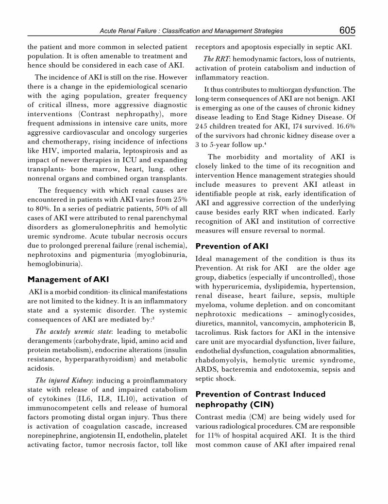
605Acute Renal Failure : Classification and Management Strategies
the patient and more common in selected patient population. It is often amenable to treatment and hence should be considered in each case of AKI.
The incidence of AKI is still on the rise. However there is a change in the epidemiological scenario with the aging population, greater frequency of critical illness, more aggressive diagnostic interventions (Contrast nephropathy), morefrequent admissions in intensive care units, more aggressive cardiovascular and oncology surgeries and chemotherapy, rising incidence of infections like HIV, imported malaria, leptospirosis and as impactofnewertherapiesinICUandexpandingtransplants- bone marrow, heart, lung. other nonrenal organs and combined organ transplants.
The frequency with which renal causes are encountered in patients with AKI varies from 25% to 80%. In a series of pediatric patients, 50% of all cases of AKI were attributed to renal parenchymal disorders as glomerulonephritis and hemolytic uremic syndrome. Acute tubular necrosis occurs due to prolonged prerenal failure (renal ischemia), nephrotoxins and pigmenturia (myoglobinuria, hemoglobinuria).
Management of AKI AKI is a morbid condition- its clinical manifestations are not limited to the kidney. It is an inflammatory state and a systemic disorder. The systemic consequences of AKI are mediated by:3
The acutely uremic state: leading to metabolic derangements (carbohydrate, lipid, amino acid and protein metabolism), endocrine alterations (insulin resistance, hyperparathyroidism) and metabolic acidosis.
The injured Kidney: inducing a proinflammatory state with release of and impaired catabolism of cytokines (IL6, IL8, IL10), activation of immunocompetent cells and release of humoral factors promoting distal organ injury. Thus there is activation of coagulation cascade, increased norepinephrine, angiotensin II, endothelin, platelet activating factor, tumor necrosis factor, toll like
receptors and apoptosis especially in septic AKI.
The RRT: hemodynamic factors, loss of nutrients, activation of protein catabolism and induction of inflammatory reaction.
It thus contributes to multiorgan dysfunction. The long-term consequences of AKI are not benign. AKI is emerging as one of the causes of chronic kidney disease leading to End Stage Kidney Disease. Of 245 children treated for AKI, 174 survived. 16.6% of the survivors had chronic kidney disease over a 3 to 5-year follow up.4
The morbidity and mortality of AKI is closely linked to the time of its recognition and intervention Hence management strategies should include measures to prevent AKI atleast in identifiable people at risk, early identification of AKI and aggressive correction of the underlying cause besides early RRT when indicated. Early recognition of AKI and institution of corrective measures will ensure reversal to normal.
Prevention of AKIIdeal management of the condition is thus its Prevention. At risk for AKI are the older age group, diabetics (especially if uncontrolled), those with hyperuricemia, dyslipidemia, hypertension, renal disease, heart failure, sepsis, multiple myeloma, volume depletion. and on concomitant nephrotoxic medications – aminoglycosides, diuretics, mannitol, vancomycin, amphotericin B, tacrolimus. Risk factors for AKI in the intensive care unit are myocardial dysfunction, liver failure, endothelial dysfunction, coagulation abnormalities, rhabdomyolyis, hemolytic uremic syndrome, ARDS, bacteremia and endotoxemia, sepsis and septic shock.
Prevention of Contrast Induced nephropathy (CIN)Contrast media (CM) are being widely used forvariousradiologicalprocedures.CMareresponsiblefor 11% of hospital acquired AKI. It is the third most common cause of AKI after impaired renal
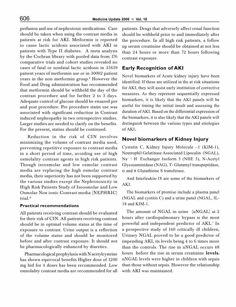
606 Medicine Update 2008 Vol. 18
perfusionanduseofnephrotoxicmedications.Careshould be taken when using the contrast media in patients at risk for AKI. Metformin is reported to cause lactic acidosis associated with AKI in patients with Type II diabetes. A meta analysis bytheCochranlibrarywithpooleddatafrom176comparative trials and cohort studies revealed no cases of fatal or nonfatal lactic acidosis in 35619 patient years of metformin use or in 30002 patient years in the non metformin group.5 However the Food and Drug administration has recommended that metformin should be withheld the day of the contrast procedure and for further 2 to 3 days. Adequate control of glucose should be ensured pre and post procedure. Pre procedure statin use was associatedwith significant reduction inContrastinduced nephropathy in two retrospective studies. Larger studies are needed to clarify on the benefits. For the present, statins should be continued.
Reduction in the risk of CIN involvesminimizing the volume of contrast media used, preventing repetitive exposure to contrast media in a short period of time, avoiding use of high osmolality contrast agents in high risk patients. Though isoosmolar and low osmolar contrast media are replacing the high osmolar contrast media, their superiority has not been supported by the various studies except the Nephrotoxicity in High Risk Patients Study of Isoosmolar and Low OsmolarNonionicContrastmedia(NEPHRIC)trial.6
Practical recommendations
All patients receiving contrast should be evaluated fortheirriskofCIN.Allpatientsreceivingcontrastshould be in optimal volume status at the time of exposure to contrast. Urine output is a reflection of the volume status and should be monitored before and after contrast exposure. It should not be pharmacologically enhanced by diuretics.
Pharmacological prophylaxis with N acetylcysteine has shown equivocal benefits Higher dose of 1200 mg bid for 4 doses has been recommended. Low osmolality contrast media are recommended for all
patients. Drugs that adversely affect renal function should be withheld prior to and immediately after the procedure. In all high risk patients, a follow up serum creatinine should be obtained at not less than 24 hours or more than 72 hours following contrast exposure.
Early Recognition of AKINovel biomarkers of Acute kidney injury have been identified. If these are utilized in the at risk situations for AKI, they will assist early institution of corrective measures. As they represent sequentially expressed biomarkers, it is likely that the AKI panels will be useful for timing the initial insult and assessing the duration of AKI. Based on the differential expression of the biomarkers, it is also likely that the AKI panels will distinguish between the various types and etiologies of AKI.
Novel biomarkers of Kidney Injury Cystatin C, Kidney Injury Molecule –1 (KIM-1), Neutrophil Gelatinase Associated Lipocalin (NGAL), Na+ / H+ Exchanger Isoform 3 (NHE 3), N-Acetyl Glycosaminidase (NAG), ϒ- Glutamyl transpeptidase, α and π Glutathione S transferase.
And Interleukin-18 are some of the biomarkers of AKI.
The biomarkers of promise include a plasma panel (NGAL and cystitis C) and a urine panel (NGAL, IL-18 and KIM-1.
The amount of NGAL in urine (uNGAL) at 2 hours after cardiopulmonary bypass is the most powerful and independent predictor of AKI. 7 In a prospective study of 140 critically ill children, Urinary NGAL proved to be a good predictor of impending AKI, its levels being 4 to 6 times more than the controls. The rise in uNGAL occurs 48 hours before the rise in serum creatinine levels. uNGAL levels were higher in children with sepsis than those without sepsis. However the relationship with AKI was maintained.

607Acute Renal Failure : Classification and Management Strategies
Approach to AKIBefore instituting measures to treat AKI it is important to identify
• whether the AKI is really acute or maskinga chronic kidney disease, History of diabetes mellitus, hypertension, glomerulonephritis or kidney disease, ultrasonographically small contracted kidneys, urinalysis with broad casts-more than 2 to 3 whiteblood cells in diameter, low carbamylated hemoglobin suggest presence of chronic kidney disease.
• whether the AKI is prerenal, renal or postrenal?
The priorities in treating AKI are to optimize fluid balance, treat underlying causes and institute RRT at the appropriate time.
Non Dialytic TherapyNon-dialytic interventions in the management of AKI include restoration of euvolemic status with
crystalloid or colloids and correcting the metabolic derangements.
Pharmacological interventions with dopamine, fenoldopam. Thyroxine, Insulin like growth factor-1, loop diuretics, atrial natriuretic peptide have been found to be promising in animal studies but have failed to make a statistically significant impact in human studies. The poor results may be linked to the time interval between occurrence of AKI and the intervention .
The results of the metaanalysis of the role of loop diuretics show that frusemide has no clinical benefit in the prevention or treatment of established AKI. Its use may increase the risk of ototoxicity.8
The use of dopamine is associated with impaired splanchnic perfusion, increased risk of gram-negative bacteremia and an increased incidence of arrhythmias, particularly atrial fibrillation in the post open-heart patients. Hence there is no role for dopamine in the treatment of AKI.9
Medical management includes tight control of blood sugars besides close monitoring of the volume status, renal biochemical parameters and electrolytes.ProteinkinaseCisausefulagentinsystemic inflammatory reaction syndrome (SIRS) inducedARFinICU.Thebackboneoftreatmentof ARF remains adequate supportive care, maintenance of renal perfusion pressure (MAP > 80 mm Hg), avoidance of future nephrotoxic insults and provision of renal replacement therapy.
Emerging pharmacological agents for treatment of AKI are antiapototic and antinecrotic agents (caspase inhibitors – nonselective and selective against Caspase 1, 3, and 7, PARP inhibitors,minocycline); anti-inflammatory (IL-10, activated proteinKinaseC,iNOSinhibitor),antisepsisagents(insulin,activatedproteinkinaseC),growthfactors(recombinant erythropoietin, hepatocyte growth factor) and vasodilators (Endothelin antagonists, ANP).10
Table 2 : Laboratory Values in Acute Kidney Injury
Prerenal Intrinsic Renal
FENa, per cent* < 1 > 1
BUN to creatinine ratio > 20:1 10 to 20:1
Urine specific gravity > 1.020 1.010 to 1.020
Urine osmolality, mOsm per kg
> 500 300 to 500
Urine sodium concentration, mEq per L (mmol per L)
< 10 (10) > 20 (20)
Urine sediment Hyaline casts
Granular casts
FENa = fractional excretion of sodium; BUN = blood urea nitrogen.
*-FENa is calculated as follows:
FENa = UUUrine sodium ÷ plasma sodiumUU × 100
Urine creatinine ÷ plasma creatinine
note: A prerenal FENa of greater than 1 per cent can occur in patients receiving chronic diuretic therapy or in patients with acute renal failure superimposed on chronic renal failure. Conversely, an intrarenal FENa of less than 1 per cent can occur with radiocontrast nephropathy and rhabdomyolysis
608 Medicine Update 2008 Vol. 18
Dialytic therapy Dialysis is one of the cornerstones of AKI treatment. Initiation of renal replacement therapy (RRT) is recommended when severe derangements in electrolyte concentration (potassium, sodium),volume overload, acid base imbalance, pronounced azotemia (BUN more than 100 mg/dl), florid symptoms of uremia (pericarditis, encephalopathy, bleeding, nausea-vomiting) are noted. Options available are peritoneal dialysis (PD), intermittent hemodialysis (IHD) extended daily dialysis (EDD), slow low efficiency dialysis (SLED) and continuous renal replacement therapy(CRRT). PDis lesspreferreddue to poor delivery of dialysis dose and difficulty in managing ultra filtration. However some trials have shown that continuous peritoneal dialysis has given better results in reducing morbidity and mortality in ICUARFandislessinjurioustoischemickidney.IHD has proved to be beneficial in many of the controlled trials inspite of the fact that it may aggravate the renal injury due to fall in blood pressure. Use of more biocompatible membranes such as polysulfones or polyacrilonitrile improves the outcome of AKI by decreasing complement activation and production of leukotrienes and other cytokines.
With AKI better defined, a recognizable at risk group evolving novel biomarkers to identify AKI early, IHD itself proving to be effective dialytic mode, AKI should become a less morbid condition in the near future. However Patients who had recovered from AKI must be advised to have regular periodical follow up.
In situations of hypovolemia, hypotension and multiorganfailure(MOF)CRRTispreferred.
RoleofCRRT:CRRThaspotentialbenefitsbyan increased hemodynamic tolerance of dialysis, improved ability to manage fluid and electrolyte balance, improved dialytic dose delivery and use of synthetic(more biocompatible) membranes. It maintains consistent homeostasis through slow gradual shifts in volume status and serum osmolality and permits continuous control of fluid balance. There is lesser need for escalation of vasopressor
therapy and lower incidence of arrhythmias It reduces need to restrict fluid administration, requires a lower volume of blood to be circulating outside the bag, has less effect on complements or leukocytes and has greater clearance of mid molecular weight solutes.CAVH(continuousarteriovenoushemofiltration),CAVHD(continuousarteriovenoushemodialysis),CAVHDF(continuousarteriovenoushemodiafiltration),CVVH(continuousvenovenoushemofiltration),CVVHD(Continuousvenovenoushemodialysis),CVVHDF(Continuousvenovenoushemodiafiltration) SCUF (slow continuousultrafiltration)aredifferentmodesofCRRT.NoparticularformofCRRThasyetbeenshownasasuperior option.
InspiteoftheadvantagesofCRRT,IHDremainsa more practical option (except in select clinical situations of hemodynamically unstable ARF patients) in our Indian scenario considering the additionalcostsinvolvedinCRRT. 11
Acute Renal Failure is better termed as Acute kidney injury. The consensus definition of AKI, consensus criteria for classifying AKI and formation of AKIN will enable meaningful intepretation of data available and concrete prospective studies to lay down evidence based recommendations in the management strategies. Identification of the risk group and use of the novel biomarkers to detect early AKI will assist prevention of AKI, institution of measures to reverse AKI to normal. This will enable us to reduce the high morbidity and mortality existing in AKI. Greater awareness and preventive management per se will ensure reduction in incidence of CIN and AKI. EarlyInitiation of RRT will assist complete recovery. IHD is an acceptable mode of RRT among various RRTavailable. In selectpatientsofAKI in ICUwho are suffering from SIRS and hemodynamically unstable,CRRTwillbethetreatmentofchoice.
References1. Molitoris BA, Levin A, Warnock DJ, Joannidis M, Mehta RL,
Kellum JA, et al. Improving outcomes of acute kidney injury: report of an initiative; Nature clinical practice Nephrology 2007 Aug ;3 (8): 439-42.

609Acute Renal Failure : Classification and Management Strategies
2. LeeVWS, Harris D, Anderson J,Schrier RW. Acute Renal Failure in Robert W. Schrier, editor. Diseases of the Kidney and Urinary Tract. 8th ed. Lippincott Williams and Wilkins;2007. p 986-1025.
3. Drumi W.Acute Renal failure is not a ‘cute’ renal failure. Intensive care medicine 2004 october;30(10) 1886-90.
4. Andreoli P. Are children with a history of acute kidney Injury at risk of later Kidney Disease? Nature clinical practice Nephrology 2006 2: 439-42.
5. ErleyC.Concomitantdrugswithexposuretocontrastmedia.Kidney Int 2006; 69 : S20- 22.
6. SolomonR,BigouriC,BettmannN.SelectionofContrastmedia. Kidney Int 2006; 69 : S39- 45.
7. Mishra J,DentC,TarabishiR,MitsnefesMM,KellyCetal. Neutrophil gelatinase associated lipocalin ( NGAL) as a
biomarker for acute renal injury after cardiac surgery. Lancet 2005; 365: 1231-8
8. Ho KM, Sheriden DJ. Metaanalysis of frusemide to prevent or treat acute renal failure. BMJ 2006; 333:420-425.
9. Freidrick JO, AdhikariN,Herridge MS, Beyrere J. Low dose Dopamine Ineffective for Acute Kidney Failure. Ann. Internal Med.2005;142: 510-24.
10. Jo SK, Rosner MH, Okusa MD. Pharmacological treatment of Acute Kidney Injury: Why Drugs Haven’t Worked and What is On the Horizon. Clin J Am Soc Nephrol 2007;2: 356-65
11. VinsonneauC,CamusC,CombesA,CostaDE,BeauregardMA,KloucheMA,BoulaisTetal.ContinuousVenovenoushemodiafiltration Versus Intermittent Hemodialysis for Acute renal failure patients with Multiorgan dysfunction Syndrome- a multicentric randomized trial. Lancet 2006; 368: 329-85.

CHAPTER
82Renal Replacement Therapy in Acute Renal FailureR. Deshpande
IntroductionAcute renal failure (ARF) is defined as an abrupt decrease in renal function sufficient to result in retention of nitrogenous waste in the body. There is no consensus on the degree of rise of serum creatinine or blood urea nitrogen required to define acute renal failure, however commonly used definitions include doubling of serum creatinine from baseline, 30% rise in serum creatinine from baseline and need for renal replacement therapy or dialysis1,2.
Serum Cystatin C appears to be a bettermarker than serum creatinine for detecting smaller increments in glomerular filtration rate however it has not been validated in acute renal failure in a prospective trial.3
The incidence of ARF varies depending on the setting. ARF occurs particularly in high incidence in seriously ill hospitalized patients. The overall mortality increases manifold in patients who develop ARF as compared to matched controls without ARF4.
Etiology of acute renal failureTraditionally ARF has been classified as
Pre renal (usually functional)
Renal (structural) and
Post renal: site of obstruction can vary from intra renal to urethral depending on the pathology.
Acute renal failure is a potentially reversible condition and the exact determination of etiology and treatment directed towards it improves the prognosis.
The role of good clinical examination followed by urine analysis, urine biochemistry, imaging studies and in select cases serologic tests and kidney biopsy to determine the etiology of acute renal failure cannot be overstated.
Clinical settings including multiple organfailure, older age, sepsis, postoperative status, trauma, burns, HIV infection, non-renal solid organ transplantation, heart failure, malignancy, liver diseases and rhabdomyolysis increase the chances of ARF and it is imperative to monitor renal function closely in these settings.
Complications of ARFDetermination of rate of occurrence of complications in patients with ARF is sometimes difficult. However the chances of cardiovascular complications including volume overload, congestive heart failure, cardiac tamponade, and hyperkalemia induced cardiac arrest are increased in ARF.4,5,6
Other complications of acute renal failure include respiratory failure, upper gastrointestinal

611Renal Replacement Therapy in Acute Renal Failure
bleeding, anorexia, vomiting, acute pancreatitis, anemia, coagulation abnormalities, neurological complications and sepsis.4,7
Conservative treatment of acute renal failureThe principles include
• Exclude reversible/ treatable causesof acuterenal failure
• Maintaineuvolemia
• Monitordrugdosagesandmodifydrugdose/dosing intervals according to renal function assessment.
• Maintainadequatenutrition
• Monitorandtreatforclinicalandbiochemicalcomplications
• Instituterenalreplacementtherapy(RRT)whenappropriate.
Renal replacement therapy in acute renal failureThe issues regarding renal replacement therapy in ARF are the source of much debate and investigation. The main questions that need to be answered include:
• WhentostartRRT
• ChoiceregardingmodalityofRRT
• Choiceofdialysermembrane
• Doseofdialysis
When to start RRT?Absolute indications for initiation of dialysis include persistent hyperkalemia, fluid overload resulting in pulmonary edema, ongoing marked acidemia, uremic pericarditis and encephalopathy. However a general trend towards earlier renal replacement therapy has occurred over the past decade based on recent studies.
In a prospective study of cardiac surgical patients, 61 patients were randomized to early or late dialysis.
Early was defined as oliguria unresponsive to diuretics and late was defined as serum creatinine greater than 5 mg/dl and /or serum potassium greater than 5.5 meq/lt. It was found that the early groupstayedlessinICU(7.9versus12days)andhad lower mortality (23 versus 55%).8 In another retrospective trial of 100 trauma patients with ARF, It was found that a lower BUN at initiation of dialysis was associated with earlier commencement of dialysis and better survival9. The earliest data that indicated early dialysis to be superior was fromCongeretalpublishedinJournaloftraumain 1970. It was a randomized prospective trial that established that early and frequent dialysis leads to improvement in mortality and also decreases complications as sepsis and hemorrhage in addition to decreasing the hospital stay10.
However there are some studies that have failed to establish the superiority of early dialysis. One study of critically ill 132 patients with ARF found an inverse relationship between serum creatinine at initiation of hemodialysis and increased mortality11.
Some other clinical advantages of starting dialysis early include ease of optimizing fluid volume balanceandnutritionalstatusinICUpatientswithARF.
Choice regarding modality of RRTGenerally available modes of therapy include conventional intermittent hemodialysis (IHD), Slow low efficiency daily dialysis (SLEDD), Intermittent andcontinuousperitonealdialysis(PD),Continuousarteriovenoushemofiltration(CAVH),continuousarteriovenoushemodialysis(CAVHD),continuousvenovenous hemofiltration (CVVH), continuousvenovenoushemodialysis(CVVHD)andcontinuousvenovenoushemodiafiltration(CVVHDF)
There has been a trend for more use of continuous therapies and less of IHD. PD has fallen out of favor due to problems with peritonitis following peritoneal catheter insertion and the fact that relative efficiency of PD as compared to IHD as regards solute removal is only 10 to 20%.
612 Medicine Update 2008 Vol. 18
Factors that are considered in choosing between different modalities include availability of necessary equipment, expertise demanded by the modality, cost, hemodynamic status, indication for therapy and the speed with which the indication needs to be corrected, availability of vascular access, issues regarding anticoagulation and associated clinical conditions of the patient.
The comparison of different modalities of RRT are shown in the Table 1.
Because of difficulties precisely matching seriously ill patients with ARF, a clear-cut consensus regardingCVVHDversusIHDaspreferablemodeof therapy has not evolved. It is likely that patients with ARF are best served by considering these modalities as complementary than competitive.
A randomized prospective trial of 166 patients by MehtaetalcomparingCVVHDtoIHDfailedtoshow significant differences in mortality between the two groups12. Two systematic reviews by Tonelli et al and Kellum et al13,14 failed to show any difference between intermittent and continuous therapies, however after adjusting for study quality and severity of illness Kellum et al found a decreased relative risk of death with continuous therapies.
ThechoicebetweenCVVHandCVVHD,whichare the commonly practiced continuous modalities, is anopenquestion.CVVHworksprimarilybyconvection while CVVHD works primarily bydiffusion. The proposed benefit of CVVH incytokine removal and hence its superiority in treatment of sepsis has not been established beyond doubt. The counter argument that both good and bad cytokines are removed has not been refuted. CVVHismuchmoreexpensivedueto the largeamount of replacement fluid needed.
Differential methodologies, small numbers of patients, study flaws, lack of standardized approaches to timing and indication of dialytic intervention make comparisons difficult between therapies.
Newer modalities of dialysis that has emerged incorporating advantages of hemodynamic stability ofCRRTcoupledwithhighratesofsoluteandfluidremoval of IHD is SLEDD (slow low efficiency daily dialysis). SLEDD offers better fluid control and more removal of solute than IHD and
Table 1 : Comparison of different RRTType of RRT
Advantages Disadvantages
PD Simple to perform
Inexpensive
No need of anticoagulant therapy
Useful in remote areas
Technical problem due to paralytic ileus which is frequent
Less efficient regarding solute removal
High risk of infection
IHD Simple to perform
Inexpensive
Efficient
Canbeperformedwithout anticoagulation
Not suitable for hypotensive patients
Risk of hemorrhage and hypotension
SLEDD Simple to perform
Less expensive
Very efficient
Better suited for hypotensive patients
Needs anticoagulation
DedicatedICUstaffandtechnicians required
CAVHD Simple to perform
Requires no machine
Efficient
Need arterial and venous catheter insertion
Cannotbeusedinhypotension
Risk of air embolism
Rarely used in clinical practice today
CVVH Efficient
Enhanced removal of solute as it works by convection
Claimedtobesuperior in septic patients
Needs dedicated machine and anticoagulation
Expensive due to large amount of replacement fluid needed
CVVHD Efficient
Canbeusedwithhypotension
Less expensive than CVVH
Needs dedicated machine and anticoagulation
Expensive than SLEDD and IHD
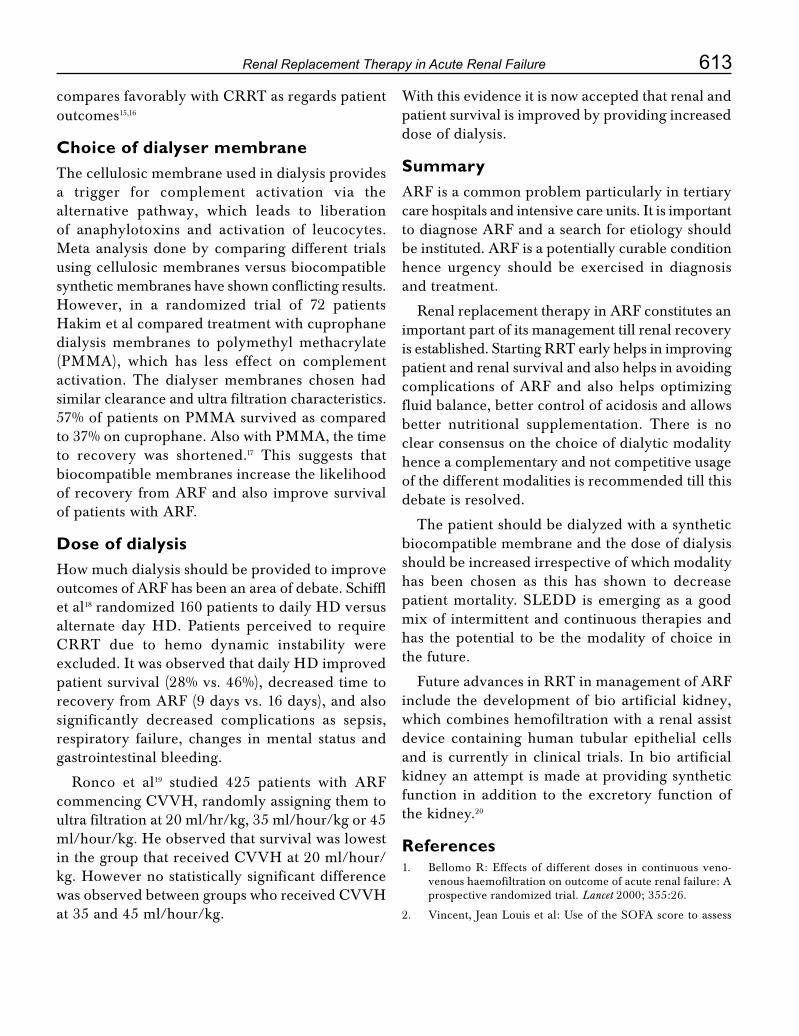
613Renal Replacement Therapy in Acute Renal Failure
comparesfavorablywithCRRTasregardspatientoutcomes15,16
Choice of dialyser membraneThe cellulosic membrane used in dialysis provides a trigger for complement activation via the alternative pathway, which leads to liberation of anaphylotoxins and activation of leucocytes. Meta analysis done by comparing different trials using cellulosic membranes versus biocompatible synthetic membranes have shown conflicting results. However, in a randomized trial of 72 patients Hakim et al compared treatment with cuprophane dialysis membranes to polymethyl methacrylate (PMMA), which has less effect on complement activation. The dialyser membranes chosen had similar clearance and ultra filtration characteristics. 57% of patients on PMMA survived as compared to 37% on cuprophane. Also with PMMA, the time to recovery was shortened.17 This suggests that biocompatible membranes increase the likelihood of recovery from ARF and also improve survival of patients with ARF.
Dose of dialysisHow much dialysis should be provided to improve outcomes of ARF has been an area of debate. Schiffl et al18 randomized 160 patients to daily HD versus alternate day HD. Patients perceived to require CRRT due to hemo dynamic instability wereexcluded. It was observed that daily HD improved patient survival (28% vs. 46%), decreased time to recovery from ARF (9 days vs. 16 days), and also significantly decreased complications as sepsis, respiratory failure, changes in mental status and gastrointestinal bleeding.
Ronco et al19 studied 425 patients with ARF commencingCVVH,randomlyassigningthemtoultra filtration at 20 ml/hr/kg, 35 ml/hour/kg or 45 ml/hour/kg. He observed that survival was lowest inthegroupthatreceivedCVVHat20ml/hour/kg. However no statistically significant difference wasobservedbetweengroupswhoreceivedCVVHat 35 and 45 ml/hour/kg.
With this evidence it is now accepted that renal and patient survival is improved by providing increased dose of dialysis.
SummaryARF is a common problem particularly in tertiary care hospitals and intensive care units. It is important to diagnose ARF and a search for etiology should be instituted. ARF is a potentially curable condition hence urgency should be exercised in diagnosis and treatment.
Renal replacement therapy in ARF constitutes an important part of its management till renal recovery is established. Starting RRT early helps in improving patient and renal survival and also helps in avoiding complications of ARF and also helps optimizing fluid balance, better control of acidosis and allows better nutritional supplementation. There is no clear consensus on the choice of dialytic modality hence a complementary and not competitive usage of the different modalities is recommended till this debate is resolved.
The patient should be dialyzed with a synthetic biocompatible membrane and the dose of dialysis should be increased irrespective of which modality has been chosen as this has shown to decrease patient mortality. SLEDD is emerging as a good mix of intermittent and continuous therapies and has the potential to be the modality of choice in the future.
Future advances in RRT in management of ARF include the development of bio artificial kidney, which combines hemofiltration with a renal assist device containing human tubular epithelial cells and is currently in clinical trials. In bio artificial kidney an attempt is made at providing synthetic function in addition to the excretory function of the kidney.20
References1. Bellomo R: Effects of different doses in continuous veno-
venous haemofiltration on outcome of acute renal failure: A prospective randomized trial. Lancet 2000; 355:26.
2. Vincent, Jean Louis et al: Use of the SOFA score to assess
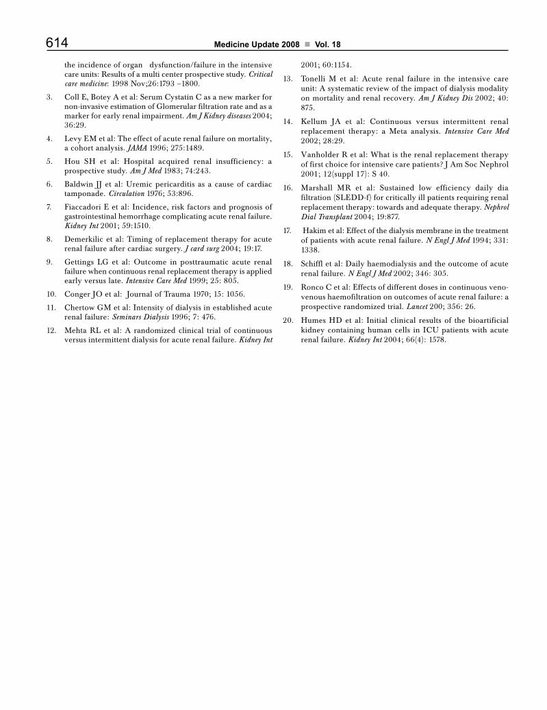
614 Medicine Update 2008 Vol. 18
the incidence of organ dysfunction/failure in the intensive care units: Results of a multi center prospective study. Critical care medicine: 1998 Nov;26:1793 –1800.
3. CollE,BoteyAetal:SerumCystatinCasanewmarkerfornon-invasive estimation of Glomerular filtration rate and as a marker for early renal impairment. Am J Kidney diseases 2004; 36:29.
4. Levy EM et al: The effect of acute renal failure on mortality, a cohort analysis. JAMA 1996; 275:1489.
5. Hou SH et al: Hospital acquired renal insufficiency: a prospective study. Am J Med 1983; 74:243.
6. Baldwin JJ et al: Uremic pericarditis as a cause of cardiac tamponade. Circulation 1976; 53:896.
7. Fiaccadori E et al: Incidence, risk factors and prognosis of gastrointestinal hemorrhage complicating acute renal failure. Kidney Int 2001; 59:1510.
8. Demerkilic et al: Timing of replacement therapy for acute renal failure after cardiac surgery. J card surg 2004; 19:17.
9. Gettings LG et al: Outcome in posttraumatic acute renal failure when continuous renal replacement therapy is applied early versus late. Intensive Care Med 1999; 25: 805.
10. CongerJOetal:JournalofTrauma1970;15:1056.
11. ChertowGMetal:Intensityofdialysisinestablishedacuterenal failure: Seminars Dialysis 1996; 7: 476.
12. Mehta RL et al: A randomized clinical trial of continuous versus intermittent dialysis for acute renal failure. Kidney Int
2001; 60:1154.
13. Tonelli M et al: Acute renal failure in the intensive care unit: A systematic review of the impact of dialysis modality on mortality and renal recovery. Am J Kidney Dis 2002; 40: 875.
14. Kellum JA et al: Continuous versus intermittent renalreplacement therapy: a Meta analysis. Intensive Care Med 2002; 28:29.
15. Vanholder R et al: What is the renal replacement therapy of first choice for intensive care patients? J Am Soc Nephrol 2001; 12(suppl 17): S 40.
16. Marshall MR et al: Sustained low efficiency daily dia filtration (SLEDD-f) for critically ill patients requiring renal replacement therapy: towards and adequate therapy. Nephrol Dial Transplant 2004; 19:877.
17. Hakim et al: Effect of the dialysis membrane in the treatment of patients with acute renal failure. N Engl J Med 1994; 331: 1338.
18. Schiffl et al: Daily haemodialysis and the outcome of acute renal failure. N Engl J Med 2002; 346: 305.
19. RoncoCetal:Effectsofdifferentdosesincontinuousveno-venous haemofiltration on outcomes of acute renal failure: a prospective randomized trial. Lancet 200; 356: 26.
20. Humes HD et al: Initial clinical results of the bioartificial kidneycontaininghumancells in ICUpatientswithacuterenal failure. Kidney Int 2004; 66(4): 1578.

CHAPTER
83Multinodular Thyroid DiseaseN. D. Moulick
Benign nodular thyroid disease constitutes a heterogeneous thyroid disorder which is highly prevalent in iodine deficient areas. On a general basis it is divided into solitary and multinodular thyroid disease.1
Multinodular goitre is the most prevalent thyroid pathological abnormality worldwide, although its geographical incidence varies greatly according to environmental iodization Most countries in central and southern Europe have endemic goitre areas with a prevalence of multinodular goitre (MG) of 3-6%. In United States, the annual incidence of nodular thyroid disease is 0.1% to 1.5% and the prevalence is 4-7%.2 In older studies as in 1950’s in Framingham, Massachusets 1% of persons in the age 30-59 yrs had a multinodular goitre. In Whickham in northeast England, palpable goitres were detected in 10% of adult women and 2% of adult men3 Multinodular goitre refers to an enlargement of the thyroid with deformation of normal parenchymal structures by the presence of nodules. These nodules vary considerably in size, morphology and function. The development of nodular goitre is very likely a continuous process that starts with thyroid hyperplasia and simple goitre. The main epidemiological determinants are iodine deficiency, age, sex and duration of goitre in iodine deficient and also iodine sufficient areas.1
Thyroid nodules are discovered by palpation in 3-7% of the population. However on ultrasonography (US) it is noted in 20-76% and by autopsy in approximately 50%.4 Increasing use of high resolution ultrasonography (HRUSG) over the last two decades has lead to an increasing prevalence of thyroid nodules in asymptomatic subjects. Moreover, 20-48% of patients with single palpable thyroid nodule are found to have additional nodules when investigated by HRUSG.5 Prevalence increases linearly with age, exposure to ionizing radiation and iodine deficiency. Thyroid nodules are more common in women.4
In contrast to solitary nodular thyroid disease that has a more uniform clinical pathological and molecular picture, one finds a combination of hyperfunctional, hypofunctional or normally functioning lesions within the same thyroid gland.3 The overall functional balance of these individual nodules determine whether patient has a euthyroid multinodular goitre (MNG) with normal TSH and free thyroid hormone levels or toxic multinodular goitre (TMNG). This can be overtly toxic with suppressed TSH and elevated free thyroid hormones or subclinical hyperthyroidism (low or suppressed TSH and normal free thyroid hormones). It is important to note that the functional status is not stationary and those who have had TMNG have been having a goitre of long standing
616 Medicine Update 2008 Vol. 18
with euthyroidism. Moreover the status of TSH indicates that a critical level of thyroid autonomy has been reached.3 The rate of progression from euthyroidism to thyrotoxicosis in patients with nodular goitres has not been studied extensively, but in two studies, the incidence of overt thyrotoxicosis was approximately 10% during 7-12 yrs of follow up.3
On functional grounds, nodules are classified as “cold”, “normal” or “hot” depending upon whether they show decreased, normal or increased uptake on scintiscan. Approximately, 85% of all nodules are cold, 10% are normal and 5% are hot nodules.1
Non toxic goitre may be defined as a diffuse or nodular enlargement that is not associated with thyrotoxicosis and does not result from an autoimmune or inflammatory process. The term endemic goitre is used when prevalence in children 6-12 yrs of age within a population is more than 10%. Goitre is called sporadic when the prevalence is 10% or less.3 The natural history of nontoxic goitre is characterized by gradual thyroid growth, nodule formation and development of functional autonomy.
Worldwide the most important environmental factor contributing to goitre is iodine deficiency. Since goitres also occur in individuals without iodine deficiency and not all individuals in an iodine deficient area develop a goitre, other suggested risk factors include cigarette smoking, infections, drugs and goitrogens.6 The impact of smoking on thyroid disease could be due to increased thiocyanate levels which exert a competitive inhibitory effect on iodide uptake and organification.
Radiation is the other risk factor both for thyroid malignancy and nodular disease. An increased prevalence of nodular disease has been associated with exposure to radionuclear fallouts and therapeutic external radiation.1
Development of nodular disease is influenced by multiple environmental components interacting with constitutional parameters of gender and age. Multinodular goitre is probably a life long
condition that has its inception in adolescence or at puberty.7 In areas of iodine deficiency with a high prevalence of goitres, many prepubertal children have diffuse goitres although these may regress in early adolesence. In areas with a lower prevalence of goitre, goitrogenesis usually starts at an older age. With time, diffuse goitres tend not only to grow but also to become nodular.
Nodular disease is 5-15 times more frequent in females. At present it is believed to be a genetic susceptibility and/or a direct impact on steroid hormones. Estrogen has been shown to have a growth promoting effect in vitro in rat FRTL-5 cells and thyroid cancer cell lines1 Also, 17 B estradiol has been shown to amplify growth factor induced signaling in normal thyroid and thyroid tumours. Use of oral contraceptives is associated with decrease in goitre but not nodules.
The thyroid enlargement in pregnancy has been related to iodine deficiency. In one German study the increased prevalence of multinodular goitre with parity was only observed in those women who had not taken iodine supplementation during an earlier pregnancy.1
Age : In a cross sectional study of patients with nontoxic goitre evidence for thyroid growth, nodular formation and development of functional autonomy with age was found. Thyroid volume was positively correlated with age and duration of goitre. Patient with MNG were older and had a large thyroid volume and significantly lower serum TSH concentration. In a borderline iodine deficient area, MNG was present in 23% of the studied population of 2656 Danish people aged 41-71 yrs and increased with age in women from 20-46% as well as in men.1
Thyroid GrowthThe increased thyroid mass of a nontoxic goitre is mainly caused by excessive cell replication as demonstrated by the finding of a highly significant positive correlation between the total DNA content of nodular goitre and their weight. Histologically, the

617Multinodular Thyroid Disease
newly generated cells appear to be mainly thyroid follicular cells and increase in interstitial tissue and colloid contribute little to the goitre growth.3 Several growth stimulating factors (endocrine or paracrine) are thought to be of importance for the increased follicular cell replication.
Thyrotropin(TSH) is the main extra thyroidal growth stimulation factor and plays an important role in pathogenesis of iodine deficiency goitre.The first comprehensive theory about the development of multinodular goitre was proposed by David Marine and studied further by Selwyn Talyor.7 He proposed that in response to iodine deficiency, the thyroid first goes through a period of hyperplasia as a consequence of the resulting TSH stimulation, but eventually, possibly because of iodine repletion or a decreased requirement for thyroid hormone, enters a resting phase characterized by colloid storage and the histologic picture of a colloid goitre. Marine believed that repetition of these two stages of cycle would eventually result in the formation of nontoxic multinodular goitre.
Since many patients with nontoxic goitre have normal serum TSH concentrations and that they grow despite administration of T4 in doses that reduce TSH below normal, it is suggested that other growth stimulating factors are involved in thyroid growth.
Growth factors such as insulin like growth factor–1,( IGF-1) epidermal growth factor (EGF) and fibroblast goitre factor (FGF) may be important for stimulation of thyroid growth.The expression of IGF-1 and FGF is increased in nodular goitres in humans.3
Expression of FGF-1 and 2 and FGF receptor-1 accompany thyroid hyperplasia and may play a role in the development of multinodular goitre.7 Acromegalic subjects with elevated levels of serum growth hormone and IGF-1 and normal TSH levels have an increased prevalence of goitre.6
In vitro IG-1, EGF and FGI stimulate proliferation of thyroid follicular cells and in vivo, administration of FGF intravenously in rats resulted in an increased
weight. In contrast, transforming growth factor (TGF) seems to inhibit thyroid growth. It acts as an autocrine growth inhibitor on follicular cells by inhibiting growth stimulating actions of TSH, IGF-1 and EGF. Tissue concentrations of TGF messenger RNA are lower in iodine deficient nontoxic goitres than in normal thyroid tissue.
Other factors promoting cell growth and differentiation identified in the last decade include cytokines, acetylcholine, norepinephrine, vasoactive intestinal peptide and substances of C-cell origin. It is however not known to whatextent these compounds play a role in the genesis of multinodular goitre.7
In addition to and possibly modulated by extracellular stimulators of thyroid growth, some alterations in intracellular mechanisms related to the control of cell replication. (e.g. increased expression of protooncogenes such as the ras protooncogene) may contribute to the growth of non toxic goitres. Also, intrathyroidal depletion of iodine may stimulate follicular growth irrespective of the serum TSH concentration.3
Nodular GrowthHeterogeneity
In a normal thyroid gland, the growth potential and functional activity of individual cells within a single follicle varies. Immunohistochemical studies have demonstrated that only a small fraction of thyroid follicular cells contains the Na / iodine cotransporter.
Despite the heterogeneity in function between individual cells within normal follicles, the balance between thyroglobulin synthesis and endocytic activity in the follicle as a whole is finely tuned, so that the size of most follicles is similar. In contrast, the follicles of nontoxic goitre vary much more widely in both functions. Thus both iodine metabolism and growth rate of cells within newly formed follicles vary widely.3 Large colloid rich follicles and small follicles co exist in most non toxic goitres.
618 Medicine Update 2008 Vol. 18
It is assumed that during formation of non toxic goitre the stimulation of follicular cells by TSH or other thyroid growth stimulating factors is relatively weak. Hence only a small fraction of follicular cells namely those with a high growth potential, will enter the mitotic cycle and contribute to the formation of new follicles. These cells transfer their high growth potential to their daughter cells and the number of replicating cells increases progressively.
Follicular cells with a high growth potential are not evenly distributed within the thyroid gland and after replication their daughter cells remain clustered. Therefore nontoxic goitres become increasingly nodular with time. There is evidence that rapidly replicating cells remain clustered during goitrogenesis as demonstrated by X-chromosome inactivation analysis that some macroscopic nodules within a nontoxic goitre are monoclonal. Despite their monoclonal nature, nodules may contain morphologically and functionally heterogeneous follicles.2,6
The growing thyroid gland also requires expansion of blood vessels. However the newly formed capillary network is often fragile and unable to supply the growing thyroid tissue adequately. This leads to areas of hemorrhagic necrosis within goitre. The necrotic areas are invaded by granulation tissue, ultimately resulting in fibrosis, scarring and even calcification. The resulting network of inelastic strands of connective tissue interferes with smooth growth of thyroid parenchyma and will further increase the formation of macroscopic nodules. Further the distended follicles may fuse to form colloid cysts which are characteristic of non toxic goitres.3
If a group of follicles generated in this way grows large enough, it may become visible as a hot or cold area on thyroid scintigraphy depending on the degree of activity of its iodine metabolism.The iodine metabolism of particular areas within a nontoxic goitre and their growth behaviors are not necessarily parallel. The areas of increased or decreased iodine uptake do not necessarily
correspond to thyroid nodules detected by physical examination or ultrasonography.
Development of Functional AutonomySome normal thyroid follicular cells take up and organify iodine in the absence of TSH during goitrogenesis, the number of cells with functional autonomy increases especially when the cells have a replicating capacity.3 The increase in the total mass of follicular cells with autonomous iodine metabolism during goitre growth would explain why a patient with nontoxic goitre can develop subclinical and later overt thyrotoxicosis.
The hypothesis that the development of thyroid autonomy is due to a gradual increase in the number of cells having relatively autonomous thyroid hormone synthesis is supported by the 27% prevalence of impaired TSH responses to TRH in patients with nodular goitre as opposed to such responses in only 1 of 15 patients with diffuse goitre.7 The fact that it is possible to induce hyperthyroidism in some patients with multinodular goitres by administration of iodide suggests that certain nodules are autonomous but unable under normal iodine intake to concentrate sufficient iodide to cause hyperthyroidism.
Prevalence of thyroid autonomy correlates with increased thyroid nodularity and increases with age. Correctionofiodinedeficiencyinapopulationresults in a decrease of thyroid autonomy as demonstrated by the impressive 73% reduction in prevalence of TMNG only 15 yrs after doubling of iodine content of salt in Switzerland.1
Iodine deficiency as the sole factor responsible for goitre seems an oversimplification. The role of genetic factors is suggested by several lines of evidence such as
the clustering of goitres within families
the higher concordance rate for goitres in monozygotic than in dizygotic twins
the female/male ratio (1:1 in endemic vs 7:1 to 9:1 in sporadic goitres)
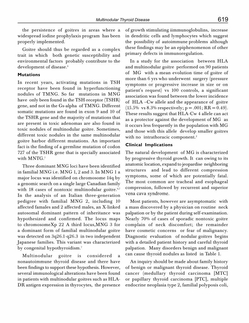
619Multinodular Thyroid Disease
the persistence of goitres in areas where a widespread iodine prophylaxis program has been properly implemented.
Goitre should thus be regarded as a complex trait in which both genetic susceptibility and environmental factors probably contribute to the development of disease.6
Mutations
In recent years, activating mutations in TSH receptor have been found in hyperfunctioning nodules of TMNG. So far mutations in MNG have only been found in the TSH-receptor (TSHR) gene, and not in the Gs-alpha of TMNG. Different somatic mutations are found in exon 9 and 10 of the TSHR gene and the majority of mutations that are present in toxic adenomas are also found in toxic nodules of multinodular goitre. Sometimes, different toxic nodules in the same multinodular goitre harbor different mutations. An important fact is the finding of a germline mutation of codon 727 of the TSHR gene that is specially associated with MNTG.7
Three dominant MNG loci have been identified in familial MNG i.e. MNG 1, 2 and 3. In MNG 1 a major locus was identified on chromosome 14q by agenomicsearchonasinglelargeCanadianfamilywith 18 cases of nontoxic multinodular goitre.6,7 In the analysis of an Italian three-generation pedigree with familial MNG 2, including 10 affected famales and 2 affected males, an X-linked autosomal dominant pattern of inheritance was hypothesized and confirmed. The locus maps to chromosomeXp 22 A third locus,MNG 3 for a dominant form of familial multinodular goitre was detected on 3q26.1-q26.3 in two independent Japanese families. This variant was characterized by congenital hypothyroidism.7
Multinodular goitre is considered a nonautoimmune thyroid disease and there have been findings to support these hypothesis. However, several immunological alterations have been found in patients with multinodular goitres such as HLA-DR antigen expression in thyrocytes, the presence
of growth stimulating iimmunoglobulins, increase in dendritic cells and lymphocytes which suggest the possibility of autoimmune problems although these findings may be an epiphenomenon of other primary defects in immunoregulation.
In a study for the association between HLA and multinodular goitre performed on 90 patients of MG with a mean evolution time of goitre of more than 6 yrs who underwent surgery (pressure symptoms or progressive increase in size or on patient's request) vs 100 controls, a significant association was found between the lower incidence ofHLA–Cwalleleandtheappearanceofgoitre(15.5% vs 8.3% respectively; p = .001; RR = 0.49). TheseresultssuggestthatHLA-Cw4allelecanactas a protector against the development of MG as it occurs less frequently in the population with MG and those with this allele develop smaller goitres with no intrathoracic component.2
Clinical Implications
The natural development of MG is characterized by progressive thyroid growth. It can owing to its anatomic location, expand to jeopardise neighboring structures and lead to different compression symptoms, some of which are potentially fatal. The most common are tracheal and esophageal compression, followed by recurrent and superior vena cava syndrome.
Most patients, however are asymptomatic with a mass discovered by a physician on routine neck palpation or by the patient during self-examination. Nearly 70% of cases of sporadic nontoxic goitre complain of neck discomfort; the remainder have cosmetic concerns or fear of malignancy. Diagnostic evaluation of nodular goitres begins with a detailed patient history and careful thyroid palpation. Many disorders benign and malignant can cause thyroid nodules as listed in Table 1.
An inquiry should be made about family history of benign or malignant thyroid disease. Thyroid cancer (medullary thyroid carcinoma [MTC]or papillary thyroid carcinoma [PTC], multipleendocrine neoplasia type 2, familial polyposis coli,

620 Medicine Update 2008 Vol. 18
CowdensdiseaseandGardener’ssyndromeareraredisorders but have to be considered. History of exposure to radiation in childhood to the head, face and neck should be especially asked for (Table 2).
Multinodular goitre with compression symptoms has a clinical profile different from that of goitre without these symptoms. Many of the symptoms of large multinodular goitre are chiefly due to the presence of an enlarging mass in the neck and its impingement upon the adjacent structures. There may be dysphagia, cough, and hoarseness. Paralysis of a recurrent laryngeal nerve may occur when the nerve is stretched taut across the surface of an expanding goitre, but this event is very unusual. When unilateral vocal cord paralysis is demonstrated, the presumptive diagnosis is cancer. Pressure on the superior sympathetic ganglions and nerves may produce a Horner’s syndrome.
In a retrospective study of 157 patients of MG with compression symptoms who underwent surgery multinodular goitre was characterized by a series of distinguishing features from 512 cases of MG with no compressive symptoms: they were older (mean age 56 vs 46 p < .001); longer evolution time (mean128 vs 78 mths) and
higher frequency of intrathoracic component; chest radiography showing tracheal displacement. Airway compression was the commonest and serious problem. Esophageal compression can occur and is more often reported when the extension is posterior. In this study the incidence of recurrent symptoms was high.8 Uncommon compression symptoms included superior vena cava syndrome and Horner’s syndrome. The surgical treatment of MG with compression symptoms has a higher rate of sternotomy and morbidity.
As the gland grows it characteristically enlarges the neck, but frequently the growth occurs in a downward direction, producing a substernal goitre. A history sometimes given by an older patient that a goitre once present in the neck has disappeared may mean that it has fallen down into the upper mediastinum, where its upper limit can be felt by careful deep palpation. Hemorrhage into this goitre can produce acute tracheal obstruction. Sometimes substernal goitres are attached only by a fibrous band to the goitre in the neck and extend downward to the arch of the aorta. They have even been observed as deep in the mediastinum as the diaphragm.
A significant proportion of patients with nodular thyroid glands develop thyrotoxicosis, and this is directly related to the duration of the goitre. Typically, the thyrotoxicosis comes about so insidiously that the patient is often unaware of the symptoms. Emotional lability, heightened neuromuscular activity, increased metabolic rate, cardiac irritability and tachycardia, and increased
Table 1 : Common causes of thyroid nodulesBenign
Colloidnodule
Hashimoto thyroiditis
Simple or hemorrhagic cyst
Follicular adenoma
Subacute thyroiditis
Malignant
Primary
Follicular cell-derived carcinoma:
PTC, follicular thyroidcarcinoma,anaplastic thyroidcarcinoma
C-cell-derivedcarcinoma:
MTC
Thyroid lymphoma
Secondary
Metastatic carcinoma
Table 2 : Increased risk of malignancy in thyroid nodule• Historyofchildhoodhead/neckirradiation
• Family history of PTC, MTC, or multiple endocrineneoplasia type 2 (MEN2)
• Age<20or>70years
• Malesex
• Enlargingnodule
• Abnormalcervicaladenopathy
• Fixednodule
• Vocalcordparalysis
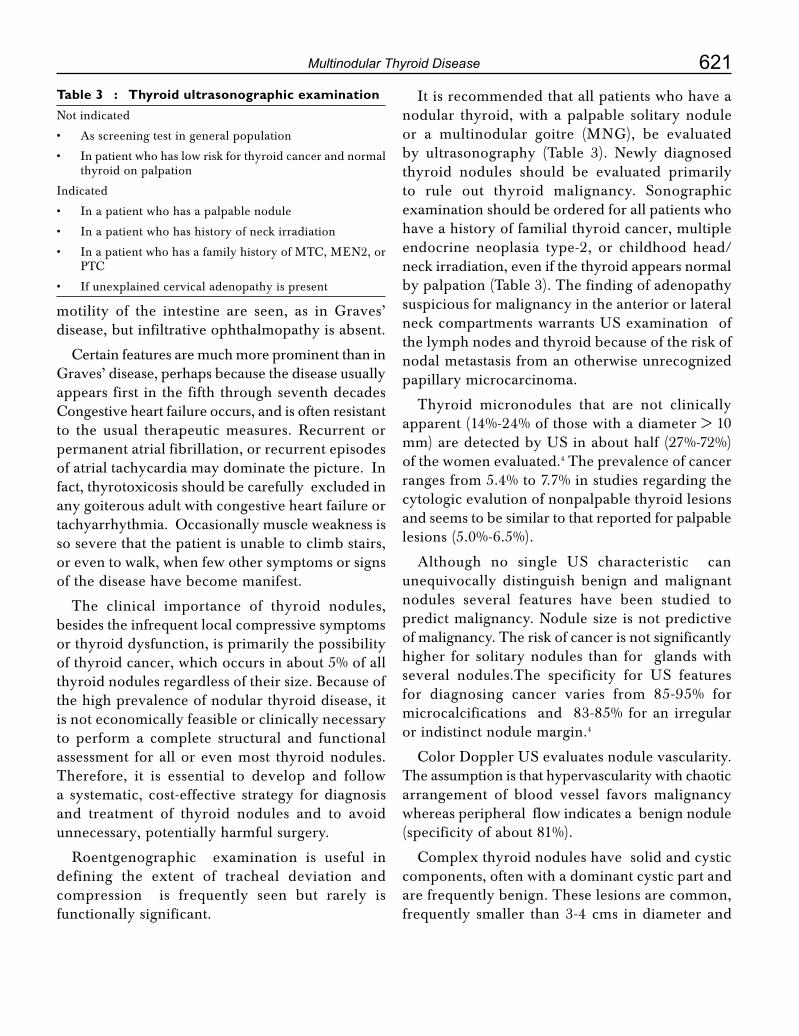
621Multinodular Thyroid Disease
motility of the intestine are seen, as in Graves’ disease, but infiltrative ophthalmopathy is absent.
CertainfeaturesaremuchmoreprominentthaninGraves’ disease, perhaps because the disease usually appears first in the fifth through seventh decades Congestiveheartfailureoccurs,andisoftenresistantto the usual therapeutic measures. Recurrent or permanent atrial fibrillation, or recurrent episodes of atrial tachycardia may dominate the picture. In fact, thyrotoxicosis should be carefully excluded in any goiterous adult with congestive heart failure or tachyarrhythmia. Occasionally muscle weakness is so severe that the patient is unable to climb stairs, or even to walk, when few other symptoms or signs of the disease have become manifest.
The clinical importance of thyroid nodules, besides the infrequent local compressive symptoms or thyroid dysfunction, is primarily the possibility of thyroid cancer, which occurs in about 5% of all thyroid nodules regardless of their size. Because of the high prevalence of nodular thyroid disease, it is not economically feasible or clinically necessary to perform a complete structural and functional assessment for all or even most thyroid nodules. Therefore, it is essential to develop and follow a systematic, cost-effective strategy for diagnosis and treatment of thyroid nodules and to avoid unnecessary, potentially harmful surgery.
Roentgenographic examination is useful in defining the extent of tracheal deviation and compression is frequently seen but rarely is functionally significant.
It is recommended that all patients who have a nodular thyroid, with a palpable solitary nodule or a multinodular goitre (MNG), be evaluated by ultrasonography (Table 3). Newly diagnosed thyroid nodules should be evaluated primarily to rule out thyroid malignancy. Sonographic examination should be ordered for all patients who have a history of familial thyroid cancer, multiple endocrine neoplasia type-2, or childhood head/neck irradiation, even if the thyroid appears normal by palpation (Table 3). The finding of adenopathy suspicious for malignancy in the anterior or lateral neck compartments warrants US examination of the lymph nodes and thyroid because of the risk of nodal metastasis from an otherwise unrecognized papillary microcarcinoma.
Thyroid micronodules that are not clinically apparent (14%-24% of those with a diameter > 10 mm) are detected by US in about half (27%-72%) of the women evaluated.4 The prevalence of cancer ranges from 5.4% to 7.7% in studies regarding the cytologic evalution of nonpalpable thyroid lesions and seems to be similar to that reported for palpable lesions (5.0%-6.5%).
Although no single US characteristic can unequivocally distinguish benign and malignant nodules several features have been studied to predict malignancy. Nodule size is not predictive of malignancy. The risk of cancer is not significantly higher for solitary nodules than for glands with several nodules.The specificity for US features for diagnosing cancer varies from 85-95% for microcalcifications and 83-85% for an irregular or indistinct nodule margin.4
ColorDopplerUSevaluatesnodulevascularity.The assumption is that hypervascularity with chaotic arrangement of blood vessel favors malignancy whereas peripheral flow indicates a benign nodule (specificity of about 81%).
Complexthyroidnoduleshavesolidandcysticcomponents, often with a dominant cystic part and are frequently benign. These lesions are common, frequently smaller than 3-4 cms in diameter and
Table 3 : Thyroid ultrasonographic examinationNot indicated
• Asscreeningtestingeneralpopulation
• Inpatientwhohaslowriskforthyroidcancerandnormalthyroid on palpation
Indicated
• Inapatientwhohasapalpablenodule
• Inapatientwhohashistoryofneckirradiation
• InapatientwhohasafamilyhistoryofMTC,MEN2,orPTC
• Ifunexplainedcervicaladenopathyispresent
622 Medicine Update 2008 Vol. 18
asymptomatic.CytologyisnecessarytodocumentmorphologybecausesomePTCs(Papillarythyroidcarcinomas) are cystic.
MRIandCTarenotrecommendedforroutineuse but are of value to assess size, substernal extension and positional relationsihip of the goitre tosurroundingstructures.Contrastmediacontainingiodine should be used with caution because it decreases subsequent iodine I131 uptake.
Positron emission tomography with fluorodeoxyglucose may add functional information to anatomic visualization provided by US. The high cost of these procedures makes them impractical for routine clinical assessment of thyroid nodules.
Fine needle aspiration biopsy
Thyroid FNA biopsy is the most accurate test for determining malignancy and is an integral part of thyroid nodule evaluation. In glands with multiple nodules, selection for cytology( FNAC) shouldbe based on US features rather than on size or clinically “dominant” nodules (Table 4). A marked hypoechogenicity of a solid lesion on USG of a nodule may suggest malignancy and warrants FNACfromthatsite.
FNAC results are categorized as diagnostic(satisfactory) or adequate if it contains no less than six groups of well-preserved thyroid epithelial cells consisting of at least 10 cells in each group. Nondiagnostic or unsatisfactory smears with an inadequate number of cells result from acellular cystic fluid, bloody smears, or poor techniques in preparing slides. The most common benign diagnosis is “colloid nodule,” which may come from a normal thyroid gland, a benign nodule,
an MNG, or a macrofollicular adenoma.
Overall, 70% of FNA specimens are benign, 5% malignant, 10% suspicious, and 15% unsatisfactory. The final FNA report is critical in dictating whether the patient’s management should be medical or surgical. FNA has improved patient selection for thyroidectomy, such that cancer yield at surgery has increased from 15% before the use of FNA to 50% with FNA use. The sensitivity and specificity of FNA in experienced hands are excellent. A major concern with FNA is the possibility of a false-negative result (i.e., a missed malignancy). Although the false-negative rate ranges from 1% to 11%, it is less than 2% in most clinics with adequate FNA experience. Table 5 illustrates some suggestions for minimizing false-negative results.
FNACcausesonlymildtemporarypainandoccasionally a minor hematoma. No seedling of tumor cells in the neck track have been reported. It is a safe, useful and cost effective procedure . Rebiopsy is suggested only on a enlarging nodule, a recurrent cyst or a nodule showing no response to suppressive therapy (Table 5).
An important recent advance is the demonstration that thyroglobulin can be measured in lymph nodes or nodular aspirates and is useful in thyroid cancer practice. To measure Tg, the needle is rinsed with 1 ml of normal saline solution immediately after FNA biopsy and Tg is measured by immunoradiometric or chemiluminescence assays. Several molecular markers and assays have shown promise in clarifying suspicious FNA results.
Serum Tg concentration correlates with iodine intake and the size of the thyroid gland rather than with nature and function of the nodule. Since Tg measurements do not influence management,
Table 4 : Indications for ultarasonography fine-needle aspiration• Palpation-guidedFNAnondiagnostic
• Complex(solid/cystic)nodule
• Palpablesmallnodule(<1.5cm)
• Impalpableincidenteloma
• normalcervicalnodes
• NodulewithsuspiciousUSfeatures
Table 5 : Ways to minimize false-negative results• Follow-upcytologicallybenignnodules
• Aspiratemultiplenodulesites
• AspiratemultiplenodulesitesinMNG
• Submitcystfluidforexamination
• Reviewslideswithexperiencedcytopathologist
623Multinodular Thyroid Disease
measurement of Tg is seldom used in nodule diagnosis.4
Measurement of serum TSH is the most useful test in the initial evaluation of thyroid nodules.Third generation TSH assays, with detection limits of about 0.001 mIU/ml should be used in clinical practice. The measurement of free thyroid hormones and thyroid peroxidase antibodies (TPO Ab) should be the second diagnostic step. Occasionally, a nodular goitre may represent Hashimoto’s thyroiditis.
Serum calcitonin is a good marker for C-celldisease and correlates well with tumor burden. It should be measured in patients who have a family historyofMTC,MEN2 orpheochromocytomaorwhenFNAsuggestMTC.Normallevelsareless than 10pg/ml.
Radioisotope scanning
Thyroid scintigraphy can be performed with 99m TcO4 or123 I, although the latter is preferred. Thyroid scanning is the only technique that allows for assessment of thyroid nodular function and detects areas of autonomy. Based on the uptake, nodules are classified as hyperfunctioning “hot” or hypofunctioning “cold”. Hot nodules are seldom, if ever malignant, whereas the reported cancer risk is 5-15% in cold nodules. The diagnostic specificity is decreased in small lesions (< 1 cm) which may not be identified on scanning.
The role of scintigraphy in the diagnostic work-up of thyroid nodules is generally limited to.
a. a single nodule with suppressed TSH, in which case no FNA is necessary
b. a large toxic or nontoxic MNG, especially with substernal extension
c. when searching for ectopic thyroid tissue,such as struma ovari, sublingual thyroid
Management
If the enlargement of the gland is moderate, there are no symptoms and serum TSH is normal, therapy is not required. If there are symptoms due
to pressure, if patient is disturbed by appearance of the goitre, if there is growth of one nodule, or possible toxicity develops, diagnostic measures and treatment are necessary.
Thyroxine therapy may be effective in reducing the thyroid volume in patients with diffuse nontoxic goitres, as measured by ultrasonography. Nonrandomized studies suggest that it is also effective in some patients with multinodular goitres.
Only two randomized trials on the effect of T4 therapy in patients with nontoxic goitre using objective thyroid volume measurements have been reported. In placebo controlled double– blind randomized trial in patients with relatively small nontoxic multinodular goitres, thyroid volume, as measured by ultrasonography, decreased substantially in 58% of the T4 treated patients, as compared with 5% of those given in placebo; the mean decrease in thyroid volume in patients who responded was 25% after 9 months of T4 treatment. Goitre size returned to baseline within 9 months after discontinuation of therapy, demonstrating that maintenance of volume reduction requires long term T4 treatment. In a more recent study, a significant decrease in goitre size was observed in 43% of patients after 2 years of T4 therapy. In nonresponders, a mean increase in thyroid volume of 16% was found.
Attempts to reduce multinodular goitre by administrating large suppressive doses of thyroid hormone are usually little or not effective and carry the risk of inducing thyrotoxicosis if autonomy of thyroid function is already present. Although this form of treatment is still being used, it is dangerous for the elderly. .
After surgical removal of nodular goitre, it seems theoretically sound to give the patient minimally replacement or suppressive doses of thyroid hormone to suppress TSH production and prevent regeneration of the goitre. However this form of therapy is controversial. Although in one report no recurrences were found during thyroid hormone administration,

624 Medicine Update 2008 Vol. 18
in more recent studies others found no difference between untreated and patients treated with thyroid hormone after operation. In one of these studies carried out over 9 years, no effect of T4 treatment after thyroidectomy was seen in 104 patients operated for nontoxic goitre (the recurrence rate was 9.5% with treatment compared with 11.3% in untreated patients). If re-growth occurs, early ablative treatment with 131 I should be considered.
There is no place for administration of iodide in sporadic multinodular goitre. It generally has little or no beneficial therapeutic effect, and in an occasional patient may be followed by rise in plasma hormone concentration and symptoms of thyrotoxicosis. This condition is the ‘Jodbasedow’ phenomenon, and is dependent on autonomy of function of some elements of the goitre. Its occurrence is not confined to regions of iodine deficiency and is seen on occasion wherever iodide is administered to patients with well established multinodular goitre. This should be remembered whenelderlypatientsaresubjectedtoCT,MRIand administered radiographic contrast media.
Management
Clinical management of thyroid nodules isinfluenced by the combined results of TSH measurement. FNA biopsy, and US and depends primary on cytologic diagnosis.4
Fine-needle aspiration-positive nodule
If cytologic results are positive for primary thyroid malignancy, surgery is almost always needed.Cancerduetometastasisrequiresfurtherinvestigations aimed at finding the primary lesion, which often precludes thyroid surgery. If preoperativeFNAresultssuggestPTC,anear-totalor total thyroidectomy is preferred.
Fine-needle aspiration-negative nodule
Administration of T4 with TSH suppression is aimed at shrinking nodule size, arresting further nodule growth and preventing the appearance of new nodules.
The use of T4 should be avoided for large thyroid nodules or long-standing goitres, particularly if the TSH value is less than 0.5 mIU/ml; in postmenopausal women or persons older than 60 years; and in patients who have osteoporosis, cardiovascular disease, or systemic illnesses. T4 treatment induces a clinically significant volume reduction only in a minority of patients and parameters of such a response are not known.
Fine-needle aspiration-suspicious nodules
Overall, about 20% of indeterminate specimens are malignant, but cancer risk varies from 15% for “follicularneoplasm” to60% for “atypicalPTC”specimens. It is generally agreed that cytologically suspicious lesions are to be surgically excised.
Fine-needle aspiration-nondiagnostic nodule
Despite experienced centres, repeat biopsy and US-FNA, a residual 5% of nodules remain nondiagnostic, which creates a management dilemma for the clinician. Nondiagnostic, large (> 3-4 cm), recurrent cysts or solid nodules should be treated surgically.
Therapeutic techniquesSurgery
Surgical options include lobectomy plus isthmectomy for a benigh nodule, less than total thyroidectomy for MNG and near-total or total thyroidectomy for malignant disease. Frozen section should be performed at the time of surgery to help guide surgical decision making but may be of limited use in distinguishing benign from malignant follicular lesions.
Radioiodine
Toxic nodular goitres are usually more radioresistant than toxic diffuse goitres and higher I doses (30-100mCi)maybeneededforsuccessfultreatment.The aim of radioiodine treatment is the ablation of thyroid autonomy, restoration of normal thyroid function and reduction of thyroid mass. Although rare (occurring in < 1% of patient), immunogenic hyperthyroidism may occur due to induction of
625Multinodular Thyroid Disease
TSH receptor autoantibodies after I treatment of toxic nodular goitre. I therapy can be repeated after 6 months if thyrotoxicosis is not cured, as documented by persistent low TSH levels. It is not the treatment of choice if compressive symptoms are present, in larger nodules requiring high doses of I (which may be resistant to treatment or if an immediate resolution of hyperthyroidism is medically indicated.
Recombinant human thyroid-stimulating hormone
The administration of small doses (0.1 - 0.3mg) of recombinant human TSH (rhTSH) to patients who have low-uptake MNG increases I uptake by more than 4-fold in 24 to 72 hours. This allows for delivery of sufficient radiation to the thyroid to cause a decrease in size and amelioration of compressive symptoms within 2 months. As in patients who have high-uptake MNG, the average decrease in goitre size is 40% and 60% by the end of the first and second years, respectively.
Nonsurgical minimally invasive procedures
Percutaneous ethanol injection (PEI) is a US-guided, mini-invasive procedure that has been used for the nonsurgical management of some thyroid nodules.
Thyroid cysts : PEI is an effective alternative to surgery in the treatment of complex nodules with a dominant fluid component. Aspiration of thyroid cysts decreases the volume, but recurrences are common, and surgery is often required to remove large, relapsing lesions. Prospective randomized studies have shown that PEI is significantly superior to aspiration alone in reducing nodule volume. A reduction of greater than 50% of the baseline size is obtained in nearly 90% of cases treated with PEI.
Cold Solid Nodules : A clinically significant decrease in nodule size after PEI has been reported in patients who have been having, solitary, solid nodules that are cold on scintigraphy.
Laser thermal ablation : PLA is a minimally invasive procedure that is proposed as an alternative
to surgery for thyroid nodules causing local symptoms or cosmetic concern. With guidance and after local anesthesia, a 21-gauge needle is carefully inserted into the thyroid mass, and a thin optical fiber is advanced into the needle sheath. The fiber tips are seen as hyper echoic spots and the area to be treated appears as an echogenic area enlarging over time on US.
Adverse effects of PLA include burning cervical pain, which decreases rapidly as the energy is turned off. Localized pain can be treated with oral analgesics. Other problems, such as permanent dysphonia, skin burning, or damage to neck structures, have not been observed. PLA is an outpatient procedure that lasts about 30 minutes, and patients can be dismissed shortly after the treatment.
Radiofrequency ablation : RF is under evaluation as a nonsurgical therapeutics modality for the ablation of benign and malignant thyroid lesions.
SummaryMultinodular thyroid disease is perhaps the commonest of all thyroid disorders in clinical practice worldwide. It is highly prevalent in iodine deficient areas and possibly has its inception in adolescence or puberty. Nodules larger than 1 cm may be detected by palpation. Genetic heterogeneity of normal follicular cells and acquisition of new inheritable qualities of replicating cells are the primary factors for nodular disease.
The goitre may well give rise to local discomfort and may, in case of large goitres, cause mechanical obstruction of the upper airway. Goitres have an annual growth potential of up to 20%, which can complicate treatment, if it is delayed, more often then requiring surgery. Progressive autonomous function of thyroid nodules can cause overt thyrotoxicosis in 5–10% of multinodular goitre patients within a 5-yr period. Even more frequently, patients develop subclinical thyrotoxicosis with its potential for osteoporosis and atrial fibrillation. Finally,
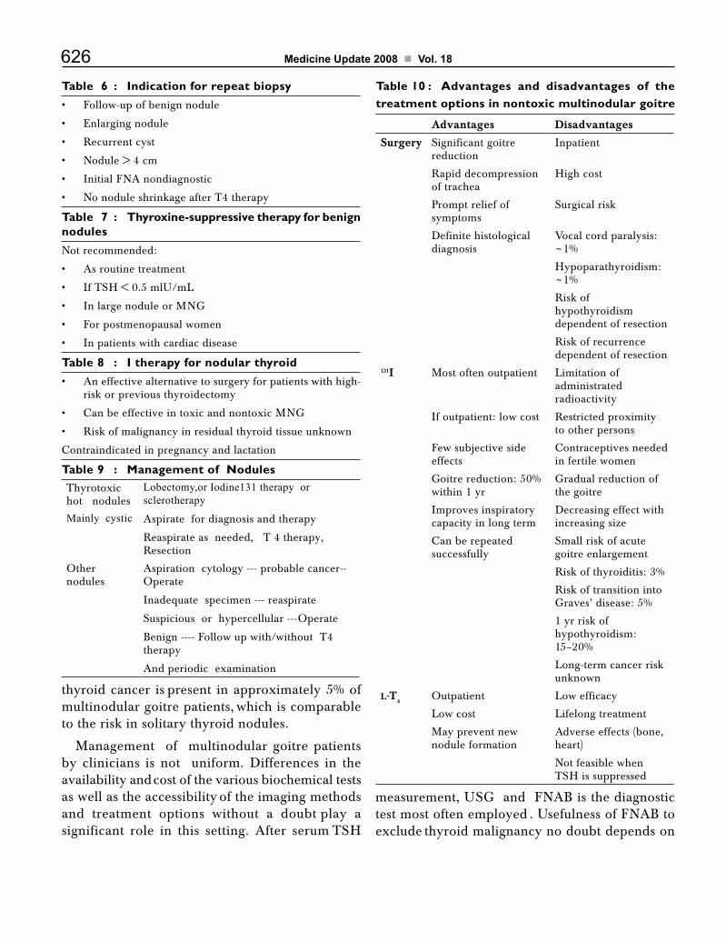
626 Medicine Update 2008 Vol. 18
Table 6 : Indication for repeat biopsy• Follow-upofbenignnodule
• Enlargingnodule
• Recurrentcyst
• Nodule>4cm
• InitialFNAnondiagnostic
• NonoduleshrinkageafterT4therapy
Table 7 : Thyroxine-suppressive therapy for benign nodulesNot recommended:
• Asroutinetreatment
• IfTSH<0.5mlU/mL
• InlargenoduleorMNG
• Forpostmenopausalwomen
• Inpatientswithcardiacdisease
Table 8 : I therapy for nodular thyroid• Aneffectivealternativetosurgeryforpatientswithhigh-
risk or previous thyroidectomy
• CanbeeffectiveintoxicandnontoxicMNG
• Riskofmalignancyinresidualthyroidtissueunknown
Contraindicatedinpregnancyandlactation
Table 9 : Management of NodulesThyrotoxic hot nodules
Lobectomy,or Iodine131 therapy or sclerotherapy
Mainly cystic Aspirate for diagnosis and therapy
Reaspirate as needed, T 4 therapy, Resection
Other nodules
Aspiration cytology --- probable cancer-- Operate
Inadequate specimen --- reaspirate
Suspicious or hypercellular ---Operate
Benign ---- Follow up with/without T4 therapy
And periodic examination
Table 10 : Advantages and disadvantages of the treatment options in nontoxic multinodular goitre
Advantages Disadvantages
Surgery Significant goitre reduction
Inpatient
Rapid decompression of trachea
High cost
Prompt relief of symptoms
Surgical risk
Definite histological diagnosis
Vocal cord paralysis: ~1%
Hypoparathyroidism: ~1%
Risk of hypothyroidism dependent of resection
Risk of recurrence dependent of resection
131I Most often outpatient Limitation of administrated radioactivity
If outpatient: low cost Restricted proximity to other persons
Few subjective side effects
Contraceptivesneededin fertile women
Goitre reduction: 50% within 1 yr
Gradual reduction of the goitre
Improves inspiratory capacity in long term
Decreasing effect with increasing size
Canberepeatedsuccessfully
Small risk of acute goitre enlargement
Risk of thyroiditis: 3%
Risk of transition into Graves’ disease: 5%
1 yr risk of hypothyroidism: 15–20%
Long-term cancer risk unknown
L-T4
Outpatient Low efficacy
Low cost Lifelong treatment
May prevent new nodule formation
Adverse effects (bone, heart)
Not feasible when TSH is suppressed
thyroid cancer is present in approximately 5% of multinodular goitre patients, which is comparable to the risk in solitary thyroid nodules.
Management of multinodular goitre patients by clinicians is not uniform. Differences in the availability and cost of the various biochemical tests as well as the accessibility of the imaging methods and treatment options without a doubt play a significant role in this setting. After serum TSH
measurement, USG and FNAB is the diagnostic test most often employed . Usefulness of FNAB to exclude thyroid malignancy no doubt depends on

627Multinodular Thyroid Disease
the cytopathological expertise available. Routine measurement of calcitonin is not recommended.
References1. Krohn K., Fuhrer D., Bayer Y. et al. Molecular Pathogenesis
of Euthyroid and Toxic Multinodular Goitre. Endocrine Reviews 2004-2005, 26: 504-524
2. Rios A., Rodriguez J. M., Moya M R., et al : Association ofHLA-CalleleswithMultinodularGoitres-Study inapopulation from Southeastern Spain. Arch Surg 2006, 141, 123-128
3. Hermus A R., Huysmans D A., : Pathogenesis of Nontoxic Diffuse and Nodular goitre in Werner and Ingbar’s The ThyroidAFundamentalandClinicalText.(eds):BravermanL E., Utiger R D 9th ed.,Lippincott Williams & Wilkins Philadelphia, 2005, : 873- 878.
4. GharibH.,PapiniE.:ThyroidNodules:ClinicalImportance,Assessment,andTreatment:EndocrinolMetabClinNAm:2007:36: 707-735
5. ChowdhuryS.,MukherjeeS.,MukhopadhyaySetal:TheThyroid Nodule-Evaluation and Management J Indian Med Assoc 2006; 104: 568-573.
6. Schlumberger M J., Filetti S., Hay I D.: Nontoxic Goitre and Thyroid Neoplasia: in Williams Textbook of Endocrinology, Chap13, (eds):LarsenPR.,Kronenberg,HMMelmedS.,Polonsky KS.,10th edition Elsivier India, New Delhi 2003: 457-490.
7. http://www.thyroid manger.org/Chap17/17-frame.htmMultinodular Goitre.
8. Rios A., Rodriquez J.M., Canteras M et al : SurgicalManagement of Multinodular Goitre With CompressionSymptoms :Arch Surg 2005; 140, 49-53
9. HermiusARHuysmans:ClinicalManifestationsandTreatmennt of Nontoxic diffuse and Nodular Goitre in Werner and Ingbar’s The Thyroid A Fundamental and ClinicalText.(eds):BravermanLE.,UtigerRD9 th ed Lippincott Williams & Wilkins Phidalphia, 2005, 875-885.
10. http://edrv.endojournal.org/cgi/contentfull/24/1/102/T9
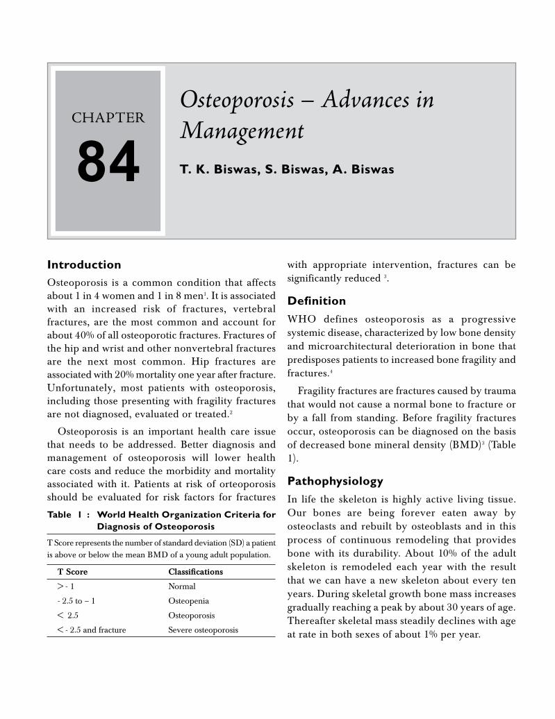
CHAPTER
84osteoporosis – Advances in ManagementT. K. Biswas, S. Biswas, A. Biswas
IntroductionOsteoporosis is a common condition that affects about 1 in 4 women and 1 in 8 men1. It is associated with an increased risk of fractures, vertebral fractures, are the most common and account for about 40% of all osteoporotic fractures. Fractures of the hip and wrist and other nonvertebral fractures are the next most common. Hip fractures are associated with 20% mortality one year after fracture. Unfortunately, most patients with osteoporosis, including those presenting with fragility fractures are not diagnosed, evaluated or treated.2
Osteoporosis is an important health care issue that needs to be addressed. Better diagnosis and management of osteoporosis will lower health care costs and reduce the morbidity and mortality associated with it. Patients at risk of orteoporosis should be evaluated for risk factors for fractures
with appropriate intervention, fractures can be significantly reduced 3.
DefinitionWHO defines osteoporosis as a progressive systemic disease, characterized by low bone density and microarchitectural deterioration in bone that predisposes patients to increased bone fragility and fractures.4
Fragility fractures are fractures caused by trauma that would not cause a normal bone to fracture or by a fall from standing. Before fragility fractures occur, osteoporosis can be diagnosed on the basis of decreased bone mineral density (BMD)3 (Table 1).
PathophysiologyIn life the skeleton is highly active living tissue. Our bones are being forever eaten away by osteoclasts and rebuilt by osteoblasts and in this process of continuous remodeling that provides bone with its durability. About 10% of the adult skeleton is remodeled each year with the result that we can have a new skeleton about every ten years. During skeletal growth bone mass increases gradually reaching a peak by about 30 years of age. Thereafter skeletal mass steadily declines with age at rate in both sexes of about 1% per year.
Table 1 : World Health Organization Criteria for Diagnosis of Osteoporosis
T Score represents the number of standard deviation (SD) a patient is above or below the mean BMD of a young adult population.
T Score Classifications
> - 1 Normal
- 2.5 to – 1 Osteopenia
< 2.5 Osteoporosis
< - 2.5 and fracture Severe osteoporosis
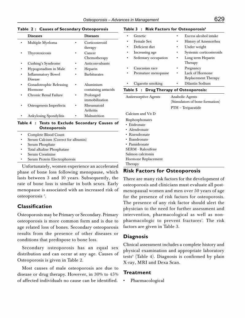
629osteoporosis – Advances in Management
Table 2 : Causes of Secondary Osteoporosis
Diseases Diseases
• MultipleMyeloma • Corticosteroid therapy• Thyrotoxicosis • Cancer Chemotherapy• Cushing’sSyndrome • Anticonvulsants• HypogonadisminMale • Heparin• InflammatoryBowel • Barbiturates Disease• GonadotrophicReleasing • Aluminium Hormone containing antacids• ChronicRenalFailure • Prolonged immobilization• OsteogenesisImperfecta • Rheumatoid Arthritis• AnkylosingSpondylitis • Malnutrition
Table 3 : Risk Factors for Osteoporosis8
• Genetic • Excessalcoholintake• FemaleSex • HistoryofAmenorrhea• Deficientdiet • Underweight• Increasingage • Systemiccorticosteroids• Sedentaryoccupation • LongtermHeparin Therapy• Caucasianrace • Pregnancy• Prematuremenopause • LackofHormone Replacement Therapy• Cigarettesmoking • DilantinSodium
Table 4 : Tests to Exclude Secondary Causes of Osteoporosis
• CompleteBloodCount• SerumCalcium(Correctforalbumin)• SerumPhosphate• TotalalkalinePhosphatase• SerumCreatinine• SerumProteinElectrophoresis
Table 5 : Drug Therapy of Osteoporosis:
Antiresorptive Agents Anabolic Agents (Stimulators of bone formation)
PTH – Teriparatide
CalciumandVitD
Bisphosphonates•Etidronate•Alendronate•Risendronate•Ibandronate•PamidronateSERM - RaloxifeneSalmon calcitonin Hormone Replacement Therapy Unfortunately, women experience an accelerated
phase of bone loss following menopause, which lasts between 3 and 10 years. Subsequently, the rate of bone loss is similar in both sexes. Early menopause is associated with an increased risk of osteoporosis 5.
ClassificationOsteoporosis may be Primary or Secondary. Primary osteoporosis is more common form and is due to age related loss of bones. Secondary osteoporosis results from the presence of other diseases or conditions that predispose to bone loss.
Secondary osteoporosis has an equal sex distributionandcanoccuratanyage.CausesofOsteoporosis is given in Table 2.
Most causes of male osteoporosis are due to disease or drug therapy. However, in 30% to 45% of affected individuals no cause can be identified.
Risk Factors for OsteoporosisThere are many risk factors for the development of osteoporosis and clinicians must evaluate all post-menopausal women and men over 50 years of age for the presence of risk factors for osteoporosis. The presence of any risk factor should alert the physician to the need for further assessment and intervention, pharmacological as well as non-pharmacologic to prevent fractures7. The risk factors are given in Table 3.
DiagnosisClinicalassessmentincludesacompletehistoryandphysical examination and appropriate laboratory tests9 (Table 4). Diagnosis is confirmed by plain X-ray, MRI and Dexa Scan.
Treatment• Pharmacological

630 Medicine Update 2008 Vol. 18
• Non-Pharmacological
Drug therapy of Osteoporosis
Details are given in Table 5.
Therapeutic FoundationBone substance is made out of protein and mineral. Not surprisingly, therefore, the foundation of any preventive or therapeutic regimen is an adequate dietary intake of these bulk materials: high quality protein, calcium and phosphorus. The various anti-resorptive and anabolic agents available to date are not capable of stopping bone loss or producing bone gain if the patient is in negative nitrogen and mineral balance because of inadequate intake of these nutrients.10
• Optimalproteinintakeintheelderlyareintherange of 1 gm / kg body weight.
• Calciumintakeshouldbeintherangeof1500mg per day.
• Phosphorusintakeshouldbeatleastatthelevelof RDA 700 mg / day and possibly more if the calcium intake come predominantly from carbohydrate or citrate based supplements.
• VitaminD–recommendedintakeis200IU(5mg) upto 50 years of age, 400 IU (10 mg) from 50 to 70 years, and 600 IU (15 mg) above the age seventy.
Pharmacotherapy
There are two broad classes of pharmacologic treatment agents now available: the antiresortive and the anabolics.
Antiresorptives include the Bisphosphonates (etidronate, alendronte, risedronate, pamidronate and zolidronate), one selective estrogen receptor modulator (raloxifene), estrogen and calcitonin.
Bisphosphonates
They are compounds that specifically bind to the hydroxyapatite crystals on bone surfaces and inhibit osteoclast functions.11
Etidronate
The first bisphosphonate available for prevention
and treatment of osteoporosis. It is effective in decreasing vertebral fractures among post-menopausal women who were at high risk for such fractures. It has no beneficial effect on hip or nonvertebral fractures. It can impair bone mineralization in the same dose as it inhibits bone resorption, it must be given cyclically with drug free intervals every three months. If given continuously it can impair bone mineralization and allow osteomalacia to develop.
Alendronate
It has a rapid antifracture effect. Metaanalysis of trials evaluating alendronate have demonstrated impressive and consistent reductions in vertebral and nonvertebral fractures among women with post-menopausal osteoporosis.12 Alendronates taken once weekly at a dose of 70 mg is convenient for patients.
Risedronate
It is an aminobisphosphate, has been shown to prevent vertebral and nonvertebral fractures effectively. Following 12 months therapy with risendronate, vertebral fractures were reduced by 61% to 65% in comparison with placebo in two trials (level 1 evidence).
Once weekly therapy with risedronate (35 mg) has comparable effects to one daily risedronate (5 mg) with respect to BMD changes in spine. Recently, it was shown that the residence of risedronate in the body is measured in months, whereas that of alendronte is measured in years. It is theoretically likely that a drug holiday of only a few months would allow teriparatide to reach maximum effect in patients who have been on risedronate but not on alendronate13.
A major side effect of bisphosphonate is esophagal or gastrointestinal intolerance.
Selective Estrogen Receptor Modulators (SERMs)
Raloxifene
It is a valuable treatment for both preventing and treating post-menopausal osteoporosis14. Raloxifene reduces incidence of new vertebral fractures by

631osteoporosis – Advances in Management
55% after 3 years therapy (60 mg / day) (level 1 evidence)15.
Raloxifene has additional benefits. It reduces total and low density lipoprotein cholesterol, fibrinogen, lipoprotein A and homocysteine, but no effect on triglycerides and HDL levels. In the MORE trial cardiac events were reduced by 40% in women at increased risk of cardiovascular disease. Impressive reductions in risk of breast cancer have been documented with 84% reduction on estrogen receptor positive breast cancers in comparison with placebo. Thromboembolic disease, however, increases with raloxifene therapy. Raloxifene can be used in combination with aminobisphonates for patients at risk of hip fractures.
Calcitonin
Synthetic salmon calcitonin given as intranasal spray or by injection has an antiresorptive effect that is 40-50 times as great as human calcitonin. It is associated with a modest increase in spine BMD and a reduction in vertebral risk 16 and is approved for the treatment of post-menopausal osteoporosis to women who are at least five years post menopausal. The Prevent Recurrence of Osteoporotic Fractures (PROOF), study evaluated calcitonin nasal spray in varying doses. Risk of vertebral fracture was significantly reduced with 200 IU / day dose17, but not with the 100 IU / day or 400 IU / day doses.
Calcitonin can be used in combination withother antiresorptive agents. It appears to have an analgesic effect that may be clinically useful in the treatment of acute painful vertebral fractures.
Hormone Replacement Therapy (HRT)
HRT has been shown in the recent Women Health. Initiative trial to reduce risk of fractures in post-menopausal women. Patients received 0.625 mg of conjugated equine estrogen with 2.5 mg of medroxyprogesterone acetate or placebo daily. At 5.2 years, relative risk of clinical, vertebral and hip fractures were reduced by 34% (level 1 evidence). In comparison with placebo, however, HRT was associated with a 29% increased incidence of cardiac events, a 41% increased risk of stroke,
a doubling of thromboembolic events and 26% increased risk of breast cancer. Benefits included a reduction in osteoporotic fractures and a 37% reduction in colorectal cancer. The overall risks associated with HRT outweigh the benefits with 5 years or more of treatment.18 HRT is at present recommended primarily for menopausal and vasomotor symptoms.
Strontium Ranelate
Strontium ranelate is one of the promising therapies in the developmental stages. It is an oral therapy composed of 2 atoms of stable nonradioactive strontium coupled with ranelic acid. Strontium ranelate is a compound that is likely to have both antiresorptive and anabolic properties. It is associated with large increase in BMD in part due to the incorporation of strontium with an atomic weight that is heavier than calcium, into bone. It decreases the risk of vertebral fractures and nonvertebral fractures.19
Teriparatide: an analog of PTH
The N-terminal fragment of PTH known as teriparatide has been evaluated in doses of 20 and 40mginanRCT.20 In this 21 month study, PTH reduced risk of vertebral fracture by 65% and 69% using 20 mg / day and 40 mg / day and risk of nonvertebral fracture by 53% and 54% using the 20 mg / day and 40 mg / day doses respectively in comparison with placebo (level 1 evidence). Side effects included nausea and headache. Persistent hypercalcemia in about 3% of patients required dose modification. The recommended dose for teriparatide is 20 mg / day. It also increases men’s BMD. Parathyroid hormone(1 – 34) therapy has been approved by the FDA, USA. Studies of PTH in combination with antiresorptive therapy indicate that these combinations are safe and effective for clinical use21.
Non Pharmacological Measures for prevention and treatment• Diet – Should be adequate in protein, total calories,
calcium and vitamin D.• High Impact Physical Activity :

632 Medicine Update 2008 Vol. 18
• Jogging – Significantly increases bonedensity in men and women
• Stairclimbing–increasesbonedensityinwomen
• Regular Exercises – helps to increasestrength and reduce the risk of falling
• WeightTraining–helpfultoincreasemusclestrength as well as bone density
• BalanceExercises-reducefalls.
• Adequate Spinal Support – avoid braces orcorsets, rigid and excessive immobilization
• UseofhipProtectors
• VertebroplastyandKyphoplasty
• Cessationofsmoking
• StoporreduceAlcoholintake
References1. Khan SA. Bone densitometry: Applications and limitation.
J obstet Gynaecal Can 2002; 24 (6): 476 – 84.
2. Hajcsar EE, Hawker G, Bogosh ER. Investigations and treatment of Osteoporosis in patients with fragility fractures. Can Med Assoc. J. 2000; 163: 819 – 22.
3. Khan A. Canadian Family Physician, 2003; 49: 441 – 47.
4. NIA Consensus Development Panel on OsteoporosisPrevention, Diagnosis and Therapy JAMA 2001; 285: 786 – 95.
5. StevensR.CutisS.ManagingOsteoporosis.The Practitioner 2003, 247: 525 -529.
6. Anderson FH. Osteoporosis in men. Int J Clin Pract 1998; 52: 176 – 80.
7. DeyAB,HandaR.ConsensusstatementoftheExpertGroupMeetingConvenedbytheOsteoporosisSocietyofIndia,NewDelhi 2003: 9 - 10.
8. ForbesCD. JacksonWF;ColorAtlasandTextofClinical
Medicine. 3rd ed El 2003. Mosley Edinborough.
9. Brown JP, Jose RG. Clinical Practice Guidelines for theDiagnosisandManagementofOsteoporosisinCanada.Can Med Assoc. J 2002, 167 (Supp 10) S1 – 34.
10. Heaney RP. Advances in Osteoporosis Clinical Medicine and Research 2003; 2: 93 – 99.
11. Fleisch H. Bisphosphonatis in Osteoporosis. Br J Clin Pract 1994; 48: 323 – 26.
12. KarpfD,ShapiroD,SeemanE.EnsurdKE, JohnstonCCJr. Adminal S ef al. Prevention of non vertebral fractures by alendronate. A meta analysis. Alendronate Osteoporosis Treatment Study Group JAMA 1997; 277: 1156 – 64.
13. Barzel US. Osteoporosis In: Conn’s Current Therapy 2006: 746 - 50.
14. Mitlak RH. Cohen FJ. Selective Oestrogen ReceptorModulators, a look ahead. Drugs 1999; 57: 653 – 63.
15. LufkinEG,WongM,DealC.Theroleofselectiveestrogenreceptor modulators in the preventions and treatment of osteoporosis. Rheu Dis Clin North Am 2001; 27: 163 – 85.
16. CranneyA,TugwellP.ZytarukNetalEndocr Rev. 2002; 23: 540 – 51.
17. ChestnutCHIII,SilvermanSI,AndrainoK.GenantHK,Gimona A,Haris S ef al. A randomized trial of nasal spray salmon calcitonin in post menopausal women with established osteoporosis. Prevent Recurrence of Osteoporotic Fractures Study. Am J Med 2000; 109: 267 – 76.
18. Writing Group for the Women’s Health Initiative investigations. Risks and benefits of estrogen and progesterone in healthy post menopausal women – principal results from the women’s health initiative randomized control trial JAMA 2002; 288: 321 - 33.
19. Reginster JY. Seeman E, De Vernejoul MC et al. J Clin Endocrinal Metab 2005; 90: 2816 – 22.
20. Neer RM Arnand C Zanchetta R, Prince R Gaich GA.Reginster JY ef al. Effect of parathyroid hormone (1 – 34) on fractures and bone mineral density in postmenopausal women with orteoporosis N Engl J Med 2001; 344: 1434 – 41.
21. Rittmaster RS. Bolgnese M. Ettinger MP. Hanley Da, Hodsman AB. Kendier Dl ef al. Enhancement of bone mass in osteoporotic women with parathyroid hormone followed by alendronate J Clin Endocrinol Metab 2000; 85: 2129 – 34.

CHAPTER
85“Prediabetes” - Early detection and interventionsM. V. Jali
IntroductionDiabetes is a global epidemic and it has been posing a biggest threat ever witnessed with devastating human, social and economic consequences. The disease claims as many lives per year as HIV/AIDS and places a severe burden on healthcare systems and economies everywhere with heaviest burden falling on low- and middle-income countries.1 Diabetes Mellitus (DM) is a metabolic disorder of multiple etiologies that is characterized by chronic hyperglycemia with disturbances of carbohydrate, fat, and protein metabolism resulting from the defects of insulin secretion, insulin action, or a combination of both.2
Type 1 diabetes is due to a virtually complete lack of endogenous pancreatic insulin production, whereas in type 2 diabetes, the rising blood glucose results due to a combination of genetic predisposition, unhealthy diet, physical inactivity, and increasing weight with a central distribution of abdominal adiposity resulting in complex patho-physiological processes under the shadow of environmental factors too. DM is associated
with the development of specific long-term organ damage due to micro-vascular related diabetes complications. Patients with diabetes are also at a particularly high risk for cardiovascular, cerebro-vascular, and peripheral artery disease.3
It is estimated that 246 million people worldwide have diabetes,1, 2, 4 representing roughly 6% of the adult population (20-79 age group) and the number is expected to reach some 380 million by 2025, representing 7.1% of adult population (International Diabetes Federation-IDF). Perhaps, even greater concern is the simultaneous dramatic increase in the number with Prediabetes. This is occurring not only in adults but, in so far poorly quantified number, of children and adolescents is a great concern.
Prediabetes3
it is also called impaired glucose tolerance (IGT), or impaired fasting glucose (IFG), depending on the test used to diagnose it. Prediabetes is a precursor condition to type 2 diabetes, and it is characterized by higher normal blood glucose levels. The transition from the early metabolic abnormalities that precede diabetes impaired fasting glucose (IFG) and impaired glucose tolerance (IGT), to diabetes may take many years. However, current estimates indicate that most individuals (perhaps up to 70%) with these pre-diabetic states eventually develop diabetes.4, 5 At some stage in the pre-diabetic state, the risk of a Cardio-VascularDisease(CVD)eventismodestlyincreased.6
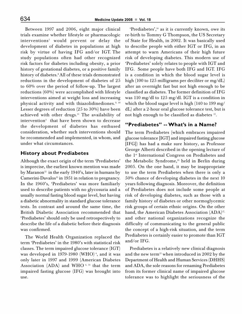
634 Medicine Update 2008 Vol. 18
Between 1997 and 2006, eight major clinical trials examine whether lifestyle or pharmacologic interventions7 would prevent or delay the development of diabetes in populations at high risk by virtue of having IFG and/or IGT. The study populations often had other recognized risk factors for diabetes including obesity, a prior history of gestational diabetes, or a positive family history of diabetes.8 All of these trials demonstrated reductions in the development of diabetes of 25 to 60% over the period of follow-up. The largest reductions (60%) were accomplished with lifestyle interventions aimed at weight loss and increasing physical activity and with thiazolidinediones.9, 10
Lesser degrees of reduction (25 to 30%) have been achieved with other drugs.11 The availability of intervention3 that have been shown to decrease the development of diabetes has enthused consideration, whether such interventions should be recommended and implemented, in whom, and under what circumstances.
History about Prediabetes Although the exact origin of the term ‘Prediabetes’ is imprecise, the earliest known mention was made by Maranon12 in the early 1940’s, later in humans by Camerini-Davalos13 in 1951 in relation to pregnancy. In the 1960’s, ‘Prediabetes’ was more familiarly used to describe patients with no glycosuria and a usually normal fasting blood sugar level, but having a diabetic abnormality in standard glucose tolerance tests. In contrast and around the same time, the British Diabetic Association recommended that ‘Prediabetes’ should only be used retrospectively to describe the life of a diabetic before their diagnosis was confirmed.
The World Health Organization replaced the term ‘Prediabetes’ in the 1980’s with statistical risk classes. The term impaired glucose tolerance (IGT) was developed in 1979-1980 (WHO) 4, and it was only later in 1997 and 1999 (American Diabetes Association (ADA) and WHO 4, 14 that the term impaired fasting glucose (IFG) was brought into use.
‘Prediabetes’,14 as it is currently known, owe its re-birth to Tommy G Thompson, the US Secretary of State for Health, in 2002. It was basically used to describe people with either IGT or IFG, in an attempt to warn Americans of their high future risk of developing diabetes. This modern use of ‘Prediabetes’ solely relates to people with IGT and IFG. Some people have both IFG and IGT. IFG is a condition in which the blood sugar level is high (100 to 125 milligrams per deciliter or mg/dL) after an overnight fast but not high enough to be classified as diabetes. The former definition of IFG was 110 mg/dl to 125 mg/dl. IGT is a condition in which the blood sugar level is high (140 to 199 mg/dL) after a 2-hour oral glucose tolerance test, but is not high enough to be classified as diabetes 15.
“Prediabetes” -- What’s in a Name? The term Prediabetes (which embraces impaired glucose tolerance [IGT] and impaired fasting glucose [IFG]) has had a make sure history, as Professor George Alberti described in the opening lecture of the 1stInternationalCongressonPrediabetesandthe Metabolic Syndrome,16 held in Berlin during 2005. On the one hand, it may be inappropriate to use the term Prediabetes when there is only a 50% chance of developing diabetes in the next 10 years following diagnosis. Moreover, the definition of Prediabetes does not include some people at risk of developing diabetes, such as those with a family history of diabetes or other normoglycemic risk groups of certain ethnic origins. On the other hand, the American Diabetes Association (ADA)17, and other national organizations recognize the difficulty of communicating to the general public the concept of a high-risk situation, and the term Prediabetes is certainly easier to promote than IGT and/or IFG.
Prediabetes is a relatively new clinical diagnosis and the new term15 when introduced in 2002 by the Department of Health and Human Services (DHHS) and ADA, the sole reasons for renaming Prediabetes from its former clinical name of impaired glucose tolerance was to highlight the seriousness of the

635“Prediabetes” - Early detection and interventions
condition and to motivate people to get appropriate treatment. Revised definition means millions more have “Pre-Diabetes”. “Pre-diabetes” -- a condition that raises a person’s risk of developing type 2 diabetes, heart disease, and stroke.7,17 The U.S. Department of Health and Human Services (DHHS) and the American Diabetes Association (ADA) estimated that 41 million Americans between the ages of 40 years to 74 years are living with Prediabetes, and most remain unaware of their condition. Without intervention and appropriate treatment, people with Prediabetes are at risk for developing type 2 diabetes within 10 years. 16
“Prediabetes” and the Metabolic Syndrome are extremely prevalent and persons with “Prediabetes and the Metabolic Syndrome” 16 are at high risk for diabetes and CVD and they are the idealtarget population for prevention or intervention programmes. In 2005,’ Prediabetes’ was given a global overview,14 in terms of isolated impaired glucose tolerance (IGT) or impaired fasting glucose (IFG), or a combination of the two, and highlighted the necessity to conduct an oral glucose test before diagnosing IGT. Prevalence data from a number of countries has generally found that IGT is more prevalent than IFG.
The epidemiology of Prediabetes On a global level,2 the type 2 diabetes epidemic in 2025 will comprise 97 million known cases, and an
equivalent number of unknown cases, with around 314 million people having IGT. By 2025, IGT is estimated that this will rise to approximately 500 million people raising concerns about a potential cardiovascular epidemic. (Table 1)
From global projections14 the major changes will occur in Eastern European states, the Middle East and India. Between 10% and 25% of western populations may already have IGT. For example, in the 2000, Australian Diabetes, Obesity and Lifestyle Study, 18 the overall prevalence of diabetes was 7.4%, but the combined prevalence of IFG and IGT was more than twice as high, at 16.4%. These glucose-intolerant, but non-diabetic, individuals represent a reservoir of potential new diabetes cases. Approximately 4 to 9% of individuals with impaired glucose tolerance go on to develop type 2 diabetes each year. Declining levels of physical activity, increasing caloric intake and consequent rises in the rate of obesity are leading to increase in the number of people with IGT from most ethnic and cultural backgrounds.16CountriesintheMiddleEast and Gulf States16 too have a high prevalence ofIGT,asdoesIndiaandChina19 (Table 2). More than 50% of people with diabetes will come from Asia over the next decade.14 In developed countries, the major increase in the number of people with diabetes will be among the young and middle-aged. However, additional data from Drs Söderberg and Ramachandran from Sweden and India, respectively, suggest that the ‘Prediabetes’ epidemic of IGT / IFG may be reaching a plateau in certain countries.16 In addition, gender differences are evident between IGT and IFG with women
Table 1
All diabetes and IGT 2003 2025
Total world population (billions) 6.3 8.0
Adult population (billions) (20-79 years)
3.8 5.3
Number of people with diabetes (millions) (20-79 years)
194 333
World diabetes prevalence (%) 5.1 6.3
Number of people with IGT (millions) (20-79 years)
314 472
IGT prevalence (%) (20-79 years) 8.2 9.0
Table 1 : 3rd edition, (IDF-Diabetes Atlas, International Diabetes Federation, 2006)
Table 2 : South East Asia – diabetes prevalence and future projections 1
2003 2005
Total Adult Population (millions) 705 1081
No. with Diabetes (millions) 39.3 81.6
Diabetes Prevalence (%) 5.6 7.5
No. with IGT (millions) 93.4 146.3
IGT Prevalence (%) 13.2 13.5
Table 2 : 3rd edition, (IDF-Diabetes Atlas, International Diabetes Federation, 2006)

636 Medicine Update 2008 Vol. 18
generally having a higher prevalence rate of IGT whilst men have a higher prevalence of IFG. On the contrary, the prevalence of IGT is higher in women than men in all age groups except over the age of 60 in Asian populations and over the age of 80 in the European groups. 18, 19. The unabated rise in the prevalence of childhood obesity has been accompanied by the appearance of a new pediatric disease, type 2 diabetes. Little is known about IGT in pediatrics.41 In adding up to differences in the overall prevalence between IGT and IFG, there are now clear evidence of differences in phenotype between the two categories. Genetic marker Studies in South Indians showed the complex nature of genetic pathology in type 2 diabetes. Certainmutations of candidate genes related to insulin secretion and insulin action such as Calpain 10,Vitamin D, Insulin receptor substrate-1, UCP2,UCP3, Apo-lipoprotein D gene are associatedwith the disorder. However; the nature of the major gene(s) responsible for the disease remains elusive.20
IGT and IFG are not equivalent metabolically, and it is therefore not surprising that there are differences in their prevalence and in the people categorized as having one or the other. In most populations, IGT is considerably more prevalent than IFG.16, 19 Furthermore, there is limited overlap between the categories - the majority of people with IGT do not have IFG, and the majority with IFG do not have IGT. Hence, the terminology of ‘isolated IGT’ and ‘isolated IFG’ has been given.3 Thus, IFG and IGT identify substantially different segments of the population with impaired glucose regulation (Table 3). The mechanisms of IGT and IFG are likely to be different; IFG is thought to be associated with a defect in insulin secretion whilst IGT is thought to be associated with hyperinsulinemia or insulin resistance. Thus ‘Prediabetes’ may encompass two different mechanisms of disease, necessitating the need for further research into both the mechanism and outcome of these two states.
Significance of IFG and IGT in IndiansOne of the earliest studies in India21 during 1986-87, in an urban population in a township in south India, the prevalence of 5% had diabetes and 14 had impaired glucose tolerance. A family history of diabetes was present in 16 of the 34 subjects with diabetes and nine of the 15 with impaired glucose tolerance. India has the highest number of IGT, prevalence of IGT and IFG are also high in India and south-east Asia in general.16, 19 which is expected to increase from 85.6 million (2003) to 132 million by 2025.
Prediabetes: Early recognition, its clinical significance and risks As mentioned in the introduction, these early stages of glucose intolerance (Prediabetes) are not only forerunners of diabetes but also carry high risk for cardiovascular diseases. Indians have high insulin resistant background in adding together to the presence of all other cardiovascular risk factors. IGT occurs at a much younger age in Indians 19 and they are predisposed to get diabetes more or less a decade prior as compared to the rest of the high risk population worldwide.
Early recognition is of extreme importance in initiating early interventions to stop the onset of diabetes related complications. Prediabetes is diagnosed with one of two blood tests—a fastingplasma glucose test or a two-hour oral glucose tolerance test (OGTT). The fasting plasma glucose test requires an eight-hour fast (no food or drink except water), after which a blood draw is performed. It is usually done in the morning. For an oral glucose tolerance test, a patient is given a drink of 75 grams of glucose, and a blood draw is taken two hours later. The following laboratory values are the American Diabetes Association (ADA) practice guidelines for the diagnosis of Prediabetes and Diabetes.17, (Tables : 3 & 4)
1. An oral glucose tolerance test plasma glucose value between 140 and 199 mg/dl (7.78 - 11.06 mmol/l) at 2 hours post-glucose load (indicating impaired glucose tolerance).
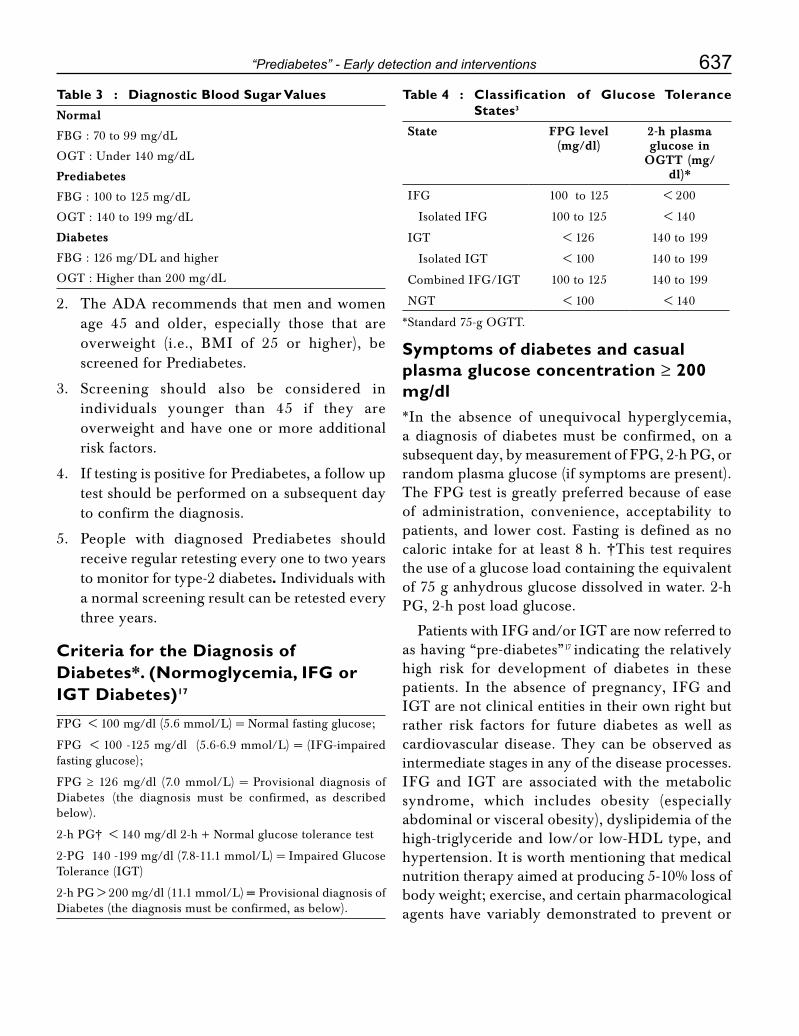
637“Prediabetes” - Early detection and interventions
2. The ADA recommends that men and women age 45 and older, especially those that are overweight (i.e., BMI of 25 or higher), be screened for Prediabetes.
3. Screening should also be considered in individuals younger than 45 if they are overweight and have one or more additional risk factors.
4. If testing is positive for Prediabetes, a follow up test should be performed on a subsequent day to confirm the diagnosis.
5. People with diagnosed Prediabetes should receive regular retesting every one to two years to monitor for type-2 diabetes. Individuals with a normal screening result can be retested every three years.
Criteria for the Diagnosis of Diabetes*. (Normoglycemia, IFG or IGT Diabetes)17
FPG < 100 mg/dl (5.6 mmol/L) = Normal fasting glucose;
FPG < 100 -125 mg/dl (5.6-6.9 mmol/L) = (IFG-impaired fasting glucose);
FPG ≥ 126 mg/dl (7.0 mmol/L) = Provisional diagnosis of Diabetes (the diagnosis must be confirmed, as described below).
2-h PG† < 140 mg/dl 2-h + Normal glucose tolerance test
2-PG 140 -199 mg/dl (7.8-11.1 mmol/L) = Impaired Glucose Tolerance (IGT)
2-h PG > 200 mg/dl (11.1 mmol/L) = Provisional diagnosis of Diabetes (the diagnosis must be confirmed, as below).
Symptoms of diabetes and casual plasma glucose concentration ≥ 200 mg/dl*In the absence of unequivocal hyperglycemia, a diagnosis of diabetes must be confirmed, on a subsequent day, by measurement of FPG, 2-h PG, or random plasma glucose (if symptoms are present). The FPG test is greatly preferred because of ease of administration, convenience, acceptability to patients, and lower cost. Fasting is defined as no caloric intake for at least 8 h. †This test requires the use of a glucose load containing the equivalent of 75 g anhydrous glucose dissolved in water. 2-h PG, 2-h post load glucose.
Patients with IFG and/or IGT are now referred to as having “pre-diabetes”17 indicating the relatively high risk for development of diabetes in these patients. In the absence of pregnancy, IFG and IGT are not clinical entities in their own right but rather risk factors for future diabetes as well as cardiovascular disease. They can be observed as intermediate stages in any of the disease processes. IFG and IGT are associated with the metabolic syndrome, which includes obesity (especially abdominal or visceral obesity), dyslipidemia of the high-triglyceride and low/or low-HDL type, and hypertension. It is worth mentioning that medical nutrition therapy aimed at producing 5-10% loss of body weight; exercise, and certain pharmacological agents have variably demonstrated to prevent or
Table 3 : Diagnostic Blood Sugar Values Normal
FBG : 70 to 99 mg/dL
OGT : Under 140 mg/dL
Prediabetes
FBG : 100 to 125 mg/dL
OGT : 140 to 199 mg/dL
Diabetes
FBG : 126 mg/DL and higher
OGT : Higher than 200 mg/dL
Table 4 : Classification of Glucose Tolerance States3
State FPG level (mg/dl)
2-h plasma glucose in
OGTT (mg/dl)*
IFG 100 to 125 < 200
Isolated IFG 100 to 125 < 140
IGT < 126 140 to 199
Isolated IGT < 100 140 to 199
CombinedIFG/IGT 100 to 125 140 to 199
NGT < 100 < 140
*Standard 75-g OGTT.

638 Medicine Update 2008 Vol. 18
delay the development of diabetes in people with IGT, the potential impact of such interventions to reduce cardiovascular risk has not been examined to date. Note that many individuals with IGT are euglycemic in their daily lives. Individuals with IFG or IGT may have normal or near normal glycated hemoglobin levels. Individuals with IGT often manifest hyperglycemia only when challenged with the oral glucose load used in the standard OGTT.
Preclinical Natural History 22, 33 Insulin Resistance, Insulin Secretion and the Balance between Alpha (ά) vs. Beta Cells (β):
Insulin resistance tends to be the “backbone” of type 2 diabetes; 80% to 85% of patients with type 2 diabetes have some degree of insulin resistance. There are two things that influence its development: genetic predisposition for insulin resistance, which some persons probably will inherit if family members have diabetes, and lifestyle and diet. If one is obese and sedentary, it contributes to the development of insulin resistance because when one gets obese. If a person is inherited the genetic potential, one need not even have to be obese to have insulin resistance. In fact, about 20% of patients who are at a normal weight or who are even underweight have insulin resistance, and about 20% of overweight patients who are clearly overweight or obese do not have insulin resistance. Obesity is important as a promoter of insulin resistance, but it is not required in order to develop insulin resistance. Typically, someone with genetic potential may also have a sedentary lifestyle, eat a diet that is hyper-caloric, and gain weight. However, someone with a genetic predisposition for abnormal beta-cell function can lose beta cells as time passes, without necessarily having insulin resistance. Finally, a person can eat a diet associated with weight gain, have a sedentary lifestyle and yet not have insulin resistance, and even maintain normal beta-cell function. So being overweight and sedentary does not necessarily mean that a person has insulin resistance, and conversely, a person does not need to have a genetic predisposition for insulin resistance in order
to have abnormal beta-cell function. But assuming that a patient has insulin resistance, the susceptible individual can take one of two pathways: patient either has genetically-programed normal beta cells, or has genetically-programed abnormal beta cells. If patient has normal beta cell function the person will go through life compensating for his hyperinsulinemia but person will always remain normoglycemic.
Those patients have the metabolic syndrome, which is a Prediabetes stage. They never develop elevated blood glucose because their beta cells are able to compensate for their insulin resistance. At this point is to do glucose challenge test, a lot of these patients do not have normal blood glucose; they have IGT. But if the patient’s beta cells are programed to function abnormally, they will not be able to compensate for their insulin resistance. They will have relative insulin deficiency, will develop hyperglycemia, and eventually, type 2 diabetes. Interestingly, only about 20% to 25% of patients at risk for diabetes, who have normal beta-cell function will deteriorate into abnormal beta-cell function. This means that about 80% of at-risk population is walking around with normal blood glucose.
The problem is that insulin resistance, IGT, compensatory hyperinsulinemia, and diabetes all accelerate atherogenesis. So just because the glucose is not elevated does not mean that there is no problem. There is a huge problem. The cardiovascular complication rate in IGT or metabolic syndrome is not quite what it is in overt diabetes, but it is still quite a bit higher than in a normal individual. How far a patient goes on this continuum is determined by how healthy their beta cells are, and elevated blood glucose is the last stage of this evolution.
Regulation of Insulin Secretion22 There are many things that affect the regulation of insulin secretion. The sulfonylurea drugs and the D-phenylalanine drugs, etc, that all attach themselves to the beta cell and do their work through transporters, etc. That is a complicated

639“Prediabetes” - Early detection and interventions
mechanism, but the purpose of this illustration is to show that drugs, neurotransmitters, nutrients, and hormones all affect insulin release from the beta cells of the pancreas.
Nutrients are often underappreciated. For example, that glucose affects insulin release. If an individual has a healthy pancreas, the higher the blood glucose, the more insulin is released. Amino acids do that too; they are pretty effective stimulators of insulin secretion. What is sometimes not appreciated is the role of free fatty acids, which are really powerful. When they are elevated, as is common in obesity, there is actually a shutdown ofinsulinproduction—calledlipo-toxicity—justasthere is with glucose elevation. When glucose is elevated chronically, there is a shutdown of insulin production called gluco-toxicity. Many patients have gluco- and lipo-toxicity. All these factors can affect insulin secretion. The newest players are the hormones: glucagon-like peptide-1 (GLP-1) principally, and the next one is gastric inhibitory polypeptide (GIP).
Islet Alpha- and Beta-cell Hormones Regulate Glucose Homeostasis 18, 22
The normal healthy subject has many beta cells in the islets that secrete insulin, and much fewer alfa cells in the islets that secrete glucagon. In type 2 diabetes it is a different situation. The number of beta cells is reduced, and they do not secrete as much insulin. The alfa cells do not decrease in
number—theglucagon-producingcellsremainaboutthe same—but they become dysfunctional. Theystart secreting glucagon, when glucagon should be suppressed. When there is type 2 diabetes, there is inappropriate secretion of glucagon from pancreatic alfa cells. There are mechanisms that are responsible for changes in beta-cell function. A normal beta cell adapts to insulin resistance by increasing secretion from each cell and increasing the number of cells (beta-cell mass). But when there is impaired beta-cell adaptation, and the inherited beta-cell weakness component, the susceptible person is destined to have impaired beta cells with decreased insulin secretion from each cell as well as reduced beta-cell mass.
Changes in Beta-Cell function over time: United Kingdom Prospective Diabetes Study (UKPDS) Data 33
From the UKPDS, that it has been shown over a very long 12- to 14-year study, beta-cell function failed progressively. By the time diabetes was diagnosed, with fasting blood glucose greater than 126 mg/dL, at least 50% of the functioning beta-cell population has been lost and maybe even 75% or 80%. (Figure 1).
The process begins 10 or 12 years before diagnosis.22 The blood glucose rises and the beta-cell mass begins to decrease. There is a very straight line that continues until, eventually, persons with type 2 diabetes, if they live long enough, will have almost no insulin production from their beta cells. Most type 2 diabetics need insulin eventually, because all the agents that are available today that are so effective in managing type 2 diabetes depend on the pancreas’ ability to make some insulin in order for these agents like Metformin, Sulfonylurea to work. With too little or no physiologic insulin production, exogenous insulin will then need to be administered. This is an important concept. Most type 2 diabetics, if they live long enough, will eventually need insulin either as supplemental or total therapy because their beta cells will continue to fail.
Figure 1
Stages in Development of Type 1 Diabetes
J. SKyler
TIME
NEWLY DIAGNOSED DIABETES
LOSS OF FIRST PHASEINSULIN RESPONSE
MULTIPLE ANTIBODY POSITIVE
GENETICALLY AT RISK
BET
A C
ELL
MA
SS
GENETICPREDISPOSITION
INSULATISBETA CELL INJURY
“PRE”-DIABETES
DIABETES

640 Medicine Update 2008 Vol. 18
Beta-Cell Mass in Normal Patients and Patients with Diabetes & Prediabetes22
In fact, autopsy studies show that people with diabetes, whether they are lean or obese, have much reduced beta-cell mass. In the mid-1970s and early 1980, islet cellautoantibodies (ICA), insulinautoantibodies(IAA), and recently, a 64-kd protein were found to be present in the serum of the majority of patients with type 1 diabetes at the time of diagnosis. This form of diabetes, which accounts for only 5–10% of
those with diabetes, previously encompassed by the terms insulin-dependent diabetes, type-1 diabetes, or juvenile-onset diabetes, results from a cellular-mediated autoimmune destruction of the ß-cells of the pancreas. Markers of the immune destruction of the ß-cell include islet cell auto-antibodies, auto-antibodies to insulin, auto-antibodies to glutamic acid decarboxylase (GAD
65), and auto-antibodies to
the tyrosine phosphatase IA-2 and IA-2ß. One and
usually more of these auto-antibodies are present in 85–90% of individuals when fasting hyperglycemia (IFG) is initially detected. Also, the disease has strong HLA associations, with linkage to the DQA and DQB genes, and it is influenced by the DRB genes. These HLA-DR/DQ alleles can be either predisposing or protective.
In this form of diabetes, the rate of ß-cell destruction is quite variable, being rapid in some individuals (mainly infants and children) and slow in others (mainly adults). Immune-mediated diabetes commonly occurs in childhood and adolescence,
but it can occur at any age, even in the 8th and 9th decades of life known as “Latent Onset of Diabetes in Adults” (LADA).22, 23, 33 Autoimmune destruction of ß-cells has multiple genetic predispositions and is also related to environmental factors that are still poorly defined. Although patients are rarely obese when they present with this type of diabetes, the presence of obesity is not incompatible with the diagnosis. These patients are also prone to other autoimmune disorders such as Graves’ disease, Hashimoto’s thyroiditis, Addison’s disease, vitiligo, celiac sprue, autoimmune hepatitis, myasthenia gravis, and pernicious anemia. Theoretically, it can be fairly predicted that immune markers are reasonably useful in early detection of type 1 diabetes as early as few years too that will help in designing early interventions and prevent Type 1 diabetes.
Insulin Patterns in Diabetes22
Abnormal Acute Insulin Response to Intravenous Glucose in Type 2 Diabetes is seen in some of patients’ cousins, uncles, aunts, and grandparents and some of these relatives will have blunted first-phase insulin release. Their blood glucose levels may be normal but the first-phase insulin release may be blunted. A very strong marker of genetically transmitted diabetes is the loss of first-phase insulin release.
Clinical significance of Prediabetes & Diabetes riskPrediabetes as a model of the metabolic syndrome
There has been in recent time’s confusion about definitions of Prediabetes. Traditionally, Prediabetes referred to studies in which subjects were followed longitudinally and one could actually identify which subjects would later develop diabetes. In
Figure 2
Source42LebovitzH.Changesinbeta-cellfunctionovertime:UKPDS data, Diabetes Rev. 1999:7, 139-53
Changes in β-Cell FunctionOver Time : UKPDS Data
Rx: Insulin, Metformin, Sulfonylurea
Type 2DiabetesPhase III
Type 2DiabetesPhase I
Type 2DiabetesPhase II
PostprandialHyperglycemia
IGT
β-CellFunction
(%)
100
75
50
25
0-12 -10 -6 -2 0 2 6 10 14
Years from diagnosisDashed line shows extrapolation forward and backward from years 0 to 6 basedon HOMA data from UKPDS.
641“Prediabetes” - Early detection and interventions
2003, the ADA suggested that certain high-risk groups such as individuals with impaired fasting glucose (IFG) and impaired glucose tolerance (IGT) should be identified as having Prediabetes. This is an unfortunate choice of a name, since many or most IGT and particularly IFG subjects will never develop diabetes.23 Given the natural history of Prediabetes, about 3%–10% of people per year with Prediabetes develop diabetes. Data are particularly well substantiated for IGT. In the Diabetes Prevention Programme24 with subjects who had IGT, with or without IFG, there was about a 10% annual rate of progression to diabetes in the control group. Overall, Prediabetes confers about a six fold increased risk of diabetes compared with normal glucose tolerance. In most populations studied, the rates of conversion from IFG and IGT to diabetes are similar, with IGT having greater sensitivity but less specificity than IFG in predicting diabetes risk.25 In an 11-year follow-up study among adults with IGT in Mauritius, 46% developed diabetes, 28% remained unchanged in category, 4% developed IFG, and glucose levels normalized in 24%. Thus, many people with Prediabetes (a quarter or more) may revert long term to having normal glucose tolerance, and after a protracted follow-up, only about 50% of people with IGT or IFG will develop diabetes.
Cardiovascular disease riskIn comparison with adults who have normal glucose tolerance, people with Prediabetes have an increased risk of developing cardiovascular disease (CVD) and cardiovascular and all-causemortality.26 There is a two- to threefold increased prospective risk of cardiovascular events, which is most marked in younger adults with Prediabetes. Prediabetes is associated with increased rates of the cardiovascular risk factors found in people with type 2 diabetes. Some data indicate that people with IGT and normal levels of fasting plasma glucose haveagreaterriskofCVDthanthosewithIFG.Inaddition,whenotherknownCVDriskfactors,such as hypertension and lipid abnormalities, are
adjusted for statistically, IGT, but possibly not IFG, remains as an independentCVDrisk factor.Anincreasing plasma glucose level in IGT is associated with a greater risk of cardiovascular death.
Associations with the metabolic syndromeThe metabolic syndrome (MetS) refers to a clustering in an individual of CVD risk factorsand diabetes susceptibility.26 People with MetS have about a twofold increased risk of developing diabetes and cardiovascular disease, compared with those without the syndrome. Several MetS definitions exist, with two being widely used. Recently, a third definition has been adopted by the International Diabetes Federation.27 Each definition has impairment of glucose metabolism as an optional criterion, although some consider only IFG. Most adults who have Prediabetes will also have MetS. Whether Prediabetes or MetS best defines diabetes and cardiovascular risk remains to be determined. However, it is not clear whether MetS and Prediabetes represent the same or different clinical entities.28 The data demonstrated by Diamantopoulos et al28 showed that MetS and Prediabetes have an overlapping pattern. MetS appears to have a more pronounced effect on early renal dysfunction and increased inflammatory activation, while Prediabetes tends to be associated with early carotid structural changes. These findings may be due to a different patho-physiologic substrate of these clinical phenotypes in terms of insulin resistance and secretion, as well as to the varying prevalence of cardiovascular risk factors.
IGT also accounts for a highly heterogeneous Japanese population,29 with the condition varying from individual to individual. In this study, findings suggest that IGT subjects with high insulin response and those with low insulin response vary greatly in regard to the number of atherosclerotic risk factors complicated and the frequency with which they are associated with the metabolic syndrome. It is also shown in middle-aged Japanese males that
642 Medicine Update 2008 Vol. 18
of the two forms of IGT, IGT with high insulin response is more closely linked to the pathogenesis of atherosclerotic cardiovascular disease. Impaired glucose tolerance (IGT) represents a Prediabetes state positioned somewhere between normal glucose tolerance and diabetes, which is also assumed to make individuals in this state highly susceptible to atherosclerotic disease.29
An observation suggested by sahib et al30 that insulin resistance may be associated with essential hypertension. There are some thoughts to favour the argument that insulin resistant Individuals are at a higher risk to develop hypertension as compared to insulin sensitive individuals.
Interventions to prevent Prediabetes3 Just as there are different potential definitions
of the natural history of IFG and IGT, there are different ways in which the natural history can be altered. The progression to diabetes is a time-dependent phenomenon; one possible alteration is simply to “reset the clock” without changing the rate of the deterioration. It is possible that some interventions will lower glycemia initially but do nothing to change the subsequent rate of rise of
glycemia. This mechanism will delay crossing the glycemia threshold that defines diabetes. Prediabetes is a condition that does not fall squarely into the primary or secondary prevention domain, and therefore tends to be inadequately addressed by interventions in either health promotion or disease management. There is substantial evidence to suggest that even at these blood glucose levels, significant risk exists for both micro and, macro vascular complications. Biuso et al31 introduces a conceptual framework of care for Prediabetes that includes both screening and the provision of up-to-date clinical therapies in conjunction with an evidence-based health coaching intervention. In combination, these modalities represent the most effective means for delaying or even preventing the onset of diabetes in a Prediabetes population.
Research studies have found that lifestyle changes3,31,34 can prevent or delay the onset of type 2 diabetes among high-risk adults. The three components of lifestyle modification are diet, exercise, and behavior therapy (Table 5 & Figure 4), Several reviews have found that standard lifestyle modification programs conducted in academic medical centers induce a mean weight reduction of approximately 8–10% of initial weight in 16–26 weeks of treatment.36 These studies included people with IGT and other high-risk characteristics
Table 5 : Treatment Recommendation for Individuals with IFG, IGT, or Both 3
Population Treatment
IFG or IGT Lifestyle modification (i.e., 5 to 10% weight loss and moderate intensity physical activity > 30 min/day) in a week
Individuals with IFG and IGT and any of the following:
• <60yearsofage
• BMI≥ 35 kg/m2
• Familyhistoryofdiabetes infirst-degree relatives
• Elevatedtriglycerides
• ReducedHDLcholesterol
• Hypertension
• A1C>6.0%
Lifestyle modification (as above) and/or Metformin*
*Metformin 850 mg twice per day.
Figure 3
Wadden TA, Butryn ML, Byrne KJ. Efficacy of lifestyle modification for long-term weight control. Obes Res 2004;12:151S-162S.
LOW-Calorie
DietBehaviorTherapy
IncreasedPhysicalActivity
Lifestyle Modification

643“Prediabetes” - Early detection and interventions
for developing diabetes. Lifestyle interventions included diet and moderate-intensity physical activity (such as walking for 2 1/2 hours each week). In the Diabetes Prevention Program, a large prevention study of people at high risk for diabetes, the development of diabetes was reduced 58% over 3 years.
In the Diabetes Prevention Program,8 people treated with the drug Metformin reduced their risk of developing diabetes by 31% over 3 years. Treatment with Metformin was most effective among younger, heavier people (those 25-40 years of age who were 50 to 80 pounds overweight) and less effective among older people and people who were not as overweight. Similarly, in the STOP-NIDDM Trial,8,24 treatment of people with IGT with the drug Acarbose reduced the risk of developing diabetes by 25% over 3 years. Other medication studies are ongoing. Besides lifestyle, various pharmacological treatments have proven their efficacy to reduce the incidence of type 2 diabetes in high-risk individuals, especially in those with impaired glucose tolerance (IGT) and/or impaired fasting glucose (IFG). Ongoing trials should confirm such a favorable effect with those drugs and may demonstrate a similar protective effect with other pharmacological approaches such as glinides or even basal insulin regimen. Therefore, the distinction between a true preventing effect and simply a masking effect is difficult with glucose-lowering drugs.34 In addition, as type 2 diabetes is a progressive disease, it is still questionable whether the effect corresponds to a prevention effect or only to a postponing of the development of the disease. Owing to the patho-physiology of the disease, the only way to block the progression to type 2 diabetes is probably to avoid the progressive loss of beta-cell function and/or mass. Whatsoever, these data obtained in large clinical trials bring further argument to support early treatment of diabetes, even at a Prediabetes state, in order to stop the vicious circle leading to an inevitable deterioration of glycemia in predisposed subjects. The demonstration by recent randomized controlled trials that type 2 diabetes mellitus is
preventable has raised hope for the possibility of reducing cardiovascular morbidity and mortality associated with diabetes. Interventions like lifestyle modification and pharmacological therapy are recommended in individuals with Prediabetes to achieve the goal of prevention of diabetes in high -risk population.35
ConclusionPresentations at the 1st International Congresson “Prediabetes” and the Metabolic Syndrome14, 16 reported that better definition and intense study of Prediabetes and the metabolic syndrome have led to some important insights in the past decade:
1. Prediabetes and the metabolic syndrome are extremely prevalent;
2. People with Prediabetes and the metabolic syndrome are at high risk for diabetes and CVD;
3. Early detection of IFG / IGT in high risk individuals and interventions to prevent progression to diabetes through Intensive lifestyle changes are effective and should be encouraged; and
4. Effective pharmacologic therapies must also be identified.
References1. Diabetes Atlas. 3rd edition, International Diabetes Federation,
2006; 1
2. Zimmet P, Alberti KG, Shaw J. Global and societal implications of the diabetes epidemic. Nature. 2001; 414:782-87.
3. Nathan DM, Davidson MB, DeFronzo RA, et al. Impaired Fasting Glucose and Impaired Glucose Tolerance: Implications forCare,Diabetes Care. 2007;30(3):753-59.
4. Alberti GK, Zimmet PZ. Definition, diagnosis and classification of diabetes mellitus and its complications. Part 1: diagnosis and classification of diabetes mellitus provisional report of a WHO consultation. Diabet Med. 1998;15:539-53
5. Eckel RH, Grundy SM, Zimmet PZ. The metabolic syndrome. Lancet. 2005; 365:1415-28.
6. TwiggSM,KampMC,DavisTMetal.Prediabetes:apositionstatement from the Australian Diabetes Society and Australian Diabetes Educators Association. 2007; 186 (9): 461-5
644 Medicine Update 2008 Vol. 18
7. Knowler WC, Barrett-Conner E, Fowler SE, et al. TheDiabetes Prevention Program Research Group: Reduction in the incidence of type 2 diabetes with lifestyle intervention or Metformin. N Engl J Med 2002; 346:393-403.
8. Chiasson JL, Josse RG, Gomis R, Hanefetd M, KarasikA, Laakso M. the STOP-NIDDM Trial Research Group: Acarbose for prevention of type 2 diabetes mellitus: the STOP-NIDDM randomized trial. Lancet 2002; 359:2072-7.
9. Torgerson JS, Hauptman J, Boldrin MN, et al. Xenical in the prevention of diabetes in obese subjects (XENDOS) study: a randomized study of orlistat as an adjunct to lifestyle changes for the prevention of type 2 diabetes in obese patients. Diabetes Care 2004; 27:151-61.
10. Ramachandran A, Snehalatha C, Mary S, et al: IndianDiabetes Prevention Programme (IDPP): the Indian Diabetes Prevention Programme shows that lifestyle modification and Metformin prevent type 2 diabetes in Asian Indian subjects with impaired glucose tolerance (IDPP-1). Diabetologia 2006;49:289-97.
11. Gerstein HC, Yusuf S, Boxch J, Pogue J, et al: DREAM(Diabetes Reduction Assessment with Ramipril and Rosiglitazone Medication) Trial Investigators: effect of rosiglitazone on the frequency of diabetes in patients with impaired glucose tolerance or impaired fasting glucose: a randomized controlled trial. Lancet 2006, 368:1096-105.
12. Vague J. Sexual differentiation. A factor affecting the forms of obesity. Presse Med. 1947; 30:339-40.
13. Avogaro P, Crepaldi G, Enzi G, et al. Associazione diiperlipemia, diabete mellito e obesita di medio grado. Acta Diabetol Lat. 1967;4:572-90.
14. Crepaldi G. Origin and development of the metabolicsyndrome. Program and abstracts of the 1st International Congress on “Prediabetes” and the Metabolic Syndrome;Berlin, Germany. 2005;13-6.
15. TheExpertCommitteeontheDiagnosisandClassificationof Diabetes Mellitus: Follow-up report on the diagnosis of diabetes mellitus. Diabetes Care 2003;26: 3160–7.
16. Grundy S. Do we need to expand the metabolic syndrome? Programandabstractsofthe1stInternationalCongresson“Prediabetes” and the Metabolic Syndrome; Berlin, Germany. 2005;13-16.
17. ADAClinicalPracticeRecommendations.PositionStatement.Diabetes Care. 2007 S46; 30:1
18. TwiggSM,KampMC,DavisTMetal.Prediabetes:apositionstatement from the Australian Diabetes Society and Australian Diabetes Educators Association. 2007; 186 (9): 461-5
19. A Ramachandran. Netaji Oration. Epidemiology of Diabetes in India – Three Decades of Research. Journal Phy of India. 2005; 53:35-8
20. GrahamAH,McCarthyMI,MohanVetal.Thegeneticsofnon-insulin dependent diabetes mellitus in south-India: An
overview. Ann Med 1992; 24:491-7.
21. Ramachandran A, Jali MV, Mohan V, et al. High prevalence of diabetes in an urban population in south India. BMJ. 1988; 297(6648):587-90
22. Sherry NA, Tsai EB, Herold KC. Natural History ofß-CellFunction inType1Diabetes,Section I:AspectsofPathophysiology, Diabetes 2005; 54:S32-S39
23. American Diabetes Association. Diagnosis and classification of diabetes mellitus. Diabetes Care 2006;29:S43-S48.
24. KnowlerWC,Barrett-ConnorE,FowlerSE,etal.DiabetesPrevention Program Research Group. Reduction in the incidence of type 2 diabetes with lifestyle intervention or Metformin. N Engl J Med 2002; 346: 393-403.
25. Unwin N, Shaw J, Zimmet P, Alberti KG. Impaired glucose tolerance and impaired fasting Glycaemic: the current status on definition and intervention. Diabet Med 2002; 19: 708-23.
26. Decode Study Group. Glucose tolerance and cardiovascular mortality: comparison of fasting and 2-hour diagnostic criteria. Arch Intern Med 2001; 161: 397-405.
27. Alberti KG, Zimmet P, Shaw J; IDF Epidemiology Taskforce Consensus Group. The metabolic syndrome — a newworldwide definition. Lancet 2005; 366: 1059-62.
28. Diamantopoulos EJ, Andreadis EA, Tsourous GI, et al. Metabolic syndrome and Prediabetes identify overlapping but not identical populations.Exp Clin Endocrinol Diabetes. 2006 ;114(7):377-83.
29. Mori Y, Hoshino K, Yokota K, et al. Japanese IGT subjects with high insulin response are far more frequently associated with the metabolic syndrome than those with low insulin response. 2006; 29(2):351-5.
30. Sahib AK, Sahu SK, Reddy KN. Prediabetes and hypertension. J Indian Med Assoc. 2007; 105(1):25-8.
31. Biuso TJ, Butterworth S, Linden A. A conceptual framework for targeting Prediabetes with lifestyle, clinical and behavioral management interventions. Dis Manag. 2007;10(1):6-15
32. Sinha R, Fisch G, Teague B, Tamborlane WV, et al. Prevalence of impaired glucose tolerance among children and adolescents with marked obesity. N Engl J Med. 2002;346(11):802-10.
33. LebovitzH.Changesinbeta-cellfunctionovertime:UKPDSdata, Diabetes Rev. 1999:7, 139-53
34. Scheen AJ. Antidiabetic agents in subjects with mild dysglycaemia: prevention or early treatment of type 2 diabetes? Diabetes Metab. 2007;33(1):3-12.
35. Zargar AH, Bashir MI. Epidemiological aspects of Prediabetes--review of the current data. J Indian Med Assoc. 2005;103(11):591-3
36. Wadden TA, Butryn ML, Byrne KJ. Efficacy of lifestyle modification for long-term weight control. Obes Res 2004;12:151S-162S.

CHAPTER
86CAD in WomenJ. Mukhopadhyay, D. Chakraborty
IntroductionThe medical community viewed women’s health with a bikini approach, focusing essentially on the breast and reproductive system. The rest of women were virtually ignored in considerations of women’s health. Traditionally, coronary heart disease (CHD) has been considered as a maleproblem,butrelevantinformationaboutCHDinwomen is rapidly escalating recently.
TheonsetofclinicalmanifestationsofCHDinwomen lags behind men by about 10 years and by as much as 20 years for more ominous events. But CHDisnotsolelyaproblemforelderlywomen.More than 9000 US women younger than 45 years sustain a myocardial infarction (MI) each year. The question of why women younger than 65 years of age are more than twice as likely to die from MI than comparably aged men is intriguing. Recent surveys reveal that most women do not appreciate their risk of developing CHD,1 and, therefore, women may not recognize that certain life style behaviors and physiological factors increase the riskofdevelopingCHD.
Many women are unaware that coronary heart disease is their main killer; their biggest fear is breast cancer. Even more worrying, however, is the apparent lack of awareness of cardiovascular disease in women among healthcare professionals.
At the time of presentation with heart disease, women tend to be 10 years older than men, and at the time of their first myocardial infarction they are usually 20 years older2,3 As coronary heart disease is a disease of the older woman, many women believe that they can postpone attempts to reduce their risk.
IncidenceWorldwide, cardiovascular disease (CVD) isthe largest single cause of death among women, accounting for one third of all deaths.4
Cardiovascular disease (CVD) is an equal-opportunity killer in men and women over their lifetimes. In Washington state, in 1991, the incidenceofCVDdeathwas42%inwomenand39% in men.5 Nationwide (US), these numbers approach 50%. Among survivors of MI, 25% of men versus 38% of women die within a year after an initial MI.6 Within 6 years after MI, 18% of men but 35% of women will have a recurrent infarction. Women are more likely to be disabled by heart failure (30% vs. 21%) after MI. Women with unstable angina have a survival advantage compared with men.6
In the Indian context the mortality data for coronary artery diseases in women is available from two studies done in 1994 and 1998, JIMI – I
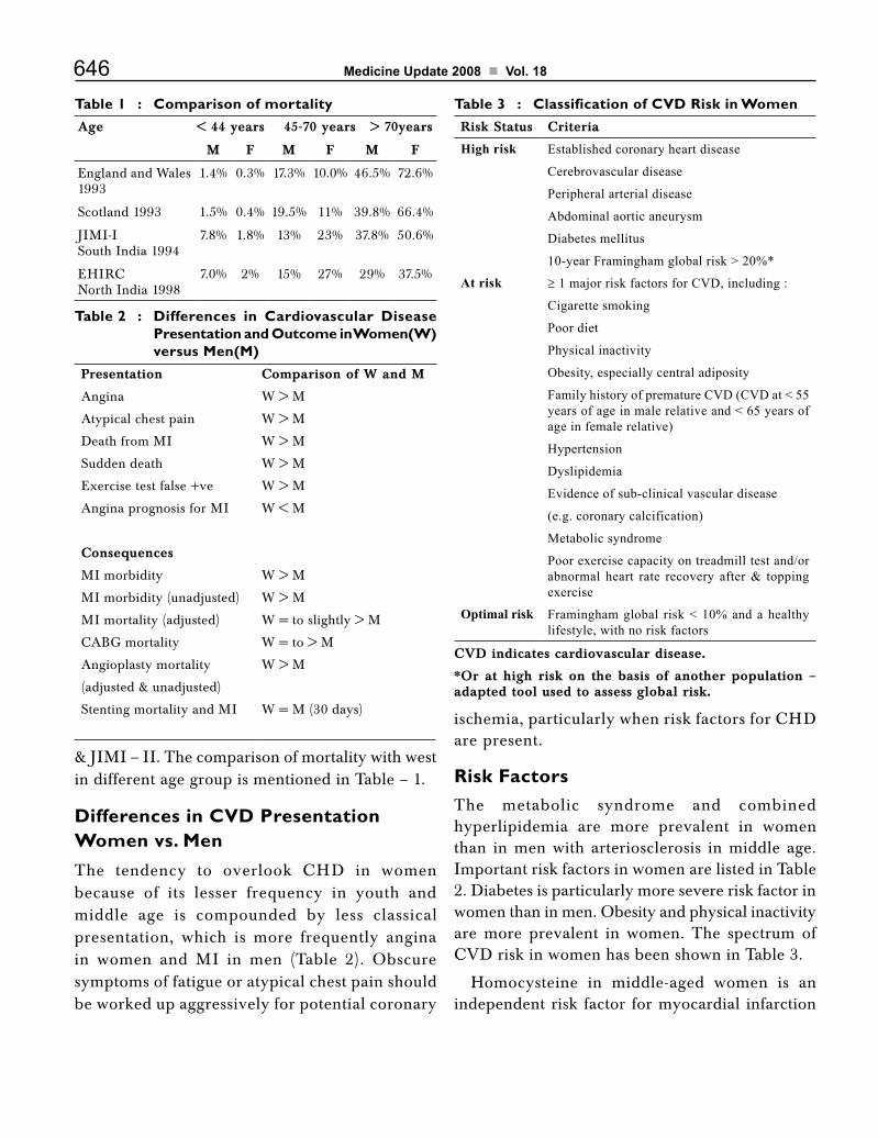
646 Medicine Update 2008 Vol. 18
& JIMI – II. The comparison of mortality with west in different age group is mentioned in Table – 1.
Differences in CVD Presentation Women vs. MenThe tendency to overlook CHD in womenbecause of its lesser frequency in youth and middle age is compounded by less classical presentation, which is more frequently angina in women and MI in men (Table 2). Obscure symptoms of fatigue or atypical chest pain should be worked up aggressively for potential coronary
ischemia,particularlywhenriskfactorsforCHDare present.
Risk FactorsThe metabolic syndrome and combined hyperlipidemia are more prevalent in women than in men with arteriosclerosis in middle age. Important risk factors in women are listed in Table 2. Diabetes is particularly more severe risk factor in women than in men. Obesity and physical inactivity are more prevalent in women. The spectrum of CVDriskinwomenhasbeenshowninTable3.
Homocysteine in middle-aged women is an independent risk factor for myocardial infarction
Table 1 : Comparison of mortalityAge < 44 years 45-70 years > 70years
M F M F M F
England and Wales 1993
1.4% 0.3% 17.3% 10.0% 46.5% 72.6%
Scotland 1993 1.5% 0.4% 19.5% 11% 39.8% 66.4%
JIMI-I South India 1994
7.8% 1.8% 13% 23% 37.8% 50.6%
EHIRC North India 1998
7.0% 2% 15% 27% 29% 37.5%
Table 2 : Differences in Cardiovascular Disease Presentation and Outcome in Women(W) versus Men(M)
Presentation
Angina
Atypical chest pain
Death from MI
Sudden death
Exercise test false +ve
Angina prognosis for MI
Consequences
MI morbidity
MI morbidity (unadjusted)
MI mortality (adjusted)
CABGmortality
Angioplasty mortality
(adjusted & unadjusted)
Stenting mortality and MI
Comparison of W and M
W > M
W > M
W > M
W > M
W > M
W < M
W > M
W > M
W = to slightly > M
W = to > M
W > M
W = M (30 days)
Table 3 : Classification of CVD Risk in WomenRisk Status Criteria
High risk Established coronary heart disease
Cerebrovascular disease
Peripheral arterial disease
Abdominal aortic aneurysm
Diabetes mellitus
10-year Framingham global risk > 20%*
At risk ≥ 1 major risk factors for CVD, including :
Cigarette smoking
Poor diet
Physical inactivity
Obesity, especially central adiposity
Family history of premature CVD (CVD at < 55 years of age in male relative and < 65 years of age in female relative)
Hypertension
Dyslipidemia
Evidence of sub-clinical vascular disease
(e.g. coronary calcification)
Metabolic syndrome
Poor exercise capacity on treadmill test and/or abnormal heart rate recovery after & topping exercise
Optimal risk Framingham global risk < 10% and a healthy lifestyle, with no risk factors
CVD indicates cardiovascular disease.
*Or at high risk on the basis of another population – adapted tool used to assess global risk.

647CAD in Women
and in particular, mortality due to myocardial infarction7. Elevated tPA antigen and to a lesser extent D-dimer are independently associated with incident coronary events among postmenopausal women. In analysis stratified by menopausal hormone therapy, tPA antigen remains a consistent marker of increased coronary risk.10
TherolethatnovelCVDriskfactors(e.g.,high-sensitivityC-reactiveprotein)andnovelscreeningtechnologies (e.g., coronary calcium scoring) should play in guiding preventive interventions is not yet defined. Further research is needed on added benefits, risks, and costs associated with such strategies before they can be incorporated into guidelines. Unique opportunities to identify women’s risk (e.g., during pregnancy) also deserve further exploration. For example, pre-eclampsia maybeanearly indicatorofCVDrisk.Womenwith pre-eclampsia/eclampsia are significantly more likely to develop hypertension and cerebrovascular disease. In addition, maternal placental syndromes in combination with traditional cardiovascular risk factors, such as pre-pregnancy hypertension or diabetes mellitus, obesity, dyslipidemia, or metabolic syndrome, may be additive in defining CVD risk in women.16 Future research should evaluate the potential for events or medical contact during unique phases in a woman’s lifespan, such as adolescence, pregnancy, and menopause, to identify women at high risk and to determine the effectiveness of preventive interventions during critical time periods.
DiagnosisRecognition of cardiovascular chest pain is difficult in women because of its atypical nature. As a result of atypical symptoms, misdiagnosis as chronic fatigue or a psychiatric disorder is not uncommon. The reason for the lack of classical anginal symptoms in many women, despite having validated myocardial ischemia, is unknown. The greater incidence of silent MI in women may also be related to the atypicality of chest pain presentation.
The exercise tolerance is not as useful in clarifying atypical chest pain in women as in men because it is too susceptible to false-positive and false-negative results. In general, radionuclide or echocardiographic imagings are recommended, if an exercise test is to be done.11 Nuclear stress perfusion testing in women can be potentially hindered by soft tissue attenuation from breast tissue with the use of thallium, so technetium may be preferred. Many authors prefer stress imaging tests, with their lower false-positive rates, to exercise stress tests for women.12 A female patient with chest pain with a positive exercise test and a negative angiogram might have arteriosclerotic vasospasm with a normal lumen. Direct referral to cardiac catheterization should occur with a high suspicion of significant CAD that might benefit from intervention orafter an abnormal noninvasive stress test. Anginal symptoms are less predictive of abnormal coronary anatomy in women than men.
In patients with UA/NSTEMI, there is a different pattern of presenting biomarkers. Men are more likely to have elevated CK-MB and troponins,whereas women are more likely to have elevated CRPandBNP.This suggests thatamultimarkerapproach may aid the initial risk assessment of UA/NSTEMI, especially in women.13
The relationship between the menstrual cycle and vascular spasm is beginning to receive increasing attention.14
Acute Coronary Syndrome (ACS)There are substantial gender differences in the presentation and natural history. After AMI, women have higher mortality during hospitalization, arrived later for evaluation after symptoms began, received less thrombolytic treatment as well as fewer invasive interventions. Women have higher mortality rates than men, even at similar ages or after similar interventions, from cardiogenic shock, sudden death, arrhythmias, myocardial rupture and electromechanical dissociation.
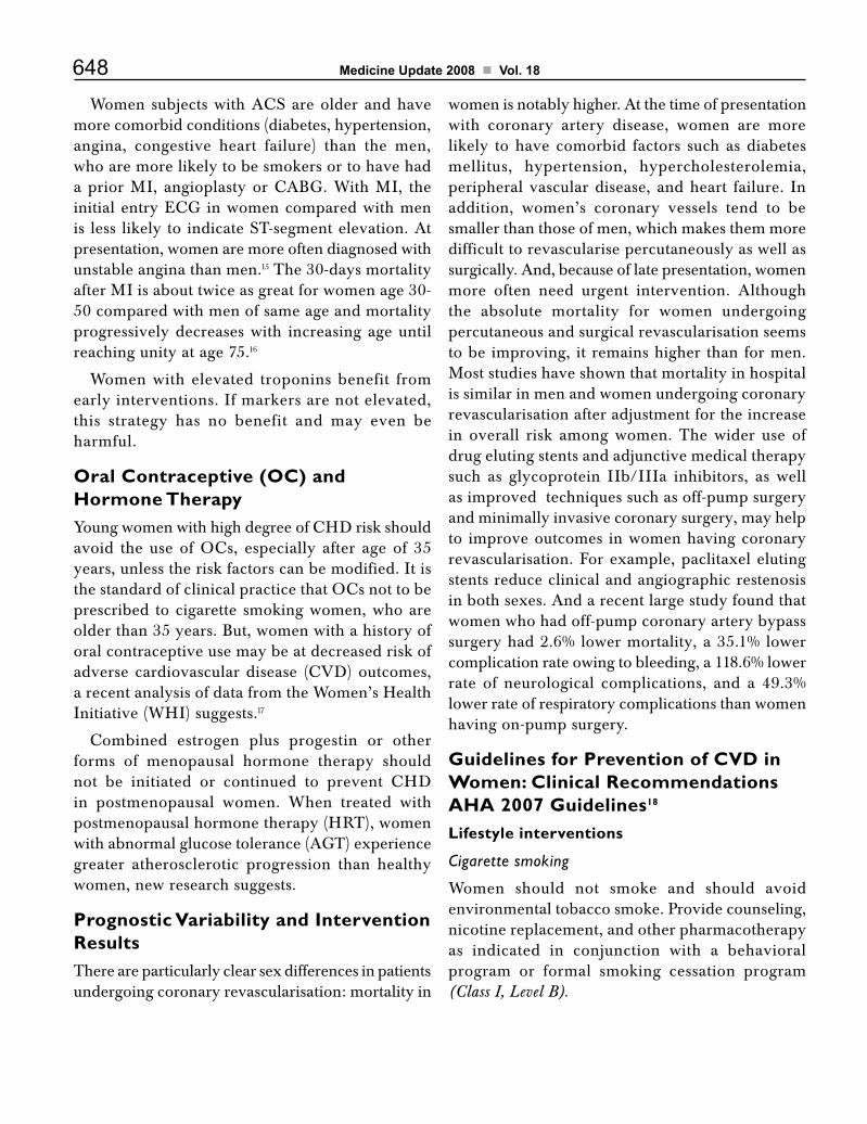
648 Medicine Update 2008 Vol. 18
WomensubjectswithACSareolderandhavemore comorbid conditions (diabetes, hypertension, angina, congestive heart failure) than the men, who are more likely to be smokers or to have had apriorMI, angioplastyorCABG.WithMI, theinitialentryECGinwomencomparedwithmenis less likely to indicate ST-segment elevation. At presentation, women are more often diagnosed with unstable angina than men.15 The 30-days mortality after MI is about twice as great for women age 30-50 compared with men of same age and mortality progressively decreases with increasing age until reaching unity at age 75.16
Women with elevated troponins benefit from early interventions. If markers are not elevated, this strategy has no benefit and may even be harmful.
Oral Contraceptive (OC) and Hormone TherapyYoungwomenwithhighdegreeofCHDriskshouldavoid theuseofOCs, especially after ageof 35years, unless the risk factors can be modified. It is thestandardofclinicalpracticethatOCsnottobeprescribed to cigarette smoking women, who are older than 35 years. But, women with a history of oral contraceptive use may be at decreased risk of adversecardiovasculardisease (CVD)outcomes,a recent analysis of data from the Women’s Health Initiative (WHI) suggests.17
Combined estrogen plus progestin or otherforms of menopausal hormone therapy should not be initiated or continued to prevent CHDin postmenopausal women. When treated with postmenopausal hormone therapy (HRT), women with abnormal glucose tolerance (AGT) experience greater atherosclerotic progression than healthy women, new research suggests.
Prognostic Variability and Intervention ResultsThere are particularly clear sex differences in patients undergoing coronary revascularisation: mortality in
women is notably higher. At the time of presentation with coronary artery disease, women are more likely to have comorbid factors such as diabetes mellitus, hypertension, hypercholesterolemia, peripheral vascular disease, and heart failure. In addition, women’s coronary vessels tend to be smaller than those of men, which makes them more difficult to revascularise percutaneously as well as surgically. And, because of late presentation, women more often need urgent intervention. Although the absolute mortality for women undergoing percutaneous and surgical revascularisation seems to be improving, it remains higher than for men. Most studies have shown that mortality in hospital is similar in men and women undergoing coronary revascularisation after adjustment for the increase in overall risk among women. The wider use of drug eluting stents and adjunctive medical therapy such as glycoprotein IIb/IIIa inhibitors, as well as improved techniques such as off-pump surgery and minimally invasive coronary surgery, may help to improve outcomes in women having coronary revascularisation. For example, paclitaxel eluting stents reduce clinical and angiographic restenosis in both sexes. And a recent large study found that women who had off-pump coronary artery bypass surgery had 2.6% lower mortality, a 35.1% lower complication rate owing to bleeding, a 118.6% lower rate of neurological complications, and a 49.3% lower rate of respiratory complications than women having on-pump surgery.
Guidelines for Prevention of CVD in Women: Clinical Recommendations AHA 2007 Guidelines18
Lifestyle interventions
Cigarette smoking
Women should not smoke and should avoid environmental tobacco smoke. Provide counseling, nicotine replacement, and other pharmacotherapy as indicated in conjunction with a behavioral program or formal smoking cessation program (Class I, Level B).

649CAD in Women
Physical activity
Women should accumulate a minimum of 30 minutes of moderate-intensity physical activity (e.g., brisk walking) on most, and preferably all, days of the week (Class I, Level B). Women who need to lose weight or sustain weight loss should accumulate a minimum of 60 to 90 minutes of moderate-intensity physical activity (e.g., brisk walking) on most, and preferably all, days of the week (Class I, Level C).
Rehabilitation
A comprehensive risk-reduction regimen, such as cardiovascular or stroke rehabilitation or a physician-guided home- or community-based exercise training program, should be recommended to women with a recent acute coronary syndrome or coronary intervention, new-onset or chronic angina, recent cerebrovascular event, peripheral arterial disease (Class I, Level A), or current/prior symptoms of heart failure and an LVEF 40% (Class I, Level B).
Dietary intake
Women should consume a diet rich in fruits and vegetables; choose whole-grain, high-fiber foods; consume fish, especially oily fish, at least twice a week; limit intake of saturated fat to 10% of energy, and if possible to 7%, cholesterol to 300 mg/d, alcohol intake to no more than 1 drink per day, and sodium intake to 2.3 g/d (approximately 1 tsp salt). Consumptionoftrans-fatty acids should be as low as possible (e.g., 1% of energy) (Class I, Level B).
Weight maintenance/reduction
Women should maintain or lose weight through an appropriate balance of physical activity, caloric intake, and formal behavioral programs when indicated to maintain/achieve a BMI between 18.5 and 24.9 kg/m2 and a waist circumference 35 in (Class I, Level B).
Omega-3 fatty acids
As an adjunct to diet, omega-3 fatty acids in capsule form (approximately 850 to 1000 mg of EPA and DHA)maybeconsideredinwomenwithCHD,and
higher doses (2 to 4 g) may be used for treatment of women with high triglyceride levels (Class IIb, Level B).
Depression
Consider screening women with CHD fordepression and refer/treat when indicated (Class IIa, Level B).
Major risk factor interventions
Blood pressure—optimal level and lifestyle
Encourage an optimal blood pressure of 120/80 mm Hg through lifestyle approaches such as weight control, increased physical activity, alcohol moderation, sodium restriction, and increased consumption of fresh fruits, vegetables, and low-fat dairy products (Class I, Level B).
Blood pressure—pharmacotherapy
Pharmacotherapy is indicated when blood pressure is 140/90 mm Hg or at an even lower blood pressure in the setting of chronic kidney disease or diabetes (130/80 mm Hg). Thiazide diuretics should be part of the drug regimen for most patients unless contraindicated or if there are compelling indications for other agents in specific vascular diseases. Initial treatment of high-risk women‡ should be with β-blockersand/orACEinhibitors/ARBs, with addition of other drugs such as thiazides as needed to achieve goal blood pressure (Class I, Level A).
Lipid and lipoprotein levels— optimal levels and lifestyle
The following levels of lipids and lipoproteins in women should be encouraged through lifestyle approaches:LDL-C100mg/dL,HDL-C50mg/dL, triglycerides 150 mg/dL, and non–HDL-C(total cholesterol minus HDL cholesterol) 130 mg/dL (Class I, Level B). If a woman is at high risk or has hypercholesterolemia, intake of saturated fat should be 7% and cholesterol intake 200 mg/d) (Class I, Level B).
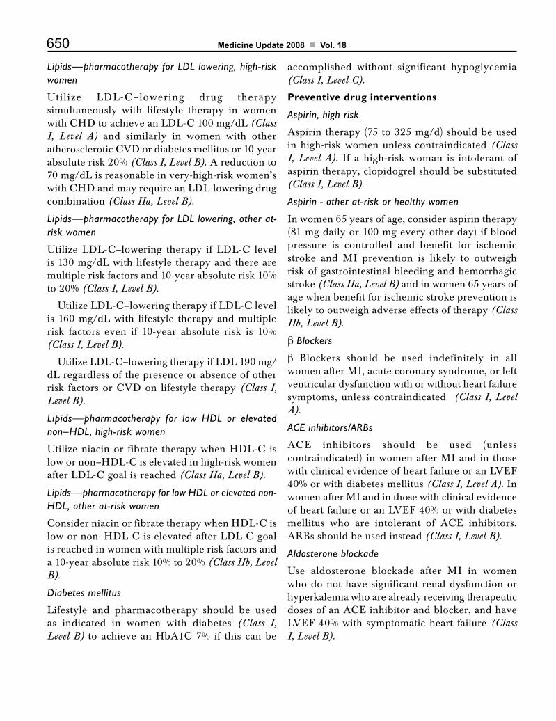
650 Medicine Update 2008 Vol. 18
Lipids—pharmacotherapy for LDL lowering, high-risk women
Utilize LDL-C–lowering drug therapysimultaneously with lifestyle therapy in women withCHDtoachieveanLDL-C100mg/dL(Class I, Level A) and similarly in women with other atheroscleroticCVDordiabetesmellitusor10-yearabsolute risk 20% (Class I, Level B). A reduction to 70 mg/dL is reasonable in very-high-risk women’s withCHDandmayrequireanLDL-loweringdrugcombination (Class IIa, Level B).
Lipids—pharmacotherapy for LDL lowering, other at-risk women
UtilizeLDL-C–lowering therapy ifLDL-C levelis 130 mg/dL with lifestyle therapy and there are multiple risk factors and 10-year absolute risk 10% to 20% (Class I, Level B).
UtilizeLDL-C–loweringtherapyifLDL-Clevelis 160 mg/dL with lifestyle therapy and multiple risk factors even if 10-year absolute risk is 10% (Class I, Level B).
UtilizeLDL-C–loweringtherapyifLDL190mg/dL regardless of the presence or absence of other risk factorsorCVDon lifestyle therapy (Class I, Level B).
Lipids—pharmacotherapy for low HDL or elevated non–HDL, high-risk women
Utilizeniacinor fibrate therapywhenHDL-C islowornon–HDL-Ciselevatedinhigh-riskwomenafterLDL-Cgoalisreached(Class IIa, Level B).
Lipids—pharmacotherapy for low HDL or elevated non-HDL, other at-risk women
ConsiderniacinorfibratetherapywhenHDL-Cislowornon–HDL-CiselevatedafterLDL-Cgoalis reached in women with multiple risk factors and a 10-year absolute risk 10% to 20% (Class IIb, Level B).
Diabetes mellitus
Lifestyle and pharmacotherapy should be used as indicated in women with diabetes (Class I, Level B) toachieveanHbA1C7% if this canbe
accomplished without significant hypoglycemia (Class I, Level C).
Preventive drug interventions
Aspirin, high risk
Aspirin therapy (75 to 325 mg/d) should be used in high-risk women unless contraindicated (Class I, Level A). If a high-risk woman is intolerant of aspirin therapy, clopidogrel should be substituted (Class I, Level B).
Aspirin - other at-risk or healthy women
In women 65 years of age, consider aspirin therapy (81 mg daily or 100 mg every other day) if blood pressure is controlled and benefit for ischemic stroke and MI prevention is likely to outweigh risk of gastrointestinal bleeding and hemorrhagic stroke (Class IIa, Level B) and in women 65 years of age when benefit for ischemic stroke prevention is likely to outweigh adverse effects of therapy (Class IIb, Level B).
βBlockers
β Blockers should be used indefinitely in all women after MI, acute coronary syndrome, or left ventricular dysfunction with or without heart failure symptoms, unless contraindicated (Class I, Level A).
ACE inhibitors/ARBs
ACE inhibitors should be used (unlesscontraindicated) in women after MI and in those with clinical evidence of heart failure or an LVEF 40% or with diabetes mellitus (Class I, Level A). In women after MI and in those with clinical evidence of heart failure or an LVEF 40% or with diabetes mellitus who are intolerant of ACE inhibitors,ARBs should be used instead (Class I, Level B).
Aldosterone blockade
Use aldosterone blockade after MI in women who do not have significant renal dysfunction or hyperkalemia who are already receiving therapeutic dosesofanACEinhibitorandblocker,andhaveLVEF 40% with symptomatic heart failure (Class I, Level B).

651CAD in Women
Class III Interventions (Not Useful/Effective and May Be Harmful) for CVD or MI Prevention in Women
Menopausal therapy
Hormone therapy and selective estrogen-receptor modulators (SERMs) should not be used for the primaryor secondarypreventionofCVD (Class III, Level A).
Antioxidant supplements
Antioxidant vitamin supplements (e.g., vitamin E, C,andbetacarotene)shouldnotbeusedfor theprimaryor secondarypreventionofCVD (Class III, Level A).
Folic acid
Folic acid, with or without B6 and B12 supplementation, should not be used for the primaryor secondarypreventionofCVD (Class III, Level A).
Aspirin for MI in women < 65 years of age
Routine use of aspirin in healthy women 65 years of age is not recommended to prevent MI (Class III, Level B).
ConclusionCoronaryarterydiseasecanoccurinwomenofallages, depending on risk factor burden. Metabolic syndrome, hypertriglyceridemia and low HDL cholesterol are more important risk factor for women. New evidence-based guidelines are available to aid clinicians in improving the preventive, diagnostic and therapeutic management of coronary artery disease in women.
SummaryEmerging data continue to highlight important sex-based differences in coronary heart disease (CHD)prevention anddiagnostic testing, in themanagement of acute coronary syndromes and in the outcome of CHD therapies. Evidence-basedguidelines have been developed that offer specific recommendations for clinicians and information for women. These guidelines are buttressed by results
that have become available from randomized, controlled clinical trials in women, and data from CHDregistriesandclinical trials involvingbothsexes but including adequate numbers of women to enable the reporting of sex-specific results.
AbbreviationsCAD – CoronaryArteryDisease,
CHD – CoronaryHeartDisease,
MI – Myocardial Infarction,
CVD – CardioVasculardisease
UA – Unstable Angina,
NSTEMI – Non ST Elevated Myocardial Infarction
References1. Wilcox S, Stefanick ML. Knowledge and perceived risk of
major diseases in middle-aged and older women. Health Psychology 1999; 18: 346-53.
2. WengerNK.Coronaryheartdisease:The femaleheart isvulnerable. Prog Cardiovasc Dis 2003;46:199-229.
3. VonderLoheE.Coronaryheartdiseaseinwomen.Berlin,Heidelberg, New York: Springer, 2003
4. Women. World Heart Federation Web site. Available at: http://www.worldheart.org/awareness-women.php. Accessed October 6, 2006.
5. Knopp RH, Zhu X, Boner B. Effects of estrogens on lipoprotein metabolism and cardiovascular disease in women. Atherosclerosis 1994; 110: [Suppl] : S83-S91.
6. WengerNK.Coronaryheartdisease : the femaleheart isvulnerable. Prog Cardiovasc Dis 2003; 46: 199-229.
7. ZylbersteinDE,BengtssonC,BjörkelundC,et al.SerumHomocysteine in Relation to Mortality and Morbidity FromCoronaryHeartDisease.A24-yearFollow-UpofthePopulation Study of Women in Gothenburg. Circulation 2004; 109: 601-606.
8. SharmaSN,KaulU,SharmaSet.al.Coronaryarteriographicprofile in young and old Indian patients with IHD : A comparative study. Indian Heart J 1990; 42 : 365-69.
9. VijayalakshmiS,RaoASC,YavagalST.JayadevaInstitute’smyocardial information ( JIMI-I) study. Abstract state conferenceofAPIandCSI.Mysore1994.
10. Pradhan AD, Lacroix AZ, Langer RD, et al. Tissue Plasminogen Activator Antigen and D-Dimer as Marker for Atherothrombotic Risk Among Healthy Postmenopausal Women. Circulation 2004; 110: 292-300.
11. WengerNK,SperoffL,PackardB.Cardiovascularhealthand disease in women. N Engl J Med 1993; 329: 247-256.

652 Medicine Update 2008 Vol. 18
12. Shaw LJ, Peterson ED, Johnson LL. Noninvasive testing techniquesfordiagnosisandprognosis.In:CharneyP,ed.CoronaryArteryDiseaseinWomen.Philadelphia:American College of Physicians; 1999: 341-345.
13. Wiviott SD, Cannon CP, Morrow DA, et al. DifferentialExpressionofCardiacBiomarkersbyGenderinPatientswithUnstable Angina / Non-ST-Elevation Myocardial Infarction. ATACTICS-TIMI18Substudy.Circulation 2004; 109: 580-586.
14. Kawano H, Motoyama T, Ohgushi M, et al. Menstrual cycle variation of myocardial ischaemia in premenopausal women with variant angina. Ann Intern Med 2001; 135 (11): 977-981.
15. Hochman JS, Tamis J, Thompson TD, et al. Sex, clinical presentation and outcome in patients with acute coronary syndromes. N Engl J Med 1999; 341: 226-232.
16. Vaccarino V, Parsons L, Every NR, et al. Sex-based differences in early mortality after myocardial infarction. N Engl J Med 1999; 341: 217-225.
17. ASRM 60th Annual Meeting : Abstract 0-130. Presented Oct. 19, 2004.
18. LoriMosca,CaroleL,EmeliaJ.Evidence-BasedGuidelinesfor cardiovascular disease prevention in women: 2007 Update. Circulation. 2007; 115 : 0000-0000. DOI: 10.1161 / Circulationaha.107.181546
CHAPTER
87Valvular Heart Disease in PregnancyM. Panja, S. Basu, D. Kumar
IntroductionValvular heart disease in young women is most commonly due to rheumatic heart disease, congenital abnormalities, or previous endocarditis and may increase the maternal and fetal risks associated with pregnancy. The likelihood of an adverse outcome is related to the type and severity of maternal valvular disease and the resulting abnormalities of functional capacity, left ventricular function, and pulmonary pressure. Clinical recommendationsconcerning valvular heart disease and pregnancy are based on limited data from case reports and observational studies or on inferences from data for other groups of patients.
Normal hemodynamic changes To understand the consequences of valvular heart disease during pregnancy, it is important to review the hemodynamic changes that occur in all pregnant women. First, blood volume increases, starting at the sixth week and rising rapidly until mid pregnancy. Thereafter, the rise continues but at a much slower rate. The increase in blood volume ranges from about 20% to 100%, with an average of 50%. Proportionately, plasma volume increases much more than erythrocyte mass, which can lead to physiologic anemia. An estrogen-mediated stimulation of the renin-angiotensin system that results in sodium and
water retention appears to be the mechanism underlying the blood volume increase. Similarly, cardiac output increases steadily during pregnancy up to about 34 weeks of gestation, when it begins to fall. The average increase in cardiac output is about 45% by 24 weeks of gestation. The increase is due to both the expansion in blood volume and the augmentation of stroke volume and heart rate. Thus, early in pregnancy, an increase in stroke volume (20% to 30%) is responsible for the increase in cardiac output. Later in pregnancy, the rise is related to an acceleration of heart rate (25%), since stroke volume decreases as a result of vena caval compression. In addition, cardiac output rises still higher and heart rate increases during labor and delivery. After delivery, when vena caval compression is relieved, there is a surge of venous return that augments cardiac output and places additional burden on the heart.
Other hemodynamic changes associated with pregnancy are a 21% decrease in systemic vascular resistance and a 34% decrease in pulmonary vascular resistance; there is no change in left ventricular contractility. Pregnant women tend to maintain normal left ventricular filling pressures because of left ventricular dilatation with an increase in left ventricular mass, as measured by echocardiography.1

654 Medicine Update 2008 Vol. 18
Symptoms of normal pregnancy can mimic those of valvular heart disease. Specifically, women with normal pregnancies may have exertional dyspnea, orthopnea, fatigue, lower extremity edema, and presyncope. Physical examination can also be confusing in a woman whose pregnancy is progressing normally. For example, a and v waves are prominent on jugular venous pressure studies, pulse pressure is increased, and the maximal apical impulse is laterally displaced. The first heart sound is accentuated, and the pulmonary component of the second heart sound is also increased. The third heart sound is heard in about 80% of pregnant women, whereas the fourth heart sound is rarely heard. The universal early ejection flow systolic murmur of less than grade 3/6 along the left sternal border is heard in 90% of pregnant women and may be enhanced by anemia.
The presence of certain physical signs in pregnant women should raise suspicion of cardiac abnormalities. These include a loud fourth heart sound, a diastolic murmur, a grade 3/6 or greater systolic murmur, a fixed split of the second heart sound, and an opening snap. The presence of one or more of these signs should signal the need for echocardiographic evaluation.
Consequences of valvular heart disease during pregnancyAlthough the prevalence of clinically significant maternal heart disease during pregnancy is low (probably less than 1 per cent)2 its presence increases the risk of adverse maternal, fetal, and neonatal outcomes3.
The American Heart Association and the American College of Cardiology have classified maternal and fetal risk during pregnancy on the basis of the type of valvular abnormality and the New York Heart Association (NYHA) functional class (Table1).4
The absolute risk conferred on a given woman by pregnancy also depends on additional clinical factors. Recent analyses of the outcomes of pregnancy in Canada2 identified predictors of adverse maternal and
fetal outcomes in a heterogeneous group of women with congenital or acquired heart disease (546 women and 599 pregnancies). Approximately40 per cent of the women had a primary valve disorder. Adverse maternal cardiac events (pulmonary edema, sustained bradyarrhythmia or tachyarrhythmia requiring therapy, stroke, cardiac arrest, or death) occurred in 13 per cent of completed pregnancies and were significantly more likely among women with reduced left ventricular systolic function (an ejection fraction below 40 per cent), left heart obstruction (aortic stenosis with a valve area of less than 1.5 cm2 or mitral stenosis with a valve area of less than 2.0 cm2) previous cardiovascular events (heart failure, transient ischemic attack, or stroke), or disease of NYHA class II or higher3.These outcomes occurred in 4 per cent of the women with none of these risk factors, 27 per cent of those with one risk factor, and 62 per cent of those with two or more risk factors. The three women who died all had two or more risk factors. Abnormal functional capacity (NYHA class II or higher) and left heart obstruction were also predictors of neonatal complications, including premature birth, intrauterine growth retardation, respiratory distress syndrome, intraventricular hemorrhage, and death. Other predictors of adverse fetal outcomes included the use of anticoagulant drugs throughout pregnancy, smoking during pregnancy, and multiple gestation. Fetal mortality was 4 per cent among pregnancies in women with one or more of these risk factors, as compared with 2 per cent among those with none of these risk factors. The risks of adverse fetal outcomes were also substantially greater among women older than 35 years of age or younger than 20 years of age than among women between these ages with similar risk factors. Indexes of risk derived from and validated in this population may be used in the counseling of women before conception.
In another cohort including 64 women with valvular heart disease,5 most adverse maternal outcomes, including heart failure and arrhythmias, occurred in patients with clinically significant mitral or aortic stenosis (valve area, < 1.5 cm). Premature birth, intrauterine growth retardation, and low birth weight were also more common

655Valvular Heart Disease in Pregnancy
Table 1 : Classification of Valvular Heart Lesions According to Maternal, Fetal and Neonatal Risk.*Low Maternal and Fetal Risk
High Maternal and Fetal Risk
High Maternal Risk High Neonatal Risk
Asymptomatic aortic stenosis with a low mean outflow gradient (< 50 mm Hg) in the presence of normal left ventricular systolic function
Aortic regurgitation of NYHA ClassIorIIwithnormalleftventricular systolic function
Mitral regurgitation of NYHA class I or II with normal left ventricular systolic function
Mitral-valve prolapsed with no mitral regurgitation or with mild-to-moderate mitral regurgitation and with normal left ventricular systolic function
Mild-to-moderate mitral stenosis (mitral-valve area > 1.5 cm2, gradient < 5 mm Hg) without severe pulmonary hypertension
Mild-moderate pulmonary- valve stenosis
Severe aortic stenosis with or without symptoms
Aortic regurgitation with NYHA class III or IV symptom
Mitral stenosis with NYHA class II, III or IV symptoms
Mitral regurgitation with NYHA class III or IV symptoms
Aortic-valve disease, mitral-valve disease, or both, resulting in severe pulmonary hypertension (pulmonary pressure > 75% of systemic pressures)
Aortic-valve disease, or both, with left ventricular systolic dysfunction (ejection fraction < 0.40)
Maternal cyanosis
Reduced functional status (NYHA class III or IV)
Reduced left ventricular systol ic function ( lef t ventricular ejection fraction < 40%)
Previous heart failure
Previous stroke or transient ischemic attack
Maternal age < 20 yr or > 35 yr
Use of anticoagulant therapy throughout pregnancy
Smoking during pregnancy
Multiple gestations
*DerivedfromACC/AHAGuidelines6 and Siu et al.4,5 NYHA denotes New York Heart Association.
among the offspring of the women in this subgroup. The fetus is at increased risk for congenital heart disease, if the underlying maternal valvular disease is congenital6. Although these studies included few patients, with pulmonary hypertension, primary pulmonary hypertension is associated with high maternal mortality (33 to 40 per cent), as well as with an increased rate of adverse neonatal events7.Secondary pulmonary hypertension due to valvular disease is associated with an increased rate of adverse maternal events, but the absolute risk of such events is unclear.
A systolic pulmonary-artery pressure that is more than 75 per cent as high as the systemic pressure places the woman at high risk.
Mitral stenosisMitral stenosis is the most common chronic rheumatic valvular lesion in pregnancy. Since the natural history of rheumatic mitral stenosis typically
includes a 20- to 25-year asymptomatic period, symptoms often first appear during pregnancy. Congenitalfusionofthecommissures,or“parachutemitral valve,” and left atrial myxoma are other causes of mitral stenosis during pregnancy.
Symptoms related to mitral stenosis reflect an increased pressure gradient across the mitral valve. This pressure gradient is a function of both the cross-sectional area of the valve and the flow through the valve. The rise in cardiac output during pregnancy increases the pressure gradient.
Hemodynamic abnormalities in a pregnant woman with mitral stenosis depend on the severity of the disease but normally include increased left atrial pressure associated with elevation of both pulmonary venous and arterial pressures. This results in pulmonary edema, pulmonary hypertension, and right ventricular failure. In addition, an increase in heart rate caused by exercise, fever, or emotional

656 Medicine Update 2008 Vol. 18
stress decreases diastolic left ventricular filling time and further elevates left atrial pressure and reduces cardiac output. This increase in pressure also predisposes pregnant women to development of atrial arrhythmias. Furthermore, loss of atrial contractility associated with a rapid ventricular response has devastating effects and can lead to pulmonary edema.
Clinical presentation
Pregnant women with mitral stenosis present clinically with symptoms of both left-sided heart failure and right ventricular failure, depending on the severity and duration of the valvular disease. Symptoms of left-sided heart failure are more common and include orthopnea, paroxysmal nocturnal dyspnea, and dyspnea on exertion. Unless the patient has long-standing valve disease, symptoms of right ventricular failure are less common and include peripheral edema and ascites, which in pregnancy are difficult to recognize as being related to valvular heart disease.
Careful examination should include a searchfor an opening snap and a diastolic rumbling murmur with presystolic accentuation, which are classic auscultatory findings in mitral stenosis. The presence of elevated jugular venous pressure, hepatomegaly, a loud pulmonary component of the second heart sound, and right ventricular heave on examination also supports a diagnosis of mitral stenosis.
Diagnostic assessment
Echocardiography is the diagnostic study of choice for evaluation of mitral stenosis in pregnant women and both confirms the diagnosis and helps determine the severity of the stenosis. In addition, the echocardiogram allows assessment of pulmonary pressures, right ventricular function, mitral regurgitation, and the configuration of the subvalvular apparatus, which is important in determining the success of percutaneous mitral balloon valvuloplasty (PMBV). Invasive diagnostic testing is rarely indicated in pregnant women with mitral stenosis.
Medical management
For women with mild or moderate symptoms during pregnancy, medical therapy is directed at the treatment of volume overload and includes diuretic therapy, the avoidance of excessive salt, and the reduction of physical activity. Beta-blockers attenuate the increases in heart rate and prolong the diastolic filling period, which provides symptomatic benefit. Development of atrial fibrillation requires prompt treatment, including cardioversion. Beta-blockers and digoxin are used for rate control. If suppressive antiarrhythmic therapy is needed, procainamide and quinidine are the drugs with which we have the most extensive experience. Because of the increased risk of systemic embolism in patients with mitral stenosis and atrial fibrillation, anticoagulant therapy is indicated. Patients with severe symptoms (NYHA class III or IV) who undergo balloon mitral valvuloplasty or valve surgery before conceiving appear to tolerate pregnancy with fewer complications than similar women who are treated medically or tighten mitral stenosis (a valve area of less than 1.0 cm2)
Most pregnant women with mitral stenosis can be managed medically. Since an increased preload contributes to the exacerbation of heart failure, it is prudent to restrict salt and fluid intake. Diuretics should be used judiciously to avoid hypotension and increased heart rate. Use of beta-blocking drugs to slow the heart rate can dramatically improve symptoms. Digoxin (Lanoxin) is not very effective because the adrenergically driven increased heart rate overrides its effect.
Balloon valvuloplasty
PMBV is an invasive procedure that is being used more often because of its proven safety. However, PMBV is contraindicated in women who have moderate to severe mitral regurgitation, calcified mitral valve, or clot in the left atrium. In addition, even though PMBV is considered a fairly safe procedure, it should be used cautiously to avoid radiation exposure during the first trimester. In patients who present with severe symptoms during pregnancy, successful percutaneous balloon
657Valvular Heart Disease in Pregnancy
mitral valvuloplasty, performed during the second trimester, has been associated with normal subsequent deliveries and excellent fetal outcomes. Risks to the fetus associated with exposure to radiation may be reduced by avoiding exposure to radiation during the first half of pregnancy.
Pregnant women who are to be exposed to radiation should have the uterus shielded and should be informed about the possible risks. Mitral valvuloplasty has also been performed under transesophageal echocardiographic guidance, eliminating these risks. Open cardiac surgery has been performed during pregnancy for severe mitral stenosis. Maternal outcomes are approximately the same as those among nonpregnant patients, but there is fetal loss in 10 to 30 per cent of cases.
Surgical intervention
In early investigations, open commissurotomy and valve replacements carried a maternal mortality rate of about 5% and a fetal mortality rate of 20% to 30%. Many factors (e.g., anesthetic agents used, hypothermia during surgery) can adversely affect the outcome. Improved cardiopulmonary bypass techniques have resulted in improved outcomes. A recent study of 168 pregnant women who underwent open commissurotomy showed no maternal mortality and a fetal mortality of 1.8%8 Prosthetic mitral valve replacement is now a feasible option in patients who are not candidates for either PMBV or open commissurotomy.
Labor and delivery
In view of the increase in cardiac output during labor and after delivery, it is important to plan management carefully. Vaginal delivery is possible in most patients with mitral stenosis. However, optimal management may require invasive haemodynamic studies in patients with moderate to severe stenosis. Oxygen should be given to reduce pulmonary pressures, and fluid restriction and use of diuretics and epidural anesthesia are recommended as well. Vigoros manual uterine massage and oxytocin infusion can reduce the risk of excessive blood loss.
Mitral regurgitation This condition is usually well tolerated in pregnancy, presumably because of left ventricular unloading secondary to the physiologic fall in systemic vascular resistance. The cause of mitral regurgitation during pregnancy has changed over the years. In the past, it was usually a consequence of rheumatic fever, but today it is more often related to mitral valve prolapse complicated by ruptured chordae tendinae. Other possible causes are Libman-Sacks endocarditis, infective endocarditis, Marfan syndrome and pseudoxanthoma elasticum, Ehlers-Danlos syndrome, and dilated cardiomyopathy.
Mitral regurgitation leads to a progressive increase in the volume of blood going to the left ventricle, which results in enlargement of both the left ventricle and the left atrium. Moreover, left ventricular cavity dilatation is associated with mitral valve annular dilatation and asynergic contraction of the papillary muscle, which exacerbate mitral regurgitation.
Increased left ventricular volume and left atrial enlargement are associated with an elevation of pulmonary venous and arterial pressures, leading to pulmonary hypertension and right-sided heart failure. Because of the decrease in left ventricular afterload associated with mitral regurgitation, systolic wall stress is also lowered. These changes are more pronounced in pregnancy because of a reduction in systemic vascular resistance.
Symptoms and physical examination
Mitral regurgitation during pregnancy is usually well tolerated. Symptoms may include dyspnea on exertion, orthopnea, and paroxysmal nocturnal dyspnea. The apical impulse of the left ventricle is shifted outward, and a holosystolic murmur is heard at the apex on auscultation. The murmur radiates toward the axilla and increases during expiration. Some pregnant women with mitral regurgitation present with atrial fibrillation associated with heart failure and cardiomegaly.
658 Medicine Update 2008 Vol. 18
If tricuspid regurgitation is associated with mitral regurgitation, peripheral edema might be present, a right ventricular heave may be noted on palpation, and a systolic murmur may be heard along the left lower sternal border on auscultation. Unlike the murmur of mitral regurgitation, the tricuspid regurgitation murmur increases with inspiration.
Assessment and diagnosis
Doppler echocardiography is useful in diagnosis of chronic mitral regurgitation. The following information can be obtained from these studies:
Evaluation of the structure of the mitral valve. Mitral valve prolapse can cause “hammocking” of the mitral valve in M mode, which is evidence of vegetative growths on the valve. Assessment of left ventricular size and function, left atrial size, and left atrial appendage thrombosis Evaluation of structure and function of subvalvular apparatus (papillary muscle, chordae tendinae) Assessment of severity of mitral regurgitation
Management
In symptomatic pregnant patients with mitral regurgitation, hydralazine hydrochloride, diuretics, and digoxin can be used when systolic function is impaired. If severe symptomatic mitral regurgitation due to mitral valve prolapse is found, surgical mitral valve repair may be a good option because it avoids the need for anticoagulant therapy. Mitral valve replacement can be done as a last resort. In most cases, maternal and neonatal outcomes are good. However, women with pulmonary arterial pressure greater than 50 mm Hg are at increased risk for complications.
Aortic stenosis Symptomatic aortic valve disease is less common than mitral valve disease in pregnant women. In the United States, congenital aortic stenosis secondary to membrane on the bicuspid aortic valve appears to be the predominant cause. In contrast, rheumatic heart disease is the most common cause in developing countries. During pregnancy, women
with bicuspid aortic valves are at risk for aortic dissection related to the effects of hormones on connective tissue.
The pressure gradient across the aortic valve is responsible for the hemodynamic changes in aortic stenosis. The increase in left ventricular systolic pressure needed to maintain sufficient pressure in arterial circulation leads to increased stress on the ventricular wall. To compensate for this, left ventricular hypertrophy develops, which can result in diastolic dysfunction, fibrosis, diminished coronary flow reserve, and late systolic failure.
An increase in stroke volume and a fall in peripheral resistance are largely responsible for the increase in the gradient across the aortic valve. The clinical consequences of the increased aortic gradient depend on the degree of preexisting left ventricular hypertrophy and left ventricular systolic function. When compensatory changes in the left ventricle are inadequate to meet the demands imposed by the need for increased cardiac output late in pregnancy, symptoms develop. This usually occurs with moderate to severe aortic stenosis.
Clinical findings
Clinical presentation and symptoms dependon the degree of aortic stenosis. Women with aortic valve areas more than 1.0 cm2 tolerate pregnancy well and are asymptomatic. However, women with more severe aortic stenosis may have symptoms of left-sided heart failure (dyspnea on exertion). Syncope or presyncope is rare, and pulmonary edema is even more unusual. However, arrhythmias are sometimes present. Because symptoms of aortic stenosis are similar to those of normal pregnancy, diagnosis of this condition is challenging. Physical findings vary with the severity of the disease. The left ventricular impulse is sustained and displaced laterally. A systolic ejection murmur is heard along the right sternal border and radiates toward the carotid arteries, and a systolic ejection click is heard. A fourth heart sound may be present, suggesting abnormal diastolic function. The presence of

659Valvular Heart Disease in Pregnancy
pulsus parvus et tardus suggests hemodynamically significant aortic stenosis.
Assessment and diagnosis
Diagnosis can be confirmed with echocardiography. The aortic gradient and valve area can be calculated by Doppler flow studies. In addition, echocardiography can detect left ventricular hypertrophy. Estimation of ejection fraction and left ventricular dimensions may be useful to predict outcome during pregnancy, labor, and delivery. Women with an ejection fraction less than 55% are at high risk for development of heart failure during pregnancy.Cardiaccatheterizationis indicatedifthe clinical picture is consistent with severe aortic stenosis, if noninvasive data are inconclusive, and if percutaneous balloon valvuloplasty is needed. Fetal echocardiography is indicated if the mother has congenital aortic stenosis, since the risk that the fetus has similar anomalies is 15%.
Management
Patients who are symptomatic or who have a peak outflow gradient of more than 50 mm Hg are advised to delay conception until after surgical correction.
Termination of pregnancy should be strongly considered if the patient is symptomatic before the end of the first trimester. Aortic-valve replacement and palliative aortic balloon valvuloplasty have been performed during pregnancy with some associated maternal and fetal risk.
The severity of the condition and its symptoms largely determines management of aortic stenosis. Most asymptomatic patients and those who have mild to moderate stenosis can be managed with medical therapy and close monitoring. It is important to maximize cardiac output and fetal blood flow by avoiding intense exercise, potent vasodilators, and diuretics. In patients with low ejection fractions, digoxin can be used, provided drug levels are monitored regularly.
Percutaneous balloon valvuloplasty and aortic valve replacement are options for management.
Balloon valvuloplasty can be used as a bridge to valve replacement in women who are too ill to undergo surgery. The risk of death in nonpregnant patients managed in centers experienced in this technique is about 5%. In pregnancy, balloon valvuloplasty carries added risk because circulation to the fetus is stopped for a short time during the procedure. However, several studies of balloon valvuloplasty for severe aortic stenosis during pregnancy suggest favorable outcomes for both mother and fetus 9
Balloon valvuloplasty is not the preferred treatment in patients with calcified aortic valves or in the presence of significant aortic regurgitation. In those circumstances, valve replacement is indicated. The choice of a bioprosthetic versus a mechanical valve should be individualized. Bioprosthetic valves avoid the need for long-term anticoagulant therapy.
Labor and delivery
Vaginal delivery is preferred unless there is an obstetric indication for cesarean section. Avoidance of severe vasodilatation and maintenance of an adequate fluid balance are paramount so that cardiac output is not compromised. Low epidural anesthesia may be used to minimize vasodilatory effects, and antibiotic prophylaxis should be given to patients with previous endocarditis. In general, outcomes for both the mother and the fetus are favorable. However, some evidence suggests that as many as 20% of women who have severe aortic stenosis choose to have therapeutic abortion.
Aortic insufficiency Aortic insufficiency in pregnancy can be either acute or chronic. The acute form is caused by aortic dissection, bacterial endocarditis, or malfunction of a prosthetic valve. Because the left ventricle has no time to adapt to volume overload, pulmonary edema and cardiogenic shock often occur. Acute aortic insufficiency should be considered a surgical emergency, and valve replacement is urgent, even in pregnancy.
660 Medicine Update 2008 Vol. 18
Another condition that requires emergency surgery is proximal aortic dissection with aortic insufficiency. Marfan syndrome, bicuspid aortic valve, and hypertension add to deleterious hormonal effects and are predisposing conditions for aortic dissection.
In pregnant women, chronic aortic insufficiency is often associated with a bicuspid aortic valve or rheumatic heart disease. The gradual increase in left ventricular volume overload allows the left ventricle to adapt by increasing left ventricular end-diastolic diameter. This adaptation appears to maintain the forward flow unless systolic dysfunction sets in. Therefore, as with mitral insufficiency, chronic aortic insufficiency is well tolerated during pregnancy.
Clinical presentation
Patients with chronic aortic insufficiency usually present with dyspnea, decreased exercise tolerance, and chest pain. Some patients have syncope due to arrhythmias and left ventricular dysfunction. Pregnant women tolerate aortic regurgitation well because of the normal peripheral vasodilatation during pregnancy, which improves hemodynamic parameters in aortic insufficiency.
On the other hand, women with aortic insufficiency and either New York Heart Association functional class I or II symptoms or systolic ventricular dysfunction do not tolerate pregnancy well. Findings on physical examination are typical of hyperdynamic circulation. Such findings may complicate diagnosis, since a hyperdynamic state also can be associated with normal pregnancy. Physical findings include a wide pulse pressure, brisk carotid pulse, and mildly displaced apical impulse. An early diastolic murmur on the left sternal border and soft second heart sounds are clear clues to aortic insufficiency.
Assessment
Transthoracic echocardiography and Doppler flow studies are helpful in making a diagnosis and assessing the severity of aortic insufficiency.
Transesophageal echocardiography can be used to detect vegetation in bacterial endocarditis and aorticdissection.Cardiaccatheterizationisusuallynot indicated, but magnetic resonance imaging can be helpful in diagnosis of aortic dissection. Fetal echocardiography is indicated in women with congenital abnormalities of the aortic valve or Marfan syndrome.
Management
In patients with chronic aortic regurgitation, management depends on the severity of the disease and symptoms. In asymptomatic patients, close monitoring is all that is needed. Symptomatic patients can be treated with vasodilators, including hydralazine, nitrates, and diuretics. Digoxin may be beneficial in patients who have systolic dysfunction. Use of angiotensin-converting enzyme inhibitors is contraindicated during pregnancy.
Special concerns with valvular heart disease Pregnant women with valvular heart disease are no more likely to have bacterial endocarditis than non-pregnant women with such heart disease. However, prophylactic therapy does seem warranted when valvular heart disease is present (see box below)10.11 Similarly, women who require anticoagulant therapy during pregnancy need special care. Recommended antibiotic prophylaxis for high-risk women undergoing genitourinary or gastrointestinal procedures
Category Drug and dosage
High-risk patient Ampicillin, 2 g IM or IV, plus gentamicin sulfate 1.5 mg/kg IV 30 min before procedure; ampicillin, 1 g IV, or amoxicillin 1 g PO 6 hr after procedure
High-risk patient who has penicillin allergy
VancomycinHCl, 1 g IVover 2hr,Plus gentamicin sulfate, 1.5 mg/kg IV 30 min before procedure
Most pregnant women with valvular heart disease can be managed medically. However, severe symptomatic disease may pose a threat to the survival of both mother and fetus. In this situation,
661Valvular Heart Disease in Pregnancy
Table 2 : Fetal Effects of, Maternal Indications for, and Risks Associated with Drugs Used in the Treatment of Maternal Valvular Heart Disease.*
Drug Fetal Effects Indications in Pregnant Patients with Valve Disease
Risk Category
Diuretics
Furosemide Increased Urinary sodium and potassium levels
TodecreaseCongestionAssociatedwithvalvular heart disease
Cm
Antihypertensive agents
Beta-blockers Possible decreased heart rate, possible lower birth weight
Hypertension, supraventricular arrhythmias, to control heart rate in women with clinically significant mitral stenosis
Dm
Methyldopa No major adverse effects Hypertension C
Vasodilator agents
Angiotensin-converting-enzyme inhibitors
Urogenital defects, death, intrauterine growth retardation
Not indicated during pregnancy and should be discountinued
Dm
Hydralazine No major adverse effects For vasodilation in cases of aortic regurgitation and ventricular dysfunction
Cm
Nitrates Possible bradycardia Rarely used to decrease venous congestion B-Cm
Anticoagulant and antithrombotic agents
Warfarin Hemorrhage, developmental abnormalities when used between wk 6 and 12 of gestation
For anticoagulation of mechanical heart valves, valvular heart disease with associated atrial fibrillation during wk 12-36 of pregnancy
Dm
Unfractionated heparin Hemorrhage, no congenital defects
For anticoagulation of mechanical heart, valves, valvular heart disease with associated atrial fibrillation during wk 6-12 and after wk 36 of pregnancy
Cm
Low-molecular-weight heparin
Hemorrhage Not currently indicated during pregnancy Dm
Aspirin Hemorrhage, prolongation of labor, low birth weight (When taken in high doses)
Low-dose aspirin (81 mg/day) occasionally used as an adjunct in patients with previous embolic events or prosthetic-valve thrombosis
C
Antiarrhythmic agents
Digoxin No major adverse effects For suppression of supraventricular arrhythmias
C
Adenosine No major adverse effects For immediate conversion of supraventricular arrhythmias
Cm
Quinidine High doses may be oxytocic
Occasionally used for suppression of atrial or ventricular arrhythmias
Cm
Procainamide No major adverse effects Occasionally used for suppression of atrial ventricular arrhythmias
Cm
Amiodarone Hypothyroidism, intrauterine growth retardation, premature birth
Rarely used during pregnancy because of side effect; may be used to suppress atrial or ventricular arrhythmias in high-risk patients
Cm
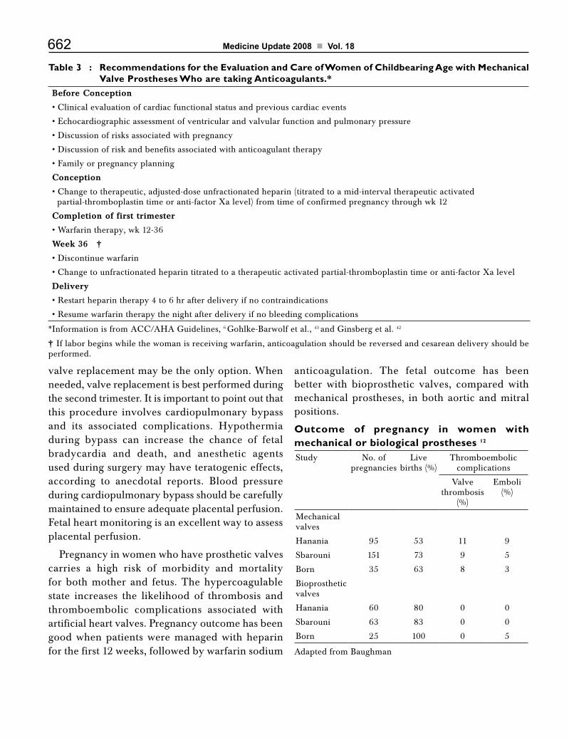
662 Medicine Update 2008 Vol. 18
Table 3 : Recommendations for the Evaluation and Care of Women of Childbearing Age with Mechanical Valve Prostheses Who are taking Anticoagulants.*
Before Conception
•Clinicalevaluationofcardiacfunctionalstatusandpreviouscardiacevents
•Echocardiographicassessmentofventricularandvalvularfunctionandpulmonarypressure
•Discussionofrisksassociatedwithpregnancy
•Discussionofriskandbenefitsassociatedwithanticoagulanttherapy
•Familyorpregnancyplanning
Conception
•Changetotherapeutic,adjusted-doseunfractionatedheparin(titratedtoamid-intervaltherapeuticactivated partial-thromboplastin time or anti-factor Xa level) from time of confirmed pregnancy through wk 12
Completion of first trimester
•Warfarintherapy,wk12-36
Week 36 †
•Discontinuewarfarin
•Changetounfractionatedheparintitratedtoatherapeuticactivatedpartial-thromboplastintimeoranti-factorXalevel
Delivery
•Restartheparintherapy4to6hrafterdeliveryifnocontraindications
•Resumewarfarintherapythenightafterdeliveryifnobleedingcomplications
*InformationisfromACC/AHAGuidelines,6 Gohlke-Barwolf et al., 43 and Ginsberg et al. 42
† If labor begins while the woman is receiving warfarin, anticoagulation should be reversed and cesarean delivery should be performed.
valve replacement may be the only option. When needed, valve replacement is best performed during the second trimester. It is important to point out that this procedure involves cardiopulmonary bypass and its associated complications. Hypothermia during bypass can increase the chance of fetal bradycardia and death, and anesthetic agents used during surgery may have teratogenic effects, according to anecdotal reports. Blood pressure during cardiopulmonary bypass should be carefully maintained to ensure adequate placental perfusion. Fetal heart monitoring is an excellent way to assess placental perfusion.
Pregnancy in women who have prosthetic valves carries a high risk of morbidity and mortality for both mother and fetus. The hypercoagulable state increases the likelihood of thrombosis and thromboembolic complications associated with artificial heart valves. Pregnancy outcome has been good when patients were managed with heparin for the first 12 weeks, followed by warfarin sodium
anticoagulation. The fetal outcome has been better with bioprosthetic valves, compared with mechanical prostheses, in both aortic and mitral positions.
Outcome of pregnancy in women with mechanical or biological prostheses 12
Study No. of pregnancies
Live births (%)
Thromboembolic complications
Valve thrombosis
(%)
Emboli (%)
Mechanical valves
Hanania 95 53 11 9
Sbarouni 151 73 9 5
Born 35 63 8 3
Bioprosthetic valves
Hanania 60 80 0 0
Sbarouni 63 83 0 0
Born 25 100 0 5
Adapted from Baughman
663Valvular Heart Disease in Pregnancy
ConclusionPregnant women who have valvular disease represent a major challenge for physicians involved in theircare.Carefulhistory takingandphysicalexamination, along with a judicious use of diagnostic tools (mainly echocardiography), can lead to better management and ultimately to excellent outcomes for both mother and baby.
If the patient has abnormal functional capacity, left ventricular dysfunction, valve obstruction, or a history of heart failure or embolic events, she should be counseled regarding the risk of adverse cardiac outcomes. In patients with more than one such risk factor, pregnancy may not be advisable. A patient who becomes pregnant should be seen by a cardiologist once each trimester and more often if complications ensue. Serial echocardiography during pregnancy generally is not warranted. Patients with prosthetic valves must be counseled regarding the risks and benefits associated with anticoagulant therapy. Although definitive data are lacking, we would recommend the use of warfarin to achieve a target INR of 2.0 to 3.0 throughout most of the pregnancy. The only exceptions are the periods between 6 and 12 weeks of pregnancy and after 36 weeks of pregnancy,when we would opt for the closely monitored use of unfractionated heparin (Table 3)
References1. Hunter S, Robson SC. Adaptation of the maternal heart in
pregnancy. Br Heart J 1992; 68(6): 540-3
2. Siu SC, Sermer M, Colman JM, et al. Prospective multicenter study of pregnancy outcomes in women with heart disease. Circulation 2001; 104:515-21.
3. Siu SC, Colman JM, Sorensen S, et al. Adverse neonatal and cardiac outcomes are more common in pregnant women with cardiac disease. Circulation 2002;105:2179-84.
4. ACC/AHA guidelines for the management of patients with valvular heart disease: a report of the American College of Cardiology/American Heart Association Task Force on Practice Guidelines (Committee on Management of Patients with Valvular Heart Dis- ease). J Am Coll Cardiol 1998;32:1486-588.
5. Hameed A, Karaalp IS, Tummala PP, et al. The effect of valvular heart disease on maternal and fetal outcome of pregnancy.J Am Coll Cardiol 2001;37:893-9.
6. Lao T, Sermer M, MaGee L, Farine D, Colman JM. Congenital aortic stenosis and pregnancy — a reappraisal. Am J Obstet Gynecol 1993;169:540-5.
7. Elkayam U, Gleicher N. Cardiac problems in pregnancy: diagnosis and management of maternal and fetal disease. NewYork: Wiley-Liss, 1998.
8. Kalra GS, Arora R, Khan JA, et al. Percutaneous mitral commissurotomy for severe mitral stenosis during pregnancy. Cathet Cardiovasc Diagn 1994;33(1):28-30
9. Lao TT, Adelman AG, Sermer M, et al. Balloon valvuloplasty for congenital aortic stenosis in pregnancy. Br J Obstet Gynecol 1993;100(12):1141-2
10. Bonow RO, Carabello B, de Leon AC Jr, et al. Guidelines for the management of patients with valvular heart disease: executive summary. A report of the American College of Cardiology/American Heart Association Task Force on Practice Guidelines (Committee on Management of Patients with Valvular Heart Disease). Circulation 1998;98(18):1949-84
11. Dajani AS, Bisno AL, Chung KJ, et al. Prevention of bacterial endocarditis: recommendations by the American Heart Association. JAMA 1990;264(22):2919-22
12. Baughman KL. The heart and pregnancy. In: Topol EJ, Califf RM, Isner J, et al, eds. Textbook of cardiovascular medicine. Philadelphia: Lippincott-Raven, 1998:797-816

CHAPTER
88Megatrials in Diabetes and Their Clinical ImplicationsB. K. Sahay, R. K. Sahay
In the last three decades lot of advances have taken place in the understanding of diabetes and its management. Studies have thrown light on the pathogenesis of both type 1 and type 2 diabetes leading to a more rationale approach to the management. Several newer molecules have been discovered which act at different sites modulating carbohydrate and lipid metabolism.
A new classification for diabetes based on the etiology and diagnostic criteria based on the risk of developing microvascular complications have been developed. This new classification and diagnostic criteria enunciated by the ADA and endorsed by the WHO expert committee provide us with a uniform standard for evaluation of the data of studies carried out throughout the world.
Several long term well planned trials have been conducted to evaluate several hitherto unanswered questions.
i. The importance of tight metabolic control with intensive therapy in preventing the development and progression of complications of diabetes
ii. The response and outcomes with different treatment modalities adopted for managing type 2 diabetes (Oral antidiabetic agents vs Insulin)
iii. The evaluation of different molecules available for the treatment of type 2 diabetes on reduction of hyperglycemia and also benefits beyond glycemic control such as effects on beta cell mass and function
With the prevalence of diabetes rising all over the world it is assuming pandemic proportions, and this has evoked particular interest in studies on prevention of both type 1 and type 2 diabetes. Several studies have focused on screening of high risk subjects and identification of prediabetic states namely IGT and IFG in whom preventive strategies have been used.
In this presentation we will be discussing some of the landmark trials, their results and implications of these on the day to day management of diabetes.
While several animal and human studies gave some idea about the role of hyperglycemia in the development of vascular complication, it was not clear whether these complications could be prevented and what degree of control was necessary. It was also not clear whether there were differences in the outcome of treatment in terms of morbidity and mortality with different therapeutic modalities. Several trials have been carried out with an objective to clarify these issues.
A correlation between glycemic control and diabetic complications in patients with type

665Megatrials in Diabetes and Their Clinical Implications
2 diabetes was first studied by the University Group Diabetes Program (UGDP)1. The UGDP followed 1000 patients with type 2 Diabetes, assigned to different therapies for about 5.5 years (range 3-8 years) and reported an increased risk of cardiovascular mortality in patients allocated to the sulfonylurea, tolbutamide, and phenformin and increased incidence of lactic acidosis with phenformin. The UGDP trial therefore threw up more controversies than giving any clarity on these issues1.
TheDCCT(DiabetesControl&ComplicationsTrial)2 and Stockholm trials3 were done in type 1 diabetic patients and demonstrated conclusively that tight control of blood glucose with intensive therapy leads to the reduction in the risk for development and progression of microvascular complications. The risk reduction for various outcomes ranged from 35-75%. Secondary analyses in these studies showed strong relationships between the risk of developing these complications and glycemic exposure over time. Moreover there was no discernible glucose threshold, i.e. there was a continuous reduction in complications as glycemic levels approached normal range. Macrovascular events e.g. combined cardiac, cerebrovascular and peripheral vascular
events were reduced by 54%.2
The United Kingdom Prospective Diabetes Study (UKPDS)4 recruited 5102 patients with newly diagnosed type 2 Diabetes, aged between 25-65 years, in 23 centers with in the UK between 1977 and 1991. Those with fasting plasma glucose greater than 110 mg/dl on two mornings 1-3 weeks apart were included in the study. The study extended over 20 years to accumulate sufficient events. The patients were stratified by their body weight. Non-obese patients were assigned to intensive treatment with insulin (30%) or sulfonylurea (40% of patients), chlorpropamide, glipizide, or glibenclamide or conventional treatment policy with diet (30% of patients). The obese patients were similarly assigned to intensive treatment with insulin (24%), or sufonylurea - chlorpropamide or glibenclamide (32%), metformin (20%) or conventional treatment with diet (24%). The conventional treatment policy aimed at maintaining the fasting plasma glucose below 270 mg/dl without symptoms.
The mean HbA1c achieved in the intensively treated group was 7.0% as compared to 7.9% in the conventionally treated group and this reduction in HbA1c brought about a remarkable decline in the development and progression of microvascular complications of diabetes (Table 1) The results of UKPDS have also brought into focus many more issues relating to the management of type 2 diabetes which are important and have far reaching consequences.
i. Almost 50% of patients with type 2 diabetes at presentation have pre-existing diabetes related tissue damage, despite this, improved blood glucose and blood pressure control reduces the risk of diabetic complications that cause both morbidity and premature mortality.
ii. Type 2 diabetes is associated with progressive hyperglycemia and decreasing β cell function irrespective of the therapy used.
iii. Tight blood glucose control is clearly the key and all means of achieving it have the same effect. The use of insulin per se confers neither
Table 1 : UKPDS: Reduction in complications with intensive glycemic control (HbA1c 0.9% lower than conventional treatment)12% for any diabetes related endpoint (p = 0.029)
25% for any microvascular endpoint (p = 0.0099)
16% for myocardial infarction (p = 0.052)
24% for cataract extraction (p = 0.046)
21% for retinopathy at 12 years (p = 0.015)
33% for albuminuria at 12 years (p = 0.00005)
Table 2 : UKPDS: Reduction in complications with Tight blood pressure control (144/82 vs 154/87 mmHg)24% for any diabetes related end point (p = 0.046)
37% for any microvascular endpoint (p = 0.0092)
21% for myocardial infarction (p = 0.13)
44% for stroke (p = 0.013)
58% for heart failure (p = 0.0043)

666 Medicine Update 2008 Vol. 18
additional advantages or disadvantages, while the use of sulfonylureas does not lead to additional risks. (chlorpropamide did perform worse as its use was associated with a rise in blood pressure over the years).
iv. Metformin is a better choice as initial therapy in obese type 2 diabetic patients.
v. Non-response to sulfonylureas after a certain duration of diabetes is because of progressive β cell failure.
vi. Intensive insulin therapy does not produce any adverse effects on macrovascular disease.
vii. Tight blood pressure control overall has larger benefits which manifest sooner than those after blood glucose control. ACE inhibitors or β blockers are equally effective in achieving the benefits of lowering blood pressure.5
viii. There is no overall deterioration in the quality of life as a result of intensified treatment.
All physicians treating diabetes should therefore be aware of the importance of good control and should ensure to pass on this information in a positive way to their patients. Most patients will respond to oral drugs initially- the choice of an appropriate agent is important in achieving good control. UKPDS has shown that the effectiveness of these oral agents will gradually diminish and addition of insulin to the above agents or substitution of therapy with insulin will eventually be required.
Steno 2 StudyThe Steno-2 Study6 was designed in 1990, when there was no evidence base for the treatment of type 2 diabetes, and studies such as the UKPDS were ongoing. Steno-2 was an attempt to validate the efficacy of daily clinical practice, i.e., the multifactorial treatment of type 2 diabetes, in high-risk type 2 diabetes patients.6 The aim of the study was to investigate the impact on microvascular and cardiovascular disorders, of a target driven behavior modification and polypharmacy as
compared to a conventional multifactorial treatment of high-risk type 2 diabetic patients with the metabolic syndrome including microalbuminuria. One hundred and sixty patients with type 2 diabetes and the metabolic syndrome including microalbuminuria were assigned to conventional therapy with their GP, or to intensive care at Steno Diabetes Center. After 4 years a 50% reductionin microvascular endpoints – nephropathy, retinopathy and neuropathy was reported. The intensive group was treated differently by means of individualized risk assessment, ambitious goal setting, focused behavior modification, more drugs and higher doses prescribed, and continued patient education and motivation. After 7.8 years duration the study showed an absolute risk reduction of 20%forCVD,andtherelativeriskreductionsformicrovascular events were as follows: nephropathy 61%, retinopathy 58% and autonomic neuropathy 63%.6
ADOPT7 was the first large, multicenter, randomized, double-blind, controlled clinical trial designed to compare the durability of glycemic control of the TZD rosiglitazone with that of metformin or the sulfonylurea glyburide as monotherapy, based on factors related to disease progression in patients with newly diagnosed (< 3 years) type 2 diabetes. ADOPT assessed the time interval of loss of glycemic control once a participant reached the maximum effective dose of each therapy and allowed investigation of the effects of beta-cell function and insulin resistance on disease progression and long-term glycemic control, among other outcomes. This international study included 4360 patients who were followed for 4 to 6 years.
The primary outcome measure was the time to monotherapy failure, defined as hyperglycemia confirmed by fasting plasma glucose (FPG) level greater than 180 mg/dL for subjects at the maximum-dictated or maximum-tolerated dose after at least 6 weeks of therapy. As patients reached the defined action point level of confirmed FPG of 140 mg/dL, they were further uptitrated to the next

667Megatrials in Diabetes and Their Clinical Implications
highest dose level based on the respective study arm. Secondary measures included the effects of monotherapy in delaying the progressive loss of glycemic control based on cumulative incidence of FPG greater than 140 mg/dL and the percentage ofpatientsremainingonmonotherapy(HbA1C<7%).
Results from ADOPT demonstrated that initial treatment with rosiglitazone significantly reduced the risk of monotherapy failure by 32% compared with metformin (P < .001), and by 63% compared with glyburide (P < .001) at 5 years. Similarly, rosiglitazone was significantly more effective in delaying the progressive loss of glycemic control as measuredbyFPGandHbA1Clevels.Riskreductionof decreasing glycemic control was 34% compared with metformin (P = .002) and 62% compared with glyburide(P<.001).Additionally,meanHbA1Clevels of less than 7% were maintained at 60 months with rosiglitazone compared with only 45 months for metformin and 33 months for glyburide.7
This study also demonstrated that rosiglitazone significantly improved insulin sensitivity vs metformin or glyburide (P < .001 at 4 years) and reduced the loss of beta-cell function vs metformin (P = .02) and glyburide (P < .001). Safety assessments were followed for 6 years in ADOPT, with no unanticipatedresults.Commonlyreportedadverseevents across the treatment groups for rosiglitazone, metformin, and glyburide, respectively, were edema (14.1%, 7.2%, 8.5%); weight gain (6.9%, 1.2%, 3.3%); gastrointestinal events (23%, 38.3%, 21.9%); and hypoglycemia (9.8%, 11.6%, 38.7%). Similar rates of discontinuation were reported in the rosiglitazone and metformin groups (37% and 38%, respectively) and were highest in the glyburide group (44%) due to an increase in hypoglycemia. Similarly, low rates ofcongestiveheartfailure(CHF)seriousadverseevents were reported with rosiglitazone (0.8%) and metformin (0.8%), while fewer such events were reported with glyburide (0.2%). Of these serious events, an independent cardiology review found 21 of51tobetrueCHF,involving9patientswithnodeaths in the rosiglitazone group, 8 patients with 1
death in the metformin group, and 4 patients with 1 death in the glyburide group. For all investigator-reportedCHFevents,therewasaslightdifferenceobserved with rosiglitazone compared with metformin (1.5% vs 1.3%, respectively), with fewer events reported for glyburide (0.6%; P = .05)7.
ADOPT is the first long-term study to demonstrate that progressive loss of glycemic control can be delayed and that durable control of targeted glycemic levels can be maintained for a longer duration with rosiglitazone than with metformin or glyburide. Therefore, these results provide evidence that suggest earlier treatment with a TZD in the management of type 2 diabetes may be warranted. In addition, this study provides the rationale that when combination therapy is required to maintain glycemic control, a TZD should be considered for use with other agents. These results, along with the potential risks and benefits, adverse events profile, and costs, must be considered by healthcare providers in choosing optimal management strategies for patients with type 2 diabetes.
Trials of Lipid Lowering and Atherosclerosis in DiabetesThe Multiple Risk Factor Intervention Trial (MRFIT) demonstrated that increases in total cholesterol levels are associated with an increase in the incidence of coronary artery disease in diabetes. The most common lipoprotein abnormalities in diabetes are an increase in the levels of the triglyceride-rich lipoproteins, decrease in HDL and increase in small dense LDL, without a significant rise in LDL levels.
Statin trials
The Scandinavian Simvastatin Survival Study (4S)8 was a secondary prevention study that included 201 patients with type 2 diabetes. The diabetics in the active treatment group had a 55% decrease in futurecoronaryevents(P=0.002).TheCholesterolandRecurrentEvents (CARE) trial included603diabetic subjects9. Coronary event reduction in

668 Medicine Update 2008 Vol. 18
the diabetics on pravastatin was 25% (P = 0.05). In the Long-Term Intervention with Pravastatin in Ischemic Disease (LIPID) Study,10 another secondary prevention trial, there were 782 type 2 diabetics. In this study, active treatment with pravastatin reduced coronary events in the diabetics by 19% (not significant).
TheCollaborativeAtorvastatinDiabetesStudy(CARDS) was a multicenter, randomized, placebo-controlled, 4-year, double-blind trial of atorvastatin 10 mg/day that was the first to evaluate statin therapy prospectively and specifically in patients with type 2 diabetes.11 Study participants were patients aged 40–75 years with type 2 diabetes, low-density lipoprotein cholesterol (LDL-C) concentration160 mg/dL or less, fasting triglycerides 600 mg/dL or less, and at least one additional risk factor (hypertension, retinopathy, microalbuminuria or macroalbuminuria, or current smoking) but nohistoryofCHD,cerebrovascularaccident,orsevere peripheral vascular disease.
Of 4053 subjects screened, 3249 (80%) entered baseline assessment, and 2838 (70%) were randomized; 1410 subjects were allocated placebo (1398 [99.1%] of whom completed follow-up) and1428 to atorvastatin 10 mg/day (1421 [99.5%] of whom completed follow-up). The median time of follow-up was 3.9 years. The group treated with atorvastatin 10 mg/day had an average 26% (54 mg/dL) reduction in total cholesterol and 40% (46 mg/dL)reductioninLDL-C. Theaveragereductionin triglyceride levels was 19%, with a 1% increase inHDL-Clevelscomparedwithplacebo.
The relative risk reduction in the primary endpointoffirstacuteCHDevent(MIincludingsilent infarction, unstable angina, acute CHDdeath, resuscitated cardiac arrest), coronary revascularization procedures, or stroke (fatal or nonfatal) was significantly reduced by 37% with atorvastatin 10 mg/day compared with placebo (P = 0.001). Stroke was significantly reduced by 48% (P = 0.016) and all-cause mortality was reduced by 27%(P=0.059).ThereductioninCHD,stroke,
and mortality endpoints with atorvastatin may have been understated because (1) some of the placebo group received nonstudy statin treatment and (2) the trial was stopped 2 years early for ethical reasons. Had the trial been allowed to continue, the differencesinCHDandstrokeoutcomesbetweentreatment groups may have been greater.
Implications and Clinical Relevance: CARDSshowed that in patients with type 2 diabetes mellitus withlowerLDL-Clevels,atorvastatin10mgdailywas safe, well tolerated, and significantly efficacious inreducingtheriskoffirstCHDevents.CARDSsupports recommendations such as that made by the American Diabetes Association that patients with type 2 diabetes mellitus should be considered as candidates for statin treatment—evenat lowerLDL-C levels. Subgroup analysis revealed thatirrespectiveofwhether thebaselineLDL-Cwasat or above, or below the median of 120 mg/dL, atorvastatin patients in both subgroups had similar relative risk reductions of 37–38% for the primary endpoint.
Fibrate trials
The Helsinki Heart Study12 was a primary prevention trial using gemfibrozil as the active agent. There were 135 subjects with type 2 diabetes in whom active treatment reduced adverse coronary events by 68%, although because of the small sample size, this result was not statistically significant. The Veterans Administration High Density Lipoprotein Intervention Trial (VA-HIT)13 also used gemfibrozil astheactiveagentinsubjectswithexistingCHD.In this study there were 627 type 2 diabetics in whom gemfibrozil reduced future coronary events by 24% (P = 0.05).
Diabetes Atherosclerosis Intervention Study: DAIS included 418 men and women with type 2 diabetes who were randomised to receive micronised fenofibrate (200 mg/day), or placebo, and followed for 3 years.14 Half of the participants had previous clinical coronary heart disease but all had at least one lesion visible on coronary angiography. In this angiographic study, the primary endpoints were

669Megatrials in Diabetes and Their Clinical Implications
changes in minimum lumen diameter, mean segment diameter, and mean percentage stenosis.15
Fenofibrate had predictable effects on the plasma lipids, with moderate but significant decreases in total and low-density lipoprotein (LDL) cholesterol, a more substantial and significant decrease in plasma triacylglycerol, and a significant increase in high-density lipoprotein (HDL) cholesterol. In terms of the primary endpoints, the fenofibrate group had a 40% reduction in progression of angiographic changes as judged by minimum lumen diameter (P = 0.029), 42% less progression as judged by changes in percentage diameter stenosis (P = 0.02), and 25% less progression in mean segment diameter (P = 0.171, not significant).
Because of the relatively small numbers of participants in a trial lasting 3 years, clinical events were not primary endpoints. It was therefore predictable that differences in clinical endpoints between the fenofibrate and placebo groups were not statistically significant. However, it was encouraging to note that when considering a composite clinical endpoint (death, myocardial infarction, coronary angioplasty, coronary bypass surgery, and hospitalisation for angina) there were 38 events in the fenofibrate group compared with 50 in the placebo group. Although not statistically significant, the magnitude of the decrease in clinical events was similar to that observed in the diabetics in other trials.14,15
Fenofibrate Intervention and Event Lowering in Diabetes (FIELD) trial16: In this trial 9795 type 2 diabetics were randomized to receive either Fenofibrate (200 mg/day) or placebo for 5 years. After a mean follow-up of five years, fenofibrate was associated with a non-significant 11% relative reduction in the primary outcome of coronary events,comprisingcoronaryheartdisease(CHD)death and non-fatal myocardial infarction (MI). There were significant reductions in non-fatal MI (24%), total cardiovascular disease events (11%), coronary revascularisation (21%) and all revascularisation (20%), with a non-significant
reduction in total stroke. In contrast, there were non-significantincreasesinCHDmortality(19%),total mortality (11%), and cardiovascular disease mortality (11%). Treatment effects appeared after approximately two years, with five-year event rates for total cardiovascular disease of 12.5% and 13.9% for the fenofibrate and placebo groups, respectively. For the primary outcome, benefit was significantly larger for patients with no previous cardiovascular disease (19% reduction, p = 0.004), and in younger (< 65 years) rather than older patients (20% reduction, p = 0.003).
Fenofibrate reduced progression of albuminuria and significantly lowered the rate of laser treatment for retinopathy These results are likely to be important among patients without previous CVD and where the prevention of both non-fatal macrovascular events and microvascular complications are important. Fenofibrate was well tolerated when used alone or in combination.
Management of Hypertension in DiabetesThe primary goal of antihypertensive treatment is to prevent clinical complications and not simply to lower elevated blood pressure. Evidence from the Systolic Hypertension in the Elderly Program and the Systolic Hypertension in Europe Trial showed that, compared with placebo, treatment of hypertension in patients with type 2 diabetes prevents major clinical complications. Data from the Hypertension Optimal Treatment trial and the U.K. Prospective Diabetes Study (UKPDS) suggest that, in patients with diabetes, greater blood pressure reduction results in greater clinical benefits.5 Although these studies document that treatment of high blood pressure is beneficial in hypertensive patients with type 2 diabetes, none of these trials provides information on the relative therapeutic benefit of individual antihypertensive agents.
Recent comparative trials and observational studies in diabetes have suggested that, for the preventionofcardiovascularevents,ACEinhibitors

670 Medicine Update 2008 Vol. 18
may be superior to alternative antihypertensive agents.ThatthegreaterbenefitofACEinhibitorswas not explained by better blood pressure control indicates that other mechanisms linked to ACEinhibition may have played an additional role in the prevention of major clinical events.
There are 4 major trials (UKPDS, ABCD,CAPPP,FACET)5, 17-19 in which patients with type 2 diabetes and hypertension were randomized to either an ACE inhibitor or to an alternativeantihypertensive treatment and were followed for ≥ 2 years. The cumulative results of 3 trials (the ABCDtrial,theCAPPP,andtheFACET)showedasignificantbenefitofACEinhibitorscomparedwith alternative treatments on the outcome of acute myocardial infarction (63% reduction, P < 0.001), cardiovascular events (51% reduction, P < 0.001), and all-cause mortality (62% reduction, P = 0.010). On the other hand in the UKPDS no difference was notedwitheitherCaptoprilcomparedtoAtenolol.TheACEinhibitorsdidnotappeartobesuperiorto other agents for the outcome of stroke in any of the trials.
ACEinhibitorsmayreducecardiovascularriskby improving endothelial dysfunction, by reducing inflammation, and by promoting fibrinolysis through inhibition of plasminogen activator inhibitor 1 . The Heart Outcome Prevention Evaluation (HOPE)20 trial showed that the reduction in cardiovascular eventswith anACE inhibitorwasmuchgreaterthan that expected from blood pressure reduction alone compared with placebo, which supports the view that additional mechanisms contribute to the prevention of cardiovascular events with ACEinhibition. In summary, blood pressure reduction per se is necessary to prevent clinical complications in hypertensive patients, but additional clinical benefits can be achieved by non-blood pressure mechanisms.
Benefit of Angiotensin Receptor Blockers in Diabetes: Three major clinical trials – the Reduction of Endpoints in NIDDM with the Angiotensin II Antagonist Losartan (RENAAL)
study,21 the Irbesartan Microalbuminuria Type 2 Diabetes in Hypertensive Patients (IRMA II)22 study, and the Irbesartan in Diabetic Nephropathy Trial (IDNT)23 have studied the role of selective AT-1 angiotensin receptor blockade (ARB) in reducing the progression of renal disease in patients with type 2 diabetes and high blood pressure (BP). In these trials, while standard care for diabetes was maintained, hypertension was managed with conventional therapy with diuretics, beta-blockers, and calcium channel blockers (but noACE inhibitorsorotherARBs), andplaceboversus losartan (RENAAL) or irbesartan (IRMA II & IDNT). BP control was similar in the placebo and ARB-treated groups. In the RENAAL study, losartan compared to placebo reduced the risk of diabetic nephropathy progressing to renal failure. A similar effect was demonstrated with irbesartan in the IRMA II and IDNT trials. Thus, the concept has emerged from these 3 trials that ARBs protect the kidney independent of BP reduction.
The JNC724 has defined the targets for blood pressure control as 130/80 mmHg or lower in patients with diabetes and lower than 125/75 mmHg in those diabetics with nephropathy. With regard to the selection of antihypertensive agent in adiabetic,theimportantroleofACEIandARBhasbeen recognized and diabetes has been identified as a compelling indication for the use of these agents in preference to the other class of drugs.24
Trials on Primary Prevention of DiabetesSeveral trials have focussed on interventions in individuals with prediabetes – IGT and IFG with the objective of preventing their progression to diabetes. All of these studies have brought out the important role of life style measures in achieving this. Addition of drugs such as metformin, alphaglucosidase inhibitors and glitazones has also been studied.
Malmo Feasibility Study : 217 middle aged men with IGT, divided into two groups - 161 treated with diet & exercise, 56 in reference group. Dietary

671Megatrials in Diabetes and Their Clinical Implications
advise and exercise training was imparted to those in the intervention group, in the initial 6-12 months. At the end of 5 years, 11% of the intervention group & 21% of the reference group developed diabetes. Therefore a 50% reduction in incidence of diabetes was brought about by lifestyle intervention.25
Finnish Diabetes Prevention Study: 522 middle aged overweight subjects (172 men & 350 women) with a mean age of 55 years and mean BMI of 31 kg/m2 with impaired glucose tolerance (IGT) were randomly assigned to receive either brief diet and exercise counseling (control group n = 257) or intensive individualized instructions on weight reduction, food intake and guidance on increasing physical activity (intervention group n = 265). The goals set for the intervention group were i) reduction in weight by 5% or more; ii) reduction in fat intake to less than 30% total energy intake; iii) reduction in saturated fat intake to < 10% of total energy intake; iv) increase in fiber intake to at least > 15 gm/day/1000 Kcal diet and v) increase in exercise to at least 30 min/day (> 150 min/week).26
After an average follow up of 3.2 years, 11% in the intervention group compare to 23% in the control group developed diabetes. There was a 58% reduction in the incidence of diabetes in the intervention group compared with the control group.26
Ranking of the subjects in both groups on the basis of success of achieving the goals showed a strong inverse correlation between the success score and the incidence of diabetes. None of the subjects who achieved four of the five goals (49 subjects in the intervention group and 15 in the control group) developed diabetes.
Diabetes Prevention Program 27: 3234 non-diabetic persons with elevated fasting (95-125 mg/dl) and post-load glucose (140-199 mg/dl) to either intensive life-style modification program (1079) with an objective to achieve 7% weight loss and150 min of physical activity per week; or to placebo (1082) or metformin 850 mg twice daily (1073). The latter two interventions were combined with standard diet and exercise
recommendations. The mean age of the subjects was 51 years and mean BMI was 34.0 kg/m2
After an average follow up of 2.8 years (range 1.8 - 4.6 years), a 58% relative reduction in the progression to diabetes was observed in the lifestyle group, a 31% relative risk reduction in the progression to diabetes in the metformin group (absolute incidence 4.8% in intensive lifestyle group, 7.8% in metformin group compared with 11% in control subjects). On an average 50% of the lifestyle group achieved the goal of ≥ 7% weight reduction and 74% maintained at least 150 min /week of modestly intense activity. No serious side effects were noted in any of the groups.
The Diabetes Reduction Assessment with ramipril and rosiglitazone Medication (DREAM) study28,29 was a large-scale, international, multi-center, randomised, double-blind, controlled, 2 x 2 factorial design trial which aimed to determine if treatment with an ACE inhibitor (ramipril) and/or a thiazolidinedione (rosiglitazone) can delay or prevent the development of type 2 diabetes (T2DM) in people with impaired glucose tolerance (IGT) or impaired fasting glucose (IFG). A total of 5269 patients (4531 IGT and 738 IFG) were randomized to either rosiglitazone, ramipril or placebo and followed for a minimum of three years with regular assessment to ascertain the occurrence of the primary outcome (new onset T2DM or all cause mortality) as well as predefined secondary outcomes.
Among study participants taking rosiglitazone, only 12 per cent developed diabetes, compared to 26 per cent who were taking the placebo. Rosiglitazone also normalized glucose levels in 51 per cent of participants versus 30 per cent of those taking a placebo. Rosiglitazone therefore reduced the chance of getting type 2 diabetes by 60 per cent among those at high risk. It benefited all participants, and particularly those who weighed the most. Ramipril, the other drug studied, did not reduce the risk of diabetes, which affected 18 per cent of participants on that drug and 20 per cent on placebo. However, significantly more people taking ramipril (43 per cent) than the placebo (38 per cent) had normal glucose levels by the end of the study.

672 Medicine Update 2008 Vol. 18
The clear messages that have emerged from these trials are that tight metabolic control is important in preserving the health of a diabetic patient.Therefore an aggressive approach to achieve tight metabolic control should be our target. Attention to the co-morbid conditions with the appropriate drugs forms an important component of the management strategy to avoid microvascular and macrovascular complications. Primary prevention of diabetes should also be aimed at with the use of life style alterations and therapeutic agents when indicated.
References1. University Group Diabetes Program: A study of the effects of
hypoglycaemic agents on vascular complications in patients with adult onset diabetes. Diabetes 1978;1: 252-61.
2. Diabetes Control and Complications Trial ResearchGroup. The effect of intensive treatment of diabetes on the development and progression of long term complications in insulin dependent diabetes mellitus. N Engl J Med 1993; 329: 977-86.
3. Reichard P, Nisson BY, Rosenquist V. The effect of long term intensified insulin treatment on the development of microvascular complications of diabetes mellitus. N Engl J Med 1970; 229: 304-309.
4. UK Prospective Diabetes Study Group. Tight blood pressure control and risk of macrovascular and microvascular complications in type 2 diabetes: UKPDS 38. Br Med J. 1998;317:703-713
5. UK Prospective Diabetes Study Group: Efficacy of atenolol and captopril in reducing the risk of macrovascular and microvascular complications in type 2 diabetes: UKPDS 39. BMJ 1998: 317:713–720.
6. Gæde P, Vedel P, Parving H-H, Pedersen O. Intensified multifactorial intervention in patients with type 2 diabetes and microalbuminuria: the Steno type 2 randomised study. Lancet 1999;353:617-22.
7. Kahn S, Haffner SM, Heise MA, et al. Glycemic durability of rosiglitazone, metformin, or glyburide monotherapy. N Engl J Med. 2006;355:2427-2443.
8. Scandinavian Simvastatin Survival Study Group. Randomised trial of cholesterol lowering in 4444 patients with coronary heart disease: the Scandinavian Simvastatin Survival Study (4S). Lancet 1994;344:1383-9
9. SacksFM,PfefferMA,MoyeLAetal.fortheCholesteroland Recurrent Events Trial Investigators. The effect of pravastatin on coronary events after myocardial infarction in patients with average cholesterol levels. N Engl J Med 1996;335:1001-9
10. The Long-Term Intervention with Pravastatin in Ischaemic Disease (LIPID) Study Group. Prevention of cardiovascular events and death with pravastatin in patients with coronary
heart disease and a broad range of initial cholesterol levels. N Engl J Med 1998;339:1349-57
11. Colhoun HM, Betteridge DJ, Durrington PN et al. onbehalfoftheCARDSInvestigators.Primarypreventionofcardiovascular disease with atorvastatin in type 2 diabetes intheCollaborativeAtorvastatinDiabetesStudy(CARDS):multicentre randomized placebo- controlled trial. Lancet 2004; 364: 685-96.
12. Frick MH, Elo O, Haapa K et al. Helsinki Heart Study: primary-prevention trial with gemfibrozil in middle-aged men with dyslipidaemia. Safety of treatment, changes in risk factors, and incidence of coronary heart disease. N Engl J Med 1987;317:1237-45
13. Robins SJ, Collins D, Wittes JT et al. for the VA-HITStudy Group. Veterans Affairs High-density Lipoprotein Intervention Trial. Relation of gemfibrozil treatment and lipid levels with major coronary events: VA-HIT: a randomised controlled trial. JAMA 2001;285:1585-91
14. Diabetes Atherosclerosis Intervention Study Investigators Effect of fenofibrate on progression of coronary-artery disease in type 2 diabetes: the Diabetes Atherosclerosis Intervention Study, a randomized study Lancet 2001;357:905-910.
15. VakkilainenJ,SteinerG,AnsquerJ-C,etal.Relationshipsbetween low-density lipoprotein particle size, plasma lipoproteins, and progression of coronary artery diseaseThe Diabetes Atherosclerosis Intervention Study (DAIS). Circulation 2003;107:1733-1737.
16. Keech A, Simes RJ, Barter P, et al. FIELD Study Investigators Effects of long-term fenofibrate therapy on cardiovascular events in 9795 people with type 2 diabetes (the FIELD study): randomized placebo-controlled trial Lancet 2003;361:2005-2016.
17. Schrier RW, Estacio RO, Mehler PS, Hiatt WR.Appropriate blood pressure control in hypertensive and normotensive type2diabetesmellitus:asummaryoftheABCDtrial.Nat Clin Pract Nephrol. 2007; 8: 428-38.
18. Niskanen L, Hedner T, Hansson L, Lanke J, Niklason A; CAPPPStudyGroup.Reduced cardiovascular morbidity and mortality in hypertensive diabetic patients on first-line therapy withanACEinhibitorcomparedwithadiuretic/beta-blocker-based treatment regimen: a subanalysis of the CaptoprilPrevention Project. Diabetes Care. 2001;24:2091-6.
19. Pahor M, Tatti P. The Fosinopril versus Amlodipine Cardiovascular Events Trial (FACET) and combinationtherapies. Am J Cardiol. 1999; 83:819-20.
20. The Heart Outcomes Prevention Evaluation Study Investigators.EffectsofanAngiotensin-Converting–EnzymeInhibitor,Ramipril,onCardiovascularEventsinHigh-RiskPatients. N Engl J Med. 2000; 342:145-153.
21. BrennerBM,CooperME,deZeeuwD,KeaneWF,MitchWE, Parving HH, Remuzzi G, Snapinn SM, Zhang Z, Shahinfar S. Effects of losartan on renal and cardiovascular outcomes in patients with type 2 diabetes and nephropathy. N Engl J Med. 2001;345(12):861-869.
22. Parving HH, Lehnert H, Brochner-Mortensen J, Gomis R, Andersen S, Arner P. The effect of irbesartan on the
673Megatrials in Diabetes and Their Clinical Implications
development of diabetic nephropathy in patients with type 2 diabetes. N Engl J Med. 2001;345(12):870-878.
23. LewisEJ,HunsickerLG,ClarkeWR,BerlT,PohlMA,LewisJB,RitzE,AtkinsRC,RohdeR,RazI.Renoprotectiveeffectof the angiotensin-receptor antagonist irbesartan in patients with nephropathy due to type 2 diabetes. N Engl J Med. 2001;345(12):851-860.
24. Aram V. Chobanian; George L. Bakris; Henry R.Black; William C. Cushman; Lee A. Green; Joseph L.Izzo Jr; Daniel W. Jones; Barry J. Materson; Suzanne Oparil; Jackson T. Wright Jr; Edward J. Roccella The Seventh Report of the Joint National Committeeon Prevention, Detection, Evaluation, and Treatment of High Blood Pressure: The JNC 7 Report JAMA, 2003; 289: 2560 - 2571.
25. Helmrich SP, Ragland DR, Leung RW, Paffenbarger RS Jr. Physical activity and reduced occurrence of non-insulin
dependant diabetes mellitus. N Engl J Med 1991; 325: 147-152.
26. Tuomilehto J, Lindstro»m J, Eriksson J, Valle T, Ha»ma»la»inen H, Ilanne-Parikka P et al. Prevention of type 2 diabetes mellitus by changes in lifestyle among subjects with impaired glucose tolerance. N Eng J of Med 2001; 344: 1343-1350.
27. Diabetes Prevention Program Research Group: Reduction in the Incidence of Type 2 diabetes with Lifestyle Intervention or Metformin. N Engl J Med 2002; 346: 393-403.
28. The DREAM Trial Investigators. Effect of Ramipril on the Incidence of Diabetes N Engl J Med 2006; 355:1551-1562.
29. The DREAM (Diabetes REduction Assessment with ramipril and rosiglitazone Medication) Trial Investigators. Effect of rosiglitazone on the frequency of diabetes in patients with impaired glucose tolerance or impaired fasting glucose: a randomised controlled trial The Lancet 2006; 368: 1096-1105.

CHAPTER
89Drug Interactions - Mechanisms and Clinical ImplicationsH. M. Lal, U. Lal
IntroductionEvery time a drug is administered with any other prescription medicine, OTC products, herbs oreven food we expose ourselves to the risk of a potentially dangerous interaction. Understanding these potential reactions and their mechanisms help us to navigate the hazardous waters of combining drugs with other medicines, food, herbs and vitamins with confidence.
A drug interaction occurs when the pharmacological effects of the object drug alters the intensity of the precipitant drug. Whenever two or more drugs are taken concurrently there is a chance of an interaction among the drugs that could manifest as an increase or decrease in their effectiveness or an adverse reaction or a totally new side effect that is not seen with either drug alone that can be severe enough to alter the clinical outcome and warrant hospital admissions, ranging upto 3.8%.1 In our clinical practice there is fast accumulating voluminous evidence of the ever increasing reporting of new drug-drug interactions between a plethora of medications being introduced every day, specially in the elderly with chronic ailments subjected to poly pharmacy for the treatment of a multitude of diseases; thus making it difficult for any physician to remember avoiding potential drug interactions. It is imperative therefore and behoves
us clinicians to have the mindset of a detective to piece together unusual experiences that defy logical explanation in patients who are stable on a set regimen and constantly remain vigilant to the possibility of a previously unknown adverse reaction arising in patients under one’s care for their safety and well being when two or more drugs are administered concomitantly. Of particular importance in assessing such adverse reactions the first thing that one should be alert to is the recent addition of any new drug to a previously stable regimen that could account for the adverse effect or alteration in the patient’s physiological functions in handling the administered drugs compared to a baseline.
Despite all such precautions the ground reality is that the potential for a drug to induce an interaction does not generally surface until it has been widely used in the real world. Therefore our endeavor has to be alert by providing the correct drugs at the right dose to a particular patient who is perforce subjected to polypharmacy. Which is not always an easy task, hence the need to remain well informed and updated.
Interactions should always be considered in the differential diagnosis of any unusual response occurringduringdrugtherapy.Cliniciansneedtobe aware of the fact that patients often see multiple
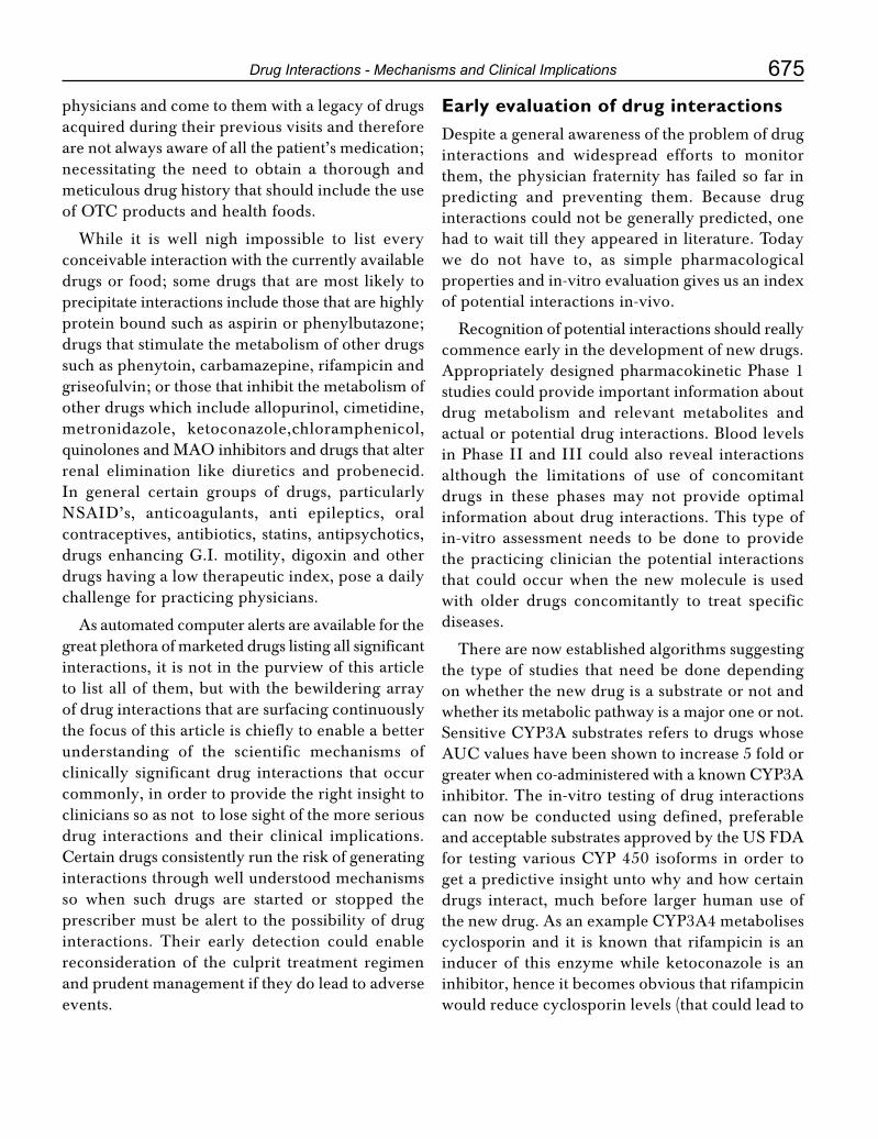
675Drug Interactions - Mechanisms and Clinical Implications
physicians and come to them with a legacy of drugs acquired during their previous visits and therefore are not always aware of all the patient’s medication; necessitating the need to obtain a thorough and meticulous drug history that should include the use ofOTCproductsandhealthfoods.
While it is well nigh impossible to list every conceivable interaction with the currently available drugs or food; some drugs that are most likely to precipitate interactions include those that are highly protein bound such as aspirin or phenylbutazone; drugs that stimulate the metabolism of other drugs such as phenytoin, carbamazepine, rifampicin and griseofulvin; or those that inhibit the metabolism of other drugs which include allopurinol, cimetidine, metronidazole, ketoconazole,chloramphenicol, quinolones and MAO inhibitors and drugs that alter renal elimination like diuretics and probenecid. In general certain groups of drugs, particularly NSAID’s, anticoagulants, anti epileptics, oral contraceptives, antibiotics, statins, antipsychotics, drugs enhancing G.I. motility, digoxin and other drugs having a low therapeutic index, pose a daily challenge for practicing physicians.
As automated computer alerts are available for the great plethora of marketed drugs listing all significant interactions, it is not in the purview of this article to list all of them, but with the bewildering array of drug interactions that are surfacing continuously the focus of this article is chiefly to enable a better understanding of the scientific mechanisms of clinically significant drug interactions that occur commonly, in order to provide the right insight to clinicians so as not to lose sight of the more serious drug interactions and their clinical implications. Certaindrugsconsistentlyruntheriskofgeneratinginteractions through well understood mechanisms so when such drugs are started or stopped the prescriber must be alert to the possibility of drug interactions. Their early detection could enable reconsideration of the culprit treatment regimen and prudent management if they do lead to adverse events.
Early evaluation of drug interactions Despite a general awareness of the problem of drug interactions and widespread efforts to monitor them, the physician fraternity has failed so far in predicting and preventing them. Because drug interactions could not be generally predicted, one had to wait till they appeared in literature. Today we do not have to, as simple pharmacological properties and in-vitro evaluation gives us an index of potential interactions in-vivo.
Recognition of potential interactions should really commence early in the development of new drugs. Appropriately designed pharmacokinetic Phase 1 studies could provide important information about drug metabolism and relevant metabolites and actual or potential drug interactions. Blood levels in Phase II and III could also reveal interactions although the limitations of use of concomitant drugs in these phases may not provide optimal information about drug interactions. This type of in-vitro assessment needs to be done to provide the practicing clinician the potential interactions that could occur when the new molecule is used with older drugs concomitantly to treat specific diseases.
There are now established algorithms suggesting the type of studies that need be done depending on whether the new drug is a substrate or not and whether its metabolic pathway is a major one or not. SensitiveCYP3AsubstratesreferstodrugswhoseAUCvalueshavebeenshowntoincrease5foldorgreaterwhenco-administeredwithaknownCYP3Ainhibitor. The in-vitro testing of drug interactions can now be conducted using defined, preferable and acceptable substrates approved by the US FDA for testingvariousCYP450 isoforms inorder toget a predictive insight unto why and how certain drugs interact, much before larger human use of thenewdrug.AsanexampleCYP3A4metabolisescyclosporin and it is known that rifampicin is an inducer of this enzyme while ketoconazole is an inhibitor, hence it becomes obvious that rifampicin would reduce cyclosporin levels (that could lead to

676 Medicine Update 2008 Vol. 18
transplant rejection;2 while the latter would increase it five to ten fold.3
However in-vitro data does not exactly translate always in the clinical scenario, because of immense variables that come in to play. Some drugs can be metabolized by more than one enzyme whereas some others like carbamazepine can not only induceaparticularisoenzyme(CYP3A4)butalsogets metabolized by it necessitating gradual dosing, while a few others can inhibit a particular isoenzyme but not be metabolized by it.
While it is therefore tempting to suggest that in vitro testing can prospectively rather than retrospectively indicate which other drugs may probably interact, the ground reality is slightly different due to the compounding factors impinging on the outcome; hence it is not so simple to think that we can get all answers this way.
Magnitude of the problemDrug interactions are complex and chiefly unpredictable as a known interaction may not occur in every individual taking the drug or even a drug in the same class. The exact incidence of drug interactions in real life situations is largely unknown because a fair number do not get reported, do not result in any substantial harm to patients or may not end in hospitalization and even when it does it gets recorded as an adverse reaction rather than a drug interaction. While significant drug interactions for commonly used drugs are recognized, there is a tendency amongst the fraternity to disregard the magnitude of evidence that potential interactions can manifest with the use of many of the drugs prescribed today.
There is a common assumption that all drug classes have a homogenous interaction potential when this is actually rare. This assumption may have had some credence when the number in a particular class was small and the mechanism of action was uncertain but with increasing knowledge about the mechanisms, this myth has been disproved. For example, amongst the macrolide
class, while erythromycin and clarithromycin inhibit CYP3A4 leading to interactions withother drugs, Azithromycin does not. Likewise while ketoconazole interacts with lovastatin and simvastatin and raises their plasma levels it does not do so with rosuvastatin4 or pravastatin.
The large inter-patient variability in the magnitude of the effect makes predicting the clinical outcome of an interaction nearly impossible as 5-7 fold differences in effect between participants is observed. However the inherent possibility of a drug interaction is intensified with the increasing use of a multitude of drugs by different routes in various doses and formulations administered concomitantly by doctors in clinics and hospitals specially to as many as 80% of elderly patients with serious chronic ailments, more so in I.C.Usettings. The matter is often compounded by patientsadditionallytakingOTCdrugsthatinteractwith their prescribed medication about which the doctor may not know; with other modifiers of drug elimination and response and genetics.
The magnitude of the problem can be gauged by the U.S Institute of Medicine’s report5 that medication errors were a major source of medical errors, with a 1999 estimate revealing 44000-98000 persons dying due to medical errors. A retrospective cross sectional analytical study of 46 million patients revealed that 374000 had been exposed to 25 potential dangerous drug interactions of clinical importance.
Sloan6 has pointed out that when 2 drugs are used in combination the interaction potential is 5-6%, that goes upto 50% with 5 drugs and when 8 drugs or more are co-prescribed the potential may even reach 100%. Earlier screening of 1800 records in a surgical setting7 revealed an incidence of 17% and a nursing home study8 showed an incidence of 19% and in a medical setting9 22% interactions. Another early study10 reported an incidence of 7% when 6-10 drugs were prescribed that rose to 40% when 15-20 drugs were given.

677Drug Interactions - Mechanisms and Clinical Implications
The earlier studies merely recorded the adverse events theoretically without realizing their clinical significance but the later studies however avoided this error by only considering potentially clinically important interactions, such as the Boston Collaborative surveillance study11 that involved 9900 patients with 83000 exposures, and reported that 234 (6%) of ADR’s were due to drug interactions and three other studies12,13,14 that reported an incidence of 4.1%, 2.9% and 1.9% of clinically important interactions respectively.
These reports reveal a discordant note but not withstanding this skepticism we need to ensure that there is no under reporting and the physician needs to be alert to the possibility of the occurrence of certain potential interactions of clinical importance. Clearlydrug interactionspresentahealth risk topatients and a great challenge to the physician as monitoring the patient’s therapy is a standard of care expected by the patients and the liability of interactions rests squarely on the physician who fails to recognize potentially harmful interactions to avoid extra costs of healthcare.
How do drug interactions occurThere are various categories of interaction with drugs:
• Drug-Diseaseinteractions
• Drug-Druginteractions
• Drug-Foodinteractions
• Drug-Herbinteractions
• DrugEnvironmentalinteractions
Knowledge of the mechanism by which a given drug interaction occurs is often useful in practice, as the mechanism could influence both the time course and methods of evading the interaction.
It is therefore incumbent upon the physician to be familiar with the basic principles of drug interactions in planning a therapeutic regimen as there are several factors affecting the likelihood of a known interaction such as age, sex, lifestyle, physiological differences, time and sequence of
administration and genetic polymorphisms in some of themainCYP isoformsnotablyCYP2D6andCYP3A4 thataffectenzyme function.Further,asthe number of drugs prescribed increases especially in the elderly, so does the susceptibility to drug interactions specially in the absence of an accurate drug history and a lack of knowledge of potential consequences.
Because of the complexity of pharmacotherapy needed for the treatment of the basic disease, its underlying causative factors, its complications and accompanying co-morbid conditions such as hypertension, diabetes and dyslipidemia, malignancy and respiratory disorders, the number of drugs prescribed increases translating into a major risk factor for potential drug interactions. Diseases apart, physiological changes in renal and hepatic function with advancing age, malnutrition and reduced homeostatic mechanisms makes the elderly more sensitive to the additive effects of two or more drugs rendering them more prone to serious drug interactions. The objectives of treatment are therefore vital while assessing the clinical implication of a drug interaction as a balance needs to be struck between increased toxicity and reduced efficacy.
An understanding therefore of the classification and mechanisms of drug interactions is essential in order to predict their occurrence and comprehend their clinical significance and is the only way a clinician can be prepared to analyze new findings systematically; as there are certain mechanisms that are encountered repeatedly but a few others are unique and many drugs that interact do so not necessarily by a single mechanism but quite often by two or more mechanisms in consonance with each other.
Drug-Drug interactionsAlthough tremendous advances have occurred in our understanding of the mechanisms of drug interactions over the last few years we still have a long way to go to understand them fully as more than one mechanism may play a part in some drug interactions.

678 Medicine Update 2008 Vol. 18
Knowledge of the mechanism by which a given drug interaction occurs is often clinically useful, since the mechanism may influence both the time course and methods of circumventing the interaction.
The etiology and clinical implications of drug-drug interactions are multifactorial and chiefly unpredictable, that include patient as well as drug factors alluded to earlier.
Drug- drug interactions reflect the modulation of the pharmacological activity of the object drug by concomitantly administering the precipitant drug resulting in a severe decrease or increase in the pharmacological properties of either drug.
The clinically most important adverse drug-drug interactions occur with drugs that have easily recognizable toxicity and a low therapeutic index, such that relatively small changes in drug effect can have clinically significant adverse consequences. Another dimension of drug-drug interactions that could have clinical significance is the seriousness of the disease that is being treated, which if left untreated could be fatal.
There are several mechanisms by which drugs may interact but most can be classified as:
• PharmacokineticInteractions
• PharmacodynamicInteractions
• AdditiveorSynergisticInteractions
• AntagonisticorOppositioninteractions
Major adverse drug interactions ordinarily do not result from the “additive” effects of drugs acting at the same receptor site or from pharmacological or physiological antagonism as their combined effects can easily be predicted from their known pharmacology. Adverse drug interactions resulting from concomitant medication are commonly associated with drugs that are chemically or biochemically antagonistic.
Thus the mechanisms usually responsible for adverse effects associated with drug interactions are those in which one drug affects the pharmacokinetic
profile of absorption, distribution, metabolic biotransformation, excretion or elimination of another; or pharmacodynamic, such as interactions between agonists and antagonists at drug receptors, leading to vastly altered clinical response and implications.
Pharmacokinetic InteractionsThe science of therapeutics does not merely involve testing of new molecules in animals and humans, but applies more importantly to the treatment of each patient holistically as an individual and it is widely recognized that individuals show wide variability in response to the same treatment.
Pharmacokinetic interactions must always be evaluated in the context of their clinical relevance. The fact that two drugs share a common metabolic pathway does not mean they will have a clinically significant interaction when co-administered; the interaction being dependent upon various factors including relative affinities of each drug for the binding site or the metabolising enzyme; as well as the effective free drug concentration available for binding. Moreover parallel pathways for elimination of one or both drugs would tend to reduce the potential for a significant pharmacokinetic interaction.
Hence, to avoid interactions; inter and intrapatient variations in disposition of a drug must be taken into consideration in choosing a treatment regime, such as the prescribed dose and its compliance, the actually administered dose, its rate and extent of absorption, plasma concentration, Tmax,AUC,distribution,metabolism,therateofelimination (t1/2), drug concentration at the site of action, genetic variations and the effect of the drug at the receptor. The major determinants of the disposition of many drugs are the physiological and pathological variations in organ function. In general, pharmacokinetic interactions are considered clinically significant when at least a 30% changeisseeninCmax,TmaxandAUC.

679Drug Interactions - Mechanisms and Clinical Implications
Pharmacokinetic interactions occur when the absorption, distribution, metabolism or elimination processes of the object drug is altered by the precipitant drug.
Drug absorption interactionsSince the oral route is the one most frequently used to administer drugs, interactions influencing absorption are more likely to occur within the gastrointestinal tract, which more often result in reduced rather than increased absorption.
While the absorption of a drug may be altered by another, the resulting interactions are of varying clinical importance as we need to make a definitive distinction between drugs that influence the rate of absorption as opposed to those alter the extent of absorption as the rate is more often unimportant in case of chronically administered drugs such as warfarin provided the extent is not markedly altered, while it is a different scenario when an analgesic is given where a high concentration may be needed rapidly to achieve an adequate effect when the interaction becomes clinically important if it results in subtherapeutic serum levels of the various possible interactions that occur due to alterations in drug absorption most clinically significant interactions occur due to the following factors:
Changes in gastrointestinal pHAbsorption in the gut is governed by the gut pH, lipid solubility and pKa of the drug, and action of the P-glycoprotein. While changes in gastric pH induced by H2 and proton pump blockers and antacids containing Al/Mg formulations have been shown to significantly reduce drug bioavailability;15 in clinical practice the outcome is a bit uncertain due to other compounding factors such as chelation and gastric motility. However the alteration in pH has certain clinical implications as it can result in a significant reduction in the absorption of ketoconazole and itraconazole which are insoluble in water and are only ionized at low pH, hence gastric acidity plays an important part in this interaction. Likewise salicylic acid absorption is greater at low pH.
The absorption of quinolones are also reduced when given along with antacids. Other drugs that are influenced by changes in pH are glipizide, glyburide, cefuroxime and cefpodoxime.
Changes induced by chelation and adsorption
Of the various possible drug interactions that occur due to alterations in drug absorption the most clinically significant interactions occur due to chelation or formation of insoluble complexes or when drugs are bound to resins that bind to bile acids.
Clinicallyimportantinteractionsrelatetouseoftetracyclines as well as ciprofloxacin that can form insolublechelateswithCa,Al,Biandiron,resultingin its reduced antibacterial effects. This interaction can however be avoided if the interval between the medications is at least 2-3hours.Chelation alsoseems to play an important part in reducing the bioavailability of penicillamine caused by some antacids.
The commonly used Kaolin-pectin suspensions in diarrheal disorders bind digoxin, when co-administered reducing its absorption by 30-50%, while resins like cholestyramine and colestipol that
Figure 1
Bile canal
Hepatocyte
Varioustransporters
P-gp
Oral drug
DrugMetabolitesDrug from enterocyte can get excreted
into intestinal lumen or undergoesmetabolism or enters the portal system
Enterocyte
Lumen
Portalsystem
(Liver)
To Generalcirculation
The hepatocyte metabolism andexcretes drug into biliary circulationbefore it enters general circulation

680 Medicine Update 2008 Vol. 18
sequester bile acids in the gut bind to a number of drugs like digoxin, levothyroxine, statins, valproic acid, steroids, loop diuretics and warfarin reducing their absorption with resultant clinical implications warranting close clinical and biochemical monitoring to avoid complications. Further estrogen metabolites in bile are deconjugated by bowel organisms and reabsorbed and if this is prevented by poorly absorbed antibiotics such as ampicillin the contraceptive effect gets reduced with a risk of pregnancy. Antibiotics that alter gastrointestinal flora can reduce the rate of synthesis of vitamin K with a resultant increase in the effect of oral anticoagulants due to a competitive mechanism between them. Dilantin was reported early on to inhibit an intestinal conjugase which was found to inhibit the absorption of folic acid specially in susceptible individuals.16, 17
Changes in gastrointestinal motilityDrugs that alter the stomach-emptying rate can affect the rate of absorption of drugs as most of them are absorbed in the small intestine. Drugs with anticholinergic properties like propantheline or those altering bowel motility like diphenoxylate may affect the absorption of other drugs. Propantheline increases the absorption of slow dissolving Digoxin by 30% as the reduced gut motility allows a slow dissolving Digoxin formulation more time to pass
into solution making a greater amount available for absorption but this effect is not seen with fast dissolving tablets. Metoclopramide on the other hand produces the opposite effects on motility and digoxin absorption.18
It may also be pertinent to point that the ultimate outcome of interactions of drugs exhibiting anticholinergic properties that decrease gut motility like tricyclic anti-depressants can be unpredictable due to several mechanisms because on one hand they may reduce the absorption of a drug like levodopa as the exposure time to intestinal mucosal metabolism is increased; while on the other they increase the absorption of dicoumarol possibly by increasing the time available for its dissolution and absorption, although the exact mechanism is not understood clearly.
Transporter based interactions Uptake into the enterocyte particularly by the active processes is mediated by specific drug uptake transport molecules. Once the drug enters the enterocyte it could enter the portal circulation, undergo metabolism or it may get excreted back into the intestinal lumen resulting in decreased systemic bioavailability (Fig. 2).
Transporter based interactions have of late been recognized much more than earlier and arise chiefly due to the induction or inhibition of many identified transporter proteins rather than due to other mechanisms earlier attributed to protein displacement or enzyme inhibition or induction.
Two mechanisms are important modulators of presystemic clearance of some drugs. The first one that has a greater clinical relevance and perhaps best studied is P-glycoprotein (P-gp) which is a product of the normal expression of the MDR1 gene and is expressed on the apical aspect of the enterocyte as well as on the canalicular aspect of the hepatocyte, works as a ‘ detoxification’ pump ejecting drugs that have diffused across the intestinal epithelial barrier resulting in a reduction of the drug absorbed and
Figure 2
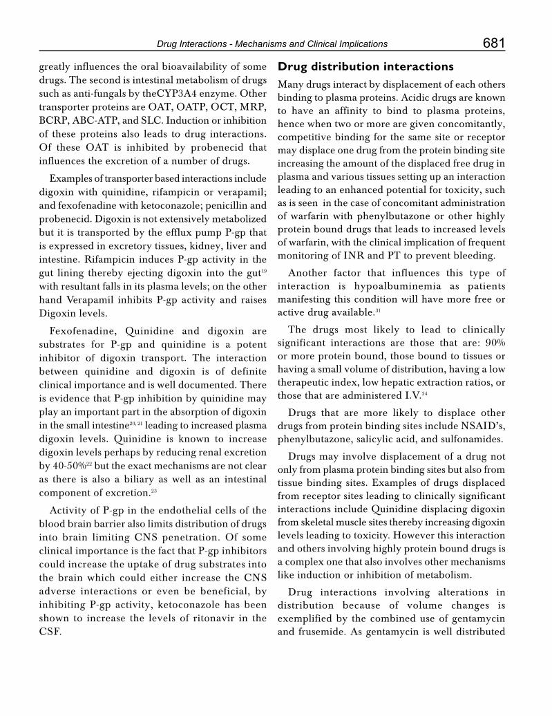
681Drug Interactions - Mechanisms and Clinical Implications
greatly influences the oral bioavailability of some drugs. The second is intestinal metabolism of drugs suchasanti-fungalsbytheCYP3A4enzyme.OthertransporterproteinsareOAT,OATP,OCT,MRP,BCRP,ABC-ATP,andSLC.Inductionorinhibitionof these proteins also leads to drug interactions. Of these OAT is inhibited by probenecid that influences the excretion of a number of drugs.
Examples of transporter based interactions include digoxin with quinidine, rifampicin or verapamil; and fexofenadine with ketoconazole; penicillin and probenecid. Digoxin is not extensively metabolized but it is transported by the efflux pump P-gp that is expressed in excretory tissues, kidney, liver and intestine. Rifampicin induces P-gp activity in the gut lining thereby ejecting digoxin into the gut19 with resultant falls in its plasma levels; on the other hand Verapamil inhibits P-gp activity and raises Digoxin levels.
Fexofenadine, Quinidine and digoxin are substrates for P-gp and quinidine is a potent inhibitor of digoxin transport. The interaction between quinidine and digoxin is of definite clinical importance and is well documented. There is evidence that P-gp inhibition by quinidine may play an important part in the absorption of digoxin in the small intestine20, 21 leading to increased plasma digoxin levels. Quinidine is known to increase digoxin levels perhaps by reducing renal excretion by 40-50%22 but the exact mechanisms are not clear as there is also a biliary as well as an intestinal component of excretion.23
Activity of P-gp in the endothelial cells of the blood brain barrier also limits distribution of drugs into brain limiting CNS penetration. Of someclinical importance is the fact that P-gp inhibitors could increase the uptake of drug substrates into thebrainwhich could either increase theCNSadverse interactions or even be beneficial, by inhibiting P-gp activity, ketoconazole has been shown to increase the levels of ritonavir in the CSF.
Drug distribution interactionsMany drugs interact by displacement of each others binding to plasma proteins. Acidic drugs are known to have an affinity to bind to plasma proteins, hence when two or more are given concomitantly, competitive binding for the same site or receptor may displace one drug from the protein binding site increasing the amount of the displaced free drug in plasma and various tissues setting up an interaction leading to an enhanced potential for toxicity, such as is seen in the case of concomitant administration of warfarin with phenylbutazone or other highly protein bound drugs that leads to increased levels of warfarin, with the clinical implication of frequent monitoring of INR and PT to prevent bleeding.
Another factor that influences this type of interaction is hypoalbuminemia as patients manifesting this condition will have more free or active drug available.31
The drugs most likely to lead to clinically significant interactions are those that are: 90% or more protein bound, those bound to tissues or having a small volume of distribution, having a low therapeutic index, low hepatic extraction ratios, or those that are administered I.V.24
Drugs that are more likely to displace other drugs from protein binding sites include NSAID’s, phenylbutazone, salicylic acid, and sulfonamides.
Drugs may involve displacement of a drug not only from plasma protein binding sites but also from tissue binding sites. Examples of drugs displaced from receptor sites leading to clinically significant interactions include Quinidine displacing digoxin from skeletal muscle sites thereby increasing digoxin levels leading to toxicity. However this interaction and others involving highly protein bound drugs is a complex one that also involves other mechanisms like induction or inhibition of metabolism.
Drug interactions involving alterations in distribution because of volume changes is exemplified by the combined use of gentamycin and frusemide. As gentamycin is well distributed

682 Medicine Update 2008 Vol. 18
inextracellularfluidanyreductioninECFinducedby frusemide reduces the volume of distribution of gentamycin increasing its serum levels with the clinical implication of nephro and ototoxicity.
Despite the factors described above for distribution interactions, recent research25 suggests that although in-vitro many commonly used drugs are capable of being displaced by others, in the body these effects seem almost always so well buffered that the outcome may not normally be clinically important. Moreover, as some interactions assumed to be originally due to protein binding have subsequently been shown to have other mechanisms involved it has been suggested that the importance of this mechanism alone being responsible for the interaction has been exaggerated. This however does not deter from the fact that cognizance of alterations in protein binding is invaluable for therapeutic drug monitoring.
Drug metabolism interactionsIn a monograph on clinically significant drug
interactions, metabolic interactions are most important and need to be examined in great detail. Recent scientific developments, particularly in the areaoftheCYP450enzymeshaverevolutionizedthe study of drug interactions resulting in a deluge of published drug interactions that has bewildered the practicing physicians.
The human body is continuously exposed to foreign substances (drugs) not found naturally in the body that modulate the body function to achieve
a therapeutic end that are modified by a plethora of enzymes. As is well known, the processes by which the enzymes alter an active drug inside the body to an inactive one or into active or toxic metabolites are referred to as drug metabolism or biotransformation.
Most drugs need to reach a receptor site in order to exert their systemic effect and need to be lipid soluble so as to be able to penetrate the lipid plasma membrane barrier. The lipid soluble drugs further need to be converted into a water soluble form to be excreted chiefly by the renal route and the chief role of metabolism is to enable these processes in two phases.
In phase I, oxidation/reduction reactions convert the drugs into a more polar form, while Phase II reactions provide another set of mechanisms involving conjugation / hydrolysis with substances like glucuronic acid and glucuronyl transferase for modifying drugs into inactive compounds to enable their excretion.
Phase I reactions are catalysed by a family of mixed function oxygenases called the ''Cytochrome P 450" class, expressed chiefly within the microsomal smooth endoplasmic reticulum hepatocytes and to a lesser extent in other cells.
The nomenclature for this class of enzyme is usuallyabbreviatedto‘CYP”followedbyanArabicnumber indicating the enzyme family and a capital letter to indicate the enzyme sub family and then an additional number to describe the specific enzymee.g.CYP2D6.Allelicformsaredescribedwith an * and a number or number letter. This enzyme complex is so named because it is bound to a membrane within a cell (Cyto) and contains a hem Pigment (chrome and P) that absorbs light atawavelengthof450nmwhenexposedtoCo2.The net result is “Cytochrome P 450”.
The interaction starts with the drug binding to theoxidized(Fe)CYP450complexwhichisthenreduced in two oxygenation/reduction steps using a reactive hem ring with an iron atom on the ultimate electron acceptor donor and NADPH as a necessary
Figure 3
Nomenclature ofCytochrome P450 Family
CYP2C19*8
Cytochrome P450
Family
Sub-family
Enzyme
Allele
(2)
(C)
(19)
(*8)

683Drug Interactions - Mechanisms and Clinical Implications
co-factor.
TheCYP450complexisessentialformetabolismof drugs and interactions mediated by it and what is significant is that out of 50 enzymes in this class, each encoded by a different gene, just 6 of them(CYP1A2,CYP2C8/9,CYP2C19,CYP2D6,CYP2E1andCYP3A4/5)togetheraccountfor90-95% of biotransformations; with the 3A4 and 2D6 sub families being responsible for a large number of clinically important interactions.26
However, there is a considerable variability in enzyme activity between patients due to medical, environmental, nutritional and genetic polymorphisms reasons with polymorphism being specially significant for CYP2D6, CYP2C9, andCYP2C19andCYP3A4.27
A drug’s action on a molecular target already results in a biological complex that could be influenced further by disease. On top of this genetic polymorphisms that influence change in this biological complex can greatly influence drug response. In this context, while 20% of Asians are poor metabolisers of drugs dependent on CYP2C19 metabolism such as phenytoin,omeprazole28 phenobarbitone and other drugs; only 2% of the same population exhibits a variant ofCYP2D6withlowactivity.Thesedifferencesinthe variability to metabolise different drugs could account for a few persons manifesting toxicity with interacting drugs while others do not exhibit
any symptoms.
The clinical implication of this polymorphism is exemplified by omeprazole, where poor metabolisers having higher drug levels with standard dosages had markedly higher cure rates (100)% than their EM counterparts; highlighting as well the need for identification of such polymorphisms early in a drug’s development. The I/D deletion polymorphism intheACEgene,G6PDdeficiencygeneandtheABCA1transportergeneinadditioncanallresultin clinically important adverse interactions of which physicians need to be cognizant.
Phase II conjugation/hydrolysis reactions provide a second set of mechanisms for modifying compounds for excretion wherein large water soluble metabolites are acted upon by non P450 enzymes such as N-acetyl and glucuronyl transferase systems which render them inactive or less active water soluble metabolites.
Clinically significant interactions however,are caused chiefly by Phase I reactions as most of the metabolism occurs in the liver via the P450 enzymes, rather than phase II metabolism. Although significant metabolism takes place in the liver, other organs like the kidney and gut are also involved. During the first passage through the liver, certain drugs are extremely metabolized and are referred to as high extraction drugs. These drugs having a short half life and inactive metabolites are clinically less affected by interactions than their low extraction counterparts. However, lower the therapeutic index of a drug, it has a greater clinical potential to produce more serious consequences of drug interactions affecting metabolism.
CrucialtotheabilitytopredictdruginteractionsisaproperunderstandingofdrugsinfluencingCYP450 enzyme induction and inhibition. Interactions involving drug metabolism can increase or decrease the amount of drug available for action by inhibition or induction of metabolism. Inhibition is usually more predictable than induction which is influenced by genetic differences between patients.
Figure 4

684 Medicine Update 2008 Vol. 18
Inhibitors compete with other drugs for a particular enzyme thus affecting the optimal level of metabolism of the substrate drug, that then accumulates in the body resulting in toxicity.
Strong inhibitors achieve a 5 fold increase in plasma AUC or an 80% decrease in clearance,while moderate inhibitors lead to a 2 fold increase inAUCand50-80%decreaseinclearanceofthesubstrate drug.
Inducers, on the other hand stimulate the productionoftheCYPisoform,thusincreasingtherate of metabolism and enabling substrate drug to clear out of the system faster. This decreases its response, rendering the drug ineffective, as it does not remain in the system long enough. Enzyme induction does not occur quickly, usually taking a week or two as its maximal effect depends on enzyme synthesis and t1/2 of the inducing drug, which in the case of phenobarbitone may require a longer time, while rifampicin with its short t1/2 can manifest its effects within 24 hours.
Conversely inhibition of CYP enzymes tendsto be rapid with maximum effect occurring when steady state concentrations of the inhibitor drug are established. However if the t1/2 of the affected drug is long it may take a week to reach steady state levels. This inhibition leads to decreased metabolism of drugs acted upon by the enzyme, prolonging its t1/2 and reducing clearance, thereby increasing plasma levels that lead to interactions.
Investigating the likelihood of CYP mediatedinteractions in a multicenter audit of patients attending daycare centers, Wilcock29 categorized drug interactions as either “clinically important” for which there is in-vitro metabolic and in-vivo clinical evidence or “potentially clinically important” if there was a theoretical but not experimental basis and “unlikely” if there is no real basis for interaction. Audited prescriptions of 160 patients revealed a median of 7 drugs co-administered, with 77% of patients having received a median of 4 drugs that were either substrates, inducers or inhibitors of theCYP450enzymesystem.24combinations
of drugs administered induced “clinically important” or: potentially clinically important” interactions involvingCYP450enzymes in20%ofpatients.Of these, 2 important interactions reported were, one with a diazepam/omeprazole combination due to CYP inhibition, causing increased diazepamconcentrations resulting in drowsiness and the other involving a phenytoin-dexamethasone combination that led to decreased dexamethasone concentrations duetoCYPinhibition.Ofthepotentiallyimportantinteractions, 50% were associated with steroids given together with amlodipine or simvastatin or trazodone or amitryptiline and 25% with analgesics.
Effects of enzyme inhibition on drug metabolismThe extent of inhibition of metabolism of a drug depends in fact on the dose and binding ability to the enzyme. Potent inhibitors of an enzyme that may not even be its substrate have the greatest potential for interactions requiring the physician tobe specially alert to them.Clinically relevantinteractions of inhibited drug metabolism occurring through oxidative processes include inhibition of warfarin metabolism by phenylbutazone, cimetidine, chloramphenicol, and metronidazole; inhibition of theophylline metabolism by quinolones and macrolides and inhibition of phenytoin by isoniazid resulting in increased therapeutic levels andtoxicity.CYP1A2inhibitorscanincreasetherisk of toxicity from theophylline or clozapine; CYP2C9 that of phenytoin and warfarin; whileCYP3A4inhibitionresultsinthehazardsoftoxicityof a larger number of drugs like carbamazepine, lovastatin and simvastatin, rifabutin, cisapride, cyclosporin, ergot, protease inhibitors and vinca alkaloids.
Pro-drugs require enzyme activation to produce their effect, hence enzyme inhibition results in a reduced activity of the drug. The analgesic effect of codeine results from its conversion to morphine by CYP2D6,henceitsinhibitiondecreasescodeine’stherapeutic effect.

685Drug Interactions - Mechanisms and Clinical Implications
Other inhibitory interactions utilizing different pathways include Azathioprine and 6MP that are metabolized by Xanthine oxidase, which is inhibited by allopurinol and the interaction of MAO inhibitors with tyramine rich products such as cheese that precipitate hypertension.
Inhibition of enzymes can occur in different ways such as is seen with ketoconazole, whose nitrogen moiety binds to the heme iron in the P450 enzyme site preventing the metabolism of concomitantly administered drugs either by competitive or irreversible inhibition that is achieved for instance by secobarbital that alkylates and inactivates the P 450 enzyme permanently (Fig. 5)
P450 inhibitory interactions can also occasionally be therapeutically useful as in the case of lopinavir whose drug concentrations are actually increased bytheco-administrationoftheCYP3A4inhibitorritonavir, since the first-pass metabolism of the former precludes it achieving therapeutic levels on its own.
Effects of enzyme induction on drug metabolismWhile some P450 enzymes are active constitutively other enzymes can be induced by different drugs. InductionofCYPisoenzymesisachievedslowlybydrugs like rifampicin, phenytoin, carbamazepine, barbiturates, glutethemide, troglitazone, rifabutin, griseofulvin and St John’s Wort, by increasing the
amount of endoplasmic reticulum in liver cells and byincreasingthecontentofCYP450.Administrationof these drugs increases the expression of genes such as MDRI over a fortnight and results in clinically significant interactions by decreasing the plasma levels of co-administered drugs such as warfarin, ketoconazole, itraconazole, quinidine, verapamil, mexiletine, low dose oral contraceptives, prednisolone and theophylline.
The process of P450 enzyme induction gets initiated by an increase in the expression of the enzyme chiefly via increased transcription or decreased degradation. Drugs or food gets bound to and activates several xenobiotic receptors e.g the Pregnane X receptor after entering the liver cells, which then heterodimerises with the Retinoid X receptor (RXR) to form a complex with coactivators to initiate transcription of the P450 enzyme. (Fig. 6)
DrugsmetabolizedbyCYP2C9andCYP3A4areparticularly susceptible to enzyme induction.
The reduction in the plasma levels of the object drugs brought about by inducers leads to a reduction in their clinical effects, that could lead an epileptic to manifest fits while on phenytoin or a pregnancy resulting while on oral contraceptives.
Ritonavir is an example of a drug that not only induces but also acts an inhibitor of the isoenzyme CYPD6dependingonthesituation.
Figure 5
Figure 6

686 Medicine Update 2008 Vol. 18
Enzyme inducing drugs can also increase the activity of phase II metabolism processes such as glucuronidation. Once the drugs responsible for induction are stopped their effects take an equally long time to disappear and this can result in major toxicity if the dose of a low therapeutic index co-administered drug that has been stabilized in the presence of an inducer is suddenly stopped; such as is seen in the case of warfarin, digoxin etc.
Some drugs are converted to toxic metabolites by enzymes and enzyme inducers can increase the formation of these toxic metabolites. Paracetamol is primarily converted to nontoxic metabolites but a small amount is converted to toxic metabolites; however if administered with an enzyme inducer it could lead to hepato-toxicity.
Drug elimination reactions The major routes for elimination of drugs remain the kidney and bile, but there are no significant drug - drug interactions through bile elimination, but only drug-disease ones.
Drugs that are chiefly excreted by the kidneys can get involved in drug interactions by different mechanisms such as Competition at active transport sites, or alterations in Glomerular Filtration, passive renal tubular reabsorption or active secretion and urinary pH.
Active secretion into the renal tubules is an important excretion pathway for some drugs, that gets affected by the co-administration of certain other drugs, thereby affecting their therapeutic response. The capacity of a drug to inhibit the renal excretion of another is dependent on an interaction at active transport sites. The beneficial probenecid - penicillin/amoxycillin interaction exemplifies one of the many reported interactions at the anion transport site; the two drugs competing for excretion by modifying active transport in the renal tubules resulting in probenecid being excreted and the antibiotics being retained and reabsorbed, with the clinical implication of increasing their plasma levels to a desirable level to increase its therapeutic
effect and prolonging the plasma t1/2.
However adverse interactions are seen with concomitant administration of digoxin with drugs like quinidine, verapamil and amiodarone, leading to digoxin toxicity.
The interaction between quinidine and digoxin is of definite clinical importance and is extremely well documented, resulting not only from quinidine reducing the renal excretion of digoxin by 50%, but also by non renal mechanisms, that includes reduction of about 50% in digoxin excretion in bile23 as well as by its P-gp mediated inhibition of transcellular transport30 and also inhibition in the gut.20 Further, salicylates have been shown to reduce the renal clearance of methotrexate leading to its toxicity. The other interaction involves the excretion of lithium which gets altered by diuretics and NSAID’s that inhibit renal tubular reabsorption leading to compensatory reabsorption of lithium with consequent toxicity,
Likewise, interactions at the caution transport site for basic drugs result from drugs like cimetidine, amiodarone and dofetelide inhibiting the excretion of procainamide. Cimetidine also competeswith metformin, (both being cationic drugs) for elimination by renal tubular secretion.
The rate of excretion of a drug or its metabolites can be influenced by other drugs that increase or decrease glomerular filtration due to changes in renal blood flow. For drugs with a low therapeutic index like digoxin, phenytoin and warfarin, any increase in renal clearance decreases their steady state plasma concentrations and conversely any reduction in their renal excretion increases circulating levels of the drugs resulting in toxicity.
Finally changes in the pH of urine that alter the excretion of weakly acidic or basic drugs can lead to interactions by affecting their ionization and consequently affecting the reabsorption of drugs that are subject to passive reabsorption from renal tubules. The implication of this mechanism is reflected in the treatment of salicylate or amphetamine poisoning by alkalinising with
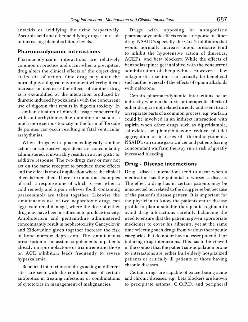
687Drug Interactions - Mechanisms and Clinical Implications
antacids or acidifying the urine respectively. Ascorbic acid and other acidifying drugs can result in increasing phenobarbitone levels.
Pharmacodynamic interactionsPharmacodynamic interactions are relatively common in practice and occur when a precipitant drug alters the clinical effects of the object drug at its site of action. One drug may alter the normal physiological environment whereby it can increase or decrease the effects of another drug as is exemplified by the interaction produced by diuretic induced hypokalemia with the concurrent use of digoxin that results in digoxin toxicity. In a similar situation of diuretic usage concurrently with anti-arrhythmics like quinidine or sotalol a much more serious toxicity in the form of Torsade de pointes can occur resulting in fatal ventricular arrhythmias.
When drugs with pharmacologically similar actions or same active ingredients are concomitantly administered, it invariably results in a synergistic or additive response. The two drugs may or may not act on the same receptor to produce these effects and the effect is one of duplication where the clinical effect is intensified. There are numerous examples of such a response one of which is seen when a cold remedy and a pain reliever (both containing paracetamol) are taken together. Likewise the simultaneous use of two nephrotoxic drugs can aggravate renal damage, where the dose of either drug may have been insufficient to produce toxicity. Amphotericin and pentamidine administered concomitantly result in nephrotoxicity Gancyclovir and Zidovudine given together increase the risk of bone marrow depression. The simultaneous prescription of potassium supplements to patients already on spironolactone or triamtrene and those on ACE inhibitors leads frequently to severehyperkalemia.
Beneficial interactions of drugs acting at different sites are seen with the combined use of certain antibiotics in treating infections or combinations of cytotoxics in management of malignancies.
Drugs with opposing or antagonistic pharmacodynamic effects reduce response to either drug.NSAID’sspeciallytheCox-2inhibitorsthatwould normally increase blood pressure tend to inhibit the hypotensive action of diuretics, ACEI’s and beta blockers. While the effects ofbenzodiazepines get inhibited with the concurrent administration of theophylline. However, a few antagonistic reactions can actually be beneficial such as the reversal of the effects of opium alkaloids with naloxone.
Certain pharmacodynamic interactions occurindirectly wherein the toxic or therapeutic effects of either drug are not related directly and seem to act on separate parts of a common process; e.g. warfarin could be involved in an indirect interaction with aspirin when other drugs such as dipyridamole, salicylates or phenylbutazone reduce platelet aggregation or in cases of thrombocytopenia. NSAID’s can cause gastric ulcer and patients having concomitant warfarin therapy run a risk of greatly increased bleeding.
Drug - Disease interactions Drug - disease interactions tend to occur when a medication has the potential to worsen a disease. The effect a drug has in certain patients may be unexpected not related to the drug per se but because of the patient’s disease pattern. It is important for the physician to know the patients entire disease profile to plan a suitable therapeutic regimen to avoid drug interactions carefully balancing the need to ensure that the patient is given appropriate medicines to cover his ailments, yet at the same time selecting such drugs from various therapeutic categories that do not or have a lesser potential for inducing drug interactions. This has to be viewed in the context that the patient sub-population prone to interactions are either frail elderly hospitalized patients or critically ill patients or those having chronic diseases.
Certaindrugsarecapableofexacerbatingacuteand chronic diseases. e.g. beta blockers are known to precipitate asthma, C.O.P.D. and peripheral

688 Medicine Update 2008 Vol. 18
vascular disease29,30 and can also blunt the signs of hypoglycemia. Certain beta blockers and thecalcium blocker verapamil by virtue of their negative inotropic and chronotropic effects have thepotentialtoprecipitateC.H.F.
Drug interactions that occur in patients with milder forms of disease and minor clinical significance assume greater significance in patients with more severe forms of diseases such as diabetes, cardiac disorders, asthma, epilepsy, hypothyroidism and liver diseases.
The risk associated with the potential for interactions with the treatment required for certain diseases like psychiatric disorders autoimmune disorders, G.I. diseases, respiratory and infections always poses a problem.
Treatment for diseases involving drugs having a narrow therapeutic window, like digoxin, lithium, phenytoin, warfarin, quinidine and theophylline, poses clinically significant possibilities of predisposition to drug interactions.
Drug - food / nutrients interactionsThe myth that natural products, not being drugs, are completely safe creates a need for responsible, public/physician education specially as they are widely used by our rural/semi-urban populace; hence the need to be cognizant of these interactions and as a large number do not inform the physicians about their intake, the potential and true incidence of these interactions is largely unknown. A lack of standardization and contamination further contribute to these interactions. The mechanisms of food-induced interactions are essentially the same as that of drug interactions, however these occur chiefly due to alterations in absorption that may impair their nutritional benefit and to some extent due to altered metabolism.
The common natural products interactions with drugs are due to St John's Wort, ginseng, glucosaminesulfate, ginko biloba, aloe, guargum, senna, grapefruit juice, garlic, fenugreek, tyramine rich foods and curcumin.
St John’s Wort is one herb that is responsible for having interactions with many drugs. It reduces absorption of digoxin, lowering its blood levels; amplifies action of clopidogrel increasing risk of bleeding. It has synergistic effects with SSRI’s and Zolpidem, increasing serotonin levels in brain leading to “Serotonin Syndrome” By enhancing expression of intestinal P-gp and CYP3A4 itimpairs absorption and stimulates metabolism of cyclosporin, resulting in sub-therapeutic levels. Interaction with estrogen results in bleeding and interacting with indinavir, lowers its plasma levels and efficacy, worsening infection. It also lowers warfarin plasma levels and increases clot formation.
It also interacts with the anticancer drugs docetaxel and irinotecan by similar metabolic mechanisms, resulting in larger inactive metabolites and less of the active SN38 metabolite of irinotecan, resulting in lower efficacy. As many cancer patients use alternative medicines with their chemotherapy, unexpected toxicities, lowered plasma levels lead to under-treatment. As treatment failure is common in cancer patients the implication of the herb’s contribution to the failure is likely to be missed.
One of the most clinically significant interactions with tyramine rich foods like cheese, bananas, chocolate, wine etc occurs when they are concurrently used with MAO inhibitors resulting in hypertensive crises occasionally. Likewise the other significant interactions with clinical implications are seen in patients on warfarin who ingest food rich in Vitamin K such as cauliflower, broccoli, cabbage, soyabean and leafy vegetables; and those on levodopa who ingest foods rich in vitamin B
6,
such as peas, pork, liver and fish, as these decrease dopamine levels resulting in antiparkinsonian effects.
Drugs, whose absorption is decreased include penicillin, tetracyclines, erythromycin, phenytoin, levodopa, digoxin.
An increase in drug absorption is seen in the case of spironolactone, griseofulvin, itraconazole.32
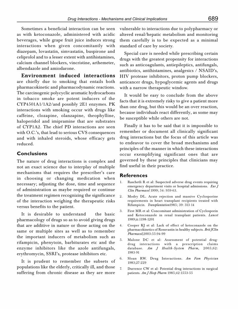
689Drug Interactions - Mechanisms and Clinical Implications
Sometimes a beneficial interaction can be seen as with ketoconazole, administered with acidic beverages, while grape fruit juice induces strong interactions when given concomitantly with diazepam, lovastatin, simvastatin, buspirone and celiprolol and to a lesser extent with antihistamines, calcium channel blockers, vincristine, arthemeter, albendazole and amiodarone.
Environment induced interactions are chiefly due to smoking that entails both pharmacokinetic and pharmacodynamic reactions. The carcinogenic polycyclic aromatic hydrocarbons in tobacco smoke are potent inducers of the CYP4501A1/1A2/andpossibly2E1enzymes.PKinteractions with smoking occur with drugs like caffeine, clozapine, olanzapine, theophylline, haloperidol and imipramine that are substrates of CYP1A2. The chief PD interactions are seenwithO.C.’s,thatleadtoseriousCVSconsequencesand with inhaled steroids, whose efficacy gets reduced.
ConclusionsThe nature of drug interactions is complex and not an exact science due to interplay of multiple mechanisms that requires the prescriber’s care in choosing or changing medication when necessary; adjusting the dose, time and sequence of administration as maybe required or continue the treatment regimen recognizing the significance of the interaction weighing the therapeutic risks versus benefits to the patient.
It is desirable to understand the basic pharmacology of drugs so as to avoid giving drugs that are additive in nature or those acting on the same or multiple sites as well as to remember the important inducers of metabolism such as rifampicin, phenytoin, barbiturates etc and the enzyme inhibitors like the azole antifungals, erythromycin, SSRI’s, protease inhibitors etc.
It is prudent to remember the subsets of populations like the elderly, critically ill, and those suffering from chronic disease as they are more
vulnerable to interactions due to polypharmacy or altered renal/hepatic metabolism and monitoring them carefully is to be expected as a minimal standard of care by society.
Special care is needed while prescribing certain drugs with the greatest propensity for interactions such as anticoagulants, antiepileptics, antifungals, antibiotics, antihistamines, analgesics / NSAID’s, HIV protease inhibitors, proton pump blockers, anticancer drugs, hypoglycemic agents and drugs with a narrow therapeutic window.
It would be easy to conclude from the above facts that it is extremely risky to give a patient more than one drug, but this would be an over reaction, because individuals react differently, as some may be susceptible while others are not.
Finally it has to be said that it is impossible to remember or document all clinically significant drug interactions but the focus of this article was to endeavor to cover the broad mechanisms and principles of the manner in which these interactions occur exemplifying significant ones that are governed by these principles that clinicians may find useful in their practice.
References1. Raschetli R et al: Suspected adverse drug events requiring
emergency department visits or hospital admissions. Eur J Clin Pharmacol 1999; 54: 959-63.
2. Modry DL. Acute rejection and massive Cyclosporinerequirements in heart transplant recipients treated with Rifampicin. Transplantation1985; 39: 313-14
3. FirstMRetal:ConcomitantadministrationofCyclosporinand Ketoconazole in renal transplant patients. Lancet 1989;ii:1198-1201
4. Cooper KJ et al: Lack of effect of ketoconazole on thepharmacokinetics of Rosuvastin in healthy subjects. Brit JClin Pharmacol;2003:55:94-99
5. Malone DC et al: Assessment of potential drug-drug interactions with a prescription claims database. Am J Health-System Pharm , 2005;62: 1983-91
6. Sloan RW. Drug Interactions. Am Fam Physician 1983;27:229
7. DurrenceCWetal:Potentialdrug interactions in surgicalpatients. Am J Hosp Pharm 1985;42:1553-55

690 Medicine Update 2008 Vol. 18
8. Blaschke TF et al: Drug-Drug-Interactions and aging. In: Jarvik LF, lg Greenbatt DJ, Harman D (eds) ClinicalPharmacology in the aged patient. New York. Raven 1981
9. Stanaszek WF, Franklin CE. Survey of potential druginteractions incidence in an outpatient clinic population. Hospital pharmacy 1978;13:255-263
10. SmithJW,SeidlLG,CluffLE.StudiesontheEpidemiologyof adverse drug reactions. V. Clinical factors influencingsusceptibility. Ann Intern Med 1969;65:629
11. Boston Collaborative study Programme Adverse Druginteraction. JAMA 1972;220:1238-39
12. Rupp MT et al: Prescribing problems and pharmacist interventions in community practice. Med care 1992;30:926-40
13. Rotman BL et al: A randomized evaluation of a computer based physician’s work station, design considerations and leas line result. Proc Anna Symp Comput Appl Med Care 1995;693-7
14. Linnarson R. Drug interactions in primary health care. A retrospective data base study and its implications for the design of a computerized decision support system. Scand J Prim Health Care 1993;11:181-6
15. Kastrup EL. In: Drug facts and comparisons, St Louis, Missouri 2000
16. Dahlke. M.B. Malabsorption of folic acid due to Diphenylhydantoin. Blood 1967;30:341
17. Davis RE, Woodliff HJ. Folic acid deficiency in patients receiving anticonvulsant. drugs. Med J Aust 1971(2);1070-2
18. Manninen et al: Effect of propantheline and metoclopramide absorption of Digoxin Lancet 1973; i : 1118-19
19. Greiner B et al: The role of intestinal P-gp in the interaction of digoxin and rifampicin. J Clin Invest 2002;110:571
20. Su.S.F, Huang JD. Inhibition of the intestinal digestion, absorption and excretion by Quinidine. Drug Metab Dis / US 1996;24:142-7
21. Pedersen KE et al: Effect of quinidne on digoxin bioavailability. Eur. J Clinical Pharm 1983;24:41-7
22. Garry M et al: Digoxin elimination reduced during Quinidine therapy. Ann Int Med 1981;94:35-37
23. Schenck – Gustaffson K. et al: Quinidine induced reduction of the biliary excretion of Digoxin in patients. Circulation 1985; 72 (suppl) 111-119
24. McInnes GT. Brodie MJ. Drug interactions that matter, a critical appraisal. Drugs 1988;36:83-110
25. Benet.LZ,HoenerBA,Changesinplasmaproteinbindinghave little clinical relevance BMJ 2002;71:115
26. Wrighton SA, Stevens JC. The human hepatic CYP450involved indrugmetabolism.CritRevToxicol1992;22:1-21
27. Wilkinson GR. Drug metabolism and variability among patients in drug response. N Eng J Med 2005;352:2211-22
28. Chong. E, Enson MH. Pharmacogenetics of P.P.I’s Asystematic review. Pharmacotherapy 2003;23:460-71
29. Wilcock A et al: Potential for Drug interactions involving CYP450inpatientsatpalliativedaycarecentres,Brit J Cl Pharm 2005;60(3):326-9.
30. Kakumoto M. et al: MDRI mediated interaction of digoxin with antiarrhythmic or antianginal drugs, Biol pharm Bull 2002;25:1604-7
31. May RJ. In: Pharmacotherapy a pathophysiological approach. Adverse drug reactions and interactions. Di Pirro JT, Talbert RL, Yee G et al. Appleton and Lange 1997;101-116
32. Kastrup EK. In: Drugs, Facts and Comparisons, St Louis1999

CHAPTER
90Management of Snake Bite - An UpdateS. Ghosh
IntroductionNearly 216 species of snake identifiable in India,of which 52 are known poisonous. The major family of snake in India are Elapidae which includes common Cobra(Najanaja),kingcobraandcommonkrait(Bungarus cerulus) viperidae includes Russell’s viper, saw called viper (Echis carinatus) and pit viper and hydrophidae(the sea snakes). The population of India(Total exceeding 1000 millions) which resides in rural areas around 500 millions and 10% of these population (50 million) are at risk of snake bite any time of life.
The infrastructure of the medical profession in India is this way maldistributed to protect this poor rural population against the snake bite. Scientifically and ethically we, the doctors can not treat the patients of snake bite properly. Moreover the ignorance the people around the snake bite victims, the misbeliefs about snake bite and ignorance of the medical profession also play a large part to care this patients in a proper way.
There are large number of conflicting protocols for dealing with first aid and treatment. In 2004 , WHO established a snakebite Treatment Group, whose role was to develop recommendations to reduce mortality according to international norms. A primary recommendation was to establish a single protocol for both first-aid and treatment
which contained evidence based procedures and was relevant to the Indian context. In July 2006, aNationalSnakebiteConferencewasconvened,including Indian and International experts. More over publications issued by the WHO Regional Office for South-East Asia, written and edited by David A Warrell in the year 2005 and enduring efforts of the scientist and doctors working in different regions of India is the back bone of these update.
We have treated about 10000 cases of snake bite patients in Medical College Hospitals, Kolkata,Tarakeswer Rural Hospitals And Seba Nursing Home, Tarakeswar, Hooghly, West Bengal, SRI Hospitals, Betai, Nadia, West Bengal since 1987. But about 1000 cases of snake bite we have studied systematically. The result of our study we have presented and published in JAPI and National CriticalCareCongressinvolvingsmallnumberofcases. The present update has been modified by the result of our study which is yet to be published.
The update includes mainly the first-aid management and treatment of the snake bite victims and the associated complications.
First Aid TreatmentThe most commonly used first aid technic is the tourniquet. This despite the weight of research
692 Medicine Update 2008 Vol. 18
showing that the technique carries the risk of ischemic damage and increasing the necrotic action of venom, the dangers of neurotoxic blockage and clotting when the tourniquet is released and the ineffectiveness of the technic in retarding venom flow.
A newer technic, which has a popular following, is the pressure Immobilization Method (PIM). This advocates tying elasticated or crepe bandage around the limb including an integral splint, in the same way as for a sprain. This method was developed in Australia in the late 1970’s and was advocated as a reliable technic to inhibit venom flow into the system.
Later research, showed that differential pressure was required depending on whether a lower or upper limb was involved, that pressure amounts outside the range could increase the venom flow, that the immobilization of victim had to be immediate and complete – the victim could not walk for more than 10 minutes and that even Emergency Room doctors could not apply the technic correctly. In view of these limitations both tourniquets and PIM are rejected for use in India.
In line with the rest of the world, incision and suction, electrical and cryotherapy, washing the wound and the use of Aspirin for pain relief are contra-indicated.
Diagnosis and Testing The use of bite marks to determine whether the biting specy was venomous or non venomous was determined to be useless. Most venomous species are in possession of more than one set of fangs and have other teeth. Many non venomous species leave just two punctures from enlarged teeth, which can appear to be fanglike. The preservation in formalin, of dead species brought to the hospital, for reliable identification, was recognized as valuable.
693Management of Snake Bite - An Update
Syndromic approach of snake biteSyndrome 1
Local envenoming (swelling etc) with bleeding/clotting disturbances = Viperidae
Syndrome 2
Local envenoming (swelling etc) with bleeding/clotting disturbances, shock or renal failure = Russell’s viper (and possibly saw-scaled viper – Echis species – in some areas)
With conjunctival edema (chemosis) and acute pituitary insufficiency = Russell’s viper, Burma
With ptosis, external ophthalmoplegia, facial paralysis etc and dark brown urine = Russell’s viper, Sri Lanka and South India
Syndrome 3
Local envenoming (swelling etc) with paralysis = cobra or king cobra
Syndrome 4
Paralysis with minimal or no local envenoming
Bite on land while sleeping = krait
Bite in the sea = sea snake
Syndrome 5
Paralysis with dark brown urine and renal failure:
Bite on land (with bleeding/clotting disturbance) = Russell’s viper, Sri Lanka/South India
Bite in the sea (no bleeding/clotting disturbances) = sea snake
The20minuteWholeBloodClottingTestwasadopted as the standard test of coagulopathy. It is simple to carry out and give reliable indication of consumption coagulopathy. Evidence of coagulopathy determines that the biting species is viperine. Neither of the Elapids i.e. cobra or Krait are known to give anti hemostatic symptoms.
For the neurotoxic poisoning the most important criteriaisclinicallybased.Confusionandalteredconsciousness, ptosis, fasciculation and other neurological manifestation should be monitored.
Edrophonium test followed by neostigmine may be useful.
The PCV which is increased in early part ofthe snake bite may be due to increased capillary permeability by snake venom and may be an useful guide to fluid therapy. However central venous pressure measurement is mandatory for proper fluid replacement. The platelet count and the other parameters of disseminated intravascular coagulation such as serum fibrinogen level, FDP in urine and abnormal RBC morphology of theperipheral blood film is very important guide for giving ASV and blood and blood products.
Arterial blood gas analysis which is very useful guide for further therapy is most of time contraindicated because of coagulation failure. However pulse oximetry and serum bicarbonate level is a good adjunct to the clinical judgement of acidosis (metabolic in case of renal failure or respiratory in case of neurotoxic poisoning producing respiratory paralysis). Serum electrolytes particularly serum potassium which is usually high indicating a combination of hypercatabolic state, myonecrosis and rhabdomyolysis, renal failure and some times by bad fluid selection.
What to do if ASV not Available The treatment of patients should not be stopped when ASV not available. There are plenty of reports available which indicate treating the patients hematologic complications by blood and blood products may improve the patients and cures the patients totally. The neurotoxic poisoning also to be treated by neostigmine (after edrophonium test) and mechanical method of ventilation. Both the above methods are valuable for initial management of the patients sometimes till the ASV unavailable.
ASV Administration CriteriaASV is scarce commodity with known accompanying risks of anaphylaxis. It should not thereof be used unless envenomation is established by the appearance of systemic symptoms or severe local symptoms. Several local symptoms are defined as

694 Medicine Update 2008 Vol. 18
swelling rapidly crossing joint or involving half the bitten limb, in the absence of a tourniquet. Once the tourniquet has been removed for more than 1 hour, if the swelling continues, this should be viewed as venom generated and not due to the continuing effect of the tourniquet. ASV may well be needed.
Anti snake Venom Dose and Administration
Definitive data which can accurately determine the level of envenomation, e.g ELISA testing; symptomatology is not a useful guide to the level of envenomation. Any ASV regimen adopted is only a best estimate. What is important is that a single protocol is established and adhered to, in order to enable us to gauge result.
The recommended initial dosages are 100 ml of polyvalent ASV for adults and children, is based on published research that Russells Viper injects on average 63 mg of venom. Logic suggests that our initial dose should be calculated to neutralize the average dose of venom injected. This ensures that the majority of victims should be covered by initial dose and keeps the cost of ASV to acceptable level. The range of venom injected is 5 mg-147 mg. The suggestion of total requirement of dosages lies between 100 ml-250 ml. and 100 ml of ASV should be administered over one hour preferably diluted with 100 ml of normal saline. The initial drip rate should be very slow and patients should be watched carefully to have any adverse reaction. The correction of coagulopathy is the most important criteria to continue the ASV treatment. Sometime it happens that hours, days and weeks may pass to have the manifestation of bleeding after the snake bite and the total dose of ASV to be continued till the Coagulopathy is corrected along with bloodand blood products- particularly in the Indian context.
Adverse Reaction to ASV
Anaphylaxis and anaphylactoid reaction major hindrance for the treatment of the ASV. For the children and young adults adrenaline is very useful way to counteract the ASV reaction. But
the adrenaline should be given intramuscularly and preferably at the first signs of any of the following:-
Urticaria, itching, fever, shaking chills, nausea, vomiting, diarrhea, abdominal cramps, tachycardia, hypotension, bronchospasm and angio-edema.
However ASV should be discontinued and restarted if reaction is abated. In case of old age we preferably use IV hydrocortisone and H-1& H-2 blockers because of appearance of fatal outcome in old age after giving intramuscular adrenaline.
Repeat Dose of ASV
In case of hematoxic poisoning repeat dose of ASV is usually required. However after the first dose of bolus ASV for more than one hour it should be repeated after six hours depending on the coagulation profile and may be repeated till the coagulation profile is corrected. In neurotoxic poisoning after the first dose has been given (100 ml of ASV) which neutralizes 120 mg of poison, another dose may be repeated after one hour provided the patients have not improved, or worsened. No further ASV is required usually, but neostigmine and mechanical ventilation should be continued, if needed.
Special Situation in snake venom poisoning Acute renal failure
Acute renal failure is very important cause in tropical countries due to snake bite. Usually a multifactorial cause is found such as hypotension, rhabdomyolysis, hemoglobinuria, myoglobinuria and sepsis. Severe and persistent hypotension is the most important cause of renal failure, early correction by fluids and ASV is very important to avoid acute renal failure. In a situation of oliguria where established renal failure has not set in a trial of I.V. mannitol may be used but caution should be made not to produce pulmonary edema. The commonest cause of acute renal failure in this situation is acute tubular necrosis. However about 10% of patients may develop persistent oliguria
695Management of Snake Bite - An Update
for weeks together which indicates acute cortical necrosis- indicating bad prognosis. All patients having persistent oliguria must have a renal biopsy donetoproveACNandaCTscanshouldbedonetodifferentiatefocalfromdefusetypeofACN.
Cardiovascular abnormality
Acute severe persistent hypotension is the commonest problem which has already been discussed in the previous paragraph. Fatal tachy arrhythmis and brady arrhythmias also pose important problem and can be tackled easily. The development of myocarditis usually respond to ASV therapy.
Hyperkalemia
Hyperkalemia is very important emergency for snake bite victim particularly in presence of renal failure. The traditional I.V. calcium and GIK solution is very important but should be watched closely. If there is no improvement by three hours then urgent dialysis(peritoneal or hemo) should be done.
Arterial puncture
Arterial puncture should be avoided as far as possible. Venepuncture should be done by an expert hand to avoid double puncture which carries a severe complication in a hematoxic poisoning.
Central venous pressure monitoring
Central venous pressure should be monitoredclosely in patients of hypotension and shock. The siteofCVPshouldbeantecubitalveinandtoavoidneck veins(jugular) which carries significant risk. Hospital made pressure manometer is sufficient for theCVPmonitoringwhichrequiresverylittlecostand skill.
Neurotoxic poisoning
In neurotoxic poisoning most of the time the patients are fully conscious but ptosis and inability to swallow make them speech-less and appearance of unconciousness. Neck muscle is totally paralysed given appearance of “neck fracture”(broken neck sign). A small number of patients may develop
total areflexia fixed dilated pupil and no response to painful stimuli; this should not be regarded as permanent brain death; as much can be done to reverse this situation. However single breath count, paradoxicalrespiration,FEV1&FVCvaluesandcapacity to move fluid upwards in closed tube by closed lips is very important way to detect early neurotoxicity.
Acute compartmental syndrome
A very small percentage of patients develop this complication may require urgent help from the surgical colleague. But the compartmental pressure should be monitored. Styker manometer or better still a hospital made 22G needle may be introduced (3-4 cm inside the compartment) and connected to three way cannula and a manometer.
Incidence of complication
Incidence of complication was directly proportional to the duration of venom in the blood prior to neutralization by ASV due to late arrival of patient at hospital. The early institution of ASV is beneficial on preventing complications however severe the systemic envenomation.
Correlation between bite to needle time and complications.
Bite to needle time
Complicated cases
Uncomplicated Cases
Total
< 6hours 10 26 36
6-24hours 04 03 07
1-3days 04 01 05
> 3 days 02 00 02
Total 20 30 50
Conclusion and Recommendation • Snake bite is an important cause of medical
emergency and mortality and chronic morbidity particularly in young active people. Real data of mortality and morbidity is still lacking because of absence of reporting and proper epidemiological survey. So to overcome the situation snake bite should be notifiable disease.
696 Medicine Update 2008 Vol. 18
• Thepoorruralpopulationwhoareengagedinthe production of food and food products having important impact on national economy, this occupational hazard should be recognized in India and preventive measures to be adopted.
• It’s shame on our part that despite the factthat snake bite cause more death than many common tropical diseases, but there are fewer paper on this aspect than others.
• It’s to be recommended that all the personsengaged in medical, agricultural and pharmaceutical industry should be engaged along with Government and NGO to promote properly designed studies for snake bite in India.
• Therearesomeproposalandplanningoftrainingof doctors and other paramedical people for treatment of snake bite in India. They should be familiar with the common problem of snake identification( medically important) and clinical diagnosis and management protocol.
• It is strongly recommended thatMCI&APIshould be brought together to put the snake bite management in Medical curriculum as well as special program and training.
• Community education should be a part ofsnake bite management plan for future India. Community medical practitioners and thosewho are engaged in the first aid management of the rural population of India. The persons are at risk of snake bite should be educated about the preventive measures.
• Mostofthetraditionaltreatmentofsnakebiteboth in south east Asia & western countries have failed to prove beneficial. However it is important to make the population of India to understand by mass education. Delay in hospital
admission should be discouraged in any way.
• Managementtobedirectedbydiagnosisoftypeof bite. Identification of dead snake and clinical syndrome especially important.
• Asyndromicapproachshouldbefollowedforproper management.
• Antisnakevenomistheonlyeffectivetreatment.But it’s a scarce commodity and should be used only when recommended like severe local reaction or systemic signs of envenomation.
AcknowledgementWe are very much thankful to the Post graduate studentofmedicalCollegeHospitalsKolkataandVivekananda Institute of Medical Sciences. We are also thankful to Block Medical officer of Health, Tarakeswar Rural Hospital, Tarakeswar, Hooghly, West Bengal and Dr. Joydeb Kole, Seba Nursing Home,Tarakeswar, West Bengal and Dr. P.P Roy SRI Hospitals Betai, Nadia, West Bengal.
References1. Dr.Ian D Simpson. Management of Snakebite-The
National,API WB branch, update in Medicine,2006 :88-93.
2. K Narvencar, Correlation Between Timing Of ASVAdministrationAndComplicationinSnakeBite,JAPI. Vol 54 September 2006.
3. GudilinesFortheClinicalManagementOfSnakeBitesInSouth- East Asia Region Written and edited by David A Warrell published by WHO, 2005.
4. J Srimannaryan, TK Dutta, A Sahai, S Badrinath. Rational Use of Anti-Snake Venom(ASV): Trial of Various Regimens in Hemotoxic snake Envenomation,JAPI.Vol 52 Oct 2004:788-792.
5. HS Bawaskar, Snake Venoms And Antivenoms: CriticalSupply Issues. JAPI. Vol 52 January 2004:11-13.
6. V paul, S pratibha, KA prahaald Jerry Earail, S Fracis, Francy Lewis, High-Dose Anti- Snake Venom Versus Low- Dose Anti Snake Venom in The treatment Of Poisonous Snake Bites- A CriticalStudy,JAPI.Vol 52 January,2004.

CHAPTER
91Tropical PyomyositisS. S. Chauhan, S. Chauhan, K. K. Singh
IntroductionThe term ‘‘tropical pyomyositis’’ (TP) is referred to “primary muscle abscess arising within the skeletal muscles”. This strictly excludes (a) intermuscular abscesses, (b) abscesses extending into muscles from adjoining tissues such as bone or subcutaneous tissues and (c) those secondary to previous septicemia.
Historically, first case of TP was described by Scriba in 1885, though some authors believe that Virchow was the first one to describe this entity.1,2 Since TP was predominantly seen in tropics, various terms like tropical pyomyositis, myositis tropicans, and tropical myositis are used, but nowadays this entity is no longer confined only to tropics. Levine in 1971, reported the first case of TP from temperate area. In the last 3 decades there has been a surge of cases from temperate areas, often affecting individuals who are immunocompromised.3,4,5 As the entity is becoming increasingly recognized in nontropical areas, it has been suggested that term TP be renamed as infectious myositis or spontaneous bacterial myositis, but till present this entity is still commonly referred to as TP.
Prevalence and IncidenceTP was earlier thought to be confined to tropical areas, but there has been a resurgence of this
disease in non tropical areas. Immunodeficient conditions like human immunodeficiency virus (HIV), diabetes, organ transplantation and iatrogenic immunosuppression by corticosteroids, chemotherapy, or immunomodulating agents, rheumatological diseases, sickle cell, renal failure, lung diseases, chronic liver disease and malignancies have been implicated in the development of pyomyositis in temperate regions in up to 75% of cases and another 9% have history of travel or have migrated from tropical areas.6,7 However in temperate regions more and more cases are being reported from individuals who are not immunocompromised.
In some tropical countries, TP accounts for 1-4% of all hospital admissions.8,9 The exact incidence and prevalence from India is not known but we come across these cases frequently during the clinical practice.6
EtiologyBacteriologic diagnosis of pyomyositis is traditionally made from cultures of surgical specimens or blood cultures. Staphylococcus aureus is the causative organism in up to 90% of cases from tropical areas and 75% from temperate areas followed by Group A Streptococci in up to 5% of cases.10 Recently virulent strains of methicillin resistant staphylococcus have been isolated from

698 Medicine Update 2008 Vol. 18
cases of TP. Less common organisms include streptococci (Group B, C, G), pneumococcus,neisseria, hemophilus, aeromonas, pseudomonas, klebsiella, escherichia.10,11,12 There are anecdotal case reports of organisms like mycobacterium, fungal, anerobes, salmonella, vibrio causing TP both in immunocompromised and immunocompetent hosts.13,14,15 Patients who have suppressed immune systems or underlying medical conditions, such as diabetes, may be more susceptible to developing infections caused by unusual organisms. However, a review of pyomyositis cases in the United States found that the distribution of causative organisms were similar among cohorts that were HIV-positive, HIV-negative with underlying medical conditions, or HIV-negative with no underlying medical conditions.
PathogenesisThe exact pathogenesis has not been elucidated till date. Diminished local resistance in the setting of transient bacteremia is usually invoked. Local predisposition to infection, or ‘‘locus minoris resistentiae,’’ may be attributed to antecedent trauma. Generally, skeletal muscles are intrinsically very resistant to infections. This is probably due to sequestration of iron by myoglobin. Iron is essential for rapid growth and proliferation of organisms. Relative lack of iron in these tissues, results in slowing the growth of bacteria’s and more time for effective host defences against invading organisms. This is borne out by the fact that of all cases where staphylococcus septicemia, the incidence of infectious myositis is less than 1%. Even direct inoculation of staphylococcus in skeletal muscles failed to produce abscess.16 Trauma as in exercise or blunt injury may facilitate hematogenous access to the muscle and provide a critical bacterial nutritional requirement in the form of iron from myoglobin. Formation of a small hematoma may provide a favorable site for the binding of staphylococci and other bacteria, and the surrounding damaged and devitalized tissue might also impede the host immune response. The permissive role of minor
muscle damage is suggested by the numerous reports of pyomyositis after vigorous exercise and athletic activity in previously healthy individuals in temperate regions In upto 20-50% of cases there is preceding history of blunt trauma or vigorous exercise. Some authors have postulated that in some of these patients the immunity against staphylococcus may be lacking as is evidenced by defective T lymphocyte responses.17
Other factors implicated include preceding viral and protozoal infections but definite cause and effect relationship is lacking. More so in these cases the abscesses are not typical of TP.
PathologyAs already mentioned the term TP should be strictly restricted to primary muscle abscess arising within the skeletal muscle and not for intermuscular abscesses or abscesses extending into muscles from adjoining tissues or those secondary to previous septicemia.
Microscopically, there is edematous separation of muscle fibers with interfiber infiltration by lymphocytes and plasma cells. The process is not diffuse but patchy myocytolysis with inflammatory infiltrate consisting of lymphocytes and plasma cells is seen with areas of complete disintegration. This is followed by either a reparative process or complete degeneration with suppuration. It is again reemphasized here that demonstrating myositis and not abscess in biopsied specimen of muscle is diagnostic for TP.18
Clinical featuresNo age and gender is immune from TP, but it has a slight preponderance in active young males in age group of 10-40 years. Usually a single muscle is affected but, it is not uncommon to see multiple muscles being involved (12-40% of cases).19 The most common site of pyomyositis is the thigh, with other bulky muscles like gastrocnemious, glutei, pectorals, biceps, also being commonly involved.20
The abdominal wall, chest wall, paraspinal muscles, and pelvic muscles are occasionally affected.11 The
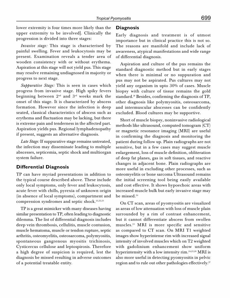
699Tropical Pyomyositis
lower extremity is four times more likely than the upper extremity to be involved]. Clinically theprogression is divided into three stages:
Invasive stage: This stage is characterised by painful swelling. Fever and leukocytosis may be present. Examination reveals a tender area of wooden consistency with or without erythema. Aspiration at this stage will not yield pus. This stage may resolve remaining undiagnosed in majority or progress to next stage.
Suppurative Stage: This is seen in cases which progress from invasive stage. High spiky fevers beginning between 2nd and 3rd weeks mark the onset of this stage. It is characterized by abscess formation. However since the infection is deep seated, classical characteristics of abscess such as erythema and fluctuation may be lacking, but there is extreme pain and tenderness in the affected part. Aspiration yields pus. Regional lymphadenopathy if present, suggests an alternative diagnosis.
Late Stage: If suppurative stage remains untreated, the infection may disseminate leading to multiple abscesses, septicemia, septic shock and multiorgan system failure.
Differential DiagnosisTP can have myriad presentations in addition to the typical course described above. These include only local symptoms, only fever and leukocytosis, acute fever with chills, pyrexia of unknown origin (in absence of local symptoms), compartment and compression syndromes and septic shock.21,22,23
TP is a great mimicker with many diseases having similar presentation to TP, often leading to diagnostic dilemma. The list of differential diagnosis includes deep vein thrombosis, cellulitis, muscle contusion, muscle hematoma, muscle or tendon rupture, septic arthritis, osteomyelitis, osteosarcoma, polymyositis, spontaneous gangrenous myositis trichinosis, Cysticercuscelluloseandleptospirosis.Thereforea high degree of suspicion is required, lest the diagnosis be missed resulting in adverse outcomes of a potential treatable entity.
DiagnosisEarly diagnosis and treatment is of utmost importance but in clinical practice this is not so. The reasons are manifold and include lack of awareness, atypical manifestations and wide range of differential diagnosis.
Aspiration and culture of the pus remains the standard diagnostic method but in early stages when there is minimal or no suppuration and pus may not be aspirated. Pus cultures may not yield any organism in upto 30% of cases. Muscle biopsy with culture of tissue remains the gold standard.18 Besides, confirming the diagnosis of TP, other diagnosis like polymyositis, osteosarcoma, and intermuscular abscesses can be confidently excluded. Blood cultures may be supportive.
Short of muscle biopsy, noninvasive radiological methodslikeultrasound,computedtomogram(CT)or magnetic resonance imaging (MRI) are useful in confirming the diagnosis and monitoring the patient during follow up. Plain radiographs are not sensitive, but in a few cases may suggest muscle enlargement, loss of muscle definition, obliteration of deep fat planes, gas in soft tissues, and reactive changes in adjacent bone. Plain radiographs are more useful in excluding other processes, such as osteomyelitis or bone sarcoma Ultrasound remains the initial screening tool being easily available and cost effective. It shows hypoechoic areas with increased muscle bulk but early invasive stage may be missed.24
OnCTscan,areasofpyomyositisarevisualizedas areas of low attenuation with loss of muscle plain surrounded by a rim of contrast enhancement, but it cannot differentiate abscess from swollen muscles.25 MRI is more specific and sensitive as compared to CT scan. On MRI T1 weightedimages show hyperintense rim with increased signal intensity of involved muscles which on T2 weighted with gadolinium enhancement show uniform hyperintensity with a low intensity rim.24,25,26 MRI is also more useful in detecting pyomyositis in pelvic region and to rule out other pathologies effectively.27

700 Medicine Update 2008 Vol. 18
For inconclusive cases, gallium scintigraphy helps to localize inflammation, but it is nonspecific and expensive.28
Anemia, leukocytosis with elevated ESR and CRP are commonly present. Eosinophilia ifpresent, suggests parasitic infection. Blood cultures are sterile in upto 90% of cases from tropical areas and upto 70% from temperate regions, but are not specific to TP. Diffuse myalgia’s with deranged liver and renal functions favor the diagnosis of leptospirosis. Bilateral symmetrical involvement of proximal muscle groups, elevated muscle enzymes (creatine phosphokinase, aldolase) and characteristic electromyography (low amplitude polyphasic potential) is consistent with polymyositis, but muscle biopsy is diagnostic. Elevated creatine phosphokinase is not a feature of TP.
Once diagnosis is made, common immuno-suppressive states (diabetes, HIV, rheumatological and malignancies) must be ruled out along with immunoglobulin levels.
TreatmentTreatment should be immediately initiated on confirmation of diagnosis. Although, rarely diagnosed during invasive stage, antibiotics alone will suffice at this stage. Once the abscess has formed, drainage is mandatory. Traditionally surgical incision and drainage have been used butnewermethods likeCTguidedpercutaneousdrainage or continuous suction with primary closure of wound are equally effective. However, if significant necrosis with large areas of involvement is present, traditional incision and drainage is the best alternative.
As with any serious infection, antibiotic therapy cannot be delayed and must be guided by the treating physician’s judgment. Impending culture and sensitive reports, it is advised that cloxacillin be the first line therapy with first generation cephalosporins like cefazolin as an alternative in penicillin sensitive individuals. Coveragefor methicillin resistant Staphylococcus aureus
(MRSA) using vancomycin or teicoplanin should be considered in severely ill patients, who have high risk of MRSA or the culture sensitivity report shows MRSA.29 Linezolid or dalfopristine-quinapristine are reserved for vancomycin intermediate sensitive Staphylococcus (VISA).29 Adding a second drug against Staphylococcus does not confer any benefit.
Penicillin remains the first line antimicrobial agent for streptococcus. Gram negative organisms require two drugs, usually a combination of third generation cephalosporin with aminoglycosides. Suspicion of anerobes warrants addition of metronidazole. If patient is immunosuppressed or very toxic, empirical treatment is started with broad spectrum antibiotics covering staphylococcus, streptococcus, gram negative and anerobes. In such situations vancomycin with an antipseudomonal carbapenems or β lactam combination are the most appropriate therapy. Other antimicrobials, aztreonam fluoroquinolone, aminoglycoside, cephalosporin or clindamicin, alone or in combination have been used successfully.
Treatment is continued for about a week after patient becomes afebrile, blood counts normalize and wound is healthy, but for metastatic infections it is recommended that treatment be continued for 4-6 weeks. Failure of fever to normalize indicates metastatic infection, drug resistance or drug fever. Radiology help may be taken to assess the course of the disease and to find out metastatic infection.
Although no recommendation are available, it is believed that eliminating nasal carriage of staphylococcus by using topical mupirocin or systemic rifampicin in patients with past episodes of pyomyositis, staphylococcus septicemia and in immunocompromised persons, further episodes of pyomyositis can be prevented.30
PrognosisWith heightened awareness, newer diagnostic modalities and effective chemotherauptic agents, the mortality in TP has been considerably reduced.
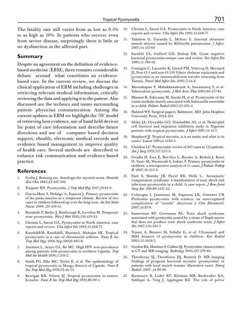
701Tropical Pyomyositis
The fatality rate still varies from as low as 0.5% to as high as 10%. In patients who recover even from severe disease, surprisingly there is little or no dysfunction in the affected part.
SummaryDespite an agreement on the definition of evidence-based medicine (EBM), there remains considerable debate around what constitutes an evidence-based care. In the current review, we discuss the clinical application of EBM including challenges in retrieving relevant medical information, critically reviewing the data and applying it to the patient. Also discussed are the technics and issues surrounding patient- physician communication. Among the current updates in EBM we highlight the ‘5S’ model of retrieving best evidence, use of hand held devices for point of care information and describe future directions and use of computer based decision support, ehealth, electronic medical records and evidence based management to improve quality of health care. Several methods are described to enhance risk communication and evidence-based practice.
References1. Scriba J. Beitrang zur, Aetiologie der myositis acuta. Deutsche
Zeit Chir 1885;22:497-502.
2. Traquair RN. Pyomyositis. J Trop Med Hyg 1947;50:81-9.
3. Garcia-Mata S, Hidalgo A, Esparza J. Primary pyomyositis of the psoas muscles in a temperate climate. Review of two cases in children followed up over the long term. An Sist Sanit Navar. 2006 ;29:419-31.
4. Bonafede P, Butler J, Kimbrough R, Loveless M. Temperate zone pyomyositis. West J Med 1992;156:419-23.
5. ChristinL,SarosiGA.PyomyositisinNorthAmerica:casereports and review. Clin Infect Dis 1992;15:668-77.
6. KaushikRM, KaushikR, SharmaA, Mahajan SK. Tropical pyomyositis in a case of rheumatoid arthritis. Trans R Soc Trop Med Hyg. 2006 Sep;100(9):895-8.
7. AnsaloniL,AcayeGL,ReMC.HighHIVsero-prevalenceamong patients with pyomyositis in northern Uganda. Trop Med Int Health 1996;1:210-2.
8. SmithPG,PikeMC,TaylorE,etal.Theepidemiologyoftropical pyomyositis in Mengo districts of Uganda. Trans R Soc Trop Med Hyg 1978;72:46-53.
9. Kerrigan KR, Nelson SJ. Tropical pyomyositis in eastern Ecuador. Trans R Soc Trop Med Hyg 1992;86:90-1.
10. ChristinL,SarosiGA.PyomyositisinNorthAmerica:casereports and review. Clin Infect Dis 1992;15:668-77.
11. Yahalom G, Guranda L, Meltzer E. Internal obturator muscle abscess caused by Klebsiella pneumoniae. J Infect. 2007;54:157-60
12. Sarubbi FA, Gafford GD, Bishop DR. Gram negative bacterial pyomyositis:unique case and review. Rev Infect Dis 1989;11:789-92
13. CouzigouC,LacombeK,GirardPM,VittecoqD,MeynardJL.Non-O:1 and non-O:139 Vibrio cholerae septicemia and pyomyositis in an immunodeficient traveler returning from Tunisia. Travel Med Infect Dis. 2007;5:44-6.
14. Mootsikapun P, Mahakkanukrauh A, Suwannaroj S, et al. Tuberculous pyomyositis. J Med Assoc Thai 2003;86:477-81.
15. Minami K, Sakiyama M, Suzuki H, et al. Pyomyositis of the vastus medialis muscle associated with Salmonella enteritidis in a child. Pediatr Radiol 2003;33:492-4.
16. Halsted WS. Surgical papers. Baltimore, MD: John Hopkins University Press, 1924:103.
17. Idoko JA, Oyeyinka GO, Giassuddin AS, et al. Neutrophil cell function and migration inhibition study in Nigerian patients with tropical pyomyositis. J Infect 1987;15:33-7.
18. Shepherd JJ. Tropical myositis, is it an entity and what is its cause? Lancet 1983;ii:1240-2.
19. CheidoziLC.Pyomyositis:reviewof205casesin112patients.Am J Surg 1979;137:255-9.
20. Ovadia D, Ezra E, Ben-Sira L, Kessler A, Bickels J, Keret D, Yaniv M, Wientroub S, Lokiec F. Primary pyomyositis in children: a retrospective analysis of 11 cases. J Pediatr Orthop B. 2007;16:153-9.
21. Park S, Shatsky JB, Pawel BR, Wells L.. Atraumatic compartment syndrome: a manifestation of toxic shock and infectious pyomyositis in a child. A case report. J Bone Joint Surg Am. 200;89:1337-42.
22. Colmegna I, Justiniano M, Espinoza LR, Gimenez CRPiriformis pyomyositis with sciatica: an unrecognized complication of “unsafe” abortions J Clin Rheumatol. 2007;13:87-8.
23. Immerman RP, Greenman RL. Toxic shock syndrome associated with pyomysitis caused by a strain of Staph aureus that does not produce toxic shock syndrome toxin. J Infect Dis 1987;156:505-7.
24. Trusen A, Beissert M, Schultz G, et al. Ultrasound and MRI features of pyomyositis in children. Eur Radiol 2003;13:1050-5.
25. GordonBA,MartinezS,CollinsAJ.Pyomyositis:characteristicsatCTandMRimaging.Radiology 1995;197:279-86.
26. Theodorou SJ, Theodorou DJ, Resnick D. MR imaging findings of pyogenic bacterial myositis (pyomyositis) in patients with local muscle trauma: illustrative cases. Emerg Radiol. 2007 ;14:89-96.
27. Karmazyn B, Loder RT, Kleiman MB, Buckwalter KA, Siddiqui A, Ying J, Applegate KE. The role of pelvic

702 Medicine Update 2008 Vol. 18
magnetic resonance in evaluating nonhip sources of infection in children with acute nontraumatic hip pain. J Pediatr Orthop. 2007;27:158-64.
28. Schiff RG, Silver L. Tropical pyomyositis: demonstration of extent and distribution of disease by gallium scintigraphy. Clin Nucl Med 1990;15:542-4.
29. Robert L, Reresiewiscz, Parosenet J. Staphylococcal infections. In: Fauci AS, Braunwald E, Martin JB, et al. Harrison’s principles of internal medicine.15th Ed. New York: McGraw-Hill, 2001;1:889-900.
30. Hudson IR. The efficacy of intranasal mupirocin in the prevention of staphylococcal infections: a review of recent experience. J Hosp Infect 1994;27:81-98.

CHAPTER
92Global Warming and Health HazardsS. Ghosh
IntroductionGlobal warming has captured the centre stage of the worldwide attention and has inspired more debates and action at every level than any other environmental issue in history. Throughout most of the human history, all climate changes were the direct results of natural forces. This has been changed with the start of the industrial revolution, when new industrial and agricultural practices began to alter the global climate and environment. Widespread use of the fossil fuels, deforestation, chemical agriculture and population growth are creating an excess of greenhouse gases in the atmosphere and contributing to global warming.
The fourth assessment report (AR4) of the Inter-governmentalPanel onClimateChange (IPCC)of the United Nations, representing the works of 2,500 scientists from more than 130 countries, published on 2nd February 2007, states that global warming is now unstoppable1, 2 and human activities are to blame3 for the heat-trapping greenhouse gases that have caused global temperature to rise dramatically since 1950. Warming in the last 100 yearshascausedabouta0.74°Cincreaseinglobalaverage temperature. According to their most recent projections, the global mean temperature willincreaseby1.1–6.4°C(bestestimatebetween1.8–4.0°C)duringthe21stcentury.4, 5 The climate change and the consequential effects of this are no
longer just a theoretical issue of environment. In fact, it is looming as the biggest human catastrophe, threatening our ultimate survival.
What Causes Global Warming ?A major part of the solar radiation coming to the earth escapes back into the outer space after being reflected from the earth surface. Many greenhouse gases occur naturally and surround the earth. They trap the heat and reflect back a part of this escaping solar radiation to the earth again. This is called the greenhouse effect, which keeps the earth warm enough to support life.
Scientists have determined that a number of human activities are adding an excessive amount of greenhouse gases to the atmosphere. As the concentration of the heat absorbent greenhouse gases increases, more and more heat becomes trapped in the atmosphere and less heat escapes back into space. This increase in the trapped heat raises the surface temperature of the earth and changes the climate. This is called global warming.
Global warming is taking place since 1950. Rapid industrialisation has accelerated it. Human use of fossil fuels is the main source of excessive greenhouse gases. By using electricity from coal-fired thermal power plants, using coal or coal-gas as the major fuel source of many industries, using
704 Medicine Update 2008 Vol. 18
petrol and diesel for driving cars, we release more and more carbon dioxide and other heat-trapping gases into the atmosphere. During the last 150 years of the industrial age, the atmospheric concentration of carbon dioxide has increased by 31%6 and the atmospheric methane level has increased by 151%6. Deforestation is another significant source of greenhouse gases, because fewer trees means less carbon dioxide is being removed from the atmosphere to be converted to oxygen.
February2007 reportof IPCCopines that themain cause of global warming is excess emission of carbon dioxide and other greenhouse gases, produced by the following human activities:7
• Widespread use of fossil fuels (coal, petrol,diesel, natural gas)
• Deforestation
• Chemicalagriculture
• Populationgrowth
Major Greenhouse Gases8
• Carbondioxide 83%
• Methane 9%
• NitrousOxide 6%
• Ground-levelOzone
• Chloro-fluoro-carbons(CFC)} 2 %
• Per-fluoro-carbons(PFC)
• Sulfur-hexa-fluoride(SF6)
These gases are heat-absorbent & hold heat in the atmosphere.
Sources of the Major Greenhouse Gases
1. Carbon dioxide
• Burningoffossilfuels(coal,petrol,diesel,natural gas ).
• Burning of solid waste, wood and woodproducts.
2. Nitrous oxide
• Burningoffossilfuelsandsolidwaste.
• Variousagriculturalandindustrialprocesses.
3. Methane
• Productionandtransportoffossilfuels.
• Decompositionoforganicwaste.
4. CFC, PFC, SF 6
• Exclusivelyindustrialproducts.
• CFC is largelyusedas refrigerant and inaerosols.
Ground-level Ozone vs. Stratospheric Ozone
• Ground-level ozone is a health hazard, as itirritates airways and injures lungs.
• Stratosphericozonelayerisgood,asitblocksthe entry of carcinogenic UV rays.
• CFCdepletesstratosphericozonelayer,causingozone hole.
Sources of Carbon dioxide and Our Task
A. The main cause of carbon dioxide accumulation is either
• Increasedproductionduetoburningoffossilfuels in thermal power plants, industries, transport vehicles or
• Decreasedremovalduetodeforestation.
B. The sources of carbon dioxide production are8
• Burningoffossilfuels 97%
• Everythingelse 3%
C. The sectors responsible for carbon dioxideproduction are8
• Electricitygeneration 40%
• Transportvehicles 32%
• Industrialuse 17%
• Residential 7%
• Commercial 4%
D. So, for the effective reduction of the accumulation of greenhouse gases in the atmosphere, we must set our practical target to reduce carbon dioxide emission, for which we must stop burning of fossil fuels in at least three major sectors:

705Global Warming and Health Hazards
1. Thermal power generation
2. Automobile emissions and
3. Industrial sector.
Per Capita Carbon dioxide Emission
USA alone is responsible for more than 24% of total global emission of greenhouse gases9 and for about 50% of total global automobile emission.10 While the world average of per capita emission of carbon dioxide in the year 2002 was 3.9 tonnes,9 the figure for USA was 5 times higher, as it produced an awesome 19.7 tonnes9 of carbon dioxide per capita. On the contrary, India, a less industrialised country, figured well below and produced only 1.0 tonne9 of carbon dioxide per capita, 4 times less than the world average. So, it is quiet clear that the developed countries are the major culprits and naturally these countries will have to share more responsibilities than the under-developed countries, to come out of this grave situation at present.
Effects of Global WarmingIncreased global surface temperature due to the effect of trapped heat will result into
• Climatechange
• Rapidwatercycle
• Meltingofglaciersandpolarice
• Oceanexpansion
1. Scorching heat and heat waves are the direct results of this climate change.
2. Breeding of mosquitoes and spread of many infections are enhanced in warm or hot climate.
3. Droughts which are becoming more common and longer lasting, can lead to starvation and the destruction of entire ways of life, particularly in regions of sub-Saharan Africa, that are least equipped to deal with such catastrophes.11
4. Raised surface water temperature and hot climate increases the evaporation of ocean water, resulting in rapid water cycle, which causes flood in many areas.
5. Melting of glaciers and the polar ice due to raised temperature will increase the quantity of ocean water and at the same time the thermal expansion of the ocean water will cause a significant rise of the sea level. Average rate of sea level rise during the years 1961-2003 was about 1.8 mm/year, which steadily increased to 3.1 mm/year during 1993-2003. Now it is estimated that sea level will rise in the range of 18 – 59 cm4, 5 at the end of 21st century.
6. As hurricane formation requires warm ocean surface along with hot climate, fiercer storms will occur. In fact, last 10 years has been the most active hurricane season in the history.
So, summarily, the results which can affect the human health are :-
• Killerheatwaves
• Spreadofvectorbornediseases
• Devastatingdroughts
• Heavyrainfall
• Risingsealevelandcoastalflood
• Fiercestorms.
Effects on Human Health Global warming not only poses a significant threat to the earth’s ecology, but also unleashes an unprecedented health risks due to heat waves, spread of infectious diseases, disasters and malnutrition. As per WHO estimate, global warming currently contributes to more than 1.5 lakh deaths11 and more than 50 lakh illnesses every year.11 50 million environmental refugees,12 displaced by floods, droughts and rising sea levels, are expected by 2010. Numbers may double by 2030.11 Experts apprehend that global warming will kill billions in this century!!12

706 Medicine Update 2008 Vol. 18
The effects of global warming on human health may be summarised below:
1. Direct Effect of Hot Climate
a. There will be an increased intensity, duration and frequency of heat waves around the world.1 So, heat related diseases will increase sharply. In August 2003, Europe suffered its worst heat wave in recent memory; the scorching weather claimed as many as 35,000 lives. In France, nearly 15,000 people succumbed as the temperature peaked at about40°C,completelyunpreparedforthatkind of heat.
b. The sick and the elderly persons are most vulnerable, for their decreased ability to increase cardiac output and decreased sweat function for cooling of body.
c. People in temperate countries, like UK, France etc., who are not accustomed to hot weather and people in countries where houses and other infrastructures are not designed to cope with hot climate, are more vulnerable.
d. Technological adaptations, e.g. installation of air-conditioners and construction of heat-minimising houses will happen quickly among the rich.
e. So, the heat waves will more affect the populations that are least able to deal with it. In the developed nations it will affect the poor more than the rich and in the developing world it will affect the nations least able to respond to these threats and stresses. Overall, global warming has more and disproportionate effect on the under-developed countries and on the poorer segments of the society.
f. There is increased risk of bushfire or forest-fire, which can kill people.
2. Indirect Effect of Hot Climate
a. More than 65% of world population are at increased risk of infection.13
b. As warm climate favors breeding of mosquitoes, there will be a massive spread of the whole range of mosquito-borne diseases1,14 e.g. Malaria, Dengue, Chikungunya, Encephalitis and Yellowfever.1 In successive two years of 2005 and 2006, Dengue and Chikungunya becamepandemic. Malaria and Encephalitis are becoming endemic in many countries. Malaria is moving to higher altitudes in Africa, Asia & Latin America.1
c. Tick and other insect-borne diseases are spreading to areas once considered too cold for them to survive, e.g. Lyme disease, Rocky mountain spotted fever, West Nile fever & Eastern equine encephalitis.1
d. Food-poisoning is increased, as these bacteriabestgrowin35–37°Ctemp.
3. Effect of Floods
a. There are health effects secondary to flooding, such as contaminated water supplies and breakdown in sewerage and garbage services,14 leading to spread of the whole range of water borne diseases,1 food borne diseases and other infectious diseases. Cholera thrives in suchsituation.
b. Contamination of swimming water alsoinvites some health hazards.
c. Water logging hampers food distribution with natural consequences on health.
4. Effect of Droughts
a. There will be a sharp increase in the nutritional diseases caused by starvation1, as a result of the poor agricultural yield due to severe and prolonged droughts. By 2020, yields from rain-fed agriculture in some African countries could be reduced by 50%.
b. Water is already in short supply. Under hot climate conditions, precipitation patterns will change, leading to severe water scarcity,15

707Global Warming and Health Hazards
with great implications on health. By 2020, 75–250 million people in Africa will be exposed to water scarcity due to climate change.
5. Effect of Increasing Air Pollution
a. Increasing air pollution from the continued burning of fossil fuels will cause higher rates of respiratory and cardiovascular diseases.1
b. With warming, there will be an increase in pollens, mold spores and release of large quantities of particulate matters, aggravating the hazards of asthma.
c. The concentration of photochemical pollutants, like ozone, increases with increasing temperature. Increased ground-level ozone damages lung tissue, induces and increases asthma14 and invites many other cardio-respiratory diseases.
d. Depletion of stratospheric ozone allows more and more UV-rays to enter into the atmosphere and thus increases the incidence of malignancies.
6. Effect of Ocean Expansion
a. Sea level may rise 18 – 59 cm by 21005.
b. It may be quite disastrous for people of low-lying islands, e.g. Maldives in Indian ocean and many south Pacific islands.16 There will be a 50 fold rise of the number of people exposed to coastal flooding per year16 in the years around 2080.
c. 13 of the 20 largest cities on earth are located at sea level on coasts.15 As sea level rises, there go our medical institutions, water treatment plants, emergency response units such as fire departments and ambulances. The bulk of the services designed to keep us healthy are almost all located in our larger cities, which are also located frequently at sea level.
d. With increasing sea level, there will be coastal erosion, contamination of freshwater supply and degradation of agricultural areas16 resulting in a massive impact on our eco-system, health and life.
7. Effect of Disasters
a. There will be a sharp rise in the storms, heavy rainfall, sea-water flood, draughts, forest-fire, earthquakes, tsunami and volcanoes.
b. Hospitals, health services, ambulances, fire-services, electricity, food & water supply, transport and all sorts of communications become ineffective altogether, after the disaster, with a massive impact on health and life.
c. These disasters invariably increase the psychological stress, depression, agony and a feeling of isolation among people affected by natural disasters16.
No country, even USA, will escape these hazards. But, ironically, the poor nations least responsible for greenhouse gases are most affected by global warming.1,17 Though it affects everyone on earth, poor people are most vulnerable to the disease and death, as they are least able to cope.1 Here lies the enormous ethical challenge.
Examples of Some Effects : Super-cyclone in Paradeep claimed 10,000 lives in October 1999. Australia had severe drought in the year of El Niño, 2002. Worst heat wave across Europe claimed 35,000 lives in August 2003. South India faced the worst tsunami in December 2004. USA had massive hurricane Katrina in August 2005. The world suffered the biggest pandemic of Dengue and Chikungunya in 2005 and 2006. Mumbaiexperienced the heavy rains followed by sea-waterfloodin2006and2007.Californiasawthedisastrous forest-fire in October 2007.

708 Medicine Update 2008 Vol. 18
Salient ObservationsIPCC Fourth Assessment Report5
Changes in the atmosphere
• Carbondioxide,methane, andnitrousoxideare all long-lived greenhouse gases, but these gases have increased markedly as a result of human activities since 1750 and now far exceed pre-industrial values.
• Theamountofcarbondioxideintheatmospherein 2005 (379 ppm) exceeds by far the natural range of the last 6,50,000 years (180 – 300 ppm). The primary source of this increase is fossil fuel use; but, land-use changes also make a contribution.
• Theamountofmethaneintheatmospherein2005(1774 ppb) exceeds by far the natural range of the last 6,50,000 years (320 – 790 ppb). The primary source of this is very likely to be a combination of human agricultural activities and fossil fuel use.
• Nitrousoxideconcentrationshaverisenfromapre-industrial value of 270 ppb to a 2005 value of 319 ppb. Most of this rise is due to human activity, primarily agriculture.
Warming of the planet
• Elevenofthetwelveyearsintheperiod1995–2006 rank among the top 12 warmest years in theinstrumentalrecordsince1850.Colddays,cold nights and frost events have become less frequent. Hot days, hot nights and heat waves have become more frequent.
• Warminginthelast100yearshascausedabouta0.74°Cincreaseinglobalaveragetemperature.Surface air warming in the 21st century will be in the rangeof1.1–6.4°C (with thebestestimatebetween1.8-4.0°C)
• Observations since1961 show that theoceanhas been absorbing more than 80% of the heat added to the climate and that ocean temperature has increased to depths of at least 9800 ft. Ocean warming causes seawater to expand, which contributes to sea level rising.
• Sea level roseatanaveragerateofabout1.8
mm/year during the years 1961-2003. The rise in sea level during 1993-2003 was at an average rate of 3.1 mm/year. It is estimated that sea level rise will be in the range of 18 – 59 cm.
• Therewillbeanincreaseinfrequencyofwarmspells, heat waves and events of heavy rainfall. There will be an increase in areas affected by droughts, intensity of tropical cyclones (including hurricanes and typhoons) and the occurrence of extreme high tides.
Hurricanes
• Therehasbeenanincreaseinhurricaneintensitysince 1970s, and this increase correlates with increases in sea surface temperature. It is likely that we will see increases in hurricane intensity during the 21st century. It is more likely that there has been some human contribution to the increases in hurricane intensity.
Coastal System
• It isprojectedwithveryhighconfidence thatcoasts will be exposed to increasing risks of coastal erosion due to climate change and sea-level rise.
• Many millions more people will be floodedevery year due to sea-level rise by the 2080s.
Fresh Water
• It is projectedwithhigh confidence that dryregions will get drier, and wet regions will get wetter. Heavy rainfall events are very likely to become more common and will increase the flood risk. At the same time drought affected areas will become larger.
• Bymid-century,annualaverageriverrunoffandwater availability are projected to increase by 10-40% in some wet tropical areas, and decrease by 10-30% over some dry regions.
Food
• Itisprojectedthatfoodproductionmayincrease,globally,fortemperaturerisesof1–3°C,butwill decrease for higher temperature range.
709Global Warming and Health Hazards
IPCC The Intergovernmental Panel on Climate Change (IPCC) is a scientific body tasked toevaluate the risk of climate change caused by human activity. The panel was established in 1988 by two organizations of the United Nations – the World Meteorological Organization (WMO) and the United Nations Environment Programme (UNEP). TheIPCCdoesnotcarryout research,nordoesit monitor climate. One of the main activities of the IPCC is topublish special reportson topicsrelevant to the harmful effects of climate change. Its first assessment was published in 1990, followed by in 1995 and 2001. The fourth assessment report has been published on 2nd February 2007 and has created a huge repercussion throughout the world. The IPCC chairman of Indian origin, RajendraPachauri, who took his office only in May 2002, shared the 2007 Nobel Peace Prize for the excellent works on this field.
Kyoto ProtocolThough the first international response to address the problems of climate change was launched in 1992, at the Earth Summit in Rio de Janeiro, an international agreement was reached by more than 160 nations, in December 1997, in Kyoto, Japan. This is known as Kyoto Protocol, which commits 38 industrialised countries to cut their greenhouse gas emissions18 and sets a binding target of 5.2% net reduction of worldwide emission of greenhouse gases, mainly carbon dioxide, below 1990 level as benchmark, by the five year period of 2008 to 201219.
Most of the industrialised nations have ratified it and have begun efforts to meet their emission target. Notable exception is USA, which accounts for about 24% of the global emission of greenhouse gases. Before election in 2001, George W. Bush promised to decrease the emission, but refused after victory.20 Alternatively, proposed a 4.5% reduction of current emission by 2010. According to US Energy Department, this will result in 30% elevation of emission over 1990 level, instead of
reduction.19 Australia also declined. In November 2004, Russia accepted it. The protocol becomes a worldwide binding force on February 16, 2005.
RemediesWe can stop global warming, if we immediately opt for
1. Alternative energy use for industrial sector
2. Alternative fuel use for transport vehicles ( Bio-diesel = Ethanol + vegetable oil. )
3. Efficient use of electricity
4. Widespread use of renewable energy
5. Organic agriculture
6. Reforestation and afforestation
While the first four measures reduce the production of carbon dioxide, organic agriculture and forestation remove carbon dioxide from the air.
Three ‘R’s may be the most useful remedial tools,
• Restrictions on biggest polluters
• Reduced automobile emission
• Renewable energy sources
• Solarenergy
• Windpower
• Hydroelectricpower
• Geo-thermalenergy
• Bio-energy
Energy RevolutionAn ‘Energy revolution’ is essential to counter the ill effects of the ‘Industrial revolution’, as
• 96%ofallenergyusedgloballyatpresent, isderived from thermal power.
• Otherenergysourcesaregrosslyinadequateforthe huge demand of the modern civilisation. The need of electricity has tremendously increased nowadays by the use of so many electrical

710 Medicine Update 2008 Vol. 18
gadgets at every home, rapid surge in installation of air-conditioners and the widespread use of computersateverylevel.Civilisationisenergy-intensive and we cannot turn it off.
• Only key to our future survival may be thewidespread use of ‘Nuclear energy’ as the clean source of energy.21 This becomes further relevant, as we know that the fossil-fuels have a very limited stock for a few more decades only. We are not panicked, unnecessarily, to recommend nuclear energy, as we judiciously use radio-active nuclear materials for the diagnosis and treatment of many diseases in the Dept. of Nuclear Medicine. Above all we use nuclear radiation to treat cancer.
• Themorethedevelopednation,themorerapidshould be the change over to clean energy source, compared to the under-developed nations, to halt our approaching devastation.
ConclusionAll sensible global citizens, particularly the physicians should raise their voice today
• Tostopburningoffossilfuels,asitistherealdriver of the climate change
• Tocallforcleanenergy
• Todrawattentionofourpolicymakersand
• Tomakethepeopleawareofthisensuingrealthreat to our ultimate survival, as energy source is fundamental for the environment of our health and for the health of our environment. Without that, we are going to have a very polluted, sick and disastrous future.
We must, must act now to save our planet, our civilisation and our next generation.
SummaryThe global mean temperature has been increasing steadily and significantly since 1950 and has reached an unstoppable state due to some human activities. Main cause is significant elevation of the concentration of atmospheric carbon dioxide
as major greenhouse gas, due to widespread use of the fossil fuels in thermal power plants, industries and transport vehicles. Deforestation and land-use changes also make a contribution. Developed countries are the major culprits. Poor people and the poor nations are the worst sufferers. Global mean temperaturewillriseby1.1-6.4°Cby2100.Thischange in climate is looming as the biggest human catastrophe, threatening our ultimate survival. The results affecting our health are killer heat waves, devastating droughts, ocean expansion, heavy rainfall, coastal flood, fierce storms, massive spread of the whole range of mosquito-borne diseases like malaria, dengue, chikungunya, encephalitis and yellow fever, along with the effects of flood, spread of water borne diseases like cholera, effects of droughts, malnutrition, water scarcity, increasing air pollution and disasters. More than 65% of world population are at increased risk of infection. WHO estimates more than 1.5 lakh deaths and 50 lakh illnesses per year now due to global warming. 50 million environmental refugees are expected by 2010. Experts say this will kill billions in this century. Stoppage of burning fossil fuels, use of clean energy, opting for organic agriculture and forestation are the remedies. Widespread use of nuclear energy may be the only key to our future survival.
References1. Michael A. McGeehin, Ph.D., Director, Division of
Environmental Hazards and Health Effects, U.S. CentersforDiseaseControlandPrevention,Atlanta;PaulEpstein,M.D., M.P.H., Associate Director, Center for Health andthe Global Environment, Harvard Medical School, Boston. Global warming poses health threats. 02-02-2007. http://www.medicinenet.com/script/main/art.asp?articlekey=79295
2. Steven Reinberg. Global warming poses health threats. 02-02-2007. http://www.medicinenet.com/script/main/art.asp?articlekey=79295
3. Larry West. Global Warming is Unstoppable and Humans are to Blame, says UN Report http://environment.about.com/od/globalwarming/a/ipcc_report.htm
4. IPCCFourthAssessmentReport.htm02-02-07http://www.ipcc.ch/
5. Intergovernmental Panel on Climate ChangeIntergovernmentalPanelonClimateChange -Wikipedia,the free encyclopedia.mht http://en.wikipedia.org/wiki/

711Global Warming and Health Hazards
IPCC_Fourth_Assessment_Report
6. Larry West. What Causes Global Warming? http://environment.about.com/od/globalwarming/a/ipcc_report.htm
7. LarryWest.HowDoHumansContributetotheGreenhouseEffect? http://environment.about.com\What are Greenhouse Gases and the Greenhouse Effect.htm
8. Sara Saksewski, Top ten things to know about global warming http://www.uwsp.edu/cnr/wcee/keep/Resources/Teaching_Resources/OnlineResources.ppt
9. Trends in international carbon dioxide emissions. Total carbon dioxide emissions per head for selected countries, 2002. International Energy Agency 2004. http://www.berr.gov.uk/files/file20356.pdf and http://www.iea.org/
10. JohnDeCiccoandFredaFung.EnvironmentalDefence2006.Global Warming on the Road. The climate impact of America’s automobiles. Page – 21 http://www.environmentaldefense.org/ documents/5301_Globalwarmingontheroad.pdf
11. Larry West. Global warming leads to 150,000 deaths every year. March 10, 2007 http://environment.about.com/Global Warming Leads to 150,000 Deaths Every Year.htm
12. Larry West. Environmental Refugees - Scholars Predict 50 Million “Enviro.htm and Global Warming May Kill Billions ThisCentury.htmhttp//environment.about.com
13. Global Warming Impacts: Health Effects - ConclusionsHealth Effects - Conclusions - Global Warming - Sierra
Club.htmhttp://www.sierraclub.org/globalwarming/health/conclusions.asp
14. Health And Disease. Page no. – 8. http://www.koshland-science-museum.org/exhibitgcc/impacts08.jsp
15. Global Warming And Your Health. 24-10-2006 http://www.terradaily.com/reports/Global_Warming_And_Your_Health_999.html
16. Warmer and sicker ? Global warming and human health Global warming-Key text.htm http://www.science.org.au/nova/
17. Larry West. Health effects of global warming hardest on poor nations About.com : Environmental Issues. http://environment.about.com/od/faqglobalwarming/f/globalwarming.htm
18. Susan Munroe. Kyoto Protocol. http://canadaonline.about.com/mbiopage.htm
19. Larry West. Should the United States ratify the Kyoto protocol? http:// environment.about.com/od/kyotoprotocol/i/kyotoprotocol_2.htm
20. Bornstein,Seth;BushAdministration:CarbonDioxideNotaPollutant;CommonDreams,Portland,MaineUSA;August29, 2003 by the Knight Ridder News Service http://www.commondreams.org/headlines03/0829-02.htm
21. Larry West. Nuclear Energy May Be Key to Survival GlobalWarmingMayKillBillionsThisCentury.htmhttp//environment.about.com
CHAPTER
93Clinical Application of MeditationH. L. Dhar
IntroductionConcept of meditation is as old as civilization.Saints and seers had taken refuge in solitude and meditation to get peace of mind. Values of meditation are beyond doubt1 but therapeutic approach is quite recent.2
The word meditation was coined from Greek word Meridi (heal). It was practiced in ancient Greece for thousands of years followed by India and first documented in the Book of Wisdom, the veda 4000 years ago.3
Medicine is one of the oldest discipline that was evolved out of human need, out of sympathy between man and man to help each other in their sorrows and sufferings. Thus meditation is the oldest known therapy although scientific evidence is quite recent. Attempt has been made to present age-old meditation in a new perspective with modern touch.
Divine healingWith the dawn of civilization came the concept of God and Divine healing as a solace to mankind and meditation became a handy approach for mental uplift and for relief of pain and suffering. Divine healing is a God related discipline and becamepopularafterJesusChristwithhismiraclehealing. Very recently divine healing has been used in Uganda as antiviral (AIDS) therapy.4
Spiritual HealingIt was originated with Buddha (500 B.C.) as agreat relief of human suffering. Buddha used to cure people instantaneously by raising his hand, however, Buddha was conscious about his limitations and created four Tantras, which he used to teach himself, and latter became Tibetan Medicine studied by all Tibetan physicians.
Spiritual Medicine500yearshenceJesusChrist,thegreathealerusedto cure people either individually or in groups with blessings and spiritual healing became a spiritual discipline. Scientific evidence shows that spiritual healing brings about the easing of symptoms, the removal of blocks in energetic flows, the retrieval of repressed memories of emotional hurts, improvements in many illnesses, improvements in relationships and an opening into spiritual awareness. All of these may be achieved with no more than the lying on of hands or the projection of healing from a distance. In recent years, interest has been created amongst chronically sick individuals in spiritual healing either through receiving blessings from highly spiritual person and/or through practice of meditation.5 It has been reported that participation in spiritual and psychological therapies may be related to beneficial clinical outcome in HIV positive individuals including improved survival.6
713Clinical Application of Meditation
However, first rational approach to healing came from Holistic Medicine of Socrates 4th century AD. Socrates warned that treating one part of the body only would not have good result. This has got its reflection on modern medicine particularly Geriatric medicine, where a physician treats his patient as a whole individual comprising of all aspects of his ailments and also looks to his economic and social background before starting treatment. The term holistic stresses, the spiritual and emotional health contributes just as much as physical and mental health to overall state of being well.7
Mind and Body Medicine In recent decades mind/body medicine has been an important field in healthcare where physicians consider that the process of mind has a major impact in influencing the health of the body. Among various types of mind /body medicine therapy, meditation remains one of the most practiced technique and many scientific studies carried out have proven the positive effects of meditation on both mental and physical health.8
Recent studies are showing that meditation can result in stable brain patterns and changes over both short and long term intervals that have not been seen before in human beings suggesting the potential for the systematic driving of positive neuro - plastic changes via such intentional practices cultivated over time. These investigations may offer opportunities for understanding the basic unifying mechanisms of the brain, mind and body that underlie awareness and our capacity for effective adaptation to stressful and uncertain conditions.9
Human suffering consists of diseases of body and mind put together. There are increasing evidence that more and more diseases are genetically linked particularly non-communicable diseases. It is here that science and spirituality meet.10 Genes get mutated according to good or bad work but due to shortage of life span it may not be able to manifest and passes to next life. Genetic predisposition is nothing but outcome of our work reflected in spirituality.
If we want to remain disease free, we can guard out own gene through meditation with spiritual uplift.
Ayurveda is a mind body medicine with spiritual background. Ancient physicians were also sages who believed human body is created out of consciousness and Ayurveda says the same. Ayurveda does not consider human body as collection of cells, tissues or organs but in terms of Quantum perspective. Quantum is invisible energy million times smaller than atom, which forms the basis of everything that exists. Quantum healing based on Ayurveda suggests that there are three powerful techniques which can cure any disease e.g. Meditation, Bliss and Primordial sound which cures different disease process like cancer.11 Very recently Ayurvedic principle has been applied to treat elephantiasis.12
YogaHowever, initial therapeutics effect of meditation came from yoga, which ends in meditation. In clinical yoga, meditation serves as mirror image leading to therapeutic meditation. Many substances like cannabies were used for religious or meditative purpose. North American Indians used particularly psychoactive plants containing hallucinogenic substances in order to drive in connection with a dance or similar ritual, into a trance like condition leading to hysterical convulsion followed by calm andrelaxationconsideredtobetreatmentofCNSdisorders particularly epilepsy. Integrated yoga of which meditation is a major component has been known for decades as a remedy for various psychosomatic disorders.13
In recent years, yoga has been used to treat breast cancer. It has been demonstrated that yoga is associated with beneficial effects on social functioning among a medically diverse sample of breast cancer survivor’s.14 Recently, Iyengar yoga has been used in osteoarthritis of knee with considerable success.15 However, Kundalini yoga meditation has been found to be specific for treating obsessive compulsive disorders (OCD),
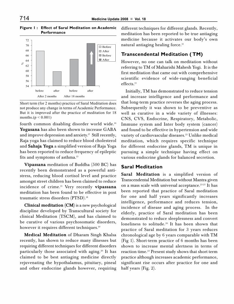
714 Medicine Update 2008 Vol. 18
fourth common disabling disorder world wide.16 Yogasana has also been shown to increase GABA and improve depression and anxiety.17 Still recently, Raja yoga has claimed to reduce blood cholesterol and Sahaja Yoga a simplified version of Raja Yoga has been reported to reduce frequency of epileptic fits and symptoms of asthma.13
VipassanameditationofBuddha(500BC)hasrecently been demonstrated as a powerful anti-stress, reducing blood cortisol level and practice amongst street children has been claimed to reduce incidence of crime.13 Very recently vipassana meditation has been found to be effective in post traumatic stress disorders (PTSD).18
Clinical meditation (CM) is a new psychological discipline developed by Transcultural society for clinical Meditation (TSCM), and has claimed tobe curative of various psychosomatic disorders, however it requires different techniques.19
Medical Meditation of Dharam Singh Khalsa recently, has shown to reduce many illnesses but requiring different techniques for different disorders particularly those associated with aging.20 It has claimed to be best antiaging medicine directly rejuvenating the hypothalamus, pituitary, pineal and other endocrine glands however, requiring
different techniques for different glands. Recently, meditation has been reported to be true antiaging medicine because it activates our body’s own natural antiaging healing force.21
Transcendental Meditation ( TM)However, no one can talk on meditation without referring to TM of Maharishi Mahesh Yogi. It is the first meditation that came out with comprehensive scientific evidence of wide-ranging beneficial effects.13
Initially, TM has demonstrated to reduce tension and increase intelligence and performance and that long-term practice reverses the aging process. Subsequently it was shown to be preventive as well as curative in a wide variety of illnesses: CNS, CVS, Endocrine, Respiratory, Metabolic,Immune system and Inter body system (cancer) and found to be effective in hypertension and wide variety of cardiovascular diseases.22 Unlike medical meditation, which requires specific technique for different endocrine glands, TM is unique in pursuing a simple technique having effect on various endocrine glands for balanced secretion.
Saral MeditationSaral Meditation is a simplified version of Transcendental Meditation but without Mantra given on a mass scale with universal acceptance.23-25 It has been reported that practice of Saral meditation for one and half years significantly increases intelligence, performance and reduces tension, incidence of disease and aging process. In the elderly, practice of Saral meditation has been demonstrated to reduce sleeplessness and convert loneliness to solitude.26 It has been shown that practice of Saral meditation for 3 years reduces chronological age by 6 years comparable with TM (Fig 1). Short-term practice of 6 months has been shown to increase mental alertness in terms of reaction time.23 Present study shows that short term practice although increases academic performance, significant rise occurs after practice for one and half years (Fig. 2).
Figure 1 : Effect of Saral Meditation on Academic Performance
Short term (for 2 months) practice of Saral Meditation does not produce any change in terms of Academic Performance. But it is improved after the practice of meditation for 18 months.(p < 0.001)
54
56
58
60
62
64
66
68
70
72
before after before after
After 2 months After 18 months
%
BeforeAfterBeforeAfter

715Clinical Application of Meditation
world population functions in beta range for normal activities.13
Quantum Meditation: A revolutionary meditation introduced recently11,27,28 claimed to reverse incurable diseases like cancer and prevent fetal abnormality if mother practices before conception and through out pregnancy. Quantum is invisible energy, million times smaller than an atom. Meditation works at molecular level bringing out inner qualities but quantum meditation acts far beyond at conscious level, untouched by disease.
SummaryConceptofmeditation isasoldascivilizationanddivine healing seems to be one of the earliest remedy to human suffering presently tried as antiviral therapy. Spiritual healing of Buddha got further impetus after Christ andmeditationbecamehandy in the reliefof pain and suffering. However, Holistic medicine, treating individual as a whole was the initial rational approach to treatment followed by mind body medicine with spiritual background.
But initial therapeutic effect of meditation came from yoga. This was followed by other
Figure 2 : Saral Meditation - Reversal of aging process(Comparison of TM and Saral Meditation)
Figure 3 : Effect of Saral Meditation
EEG before
EEG after one year
Activity at 8-c/s on posterior region.
Activity at 9 c/s on posterior region.
β
β
α
α
Low voltage fast activity at 20-22c/s on both sides
Low voltage fast activity at 18-20c/s on both sides
Preliminary studies also showed that practice of Saral meditation for more than 6 months increases alfa activity in EEG.23 Present study confirms that practice of Saral meditation for more than one year showed considerable increase in alfa activity compared to control (Fig 3). Normally 95% of
-14
-12
-10
-8
-6
-4
-2
0
Long term5 yrs
Short term2 yrs
Saral MedTM
Short term3 yrs
Controls

716 Medicine Update 2008 Vol. 18
types of meditations e.g. Vipassana meditation, Clinicalmeditation,Medicalmeditationetc.butTranscendental Meditation (TM) was the first which came out with comprehensive evidence of wide ranging therapeutic effects as preventive as well as curative against wide variety of illnesses in addition to its powerful antiaging effect. Saral meditation, a simplified version of TM but without Mantra, over the years has been shown to have similar effects.
Meditation works at molecular level bringing out inner qualities but recently, Quantum meditation has claimed to act at conscious level curing incurable diseases like cancer and preventing fetal abnormality through mother.
References1. Dhar HL. Values of Meditation Bhavans Journal 2006;
53(8): 69-70.
2. Dhar HL. Meditation as Medicine BHJ 2007 (Press).
3. Dhar HL. Science and spirituality BHJ 2006; 48 (2): 325 – 325.
4. Wanyama,JeneaCastoinurovoetal.Beliefindivinehealingcan be a barrier to anti viral therapy adherence in Uganda. AIDS 2007,21 (11) ; 1482 – 1492.
5. Dhar HL. Meditation for the young, old and for the sick. Ind J Clin Pract. 2006 11(10): 25-27 & 35.
6. Fitzpatrikal AL, Standish LJ, Berger J et al. Survival in HIV – 1 positive adults practicing psychological or spiritual activities for one year Alter Ther Health Med 2007, 5: 18-20.
7. Dhar HL. Holistic and spiritual approach to health care: Bhavan’s Journal 2007 (Press).
8. Barrows KA, Jacobes BP. Mind body medicine. Med Clin North Am 2002:86:11-35.
9. Seminar on investigating mind. The science and clinical applications of Meditation. DAR Constitution Hall.WashingtonDCNov.8-10,2005.
10. Kashinath Padhiary. Medicine and Spirituality. Medicine Update 2004; 14:685 – 688.
11. Dhar H L . Quantum Meditation in Reversal of aging and disease process. BHJ 2006; 48(2) : 326 – 329.
12. Narahari SR, Ryan TJ , Mahadevan PE et al. Integrated management of filarial lymphoma for rural communities. Lymphology 2007, 40 (1) ; 3 –13.
13. DharHL.ClinicalApplicationofMeditation.CurrentIssuesin Geriatrics P S Shankar ed, Geriatric Society of India, New Delhi. 2007:22-29.
14. MoadelAB,ShahC,Wylie–RosettJetal.Randomizedcontrolled trial of yoga among a multiethnics sample of breast cancer patients. J Clin Oncol. Oct.2007; 25(28) ; 4387 – 4395.
15. BakowskiEL,CaonwayA,GlentzLAetal.TheeffectofIyengar Yoga and strengthening exercise for people living with osteoarthritis of knee. Int Q community Health edu 2007 Mar 126(3): 287-305.
16. David S Shannahoff-Khalsa. An introduction to Kundalini yoga meditation technique that are specific for treatment of psychic disorders. The J Alt Comp Med 2004;10(1):1-11.
17. Streeter CC, Jensen JE, Perlmutter RM is et al. YogaAsana sessions increase GABA levels: A pilot study. J Altern Complement Med 2007 May 13(4): 419-426.
18. Simpson TL, Kaysen D, Bowen S et al. PTSD symptoms, substance use and Vipassana meditation among incarcerated individuals. J Trauma stress 2007,3: 239-249.
19. Transcultural Society for clinical meditation. Transcultural meditation studies. A new discipline constructivism in human science 2004; 6: 95-112.
20. Arias AJ, Steinberg K, Banga A et al. Systemic reviews of the efficacy of meditation techniques as treatment for medical illness. Alter Comp Med 2006; 12(8): 817-832.
21. Dhar HL . Recent advances on antiaging J Gerontol 2007; 21(1): 87-90.
22. BarnesVermonA,Orme-Johnson,DavidM.Clinicalandpre-clinical applications of Transcendental meditation program in preventing and treatment of essential hypertension and cardiovascular disease in youth and adults. Hypertension 2006; 2(3): 207-218.
23. Dhar HL. Scientific approach to Meditation. Current Med Trends. 2006; 10: 1930-1938.
24. DharHL.CurrentconceptsofMeditationBHJ 2006; 48(4): 623-625.
25. Dhar HL. Saral Meditation-Scientific evidences – special reference to Quantum Meditation. Ind J Clin Pract. 2007 (Press).
26. Dhar HL. Mechanism of Saral Meditation without mantra that improves all round quality of life. BHJ 2004; 46(3): 291-294.
27. DharHL.CurrentconceptofQuantumMeditation.BhavansJournal 2007 (Press).
28. Dhar HL. Meditation – A balancing act. Current MedTrends 2007 (Press).
CHAPTER
94Recent Advances in the Management of Lung CancerK. Pavithran, B. Thomas
IntroductionLung cancer constitutes the main cause of cancer deaths for both men and women worldwide. In India the annual crude incidence rate of lung cancer is 0.1 per million of urban population. It occupies the first place among all cancers in the males in Mumbai, Delhi and Bhopal cancer registries.1 Closeto70%ofpatientswithlungcancerpresentwith locally advanced or metastatic disease at the time of diagnosis. Only about 15% of all patients with NSCLC survive this disease longer thanfive years after diagnosis. Approximately 85% of all cases of lung cancer are of the non–small-cell type(NSCLC).210-15%issmallcelltype(SCLC).Relapse most often occurs at distant sites, suggesting that lung cancer is commonly a systemic disease at diagnosis.
Staging
Appropriate management of lung cancer depends on precise disease staging and accurate response assessment. International association for study of lungcancer(IASLC)hasproposedchangesinthestagingofNSCLCandSCLCforthefourthcomingrevisionoftheAJCC/UICCstagingbasedontheanalysis of more than 100,000 patient data base. The proposed changes in the staging are given below.
The recommendations of the T descriptors sub-committee were:
• T1as
• T1a(≤ 2 cm) or
• T1b(>2cmto≤ 3 cm); and
• T2as
• T2a (>3 to≤ 5 cm or T2 by other factor and ≤ 5 cm) or
• T2b(>5to≤ 7 cm).
• ReclassifyT2tumours>7cmasT3.
• ReclassifyT4tumoursbyadditionalnodule/sin the lung (primary lobe) as T3.
• Reclassify M1 by additional nodule/s in theipsilateral lung (different lobe) as T4.
• Reclassify pleural dissemination (malignantpleural or pericardial effusions, pleural nodules) as M1.
The recommendations of the M descriptors sub-committee were:
• Reclassify pleural dissemination (malignantpleural effusions, pleural nodules) from T4 to M1a.
• Subclassify M1 by additional nodules in thecontralateral lung as M1a.
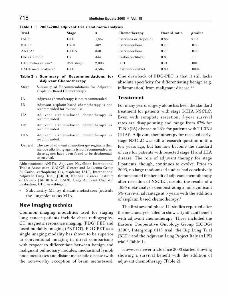
718 Medicine Update 2008 Vol. 18
• SubclassifyM1bydistantmetastases (outsidethe lung/pleura) as M1b.
New imaging technicsCommon imaging modalities used for staginglung cancer patients include chest radiography, CT,magneticresonanceimaging,(FDG)PETandfusedmodalityimaging(PET-CT).FDG-PETasasingle imaging modality has shown to be superior to conventional imaging in direct comparisons with respect to differentiate between benign and malignant pulmonary nodules, mediastinal lymph node metastases and distant metastatic disease (with the noteworthy exception of brain metastases).
One drawback of FDG-PET is that it still lacks absolute specificity for differentiating benign (e.g. inflammation) from malignant disease.3-5
TreatmentFor many years, surgery alone has been the standard treatment forpatientswith stage I-IIIANSCLC.Even with complete resection, 5-year survival rates are disappointing and range from 67% for T1N0 (IA) disease to 23% for patients with T1-3N2 (IIIA)6. Adjuvant chemotherapy for resected early-stageNSCLCwasstillaresearchquestionuntilafew years ago, but has now become the standard of care for patients with resected stage II and IIIA disease. The role of adjuvant therapy for stage I patients, though, continues to evolve. Prior to 2003, no large randomized studies had conclusively demonstrated the benefit of adjuvant chemotherapy afterresectionofNSCLC,despitetheresultsofa1995 meta-analysis demonstrating a nonsignificant 5% survival advantage at 5 years with the addition of cisplatin based chemotherapy.7
The first several phase III studies reported after the meta-analysis failed to show a significant benefit with adjuvant chemotherapy. These included the Eastern Cooperative Oncology Group (ECOG)35908, Intergroup 0115 trial, the Big Lung Trial (BLT) 9 and the Adjuvant Lung Project Italy (ALPI) trial10 (Table 1).
However newer trials since 2003 started showing showing a survival benefit with the addition of adjuvant chemotherapy (Table 2).
Table 1 : 2003–2006 adjuvant trials and meta-analysesTrial Stage n Chemotherapy Hazard ratio p-value
IALT5 I–III 1,867 Cis/vincaoretoposide 0.86 <.03
BR.106 IB–II 482 Cis/vinorelbine 0.70 .012
ANITA8 I–IIIA 840 Cis/vinorelbine 0.79 .013
CALGB96339 IB 344 Carbo/paclitaxel 0.8 .10
UFT meta-analysis21 95% stage I 2,003 UFT 0.74 .001
LACEmeta-analysis10 I–III 4,584 Platinum doublet 0.89 .0004
Table 2 : Summary of Recommendations for Adjuvant Chemotherapy
Stage Summary of Recommendations for Adjuvant Cisplatin-BasedChemotherapy
IA Adjuvant chemotherapy is not recommended
IB Adjuvant cisplatin-based chemotherapy is not recommended for routine use
IIA Adjuvant cisplatin-based chemotherapy is recommended.
IIB Adjuvant cisplatin-based chemotherapy is recommended
IIIA Adjuvant cisplatin-based chemotherapy is recommended.
General The use of adjuvant chemotherapy regimens that include alkylating agents is not recommended as these agents have been found to be detrimental to survival.
Abbreviations: ANITA, Adjuvant Navelbine International TrialistAssociation;CALGB,CancerandLeukemiaGroupB; Carbo, carboplatin; Cis, cisplatin; IALT, InternationalAdjuvant Lung Trial; JBR.10, National Cancer Instituteof Canada JBR.10 trial; LACE, Lung Adjuvant CisplatinEvaluation; UFT, uracil-tegafur.

719Recent Advances in the Management of Lung Cancer
Role of Adjuvant Chemotherapy in stage I-IIIA completely resected NSCLC
Chemotherapeutic agentsThe use of adjuvant chemotherapy involving alkylating agents is not recommended because it has been found to shorten survival.11 Platinum is the mainstay of treatment for patients with lung cancer. Usually doublets are used i.e. combination of platinum with other drugs like etoposide, gemcitabine, taxanes and vinorelbine. NCIC-CTG JBR.10, IALT, & ANITA trials andLACE metaanalysis achieved a statistically andclinically significant survival benefit for adjuvant chemotherapy using vinorelbine combined with cisplatin.12-13 Major drawback in the use of this regimen is the weekly administration schedule. A recent randomized trial showed that, in advanced NSCLC,a3-weeklycycleofvinorelbineondays1 and 8 and cisplatin on day 1 has better tolerance and similar efficacy to the regimen administered in the adjuvant setting described above.14 Of the platinums (cisplatin and carboplatin), cisplatin-based regimen remain the standard in the setting of adjuvant chemotherapy for resected early stage NSCLC,withcarboplatinasa substituteonly inpatients with clear contraindications to cisplatin.15
Adjuvant chemotherapy in the elderlyThe Elderly Vinorelbine Study (ELVIS) was a landmark trial in advanced NSCLC, which ina randomized fashion demonstrated that the elderly enjoy not only a survival benefit from chemotherapy, but an improvement in quality of life as well.16 A retrospective analysis of the JBR.10 trial concluded that patients over the age of 65 years with a good performance status benefit from adjuvant chemotherapy, but those over 75 years of age require further study.17
Treatment of Unresectable Stage IIIA/IIIB Lung CancerFor patients with clinical stage IIIB unresectable
N SCLC, combined-modali ty treatment(chemotherapy with radiation) is the standard of care.18-20 Two treatment strategies, induction chemotherapy and concurrent chemoradiotherapy, have demonstrated favorable effects on survival and are superior to radiotherapy alone. Several groups of investigators have directly compared the two treatment strategies in phase III clinical trials. The first published data came from Japanese investigators, who compared the use of sequential chemoradiotherapy with that of concurrent chemoradiotherapy. The median survival time was 16 months vs 13 months for the concurrent administration.21 Similar results were observed in the Radiation Therapy Oncology Group study 9410, which showed superior 5-year survival rates with concurrent therapy compared to sequential therapywithCDDPandvinblastine.22
Treatment of Metastatic Lung CancerIt has been proved in many randomized trials that systemic chemotherapy is superior to best supportive care in patients with locally advanced and metastatic lung cancer.23 Platinum-based chemotherapy has been widely accepted as the standard of care compared to non-platinum based therapy.24-25Cisplatin-basedchemotherapyresultedin increased median survival time (1.5 months) and reduced the risk of death by 27%.24 In the first line setting platinums have been combined with agents such as Paclitaxel, Docetaxel, Gemcitabine and Vinorelbine have been proved to be equally effective.
Platinum-Based or Non-Platinum-Based Regimens?Due to the toxicities associated with platinum based chemotherapy, non-platinum-based regimens, in particular taxane (paclitaxel/docetaxel) -based regimens, have been the focus of intense research.
A recent randomized study26 of 413 patients compared a platinum-based regimen, CISPLATIN+VINORELBINE, with a non-
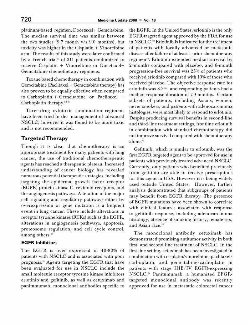
720 Medicine Update 2008 Vol. 18
platinum-based regimen, Docetaxel+ Gemcitabine. The median survival time was similar between the two studies (9.7 month v/s 9.0 months), but toxicitywashigherintheCisplatin+Vinorelbinearm. The results of this study were later confirmed by a French trial27 of 311 patients randomized to receive Cisplatin + Vinorelbine or Docetaxel+Gemcitabine chemotherapy regimens.
Taxane based chemotherapy in combination with Gemcitabine (Paclitaxel + Gemcitabine therapy) has also proven to be equally effective when compared toCarboplatin+Gemcitabine or Paclitaxel +Carboplatintherapy.28-29
Three-drug cytotoxic combination regimens have been tried in the management of advanced NSCLC;however itwas found tobemore toxicand is not recommended.
Targeted TherapyThough it is clear that chemotherapy is an appropriate treatment for many patients with lung cancer, the use of traditional chemotherapeutic agents has reached a therapeutic plateau. Increased understanding of cancer biology has revealed numerous potential therapeutic strategies, including targeting the epidermal growth factor receptor (EGFR)proteinkinaseC,rexinoidreceptors,andthe angiogenesis pathways. Alteration of the major cell signaling and regulatory pathways either by overexpression or gene mutation is a frequent event in lung cancer. These include alterations in receptor tyrosine kinases (RTKs) such as the EGFR, alterations in angiogenesis pathways, apoptosis, proteosome regulation, and cell cycle control, among others.30
EGFR Inhibitors
The EGFR is over expressed in 40-80% of patientswithNSCLCandisassociatedwithpoorprognosis.31 Agents targeting the EGFR that have been evaluated for use in NSCLC include thesmall molecule receptor tyrosine kinase inhibitors erlotinib and gefitinib, as well as cetuximab and panitumumab, monoclonal antibodies specific to
the EGFR. In the United States, erlotinib is the only EGFR-targeted agent approved by the FDA for use inNSCLC.32 Erlotinib is indicated for the treatment of patients with locally advanced or metastatic disease after failure of at least 1 prior chemotherapy regimen33. Erlotinib extended median survival by 2 months compared with placebo, and 6-month progression-free survival was 25% of patients who received erlotinib compared with 10% of those who received placebo. The objective response rate for erlotinib was 8.2%, and responding patients had a medianresponsedurationof7.9months.Certainsubsets of patients, including Asians, women, never smokers, and patients with adenocarcinoma histologies, were most likely to respond to erlotinib. Despite producing survival benefits in second-line and third-line treatment settings, frontline erlotinib in combination with standard chemotherapy did not improve survival compared with chemotherapy alone.34
Gefitinib, which is similar to erlotinib, was the first EGFR targeted agent to be approved for use in patientswithpreviouslytreatedadvancedNSCLC.Currently,onlypatientswhobenefitedpreviouslyfrom gefitinib are able to receive prescriptions for this agent in USA. However it is being widely used outside United States. However, further analysis demonstrated that subgroups of patients may benefit from EGFR therapy. The presence of EGFR mutations have been shown to correlate with clinical features associated with response to gefitinib response, including adenocarcinoma histology, absence of smoking history, female sex, and Asian race.35
The monoclonal antibody cetuximab has demonstrated promising antitumor activity in both first-andsecond-linetreatmentofNSCLC.Inthefirst-line setting, cetuximab has been investigated in combination with cisplatin/vinorelbine, paclitaxel/carboplatin, and gemcitabine/carboplatin in patients with stage IIIB/IV EGFR-expressing NSCLC.36 Panitumumab, a humanized EFGR-targeted monoclonal antibody was recently approved for use in metastatic colorectal cancer

721Recent Advances in the Management of Lung Cancer
and has also demonstrated an acceptable safety profile combined with carboplatin/paclitaxel in patientswithNSCLC.37
VEGF InhibitorsAngiogenesis, which involves formation of new blood vessels, is critical to tumor growth and metastasis. As a result, antiangiogenic therapies, such as bevacizumab, are being actively investigated in a variety of cancers.38 Bevacizumab, a recombinant humanized antibody directed against the VEGF, was the first agent in its class to receive approval for the treatment of patients with cancer. The introduction of bevacizumab has extended survival in the first-line setting when added to carboplatin/paclitaxel, representing the first improvement in survivalforNSCLCsinceplatinum-baseddoubletchemotherapy was introduced 20 years ago.39 On the basis of the Eastern Cooperative OncologyGroup(ECOG)4599trialfindingstheFDArecentlyapproved bevacizumab for first-line treatment ofNSCLC in combinationwith carboplatin andpaclitaxel.40
Multitargeted Kinase InhibitorsMultitargeted receptor tyrosine kinase inhibitors of angiogenic signaling are also being evaluated in NSCLC.Vandetanib,targetsboththeEGFRandVEGFR, Sunitinib principally a VEGF inhibitor but also inhibits the platelet-derived growth factor receptor and other tyrosine kinases & Sorafenib, which inhibits VEGF and platelet-derived growth factor receptor, but also inhibits the growth signaling molecule Raf are the agents in various stages of development.41-43
VaccinesAt least three “immunologically based” therapeutics arebeinginvestigatedinlargestudiesinNSCLC.BL25, a Muc-1 vaccine (Stimuvax) is being tested in stage III unresectable patients in a phase III trial. Recombinant Mage-3 has been investigated in the adjuvant setting in a large phase II randomized study and is proceeding into phase III. Two phase
III studies of a Toll-9 receptor agonist have recently beencompletedinadvancedNSCLCandresultsare awaited.
Molecular profilingRecent studies have also indicated the ability to prospectively identify patients who are most likely to benefit from adjuvant therapy. A retrospective analysis of a subset of patients from the IALT trial indicatesthatIHCstainingforERCC1mayhelppredict those patients most likely to benefit from adjuvant cisplatin-based therapy.44 ERCC1 is anucleoside excision repair enzyme involved with the repair of cisplatin-induced DNA adducts. In thatanalysis,thosepatientswithERCC1-positivetumors were far less likely to benefit than those with lower levels of this DNA repair enzyme. This is an important step toward identifying the group of patients most likely to benefit from adjuvant cisplatin-based therapy. in the current situation, in which the overall survival benefit from adjuvant therapy is on the order of 5%–10%, and the treatment includes 3 months of relatively toxic chemotherapy, the ability to identify those most likely to benefit is crucial. How best to help those who may be less likely to benefit from cisplatin-based therapy needs further exploration.
EGFR Gene Mutations and Response to EGFR-TK Inhibitors The observation that certain subgroups of patients, mainly female patients, never-smokers, patients with adenocarcinoma histology, and patients of Asian origin haveahigher response rateandclinicalbenefitwithgefitinib and erlotinib therapy prompted research toelucidate the molecular mechanism responsible for this increased response. Three different research groups have presented studies showing a positive relationship between the presence of activating mutations in the EGFRTKdomainandaclinicalresponsetogefitinib.45-47 The most common mutations were in-frame deletions that resulted in the insertion of a serine residue in three mutations (del E746-A750, delL747-T751insS, and delL747- P753insS) on exon 19. Other mutations

722 Medicine Update 2008 Vol. 18
consisted of an amino acid substitution within exon 21 (leucine to arginine [L858R] and leucine to glutamine [L861Q]). EGFR mutations (L858R and delL747- P753insS) had increased TK activity compared with wild-type receptors and were more sensitive to inhibition by gefitinib.
Small cell lung cancerRecent epidemiological data suggest that the incidence of small-cell lung cancer is falling and that the proportion of lung cancer patients with small-cell histology is now ~ 13% (compared with 20–25%previously).Characterisationofpatientswith SCLC into limited-stage disease (LD) andextensive-stage disease (ED) as defined by the Veterans Administration Lung Study Group (VALG) has been the basis of treatment choice for a number of years. This system is generally accepted in clinical practice; however, it does not accurately segregate patients into homogenous prognostic groups and the appropriate classification of selected sites remains controversial. As a result, the consensus report of the InternationalAssociationofLungCancer(IASLC)revised the VALG classification, in accordance with the tumor, node, metastasis (TNM) system. IntheIASLCsystem,LDisdefinedessentiallyasstages I–IIIB, and ED is limited to patients with distant metastasis. Treatment with cisplatin plus etoposide with early, concurrent radiotherapy is the standard of care for patients with limited-stage disease (LD) suitable for this approach. A 5-year survival rate of 25% has been reported for concurrent hyperfractionated radiotherapy. Patients unsuitable for concurrent chemo-radiotherapy are usually treated with a sequential approach – i.e. Initial chemotherapy followed by radiation. Patients with LD who achieves complete response should be offered prophylactic cranial irradiation (PCI).Patientswithextensive-stagediseasebutfewother adverse prognostic factors should be treated with a platinum compound plus etoposide. Patients with relapsed disease have a poor prognosis, but there is evidence of a survival benefit for salvage chemotherapy in those fit for treatment. The choice
of treatment will depend on a number of factors, including the disease-free interval. Topotecan is the only drug licensed in this indication, but myelosuppression is considerable.48
References1. NationalCancerRegistryProgramme,ConsolidatedReport
ofPopulationBasedCancerRegistries:1990-1996, IndianCouncilofMedicalResearch,August2001.
2. AmericanCancerSociety:CancerFactsandFigures2006.Atlanta,GA,AmericanCancerSociety,2006.
3. MacManus MP, Hicks RJ, Matthews JP, et al: High rate of detection of unsuspected distant metastases by pet in apparent stage III non-small-cell lung cancer: Implications for radical radiation therapy. Int J Radiat Oncol Biol Phys 2001. 50:287-293.
4. Vesselle H, Pugsley JM, Vallieres E, et al: The impact of fluorodeoxyglucose F 18 positronemission tomography on the surgical staging of non-small cell lung cancer. J Thorac Cardiovasc Surg 2002. 124:511-519.
5. Albes JM, Dohmen BM, Schott U, et al: Value of positron emission tomography for lung cancer staging. Eur J Surg Oncol 2002. 28:55-62.
6. Mountain CF: Revisions in the international system forstaging lung cancer. Chest 1997. 111:1710-1717.
7. Chemotherapyinnon-smallcelllungcancer:Ameta-analysisusing updated data on individual patients from 52 randomised clinical trials.Non smallCellLungCancerCollaborativeGroup. BMJ 1995;311:899 –909.
8. Keller SM, Adak S, Wagner H et al. A randomized trial of postoperative adjuvant therapy in patients with completely resected stage II or IIIA nonsmall- cell lung cancer. Eastern CooperativeOncologyGroup.N Engl J Med 2000;343:1217–1222.
9. WallerD,PeakeMD,StephensRJetal.Chemotherapyforpatients with non-small cell lung cancer: The surgical setting of the Big Lung Trial. Eur J Cardiothorac Surg 2004;26:173–182.
10. Scagliotti GV, Fossati R, Torri V et al. Randomized study of adjuvant chemotherapy for completely resected stage I, II, or IIIA non-small-cell lung cancer. J Natl Cancer Inst 2003;95:1453–1461.
11. Non-Small Cell Lung Cancer Collaborative Group:Chemotherapy in non-small cell lung cancer: A meta-analysis using updated data on individual patients from 52 randomised clinical trials. BMJ 311:899-909, 1995
12. Pignon JP, Tribodet H, Scagliotti G, et al: Lung Adjuvant Cisplatin Evaluation (LACE): A pooled analysis of fiverandomized clinical trials including 4,584 patients. J Clin Oncol 24:366s, 2006 (suppl; abstr 7008).
13. Pignon JP, Tribodet H, Scagliotti G, et al: Lung Adjuvant Cisplatin Evaluation (LACE): A pooled analysis of fiverandomized clinical trials including 4,584 patients.

723Recent Advances in the Management of Lung Cancer
14. GebbiaV,GalettaD,CarusoM:Weeklyvinorelbinepluscisplatin versus day 1 _ 8 vinorelbine plus cisplatin in advanced non-small cell lung cancer: End results of a phase III trial. Lung Cancer 49:S32, 2005 (suppl 2; abstr).
15. ArdizzoniA,TiseoM,BoniLet al.CISCA (cisplatinvs.carboplatin) metaanalysis: An individual patient data meta-analysis comparing cisplatin versus carboplatin-based chemotherapy in first-line treatment of advanced non-small celllungcancer(NSCLC). J Clin Oncol 2006;24:366s.
16. GridelliC.TheELVIStrial:AphaseIIIstudyofsingle-agentvinorelbine as first-line treatment in elderly patients with advancednon-smallcelllungcancer.ElderlyLungCancerVinorelbine Italian Study. The Oncologist 2001;6(suppl 1):4 –7.
17. Winton T, Livingston R, Johnson D, et al: Vinorelbine plus cisplatin vs. observation in resected non-small-cell lung cancer. N Engl J Med 2005. 352:2589- 2597.
18. Kim TY, Yang SH, Lee SH, et al: A phase III randomized trial of combined chemoradiotherapy versus radiotherapy alone in locally advanced non-small-cell lung cancer. Am J Clin Oncol 2002. 25:238-243.
19. Sause W, Kolesar P, Taylor SI, et al: Final results of phase III trial in regionally advanced unresectable non-small cell lung cancer: Radiation Therapy Oncology Group, Eastern Cooperative Oncology Group, and Southwest OncologyGroup. Chest 2000. 117:358-364.
20. Dillman RO, Herndon J, Seagren SL, et al: Improved survival in stage III non-small-cell lung cancer: Seven-year follow-up ofcancerandleukemiagroupB(CALGB)8433trial.J Natl Cancer Inst 1996. 88:1210-1215.
21. Furuse K, Fukuoka M, Kawahara M, et al. Phase III study of concurrent versus sequential thoracic radiotherapy in combination with mitomycin, vindesine, and cisplatin in unresectable stage III non-small-cell lung cancer. J Clin Oncol 1999; 17:2692–2699.
22. CurranWJ,ScottCB,LangerCJ,etal.Long-termbenefitis observed in a phase III comparison of sequential vs concurrent chemo-radiation for patients with unresected stageIIINSCLC:RTOG9410[abstract].ProcAmSocClinOncol 2004; 22:621.
23. Scagliotti GV, De Marinis F, Rinaldi M, et al. Phase III randomized trial comparing three platinum-based doublets in advanced non-small-cell lung cancer. J Clin Oncol 2002; 20:4285–4291
24. Barlesi F, Pujol JL, Daures JP. Should chemotherapy (Cx) for advanced non-small cell lung cancer (NSCLC)be platinumbased? A literature-based meta-analysis of randomized trials. J Clin Oncol 2005; 24:673s.
25. D’Addario G, Pintilie M, Leighl NB, et al. Platinum-based versus non-platinum-based chemotherapy in advanced non small- cell lung cancer: a meta-analysis of the published literature. J Clin Oncol 2005; 23:2926–2936.
26. Georgoulias V, Ardavanis A, Tsiafaki X, et al. Vinorelbine plus cisplatin versus docetaxel plus gemcitabine in advanced nonsmall- cell lung cancer: a phase III randomized trial. J Clin Oncol 2005; 23:2937–2945.
27. Pujol JL, Breton JL, Gervais R, et al. Gemcitabine-docetaxel versus cisplatin-vinorelbine in advanced or metastatic nonsmall- cell lung cancer: a phase III study addressing the case for cisplatin. Ann Oncol 2005; 16:602–610.
28. KosmidisPA,KalofonosC,SyrigosK,etal.Paclitaxelandgemcitabine vs. carboplatin and gemcitabine: a multicenter phase III randomized trial in patients with advanced inoperablenon-smallcelllungcancer(NSCLC).J Clin Oncol 2005;23:621s.
29. TreatJ,BelaniCP,EdelmanMJ,etal.ArandomizedphaseIII trial of gemcitabine (G) in combination with carboplatin (C) or paclitaxel (P) versus paclitaxel plus carboplatinin advanced (stage IIIB, IV) non-small cell lung cancer (NSCLC):updateofAlphaOncologytrial (A1–99002L). J Clin Oncol 2005; 23:627s
30. Velu TJ, Beguinot L, Vass WC, et al. Epidermal-growth-factor-dependent transformation by a human EGF receptor proto-oncogene. Science. 1987;238:1408- 1410.
31. Mendelsohn J, Baselga J. Status of epidermal growth factor receptor antagonists in the biology and treatment of cancer. J Clin Oncol 2003; 21:2787–2799.
32. ShepherdFA,RodriguesPereiraJ,CiuleanuT,etal.Erlotinibin previously treated non-small-cell lung cancer. N Engl J Med. 2005;353:123-132.
33. Non-small cell lung and pancreatic cancer. Available at http://www.tarceva.com/tarceva/patient/index.jsp. Accessed January 3, 2007.
34. Herbst RS, Prager D, Hermann R, et al. TRIBUTE: a phase III trial of erlotinib hydrochloride (OSI-774) combined with carboplatin and paclitaxel chemotherapy in advanced non-small-cell lung cancer. J Clin Oncol. 2005;23:5892-5899.
35. Bell DW, Lynch TJ, Haserlat SM, et al. Epidermal growth factor receptor mutations and gene amplification in non-small-cell lung cancer: molecular analysis of the IDEAL/INTACTgefitinibtrials.J Clin Oncol. 2005;23:8081-8092.
36. RosellR,DanielC,RamlauR,etal.RandomizedphaseIIstudyof cetuximab incombinationwithcisplatin (C)andvinorelbine (V)vsCValone in the first-line treatmentofpatients (pts) with epidermal growth factor receptor (EGFR)-expressing advanced non–small-cell lung cancer. Program and abstracts of the 40th Annual Meeting of the American SocietyofClinicalOncology;June5-8,2004;NewOrleans,Louisiana. Abstract. 7012.
37. Blumenschein G Jr, Sandler A, O’Rourke T, et al. Safety and pharmacokinetics (PK) of AMG 706, panitumumab, andcarboplatin/paclitaxel(CP)forthetreatmentofpatients(pts)withadvancednon-small cell lungcancer (NSCLC).Program and abstracts of the 42nd Annual Meeting of the American Society of Clinical Oncology; June 2-6, 2006;Atlanta, Georgia. Abstract 7119.
38. Jain RK, Duda DG, Clark JW, Loeffler JS. Lessons fromphase III clinical trials on anti- VEGF therapy for cancer. Nat Clin Pract Oncol. 2006;3:24-40.
39. Sandler AB, Gray R, Brahmer J, et al. Randomized phase II/IIItrialofpaclitaxel(P)pluscarboplatin(C)withorwithoutbevacizumab(NSC#704865)inpatientswithadvancednon-
724 Medicine Update 2008 Vol. 18
squamousnon-smallcelllungcancer(NSCLC):anEasternCooperativeOncologyGroup(ECOG)trial—E4599.
40. SandlerA,GrayR,PerryMC,etal.Paclitaxel-carboplatinalone or with bevacizumab for non-small-cell lung cancer. N Engl J Med. 2006;355:2542-2550.
41. Natale R, Bodkin D, Govindan R, et al. A comparison of the antitumour efficacy of ZD6474 and gefitinib (Iressa) in patientswithNSCLC:resultsofarandomized,doubleblindphase II study. Program and abstracts of the 11th World Conference on Lung Cancer; July 3-6, 2005; Barcelona,Spain. Abstract O-103.
42. Socinski MA, Novello S, Sanchez JM, et al. Efficacy and safety of sunitinib in previously treated, advanced non-small cell lungcancer(NSCLC):preliminaryresultsofamulticenterphase II trial. Program and abstracts of the 42nd Annual MeetingoftheAmericanSocietyofClinicalOncology;June2-6, 2006; Atlanta, Georgia. Abstract 7001.
43. Gatzemeier U, Blumenschein G, Fosella F, et al. Phase II trial of single-agent sorafenib in patients with advanced non-small cell lung carcinoma. Program and abstracts of the
42ndAnnualMeetingof theAmericanSocietyofClinicalOncology; June 2-6, 2006; Atlanta, Georgia. Abstract 7001.
44. Olaussen KA, Dunant A, Fouret P et al. DNA repair by ERCC1 innonsmall- cell lungcancerandcisplatin-basedadjuvant chemotherapy. N Engl J Med 2006;355:983–991.
45. PaezJG,JannePA,LeeJC,etal.EGFRmutationsinlungcancer: correlation with clinical response to gefitinib therapy. Science 2004; 304:1497–1500.
46. Lynch TJ, Bell DW, Sordella R, et al. Activating mutations in the epidermal growth factor receptor underlying responsiveness of non-small-cell lung cancer to gefitinib. N Engl J Med 2004; 350:2129–2139.
47. Pao W, Miller V, Zakowski M, et al. EGF receptor gene mutations are common in lung cancers from “never smokers” and are associated with sensitivity of tumors to gefitinib and erlotinib. Proc Natl Acad Sci U S A 2004; 101:13306–13311.
48. FerraldeschiR,BakaS,JyotiB,Faivre-FinnC,ThatcherN,LoriganP.ModernManagementofSmall-CellLungCancer.Drugs 2007;67:2135-52.

CHAPTER
95Pre-operative Cardio Pulmonary Assessment R. S. Bhatia
IntroductionSurgical interventions are continuously rising from 10% to 15% because of increasing number in surgical subspecialities providing operative management. Atleast one third of these cases belong to geriatric group who need mandatory pre-operative cardiopulmonary assessment1. Mortality rate of 0.5-1% increases to 5% as the age advances, especiallyafter65yearsofage.Cardiacreasonsformorbidity and mortality are more within 48 hours of surgery while other causes such as pneumonia, sepsis, renal failure, pulmonary embolism & even cardiac arrest are responsible for death after 48 hours of surgery. Doctors are often asked to rule out high risk patients, not fit for surgery and also to help in stabilising & preparing for surgery with medication.
Expected cardiac risks
Incidence of post operative myocardial infarction in patients above 50 years is 0.5-1% and might escalate to 3% in those undergoing vascular surgery. These are ofcourse, influenced by functional status of the patient, age, co-morbidities and post operative cardiac complications, if any. Emergency major surgical procedure for prolonged duration are considered high cardiac risk surgery while breast surgery, cataract surgery, superficial surgical and endoscopic procedures are considered low
cardiac risk2. The advancement in anesthesia and perioperative surgical management has reduced the risk of mortality from 1 in 500 to 1 in 2,50,00 in the last 50 years. Monitoring in high risk patients for myocardial ischemia is required in post-operative period in patients with hypertension, coronary artery disease, left ventricular hypertrophy, diabetes mellitus3 and in those taking digitalis. Attending critical care physician should be vigilant as post operative myocardial infarctions are painless in more than 60% of cases4.
Cardio vascular assessment
History of recent ischemic heart disease is major risk factor, because the risk of perioperative & post operative myocardial ischemia will increase from 5% to 65% and to 35% if the past history of suffering from myocardial infarction is more than six months, less than six months or less than three months, respectively. Negative stress testing, however, can permit surgical procedure even after six weeks, under the expertised anesthetic peri & post operative care. If there is no contra-indication, treatment with beta blockers, few days prior to surgery, to bring down heart rate 60/minute, decreases the subsequent cardiac events in high risk patients.5
In those with aortic stenosis, having a fixed cardiac output, the risk of surgery is increased as
726 Medicine Update 2008 Vol. 18
they can not tolerate vasodilation associated with spinal or general anesthesia. Those with preserved left ventricular functions6 such as patients with mitral or aortic regurgitation, are less likely to have cardiac events due to valve dysfunction. Patients with moderate to severe mitral stenosis are also high risk patients for surgery. Prophylaxis against infective endocarditis is mandatory in all these patients.
Patients with congestive heart failure are high risk patients to develop pulmonary edema. But those with no previous history of congestive heart failure, develop symptoms, may be in 5% of cases, within first hour, requiring urgent management with diuretics. These cases require on line fluids with caution, nodoubt; but at the sametime, dehydration should be avoided as it might lead to hypovolemic hypotension during anesthesia.
Patients with supraventricular arrhythmias are required to be rate-controlled before being subjected to surgery. Intra operative pacing may be required in patients with second or third degree block; patients with first degree block can tolerate surgery. Those with, ventricular arrhythmias need cardiologist’s evaluation pre operatively.
Hypertension is another risk factor, pre-operative systolic and diastolic pressure of 180 mmHg & 110 mmHg can safely go for anesthesia & surgical procedure. Antihypertensive therapy has to be continued.
Functional status of the patient is a good predictor ofpatientsriskforsurgery.Clinicalevaluationandconsideration of risk factors do help in evaluation of the patients. Those with history of coronary angiography 2 years back and are clinically & functionally fit, can goahead with surgery, if other risk factors are not hindering.
Pulmonary assessment
This is required in chronic smokers7, patients with COPD and those with suspected lung disease,because 50% of overall mortality is expected to occur in those getting complicated with hypoventilation,
atelectasis and pneumonia, that occurs in almost one third of cases getting pulmonary complications.8 For surgical risk, forced expiratory volume in one second (FEVI) is the best indicator, where patients with FEVI less than one are most prone to post operative pulmonary complications and those with FEVI more than two can safely be operated upon. ChestX-rayforinfectioninCOPDpatients&thosewith tuberculosis is indicated. Patients with severe COPD might be subjected to arterial blood gasanalysis.
Treatment of chest infection, cessation of smoking few weeks earlier and use of bronchodilators few days prior to surgery reduces pulmonary risks. Post-operative measures such as early mobilisation, with chest physiotherapy, bronchodilators, & incentive spirometryforpatientsofCOPDisrewarding.
Pre-operative requirements, apart from routine tests,9,10 chest X-ray is recommended for every patientabove50yearsofageandECG11 for any patient above 40 years, known diabetics and in those with known history of ischemic heart disease.
A proper history of A-for allergy, B-bleeding disorder, C-coronary artery disease/ corticoidsbeing used/ COPD/ CHF, D-Drugs, (NSAIDs,Clopidogrel)/diabetes,E-endocrinalstatus(Thyroid),F-family history of malignant hypertension/ (Fever), G- Glaucoma, H-HIV/ Hepatitis, I-infections including tuberculosis. This helps in better planning and pre-operative interaction among anesthetist, surgeon & the patient. High risk patients can get additional stress cardiac testing & even (though rare) angiography. Preoperative use of beta blockers in high risk patient with angina, positive TMT test, myocardial infarction, hypertension or coronary artery disease is useful in reducing post operative cardiac ischemic complication by 50%.
Comments
Constructiveinvolvementofinternistintakingwellstructured history, followed by focused clinical systematic examination to evolve efficacious evaluation of the patient before surgery, shall be

727Pre-operative Cardio Pulmonary Assessment
able to provide proper pre-operative information to the anesthetist to have interaction with the operating surgeon for proper peri & post operative care to avoid cardiopulmonary complications.12
References1. Beliveau MM, Multach M; Perioperative care for the elderly
patient; Med Clin North Amer; 2003;87:273-89
2. EagleKA;etal;ACC/AMAguidelineupdateforperioperativecardiovascular evaluation for non cardiac surgery, Circulation; 2002; 105:1257-67
3. AshTonCMetal;theincidenceofperioperativemyocardialinfarction in men undergoing non-cardiac surgery; Ann Intern Med; 1993; 118:504-10
4. Ghosh AK: Pre operative evaluation of non-cardiac surgery; JAPI; 2005;53:306-ll
5. Anerbach AD; GoldmanL; Beta blockers & reduction
of Cardiac events in non Cardiac surgery; JAMA;2002; 287:1435-44
6. Bhatia RS; et al; cardiac surgery in congenital heart disease-Med. Pulse; I 1997;4:43-44.
7. Bhatia RS; The challenge of smokeless tobacco-Cardiopulmonary effects; IJCP; 1995;6:82-85
8. Arozullah AM et al; Preoperative evaluation for post operative pulmonary - complications; Med. Clin North Amer; 2003;87:153-73
9. Smetana GW, MacpherSon DS; The case against routine preoperative laboratory testing Med. Clin North Amer; 2003;87:7-04
10. Bhatia RS; Batta S; Routine investigations - Let us be relative and rational; JAPI; 1992;40:493
11. Ahuja IS,BhatiaRS,AhujaGKunderstandingECG;PeePee Publisher New Delhi 2007
12. BhatiaRS;Cardiopulmonaryexercise testing indicationsand contra- indications; IJCP; 1996;6:36-40
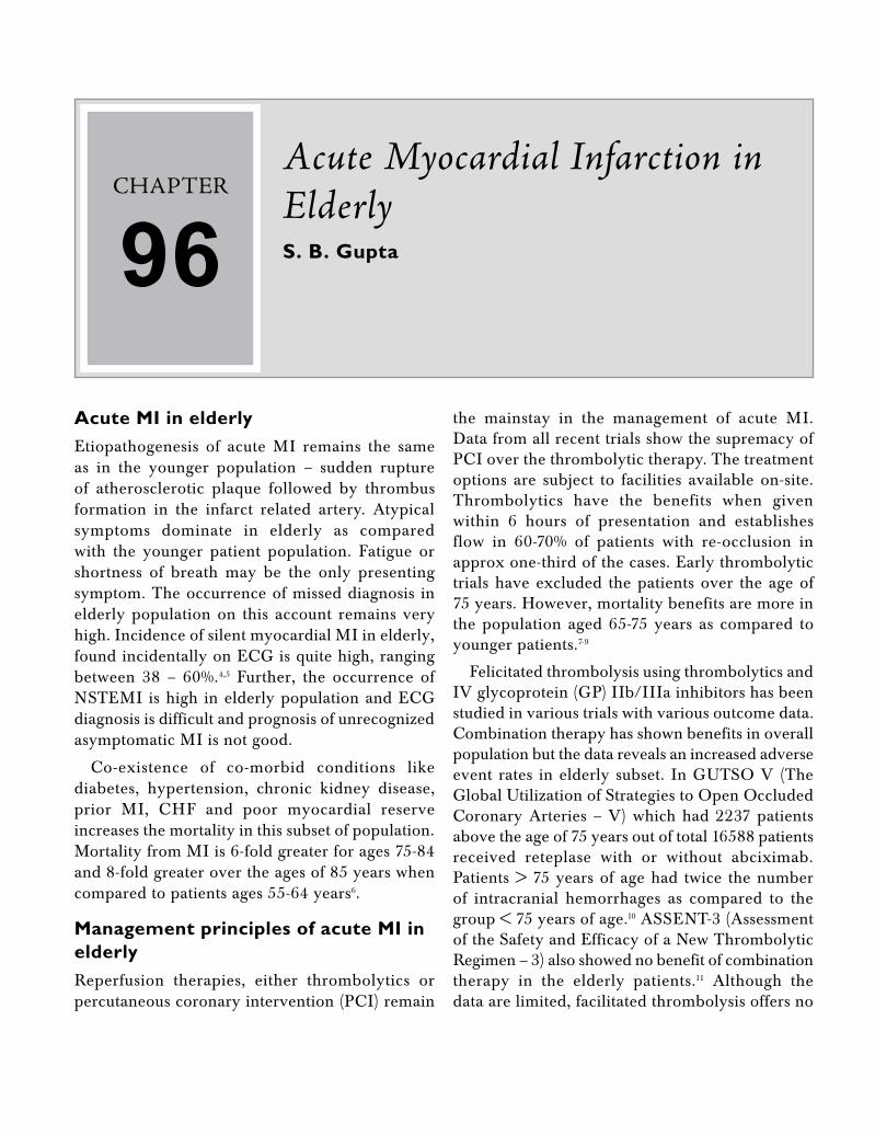
CHAPTER
96Acute Myocardial Infarction in ElderlyS. B. Gupta
Acute MI in elderlyEtiopathogenesis of acute MI remains the same as in the younger population – sudden rupture of atherosclerotic plaque followed by thrombus formation in the infarct related artery. Atypical symptoms dominate in elderly as compared with the younger patient population. Fatigue or shortness of breath may be the only presenting symptom. The occurrence of missed diagnosis in elderly population on this account remains very high. Incidence of silent myocardial MI in elderly, foundincidentallyonECGisquitehigh,rangingbetween 38 – 60%.4,5 Further, the occurrence of NSTEMI ishigh inelderlypopulationandECGdiagnosis is difficult and prognosis of unrecognized asymptomatic MI is not good.
Co-existence of co-morbid conditions likediabetes, hypertension, chronic kidney disease, prior MI, CHF and poor myocardial reserveincreases the mortality in this subset of population. Mortality from MI is 6-fold greater for ages 75-84 and 8-fold greater over the ages of 85 years when compared to patients ages 55-64 years6.
Management principles of acute MI in elderlyReperfusion therapies, either thrombolytics or percutaneouscoronaryintervention(PCI)remain
the mainstay in the management of acute MI. Data from all recent trials show the supremacy of PCIoverthethrombolytictherapy.Thetreatmentoptions are subject to facilities available on-site. Thrombolytics have the benefits when given within 6 hours of presentation and establishes flow in 60-70% of patients with re-occlusion in approx one-third of the cases. Early thrombolytic trials have excluded the patients over the age of 75 years. However, mortality benefits are more in the population aged 65-75 years as compared to younger patients.7-9
Felicitated thrombolysis using thrombolytics and IV glycoprotein (GP) IIb/IIIa inhibitors has been studied in various trials with various outcome data. Combinationtherapyhasshownbenefitsinoverallpopulation but the data reveals an increased adverse event rates in elderly subset. In GUTSO V (The Global Utilization of Strategies to Open Occluded CoronaryArteries–V)whichhad2237patientsabove the age of 75 years out of total 16588 patients received reteplase with or without abciximab. Patients > 75 years of age had twice the number of intracranial hemorrhages as compared to the group < 75 years of age.10 ASSENT-3 (Assessment of the Safety and Efficacy of a New Thrombolytic Regimen – 3) also showed no benefit of combination therapy in the elderly patients.11 Although the data are limited, facilitated thrombolysis offers no
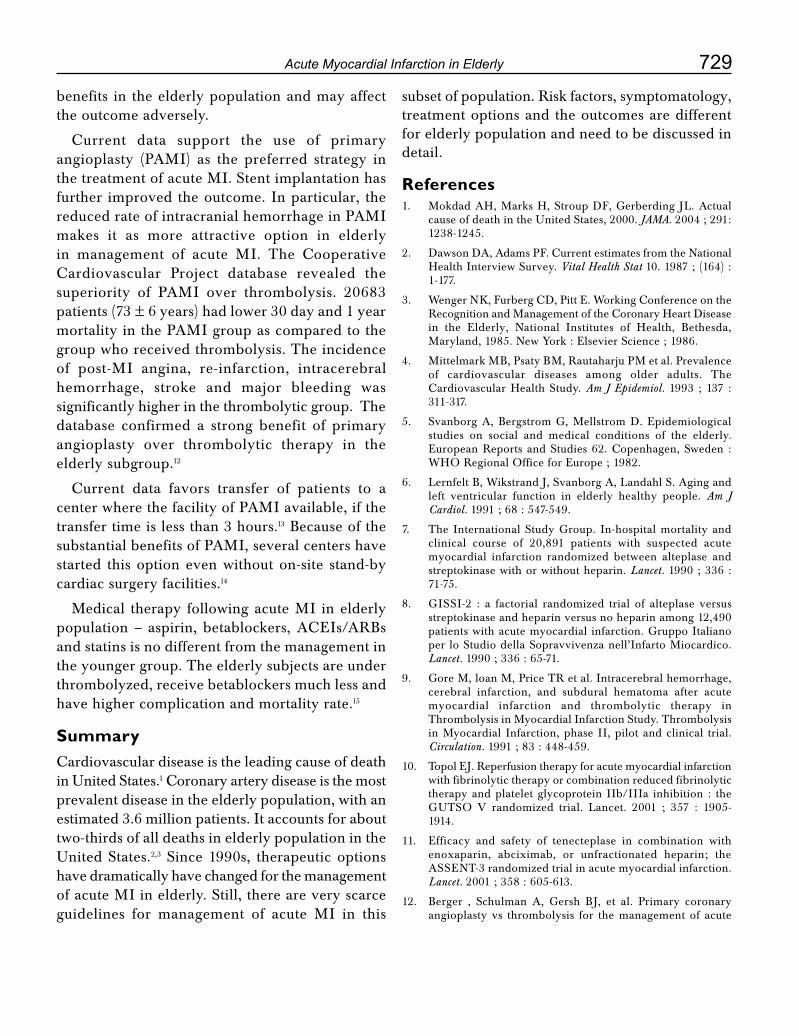
729Acute Myocardial Infarction in Elderly
benefits in the elderly population and may affect the outcome adversely.
Current data support the use of primaryangioplasty (PAMI) as the preferred strategy in the treatment of acute MI. Stent implantation has further improved the outcome. In particular, the reduced rate of intracranial hemorrhage in PAMI makes it as more attractive option in elderly in management of acute MI. The CooperativeCardiovascular Project database revealed thesuperiority of PAMI over thrombolysis. 20683 patients (73 ± 6 years) had lower 30 day and 1 year mortality in the PAMI group as compared to the group who received thrombolysis. The incidence of post-MI angina, re-infarction, intracerebral hemorrhage, stroke and major bleeding was significantly higher in the thrombolytic group. The database confirmed a strong benefit of primary angioplasty over thrombolytic therapy in the elderly subgroup.12
Current data favors transfer of patients to acenter where the facility of PAMI available, if the transfer time is less than 3 hours.13 Because of the substantial benefits of PAMI, several centers have started this option even without on-site stand-by cardiac surgery facilities.14
Medical therapy following acute MI in elderly population–aspirin,betablockers,ACEIs/ARBsand statins is no different from the management in the younger group. The elderly subjects are under thrombolyzed, receive betablockers much less and have higher complication and mortality rate.15
SummaryCardiovasculardiseaseistheleadingcauseofdeathin United States.1Coronaryarterydiseaseisthemostprevalent disease in the elderly population, with an estimated 3.6 million patients. It accounts for about two-thirds of all deaths in elderly population in the United States.2,3 Since 1990s, therapeutic options have dramatically have changed for the management of acute MI in elderly. Still, there are very scarce guidelines for management of acute MI in this
subset of population. Risk factors, symptomatology, treatment options and the outcomes are different for elderly population and need to be discussed in detail.
References1. Mokdad AH, Marks H, Stroup DF, Gerberding JL. Actual
cause of death in the United States, 2000. JAMA. 2004 ; 291: 1238-1245.
2. DawsonDA,AdamsPF.CurrentestimatesfromtheNationalHealth Interview Survey. Vital Health Stat 10. 1987 ; (164) : 1-177.
3. WengerNK,FurbergCD,PittE.WorkingConferenceontheRecognitionandManagementoftheCoronaryHeartDiseasein the Elderly, National Institutes of Health, Bethesda, Maryland, 1985. New York : Elsevier Science ; 1986.
4. Mittelmark MB, Psaty BM, Rautaharju PM et al. Prevalence of cardiovascular diseases among older adults. The CardiovascularHealthStudy.Am J Epidemiol. 1993 ; 137 : 311-317.
5. Svanborg A, Bergstrom G, Mellstrom D. Epidemiological studies on social and medical conditions of the elderly. EuropeanReportsandStudies62.Copenhagen,Sweden :WHO Regional Office for Europe ; 1982.
6. Lernfelt B, Wikstrand J, Svanborg A, Landahl S. Aging and left ventricular function in elderly healthy people. Am J Cardiol. 1991 ; 68 : 547-549.
7. The International Study Group. In-hospital mortality and clinical course of 20,891 patients with suspected acute myocardial infarction randomized between alteplase and streptokinase with or without heparin. Lancet. 1990 ; 336 : 71-75.
8. GISSI-2 : a factorial randomized trial of alteplase versus streptokinase and heparin versus no heparin among 12,490 patients with acute myocardial infarction. Gruppo Italiano per lo Studio della Sopravvivenza nell’Infarto Miocardico. Lancet. 1990 ; 336 : 65-71.
9. Gore M, loan M, Price TR et al. Intracerebral hemorrhage, cerebral infarction, and subdural hematoma after acute myocardial infarction and thrombolytic therapy in Thrombolysis in Myocardial Infarction Study. Thrombolysis in Myocardial Infarction, phase II, pilot and clinical trial. Circulation. 1991 ; 83 : 448-459.
10. Topol EJ. Reperfusion therapy for acute myocardial infarction with fibrinolytic therapy or combination reduced fibrinolytic therapy and platelet glycoprotein IIb/IIIa inhibition : the GUTSO V randomized trial. Lancet. 2001 ; 357 : 1905-1914.
11. Efficacy and safety of tenecteplase in combination with enoxaparin, abciximab, or unfractionated heparin; the ASSENT-3 randomized trial in acute myocardial infarction. Lancet. 2001 ; 358 : 605-613.
12. Berger , Schulman A, Gersh BJ, et al. Primary coronary angioplasty vs thrombolysis for the management of acute

730 Medicine Update 2008 Vol. 18
myocardial infarction in elderly patients. JAMA. 1999 ; 282: 341-348.
13. Widimsky P, Budesinsky T, Vorac D, et al. Long distance transport for primary angioplasty vs immediate thrombolysis in acute myocardial infarction. Final results of the randomized national multicentre trial – PRAGUE-2. Eur Heart J. 2003 ; 24 : 94-104.
14. Aversano T, Aversano LT, Passamani E, et al. Thrombolytic therapy vs primary percutaneous coronary intervention for myocardial infarction in patients presenting to hospitals without on-site cardiac surgery : a randomized controlled trial. JAMA. 2002 ; 287 : 1943-1951.
15. HolayMP,JanbandhuA,JavahiraniA,etal.Clinicalprofileof acute myocardial infarction in elderly (Prospective study). JAPI. 2007 ; 55 : 188-192.
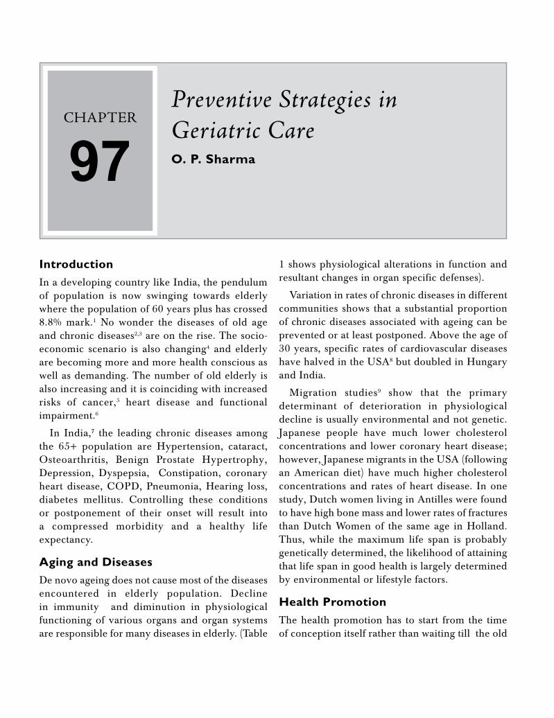
CHAPTER
97Preventive Strategies in Geriatric CareO. P. Sharma
IntroductionIn a developing country like India, the pendulum of population is now swinging towards elderly where the population of 60 years plus has crossed 8.8% mark.1 No wonder the diseases of old age and chronic diseases2,3 are on the rise. The socio-economic scenario is also changing4 and elderly are becoming more and more health conscious as well as demanding. The number of old elderly is also increasing and it is coinciding with increased risks of cancer,5 heart disease and functional impairment.6
In India,7 the leading chronic diseases among the 65+ population are Hypertension, cataract, Osteoarthritis, Benign Prostate Hypertrophy, Depression, Dyspepsia, Constipation, coronaryheart disease,COPD,Pneumonia,Hearing loss,diabetes mellitus. Controlling these conditionsor postponement of their onset will result into a compressed morbidity and a healthy life expectancy.
Aging and Diseases De novo ageing does not cause most of the diseases encountered in elderly population. Decline in immunity and diminution in physiological functioning of various organs and organ systems are responsible for many diseases in elderly. (Table
1 shows physiological alterations in function and resultant changes in organ specific defenses).
Variation in rates of chronic diseases in different communities shows that a substantial proportion of chronic diseases associated with ageing can be prevented or at least postponed. Above the age of 30 years, specific rates of cardiovascular diseases have halved in the USA8 but doubled in Hungary and India.
Migration studies9 show that the primary determinant of deterioration in physiological decline is usually environmental and not genetic. Japanese people have much lower cholesterol concentrations and lower coronary heart disease; however, Japanese migrants in the USA (following an American diet) have much higher cholesterol concentrations and rates of heart disease. In one study, Dutch women living in Antilles were found to have high bone mass and lower rates of fractures than Dutch Women of the same age in Holland. Thus, while the maximum life span is probably genetically determined, the likelihood of attaining that life span in good health is largely determined by environmental or lifestyle factors.
Health PromotionThe health promotion has to start from the time of conception itself rather than waiting till the old
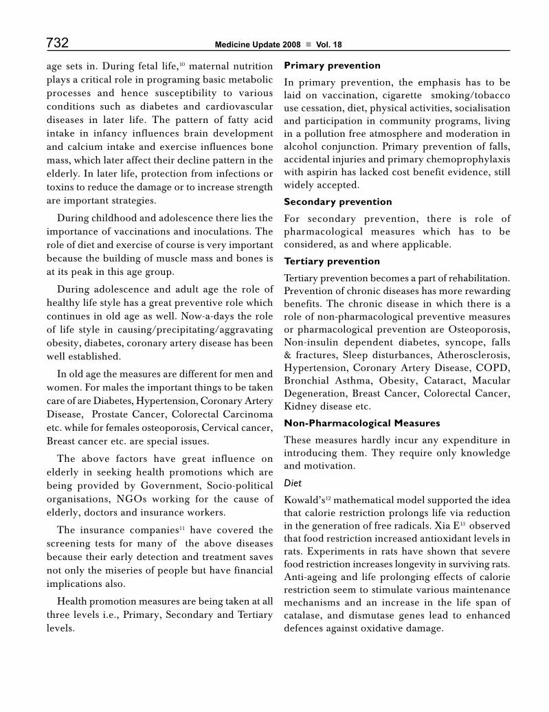
732 Medicine Update 2008 Vol. 18
age sets in. During fetal life,10 maternal nutrition plays a critical role in programing basic metabolic processes and hence susceptibility to various conditions such as diabetes and cardiovascular diseases in later life. The pattern of fatty acid intake in infancy influences brain development and calcium intake and exercise influences bone mass, which later affect their decline pattern in the elderly. In later life, protection from infections or toxins to reduce the damage or to increase strength are important strategies.
During childhood and adolescence there lies the importance of vaccinations and inoculations. The role of diet and exercise of course is very important because the building of muscle mass and bones is at its peak in this age group.
During adolescence and adult age the role of healthy life style has a great preventive role which continues in old age as well. Now-a-days the role of life style in causing/precipitating/aggravating obesity, diabetes, coronary artery disease has been well established.
In old age the measures are different for men and women. For males the important things to be taken careofareDiabetes,Hypertension,CoronaryArteryDisease, ProstateCancer,ColorectalCarcinomaetc.whileforfemalesosteoporosis,Cervicalcancer,Breast cancer etc. are special issues.
The above factors have great influence on elderly in seeking health promotions which are being provided by Government, Socio-political organisations, NGOs working for the cause of elderly, doctors and insurance workers.
The insurance companies11 have covered the screening tests for many of the above diseases because their early detection and treatment saves not only the miseries of people but have financial implications also.
Health promotion measures are being taken at all three levels i.e., Primary, Secondary and Tertiary levels.
Primary prevention
In primary prevention, the emphasis has to be laid on vaccination, cigarette smoking/tobacco use cessation, diet, physical activities, socialisation and participation in community programs, living in a pollution free atmosphere and moderation in alcohol conjunction. Primary prevention of falls, accidental injuries and primary chemoprophylaxis with aspirin has lacked cost benefit evidence, still widely accepted.
Secondary prevention
For secondary prevention, there is role of pharmacological measures which has to be considered, as and where applicable.
Tertiary prevention
Tertiary prevention becomes a part of rehabilitation. Prevention of chronic diseases has more rewarding benefits. The chronic disease in which there is a role of non-pharmacological preventive measures or pharmacological prevention are Osteoporosis, Non-insulin dependent diabetes, syncope, falls & fractures, Sleep disturbances, Atherosclerosis, Hypertension,CoronaryArteryDisease,COPD,Bronchial Asthma, Obesity, Cataract, MacularDegeneration,BreastCancer,ColorectalCancer,Kidney disease etc.
Non-Pharmacological Measures
These measures hardly incur any expenditure in introducing them. They require only knowledge and motivation.
Diet
Kowald’s12 mathematical model supported the idea that calorie restriction prolongs life via reduction in the generation of free radicals. Xia E13 observed that food restriction increased antioxidant levels in rats. Experiments in rats have shown that severe food restriction increases longevity in surviving rats. Anti-ageing and life prolonging effects of calorie restriction seem to stimulate various maintenance mechanisms and an increase in the life span of catalase, and dismutase genes lead to enhanced defences against oxidative damage.

733Preventive Strategies in Geriatric Care
Fruits and vegetables14 may act through various mechanisms, like increasing folic acid to decrease homocysteine, increasing potassium and magnesium to decrease blood pressure besides providing antioxidants. High fruit and vegetable intakes have been most consistently associated with protection of cataract, macular degeneration, visual loss, respiratory diseases and cancers such as breast, stomach and colorectal. The discrepancy in benefits between isolated supplementation of antioxidants and fruit and vegetable consumption may be due to other phytonutrients or their synergistic effects. The role of lycopene (found in tomatoes) in protecting elderly male from cancer prostate has been quite promising.15 Carrots have a roleto play in preventing atherosclerosis and amla in promoting immunity.
Ageing may be associated with less efficient processing of essential nutrients-like poor ability to synthesise Vitamin D in the skin, a major source for Indians and poorer ability of the gut to absorb nutrients; requiring higher intakes of nutrients. A committee on medical aspects of food policy recommended higher intake of vitamins, minerals and fatty acids, which can be achieved by diets high in fruit, vegetables, complex carbohydrates and replacement of saturated fats with oils rich in unsaturated fats.
A diet rich in unsaturated fat is supposed to be the cause behind low atherosclerosis and high life expectancy in the Mediterranean region, and Japan.16 High saturated fat diet has been associated with increased atherosclerosis. In a secondary prevention trial in the elderly, an advice to eat fatty fish twice a week reduced cardiovascular deaths by 30%. In another secondary prevention trial, replacing dairy and animal fats with Mediterranean diet reduced mortality by 70% after four years.
The addition of soybean (phytoestrogens) in the diet of elderly women in protecting them from osteoporosis has been very promising.17
Dietary sodium reduction in elderly results in a greater blood pressure fall than in younger subjects, however during summers in temperate zones where both sensible and insensible sweating is more; drastic reductions in salt intake often lead to a state of confusion and anuresis as a result of hyponatremia that ensues.
In hilly terrain iodized salts are preferred over the normal salts to help elderly to retain their normal thyroid function.
Exercise
Exercise can certainly be viewed as a source of primary prevention, and consistently has been noted to benefit the older adult. There is increasing evidence to suggest that habitual aerobic exercise, such as walking, cycling, circuit weight training, swimming, and jogging, can improve strength and aerobic capacity.18,19
Aerobic exercise is defined as physical activity that primarily stimulates mitochondrial oxidative metabolism. Aerobic exercise can also prevent and help manage diseases such as osteoporosis and fractures, coronary artery disease, and non insulin-dependent diabetes mellitus.20 It can also decrease the risk of falling,21 reduce physical disability,22 improve sleep,23 enhance mood and general well-being,24 provide added physiologic reserve, and slow development of disability.25
It also has a protective effect in breast and colon cancers. Even moderate activities such as walking promote physical and mental well being besides their beneficial effect on diabetes, hypertension, obesity and cardiovascular diseases.
Exercise programs are important to improve balance. Flexibility exercises protect against falls, a major cause of morbidity in the elderly. Resistance exercises for legs increase walking speed and facilitate rising from a chair in the elderly. Upper body resistance exercises help in activities of daily living. In long term prospective studies, ongoing physical activity in those aged 60 to 84 years reduced mortality by 50% over
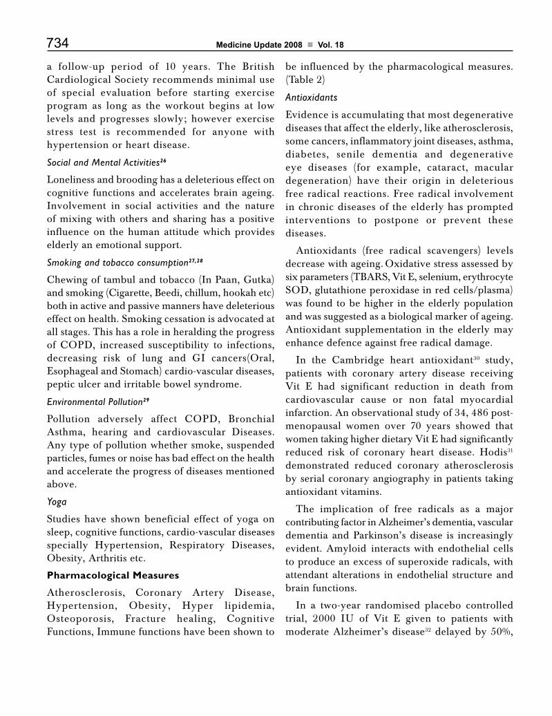
734 Medicine Update 2008 Vol. 18
a follow-up period of 10 years. The British CardiologicalSocietyrecommendsminimaluseof special evaluation before starting exercise program as long as the workout begins at low levels and progresses slowly; however exercise stress test is recommended for anyone with hypertension or heart disease.
Social and Mental Activities26
Loneliness and brooding has a deleterious effect on cognitive functions and accelerates brain ageing. Involvement in social activities and the nature of mixing with others and sharing has a positive influence on the human attitude which provides elderly an emotional support.
Smoking and tobacco consumption27,28
Chewingoftambulandtobacco(InPaan,Gutka)andsmoking(Cigarette,Beedi,chillum,hookahetc)both in active and passive manners have deleterious effect on health. Smoking cessation is advocated at all stages. This has a role in heralding the progress of COPD, increased susceptibility to infections,decreasing risk of lung and GI cancers(Oral, Esophageal and Stomach) cardio-vascular diseases, peptic ulcer and irritable bowel syndrome.
Environmental Pollution29
Pollution adversely affect COPD, BronchialAsthma, hearing and cardiovascular Diseases. Any type of pollution whether smoke, suspended particles, fumes or noise has bad effect on the health and accelerate the progress of diseases mentioned above.
Yoga
Studies have shown beneficial effect of yoga on sleep, cognitive functions, cardio-vascular diseases specially Hypertension, Respiratory Diseases, Obesity, Arthritis etc.
Pharmacological Measures
Atherosclerosis, Coronary Artery Disease,Hypertension, Obesity, Hyper lipidemia, Osteoporosis, Fracture healing, CognitiveFunctions, Immune functions have been shown to
be influenced by the pharmacological measures. (Table 2)
Antioxidants
Evidence is accumulating that most degenerative diseases that affect the elderly, like atherosclerosis, some cancers, inflammatory joint diseases, asthma, diabetes, senile dementia and degenerative eye diseases (for example, cataract, macular degeneration) have their origin in deleterious free radical reactions. Free radical involvement in chronic diseases of the elderly has prompted interventions to postpone or prevent these diseases.
Antioxidants (free radical scavengers) levels decrease with ageing. Oxidative stress assessed by six parameters (TBARS, Vit E, selenium, erythrocyte SOD, glutathione peroxidase in red cells/plasma) was found to be higher in the elderly population and was suggested as a biological marker of ageing. Antioxidant supplementation in the elderly may enhance defence against free radical damage.
In the Cambridge heart antioxidant30 study, patients with coronary artery disease receiving Vit E had significant reduction in death from cardiovascular cause or non fatal myocardial infarction. An observational study of 34, 486 post-menopausal women over 70 years showed that women taking higher dietary Vit E had significantly reduced risk of coronary heart disease. Hodis31 demonstrated reduced coronary atherosclerosis by serial coronary angiography in patients taking antioxidant vitamins.
The implication of free radicals as a major contributing factor in Alzheimer’s dementia, vascular dementia and Parkinson’s disease is increasingly evident. Amyloid interacts with endothelial cells to produce an excess of superoxide radicals, with attendant alterations in endothelial structure and brain functions.
In a two-year randomised placebo controlled trial, 2000 IU of Vit E given to patients with moderate Alzheimer’s disease32 delayed by 50%,
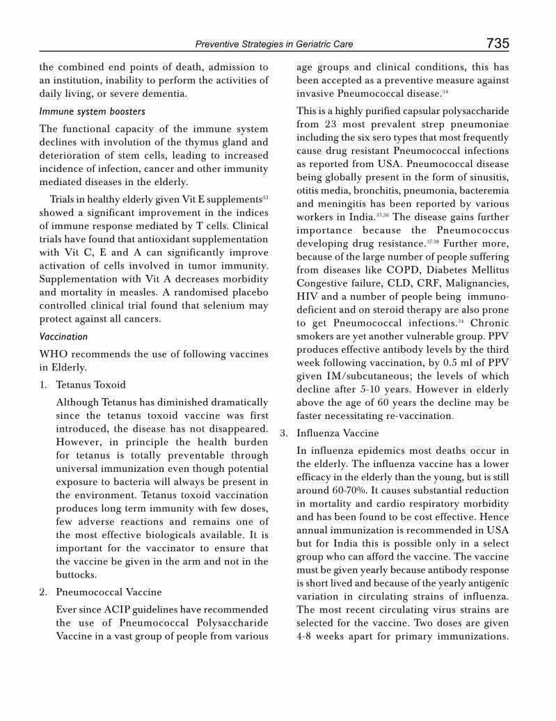
735Preventive Strategies in Geriatric Care
the combined end points of death, admission to an institution, inability to perform the activities of daily living, or severe dementia.
Immune system boosters
The functional capacity of the immune system declines with involution of the thymus gland and deterioration of stem cells, leading to increased incidence of infection, cancer and other immunity mediated diseases in the elderly.
Trials in healthy elderly given Vit E supplements33 showed a significant improvement in the indices ofimmuneresponsemediatedbyTcells.Clinicaltrials have found that antioxidant supplementation with Vit C, E and A can significantly improveactivation of cells involved in tumor immunity. Supplementation with Vit A decreases morbidity and mortality in measles. A randomised placebo controlled clinical trial found that selenium may protect against all cancers.
Vaccination
WHO recommends the use of following vaccines in Elderly.
1. Tetanus Toxoid
Although Tetanus has diminished dramatically since the tetanus toxoid vaccine was first introduced, the disease has not disappeared. However, in principle the health burden for tetanus is totally preventable through universal immunization even though potential exposure to bacteria will always be present in the environment. Tetanus toxoid vaccination produces long term immunity with few doses, few adverse reactions and remains one of the most effective biologicals available. It is important for the vaccinator to ensure that the vaccine be given in the arm and not in the buttocks.
2. Pneumococcal Vaccine
EversinceACIPguidelineshaverecommendedthe use of Pneumococcal Polysaccharide Vaccine in a vast group of people from various
age groups and clinical conditions, this has been accepted as a preventive measure against invasive Pneumococcal disease.34
This is a highly purified capsular polysaccharide from 23 most prevalent strep pneumoniae including the six sero types that most frequently cause drug resistant Pneumococcal infections as reported from USA. Pneumococcal disease being globally present in the form of sinusitis, otitis media, bronchitis, pneumonia, bacteremia and meningitis has been reported by various workers in India.35,36 The disease gains further importance because the Pneumococcus developing drug resistance.37-38 Further more, because of the large number of people suffering from diseases like COPD, Diabetes MellitusCongestivefailure,CLD,CRF,Malignancies,HIV and a number of people being immuno-deficient and on steroid therapy are also prone to get Pneumococcal infections.34 Chronicsmokers are yet another vulnerable group. PPV produces effective antibody levels by the third week following vaccination, by 0.5 ml of PPV given IM/subcutaneous; the levels of which decline after 5-10 years. However in elderly above the age of 60 years the decline may be faster necessitating re-vaccination.
3. Influenza Vaccine
In influenza epidemics most deaths occur in the elderly. The influenza vaccine has a lower efficacy in the elderly than the young, but is still around 60-70%. It causes substantial reduction in mortality and cardio respiratory morbidity and has been found to be cost effective. Hence annual immunization is recommended in USA but for India this is possible only in a select group who can afford the vaccine. The vaccine must be given yearly because antibody response is short lived and because of the yearly antigenic variation in circulating strains of influenza. The most recent circulating virus strains are selected for the vaccine. Two doses are given 4-8 weeks apart for primary immunizations.

736 Medicine Update 2008 Vol. 18
Newer vaccines such as subunit (hemagglutinin) vaccine, live (recombinant) vaccine and cold adapted strains vaccine are in the development stage.
Hormonal Intervention
Human Growth Hormone replacement in Growth Hormone deficit elderly has shown beneficial results in lipid profile as well as abdominal obesity.39
Testosterone supplementation in andropausal men has shown beneficial results in cognitive functions, onset of alzhemier disease, osteoporosis, metabolism besides improving erectile dysfunctions.40
Melatonin supplementations prevent age-related diseases and prolong the lifespan and improve the quality of life of elderly people.41
Estrogen or estrogen and progesterone replacement therapy within the first 5 years of menopause offers protection against osteoporosis in women. It also has a primary preventive role against cardiovascular events in postmenopausal women as seen in the Nurses Health Study. Estrogen alone or several combinations of estrogen and progestin improved the coronary risk profile of subjects in the postmenopausal Estrogen/progestin intervention (PEPI) trial. However, the benefit fell with long term treatment due to increased risk of breast cancer. Raloxifene, a selective estrogen receptor modulator has a promising estrogen-like action on the skeletal and cardiovascular system without any evidence of increased incidence of endometrial or breast cancer.
Non-Steroidal Anti-inflammatory Drugs
The benefit of aspirin is due to its antiplatelet as well as anti-inflammatory action. Giving aspirin shortly after ischemic stroke has been associated with reduction in recurrent stroke and death.
The United States Physician Health Study and British Doctors Trial have used aspirin for primary prevention of myocardial infarction. In the US study, 22000 male US physicians, aged between 40 and 84 years, received 162.5 mg aspirin daily and had 44% relative reduction in acute MI (0.4%
to 0.2% per year) in physicians above 50 years age. But the British study, involving 5000 doctors aged 50-78 years receiving 500 mg aspirin daily, showed no difference. There was a slight increase in hemorrhagic stroke and GI hemorrhage in both studies.
Aspirin should be considered in a dose of 162.5 mg /day in persons over 50 years and those with poorly controlled risk factors like diabetes, smoking and hypercholesterolemia if there are no contraindications.
Antibiotics
Commonchronicinfectionshavebeenimplicatedin the causation of atherosclerosis. Evidence for C pneumoniae is quite strong. Treatment with antibiotics has been shown to be beneficial in secondary prevention. Larger trials are needed to substantiate benefits in primary or secondary prevention.
Lipid lowering drugs
Lowering of hypercholesterolemia by statins has lowered the incidence of fatal and non-fatal MI by 30-35% in primary as well as secondary prevention trials. This also reduces the incidence of coronary revascularization procedures and stroke. It improves quality of life for the elderly. In the Indian context, agents lowering triglycerides and raising HDL may be of more benefit.
Miscellaneous
Even in normotensive Diabetics, the addition of Ramipril showed reno-protective effect as documented in HOPE42 study.
Laser therapies in diabetic retinopathy have helped in prevention of blindness.
References1. Population projections for India and States-1996-2016.
CensusofIndia1991(Report).NewDelhi:RegistrarGeneral,India 1996.
2. ShahB,PrabhakarAK.ChronicMorbidityprofileamongelderly. Indian Journal of Medical Research 1997;106:265-272.
3. Goha Roy S. Morbidity related epidemiological determinants in Indian aged. An overview. In Ramachandran C.R. &

737Preventive Strategies in Geriatric Care
B.Shah,(eds) Public health implications of ageing in India. NewDelhi:IndianCouncilofMedicalResearch,1994114-125.
4. Bali A. Socio Economic status and its relationship to morbidity among elderly. Indian Journal of Medical Research 1997;106:349-360.
5. SaltzsteinS,BehlingC,BaergenR.Features of cancer innonagenarians and centenarians. J Am Geriatr Soc 1998;46:994-998.
6. Skelton D, McLaughlin A. Maintaining functional ability in old age. Physiotherapy 1996;82:11-20.
7. Sharma OP,Delivery Of Healthcare In India , Principles and Practice of Geriatric Medicine Fourth Edition J W & Sons 2006, Vol2 1993-99.
8. KestelootH.Changing trends inmortality. In:YamoriY,Strasser T (eds). New horizon in preventing cardiovascular diseases. Amsterdam: Elsevier 1989.
9. Worth RM, Kato H, Rhoads GG, Kagan A, Syme SL. Epidemiologic studies of coronary heart disease and stroke in Japanese men living in Japan, Hawaii and California:Mortality. Am J Epidemiol 1975;102:481-490.
10. Barker DJP. Maternal and fetal origins of coronary heart disease. JR Coll Phys London 1994;28:544-551.
11. McCarthyE,BurnsR,FreundK,et al . Mammography use, breast cancer stage at diagnosis, and survival among older women. J Am Geriatr Soc. 2000;48:1226-1233.
12. Kowald A, Kirkwood TB. Towards a network theory of ageing: A model combining the free radical and the protein error theory. J Theor Biol 1994;168:75-94.
13. Xia E, Rao G, Van-Remmen H, Heydar-i AR, Richardson A. Activities of antioxidant enzymes in various tissues of male Fischer 344 rats are altered by food restriction. J Nutr 1995;125:195-201.
14. GillmanMW,CupplesLA,GagnonD,et al. Protective effect of fruits and vegetables on development of stroke in men. JAMA 1995;273:1113-1117.
15. Vaishampayan U et al Lycopene and soy isoflavones in the treatmentofprostatecancer.NutrCancer.2007;59:1-716.Hunter G, Wetzstein C, Fields D, Brown A, Bamman M.Resistance training increases total energy expenditure and free-living physical activity in older adults. J Appl Physiol 2000;89:977-984.
16. Department of Health. Nutritional aspects of cardiovascular disease.ReportoftheCardiovascularReview.
17. Vatanparast H et al Does the effect of soy phytoestrogens on bone in postmenopausal women depend on the equol-producing phenotype? Nutr Rev. 2007. 65:294-9.
18. Jette A, Harris B, Leeper L, et al. A home-based exercise program. J Am Geriatr Soc 1996;44:644-649.
19. Bravo G, Gauthier P, Roy P, et al. Impact of a 12-month
exercise program on the physical and psychological health of osteopenic women. J Am Geriatr Soc 1996;44:756-762.
20. Williams PT et al Vigorous Exercise and Diabetic, Hypertensive, and Hypercholesterolemia Medication Use. Med Sci Sports Exerc. 2007 Nov;39(11):1933-1941.
21. SteinbergM,CartwrightC,PeelN,WilliamsG.Asustainableprogramme to prevent falls and near falls in community dwelling older people: results of a randomized trial. J Epidemiol Commun Health 2000;54:227-232.
22. Meuleman J, Breehue W, Kubilis P, Lowenthal D. Exercise training in the debilitated aged: strength and functional outcomes. Arch Phys Med Rehabil 1999;81:312-318.
23. King A, Blair S, Bild D, et al. Determinants of physical activity and interventions in adults. Med Sci Sports Exerc 1992;24:s 221-s223.
24. LampinenP,HeikkinenR,RuoppilaI.Changesinintensityof physical exercise as predictors of depressive symptoms among older adults: an eight-year follow-up. Prev Med 2000;30:371-380.
25. Brown M, Sinacore D, Ehsani A, Binder E, Holloszy J, Kohrt,W. Low intensity exercise as a modifier of physical frailty in older adults. Arch Phys Med Rehabil 2000;81:960-965.
26. Jamuna D. Challenges of changing socio-economic &psychological status of the age, Help Age- India, R & D Journal 1999.
27. GillesM,GreeneH.Cigarettesmoking.InGreeneH,ed.ClinicalMedicine.2nded.St.Louis:Mosby;1996:23-256.
28. Indian J Pharmacol | October 2006 | Vol 38 | Issue 5 | 320-329 325 Yadav Table 2 Percentage of household members who chew pan masala/tobacco or smoke in India during 1998-1999
29. Pandey MR. Domestic smoke pollution and chronic bronchitis in a rural community of the hill region of Nepal. Thorax 1984;39:337-339.
30. Kushi LH, Folsom AR, Prineas RJ, Mink PJ, Wvy, Bostic RM. Dietary antioxidant vitamins and death from coronary heart disease in post menopausal women. N Engl J Med 1996;334:1156-62.
31. Hodis HN Mack WJ, Labree L, et al. Serial coronary angiographic evidence that antioxidant Vitamin intake reduces progression of coronary artery atherosclerosis. JAMA 1995;273:1849-1854.
32. SanoM,ErnestoC,ThomasRG, et al. A controlled trial of Selgiline, alpha-tocopherol or both as treatment for Alzheimer’s disease. The Alzheimer’s disease cooperative study. N Engl J Med 1997;336:1216-1222.
33. Meydani SN, Meydani M, Blumberg JB, et al. Vit E supplementation and in vivo immune response in healthy elderly subjects: A randomised controlled trial. JAMA 1997;277:1380-1386.

738 Medicine Update 2008 Vol. 18
34. MMWR. Prevention of Pneumococcal disease: recommendations of the Advisory Committee onImmunizationPractice(ACIP).MMR Morb Mortal Wkly Rep 1997:46:1- 24.
35. PMP Singh et al Outbreak of Pneumococcal Pneumonia in Miltary Barracks, Indian Journal of Community Medicine, 2006, Vol. 31, No. 3.
36. Thomas K, Prospective multicentre hospital surveillance of Streptococcus Pneumoniae disease in India, Lancet 1999: 353; 1216-21
37. Kanungo R et al Emerging Antibiotic Resistant Pneumococci In Invasive Infections In South India: Need For Monitoring
Indian Journal of Pharmacology 2002; 34: 38-43
38. WattalC,MicrobiologyNewsletter,2006
39. Giannoulis MG,et. al J Clin Endoc Metab. 2006:477-84
40. Med Hypotheses 2004(64);62;14-8
41. Kedziora-Kornatowska Ket.al J Pineal Res. 2007;42:153-8.
42. The Heart Outcomes Prevention Evaluation Study Investigators. Effects of an angiotensin-converting-enzyme inhibitor, ramipril, on death from cardiovascular causes, mycardial infarction, and stroke in high-risk patients. N Engl. J Med 2000.

CHAPTER
98Resurging Infections : DengueA. Muruganathan
Introduction Dengue is an acute mosquito-transmitted viral disease characterized by fever, headache, muscle and joint pain, rash, nausea and vomiting. The most severe form of the disease is dengue hemorrhagic fever, which is characterized by thrombocytopenia, bleeding, and shock. The hemorrhagic form continues to be the leading viral hemorrhagic fever in the world.
Dengue virus infection - Dengue fever - Dengue hemorrhagic fever - Dengue shock syndrome.
Other namesBreak bone fever, named by Dr. Benjamin Rush in Philadelphia, 1780; Dandy fever: Seven-day fever, Duengero - from the Spanish duengo, Ki denga pepo – Swahili: “it is a sudden overtaking by a spirit”.
Dengue, a f lavivirus in the family Arboviridae, has four known serotypes. Studies show that infection with and subsequent immunization from one dengue serotype actually increases the odds of developing dengue hemorrhagic fever during infection with a second serotype. This is especially notable in areas where multiple serotypes have overlapping, endemic regions. Essentially, exposure to a mild form of dengue (in some cases, there is no apparent
illness) seems to sensitize the immune system to the hemorrhagic form of the disease.
OccurrenceAs of the late 1990’s Dengue, a disease found in most tropical and subtropical areas of the world, has become the most common arboviral disease of humans. More than 2.5 billion persons now live in areas where dengue infections can be locally acquired. Reported attack rates for disease during epidemics range from 1 per hundred to 1 per thousand of the population.
However, because persons with milder illness may not seek medical attention and subsequently be reported, the actual number of infections in a population may be 5 to 10 times greater than the number reported. Epidemics caused by all four virus serotypes have become progressively more frequent and larger in the past 25 years. As of 2005, dengue fever is endemic in most tropical countries of the South Pacific, Asia, the Caribbean,theAmericas,andAfrica(seeMap).The incidence of the severe disease, DHF, has increased dramatically in Southeast Asia, the South Pacific, and the American tropics in the past 25 years, with major epidemics occurring in many countries every 3-5 years.

740 Medicine Update 2008 Vol. 18
World Distribution of Dengue 2007 • The eggs can lie dormant in dry conditionsfor up to about 9 months, after which they can hatch if exposed to favorable conditions, i.e. water and food.
• A.aegypticannotwithstandtemperaturesbelow480 F, and will die after less than an hour of 320 F. It is currently limited to a range below 35N latitude.
• Globalwarmingwilllikelyexpandtherangeofthe vector mosquito.
Breeding Habit• Apuddleofwateraboutthesizeanddepthof
20-cent coin is sufficient for an Aedes mosquito to breed in.
• TheAedesmosquitocanalsobreedinunusualplaces such as water trapped in the hardened soil in potted plates, and the rim of unwanted pails.
Favoured Breeding PlacesDesert coolers, Drums, Jars, Pots, Buckets, Flower vases,Plantsaucers,Tanks,Cisterns,Bottles,Tins,Tyres,Roofgutters,Refrigeratordrippans,Cementblocks,Cemeteryurns,Bamboostumps,Coconutshells, Tree holes and many more places where rainwater collects or is stored.
VirusFig. 1 Electron Micrograms
A chart showing the number of dengue cases reported in various states
Fast facts about the mosquito • Aedesaegyptiistheprincipalvectorofdengue
/ dengue hemorrhagic fever. Aedes albopictus also transmits the disease
• Onlythefemaleaedesmosquitobitesasitneedsthe protein in blood to develop its eggs.
• Themosquitobecomesinfectiveapproximately7 days after it has bitten a person carrying the virus. This is the extrinsic incubation period, during which time the virus replicates in the mosquito and reaches the salivary glands.
• Peakbitingisatdawnanddusk.
• TheaveragelifespanofanAedesmosquitoinNature is 2 weeks
• Themosquitocanlayeggsabout3timesinitslifetime, and about 100 eggs are produced each time.
Areas infested with Aodos aegyp5Areas with Aedes aegyp5and recent dengu epidemic activity
Dengue Cases Reported India for Sept` 06
Status
Tamil Nadu Maharashtra UttarPradesh
Karnataka AndhraPradesh
WestBengal
GujaratKeralaNew Delhi
900
800
700
600
500
400
300
200
100
0
886
713
424
326 314 304226
70 65 529
Average
Rajasthan

741Resurging Infections : Dengue
Dengue viruses are members of the family Flaviviridae, which include the Japanese encephalitis virus and the yellow fever virus. Four dengue virus serotypes and various biotypes can be differentiated. Infection with one serotype provides life-long immunity to that virus but not to the others.
1. Mosquitoes transmit dengue to human dendritic cells
2. Dengue targets areas with high WBC counts(liver, spleen, lymph nodes, bone marrow, and glands)
3. DengueentersWBCsandlymphatictissue
4. Dengue enters blood circulation
The transmission cycle for dengue starts when:
• Infected Aedes mosquito bites a healthyperson.
• 4-7dayslater,theinfectedpersondevelopsfever(after the virus multiplies i.e., incubation period). The person usually then sees a doctor.
• Whenfeverstarts,thepersonbecomesinfectiousfor about 5 days.
• IfanAedesmosquitobites thepersonduringthis period when he is infectious, it will pick up the dengue virus in his blood.
• The virus takes 7-10 days to multiply in thesecond mosquito.
• The mosquito then becomes infective andthe cycle starts again when it bites another person.
Clinical FeaturesThis infectious disease is manifested by a sudden onset of fever, It persists for 5 to 6 days. Fever is characteristically biphasic and returns to almost normal in the middle of the febrile period giving rise to the saddleback temperature chart. It reaches its highest level during the last 24 hours before abatement. There is often a macular rash on the first day as well as adenopathy, palatal vesicles, and scleral injection. Other Symptoms include muscle pain, Bone pain, loss of appetite, Nausea, vomiting, Sore throat, abdominal pain, diarrhea, intense headache, usually frontal, and retroorbital pain, particularly when pressure is applied to the eyes. (“Fire is coming out of my eyes”). Epistaxis and scattered petechiae are often noted in uncomplicated
• DengueismadeupofSingle,positive-strandedRNA surrounded by an icosahedral core
• 90glycoproteinEdimersoverlyMproteins
• Protein E is most important characteristic ofdengue
How is Dengue Fever spread?
2
1
34

742 Medicine Update 2008 Vol. 18
dengue, and preexisting gastrointestinal lesions may bleed during the acute illness. Some cases develop much milder symptoms which can, when no rash is present, be misdiagnosed as influenza or other viral infection.
Dengue Hemorrhagic Fever (DHF)
The incubation period of DHF is unknown but is probably similar to that of DF.
DF/DHF
Grade Clinical picture
DF As described in dengue fever
DHF I Above plus positive tourniquet test
DHF II Above signs plus spontaneous bleeding in the form of skin and /or other hemorrhages
DHF III Above signs plus circulatory failure(cold clammy skin, rapid pulse weak pulse pressure, restlessness
DHF IV Profound shock with undetectable pulse and blood pressure
Dengue Shock Syndrome (DSS)
DHF grade III and IV are also called DSS. The four warning signs for impending shock are intense, sustained abdominal pain, persistent vomiting, restlessness or lethargy, and a sudden change from fever to hypothermia with sweating and prostration.
PathogenesisDHF is almost always found in individuals who had a previous experience with at least one of the four serotypes of dengue virus. This leads to the hypothesis of heterotypic antibodies from a previous dengue infection promoting the viral replication within the mononuclear leucocytes – the phenomenon of antibody-dependent enhancement. Furthermore, the immunologic processes aimed at eliminating dengue virus infected cells can result in release of histamine and substances with vasoactive and procoagulant properties, the release of interferon-gamma, and the activation of complement.
Grade I
Grade II
Grade III
Grade IV
Coagulopathy
Disseminatedin travascularcoagulation
Severe bleedingDeath
Shock
ThrombocytopeniaHepatomegalyIncreasedvascular
permeability
Positivetourniquet testFever
Other hemorrhagicmanifestations
Rising hematocritHypoproteinemiaSerous effusion
Leakageof plasma
Hypovolaemia
Dengue Infection

743Resurging Infections : Dengue
DHF results from an infection by a more virulent biotype of the virus or even from unfavorable host factors such as concomitant bacterial infections. DHF is known to be more common in Southeast Asia compared to Africa and America. Black individuals are relatively resistant to DHF / DSS due to a speculated “resistant gene”. The cause of bleeding in DHF appears to be due to thrombocytopenia, platelet dysfunction, disseminated intravascular coagulation and micro vascular injury.
DiagnosisTotal White Blood Cell Count: In case of dengue, this test will reveal leukopenia. The presence of leukocytosis and neutrophilia excludes the possibility of dengue and bacterial infections (leptospirosis, meningoencephalitis, septicemia, pyelonephritis etc).
Thrombocytopenia (< 100.00 / mm3): Total platelet count must be obtained in every patient with symptoms suggestive of dengue for three or more days of presentation. Leptospirosis, measles, rubella, meningococcemia and septicemia may also present with thrombocytopenia.
Hematocrit (micro-hematocrit): According to the definition of DHF, the presence of hemoconcentration (hematocrit elevated by > 20%) is necessary; when it’s not possible to know the previous value of hematocrit, we must regard as significantly elevated the results > 45%.
Thrombocytopenia with concurrent high hematocrit levels differentiates DHF from classic DF.
Currentlyroutinelaboratorydiagnosisofdengueinfections depend on virus isolation or the detection of dengue virus-specific antibodies. The isolation of viruses from clinical specimens can be carried out inculturedmosquitocells,suchasAP-61orC6/36cells cultures. When dengue virus serotype specific monoclonal antibodies are used, virus identification by indirect immunofluorescence can be achieved within 2 weeks.
The commonly used serologic test is the hemagglutination inhibition test. In a primary infection dengue hemagglutination inhibition antibody titer is generally less than 1:20 in a sample collected within the first 4 days after the onset of symptoms. In the convalescent phase sample (collected 1 to 4 weeks after the onset of symptoms) a fourfold or greater rise in antibody titer is detected, with antibody titer â 1.1280
A secondary dengue infection is characterized by the rapid appearance of broadly cross-reactive antibodies. Hemagglutination inhibition titers of 1:20 in the acute-phase sample rise to ô to 1:2560 in the convalescent phase sample. An improved and less time-consuming method is a capture enzyme-linked immunosorbent assay that can detect specific anti-dengue IgM in a single acute-phase sample.
Recently commercial kits for the detection of specific IgG as well as IgM antibodies have become available. They are based on a dot enzyme assay or a nitrocellulose membrane-based capture format, respectively. An alternative to virus isolation is the detection of viral RNA by reverse transcription polymerase chain reaction. Reverse transcription polymerase chain reaction is a highly sensitive technique of particular value in the early diagnosis of dengue infection.
Suspect Cases: Acute onset and high fever of 2-7 days duration, and two or more of the following:
Headache, retro-orbital pain, myalgia, arthralgia, rash, hemorrhagic manifestations, and leukopenia.
Probable case: Suspect case and one or more of the following:
Occurrence of confirmed cases of dengue in the same place and time. Detection of IgM antibody.
Confirmed case: Suspect or probable case and one or more of the following:
Isolation of virus or detection of viral genomic sequences. Fourfold rise in titers of IgG or IgM antibody.

744 Medicine Update 2008 Vol. 18
Differential DiagnosisPhysicians should consider dengue in the differential diagnosis of all patients who have fever and a history of travel to a tropical area within 2 weeks of onset of symptoms.
Leptospirosis – Increased erythrocyte sedimentation rate, total WBC elevated withneutrophilia, transaminases levels slightly elevated and increased BUN and serum creatinine. The presence of jaundice (indicative of severe forms of leptospirosis) + epidemiologic data practically exclude the diagnosis of dengue.
Respiratory Infections – ‘Common Cold’ isseldom mistaken with dengue due to the absence of fever. In relation to the ‘influenza – like syndromes’, differential diagnosis is made by the presence of respiratory symptoms (cough, sore throat, nasal discharge), with higher incidence in the winter; Bacterial pneumonias usually present with chest pain(pleurodynia),productivecoughandtotalWBCelevated with neutrophilia. Diagnosis can be made by chest radiography and sputum bacterioscopy by the method of Gram.
Measles - The pre exanthematic phase (cough, nasal discharge, and conjunctivitis) doesn’t occur in dengue. The morbilliform rash usually begins on the face, with a cefalo-caudal progression. The presence of ‘koplik’ lesions in the jugal mucous membrane just before the exanthematic phase is a pathognomonic sign of measles. A positive vaccination history doesn’t exclude the diagnosis, because an inadequate immunization may have occurred.
Rubella (German Measles) – Fever with an insidious onset, absence of systemic symptoms and lymphoadenomegaly (retroauricular, suboccipital, cervical) preceding a rash which usually begins on the face are typical of rubella. The diagnosis of rubella cannot be made on clinical basis, but by serologic method.
Malaria – Diagnosis is made by detection of Plasmodium forms on serial blood examination.
Fever in malaria is initially of daily presentation, and spleen may be enlarged and tender; jaundice may also be present.
Yellow Fever – The initial clinical manifestations are indistinguishable from dengue. However, the period of incubation usually doesn’t exceed 6 days. Laboratory findings include leukopenia and neutrophilia, a very low erythrocyte sedimentation rate (near by) mm) and a marked increase in the serum transaminases levels. A positive vaccination history practically excludes the diagnosis of yellow fever.
Meningoencephalitis – Headache, presence of petechiae and shock with an onset < 24-48 hours indicate the obligatory exclusion of meningococcemia (in the severe forms of dengue these manifestations usually occur after the third day of disease). Leukocytosis and neutrophilia, thrombocytopenia and hemoconcentration may be present. Besides, neurologic manifestations tend to be absent in dengue fever, in contrast with meningoencephalitis. The evaluation of the cerebral spinal fluid is the basis of diagnosis, because in denguefevertheCSFisusuallynormal.
Pyelonephritis – The diagnosis is made based on the urine bacterioscopy by the method of Gram and Urinocultures. Urinalysis is inadequate for the evaluationof theurinary tract infections. WBCmay show leukocytosis and neutrophilia.
Septicemia – The onset of symptoms is more insidious and it’s usually possible for the clinician to detect a primary infectious focus. Splenomegaly, leukocytosis / leukopenia, metabolic acidosis and neurologic disturbances may be present. Related diseases like diabetes mellitus, alcoholism, neoplasms and malnutrition may lead to the correct diagnosis, which is made by hemocultures.
Management of Dengue Fever• Earlyreportingofthesuspecteddenguefever
• Managementofdengue fever is symptomaticand supportive. Give Paracetamol but no aspirin or brufen. In cases with severe pain

745Resurging Infections : Dengue
analgesics or mild sedatives are to be given. Bed rest is essential. Oral fluids and electrolyte therapy are required for patients with excessive sweating or vomiting. Follow up for any change in platelet/hematocrit. During afebrile phase (2-3 days after febrile period) check platelet/hematocrit. In convalescent phase no special instructions. Normal diet. Patients almost always recover but often have prolonged asthenia and depression.
Management of DHF Grade I and IIDuration is 2-3 days after the febrile phase. Treat on OPD/inpatientbasis.GiveORS.Checkplatelet/hematocrit. If Hct > 20% start IV therapy. Monitor vitals, urine output, and hematocrit.
Management of DHF Grade III and IVDurationis2-3daysafterthefebrilephase.Checkplatelet/hematocrit. Start IV therapy (isotonic solutions). Monitor vitals, urine output, and hematocrit.
If hematocrit is increasing change IV fluid to colloidal solution preferably Dextran or plasma. If hematocrit is decreasing from initial value, give fresh whole blood transfusion. In case of profound shock give IV fluid bolus one or two times. Give oxygen therapy. Steroids in DSS are not helpful. In some cases platelet transfusion may be necessary.
Monitoring should be continued for at least a day after defervescence. Once the patient begins to recover extravasated fluid is rapidly reabsorbed, causing a drop in hematocrit. Before discharge, the patient should meet the following criteria: absence of fever for 24 h (without antipyretics) and a return of appetite; improvement in the clinical picture; hospital care for at least 3 days after recovery from shock; no respiratory distress from pleural effusion or ascites; stable hematocrit; and platelet count greater than 50,000 / m176 because convalescent-phase diagnostic samples are often difficult to obtain, a second blood sample should always be taken on the day of discharge.
Emerging treatmentsEmerging evidence suggests that mycophenolic acid and ribavirin inhibit dengue replication. Initial experiments showed a fivefold increase in defective viral RNA production by cells treated with each drug. In vivo studies, however, have not yet been done.
Preventiona. preventive surveillance and control;
b. publ ic educat ion and community involvement;
c. enforcement; and
d. research.
Personal Prevention• Useofmosquitorepellentcreams,liquids,coils,
mats etc.
• Wearingoffullsleeveshirtsandfullpantswithsocks
• Useofbednetsforsleepinginfantsandyoungchildren during day time
• Flywire/screeningondoorsandwindows.
• Noneoftheseareeffectivebythemselvesalone,use combination.
Vector Control• As Aedes aegypti breeds in containers and
receptacles detection and elimination of mosquito breeding sources is the most important activity.
• Management of roof tops, porticos andsunshades
• Propercoveringofstoredwater
• Reliablewatersupply
• Observationofweeklydryday
• Checknolarvaeswimminginwater.
• CantreatwithAbate®(temephos)(orusefishin ponds).

746 Medicine Update 2008 Vol. 18
Control of case• Instructpatientandimmediateroom-matesto
use anti-mosquito measures for 12 days post onset.
• Investigatesourceofinfection.
Control of contactsPatient asked about sick family members, work colleagues or room-mates, follow up interview with these and suggest sera samples sought if dengue suspected.
Tyres collect rainwater – dispose or cover
Overhead storage need tight covers
Coverwaterjars,drums,coolers and tanks
Flower pot with water collection – empty weekly
Health Education and Community ParticipationImpart knowledge to common people regarding the disease and vector through various media sources likeT.V.,Radio,Cinemaslides,etc.Sensitizingandinvolving the community for detection of Aedes breeding places and their elimination.
Vaccine DevelopmentThere is no commercially available vaccine for the dengue flavivirus.
An effective vaccine will have to be tetravalent because pre-existing heterotypic dengue antibody is a risk factor for DHF. Live attenuated vaccine
viruses have been evaluated in phase I and II trials in Thailand, and a tetravalent formulation is currently undergoing repeat phase I and II trials. Advances have also been made with second generation recombinant dengue vaccines.
Vaccine UpdateIn a journal published 24 March 2006, by Zhou H. and Deem M.W., a novel vaccine procedure known as a polytopic injection can be used to elicit a host immune response to dengue fever and reduce immunodominance. Using the polytopic injection, different vaccine serotypes are injected into different regions of the body. These injections contain epitopes of different dengue serotypes (1-4) that are subdominant; therefore eliciting T cell responses in separate regional lymph nodes. This will then prime the immune system to seek out the subdominant determinants instead of the cross-reactive dominant epitopes for each of the 4 dengue serotypes. This will allow for the avoidance of an immunodominance reaction and provide the host immunity against each serotype of dengue virus.
PrognosisWith early detection and proper case management and symptomatic treatment, mortality can be reduced substantially.
The case fatality rate in DHF can be as low as 0.2% if detected early and treated. Once shock has set in, the fatality rate may be as high as 12% to 44%.
ConclusionFuture dengue incidence in specific locales cannot be predicted accurately, but a high level of dengue transmission is anticipated in all tropical areas of the world for the indefinite future. As there is no Vaccine for dengue, it can be prevented and controlled only with the full support and cooperation of Government, NGOs, Medical Team and the Public.

747Resurging Infections : Dengue
Reference1. Rigau-PerezJG,ClarkGG,GublerDJ,ReiterP,SandersEJ,
Vorndam AV. Dengue and Dengue Haemorrhagic Fever
2. (2006) “etomologia: dengue”. Emerging Infectious Diseases 12: 893
3. “Dengue Fever – Information Sheet” - World Health Organization
4. Harrisons Principles of Internal Medicine
5. Dengue and Dengue Hemorrhagic Fever: Information forHealthCarePractitionersCenter forDiseaseControl.October 22, 2007
6. Rigau-Perez JG, Clark GC, Gubler DJ, Reiter P, SandersEJ, Vorndam AV. Dengue and Dengue Haemorrhagic fever. Lancet???352 (9132): 971-7
7. http://www.tarakharper.com/v_dengue.htm
8. Rico-Hesse R. Molecular evoluation and distribution of dengue viruses type 1 and 2 in nature. Virology 174; 479-93
9. www.cdc.gov/travel/yellowbookch4-denguefever.aspx
10. Khin MM, Than KA. Transovarian transmission of dengue 2 viruses by Aedes aegypti in nature. Am J Trop Med Hyg
11. India says dengue outbreak serious as death toll rises, news.yahoo.com, 7 October 2006
12. Kuhn, Zhang, Rossmann, et. Al. 2002; http://elsevier.com/dengue.html
13. http://en.wikipedia.org/wiki/2006_dengue_outbreak_in_India
14. Schechter,MEMarangoni,DVChapter2:Complicacoesinfecciosas hospitalares, section VIII Dengue 1 Edition
15. http://delhimedicalcouncil.nic.in/Dengue.html
16. Martins, FSV e Setubal, S. Dengue: diagnostico e tratamento. Iforme Tecnico 3. Secretaria de Estado de saude do estado do Rio de Janerio
17. http://www.dengue.gov.sg/subject.asp?id=12
18. Halstead SB. Dengue. In Warren KS, Mahmud AAF, editors. Tropical and geographical medicine. New York: McGraw – Hill 675-85.
19. http://www.webindia123.com/article/dengue/dengue.htm
20. Tassniyoms,VasanawathanaS,ChirawatkulA,RojanasuphotS, and Failure of high-dose methylprednisolone in established dengue shock syndrome: a placebo-controlled, double-blind study.
21. Nimmannitya S. Dengue fever/dengue haemorrhagic fever: case management. Trop Med (Nagasaki)
22. HalsteadSB.PathogenesisofDengue:Challengetomolecularbiology

CHAPTER
99Hepatopulmonary Syndrome: A Clinical ViewM. Beg, A. Gupta
IntroductionIn 1884 Flückiger first described a woman with liver cirrhosis, cyanosis, and digital clubbing.1 The term ‘hepatopulmonary syndrome’, the triad of liver disease, an increased alveolar-arterial gradient while breathing room air, and evidence of intrapulmonary vascular dilatations, was coined in 1977 by Kennedy and Knudson.2 These vascular abnormalities predominate in the lower lung fields. As gravity induces increased blood flow to the lower lung fields hypoxemia is increased when changing from supine to upright position. Mild hypoxemia occurs in approximately one third of all patients with chronic liver disease and often is multifactorial,3,4 because other cardiopulmonary abnormalities (e.g., pleural effusion, ascites) are common in these patients and may coexist with HPS. The special qualities of HPS are platypnea,5 defined as dyspnea induced by the upright position and relieved by recumbency and orthodeoxia,6 defined as arterial deoxygenation induced by the upright position and relieved by recumbency. Although these phenomena are not pathognomonic for HPS, they strongly suggest this diagnosis in the setting of liver dysfunction.7
DefinitionHPS is a disease process with a triad of:
1. Liver disease / Portal hypertension.
2. Widespread intrapulmonary vasodilatation.
3. Gas exchange abnormality presenting with increased alveolar arterial oxygen gradient (∆P (A-a)O2) while breathing room air, that results ultimately in hypoxemia.
The most common liver disease responsible for HPS is liver cirrhosis. Other liver diseases may contribute like Non cirrhotic portal hypertension, Extrahepatic portal vein obstruction, Chronicactive hepatitis, Fulminant hepatic failure.
PrevalenceStudies on HPS report a wide range of prevalence of the disease which can be due to different patient groups and study designs. Usually it is reported to be between 9 and 29% of patients with liver disease.
PathophysiologyVasodilatation
Imbalance of vasodilator and vasoconstrictor agents favoring vasodilators. This could be due to Overproduction of the vasodilators from injured hepatobiliary system, Decrease in their clearance by the liver, Production of a vasoconstrictor inhibitor or Normal sensitivity of the pulmonary vessels to vasoconstrictors in response to hypoxemia is blunted in HPS. Numerous vasodilators are suspected but
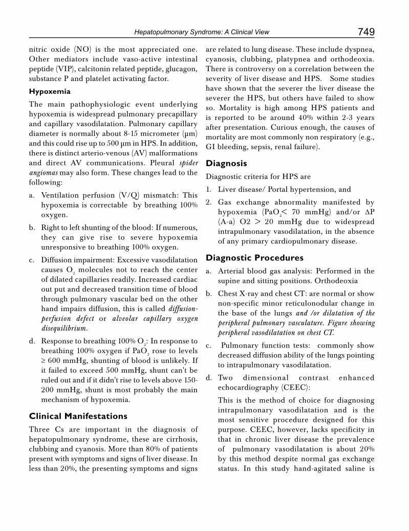
749Hepatopulmonary Syndrome: A Clinical View
nitric oxide (NO) is the most appreciated one. Other mediators include vaso-active intestinal peptide (VIP), calcitonin related peptide, glucagon, substance P and platelet activating factor.
Hypoxemia
The main pathophysiologic event underlying hypoxemia is widespread pulmonary precapillary and capillary vasodilatation. Pulmonary capillary diameter is normally about 8-15 micrometer (µm) and this could rise up to 500 µm in HPS. In addition, there is distinct arterio-venous (AV) malformations and direct AV communications. Pleural spider angiomas may also form. These changes lead to the following:
a. Ventilation perfusion (V/Q) mismatch: This hypoxemia is correctable by breathing 100% oxygen.
b. Right to left shunting of the blood: If numerous, they can give rise to severe hypoxemia unresponsive to breathing 100% oxygen.
c. Diffusion impairment: Excessive vasodilatation causes O
2 molecules not to reach the center
of dilated capillaries readily. Increased cardiac out put and decreased transition time of blood through pulmonary vascular bed on the other hand impairs diffusion, this is called diffusion-perfusion defect or alveolar capillary oxygen disequilibrium.
d. Response to breathing 100% O2: In response to
breathing 100% oxygen if PaO2 rose to levels
≥ 600 mmHg, shunting of blood is unlikely. If it failed to exceed 500 mmHg, shunt can't be ruled out and if it didn't rise to levels above 150-200 mmHg, shunt is most probably the main mechanism of hypoxemia.
Clinical ManifestationsThree Cs are important in the diagnosis ofhepatopulmonary syndrome, these are cirrhosis, clubbing and cyanosis. More than 80% of patients present with symptoms and signs of liver disease. In less than 20%, the presenting symptoms and signs
are related to lung disease. These include dyspnea, cyanosis, clubbing, platypnea and orthodeoxia. There is controversy on a correlation between the severity of liver disease and HPS. Some studies have shown that the severer the liver disease the severer the HPS, but others have failed to show so. Mortality is high among HPS patients and is reported to be around 40% within 2-3 years afterpresentation.Curiousenough, thecausesofmortality are most commonly non respiratory (e.g., GI bleeding, sepsis, renal failure).
DiagnosisDiagnostic criteria for HPS are
1. Liver disease/ Portal hypertension, and
2. Gas exchange abnormality manifested by hypoxemia (PaO
2< 70 mmHg) and/or ∆P
(A-a) O2 > 20 mmHg due to widespread intrapulmonary vasodilatation, in the absence of any primary cardiopulmonary disease.
Diagnostic Proceduresa. Arterial blood gas analysis: Performed in the
supine and sitting positions. Orthodeoxia
b. ChestX-rayandchestCT:arenormalorshownon-specific minor reticulonodular change in the base of the lungs and /or dilatation of the peripheral pulmonary vasculature. Figure showing peripheral vasodilatation on chest CT.
c. Pulmonary function tests: commonly show decreased diffusion ability of the lungs pointing to intrapulmonary vasodilatation.
d. Two dimensional contrast enhanced echocardiography(CEEC):
This is the method of choice for diagnosing intrapulmonary vasodilatation and is the most sensitive procedure designed for this purpose.CEEC,however, lacks specificity inthat in chronic liver disease the prevalence of pulmonary vasodilatation is about 20% by this method despite normal gas exchange status. In this study hand-agitated saline is

750 Medicine Update 2008 Vol. 18
injected into venous circulation, This contrast is seen filling the right atrium then the right ventricle. If this saline bubbles appear in left atrium and left ventricle after 5 heart beats, this test is interpreted as Positive Bubble Test. Contrast enhanced trans-esophagealechocardiography is more sensitive than trans-thoracic echocardiography, and correlates more with gas exchange abnormality.
vasodilatation is about 84% and its specificity is 100% (8,9). In addition, shunt fraction can be calculated by this procedure.
e. Macro aggregated albumin scanning
Technetium 99m- labeled macroaggregated albumin is used. The estimated sensitivity of this method for diagnosing intrapulmonary
f. Pulmonary angiography: Two different angiographic patterns in HPS:
Type I: more common. There are minimal changes with diffuse spider like branches to more advanced changes with a blotchy, spongy appearance ( the type that responds to breathing 100% oxygen).
Type II: less common. There are vascular lesions as vascular dilatations representing A-V communications ( the type that responds poorly to breathing oxygen and liver transplantation is not as suitable as for type I vascular lesions).
Technetium 99m macroaggregated albumin scanning reveals A) normal uptake of the radionuclide in the lungs. Uptake is alsodisplayed in theB)brainandC)kidneys, suggestingaright to left shunt.
Negative echocardiogram showing no air bubbles in the left heart chambers
Positive echocardiogram showing air bubbles (arrow) in the heart chambers

751Hepatopulmonary Syndrome: A Clinical View
g. Pulmonary artery catheterization: is not used commonly for diagnosing HPS.
TreatmentI. Medical therapy: There are currently no
medications proved to have persistent, adequate or acceptable effect on HPS. The following are tried:
a. Almitrin bimesylate: is a stimulator of arterial chemoreceptors(usedinCOPD).
b. Indomethacin : To cause inhibition of prostaglandin production which has a putative role of vasodilatation.
c. Methylene blue: Is a potent inhibitor of NO and its intracellular mediator, gunaylate cyclase and is potentially effective for treatment of HPS although transiently. It might be used in the post-operative period of liver transplantation in cases with transient hypoxemia, however its routine and long term use is not recommended yet.
II. Interventions other than liver transplantation
a. Embolotherapy: It is recommended that pulmonary angiography be done for HPS patients who respond poorly to breathing 100% oxygen i.e., PaO
2 < 150-200 mmHg.
If type II vascular lesions are diagnosed, embolotherapy with 22-coil spring devices must be tried.
b. Portal decompression with transjugular intrahepatic portosystemic shunt (TIPS): There is controversy regarding the beneficial effects of this technic on HPS. Some studies confirmed the improvement of hypoxemia
and others ruled out any usefulness of TIPS. More researches are needed undoubtfully.
III. Orthotopic Liver transplantation (OLT)
Previously, hypoxemia was considered as an absolute contraindication for OLT. Today the trend is to give a chance to this group of patients with the logic that HPS is a progressive and fatal disease and there isn’t an effective therapy which could improve oxygenation significantly. The rate of improvement of HPS patients with type I vascular lesions undergoing OLT is about 80% , but is much less in those with type II lesions.
References1. Flückiger M. Vorkommen von trommelschlegelförmigen
Fingerendphalangen ohne chronische Veränderungen an den Lungen oder am Herzen. Wien Med Wochenschr. 1884;34:1457–1458.
2. KennedyTC,KnudsonRJ.Exercise-aggravatedhypoxemiaand orthodeoxia in cirrhosis. Chest. 1977;72:305–309.
3. Sherlock S. The liver-lung interface. Semin Resp Med. 1988;9:253.
4. Furukawa T, Hara N, Yasumoto K, Inokuchi K. Arterial hypoxemia in patients with hepatic cirrhosis. Am J Med Sci. 1984;287:10–13.
5. Altman M, Robin ED. Platypnea (diffuse zone I phenomenon?). N Engl J Med. 1969;281:1347–1348.
6. Robin ED, Laman D, Horn BR, Theodore J. Platypnea related to orthodeoxia caused by true vascular lung shunts. N Engl J Med. 1976;294:941–943.
7. Arguedas MR, Fallon MB, Fallon MB. Hepatopulmonary syndrome. Curr Treat Options Gastroenterol. 2005;8:451–456.
8. Wolfe JD, Tashkin DP, Holly FE, Brachman MB, Genovesi MG. Detection of abnormal small pulmonary vascular channels by a quantitative radionuclide method. Am J Med. 1997;63:746–754. doi: 10.1016/0002-9343(77)90161-9.
9. Rodriguez-Roisin R, Krowka MJ, Herve P, Fallon MB. Pulmonary-Hepatic vascular Disorders (PHD). Eur Respir J. 2004;24:861–880. doi: 10.1183/09031936.04.00010904.

CHAPTER
100Post Exposure Prophylaxis in HIV R. Sajith Kumar
Management of Exposures to HIV and Post Exposure ProphylaxisAlthough preventing exposures to blood is the primary means of preventing occupationally acquired HIV infection, appropriate post exposure management is an important element of workplace safety.InJanuary1990,CDCissuedastatementonthe management of HIV exposures that included considerations for zidovudine (ZDV) use for post exposure prophylaxis (PEP). At that time, data were insufficient to assess the efficacy of ZDV. Although there are still only limited data to assess safety and efficacy, additional information is now available that is relevant to this issue and newer modifications have been released.
Who needs? In whom PEP indicated?Health-careworker(HCW)isdefinedasanyperson(e.g., an employee, student, attending clinician, public-safety worker, or volunteer) whose activities involve contact with patients or with blood or other body fluids from patients in a health-care or laboratory setting. An “exposure” that may place an HCW at risk for HIV infection andtherefore requires consideration of PEP is defined as a percutaneous injury (e.g., a needle stick or cut with a sharp object), contact of mucous membrane
or nonintact skin (e.g., when the exposed skin is abraded, or afflicted with dermatitis), or contact with intact skin when the duration of contact is prolonged (i.e, several minutes or more) or involves an extensive area, with blood, tissue, or other body fluids. Body fluids include a) semen, vaginal secretions, or other body fluids contaminated with visible blood and b) cerebrospinal, synovial, pleural, peritoneal, pericardial, and amniotic fluids. In the absence of visible blood in the saliva, exposure to saliva from a person infected with HIV is not considered risk for transmission; also, exposure to tears, sweat, or non-bloody urine or feces does not require postexposure follow-up. Human breast milk has been implicated in perinatal transmission of HIV. However, occupational exposure to human breast milk has not been implicated in HIV transmissiontoHCWs.
Is there a risk at all ? What is the risk?Prospective studies of HCWs have estimatedthat the average risk for HIV transmission after a percutaneous exposure to HIV-infected blood is approximately 0.3% and after a mucous membrane exposure is 0.09%. Although episodes of HIV transmission after skin exposure have been documented, the risk for transmission by this route has not been precisely quantified because

753Post Exposure Prophylaxis in HIV
no HCW enrolled in prospective studies haveseroconverted after an isolated skin exposure. The risk for transmission is less than the risk for mucous membrane exposures.
Epidemiologic and laboratory studies suggest that several factors affect the risk for HIV transmission. Theoneretrospectivecase-controlstudyofHCWswho had percutaneous exposure to HIV found that the risk for HIV transmission was increased with exposure to a larger quantity of blood from the source patient as indicated by
a. a device visibly contaminated with the patient’s blood,
b. a procedure that involved a needle placed directly in a vein or artery, or
c. a deep injury.
The risk also was increased for exposure to blood from source patients with terminal illness, possibly reflecting either the higher titer of HIV in blood late in the course of AIDS. The risk for HIV transmission from exposures that involve a larger volume of blood, particularly when the source patient’s viral load is probably high, exceeds the average risk of 0.3%.
AccordingtoCDC,ofthosehealthcarepersonnelfor whom case investigations were completed from 1981-2006, 57 had documented seroconversion to HIV following occupational exposures (see table for occupations). The routes of exposure resulting in infection were: 48 percutaneous (puncture/cut injury); five, mucocutaneous (mucous membrane and/or skin); two, both percutaneous and mucocutaneous; and two were of unknown route. Forty-nine healthcare personnel were exposed to HIV-infected blood; three to concentrated virus in a laboratory; one to visibly bloody fluid; and four to an unspecified fluid. Majority were nurses and lab. staff, Ub personnel followed by non surgical doctors and residents.
There have been reports in the literature on occupational hazards of HIV in developing countries. One study evaluated occupational
exposure to HIV in healthcare workers in South Africa. Thirteen per cent of the staff reported injuries with HIV positive patients. Registrars in training were the highest risk group (60%). Of the injuries, 94% were percutaneous and 65% occurred during emergency surgery. The commonest place of injury was the operating theater (46%) and the commonest procedure associated with accidental exposure was cesarean section (57%). Fifty-one per cent were not wearing eye protection during procedures and although 83% initiated post-exposure prophylaxis (PEP), 48% discontinued treatment due to side effects of the drugs. Occupational exposure to HIV is common in the developing world.
On the basis of, for example, a surgeon sustaining three percutaneous injuries over 12 months and not taking PEP after each, the annual risks ranged from 1 in 2,000,000 for urological/renal surgeons to 1 in 200,000 for those performing general surgery/ENT/gynecological procedures. The administration of PEP after each injury would reduce these rates to 1 in 10,000,000 and 1 in 1,000,000 respectively.
When should we watch? Time for seroconversion 81% experienced a syndrome compatible with primary HIV infection a median of 25 days after exposure. In a recent analysis, the median interval from exposure to seroconversion was 46 days (mean: 65 days); an estimated 95% seroconverted within 6 months after the exposure. These data suggest that the time course of HIV seroconversion inHCWs is similar to that inotherpersonswhohave acquired HIV through non occupational modes of transmission.
How does it work ? Information about primary HIV infection indicates that systemic infection does not occur immediately, leaving a brief “window of opportunity” during which post exposure anti retro viral intervention may modify viral replication. In a primate model of simian immunodeficiency virus (SIV) infection, infection of dendritic-like cells occurred at the site of inoculation

754 Medicine Update 2008 Vol. 18
during the first 24 hours following mucosal exposure to cell-free virus. During the subsequent 24-48 hours, migration of these cells to regional lymph nodes occurred, and virus was detectable in the peripheral blood within 5 days. HIV replication is rapid (generation time: 2.5 days) and results in bursts of up to 5,000 viral particles from each replicating cell.
Which all drugs ? Several antiretroviral agents are available for the treatment of HIV disease. These include the nucleoside analog reverse transcriptase inhibitors (NRTIs), nonnuceloside reverse transcriptase inhibitors (NNRTIs), and protease inhibitors (PIs). Among these drugs, ZDV is the only agent shown to prevent HIV transmission in humans. There are no data to directly support the addition of other antiretroviral drugs. However, in HIV-infected patients, combination regimens have proved superior to monotherapy regimens in reducing HIV viral load. Thus, theoretically a combination of drugs with activity at different stages in the viral replication cycle (e.g., NRTIs with a PI) could offer an additive preventive effect in PEP, particularly for occupational exposures that pose an increased risk for transmission. Guidelines for the treatment of early HIV infection recommend the use of three drugs (two NRTIs and a PI); however, the applicability of these recommendations to PEP remains unknown. In addition, the routine use of three drugs for all occupational HIV exposures may not be needed. Although the use of a highly potent regimen can be justified for exposures that pose an increased risk for transmission, it is uncertain whether the potential additional toxicity of a third drug is justified for lower-risk exposures. 3TC(Lamivudine) was recommended as a second agent for PEP based on greater antiretroviral activity of the ZDV/3TC combination and its activityagainst many ZDV-resistant HIV strains without substantially increased toxicity. The present options available include
1. Zidovudine (ZDV)-600 mg in divided doses (300 mg/twice a day for 4 weeks) + Lamivudine
(3TC)–150mgtwiceadayfor4weeks.
2. Zidovudine (as above) Plus Emtricitabine 200 mg capsule once each day.
3. Tenofovir (TDF) 300 mg once daily plus Lamivudine (3TC)300mgoncedailyor150mg twice daily.
The addition of a PI as a third drug is based on the site of activity in the replication cycle and demonstrated effectiveness in reducing viral burden. The NNRTIs have not been included in these recommended regimens for PEP. However, concerns about side effects and the availability of alternative agents argue against routinely using this class of drugs for initial PEP.
Many other combinations using other NRTIs, PIs and boosted PIs and even fusion inhibitors are being evaluated.
Are they safe ? An important goal of PEP is to encourage and facilitate compliance with a 4-week PEP regimen. Therefore, the toxicity profile including the frequency, severity, duration, and reversibility of side effects, is a relevant consideration. All anti retroviral agents have been associated with side effects. However, studies of adverse events have been reported primarily for persons with advanced disease (and longer treatment courses) and therefore may not reflect the experience of persons with less advanced disease or those who are uninfected. Side effects associated with many of the NRTIs are chiefly gastrointestinal and in general the incidence has not been greater when these agents are used in combination. Commonsymptoms included nausea, vomiting, malaise or fatigue, headache, or insomnia. Mild decrease in hemoglobin and absolute neutrophil count also were observed. All side effects were reversed when PEP was discontinued.
What to do in case ? Treatment of an Exposure Site: Wounds and skin sites that have been in contact with blood

755Post Exposure Prophylaxis in HIV
or body fluids should be washed with soap and water; mucous membranes should be flushed with water. There is no evidence that the use of antiseptics for wound care or expressing fluid by squeezing the wound further reduces the risk for HIV transmission. However, the use of antiseptics is not contraindicated. The application of caustic agents (e.g., bleach) or disinfectants into the wound is not recommended.
Assessment of Infection Risk: After an occupational exposure, the source-person and the exposedHCWshouldbeevaluatedtodeterminethe need for HIV PEP. Follow-up for hepatitis B andCvirusinfectionsalsoshouldbeconducted.
Evaluation of exposure: The exposure should be evaluated for potential to transmit HIV based on the type of body substance involved and the route and severity of the exposure. Exposures to blood, fluid containing visible blood, or other potentially infectious fluid (including semen; vaginal secretions; and cerebrospinal, synovial, pleural, peritoneal, pericardial, and amniotic fluids) or tissue through a percutaneous injury (i.e., needlestick or other sharps-related event) or through contact with a mucous membrane are situations that pose a risk for bloodborne transmission and require further evaluation
For skin exposures, follow-up is indicated if it involves direct contact with a body fluid listed above and there is evidence of compromised skin integrity (e.g., dermatitis, abrasion, or open wound). However, if the contact is prolonged or involves a large area of intact skin, postexposure follow-up may be considered on a case-by-case basis or if requestedbytheHCW.
Evaluation and testing of an exposure source: The person whose blood or body fluids are the source of exposure should be evaluated for HIV infection. Information available in the medical record at the time of exposure (e.g., laboratory test results, admitting diagnosis, or past medical history) or from the source person may suggest or rule out possible HIV infection. Examples of information
to consider when evaluating an exposure source for possible HIV infection include laboratory information (e.g., prior HIV testing results or resultsofimmunologictesting{e.g.,CD4+count}),clinical symptoms (e.g., acute syndrome of primary HIV infection or undiagnosed immunodeficiency disease), and history of possible HIV exposures (e.g., injecting-drug use, unprotected sexual contact with multiple partners (heterosexual and/or homosexual), or receipt of blood or blood products.
If the source is known to have HIV infection, available information about this person’s stage of infection(i.e.,asymptomaticorAIDS),CD4+T-cellcount, results of viral load testing, and current and previous antiretroviral therapy, should be gathered for consideration in choosing an appropriate PEP regimen. If this information is not immediately available, and if PEP is indicated then start treatment and change regimen when appropriate.
If the HIV serostatus of the source person is unknown, the source person should be informed of the incident and with consent, tested for serologic evidenceofHIVinfection.Confidentialityofthesource person should be maintained at all times. HIV-antibody testing of an exposure source should be performed as soon as possible. Hospitals, clinics, andothersitesthatmanageexposedHCWsshouldconsult their laboratories regarding the most appropriate test to expedite these results. Repeatedly reactive results by EIA (spot) or rapid HIV-antibody tests are considered highly suggestive of infection, whereas a negative result is an excellent indicator of theabsenceofHIVantibody.Confirmationofresultby Western blot or immunofluorescent antibody is not necessary for making initial decisions about postexposure management. If the source is HIV seronegative and has no evidence of acquired immunodeficiency syndrome (AIDS) or symptoms of HIV infection, no further testing of the source is indicated. The use of source-person viral load as a surrogate measure of viral titer for assessing transmission risk has not yet been established. Plasma viral load (e.g., HIV RNA) reflects only

756 Medicine Update 2008 Vol. 18
the level of cell-free virus in the peripheral blood; latently infected cells might transmit infection in the absence of viremia. Although a lower viral load (e.g., < 1,500 RNA copies/mL) or one that is below the limits of detection probably indicates a lower titer exposure, it does not rule out the possibility of transmission.
HIV testing of needles or other sharp instruments associated with an exposure is not recommended.
PEP must be done under expert centres/ personnel only in cases of delayed exposure reporting, unknown source, known or suspected pregnancy, breast feeding, resistance of the source virus to ARV drugs and in cases of hypersensitivity or drug reactions to first line drugs.
How to evaluate Exposed HCWs ? Exposed HCWs should be evaluated forsusceptibility to bloodborne pathogen infections. Baseline testing (i.e., testing to establish serostatus at the time of exposure) for HIV antibody should be performed. If the source person is seronegative for HIV, baseline testing or further follow-up of the HCWnormallyisnotnecessary.Serologictestingshouldbemade available to allHCWswhoareconcerned that they may have been exposed to HIV.
How to explain? Recommendations for chemoprophylaxis should be explained to HCWs who have sustainedoccupational HIV exposures. For exposures for which PEP is considered appropriate, HCWsshould be informed that a) knowledge about the efficacy and toxicity of drugs used for PEP are limited; b) only ZDV has been shown to prevent HIV transmission in humans; c) there are no data to address whether adding other antiretroviral drugs provides any additional benefit for PEP, but experts recommend combination drug regimens because of increased potency and concerns about drug-resistant virus; d) data regarding toxicity of antiretroviral drugs in persons without HIV infection are limited for ZDV and not known
regarding other antiretroviral drugs; and e) any or alldrugs forPEPmaybedeclinedby theHCW.HCWswhohaveoccupationalexposuresforwhichPEP is not recommended should be informed that the potential side effects and toxicity of taking PEP outweigh the negligible risk of transmission posed by the type of exposure.
How fast should we act ? PEP should be initiated as soon as possible. The interval within which PEP should be started for optimal efficacy is not known. Animal studies have demonstrated the importance of starting PEP within hours after an exposure. To assure timely access to PEP, an occupational exposure should be regarded as an urgent medical concern and PEP started as soon as possible after the exposure (i.e., within a few hours rather than days. The optimal duration of PEP is unknown. Because 4 weeks of ZDVappearedprotectiveinHCWs,PEPprobablyshould be administered for 4 weeks, if tolerated.
How to follow up ?HCWswithoccupationalexposuretoHIVshouldreceive follow-up counseling, postexposure testing, and medical evaluation regardless of whether they receive PEP. HIV-antibody testing should be performed for at least 6 months postexposure (e.g., at 6 weeks, 12 weeks, and 6 months). ExposedHCWsshouldbeadvisedtoseekmedicalevaluation for any acute illness that occurs during the follow-up period. Such an illness, particularly if characterized by fever, rash, myalgia, fatigue, malaise, or lymphadenopathy, may be indicative of acute HIV infection but also may be due to a drug reaction or another medical condition.
ExposedHCWswhochoosetotakePEPshouldbe advised of the importance of completing the prescribed regimen.
Is it cost effective?Assuming that 35% of exposures were to HIV-positive sources, the zidovudine regimen prevented 53 HIV seroconversions per 100,000 exposures,
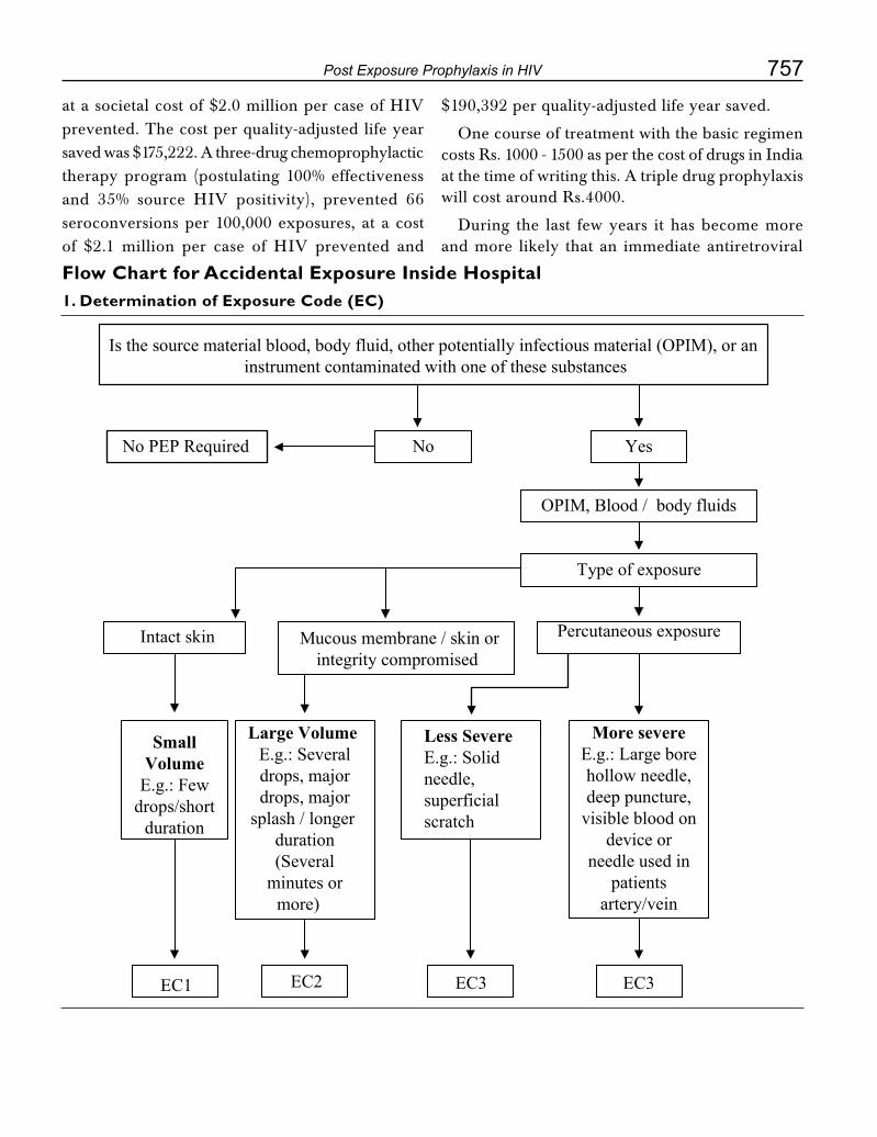
757Post Exposure Prophylaxis in HIV
at a societal cost of $2.0 million per case of HIV prevented. The cost per quality-adjusted life year saved was $175,222. A three-drug chemoprophylactic therapy program (postulating 100% effectiveness and 35% source HIV positivity), prevented 66 seroconversions per 100,000 exposures, at a cost of $2.1 million per case of HIV prevented and
$190,392 per quality-adjusted life year saved.
One course of treatment with the basic regimen costs Rs. 1000 - 1500 as per the cost of drugs in India at the time of writing this. A triple drug prophylaxis will cost around Rs.4000.
During the last few years it has become more and more likely that an immediate antiretroviral
Flow Chart for Accidental Exposure Inside Hospital1. Determination of Exposure Code (EC)
Is the source material blood, body fluid, other potentially infectious material (OPIM), or an instrument contaminated with one of these substances
No PEP Required No Yes
OPIM, Blood / body fluids
Intact skin
SmallVolume
E.g.: Fewdrops/short
duration
Large Volume E.g.: Severaldrops, majordrops, major
splash / longer duration(Several
minutes ormore)
EC2 EC3 EC3EC1
Less Severe E.g.: Solid needle,superficialscratch
More severeE.g.: Large borehollow needle,deep puncture,
visible blood ondevice or
needle used inpatients
artery/vein
Mucous membrane / skin orintegrity compromised
Type of exposure
Percutaneous exposure
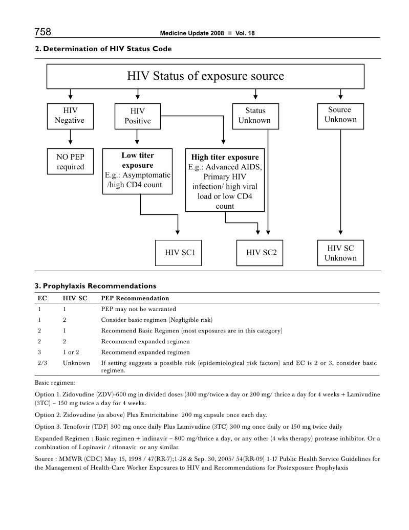
758 Medicine Update 2008 Vol. 18
2. Determination of HIV Status Code
3. Prophylaxis Recommendations
EC HIV SC PEP Recommendation
1 1 PEP may not be warranted
1 2 Considerbasicregimen(Negligiblerisk)
2 1 Recommend Basic Regimen (most exposures are in this category)
2 2 Recommend expanded regimen
3 1 or 2 Recommend expanded regimen
2/3 Unknown If setting suggests a possible risk (epidemiological risk factors) and EC is 2 or 3, consider basicregimen.
Basic regimen:
Option 1. Zidovudine (ZDV)-600 mg in divided doses (300 mg/twice a day or 200 mg/ thrice a day for 4 weeks + Lamivudine (3TC)–150mgtwiceadayfor4weeks.
Option 2. Zidovudine (as above) Plus Emtricitabine 200 mg capsule once each day.
Option3.Tenofovir(TDF)300mgoncedailyPlusLamivudine(3TC)300mgoncedailyor150mgtwicedaily
Expanded Regimen : Basic regimen + indinavir – 800 mg/thrice a day, or any other (4 wks therapy) protease inhibitor. Or a combination of Lopinavir / ritonavir or any similar.
Source:MMWR(CDC)May15,1998/47(RR-7);1-28&Sep.30,2005/54(RR-09)1-17PublicHealthServiceGuidelinesfortheManagementofHealth-CareWorkerExposurestoHIVandRecommendationsforPostexposureProphylaxis
HIV Status of exposure source
StatusUnknown
High titer exposureE.g.: Advanced AIDS,
Primary HIVinfection/ high viral
load or low CD4count
Low titerexposure
E.g.: Asymptomatic/high CD4 count
NO PEPrequired
HIVNegative
HIVPositive
Source Unknown
HIV SCUnknown
HIV SC2HIV SC1
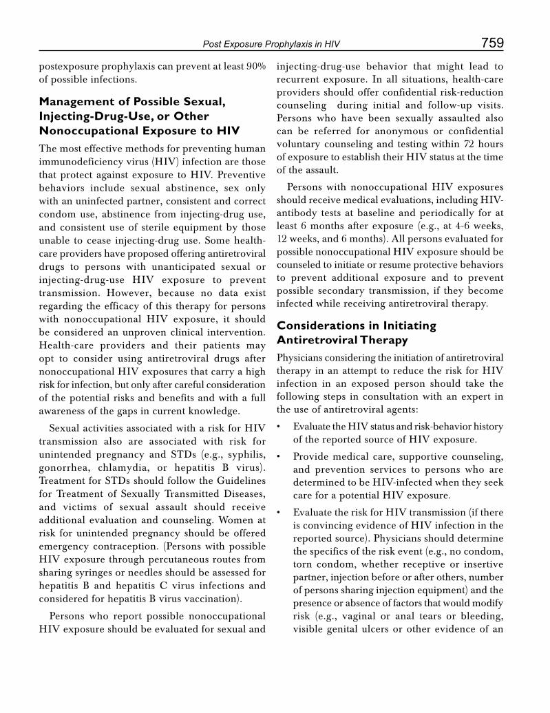
759Post Exposure Prophylaxis in HIV
postexposure prophylaxis can prevent at least 90% of possible infections.
Management of Possible Sexual, Injecting-Drug-Use, or Other Nonoccupational Exposure to HIVThe most effective methods for preventing human immunodeficiency virus (HIV) infection are those that protect against exposure to HIV. Preventive behaviors include sexual abstinence, sex only with an uninfected partner, consistent and correct condom use, abstinence from injecting-drug use, and consistent use of sterile equipment by those unable to cease injecting-drug use. Some health-care providers have proposed offering antiretroviral drugs to persons with unanticipated sexual or injecting-drug-use HIV exposure to prevent transmission. However, because no data exist regarding the efficacy of this therapy for persons with nonoccupational HIV exposure, it should be considered an unproven clinical intervention. Health-care providers and their patients may opt to consider using antiretroviral drugs after nonoccupational HIV exposures that carry a high risk for infection, but only after careful consideration of the potential risks and benefits and with a full awareness of the gaps in current knowledge.
Sexual activities associated with a risk for HIV transmission also are associated with risk for unintended pregnancy and STDs (e.g., syphilis, gonorrhea, chlamydia, or hepatitis B virus). Treatment for STDs should follow the Guidelines for Treatment of Sexually Transmitted Diseases, and victims of sexual assault should receive additional evaluation and counseling. Women at risk for unintended pregnancy should be offered emergency contraception. (Persons with possible HIV exposure through percutaneous routes from sharing syringes or needles should be assessed for hepatitis B and hepatitis C virus infections andconsidered for hepatitis B virus vaccination).
Persons who report possible nonoccupational HIV exposure should be evaluated for sexual and
injecting-drug-use behavior that might lead to recurrent exposure. In all situations, health-care providers should offer confidential risk-reduction counseling during initial and follow-up visits. Persons who have been sexually assaulted also can be referred for anonymous or confidential voluntary counseling and testing within 72 hours of exposure to establish their HIV status at the time of the assault.
Persons with nonoccupational HIV exposures should receive medical evaluations, including HIV-antibody tests at baseline and periodically for at least 6 months after exposure (e.g., at 4-6 weeks, 12 weeks, and 6 months). All persons evaluated for possible nonoccupational HIV exposure should be counseled to initiate or resume protective behaviors to prevent additional exposure and to prevent possible secondary transmission, if they become infected while receiving antiretroviral therapy.
Considerations in Initiating Antiretroviral Therapy Physicians considering the initiation of antiretroviral therapy in an attempt to reduce the risk for HIV infection in an exposed person should take the following steps in consultation with an expert in the use of antiretroviral agents:
• EvaluatetheHIVstatusandrisk-behaviorhistoryof the reported source of HIV exposure.
• Providemedical care, supportivecounseling,and prevention services to persons who are determined to be HIV-infected when they seek care for a potential HIV exposure.
• EvaluatetheriskforHIVtransmission(ifthereis convincing evidence of HIV infection in the reported source). Physicians should determine the specifics of the risk event (e.g., no condom, torn condom, whether receptive or insertive partner, injection before or after others, number of persons sharing injection equipment) and the presence or absence of factors that would modify risk (e.g., vaginal or anal tears or bleeding, visible genital ulcers or other evidence of an
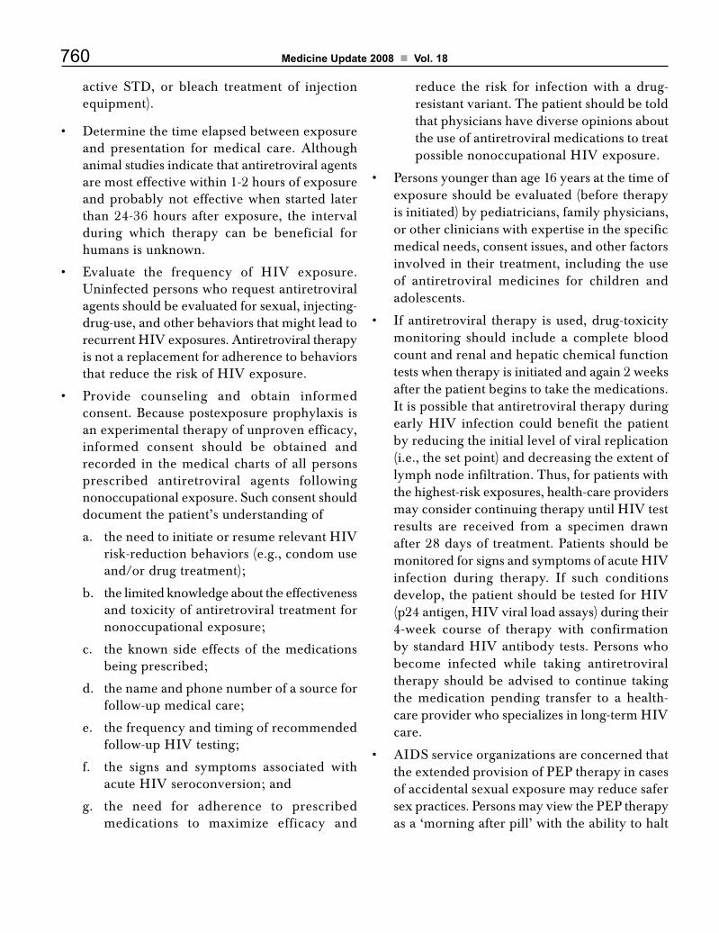
760 Medicine Update 2008 Vol. 18
active STD, or bleach treatment of injection equipment).
• Determinethetimeelapsedbetweenexposureand presentation for medical care. Although animal studies indicate that antiretroviral agents are most effective within 1-2 hours of exposure and probably not effective when started later than 24-36 hours after exposure, the interval during which therapy can be beneficial for humans is unknown.
• Evaluate the frequency of HIV exposure.Uninfected persons who request antiretroviral agents should be evaluated for sexual, injecting-drug-use, and other behaviors that might lead to recurrent HIV exposures. Antiretroviral therapy is not a replacement for adherence to behaviors that reduce the risk of HIV exposure.
• Provide counseling and obtain informedconsent. Because postexposure prophylaxis is an experimental therapy of unproven efficacy, informed consent should be obtained and recorded in the medical charts of all persons prescribed antiretroviral agents following nonoccupational exposure. Such consent should document the patient’s understanding of
a. the need to initiate or resume relevant HIV risk-reduction behaviors (e.g., condom use and/or drug treatment);
b. the limited knowledge about the effectiveness and toxicity of antiretroviral treatment for nonoccupational exposure;
c. the known side effects of the medications being prescribed;
d. the name and phone number of a source for follow-up medical care;
e. the frequency and timing of recommended follow-up HIV testing;
f. the signs and symptoms associated with acute HIV seroconversion; and
g. the need for adherence to prescribed medications to maximize efficacy and
reduce the risk for infection with a drug-resistant variant. The patient should be told that physicians have diverse opinions about the use of antiretroviral medications to treat possible nonoccupational HIV exposure.
• Personsyoungerthanage16yearsatthetimeofexposure should be evaluated (before therapy is initiated) by pediatricians, family physicians, or other clinicians with expertise in the specific medical needs, consent issues, and other factors involved in their treatment, including the use of antiretroviral medicines for children and adolescents.
• Ifantiretroviral therapy isused,drug-toxicitymonitoring should include a complete blood count and renal and hepatic chemical function tests when therapy is initiated and again 2 weeks after the patient begins to take the medications. It is possible that antiretroviral therapy during early HIV infection could benefit the patient by reducing the initial level of viral replication (i.e., the set point) and decreasing the extent of lymph node infiltration. Thus, for patients with the highest-risk exposures, health-care providers may consider continuing therapy until HIV test results are received from a specimen drawn after 28 days of treatment. Patients should be monitored for signs and symptoms of acute HIV infection during therapy. If such conditions develop, the patient should be tested for HIV (p24 antigen, HIV viral load assays) during their 4-week course of therapy with confirmation by standard HIV antibody tests. Persons who become infected while taking antiretroviral therapy should be advised to continue taking the medication pending transfer to a health-care provider who specializes in long-term HIV care.
• AIDSserviceorganizationsareconcernedthatthe extended provision of PEP therapy in cases of accidental sexual exposure may reduce safer sex practices. Persons may view the PEP therapy as a ‘morning after pill’ with the ability to halt
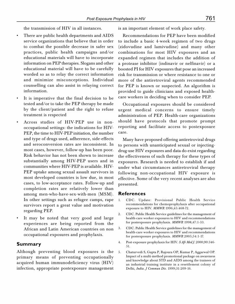
761Post Exposure Prophylaxis in HIV
the transmission of HIV in all instances.
• TherearepublichealthdepartmentsandAIDSservice organizations that believe that in order to combat the possible decrease in safer sex practices, public health campaigns and/or educational materials will have to incorporate information on PEP therapies. Slogans and other educational material will have to be carefully worded so as to relay the correct information and minimize misconceptions. Individual counselling can also assist in relaying correct information.
• It is imperative that the final decision to betested and/or to take the PEP therapy be made by the client/patient and the right to refuse treatment is respected
• Across studies of HIV-PEP use in non-occupational settings: the indications for HIV-PEP, the time to HIV-PEP initiation, the number and type of drugs used, adherence, side-effects and seroconversion rates are inconsistent. In most cases, however, follow-up has been poor. Risk behavior has not been shown to increase substantially among HIV-PEP users and in communities where HIV-PEP is available. HIV-PEP uptake among sexual assault survivors in most developed countries is low due, in most cases, to low-acceptance rates. Follow-up and completion rates are relatively lower than among men-who-have-sex-with-men (MSM). In other settings such as refugee camps, rape survivors report a great value and motivation regarding PEP.
• It may be noted that very good and largeexperiences are being reported from the African and Latin American countries on non occupational exposures and prophylaxis.
SummaryAlthough preventing blood exposures is the primary means of preventing occupationally acquired human immunodeficiency virus (HIV) infection, appropriate postexposure management
is an important element of work place safety.
Recommendations for PEP have been modified to include a basic 4-week regimen of two drugs (zidovudine and lamivudine) and many other combinations for most HIV exposures and an expanded regimen that includes the addition of a protease inhibitor (indinavir or nelfinavir) or a boosted PI for HIV exposures that pose an increased risk for transmission or where resistance to one or more of the antiretroviral agents recommended for PEP is known or suspected. An algorithm is provided to guide clinicians and exposed health-care workers in deciding when to consider PEP.
Occupational exposures should be considered urgent medical concerns to ensure timely administration of PEP. Health-care organizations should have protocols that promote prompt reporting and facilitate access to postexposure care.
Many have proposed offering antiretroviral drugs to persons with unanticipated sexual or injecting-drug-use HIV exposures and data do exist regarding the effectiveness of such therapy for these types of exposures. Research is needed to establish if and under what circumstances antiretroviral therapy following non-occupational HIV exposure is effective. Some of the very recent analyses are also presented.
References 1. CDC. Update: Provisional Public Health Service
recommendations for chemoprophylaxis after occupational exposure to HIV. MMWR 1996;45:468-72.
2. CDC.PublicHealthServiceguidelinesforthemanagementofhealth-care worker exposures to HIV and recommendations for postexposure prophylaxis. MMWR 1998;47:1-33.
3. CDC.PublicHealthServiceguidelinesforthemanagementofhealth-care worker exposures to HIV and recommendations for postexposure prophylaxis. MMWR 2005;54:1-17.
4. Post-exposure prophylaxis for HIV. S Afr Med J. 2000;90:346-51.
5. ChaturvediS,GuptaP,RajouraOP,KumarP,AggarwalOP.Impact of a multi-method promotional package on awareness and knowledge about STD and AIDS among the trainees of an industrial training institute in a resettlement colony of Delhi, India. J Commun Dis. 1999;31:209-16.

762 Medicine Update 2008 Vol. 18
6. MackieNE,CokerRJ.Post-exposureprophylaxisfollowingnon-occupational exposure to HIV: risks, uncertainties, and ethics. Int J STD AIDS. 2000 Jul;11:424-7.
7. GrimePR,RisiLJ,CarruthersJR.Asurveyoftheuseofpost-exposure prophylaxis for occupational exposure to human immunodeficiency virus. Occup Med (Lond). 2000;50:164-6.
8. DiproseP,DeakinCD,SmedleyJ.Ignoranceofpost-exposureprophylaxis guidelines following HIV needlestick injury may increase the risk of seroconversion. Br J Anaesth. 2000;84:767-70.
9. Gisondi MA. Post-exposure prophylaxis for HIV following possible sexual transmission: an ethical evaluation. Camb Q Healthc Ethics. 2000 Summer;9:411-7.
10. Donovan B. The repertoire of human efforts to avoid sexually transmissible diseases: past and present. Part 2: Strategies used during or after sex. Sex Transm Infect. 2000;76:88-93.
11. Gounden YP, Moodley J. Exposure to human immunodeficiency virus among healthcare workers in south africa. Int J Gynaecol Obstet. 2000;69:265-70.
12. DabisF,LeroyV,CastetbonK,SpiraR,NewellML,SalamonR. Preventing mother-to-child transmission of HIV-1 in Africa in the year 2000. AIDS. 2000 26;14:1017-26.
13. RichardVS,KennethJ,CherianT,ChandyGM.Preventingtransmission of blood-borne pathogens to health care workers. Natl Med J India. 2000;13:82-5.
14. Puro V. Post-exposure prophylaxis for HIV infection. Italian Registry of Post-Exposure Prophylaxis. Lancet. 2000 29;355:1556-7.
15. Parschalk B, Pernerstorfer-Schon H. Human immunodeficiency virus: post-exposure prophylaxis. Curr Opin Urol. 1999;9:51-5.
16. Pinkerton SD, Holtgrave DR, Kahn JG. Is post-exposure prophylaxis affordable? AIDS. 2000 18;14:325.
17. Parkin JM, Murphy M, Anderson J, El-Gadi S, Forster G, Pinching AJ. Tolerability and side-effects of post-exposure prophylaxis for HIV infection. Lancet. 2000 26;355:722-3.
18. GoldbergD,JohnstonJ,CameronS,FletcherC,StewartM,et.al. Risk of HIV transmission from patients to surgeons in the era of post-exposure prophylaxis. J Hosp Infect. 2000;44:99-105.
19. Henderson DK. Weighing the consequence of doing nothing versus those of doing something: post-exposure chemoprophylaxis for occupational exposures to HIV. J Hosp Infect. 1999;43 Suppl:S225-33.
20. Sidwell RU, Green JS, Novelli V.Management of occupational exposuretoHIV--whatactuallyhappens.CommunDisPublicHealth. 1999;2:287-90.
21. Papacchini M, Delle Piane R, Palmi S, Tomao P. Prevention, safety and prophylactic measures in occupational HIV infection in Italy, European Union and the USA. Med Lav.1999;90:681-92.
22. Gerberding JL, Katz MH. Post-exposure prophylaxis for HIV. Adv Exp Med Biol. 1999;458:213-22.
23. Marin MG, Van Lieu J, Yee A, Bonner E, Glied S.Cost-effectiveness of a post-exposure HIV chemoprophylaxis program for blood exposures in health care workers. J Occup Environ Med. 1999;41:754-60.
24. Sahlu T, Kassa E, et al. Sexual behaviours, perception of risk of HIV infection, and factors associated with attending HIV post-test counselling in Ethiopia. AIDS. 1999 9;13:1263-72.
25. Duff SE,WongCK,MayRE.Surgeons‘ andoccupationalhealth departments‘ awareness of guidelines on post-exposure prophylaxis for staff exposed to HIV: telephone survey. BMJ. 1999 17;319:162-3.
26. Petty TL.When the unthinkable happens: post-exposure prophylaxis of HIV-contaminated percutaneous injuries. J Can Dent Assoc. 1999;65:293-4.
27. Laurence J. Post-exposure prophylaxis: a new form of prevention? AIDS Patient Care STDS. 1999;13:261-3.
28. Seuffer RH. German-Austrian recommendations for post-exposure prophylaxis after HIV infection. Dtsch Med Wochenschr. 1999 26;124:373.
29. Torbati SS, Guss DA. Emergency department management of occupational exposures to HIV-infected fluids.J Emerg Med. 1999;17:261-4.
30. Sudre P, San Millan Ruiz D, etal.Post-exposure HIV prevention: eligibility of patients recently infected by sexual exposure. Schweiz Med Wochenschr. 1999 27;129:314-8.
31. Battegay M, Zimmerli W. HIV post-exposure prophylaxis after sexual risk contact. Schweiz Med Wochenschr. 1999 27;129:311-3.
32. GamesterCF,TilzeyAJ,BanatvalaJE.Medicalstudents‘riskof infection with bloodborne viruses at home and abroad: questionnaire survey. BMJ. 1999 16;318:158-60.
33. Katz MH, Gerberding JL. Postexposure treatment of people exposed to the human immunodeficiency virus through sexual contact or injection-drug use. N Engl J Med 1997;336:1097-9.
34. Kaplan EH, Heimer R. A model-based estimate of HIV infectivity via needle sharing. J Acquir Immune Defic Syndr 1992;5:1116-8.
35. SchackerT,CollierAC,HughesJ,SheaT,CoreyL.Clinicaland epidemiologic features of primary HIV infection. Ann Intern Med 1996;125:257-64.
36. Pinto LA, Landay AL, Berzofsky JA, Kessler HA, Shearer GM. Immune response to human immunodeficiency virus (HIV) in healthcare workers occupationally exposed to HIV-contaminated blood. Am J Med 1997;102:21-4.
37. Saag MS. Candidate antiretroviral agents for use inpostexposure prophylaxis. AmJMed 1997;102:25-31.
38. MartinLN,Murphey-CorbM,SoikeKF,Davison-FairburnB,Baskin GB. Effects of initiation of 3’-azido,3’-deoxythmidine (zidovudine) treatment at different times after infection of rhesus monkeys with simian immunodeficiency virus. J Infect Dis 1993;168:825-35.
39. TsaiC-C,FolisKE,SaboA,etal.PreventionofSIVinfectionin macaques by (R)-9-(2-phosphonylmethoxypropyl)adenine.

763Post Exposure Prophylaxis in HIV
Science 1995;270:1197-9.
40. CDC.Case-controlstudyofHIVseroconversioninhealth-care workers after percutaneous exposure to HIV-infected blood -- France, United Kingdom, and United States, January 1988-August 1994. MMWR 1995;44:929-33.
41. Jochimsen EM. Failures of zidovudine postexposure prophylaxis. Am J Med 1997;102:52-5.
42. Ippolito G, Puro V, and the Italian Registry of Antiretroviral Prophylaxis. Zidovudine toxicity in uninfected healthcare workers. Am J Med 1997;102:58-62.
43. Forseter G, Joline C, Wormser GP. Tolerability, safety,and acceptability of zidovudine prophylaxis in health care workers. Arch Intern Med 1994;154:2745-9.
44. Bell DM, Gerberding JL, eds. Human immunodeficiency virus (HIV) postexposure management of healthcare workers. Am J Med 1997;102(suppl 5B).
45. CDC.RecommendationsforpreventionofHIVtransmissionin health-care settings. MMWR 1987;36(suppl no. 2S).
46. CDC. Update: universal precautions for prevention oftransmission of human immunodeficiency virus, hepatitis B virus, and other bloodborne pathogens in health-care settings. MMWR1988;37:377-82,387-8.
47. Occupational Safety and Health Administration, Department of Labor. 29 CFR Part 1910.1030, occupational exposure
to bloodborne pathogens; final rule. Federal Register 1991;56:64004-182.
48. Bell DM. Occupational risk of human immunodeficiency virus infection in healthcare workers: an overview. Am J Med 1997;102(suppl 5B):9-15.
49. CardoDM,BellDM.Postexposuremanagement.In:DeVitaVT Jr, Hellman S, Rosenberg SA, eds. AIDS: biology, diagnosis, treatment and prevention. 4th ed. Philadelphia, PA: Lippincott-Raven Publishers, 1997:701-8.
50. CDC.HIV/AIDSsurveillancereport1997;9:15.
51. Ridzon R, Gallagher K, Ciesielski C, et al. Simultaneoustransmission of human immunodeficiency virus and hepatitis Cvirusfromaneedle-stickinjury.N Engl J Med 1997;336:919-22.
52. CDC.Immunizationofhealth-careworkers--recommendationsof the Advisory Committee on Immunization Practices(ACIP)andtheHospitalInfectionControlPracticesAdvisoryCommittee(HICPAC).MMWR 1997;46(no. RR-18):21-2.
53. CDC.Recommendationsforfollow-upofhealth-careworkersafter occupational exposure to hepatitis C virus. MMWR 1997;46:603-6.
54. Useful websites: http://www.cdc.gov/ncidod/dhqp/bp_hiv.html, http://www.cdc.gov/niosh, www.unaids.org,

CHAPTER
101Mixed DyslipidemiaV. Panikar
Mixed dyslipidemia is one in which there is an elevated LDL cholesterol and triglyceride levels combined with decreased levels of HDL cholesterol. This kind of mixed dyslipidemia is commonly seen in patients with diabetes and metabolic syndrome.
The gold standard for treating dyslipidemia in patientswithorathigh risk forCVDcentersonintensiveLDL-Cloweringwithstatins1. However, evenamongpatientswhoachieveLDL-Clevels<70mg/dL, theresidualrisk forsubsequentCHDevents remains high.
TheroleofelevatedTG,elevatednon–HDL-C,andlowHDL-ConresidualCHDrisk,guidelinesfor treating beyond LDL-C, and clinical trialevidence supporting the use of TG and HDL-Cmodifying therapies will be reviewed.
AccordingtotheNCEPATPIII2, elevated TGs are widely recognized as a marker for increased CVD risk. In this context, elevations in serumTG can be considered a marker for atherogenic remnant lipoproteins.
VLDL-Cisthemostreadilyavailablemeasureof atherogenic remnant lipoproteins for clinical practice.AnormalVLDL-Cistypically≤ 30 mg/dL (TG/5) when TGs are < 150 mg/dL. The goal for non–HDL-Cis30mgabovethegoalforLDL-C.Non–HDL-CiscalculatedbysubtractingHDL-Cfromtotalcholesterol (TC)oraddingtheLDL-CandVLDL-C(VLDL-C=TG/5).InthepresenceofhighserumlevelsofTG,non–HDL-Cwillbetterrepresent the concentrations of all atherogenic lipoproteinsthanwillLDL-Calone.
Thus, the guidelines (Table 1) recommend that non–HDL-C be a secondary target of therapywhen TG levels are ≥ 200mg/dL.Non–HDL-Cincorporates all of the cholesterol in LDL and VLDL and all of the apolipoprotein-B containing particles.
Table 1 : ADA/AHA 2007: Primary Prevention of CVD in Patients With Diabetes • Elevated LDL-C is a primary target of lipid-lowering
therapy
• LDL-Cgoal<100mg/dL
• TG-richlipoproteins,especiallyVLDL,areoftenelevatedin patients with diabetes, appear to be atherogenic, and represent a secondary target of lipid-lowering therapy
• TGgoal:<150mg/dL;HDL-Cgoal: males>40mg/dL females > 50 mg/dL
• IfTGare200–499mg/dL,non–HDL-Cgoal≤ 130 mg/dL
• If TG are ≥ 500 mg/dL, lowering TG is primary target
• CombinationtherapyofLDL-C–loweringdrugs(statins)with fibrates or niacin may be necessary to achieve lipid targets3
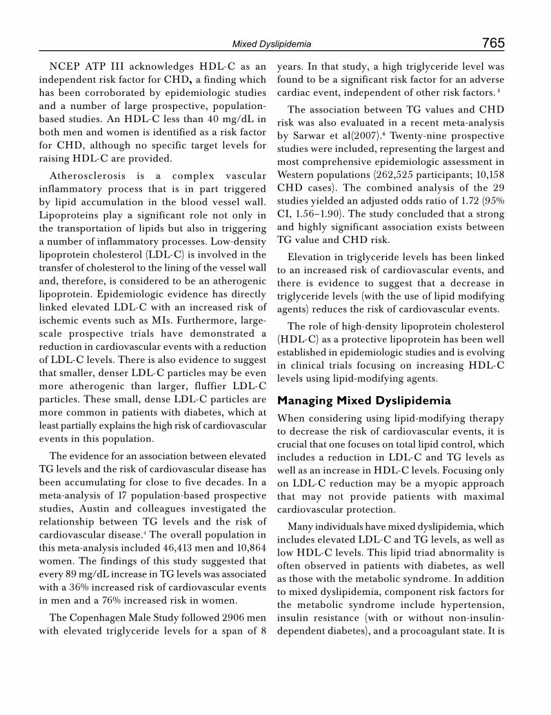
765Mixed Dyslipidemia
NCEP ATP III acknowledges HDL-C as anindependentriskfactorforCHD, a finding which has been corroborated by epidemiologic studies and a number of large prospective, population-basedstudies.AnHDL-Cless than40mg/dLinboth men and women is identified as a risk factor for CHD, although no specific target levels forraisingHDL-Careprovided.
Atherosclerosis is a complex vascular inflammatory process that is in part triggered by lipid accumulation in the blood vessel wall. Lipoproteins play a significant role not only in the transportation of lipids but also in triggering a number of inflammatory processes. Low-density lipoproteincholesterol(LDL-C)isinvolvedinthetransfer of cholesterol to the lining of the vessel wall and, therefore, is considered to be an atherogenic lipoprotein. Epidemiologic evidence has directly linkedelevatedLDL-Cwithan increased riskofischemic events such as MIs. Furthermore, large-scale prospective trials have demonstrated a reduction in cardiovascular events with a reduction ofLDL-Clevels.Thereisalsoevidencetosuggestthatsmaller,denserLDL-Cparticlesmaybeevenmore atherogenic than larger, fluffier LDL-Cparticles.Thesesmall,denseLDL-Cparticlesaremore common in patients with diabetes, which at least partially explains the high risk of cardiovascular events in this population.
The evidence for an association between elevated TG levels and the risk of cardiovascular disease has been accumulating for close to five decades. In a meta-analysis of 17 population-based prospective studies, Austin and colleagues investigated the relationship between TG levels and the risk of cardiovascular disease.4 The overall population in this meta-analysis included 46,413 men and 10,864 women. The findings of this study suggested that every 89 mg/dL increase in TG levels was associated with a 36% increased risk of cardiovascular events in men and a 76% increased risk in women.
TheCopenhagenMaleStudyfollowed2906menwith elevated triglyceride levels for a span of 8
years. In that study, a high triglyceride level was found to be a significant risk factor for an adverse cardiac event, independent of other risk factors. 5
TheassociationbetweenTGvaluesandCHDrisk was also evaluated in a recent meta-analysis by Sarwar et al(2007).6 Twenty-nine prospective studies were included, representing the largest and most comprehensive epidemiologic assessment in Western populations (262,525 participants; 10,158 CHD cases). The combined analysis of the 29studies yielded an adjusted odds ratio of 1.72 (95% CI,1.56–1.90).Thestudyconcludedthatastrongand highly significant association exists between TGvalueandCHDrisk.
Elevation in triglyceride levels has been linked to an increased risk of cardiovascular events, and there is evidence to suggest that a decrease in triglyceride levels (with the use of lipid modifying agents) reduces the risk of cardiovascular events.
The role of high-density lipoprotein cholesterol (HDL-C)asaprotectivelipoproteinhasbeenwellestablished in epidemiologic studies and is evolving in clinical trials focusing on increasing HDL-Clevels using lipid-modifying agents.
Managing Mixed DyslipidemiaWhen considering using lipid-modifying therapy to decrease the risk of cardiovascular events, it is crucial that one focuses on total lipid control, which includesa reduction inLDL-CandTG levelsaswellasanincreaseinHDL-Clevels.FocusingonlyonLDL-Creductionmaybeamyopicapproachthat may not provide patients with maximal cardiovascular protection.
Many individuals have mixed dyslipidemia, which includeselevatedLDL-CandTGlevels,aswellaslowHDL-Clevels.Thislipidtriadabnormalityisoften observed in patients with diabetes, as well as those with the metabolic syndrome. In addition to mixed dyslipidemia, component risk factors for the metabolic syndrome include hypertension, insulin resistance (with or without non-insulin-dependent diabetes), and a procoagulant state. It is

766 Medicine Update 2008 Vol. 18
well established that the presence of diabetes or the metabolic syndrome substantially increases the risk of cardiovascular events. Therefore, aggressive and global lipid management becomes of paramount importance in these patients.
Reduction of cardiovascular events has been consistently demonstrated with statins through a large number of prospective, randomized controlled trials in both primary and secondary prevention settings. These trials have demonstrated the ability of statins to reduce the risk of cardiovascular events by 25% to 38%.7-11 But these data suggest that even withaggressiveLDL-Clowering,abouttwo-thirdsof major coronary events are not prevented.
The staggering rate of remaining residual cardiovascular events in these statin trials calls for a more global approach to cardiovascular risk reduction.12 This includes more aggressive lifestyle modification (e.g., exercise, dietary improvement, smoking cessation, and moderation of alcohol consumption) and more intensified therapy to achieve optimal blood pressure as well as lipid
control. As evidence continues to demonstrate the significance of reduction of TG levels and increase inHDL-C levels, lipidmanagement should takea more global perspective, perhaps focusing on combination therapy to more effectively reduce LDL-CandTGlevelsaswellastoincreaseHDL-Clevels.
An update to the NCEP-ATP III guidelineshighlighted the consideration of combining a fibrateorniacinwithLDL-Cloweringdrugswhenahigh-riskpatienthashighTGlevelsorlowHDL-Clevels.13 One potential concern about combination therapy is the risk of myopathy, which may be minimized by combining a low-to-moderate dose of a statin with a fibrate or niacin. One fibrate in particular, fenofibrate, has been recognized as a potentially safer fibrate since it does not seem to interfere with the metabolism of statins.14-15
The patient population that would most likely be in need of combination therapy would be those with diabetes or the metabolic syndrome. There is a high rate of lipid triad abnormality (mixed dyslipidemia) in these groups of patients, making the need for combination of fibrate or niacin with a statin an eminent one.
The American Diabetes Association (ADA) guidelines (Table 2) recommend the combination of a statin and a fibrate for patients with diabetes whose lipid abnormality is not corrected by a statin alone; a secondary consideration is given to a niacin and statin combination due to potential glycemic changes that may occur with niacin.16 Regardless of the choice of combination therapy (e.g., statin + fibrate or statin + niacin), the most important factor to keep in mind is that all atherogenic lipoproteins should be addressed and, after correcting for LDL-Cvalues,onehastofocusonthereductionofnon-HDL-Clevels,potentiallywith theuseofcombination therapy, to reduce the global risk of cardiovascular events.
Although NCEP does not suggest a target towhichHDL-Cshouldberaisedinpatientsatrisk,itdoesrecommendthatpatientswithlowHDL-C
Table 2 : ADA Standards of Medical Care in Diabetes: Dyslipidemia Management17-18
First Priority Second Priority
LDL-Clowering Goal: < 100 mg/dL < 70 mg/dL is an option in patients withovertCVD.
• Lifestylechanges
• Statins
• Niacin,ezetimibe, bile acid sequestrants, or fenofibrate
HDL-Craising Goal: > 40 mg/dL > 50 mg/dL in women
• Lifestylechanges
• Niacinorfibrates
TG lowering Goal: < 150 mg/dL
• Lifestylechanges
• Glycemiccontrol
• Fibrates(fenofibrate, gemfibrozil)
• Niacin
• Statins(ifalsohave high LDL-C)
Combined hyperlipidemia
• Glycemiccontrol + high-dose statin
• Glycemiccontrol + statin + fibrate
• Glycemiccontrol + statin + niacin

767Mixed Dyslipidemia
be treated with lifestyle modification (weight loss, diet, smoking cessation, aerobic exercise) and drug therapy as indicated. The Expert Group on HDL CholesterolandtheEuropeanConsensusPanelonHDL-CrecommendthatHDLberaisedto≥ 40 mg/dLinpatientswithCADandthoseathighriskforCAD(metabolicsyndrome,diabetesmellitus,10 yr Framingham risk > 20%). The American DiabetesAssociationrecommendsHDL-Ctargetsof ≥ 40 mg/dL in diabetic men and ≥ 50 mg/dL in diabetic women.
Statins, fibrates, niacin and resins all have varing effects on the different lipid entities. Table 3 gives an overviewofmajorHDL-Craisingpharmacologicaloptions
Because all of the available drugs that increase HDL-C levels have significant effects on otherlipoproteins, it has not been possible to prove in clinical trials thebenefitof raisingHDL-C inisolation. However, because of the known correlation of low HDL-C levels with increased CHD risk,themagnitudeoftheHDL-Celevationachievablewith pharmacologic treatments has become a point of interest in many trials of lipid-altering therapy. Niacin and fibrates individually increase HDL-Cmorethanthestatinsandcombinationofniacin with fibrates gives the maximum increase in HDL-C.(Table4.)
Some important studies using combination therapies to manage mixed dyslipidemias
The HATS study (HDL-Atherosclerosis Treatment Study) (2001) showed that the addition of niacin to simvastatin therapy inpatientswithCADwithlowHDL-Cand“normal”LDL-Cresultedinslightregression of coronary atherosclerosis and a significant reduction (90%) in clinical coronary events over 3 years.22 Niacin/simvastatin combination therapy reducedCHDprogressionby90%andcardiovascularevents by 40% in patients with metabolic syndrome. These data suggest that patients with metabolic syndrome should be treated more aggressively, and simvastatin plus niacin combination therapy appears to be an effective therapy.23
SAFARI trial24 – (Effectiveness and tolerability of simvastatin plus fenofibrate for combined hyperlipidemia.) (2005) demonstrated that combination therapy with simvastatin plus
Table 3 : Overview of Major HDL-C–Raising Pharmacologic Options 19-20
Drug Class
ChangesinLipidParameter
Benefits Adverse Events/CommentsLDL-CHDL-C TG
Statins ↓↓↓ ↑ ↓ Reduce risk for total andCHDmortality
Well tolerated, no additional risk for non-CHDdeathseen in large prevention studies
Niacin ↓↓ ↑↑↑ ↓↓↓ Reduce risk for total andCHDmortality
Flushing, GI discomfort reduce compliance; hyperglycemia restricts use in pts with diabetes
Fibrates ↓ ↑↑↑ ↓↓↓ Reduce risk forCHDmortality
Raise some concerns over additional risk fornon-CHDmortality
Resins ↓↓ ↑ ↑ Reduce risk forCHD
Decrease absorption of many drugs; may be inconvenient to administer; unpalatable
Table 4 : HDL-C Response to Pharmacologic Intervention 21
Monotherapy HDL-C Elevation
Niacin 15%–35%
Fibrates 6%–18%
Statins 4%–10%
Combination Therapy
Niacin + fibrate 45%–48%
Niacin + statin 17%–30%
Fibrate + statin 14%–28%
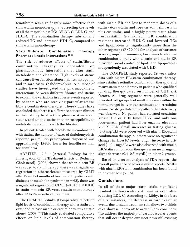
768 Medicine Update 2008 Vol. 18
fenofibrate was significantly more effective than simvastatin monotherapy at correcting the levels ofallthemajorlipids:TGs,VLDL-C,LDL-C,andHDL-C. The combination therapy substantiallyreducedTGandincreasedHDL-C,comparedtosimvastatin monotherapy.
Statin/Fibrate Combination Therapy Pharmacokinetic Interactions 25-26
The risk of adverse effects of statin/fibrate combination therapy is dependent on pharmacokinetic interactions that alter statin metabolism and clearance. High levels of statins can cause liver function abnormalities, myopathy, and in rare cases, rhabdomyolysis. A number of studies have investigated the pharmacokinetic interactions between different fibrates and statins to explain the variations in adverse events reported by patients who are receiving particular statin/fibrate combination therapies. These studies have concluded that there is a difference between fibrates in their ability to affect the pharmacokinetics of statins, and among statins in their susceptibility to metabolic interactions with the fibrates.
In patients treated with fenofibrate in combination with statins, the number of cases of rhabdomyolysis reported per million prescriptions dispensed was approximately 15-fold lower for fenofibrate than for gemfibrozil.27
ARBITER 1,2,3 28 (Arterial Biology for the Investigation of the Treatment Effects of Reducing Cholesterol)(2006)showedthatwhenniacinERwas added to statin therapy, there was a significant regression inatherosclerosismeasuredbyCIMTafter 12 and 24 months of treatment. In patients with diabetes or metabolic syndrome (n = 62), there was asignificantregressionofCIMT(–0.046,P<0.001)in statin + niacin ER versus statin monotherapy after 12 to 24 months of treatment.
TheCOMPELLstudy-(Comparativeeffectsonlipid levels of combination therapy with a statin and extended-release niacin or ezetimibe versus a statin alone) (2007).29 This study evaluated comparative effects on lipid levels of combination therapy
with niacin ER and low-to-moderate doses of a statin (atorvastatin and rosuvastatin), simvastatin plus ezetimibe, and a highly potent statin alone (rosuvastatin). Statin/niacin ER combination regimens increased HDL-C and lowered TGsand lipoprotein (a) significantly more than the other regimens (P < 0.001 for analysis of variance across groups). In summary, low-to-moderate dose combination therapy with a statin and niacin ER provided broad control of lipids and lipoproteins independentlyassociatedwithCHD.
TheCOMPELLstudyreported12-weeksafetydata with niacin ER/statin combination therapy, simvastatin/ezetimibe combination therapy, and rosuvastatin monotherapy in patients who qualified for drug therapy based on number of CHD riskfactors. All drug regimens were generally well tolerated. All groups had small increases (within the normal range) in liver transaminases and creatinine kinase. No drug-related myopathy or hepatotoxicity was observed. No patient had elevated creatinine kinase > 5 or > 10 times ULN, and only one rosuvastatin patient had liver enzyme elevation > 3 X ULN. Small increases in fasting glucose (3–5 mg/dL) were observed with niacin ER/statin combination therapy, but there were no significant changes inHbA1C levels.Slight increase inuricacid (~ 0.1 mg/dL) were also observed with niacin ER/statin combination therapy versus no change or slight decrease (0.4–0.5 mg/dL) in other 2 groups.
Based on a recent analysis of FDA reports, the overall prevalence of adverse event reports (AERs) with niacin ER/statin combination has been found to be quite low (≤ 1%).
ConclusionsIn all of these major statin trials, significant residual cardiovascular risk remains even after reducingLDL-C.AccordingtoLibby,inthebestof circumstances, the decrease in cardiovascular events due to statin treatment still allows two-thirds of cardiovascular events to occur. Libby concludes, “To address the majority of cardiovascular events that still occur despite our most powerful existing
769Mixed Dyslipidemia
therapies, we must combine lifestyle change and evaluate new pharmacologic strategies that will move us toward the goal of eradicating cardiovascular disease in the future.”30
Lipid abnormalities beyond LDL-C (non–HDL-C, TG, HDL-C) should be intensively treated to reduce residual CVD risk. Clinical trial data support the efficacy of lifestyle change, niacin, and fibrates for reducing CVD risk when used alone and in combination with statins. Clinical trial and surveillance data support the safety of Niacin ER and fenofibrate when used alone and in combination with statins.
References1. Heart Protection Study Collaborative Group. MRC/
BHF Heart Protection Study of cholesterol lowering with simvastatin in 20,536 high-risk individuals: a randomised placebo-controlled trial. Lancet. 2002;360:7-22.
2. ThirdReportoftheNationalCholesterolEducationProgram(NCEP)ExpertPanelonDetection,Evaluation,andTreatmentofHighBloodCholesterolinAdults(AdultTreatmentPanelIII) final report. Circulation. 2002;106:3143-3421.
3. BuseJBetal.DiabetesCare.2007;30:162-172.
4. Austin MA, Hokanson JE, Edwards KL. Hypertriglyceridemia as a cardiovascular risk factor. Am J Cardiol. 1998;81:7B-12B.
5. Jeppesen J, Hein HO, Suadicani P, Gyntelberg F. Triglyceride concentration and ischemic heart disease: an eight-year follow-up in the Copenhagen Male Study. Circulation.1998;97:1029-1036.
6. Sarwar N et al. Circulation. 2007;115:450-458
7. Prevention of cardiovascular events and death with pravastatin in patients with coronary heart disease and a broad range of initial cholesterol levels. The Long-Term Intervention with Pravastatin in Ischaemic Disease (LIPID) Study Group. N Engl J Med. 1998;339:1349-1357.
8. Randomised trial of cholesterol lowering in 4444 patients with coronary heart disease: the Scandinavian Simvastatin Survival Study (4S). Lancet. 1994;344:1383-1389.
9. DownsJR,ClearfieldM,WeisS,etal.Primarypreventionof acute coronary events with lovastatin in men and women with average cholesterol levels: results of AFCAPS/TexCAPS. Air Force/Texas Coronary Atherosclerosis Prevention Study. JAMA. 1998;279:1615-1622.
10. Sacks FM, Pfeffer MA, Moye LA, et al. The effect of pravastatin on coronary events after myocardial infarction in patients with average cholesterol levels. Cholesteroland Recurrent Events Trial investigators. N Engl J Med. 1996;335:1001-1009.
11. Shepherd J, Cobbe SM, Ford I, et al. Prevention ofcoronary heart disease with pravastatin in men with hypercholesterolemia.WestofScotlandCoronaryPreventionStudy Group. N Engl J Med. 1995;333:1301-1307.
12. Libby P. The forgotten majority: unfinished business in cardiovascular risk reduction. J Am Coll Cardiol. 2005;46:1225-1228.
13. GrundySM,Cleeman JI,MerzCN,et al. ImplicationsofrecentclinicaltrialsfortheNationalCholesterolEducationProgram Adult Treatment Panel III guidelines. Circulation. 2004;110:227-239.
14. Davidson MH. Combination therapy for dyslipidemia:safety and regulatory considerations. Am J Cardiol. 2002;90(suppl):50K-60K.
15. Bergman AJ, Murphy G, Burke J, et al. Simvastatin does not have a clinically significant pharmacokinetic interaction with fenofibrate in humans. J Clin Pharmacol. 2004;44:1054-1062.
16. Haffner SM. Dyslipidemia management in adults with diabetes. Diabetes Care. 2004;27(suppl 1):S68-S71.
17. American Diabetes Association. Dyslipidemia management in adults with diabetes. Diabetes Care. 2004; 27:S68-S71.
18. American Diabetes Association. Standards of medical care indiabetes—2007.Diabetes Care. 2007;30(suppl 1):S4-S41.
19. IslesCGetal.QJM. 2000;93:567-574.
20. Guyton JR. Expert Opin Pharmacother. 2004;5:1385-1398.
21. Hersberger M, von Eckardstein A. Low high-density lipoprotein cholesterol: physiological background, clinical importance and drug treatment. Drugs. 2003;63:1907-1945.
22. BrownBG,ZhaoXQ,ChaitA,etal.Simvastatinandniacin,antioxidant vitamins, or the combination for the prevention of coronary disease. N Engl J Med. 2001;345:1583-1592.
23. ZhaoX-Q,Morse J,ChaitA,etal.Simvastatinplusniacinprotect against atherosclerosis progression and clinical events in coronary artery disease patients with metabolic syndrome. J Am Coll Cardiol. 2002;39(suppl A):242A; abstract 1130-1173.
24. Grundy SM, Vega GL, Yuan Z, et al. Effectiveness and tolerability of simvastatin plus fenofibrate for combined hyperlipidemia (the SAFARI trial). Am J Cardiol. 2005;95:462-468.
25. Bergman AJ, Murphy G, Burke J, et al. Simvastatin does not have a clinically significant pharmacokinetic interaction with fenofibrate in humans. J Clin Pharmacol. 2004;44:1054-1062.
26. PrueksaritanontT,TangC,QiuY,etal.Effectsoffibrateson metabolism of statins in human hepatocytes. Drug Metab Dispos. 2002;30:1280-1287.
27. Taylor AJ, Lee HJ, Sullenberger LE. The effect of 24 months of combination statin and extended-release niacin on carotid intima-media thickness: ARBITER 3. Curr Med Res Opin. 2006;22:2243-2250.
28. Jones PH, Davidson MH. Reporting rate of rhabdomyolysis with fenofibrate + statin versus gemfibrozil + any statin. Am J Cardiol. 2005;95:120-122.
29. McKenney JM, Jones PH, Bays HE, et al. Comparativeeffects on lipid levels of combination therapy with a statin and extended-release niacin or ezetimibe versus a statin alone (theCOMPELLstudy).Atherosclerosis. 2007;192:432-437.
30. Libby PJ. The forgotten majority: unfinished business in cardiovascular risk reduction. J Am Coll Cardiol. 2005:46:1225-1228.

CHAPTER
102Hope and Hype of Adult Stem Cell PlasticityR. Kumar
IntroductionHuman embryonic stem cell (ESC) lines werefirst described in 1998.1 Their ability to reproduce limitlessly and differentiate into almost all cell types has generated a lot of scientific and media interest. Scientific research into human ESC has beenproliferating steadily in the last few years. This has led to premature and unjustified expectations that embryonic stem cells will cure a host of diseases affecting the heart, brain, endocrines, spinal cord. However, there are ethical issues involved in the useofESCandresearchonembryoniccellshasbeen banned in many countries.
Use of postnatal or adult stem cells, such as hematopoietic stem cells (HSCs), does not poseanyethicalissue.Moreover,HSCshavebeeninusein hematopoietic stem cell transplantation for more than 30 years in a variety of malignant and non-malignant disorders. More recently, there have been reports based on laboratory and animal studies, suggesting that adult stem cells can also exhibit “plasticity” or the ability of “transdifferentiation”, thereby allowing cells of one lineage to form cells belonging to another lineage. This has evoked excitement, as there would be no ethical obstacles in use of these cells.
Despite the enthusiasm based primarily on laboratory data or animal experiments, evidence
for adult stem cell plasticity is itself controversial. There are no clinical indications for use of adult stem cells, except in hematopoietic stem cell transplantation. Umbilical cord blood cells, which are committed adult stem cells in a newborn, have also been studied in the laboratory. A number of private companies are also propagating cryopreservation of cord blood at birth for regenerative purposes. This does not have the sanction of any professional body. In this article plasticity of adult stem cells will be reviewed with emphasis on the current clinical trials involving HSCs. Many other aspects ofstem cell research dealing with embryonic stem cells or tissue specific stem cells are not being addressed.
Stem cell developmentA Stem Cell is defined as an undifferentiatedcell capable of self-renewal and differentiation. Self-renewal is a process by which the stem cell produces daughter cells that are also stem cells. Differentiation implies commitment to a specific cell type via a differentiation pathway leading to production of mature progeny cells.2 Stem cells have the ability to divide for indefinite periods in culture and to give rise to specialized cells, which gradually lose the ability to proliferate indefinitely. Stem cells may be classified according to their development
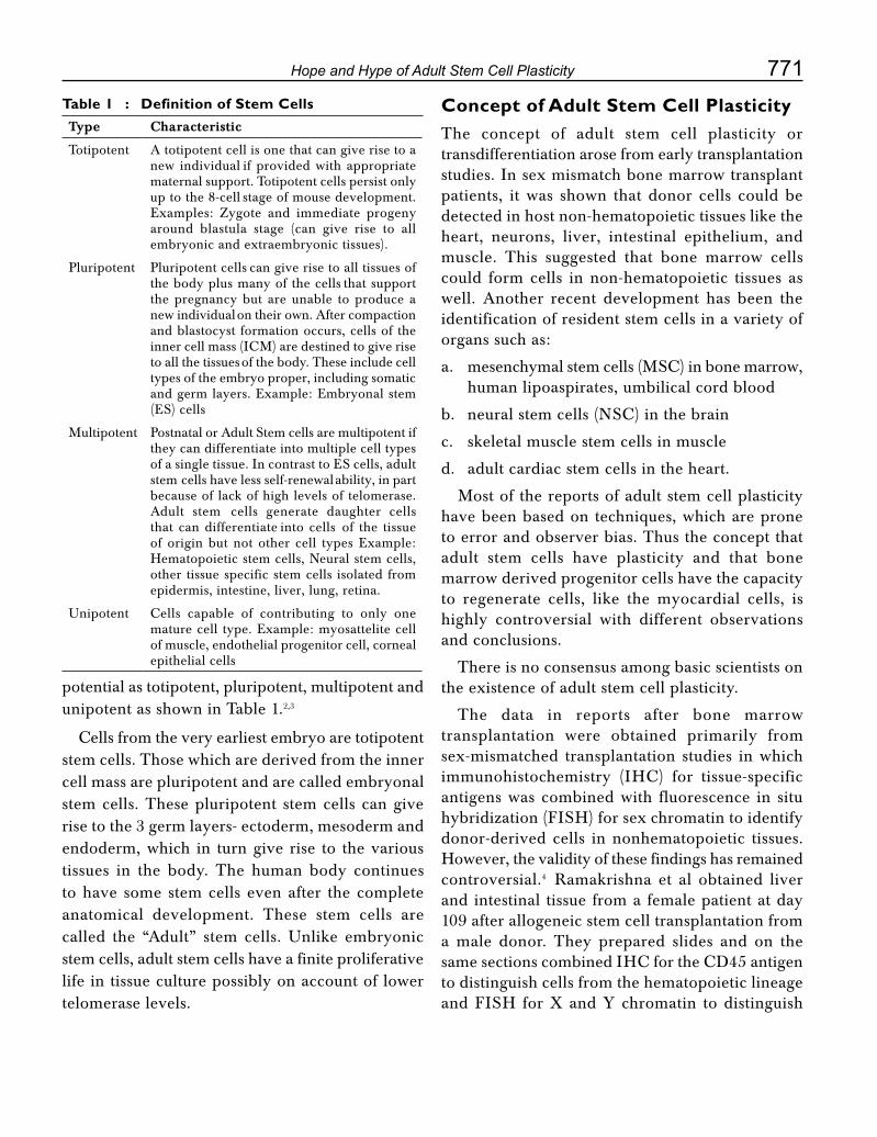
771Hope and Hype of Adult Stem Cell Plasticity
potential as totipotent, pluripotent, multipotent and unipotent as shown in Table 1.2,3
Cellsfromtheveryearliestembryoaretotipotentstem cells. Those which are derived from the inner cell mass are pluripotent and are called embryonal stem cells. These pluripotent stem cells can give rise to the 3 germ layers- ectoderm, mesoderm and endoderm, which in turn give rise to the various tissues in the body. The human body continues to have some stem cells even after the complete anatomical development. These stem cells are called the “Adult” stem cells. Unlike embryonic stem cells, adult stem cells have a finite proliferative life in tissue culture possibly on account of lower telomerase levels.
Concept of Adult Stem Cell PlasticityThe concept of adult stem cell plasticity or transdifferentiation arose from early transplantation studies. In sex mismatch bone marrow transplant patients, it was shown that donor cells could be detected in host non-hematopoietic tissues like the heart, neurons, liver, intestinal epithelium, and muscle. This suggested that bone marrow cells could form cells in non-hematopoietic tissues as well. Another recent development has been the identification of resident stem cells in a variety of organs such as:
a. mesenchymalstemcells(MSC)inbonemarrow,human lipoaspirates, umbilical cord blood
b. neuralstemcells(NSC)inthebrain
c. skeletal muscle stem cells in muscle
d. adult cardiac stem cells in the heart.
Most of the reports of adult stem cell plasticity have been based on techniques, which are prone to error and observer bias. Thus the concept that adult stem cells have plasticity and that bone marrow derived progenitor cells have the capacity to regenerate cells, like the myocardial cells, is highly controversial with different observations and conclusions.
There is no consensus among basic scientists on the existence of adult stem cell plasticity.
The data in reports after bone marrow transplantation were obtained primarily from sex-mismatched transplantation studies in which immunohistochemistry (IHC) for tissue-specificantigens was combined with fluorescence in situ hybridization (FISH) for sex chromatin to identify donor-derived cells in nonhematopoietic tissues. However, the validity of these findings has remained controversial.4 Ramakrishna et al obtained liver and intestinal tissue from a female patient at day 109 after allogeneic stem cell transplantation from a male donor. They prepared slides and on the samesectionscombinedIHCfortheCD45antigento distinguish cells from the hematopoietic lineage and FISH for X and Y chromatin to distinguish
Table 1 : Definition of Stem Cells Type Characteristic
Totipotent A totipotent cell is one that can give rise to a new individual if provided with appropriate maternal support. Totipotent cells persist only up to the 8-cell stage of mouse development. Examples: Zygote and immediate progeny around blastula stage (can give rise to all embryonic and extraembryonic tissues).
Pluripotent Pluripotent cells can give rise to all tissues of the body plus many of the cells that support the pregnancy but are unable to produce a new individual on their own. After compaction and blastocyst formation occurs, cells of the innercellmass(ICM)aredestinedtogiveriseto all the tissues of the body. These include cell types of the embryo proper, including somatic and germ layers. Example: Embryonal stem (ES) cells
Multipotent Postnatal or Adult Stem cells are multipotent if they can differentiate into multiple cell types of a single tissue. In contrast to ES cells, adult stem cells have less self-renewal ability, in part because of lack of high levels of telomerase. Adult stem cells generate daughter cells that can differentiate into cells of the tissue of origin but not other cell types Example: Hematopoietic stem cells, Neural stem cells, other tissue specific stem cells isolated from epidermis, intestine, liver, lung, retina.
Unipotent Cells capable of contributing to only onemature cell type. Example: myosattelite cell of muscle, endothelial progenitor cell, corneal epithelial cells
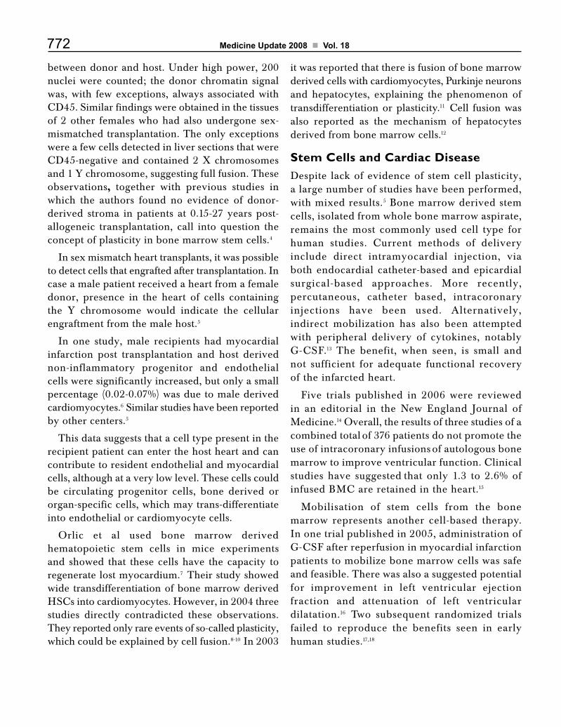
772 Medicine Update 2008 Vol. 18
between donor and host. Under high power, 200 nuclei were counted; the donor chromatin signal was, with few exceptions, always associated with CD45.Similarfindingswereobtainedinthetissuesof 2 other females who had also undergone sex-mismatched transplantation. The only exceptions were a few cells detected in liver sections that were CD45-negativeandcontained2Xchromosomesand 1 Y chromosome, suggesting full fusion. These observations, together with previous studies in which the authors found no evidence of donor-derived stroma in patients at 0.15-27 years post-allogeneic transplantation, call into question the concept of plasticity in bone marrow stem cells.4
In sex mismatch heart transplants, it was possible to detect cells that engrafted after transplantation. In case a male patient received a heart from a female donor, presence in the heart of cells containing the Y chromosome would indicate the cellular engraftment from the male host.5
In one study, male recipients had myocardial infarction post transplantation and host derived non-inflammatory progenitor and endothelial cells were significantly increased, but only a small percentage (0.02-0.07%) was due to male derived cardiomyocytes.6 Similar studies have been reported by other centers.5
This data suggests that a cell type present in the recipient patient can enter the host heart and can contribute to resident endothelial and myocardial cells, although at a very low level. These cells could be circulating progenitor cells, bone derived or organ-specific cells, which may trans-differentiate into endothelial or cardiomyocyte cells.
Orlic et al used bone marrow derived hematopoietic stem cells in mice experiments and showed that these cells have the capacity to regenerate lost myocardium.7 Their study showed wide transdifferentiation of bone marrow derived HSCsintocardiomyocytes.However,in2004threestudies directly contradicted these observations. They reported only rare events of so-called plasticity, which could be explained by cell fusion.8-10 In 2003
it was reported that there is fusion of bone marrow derived cells with cardiomyocytes, Purkinje neurons and hepatocytes, explaining the phenomenon of transdifferentiation or plasticity.11 Cell fusionwasalso reported as the mechanism of hepatocytes derived from bone marrow cells.12
Stem Cells and Cardiac DiseaseDespite lack of evidence of stem cell plasticity, a large number of studies have been performed, with mixed results.5 Bone marrow derived stem cells, isolated from whole bone marrow aspirate, remains the most commonly used cell type for human studies. Current methods of deliveryinclude direct intramyocardial injection, via both endocardial catheter-based and epicardial surgical-based approaches. More recently, percutaneous, catheter based, intracoronary injections have been used. Alternatively, indirect mobilization has also been attempted with peripheral delivery of cytokines, notably G-CSF.13 The benefit, when seen, is small and not sufficient for adequate functional recovery of the infarcted heart.
Five trials published in 2006 were reviewed in an editorial in the New England Journal of Medicine.14 Overall, the results of three studies of a combined total of 376 patients do not promote the use of intracoronary infusions of autologous bone marrowtoimproveventricularfunction.Clinicalstudies have suggested that only 1.3 to 2.6% of infusedBMCareretainedintheheart.15
Mobilisation of stem cells from the bone marrow represents another cell-based therapy. In one trial published in 2005, administration of G-CSFafterreperfusioninmyocardialinfarctionpatients to mobilize bone marrow cells was safe and feasible. There was also a suggested potential for improvement in left ventricular ejection fraction and attenuation of left ventricular dilatation.16 Two subsequent randomized trials failed to reproduce the benefits seen in early human studies.17,18
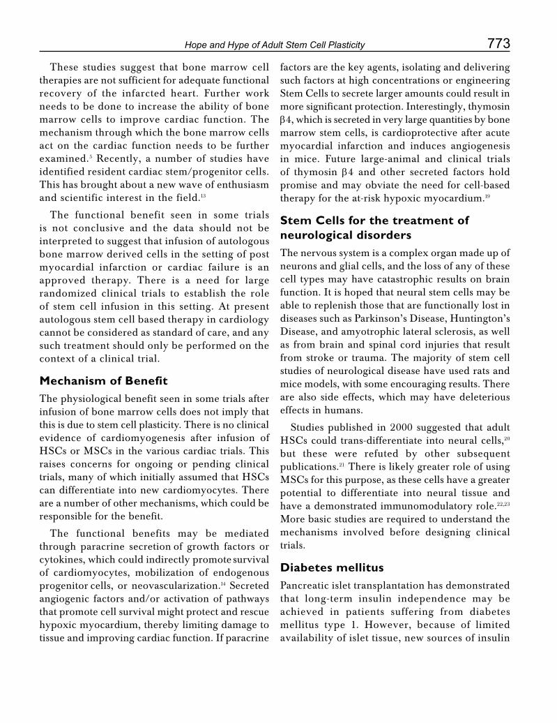
773Hope and Hype of Adult Stem Cell Plasticity
These studies suggest that bone marrow cell therapies are not sufficient for adequate functional recovery of the infarcted heart. Further work needs to be done to increase the ability of bone marrow cells to improve cardiac function. The mechanism through which the bone marrow cells act on the cardiac function needs to be further examined.5 Recently, a number of studies have identified resident cardiac stem/progenitor cells. This has brought about a new wave of enthusiasm and scientific interest in the field.13
The functional benefit seen in some trials is not conclusive and the data should not be interpreted to suggest that infusion of autologous bone marrow derived cells in the setting of post myocardial infarction or cardiac failure is an approved therapy. There is a need for large randomized clinical trials to establish the role of stem cell infusion in this setting. At present autologous stem cell based therapy in cardiology cannot be considered as standard of care, and any such treatment should only be performed on the context of a clinical trial.
Mechanism of BenefitThe physiological benefit seen in some trials after infusion of bone marrow cells does not imply that this is due to stem cell plasticity. There is no clinical evidence of cardiomyogenesis after infusion of HSCsorMSCsinthevariouscardiactrials.Thisraises concerns for ongoing or pending clinical trials,manyofwhichinitiallyassumedthatHSCscan differentiate into new cardiomyocytes. There are a number of other mechanisms, which could be responsible for the benefit.
The functional benefits may be mediated through paracrine secretion of growth factors or cytokines, which could indirectly promote survival of cardiomyocytes, mobilization of endogenous progenitor cells, or neovascularization.14 Secreted angiogenic factors and/or activation of pathways that promote cell survival might protect and rescue hypoxic myocardium, thereby limiting damage to tissue and improving cardiac function. If paracrine
factors are the key agents, isolating and delivering such factors at high concentrations or engineering StemCellstosecretelargeramountscouldresultinmore significant protection. Interestingly, thymosin β4, which is secreted in very large quantities by bone marrow stem cells, is cardioprotective after acute myocardial infarction and induces angiogenesis in mice. Future large-animal and clinical trials of thymosin β4 and other secreted factors hold promise and may obviate the need for cell-based therapy for the at-risk hypoxic myocardium.19
Stem Cells for the treatment of neurological disordersThe nervous system is a complex organ made up of neurons and glial cells, and the loss of any of these cell types may have catastrophic results on brain function. It is hoped that neural stem cells may be able to replenish those that are functionally lost in diseases such as Parkinson’s Disease, Huntington’s Disease, and amyotrophic lateral sclerosis, as well as from brain and spinal cord injuries that result from stroke or trauma. The majority of stem cell studies of neurological disease have used rats and mice models, with some encouraging results. There are also side effects, which may have deleterious effects in humans.
Studies published in 2000 suggested that adult HSCscould trans-differentiate intoneuralcells,20 but these were refuted by other subsequent publications.21 There is likely greater role of using MSCsforthispurpose,asthesecellshaveagreaterpotential to differentiate into neural tissue and have a demonstrated immunomodulatory role.22,23 More basic studies are required to understand the mechanisms involved before designing clinical trials.
Diabetes mellitusPancreatic islet transplantation has demonstrated that long-term insulin independence may be achieved in patients suffering from diabetes mellitus type 1. However, because of limited availability of islet tissue, new sources of insulin

774 Medicine Update 2008 Vol. 18
producing cells are required. Development of pancreatic beta-cell lines from rodent or human origin has progressed slowly in recent years. Current experiments for ex vivo expansion ofbeta cells and in vitro differentiation of embryonic and adult stem cells into insulin producing beta-cell phenotypes led to promising results. Nevertheless, the cells generated to date lack important characteristics of mature beta cells and generally display reduced insulin secretion and loss of proliferative capacity. Therefore, much better understanding of the mechanisms that regulate expansion and differentiation of stem/progenitor cells is necessary.24
Animal studies suggested that bone marrow derived stem cells could transdifferentiate into beta-cells and correct the diabetic phenotype.25 Subsequent studies failed to confirm these findings.26,27 There is no peer reviewed published clinical data to suggest a clinical benefit from cell based therapy in diabetes mellitus. In mice, there are conflicting data as to whether hematopoietic stem cells contribute to pancreatic beta cells. To establish whether hematopoietic stem cells (derived from adult donors) transdifferentiate into pancreatic beta cells in adult humans, an autopsy study was carried out in hematopoietic stem cell transplant recipients. A study was done in 31 human pancreata obtained at autopsy from hematopoietic stem cell transplant recipients who had received their transplant from a donor of the opposite sex. Whereas some donor-derived cells were observed in the nonendocrine pancreata, no pancreatic beta-cells were identified that were derived from donor hematopoietic stem cells, including two cases with type 2 diabetes. The authors concluded that hematopoietic stem cells derived from adult donors contribute minimally to pancreatic beta-cells in nondiabetic adult humans. This data does not rule out the possibility that hematopoietic stem cells contribute to pancreatic beta-cells in childhood or in individuals with type 1 diabetes.28
Liver DiseaseInitial publications on adult stem cell plasticity were greatly influenced by a publication by Lagasseetal,showingthatHSCscouldapparentlytransdifferentiate into hepatocytes in mice, with improvement of tyrosinemia.29 Subsequently it was shown that these findings were due to cell fusion between the donor monocytes/macrophages rather than transdifferentiation.12 A phase I study from Teheran involving infusion of autologous bone marrowHSCsthroughthehepaticarteryinpatientswith decompensated cirrhosis was prematurely interrupted due to side effects.30 There is no clinical data published in peer reviewed journals to suggest benefit of stem cell therapy in any hepatic disorder.
Autologous or Family Storage (Private or Commercial Cord Blood Banking)Recently, a number of private companies have started facilities for commercial cord blood banking for autologous use. They advertise “cure for many life threatening diseases”, speak of “unimaginable possibilities ……answers to curing diseases such as diabetes, breast cancer,……. rheumatoid arthritis, Parkinson’s disease, regeneration of damaged heart tissue.”31 Such advertising induces parents to make a “one in a lifetime investment for their child”. Recently a number of professional societies have issued guidelines or opinions on the subject. An opinion paper of the European Group on Ethics in Science and New Technologies and a Policy statement by the World Marrow Donors Association are freely available on the internet.31,32 Most Professional bodies are against such banks and false advertising.33-35
Private cord blood storage companies have developed in many countries to sell cord blood storage to families for potential future autologous (patient’s own cells) or family use. This is called “private storage” because the units are collected and stored solely to be available for the individual donor or the immediate family. These companies charge a collection fee, generally between $1000-
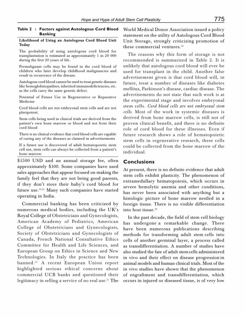
775Hope and Hype of Adult Stem Cell Plasticity
$1500 USD and an annual storage fee, often approximately $100. Some companies have used sales approaches that appear focused on making the family feel that they are not being good parents, if they don’t store their baby’s cord blood for future use.33,34 Many such companies have started operating in India.
Commercial banking has been criticized bynumerous medical bodies, including the UK’s RoyalCollegeofObstetricians and Gynecologists, American Academy of Pediatrics, American College of Obstetricians and Gynecologists,Society of Obstetricians and Gynecologists of Canada, French National Consultative EthicsCommittee for Health and Life Sciences, andEuropean Group on Ethics in Science and New Technologies. In Italy the practice has been banned.34 A recent European Union report highlighted serious ethical concerns about commercial UCB banks and questioned theirlegitimacy in selling a service of no real use.31 The
World Medical Donor Association issued a policy statementontheutilityofAutologousCordBloodUnit Storage, strongly criticizing promotion of these commercial ventures.32
The reasons why this form of storage is not recommended is summarized in Table 2. It is unlikely that autologous cord blood will ever be used for transplant in the child. Another false advertisement given is that cord blood will, in future, treat a number of diseases like diabetes mellitus, Parkinson’s disease, cardiac disease. The advertisements do not state that such work is at the experimental stage and involves embryonal stem cells. Cord blood cells are not embryonal stem cells. Most of the work in systemic diseases is derived from bone marrow cells, is still not of proven clinical benefit, and there is no definite role of cord blood for these illnesses. Even if future research shows a role of hematopoietic stem cells in regenerative research, these cells could be collected from the bone marrow of the individual.
ConclusionsAt present, there is no definite evidence that adult stem cells exhibit plasticity. The phenomenon of extramedullary hematopoiesis, which occurs in severe hemolytic anemia and other conditions, has never been associated with anything but a histologic picture of bone marrow nestled in a foreign tissue. There is no visible differentiation into host tissue.36
In the past decade, the field of stem cell biology has undergone a remarkable change. There have been numerous publications describing methods for transforming adult stem cells into cells of another germinal layer, a process called as transdifferentiation. A number of studies have also studied the fate of adult stem cells administered in vivo and their effect on disease progression in animal models and human clinical trials. Most of the in vivo studies have shown that the phenomenon of engraftment and transdifferentiation, which occurs in injured or diseased tissue, is of very low
Table 2 : Factors against Autologous Cord Blood Banking
Likelihood of Using an Autologous Cord Blood Unit Today
The probability of using autologous cord blood for transplantation is estimated as approximately 1 in 20 000 during the first 20 years of life.
Premalignant cells may be found in the cord blood of children who later develop childhood malignancies and result in recurrence of the disease.
Autologous cord blood cannot be used to treat genetic diseases like hemoglobinopathies, inherited immunodeficiencies, etc. as the cells carry the same genetic defect.
Potential of Future Use in Regenerative or Reparative Medicine
Cordbloodcellsarenotembryonalstemcellsandarenotpluripotent.
Stem cells being used in clinical trials are derived from the patient’s own bone marrow or blood and not from their cord blood
There is no clinical evidence that cord blood cells are capable of curing any of the diseases as claimed in advertisements
If a future use is discovered of adult hematopoietic stem cell use, stem cells can always be collected from a patient’s bone marrow.
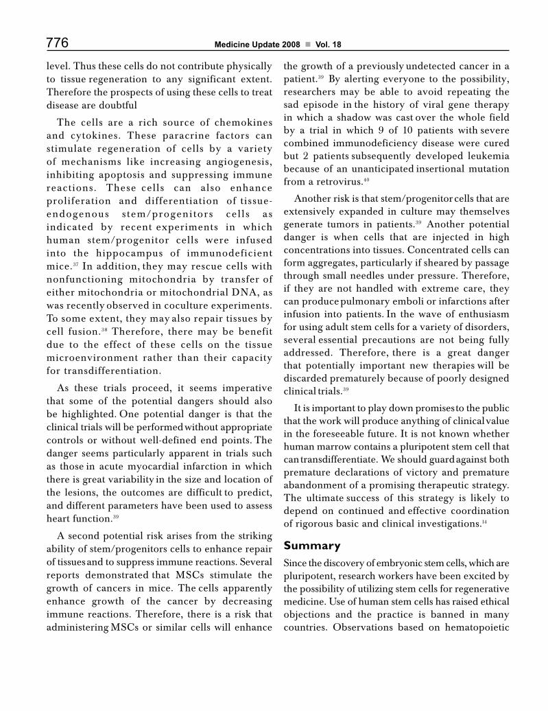
776 Medicine Update 2008 Vol. 18
level. Thus these cells do not contribute physically to tissue regeneration to any significant extent. Therefore the prospects of using these cells to treat disease are doubtful
The cells are a rich source of chemokines and cytokines. These paracrine factors can stimulate regeneration of cells by a variety of mechanisms like increasing angiogenesis, inhibiting apoptosis and suppressing immune react ions. These cel ls can also enhance proliferation and differentiation of t issue-endogenous s tem/progeni tors ce l l s a s indicated by recent experiments in which human stem/progenitor cells were infused
into the hippocampus of immunodeficient mice.37 In addition, they may rescue cells with nonfunctioning mitochondria by transfer of either mitochondria or mitochondrial DNA, as was recently observed in coculture experiments. To some extent, they may also repair tissues by cell fusion.38 Therefore, there may be benefit due to the effect of these cells on the tissue microenvironment rather than their capacity for transdifferentiation.
As these trials proceed, it seems imperative that some of the potential dangers should also be highlighted. One potential danger is that the clinical trials will be performed without appropriate controls or without well-defined end points. The danger seems particularly apparent in trials such as those in acute myocardial infarction in which there is great variability in the size and location of the lesions, the outcomes are difficult to predict, and different parameters have been used to assess
heart function.39
A second potential risk arises from the striking
ability of stem/progenitors cells to enhance repair of tissues and to suppress immune reactions. Several reports demonstrated that MSCs stimulate thegrowth of cancers in mice. The cells apparently enhance growth of the cancer by decreasing
immune reactions. Therefore, there is a risk that administering MSCsorsimilarcellswillenhance
the growth of a previously undetected cancer in a patient.39 By alerting everyone to the possibility,
researchers may be able to avoid repeating the sad episode in the history of viral gene therapy in which a shadow was cast over the whole field by a trial in which 9 of 10 patients with severe combined immunodeficiency disease were cured but 2 patients subsequently developed leukemia because of an unanticipated insertional mutation from a retrovirus.40
Another risk is that stem/progenitor cells that are extensively expanded in culture may themselves
generate tumors in patients.39 Another potential danger is when cells that are injected in high
concentrationsintotissues.Concentratedcellscanform aggregates, particularly if sheared by passage
through small needles under pressure. Therefore,
if they are not handled with extreme care, they can produce pulmonary emboli or infarctions after infusion into patients. In the wave of enthusiasm for using adult stem cells for a variety of disorders, several essential precautions are not being fully addressed. Therefore, there is a great danger that potentially important new therapies will be discarded prematurely because of poorly designed clinical trials.39
It is important to play down promises to the public that the work will produce anything of clinical value in the foreseeable future. It is not known whether human marrow contains a pluripotent stem cell that can transdifferentiate. We should guard against both premature declarations of victory and premature
abandonment of a promising therapeutic strategy. The ultimate success of this strategy is likely to depend on continued and effective coordination of rigorous basic and clinical investigations.14
SummarySince the discovery of embryonic stem cells, which are pluripotent, research workers have been excited by the possibility of utilizing stem cells for regenerative medicine. Use of human stem cells has raised ethical objections and the practice is banned in many countries. Observations based on hematopoietic
777Hope and Hype of Adult Stem Cell Plasticity
and cardiac transplantation suggested that adult stem cells also possess the property of ‘plasticity’ or ‘trans-differentiation’, thereby forming cells beyond the limitations of their cell line commitment. A number of laboratory experiments seemed to confirm this, raising hopes of using cells like the hematopoietic stem cells to generate tissues of any organ. Many of these observations and experiments utilized techniques, which were prone to error. Subsequent studies in the laboratory, animal experiments, and review of human tissue data failed to confirm the original observations. There is a major debate among basic scientists about the very existence of adult stem cell plasticity. Despite lack of definite data, a large number of clinical trials were started utilizing bone marrow cells for a number of diseases with special emphasis on cardiology disorders. The results have been mixed. Some studies have shown benefit, which is usually minor, whereas others have shown no difference. Any benefit does not imply that this is due to the property of ‘plasticity’. A number of paracrine effects of infusing or transplanting adult stem cells can contribute to tissue repair. It is important to continue basic research in stem cell biology and conduct well planned human trials to increase our understanding of this exciting field. At present use of stem cells is not standard of care except for hematopoietic stem cell transplantation. The hype of stem cell therapy appears premature but the hope remains that continued efforts in this field will result in clinical benefit.
References1. Thomson JA, Itskovitz-Eldor J, Shapiro SS, et al. Embryonic
stem cell lines derived from human blastocysts. Science 1998;282: 1145-7.
2. SerafiniM,VerfaillieCM.Pluripotencyinadultstemcells:state of the art. Semin Reprod Med 2006;24: 379-88.
3. VerfaillieCM,PeraMF,LansdorpPM.Stemcells:hypeandreality. Hematology Am Soc Hematol Educ Program 2002: 369-91.
4. Ramakrishnan A, Shi D, Torok-Storb B. Characterizingdonor-derived cells in nonhematopoietic tissue. Biol Blood Marrow Transplant 2006;12: 990-2.
5. ChristoforouN,GearhartJD.Stemcellsandtheirpotentialincell-basedcardiactherapies.ProgCardiovascDis2007;49:396-413.
6. Hocht-Zeisberg E, Kahnert H, Guan K, et al. Cellularrepopulation of myocardial infarction in patients with sex-mismatched heart transplantation. Eur Heart J 2004;25: 749-58.
7. OrlicD,Kajstura J,ChimentiS, et al.Bonemarrowcellsregenerate infarcted myocardium. Nature 2001;410: 701-5.
8. MurryCE,SoonpaaMH,ReineckeH,etal.Haematopoieticstem cells do not transdifferentiate into cardiac myocytes in myocardial infarcts. Nature 2004;428: 664-8.
9. BalsamLB,WagersAJ,ChristensenJL,KofidisT,WeissmanIL,RobbinsRC.Haematopoietic stemcellsadoptmaturehaematopoietic fates in ischaemic myocardium. Nature 2004;428: 668-73.
10. Nygren JM, Jovinge S, Breitbach M, et al. Bone marrow-derived hematopoietic cells generate cardiomyocytes at a low frequency through cell fusion, but not transdifferentiation. Nat Med 2004;10: 494-501.
11. Alvarez-Dolado M, Pardal R, Garcia-Verdugo JM, et al. Fusion of bone-marrow-derived cells with Purkinje neurons, cardiomyocytes and hepatocytes. Nature 2003;425: 968-73.
12. WangX,WillenbringH,AkkariY,etal.Cellfusionistheprincipal source of bone-marrow-derived hepatocytes. Nature 2003;422: 897-901.
13. Liao R, Pfister O, Jain M, Mouquet F. The bone marrow--cardiacaxisofmyocardial regeneration.ProgCardiovascDis 2007;50: 18-30.
14. Rosenzweig A. Cardiac cell therapy--mixed results frommixed cells. N Engl J Med 2006;355(12):1274-7.
15. HofmannM,WollertKC,MeyerGP,etal.Monitoringofbonemarrow cell homing into the infarcted human myocardium. Circulation2005;111:2198-202.
16. Ince H, Petzsch M, Kleine HD, et al. Prevention of left ventricular remodeling with granulocyte colony-stimulating factor after acute myocardial infarction: final 1-year results of the Front-Integrated Revascularization and Stem CellLiberation in Evolving Acute Myocardial Infarction by GranulocyteColony-StimulatingFactor(FIRSTLINE-AMI)Trial.Circulation2005;112(9Suppl):I73-80.
17. Ripa RS, Jorgensen E, Wang Y, et al. Stem cell mobilization induced by subcutaneous granulocyte-colony stimulating factor to improve cardiac regeneration after acute ST-elevation myocardial infarction: result of the double-blind, randomized, placebo-controlled stem cells in myocardial infarction (STEMMI) trial. Circulation 2006;113: 1983-92.
18. Zohlnhofer D, Ott I, Mehilli J, et al. Stem cell mobilization by granulocyte colony-stimulating factor in patients with acute myocardial infarction: a randomized controlled trial. Jama 2006;295: 1003-10.
19. Srivastava D, Ivey KN. Potential of stem-cell-based therapies for heart disease. Nature 2006;441: 1097-9.
20. Brazelton TR, Rossi FM, Keshet GI, Blau HM. From marrow to brain: expression of neuronal phenotypes in adult mice. Science 2000;290: 1775-9.

778 Medicine Update 2008 Vol. 18
21. CastroRF,JacksonKA,GoodellMA,RobertsonCS,LiuH,Shine HD. Failure of bone marrow cells to transdifferentiate into neural cells in vivo. Science 2002;297: 1299.
22. Wislet-Gendebien S, Hans G, Leprince P, Rigo JM, Moonen G, Rogister B. Plasticity of cultured mesenchymal stem cells: switch from nestin-positive to excitable neuron-like phenotype.StemCells2005;23:392-402.
23. ZappiaE,CasazzaS,PedemonteE,etal.Mesenchymalstemcells ameliorate experimental autoimmune encephalomyelitis inducing T-cell anergy. Blood 2005;106: 1755-61.
24. LimbertC,PathG,JakobF,SeufertJ.Beta-cellreplacementand regeneration: Strategies of cell-based therapy for type 1diabetesmellitus.DiabetesResClinPract2007.Sep11;[Epub ahead of print]
25. Ianus A, Holz GG, Theise ND, Hussain MA. In vivo derivation of glucose-competent pancreatic endocrine cells frombonemarrowwithoutevidenceof cell fusion. JClinInvest 2003;111: 843-50.
26. Mathews V, Hanson PT, Ford E, Fujita J, Polonsky KS, Graubert TA. Recruitment of bone marrow-derived endothelial cells to sites of pancreatic beta-cell injury. Diabetes 2004;53: 91-8.
27. Lechner A, Yang YG, Blacken RA, Wang L, Nolan AL, Habener JF. No evidence for significant transdifferentiation of bone marrow into pancreatic beta-cells in vivo. Diabetes 2004;53: 616-23.
28. Butler AE, Huang A, Rao PN, et al. Hematopoietic stem cells derived from adult donors are not a source of pancreatic beta-cells in adult nondiabetic humans. Diabetes 2007;56: 1810-6.
29. Lagasse E, Connors H, Al-Dhalimy M, et al. Purifiedhematopoietic stem cells can differentiate into hepatocytes in vivo. Nat Med 2000;6: 1229-34.
30. Mohamadnejad M, Namiri M, Bagheri M, et al. Phase 1 human trial of autologous bone marrow-hematopoietic stem cell transplantation in patients with decompensated cirrhosis. World J Gastroenterol 2007;13: 3359-63.
31. http://ec.europa.eu/european group ethics/docs/avis 19 en.pdf (accessed 26 Nov 2003)
32. www.worldmarrow.org/…/DRWG/CordBloodRegistries/WMDA Policy Statement Final 02062006.pdf (accessed 26 Nov 2006)
33. Edozien LC. NHS maternity units should not encouragecommercial banking of umbilical cord blood. BMJ 2006;333: 801-4.
34. FiskNM,RobertsIA,MarkwaldR,MironovV.Canroutinecommercial cord blood banking be scientifically and ethically justified? PLoS Med 2005;2: e44.
35. LubinBH,ShearerWT.Cordbloodbanking forpotentialfuture transplantation. Pediatrics 2007;119: 165-70.
36. Schwartz RS. The politics and promise of stem-cell research. N Engl J Med 2006;355: 1189-91.
37. Munoz JR, Stoutenger BR, Robinson AP, Spees JL, Prockop DJ. Human stem/progenitor cells from bone marrow promote neurogenesis of endogenous neural stem cells in the hippocampus of mice. Proc Natl Acad Sci U S A 2005;102: 18171-6.
38. OgleBM,CascalhoM,PlattJL.Biologicalimplicationsofcellfusion.NatRevMolCellBiol2005;6:567-75.
39. Prockop DJ, Olson SD. Clinical trials with adult stem/progenitor cells for tissue repair: let’s not overlook some essential precautions. Blood 2007;109: 3147-51.
40. Hacein-Bey-AbinaS,VonKalleC,SchmidtM,etal.LMO2-associated clonal T cell proliferation in two patients after genetherapyforSCID-X1.Science2003;302:415-9.
CHAPTER
103Improving Longevity and Adding Quality to Life – Caring for the ElderlyY. S. Raju
Individuals beyond the age 60 years are said to be in the Geriatric age. As life expectancy is increasing, the geriatric population is also on the rise. Life expectancy at the time of our independence was just 27 years. Due to the advances in the field of medicine and improvement in socio economic conditions, there is a steady rise in the expectation of life. In general women have higher life expectancy than men, a phenomenon observed globally.
Geriatrics Medicine is a multi disciplinary approach to elderly patients. In 1900 there were 1 crore 20 lacs elderly, in 1961 there were 2 crore 40 lacs, in 1991 there were 5 crore 60 lacs and in the 2001 census there were 7 crore elderly in India. It goes to show the magnitude of the care which has to be provided for the ageing population not necessarily patients above the age of 60 years.
There is a need for multi-pronged approach to improve the longevity and quality of life in the elderly.
1. Role of the Doctor
2. Role of nurses
3. Role of the elderly themselves
4. Role of society
5. Anti ageing measures
1. Role of doctors is vital when the elderly are suffering from diseases and also features
attributed to ageing. These two should be distinguished so as not to miss subtle signs of devastating diseases. For example acute myocardial infarction may be manifested as only off color or extreme weakness, dyspnea , unusual sweating and completely mislead the doctor unless the possibility of MI is borne in mind. Manifestations of the musculoskeletal system may be masked by the bedridden elderly.
There is always the problem of improper history because of confusion, fear, dementia, and lack of attention, usage of different words, deafness and language problems. There may be under-reporting because of indifference, spouse is also elderly, financial problems, neglect and loneliness.
The investigations should be appropriately advised and interpreted keeping in mind the variations that occur in the elderly.
Medical interventions are curative, preventive and promotive. A geriatrician supervises all the medical treatment. As per a survey it was observed that the following conditions occur more frequently in the elderly A) Visual impairmentB)locomotordisordersC)jointandmuscle problems D) cardiovascular diseases. E) neurological Disorders F) respiratory
780 Medicine Update 2008 Vol. 18
diseases. G) skin problems H) GI problems I) psychological problems J) hearing impairment and H) Genito urinary problems are more common in the elderly.
The availability of modern advances in treatmentlikeangioplasty,stents,CABG,earlysurgical and radiological interventions, dialysis, renal transplantation, ventilators, hearing aids, joint replacement etc., can all be utilized to decreased morbidity and mortality and thereby increase longevity and quality of life. Early interventionisthekey.ForexampleifaCABGor PTCA or placement of a Stents is offeredbefore myocardial damage occurs it prevents considerable morbidity and improves the quality of life than when myocardial damage occurs. A geriatrician supervises and coordinates all the medical management.
Preventive measures include immunization with influenza, tetanus toxoid and pneumococcal vaccines. Advice on HRT, oral calcium, bisphosphonates, multivitamins to all needy individuals prevents much of morbidity and improves quality of life.
Periodic screening like Blood pressure check up, blood glucose measurement, lipid profile, cancer screening and advice to see the physician at the earliest in case of any symptom all help to detect a condition early and treat appropriately before organ damage occurs.
Polypharmacy which is practiced in the elderly should be given keeping in mind the drug interaction and side-effects such as orthostatic hypotension and similar complications.
2. Role of nurses is also vital as they are the caregivers in the hospitals. A sympathetic approach is always rewarding. The important area to concentrate is the drug dispensing; any negligence could be devastating.
In those with severe arthritis, hemiplegia, parkinsonism and individuals with lower limb amputations etc., rehabilitative measures
improve their quality of life and make them more independent. Use of walking stick, wheel chair and various physiotherapy measures are all rehabilitative measures which improve quality of life of the elderly.
The role of sisters goes beyond only medical care and they now play a role in end of life situations where do not resuscitate instructions are slowly moving towards respecting their last wishes and helping them to accept natural deaths. It is the rare distinction shared by doctors and nurses that they are playing a vital role towards the end of life situations.
3. Role of elderly is important as small modifications in their life style can help them lead a longer disease free, useful life adding betterquality.Certaindo’sanddon’tsinrespectof elderly persons, if properly implemented by them, will go a long way in improving longevity and quality of life. The elderly to be informed about the various aspects which influence his health, quality of life--about the type of food, exercise and personal hygiene.
Stress on avoidance of tobacco in any form, restriction/avoidance of alcohol is important. More emphasis to be given to the importance of “use it or you will loose it”.
Advice on adequate intake of calories, proteins, correct intake of fats with suitable proportion of MUFA, PUFA and Sat. fats, advice on inclusion of antioxidants in diet (like dark colored vegetables, fruits, onion, turmeric, ginger etc).Stress on control of weight and reduction of obesity. Also, the advice on inclusion of green leafy vegetables and all seasonal fruits and fiber rich food items is very important.
Exercise in the form of walking daily for 30 minutes is the simplest of the exercises. In those with arthritis upper body exercises can be advocated.
Their role is taking in good nutritious food doing regular exercise within their limits and
781Improving Longevity and Adding Quality to Life – Caring for the Elderly
avoiding falls by improving and improvising small changes in their houses and work places. Elderly are vulnerable to fall. Osteoporosis contributes to their vulnerability to sustain fractures. Also the elderly because of arthritis, musculoskeletal problems and poor visibility are unable to use normal toilets, stair case etc. Certainmodificationsathomesandsurroundingsreduce the risk of falls and thereby fracture risk and also facilitate the elderly to be independent. Use of non slippery flooring particularly in bathrooms, western style toilets, handlebars for stair cases, adequate lighting, reachable light switches, properly laid carpets etc are some of the simple environmental modifications which make life of elderly smoother and happier.
Any slippery floor, in and around the house, to be suitably modified.
4. Role of Society The family system in India to this day respect elderly providing physical, nutritional, medical, social and psychological support to the individuals. Financial support also is vital for the security and medical expenditure to be incurred by the elderly.
The depression is the very major problem faced by the elderly due to helplessness and neglect. It is estimated that around 30% of the elderly are living in below poverty line conditions and many more can not afford basic medical treatment leave alone costly interventions. This results in mortality, morbidity and lack of qualitative life. It is observed that single elderly (widows), elderly living alone when their offspring live away or abroad, elderly spending lonely at home while their children and grand children go out early and return late, are some of the social problems being encountered. Social problems reflect on the physical and psychological health of the elderly thus contributing to psychological problems and decrease in quality of life and a state of helplessness. Strengthening of old family system, day care centers, old age homes, paying guest system, recreational facilities etc,
are the remedies suggested.
Interventions to overcome economic problems in the elderly include various income generating projects aimed at the elderly, financial support by govt and non govt agencies, adopt a granny scheme by Help Age India ( a voluntary organization wholly committed to the cause of the elderly), Health Insurance schemes, various concessions offered to the elderly etc.
By strengthening the economic status of the individual morbidity and mortality decrease and quality of life improves. This has to be addressed by the family members, friends and society.
5. Anti aging measures are still in the experimental stages. The desire for a pharmaceutical intervention to halt or delay the effects of aging has produced a thriving antiaging industry ready to address the demand. Some of the products provided by this industry may be useful, but many are not, and others may carry the risk of serious harm. and only proven measure is consumption of small quantities of good balanced nutritious foods.
For most patients who are experiencing or seeking to prevent the effects of aging, lifestyle changes will have more benefit than any ointment, pill, or dietary supplement. Furthermore, the psychosocial effect of these interventions on a person’s health cannot be overestimated. Patients who come to a physician’s clinic inquiring about available hormonal supplements and asking which ones will effectively restore energy, enthusiasm, muscle mass, ability to sleep, and so on, may bestbeadvised—assumingaseriousdiseaseorcondition can be ruled out—to consider anyof a number of lifestyle modifications. The underlying assumption is that a systemic, or holistic, view of health is likely to be the most effective approach to these types of problems. So, before diving into the world of hormones or
782 Medicine Update 2008 Vol. 18
micronutrients, patients might consider making more fundamental life changes.
Eligibility for testosterone therapy, while still controversial, will usually require the presence of primary hypogonadal signs, including anemia, diminished muscle mass, and low bone density, as well as a testosterone level below 300 ng/dL
Estrogen replacement therapy (ERT), both with and without progesterone, for the symptoms of menopause has been associated with increased risk of heart attack, stroke, blood clots, and breast cancer. In very low dosages, the benefits of ERT may outweigh the risks for some patients who seek relief from menopausal symptoms. However, this therapy is not recommended for osteoporosis, since a number of preventive agents are available without the same degree of risk as ERT.
Recombinant human growth hormone (hGH) has been shown in a number of studies to improve several of the physical symptoms associated with aging, such as muscle-mass loss, reduced energy level, and fatigue.
The other experimental measures are Melatonin, Dehydroepiandrosterone. Small studies of dehydroepiandrosterone (DHEA), a sex steroid that converts to estrogen and testosterone, has shown potential benefit in patients with depression and adrenal insufficiency and in increasing bone density and libido in postmenopausal women.
Caloric restriction (CR) has long been known to extend life and maintain health.
Stem cell research could be the future. Still in the early stages at present, stem cell research may be used to develop “replacement parts” such as organs that could work in conjunction with developments in CR medicine. For example, while CR-based medicines may allow people to live longer and healthier, certain organs and vital systems are
likely to wear out over time. These worn-out parts could then be grown via stem cell technology and transplanted into the patient. Another way CR and applications derived from stem cell research could work together would be to up regulate the antiaging pathway in stem cells genetically, and then implant the stem cells to create fitter, healthier organs or cells, which could then be reimplanted or reinserted into the patient.
These are some of multi disciplinary approaches which though seem insignificant can contribute tremendously to the short term and long term measures to improve the longevity and add quality to life and help in caring for our elderly.
References1. Brocklehurst’s Text book of geriatric Medicine and
Gerontology.
2. Evans WJ. Exercise training guidelines for the elderly. Med Sci Sports Exerc. 1999;31:12-17.
3. Christmas C, Andersen RA. Exercise and older patients:guidelines for clinicians. J Am Geriatr Soc. 2000;48:318-324.
4. American Geriatrics Society, British Geriatrics Society, and American Academy of Orthopaedic Surgeons Panel on Falls Prevention. Guideline for the prevention of falls in older persons. J Am Geriatr Soc. 2001;49:664-672.
5. Edelberg HK. Falls and function: how to prevent falls and injuries in patients with impaired mobility. Geriatrics. 2001;56:41-45.
6. Rhoden EL, Morgentaler A. Risks of testosterone-replacement therapy and recommendations for monitoring. N Engl J Med. 2004;350:482-492.
7. Rhoden EL, Morgentaler A. Testosterone replacement therapy in hypogonadal men at high risk for prostate cancer: results of 1 year of treatment in men with prostatic intraepithelial neoplasia. J Urol. 2003;170 (6 Pt 1):2348-2351.
8. Brill KT, Weltman AL, Gentili A, et al. Single and combined effects of growth hormone and testosterone administration on measures of body composition, physical performance, mood, sexual function, bone turnover, and muscle gene expression in healthy older men. J Clin Endocrinol Metab. 2002;87:5649-5657.
9. Dhatariya KK, Nair KS. Dehydroepiandrosterone: is there a role for replacement? Mayo Clin Proc. 2003;78:1257-1273.
10. IGNOU Postgraduate diploma in Greiatric medicine teaching Material.
CHAPTER
104Broad Q R S Tachycardia - Dilemma in DiagnosisB. Majumdar, A. Kumar, P. Ranjan, S. Mondal
Introduction Wide QRS Complex tachycardia (WCT) acommon arrhythmia with important therapeutic and prognostic implication and often present a diagnostic challenge. When confronted with a tachycardia having a broad QRS complex, it is important to be able to differentiate between a supraventricular (SVT) and a ventricular tachycardia (VT). Medication given for the treatment of SVT may be harmful to a patient with a ventricular tachycardia (VT). Familiarity with theelectrocardiogram(ECG)signallowingthediagnosisofaVTisthereforeessential.ECGnot only help to diagnose type of arrhythmia but also its etiology and its site of origin. Both these aspects are important in decision making about the prognostic significance of the WCT andcorrect treatment.
Definition• WideQRScomplextachycardiamaybedefined
as tachycardia irrespective of site of origin having QRS duration of < 120 msec.
• Ventricular tachycardia is defined as threeor more consecutive ventricular beats with a rate of 100 beat/min or more. It is defined nonsustained if it lasts less than 30 seconds and sustained if lasts more than 30 sec or requires
therapeutic intervention for termination. It can originate anywhere below the AV node, including the His bundle, bundle branch, fascicles, Purkinje fibers, and ventricular tissue.
• Supraventricular tachycardia can be definedas any tachycardia using the normal AV conduction system for ventricular excitation, with tachycardia originating in the atria or AV node and requiring the AV node for its maintenance.
EtiologyUnder normal circumstances, activation through the His bundle depolarizes both ventricles simultaneously through the bundle branches and the specialized Purkinje network. This process of depolarization normally takes 80–120 msec. Prolongation of QRS duration occurs in the two under mentioned condition
1. Sequentially rather than simultaneous ventricular activation
seen in
• Bundlebranchblock
• Ventriculartachycardia
• Accessorypathway(WPWsynd).

784 Medicine Update 2008 Vol. 18
2. ConductionabnormalityoverH.P.Mpathway(His- Purkinje – myocardium)
seen in
• Ischemia
• Drug(Procainamide)
• Electrolyteimbalance(Hyperkalemia)
ClassificationThe wide complex tachycardia (WCT) can bedivided in two broad group depending on regularity of the arrhythmia as depicted in Table-1
This classification has one important aspect during dilemma while dealing WCT is that, thediagnostic bolus of adenosine should never be trieduponWCTwithirregularrhythmbecauseifit is given in patient having accessory pathway, the rhythm of SVT may degenerate in ventricular fibrillation that may be fatal.
Approach to PatientTheECGandhemodynamicstatusofthepatientis the prime guide for the management of a patient withWCT.Whentimeavailableandhemodynamicstatus allows, history should be elicited as it gives clue regarding diagnosis as well as etiology hence should not be omitted when confronting wide QRS complex tachycardia.
A. History
Risk Factor: Presence of structural heart disease especially coronary artery disease and previous
myocardial infarction or congestive heart failure strongly suggests for VT.2 In > 98% of patients with previous myocardial infarction, the cause ofWCTisVT,whereasonly7%ofpatientswithSVT have this history.1
Duration: It is some help and when it has been present for more than 3 years, any SVT is more likely3
B. Physical Examination
AV Dissociation: Dissociation between atrial and ventricular activity during tachycardia is a hallmark of VT and the clinical sign of AV dissociation should be looked during physical examination, which are as follows. • Cannonawaves4
• Variablefirstheartsound4
• Changesinsystolicbloodpressure4
Vagal Maneuvers : Carotid sinus massageleading to termination of tachycardia suggests that AV node is a critical link in the tachycardia circuit and favor diagnosis of SVT.
C. Chest Radiograph
Presence of cardiomegaly or evidence of prior cardiovascular surgery strongly favors the diagnosis of VT because it implies underlying structural heart disease.
D. Electrocardiogram
Evaluation of 12-lead & rhythm strip is most importantstepindeterminingetiologyofWCT.The following points should be given utmost importancewhilegoingthroughtheECG
1. AV Dissociation
• Demonstration of atrio-ventricular dissociation during tachycardia is suggestive of VT.
• But AV dissociation is present in only 60 – 75% of patient, while 25% VT patients demonstrate ventriculo–atrial conduction (VA conduction)especially at slow VT rate.5
Table 1Wide QRS tachycardia with regular rhythm
• Ventriculartachycardia
• SupraventricularTachycardiawithBBB
• AntidromicAVre-entryTachycardia
Wide QRS tachycardia with irregular rhythm
Torsades de pointes•
LBBB with AF or AFL with Variable block•
WPW with AF or AFL with variable block•
BBB – Bundle branch block; AF – Atrial Fibrillation; AFL – Atrial Flutter

785Broad Q R S Tachycardia - Dilemma in Diagnosis
• Capturebeatsandfusionbeatsmaybeseen in the presence of AV dissociation which occur when a dissociated p wave totally (capture) or partially (fusion) activates the ventricle in advance of the next VT cycle.
2. QRS Axis
• Asignificant shift in axis during tachycardia is suggestive of VT.
• Mean QRS axis within normal rangefavor SVT.
• Extreme LAD (Lt. Axis deviation) orExtreme RAD or northwest axis seldom seen in conditions other than VT.
• NothelpfulwhenWCTisduetoaccessorypathway
4. QRSConcordance.
• VToriginatingfromapexhassuperioraxis while that of originating from base has inferior axis.
• RBBBshapedWCTwithsuperioraxisandLBBB shapedWCTwith inferioraxis strongly suggest VT.
3. QRS Duration
• QRS duration more that 0.14 sec inRBBBmorphologyWCTandmorethan0.16 sec in LBBB morphology WCTargues for a VT.5
• QRS duration is very wide whenarrhythmia originate from lateral free wall leading to sequential activation of ventricle, while duration is small when it has its origin in or close to intraventricular septum.
• Whenallprecordial leadsshoweithernegative or positive QRS complexes are called negative or positive concordance respectively.
• NegativeconcordanceisdiagnosticofVTarising from antero–apical region.
II
I
I
IIIII II
VT origin infero-apical→ Frontal QRS axis ↑
VT origin antero-apical→ Frontal QRS axis ↓
II
I
III
VT origin for from interventricular septum→ sequential ventricular activation→ wide QRS
400 msec
II
I
IIIVT origin close to interventricular septum→ more stimultaneous ventricularactivation → more narrow QRS
PosteriorPosterior
AnteriorAnterior
V6
V1 V1
V6LV LVRV RV

786 Medicine Update 2008 Vol. 18
• Positive concordance is seen in VToriginating from postero-basal left ventricle, or SVT due to left posterior accessory pathway.
5. QRS Narrowing During Tachycardia
• When during tachycardia the QRS is more narrower than during sinus rhythm a VT should be diagnosed. Because wide QRS during sinus rhythm is due to sequential activation become narrower during tachycardia can only be explained by a ventricular origin close to the intraventricular septum and more simultaneous activation of the two ventricles.
• WheninV6R:Sratio<1orQSpatternis suggestive of VT.
Left bundle branch Block pattern: (predominantly negative in V1)
• Initial positive QRS with positivitymeasuring more than 0.03 seconds favor VT.
• SlurringornotchingofdownslopeofSwave
• When the V6 shows QR or QS issuggestive of VT.
6. QRS Morphology
Right bundle branch Block Pattern: - (predominantly positive in V1)
• Triphasiccomplexorbiphasicwithr<R’ in V1 lead favor SVT.
• AmonophasicorQRpatterninV1leadfavours VT
Lead V 1-2
QR
QRQS RS**
MonophasicR
AND Lead V 6
LBBB - like QRS (predominantly negative in lead V )1
RS interval > 100 msec (onset of R to nadir of S) in any precordial lead.
7. Ventricular Activation Velocity Ratio (Vi/Vt):-
An index of slow conduction at the beginning and at the end of the QRS complex obtained by measuring voltage in millivolt on the ECG tracing the impulse traveledvertically during initial 40 m sec (Vi) and the terminal 40 sec(Vt) of bi or multiphasic QRS complex, in any lead having initial ventricular activation most rapid.
A B
I
II
III
I
II
III
avr
avl
avf
avr
avl
avf
V1
V2
V3
V4
V5
V6
V1
V2
V3
V4
V5
V6
Tachycardia QRS smaller than QRS during sinus rhythm. On the leftsinus rhythm is present with a very wide QRS because of anterolateralmyocardial information and the pronounced delay in left ventricular activation. On the right a VT arising on the right side of the interventricular septum result in moresimultaneous activation of the right and left ventricle than during sinus rhythmand therefore a smaller QRS complex.
400 msec
AND Lead V6 QS
QR Monophasic RA: > 30 ms FAVOURS VTB: NOTCHING, SLURRING FAVOURS VTC: > 70 ms FAVOURS VT
C
B
A
V1 and V2

787Broad Q R S Tachycardia - Dilemma in Diagnosis
• Aratioof>1suggestiveofSVT
• A ratio of < 1 is highly suggestive ofVT
6. Diagnostic CriteriaMost commonly used diagnostic criteria for diagnosing SVT over VT in clinical parlance is Brugada criteria.
It has sensitivity of 99% and specificity of 97%
Yes Step I R S WAVE
No Step II R S INTERVAL Yes
100m sec
No
Step III Evidence of AV dissociation Yes
No
Step IV Morphological criteria of VT Yes No
Brugada criteria for broad QRS Tachycardia.
WIDE QRS TACHYCARDIA
SUPRAVENTRICULAR TACHYCARDIA
V E NT R I C U LAR TACH Y C R D I A
A recently published new algorithm for diagnosing ventricular tachycardia by Verecrie et alusingVi/Vtratio is thatduringWCTdue toSVT, the activation of septum should be invariably rapid and the intraventricular conduction delay causing the wide QRS complex occurs in the mid to terminalpartof theQRS.WhileduringWCTdue to VT, however, an initial slower muscle to muscle spread of activation occurs until the impulse reaches the His-purkinje system, after which the rest of myocardium is more rapidly activated and hence Vi /Vt will be greater than < 1.
Value and limitation of E.C.G. finding in diagnosing Broad QRS tachycardia
• AVdissociationsuggestsVT,butVAconductionmay be present during VT.
• A QRS width of > 160 ms suggests VT, butneed to rule out:
• Pre–existentBBB(especiallyLBBB)
• SVTwithAVconductionoveranAP
• Use of drugs slowing intraventricularconduction (flecainide).
Keep in mind – VT arising close to or in the intraventricular conduction system may have a width of < 140 ms
• Leftaxisdeviation(totheleftof-30suggestsVT, but is not helpful in:
• LBBBshapedQRS
• SVTwithconductionoverarightsidedorposteroseptal AP
• SVTduringuseofclass1Cdrugs
• Right axis deviation (to the right of + 90) suggests VT in LBBB shaped QRS
• Concordant pattern in precordial leads suggests VT, but positive concordance may occur during SVT with AV conduction over a left posterior AP
• R nadir S > 100 ms in one or more precordial leads suggests VT, but may be found in:
• SVT on drugs slowing intraventricularconduction
• SVTwithAVconductionoveranAP
• Pre–existenceBBB(especiallyLBBB)
• QR complexes during VT suggests previousmyocardial infarction as etiology
Wide QRS Tachycardiayes
Step I Av dissociation Present
No
yesStep II
Step III
Step IV
Initial R wave in aVR present
Noyes
QRS morphology unlike RBBB OR FB
No
No
yesVi/Vt Ratio < 1
Supraventricular tachycardia
VENTRICULAR
TACHYCARDIA

788 Medicine Update 2008 Vol. 18
References1. Akhtar M, Shenasa M , Jazayeri M, et al: Wide QRS
Tachycardia, reappraisal of a common clinical problem. Ann Intern Med 1933. 109:905 – 912.
2. Baerman JM; Morady F, Dicar 10 LA, et al: differentiation of Ventricular tachycardia from Supraventricular tachycardia with aberration ; Value of the clinical history. Ann Emerg Med 1987. 16 : 40 – 43.
3. Andrics E, Brugada P, Brugada J, et al: A practical approach to the diagnosis of a tachycardia with a wide QRS complex. In podrid PJ; Cowey PR(eds) : Cardiac Arrhythmias :Mechanism , diagnosis and management . Baltimore , William
& Wilkins 1995:7 PP b 1022 – 1038.
4. Harvey WP, Ronan JAS; Bedside diagnosis of arrhythmias. Prog cardiovasc Dis 1966: 8 : 419 – 45.
5. Wellens HJJ, Bar FWHM, Lie Ki. The value of electrocardiogram in differential diagnosis of tachycardia with widened QRS complex Am J Med 1978 : 64 : 27 – 33
6. Andras Vere ckei, Gabor Durag; Gabors, et al . Application of a new alogorithm in the diagnosis of wide QRS tachycardia Euro Heart 2007 : 28, 589 – 600
7. BrugadaP; BrugadaM et al . A new approach to the differential diagnosis of regular tachycardia with wide QRS complex . Circulation 1991 : 83 : 1649 – 1659.
CHAPTER
105Non-Hodgkin’s Lymphoma Current Treatment StrategiesM. Thomas, K. Pavithran
Introduction Currently Non Hodgkin’s Lymphoma (NHL)comprises all malignancies of the lymphoid system except Hodgkin’s disease (HD). Development of the lymphoid system is a highly complex but regulated process. It is characterized by differential expression of a number of cell surface and intracytoplasmic proteins as well as T-cell receptors or immunoglobulin gene rearrangements. This happens to the lymphoid cells. Dysregulation of this orderly process results in humoral deficiencies, auto immunity or malignancy. A systematic classification of the NHL has been difficult and often confusing. A new classification the Revised American-European Lymphoma (REAL) was proposed in 19941 and more recently this has been updated by the World Health Organization (WHO). WHO classification which incorporates morphologic, immunophenotypic and genetic information is the universally used classification (Table 1).2
This also includes many newly identified entities.
NHL forms 5% of all malignancies and is the fifth common cancer and 8th leading cause of cancer deaths in USA. In India non-Hodgkin’s Lymphoma (in all urban registries, except Bhopal) is one of ten leading sites of cancer in either sex3. NHL forms the fifth, sixth and eighth common cancer
among males in Delhi, Bangalore and Chennairespectively. Though there are many entities listed under the WHO classification a comprehensive description of many of them are not yet available. B-cell lymphomas formed 79.1% of the NHLs, whereas T-cell lymphomas formed 16.2% of the total. The most common subtypes of NHL seen in India are4
1. Diffuse large B cell Lymphoma (DLBCL) -34%.
2. Follicular lymphoma (FL) - 12.6%
3. Smalllymphocyticlymphoma(CLL) -5.7%
4. Mantle cell lymphoma - 3.4%
5. Marginal zone B cell lymphoma (including mucosa- associated lymphoid tissue (MALT) type - 8.2%.
6. T-cell lymphoblastic lymphoma - 6%
7. Anaplastic large-cell lymphomas of T/null-cell type - 4.3%
8. Other peripheral T-cell lymphomas - 2.9%
Management of only the major types will be discussed in this article.
Management Ideal and evidence based management of NHL depends on
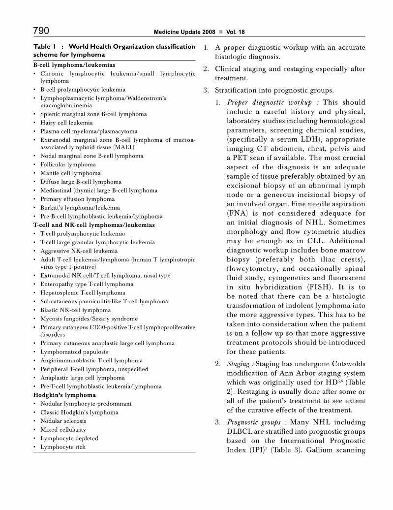
790 Medicine Update 2008 Vol. 18
1. A proper diagnostic workup with an accurate histologic diagnosis.
2. Clinical stagingand restagingespeciallyaftertreatment.
3. Stratification into prognostic groups.
1. Proper diagnostic workup : This should include a careful history and physical, laboratory studies including hematological parameters, screening chemical studies, (specifically a serum LDH), appropriate imaging-CT abdomen, chest, pelvis anda PET scan if available. The most crucial aspect of the diagnosis is an adequate sample of tissue preferably obtained by an excisional biopsy of an abnormal lymph node or a generous incisional biopsy of an involved organ. Fine needle aspiration (FNA) is not considered adequate for an initial diagnosis of NHL. Sometimes morphology and flow cytometric studies may be enough as in CLL. Additionaldiagnostic workup includes bone marrow biopsy (preferably both iliac crests), flowcytometry, and occasionally spinal fluid study, cytogenetics and fluorescent in situ hybridization (FISH). It is to be noted that there can be a histologic transformation of indolent lymphoma into the more aggressive types. This has to be taken into consideration when the patient is on a follow up so that more aggressive treatment protocols should be introduced for these patients.
2. Staging : StaginghasundergoneCotswoldsmodification of Ann Arbor staging system which was originally used for HD5,6 (Table 2). Restaging is usually done after some or all of the patient’s treatment to see extent of the curative effects of the treatment.
3. Prognostic groups : Many NHL including DLBCLarestratifiedintoprognosticgroupsbased on the International Prognostic Index (IPI)7 (Table 3). Gallium scanning
Table 1 : World Health Organization classification scheme for lymphomaB-cell lymphoma/leukemias• Chronic lymphocytic leukemia/small lymphocytic
lymphoma• B-cellprolymphocyticleukemia• Lymphoplasmacyticlymphoma/Waldenstrom’s
macroglobulinemia• SplenicmarginalzoneB-celllymphoma• Hairycellleukemia• Plasmacellmyeloma/plasmacytoma• Extranodal marginal zone B-cell lymphoma of mucosa-
associated lymphoid tissue (MALT)• NodalmarginalzoneB-celllymphoma• Follicularlymphoma• Mantlecelllymphoma• DiffuselargeB-celllymphoma• Mediastinal(thymic)largeB-celllymphoma• Primaryeffusionlymphoma• Burkitt’slymphoma/leukemia• Pre-B-celllymphoblasticleukemia/lymphomaT-cell and NK-cell lymphomas/leukemias• T-cellprolymphocyticleukemia• T-celllargegranularlymphocyticleukemia• AggressiveNK-cellleukemia• AdultT-cellleukemia/lymphoma(humanTlymphotropic
virus type 1-positive)• ExtranodalNK-cell/T-celllymphoma,nasaltype• EnteropathytypeT-celllymphoma• HepatosplenicT-celllymphoma• Subcutaneouspanniculitis-likeT-celllymphoma• BlasticNK-celllymphoma• Mycosisfungoides/Sezarysyndrome• PrimarycutaneousCD30-positiveT-celllymphoproliferative
disorders• Primarycutaneousanaplasticlargecelllymphoma• Lymphomatoidpapulosis• AngioimmunoblasticT-celllymphoma• PeripheralT-celllymphoma,unspecified• Anaplasticlargecelllymphoma• Pre-T-celllymphoblasticleukemia/lymphomaHodgkin’s lymphoma• Nodularlymphocyte-predominant• ClassicHodgkin’slymphoma• Nodularsclerosis• Mixedcellularity• Lymphocytedepleted• Lymphocyterich

791Non-Hodgkin’s Lymphoma Current Treatment Strategies
Management of Diffuse Large B-Cell Lymphomas (DLBCL) ThisisthemostcommonformofNHL.Currentlyfor management purposes two or three other major categories of aggressive lymphomas are also treated according to the DLBCL practiceguidelines. They are anaplastic large cell lymphoma, peripheral T cell lymphoma and follicular lymphoma grade 3.
ThegoaloftreatmentofDLBCLisacuresince50% of the patients with this disease can be cured with conventional therapy. Treatment may be divided into the following groups.9-12
1. Patients with localized disease (Ann Arbor stage I-II) with nonbulky disease (less than 10 cm). who do not have adverse risk factors such as an elevated LDH, stage II disease, age above 60 yearsorECOG(EasternCooperativeOncologyGroup ) performance status equal or more than 2.
2. Patients with localized disease with non bulky disease who have one or more adverse risk factors.
3. Patients with bulky disease (more than 10 cm).
4. Patients with advanced-stage disease who fall into low or low intermediate risk category.
5. Patients with advanced-stage disease who fall into the IPI high-intermediate or high-risk category.
Table 2 : Cotswold staging classificationStage Description
Stage I Involvement of a single lymph node region or lymphoid structure (e.g., spleen, thymus, Waldeyer’s ring) or involvement of a single extralymphatic site (IE)
Stage II Involvement of two or more lymph node regions on the same site of the diaphragm (II) or localized contiguous involvement of only one extranodal organ or side and its regional lymph nodes with or without other lymph node regions on the same side of the diaphragm (IIE)
Note: The number of anatomic regions involved may be indicated by a subscript (e.g., II3).
Stage III Involvement of lymph node regions on both sides of the diaphragm (III), which also may be accompanied by involvement of the spleen (IIIS) or by localized contiguous involvement of only one extranodal organ side (IIIE) or both (IIISE)
Stage IV Disseminated (multifocal) involvement of one or more extranodal organs or tissues, with or without associated lymph node involvement or isolated extralymphatic organ involvement with distant (nonregional) nodal involvement
Designations applicable to any disease stage
A No symptoms
B Fever (temperature > 380 C), night sweats,unexplained loss of more than 10% of body weight during the previous 6 months.
X Bulky disease
E Involvement of a single extranodal site that is contiguous or proximal to the known nodal site
Table 3 : International Prognostic Index (patients of all ages)
Risk group* Risk factors CR rate (%)
Survival rates
5 yrs(%)
Low 0,1 87 73
Low-Intermediate
2 67 51
High-intermediate
3 55 43
High 4,5 44 26
Risk factors include- Age > 60 yrs, LDH > normal, Performance status > 1, stage III/IV, Extranodal involvement > 1 site; score 0 or 1 for each factor; 0 = absent, 1 present
is now increasingly used to prognosticate NHL. This is positive in nearly all cases of aggressive lymphoma and in approximately 50% of the indolent ones. Those patients who remain gallium avid at the end of the treatment are likely to relapse than those who become gallium negative. Early assessment of response to chemotherapy with fluorine-18 fluorodeoxyglucose positron emission tomography (FDG-PET) is becoming a routine part of management in patients with aggressive NHL. Changes in FDGuptake can occur soon after the initiation of therapy and they precede changes in tumor volume8.

792 Medicine Update 2008 Vol. 18
6. Patients with partial remission and relapsed disease.
Patients with localized non bulky disease without adverse risks may be treated with an abbreviated course (three cycles) of R-CHOP (rituximab,cyclophosphamide, doxorubicin, vincristine, and perdinosone) combined with involved field RT (radiotherapy). The dose and time schedule of R-CHOPisasfollows.
Rituximab 375 mg/m2 on day 1 intravenously (IV)
Cyclophosphamide 750 mg/m2 day 1 IV
Doxorubicin (adriamycin)
50 mg/m2 day 1 IV
Vincristine 1.4 mg/m2 (max 2 mg/ dose) day 1 IV
Prednisone 100 mg/day orally for 5 days. This cycle is given once in 21 days.
Patients with localized non bulky disease with adverse risks may be treated with 6 – 8 cycles of CHOP+R.Patient shouldalsoreceiveadditionaladjuvant RT.
Patients with localized but bulky disease and/or local extra nodal disease may be effectively treated with a full course of CHOP chemotherapy (6-8cycles) with rituximab and involved field RT.
Patients with advanced stage disease with low or intermediate risk (as indicated by normal LDH serum level and normal performance status, ECOG0or1)aregivenfullcourseR-CHOP(6-8cycles).
Patients with advanced stage disease who fall into the IPI high-intermediate or high-risk category have less than 50% chance of being cured with standard chemotherapy. For this reason the general consensus is that if possible these patients should be treated or should be placed in different clinical trials. If they cannot get a chance in a clinical trial, thealternativewouldbe6-8cyclesofCHOPwithrituximab.
Patients who are receiving induction chemotherapy should undergo repeat radiographic evaluation after 3-4 cycles of treatment. This early restaging is done to identify at the earliest possible point, patients who do not respond or has progressed despite induction treatment. After completing the induction therapy, all positive radiographic studies should be repeated. Functional imaging (gallium or PET scans) may be useful in determining whether residual masses represent fibrosis or active tumor. A repeat biopsy of the residual mass is always warranted if they remain positive on functional imaging scan after completing the induction therapy. This repeat biopsy may yield other diagnosis like tuberculosis, sarcoidosis, fungal infection, a different lymphoma, desmoid tumor, nonspecific inflammatory process, follicular hyperplasia etc.
Patients who have completed the chemotherapy and is in complete remission should undergo periodic follow up because there is a good chance for a recurrence. Most patients who relapse will do so in the first 2-3 years. Patients may be reviewed at 2 monthly interval for the first year, 3 monthly intervals for the second year, 4 monthly intervals for the third year, twice a year for the 4th and 5th years and then annually indefinitely.
For patients with partial remission, autologous stem cell transplantation or therapy with higher RT dose may be considered. Appropriate clinical trails are also recommended for partial remission.
Those patients who relapse after initial remission or those who have refractory disease are candidates for non-cross-resistant combination chemotherapeuticregimens.SomeoftheseareICE(ifosfamide, carboplatin and etoposide), DHAP (dexamethasone, cytarabine, and cisplatin), MINE (mitoxantrone, ifosfamide, mesna, etoposide) etc. Patients responding to this chemotherapy should be considered for further consolidation with high dose therapy and stem cell support. Additional RT can be given before or after stem cell transplant to sites of bulky disease. Those who relapse after this should enter a clinical trial or treated individually. Disease

793Non-Hodgkin’s Lymphoma Current Treatment Strategies
progression after 3 successive chemotherapeutic regimens is not likely to benefit from currently available standard therapy except for patients with a long disease free interval.
Management of Follicular LymphomaFollicular Lymphoma (FL) is the next common form of NHL and its characteristic immunophenotype includes CD 10+,bcl 2+, CD 23±, CD 43-, CD5-, CD 20+, cyclin D1-. 90% of cases have achromosome translocation, t(14:18).
Treatment of Grade 1 and 2 follicular lymphoma depends on the extent of the initial disease.13,14 Grade 3 FL is treated according to the guidelines forDLBCL.
Patients are grouped into the following.
1. Patients with non bulky localized (Ann Arbor stage I-II).
2. Patients with localized bulky (Ann Arbor stage II), abdominal or stage III or IV disease.
3. Patients with relapsed disease.
Radiotherapy alone (with doses of 30 to 36 Gy) is standard treatment for patients with CS I to IIfollicular grade I to II lymphoma. 10-year over all survival ranges from 43% to 79%, with a median survival of 11.9 to 15.3 years. If patients relapse following localized RT or have no response to initial therapy, they should be managed in the same manner as patients with systemic presentation of FL.
Patients with more extensive disease should be treated based on the following indications: symptoms, threatened end organ dysfunction, cytopenia secondary to lymphoma, bulky disease at presentation, and steady progression of the disease and/or patient preference. Patients should also be given a chance to enter a clinical trial. In the absence of an appropriate clinical trial numerous treatment options are available including loco regional RT and single agent or combination chemotherapy. The selection of treatment should be highly individualized taking into consideration the age, extent of disease, co-morbid conditions and the goals
of therapy. Lack of survival advantage and an early use of an anthracycline are not universally accepted by NCCN (National Comprehensive CancerNetwork) panel. However the addition of rituximab to chemotherapy regimens for follicular lymphoma has consistently improved overall response rate, complete response rate and progression free survival. Clear evidence supporting a survivaladvantage is still lacking. Currently rituximabwith combination chemotherapy is the preferred mode of treatment. Maintenance treatment with rituximab has been shown to improve survival but many questions remain to be answered.15
Single agent drugs include : cyclophosphamide, rituximab, and chlorambucil
Combination drugs include : CVP(cyclophosphamide, vincristine, prednisone); F N D±R ( f ludarab ine , mi toxant rone , dexamethasone , r i t ux imab ) ; C H O P(cyclophosphamide, doxorubicin, vincristine, prednisone) combined with rituximab.
Patients who are responding to the treatment are usually followed until their disease recurs. At recurrence, a repeat biopsy is usually indicated to find out if there is a histologic transformation especially when there is a rising LDH levels, disproportional growth in one area, development of extranodal disease or new “B” symptoms. If repeat biopsy shows transformation to DLBCL,patient may be given anthracycline based therapy or chemotherapy ± rituximab if the patient has had minimal prior chemotherapy or did not have one. This may be followed by consideration of either an autologous or allogenic stem cell transplant.
Two new radio immunotherapy agents, 90Y-ibritumomab tiuxetan (Zevalin) and iodine-131I-tositumomab(Bexxar)directedagainsttheCD20surface antigen found on normal mature B cells are available for therapeutic use and have significant activity in the treatment of relapsed and refractory disease. Both compounds produce similar clinical outcomes (approximately 20%-40% complete response rates and 60%-80% overall response

794 Medicine Update 2008 Vol. 18
rates for patients with indolent B-cell NHL).16 High dose chemotherapy with an autologous or allogenic source of stem cell support may also be an appropriate option.
Management of chronic lymphocytic leukemia/ Small lymphocytic lymphoma (CLL/SLL)These are considered to be the different manifestations of the same disease and managed almost in the same manner. The typical immunophenotype include CD5+, CD19+, CD20 dim, CD23+, CD43±,CD10-,andcyclinD1-.Mantlecell lymphomaisdifferentiated from theseby thepoint thatCLL/SLL iscyclinD1-.Cytogenetics like t(11:14)alsodistinguishesmantlecell lymphoma (MCL) fromCLL.PrognosticstratificationofCLLcannowbemade depending on FISH, gene mutation and flow cytometric studies.
Patient stratification for treatment is:
1. Patients with localized (Ann Arbor I-II).
2. Patients with advance (Ann Arbor III-IV) disease.
Loco regional RT or observation is an appropriate option for localized disease. Patients are treated as needed for symptoms, threatened end organ function, cytopenia, bulky disease at presentation, steady progression of the disease, histological transformation and or according to the patient’s preference, if the disease progresses. Since the
disease is incurable with the currently available standard therapy patient may be given a chance to enter a clinical trial. Patients with advance disease are also treated as needed in keeping with the above indications. Patients with recurrent infections may benefit from intravenous immunoglobulin. Rai good risk disease does not need treatment (Rai staging Table 4). Intermediate risk disease can be observed and high risk disease is treated at presentation.
Currently options for systemic chemotherapyinclude the following groups of drugs.
1. An alkylating agent – chlorambucil
2. Purine analog with or without rituximab – fludarabine with or without rituximab
3. An alkylating agent based combination chemotherapeuticregimen–Cyclophosphamidewith or without prednisone, CVP(cyclophosphamide vincristine and prednisone), FCR(fludarabine,cyclophosphamidewithorwithout rituximab).
Different centers use one of the above regimens. It is generally observed that addition of rituximab to fludarabine prolongs progression free and over all survival.
The dose schedules of the different drugs are given below:
Chlorambucil-Differentdosesandschedulesareused.Commonlyuseddose is10mgdaily for2weeks, repeated every month.
Table 4 : Staging of Chronic lymphatic leukemiaRai Stage
Modif ied Rai Stage
Description Binet Stage
Description
0 Low risk Lymphocytosis only A Two or fewer lymphoid-bearing areas
1 Intermediate risk Lymphocytosis and lymphadenopathy B Three or more lymphoid-bearing areas
2 Intermediate risk Lymphocytosis and splenomegaly with/without lymphadenopathy
— —
3 High risk Lymphocytosis and anemia (hemoglobin, < 11 g/dL)
C Anemia (hemoglobin, < 10 g/dL) or thrombocytopenia (platelets, 100 x 106/dL)
4 High risk Lymphocytosis and thrombocytopenia (platelets, < 100 x 106/dL)
— —
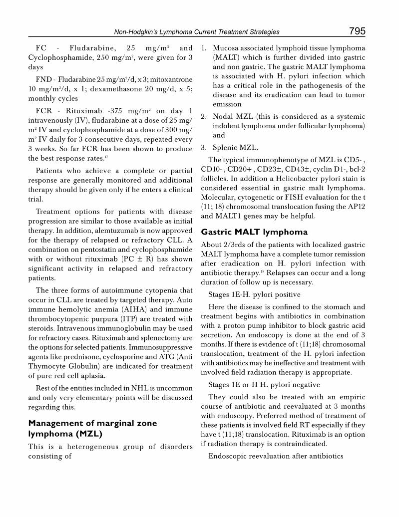
795Non-Hodgkin’s Lymphoma Current Treatment Strategies
FC - Fludarabine, 25 mg/m 2 and Cyclophosphamide,250mg/m2, were given for 3 days
FND - Fludarabine 25 mg/m2/d, x 3; mitoxantrone 10 mg/m2/d, x 1; dexamethasone 20 mg/d, x 5; monthly cycles
FCR - Rituximab -375 mg/m2 on day 1 intravenously (IV), fludarabine at a dose of 25 mg/m2 IV and cyclophosphamide at a dose of 300 mg/m2 IV daily for 3 consecutive days, repeated every 3weeks.SofarFCRhasbeenshowntoproducethe best response rates.17
Patients who achieve a complete or partial response are generally monitored and additional therapy should be given only if he enters a clinical trial.
Treatment options for patients with disease progression are similar to those available as initial therapy. In addition, alemtuzumab is now approved for the therapyof relapsedor refractoryCLL.Acombination on pentostatin and cyclophosphamide with or without rituximab (PC ± R) has shownsignificant activity in relapsed and refractory patients.
The three forms of autoimmune cytopenia that occurinCLLaretreatedbytargetedtherapy.Autoimmune hemolytic anemia (AIHA) and immune thrombocytopenic purpura (ITP) are treated with steroids. Intravenous immunoglobulin may be used for refractory cases. Rituximab and splenectomy are the options for selected patients. Immunosuppressive agents like prednisone, cyclosporine and ATG (Anti Thymocyte Globulin) are indicated for treatment of pure red cell aplasia.
Rest of the entities included in NHL is uncommon and only very elementary points will be discussed regarding this.
Management of marginal zone lymphoma (MZL)This is a heterogeneous group of disorders consisting of
1. Mucosa associated lymphoid tissue lymphoma (MALT) which is further divided into gastric and non gastric. The gastric MALT lymphoma is associated with H. pylori infection which has a critical role in the pathogenesis of the disease and its eradication can lead to tumor emission
2. Nodal MZL (this is considered as a systemic indolent lymphoma under follicular lymphoma) and
3. Splenic MZL.
ThetypicalimmunophenotypeofMZLisCD5-,CD10-,CD20+,CD23±,CD43±,cyclinD1-,bcl-2follicles. In addition a Helicobacter pylori stain is considered essential in gastric malt lymphoma. Molecular, cytogenetic or FISH evaluation for the t (11; 18) chromosomal translocation fusing the AP12 and MALT1 genes may be helpful.
Gastric MALT lymphomaAbout 2/3rds of the patients with localized gastric MALT lymphoma have a complete tumor remission after eradication on H. pylori infection with antibiotic therapy.18 Relapses can occur and a long duration of follow up is necessary.
Stages 1E-H. pylori positive
Here the disease is confined to the stomach and treatment begins with antibiotics in combination with a proton pump inhibitor to block gastric acid secretion. An endoscopy is done at the end of 3 months. If there is evidence of t (11;18) chromosomal translocation, treatment of the H. pylori infection with antibiotics may be ineffective and treatment with involved field radiation therapy is appropriate.
Stages 1E or II H. pylori negative
They could also be treated with an empiric course of antibiotic and reevaluated at 3 months with endoscopy. Preferred method of treatment of these patients is involved field RT especially if they have t (11;18) translocation. Rituximab is an option if radiation therapy is contraindicated.
Endoscopic reevaluation after antibiotics

796 Medicine Update 2008 Vol. 18
Four distinct outcomes are observed.
1. Patients who have both microbiologic and tumor response are just observed
2. Patients who have no evidence of H. pylori but have persistent lymphoma. RT is indicated for patients with significant disease progression or symptoms. Other asymptomatic patients may be observed every 3 months or loco regional RT is appropriate.
3. Patients with persistent H. pylori and regressing or stable symptoms are treated with second line antibiotics.
4. Patients with H. pylori positive and persistent lymphoma. RT is given for progressive disease and second line antibiotics if the disease is stable.
All the patients should be followed up for a long time with repeat endoscopy. Generally recurrence of lymphoma is treated with loco regional RT if not previously treated. Those who do not respond to radiation may be treated with single agent or combinational chemotherapy. Surgery is reserved for those who do not respond to other therapeutic modalities.
Stages III/IV
These patients with disseminated disease, management is similar to the management of other advanced stage follicular lymphoma.
Non-gastric MALT Lymphoma
They can arise from a large number of sites including skin, lung, salivary gland, conjunctiva, prostate, ovary, small bowel and colon. For patients with 1E or II disease, loco regional RT is appropriate. For certain sites of the disease (e.g.: lung, colon, skin, thyroid, small intestine, breast) primary surgery is appropriate. Patients with advanced stage disease are treated in the same way as follicular lymphoma.
Management of splenic MZLThe diagnosis is often presumptive based on the
findings of splenomegaly with peripheral blood flow cytometry usually revealing a monoclonal B cellpopulation.ThisisdistinguishedfromCLLwiththeabsenceofCD5expressionandstrongCD20expression.PatientswhoarepositiveforhepatitisCshould have appropriate consultation to determine if there are indications for treatment of the viral infection. If antiviral treatment is given, the patient should be monitored for tumor response. All other patients should be observed if in the absence of cytopenias or symptoms. Splenectomy is indicated if there are cytopenias or symptoms and they should be monitored at regular basis. If splenectomy is contraindicated, they are treated as advanced stage follicular lymphoma.
Management of Mantle cell LymphomaThe diagnosis is established by histological examina t ion in combina t ion wi th immunohistochemistry.TheprofileincludesCD5+,CD10±,CD20+,CD23±,CD43+andcyclinD1+.Mantle cell lymphoma has the worst characteristic combination of both indolent and aggressive NHL. Like many other indolent lymphoma it is incurable with conventional chemotherapy but it does not have an indolent natural history. It has a shorter disease free and overall survival more like an aggressive lymphoma. Therefore there is no established standard of care. Patients should be referred for participation in prospective clinical trials. Outside a clinical trial the recommendation is that patient should have either combined modality therapy or involved field radiation therapy. Advanced stage disease requires systemic therapy. Several regimens have shown significant activity including R-Hyper CVAD (rituximab, cyclophosphamide,vincristine, doxorubicin, and dexamethasone) alternating with methotrexate and cytarabine, R-CHOP and R-EPOCH (rituximab, etoposide,prednisone, vincristine, cyclophosphamide and doxorubicin). Adjuvant stem cell transplant may be given or initial remission may be followed by stem cell transplantation. The optimal approach for
797Non-Hodgkin’s Lymphoma Current Treatment Strategies
recurrent disease remains to be defined. Entry into clinical trials is strongly encouraged. Single agents like cladribine and bortezomib, or combination chemotherapy including a number of drugs have been suggested. Marked anti-tumor activity has been shown for rituximab plus thalidomide in patients with relapsed/refractory MCL. Radioimmunotherapy has been shown to be active for bothuntreatedandrelapsedMCL.19
Highly aggressive lymphomasBurkitt’s lymphoma and lymphoblastic lymphomas are included in this category as they have an exponential growth rate and a tendency to disseminate to the bone marrow and the meninges. They are aggressive B cell tumors involving extra nodal disease sites. These tumors pose a high risk for tumor lysis. So appropriate treatment includes allopurinol and hydration. Patients with Burkitt’s lymphoma who have a completely resected abdominal lesion or a single extra abdominal mass and a normal LDH level are considered to have low risk disease. All others have high risk disease and should be treated by combination chemotherapy regimen including intensive alkylating agent, anthracycline, intrathecal chemotherapy and high dose methotrexate with or without rituximab.20 Patients with relapse should be treated in the context of a clinical trial whenever possible. Lymphoblastic lymphoma is treated with regimens appropriate for acute lymphoblastic leukemia (ALL).
AIDS related B-Cell LymphomaPatients with AIDS can develop several forms on NLH e.g.: Burkitt’s lymphoma, DLBCL andprimary CNS lymphoma. Patients who developBurkitt’s lymphomagenerallyhave a goodCD4count while patients with CNS lymphoma havevery low CD4 count and uncontrolled AIDS.Optimal management of HIV associated lymphoma has not been established. Several key features have emerged which are critically important. Early introduction of HAART has good long term results. Prophylactic intrathecal chemotherapy is
very important. Inclusion of rituximab appears to increase the risk of neutropenia and infection and is found to have no benefit in patients with HIV associated lymphoma.21
Peripheral T cell lymphoma The term peripheral T cell lymphoma means, lymphoma of a mature T cell phenotype and not the site of involvement by lymphoma. Majority of these patients present with stage IV disease. The clinical course is aggressive, and relapses may be more common than in large B-cell lymphoma. Treatment regimens used for peripheral T-cell lymphomaaresameasthatusedforDLBCL,withthe omission of rituximab. Because of the poorer OS in peripheral T-cell lymphoma as compared with DLBCL, bone marrow transplantationis more frequently required. Bone marrow transplantation may be as effective in peripheral T-cell lymphoma as in DLBCL. In the settingof recurrent disease, purine analogs may have modest activity.20
In summary, a great progress has been made in the understanding and management of NHL which are relatively common neoplasms. It is strongly recommended that a correct and most appropriate diagnosis should be made before starting the treatment. This has been made possible by various morphologic, cytogenetic, molecular and FISH techniques. One should know thoroughly regarding all aspects of commonly occurring NLH likeDLBCL,FL,CLL/SLL,MZLandmantlecelllymphoma. Other rare entities should be kept in mind so that appropriate diagnosis and treatment are instituted. The most consoling aspect of treatmentofNHListhatmorethan50%ofDLBCL(the most common form of NHL) can be cured with modern treatment. Future of NHL includes a general awareness of the disease so that the disease could be diagnosed at a very early stage with the possibility of cure in many more patients. Newer and less toxic chemotherapeutic agents are on the horizon and radio immunotherapy has shown good promise. To improve the Indian situation,
798 Medicine Update 2008 Vol. 18
there should be an online NHL registry under the responsibility of some good center where all NHL cases in India should be reported including diagnosis, management and outcome. This would give us a strong footing in the international arena.
References1. Harris NL, Jaffe ES, Stein H, et al. A revised European-
American classification of lymphoid neoplasms: a proposal from the International Lymphoma Study Group. Blood 1994;84:1361–1392.
2. Harris NL, Jaffe ES, Diebold J, et al. World Health Organization classification of neoplastic diseases of the hematopoietic and lymphoidtissues:reportoftheClinicalAdvisoryCommitteemeeting—AirlieHouse,Virginia,November1997.J Clin Oncol 1999;17:3835–3849.
3. NationalCancerRegistryProgramme,ConsolidatedReportofPopulationBasedCancerRegistries:1990-1996, IndianCouncilofMedicalResearch,August2001.
4. NareshKN,SrinivasV,SomanCS.Distributionofvarioussubtypes of non-Hodgkin’s lymphoma in India: a study of 2773lymphomasusingR.E.A.L.andWHOClassifications.Ann Oncol 2000;11 Suppl 1:63-7.
5. ArmitageJO.Stagingnon-Hodgkinlymphoma.CACancerJ Clin 2005 ;55:368-76.
6. Lister TA, Crowther DM, Sutcliffe SB, et al. Report of acommittee convened to discuss the evaluation and staging of patientswithHodgkin’sdisease:Cotswoldsmeeting.J Clin Oncol 1989;7: 1630–6.
7. A predictive model for aggressive non-Hodgkin’s lymphoma: the International Non-Hodgkin’s Lymphoma Prognostic Factors Project. N Engl J Med 1993;329:987–994.
8. Jhanwar YS, Straus DJ The role of PET in lymphoma. J Nucl Med 2006;47:1326-34.
9. AbbottBL.Diagnosisandmanagementoflymphoma.ClinLymphoma Myeloma. 2006 ;7:30-2.
10. JacobsenE,LaCasceA. Updateon the therapyofhighlyaggressive non-Hodgkin’s lymphoma. Expert Opin Biol Ther 2006 ;6:699-708.
11. Armitage JO. How I treat patients with diffuse large B-cell lymphoma. Blood 2007;110: 29-36.
12. Marcus R, Hagenbeek A. The therapeutic use of rituximab in non-Hodgkin’s lymphoma. Eur J Haematol 2007: 78(Suppl. 67): 5–14.
13. Gribben JG. How I treat indolent lymphoma. Blood 2007;109: 4617-4626.
14. Cheson BD. Anti-CD20 Monoclonal Antibodies in theTreatment of Indolent Lymphoma and CLL. Clin AdvHematol Oncol 2007 ;5(2 Suppl 3):1-16.
15. CartronG,Solal-CélignyP.Maintenance therapy for low-grade lymphomas: has the time come? Curr Opin Oncol 2007; 19:425-32.
16. Macklis RM. Radioimmunotherapy as a therapeutic option for Non-Hodgkin’s lymphoma. Semin Radiat Oncol 2007 ;17:176-83.
17. Keating MJ, O’Brien S, Albitar M, et al. Early results of a chemoimmunotherapy regimen of fludarabine, cyclophosphamide, and rituximab as initial therapy for chronic lymphocytic leukemia. J Clin Oncol 2005;23:4079-4088.
18. Morgner A, Schmelz R, Thiede C, Stolte M, Miehlke S.Therapy of gastric mucosa associated lymphoid tissue lymphoma. World J Gastroenterol 2007; 13:3554-66.
19. Goy A. Mantle cell lymphoma: evolving novel options. Curr Oncol Rep 2007 ;9:391-8.
20. Canellos GP, Lister TA, Young B. The Lymphomas, 2nd Edition. Philadelphia, Saunders 2006.
21. MounierN,SpinaM,GisselbrechtC.Modernmanagementof non-Hodgkin lymphoma in HIV-infected patients. Br J Haematol 2007;136:685-98.
CHAPTER
106Leptospirosis -Current Scenario in IndiaS. Shivakumar
IntroductionLeptospirosis has long been considered a rare zoonotic disease in India with only sporadic cases being recorded1,2. Since 1980’s the disease has been reported from various states during monsoon months in mini epidemic proportions. The disease is endemic in Kerala, Tamilnadu, Gujarat, Andamans, Karnataka, Maharashtra. It has also been reported from Andhra Pradesh, Orissa, West Bengal, Uttar Pradesh, Delhi & Puducherry3,4.
Leptospirosis has been under-reported and under-diagnosed from India due to lack of awareness of the disease and lack of appropriate laboratory diagnostic facilities in most parts of the country. Combining clinical expertise andawareness with confirmatory laboratory back up dramatically increases the recognition of patients withleptospirosis.Clinicalfeaturesofleptospirosisvary from mild illness to severe life threatening illness. Leptospirosis can be diagnosed only by laboratory tests as the clinical features are nonspecific. But the laboratory tests are complex and hence definite guidelines for diagnosis of human leptospirosis is necessary. In this article, the current scenario of leptospirosis in various endemic states of India will be highlighted and the problems in diagnosis and management will be discussed.
EpidemiologyLeptospirosis is an infectious disease caused by leptospira interrogans complex, which has over 20 serogroups and more than 200 serovars. Rodents, domestic & wild animals form the reservoir of infection where domestic animals such as cattle, dogs, and pigs may act as carriers for several months (temporary carrier) while rodents usually remain carrier throughout their life (permanent carrier). Rodents are therefore considered as the major reservoir of infection. Leptospires are excreted in the urine of the animals and they affect man when he comes into contact with urine of infected animals, directly or indirectly, when he is exposed to an environment contaminated by the urine of the infected animals such as soil and surface water following monsoon rains. Therefore this illness commonly occurs during the monsoon months.The infection is probably transmitted when they wade through stagnant rainwater contaminated by infected urine of animals. These organisms can survive for 6 hours in dry soil and for 6 months in flooded condition. They enter the host through the abrasions of the skin of the feet or intact mucous membranes of eye, throat and gut5.
Leptospirosis can occur in both urban and rural areas. In urban areas of developing countries, a contaminated environment due to various factors

800 Medicine Update 2008 Vol. 18
such as overcrowded slums, inadequate drainage and sanitation facilities for man and animals, presence of stray dogs, cattle, pigs, domestic rats, bandicoots, poor condition of slaughter houses and people walking bare foot contribute to the spread of the illness. In rural areas, high-risk groups are workers in rice fields, cane fields and other agricultural crops and animal husbandry staff. In addition, workers in sewers mines and military personnel are also at risk. History of animal contact is not essential for diagnosis for leptospirosis in developing countries. It is impossible to trace the source of infection and any person can be infected, irrespective of direct contact with animals, due to contaminated environment.Therefore the more important epidemiological factors are rainfall and contact with contaminated environment5.
Persons of all ages and races are susceptible. Adult men however are more frequently infected because they tend to work in high-risk jobs. The number of cases in a region often fluctuates from year to year due to various factors such as rainfall, flooding and animal infections. Leptospiral infections tend to occur as individual/small cluster of cases or large outbreaks/epidemics. In India, urban leptospirosis has been reported from Chennai & Mumbaiwhile rural leptospirosis has been reported from Gujarat, Kerala and Andamans. Non-reporting of leptospirosis from other states of India does not mean that it is absent in those parts.
Clinical FeaturesLeptospirosis can manifest in many ways.6 The various syndromes of presentation are as follows.
1. Acute febrile illness
2. Weil’s syndrome characterized by jaundice, renal failure and myocarditis with cardiac arrhythmias
3. Pulmonary Hemorrhage with respiratory failure
4. Meningitis / Meningo encephalitis
Theincubationperiodis7—14days,butrangesfrom2—21days.
The incidence rate ranges from 0.1 – 1 / 100,000 per year in temperate climates to 10- 100 / 100,000 in tropical countries. During outbreak the incidence may reach over 100 / 100,000. Hospital based data on clinical manifestations confirmed by laboratory tests ( Rapid tests / MAT) are usually needed to obtain the incidence rates. Mild cases may not be admitted to hospitals and hence these data may result in a bias towards severity in assessing the public health importance of leptospirosis.6
The prevalence rates are obtained from asymptomatic individuals of selected high risk groups. Sero surveillance provides data on infection rather than as a disease. MAT is required for sero surveys.
Indian ScenarioThe current scenario of leptospirosis in various endemic states will be discussed. Data from other states will also be analyzed. In addition, the problems in diagnosis and management will be highlighted.
Andaman and Nicobar IslandsAndaman and Nicobar Islands are endemic for leptospirosis since early part of the 20th century. Outbreaks of Andaman Hemorrhagic fever (AHF) were reported since 1988.3,7,8 This was proved to be leptospirosis in 1994. 524 cases of AHF (leptospirosis) were reported from 1988-97. The disease presented as febrile illness with pulmonary hemorrhage during post monsoon periods. As the disease presented with predominant pulmonary involvement, a Leptospiral etiology was never considered. In addition, absence of diagnostic facilities were responsible for not diagnosing leptospirosis7.
During 2000-04, 544 cases were reported in Andamans by disease surveillance system. There were total of 93 deaths with the highest incidence in 2002.3 At present, Andaman islands has probably
801Leptospirosis - Current Scenario in India
the highest incidence rates of leptospirosis in the country with figures ranging between 50- 65 cases / 100,000 per year.7
In 2005, 58 cases of confirmed leptospirosis wereadmittedand14patientsdied[CaseFatalityRate(CFR)-24.1%].Majorityofdeathsweredueto pulmonary hemorrhage and occurred within 48 hours. Rural urban ratio was 46 : 12 with exposure to agriculture being 69% and history of contact with animals being 72.4%3.
In 2004, 322 of 611 sera samples from different high risk populations were positive giving an overall sero prevalence of 52.7%. The sero prevalence was highest among agricultural workers ( 62.5 %) followed by sewage workers (39.4%) , animal handlers (37.5%), butchers (30%) and forest workers ( 27.3%). Among the control group the sero prevalence was 14.7%. Grippotyphosa followed by Australis were the common sero groups identified.9
These studies were done at the Regional Medical Research Centre (ICMR), WHO CollaboratingCentre for Diagnosis Reference, Research andtraining in leptospirosis which is situated in Andaman and Nicobar Islands.
Gujarat The disease is endemic in south Gujarat since 19943,10,11. The endemic districts are Valsad, Navsari and Surat. Cases are seen during the monsoonmonths. The annual data of leptospirosis in Gujarat are shown in Table 1 which shows yearly fluctuations innumberswith thahighestCFR in2005 ( 20.6 %).3,11
In the year 2005, 392 cases and 81 deaths due to leptospirosis were reported from various districts of south Gujarat (Table 2). There were 310 males and 82 females, mostly in the age group of 26- 45 year.11 Jaundice, renal failure and hemorrhagic pneumonitis were the common complications noted.
Based on extensive studies conducted in Gujarat, it was highlighted that agro-climatic conditions for south Gujarat favor endemicity for leptospirosis. These include heavy rainfall, clay soil and high water table3.
Public health control measures have been directed towards source reduction to begin with followed by case reduction and then subsequent reduction in case mortality. This is achieved
Table 1 : Year wise cases & deaths due to leptospirosis in South GujaratYear 1997 1998 1999 2000 2001 2002 2003 2004 2005
Cases 657 515 357 156 4 37 373 630 392
Death 76 40 32 16 0 6 40 92 81
CFR % 11.5 7.77 8.96 10.26 0 16.2 10.7 14.6 20.66
Table 2 : Summary of Leptospirosis cases and associated deaths in the year 2005Districts Cases Deaths CFR Taluks PHCs Villages
Surat 185 43 23.24 10 41 123
Navsari 114 26 22.80 5 31 70
Valsad 88 11 12.50 5 25 58
Bharuch 2 0 0 2 2 2
Gandhinagar 2 1 50 2 2 2
Others 1 0 0 1 1 1
Total 392 81 20.66 25 102 256
802 Medicine Update 2008 Vol. 18
through a mulitsectoral approach involving collaborative work between Department of health, Irrigation, Agriculture, Animal Husbandry, Tribal Development and Public works3.
Gujarat has a well organized leptospirosis control program extending from primary health center to district hospital / medical college hospitals in endemicareas.AmedicalofficeratPHCcantreatany febrile illness during monsoon months with chloroquine and doxycycline. If leptospirosis is suspected, I.V Penicillin is given. If there is organ dysfunction, he refers them to the nearest hospital for laboratory diagnosis and management.11
Chemoprophylaxis is given to all personsworking in agricultural farms and those involved in animal husbandry. They are given doxycycline 200 mg once a week for a period of 6 weeks, during the period of maximum rains and water stagnation in a particular district. Public awareness is created byTelevisionandpamphletsatPHCs.11
MaharastraLeptospirosis has been reported regularly since 1998.3,12,13 The annual data from 1998- 2005 is shown in table 3.
2355 cases and 167 deaths were reported in 2005, mainly due to large outbreak during the post monsoon floods. The number of districts in Maharastra reporting leptospirosis has expanded from two in 1998 to ten districts in 2005. The important districts are Mumbai, Thane, Kolhapur, Sangli and Sindhudurg which are affected by leptospirosis. The serovars isolated were L. icterhemorrhagie (rats), L. canicola (canines) and L. australis (cattle).
In a study of 74 cases , hemorrhagic pneumonitis occurred in 35.1 % cases. Of the 11 deaths ( 14.8 %), 9 were due to pulmonary hemorrhage14. Autopsy
findings of 62 cases of leptospirosis revealed pulmonary intra alveolar hemorrhage in 48 cases and renal acute tubular necrosis / acute interstitial nephritis in 45 cases.15
In a study of ICU admissions, 7.2% of caseswere due to leptospirosis (60/834). Mortality due to leptospirosis was 52% and 95% of these patients needed ventilatory support for respiratory failure.16
KeralaLeptospirosis is endemic in many areas of kerala. Kolenchery is in the midlands of Kerala. In this area leptospirosis was rarely diagnosed before 1987. Since then a yearly increase in incidence was observed. In a study of 976 cases of leptospirosis confirmed by culture and / or serological tests , a mortality rate of 5.32 % was observed. Autumnalis, Australis and Icterohemorrhagiae were the common serogroups identified17.
In study of 282 cases of leptospirosis from Calicut, hepatic (69.8%), renal (56.3%) involvement and thrombocytopenia (65.8%) were the common complications noted. The mortality was 6.03%.18 Sero prevalence in Calicut among high risk groups was 38.1% (Fishermen – 52.8%, Sanitary workers – 56.2%, Agricultural workers - 30%, and sewarage workers was 28.2%). The rate in healthy control was 24%. Pomona, Shermani, Canicola were the common serogroups identified.19
In a study of leptospirosis from kottayam of 900 cases treated over 10 years, Jaundice- (80 %), renal failure (59 %), hypotension (20 %) were the common complications noted. The disease was commonly seen in agricultural workers, fishermen and oyster shell catchers (82 %). 74 % were seen during the monsoon months with a male / female ratio 7:1.20
Table 3 : Year wise cases and deaths due to leptospirosis ( 1998- 2005)Year 1998 1999 2000 2001 2002 2003 2004 2005
Cases 197 120 324 860 53 350 225 2355
Deaths 7 3 59 111 5 24 18 167
803Leptospirosis - Current Scenario in India
A model leptospirosis control program has been formulated by Kerala state and is awaiting implementation. A state level diagnostic and epidemiology center at each districts has been established to provide technical leadership with the aim to reduce the incidence / prevalence of leptospirosis. This is not a separate program to control leptospirosis but is integrated with other illness at the district levels21.
TamilnaduLeptospirosis has been reported from Chennaisince 1980’s.22,23 The leptospirosis laboratory at Institute of Microbiology, Madras Medical Collegewasestablishedin1994.24 This laboratory receives samples from both government and private hospitals. Data on leptospirosis from government hospitals during the period 2004 – 2006 is given in Table 4.
There has been a dramatic increase in the number of leptospirosis cases & during 2006, 2765 cases were reported. The data on leptospirosis from variousmajorpublicsectorhospitalsfromChennaicity is given in Table 5.
All the Chennai city government hospitalsreported cases of leptospirosis. Data on leptospirosis in private sector hospitals are not available and therefore the incidence of leptospirosis is under-reported.
During the period 1987 – 91, there were 159 cases ofleptospirosisattheGeneralHospital,Chennai.There were 108 males and the mean age was 40.1 years. 136 ( 85 %) had jaundice and 120 ( 75 %)
had renal failure. 70 patients were dialyzed and 25 patients died (15.6 %).25
In the recent past, acute renal failure due to leptospirosis at general hospital Chennai hassignificantly declined from 31% in 1987 – 91 to 7.5 % in 1995-2004.26 Of the 120 cases of leptospiral ARF during the period 1987-91, the highest number of 45 cases were reported in 1990. Since 1992 there has been a decline in leptospiral renal failure cases and during a 10 year period from 1995 -2004 only 84 cases were reported.27
Though severe leptospirosis has declined, mild leptospirosis has increased. In a collaborative study with Leptospirosis Laboratory, Barbados of 57 cases in 1990-91 Jaundice occurred in 84%, and acute renal failure occurred in 72%. Sero group.
Autumnalis was the most common sero group encountered. 26 patients were dialyzed and 2 patients died.28 In a recent study of 106 cases of leptospirosis from north Chennai, Jaundiceoccurred in 17.8% and renal failure occurred in 10.3% showing a decline in complications. Only two patients were dialyzed and there were no deaths. Fever, headache, myalgia were the common presentations. Contaminated environment(95%) and rainfall (50%) were the important epidemiological risk factors. Icterohemorrhagiae was the most common serogroup and Autumnalis was not detected.27
The reasons for the decline in severe leptospirosis suggested were greater awareness of disease, availability of better diagnostic facilities and widespread use of antibiotics. In addition, serogroup Autumnalis, a virulent serogroup causing severe leptospirosis has also declined since 1995. The increase in mild leptospirosis suggest that contaminated environment plays an
Table 4 : Leptospirosis: Annual data of public sector hospitals-Chennai (2004-2006)
Year 2004 2005 2006
Leptospirosis 963 1724 2765
Table 5 : Year 2006- Government hospital data- Chennai (no-2765 cases)Hospital General Hospital Stanley hospital KilpaukMC
Hospital
Royapettah
Hospital
Children’sHospital
Leptospirosis 965 511 563 169 557
804 Medicine Update 2008 Vol. 18
important role in the persistence and spread of the disease.27
Leptospirosis is an important cause of acute febrile illness. In a recent study of 500 cases of fever at government Stanley hospital, leptospirosis was the second common cause of fever contributing to 17%, following malaria which was 27%. Co-infection of leptospirosis (48 cases) with malaria (220 cases) occurred in 22 % of cases.29Co-infectionof Malaria and Leptospirosis has been reported fromChandigarh.30
AserosurveyinChennairevealedaseroprevalencerate of 32.9% (Range 17.8%- 40.5%).31 Uveitis due to leptospirosis has been reported from Madurai.32 A majority of 73 cases had panuveitis(95.5%), retinal phlebitis (51.4%) and hypopyon (12.6%).
PuducherryIn a study of 33 icteric patients from Puducherry, 22 had altered sensorium and 20 had multiorgan failure and thrombocytopenia. 13 patients died (39.3%).33
KarnatakaLeptospirosis outbreaks have been reported from 15 districts of Karnataka. The highest incidence of cases have occurred in Bangalore city, Uttara kannada, Shimoga, Bidar, Gulbarga, Udupi and dakshina kannada districts. During the year 2004, 152 cases and 11 deaths were reported and during 2005, 224 cases and 19 deaths were reported. Patients responded to treatment with amoxicillin and paracetamol3.
In study of 733 patients suspected of leptospirosis, 84 (11.45%) were found positive by ELISA. The important complications noted were hepatic (65%) and renal failure (63 %).
Diarrhea occurred in 24% of cases. 54.7% were agriculture workers and 55.9% gave history of contact with animals.34
OrissaAfter the cyclone during the October-November 1999, 142 patients with febrile illness and hemorrhagic manifestations were evaluated. 28 (19.2%) had evidence of leptospirosis which was confirmed by MAT. 6 were positive by culture / PCR35.
143 people suspected of leptospirosis in a remote village of Mayurbhanj district in north Orissa was evaluated by the Orissa Multi-disease Surveillance System (OMDSS) during the period June-July 2002. The attack rate was 5.95% (143 / 2404) and the CFRwas7.69%(11/143).Therewasexposuretoinfected water in a canal which was probably the source of infection.36
Other StatesData from Anthra Pradesh, Uttar Pradesh, West Bengal and Delhi are becoming available. Evaluation of acute febrile patients in Uttar Pradesh revealed that 7% had leptospirosis ( 25/ 346). 17 of the 25 patients had jaundice.37 In a study of 55 cases of leptospirosis in Hyderabad, 52 % had renal failure and jaundice occurred in 42%.38 Out of 42 persons withjaundicewhowereevaluatedinCalcutta,10(23.8 % ) were found positive for leptospirosis.39 75 patients from Delhi with symptoms of leptospirosis were evaluated, 32 were found positive for leptospirosis and 5 died.40 180 febrile patients from urban slums of Delhi were evaluated and 27 (15 %) were positive for leptospirosis.41
All the available evidence from endemic states suggests that the disease is now emerging in India as an important public health problem. Inspite of adequate knowledge, we do not have an accurate estimate of the disease burden in the country, as data from many other states is not available probably because of lack of diagnostic facilities. The importance of early diagnosis and case management should be emphasized and appropriate modification in approach is essential.3
This should include,
a. Guidelines for simple case definition and

805Leptospirosis - Current Scenario in India
empiric therapy in small rural hospitals, where diagnostic facilities are not available.
b. Diagnosis of leptospirosis utilizing Modified Faine’s Criteria for in-patients admitted todistrict / teaching hospitals where diagnostic facilities are available3.
Diagnosis of LeptospirosisLaboratory support is needed:
1. To confirm the diagnosis
2. For epidemiological and public health reasons, to determine which serovar caused the infection, the likely source of infection, potential reservoir and its location.
The tests depend on the phase of the infection. During leptospiremic phase (< 7days) leptospires canbeisolatedbybloodcultureandPCR,whilein the immune phase, rising antibodies can be detected by serological tests.
Culture: The isolation of leptospirosis by cultureofblood,CSFandurineisthemostdefiniteway of confirming the diagnosis of leptospirosis. Unfortunately, culture of blood does not contribute to an early diagnosis as results come late, weeks or even months after inoculation of culture medium, however it is valuable in critically ill patients who might die in the first week before the development of antibodies.
PCR is promising on both sensitivity and specificity, but is complicated and expensive. Its value for rapid diagnosis is being evaluated and is used in higher centers.42,43
Serology: The serological tests for diagnosis of leptospirosis have been classified as serovar specific tests and genus specific tests.
Serovar specif ic tests: Microscopic agglutination test (MAT): MAT is the gold standard test for diagnosis of leptospirosis because of its unsurpassed diagnostic specificity. The main advantage is that serovars can be identified which is of epidemiological importance. The difficulties in utilizing MAT are due to the following factors.44
a. The antibody titers rise and peak only in 2nd or 3rd week, making it a less sensitive test. A study of 108 cases of leptospirosis from Brazil have revealed that 65% of the first sample were positive by SAT compared to 44% by MAT.45
b. A four fold rise in titer or seroconversion is the most definitive criteria for diagnosis of leptospirosis. Therefore a second sample is mandatory, which is difficult to obtain. In such circumstances, a single high titer in MAT can be taken as diagnostic criteria. As MAT titers peak and persist for a long time ( 5-10 years), they would interfere with current diagnosis. Therefore many workers use different criteria.
A titer of 1:100 is taken as significant criteria, but there is controversy on the single diagnostic titer as they depend on endemicity. In endemic areas, a titer of 1/100 or 1/200 is considered low; while high titer is usually > 1/400 ( some consider 1/800 or 1/1600 as diagnostic criteria). In non- endemic areas, 1/100 titer is taken as the diagnostic criteria. It is preferable to do rapid tests along with single high titers. Positive rapid tests with high titers suggest current infection while negative rapid tests is probably due to past infection. In Andamans, a titer of 1/200 is taken as diagnostic titer. Serosurvey in the asymptomatic high risk group should be done with MAT only and a titer of > 1/50 can be taken as cut off titer.
c. The test is complicated requiring dark field microscopy and cultures of various live serovars. This may not be available in small laboratories.
Genus specific tests (Rapid tests): The common tests are the ELISA,Macroscopic slide agglutination test (MSAT), latex agglutination test, Dipstick tests ( Lepto dipstick, Lepto Tek lateral flow) and Lepto Tek Dri-Dot test.6,11,44,46,47 The genus specific tests are the tests of choice for the diagnosis of current infection. These tests are simple, more sensitive and become positive earlier than MAT. These tests detect genus specific antibodies, which are shared

806 Medicine Update 2008 Vol. 18
by pathogenic and saprophytic leptospira. These tests become positive early in the disease (5-6th day) as they detect specific IgM antibodies and help in the rapid diagnosis of current infection.
Laboratory Criteria for Diagnosis of Current LeptospirosisConfirmed
1. Culture: Positive
2. MAT: a) Seroconversion / 4 fold rise in the titer
Probable
1. Rapid tests: Positive
2. MAT: High titer (Single sample)
The approach to diagnostic tests for leptospirosis is given in Table 6.
Comments1. Rapid tests are adequate for diagnosis of
current infection. This can be done in smaller laboratories in both rural and urban areas. If positive, confirm the diagnosis with MAT,
Table 6 : Approach to Diagnosis of Leptospirosis
Clinical features suggestive of current leptospirosis
Leptospiremic phase < 7days Immune phase > 7 days
Negative Positive
Positive Negative
Repeat (if low titer)
Rising titer
Repeat (> 3 days)
Blood culture / PCR Rapid tests
Repeat
Seroconversion
High titer
MAT

807Leptospirosis - Current Scenario in India
Table 7 : Diagnosis of Leptospirosis-Modified Faine’s Criteria
Name: Age: Sex: Occupation:
Residence ( rural / urban ): Date:
Part A: Clinical Data Score Part B: Epidemiological factors Score
Headache 2 Rainfall 5
Fever 2 Contactwithcontaminated
Temp > 390C 2 Environment 4
Conjunctivalsuffusion 4 AnimalContact 1
Meningism 4
Myalgia 4
Conjunctivalsuffusion Part C : Bacteriological Lab findings
Meningism } 10 IsolationofleptospirainCulture–
Myalgia Diagnosis certain
Jaundice 1 Positive Serology
Albuminuria / 2 ELISA IgM Positive 15
Nitrogen retention SAT - Positive 15
MAT-Single positive 15
in high titer
Rising titer / seroconversion
(paired sera) 25
Presumptive diagnosis of leptospirosis is made of:
Part A or part A & part B score : 26 or more
PartA,B,C(Total):25ormore
A score between 20 and 25 suggests leptospirosis as a possible diagnosis.
which would be available in larger specialized laboratories.
2. MAT—Seroconversion/4foldriseinthetitreis necessary for diagnosis. (2nd sample is essential). Single high titre in MAT combined with positive rapid tests confirms the diagnosis of leptospirosis.
3. Bloodculture—notsensitivebutcanbedoneincritically ill patient. (As they may not survive to produce antibodies).
Modified Faine’s CriteriaFaine has evolved criteria for diagnosis of leptospirosis on the basis of clinical, epidemiological and laboratory data (WHO guidelines). Certain
necessary modifications have been made by us in the epidemiological (Part B) and the laboratory criteria(PartC)oforiginalFaine’scriteriatomakethe diagnosis more practical in Indian institutions. (showninTable7).IntheModifiedFaine’sCriteriarapid tests (ELISA / SAT) have been introduced inPartCandRainfallhasbeen included inPartB to make the diagnosis early and simple.48,49 This criteria can be utilized for diagnosis of leptospirosis in district / teaching institutes3.
ManagementMild leptospirosis can be treated with Doxycycline or Amoxycillin or Erythromycin and severe leptospirosiswithI.V.PenicillinorCeftriaxone.
808 Medicine Update 2008 Vol. 18
Recommendations for Management Based on the Availability of Diagnostic FacilitiesIn centers where no diagnostic facilities are available (Rural areas)
The common causes of acute febrile illnesses are Malaria, Leptospirosis, Dengue and Viral respiratory diseases. It is difficult to diagnose these illness without laboratory facilities. It is recommended that all febrile patients can be treated with doxycycline and chloroquine which is the empiric therapy for Malaria & Leptospirosis. If there is organ dysfunction and / or fever persists they should be transferred to higher centers for further management. This is being implemented in the state of Gujarat.11
In centers where diagnostic facilities are available
Even in centers with laboratory facilities, empiric therapy is recommended for leptospirosis where the disease is endemic, since serological tests become positive only after one week (unless PCRisavailable).Mildcasescanbetreatedwithchloroquine and doxycycline and severe cases with I.V. crystalline penicillin / quinine or artemisinin and doxycycline.50 If they are admitted later (after a week), rapid tests would confirm leptospirosis and appropriate treatment can be given. It is essential that all febrile patients are investigated for leptospirosis, Malaria and Dengue fever as co-infection can occur.29 In addition, dialysis and ventilatory support for renal and respiratory failure would definitely decrease mortality.
To conclude, data on leptospirosis is urgently needed from all states of the country. This can be done by making rapid tests available at all district / teaching hospitals. The National Institute of Communicable Diseases (NICD) utilizing theIntegrated Disease Surveillance Programme (IDSP) should organize the availability of rapid tests3. Appropriate guidelines for management should be implemented to reduce the morbidity and mortality of leptospirosis.
References1. SinghJandSokheyJ.Epidemiology,Presentation&Control
of leptospirosis. Proceedings of the third round table conference. Series-leptospirosis. Ranbaxy science foundation(3);1998: 17-31
2. Rao RS, Gupta N, Bhalla P et al. Leptospirosis in India and the rest of the world. Braz J infec Dis 2003; 7(3) 178-193.
3. Report of the Brainstorming meeting on Leptospirosis Prevention and control. Mumbai, 16- 17 February 2006. Joint Publication by Office of WHO, Representative to India, New DelhiandRegionalMedicalResearchCentre(ICMR),WHOCollaboratingCentreforDiagnosis,Research,ReferenceandTraining in Leptospirosis.
4. Kamath SA, Joshi SR. Re-emerging infections in urban India – Focus Leptospirosis. .J.Assoc Phys India 2003,51:247-248
5. Faine S. guidelines for the control of Leptospirosis. WHO offset publication. 1982; 67
6. Terepstra et al. Human Leptospirosis: Guidelines for Diagnosis, Surveillance & Control {WHO} 2003:1-109
7. SehgalSC, Emergence of leptospirosis as a public healthproblem. Proceedings of the third round table conference.Series-leptospirosis.Ranbaxy science foundation(3);1998: 7-12.
8. SinghSS,VijayachariP,SinhaAetal.Clinical-Epidemiologicalstudy of hospitalized cases of severe leptospirosis. Indian J Med Research 1999. 109:94-9
9. Sharma S, Vijayachari P,Sugunan AP et al.Seroprevalence of leptospirosis among high risk population of Andaman Islands, India. Am J Trop Med Hyg 74 (2) 2006: 278-283.
10 . Clerke AM,Leuva AC, Joshi C et al. Clinical profile ofleptospirosis in south Gujarat. J Post Grad Med 2002. 48: 117-118.
11. Patel BK, Gandhi SJ, Desai DC. Clinico-epidemiologicalaspects of leptospirosis in south Gujarat. Indian J Med Microbiol 2006 24(4). 322-326.
12. Karande S, Kulkarni H, Kulkarni M et al. Leptospirosis in children in Mumbai slums. (2002). Indian Journal of Pediatrics 69, 855-858.
13. Karande S, Bhatt M, Kelkar A et al. An observational study to detect leptospirosis in Mumbai , India 2000. Archieves of disease in childhood.2003. 88( 12) 1070-1075.
14. Bharadwaj R, Bal AM, Joshi SA. An Urban Outbreak of leptospirosis in Mumbai, India. Jpn J Infect Dis 2002:55.194-196.
15. Saikada AP, Divale S, Deshpande. A study of autopsy findings in 62 cases of leptospirosis in a Metropolitan city in India. J Post Grad Med 2005. 51: 169-173.
16. ChawlaV,TrivediTH,Yeolkar.Epidemicofleptospirosis-AnICUexperience. . J.Assoc Phys India 2004:32:619-622.
17. KuriakoseM,EapenCK,PaulR.LeptospirosisinKolenchery,Kerala,India. Epidemiology, Prevalent local serogroups and serovars and a new serovar. European Journal of Epidemiology.1997. 13 (6). 691-697.
18. Pappachan MJ,Mathew S,Aravindan KP et al. Risk factors for mortality in patients with leptospirosis during an epidemic

809Leptospirosis - Current Scenario in India
in Northern Kerala. Nat Med J India 2004; 17:240-2.
19. Swapna RN, Tuteja U, Nair L et al.Seroprevalence of leptospirosis in high risk groups in calicut, North Kerala, India. .Indian J Med Microbiol 2006 24(4). 349-352.
20. Vimala A , Kasi Visweswaran . Leptospirosis in kottayam. South Asian Work shop on Diagnostic Methods in Leptospirosis (SAWDIL). Madras.1995:61
21. John TJ. The prevention and control of Human leptospirosis. J Post Grad Med 2005.57: 205-209.
22. Ratnam S, Sundararaj T, Thiagarajan SP et al. Serological evidence of leptospirosis in Jaundice and Pyrexia of unknown origin. Ind J Med Res. 1983. 77:427-430.
23. Ratnam S, Suramaniam S, Madanagopalan T. Isolation of leptospirosis and demonstration of antibodies in Human leptospirosis in Madras, India. Trans R Soc Trop Med Hyg 1983. 77:455-458.
24. Chinari Pradeep KS, Sumathi G, Vimala RangaRao G,ShivakumarSLeptospirosislaboratory.ChennaiMedicalCollege–Athreeyearexperienceinsero-diagnosis(1995-1997). Indian J Med Microbiol (1999) 17(10): 50-51.
25. Muthusethupati MA, Shivakumar S, Vijaykumar R et al. Renal involvement in Leptospirosis- Our Experience in MadrasCity. J Post Grad Med1994;40(3):127-31.
26. Jayakumar M, Ram Prabakar H, Edwin Fernando M et al. Epidemiologic trend changes in acute renal failure – A tertiary centre experience from South India. Renal Failure 2006; 28:405-10.
27. ShivakumarS.LeptospirosisinChennai-ChangingclinicalProfile. J.Assoc Phys India 2006,54:964-965.
28. Muthusethupathi MA, Shivakumar S, et al. Leptospirosis in Chennai.Aclinical&serologicalstudy.J.Assoc. phys. India. 1995; 43: 450-58
29. LoganathanN,ShivakumarS,RavishankarD.Co-infectionofMalaria and Leptospirosis – A Study of 48 cases (Abstract).62nd Annual Conference of Association of Physicians of India 2007.Goa.
30. Srinivas R, Agarwal R, Gupta D. Severe sepsis due to severe Falciparum Malaria and leptospirosis co-infection treated withactivatedProteinC. Malaria Journal 2007.6:42.
31. Ratnam S, Evarard COR, Alex JC et al. Prevalence ofleptospiral agglutinins among conservancy workers in MadrasCity,India.Journal Of Tropical Medicine and Hygiene 1993; 96:41-45.
32. Rathinam SR, Rathinam S, Selvaraj D et al. Uveitis associated with an epidemic outbreak of leptospirosis. Am J Ophthalmol 1997;124:71-9.
33. DuttaTK,ChristopherM,Leptospirosis–AnOverview.J. Assoc Phys India 2005,52:545-551.
34. Jagadishchandra K,Prathb AG, Rao SP. Clinical andepidemiological correlation of leptospirosis among patients attendingKMCH. Indian J Med Sci 2003;57:101-104.
35. Sehgal SC, Sugunan AP, Vijayalakshmi P. Outbreak ofleptospirosis after the cyclone in Orissa. Nat Med J India. 2002.15, 22-23.
36. Jena AB, Mohanty KC, Devadasan N. An outbreak ofleptospirosis in Orissa, India: the importance of surveillance.Tropical Medicine and International Health 2004.9(9).1016-1021.
37. Manocha H, Gushal V, Singh SK et al. Frequency of leptospirosis in patients of acute febrile illness in Uttar Pradesh. J.Assoc Phys India 2004,52:623-625.
38. Velineni S,Asuthkar S, Umabala P et al. Serological evaluation of leptospirosis in Hyderabad,Andhra Pradesh: A retrospective hospital – based study. Indian J Med Microbiol 2007; 25:24-27.
39. DebnathC,PalNK,PramanikAKetal.Aserologicalstudyofleptospirosis among hospitalized jaundice patients in around Kolkata. . Indian J Med Microbiol 2005; 23:68-.
40. ChaudhryR,PremalathaMM,MohantySetal.Emergingleptospirosis, North India. Emerging Infectious Disease 2002. 8 (12): 1526-1527.
41. Kaur I, Sachdeva R, Arora V et al. Preliminary survey of leptospirosis among febrile patients from Urban slums of E.Delhi. J.Assoc Phys India 2003,51:249-251.
42. BaburajP,NandakumarVS,KhannaLV.PCRinthediagnosisof leptospiral infection. . J.Assoc Phys India 2006,54:339-340.
43. Nizamuddin M, Tuteja V, Shukla J. Early diagnosis of Human leptospirosis by Antigen Detection in Blood. Indian J Med Microbiol 2006.24 (4):342-345.
44. Shivakumar S, Krishnakumar B. Diagnosis of Leptospirosis- Role of MAT. J.Assoc Phys India 2006,54:338-339.
45. Angelo P. Brendo et al. Macroscopic agglutination test for Rapid diagnosis of Human Leptospirosis. Journal of Clinical Microbiology {1998}; 36(11): 3138-3142
46. SumathiG,ChinariPradeepKS&ShivakumarSMSAT–Ascreening test for Leptospirosis. Indian J Med Microbiol(1997) 15:84
47. Maskey M, Shastri JS, Saraswathi K. Leptospirosis in Mumbai. Post deluge outbreak. Indian J Med Microbiol 2006.24: 337-338
48. Shivakumar S Leptospirosis –Evaluation of clinical criteria J.Assoc Phys India 2003,51:329-330
49. Shivakumar S, Shareek PS. Diagnosis of Leptospirosis –Utilizing modified faine’s criteria. J.Assoc Phys India 2004, 52:678-679
50. Kothari MV, Karnad DR,Bichile LS. Tropical infection in ICU. J. Assoc Phys India 2006,54:291-8.

CHAPTER
107Management of Neuropathy in Diabetes MellitusJ. Singh
IntroductionDiabetic neuropathy is probably the commonest complication of diabetes. It occurs with the same frequency in both type 1 and type 2 diabetes,1, 2 and is responsible for some of the most unpleasant symptoms of any chronic diabetic complication. The management problems that it poses are amongst the most challenging and formidable ones encountered in the treatment of diabetes.
The early recognition and appropriate management of neuropathy in the patient with diabetes is important for a number of reasons
• nondiabetic neuropathies may be present inpatients with diabetes and may be treatable;
• a number of treatment options exist forsymptomatic diabetic neuropathy;
• upto50%ofDiabeticPeripheralNeuropathy(DPN) may be asymptomatic and patients are at risk of insensate injury to their feet;
• autonomic neuropathy may involve everysystem in the body; and
• cardiovascular autonomic neuropathy causessubstantial morbidity and mortality.
Specific treatment for the underlying nerve damage is currently not available, other than improved glycemic control, which may slow progression but
rarely reverses neuronal loss. Effective symptomatic treatments are available for DPN and automatic neuropathy.
American Diabetes Association (ADA) Recommendations3 • All patients should be screened for distal
symmetric polyneuropathy at diagnosis and at least annually thereafter, using simple clinical tests.
• Electrophysiological testing is rarely everneeded, except in situations where the clinical features are atypical.
• Once the diagnosis of DPN is established,special foot care is appropriate for insensate feet to decrease the risk of amputation.
• Simple inspection of insensate feet shouldbe performed at 3 to 6 month intervals. An abnormality should trigger referral for special footwear, preventive specialist, or podiatric care.
• Screeningforautonomicneuropathyshouldbeinstituted at diagnosis of type 2 diabetes and 5 years after diagnosis of type 1 diabetes. Special electrophysiological testing for autonomic neuropathy is rarely needed and may not affect management and outcomes.
811Management of Neuropathy in Diabetes Mellitus
• Educationofpatientsaboutself-careofthefeetand referral for special shoes / inserts are vital components of patient management.
• Awidevarietyofmedicationsisrecommendedfor the relief of specific symptoms related to autonomic neuropathy and are recommended, as they improve the quality of life of the patient.
Treatment ModalitiesThe treatment of this difficult complication can be classified into two broad categories4. 1) Treatment designed to modify the course of diabetic neuropathy and 2) Symptomatic treatment. A brief description of this is as follows :
Management of diabetic neuropathy
A. Disease modifying therapy
1. Measures of established clinical value : Optimized Glycemic control.
2. Measures approaching clinical usefulness:
a. Aldose reductase inhibitors.
b. Essential fatty acids.
c. Vasodilator drugs.
3. Measure s under exper imenta l investigation
a. Inhibitors of glycation.
b. Antioxidants.
c. Agents promoting nerve growth and repair.
B. Symptomatic Therapy
1. Treatment of neuropathic pain.
2. Treatment of symptomatic autonomic neuropathy.
3. Treatment of mononeuropathies.
4. Treatment of diabetic foot.
Glycemic Control
Of all the treatments, tight and stable glycemic control is probably the only one that may provide
symptomatic relief as well as slow down the relentless progression of the neuropathic state. Even very badly damaged nerves have been found to show some improvement with maintenance of normoglycemia. Since it has been suggested that rapid surges in blood glucose level from hypo to hyperglycemia can aggravate and induce neuropathic pain, the stability rather than the actual level of glycemic control may be more important in relieving neuropathic pain.
In type 2 DM subjects in whom strict glycemic control is achieved with diet and oral antidiabetics, insulin need not be substituted. Some believe in using insulin as it has got membrane stabilizing effect. A few believe in intensive insulin regimen to ensure round the clock glycemic control. In a few patients with poorly controlled diabetes the pain may worsen initially with tight glycemic control with insulin. This is due to vasoconstriction with glucose control and in the due course with the stabilization of the glucose level the pain subsides.
Aldose reductase inhibitors (ARIs)
ARIs block the rate limiting enzyme in the polyol pathway and, thus, reduce the intra-neuronal fructose and sorbitol accumulation. The suggested mechanism of action for ARIs also include altered phospho-inositide metabolism and Na+-K+ ATPase activity, or through reduced glutathione levels, or by vasodilation and improved blood flow to nerves. The ability of ARIs to prevent and reverse nerve conduction defects and biochemical abnormalities in diabetic animals is firmly established, but the results of clinical trials in human have not been very convincing. Sorbinil and tolerastat are the two ARIs most studied in humans. Tolerastat has also been found to improve autonomic nervous system function in patients with diabetic autonomic neuropathy.
Essential fatty acids (EFAs)
The first step in the metabolism of EFA, linolenic acid (i.e. delta- 6 desaturation of linoleic acid to gama linoleic acid) is impaired in diabetes, and this defect can be bypassed by the administration

812 Medicine Update 2008 Vol. 18
of GLA. This may be a rate limiting step for the synthesis of many biologically important eicosanoids like PGE1, PGE2 and prostacyclins. Several studies have confirmed clinical and electrophysiolgical improvement in peripheral nerve function when GLA is administered to neuropathic patients.
Vasodilators
In humans, it has been shown that the endoneurial vascular abnormalities accompany and parallel the severity of diabetic neuropathy and that nerve blood flow is reduced. Many vasodilator drugs are reported to improve nerve functions and correct endoneurial capillary abnormalities in diabetic animals. The most promising agents include alpha 1- adrenergic antagonists, ACEinhibitors and vasodilator prostanoids. Recently EFAs and electrical stimulation have shown beneficial vasodilator effect in diabetic neuropathic subjects.
Inhibitors of glycation
Advanced glycation products may play an important role in chronic diabetic complications. Aminoguanidine inhibits non-enzymatic glycation and has a beneficial effect in experimental diabetic neuropathy.
Agents promoting nerve growth and repair
Neuronal sprouting and growth are stimulated by nerve growth factor (NGF) and insulin-like growth factor – 1 (IGF-1). NGF administration has been shown to protect against experimental diabeticneuropathy.Thecorticotrophin(ACTH)analogue, ORG2766, and gangliosides (which are normal components of the neuronal membrane) are known to promote neuronal regeneration and growth. But their role in preventing or improving human diabetic neuropathy is not clearly established. In a recent study, patients receiving NGF improved in the sensory component of the neurological examination, two quantitative sensory tests, and subjective impression. The ganglioside preparations are derived from bovine brain extract. They have to be given parenterally
but have been said to be associated with Guillain Barre syndrome.
Antioxidants
Oxidative stress, as reflected by increased peroxidation of nerve lipids in experimental diabetes, may comprise numerous neuronal and endoneurial vascular functions. But the role of antioxidants in inhibiting oxidative damage is not yet established.
Protein Kinase C inhibitors (PKC)
Apotential roleofPKC inhibitors inpreventingdiabetic nerve damage is being investigated and appears to be promising.
Role of vitamins
No conclusive evidence, backed by controlled studies, exist to support the prescription of vitamins for the treatment of diabetic neuropathy.
Mecobalamin
Mecobalamin is one of the two active coenzyme forms of vitamin B12 (the other being adenosylcobalamine). It is cofactor in the enzyme methionine synthatase which functions to transfer methyl groups of regeneration of methionine from homocysteine. Mecobalamin promotes axonal transport and regeneration.
Mecobalamin promotes myel inat ion (Phospholipids synthesis). It promotes the synthesis of lecithin, the main constituent of medullary sheath lipid and increases myelination of neurons.
Dosage: Peripheral Neuropathy: The usual dose for adults is 1 ml (500 mcg of Mecobalamin) daily by IM or IV three times in a week. The dose may be adjusted depending upon the patient’s age and symptoms or oral 500 mcg to 1000 mcg per day.
Nandrolone
Nandrolone is a non-steroidal anabolic drug available in the strength of 25 mg/ml/ampules. It has to be administered deep intramuscular biweekly or weekly for two months.
813Management of Neuropathy in Diabetes Mellitus
Anti-convulsant drugs
Anti-convulsant drugs are in use for the treatment of painful diabetic neuropathy. These drugs suppress abnormal discharge and raise the threshold for neuronal impulse propagation. There has been an explosion in the number of availability of anticonvulsant drugs for the treatment of diabetic neuropathy. Carbamazepine works by slowingthe recovery rate of voltage gate sodium channels from depolarization. Phenytoin has a similar mechanism of action. Valproic acid additionally increases GABA levels by decreasing degradation. Gabapentin has been in use since 1994 as an anticonvulsant. Its efficacy for painful diabetic neuropathy is comparable to Amitriptyline. The mechanism of action is postulated to be a central voltagedependent1-typeCa++channel.Theolderanticonvulsant drugs have been largely replaced by the newer ones which are better tolerated and safe like Oxcarbamazepine, Toporamate, Pregabalin and Tamotrigine. But different anticonvulsant drugs of choice for different painful states have not yet been determined.
Normal Saline Infusion
Normal saline infusion of 540 ml daily for 6 to 7 days has been tried in treatment of painful diabetic neuropathy. This is not useful in neuropathy due to ischemia. Osmotic dissociation has been hypothesized as the mechanism of action.
Drugs to Treat Symptomatic DPN
1. Burning pain
a. Tricyclic agents (Imipramine:
Amitriptyline ± fluphenazine)
b. Capsaicin
c. Carbamazepine
2. Lancinating pain
a. Anticonvulsants(Carbamazepine,Phenytoin,Valproate, Gaba pentin, Pregabalin)
b. Tricyclic agents
c. Capsaicin
3. Contactdiscomfort(Allodynia)
a. Changeofclothing
b. Protective mechanical barrier (Opsite skin films)
4. Restlesslegs:Clonazepam(0.5-1.0mg.HS)
5. Painful cramps: Quinine sulfate (300 mg HS)
6. Painful paraesthesia: Tricyclic agents
7. Gabapentin: Effective in all types of pain
8. Epidural spinal cord stimulationClass Examples Typical doses
Tricyclic drugs Amitriptyline 10-75 mg at bedtime
Nortriptyline 25-75 mg at bedtime
Imipramine 25-75 mg at bedtime
Anticonvulsants Gabapentin 300-1, 200 mg t.i.d.
Carbamazepine 200-400mg t.i.d.
Pregabalin 100 mg t.i.d.
5- hydroxytry- Duloxitine 60-120 mg ptamine and daily norepinephrine uptake inhibitor
Substance P inhibitor Capsaicincream 0.025-0.075% applied t.i.d.- q.i.d.
Treatment of Autonomic Neuropathy
The first step in the management of autonomic neuropathy is to identify correctly those patients who are affected because once the autonomic nervous system is involved, the mortality rate may be as high as 50% within five years, pointing to the serious consequences of this complication. Diagnosis depends upon the correct interpretation and recognition of often disguised and ambiguous problems like dizzy turns, blackouts (postural hypotension), recurrent urinary tract infections (bladder dysfunction), impotency (organic or functional).

814 Medicine Update 2008 Vol. 18
a. Diabetic gastroparesis
It may require hospital admission, intravenous fluids and occasional parenteral nutrition and nasogastric drainage or sometimes percutaneous intra-jejunal tube feeding. Useful options include drugs enhancing gut motility like metoclopramide, domperidone, cisapride and erythromycin. Surgical procedures have high complication rates and outcome is often unsatisfactory.
b. Problems of lower GIT
These include constipation, the most common problem, diabetic diarrhea (usually paroxysmal with nocturnal exacerbation), and fecal incontinence (because of internal anal sphincter dysfunction). The constipation usually responds to stimulant laxatives (e.g. senna, codanthrustate) at night and diarrhea to codeine or loperamide or if due to bacterial overgrowth of small bowel to short course (5-7 days) of antibiotics (tetracycline or erythromycin).
c. Postural hypotension
Mild symptoms may respond to raising the head end of the bed by 10 centimeters, which helps to maintain the postural vascular tone. Fludrocortisone is the drug of choice that increases the peripheral vascular tone and extracellular blood and fluid volume, but it can cause hypokalemia and hypertension.
d. Bladder dysfunction
For poor bladder emptying, mechanical measures like manual suprapubic pressure or intermittent self-catheterization are usually sufficient. Long-term cyclical antibiotic therapy may be needed for recurrent urinary tract infection.
e. Abnormal sweating
Excessive and inappropriate sweating especially gustatory sweating is a very distressing problem and no satisfactory treatment for this is yet available.Clonidinehasbeenusedwithsomesuccess. Other drug available is polidine but It is not well tolerated because of its anticholinergic
side effects like tachycardia, dryness of mouth, urinary retention etc.
f. Impotence
Impotence is extremely common in men with diabetes and is a major source of frustration. Once non-organic causes are excluded, treatment can be mechanical (vacuum device, rubber bands, semirigid and malleable or the inflatable penile prosthesis) or pharmacological. Unquestionably, the pharmacological agent receiving the greatest fanfare and publicity for the treatment of impotence has been sildenafil. It functions by inhibiting hydrolysis of cyclic guanosine monophosphate in the corpus cavernosum, leading to an accentuated penile response to sexual stimuli. The most common side effects are headache, flushing and dyspepsia occurring in 6-18% of men. Sildenafil is contraindicated in those on nitrate therapy for coronary heart disease.
Diabetic Autonomic Neuropathy Management6
Dysfunction Management
Diarrhea Tetracycline
Loperamide
Clonidine
Gastroparesis Metoclopramide
Bethanacol
Mozapride
Domperidone
Erythromycin
Bladder Dysfunction Prazosin
Bladder neck Resection
Orthostatic Hypotension Elastic stockings
Volume expanders
Vaso constrictor agents
Pindolol
Indomethacin
Erectile Impotence Papaverin injection
Prosthesis
Sildenafil
815Management of Neuropathy in Diabetes Mellitus
Treatment of MononeuropathiesMononeuropathies can be of two types: compressive and non-compressive. Non-compressive mononeuropathies include diabetic amyotrophy (proximal motor neuropathy of lower extremities more common in type 2 diabetes), cranial mononeuropathies (involving the third, fourth, sixth and seventh cranial nerves), truncal or thoracoabdominal neuropathy or radiculopathy. These usually affect diabetic individuals above age 50 and resolve spontaneously. General measures including pain relief, psychological support and physiotherapy are often sufficient. Compressivemononeuropathies, especially carpal tunnel syndrome, often require surgical relief of pressure, but the outcome of surgery may be less favourable than in non-diabetic subjects. Also this is to be carried out before irreversible damage occurs.
Foot care recommendations2
• Perform a comprehensive foot examinationand provide foot self-care education annually on patients with diabetes to identify risk factors predictive of ulcers and amputations.
• Thefootexaminationcanbeaccomplishedina primary care setting and should include the use of a monofilament, tuning fork, palpation, and a visual examination.
• Amultidisciplinaryapproachisrecommendedfor individuals with foot ulcers and high-risk feet, especially those with a history of prior ulcer or amputation.
• Referpatientswhosmokeorwithpriorlower-extremity complications to foot care specialists for ongoing preventive care and life-long surveillance.
• Initialscreeningforperipheralarterialdisease(PAD) should include a history for claudication andanassessmentofthepedalpulses.Considerobtaining an ankle-brachial index (ABI), as many patients with PAD are asymptomatic.
• Referpatientswithsignificantclaudicationora positive ABI for further vascular assessment and consider exercise, medications, and surgical options.
Patients with diabetes and high-risk foot conditions should be educated regarding their risk factors and appropriate management. Patients at risk should understand the implications of the loss of protection sensation and the importance of foot monitoring on a daily basis.
In India, the neuropathic foot is more common as compared to the ischemic foot.7 This diabetic foot can be treated effectively by off loading the body weight with proper foot wear in the grade 0-3 and total contact casting and bed rest in grade 4 alongwith the use of appropriate antibiotics. The following measures can prevent many amputations.
Assess the patient’s knowledge of foot care practices.
Advise essential guidelines for preventive foot care.
Advise to consult the doctor in case of swelling of foot, color change of toe / nail, pain or throbbing, thick hard skin or corns, breaks in skin, cracks, blisters or sores.
Identify foot at risk (low and high risk) and take measures to prevent foot ulceration in them.
Assess at each visit for protective sensation (touch, pain and vibrations), foot structure, biomechanics, vascular status and skin integrity.
Evaluate for additional risk factors and plan strategies accordingly.
Newer Developments Future management of diabetic neuropathy will be more specific with developments in the field of immunotherapy and nerve growth factors.8 A large number of neurotrophic factors that exert specific effects on the specific populations in the peripheral nervous system have been discovered. Among the most promising agents are nerve growth factor, brain derived neurotrophic factor, neurotrophin (NT)-3,

816 Medicine Update 2008 Vol. 18
and NT 4/5), insulin like growth factor (IGF)-II, and glial cell derived neurotrophic factor. Of these NGF and the IGF’s have been tested most extensively in animal models of diabetic neuropathy, with encouraging results. Recombinant human nerve growth factor (rh NGF) has been tested in phase – II clinical trials for treatment of patients with diabetes and the results have been encouraging. Phase III trials of rh NGF have been completed and clinical trials of other neurotrophic factors are likely to be conducted in the next few years.
SummaryDiabetic Neuropathy is possibly the commonest complication of Diabetes Mellitus occurring with equal frequency in Type 1 and Type 2 Diabetes, and yet it remains poorly understood and inadequately explored. The management of diabetic neuropathy is therefore quite challenging and formidable. At the same time, it causes tremendous discomfort to the patient because of its unpleasant symptoms. The first step in treating such patients should be to aim for stable and optimal glycemic control. Observational studies suggest that neuropathic symptoms improve not only with optimization of glycemic control but also with avoidance of extreme blood glucose fluctuations. Most patients require pharmacological treatment for painful symptoms and for this a number of agents are used of which some have efficacy confirmed through published randomized trials while others are still experimental. Similarly, for autonomic neuropathy, a wide variety of non-pharmacological as well as
pharmacological approaches are followed which may not alter the underlying pathology but help alleviate symptoms thus improving quality of life of the patient. Future management options are expected to be more specific with developments in the field of immunotherapy and nerve growth factors. The years to come hold promise of therapeutic modalities with potential to arrest or even reverse the disorder.
References1. AlvinC.Powers.DiabetesMellitus.In:DennisL.Kasper,
A.S. Fauci, E Braunwald et al (Eds.) Harrison’s Principles of Internal Medicine, 16th ed. New York. The Mc Graw-Hill Companies,Inc.2005:2165-66.
2. Peter D. Donofrio. Diabetic Neuropathy. In : Medical Management of Diabetes Mellitus. New York : Ed. Leahy JL,ClarkNG,CefaluWT.NewYork,2000;479-97.
3. American Diabetes Association. Position Statement. Diabetes Care 2007; 30 : (Suppl I) S 22-24.
4. Afzaal S, Singh M, Saleem I. Aetiopathogenesis and Management of Diabetic Neuropathy. JAPI 2002; 50: 707-11.
5. Backonja M, Beydoun A, Edwards KR, Schwartz SL, Fonseca V, Hes M, La Moraux. L, Garofalo E for Gabapentin Diabetic Neuropathy Study Group. Gabapentin for the symptomatic treatment of painful neuropathy in patients with diabetes mellitus : a randomized control trial. JAMA 1998; 280:1831-36.
6. Seshiah V. Diabetic Neuropathy. In V. Seshiah (Ed.) A Hand Book on Diabetes Mellitus, 2nd ed. Chennai. All IndiaPublishers and Distributors. 2004, 190-96.
7. Sahay BK. Indian Diabetes Guidelines. JAPI 2002; 50: 335-36.
8. AshokKumarDas.NeurologicalComplicationsofDiabetes.In. Ahuja MMS, Tripathy BB et al (Eds.) RSSDI Textbook of Diabetes Mellitus. Hyderabad. Publishers RSSDI, 2002; 612-14.

CHAPTER
108Heat StrokeT. K. Dutta, R. Sahoo
IntroductionHeatstroke is the most severe form of the heat-related illnesses and is caused by an excessive rise in deep body temperature due to thermoregulatory failure. It is characterized primarily by hyperthermia usuallywithcoretemperatureabove40.6ºC(105ºF), central nervous system dysfunction, metabolic derangement and coma1. Heat stroke is the least common but most serious form of heat disorder. It needs to be distinguished from heat exhaustion, a benign condition. Unlike heat exhaustion, it carries a high mortality if effective treatment is not given immediately (Table 1).
Two forms of heatstroke exist, ‘classical’ non-exertional heatstroke (NEHS) and Exertional heatstroke (EHS)2.
EpidemiologyWith the influence of global warming, it is predicted that the incidence of heatstroke cases and fatalities will become more prevalent. The exact data on incidence of heat stroke is not precise. Exertional heatstroke (EHS) generally occurs in young and fit individuals, usually military recruits, athletes who engage in strenuous physical activity for a prolonged period of time in a hot environment. Approximately 40-50 cases of heatstroke occur per annum in Indian Armed forces3. Lack of acclimatization, wearing of heavy / inappropriate clothings and dehydration are important predisposing factors for exertional heatstroke. Deployment of troop in desert terrain causes an understandable increase in number of such cases3.
Classical non-exertional heatstroke (NEHS)more commonly affects sedentary elderly individuals, persons who are chronically ill, and veryyoungpersons.ClassicNEHSoccursduringenvironmental heat waves and is more common in areas that have not experienced a heat wave in many years. Both types of heatstroke are associated with a high morbidity and mortality, especially when therapy is delayed. Risk factors that increase
Table1 : Differentiating features between heat exhaustion and heat stroke
Heat exhaustion Heat stroke
Body temperature
37-40°C >40.6°C
Skin Moist Dry
Sweating Profuse Absent
Hydration Dehydrated Normal
Pulse Weak Full-bound and strong
CNSsymptoms Absent Delirium, Convulsions,Coma
Prognosis Good May be life threatening

818 Medicine Update 2008 Vol. 18
the likelihood of heat-related illnesses include a preceding viral infection, dehydration, fatigue, obesity, lack of sleep, poor physical fitness and unacclimatization2. While non-acclimatization is a risk factor for heatstroke, EHS also can occur in acclimatized individuals who are subjected to moderately intense exercise.
Causes2
Increased heat production
• Increasedmetabolism
• Infections,Sepsis
• Encephalitis
• Stimulantdrugs
• Thyroidstorm
Increased muscular activity
• Exercise
• Convulsions
• Tetanus
• Sympathomimetics
• Thyroidstorm
• Moderatephysicalexercise, convulsions, andshivering can double heat production and result in temperature elevations that generally are self-limited and resolve with discontinuation of the activity.
• Strenuous exercise and status epilepticuscan increase heat production 10-fold and, when uninterrupted, can overwhelm the body’s heat-dissipating mechanisms, leading to dangerous rises in body temperature.
• Stimulant drugs, including cocaine andamphetamines, can generate excessive amounts of heat by increasing metabolism and motor activity through the stimulatory effects of dopamine, serotonin and norepinephrine. The development of heatstroke in individuals intoxicated with stimulants is multifactorial and may involve a complex interaction between dopamine
and serotonin in the hypothalamus and the brain stem.
Decreased heat loss
• Reducedsweating
• Dermatologicdiseases
• Drugs
• Burns
• ReducedCNSresponses
• Advancedage
• Toddlersandinfants
• Alcohol
• Barbiturates
• Othersedatives
• Reducedcardiovascularreserve
• Elderlypersons
• Beta-blockers
• Calciumchannelblockers
• Diuretics
• Cardiovascular drugs - Interfere with thecardiovascular responses to heat and, therefore, can interfere with heat loss
• Drugs
• Anticholinergics
• Neuroleptics
• Antihistamines
• Exogenousfactors
• Highambienttemperatures
• Highambienthumidity
Reduced ability to acclimatize
• Childrenandtoddlers
• Elderlypersons
• Diureticuse
• Hypokalemia
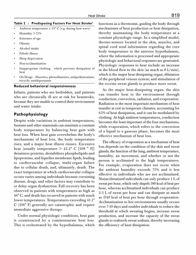
819Heat Stroke
Reduced behavioral responsiveness
Infants, patients who are bedridden, and patients who are chronically ill are at risk for heatstroke, because they are unable to control their environment and water intake.
PathophysiologyDespite wide variations in ambient temperatures, humans and other mammals can maintain a constant body temperature by balancing heat gain with heat loss. When heat gain overwhelms the body’s mechanisms of heat loss, the body temperature rises, and a major heat illness ensues. Excessive heat (usually temperature > 42.2° C [108 ° F])denatures proteins, destabilizes phospholipids and lipoproteins, and liquefies membrane lipids, leading to cardiovascular collapse, multi-organ failure due to cellular death, and, ultimately, death. The exact temperature at which cardiovascular collapse occurs varies among individuals because coexisting disease, drugs, and other factors may contribute to or delay organ dysfunction. Full recovery has been observed in patients with temperatures as high as 46°C,anddeathhasoccurredinpatientswithmuchlower temperatures. Temperatures exceeding 41.1° C(106°F)generallyarecatastrophicandrequireimmediate aggressive therapy.
Under normal physiologic conditions, heat gain is counteracted by a commensurate heat loss. This is orchestrated by the hypothalamus, which
functions as a thermostat, guiding the body through mechanisms of heat production or heat dissipation, thereby maintaining the body temperature at a constant physiologic range. In a simplified model, thermo-sensors located in the skin, muscles, and spinal cord send information regarding the core body temperature to the anterior hypothalamus, where the information is processed and appropriate physiologic and behavioral responses are generated. Physiologic responses to heat include an increase in the blood flow to the skin (as much as 8 L/min), which is the major heat-dissipating organ; dilatation of the peripheral venous system; and stimulation of the eccrine sweat glands to produce more sweat.
As the major heat-dissipating organ, the skin can transfer heat to the environment through conduction, convection, radiation, and evaporation. Radiation is the most important mechanism of heat transfer at rest in temperate climates, accounting for 65% of heat dissipation, and it can be modulated by clothing. At high ambient temperatures, conduction becomes the least important of the four mechanisms, while evaporation, which refers to the conversion of a liquid to a gaseous phase, becomes the most effective mechanism of heat loss.
The efficacy of evaporation as a mechanism of heat loss depends on the condition of the skin and sweat glands, the function of the lung, ambient temperature, humidity, air movement, and whether or not the person is acclimated to the high temperatures. For example, evaporation does not occur when the ambient humidity exceeds 75% and is less effective in individuals who are not acclimatized. Nonacclimatized individuals can only produce 1 L of sweat per hour, which only dispels 580 kcal of heat per hour, whereas acclimatised individuals can produce 2-3 L of sweat per hour and can dissipate as much as 1740 kcal of heat per hour through evaporation. Acclimatization to hot environments usually occurs over 7-10 days and enables individuals to reduce the threshold at which sweating begins, increase sweat production, and increase the capacity of the sweat glands to reabsorb sweat sodium, thereby increasing the efficiency of heat dissipation.
Table 2 : Predisposing Factors For Heat Stroke4
• Ambienttemperature≥35°C(e.g.duringheatwave)
• Humidity>75%
• Extremesofage
• Obesity
• Alcoholintake
• Febrileillness
• Sleepdeprivation
• Non-acclimatization
• Inappropriate clothing - which prevents dissipation ofheat
• OnDrugs–Diuretics,phenothiazines,antiparkinsonians,tricyclic antidepressants

820 Medicine Update 2008 Vol. 18
When heat gain exceeds heat loss, the body temperature rises. Classic heatstroke occurs inindividuals who lack the capacity to modulate the environment (e.g. infants, elderly individuals, individuals who are chronically ill). Furthermore, elderly persons and patients with diminished cardiovascular reserves are unable to generate and cope with the physiologic responses to heat stress and, therefore, are at risk of heatstroke. Patients with skin diseases and those taking medications that interfere with sweating also are at increased risk for heatstroke because they are unable to dissipate heat adequately. Additionally, the redistribution of blood flow to the periphery, coupled with the loss of fluids and electrolytes in sweat, place a tremendous burden on the heart, which ultimately may fail to maintain an adequate cardiac output, leading to additional morbidity and mortality.2
Factors that interfere with heat dissipation include an inadequate intravascular volume, cardiovascular dysfunction, and abnormal skin. Additionally, high ambient temperatures, high ambient humidity, and many drugs can interfere with heat dissipation, resulting in a major heat illness. Similarly, hypothalamic dysfunction may alter temperature regulation and may result in an unchecked rise in temperature and heat illness.
On a cellular level, many theories have been hypothesized and clinically scrutinized. Generally speaking, heat directly influences the body on a cellular level by interfering with cellular processes along with denaturing proteins and cellular membranes. In turn, an array of inflammatory cytokines and heat shock proteins (HSPs), HSP-70 in particular, which allows the cell to endure the stress of its environment), are produced. If
the stress continues, the cell will succumb to the stress (apoptosis) and die. Certain preexistingfactors, such as age, genetic makeup, and the nonacclimatized individual, may allow progression from heat stress to heatstroke, multiorgan-dysfunction syndrome (MODS), and ultimately death. Progression to heatstroke may occur through thermoregulatory failure, an amplified acute-phase response, and alterations in the expression of HSPs. Thermoregulatory failure switches off vasodilatation and sweating which leads to anhidrosis and subsequent rapid rise of body temperature causing cellular death of brain, liver, kidney and muscle. In brain, there occurs petechial hemorrhage and cerebral edema.
Clinical featuresThere are essential differences between presentation of ‘classical (non-exertional)’ and ‘exertional’ heat stroke1 (Table 3).
Signs and Symptoms of Heatstroke5,6,7,8
Onset - Sudden
Coretemperature(rectal):>40.6°Cor>105°F(occasionallyupto45°C[113°F])
Signs SymptomsAnhidrosis Prodrome (only in 25% cases)Rapid and shallow breathing - Nausea Full bound pulse - VomitingMild BP↑ - IrritabilityConfusion - Dizziness Slurred speech IrritabilityComa SeizuresAtaxia
Table 3 : Presentation of ‘classical (non-exertional)’ and ‘exertional’ heat stroke
Classical (NEHS) Exertional (EHS)
Age group Infants, elderly 15-65 yrs
Health status Acute/chronic illness Usually healthy
Activity Sedentary Usually highly active e.g. athletes, army personnel
Drug use Diuretics, Antiparkinsonians, Anticholinergics, Tricyclic antidepressants
-
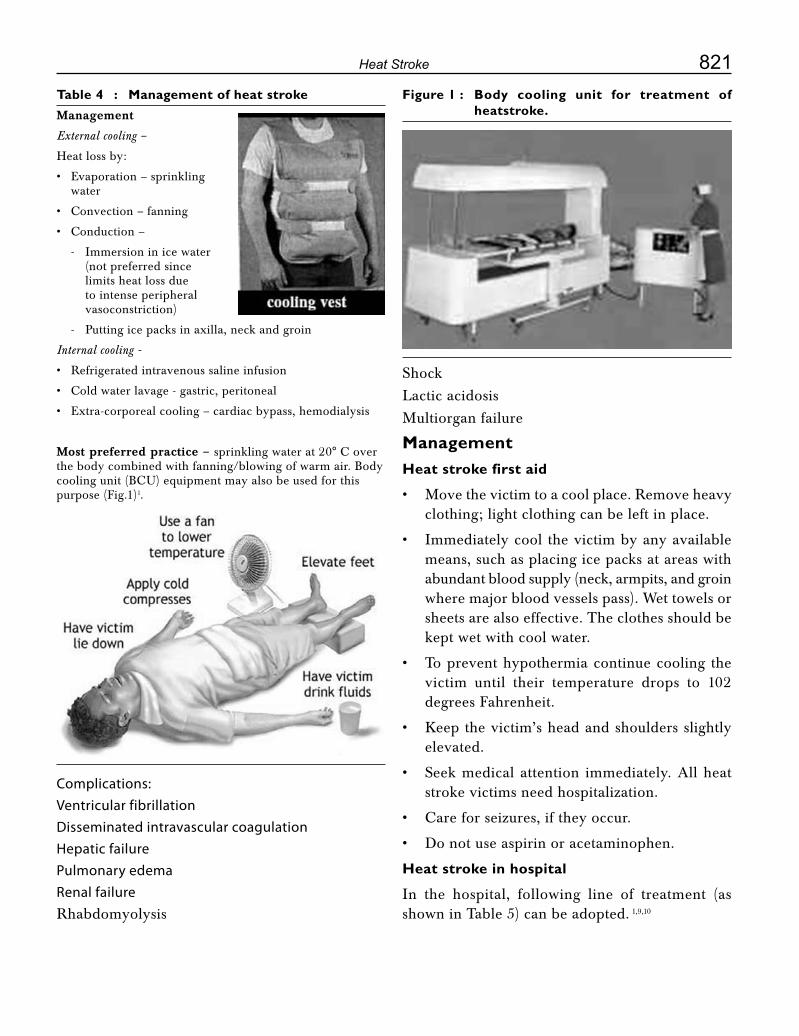
821Heat Stroke
Table 4 : Management of heat strokeManagement
External cooling –
Heat loss by:
• Evaporation–sprinklingwater
• Convection–fanning
• Conduction–
- Immersion in ice water (not preferred since limits heat loss due to intense peripheral vasoconstriction)
- Putting ice packs in axilla, neck and groin
Internal cooling -
• Refrigeratedintravenoussalineinfusion
• Coldwaterlavage-gastric,peritoneal
• Extra-corporealcooling–cardiacbypass,hemodialysis
Most preferred practice – sprinklingwaterat20°Coverthe body combined with fanning/blowing of warm air. Body coolingunit(BCU)equipmentmayalsobeusedforthispurpose (Fig.1)1.
Figure 1 : Body cooling unit for treatment of heatstroke.
Complications:Ventricular fibrillationDisseminated intravascular coagulationHepatic failurePulmonary edemaRenal failureRhabdomyolysis
Shock
Lactic acidosis
Multiorgan failure
ManagementHeat stroke first aid
Move the victim to a cool place. Remove heavy •clothing; light clothing can be left in place.
Immediately cool the victim by any available •means, such as placing ice packs at areas with abundant blood supply (neck, armpits, and groin where major blood vessels pass). Wet towels or sheets are also effective. The clothes should be kept wet with cool water.
To prevent hypothermia continue cooling the •victim until their temperature drops to 102 degrees Fahrenheit.
Keep the victim’s head and shoulders slightly •elevated.
Seek medical attention immediately. All heat •stroke victims need hospitalization.
Careforseizures,iftheyoccur.•
Do not use aspirin or acetaminophen. •
Heat stroke in hospital
In the hospital, following line of treatment (as shown in Table 5) can be adopted. 1,9,10
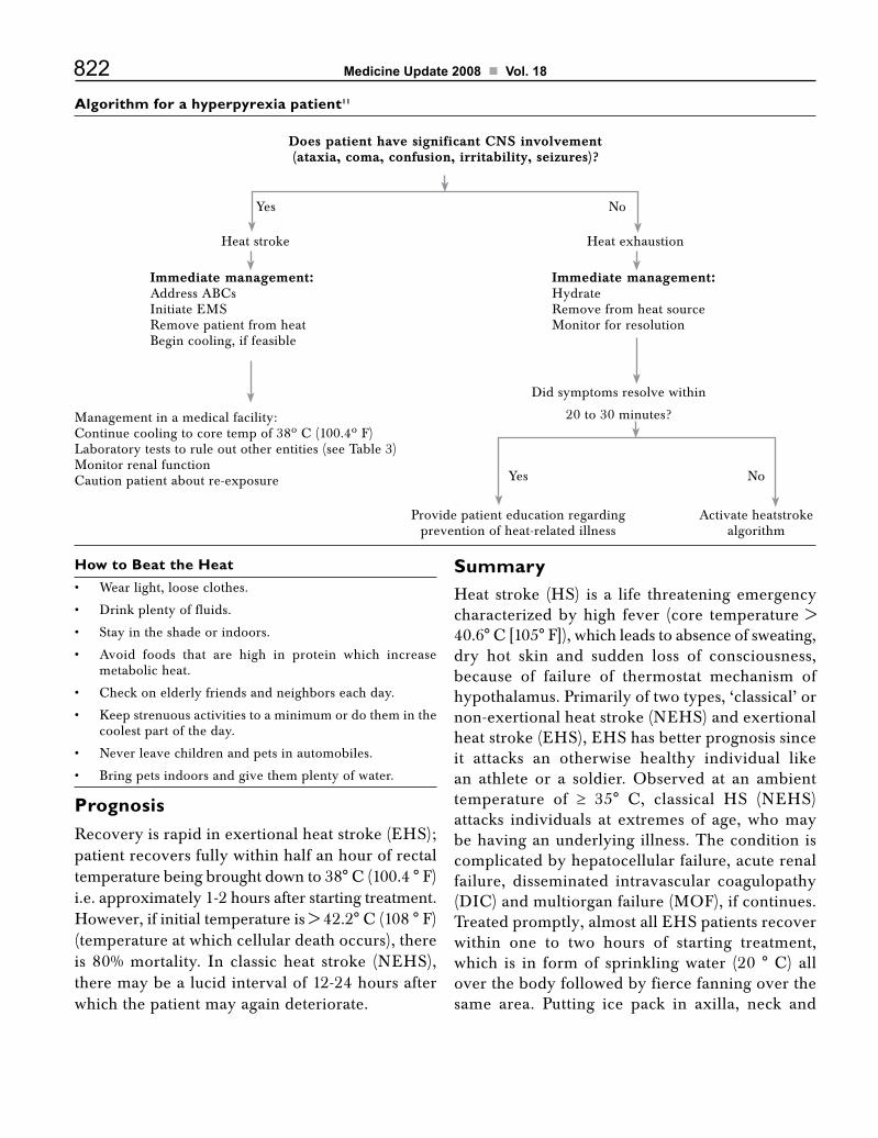
822 Medicine Update 2008 Vol. 18
Algorithm for a hyperpyrexia patient11
Does patient have significant CNS involvement(ataxia, coma, confusion, irritability, seizures)?
Yes
Yes
Heat stroke
Immediate management:AddressABCsInitiate EMSRemove patient from heatBegin cooling, if feasible
Management in a medical facility:Continuecoolingtocoretempof38ºC(100.4ºF)Laboratory tests to rule out other entities (see Table 3)Monitor renal functionCautionpatientaboutre-exposure
Provide patient education regardingprevention of heat-related illness
Activate heatstrokealgorithm
Immediate management:HydrateRemove from heat sourceMonitor for resolution
Did symptoms resolve within
20 to 30 minutes?
Heat exhaustion
No
No
SummaryHeat stroke (HS) is a life threatening emergency characterized by high fever (core temperature > 40.6°C[105°F]),whichleadstoabsenceofsweating,dry hot skin and sudden loss of consciousness, because of failure of thermostat mechanism of hypothalamus. Primarily of two types, ‘classical’ or non-exertional heat stroke (NEHS) and exertional heat stroke (EHS), EHS has better prognosis since it attacks an otherwise healthy individual like an athlete or a soldier. Observed at an ambient temperature of ≥ 35° C, classical HS (NEHS)attacks individuals at extremes of age, who may be having an underlying illness. The condition is complicated by hepatocellular failure, acute renal failure, disseminated intravascular coagulopathy (DIC)andmultiorganfailure(MOF),ifcontinues.Treated promptly, almost all EHS patients recover within one to two hours of starting treatment, which is in formof sprinklingwater (20 °C) allover the body followed by fierce fanning over the same area. Putting ice pack in axilla, neck and
How to Beat the HeatWear light, loose clothes.•
Drink plenty of fluids.•
Stay in the shade or indoors.•
Avoid foods that are high in protein which increase •metabolic heat.
Checkonelderlyfriendsandneighborseachday.•
Keep strenuous activities to a minimum or do them in the •coolest part of the day.
Never leave children and pets in automobiles.•
Bring pets indoors and give them plenty of water.•
PrognosisRecovery is rapid in exertional heat stroke (EHS); patient recovers fully within half an hour of rectal temperaturebeingbroughtdownto38°C(100.4°F)i.e. approximately 1-2 hours after starting treatment. However,ifinitialtemperatureis>42.2°C(108°F)(temperature at which cellular death occurs), there is 80% mortality. In classic heat stroke (NEHS), there may be a lucid interval of 12-24 hours after which the patient may again deteriorate.
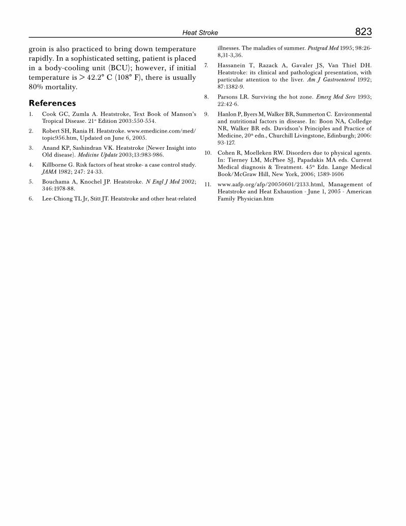
823Heat Stroke
groin is also practiced to bring down temperature rapidly. In a sophisticated setting, patient is placed inabody-coolingunit (BCU);however, if initialtemperatureis>42.2°C(108°F),thereisusually80% mortality.
References1. Cook GC, Zumla A. Heatstroke, Text Book of Manson’s
Tropical Disease. 21st Edition 2003:550-554.
2. Robert SH, Rania H. Heatstroke. www.emedicine.com/med/topic956.htm, Updated on June 6, 2005.
3. Anand KP, Sashindran VK. Heatstroke (Newer Insight into Old disease). Medicine Update 2003;13:983-986.
4. Killborne G. Risk factors of heat stroke- a case control study. JAMA 1982; 247: 24-33.
5. Bouchama A, Knochel JP. Heatstroke. N Engl J Med 2002; 346:1978-88.
6. Lee-ChiongTLJr,StittJT.Heatstrokeandotherheat-related
illnesses. The maladies of summer. Postgrad Med 1995; 98:26-8,31-3,36.
7. Hassanein T, Razack A, Gavaler JS, Van Thiel DH. Heatstroke: its clinical and pathological presentation, with particular attention to the liver. Am J Gastroenterol 1992; 87:1382-9.
8. Parsons LR. Surviving the hot zone. Emerg Med Serv 1993; 22:42-6.
9. HanlonP,ByersM,WalkerBR,SummertonC.Environmentalandnutritional factors indisease. In:BoonNA,ColledgeNR, Walker BR eds. Davidson’s Principles and Practice of Medicine, 20thedn.,ChurchillLivingstone,Edinburgh;2006:93-127.
10. CohenR,MoellekenRW.Disordersduetophysicalagents.In:TierneyLM,McPheeSJ,PapadakisMAeds.CurrentMedical diagnosis & Treatment. 45th Edn. Lange Medical Book/McGraw Hill, New York, 2006; 1589-1606
11. www.aafp.org/afp/20050601/2133.html, Management of Heatstroke and Heat Exhaustion - June 1, 2005 - American Family Physician.htm
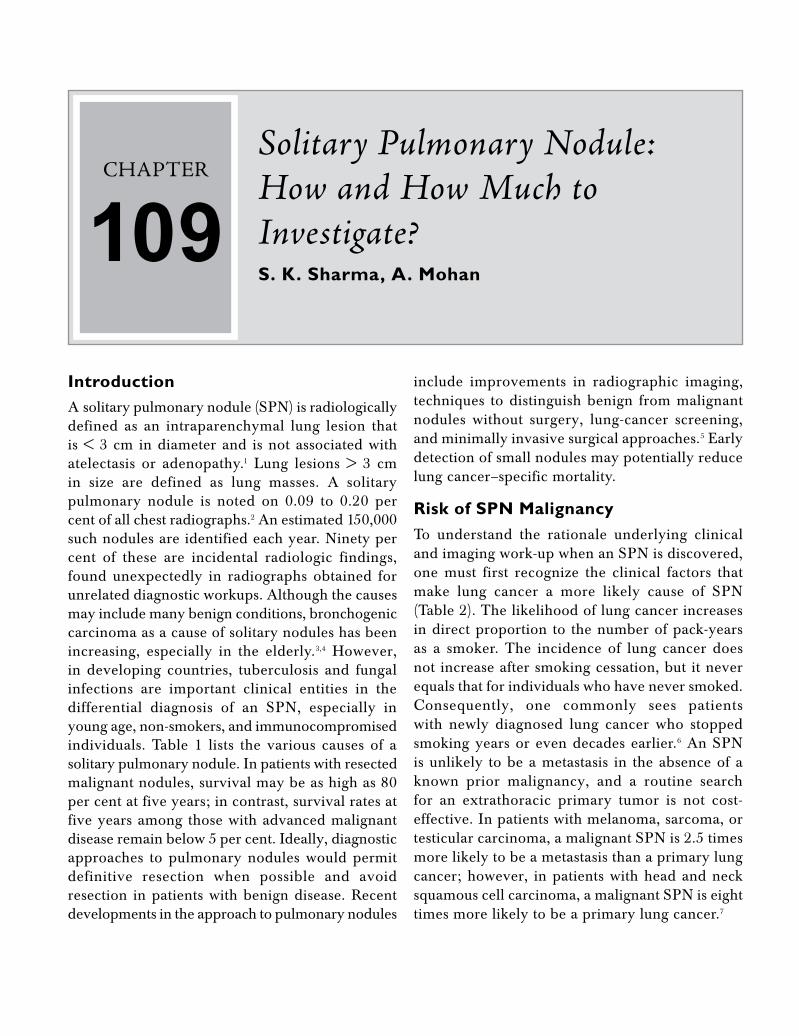
CHAPTER
109Solitary Pulmonary Nodule: How and How Much to Investigate?S. K. Sharma, A. Mohan
IntroductionA solitary pulmonary nodule (SPN) is radiologically defined as an intraparenchymal lung lesion that is < 3 cm in diameter and is not associated with atelectasis or adenopathy.1 Lung lesions > 3 cm in size are defined as lung masses. A solitary pulmonary nodule is noted on 0.09 to 0.20 per cent of all chest radiographs.2 An estimated 150,000 such nodules are identified each year. Ninety per cent of these are incidental radiologic findings, found unexpectedly in radiographs obtained for unrelated diagnostic workups. Although the causes may include many benign conditions, bronchogenic carcinoma as a cause of solitary nodules has been increasing, especially in the elderly.3,4 However, in developing countries, tuberculosis and fungal infections are important clinical entities in the differential diagnosis of an SPN, especially in young age, non-smokers, and immunocompromised individuals. Table 1 lists the various causes of a solitary pulmonary nodule. In patients with resected malignant nodules, survival may be as high as 80 per cent at five years; in contrast, survival rates at five years among those with advanced malignant disease remain below 5 per cent. Ideally, diagnostic approaches to pulmonary nodules would permit definitive resection when possible and avoid resection in patients with benign disease. Recent developments in the approach to pulmonary nodules
include improvements in radiographic imaging, techniques to distinguish benign from malignant nodules without surgery, lung-cancer screening, and minimally invasive surgical approaches.5 Early detection of small nodules may potentially reduce lung cancer–specific mortality.
Risk of SPN MalignancyTo understand the rationale underlying clinical and imaging work-up when an SPN is discovered, one must first recognize the clinical factors that make lung cancer a more likely cause of SPN (Table 2). The likelihood of lung cancer increases in direct proportion to the number of pack-years as a smoker. The incidence of lung cancer does not increase after smoking cessation, but it never equals that for individuals who have never smoked. Consequently, one commonly sees patientswith newly diagnosed lung cancer who stopped smoking years or even decades earlier.6 An SPN is unlikely to be a metastasis in the absence of a known prior malignancy, and a routine search for an extrathoracic primary tumor is not cost-effective. In patients with melanoma, sarcoma, or testicular carcinoma, a malignant SPN is 2.5 times more likely to be a metastasis than a primary lung cancer; however, in patients with head and neck squamous cell carcinoma, a malignant SPN is eight times more likely to be a primary lung cancer.7
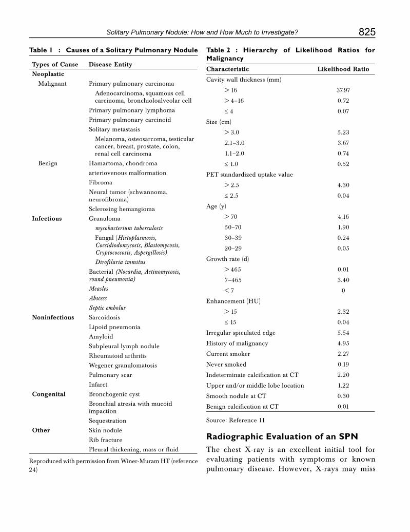
825Solitary Pulmonary Nodule: How and How Much to Investigate?
Table 1 : Causes of a Solitary Pulmonary Nodule
Types of Cause Disease Entity
Neoplastic
Malignant Primary pulmonary carcinoma
Adenocarcinoma, squamous cell carcinoma, bronchioloalveolar cell
Primary pulmonary lymphoma
Primary pulmonary carcinoid
Solitary metastasis
Melanoma, osteosarcoma, testicular cancer, breast, prostate, colon, renal cell carcinoma
Benign Hamartoma, chondroma
arteriovenous malformation
Fibroma
Neural tumor (schwannoma, neurofibroma)
Sclerosing hemangioma
Infectious Granuloma
mycobacterium tuberculosis
Fungal (Histoplasmosis, Coccidiodomycosis, Blastomycosis, Cryptococcosis, Aspergillosis)
Dirofilaria immitus
Bacterial (Nocardia, Actinomycosis, round pneumonia)
Measles
Abscess
Septic embolus
Noninfectious Sarcoidosis
Lipoid pneumonia
Amyloid
Subpleural lymph nodule
Rheumatoid arthritis
Wegener granulomatosis
Pulmonary scar
Infarct
Congenital Bronchogenic cyst
Bronchial atresia with mucoid impaction
Sequestration
Other Skin nodule
Rib fracture
Pleural thickening, mass or fluid
Reproduced with permission from Winer-Muram HT (reference 24)
Table 2 : Hierarchy of Likelihood Ratios for MalignancyCharacteristic Likelihood Ratio
Cavitywallthickness(mm)
> 16 37.97
> 4–16 0.72
≤ 4 0.07
Size (cm)
> 3.0 5.23
2.1–3.0 3.67
1.1–2.0 0.74
≤ 1.0 0.52
PET standardized uptake value
> 2.5 4.30
≤ 2.5 0.04
Age (y)
> 70 4.16
50–70 1.90
30–39 0.24
20–29 0.05
Growth rate (d)
> 465 0.01
7–465 3.40
< 7 0
Enhancement (HU)
> 15 2.32
≤ 15 0.04
Irregular spiculated edge 5.54
History of malignancy 4.95
Currentsmoker 2.27
Never smoked 0.19
IndeterminatecalcificationatCT 2.20
Upper and/or middle lobe location 1.22
SmoothnoduleatCT 0.30
BenigncalcificationatCT 0.01
Source: Reference 11
Radiographic Evaluation of an SPNThe chest X-ray is an excellent initial tool for evaluating patients with symptoms or known pulmonary disease. However, X-rays may miss

826 Medicine Update 2008 Vol. 18
Figure 1 : Chest CT scans (5-mm section width) in a female 48-year-old former smoker (a) scan shows a10-mm solid nodule (arrow) in the right lower lobe. (b) Transverse thin-section (1.25-mm section width) scan shows irregular margins and central lucency.
upto 19% of SPNs and have been shown to cause substantial delay in diagnosis of lung cancer with upstaging of lesions from T1 to T2 during the delay period.8SpiralCTwithIVcontrastenhancementis the imaging modality of choice for the SPN and should be obtained on all newly diagnosed SPNs. (Figure 1)
At chest radiography, an SPN is seldom evident until it is at least 9 mm in diameter. Traditionally, absence of growth over a 2-year time period has been believed to be a reliable indicator of benign disease. Therefore, for patients with an SPN that isvisibleontheCXR,allpreviousCXRsshouldbereviewed.ForallpatientswithpreviousCXRs,an SPN that is unchanged for > 2 years does not require further diagnostic evaluation. Benign lesions typically have a doubling time of either < 1 month or > 16 months.9 Malignant nodules have a doubling time from anywhere from 40 to 360 days.10 Although the optimal frequency of follow-up imaging is not known, the traditional standard of imaging at three-month intervals during the first year after a nodule is discovered and then at six-month intervals during the next year is logical, provided thathigh-resolutionCT isused, ratherthan plain film radiography.
SPN SizeThe size of the SPN is not a reliable predictor of benignity; however, the larger the nodule (approaching 3 cm in diameter), the more likely it is to be malignant. More than 90% of nodules that are smaller than 2 cm in diameter are benign.11,12 In 64 patients with SPNs 1 cm or smaller in diameter who were referred for video-assisted thoracoscopic surgery, 58% of SPNs, including six that were smaller than 5 mm in diameter, were malignant.13 In comparison, the Early Lung Cancer Action Project screening study showedthat only 8% of lesions smaller than 1 cm in diameter were malignant.14
SPN Location Lung cancer is 1.5 times more likely to occur in the right lung than in the left lung.15 Studies have shown that 70% of lung cancers are located in the upper lobes and occur most frequently in the right lung.16,17 Thus, one should carefully scrutinize the upper lobes when reviewing chest images, as most missed lung cancers are located in the right upper lobe. As benign nodules are equally distributed throughout the upper and lower lobes, location alone cannot be used as an independent predictor of malignancy. Approximately half of primary pulmonary adenocarcinomas manifest as isolated peripheral SPNs, while squamous cell carcinomas that manifest as SPNs are more likely to be centrally located.18
CalcificationThe most important imaging feature that can be used to distinguish benign SPNs from malignant SPNs is calcification. Benign nodules can be diagnosed confidently if the lesion is smaller than 3 cm in diameter and exhibits one of the following patterns of calcification: central nidus, laminated, popcorn, or diffuse. When one of these patterns is seen, the likelihood of benignity approaches 100%.3,19 Popcorn calcifications are observed in one-third of hamartomas, and the other patterns are seen with histoplasmosis and tuberculosis.
827Solitary Pulmonary Nodule: How and How Much to Investigate?
Figure 2 : CT scan shows eccentric dense calcification in a right lower lobe carcinoid tumor.
Figure 3 : CT scan showing a nodule with a corona radiata. Multiple fine striations extend perpendicularly to the surface of the nodule, which is surrounded by a radiolucent halo formed by emphysematous lung tissue.
WhileCTstudieshave shown thatup to13%oflung cancers have some calcification,20,21 this is true of only 2% of lung cancers smaller than 3 cm in diameter.20 Eccentric calcification should not be considered a benign finding. It may represent a benign lesion that has calcified in an eccentric fashion or a malignant lesion that has dystrophic calcification or has engulfed a benign calcified lesion. (Figure 2) Furthermore, central calcification in a speculated SPN should prompt concern for malignancy, as most benign SPNs have smooth orminimally lobulatedmargins.Calcification inlung cancers may appear amorphous, stippled, or diffuse. A stippled appearance or psammomatous calcification can be seen in SPNs that are metastases from mucin-secreting tumors, such as colon or ovarian cancers. With the exception of SPNs in patients with a history of bone malignancy, SPNs with a benign pattern of calcification are indeed benign.
Nodule AttenuationTheadventofCThasledtoimprovedrecognitionof the frequency with which nodules are nonsolid, partly solid, or solid. Approximately 34% of nonsolid
nodules are due to malignancy. Malignancies such as bronchioloalveolar carcinomas or invasive adenocarcinomas with bronchioloalveolar cell features may appear to be nonsolid nodules. Nonsolid nodules are often caused by benign conditions, such as inflammatory disease, and may contain premalignant lesions, such as atypical adenomatous hyperplasia or bronchoalveolar hyperplasia. Although solid nodules are the most common type of nodule, they are less likely to be malignant than are partly solid or nonsolid nodules. Inflammatory diseases of the lung, particularly tuberculosis and mycoses, usually produce solid nodules that may eventually calcify and permit the designation of benign disease. While solid nodules are usually noncancerous (granulomas), most lung cancers are found in solid nodules. Histologic types of cancerous solid nodules include adenocarcinomas and squamous cell, large-cell anaplastic, neuroendocrine, carcinoid, and (rarely) small-cell carcinomas.22
MarginTwo patterns of the margins of a nodule are relatively specific for cancer. One is the corona radiata sign, consisting of very fine linear strands extending 4 to 5 mm outward from the nodule,

828 Medicine Update 2008 Vol. 18
Figure 4 : CT scan showing a solitary pulmonary nodule with regular margins before (left frame) and after (right frame) contrast administration showing enhancement of the walls indicating benignity
Figure 5 : CT scan showing a 2.5-cm left upper lobe cavitary nodule. The wall is irregular and the cavity wall is thick.
originally described on plain tomographs; they have a spiculated appearance on plain radiographs (Fig. 3). A scalloped border is associated with an intermediate probability of cancer, whereas a smooth border is more suggestive of a benign diagnosis. Edge characteristics indicative of malignancy include irregularity, spiculation, and lobulation. Nodule halos (peripheral nonsolid component) should not be confused with the corona radiata, which is a radiolucent halo associated with paracicatricial emphysema. The presence of spiculation has a predictive value for malignancy of approximately 90% and should prompt an aggressive work-up.3,11,19 While an irregular margin is indicative of malignancy, it can occasionally be seen in granulomatous disease, lipoid pneumonia, organizing pneumonia, and progressive massive fibrosis. A smooth margin does not indicate benignity, as up to one-third of malignant lesions have smooth margins and many of these tumors are metastatic.23 Adjacent tiny nodules, called satellite nodules, may mimic the appearance of a lobulated margin, and the presence of these nodules is strongly associated with benignity. Another indicator of benignity is the presence of enhancement of the wall of the nodule after contrast administration (Figure 4).24
CavitationBoth benign and malignant nodules can form a cavity. Up to 15% of lung cancers form a cavity, but most are larger than 3 cm in diameter.25 However, cavitation may be seen in SPNs as small as 7 mm in diameter (Figure 5). SPNs with irregular-walled
cavities thicker than 16 mm tend to be malignant (84%–95% of SPNs), while benign cavitated lesions usually have thinner smoother walls; approximately 95% of lesions with cavity walls thinner than 4 mm are benign.26
Positron Emission TomographyPositron emission tomography (PET) is rapidly becoming a front-line modality in the evaluation of SPNs. Its diagnostic ability is based on increased glucose consumption of malignant cells. The radiopharmaceutical fluorine 18 fluorodeoxyglucose (FDG) is a glucose analog that is injected intravenously, transported through the cell membrane, and phosphorylated through normal glycolytic pathways, remaining unmetabolized in the cell. For solid pulmonary nodules 1–3 cm in diameter, sensitivity and specificity are approximately 94% and 83%, respectively.27 (Figure 6) The probability of malignancy in association with positive FDG PET findings is high (90% if the patient is older than 60 years); likewise, the probability of malignancy in association with negative FDG PET findings is low (< 5%).28,29 False-positive
829Solitary Pulmonary Nodule: How and How Much to Investigate?
PET findings are associated with focal infections, inflammation, and nonneoplastic diseases (e.g., tuberculosis, sarcoidosis, and rheumatoid disease) and are more frequent in regions with endemic fungal diseases such as Histoplasma and Coccidioides infections. However, certain neoplasms, such as carcinoid and bronchioloalveolar cell carcinoma, have a low metabolic rate that may result in false-negative examinations. Furthermore, sensitivity and specificity are not as high for nodules that are smaller than 1 cm in diameter.29
Obtaining Tissue Diagnosis A. Transthoracic Fine-Needle Aspiration Biopsy
Transthoracic fine-needle aspiration biopsy identifies peripheral pulmonary lesions as malignant or benign in up to 95 per cent of cases. For malignant lesions, the sensitivity is 80 to 95 per cent and the specificity is 50 to 88 per cent. The positive predictive value in one study involving more than 200 patients was 98.6 per cent; the negative predictive value was 96.6 per cent.30 Even for lesions that are less than 2 cm
in diameter, transthoracic fine-needle aspiration biopsy has a sensitivity of more than 60 per cent for detecting a malignant process. However, the falsenegativerateis3to29percent.Complicationrates are higher than those for bronchoscopy, with an incidence of pneumothorax of up to 30 per cent, although in most cases, treatment is not required.31,32 Unfortunately, the sensitivity of TTNA for a specific benign diagnosis is 12 to 68% but only 12% in a number of studies.32 Adding automated cutting (core needle biopsy) to TTNA may increase the yield of a specific diagnosis of benign disease from 12 to 75%.33 Relative contraindications to this procedure are the patient with pulmonary hypertension, coagulopathy or a bleeding diathesis, severe COPD, or vascular malformations. The mostfrequent complication of TTNA is pneumothorax in 25 to 30% of patients, with 5 to 10% of these patients requiring a chest tube. Pneumothorax is decreased by avoiding crossing pulmonary fissures and multiple punctures of the lung parenchyma. There can be up to a 10% incidence of hemoptysis and hemorrhage, which is increased by the use of cutting needles. Air embolus and tumor seeding are rare, 0.1% and 0.05% respectively.34
Bronchoscopy
The sensitivity of bronchoscopy for detecting a malignant process in a solitary pulmonary nodule ranges from 20 to 80 per cent, depending on the size of the nodule, its proximity to the bronchial tree, and the prevalence of cancer in the study population.35,36 For nodules that are less than 1.5 cm in diameter, the sensitivity is 10 per cent, and for those that are 2.0 to 3.0 cm in diameter, it is 40 to 60percent.WhenCTrevealsabronchusleadingtothe lesion, bronchoscopy has 70 per cent sensitivity. Ultrathin bronchoscopy, which involves the use of fiberoptic technology in thin bronchoscopes that can reach beyond eighth-generation bronchi, has been used experimentally to allow direct visualization of peripheral lesions.
Figure 6 : PET scan image showing increased uptake in a nodule in the upper lobe of left lung
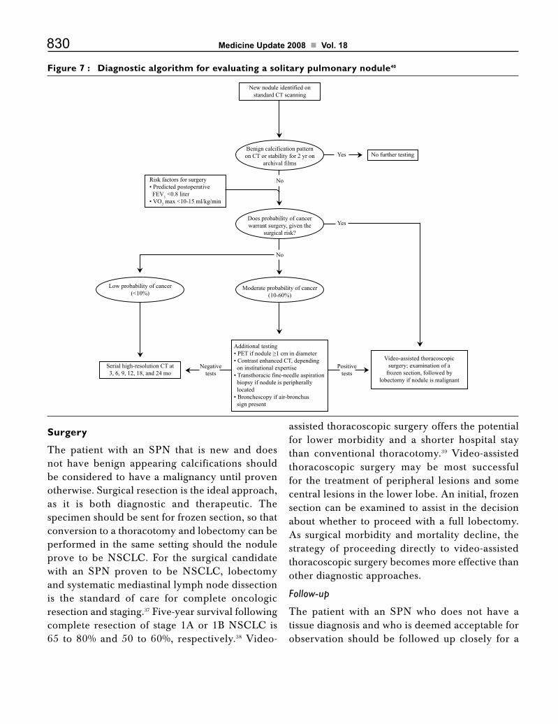
830 Medicine Update 2008 Vol. 18
Figure 7 : Diagnostic algorithm for evaluating a solitary pulmonary nodule40
Surgery
The patient with an SPN that is new and does not have benign appearing calcifications should be considered to have a malignancy until proven otherwise. Surgical resection is the ideal approach, as it is both diagnostic and therapeutic. The specimen should be sent for frozen section, so that conversion to a thoracotomy and lobectomy can be performed in the same setting should the nodule prove to be NSCLC. For the surgical candidatewith an SPN proven to be NSCLC, lobectomyand systematic mediastinal lymph node dissection is the standard of care for complete oncologic resection and staging.37 Five-year survival following complete resectionof stage1Aor1BNSCLC is65 to 80% and 50 to 60%, respectively.38 Video-
assisted thoracoscopic surgery offers the potential for lower morbidity and a shorter hospital stay than conventional thoracotomy.39 Video-assisted thoracoscopic surgery may be most successful for the treatment of peripheral lesions and some central lesions in the lower lobe. An initial, frozen section can be examined to assist in the decision about whether to proceed with a full lobectomy. As surgical morbidity and mortality decline, the strategy of proceeding directly to video-assisted thoracoscopic surgery becomes more effective than other diagnostic approaches.
Follow-up
The patient with an SPN who does not have a tissue diagnosis and who is deemed acceptable for observation should be followed up closely for a
New nodule identified onstandard CT scanning
No further testingYes
Yes
No
No
Benign calcification patternon CT or stability for 2 yr on
archival films
Does probability of cancerwarrant surgery, given the
surgical risk?
Moderate probability of cancer(10-60%)
Additional testing• PET if nodule ≥1 cm in diameter• Contrast enhanced CT, depending on institutional expertise• Transthoracic fine-needle aspiration biopsy if nodule is peripherally located• Bronchescopy if air-bronchus sign present
Positivetests
Video-assisted thoracoscopicsurgery; examination of a
frozen section, followed bylobectomy if nodule is malignant
Serial high-resolution CT at3, 6, 9, 12, 18, and 24 mo
Negativetests
Low probability of cancer(<10%)
Risk factors for surgery• Predicted postoperative FEV1 <0.8 liter• VO2 max <10-15 ml/kg/min

831Solitary Pulmonary Nodule: How and How Much to Investigate?
minimum of 2 years. This should include an initial CXR,andCTscanningat3,6,12,and24monthsfor best monitoring for nodule growth. There is very little objective evidence for frequency of surveillance monitoring. Figure 5 shows the general recommended scheme for workup of a patient with SPN.
Conclusions and recommendations41
1. ForpatientswithanSPNthatisvisibleonCXR,allpreviousCXRsshouldbereviewed.
2. ForallpatientswithpreviousCXRs,anSPNthat is unchanged for > 2 years does not require further diagnostic evaluation.
3. Forpatientswith anSPNvisibleonCXR inwhich benign central calcification is present, no further diagnostic evaluation is necessary.
4.ForpatientswithanSPN,aspiralCTofthechestwith contrast is indicated to better characterize thenodule,parenchyma,andmediastinum.CTcan be useful in identifying nodules more likely to be benign and obviate the need for further diagnosticevaluation.Additionally, chestCTplays an important role in staging (as delineated in the chapter on noninvasive staging elsewhere in these guidelines).
5. For patients with an SPN, MRI is not indicated except in these special instances.
6. For patient with an SPN _ 1 cm in size, PET scanning is not currently recommended.
7. For patients with an SPN who are surgical candidates and have a negative mediastinal evaluationonCT,PETscanningwithFDGasan investigational tool, where available, may be warranted.
8. For patients with an SPN who are marginal surgical candidates, if PET scanning with FDG resultsarenegative,arepeatCTscanisrequiredat least once in 3 months.
9. For patients with an SPN who are marginal surgical candidates, if there are unchanged results from prior CXR and negative PET
scan findings, serial follow-up is recommended, consistingofaninitialCXR,andCTscanningat 3, 6, 12, and 24 months.
10. For the patients with an SPN who are operable candidates, TTNA is not indicated. Level of evidence, good; benefit, none; grade of recommendation, D. For operable patients with an SPN who decline surgical intervention, TTNA or Transbronchial needle biopsy is the preferred procedure for establishing a diagnosis.
11. For patients with an SPN who are not operable candidates, or are at high risk, TTNA may be helpful to establish tissue diagnosis.
12. For patients with an SPN, bronchoscopy is usually not indicated.
13. For operable patients with an SPN, if the lesion is amenable to a wedge resection, then a wedge resection is the procedure of choice followed by a lobectomy if the pathologic finding is positive for cancer.
14. For operable patients with an SPN, if the lesion is not amenable to a wedge resection, a diagnostic lobectomy is acceptable.
15. All pulmonary resections, anatomic or nonanatomic, must include a systematic lymph node dissection.
16. For patients with an SPN who are marginal surgical candidates, a wedge resection or segmentectomy is acceptable.
17. For patients with an SPN without a definitive tissue diagnosis, a minimum follow-up of 2 years is recommended. This should include an initialCXR,andCTscanningat3,6,12,and24 months.
References1. Tuddenham WI. Glossary of terms for thoracic radiology:
recommendationsof theNomenclatureCommitteeof theFleischner Society. Am J Roentgenol 1984; 43:509–17.
2. Swensen SJ, Silverstein MD, Edell ES, et al. Solitary pulmonary nodules: clinical prediction model versus physicians. Mayo ClinProc1999;74:319–29.
3. Siegelman SS, Khouri NF, Leo FP, Fishman EK, Braverman RM, Zerhouni EA. Solitary pulmonary nodules: CT

832 Medicine Update 2008 Vol. 18
assessment. Radiology 1986;160:307-12.
4. Bateson EM. An analysis of 155 solitary lung lesions illustrating the differential diagnosis of mixed tumors of the lung.ClinRadiol1965;16:51–65.
5. Murthy SC, Rice TW. The solitary pulmonary nodule: aprimerondifferentialdiagnosis.SeminThoracCardiovascSurg 2002; 14:239–249.
6. TongL,SpitzMR,FuegerJJ,AmosCA.Lungcarcinomainformersmokers.Cancer1996;78:1004–10.
7. Quint LE, Park CH, Iannettoni MD. Solitary pulmonarynodules in patients with extrapulmonary neoplasms. Radiology 2000; 217:257–61.
8. Quekel LO, Kessels, AG, Goei R, et al. Miss rate of lung canceron thechest radiograph inclinicalpractice.Chest1999; 115:720–32.
9. Fraser RG, Pare JA. Diagnosis of diseases of the chest. 3rd ed. Vol 2. Philadelphia, PA; W.B. Saunders, 1989; 1327–1699.
10. LillingtonGA,CaskeyCI.Evaluationandmanagementofsolitarymultiplepulmonarynodules.ClinChestMed1993;14:111–19.
11. Gurney JW. Determining the likelihood of malignancy in solitary pulmonary nodules with Bayesian analysis. I. Theory. Radiology 1993;186:405–13.
12. SwensenSJ, Jett JR,HartmanTE,et al.CTscreening forlung cancer: 5-year prospective experience. Radiology 2005;235(1): 259–65.
13. Munden RF, Pugatch RD, Liptay MJ, Sugarbaker DJ, LinhUL.SmallpulmonarylesionsdetectedatCT:clinicalimportance. Radiology 1997;202:105–10.
14. HenschkeCI,McCauleyDI,YankelevitzDF, et al.Earlylung cancer action project: overall design and findings from baseline screening. Lancet 1999;354:99–105.
15. Garland LH. Bronchial carcinomas: lobar distribution of lesionsin250cases.CalifMed1961;94:7–8.
16. Swensen SJ, Viggiano RW, Midthun DE, et al. Lung noduleenhancementatCT:multicenter study.Radiology2000;214:73–80.
17. Winer-Muram HT, Jennings SG, Tarver RD, et al. Volumetric growth rate of stage I lung cancer prior to treatment: serial CTscanning.Radiology2002;223:798–805.
18. Quinn D. Gianlupi A, Broste S. The changing radiographic presentation of bronchogenic carcinoma with reference to celltypes.Chest1996;110:1474–9.
19. Zerhouni EA, Stitik FP, Siegelman SS, et al. CT of thepulmonary nodule: a cooperative study. Radiology 1986;160(2):319–27.
20. GrewalRG,AustinJH.CTdemonstrationofcalcificationincarcinomaofthelung.JComputAssistTomogr1994;18:867–71.
21. MahoneyMC,ShipleyRT,CorcoranHL,DicksonBA.CTdemonstration of calcification in carcinoma of the lung. AJR Am J Roentgenol 1990;154:255–8.
22. Libby DM, Smith JP, Altorki NK, Pasmantier MW, YankelevitzD,HenschkeCI.ManagingthesmallpulmonarynodulediscoveredbyCT.Chest2004;125:1522–9.
23. Seemann MD, Seeman O, Luboldt W, et al. Differentiation
of malignant from benign solitary pulmonary lesions using chest radiography, spiral CT and HRCT. Lung Cancer2000;29:105–24.
24. Winer Muram HT. The Solitary Pulmonary Nodule. Radiology 2006; 239: 34-49.
25. WoodringJH,FriedAM,ChaungVP.Solitarycavitiesofthelung: diagnostic implications of cavity wall thickness. AJR Am J Roentgenol 1980;135:1269–71.
26. Woodring JH, Fried AM. Significance of wall thickness in solitary cavities of the lung: a follow up study. AJR Am J Roentgenol 1983;140:473–4.
27. GouldMK,MacleanCC,KuschnerWG,RydzakCE,OwensDK. Accuracy of positron emission tomography for diagnosis of pulmonary nodules and mass lesions:a meta-analysis. JAMA 2001;285:914–24.
28. Dewan NA, Shehan CJ, Reeb SD, Gobar LS, Scott WJ,Ryschon K. Likelihood of malignancy in a solitary pulmonary nodule: comparison of Bayesian analysis and results of FDG-PETscan.Chest1997;112:416–22.
29. Goldsmith SJ, Kostakoglu L. Nuclear medicine imaging of lungcancer.RadiolClinNorthAm2000;38:511–24.
30. Conces DJ Jr, Schwenk GR Jr, Doering PR, Glant MD.Thoracic needle biopsy: improved results utilizing a team approach.Chest1987;91:813-6.
31. Weisbrod GL. Transthoracic needle biopsy. World J Surg 1993; 17:705–11.
32. Lacasse Y, Wong E, Guyatt GH, et al. Transthoracic needle aspiration biopsy for the diagnosis of localized pulmonary lesions: a meta-analysis. Thorax 1999; 54:884–93.
33. Doiselle PM, Shepard, IO, Mark EL. Routine addition of an automated biopsy device to fine-needle aspiration of the lung: a prospective assessment. AJR Am J Roentgenol 1997; 169: 661–6.
34. MurphyJM,GleesonFV,FlowerCD.Percutaneousneedlebiopsy of the lung and its impact on patient management. World J Surg 2001; 25:373–80.
35. Stringfield JT, Markowitz DJ, Bentz RR, et al. The effect of tumor size and location on diagnosis by fiberoptic bronchoscopy.Chest1977;72:474–6.
36. Baaklini WA, Reinoso MA, Gorin AB, et al. Diagnostic yield of fiberoptic bronchoscopy in evaluating solitary pulmonary nodules.Chest2000;117:1049–54.
37. Martini N, Ginsberg RJ. Treatment of stage I and stage II disease. In: Aisner R, Arriga R, Green N, et al, eds. The comprehensive textbook of thoracic oncology. Baltimore, MD: Williams and Wilkens, 1996; 338–50.
38. Warren WH, Faber LP. Segmentectomy vs lobectomy in patients with stage I pulmonary carcinoma. J Thorac CardiovascSurg1994;107:1087–93.
39. Suzuki K, Nagai K, Yoshida J, et al. Video-assisted thoracoscopic surgery for small indeterminate pulmonary nodules.Chest1999;115:563–8.
40. Ost D, Fein AM, Feinsilver SH. The Solitary Pulmonary Nodule. New Engl J Med 2003; 348: 2535-42.
41. Tan BB, Flaherty KR, Kazerooni EA, Iannettoni MD; American College of Chest Physicians. The solitarypulmonarynodule.Chest2003;123(1suppl):89S–96S.

CHAPTER
110Management of Non Variceal BleedR. K. Jain
IntroductionNon –variceal upper gastrointestinal bleeding (UGIB) remains a common and challenging emergency for gastroenterologists and general physician. The annual incidence is 50 to 150 per 100,000 of the population, and even though there have been significant improvements in endoscopic and supportive therapies, the overall mortality remains around 10%, and may even reach 35% in hospitalised patients with serious co-morbidity. Patients aged over 80 years of age now account for around 25%of all UGIB and 33% of UGIB occurring in hospitalised patients and therefore
tend to account for much of the poor outcome of this condition.1
Etiologies of non-variceal UGI bleedingThe major etiologies of acute non-variceal UGI bleeding are listed in Table 1. Peptic ulcer diseases accountsformostcases,followedbylessCommoncauses including mucosal erosive diseases; Mallory-Weiss tears; gastric antral vascular ectasia (watermelon stomach); angiomas; tumors; and Dieulafoys lesions. Rare causes, such as hemobilia, aortoenteric fistula and vasculitis, must be considered in the appropriate clinical situation.
Although the commonly quoted figure of 50% for peptic ulcer bleeding may be overestimated. In arecent largeCORI(ClinicalOutcomeResearchInitiative) study of UGIB, peptic ulcer was the probable cause of UGIB in only 20% of cases.2 The incidence of peptic ulcer disease is expected to continue to decline with more widespread helicobacter pylori eradication and proton pump inhibitor (PPI) usage.
The use of NSAIDs has been shown to be associated with an increased risk of ulcer bleeding in several studies;3,4 this risk increases with the use of conventional NSAIDs (versus selective cyclooxygenase-2 inhibitors), comorbid conditions,
Table 1 : Etiology of non-variceal UGI bleedingSymptom Frequency (%)
Peptic ulcers 50
Erosions 25-30
Mallory-Weiss tear 8
Watermelon stomach 3-5
Carcinomas 1-5
Angiomata 1-2
Dieulafoy’s lesion 1-2
Hemobilia <1
Aorto-enteric fistula <1
Other 6-10
Adapted from gupta PK, Nonvariccal upper gastrointestinal bleeding.MedClinNorth Am 1193;77:973-92
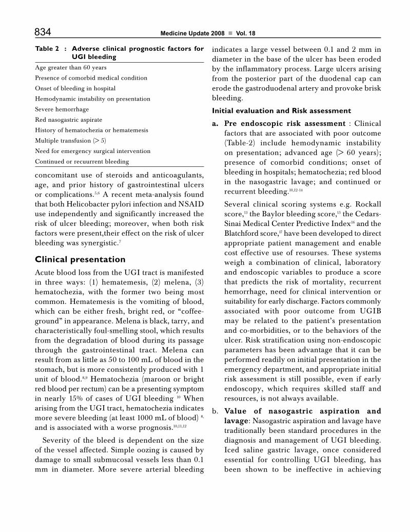
834 Medicine Update 2008 Vol. 18
concomitant use of steroids and anticoagulants, age, and prior history of gastrointestinal ulcers or complications.5,6 A recent meta-analysis found that both Helicobacter pylori infection and NSAID use independently and significantly increased the risk of ulcer bleeding; moreover, when both risk factors were present,their effect on the risk of ulcer bleeding was synergistic.7
Clinical presentationAcute blood loss from the UGI tract is manifested in three ways: (1) hematemesis, (2) melena, (3) hematochezia, with the former two being most common. Hematemesis is the vomiting of blood, which can be either fresh, bright red, or “coffee-ground” in appearance. Melena is black, tarry, and characteristically foul-smelling stool, which results from the degradation of blood during its passage through the gastrointestinal tract. Melena can result from as little as 50 to 100 mL of blood in the stomach, but is more consistently produced with 1 unit of blood.8,9 Hematochezia (maroon or bright red blood per rectum) can be a presenting symptom in nearly 15% of cases of UGI bleeding 10 When arising from the UGI tract, hematochezia indicates more severe bleeding (at least 1000 mL of blood) 8, and is associated with a worse prognosis.10,11,12
Severity of the bleed is dependent on the size of the vessel affected. Simple oozing is caused by damage to small submucosal vessels less than 0.1 mm in diameter. More severe arterial bleeding
indicates a large vessel between 0.1 and 2 mm in diameter in the base of the ulcer has been eroded by the inflammatory process. Large ulcers arising from the posterior part of the duodenal cap can erode the gastroduodenal artery and provoke brisk bleeding.
Initial evaluation and Risk assessment
a. Pre endoscopic risk assessment : Clinicalfactors that are associated with poor outcome (Table-2) include hemodynamic instability on presentation; advanced age (> 60 years); presence of comorbid conditions; onset of bleeding in hospitals; hematochezia; red blood in the nasogastric lavage; and continued or recurrent bleeding.10,12-14
Several clinical scoring systems e.g. Rockall score,13 the Baylor bleeding score,15theCedars-SinaiMedicalCenterPredictiveIndex16 and the Blatchford score,17 have been developed to direct appropriate patient management and enable cost effective use of resourses. These systems weigh a combination of clinical, laboratory and endoscopic variables to produce a score that predicts the risk of mortality, recurrent hemorrhage, need for clinical intervention or suitability for early discharge. Factors commonly associated with poor outcome from UGIB may be related to the patient’s presentation and co-morbidities, or to the behaviors of the ulcer. Risk stratification using non-endoscopic parameters has been advantage that it can be performed readily on initial presentation in the emergency department, and appropriate initial risk assessment is still possible, even if early endoscopy, which requires skilled staff and resources, is not always available.
b. Value of nasogastric aspiration and lavage: Nasogastric aspiration and lavage have traditionally been standard procedures in the diagnosis and management of UGI bleeding. Iced saline gastric lavage, once considered essential for controlling UGI bleeding, has been shown to be ineffective in achieving
Table 2 : Adverse clinical prognostic factors for UGI bleeding
Age greater than 60 years
Presence of comorbid medical condition
Onset of bleeding in hospital
Hemodynamic instability on presentation
Severe hemorrhage
Red nasogastric aspirate
History of hematochezia or hematemesis
Multiple transfusion (> 5)
Need for emergency surgical intervention
Continuedorrecuurrentbleeding

835Management of Non Variceal Bleed
hemostasis and is no longer recommended.18-21 Nasogastric aspiration, however, may provide useful prognostic information. In the American Society for Gastrointestinal Endoscopy (ASGE) survey on UGI bleeding, a clear gastric aspirate on presentation was associated with a 6% mortality rate, compared with 18% when the aspirate revealed red blood, and nearly 30% when the aspirate and stool both contained red blood.11
c. Timing of endoscopy : It is now widely accepted that upper endoscopy is the best test for determining the location and nature of the bleeding lesion, and should be performed in virtually all patients presenting with UGI bleeding. Although the optimal timing of endoscopy has not been clearly established, early endoscopy within 12-24 hours has been advocated by most Gastroenterologists to achieve prompt diagnosis, provide risk stratification, and perform therapeutic hemostasis in high-risk patients. Low-risk patients may also benefit from
early endoscopy by safely avoiding unnecessary hospitalization.22
d. Stigmata of recent hemorrhage (SRH) : The modified Forrest criteria are internationally accepted for endoscopic risk stratification of peptic ulcer bleed, (Table-3 ). An actively bleeding ulcer is identified in approximately 5% to 25% of patients at the time of endoscopy, and is associated with continued or recurrent bleeding in over half of all cases.3 It is important to distinguish spurting hemorrhage (Fig. 1) from oozing hemorrhage, because the former is associated with a significantly higher risk of recurrent bleeding. The term “nonbleeding visible vessel” is used to describe a 2-to 3 mm protuberance in the base of an ulcer (Fig. 2). The protruding structure is usually not the vessel itself, but rather an adherent sentinel clot plugging the eroded artery.23 The color of the protuberance may be predictive of rebleeding, with non pigmented lesion (white-pale) having a higher risk of rebleeding (71%) than red or purple lesion (38%).24
Stigmata of recent hemorrhage
TheNIHConsensusConferencealsorecommendedtherapy to ulcers with non-bleeding visible vessels as the risk of rebleeding approaches 50 per cent.25 It is debated whether ulcers covered with blood clot (Fig. 3) should be treated or left undisturbed. Laine has shown that up to one-third of blood
Table 3 : Forrest Classificiation (Modified) Stigmata of Haemorrhage With Risk Of Rebleed
Risk of Bleeding
Stage Ia Spurting Vessel 70-90%
Stage Ib Oozing Ulcer 70-90%
Stage IIa Visible Non Bleeding Vessel 50%
Stage IIbAdherentClot 8-20%
Stage III FlatSpot-ClearBase <5%
Figure 1
Figure 2
836 Medicine Update 2008 Vol. 18
clot can be mechanically removed from ulcers to uncover major stigmata of recent hemorrhage26. As benefits of treatment outweigh risk of complications we favor therapy. Ulcers clearly seen with flat pigmented spot or clean bases (Fig. 4) can be left untreated as they are associated with a low risk of further bleeding.
Endoscopic Therapy
Resuscitation including stabilisalion of blood pressure & restoration of intra vascular volume and management of medical co-morbidities, often in intensive care, remains the mainstay of the initial management of patients prior to endoscopy. Endoscopic hemostasis is thus the key therapeutic tool for management of all high risk cases of non-variceal bleed. In two meta-analyses comprising over 30 randomized trials involving over 2400 patients, endoscopic therapy significantly reduced rebleeding, need for emergency surgery and mortality27,28. Bolus administration of intravenous erythromycin prior to endoscopy has been shown to clear the stomach of blood, increase the likelihood of successful hemostasis and reduce the need for subsequent intervention.29,30
Hemostatic techniques available at the time of endoscopy could be classified as shown in Table 4. Selection of the optimal hemostatic device would depend primarily on the characteristics of the lesion, local expertise, and equipment availability and of course cost of the procedure.
Endoscopic Injection
Epinephrine : Diluted epinephrine (1:10,000) is the single most widely used agent. It causes vasoconstriction and promotes platelet aggregation without causing tissue necrosis or vessel thrombosis.31,32 Transfusion requirement, hospital stay and operative intervention are less with endoscopic injection of epinephrine.33 Large volume can be injected without systemic effects except in patients with hepatic decompensation.34 The hemostatic mechanism of epinephrine injection has been studied using normal saline, concentrated saline, 50 per cent dextrose, absolute alcohol and pure water, the result suggests that hemostasis results from tamponade by the solvent rather than by the effect of solute34-36. Because of its low cost,
Figure 3
Figure 4
Table 4 : Endoscopic modalities available for management of UGI bleed
Injection Thermal Mechanical
Adrenaline Heater probe Hemoclips
Fibrin Bicap probe Banding
Human thrombin Gold probe Endoloops
Sclerosants Argon plasma Staples
Coagulation
Alcohol Laser therapy Sutures

837Management of Non Variceal Bleed
simplicity and availability, epinephrine remains popular and forms the essential component of combination therapies.
Sclerosing agents : Ethanolamine, polidocanol and sodium tetradecyl sulfate are common sclerosants. Tissue necrosis is a problem when large volume is injected and hence they are usually used in combination with epinephrine injection37-39 after initial hemostasis with epinephrine, a sclerosant is targeted at the vessel. Sclerosants effectively stop bleeding but offer no advantage over epinephrine injection alone,40-42 conversely major complications have been reported which raise safety issue in using sclerosants.43-44
Alcohol : The usefulness of alcohol has been confirmed by several groups in ulcers with active bleeding and non-bleeding visible vessel.45 Like sclerosants, adding absolute alcohol to epinephrine offers no added advantages.46 In practice, absolute alcohol causes tissue damage and is subjected to the same limitations as other sclerosants which limit their popularity.
Procoagulants ( Thrombogenic Agents)
Endoscopic injection of fibrin or bovine thrombin with or without a second agent has been shown to achieve hemostasis in bleeding peptic ulcers. Repeated daily injection of fibrin glue following treatment with dilute adrenaline in patients with active bleeding or nonbleedind visible vessel until the ulcer base is clean or covered is expensive but reduces rebleeding although not mortality rates.47
Thermal hemostatic devices
All thermal devices generate heat either directly (heater probe) or indirectly by tissue absorption of light energy (laser) or passage of electric current through tissue (multipolar probes, argon plasma coagulator). Heating leads to edema, coagulation of tissue protein and contraction of vessels resulting in a hemostatic bond.
Heater probe, bipolar and multipolar electric probes are commonly used thermal devices.48-50 Monopolar probe directs electric current through
the human body and is no longer used for safety reasons. The success of contact thermal devices is based on the principle of coaptive coagulation which practically requires: (i) forceful tamponade using a larger 3.2 mm probe. (ii) 15 to 25 watt power setting, and (iii) sustained coagulation with consecutive pulses for at least 8 seconds.
The only non-contact thermal techniques currentlyavailableareArgonPlasmaCoagulation(APC) and laser (ND: YAG). APC involvesconduction of a high frequency electric current through a beam of ionized argon gas, resulting in superficial tissue damage and coagulation. A prospective observational study of APC in 254patients with non-variceal UGIB revealed initial hemostasis rates of 75.9% and re-bleeding rates of 5.7%.51
The addition of a second hemostasis techniques increased successful hemostasis to 99.6%. The only comparativerandomisedtrialinvolvingAPCalonewith heater probe was underpowered, although rates of haemostasis, rebleeding, emergency surgery and 30 days mortality were similar for the two techniques.52APC is especiallyuseful fordiffusebleeding arising from a large area, bleeding owing to coagulation disorder or tumor bleeding. It has been used successfully to treat gastric antral vascular ectasis (GAVE),53 angiodysplasia and hemorrhagic telngiectasia. Treatment of bleeding ulcers with APCdoesnotappeartoconferanyadvantageoverthe heater probe for endoscopic haemostasis. ND : YAG laser therapy has been shown to be as effective than injection with adrenaline-polidocanol,54 but,
838 Medicine Update 2008 Vol. 18
due to technical constraints of the technique, laser therapy is not routinely used in the management of non-variceal UGIB.
Mechanical hemostasis
Mechanical hemostasis with endoloops or clips, e.g. the Hemoclip has an increasing role in the control of non-variceal UGIB. Endoclip are deployed on a visible vessel to achieve vascular compression and can achieve homeostasis in up to 100% of cases.55 Comparativestudiessuggestlowerrebleedingratesthan adrenaline injection,56 ethanol58 or saline/adrenaline injection.58 The additional benefit of adrenaline with a mechanical method is unclear,59 although one randomised comparative study of combination epinephrine-polidocanol injection and Hemoclip versus Hemoclip alone for bleeding peptic ulcers showed clipping to be inferior to combination therapy.60 Two small studies have evaluated Hemoclips for control of bleeding due to Dieulafoy’s lesion, demonstrating a trend towards reduction in the need for repeat procedures.61,62 Hemoclips can be technically difficult to apply if the ulcer is relatively inaccessible, for instance high on the gastric lesser curve or on the posterior duodenal wall. In fact, application of a clip with successful hemostasis in either of these locations has been as low as 30% in published series. Rotatable, versatile endoclip that can deploy multiple and stronger clips are needed.
Endoscopic band ligation (EBI) is currently technically easier to use than endoclips and has been shown to be safe and effective for control of small lesion in a small series of acute peptic ulcer63 bleeding and with bleeding due to Dielafoy’s lesions.64
Combined modalities
There is trend towards combined use of two endoscopic modalities using injection and mechanical or injection and thermal probe therapy in actively bleeding peptic ulcer. Adrenaline injection and thermocoagulation combined have shown lesser rebleed rates than injection alone in some studies whereas others have not been as conclusive. It is recommended by international and American Society of Gastro-Intestinal endoscopy (ASGE) guidelines also.65,66
“Second-look” endoscopy and endoscopic re-treatment
The major challenge in applying endoscopic therapy for bleeding peptic ulcers is that hemostasis is not permanent and re-bleeding occurs in about 15-20% of the cases. Predictors of an increased risk of rebleeding and death (as well as failure of endoscopic therapy) include (i) clinical factors such as shock at the time of presentation, advanced age, co-existing illnesses, (ii) endoscopic features such as ulcer location (posterior duodenal ulcer), size of the ulcer > 2cm, stigmata of recent hemorrhage and the presence of blood at the time of endoscopy as well as (iii) laboratory features such as hemoglobin < 10 g/dl and elevated blood urea levels.67-70 Endoscopic treatment would avoid the surgical risk. However, delay in establishing hemostasis may result in hypotension and adversely affect the survival. In patients with peptic ulcers and recurrent bleeding after initial endoscopic control of bleeding, endoscopic retreatment reduces the need for surgery without increasing the risk of death and is associated with fewer complications than is surgery.71
Routine “second look” endoscopy, in the absence of established rebleeding or patient instability, has

839Management of Non Variceal Bleed
gone out of vogue after studies showed no benefit with regards to clinically significant outcomes for unselected patient population,72 although there may be a role in high risk patients.73-74
Pharmacological Therapy
In vitro studies of the effect of gastric pH on platelet aggregation and coagulation provide the rationale for acid suppression in UGIB. If gastric pH is maintained above pH 6 (by infusional PPI), platelet aggregation is optimized and fibrinolysis relatively inhibited, thereby potentially improving the likelihood of clot stability at an ulcer site. Individual trials of H2 receptor antagonists (H2RA) have generally failed to demostrate a clinical benefit in UGIB75 although one meta-analysis has suggested a weak effect.76 A recent consensus statement suggested that the available data on H2RAs does not support their use in ulcer bleeding.77
Several studies have evaluated intravenous proton pump inhibitors (PPI) for non-variceal UGIB; unfortunately, these trials are heterogeneous in terms of patient population, regimen of PPI and timing/type of endoscopic intervention, making comparisons difficult. However, meta-analysis of PPIs in non-variceal UGIB have now shown a benefit in terms of re-bleeding and need for surgery, but not for mortality.2,78-81 The usual intravenous regime for omeprazole therapy in the most robust studies was an 80 mg intravenous bolus of omeprazole followed by a continuous infusion of 8mg/hour for up to 72 hours. This regime resulted in a reduction of rebleeding from 22.5% to 6.7%.80 Subsequent studies using lower intravenous doses of omerprazole or82 high dose oral omeprazole83-85 also demonstrated a reduction in rebleeding rate. Further study is required to determine the optimum dose, route of administration and dosing schedule of PPI in UGIB. In the meantime, and with the evidence currently available, it seems appropriate to treat patients with high risk peptic ulcers with intravenous or high dose oral PPI after endoscopic therapy has been administered.
ASGE guidelines recommended the use of PPI prior to endoscopy for patients with bleeding peptic ulcers or in those with suspected peptic ulcer bleeding in whom endoscopy is delayed or unavailable.66 Oral PPI, even used at high dose, might not reliably sustain pH at a desired level of 6.86 The usefulness of other pharmacological therapies such as somatostatin and its analogue octreotide is still a matter of debate.87
Future Directions in Endoscopy
Currentlyavailablesuturingdevicesaresomewhatawkward to use and are not suitable for management of bleeding, although the principle of suturing peptic ulcers to control bleeding is well established in surgery. Further development is required before suturing becomes possible in the endoscopic sphere.
The risks associated with application of heat to bleeding lesions are due to the requirement for tissue contact, lack of control of depth of injury and difficulty in treating multiple or diffuse lesions. Gastric freezing to achieve hemostasis during variceal and non-variceal bleeding has been possible for several decades although evidence of therapeutic benefits from the original techniques was lacking and delivery systems were clumsy.88 However, recent delivery of new liquid nitrogen or nitrous oxide delivery systems has made endoscopic cryotherapy feasible although still experimental.89-91 CryotherapyusingnitrousoxidereliesontheJoule-Thompson effect : rapid expansion of compressed gas results in a drop in temperature of the gas. The resultanat “no contact” therapy has been tested in proctitis and may also be possible in upper gastrointestinal lesions.
Radiological approach
Angiography with transcatheter embolization provides a non-operative option for patients whose acute bleeding has not been identified or controlled by endoscopy.92 Recent studies support the safety and effectiveness of this approach for selected patients with acute non-variceal GI hemorrhage,93-94 given the appropriate expertise.

840 Medicine Update 2008 Vol. 18
Surgery
Early consultation of surgical colleagues is part of a recommended multidisciplinary approach to patients with acute upper GI hemorrhage.94 Epidemiologic studies have demonstrated that despite major advances in endoscopic treatment, the incidence of emergency surgery has not significantly changed.95 Vagotomy and drainage procedures are technically simpler but are usually associated with higher ulcer recurrence rate much in contrast, vagotomy 2 resection approaches offer lower ulcer recurrences but represent more challenging operations and are associated with considerable morbidity and mortality.96
Follow Up
Patients admitted for bleeding peptic ulcer should be discharged with oral proton pump inhibitors. Those with gastric ulcers should be re-endoscoped
in 6 weeks to assess healing and rule out malignancy. Attention should be paid to Helicobacter pylori eradication for all H.pylori positive ulcers. The latter is also recommended for those on long-term aspirin. Those who need to continue on NSAIDs should consider COX-2 inhibitors, or the leastdamaging NSAID with a proton pump inhibitor.
Consensus Recommendations for Endoscopic Management of Non-variceal upper GI bleed77
• Earlyendoscopy(withinthefirst24hours)withrisks classification by clinical and endoscopic criteria allows for safe and prompt discharge of patients classified as low risk; improves outcomes for patients classified as either low or high risk.
• A finding of low-risk endoscopic stigmata isnot an indication for endoscopic hemostatic therapy. A finding of a clot in an ulcer bed
Treatment Algorithm For Acute Peptic Ulcer Hemorrhage
Initial assessmentAntisecretory therapy
Resuscitation & stabilization
Pre-endoscopic riskStratifiation
Low-risk group High-risk group
Admit to ward or hold in ER AdmittoICU
Endoscopy within 24 hours Urgent Endoscopy
Major SRH(active bleeding visible vessel)
Minor SRH Clearbase
adherent clot*flat spot+
Endoscopic therapyICU1day,ward*2days
Ward *3 days Early discharge
*Patient at higher risk for rebleeding based on clinical predictors should be considered for active clot removal and appropriate endoscopic therapy based on underlying stigmata – see
text.* Selected low-risk patients may be considered for early discharege.

841Management of Non Variceal Bleed
warrants targeted irrigation in an attempt at dislodgement, with appropriate treatment of the underlying lesion.
• No single solution for endoscopic therapy issuperior to another for hemostasis.
• Nosinglemethodofthermalcoaptivetherapyis superior to another.
• Monotherapy with injection or thermalcoagulation, is an effective endoscopic hemostatic technique for high risk stigmata; the combination is superior to either alone.
• Theplacementofclipsisapromisingendoscopichemostatic therapy.
• Routine second look endoscopy is notrecommended.
• In cases of rebleeding, a second attemptat endoscopic therapy is generally recommended.
ConclusionNon-variceal UGIB is one of the most common emergencies that gastroenterologists encounter, and continues to be a significant cause of morbidity and mortality. The keys to management are rapid resuscitation and stabilization; appropriate triage based on pre-endoscopic risk factors; early endoscopy to achieve prompt diagnosis and implement hemostatic therapy to high-risk lesions; and aggressive antisecretory therapy (in the case of peptic ulcer bleeding ) to reduce the risk of continued or recurrent bleeding.
References1. RockallTA,LoganRF,DevlinHB,NorthfieldTC.Incidence
of and mortality from acute upper gastrointestinal hemorrhage intheUnitedKingdom.SteeringCommitteeandmembersof the National Audit of Acute Upper Gastrointestinal Haemorrhage BMJ 1995 ; 311 (6999): 222-6.
2. BoonpongmaneeS,FleischerDE,,PezzulloJC,CollierK,Mayoral W, AL-Kawas F, et al . The frequency of peptic ulcer as a cause of upper- GI bleeding is Exaggerated. Gastrointest Endosc 2004; 59(7): 788-94.
3. Laine L, Peterson WL. Bleeding peptic ulcer. Time trends. N EngI J Med 1994;331:717-27.
4. GabrielSE,JaakkimainenL,BombardierC.Riskforserious
gastrointestinal complication related to use of nonsteroidal anti-inflammatory drugs: a meta analysis Ann Intern med 1991;115:787-96.
5. GodilA,DeguzmanL,SchillingRCIII,KhanSA,ChenYK.Recent nonsteroidal anti-inflammatory drug use increases the risk of early recurrence of bleeding in patients presenting with bleeding ulcer. Gastrointest 2000;146-51.
6. Laine L. Approaches to nonsteroidal anti-inflammatory drug use in the high-risk patient. Gastroendterology 2001;120:594-606.
7. Huang JQ, Sridhar S, Hunt RH . Role of Helicobacter pylori infection and non- steroidal anti-inflammatory drugs in peptic-ulcer disease: a meta-analysis. 2001;120:594-606.
8. Schiff L, Stevens R, Shapiro N, Goodman S. Observations on the oral administration citrated blood in man. II: The effect on the stools. AM J Med Sci 1942,203:409-12.
9. Daniel W, Egan S. The quantity of blood required to produce a tarry stool. JAMA 1939;15:131-4.
10. Wilcox CM, Alexander LN, Cotsonis G.A prospectivecharacterization of upper Gastrointestinal hemorrhage presenting with hematochezia. Am J Gastroenterol 1997;92:231-5.
11. Silverstein FE, Gilbert DA, Tedesco FJ Buenger NK, Persing J. The national ASGE survey on upper gastrointestinal bleeding. 11. clinical prognostic factors. Gastrointest Endosc 1981;27:80-93.
12. Provenzale D, Sndler RS, Wood DR, Levinson SL, Frakes JT, Sartor RB, et al. Development of a scoring system to predict mortality form upper gastrointestinal bleeding. Am J Med Sci 1987;294:26-32.
13. RockallTA,LoganRF,DevlinHB,NorthfieldTC.Riskassessment after acute upper gastrointestinal hemorrhage. Gut 1996;38:316-21.
14. CorleyD,StefanA,WolfM,CookE,LeeT.Earlyindicatorsof prognosis in upper gastrointestinal hemorrhage. Am J Gastroenterol 1998;93:336-40.
15. SaeedZ,RamirezF,HeppsK,ColeR,GrahamD.Prospectivevalidation of the Baylor bleeding score for predicting the likelihood of rebleeding after endoscopic hemostasis of peptic ulcers. Gastrointest Endosc 1995;41:561-5.
16. Hay J, Lyubashevsky E, Elashoff J, Maldonado L, Weingarten S, Ellrodt A. Upper gastrointestinal hemorrhage clinical guideline: determining the optimal hospital length of stay. Am J Med 1996;100:313-22.
17. Blatchford O, Murray WR, Blatchford M. A risk score to predict need for treatment for upper – gastrointestinal hemorrhage. Lancet 2003;356:1318-21.
18. Gilbert DA, Saunders D, Peoples J, Sillery J, Gulaseik C. Failure of iced saline lavage to suppress hemorrhagefrom experimental bleeding ulcers. Gastroenterology 1979;76:1138.
19. Gilbert DA, Saunders D. Iced saline lavage does not slow bleeding from experimental canine gastric ulcers. Dig Dis Sci 1981;26:1065-8.

842 Medicine Update 2008 Vol. 18
20. Ponsky J, Hoffman M, Swayngim D. Saline irrigation in gastric hemorrhage: the effect of temperature. J Surg Res 1980;28:204-5.
21. Andrus C. Ponksky J. The effect of irrigant temperaturein upper gastrointestinal hemorrhage. Am J Gastroenterol 1987:82:1062-4.
22. RockallTA.LoganRF.DevinHB,NorthfieldTC.Selectionof patients for early discharge or outpatient care after acute upper gastrointestinal hemorrhage. National Audit of Acute Upper Gastrointestinal Hemorrhage. Lancet 1996:347:1138-40.
23. SwainCP,storeyDW,BownSG,HeathJ,MillsTN,SalmonPR, et al. Nature of the bleeding vessel in recurrently bleeding gastric ulcers. Gastroenterology 1986;90:595-608.
24. Freeman ML. Cass OW Peine CJ Onstad GR. The non-bleeding visible vessel versus the sentinel clot: natural history and risk of rebleeding. Gastrointest Endosc 1993:39;359-66.
25. NIH Consensus Conference. Therapeutic endoscopy andbleeding ulcers. JAMA 1989;262:1369.
26. LaineL,SteinC,SharmaV.Aprospectiveoutcomestudyof patients with clot in an ulcer and the effect of irrigation Gastrointest Endosc 1996;43:107-10.
27. CookDJ.GuyattGH.SalenaBJ.LaineI.AEndoscopictherapyfor acute non-variceal upper gastrointestinal hemorrhage: a Meta analysis. Ann Intern Med 1992;123:280-7.
28. Sacks HS. Chalmers TC, Blum AI. Et al Endoscopichemostasis: an effective therapy for bleeding peptic ulcers. JAMA 1990;264:494-9.
29. CoffinB,PocardM,PanisY,RicheF,LaineMJBitounA,et al. Erythromycin improves the quality of EGD in patients with acute upper GI bleeding: a randomized controlled study Gastrointest Endosc 2002;56(2): 174-9.
30. Frossard JL, Spahr L,Queneau PE GiostralE,Burckhardt B,Ory G,et al Erythromycin intravenous bolus in fusion in acute upper gastrointestinal bleeding: a randomized, controlled, double-blind trial. Gastroenterology 2002;123(1):17-23.
31. Rutgeerts P, Geboes K, Vantrappen G. Experimental studies of injection therapy for severe non-variceal bleeding in dogs. Gastroenterology 1989;97:610-21.
32. Rajgopal C, Lessels A, Palmer KR. Mechanism of actionof injection of therapy for bleeding peptic ulcer. Br J Surg 1992;79:782-84.
33. ChungSCS,LeungJW,SteeleRJetal.Endoscopicinjectionof adrenaline for actively bleeding ulcers: a randomized trial BMJ 1988;296:1631-33.
34. SungJY,ChungSC,LowJMetal.Systemicabsorptionofepinephrine after endoscopic submucosal injection in patients with bleeding peptic ulcers. Gastrointest Endosc 1993;39:20-22.
35. LinHJ,PerngCL,LeeFYetal.Endoscopic injection forthe arrest of peptic ulcer hemorrhage – final result of a prospective, randomized comparative trial. Gastrointest Endoc 1993;39:15.
36. Lai KH, Peng SN, Guo WS et al. Endoscopic injection for the treatment of bleeding ulcers: local tamponade or drug effect, Endoscopy 1994;26:338.
37. PanesJ,ViverJ,ForneMetal.Controlledtrialofendoscopicsclerosis in bleeding peptic ulcers. Lancet 1987; ii:1292-94.
38. Rajgopal C, Palmer KR. Endoscopic injection sclerosis– effective treatment for bleeding peptic ulcer. Gut 1991; 32:727-29.
39. OxnerRBG,SimmondsNJ,GertnerDJetal.Controlledtrialof endoscopic injection treatment for bleeding peptic ulcers with visible vessels. Lancet 1992;339:966-68.
40. ChungSCS,LeungJWC,LeongHTetal.Addingasclerosantto endoscopic epinephrine injection in actively bleeding ulcers – a randomized trial. Gastrointest Endosc 1993;39:611.
41. VillaneuvaC,BalanzoJ,EspinosJCetal.Endoscopicinjectiontherapy of bleeding ulcer – a prospective and randomized comparisonofadrenalinealoneorwithpolidocanol.JClinGastroenterol 1993; 17:195.
42. ChoudariCP,PalmerKR.Endoscopicinjectionforbleedingpeptic ulcer – a comparison of adrenaline alone with adrenaline plus ethanolamine oleate. Gut 1994;35:608.
43. Levy J, Khakoo S, Barton R et al. Fatal injection sclerotherapy of a bleeding peptic ulcer. Lancet 1991;337:504 (Letter).
44. Luman W, Hudson N, Choudari CP et al. Distal biliarystricture as a complication of sclerosant injection for bleeding duodenal ulcer. Gut 1994;35:1665-67.
45. SugawaC,FugitaY,IkedaTetal.Endoscopichemostasisofbleeding of the upper gastrointestinal tract by local injection of ninety-eight per cent dehydrated ethanol. Surg Gynecol Obstet 1986;162:159-63.
46. Rutgeerts P, Gevers AM, Hiele M et al. Endoscopic injection therapy to prevent rebleeding from peptic ulcers with a protruding vessel - a controlled comparative trial Gut 1993;34:348.
47. Rutgeerts P, Rauws E, Wara P, Swain P, Hoos A, Solleder E, et al. Randomized trial of single and repeated fibrin glue compared with injection of polidocanol in treatment of bleeding peptic ulcer. Lancet 1997;350(9079):692-6.
48. Laine L, Multipolar electrocoagulation in the treatment of active upper gastrointestinal tract hemorrhage – a prospective controlled trial. N Engl J Med 1987;316:1613.
49. Laine L. Multipolar electrocoagulation in the treatment of peptic ulcers with nonbleeding visible vessels – a prospective, controlled trial Ann Intern Med 1989;110:510.
50. Jaramillo JL,CarmonaC,GalvezCet al.Efficacyof theheater probe in peptic ulcer with a nonbleeding visible vessel – a controlled, randomised study. Gut 1993;34:1502.
51. Kanai M, Hamada A, Endo Y, Takeda Y, Yamakawa M, Nishikawa H, et al. Efficacy of argon plasma coagulation in nonvariceal upper gastrointestinal bleeding. Endoscopy 2004; 36(12): 1085-8.
52. CipollettaL,BiancoMA,RotondanoG,PiscopoR,PriscoA, Garofano ML. Prospective comparison of argon plasma coagulator and heater probe in the endoscopic treatment

843Management of Non Variceal Bleed
of major peptic ulcer bleeding. Gastrointest Endosc 1998;48(2):191-5.
53. Probst A, Scheubel R, Wienbeck M. Treatment of watermelon stomach (GAVE syndrome) by means of endoscopic argon plasmacoagulation (APC): long termoutcome.Zeitschriftfor Gastroenterologic 2001;39:447-52.
54. Rutgeerts P, Vantrappen G, Broeckaert L, Coremans G,JanssensJ,HieleM.Comparisonofendoscopicpolidocanolinjection and TAG laser therapy for bleeding peptic ulcers. Lancet 1989;1(8648):1164-7.
55. Chou YC, Hsu PI, Lai KH, LoCC, Chan HH, Lin CP, etal. A prospective randomized trial of endoscopic hemoclip placement and distilled water injection for treatment of high-risk bleeding ulcers. Gastrointest Endosc 2003;57(3): 324-8.
56. Nagasu N, DiPalma JA. Bleeding ulcer: inject or clip? Am J Gastroenterol 1998;93(10):1998.
57. Nagayama K. Tazawa J, Sakai Y, Miyassaka Y, Yu SH, Sakuma I, et al. efficacy of endoscopic clipping for bleeding gastroduodenal ulcer: comparison with topical ethanol injection. Am J Gasstroenterol 1999;94(10):2897-901.
58. ChungIK,HamJS,KimHS,ParkSH,LeeMH,KimSJ.Comparison of the hemostatic efficacy of the endoscopichemoclip method with hypertonic saline-epinephrine injection and a combination of the two for the management of bleeding peptic ulcers. Gastrointest Endosc 1999;49(1):13-8.
59. BuffoliF,GraffeoM,NicosiaF,GentileC,CesariP,RolfiF,et al. Peptic ulcer bleeding: comparison of two hemostatic procedures. Am J Gastroenterol 2001; 96(1):89-94.
60. Gevers AM, De Goede E, Simoens M, Hiele M, Rutgeerts P, A randomized trial comparing injection therapy with hemoclip and with injection combined with hemoclip for bleeding ulcers. Gastrointest Endosc 2002;55(4):466-9.
61. ParkCH,SohnYH,LeeWS,JooYE,ChoiSK,RewJS,etal. The usefulness of endoscopic hemoclipping for bleeding Dieulafoy lesions. Endoscopy 2003; 35(5):388-92.
62. Yamaguchi Y, Yamato T, Katsumi N, Imao Y, Aoki K, Morita Y et ac. Short-term and long-term benefits of endoscopic hemoclip application for Dieulafoy’s lesion in the upper GI tract. Gastrointest Endosc 2003; 57(6):653-6.
63. Park CH, Lee WS, Joo YE, Choi SK, Rew JS, Kim SJ.Endoscopic band ligation for control of acute peptic ulcer bleeding. Endoscopy 2004; 36(1):79-82.
64. Mumtaz R, ShaukatM,RamirezFC.Outcomesofendoscopictreatment of gastroduodenal Dieulafoy’s lesion with rubberbandligationandthermal/injectiontherapy.JClinGastroenterol 2003;36(4):310-4.
65. Barkun A, Bardou M, Marshall JK, Consensusrecommendations for managing patients with non-variceal upper gastrointestinal bleeding. Ann intern Med 2003; 139:843-57.
66. AdlerDG,LeightonJA,DavilaRE,HirotaWK,JacobsonBC,Qureshi WA, et al. ASGE guide-line: The role of endoscopy in acute non-variceal upper-GI hemorrhage. Gastrointest Endosc 2004; 60:297-504.
67. Wong SK, Yu LM, Lau JYW et al. Prediction of therapeutic failure after adrenaline injection plus heater probe treatment in patients with bleeding peptic ulcer. Gut 2002; 50:322-5.
68. PundziusJ.Clinicalandendoscopicsignsforthepredictionof recurrent bleeding from gastroduodenal ulcers. Eur J Surg 1994;160:689-92.
69. Thomopoulos et al. Factors associated with failure of endoscopic injection hemostasis in bleeding peptic ulcers. Scand J Gastroenterol 2001; 36:664-8.
70. Hasselgren G et al. Risk for rebleeding and fatal outcome in elderly patients with acute peptic ulcer bleeding. Eur J Gastroenterol Hepatol 1998;10:667-72.
71. LauYW,SungJYLamY,ChanCW,NgKWLeeWH,ChanKL,SuenCY,ChungSC.Endoscopicretreatmentcomparedwith surgery in patients with recurrent bleeding after initial endoscopic control of bleeding ulcers. N Engl.J.Med. 1999;340:751-56.41
72. Messmann H, Schaller P, Andus T, Lock G, Vogt W, Gross V, et al. effect of programmed endoscopic follow-up examinations on the rebleeding rate of gastric or duodenal peptic ulcers treated by injection therapy: a prospective, randomized controlled trial. Endoscopy 1998; 30(7):583-9.
73. VillanuevaC,BalanzoJ,TorrasX,SorianoG,SainzS,VilardellF. Value of second look endoscopy after injection therapy for bleeding peptic ulcer: a prospective and randomized trial. Gastrointest Endosc 1994; 40(1): 34-9.
74. Saeed ZA, Second thoughts about second-look endoscopy for ulcer bleeding? Endoscopy 1998;30(7): 650-2.
75. WaltRP,Cottrell J,MannSG,Freemantle NP,LangmanMJ.Continuousintravenousfamotidineforhemorrhagefrompeptic ulcer. Lancet 1992; 340(8827): 1058-62.
76. Collins R, Langman M. Treatment with histamine H2antagonists in acute upper gastrointestinal hemorrhage. Implications of randomized trials. N Engl J Med 1985; 313(11): 660-6.
77. Barkun A, Bardou M, Marshall JK. Consensusrecommendations for managing patients with non-variceal upper gastrointestinal bleeding. Ann Intern Med 2003; 139(10):843-57.
78. SelbyNM,KubbaAK,HawkeyCJ,Acidsuppressioninpepticulcer haemorrhage: a ‘Meta-analysis’. Aliment Pharmacol Ther 2000;14(9):1119-26.
79. Gisbert JP, Gonzalez L, Calvet X, Roque M, Gabriel R,Pajares JM. Proton pump inhibitors versus H2-antagonists: a meta-analysis of their efficacy in treating bleeding peptic ulcer. Aliment Pharmacol Ther 001;15(7):917-26.
80. Khuroo MS, Farahat KL, Kagevi IE. Treatment with proton pump inhibitors in acute non-variceal upper gastrointestinal bleeding: a meta-analysis.J Gastroenterol Hepatol 2005;20(1):11-25.
81. LeontiadisGI,SharmaVK,HowdenCW.Systematicreviewand meta-analysis of proton pump inhibitor therapy in peptic ulcer bleeding BMJ 2005; 330(7491):568.

844 Medicine Update 2008 Vol. 18
82. Udd M, Miettinen P, Palmu A, Heikkinen M, Janatuinen E, Pasanen P, et al. Regular-dose versus high-dose omeprazole in peptic ulcer bleeding: a prospective randomized double-blind study. Scand J Gastroenterol 2001;36(12):1332-8.
83. Khuroo MS, Yattoo GN, Javid G, Khan BA Shah AA, Gulzar GM, et al A comparison of omeprazole and placebo for bleeding peptic ulcer. N Engl J Med 1997; 336(15): 1054-8.
84. Kaviani MJ, Hashgemi MR, Kazemifar AR, Roozitalab S, Mostaghni AA, Merat S, et al. Effect of oral omeprazole in reducing re-bleeding in bleeding peptic ulcers: a prospective, double-blind, randomized, clinical trial. Aliment Pharmacol Ther 2003; 17(2):211-06.
85. Javid G, Masoodi I, Zargar SA, Khan BA, Yatoo GN, Shah AH, et al. Omeprazole as adjuvant therapy to endoscopic combination injection sclerotherapy for treating bleeding peptic ulcer. Am J Med 2001; 111(4):280-4.
86. Pais SA, Nathwani RA, Dhar V, Nowain A, Laine L. Effect of frequent dosing of an oral proton pump inhibitor on intragastric pH. Aliment Pharmacol Ther 2006; 23:1607-13.
87. Arabi Y, AI knawy B, Barkun AN, Bardou M. Pro/con debate: octreotide has an important role in the treatment of gastrointestinalbleedingofunknownorigin?CritCare2006;10:218.
88. Wangensteen Sl, Smith RB, Barker HG, Gastric cooling and gastric“freezing”SurgClinNorthAm1966;46(2):463-75.
89. Pasricha PJ, Hill S, Wadwa KS, Gislason GT, Okolo PI, 3rd Magee CA, et al. Endoscopic cryotherapy: experimentalresults and first clinical use. Gastropintest Endosc 1999; 49(5):627-31.
90. KantsevoySV,Cruz-CorreaMR,VaughnCA, JagannathSB, Pasricha PJ, Kalloo AN. Endoscopic cryotherapy for the treatment of bleeding mucosal vascular lesions of the GI tract: a pilot study. Gastrointest Endosc 2003;57(3):403-6
91. Johnston CM, Schoenfeld LP, Mysore JV, Dubois A.Endoscopic spray cryotherapy: a new technique for mucosal ablation in the esophagus. Gastrointest Endosc 1999; 50(1):86-92.
92. Defreyne L, Vanlanagenhove P, De Vos N,. Pattyn P, Van Maele G, Decruyenaere J. et al. Embolization as a first approach with endoscopically unmanageable acute nonvariceal gastrointestinal hemorrhage. Radiology 2001:218:739-48.
93. Rafique MZ, UL Haq Din GN, Ud Din MA, Chisty IA,Usman MU. Transcatheter embolization of acute non-variceal gastrointestinalhemorrhage.JCollPhysiciansSurgPak2005;15:81-4.
94. Park MH, Park GS.Park SW, Jhu LK, Jung PJ, Lee NH et al. Clinicaleffectivenessoftranscatheterarterialembolizationfor acute upper and lower non variceal gastrointestinal bleeding. Korean J Gastroenterol 2005; 46:262-8.
95. Ohmann C, Imhof M, Roher HD. Trends in peptic ulcerbleeding and surgical treatment. World J Surg 2000;24:284-93.
96. de la Fuente SG, Khuri SF, Schifftner T, Henderson WG, MantyhCR.PappasTN.Comparativeanalysisofvagotomyand drainage versus vagotomy and resection procedures for bleeding peptic ulcer disease: results of 907 patients from the Department of Veterans Affairs National Surgical Quality Improvement Program database. J Am Coll Surg 2006;202:78-86.

CHAPTER
111Communicating with Anxious or Angry PatientA. Chaturvedi
Introduction“Life is short and art is long; occasions fleeting, experience fallacious and judgement difficult”.
-Hippocrates (460 BC – 370 BC)
Physician – patient communication is an integral part of clinical practice. A perfect communication creates consensus cooperation, continuity, courage of conviction, commitment and credibility. A lack of communication leads to confusion, chaos, contradiction, conflict and crisis of confidence. A communication gap results in mistrust and misunderstanding. The ability to communicate effectively and sensitively is central to all medical activities and to those working in all specialties. Communication is imparting, conveying orexchanging ideas/ knowledge. The ability to communicate well with the patients to build up a trusting relationship within which curing, relieving and comforting can take place is a great challenge. Communicationisnotanaddition;itisattheheartof patient care. The purpose of communication is to reassure the patient, solve their problems, form and maintain relationship so that we can alleviate distress by conveying our feeling. Thus satisfying patient with care they receive.
“Words of comfort, skillfully administered are the oldest therapy known to man.”
-Louis Nizer
As many as 15 per cent of patient-physician encounters are rated as “difficult” by the physicians involved.1 Patient characteristics that suggest the likelihood of difficult encounters include anxious, aggressive and violent patients. Anxiety is of course, a normal in sometimes healthy response to live events. It is important for a physician to recognize an anxious patient. They may:
• Show thephysical signsofanxiety: sweating,flushing, trembling, fidgeting.
• Speakrapidlyinanuncontrolledway.
• Seemtobemakingexcessivedemandsonyou,particularly for reassurance.
• Youshouldalsotrytounderstandwhytheyareanxious:
• Itmaybe theirusualbehavior: theymayhave an ‘anxious personality’, or be suffering from a chronic anxiety state.
• It may be their response to their illnessand to receiving medical care. Most of us feel some degree of anxiety in these circumstances:
• Fearofdependency,ofwhatmightbewrongwith us, of the future.
• Theymayfeelanxiousaboutotherproblemsin their lives.

846 Medicine Update 2008 Vol. 18
If physician sense that a patient is fearful about a diagnosis or treatment, encourage the patient to talk about it, and assess whether the fear is appropriate in proportion to the situation. This may help to establish a context for the fear, allowing the patient to deal with it more constructively.2
Guidelines for helping the anxious patient• Becalmandpreparedtospendtimewiththe
patient.
• Explain thatmostpatients feel someanxietyand that this is appropriate. If the patient is talking too much, try to keep them to the point by summarizing what they have told you and explaining what further information you need and why you need it.
• Bespecificaboutwhatyoumaywantthemtodo during and after the consultation.
• Ifthepatientpressesyouforthecauseoftheirsymptoms and seeks reassurance, explain that you are a student and refer them to their own doctor.
There are situations at times when we have to interact with all our tact, intellect and wisdom with anxious, angry, aggressive and violent patients. In those down moments that each of us has experienced, it may be best to remain silent if we cannot say things that are helpful and kind.
“Destructive language tends to produce destructive results”
- John M. Templeton
According to Rabbi Harold Kushner,3 “God has created a world in which many more good things than bad things happen., We find life’s disasters upsetting not only because they are painful, but because they are exceptional. Most people wake up on most days feeling good. Most illnesses are curable. Most airplanes take off and land safely…….. The accident, the robbery, the inoperable tumor are life-shattering exceptions, but they are very rare exceptions. When you have been hurt by life, it may be hard to keep that in mind. When you are
standing very close to a large object, all you can see is the object. Only by stepping back from it can you also see the rest of the setting around it. When we are stunned by some tragedy, we can only see and feel the tragedy. Only with time and distance can we see the tragedy in the context of a whole life and a whole world.”
If we could keep this perspective in mind when situations are disruptive or disturbing and learn to “hold our tongue” until the bigger picture becomes more clear. The time-honored adage “If you can’t say something good, then don’t say anything at all” is the best policy in difficult situation. It is better, talk to a colleague, if necessary.
The angry, aggressive patients An increasing number of health care staff are physically attacked or verbally abused by patients. This is not confined to Accident and Emergency services, even though this is often portrayed in television soap dramas. Violence may be directed at us because the patient is angry with something we may have done (or forgotten to do), such as having kept a patient waiting. Tempers may also flare up because the patient feels frightened and helpless, or has received bad news. Whatever the cause, your communication skills will be extensively tested and, in all likelihood, the outcome- whether someone is assaulted or the threat abates – will largely depend on what you do and say.
The most important tasks are to verbally break the cycle of anger and aggression and reduce the threat of harm to everyone, including the patient. You should not contradict the patient or behave in a threatening way; usually, this will only make the problem worse. Your priority should be to create a calm atmosphere so that normal activities can proceed without a threat of violence.4 Prevention is best:
• Donotbecomecombative.
• Be‘StreetWise’:donotworkaloneinsettingswhere there is a potential threat.
• Somedoctorsdonotwear jewellerybecausesharp objects might increase the risk of injury.
847Communicating with Anxious or Angry Patient
• Memorisethetelephonenumberofthesecurityguards, or at least always keep the number by the telephone.
The best advice when confronted with a threatening patient is to stop and think before acting. You should follow the following guidelines for dealing with the angry or aggressive patient.
• Is the patient agitated, restless or readyto explode? What does their behaviour communicate to you?
• Showwillingnesstotalkandlisten.Acknowledgetheir anger or annoyance.
Never redefine their behavior as fear or anxiety, even if they seem to manifest these feelings.
• Keepasafedistance:neithertooclose,nortoofar away.
• Do not: interrupt their outburst; caution aswearing person about their choice of words; threaten them in any way.
• Ask open rather than closed questions.Encourage them to talk: talking is preferable to violent behavior.
• Do not make agreements or promises thatcannot be kept; be reasonable and honest.
• Help the patient to feel they have choices:people are most often aggressive when they feel they have few or no alternatives.
• Do not talk to them from behind: this canbe threatening and unnerving. Also, do not attempt to touch them: any movement could seem threatening. On the other hand, do not block their path: ensure they have an escape route.
• Do not take personal offence at what mightbe said; this could make you aggressive or defensive and so escalate violence.
• Never letdownyourguarduntil the incidentis over. Fatigue, or a sense that the argument is ending, could lead you to take risks and so start up the problem again.
• Ifsecuritystaffaresummoned,trytosupervisetheir actions so that you maintain some control over the situation.
“There are three things that ought to be considered before some things are spoken:
the manner, the place, and the time.”
- Robert Southey
Signs of distressWe must learn to recognize signs of anger or distress in order to prevent situation before it gets out-of-hand:
• Speech (becoming louder and quicker orbecoming quiet)
• Facialexpression(changing,flushed,lossofeyecontact)
• Manner(impatienceornon-compliance)
• Body language (closing in, or sudden orexpansive movements).
Both you and your patient may experience one or more of the above signs. Do not deny reality, no matter how painful. Learn to confront it and open up communication. We fail our patients and ourselves, if we avoid, dismiss their concern. Although a doctor's word can injure, they have a far greater potential for healing, patients crave caring, which is dispensed largely with words, talk, which can be therapeutic, is one of the underrated tools in a physicians armamentarium.5 Medical experience provides constant reminders of the healing power of words.
ConclusionAcknowledging our limitations and being prepared to challenge them occasionally can help us to understand our patients as well as ourselves. We can learn and practice skills to help us relieve and cope with unpleasant situations in our day to day practice. Anger and violence are as much a part of grieving as are acceptance or sadness. To deny or dismiss anger and violence prematurely can delay a necessary process in healing. People in stressful situations may well behave out of character.
848 Medicine Update 2008 Vol. 18
The efficiency and the effectiveness of your interaction with the patient will be enhanced if you converse with concern and create an expression to leave an ever-lasting impression. It is a distinct art to talk medicine in the language of non-medical man. Patient don’t care what you know but they know that we care with concern, compassion, courtesy commitment thus bringing credibility in our relationship. In healing, we should not hurt the patients.
“The meaning of good or bad, of better or worse, is simply helping and hurting.”
- Ralph Waldo Emerson.
References1. Sharon K. Hull, MD, MPH; Karen Broquet, MD How to
Manage Difficult patient Encounters Fam Pract Mang. 2007; 14(6): 30-34.
2. Margaret Lloyr, MD, FRCP; FRCGP, Robert Bor,Communication Skill for medicine Churchill Livingstone1996 (Pg-134)
3. John Marks Templeton “Worldwide Laws of Life” Manjul Publishing House – 2004 (Pg - 366)
4. Jackson JL, Kroenke K. Difficult patient encounters in the ambulatory clinic; clinical predictors and outcomes. Arch Intem Med. 1999;.
5. BMHegdeTheStethoscopeSongClinicalMedicineUpdate– 2006 (Pg - 4).

CHAPTER
112Diabetes Mellitus:Targetting School Children and Trigger Groups in Diabetes PreventionD. K. Hazra, A. K. Gupta, K. K. Dang, S. Bansal, A. Kulshreshta, K. Vishwani, N. Hazra, S. K. Kalra, P. Khanna, P. K. Gangwar, P. Seth
Diabetes Mellitus constitutes a major global challenge in this century, and the epidemic particularly afflicts India which has been termed the Diabetes capital of the world. 33 million subjects were afflicted in 2003 and this was projected to reach a figure of 57.2 million in 2025 and 80.9 million by 2030. By 2025 75% of the world’s population would be from the developing countries. WHO estimated that Diabetes currently costs 2.5-15% of national health budgets and this was forecast to increase by 2025 in high prevalence countries to 40%.
Diabetes and related complications involve both direct costs (drugs, laboratory and hospital costs), indirect costs (production losses attributed to sickness, disability, premature retirement and death), and so called intangible costs (impaired quality of life due to pain, anxiety and stress).These costs are a formidable challenge even to Western countries (USA:$44 billion per annum in 1997) and so far as India is concerned it would be a crippling burden, especially when considering the complications affecting the heart, kidneys, eyes and feet!
Diabetes Mellitus Type 2 which constitutes the major share of the Diabetes burden in India is today considered one part of the metabolic syndrome also termed Syndrome X or Reaven’s syndrome. Other manifestations are obesity,
acanthosis nigricans, hypertension, coronary artery disease, hyperuricemia and the polycystic ovarian syndrome. Sleep disorders and nonalcoholic hepatic steatosis are also very likely related to the metabolic syndrome. Further interrelationships to inflammation markers, atherosclerosis (the so calledinflammationoftheblood),CancerandtoAge related Neuronal Degeneration, Age related Macular Degeneration, Alzheimer’s Disease, and other disorders that imperil healthy ageing and even shorten life span are currently the focus of interest.Possible common threads are antioxidants and free radical stress as well as adipocytokines. Following the Barker hypothesis, Yajnik1 from Pune has stressed the intrauterine environment in the genesis of the metabolic syndrome, and emphasized that apart from maternal malnutrition, the widespread administration of folic acid to pregnant mothers without balancing Vitamin B12 could also be playing a role. Insulin resistance, hyperinsulinemia conditioned leptin resistance (Lustig,2007)2 have been incriminated in causing obesity, and a faulty life style is another common threadbetweenCoronaryarterydiseaseandType2 Diabetes (Hazra, 2005).3
Even considering that multiple or individual genes diversely predispose to the Metabolic syndrome or Diabetes, there is increasing evidence that cytokines such as TNF alpha and NKB may
850 Medicine Update 2008 Vol. 18
play a role in enabling the expression of adverse genes, and many of these cytokines depend on environmental factors!
Despite controversies on the exact criteria to be used to define the metabolic syndrome or the exact metabolic syndrome, these common mechanisms and protean manifestations constitute a`public health problem by reason of their additive/overlapping morbidity and mortality and therefore urgent preventive measures are mandatory.
CanwestoporarrestthisJuggernaut?Itisclearthat preventive measures have been successful in preventing Diabetes in Impaired Glucose Tolerance.The STOP NIDDM study, the Da Quing study, the US Diabetes Prevention Study, and the Finnish Study have all demonstrated that life style intervention irrespective of pharmacological intervention can sharply decrease the transition from Impaired Glucose Tolerance to overt Diabetes Mellitus.(Whether such life style behavioral modification programs are successful in obese children is, however, moot:Ritchie et al, 2001, despite other reports of benefit, Epstein et al 2001).4,5
Equally important, all primary prevention programs give the added benefit of early detection of a disease process, making it easier to limit damage and prevent complications, secondary and tertiary prevention.
The most important question in Diabetes prevention is the approach to be adopted. In theory the benefits of prevention should be applied to the general population, but it appears that successful modification of life style hinges around not only imparting advice, but also repeated reinforcement and followup so that the adoption of healthy life styles is not only monitored but also reemphasised.Effectively doing this for the general population is difficult, expensive and logistically impractical.
We therefore believe in a 3 pronged strategy, the prongs being addressed to (a) the high risk category, (b) the general population and (c) the trigger groups that can catalyse life style change,
irrespective of they themselves being at enhanced risk.
We describe some essential elements of each of these,along with a discussion of the role that school children can play in each of these. School children directed programs comprise inter alia, (i) Healthy life style education in schools (ii) school health assessment/care programs and (iii) attracting school children to diabetes education melas/camps/public lectures.
Finally we also discuss barriers to the life style modification approach, and some solutions to these barriers.
a. Intensive prevention strategies for high risk groups
Identifying high risk categories can be conceptualized in various ways. Glucose measurements to identify impaired fasting glucose or impaired glucose tolerance are relatively expensive on a large scale, and logistically difficult to implement. Serological or genetic markers assays for future diabetes are even more costly. A questionnaire approach, self administered and widely publicized to identify diabetes risk has therefore been advocated, and the American Diabetes Association and other groups have devised questionnaires for this purpose.
Type 2 Diabetes in particular and the Metabolic syndrome in general display a strong familial clustering. Over 75% Type 2 diabetics have a positive history of Diabetes amongst first degree relatives. With both parents having diabetes the risk of diabetes in the offspring approaches 70%; with one parent the risk is over 50% and with one sibling diabetic the risk is over 40%.
We have therefore at Agra embarked on a long term prospective preventive study, the Agra Preventive Intervention Diabetes Study(APIDS) directed at the offspring of diabetic couples.This usually constitutes a highly motivated group as the parents and very often some

851Diabetes Mellitus : Targetting School Children And Trigger Groups In Diabetes Prevention
sibs have already experienced the ravages of diabetes and its complications. Such families are registered in the Agra Diabetic Family Registry, derived from index cases encountered in outpatients, diabetic clinics, or in general practice referred to us. This high risk group is identified at zero cost without any serological or biochemical tests. Many of these offspring already show one or more manifestations of the metabolic syndrome or abnormal glucose tolerance patterns (Impaired fasting glucose, impaired glucose tolerance, diabetes mellitus) or alterations in Beta cell insulin secretion or Insulin resistance, as studied in various cross sectional analyses (Hazra, Gupta et al, 2000 and 20036,7). Effectively tracking these high risk families has involved (a) the Diabetes Clinic of the S N Medical College (b) theregular involvement of postgraduate students in medicine and related disciplines, many of whom are motivated to offer diabetes prevention/care subjects for their dissertations (c) the creation of the Agra Diabetes Forum, a group of physicians dedicated to, and enthusiastic about not only diabetes management but also prevention.
All the school children programs listed earlier can contribute to the high risk prevention strategy.
Efforts directed at school children and trigger groups such as teachers, often help in the identification of such diabetic families.
Anthropometric measurements in schools identify obese children who themselves obviously constitute a high risk group. Interaction with these children also reveal subjects with unhealthy dietary/television /physical exercise/stress/pollution exposure patterns,who are also at risk. Nutritional and lifestyle counseling is offered to such children and their parents in an unobtrusive, confidential manner, so as to prevent peer ridicule of children categorized as obese.
Continuing interaction with school healthphysicians, physical education teachers, school nurses and school officials and teachers in general give further recruits to the high risk group, but such continued interaction is dependant on the availability of the diabetes health team for helping with other health problems, and the offering of preferential track medical access.To successfully offer this, integration with the state supported medical care system such as in teaching hospitals, medical colleges or the district hospitals is very desirable.
b. General population directed Strategy
The message for a healthy life style albeit using diabetes and heart disease as examples, must in our view nevertheless emphasise that the healthy life style is not only needed for prediabetics/diabetics but also enhances general health, longevity, cognitive function, looking good, and the prevention of high blood pressure, cancer, stress. This will avoid reactions such as “Mein kyon chini kam karoo? Mujhe diabetes to nahin hain!: Why should I reduce sugars? I’m not diabetic!)
The message re diet/exercise patterns/sleep patterns/avoiding tobacco and other addictions, particularly chews, dental powders containing tobacco or worse noxious agents, and stress avoidance must be skillfully integrated with what appeals to the Indian psyche and is culturally acceptable.Yoga, Pranayam, Prayer can all be interlinked with this message.
As indicated earlier, the message should include information about indicators of high risk, e.g. through a check list or questionnaire for self administration so that individuals at high risk report for assessment to their health care providers and are impelled to modify their life style.
Multimedia presentations and using television channels with stars/sportsmen can no doubt contribute to general population education,
852 Medicine Update 2008 Vol. 18
thanks to our extensive satellite network, but these are expensive unless national publicly funded channels are used !
Local iconic figures such as physicians, teachers, sports champions utilizing local newspapers/news channels can inexpensively achieve the same purpose.
How can school children directed efforts contribute to this general population education?
School children and trigger group directed programs enhance the acceptability of the general population message, ensuring that the TV health message is not “switched off”. The message honed and polished through school presentations is well adapted to use in general population presentations, e.g. the answers to commonly asked questions are included (Why do some people stay thin despite eating lots? Is not fruit juice good for health? Did not our ancestors take lots of fat and stay healthy? Are exercise and diet modification both needed? What are trans fats? Is not Safflower oil good for theheart?Cantoneeatriceandstayhealthy?).This has helped us improve the content and delivery of our general population programs. School directed programs have also helped our local speakers improve their communication, teaching and propaganda skills e.g. the effective use of attention getting openings/humour to get and hold the audience attention.
It is pertinent to point out that mass media presentations involving local schoolchildren (Our child on TV!): skits/plays/poetry/debates are popular in themselves, increasing the mass medium message audience.
c. Efforts directed at School Children and Trigger groups
Catching them young has been a timehonoured strategy for education. Whether it is mathematical ability, artistic or musical skills or logical thought, the need to start early to fashion
plastic minds rather than attempt to change hardened habit sets appears logical. It is said that "Ideas have legs", and therefore societal life style change involves changing mind sets. The prevention of disease is a campaign akin to the prevention of war, and the declaration of theUNICEFcharterattheendofWorldWarII stated ‘As wars begin in the minds of men, it is in the minds of men that the defences of peace must be constructed!’
School based health education programs have the advantage that the message delivered in the presence of the teacher is reinforced by the authorityoftheschool.Childreninturnaffectparents, and both affect neighbours. What is sought is a thought revolution to effect life style change in society.
This is especially relevant when the forces of globalization, Westernisation, and multinational commerce seek to replace healthy food choices in the developing countries by empty calories, junk foods and fizzy drinks.
It must be admitted that some of the foreign assaults have permeated indigenous food styles over centuries. Sugar cane and the potato both were introduced in India and other colonies by giant companies such as the East India company which had access to these plants discovered in the Americas. 'Chini ka parantha', 'Aloo ka Samosa' and'Makhan bati' exemplify junk food in the Indian cuisine!
The problem of high carbohydrate intake was compounded by an unwise change to hydrogenated fats and to n-6 unsaturated fats such as safflower oil, and target audiences for school programs have already received disinformation through advertisements.
Motorisation, television, computer games and spectator sports also have reduced physical activity, so that the high calorie intakes are not being balanced by physical activity.
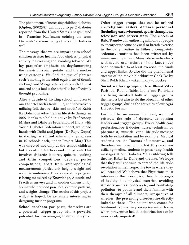
853Diabetes Mellitus : Targetting School Children And Trigger Groups In Diabetes Prevention
The phenomena of increasing childhood obesity (Ogden, 2002)(8), childhood Type 2 diabetes reported from the United States encapsulated in Francine Kaufmann coining the term Diabesity9 are now being observed in India as well.
The message that we are imparting to school children stresses healthy food choices, physical activity, destressing and avoiding tobacco. We lay particular emphasis on degslamorising the television couch potato and the smoker using cartoons. We find the use of phrases such 'Smoking is the adult equivalent of thumb sucking!' and 'A cigarette is a stick with a fire at one end and a fool at the other!' to be effectively thought provoking.
After a decade of inviting school children to our Diabetes Melas from 1997, and innovatively utilizing folk theatre, skits and modified Kabir Ke Dohe to involve them in life style change, in 2007 thanks to a bold initiative by Prof Anoop Mishra and Diabetes Federation of India with World Diabetes Federation support, we joined hands with Delhi and Jaipur (Dr Rajiv Gupta) in starting in school educational programs in 10 schools each, under Project Marg.This was directed not only at the school children but also at the teachers and the parents.This involves didactic lectures, quizzes, cooking and tiffin competitions, debates, poster competitions, apart from anthropological measurements particularly height, weight and waist circumference.The success of the program is being measured by Knowledge, Attitude and Practices surveys ,and in another dimension by seeing whether food practices, exercise patterns, and weights change. The results of this project will, it is hoped, be extremely interesting in designing further programs.
School teachers, pari passu, themselves are a powerful trigger group with a powerful potential for encouraging healthy life styles.
Other trigger groups that can be utilized are religious leaders, defence personnel (including exservicemen), sports champions, television and screen stars. The success of Baba`Ramdeva in enthusing millions of people to incorporate some physical or breath exercise in the daily routine in hitherto completely sedentary routines has been witnessed by numerous physicians. Many obese individuals with severe osteoarthritis of the knees have been persuaded to at least exercise the trunk and upper limbs. So also did the phenomenal successofthemovieblockbusterChakDebyShah Rukh Khan awaken many to hockey!
Social welfare groups such as Bharat Vikas Parishad, Round Table, Lions and Rotarians are being involved both as trigger groups themselves but also to aid the education of other trigger groups, during the activities of our Agra Diabetic Forum.
Last but by no means the least, we must reiterate the role of doctors, as opinion leaders and trigger groups. Every health care professional-be it doctor, nurse, technician or pharmacist, must deliver a life style message both by exhortation and by example! Medical students are the Doctors of tomorrow, and therefore we have for the last 10 years been utilizing medical students in presenting health messages at our Diabetes Melas utilizing folk theatre, Kabir ke Dohe and the like. We hope that they will continue to spread the life style revolution in their respective towns where they will practice! We believe that Physicians must interweave the preventive health messages of healthy diet, physical exercise, avoiding stressors such as tobacco etc, and combating pollution to patients and their families with their therapy of all ailments, irrespective of whether the presenting disorders are directly linked to these ! The patient who comes for treatment is in a very receptive mind frame where preventive health indoctrination can be more easily imparted!

854 Medicine Update 2008 Vol. 18
Barriers to preventive strategies especially in children, and possible solutionsPathophysiological barriers
Promoting physical activity is an uphill task when the obesity leads to easy tiring, breathlessness and painful joints during unaccustomed exercise. Gradual and selective exercise patterns can overcome this:one can also emphasise that Fat active is better than Fat inactive!Some tennis players are highly motivated and successful despite being obese.
Peer ridicule of the fat child last in the race is also a very real problem:individual activity such as skipping, dancing or long walks, swimming, exercise cycles or Yoga are possible ways out.
Lustig, 2006,2 in an excellent review has emphasized that biological susceptibility and societal accountability must be acknowledged and personal responsibility and guilt deemphasized.
The concept of "Personal responsibility" permits governments and business interests the luxury and absolution of abdicating their responsibility. To quote him "The concept of personal responsibility" is, however, not tenable in children. No child chooses to be obese. Obese children are ostracized by their peers, and their quality of life, as measured by self reported distress, is comparable to hose receiving cancer chemotherapy. Young children among whom obesity is rampant are not responsible for their food chices and are incapable of accepting personal responsibility" The society that permits the sale, promotion and availability of junk foods must change its laws and attitudes. How does one can react to purveyors of pornography to children or for that matter tobacco advertisements to the vulnerable? Why not then a similar attitude to the promotion of junk food!
Further, Lustig reviews the central nervous system mechanisms underlying weight gain.Hyperinsulinemia promotes leptin resistance, insulin interfering with leptin signal transduction
being an endogenous antagonist. Ordinarily elevated leptin should lead to increased sympathetic (SNS)and decreased vagal outflow, with increased resting energy expenditure(REE), lipolysis and decreased food intake. Hyperinsulinaemia induced leptin resistance should decrease SNS activity to reduce REE and increase vagal activity to promote energy storage. The hypothetical hypothalamic set point being dysfunctional in obesity, a higher level of leptin is required to signal the hypothalamus to maintain a normal REE. Reductions in REE are associated with fatigue, malaise, decreased quality of life and decreased physical activity. The obese subject therefore increases calorie intake to raise leptin concentration above the level at which leptin resistance occurs,so as to maintain normal energy expenditure and quality of life. Work on pharmacological interruption of these vicious cycles such as SOCS3 inhibitors is inprogress,but even when developed these are likely to be inexpensive.
Hyperinsulinemia also decreases dopamine clearance and uptake in the hedonic pathway-the Nucleus Accumbens and Ventral Tegmental Area.The latter is known for initiating feeding behavior on the basis of palatability rather than energy need.The dopamine changes promote an increased pleasure reward of food that causes further food intake, as eating becomes more pleasurable!
The induced obesity induces further hyperinsulinaemia, thus constituting both these mechanisms as vicious cycles!
Thus biologically hyperinsulinemia and insulin resistance predispose to excessive eating and decreased energy expenditure!
Treating and preventing childhood obesity must therefore address the causes of hyperinsulinemia.
Causes of HyperinsulinemiaAltered insulin dynamics, including increased secretion exist in certain ethnic groups even prior to obesity (Arslanian et al, 2002, Preeyasombat C, 2005).10,11 We have described these in the

855Diabetes Mellitus : Targetting School Children And Trigger Groups In Diabetes Prevention
offspring of diabetic couples in the APIDS study.6,7 Hyperinsulinemia and insulin resistance have been described in both small for gestational age and large for gestational age subjects as well as in preterm infants (Stocker, 2005; Yajnik, 2002 and Hofman, 2004).12-14 Canthisbecorrectedbypropermaternalnutrition?
The typical Western diet has been categorized as highly insulinogenic (high energy density, glycemic index, fructose content and low fiber and dietary content(Isganaitis and Lustig, 2005), and we have noted the sad westernization threatening the traditional Indian life style.3
Apart from these, the increasing frequency of diabetes despite the unaltered genetic pool could also be due to pancreatotoxic factors: viruses, pollutants and perhaps unknown agents. (Hazra, 2007)15
Logistic difficulties in effecting life style changes in childrenPhysical activity
Unfortunately, there are few schools which have good areas for physical exercise activities (Sujatha, 2007),16 even in the capital of economically affluent states such as Tamil Nadu. To quote this, “..there are quite a few schools in Chennai that do not have a playground. Whatstudents get in these schools is an apology of a playground and utilities. Such schools lease out a neighbouring vacant plot or use the neighbouring Corporationplayground.Oftenschoolscarryonwith rudimentary equipment that is not replaced until they wear out completely.”
There are many areas devoid even of safe public play areas. It is a wonder that determined children use even streets and lanes to play cricket or badminton.
Until physical activity is included in the subjects that earn marks in the school leaving examinations, and given credit in the selection criteria for professional courses, students will continue to remain sedentary bookworms moving from tutorial
to tutorial with no time for physical activity either in the school or at home.
Diet
Many of the health promoting foods:fruits,dairy products, pulses, salad vegetables are relatively expensive as compared to the empty calorie starch and sweet foods. The midday meal school program run by various schools gives an opportunity to effectively supplement these but unfortunately many of these programs are plagued by corruption and/or inefficiency. An alert public opinion is needed to monitor and correct these.
ConclusionThe task of disease prevention is difficult, the path long, and the work unglamorous, but changing mind sets we believe is worthwile. Education for Life style changes must however go hand in hand with overcoming the logistic and pathophysiological barriers to behavioural change referred to above.As we strive for these, let us remember the exhortation:
“Awake, Arise and Stop not, until the Goal is reached”
Swami Vivekananda
References1. Yajnik,CSPersonalcommunicationattheStenoconference,
Agra, April, 2007.
2. Lustig,RJ Childhood obesity:behavioral aberrationor biochemical drive?Reinterpreting the First Law of Thermodynamics Nat Clin Pract Endocrinol Metab. (2006) 2(8):447-458
3. Hazra,DK Lifestyle modifications in the Prevention and Management of Diabetes Mellitus,in Life Stryle Modifications, Ed Dang, KK, Indian College of Physicians,Mumbai, (2005) Ist Ed:115-114.
4. Ritchie,L et al(2001) Pediatric overweight, a review of the literature [http://www.cnr.berkely.edu/cwh/PDFs/Full_COPL_secure.pdf](accessed17May2006)
5. Epstein,LH et al Behavioral therapy in the treatment of pediatric obesity. Pediatr. Clin. North. Am (2001) 48:909-930.
6. Hazra DK,Gupta AK Homa model crosssectional evaluation of offspring of biparental diabetics Proceedings of the IDF Congress,Mexico,2000
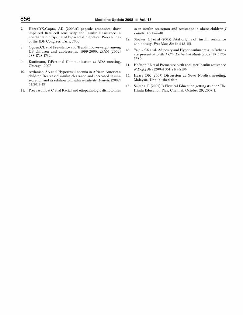
856 Medicine Update 2008 Vol. 18
7. HazraDK,Gupta, AK (2003)C peptide responses showimpaired Beta cell sensitivity and Insulin Resistance in nondiabetic offspring of biparental diabetics. Proceedings oftheIDFCongress,Paris,2003.
8. Ogden,CLetalPrevalenceandTrendsinoverweightamongUS children and adolescents, 1999-2000. JAMA (2002) 288:1728-1732.
9. Kaufmann, F:Personal Communication at ADA meeting,Chicago,2007
10. Arslanian, SA et al Hyperinsulinaemia in African-American children.Decreased insulin clearance and increased insulin secretion and its relation to insulin sensitivity. Diabetes (2002) 51:3014-19
11. PreeyasombatCetalRacialandetiopathologicdichotomies
in in insulin sectretion and resistance in obese children J Pediatr 146:474-481
12. Stocker,CJetal (2005)Fetaloriginsof insulin resistanceand obesity. Proc Nutr. Soc 64:143-151.
13. Yajnik,CSetal.AdiposityandHyperinsulinaemiainIndiansare present at birth J Clin Endocrinol.Metab (2002) 87:5575-5580
14. Hofman PL et al Premature birth and later Insulin resistance N Engl J Med (2004) 351:2179-2186.
15. Hazra DK (2007) Discussion at Novo Nordisk meeting, Malaysia. Unpublished data
16. Sujatha, R (2007) Is Physical Education getting its due? The HinduEducationPlus,Chennai,October29,2007:1.

CHAPTER
113Postprandial Lipids In Diabetes and Pre-Diabetes- Role in AtherosclerosisS. V. Madhu, B. Sinha
Microangiopathy and in particular macroangiopathy contributes to excess morbidity and premature death in patients with type 2 DM1. At diagnosis, patients with type 2 DM have a 3-4 fold higher risk for cardiovascular disease than non-diabetic persons2,3. Risk factors for macroangiopathy in patients with type 2 DM include an elevated fasting hyperglyceride level, a low HDL cholesterol level, and accumulation of small dense low density lipoprotein (LDL) particles which are early oxidized and are atherogenic4. Epidemiological studies have reported a higher risk of coronary artery disease (CAD)inthosewithelevatedfastingtriglyceridesin the serum. Fasting hypertriglyceridemia has also been consistently shown to be associated with type 2 DM and persons with visceral obesity. In a Finnish 7 year prospective study, high triglycedemia (TG) levels (>203 mg/dl) were associated with a 2 fold increase in risk forCADevents.This shows thatelevatedTGlevelmaybeabetterpredictorofCADthan elevated LDL levels5-8.
Recent studies have shown that post-prandial handling of triglyceride rich lipoprotein (TRLs) is important for the propensity of endothelial dysfunction and atherosclerosis4,9-12. Although fasting lipid and lipoprotein levels reflect steady state lipid metabolism even healthy subjects are in a state of post-prandial hypertriglyceridemia most of the time due to meal frequency11. Serum triglycerides
are generally increased maximally by 3-4 hrs post-prandially in non-diabetic / healthy individuals and by 6-10 hrs in pre-diabetic and diabetics12-15. Once post-prandial lipidemia occurs it is exacerbated by the next meal and thus hypertriglycedemia persists for the entire day. Clearly body’s vasculature isexposed to post-prandial lipemia for most of the day. Therefore, it would appear logical that most of the endothelial dysfunction that finally leads to atherosclerosis should be taking place during post-prandial state1,9,14,16,17.
Elevated postprandial lipemia has been seen in type 2 DM, prediabetes, first degree relatives of type 2 DM, obese and asymptomatic person with raised fasting serum triglyceride levels14,16. Therefore measurement of lipid especially triglycerides in post-prandial state would be a more reliable and sensitive indicator to predict future cardiovascular risk.
In 1979, Zilversmit, proposed that postprandial accumulation of ‘Triglyceride Rich Lipoproteins (TRL’s) resulted from a reduction in the rate of clearance of the TG rich dietary remanant particles at the endothelial surface and promoted the development of atherosclerosis. Remnants of TRL’s are certainly atherogenic in fat fed experimental animals and in humans with type III hyperlipoproteinemia9,18. TRL’s derived from

858 Medicine Update 2008 Vol. 18
hypertriglyceridemic humans are toxic to endothelial cells and are taken up by macrophages resulting in foam cell formation. Moreover, case control studies have found an elevated level of postprandial TRL’sinthosewithangiographicallyverifiedCADas compared to normal controls19,20,21. In persons with prolonged increases of plasma triglycerides, either fasting or postprandial, the process of lipid exchange would enrich the triglyceride rich particles in cholesteryl ester and thereby make these particles more atherogenic.
Studies involving measurements of specific triglyceride rich lipoprotein fractions have provided support for the hypothesis that particular types of triglyceride rich particles may be directly atherogenic.
Farideh, Helen and Michael studied the acute effect of low and moderate fat intakes on post-prandial lipemia. Based on a observation over a 7 day period involving more than 3000 eating occasions, they have shown that on 26% of occasions fat ingestion was below 5 gm, on 41% of occasion it was between 5-20 gm and only 33% occasions, fat intake was above 30 gm. They observed that any meal intake with fat content >15 gms would elicit a post-prandial lipemic response (triacylglycerol concentration) that will affect endothelial function and is capable of affecting the composition and concentration of HDL and LDL. He also concluded that sex had no effect on post-prandial lipemia if other compounding features are matched22. Therefore, it can be concluded that in diabetic and prediabetic individuals who already harbour risk factors for increased endothelial dysfunction, even moderate intake of fat (>15 gms) would be a catastrophe leading to increase inCVmorbidityand mortality.
Murphy et al showed that doses of 20 g fats were capable of eliciting a plasma genetic inhibiting polypeptide (GIP) augments insulin mediated stimulation of lipoprotein lipase, the enzyme that catalyses plasma TAG clearance. Thus, there is impaired post-prandial clearance of TAG in diabetic
and pre-diabetic individuals due to either insulin resistance or decreased insulin secretion (secretory defect is IGT individuals)23.
The high TRL’s associated with alimentary lipemia lead to activation of factor VII and increased levels of PAI-124. Though, if does not lead to any thrombus formation in itself, the procoagulated state augments the potential for thrombus formation in event of plaque rupture.
Taskinen et al studied post-prandial hypertriglyceridemia and insulin resistance in normoglycemic, normotriglyceridemic first degree relatives of patients with type 2 DM. They found that these subjects exhibited post-prandial hypertriglyceridemia after a mixed meal (meal containing 49% fat, 36% carbohydrate, 14% protein), which suggests an inherited defect in post-prandial lipid metabolism1. This defect probably linked to insulin resistance in first degree relatives1. Activation of PI3-kinase enzyme is required for initiating insulin action which in turn is necessary to suppress the release of endogenous VLDL cholesterol25. An impaired cellular activation of PJ-3 kinase has been suggested to be responsible for PPL as one of the mechanisms in diabetic, pre-diabetic and healthy first degree relatives of type 2 DM26,27.
Studies from our institute clearly shows association of post-prandial lipemia with endothelial dysfunction in type 2 diabetic individuals, irrespective of fasting triglyceride levels15,16. This was independent of glycemic control and insulin sensitivity but was related to the interaction of diabetic state and obesity. This was observed both in older type 2 diabetic as well as young ketosis resistance subjects16.
In a study in Prediabetes , significant elevated PPTGs were demonstrated only in Newly detected Diabetes following OGTT and not in the pprediabetic groups.
Presently one of the study is being carried out to demonstrate post-prandial lipemia in prediabetics (i.e. impaired fasting glucose and impaired glucose tolerant subjects) and to find its association with

859Postprandial Lipids In Diabetes and Pre-Diabetes- Role in Atherosclerosis
endothelial function and presence of family history of type 2 DM. Results of this study will be available by April 2008.
Antonio et al studied post-prandial hypertriglyceride and hyperglycemia as an independent evidence for endothelial dysfunction and oxidative stress generation in diabetic and non-diabetic adults. They concluded that meal absorption is a complex phenomenon and post-prandial hypertriglyceridemia and hyperglycemia and simultaneously present in the post-absoptive phase, particularly in children and also in subjects with impaired glucose tolerance. When hyperglycemia and hypertriglyceridemia were simultaneously present, there was greater impairment of endothelial function as compared with that observed during either hyperglycemia or hyperglyceridemia alone suggesting a cumulative effect on endothelial cells28. This event is mediated through production of an oxidative stress as depicted in Figure given below29:
Konukoglu studied endothelial dysfunction in prediabetes, i.e. impaired glucose tolerant subjects. He found that NO levels significantly decreased in prediabetic as compared to controls (healthy subjects). TBARs (thiobarbituric acid reactivesubstances)wereelevatedinIGT,Cu-Znsuperoxide dismutase, which acts as an antioxidant were significantly reduced in prediabetic. Thus, they concluded that pre-diabetic state is a stage of enhanced oxidative stress which can lead to increase in endothelial dysfunction30.
Gofman and colleagues have shown that IDL (intermediate density lipoprotein) particles are strong determinants of endothelial dysfunction and hence atherosclerosis as compared to LDL particles31. MARS (monitored atherosclerosis regression study) trial suggest that triglyceride rich lipoprotein particles particularly small VLDL and IDL are involved in atherosclerosis progression more than LDL32.
Oxidative stress Increase O2-
(superoxide anion) Peroxynitrite radicals
+ NO
VLDL peroxidation, antioxidant defense decreased, direct cytotoxic to endothelial cells, formation of nitrotyrosine, oxidizes sulfhydryl group of proteins
Increased apoptosis of myocytes, endothe lial cells, fibroblasts
(long ½ life)
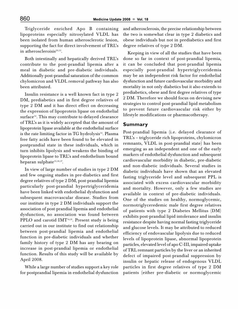
860 Medicine Update 2008 Vol. 18
Triglyceride enriched Apo B containing lipoproteins especially nitrosylated VLDL has been isolated from human atherosclerotic lesion, supporting the fact for direct involvement of TRL’s in atheroscleroisis29,33.
Both intestinally and hepatically derived TRL’s contribute to the post-prandial lipemia after a meal in diabetic and pre-diabetic individuals. Additionally post-prandial saturation of the common chylomicron and VLDL removal pathway has also been attributed.
Insulin resistance is a well known fact in type 2 DM, prediabetics and in first degree relatives of type 2 DM and it has direct effect on decreasing the expression of lipoprotein lipase on endothelial surface34. This may contribute to delayed clearance of TRL’s as it is widely accepted that the amount of lipoprotein lipase available at the endothelial surface is the rate limiting factor in TG hydrolysis34. Plasma free fatty acids have been found to be elevated in postprandial state in these individuals, which in turn inhibits lipolysis and weakens the binding of lipoprotein lipase to TRL’s and endothelium bound heparan sulphate35,36,37.
In view of large number of studies in type 2 DM and few ongoing studies in pre-diabetics and first degree relatives of type 2 DM, post-prandial lipemia particularly post-prandial hypertriglyceridemia have been linked with endothelial dysfunction and subsequent macrovascular disease. Studies from our institute in type 2 DM individuals support the association of post-prandial lipemia and endothelial dysfunction, no association was found between PPLO and carotid IMT14,16. Present study is being carried out in our institute to find out relationship between post-prandial lipemia and endothelial function in pre-diabetic individuals and whether family history of type 2 DM has any bearing on increase in post-prandial lipemia or endothelial function. Results of this study will be available by April 2008.
While a large number of studies support a key role for postprandial lipemia in endothelial dysfunction
and atherosclerosis, the precise relationship between the two is somewhat clear in type 2 diabetics and obese individuals but not in prediabetics and first degree relatives of type 2 DM.
Keeping in view of all the studies that have been done so far in context of post-prandial lipemia, it can be concluded that post-prandial lipemia especially post-prandial hypertriglyceridemia may be an independent risk factor for endothelial dysfunction and future cardiovascular morbidity and mortality in not only diabetics but it also extends to prediabetics, obese and first degree relatives of type 2 DM. Therefore we should focus on devising new strategies to control post-prandial lipid metabolism to prevent future cardiovascular risk either by lifestyle modifications or pharmacotherapy.
SummaryPost-prandial lipemia (i.e. delayed clearance of TRL’s – triglyceride rich lipoproteins, chylomicron remnants, VLDL in post-prandial state) has been emerging as an independent and one of the early markers of endothelial dysfunction and subsequent cardiovascular morbidity in diabetic, pre-diabetic and non-diabetic individuals. Several studies in diabetic individuals have shown that an elevated fasting triglyceride level and subsequent PPL is associated with excess cardiovascular morbidity and mortality. However, only a few studies are available in context of pre-diabetic individuals. One of the studies on healthy, normoglycemic, normotriglyceredemic male first degree relatives of patients with type 2 Diabetes Mellitus (DM) exhibits post-prandial lipid intolerance and insulin resistance despite having normal fasting triglyceride and glucose levels. It may be attributed to reduced efficiency of endovascular lipolysis due to reduced levels of lipoprotein lipase, abnormal lipoprotein particles,elevatedlevelofapoC-III,impaireduptakeof TRL remnant particles by the liver or an inherited defect of impaired post-prandial suppression by insulin or hepatic release of endogenous VLDL particles in first degree relatives of type 2 DM patients (either pre-diabetic or normoglycemic

861Postprandial Lipids In Diabetes and Pre-Diabetes- Role in Atherosclerosis
individuals). Studies from our institute have clearly shown that post-prandial hypertryglyceridemia is present in type 2 DM patients with a wide range of age and body mass index and that it is associated with endothelial dysfunction and post prandial oxidative stress. Subsequently, we have also studied this abnormality in prediabetes also. One of the studies being carried out currently in our institute is to demonstrate post-prandial lipemia (hypertriglyceridemia) in prediabetic subjects (without or without family history of Type 2 DM) and to find its association with endothelial function. It has been proposed that post-prandial lipemia in non-diabetic, pre-diabetic, diabetic or obese individuals has an independent and cumulative effect in determining endothelial dysfunction and oxidative stress may be the common mediator of this phenomenon. All these recent observations, suggest that post-prandial lipemia (hypertryglyceridemia) is an independent risk factor for cardiovascular morbidity and mortality, particularly among diabetic and pre-diabetic individuals. This opens up avenues for developing strategies to target post-prandial lipemia particularly hypertriglyceridemia to prevent future cardiovascular risk in patients with Diabetes ,Pre-diabetes andin first degree relatives of type 2 DM patients.
References1. Axelsen M, Smith U, Eriksson JW, Taskinen MR, Jansson
PA. Postprandial hypertriglyceridemia and insulin resistance in normoglycemic first-degree relatives of patients with type 2 diabetes. Ann Intern Med 1999; 131: 27-31.
2. Fontbonne A, Eschwège E, Cambien F, Richard JL,DucimetièreP,ThibultN,WarnetJM,ClaudeJR,RosselinGE. Hypertriglyceridaemia as a risk factor of coronary heart disease mortality in subjects with impaired glucose tolerance or diabetes. Results from the 11-year follow-up of the Paris Prospective Study. Diabetologia 1989; 32: 300-4.
3. Uusitupa MI, Niskanen LK, Siitonen O, Voutilainen E, Pyörälä K. Ten-year cardiovascular mortality in relation to risk factors and abnormalities in lipoprotein composition in type 2 (non-insulin-dependent) diabetic and non-diabetic subjects. Diabetologia 1993; 36: 1175-84.
4. Syvänne M, Taskinen MR. Lipids and lipoproteins as coronary risk factors in non-insulin-dependent diabetes mellitus. Lancet 1997; 350: SI20-3.
5. Fontbonne A. Relationship between diabetic dyslipoproteinemia and coronary heart disease risk in non-insulin dependent diabetes. Diabetes Metab Rev 1991; 7: 179-189.
6. Taskinen MR. Quantitative and qualitative lipoprotein abnormalities in diabetes mellitus. Diabetes 1992; 41: 12-7.
7. Laakso M, Rönnemaa T, Pyörälä K, Kallio V, Puukka P, Penttilä I. Atherosclerotic vascular disease and its risk factors in non-insulin-dependent diabetic and nondiabetic subjects in Finland. Diabetes Care 1988; 11: 449-63.
8. WestKM,AhujaMM,BennettPH,CzyzykA,DeAcostaOM, Fuller JH, Grab B, Grabauskas V, Jarrett RJ, Kosaka K, et al. The role of circulating glucose and triglyceride concentrations and their interactions with other “risk factors” as determinants of arterial disease in nine diabetic population samples from the WHO multinational study. Diabetes Care 1983; 6: 361-9.
9. Zilversmit DB. Atherogenesis: a postprandial phenomenon. Circulation 1979; 60: 473-85.
10. Patsch JR. Triglyceride-rich lipoproteins and atherosclerosis. Atherosclerosis. 1994; 110: S23-6.
11. DuboisC,ArmandM,Azais-BraescoV,PortugalH,PauliAM,BernardPM,LatgéC,LafontH,BorelP,LaironD.Effects of moderate amounts of emulsified dietary fat on postprandial lipemia and lipoproteins in normolipidemic adults. Am J Clin Nutr 1994; 60: 374-82.
12. Lewis GF, O’Meara NM, Soltys PA, Blackman JD, Iverius PH, Pugh WL, Getz GS, Polonsky KS. Fasting hypertriglyceridemia in noninsulin-dependent diabetes mellitus is an important predictor of postprandial lipid and lipoprotein abnormalities. J Clin Endocrinol Metab 1991; 72: 934-44.
13. ChenYD,SwamiS,SkowronskiR,CoulstonA,ReavenGM. Differences in postprandial lipemia between patients with normal glucose tolerance and noninsulin-dependent diabetes mellitus. J Clin Endocrinol Metab 1993; 76: 172-7.
14. Madhu SV, Mittal V, Krishnaram B, Srivastava DK, Kaur J, sethi K. Postprandial triglyceride metabolism in obese and nonobese patients with non insulin dependent diabetes mellitus. Diabetes Res Clin Pract 2000; 50: 5318.
15. Wasir J, Ram BK, Madhu SV, Kaur J, Sethi K. Post prandial triglyceride metabolism in young non obese duabetics and its relation to insulin sensitivity. J Assoc Physician India 2001; 49: 204.
16. Madhu SV, Malhotra A, Mehrotra G, Srivastava DK. Post-prandial lipemia and carotid intimal medial thickness in patients with type 2 diabetes mellitus. Diabetologia 2003; 46: A380.
17. DeManFH,CabezasMC,VanBarlingenHH,ErkelensDW,de Bruin TW. Triglyceride-rich lipoproteins in non-insulin-dependent diabetes mellitus: post-prandial metabolism and relation to premature atherosclerosis. Eur J Clin Invest 1996; 26: 89-108.
18. RyuJE,HowardG,CravenTE,BondMG,HagamanAP,Crouse JR 3rd. Postprandial triglyceridemia and carotidatherosclerosis in middle-aged subjects. Stroke 1992; 23:
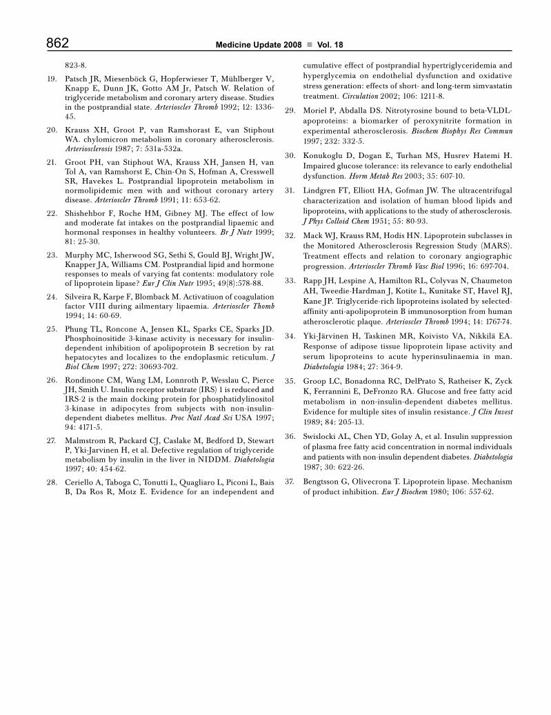
862 Medicine Update 2008 Vol. 18
823-8.
19. Patsch JR, Miesenböck G, Hopferwieser T, Mühlberger V, Knapp E, Dunn JK, Gotto AM Jr, Patsch W. Relation of triglyceride metabolism and coronary artery disease. Studies in the postprandial state. Arterioscler Thromb 1992; 12: 1336-45.
20. Krauss XH, Groot P, van Ramshorast E, van Stiphout WA. chylomicron metabolism in coronary atherosclerosis. Arteriosclerosis 1987; 7: 531a-532a.
21. Groot PH, van Stiphout WA, Krauss XH, Jansen H, van TolA,vanRamshorstE,Chin-OnS,HofmanA,CresswellSR, Havekes L. Postprandial lipoprotein metabolism in normolipidemic men with and without coronary artery disease. Arterioscler Thromb 1991; 11: 653-62.
22. Shishehbor F, Roche HM, Gibney MJ. The effect of low and moderate fat intakes on the postprandial lipaemic and hormonal responses in healthy volunteers. Br J Nutr 1999; 81: 25-30.
23. MurphyMC,IsherwoodSG,SethiS,GouldBJ,WrightJW,KnapperJA,WilliamsCM.Postprandiallipidandhormoneresponses to meals of varying fat contents: modulatory role of lipoprotein lipase? Eur J Clin Nutr 1995; 49(8):578-88.
24. Silveira R, Karpe F, Blomback M. Activatiuon of coagulation factor VIII during ailmentary lipaemia. Arterioscler Thomb 1994; 14: 60-69.
25. PhungTL,RonconeA,JensenKL,SparksCE,SparksJD.Phosphoinositide 3-kinase activity is necessary for insulin-dependent inhibition of apolipoprotein B secretion by rat hepatocytes and localizes to the endoplasmic reticulum. J Biol Chem 1997; 272: 30693-702.
26. RondinoneCM,WangLM,LonnrothP,WesslauC,PierceJH, Smith U. Insulin receptor substrate (IRS) 1 is reduced and IRS-2 is the main docking protein for phosphatidylinositol 3-kinase in adipocytes from subjects with non-insulin-dependent diabetes mellitus. Proc Natl Acad Sci USA 1997; 94: 4171-5.
27. MalmstromR,PackardCJ,CaslakeM,BedfordD,StewartP, Yki-Jarvinen H, et al. Defective regulation of triglyceride metabolism by insulin in the liver in NIDDM. Diabetologia 1997; 40: 454-62.
28. CerielloA,TabogaC,TonuttiL,QuagliaroL,PiconiL,BaisB, Da Ros R, Motz E. Evidence for an independent and
cumulative effect of postprandial hypertriglyceridemia and hyperglycemia on endothelial dysfunction and oxidative stress generation: effects of short- and long-term simvastatin treatment. Circulation 2002; 106: 1211-8.
29. Moriel P, Abdalla DS. Nitrotyrosine bound to beta-VLDL-apoproteins: a biomarker of peroxynitrite formation in experimental atherosclerosis. Biochem Biophys Res Commun 1997; 232: 332-5.
30. Konukoglu D, Dogan E, Turhan MS, Husrev Hatemi H. Impaired glucose tolerance: its relevance to early endothelial dysfunction. Horm Metab Res 2003; 35: 607-10.
31. Lindgren FT, Elliott HA, Gofman JW. The ultracentrifugal characterization and isolation of human blood lipids and lipoproteins, with applications to the study of atherosclerosis. J Phys Colloid Chem 1951; 55: 80-93.
32. Mack WJ, Krauss RM, Hodis HN. Lipoprotein subclasses in the Monitored Atherosclerosis Regression Study (MARS). Treatment effects and relation to coronary angiographic progression. Arterioscler Thromb Vasc Biol 1996; 16: 697-704.
33. RappJH,LespineA,HamiltonRL,ColyvasN,ChaumetonAH, Tweedie-Hardman J, Kotite L, Kunitake ST, Havel RJ, Kane JP. Triglyceride-rich lipoproteins isolated by selected-affinity anti-apolipoprotein B immunosorption from human atherosclerotic plaque. Arterioscler Thromb 1994; 14: 1767-74.
34. Yki-Järvinen H, Taskinen MR, Koivisto VA, Nikkilä EA. Response of adipose tissue lipoprotein lipase activity and serum lipoproteins to acute hyperinsulinaemia in man. Diabetologia 1984; 27: 364-9.
35. GroopLC,BonadonnaRC,DelPratoS,RatheiserK,ZyckK, Ferrannini E, DeFronzo RA. Glucose and free fatty acid metabolism in non-insulin-dependent diabetes mellitus. Evidence for multiple sites of insulin resistance. J Clin Invest 1989; 84: 205-13.
36. SwislockiAL,ChenYD,GolayA,etal.Insulinsuppressionof plasma free fatty acid concentration in normal individuals and patients with non-insulin dependent diabetes. Diabetologia 1987; 30: 622-26.
37. Bengtsson G, Olivecrona T. Lipoprotein lipase. Mechanism of product inhibition. Eur J Biochem 1980; 106: 557-62.

CHAPTER
114Digital Gangrene - How and How Much to Investigate?L. S. Bichile, M. S. Kubal
IntroductionPeripheral vascular disease affects nearly 10 million of the Indian population.1 It is also associated with decrease in functional capacity and quality of life and an increased risk of amputations.
The underlying clinical conditions which present with features of peripheral limb ischemia are numerous. Since the differential diagnosis of the peripheral ischemia is vast, there are a multitude of clinical and lab tests available for diagnosis of the condition. This is further complicated by the numerous invasive and non invasive imaging modalities available at the clinician’s disposal. The choice of the best modality of investigation or treatment needs to be individualized in each clinical scenario for optimal management of the patient.
Mechanisms of Vascular compromise leading to Peripheral limb ischemiaEmbolic Occlusion
80% of the emboli originate from cardiac source – vegetation on valves, mural thrombi dislodged during episodes of atrial fibrillation or other arrthymias.3 20% are from non-cardiac source - thrombosed aneurysm, ulcerated atherosclerotic plaque
In India there is increased incidence of embolic occlusion due to widespread prevalence of rheumatic heart disease.
Most commonly emboli lodge in the femoral artery bifurcation, aortoiliac arterial system, brachial artery.
Thrombotic Occlusion
• Atheroscleroticthrombusformationespeciallyin diseased arteries e.g. popliteal artery
• Secondary thrombi in pre existing vasculargrafts are common.
• Accelerated atherosclerosis occurs inautoimmune conditions like SLE.
Extrinsic compression of arterial lumen
• Aorticorvasculardissection,creatingpseudolumen which compromises true lumen
• Compartmentsyndromeintraumaorburns.
• Thoracic outlet syndrome: Condition withscalene muscle scarring and/or a cervical rib causing neurovascular compression in the superior thoracic cavity, leading to arm pain and paresthesias.
Vasospasm
Raynaud’s phenomenon: A vasospastic condition causing well-demarcated ischemia to fingers and toes. Three stages: Pallor (hypoperfusion),
864 Medicine Update 2008 Vol. 18
then cyanotic (hypoxemia), then hyperaemia (reperfusion), usually bilateral limb involvement.
It can be primary Raynaud’s Disease or Secondary to autoimmune diseases like SLE, Scleroderma, etc.
Causes of peripheral limb ischemiaThere are a wide variety of conditions causing peripheral limb ischemia other than the traditional causes like atherosclerosis. Though rare these conditions should be kept in mind whenever evaluating a patient with either acute or chronic ischemia. The following table enlists the variety of etiological conditions which may present with limb ischemia or peripheral vascular disease. Two major contributors of limb ischemia i.e. atherosclerosis and autoimmune diseases are discussed in brief in the following text.
Atherosclerosis
Atherosclerosis is a major cause of peripheral artery disease and limb ischemia. Atherosclerosis affects up to 10% of the Western population older than 65 years. With the elderly population expected to increase 22% by the year 2040, atherosclerosis is expected to have a huge financial impact on medicine. When claudication is used as an indicator, estimates are that 2% of the population aged 40-60 years and 6% older than 70 years are affected (4). 40 % of patients with atherosclerotic coronary artery disease also have peripheral vascular disease. Most of the autoimmune disorders like systemic lupus erythematosus also cause arterial disease by the process of accelerating atherosclerosis.
Rheumatological conditions
Rheumatological conditions presenting as peripheral limb ischemia or gangrene are numerous, but generally underdiagnosed. A variety of conditions causing accelerated atherosclerosis get overlooked during evaluation and only the atherosclerotic nature of the illness treated. The tables 2 shows the prevalence of peripheral limb disease in a cohort of patients attending the rheumatology clinic in K.E.M Hospital Mumbai.
As seen in the above results a significant amount of peripheral limb ischemia can be attributed to autoimmune rheumatological diseases apart from the traditional causes like atherosclerosis.
Need for early diagnosis and treatment• Coronaryarterydisease is frequently seen in
association with peripheral arterial occlusive disease especially when the cause is accelerated atherosclerosis.
• Coronary artery disease with a subsequentmyocardial event is the major contributor to outcome to the mortality in patients with peripheral vascular disease.
• Themorbidityofarterialocclusionisrelatedtothe development of critical limb ischemia and subsequent need for amputations.
• Predicted mortality rates for patients withclaudication at 5, 10, and 15 years of follow-up are approximately 30%, 50%, and 70%, respectively.
• “Timeislimbandlife”
Golden Time Window = 6 hours (before irreversible neuromuscular damage)
Amputation rate = 6-20%
• 6%ifrevascularizationperformedwithin12hrs of symptom onset.
• 12%ifrevascularizationperformedduring12-24 hours.
• 20%ifrevascularizationperformedafter24hrs.
Hence the earlier the diagnosis and early specific pathology directed treatment would reduce both the morbidity and mortality of peripheral limb ischemia (7).
Clinical Features of Peripheral Vascular DiseaseThe classic historical description of peripheral vascular disease is the six classic “P”s

865Digital Gangrene - How and How Much to Investigate?
Table 1Sr no Etiology Important Points to be Noted
1 Atherosclerosis Atherosclerosis affects the arteries in segments with areas of minimal to critical narrowing. The initial lesion is a fatty streak followed by plaque formation. The rupture of such plaques with subsequent thrombus formation leads to vascular lumen compromise.
2 Autoimmune
SLE
Scleroderma
APLA Syndrome
Vascular involvement in SLE may be due to accelerated atherosclerosis, hyperlipedemia, associated Anti Phospholipid antibodies or associated vasculitis (4).
Characterisedbyarterialvasospasm,smoothmuscleproliferationandeventualvascularocclusion. Nailfold capillary changes, telangectasia, Raynaud’s phenomenon and claudication are the clinical manifestataions in scleroderma. Severe cases manifest as digital ulcers and gangrene.
Arterial and/or venous thrombosis, recurrent pregnancy loss, thrombocytopenia and the presence of Anti Phospholipid antibodies.
3 Vasculitis
Buerger’s disease
Temporal arteritis
Takayasu’s arteritis
Segmental, inflammatory vaso-occlusive disease of small and medium arteries. Heavy tobaccouseisasignificantriskfactor.Commoninyoungmales.
Inflammatory condition of medium and large arteries
Chronic,inflammatory,large-vesseldiseaseofaortaanditslargebranches,foundinyoung women (age 20-30), clinically a pulseless upper extremity.
4 Shock
CHF,dehydration,sepsis
5 Thrombophilias
Factor V mutation
Hyperhomocystenemia
Antithrobmin III def.
ProteinC,Sdeficiency
6 Trauma
Crushinjuries,burns,compartment syndrome.
7 Infections
Chlamydia, salmonella,syphilis.
8 Drugs
Ergot abuse
9 Insect bites
Brown Recluse Spider
10 Parasites
Filariasis
11 Others
Radiculopathies and spinal stenosis
Patients experience “pseudo claudication”, which is leg pain which is exacerbated by walking and relieved with rest and, uniquely, leaning forward. Spine flexion actually increases the diameter of the neural canal and there is less impingement of the spinal cord and peripheral nerve roots. Patients are often misdiagnosed with mild claudication from peripheral vascular disease instead (6).
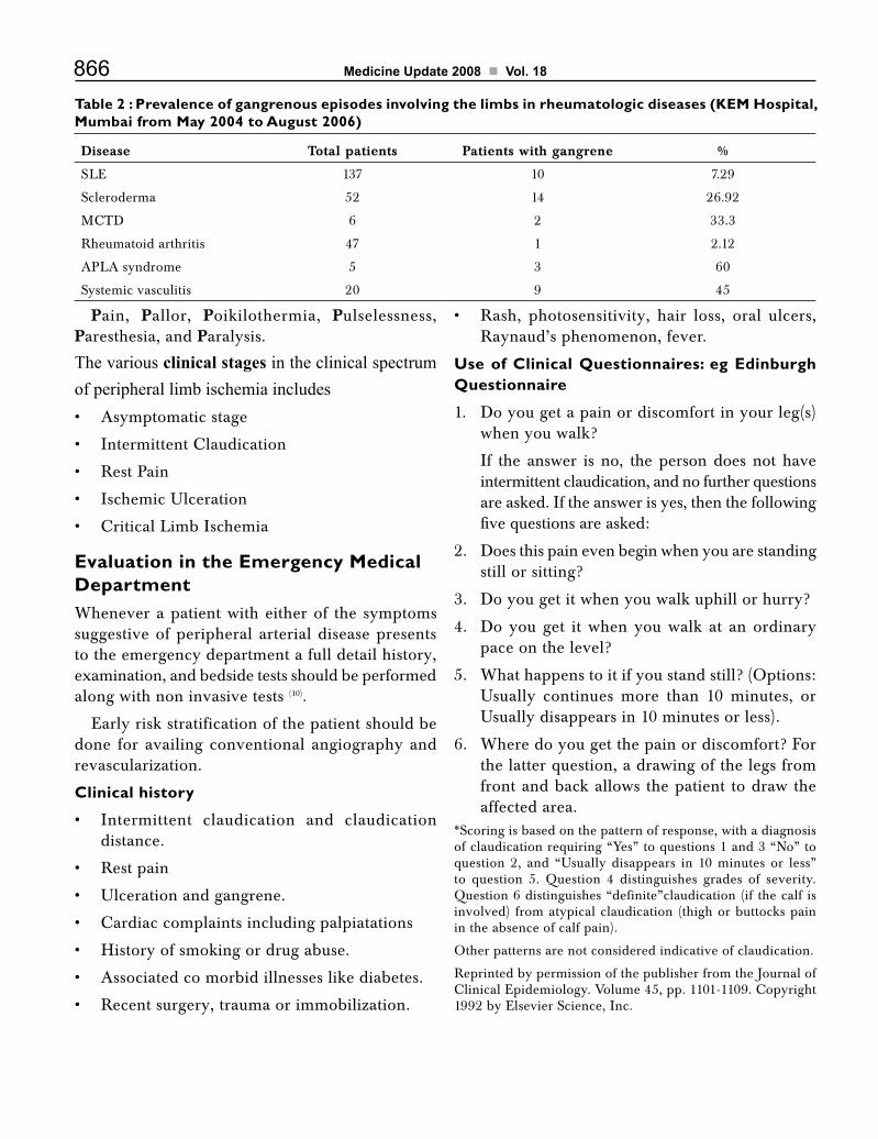
866 Medicine Update 2008 Vol. 18
Table 2 : Prevalence of gangrenous episodes involving the limbs in rheumatologic diseases (KEM Hospital, Mumbai from May 2004 to August 2006)
Disease Total patients Patients with gangrene %
SLE 137 10 7.29
Scleroderma 52 14 26.92
MCTD 6 2 33.3
Rheumatoid arthritis 47 1 2.12
APLA syndrome 5 3 60
Systemic vasculitis 20 9 45
Pain, Pallor, Poikilothermia, Pulselessness, Paresthesia, and Paralysis.
The various clinical stages in the clinical spectrum of peripheral limb ischemia includes• Asymptomaticstage
• IntermittentClaudication
• RestPain
• IschemicUlceration
• CriticalLimbIschemia
Evaluation in the Emergency Medical DepartmentWhenever a patient with either of the symptoms suggestive of peripheral arterial disease presents to the emergency department a full detail history, examination, and bedside tests should be performed along with non invasive tests (10).
Early risk stratification of the patient should be done for availing conventional angiography and revascularization.
Clinical history
• Intermittent claudication and claudicationdistance.
• Restpain
• Ulcerationandgangrene.
• Cardiaccomplaintsincludingpalpiatations
• Historyofsmokingordrugabuse.
• Associatedcomorbidillnesseslikediabetes.
• Recentsurgery,traumaorimmobilization.
• Rash, photosensitivity, hair loss, oral ulcers,Raynaud’s phenomenon, fever.
Use of Clinical Questionnaires: eg Edinburgh Questionnaire
1. Do you get a pain or discomfort in your leg(s) when you walk?
If the answer is no, the person does not have intermittent claudication, and no further questions are asked. If the answer is yes, then the following five questions are asked:
2. Does this pain even begin when you are standing still or sitting?
3. Do you get it when you walk uphill or hurry?
4. Do you get it when you walk at an ordinary pace on the level?
5. What happens to it if you stand still? (Options: Usually continues more than 10 minutes, or Usually disappears in 10 minutes or less).
6. Where do you get the pain or discomfort? For the latter question, a drawing of the legs from front and back allows the patient to draw the affected area.
*Scoring is based on the pattern of response, with a diagnosis of claudication requiring “Yes” to questions 1 and 3 “No” to question 2, and “Usually disappears in 10 minutes or less” to question 5. Question 4 distinguishes grades of severity. Question 6 distinguishes “definite”claudication (if the calf is involved) from atypical claudication (thigh or buttocks pain in the absence of calf pain).
Other patterns are not considered indicative of claudication.
Reprinted by permission of the publisher from the Journal of ClinicalEpidemiology.Volume45,pp.1101-1109.Copyright1992 by Elsevier Science, Inc.

867Digital Gangrene - How and How Much to Investigate?
Clinical Signs of Peripheral Vascular Disease
Classic appearance of extremity• Paleandcool
• Mottledcyanosis
• Dependentrubor
• Muscle rigidityand limbwoodinessare signsof an unsalvageable limb
Physical Examination: Best tests
• Palpatefordecreasedorabsentpulses.
• Patternsofpulseabnormalitymayindicatesitesof stenosis.
• Auscultateforbruits,whichmayindicatearterialstenosis.
• Sites of Bruits: Abdomen, Pelvis, andInguinal areas.
• Checkforfootpallor
• Atrest,withlegelevation,andafterexerciseof the calf muscles.
• Signsofchroniclimbischemia
• Subcutaneousatrophy
• Hairloss
• Coolness
• Pallor
• Cyanosis,dependentrubor,orboth
• Petechiae,fissures,ulceration,andgangrenewith critical limb ischemia.
• CapillaryRefillTime
• Aprolongedcapillaryrefilltime(>5sec)hasa likelihood ratio (LR) 1.9 for mod-severe peripheral arterial disease. A caveat being thatCRtimeisnon-diagnosticindiabeticpatients (9).
Bedside non invasive testingAnkle Brachial Pressure Index
This is the most useful initial screening method especially in the lower extremity. It is determined
either by ausculatatory method or by using continuous wave Doppler recording of the lower limb pulses. It is checked in the arm for the brachial artery and the ankle for the dorsalis pedis or posterior tibial artery (10).
ABPI = ankle systolic pressure / brachial systolic pressure.
ABPI < 1.0 implies that peripheral arterial disease may be present.
ABPI < 0.9 implies definite evidence of arterial disease with 95 % sensitivity and 100% sensitivity.
ABPI < 0.8 implies a definite high risk of cardiovascular morbidity irrespective of peripheral arterial symptoms.
Caveatsincludeoccultupperlimbarterialdiseaseand diabetes. In such cases a toe systolic pressure index (> 0.6) can be used.
The choice between auscultatory and Doppler methods of ABPI measurement is governed by the likelihood ratios. Recent studies have shown that Doppler is superior to auscultatory method.
The auscultatory method has a good negative predictive value when the measured ABPI is > 0.9 by the auscultatory method (11).
Duplex Ultrasonography and Doppler colour flow imaging:
The ultrasound diagnostic criteria include gray scale imaging; Doppler pulse continous wave spectral imaging and Doppler colour flow imaging.
Gray scale imaging is used to characterize the morphology of the vessel and denote the presence of arterial plaques.
Colourflowimagingisusedtoidentifysubtotalocclusion of arteries with collateral formation and direction of blood flow (12).
On continuous wave spectral imaging, the peak systolic velocity is the most reliable form of measurement with the minimal interobserver variability.

868 Medicine Update 2008 Vol. 18
Electro Cardio Gram
To assess for underlying coronary artery disease or cardiomyopathy and especially to check for atrial fibrillationabaselineECGisamust.
It provides information regarding potential cardiac conditions which may affect the treatment of a patient e.g during anaesthesia for vascular bypass surgery. It may also highlight some potential cardiac conditions which may predispose to formation of a mural thrombus or vegetations.
Acute Limb Ischemia Categorization (Table 3)After an initial assessment of the patient by clinical tests and bedside non invasive tests, the patient should be categorised into various Rutherford classes as given below and the further line of management decided accordingly.
• Usually thrombotic occlusions are class I orIIA.
• UsuallyembolicocclusionsareclassIIBorIII.
Class I and IIA patients can be workedup for definite cause of ischemia and treated accordingly.
Class IIB and III patients require an urgentangiogram and revascularization procedure.
Class I and IIA patients can be subjected tofurther evaluation for the aetiology of the peripheral vascular disease and also the extent of peripheral vascular disease.
Assessment of patient triaged to Critical Limb IschemiaThe general management principles include urgent imaging procedures and urgent revascularization.13
• A patient with class III limb ischemia withsensory loss of the affected extremity is generally taken up for emergency revascularization procedure rather than waiting for an angiogram. Revascularization includes catheter embolectomy with intra operative intra arterial thrombolysis if required.
• OccasionallyclassIIBpatientsmaybereferredfor an angiogram which is the gold standard for imaging. The choice is between a Digital Subtraction Angiogram versus a MR angiogram versusaCTangiogram.
• DSAisthepreferredchoicebecauseifrequiredintra arterial thrombolysis can be initiated duringtheprocedure.ConsiderabletimeislostinCTandMRangiogram.
Current Imaging RecommendationContinuewithangiographyasfirst-choiceimagingapproachforACUTElimbischemia,becauseofthebenefit of concurrently diagnosing and treating the arterial occlusion. Sending a patient to ultrasound, CT,orMRIwithactive limb ischemiawilloftendelay treatment.
Table 3 : Assessment of Peripheral Limb Ischemia Adapted from Society of Vascular surgery/ International Society for Cardiovascular SurgeryClass Category Prognosis Sensory
LossMuscle Weakness
Arterial Doppler Signal
Venous Doppler Signal
I Viable No immediate limb threat
None None Audible Audible
II A Threatened : Marginal
Salvageable if treated promptly
Minimal – none
None Audible Audible
II B Threatened : immediate
Salvageable if treated immediately
More than just toes
Mild- Moderate Rare audible Audible
III Irreversible Limb loss or Permanent damage
Profound Profound None None

869Digital Gangrene - How and How Much to Investigate?
Table 4 : Likelihood ratios for symptoms, signs and tests for limb ischemiaAsymptomatic patient Symptomatic patient
Parameter Likelihood Ratio Parameter Likelihood Ratio
IC Present 3.30(95%CI) Cool Skin 5.90(95%CI)
IC absent 0.57(95%CI) Skin Discoloration 2.80(95%CI)
Femoral Bruit 4.80(95%CI) Ischemic Sores 5.90(95%CI)
Abnormal pulses 3.10(95%CI) Bruit Present 5.60(95%CI)
Normal Pulses 0.86(95%CI) Bruit Absent 0.39(95%CI)
Pulse Abnormality 4.70(95%CI)
Pulse Normal 0.38(95%CI)
Edinburg Questionnaire
Positive
Negative
90.0 (95%CI)
0.1(95%CI)
ABPI < 1.0
ABPI > 1.0
8.6(95%CI)
0.03(95%CI)
• Perform amputation without imaging forhemodynamically unstable patient – Reduces risk for further multiorgan failure and cardiovascular collapse. “Save life over limb.”
Assessment of a patient triaged to chronic limb ischemia: Likelihood ratios.The assessment of chronic ischemia can be done in details guided by the principles of pre test probability and likelihood ratios. These have been established for clinical symptoms, signs and investigations available for better treatment of the patient. Table 4 shows the likelihood ratios for both symptomatic and asymptomatic patients.
According to the above ratios the Edinburg questionnaire was the best tool in symptomatic patients to predict underlying chronic limb ischemia.
In asymptomatic patients, the presence of symptoms of intermittent claudication or presence of femoral bruits or presence of abnormal pulses on palpation were good predictors for underlying arterial disease.
Emergency physicians should always look out for these parameters which have a good post test probability for predicting arterial ischemia (14, 15).
Figure 1: Algorithm for Evaluation of Peripheral Ischemia*
Suspected LimbIschemia
Rule out critical limb ischemia• 5 Ps of ischemic limb.• Class IIB and III patients by Rutherford classification. • Prolonged Capillary Refill time > 5 sec and ABPI < 0.9
CRITICAL LIMB ISCHEMIA present• Urgent Angiogram (Conventional or MR angio)• Urgent revascularization (Surgical or medical thrombolysis)
Signs of Chronic Critical Ischemia• Rest pain, gangrene• Non healing ulcer• ABPI < 0.6
PresentEvaluate for revasularization or anticoagulation by Doppler or Angiography
Not seenConsider nonatherosclerotic causes
*Exceptions
• Duplexultrasonographyif:Infrainguinalarterialdisease, severe contrast allergy, high risk for contrast-induced nephropathy, or equivocal exam for ALI.
• CT angiography or MR angiography if:Equivocal exam for ALI and no immediate intervention needed.

870 Medicine Update 2008 Vol. 18
Peripheral Limb Ischemia
Females
Consider atherosclerosis in elderly, DM, HT, obese.
Consider vasculitis if upper limb involvement, bilateral involvement, history of painful arteries.
Consider autoimmune diseases if specific symptoms present in young female.
Consider thrombophilias if recurrent thromboticevents in young female.
Consider embolism ifRHD with arrhythmias
Males
YoungElderly
Consideratherosclerosis in HT, DM, IHD, CVA, Smokers.
Consider embolism inIHD, DCMP, valvularHD, with arrhythmias.
Consider aneurysmsand dissection in caseof severe pain &pulsating mass.
Malignancy.
Consider as TAO ifyoung smoker.
Consider embolism ifRHD with arrhythmias
Thrombophilias if past thrombotic events.
HIV and syphilis if high risk behaviorpresent.
Figure 2: Chronic Limb Ischemia: Algorithmic approach to aetiology
Peripheral Limb Ischemia and Digital Gangrene: How much to and how to investigate?So out of a constellation of symptoms, signs and clinical conditions which may present as peripheral limb ischemia, how to decide on the most clinically relevant and specific investigation to arrive at a diagnosis?
Table 5 denotes the common clinical scenarios faced in daily practice with the most relevant diagnosis and the most specific investigation.
ConclusionThe vast majority of disorders that present with peripheral limb ischemia generally have limited
therapeutic options in the form of antiplatelet therapy, anti coagulation, rheological agents, surgical bypass grafts and thrombectomy.
All modalities of therapy have reduced the mortality in this condition and have reduced the morbidity in the form of amputations.
The real expertise on the part of the treating physician, surgeon or a vascular surgeon depends on the accurate diagnosis of the underlying disease which manifests as peripheral limb ischemia.
The use of clinical history, likelihood ratios and appropriate investigations asked in the right clinical scenario greatly enhance the accuracy of diagnosis of the underlying disease entity.
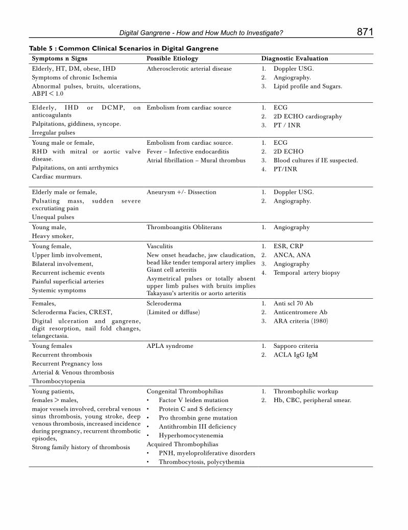
871Digital Gangrene - How and How Much to Investigate?
Table 5 : Common Clinical Scenarios in Digital GangreneSymptoms n Signs Possible Etiology Diagnostic Evaluation
Elderly, HT, DM, obese, IHDSymptoms of chronic IschemiaAbnormal pulses, bruits, ulcerations, ABPI < 1.0
Atherosclerotic arterial disease 1. Doppler USG.2. Angiography.3. Lipid profile and Sugars.
Elderly, IHD or DCMP, onanticoagulantsPalpitations, giddiness, syncope.Irregular pulses
Embolism from cardiac source 1. ECG2. 2DECHOcardiography3. PT / INR
Young male or female,RHD with mitral or aortic valve disease.Palpitations, on anti arrthymicsCardiacmurmurs.
Embolism from cardiac source.Fever – Infective endocarditisAtrial fibrillation – Mural thrombus
1. ECG2. 2DECHO3. Blood cultures if IE suspected.4. PT/INR
Elderly male or female,Pulsating mass, sudden severe excrutiating pain Unequal pulses
Aneurysm +/- Dissection 1. Doppler USG.2. Angiography.
Young male,Heavy smoker,
Thromboangitis Obliterans 1. Angiography
Young female,Upper limb involvement,Bilateral involvement,Recurrent ischemic eventsPainful superficial arteriesSystemic symptoms
VasculitisNew onset headache, jaw claudication, bead like tender temporal artery implies Giant cell arteritisAsymetrical pulses or totally absent upper limb pulses with bruits implies Takayasu’s arteritis or aorto arteritis
1. ESR,CRP2. ANCA,ANA3. Angiography4. Temporal artery biopsy
Females,SclerodermaFacies,CREST,Digital ulceration and gangrene, digit resorption, nail fold changes, telangectasia.
Scleroderma (Limited or diffuse)
1. Anti scl 70 Ab2. Anticentromere Ab3. ARA criteria (1980)
Young femalesRecurrent thrombosisRecurrent Pregnancy lossArterial & Venous thrombosisThrombocytopenia
APLA syndrome 1. Sapporo criteria2. ACLAIgGIgM
Young patients, females > males, major vessels involved, cerebral venous sinus thrombosis, young stroke, deep venous thrombosis, increased incidence during pregnancy, recurrent thrombotic episodes,Strong family history of thrombosis
CongenitalThrombophilias• FactorVleidenmutation• ProteinCandSdeficiency• Prothrombingenemutation• AntithrombinIIIdeficiency• HyperhomocystenemiaAcquired Thrombophilias• PNH,myeloproliferativedisorders• Thrombocytosis,polycythemia
1. Thrombophilic workup2. Hb,CBC,peripheralsmear.
872 Medicine Update 2008 Vol. 18
References1. Kinare SG et al. Pattern of occlusive PVD in India. Angiology
1976; 27: 165- 180.
2. CollinsTC,PetersenNJ,Suarez-AlmazorM,AshtonCM.Theprevalence of peripheral arterial disease in a racially diverse population. Arch Intern Med. Jun 23 2003; 163(12):1469-74.
3. Abbott,WM,Randolph,DM,McCabe,CC,etal.Arterialembolism: A 44 year perspective. Am J Surg 1982; 143:460.
4. Forrest KY. Becker DJ. Kuller LH. Wolfson SK. Orchard TJ. Are predictors of coronary heart disease and lower-extremity arterial disease in type 1 diabetes the same? A prospective study. Atherosclerosis 2000; 148: 159-169.
5. Shead GV, Oomen RM et al. Pattern of non diabetic PVD in south India. Br J of Surg 1978; 65: 49 – 53.
6. KatzenBT.ClinicalDiagnosisandPrognosisofAcuteLimbIschemia. Rev Cardiovasc Med. 2002; 3(suppl 2):S2-S6.
7. Eliason JL, Wainess RM, Proctor MC, et al. A Nationaland Single Institutional Experience in the ContemporaryTreatment of Acute Lower Extremity Ischemia. Ann Surg. 2003; 238(3):382-389.
8. Landis JR, Koch GG. The measurement of observer agreement for categorical data. Biometrics. 1977; 33:159–74.
9. Khan NA, Rahim SA, Anand SS, Simel DL, Panju A. Does the clinical examination predict lower extremity peripheral arterial disease? JAMA. 2006 Feb 1; 295(5): 536-46.
10. FaxonDP,etal.AtheroscleroticVascularDiseaseConference:Executive Summary: Atherosclerotic Vascular Disease
ConferenceProceeding forHealthcareProfessionalsFroma Special Writing Group of the American Heart Association. Circulation. 2004; 109:2595-2604.
11. Ray SA, Srodon PD et al. Reliability of ankle: Brachial pressure index measurement by junior doctors. Br J of Surg 2005; 81(2), 188 – 190.
12. Ascher E, Hingorani A, Markevich N, et al. Acute Lower Limb Ischemia: The Value of Duplex Ultrasound Arterial Mapping (DUAM) as the Sole Preoperative Imaging Technique. Ann Vasc Surg. 2003; 17(3):284-289.
13. Karthikeshwar Kasirajan, Kenneth Ouriel. Current Options in the Diagnosis and Management of Acute Limb Ischemia. Prog Cardiovasc Nurs 17(1):26-34, 2002.
14. Criqui MG, Fronek A, Klauber MR, Barrett-Connor E,Gabriel S. The sensitivity, specificity and predictive value of traditional clinical evaluation of peripheral arterial disease: results from noninvasive testing in a defined population. Circulation. 1985; 71:516–22.
15. Rose Hatala et al. An Evidence-Based Approach to the ClinicalExamination.Gen Intern Med. 1997 March; 12(3): 182–187.
16. Khan N A, Rahim S A, Anand S S, Simel D L, Panju A. Does the clinical examination predict lower extremity peripheral arterial disease? JAMA 2006; 295(5): 536 – 546.
17. QuanheYangetal. Improving thePredictionofComplexDiseases by Testing for Multiple Disease-Susceptibility Genes. Am. J. Hum. Genet. 72:636–649, 200.
18. CherylD.Bushnelletal.DiagnosticTestingforCoagulopathiesin PatientsWith Ischemic Stroke. Stroke 2000; 31; 3067-3078.

CHAPTER
115Medical Errors – Aiming to Improve the Art and Science of HealthcarePM Parikh, V Nair, H Malhotra, AA Ranade, GS Bhattacharyya
IntroductionMedicine is not mathematics. Hence seldom is 2 and 2 equal to 4. If such was, in fact, the case, doctors could easily be replaced by supercomputers that would cure every patient. Obviously, such is not the case. The prime reason is that the biology of the human body, the myriads of diseases and the various ways of managing them (both diagnosis and treatment) have infinite variations and permutations. Therefore, medicine is as much an Art as it is a Science.
Nevertheless, the primary aim of the medical profession is to ensure, first and always, patient safety. For this reason, there is a need to evaluate the magnitude of medical errors existing today, find out which of these are preventable ones and focus on devising a system that would eliminate (or at least minimize) them.
Alleged Magnitude of Medical ErrorsMedical Errors are not new. They have been reported in English literature at least since 1964 (Schimmel EM: The hazards of hospitalization. Ann Int Med 1964; 60: 100 pp10).
Of late, there has been a lot of focus and debate on its magnitude and implications. For instance in 2000, in their executive summary, the presidential committee in US quoted the 1999 the Institute of
Medicine (IOM), USA reported that upto 98,000 Americans die each year as a result of preventable medical errors, nearly half of the adverse events that occur in US patients are due to avoidable medical errors and that they cost 17 to 29 billion US dollars (Doing what counts for patient safety: Federal actions to reduce medical errors and their impact, February 2000 by Donna Shalala and Alexis Herman). It also stated that there could beasmanyas twoerrors in the ICUeveryday.(Institute of Medicine Report – To err is human: building a safer health system Nov 1999 http://www.nap.edu/books/0309068371/html/ accessed on 1st Dec 2007)
This was quickly picked up by the lay press and dramatized all over the electronic and print media globally. In India as well, the fourth estate highlights bad news about patient outcomes and attribute them to medical errors from time to time.
What is Medical Error?IOMs (IOMs report “To Err is Human: Building a Safer Health System” on November 30th 1999) definition of Medical Error is as follows: An error is defined as the failure of a planned action to be completed as intended or the use of a wrong plan to achieve an aim. Errors can include problems in practice, products, procedures and systems (added byQuIC)
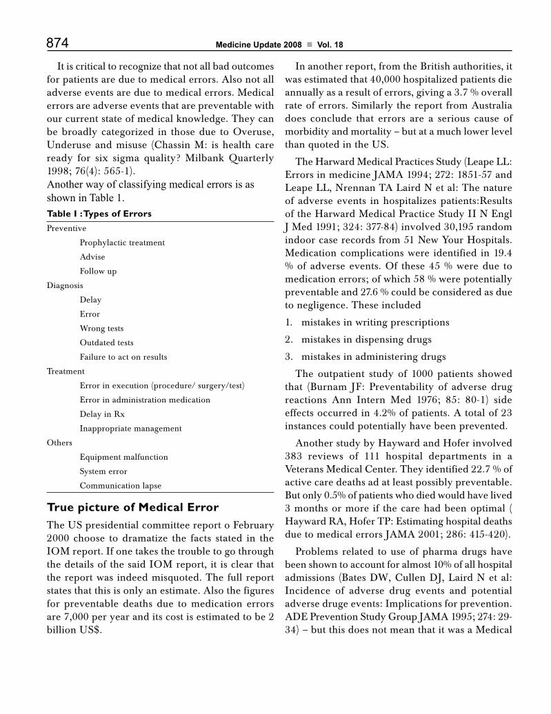
874 Medicine Update 2008 Vol. 18
It is critical to recognize that not all bad outcomes for patients are due to medical errors. Also not all adverse events are due to medical errors. Medical errors are adverse events that are preventable with our current state of medical knowledge. They can be broadly categorized in those due to Overuse, Underuseandmisuse (ChassinM: ishealthcareready for six sigma quality? Milbank Quarterly 1998; 76(4): 565-1). Another way of classifying medical errors is as shown in Table 1.Table I : Types of ErrorsPreventive
Prophylactic treatment
Advise
Follow up
Diagnosis
Delay
Error
Wrong tests
Outdated tests
Failure to act on results
Treatment
Error in execution (procedure/ surgery/test)
Error in administration medication
Delay in Rx
Inappropriate management
Others
Equipment malfunction
System error
Communicationlapse
True picture of Medical ErrorThe US presidential committee report o February 2000 choose to dramatize the facts stated in the IOM report. If one takes the trouble to go through the details of the said IOM report, it is clear that the report was indeed misquoted. The full report states that this is only an estimate. Also the figures for preventable deaths due to medication errors are 7,000 per year and its cost is estimated to be 2 billion US$.
In another report, from the British authorities, it was estimated that 40,000 hospitalized patients die annually as a result of errors, giving a 3.7 % overall rate of errors. Similarly the report from Australia does conclude that errors are a serious cause of morbidity and mortality – but at a much lower level than quoted in the US.
The Harward Medical Practices Study (Leape LL: Errors in medicine JAMA 1994; 272: 1851-57 and Leape LL, Nrennan TA Laird N et al: The nature of adverse events in hospitalizes patients:Results of the Harward Medical Practice Study II N Engl J Med 1991; 324: 377-84) involved 30,195 random indoor case records from 51 New Your Hospitals. Medication complications were identified in 19.4 % of adverse events. Of these 45 % were due to medication errors; of which 58 % were potentially preventable and 27.6 % could be considered as due to negligence. These included
1. mistakes in writing prescriptions
2. mistakes in dispensing drugs
3. mistakes in administering drugs
The outpatient study of 1000 patients showed that (Burnam JF: Preventability of adverse drug reactions Ann Intern Med 1976; 85: 80-1) side effects occurred in 4.2% of patients. A total of 23 instances could potentially have been prevented.
Another study by Hayward and Hofer involved 383 reviews of 111 hospital departments in a VeteransMedicalCenter.Theyidentified22.7%ofactive care deaths ad at least possibly preventable. But only 0.5% of patients who died would have lived 3 months or more if the care had been optimal ( Hayward RA, Hofer TP: Estimating hospital deaths due to medical errors JAMA 2001; 286: 415-420).
Problems related to use of pharma drugs have been shown to account for almost 10% of all hospital admissions (BatesDW,CullenDJ,LairdNetal:Incidence of adverse drug events and potential adverse druge events: Implications for prevention. ADE Prevention Study Group JAMA 1995; 274: 29-34) – but this does not mean that it was a Medical

875Medical Errors – Aiming to Improve the Art and Science of Healthcare
Error. Even when an event is termed as medical error, it is as a consequence of the healthcare team having weighted the options (including the drug reaction possibility) and the patients condition still required that particular drug to be administered. Even under such circumstances, most errors do not result in serious consequences for the patient.
One study tried to find out the cause of medication errors. It evaluated the understanding of 5 drug prescription labels among outpatients. The results indicated that only 34.7% of patients understood how many tablets are to be taken daily when the label read “take two tablets by mouth twice daily”. The more the no. of medications the patient was required to take, the greater was the risk ofmisunderstanding. (DavisTC,WoldMSetal:Literacy and misunderstanding prescription drug labels Ann Int Med 2006; 145: 887- 895)
Medical Errors – patient versus physician perceptionsUpto 95 % of physicians have reported being witness to a medical error. And 61% of health care professionals actually believe that errors are a routine part of medical practice. (Rajendran PR: Ethical issues involved in disclosing medical errors JAMA 2001; 286: 1078) (Medical errors: The scope of the problem. Publication No AHRQ 00-PO37 [[email protected]] Feb 2000)
Another study showed that when faced with such a medical mistake, about 46 % of US doctors fail to inform the institution about any incompetent or unethical practice of their colleagues. The reasons cited include not knowing what really happened, fear of reprisal and the feeling that a lawsuit would not bring back the dead person. Reasons for actually reporting include the duty of being truthful, believing in the principle of reparations and the need to protect future patients. (Forst N: Ethical Issues in Whistleblowing JAMA 2001; 286: 1079-1083).
Often the debate is between opinion about the best way to manage a patient versus actual medical
error. Such ethical dilemmas in day to day life are particularly difficult for medical students, since neither do they have insight into the medical problem nor the experience of how to choose the best treatment option (Forst N: Ethical Issues in Whistleblowing JAMA 2001; 286: 1079-1083).
Most doctors (93%) agreed that they should treat patients irrespective of their ability to pay. However, only 69 % did accept uninsured patients for treatment. (Gallagher TH, Waterman AD et al: Patients and physicians attitudes regarding the disclosure of medical errors JAMA 2003; 289: 1001-1007)
Most patients and family members want to know immediately (76%) and completely (88%) if a medical error has taken place. They also are happy to recommend that such errors also be reported to government agencies (92%) and hospitalcommittees(99%).(HobgoodC,PeckCR,GilbertB,ChappellK:Medicalerrors–whatandwhen: what do patients want to know? Acad Emerg Med 2002; 9: 1156-1161) Another study confirmed that 98.8% wanted full disclosure, 83% favoured financial compensation and even then, upto 47% of patients would still seek legal advise with a view of filing a lawsuit (Mazor KM, Simon SR et al: Health plan members view about disclosure of medical errors Ann Int Med 2004; 140: 409-418).
Recently, Judith Graham, staff reporter of the Tribune in USA (August 19, 2007 issue; [email protected]) reported about the changing response of physicians to medical errors. Dr. Divyesh Mehta, chief of oncology at the University of Illinois at ChicagoMedicalCenter(andawellknownnamein India as well) walked up to a patient with breast cancer and admitted that she was administered doublethedoseofG-CSFbyerrorandapologizedfor the same. He recommended increasing her hospital stay be a couple of days as a precaution. This action was applauded as a welcome change in the attitude of doctors and an ideal way of building relationships with patients. Not long ago, this encounter would have been almost unthinkable.

876 Medicine Update 2008 Vol. 18
Medical foul-ups were rarely discussed among physicians and almost never acknowledged to patients. Doctors were too proud, too afraid of malpractice lawsuits, too worried about losing face.
The system needs improvementIOM reported that majority of medical errors today are NOT produced by negligence, lack of education or lack of training. Rather errors occur in our health care system due to poor systems design and organizational factors - e.g. health care workers are expected to work 24 hour shifts, leading to overwork, fatigue and decreased mental concentration and alertness. Another important reason is the decentralized and fragmented nature of how health care is delivered. As a result, multiple providers at different locations may not know the full picture or have complete information regarding the patient’s medical illness, current medication and past adverse events or allergies.
Errors occur everywhere the patient is looked after, including but not limited to,
• Hospitals
• Physiciansoffice
• Nursinghomes
• Pharmacies
• Casualty
• Homecare
• Hospice
Knowhow already exists to prevent several medical errors. The value of Six Sigma Quality (greek letter use to represent standard deviation from the mean of any normally distributed curve) has already been well documented in other fields and industries. It is also important in healthcare delivery. Factors that need to be kept in mind include:
• Accountability
• Learningfromerrors
• Peer review protection – protect reportingsystems from being used in litigation
• Raisingthestandardofhealthcareorganizationsand professionals
• Buildingpublicawareness
• Usingstandardizedprocedures,checklistsandresults
• Dataintegrationandintegrity
Lessons about Error Reducing Efforts from Other Industries include:
• Make safetyanderror reductiona institutionpolicy of importance
• Proactively identify error rateparticularly inhigh risk populations
• Sethighgoalstoreducethem
• Setus systems for reportingerrors andclosecalls
• Promptly and thoroughly analyse errors toidentify the root causes
• Call upon the services of experts as andwhen required (clinical, epidemiological and management experts)
• Systemshouldspecificallybedesignednot toblame or find faults with individuals
• Earmarkfundsandhumanresourcestosupporterror prevention, remedy and develop safety culture
• Encourage out of the box thinking andsolutions
• Make public, regulatory authorities,pharmaceutical and medical device companies as well as private purchasers involved in the process as important stakeholders
Obstacles to improving safety of healthcare services include:
• Lack of widespread awareness about theproblem
• Incompletedataonthenatureandmagnitudeof the problem

877Medical Errors – Aiming to Improve the Art and Science of Healthcare
• Disorganized systemofprovidinghealthcareservices
• Mismatchbetweensupplyanddemand
• Lackofcompleteinformationwitheachmemberof the healthcare team
• Tradition of blaming individuals physiciansirrespective of root causes
• Physicianadministrationdisconnect
• Lack of protection from legal liability andlitigation
• Lack of functional system for medical errorreporting and remedy
• Unwillingnessof theadministrationtoaccepttheir responsibility for system deficiency and allocation of resources to prevent errors
Figure 1 shows a flowchart framework to address the problem of Medical Errors in any location/ institution. Using such techniques medical errors have already been reduced significantly. For instance, presence of pharmacists on medical
rounds, have reduced errors of medication by 66 %. In Surgical Anaesthesia, error were reduced from 25 to as little as 5.4 per million by using standard equipment, procedures and guidelines (Orkin FW: Patient monitoring during anesthesia as an exercise in technology assessment. In: Saidman LJ, Smith NT (eds). Monitoring in Asesthesia, 3rd ed. London, United Kingdom: Butterworth-Heineman 1993). A third example is that of the VA Hospital, which used hand held wireless computer and bar coding and found that medication errors reduced by 70 % (Medical errors: The scope of the problem. Publication No AHRQ 00-PO37 [[email protected]] Feb 2000)
Healthcare professional and scientific bodies should implement periodic re-examination or relicensing dependent on participation on continuingmedicaleducation.MCI,NationalBoard(DNB) and Health Universities should also develop a curriculum on patient safety. Information about medical errors should be disseminated on a regular basis to all healthcare professionals without letting it disappear in the existing information overload.
Figure 1 : Flowchart to address the problem of Medical Errors
Evaluation of Patient
Treatment
No Medical Errors
FavourableOutcome
UnfavourableOutcome
Unpreventable –No Medical Error
Medical Error detected
Non Serious
Not Identified in time to intervene
Minor Consequence
Document, Learn
and Rectify
Identified in time to intervene
Serious
Identified in time to intervene
Not identified in time to intervene
Serious Consequence
Document, Learn
and Rectify
Identified in time to intervene
Document and Learn from Event
No action required Action required
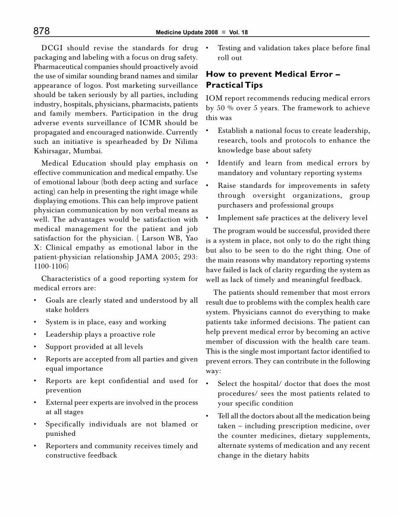
878 Medicine Update 2008 Vol. 18
DCGI should revise the standards for drugpackaging and labeling with a focus on drug safety. Pharmaceutical companies should proactively avoid the use of similar sounding brand names and similar appearance of logos. Post marketing surveillance should be taken seriously by all parties, including industry, hospitals, physicians, pharmacists, patients and family members. Participation in the drug adverse events surveillance of ICMR should bepropagatedandencouragednationwide.Currentlysuch an initiative is spearheaded by Dr Nilima Kshirsagar, Mumbai.
Medical Education should play emphasis on effective communication and medical empathy. Use of emotional labour (both deep acting and surface acting) can help in presenting the right image while displaying emotions. This can help improve patient physician communication by non verbal means as well. The advantages would be satisfaction with medical management for the patient and job satisfaction for the physician. ( Larson WB, Yao X: Clinical empathy as emotional labor in thepatient-physician relationship JAMA 2005; 293: 1100-1106)
Characteristicsof a good reporting system formedical errors are:
• Goalsareclearlystatedandunderstoodbyallstake holders
• Systemisinplace,easyandworking
• Leadershipplaysaproactiverole
• Supportprovidedatalllevels
• Reportsareacceptedfromallpartiesandgivenequal importance
• Reports are kept confidential and used forprevention
• Externalpeerexpertsareinvolvedintheprocessat all stages
• Specifically individuals are not blamed orpunished
• Reportersandcommunityreceivestimelyandconstructive feedback
• Testingandvalidationtakesplacebeforefinalroll out
How to prevent Medical Error – Practical TipsIOM report recommends reducing medical errors by 50 % over 5 years. The framework to achieve this was
• Establishanationalfocustocreateleadership,research, tools and protocols to enhance the knowledge base about safety
• Identify and learn from medical errors bymandatory and voluntary reporting systems
• Raise standards for improvements in safetythrough oversight organizations, group purchasers and professional groups
• Implementsafepracticesatthedeliverylevel
The program would be successful, provided there is a system in place, not only to do the right thing but also to be seen to do the right thing. One of the main reasons why mandatory reporting systems have failed is lack of clarity regarding the system as well as lack of timely and meaningful feedback.
The patients should remember that most errors result due to problems with the complex health care system. Physicians cannot do everything to make patients take informed decisions. The patient can help prevent medical error by becoming an active member of discussion with the health care team. This is the single most important factor identified to prevent errors. They can contribute in the following way:
• Selectthehospital/doctorthatdoesthemostprocedures/ sees the most patients related to your specific condition
• Tellallthedoctorsaboutallthemedicationbeingtaken – including prescription medicine, over the counter medicines, dietary supplements, alternate systems of medication and any recent change in the dietary habits
879Medical Errors – Aiming to Improve the Art and Science of Healthcare
• Informallthedoctorsaboutanyallergyorpastadverse reactions.
• Ifyouhaveatest,findoutwhatitsreportis
• Askaboutthestepsforeachprocedure/surgerythat you need to undergo
• Askallhealthcareworkerswhethertheyhavewashed their hands before being in contact with you
• Askafamilymemberorfriendtobewithyouat times of important discussions
• Make notes about anything that you donot understand and ask them at the first opportunity
• Atthetimeofdischarge,askthemtodescribeto your satisfaction the treatment to be taken at home. Read and understand the prescriptions written by the doctor. Ask if you do not understand particularly about how many tablets, how many times and at what time of the day/ night.
• Confirm with the medical store that youhave been provided the medicine exactly as specifically prescribed by the doctor without any substitution
• Buyandsavethebestdevisetomeasureliquidmedicines. Do not use your kitchen will provide you with a substitute measurement devise.
• Askforwritteninformation/resourceregardingside effects of your medicine.
Unintended consequences of reporting Medical ErrorAcknowledging medical errors with an apology is a noble intention to heal by neutralizing patients’ anger and easing physicians’ guilt. Some advocate it as a means to reduce potential malpractice lawsuits and payouts. However, though most doctors acknowledge their intention to be honest, majority are reluctant to reveal serious errors in practice. The main reason stated is the fear to their reputation as well as the risk of compromising their
legal liability. In the US more than 30 states, have passed “apology laws” that prevent such statements from being admissible against physicians in court. The reality is that lawyers are skeptical that it would work. Insurance companies continue to insist that doctors do not acknowledge such errors and in fact, recommend termination of all communication with the patients or families. This is based on the premise that such admission of wrongdoing will strengthen the patient’s case in the eyes of the courts. Such a thought process is backed by at least one Harvard University study that concluded that malpractice suits will increase as more patients become aware of errors.
Public reporting of information related to medical errors or competence has another downside. In the US, the courts would decide the level of the physician’s competence based on his adverse event rate in the past. As a consequence, doctors would only take up standard risk patients and refer complicated cases elsewhere. They would also strive to get better rates of success by doing interventions even when the patients condition indicated it was of questionable need. (Werner RM, Asch DA: The unintended consequences of publicly reporting quality information JAMA 2005; 293: 1239-1244.)
In India as well, the consequence of laws related to medical negligence, medical error and consumer courts have led to the practice of defensive medicine.
Situation in India – Special Considerations Dr Vasant Jaykar a prominent physician and cardiologist in Mumbai was shot dead in his own clinic. A disgruntled family member of a patient who died and threatened the doctor was arrested. Aparantly he had issued a “Supari” to have to doctor killed. Several years later, the accused were discharged by the court - due to lack of evidence. This is a very high price to pay by any physician, particularly a dedicated and sincere one of Dr Jaykar’s stature.
880 Medicine Update 2008 Vol. 18
Developing countries like India have a serious resource crunch in the healthcare sector. We have a dismal doctor to population ratio. This is also true for no of hospital beds per 1,00,000 of our population. Any government or teaching hospital will have hordes of patients flocking for medical management. When a doctor is faced with 300 to 500 patients every day, the time available for each patient is limited. This highlights the problem with the system. In spite of being well documented, the resource allocation for healthcare remains one of the lowest in any country. Under such circumstances, it is impossible to prevent medical errors.
On top of that, we have faced with frivolous lawsuits on several instances. One such example is about a lady who was suspected to have lung cancer. After cytological examination, which documented presence of lung cancer cells, she was advised admission for treatment. She chooses to go elsewhere, where a doubt was raised about the diagnosis. Tuberculosis was suspected and she was started on empirical anti-tubercular therapy. The patient’s condition continued to deteriorate. After a couple of years, she filed a complaint that she was wrongly diagnosed as having lung cancer, subject to mental stress and that she could have faced the consequences of wrongly given chemotherapy. In another instance, a patient was given chemotherapy elsewhere and died of progressive disease. His father then filed a case against Tata Hospital stating that the chemotherapy advise (given six months before the actual chemotherapy was administered at native place) was wrong and this caused the death of the patient.
The outcome of lawsuits in the court is also a cause of concern. In one instance a patient with headache was subsequently found to have a brain tumor. He went to court against the delay in diagnosis. The plea of the neurologist was that allpatientswithheadachedonotneedaCTScanespecially when the neurological examination was normal.BesidesthepatientcouldnotaffordtheCTScan. The hon’ble court made a remark that the patient’s life was more important than Rs 5,000 for
theCTScan.Inanotherinstance,apediatricianwasjailed and fined for prescribing medication that was licensed for marketing in India. The court decision was based on the fact that the medicine was banned in USA and hence considered dangerous.
Patients and their families often make it difficult for healthcare professionals to impart proper medical management. When faced with an unfavourable prognosis, they will often shop from doctor to doctor. They will then focus only on the information that they like – often from different sources and without proper reference to the context. Subsequently they will then expect the treating team to deliver the outcome apparently promised by someone else. Another common occurrence is not revealing the full history to the doctors. In fact some patients will attribute their opinions to one doctor while talking to another member of the healthcare team – creating miscommunication and confusion. Patients are even given (sometimes without their knowledge) various sorts of medicines and nutraceuticals on the sly, deliberately withholding this information from their doctors. And this happens irrespective of literacy or socioeconomic background. This is particularly applicable when indigenous systems of medicine are being used. Thereafter, when any adverse reaction occurs, they are quick to blame the treating allopathic doctors.
Hospital administrators, particularly in India, often forget their pivotal responsibility in mitigating medical errors. Do they accept and acknowledge their role and responsibility in medical errors? Unfortunately, they are the first to pass the buck and look for a “bakra” to blame and cover their deficiencies. Senior and internationally renowned Head & Neck OncoSurgeon was recently put in the dock by the biased administration of his own hospital. He was accused of ethical misconduct for something that happened when he was not even present. In fact, the sequences of events clearly indicate that it was a known complication that had occurred in spite of comprehensive medical management. The way this was blown out of proportion and action intended to be initiated
881Medical Errors – Aiming to Improve the Art and Science of Healthcare
against him for dereliction of duty in reporting the event could probably be explained by someone in the administration mounting a personal vendetta against him for “other” considerations. Fortunately there were enough sensible and ethical persons to ascertain the true picture and clearing him from allegations.
• Incidence of medical errors in India isnot documented but they probably occur frequently
• Medical errors are usually underreported,overlooked and remain a controversial area.
• Mosterrorsareduetosystemrelatedproblemsand failures, not due to individual faults.
• Effortstoreduceandeliminatemedicalerrorareneeded in a systematic and proactive manner
• Experiencefromother industries/ fieldshave
the potential to teach us a lot how this can be done effectively
• Publicopinion,legislation,voluntaryreportingand a system of reporting with legal protection needs to be mobilized.
• SOPshouldspecificallybedesignedto
a. Find root causes
b. Avoid tendency to blame individuals
c. Allocate sufficient resources for all objectives
d. Provide timely feedback in a manner that it is useful
e. Including this topic in the medical curriculum
f. Look at unique Indian environment to design “out-of-the-box” solutions
CHAPTER
116Atherosclerosis in Rheumatic DiseasesSukumar Mukherjee
IntroductionAs association between accelerated atherosclerosis and systemic lupus erythematosus (SLE), typical by the occurrence of a myocardial infarction in a 30 year old woman who had had the disease for more than 10 years was suggested in 1976.1 The same author reported bimodal pattern of mortality in SLE, with early death from active disease and late deaths from cardiovascular disorders. Several subsequent reports have supported an increased cardiovascular risk among SLE patients. This opened up the interest in the new understanding of atherosclerosis and systemic rheumatic diseases.
Though there is a wide spectrum of systemic rheumatic diseases but SLE, Rheumatoid arthritis, antiphospholipid antibody syndrome and systemic sclerosis are the common ones wherein studies on atherosclerotic cardiovascular and cerebrovascular disease are increasingly available. Traditional cardiovascular risk factors, such as hypertension and dyslipidemia are more prevalent in SLE patients and certainly contribute to an increased incidence ofcardiovasculardisease(CVD)2. However, cohort studies have shown that the increased cardiovascular risk in SLE cannot be explained by traditional risk factors only but disease specific factors contribute significantly as well.3,4,5
The most studied disease in this direction is SLE which is a complex autoimmune prototype multisystem disease characterized by chronic inflammation of virtually every organ including blood vessels. The atherogenic cascade of events and chronic immunoinflammation with autoantibodies are interlinked in systemic rheumatic diseases to contribute premature atherosclerosis.
Evidence for increased incidence of atherosclerotic CVD in SLEIn2prospectivefollow-upstudiesCVDendpointsin SLE patients were compared with those in a reference population without SLE. Manji et al6 observed in 498 patients with 6.7 years follow-up that incidence of myocardial infarction (M1) in all age groups of women with SLE with a 7-fold higher incidence in all age groups combined and a particularly increased risk more than 50 fold in women aged 35-44 years. Jonsson et al7 observed myocardial infarction occurred 9 times more frequently in 86 adult patients on a follow-up of 6 years.
In two large hospitalized retrospective studies CVDincidenceis2-4timesmorecommoninSLEpatients compared with those of non-SLE patients.9,10 A case control study comparing 8,688 patients with AM1 with 33,923 controls from the General Practice Research Database (GPRD) in UK found
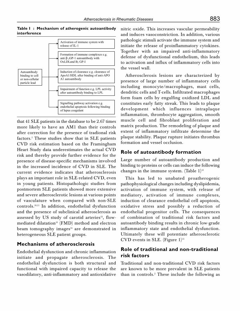
883Atherosclerosis in Rheumatic Diseases
that 41 SLE patients in the database to be 2.67 times more likely to have an AM1 than their controls after correction for the presence of tradional risk factors.5 These studies show that in SLE patients CVD risk estimation based on the FraminghamHeartStudydataunderestimatestheactualCVDrisk and thereby provide further evidence for the presence of disease-specific mechanisms involved in the increased incidenceofCVD inSLE.Thecurrent evidence indicates that atherosclerosis playsanimportantroleinSLE-relatedCVD,evenin young patients. Histopathologic studies from postmortem SLE patients showed more extensive and severe atherosclerotic lesions at various points of vasculature when compared with non-SLE controls.10,11 In addition, endothelial dysfunction and the presence of subclinical atherosclerosis as assessed by US study of carotid arteries12, flow-mediated dilatation13 (FMD) method and electron beam tomography images14 are demonstrated in heterogeneous SLE patient groups.
Mechanisms of atherosclerosisEndothelial dysfunction and chronic inflammation initiate and propagate atherosclerosis. The endothelial dysfunction is both structural and functional with impaired capacity to release the vasodilatory, anti-inflammatory and antioxidative
nitric oxide. This increases vascular permeability and induces vasoconstriction. In addition, various pathologic stimuli activate the immune system and initiate the release of proinflammatory cytokines. Together with an impaired anti-inflammatory defense of dysfunctional endothelium, this leads to activation and influx of inflammatory cells into the vessel wall.
Atherosclerosis lesions are characterized by presence of large number of inflammatory cells including monocyte/macrophages, mast cells, dendritic cells and T-cells. Infiltrated macrophages form foam cells by engulfing oxidized LDL and constitutes early fatty streak. This leads to plaque development which influences intraplaque inflammation, thrombocyte aggregation, smooth muscle cell and fibroblast proliferation and matrix production. The remodeling of plaque and extent of inflammatory infiltrate determine the plaque stability. Plaque rupture initiates thrombus formation and vessel occlusion.
Role of autoantibody formationLarge number of autoantibody production and binding to proteins or cells can induce the following changes in the immune system. (Table 1)15
This has led to unabated proatherogenic pathophysiological changes including dyslipidemia, activation of immune system, with release of mediatory, activation of immune complexes, induction of clearance endothelial cell apoptosis, oxidative stress and possibly a reduction of endothelial progenitor cells. The consequences of combination of traditional risk factors and autoantibody binding results in chronic low-grade inflammatory state and endothelial dysfunction. Ultimately these will potentiate atherosclerotic CVDeventsinSLE.(Figure1)15
Role of traditional and non-traditional risk factorsTraditionalandnon-traditionalCVDrisk factorsare known to be more prevalent in SLE patients than in controls.2 These include the following as
Table I : Mechanism of atherogenic autoantibody interference
Autoantibody binding to cell or non-cellular particle lead
Activation of immune system with release of IL-1
Formation of immune complexes e.g. anti β 2GP-1 autoantibody with OxLDLand B2 GP-I
Induction of clearance e.g. clearance of ApoA1/HDL after binding of anti-APO A1 autoantibody
Impairment of function e.g. LPL activity after autoantibody binding to LPL
Signalling pathway activation e.g. endothelial apoptosis following binding of lupus coagulant

884 Medicine Update 2008 Vol. 18
Table 2 : Risk factors for risk factors in SLE-CVDTraditional risk factors Non-traditional risk
factorsHypertension
Hyper cholesterolemia
Hyper homocysteinemia
Low HDL
Diabetes mellitus
Renal impairment
ElevatedCRP
Insulin resitance
Smoking
Steroid therapy
Proinflammatory HDL
Endothelial apoptosis
Anti HSP antibodies
Increased Ox-LDL
IncreasedCIC
Elevated inflammatory cytokines
Dendritic cell overexpression
Decreased endothelial progenitor cells
Figure 1 : Overview of potential mechanisms underlying the pathophysiology of accelerated cardiovascular disease in systemic lupus erythematosus
Medication effects
Autoimmune responses & dysregulation of inflammation
Traditional risk factors
Oxidative Stress
Endothelial apoptosis
Endothelial progenitor Cells �
Inflammatory mediators
Endothelial dysfunction Inflammatory infiltrate
Atherosclerosis
Cardiovascular event
in Table 2.2,3,16,17,18,19 Several studies in SLE patients that prolonged use of prednisone is associated with atherosclerotic changes16 although there are some contradictory observation.20 Roman et al found that more aggressive immunosuppression, including higher doses of prednisone and use of cytophosphamide or hydroxychloroquin was associated with the absence of plaque.21
Traditional risk factors may be influenced by SLE-specific factrors. Antiphospholipid antibodies in 30-
50% SLE patients adversely affect the lipid profile and induce lipid peroxidation. Autoantibodies are produced against other lipid components like HDL, Apo-A1 and lipoprotein lipase (LPL). All there increase lipid peroxidation and make these lipid components dysfunctional.22,23 Hence, HDL is proatherogenic and proinflammatory because of autoantibody binding to HDL/APO-I; anti-LPL antibody correlate with triglyceride levels, disease activity and markers of inflammation.
Endothelial cell apoptosisAutoantibodies in SLE bind to endothelium to target 60Kd heat shock protein (HSP) and cause endothelial cell apoptosis upon binding of anti HSP 60 antibodies.19 Similarly lupus anticoagulant and anti-ds DNA antibodies induce endothelial cell apoptosis. Endothelial apoptosis contributes to the loss of endothelial integrity and thereby to the initiation of atherosclerosis.24 Apoptotic endothelium is also prothrombotic in SLE25 which is further aggravated by circulating annexin V and plasma tissue factor.26 Increased endothelial cell apoptosis may represent an important mechanism for the development of both atherosclerosis and thrombosis.
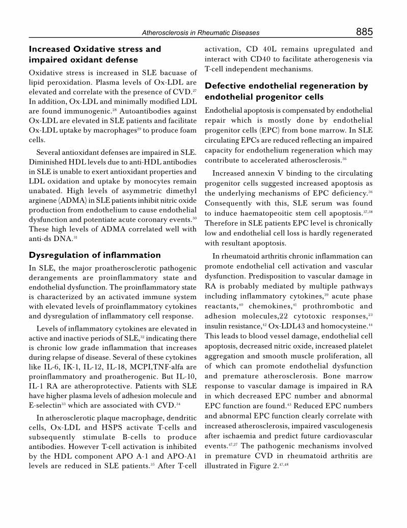
885Atherosclerosis in Rheumatic Diseases
Increased Oxidative stress and impaired oxidant defenseOxidative stress is increased in SLE bacuase of lipid peroxidation. Plasma levels of Ox-LDL are elevatedandcorrelatewiththepresenceofCVD.27 In addition, Ox-LDL and minimally modified LDL are found immunogenic.28 Autoantibodies against Ox-LDL are elevated in SLE patients and facilitate Ox-LDL uptake by macrophages29 to produce foam cells.
Several antioxidant defenses are impaired in SLE. Diminished HDL levels due to anti-HDL antibodies in SLE is unable to exert antioxidant properties and LDL oxidation and uptake by monocytes remain unabated. High levels of asymmetric dimethyl arginene (ADMA) in SLE patients inhibit nitric oxide production from endothelium to cause endothelial dysfunction and potentiate acute coronary events.30 These high levels of ADMA correlated well with anti-ds DNA.31
Dysregulation of inflammationIn SLE, the major proatherosclerotic pathogenic derangements are proinflammatory state and endothelial dysfunction. The proinflammatory state is characterized by an activated immune system with elevated levels of proinflammatory cytokines and dysregulation of inflammatory cell response.
Levels of inflammatory cytokines are elevated in active and inactive periods of SLE,32 indicating there is chronic low grade inflammation that increases during relapse of disease. Several of these cytokines likeIL-6,IK-1,IL-12,IL-18,MCPI,TNF-alfaareproinflammatory and proatherogenic. But IL-10, IL-1 RA are atheroprotective. Patients with SLE have higher plasma levels of adhesion molecule and E-selectin33whichareassociatedwithCVD.34
In atherosclerotic plaque macrophage, dendritic cells, Ox-LDL and HSPS activate T-cells and subsequently stimulate B-cells to produce antibodies. However T-cell activation is inhibited by the HDL component APO A-1 and APO-A1 levels are reduced in SLE patients.35 After T-cell
activation, CD 40L remains upregulated andinteractwithCD40to facilitateatherogenesisviaT-cell independent mechanisms.
Defective endothelial regeneration by endothelial progenitor cellsEndothelial apoptosis is compensated by endothelial repair which is mostly done by endothelial progenitorcells(EPC)frombonemarrow.InSLEcirculatingEPCsarereducedreflectinganimpairedcapacity for endothelium regeneration which may contribute to accelerated atherosclerosis.36
Increased annexin V binding to the circulating progenitor cells suggested increased apoptosis as theunderlyingmechanismsofEPCdeficiency.36 Consequently with this, SLE serum was foundto induce haematopeoitic stem cell apoptosis.37,38 ThereforeinSLEpatientsEPClevelischronicallylow and endothelial cell loss is hardly regenerated with resultant apoptosis.
In rheumatoid arthritis chronic inflammation can promote endothelial cell activation and vascular dysfunction. Predisposition to vascular damage in RA is probably mediated by multiple pathways including inflammatory cytokines,39 acute phase reactants,40 chemokines,41 prothrombotic and adhesion molecules,22 cytotoxic responses,23 insulin resistance,42 Ox-LDL43 and homocysteine.44 This leads to blood vessel damage, endothelial cell apoptosis, decreased nitric oxide, increased platelet aggregation and smooth muscle proliferation, all of which can promote endothelial dysfunction and premature atherosclerosis. Bone marrow response to vascular damage is impaired in RA in which decreased EPC number and abnormalEPCfunctionarefound.45ReducedEPCnumbersandabnormalEPCfunctionclearlycorrelatewithincreased atherosclerosis, impaired vasculogenesis after ischaemia and predict future cardiovascular events.47,27 The pathogenic mechanisms involved in premature CVD in rheumatoid arthritis areillustrated in Figure 2.47,48
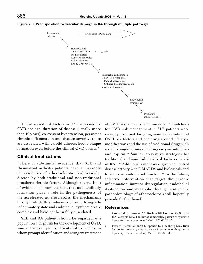
886 Medicine Update 2008 Vol. 18
Figure 2 : Predisposition to vascular damage in RA through multiple pathways
Rheumatoid arthritis
RA blocks EPC release
Homocysteine TNF-α , IL-1, IL-6, CD4, CD28 cells Modified lipids Adhesion molecules Insulin resitance PAI-1, CRP, MCP-1
Endothelial cell apoptosis � NO � Free radicals � Platelet aggregation � Collagen breakdown smooth muscle proliferation
Endothelial dysfunction
Premature atherosclerosis
The observed risk factors in RA for premature CVDare age, durationof disease (usuallymorethan 10 years), co-existent hypertension, persistent chronic inflammation and disease severity; these are associated with carotid atherosclerotic plaque formationevenbeforetheclinicalCVDevents.49
Clinical implications There is substantial evidence that SLE and rheumatoid arthritis patients have a markedly increased risk of atherosclerotic cardiovascular disease by both traditional and non-traditional proatherosclerotic factors. Although several lines of evidence support the idea that auto-antibody formation plays a role in the pathogenesis of the accelerated atherosclerosis, the mechanisms through which this induces a chronic low-grade inflammatory state and endothelial dysfunction are complex and have not been fully elucidated.
SLE and RA patients should be regarded as a populationathighriskforthedevelopmentofCVD,similar for example to patients with diabetes, in whom prompt identification and stringent treatment
ofCVDriskfactorsisrecommended.50 Guidelines forCVDriskmanagement inSLEpatientswererecently proposed, targeting mainly the traditional CVDrisk factorsandcenteringaround life stylemodifications and the use of traditional drugs such a statins, angiotensin-converting enzyme inhibitors and aspirin.50 Similar preventive strategies for traditional and non-traditional risk factors operate in RA.51,52 Additional emphasis is given to control disease activity with DMARDS and biologicals and to improve endothelial function.53 In the future, selective intervention that target the chronic inflammation, immune dysregulation, endothelial dysfunction and metabolic derangement in the pathophysiology of atherosclerosis will hopefully provide further benefit.
References 1. Urotiwz MB, Bookman AA, Koehler BE, Gordon DA, Smythe
HA, Ogryzlo MA. The bimodal mortality pattern of systemic lupus erythematosus. Am J Med 1976;60:221-5.
2. PetriM,Perez-GuthannS,SpenceD,HochbergMC.Riskfactors for coronary artery disease in patients with systemic lupus erythematosus. Am J Med 1992;93:513-9.
887Atherosclerosis in Rheumatic Diseases
3. Esdaile JM, Abrahamowicz M, Grodzicky T, Li Y, Panaritis C,duBergerR,etal.TrationalFraminghamriskfactorsfailto fully amount for accelerated atherosclerosis in systemic lupus erythematosus. Arthritis Rheum 2001;44:2331-7.
4. Bessant R, Hingorani A, Patel L, MacGregor A, Isenberg DA, Rahman A. Risk of coronary heart disease and stroke in a large British cohort of patients with systemic lupus erythematosus.
5. FischerLM,SchliengerRG,MatterC, JickH,MeierCR.Effect of rheumatoid arthritis or systemic lupus erythematosus on the risk of first-time acute myocardial infarction. Am J Cardiol2004;93:198-200.
6. ManziS,MeilahnEN,RairieJE,ConteCG,MedsgerTAjr,Jansen-McWilliams L, et al. Age-specific incidence rates of myocardial infarction and angina in women with systemic lupus erythematosus: comparision with the Framingham Study. Am J Epidemiol 1997;145:408-15.
7. Jonson H, Nived O, Sturfelt G. Outcome in systemic lupus erythematosus: a prospective study of patients from a defined population. Medicine (Baltimore) 1989;68:141-50.
8. Biornadal I, Yin L, Granath F, Klareskog I, Ekbom A. Cardiovasculardiseaseahazarddespiteimprovedprognosisin patients with systemic lupus erythematosus: results from a Swedish population based study 1964-95. J Rheumatol 2004;31:713-9.
9. Ward MM. Premature morbidity from cardiovascular and cerebrovascular disease in women with systemic lupus erythematosus. Arthritis Rheum 1999;42:338-46.
10. Bulkley BH, Roberts WC. The heart in systemic lupuserythematosus and the changes induced in it by corticosteroid therapy: a study of 36 necropsy patients. Am J Med 1975;58:243-64.
11. HaiderYS,RobertsWC.Coronaryarterialdiseaseinsystemiclupus erythematosus; quantification of degrees of narrowing in 22 necropsy patients (21 women) aged 16 to 37 years. Am J Med 1981;70:775-81.
12. Roman MJ, Devereux RB, Schwartz JE, Lockshin MD, Paget SA, Davis A, et al. Arteroa; stoffness in chronic inflammatory diseases. Hypertension 2005;46:194-9.
13. VerhaarMC,RabelinkTJ.Endothelial function: strategiesforearlyintervention.CardiovascDrugsTher1998;12Suppl1:125-34.
14. Asanuma Y, Oeser A, Shintani AK, Turner E, Oslen N, Fazio S, et al. Premature coronary-artery atherosclerosis in systemic lupus erythematosus. N Engl J Med 2003;349:2407-15.
15. Hill MB, Phipps JL, Hughs P et al. Anti endothelial cell antibodies in primary APLA and SLE. Br J Haematology 1998;103;416-21.
16. Manzi S, Selzer F, Sutton-Tyrrell K, Fitzgerald SG, Rairie JE, Tracy RP, et al. Prevalence and risk factors of carotid plaque in owmen with systemic lupus erythematosus. Arthritis Rheum 1999;42:51-60.
17. Doria A, Shoenfeld Y, Wu R, Gambari PF, Puato M, Ghirardello A, et al. Risk factors for subclinical atherosclerosis in a prospective cohort of patients with systemic lupus
erythematosus. Ann Rheum Dis 2003;62:1071-7.
18. Hill MB, Phipps JL, Hughes P, Greaves M. Anti-endothelial cell antibodies in primary antiphospholipid syndrome and SLE: paterns of reactivity with membrane antigens on microvascular and umbilical venous cell membranes. Br J Haematol 1998;103:416-21.
19. Dieude M, Senecal JL, Raymond Y. Induction of endothelial cell apoptosis by heat-shock protein 60-reactive antibodies from anti-endothelial cell autoantibody-positive systemic lupus erythematosus patients. Arthritis Rheum 2004;50:3221-31.
20. Salmon JE, Roman MJ. Accelerated atherosclerosis in systemic lupus erythematosus: implications for patient management.CurrOpinRheumatol2001;13:341-4.
21. Roman MJ, Shanker BA, Davis A, Lockshin MD, Sammaritano I, Simantov R, et al. Prevalence and correlates of accelerated atherosclerosis in systemic lupus erythematosus. N Engl J Med 2003;349:2399-406.
22. NcNahon M, Grossman J, FitzGerald J, Dahlin-Lee E, Wallace DJ, Thong By, et al. Proinflammatory high-density lipoprotein as a biomarker for atherosclerosis in patients with systemic lupus erythematosus and rheumatoid arthritis. Arthritis Rheum 2006;54:2541-9.
23. BorbaEF,BonfaE,VanagreCG,RamiresJA,MaranhaoRC.Chylomicron metabolism is markedly altered in systemiclupus erythematosus. Arthritis Rheum 2000;43:1033-40.
24. Stoneman VE, Bennett MR. Role of apoptosis in atherosclerosis and its therapeutic implications. Clin Sci(Lond) 2004;107:343-54.
25. Durand E, Scoazee A, Boddaert J, Al Hajzen A, Addad R, et al. In vivo induction of endothelial apoptosis leads to vessel thrombosis and endothelial denudation: a clue to the understanding of the mechanisms of thrombotic plaque erosion.Circulation2004;109-2503-6.
26. RajagopalanS,SomerisEC,BrookRD,KehrerC,PfenningerD, Lewis E, et al. Endothelial cell apoptosis in systemic lupus erythematosus: a common pathway for abnormal vascular function and thrombosis propensity. Blood 2004;103:3677-83.
27. Frostegard J, Svenungasson E, Wu R, Gunnarsson I, Lundberg IE, Klareskog I, et al. Lipid peroxidation is enhanced in patients with systemic lupus erythematosus and is associated with arterial and renal disease manifestations. Arthritis Rheum 2005;52:192-200.
28. Witztum JL. The oxidation hypothesis of atherosclerosis. Lancet 1994;344-793-5.
29. Lopes-Virella MF, Binzafar N, Rackley S, Takei A, La Via M, Virella G. The uptake of LDL-IC by humanmacrophages:predominant involvement of the Fcy RI receptor. Atherosclerosis 1997;135:161-70.
30. Fliser D. Asymmetric dimethylarginine (ADMA): the silent transition from an ‘uraemic toxin’ to a global cardiovascular riskmolecule.ErrJClinInvest2005;35:71-9.
31. Bultink IE, Teerlink T, Heijst JA, Dijkmans BA, Voskuyl AE. Raised plasma levels of asymmetric dimethylarginine are
888 Medicine Update 2008 Vol. 18
associated with cardiovascular events disease activity and organ damage in patients with systemic lupus erythematosus. Ann Rheum Dis 2005;64:1362-5.
32. CapperER,MaskillJK,GordonC,BlakemoreAI.Interleukin(IL)-10, IL-1ra and Il-12 profiles in active and quiescent systemic lupus erythematosus: could longitudinal studies revealpatient subgroupsofdifferingpathology?ClinExpImmunol 2004;138:348-56.
33. MrowkaC,SieberthHG.DetectionofcirculatingadhesionmoleculesICAM-1,VCAM-1andE-selectioninWegener’sgranulomatosis, systemic lupus erythematosus and chronic renalfailure.ClinNephrol1995;43:288-96.
34. HwangSJ,BallantyneCM,SharrettAR,SmithLC,DavisCE, Gotto AM Jr, et al. Circulating adhesion moleculesVCAM-1,ICAM-1andE-selectionincarotidatherosclerosisand incident coronary heart disease cases: the Atherosclerosis RiskinCommunities(ARIC)Study.Circulation1997;96:4219-25.
35. Delgado Alves J, Ames PR, Donohue S, Stanyer I, NouroozZadehJ,RavirajanC,etal.Antibodiestohighdensitylipoprotein and →
2-glycoprotein I are inversely correlated
with paraoxonase activity in systemic lupus erythematosus and primary antiphospholipid syndrome. Arthritis Rheum 2002;46:2686-94.
36. Westerweel PE, Luijten RK, Hoefer IE, Koomans HA, DerksenRH,VerhaarMC.Haematopoieticandendothelialprogenitor cells are deficient in quiescent systemic lupus erythematosus. Ann Rheum Dis. In press.
37. Liu H,Ozaki K, Matsuzaki Y, Abe M, Kosaka M, Saito S. Suppression of haematopoiesis by IgG autoantibodies from patientswithsystemiclupuserythematosus(SLE).ClinExpImmunol 1995;100:480-5.
38. Tiefenthaler M, Bacher N, Linert H, Muhlmann O, Hofer S, SeppN,etal.ApoptosisofCD34+cellsafterincubationwithsera of leukopenic patients withsystemic lupus erythematosus . Lupus 2003;12:471-8.
39. Love PE, Santoro SA. Antiphospholipid antibodies: anticardiolipin and the lupus anticoagulant in systemic lupus erythematosus (SLE) and in non-SLE disorders; prevalence and clinical significance. Ann Intern Med 1990;112:682-98.
40. Asanuma Y, Chung CP, Oeser A, Shintani A, Stanley E,Raggi P, et al. Increased concentration of proatherogenic inflammatory cytokines in systemic lupus erythematosus: relationship to cardiovascular risk factors. JRheumatol
2006;33:539-45.
41. Ross R. Atherosclerosis – an inflammatory disease. N Engl J Med 1999;340:115-26.
42. Borba EF, Bonfa E. Dyslipoproteinemias in systemic lupus erythematosus: influence of disease, activity and anticardiolipin antibodies. Lupus 1997;6:533-9.
43. Bruce IN, Urowitz MB, Gladman DD, Hallett DC.Natural history of hypercholesterolemia in systemic lupus erythematosus. J Rheumatol 1999;26:2137-43.
44. MosatoS,PratesiF,BongiomiF,ScavuzzoMC,ChimentiD, Bombardieri S, et al. Endothelial cell binding by systemic lupus antibodies: functional properties and relationship with anti-DNA activity. J Autoimmun 2002;18:231-8.
45. NakamuraN,ShidaraY,KawaguchiN,AzumaC,MitsudaN, Onishi S, et al. Lupus anticoagulant autoantibody induces apoptosis in umbilical vein endothelial cells: involvement of annexinV.BiochemBiophysResCommun1994;205:1488-93.
46. CederholmA,FrostegardJ,AnnexinA5incardiovasculardisease and systemic lupus erythematosus. Immunobiology 2005;210:761-8.
47. Branen L, Hovgaard L, Nitulescu M, et al. Inhibition of tumor necrosis factor-alpha reduces atherosclerosis knockout mice. Arterioscler Thromb Vasc Biol 2004:24:2137-2142.
48. GersztenRE,Garcia-ZepedaEA,LimYC,etalMCP-1andIL-8 trigger firm adhesion of monocytes to vascular under flow conditions. Nature 1999;398:718-723.
49. KaplanJM.CardiovasculardiseaseinRheumatoidArthritis.CurrOpinRheumatol2006;18(3):289-297.
50. Wajed J, Ahmad Y, Durrington PN, Bruce IN, Prevention of cardiovascular disease in systemic lupus erythematosus – proposed guidelines for risk factor management. Rheumatology (Oxford) 2004;43:7-12.
51. Ablin JN, Bogusiavski V, Aloush V, et al. Infliximab increases levels of circulating endothelial progenitor cells and improves their functional properties in patients with rheumatoid arthritis. Arthritis Rheum 2005;52:S581.
52. MaxwellSR,MootsRJ,KendallMJ.Corticosteroids:dotheydamage the cardiovascular system? Postgraduate Medicine 2004;870.
53. Hurlimann D, Forster A, Noll G, et al. Anti-tumor necrosis factor-alpha treatment improves endothelial function in rheumatoidarthritis.Circulation2002;106:2184-2187.

Abridged IndexCardiology AMI in elderly 728 Antiplatelet drug resistance 105 Bare metal stents 53 Broad QRS tachycardia 783 CAD & Sleep disorders 182 CAD in women 645 Chronic heart failure- diagnosis 590 Drug eluting stents 57 Early metabolic intervention 421 Heart failure 192 Hypertension and obesity 548 Hypertension- changing guidelines 426 Hypertension- Combination therapy 434 Hypertension- endothelial cell dysfunction 32 Hypertension in Type 2 diabetes 439 Newer thrombolytics Vs primary angioplasty
14 Pre-operative cardio-pulmonary assessment
725 Refractory heart failure- devices and surgery
596 Safety of stents 70 STEMI- newer drugs 112 Thrombolysis in AMI 535 Valvular heart disease in pregnancy 653 Young hypertensive- investigation 570 Clinical Pharmacology Chirality of drugs 178 Drug interactions- mechanisms 674 Probiotics and Prebiotics 258 Critical Care & Emergency Medicine Bio-terrorism 406 DIC 230 Febrile emergencies 217 Heat stroke 817 Metabolic abnormalities in critically ill 500 Resuscitation- trauma in pregnancy 42
Snake bite- update on management 691 Statins in critical care 491 Wavelet analysis of heart rate variability 142 Endocrinology Diabetes- mega trials 664 Diabetes prevention 849 Glucagon analogs 459 Incretin hormones 1 Insulin in Type 2 Diabetes 453 Maternal Hypothyroidism & Pregnancy 342 Multinodular thyroid disease 615 Neuropathy in diabetes- management 810 Obesity- experience with sibutramine 540 Obesity- SCOUT trial 544 Oral hypoglycemics- current status 441 Prediabetes- interventions 633 Thyroid disease in India 334 TypeIIdiabetes-profile94
Gastroenterology IBD in India 250 IBD-recent advances 264 Hepato-pulmonary syndrome 748 Non-variceal bleed- management 833
Geriatrics Abdominal pain in elderly 239 Improving longevity 779 Preventive strategies 731
Haematology Polycythemia- Investigation 358 DVT- Investigation 368 Infections & Tropical Medicine Enteric fever 201 Antileishmanial resistance 486 Antimalarial drug resistance 477 Chikungunya 205
890 Medicine Update 2008 Vol. 18
Colonization Vs infection 330 Dengue- resurging infection 739 Leptospirosis in India 799 Nosocomial Pneumonia 290 Post-exposure prophylaxis in HIV 752 Tropical chronic pancreatitis 323 Tropical pyomyositis 697
Lipidology Aggressive lipid lowering 298 HDL-therapy 313 Isolated Triglyceridemia 302 Mixed dyslipidemias 764 Postprandial lipids- role in atherosclerosis 857
Nephrology Acute renal failure- management 603 Anemia in CKD 163 Glomerulonephritis- management 82 Nephrotoxicity- herbal drugs 150 Proteinuria- complications 156 Renal replacement therapy 610
Neurology Acute ischaemic stroke 117 First seizure- management 246 Migraine prophylaxis 378 Parkinson’s Disease- Non motor symptom
complex 9 Refractory Epilepsy- Investigation 578 Stroke Unit 131 Thrombolysis in ischemic stroke 519 TIAs 347 Oncology Infection & cancers 168 Non Hodgekin's lymphoma- treatment 789 Solid tumours- targeted therapy 508 Psychiatry Depression- new horizons 404
Pulmonology Bronchial asthma- management 283 COPD- long term management 271 COPD- Non invasive ventilation 383 Hemoptysis with normal X-Ray chest 587 Lung cancer- recent advances 717 Pulmonary embolism- thrombolysis 529 Solitary pulmonary nodule- investigation 824 Rheumatology Ankylosing spondylitis- management 472 Atherosclerosis 882 Monoarthritis- Investigation 374 Osteoporosis-recent advances 628 Rheumatoid arthritis- disease control 466 Vasculitis 76 Miscellaneous Amyloidosis- recent advances 391 Communication skills 557 Communication with angry patient 845 Diagnostic reasoning- Baye's theorem 563 Digital Gangrene 863 Evidence based medicine- Update 21 Global warming- health hazards 703 Medical Errors 873 Meditation- clinical apllication 712 Stem cell plasticity 770

NOTES

NOTES
Related Documents