Optimization of diffusion spectrum imaging and q-ball imaging on clinical MRI system Li-Wei Kuo, a Jyh-Horng Chen, a Van Jay Wedeen, d and Wen-Yih Isaac Tseng b,c, ⁎ a Interdisciplinary MRI/MRS Lab, Department of Electrical Engineering, National Taiwan University, Taipei, Taiwan b Department of Medical Imaging, National Taiwan University Hospital, Taipei, Taiwan c Center for Optoelectronic Biomedicine, National Taiwan University College of Medicine, Taipei, Taiwan d Department of Radiology, MGH Martinos Center for Biomedical Imaging, Harvard Medical School, Charlestown, MA, USA Received 16 October 2007; revised 29 January 2008; accepted 14 February 2008 Available online 26 February 2008 Mapping complex crossing fibers using diffusion MRI techniques requires adequate angular precision and accuracy. Beyond diffusion tensor imaging (DTI), high angular resolution sampling schemes such as diffusion spectrum imaging (DSI) and q-ball imaging (QBI) were proposed to resolve crossing fibers. These schemes require hundreds of data approximately five to ten times more than DTI, offsetting their clinical feasibility. To facilitate its clinical application, optimum values of highest diffusion sensitivity (bmax) must be investigated under the constraint of scan time and gradient performance. In this study, simulation of human data sets and a following verification experiment were performed to investigate the optimum bmax of DSI and QBI. Four sampling schemes, two with high sampling number, i.e., DSI515 and QBI493, and two with low sampling number, i.e., DSI203 and QBI253, were compared. Deviation angle and angular dispersion were used to evaluate the precision and accuracy among different bmax of each scheme. The results indicated that the optimum bmax was a trade-off between SNR and angular resolution. At their own optimum bmax, the reduced sampling schemes yielded angular precision and accuracy comparable to the high sampling schemes. On our current 3 T system, the optimum bmax (s/mm 2 ) were 6500 for DSI515, 4000 for DSI203, 3000 for QBI493 and 2500 for QBI253. DSI was incrementally more accurate than QBI, but required a greater demand for gradient performance. In conclusion, our systematic study of optimum bmax in different sampling schemes and the consideration derived wherein could be helpful to determine optimum sampling schemes in other MRI systems. © 2008 Elsevier Inc. All rights reserved. Keywords: Diffusion MRI; Diffusion spectrum imaging; q-ball imaging; Optimum parameters Introduction Diffusion MRI has been widely used to assess the integrity of axonal fibers because of its unique ability to map fiber orientations in vivo (Le Bihan, 2003; Mori and van Zijl, 2002). To measure fiber orientation, diffusion tensor was proposed to estimate the probability distribution of water molecules using 3-dimensional (3-D) Gaussian approximation, from which the principal direction of the tensor was inferred to the fiber orientation (Basser et al., 1994; Pierpaoli et al., 1996). This method, called diffusion tensor imaging (DTI), can accurately define the fiber orientation of a voxel containing fibers with coherent directions, but cannot define directions of heterogeneous fibers presented with crossing or kissing patterns (Frank, 2001, 2002; Tuch et al., 2002; Wiegell et al., 2000). To address this problem, high angular resolution sampling schemes such as diffusion spectrum imaging (DSI) and q-ball imaging (QBI) were proposed to resolve local crossing fibers within each voxel (Gilbert et al., 2006a,b; Lin et al., 2003b; Schmahmann et al., 2007; Tuch, 2004; Tuch et al., 2003, 2005; Wedeen et al., 2005). Typically, these methods sample hundreds of data, approximately five to ten times more than DTI, offsetting its advantage in clinical applications (Hagmann et al., 2006; Khachatur- ian et al., 2007). Recently, diffusion MRI has been considered a potential tool to study abnormal connectivity of neural circuit in patients with neuropsychiatric disease (Ciccarelli et al., 2006; Ge et al., 2005; Jones et al., 2006; Kubicki et al., 2007). In addition, diffusion MRI and especially high b-value and angular resolution techniques are important to study normal and abnormal neural circuitry (Hagmann et al., 2007). It is a timely need to investigate the optimum setting of DSI and QBI for clinical scanners. To perform DSI, we need hundreds of diffusion-attenuated images with variable directions and strengths of diffusion-sensitive gradients (Lin et al., 2003b; Wedeen et al., 2005). A spectral bandwidth (bmax) larger than 10,000 s/mm 2 is recommended to sample diffusion- encoding points over the 3-D q-space so that the probability density function (PDF) with sufficient resolution and field-of-view (FOV) can www.elsevier.com/locate/ynimg NeuroImage 41 (2008) 7 – 18 ⁎ Corresponding author. Center for Optoelectronic Biomedicine, National Taiwan University College of Medicine, No. 1, Sec 1, Jen-Ai Rd., Taipei 100, Taiwan. Fax: +886 2 2392 6922. E-mail address: [email protected] (W.-Y.I. Tseng). Available online on ScienceDirect (www.sciencedirect.com). 1053-8119/$ - see front matter © 2008 Elsevier Inc. All rights reserved. doi:10.1016/j.neuroimage.2008.02.016

Welcome message from author
This document is posted to help you gain knowledge. Please leave a comment to let me know what you think about it! Share it to your friends and learn new things together.
Transcript

www.elsevier.com/locate/ynimg
NeuroImage 41 (2008) 7–18Optimization of diffusion spectrum imaging and q-ballimaging on clinical MRI system
Li-Wei Kuo,a Jyh-Horng Chen,a Van Jay Wedeen,d and Wen-Yih Isaac Tsengb,c,⁎
aInterdisciplinary MRI/MRS Lab, Department of Electrical Engineering, National Taiwan University, Taipei, TaiwanbDepartment of Medical Imaging, National Taiwan University Hospital, Taipei, TaiwancCenter for Optoelectronic Biomedicine, National Taiwan University College of Medicine, Taipei, TaiwandDepartment of Radiology, MGH Martinos Center for Biomedical Imaging, Harvard Medical School, Charlestown, MA, USA
Received 16 October 2007; revised 29 January 2008; accepted 14 February 2008Available online 26 February 2008
Mapping complex crossing fibers using diffusion MRI techniquesrequires adequate angular precision and accuracy. Beyond diffusiontensor imaging (DTI), high angular resolution sampling schemes such asdiffusion spectrum imaging (DSI) and q-ball imaging (QBI) wereproposed to resolve crossing fibers. These schemes require hundreds ofdata approximately five to ten times more than DTI, offsetting theirclinical feasibility. To facilitate its clinical application, optimumvalues ofhighest diffusion sensitivity (bmax) must be investigated under theconstraint of scan time and gradient performance. In this study,simulation of human data sets and a following verification experimentwere performed to investigate the optimum bmax of DSI and QBI. Foursampling schemes, two with high sampling number, i.e., DSI515 andQBI493, and two with low sampling number, i.e., DSI203 and QBI253,were compared. Deviation angle and angular dispersion were used toevaluate the precision and accuracy among different bmax of eachscheme. The results indicated that the optimum bmax was a trade-offbetween SNR and angular resolution. At their own optimum bmax, thereduced sampling schemes yielded angular precision and accuracycomparable to the high sampling schemes. On our current 3 T system,the optimum bmax (s/mm2) were 6500 for DSI515, 4000 for DSI203,3000 for QBI493 and 2500 for QBI253. DSI was incrementally moreaccurate than QBI, but required a greater demand for gradientperformance. In conclusion, our systematic study of optimum bmax indifferent sampling schemes and the consideration derived wherein couldbe helpful to determine optimum sampling schemes in other MRIsystems.© 2008 Elsevier Inc. All rights reserved.
Keywords: Diffusion MRI; Diffusion spectrum imaging; q-ball imaging;Optimum parameters
⁎ Corresponding author. Center for Optoelectronic Biomedicine, NationalTaiwan University College of Medicine, No. 1, Sec 1, Jen-Ai Rd., Taipei100, Taiwan. Fax: +886 2 2392 6922.
E-mail address: [email protected] (W.-Y.I. Tseng).Available online on ScienceDirect (www.sciencedirect.com).
1053-8119/$ - see front matter © 2008 Elsevier Inc. All rights reserved.doi:10.1016/j.neuroimage.2008.02.016
Introduction
Diffusion MRI has been widely used to assess the integrity ofaxonal fibers because of its unique ability to map fiber orientations invivo (Le Bihan, 2003; Mori and van Zijl, 2002). To measure fiberorientation, diffusion tensor was proposed to estimate the probabilitydistribution of water molecules using 3-dimensional (3-D) Gaussianapproximation, from which the principal direction of the tensor wasinferred to the fiber orientation (Basser et al., 1994; Pierpaoli et al.,1996). This method, called diffusion tensor imaging (DTI), canaccurately define the fiber orientation of a voxel containing fibers withcoherent directions, but cannot define directions of heterogeneousfibers presented with crossing or kissing patterns (Frank, 2001, 2002;Tuch et al., 2002; Wiegell et al., 2000). To address this problem, highangular resolution sampling schemes such as diffusion spectrumimaging (DSI) and q-ball imaging (QBI) were proposed to resolvelocal crossing fibers within each voxel (Gilbert et al., 2006a,b; Linet al., 2003b; Schmahmann et al., 2007; Tuch, 2004; Tuch et al., 2003,2005;Wedeen et al., 2005). Typically, thesemethods sample hundredsof data, approximately five to ten times more than DTI, offsetting itsadvantage in clinical applications (Hagmann et al., 2006; Khachatur-ian et al., 2007). Recently, diffusion MRI has been considered apotential tool to study abnormal connectivity of neural circuit inpatients with neuropsychiatric disease (Ciccarelli et al., 2006; Geet al., 2005; Jones et al., 2006; Kubicki et al., 2007). In addition,diffusion MRI and especially high b-value and angular resolutiontechniques are important to study normal and abnormal neuralcircuitry (Hagmann et al., 2007). It is a timely need to investigate theoptimum setting of DSI and QBI for clinical scanners.
To performDSI, we need hundreds of diffusion-attenuated imageswith variable directions and strengths of diffusion-sensitive gradients(Lin et al., 2003b;Wedeen et al., 2005). A spectral bandwidth (bmax)larger than 10,000 s/mm2 is recommended to sample diffusion-encoding points over the 3-D q-space so that the probability densityfunction (PDF)with sufficient resolution and field-of-view (FOV) can

8 L.-W. Kuo et al. / NeuroImage 41 (2008) 7–18
be obtained (Wedeen et al., 2005). High sampling number of DSIprolongs the scan time, making implementation of this method moresusceptible to motion-induced errors (Jiang et al., 2002). Using highbmax poses a stringent challenge to the gradient performance incurrent clinical systems (Le Bihan et al., 2006). In addition, the highbmax used in clinical scanners resulted in low signal-to-noise ratio(SNR) due to prolonged TE and substantial diffusion-induced signaldecay (Meca et al., 2004). Poor SNR affects the accuracy of PDForientation and consequently the accuracy of fiber orientation. Inorder to overcome these limitations, one approach is to reduce thenumber of the diffusion-encoding gradients as well as the bmax ofDSI. For example, by reducing the routine number of diffusion-encoding gradients from 515 to 203, the scan time can be reducedfrom approximately 1 h to 30 min. By lowering bmax, the maximumdiffusion gradient strength can be reduced to secure gradient stability.Moreover, diffusion time and TE can be reduced to provide betterSNR for the diffusion-weighted images.
More efficient than DSI, QBI samples data on a shell of a constantb-value in the q-space (Tuch, 2004; Tuch et al., 2003). Typically, itsbmax and number of gradient encoding are approximately two- tothree-fold lower than DSI, thus is considered more feasible in clinicalapplications. In QBI, orientation distribution function (ODF) alongeach radial direction is derived and the local fiber orientation can beinferred by the local maxima of ODF at each voxel.
Although QBI and DSI with reduced bmax and encoding numberare potentially advantageous for reducing scan time and improvinggradient stability, insufficient sampling rate and inadequate bmax overthe q-space may lead to inaccurately estimating fiber orientations. ForDSI, insufficient sampling rate within the 3-D q-space may result inaliasing in the PDF profile. On the other hand, inadequate bmax mayresult in truncation in Fourier transform, causing a ringing artifact toPDF (Wedeen et al., 2005). As for QBI, it is known that the resolutionof ODF depends on the bmax. Accordingly, reduced bmax maydegrade the angular resolution of QBI (Tuch, 2004). All the aboveproblems may lead to inaccurately estimating local fiber orientation.Therefore, a systematic study on how to determine the optimum bmaxand encoding number for clinical application is needed.
To facilitate clinical application, it is necessary to investigateoptimum values of bmax under the constraint of scan time andgradient performance on current clinical system. Thus, the purposeof this study is to determine the optimum sampling scheme for DSIand QBI obtained from 3 Tclinical system. In either DSI or QBI, oneschemewith a higher encoding number (approximately 500) and onewith a lower encoding number (approximately 200) were studied.For each scheme, the precision and accuracy of fiber orientationwere quantified and compared between different bmax values. Sinceit is exhausting to perform all the experiments on clinical system,simulation from human data sets was first performed to determinethe optimum parameters. Based on the simulation results, selectiveranges of optimum bmax for each sampling schemewere decided forthe verification study. Finally, the combined effects of gradientnumber and bmax on the angular resolution of DSI and QBI werediscussed and the strategy of determining optimum samplingschemes on clinical scanners was recommended.
Materials and methods
Diffusion spectrum imaging (DSI) and q-ball imaging (QBI)
The concept of DSI is based on the Fourier relationshipbetween the attenuated echo signal in q-space E(q) and the
probability density function (PDF) of water molecular diffusionPs(r)
E qð Þ ¼ RPs R;Dð Þexp i2pqRð ÞdR; ð1Þ
where R is the relative displacement of water molecular diffusionduring the diffusion time (Callaghan, 1991). Based on this relation-ship, 3-D Fourier transform of the echo signal over the q-space yieldsthe 3-D PDF (Wedeen et al., 2005). In practice, the diffusion spectrumis reconstructed by applying the 3-D discrete Fourier transform to thegrid q-space data E(q). For each voxel, the attenuated echo signals arefilled into the 3-D Cartesian coordinate space (17×17×17) accordingto their respective position vectors. As suggested by Wedeen et al.(2005), a Hanning window is used to smooth the attenuated echosignal to prevent the truncation artifact. In our analysis, the Hanningwindow with a raised cosine function, h(r)=0.5×cos(2πr/17), wasapplied for all the DSI schemes. After 3-D Fourier transform appliedon E(q), a discrete 3-D PDF space can be derived in a 3-D Cartesiancoordinate space (17×17×17). In order to characterize the magnitudeof directional diffusion probability, orientation distribution function(ODF) was then calculated based on the following definition. Thedefinition of the ODF in the direction of the unit vector u for DSI is
ODF uð Þ ¼Z rmax
0Ps ruð Þr2dr: ð2Þ
This approach can be viewed as a weighted radial summation ofPs and the local fiber orientations were inferred by the orientationsof the local maxima of ODF (Lin et al., 2003b; Wedeen et al.,2005).
QBI is reconstructed based on the relationship of the interestedODF vector and its orthogonal plane projected onto the acquiredq-space data, so-called Funk–Radon transform (Tuch, 2004; Tuchet al., 2003, 2005). The ODF was directly calculated from theattenuated echo signal on a shell in the q-space with a fixed b-valuebased on the Funk-Radon transform approach. The detailed pro-cedures of QBI reconstruction can be found in Tuch's papers, andwere described very briefly here. It bypasses the computation ofPDF and estimates ODF and local fiber orientations directly. Toderive ODF in a desired direction, the circular integral is performedalong the equator whose plane is perpendicular to this particularODF direction based on the following equation:
ODF uð Þ ¼ 1Z
Zq8u
E q;Dð Þdq; ð3Þ
where u is the unit vector for the desired ODF direction and Z is thenormalization constant. In practice, the signals on the equator haveto be interpolated and the interpolation kernel width (σ) closelyaffects the accuracy of the ODF estimation. According to Tuch'ssimulation results, we performed QBI reconstruction using σ=5°to achieve a trade-off between the accuracy and stability (Tuch,2004). To further improve ODF accuracy, appropriate smoothingfunction was applied to the estimated ODF. To simplify thecomparison, a simple average smoothing function with the samesmoothing window was performed to post-process the QBI data.For both DSI and QBI, ODF within each voxel was reconstructedto 162 radial directions pointing at the vertices of regular triangularmesh on the unit sphere surface. Reconstruction of DSI and QBIdata was performed using an in-house program written in MATLAB7.0 (The Mathworks, Natick, MA, USA).

Table 1The radii and encoding numbers of DSI schemes
DSI925 DSI691 DSI515 DSI203
Radius (r) 6.0 5.4 5.0 3.6Encoding numbers 925 691 515 203
9L.-W. Kuo et al. / NeuroImage 41 (2008) 7–18
Simulation
To optimize bmax for different DSI or QBI encoding schemes,simulation from in vivo human data was performed to estimate theoptimum range of bmax. The simulation consisted of threeprocedures: subsampling, reconstruction and angular analysis. Atotal of five volunteers were recruited in the simulation study. For eachvolunteer, a DSI data set of high sampling rate and bandwidth inq-space was acquired to support the full sampling reference, sub-sampled DSI and QBI data. In the present study, a DSI data set with925 encoding grid points over the 3-D q-space (DSI925) with bmax of9000 s/mm2 was acquired. Considering sufficient SNR,we chose 691grid points (DSI691) with the same sampling rate as DSI925 in theq-space to serve as the full sampling reference. Theoretically, usinghigher bmax can achieve better angular resolution to resolve fibercrossing and can reduce the truncation artifact in 3-D Fouriertransform. In order to achieve high bmax on clinical scanners,however, long δ and Δ have to be applied, and this leads to long TE
Fig. 1. Illustration of the encoding schemes over the q-space. Panels a–f correspondQBI253, respectively. The maximum q-value was normalized to unit for each plot.DSI925. In verification study, data sets of DSI691 (b) and one of four encoding s
and consequently reduced SNR. Insufficient SNR may bias theestimated orientation and offset the advantages brought by using highbmax values. In fact, we found that fiber vectors reconstructed fromDSI925 with bmax of 9000 s/mm2 showed large noise fluctuation,whereas fiber vectors produced from DSI691 with bmax of 7000 s/mm2were reasonable and reproducible. Based on this observation, wedecided to use the DSI691 data set as a reference in the simulationstudy. Therefore, the rationale of choosing DSI691 as the reference isthat DSI691with bmax of 7000 s/mm2 is the data set attainable on ourcurrent system with sufficiently high bmax and sampling densitywithout compromising SNR.
Four plausible DSI and QBI encoding schemes were investi-gated, and their optimum settings were determined by comparingthem with the full sampling reference. For DSI, two fixed gradientnumbers, 203 (DSI203) and 515 (DSI515), were subsampled withbmax of 1000, 2000, 3000, 4000, 5000, 6000 and 7000 s/mm2.For DSI schemes, the position vectors are the entire grid points(qx, qy, qz) over the 3-D q-space under the relationship that(qx
2+qy2+qz
2)≤ r2, where r is the radius specified for a DSI encodingscheme. The DSI encoding schemes and specified radii are shownin Table 1. To obtain QBI data sets with comparable subsamplednumbers, including one null image with no diffusion weighting,252 (QBI253) and 492 (QBI493) gradient numbers were sub-sampled with the same bmax values as those for DSI subsamplingschemes. For QBI schemes, the position vectors are sampled
to DSI925, DSI691 (full sampling reference), DSI515, DSI203, QBI493 andIn simulation, all of the encoding schemes from b to f were subsampled fromchemes from c to f were acquired.

10 L.-W. Kuo et al. / NeuroImage 41 (2008) 7–18
symmetrically on a unit sphere in the q-space and tessellated fromicosahedrons. Specifically, the encoding points of QBI493 andQIBI253 are obtained from the vertices of seven-fold and five-foldtessellated icosahedrons, respectively. All of the subsampled datasets were obtained by performing linear interpolation on the DSI925data set using MATLAB built-in function. The full samplingreference, DSI691, was a subset of DSI925 without performinginterpolation. Note that for each encoding scheme, the number ofencoding points was fixed, and bmax was the only variable. Fig. 1illustrates the encoding schemes of DSI925, full sampling reference(DSI691) and four subsampled DSI and QBI, showing, respectively,grid and spherical encoding points over the q-space. Aftersubsampling, all subsampled data sets and full sampling referencewere reconstructed to determine the local fiber orientations.
The angular precision and accuracy of fiber orientation wereassessed by the deviation angle and angular dispersion between thereference and the four schemes. Voxels in the cerebral white matterwere selected for comparison, the white matter was segmented bysetting the threshold of the diffusion anisotropy (DA) of the fullsampling reference (Kuo et al., 2003). These voxels werecategorized into two groups, one containing single fiber and onecontaining crossing fibers. The number of local maxima orientationsderived from the full sampling reference was used to categorize thesingle-fiber or crossing-fiber groups. For the angular analysis, onlythe first two longest local maxima vectors were compared. This ledto four types of combinations; as illustrated in Fig. 2a, types 1 and 2for single-fiber group and types 3 and 4 for crossing-fiber group. Fortypes 1 and 2, the pair of vr1 and vc1 was used to evaluate the angularprecision and dispersion. For type 3, both vr1 and vr2 were comparedwith vc1, resulting in two pairs for analysis; for type 4, two pairs wereused for analysis, (vr1, vc1) and (vr2, vc2). Note that in the crossing-fiber group only the voxels with the ODF ratio higher than 0.7 were
Fig. 2. Procedures of angular analysis. (a) Four types of combination for single-fibein a voxel of the full sampling reference and the right column represents the fibers inthe full sampling reference and in the compared data were denoted, respectively, bywere denoted respectively by the subscripts 1 and 2. (b) Transformation of the referethe transformation from vr to vr′, R was then applied to the corresponding comparedon the unit hemisphere.
used for angular analysis, the ODF ratio was defined as the ratio oflength of the second longest vector to that of the first longest vector.
To perform angular analysis for single- and crossing-fibergroups, vector transformation was applied to each pair; a rotatingmatrix which rotated the reference vector of each pair to align withthe z-axis was determined and the same matrix was applied to thecompared vector (Fig. 2b). After this step, all the compared vectorswere distributed around the z-axis (Fig. 2c). The distribution on theunit hemisphere of these transformed compared vectors wasassumed unimodal (Mardia and Jupp, 2000).
After vector transformation, two indices were derived toevaluate the angular precision and angular dispersion for bothsingle-fiber and crossing-fiber groups. Angular precision (Pa) wasdefined as the mean of the angles distended by the individualtransformed compared vectors and the reference vector,
Pa ¼ 1n
Xni¼1
hi; ð4Þ
where θi was the deviation angle between ith transformed vectorand the reference vector and n was the total number of transformedvectors. Angular dispersion (D) was defined as the first eigenvalueof the scatter matrix (S), where
S ¼ 1
n
Xni¼1
vivTi ; ð5Þ
vi was the ith transformed vector, T represented the vector transposefunction, and nwas the total number of transformed vectors (Mardiaand Jupp, 2000). This index indicated the degree of dispersion ofthese vectors on a unit sphere, and was used to investigate theuncertainty of mapping fiber orientations or angular accuracy. Infact, the quantity Pa in our study is the mean deviation angle, and is
r (1–2) and crossing-fiber (3–4) groups. The left column represents the fibersthe same voxel position of the compared DSI or QBI data set. The vectors inthe subscripts r and c, and the first and second longest local maxima vectorsnce vectors and compared vectors. The rotating matrix R was determined byvector vc to obtain vc′. (c) Distribution of the transformed compared vectors

Table 3Summary of TE and corresponding bmax for DSI and QBI encodingschemes
Schemes TE(bmax) (TE: ms, bmax: s/mm2)
DSI925 155(9000)DSI691 147(7000)DSI515, sameTE 147(5000, 6000, 7000)DSI515, minTE 137(5000), 142(6000), 147(7000)DSI203, sameTE 147(1000, 2000, 3000, 4000, 5000)DSI203, minTE 101(1000), 114(2000), 123(3000),
130(4000), 137(5000), 142(6000)QBI493, sameTE 147(2000, 3000, 4000, 5000)QBI493, minTE 114(2000), 123(3000), 130(4000), 137(5000)QBI253, sameTE 147(1000, 2000, 3000, 4000, 5000)QBI253, minTE 101(1000), 114(2000), 123(3000),
130(4000), 137(5000), 142(6000)
“sameTE”means a series of study performed with the same TE for all bmax,“minTE” means a series of study performed with the minimum TE forindividual bmax.
Table 2Sequence parameters for DSI and QBI encoding schemes
Schemes Field-of-view (mm) Matrix size×slice numbers Voxel dimensions (mm) TR (ms) Δ/δ (ms) gmax (mT/m)
DSI925 (simulation) 350×350 128×128×15 2.7×2.7×2.7 2900 85/38 35DSI691/DSI515/DSI203/
QBI493/QBI253 (verification)350×350 128×128×9 2.7×2.7×2.7 2000 80/35 35
Healthy volunteers were studied in the simulation (N=5) and verification studies (N=8).
11L.-W. Kuo et al. / NeuroImage 41 (2008) 7–18
equivalent to the angular resolution in terms of deviation from thisdirection in degree for a given reference direction. The quantity D isa metric indicating the orientation spread function, implying thecapability of resolving two directions of diffusion maximum.
To summarize our simulation approach, first, in vivo humandata of high sampling density and bmax (DSI925) were acquired.A full sampling reference (DSI691) with bmax of 7000 s/mm2 andfour subsampling schemes (DSI515, DSI203, QBI493 andQBI253) were obtained by interpolating DSI925. Different bmaxfrom 1000 to 7000 s/mm2 with a step size of 1000 s/mm2 wasinvestigated to determine the optimum bmax for each subsamplingscheme. To evaluate the angular precision and accuracy betweeneach scheme and the full sampling reference, two indices, Pa andD, were derived for both single- and crossing-fiber groups. Insimulation, the results among five subjects were averaged todetermine the optimum range of bmax for each scheme, accordingto which a verification study was performed.
Verification study
To confirm the simulation results, a verification study wasperformed on other eight normal volunteers. In addition to the DSIand QBI schemes used in the simulation, another comparison wasperformed among schemes using the minimum echo time (TE) toattribute the best SNR for each scheme. Therefore, a total of eightschemes were compared, including DSI203, DSI515, QBI253 andQBI493 with the same TE as the full sampling reference, and thosewith the minimum TE. The range of bmax for each sampling schemewas decided based on the simulation results, which reduced the totalscan time for each subject to prevent artifacts due to long scan time,such as motion or B0 shift. Similar to the simulation, angularprecision and accuracy were analyzed, from which the optimumbmax for each sampling scheme was determined.
MR experiments
MR experiments were performed on a 3 T MRI system with themaximum gradient strength of 40 mT/m and maximum slew ratealong a single direction of 200 mT/m/ms (Trio, Siemens, Erlangen,Germany). To secure gradient power supply, the maximum diffusiongradient strength was restricted to 35 mT/m. A Siemens eight-channel head array coil was used as a receiver and no parallelimaging technique was applied in this study. Subjects' heads werefixed by a vacuum sponge. To reduce the eddy current effect, twice-refocused balanced spin-echo diffusion echo-planer imaging (EPI)sequence was used to acquireMR diffusion-weighted images (DWI)(Reese et al., 2003). The protocol was approved by the hospitalethics committee and informed consent was obtained from all thevolunteers. In the simulation study, MR experiments were per-formed on five healthy adult volunteers (four males and one female,24–35 years old). Note that only one DSI data set (DSI925) withbmax of 9000 s/mm2 was acquired in the simulation study for each
volunteer. Fifteen slices were acquired covering the middlecerebrum with TR of 2900 ms, resulting in the total scan time ofapproximately 60 min. DWI at the in-plane resolution and slicethickness of 2.7 mm was obtained with TE minimized to 155 ms,diffusion time (Δ) of 85 ms and diffusion gradient duration (δ)of 38 ms, resulting in that qmax=γgδ=0.57 μm
−1 and rmax=Δq−1=
11 μm, where γ is the gyromagnetic ratio and g is the diffusiongradient vector. In the verification study, MR experiments wereperformed in eight healthy adult volunteers (two males and sixfemales, 22–35 years old). For each volunteer, we obtained a fullsampling reference data set DSI691 with a bmax of 7000 s/mm2 anda TE/Δ/δ of 147/80/35 ms as well as one of the four subsamplingschemes with variable values of bmax decided from the simulationstudy. Note that four volunteers were performed with the same TE asthe full sampling reference and the other four were performed withthe minimum TE. A total of nine slices covering the middlecerebrum were acquired with a TR of 2000 ms. The minimum TEvalues were 101, 114, 123, 130, 137, 142 and 147 ms, respectively,corresponding to bmax values from 1000 to 7000 s/mm2 with anequal step size of 1000 s/mm2. The sequence parameters andencoding schemes are summarized in Tables 2 and 3, respectively.
Results
Simulation
Table 4 lists the summary of the simulation study. For the single-fiber group of DSI515, there was a significant difference (pb0.05) ofPa and D between bmax of 6000 s/mm2 and any other bmax except7000 s/mm2. For the crossing-fiber group of DSI515, there was asignificant difference (pb0.05) between bmax of 6000 s/mm2 and anyother bmax. For both groups of DSI203, significant differences

Table 4Summary of simulation results
Scheme bmax (s/mm2)
1000 2000 3000 4000 5000 6000 7000
DSI515 Single-fiber Pa (°) 53.72±3.92 21.46±2.31 10.11±0.83 6.98±0.32 4.53±0.15 2.64±0.09 2.67±0.14D 0.402±0.033 0.816±0.030 0.936±0.007 0.960±0.002 0.976±0.001 0.986±0.001 0.986±0.001
Crossing-fiber Pa (°) 57.88±0.40 44.33±1.41 33.73±0.94 27.17±1.17 21.69±1.12 16.53±0.93 18.07±1.29D 0.354±0.005 0.513±0.020 0.644±0.010 0.719±0.013 0.777±0.013 0.829±0.011 0.811±0.015
DSI203 Single-fiber Pa (°) 17.15±1.08 9.83±0.23 4.25±0.19 3.91±0.35 5.30±0.78 6.43±1.24 7.71±1.63D 0.870±0.013 0.940±0.002 0.977±0.001 0.979±0.002 0.970±0.006 0.962±0.010 0.952±0.014
Crossing-fiber Pa (°) 40.99±0.88 31.67±0.89 22.09±1.00 23.09±1.81 26.99±3.37 29.69±3.59 31.79±3.61D 0.558±0.012 0.672±0.011 0.772±0.013 0.758±0.021 0.715±0.040 0.684±0.042 0.661±0.042
QBI493 Single-fiber Pa (°) 5.77±0.80 6.00±1.63 10.02±4.21 12.05±3.51 13.35±1.58 15.37±1.89 18.39±2.57D 0.968±0.005 0.962±0.014 0.927±0.043 0.909±0.037 0.870±0.016 0.875±0.020 0.841±0.029
Crossing-fiber Pa (°) 28.66±0.97 27.42±4.31 32.15±5.43 34.90±4.81 36.85±3.23 38.06±2.29 40.03±2.46D 0.697±0.010 0.705±0.051 0.654±0.065 0.624±0.060 0.601±0.040 0.589±0.028 0.564±0.031
QBI253 Single-fiber Pa (°) 5.87±0.82 6.16±1.62 10.06±4.15 12.15±3.49 13.54±1.59 15.51±1.84 18.74±2.62D 0.967±0.005 0.961±0.014 0.926±0.042 0.908±0.037 0.895±0.016 0.874±0.019 0.836±0.029
Crossing-fiber Pa (°) 28.61±1.14 27.31±4.17 32.42±5.32 34.81±4.63 36.52±3.40 37.99±2.40 40.37±2.26D 0.699±0.013 0.708±0.049 0.651±0.065 0.625±0.057 0.606±0.042 0.590±0.029 0.560±0.029
12 L.-W. Kuo et al. / NeuroImage 41 (2008) 7–18
(pb0.05) were found between bmax of 4000 s/mm2 and any otherbmax except 3000 s/mm2. For both QBI493 and QBI253, significantdifferences (pb0.05) were found between bmax of 1000 s/mm2 andany other bmax except 2000 s/mm2 in either single-fiber or crossing-fiber group.
Fig. 3a shows average values of Pa against different bmax inDSI515. The optimum bmax was found in the range from 6000 to7000 s/mm2 in the single-fiber group and 6000 s/mm2 in the crossing-fiber group. As shown in Table 4, the minimum values of Pa averagedover the five subjects was 2.64°±0.09° in the single-fiber group, and16.53°±0.93° in the crossing-fiber group. Fig. 3b shows average
Fig. 3. Angular precision (Pa) and accuracy (D) against different bmax of the DSI sfive subjects were studied. The error bars represent the standard deviation values
values ofD against different bmax in DSI515. Consistent with Pa, theoptimum bmax was found in the range from 6000 to 7000 s/mm2 inthe single-fiber group and 6000 s/mm2 in the crossing-fiber group.The maximum values of D were 0.986±0.001 in the single-fibergroup and 0.829±0.011 in the crossing-fiber group (Table 4).
Fig. 3c shows average values of Pa against different bmax inDSI203. The optimum bmax was found in the range from 3000 to4000 s/mm2 for both single- and crossing-fiber groups. As shown inTable 4, the minimum values of Pa averaged over the five subjectswere 3.91°±0.35° in the single-fiber group and 22.09°±1.00° in thecrossing-fiber group. Fig. 3d shows average values of D against
chemes in the simulation study, (a–b) DSI515 and (c–d) DSI203. A total ofof Pa and D among these five subjects.

Table 5Summary of the verification results
Scheme Single-fiber Crossing-fiber
Pa (°) D bmax(s/mm2)
Pa (°) D bmax(s/mm2)
DSI515 (sameTE) 8.89 0.942 7000 35.33 0.619 7000DSI515 (minTE) 8.11 0.948 6000 34.24 0.631 5000DSI203 (sameTE) 8.53 0.945 4000 35.22 0.616 4000DSI203 (minTE) 10.52 0.927 4000 36.19 0.607 4000QBI493 (sameTE) 11.53 0.917 2000 36.47 0.606 3000QBI493 (minTE) 15.06 0.881 2000 41.20 0.551 3000QBI253 (sameTE) 10.43 0.932 1000 35.98 0.612 2000QBI253 (minTE) 11.76 0.914 2000 35.55 0.616 3000
Only the optimum bmax and its corresponding Pa and D values were shownin this table.The cases of same TE and minimum TE were denoted by sameTE andminTE, respectively.
13L.-W. Kuo et al. / NeuroImage 41 (2008) 7–18
different bmax in DSI203. Consistent with Pa, the optimum bmaxwas found in the range from 3000 to 4000 s/mm2 for both single- andcrossing-fiber groups. Themaximum values ofDwere 0.979±0.002in the single-fiber group and 0.772±0.013 in the crossing-fibergroup (Table 4).
Fig. 4 shows the plots of Pa andD against different bmax for bothsingle- and crossing-fiber groups in two QBI schemes, namely,QBI493 and QBI253. Similar tendency of Pa andDwas observed inthese two schemes; the optimum bmax was found in the range from1000 to 2000 s/mm2 for both single- and crossing-fiber groups. Inthe single-fiber group, the minimum values of Pa and maximumvalue of D were 5.77°±0.80° and 0.968±0.005 for QBI493,5.87°±0.82° and 0.967±0.005 for QBI253. In the crossing-fibergroup, they were 27.42°±4.31° and 0.705±0.051 for QBI493 and27.31°±4.17° and 0.708±0.049 for QBI352 (Table 4).
Verification study
Table 5 lists theminimumPa, maximumD and the correspondingbmax for different encoding schemes. For DSI515 (Fig. 5), theoptimum bmax values were 7000 s/mm2 under same TE and 6000 s/mm2 under minimum TE in the single-fiber group, 7000 s/mm2
under same TE and 5000 s/mm2 under minimum TE in the crossing-fiber group. For DSI203 (Fig. 6), the optimum bmax values were4000 s/mm2 for both same TE and minimum TE cases, and in bothsingle- and crossing-fiber group.
For QBI493 (Fig. 7), the optimum bmax values were 2000 s/mm2
under both same TE and minimum TE in the single-fiber group; theywere 3000 s/mm2 in the crossing-fiber group. ForQBI253 (Fig. 8), theywere 1000 s/mm2 under same TE and 2000 s/mm2 under minimumTE in the single-fiber group, and they were 2000 s/mm2 under sameTE and 3000 s/mm2 under minimum TE in the crossing-fiber group.
Fig. 4. Angular precision (Pa) and accuracy (D) against different bmax of the QBI sfive subjects were studied. The error bars represent the standard deviation values
Fig. 9 shows ODF maps reconstructed from the full samplingreference and the corresponding optimum sampling schemes in theverification study. For the single-fiber group, all the optimumsampling schemes could map homogeneous white matter structuresimilar to the full sampling reference, especially in the region ofcorpus callosum. For the crossing-fiber group, especially in theintersection of corpus callosum and internal capsule, both DSI andQBI optimum sampling schemes achieved similar results with thefull sampling reference visually.
Discussion
In this paper, we employed a systematic approach to determinethe optimum bmax for different DSI or QBI sampling schemes on a
chemes in the simulation study, (a–b) QBI493 and (c–d) QBI253. A total ofof Pa and D among these five subjects.

Fig. 5. Angular precision (Pa) and accuracy (D) against different bmax of DSI515 in the verification study, under same TE (a–b) and minimum TE (c–d). Forboth cases, a single subject was investigated and the bmax values in the verification study were 5000, 6000 and 7000 s/mm2.
14 L.-W. Kuo et al. / NeuroImage 41 (2008) 7–18
3 T MRI system. We first used human data sets to generate multiplesubsampled data sets. The subsampled data sets were categorizedaccording to different sampling numbers and bmax values. The
Fig. 6. Angular precision (Pa) and accuracy (D) against different bmax of DSI203both cases, a single subject was investigated. The bmax values used in verificationminimum TE.
results in the simulation were then verified with in vivo experiments.Under the optimum condition for each sampling scheme, we foundthat DSI achieved incrementally better angular precision and
in the verification study, under same TE (a–b) and minimum TE (c–d). Forstudy were 1000 to 5000 s/mm2 for same TE and 1000 to 6000 s/mm2 for

Fig. 7. Angular precision (Pa) and accuracy (D) against different bmax of QBI493 in the verification study, under same TE (a–b) and minimum TE (c–d). Forboth cases, a single subject was investigated and the bmax values used in the verification study were 2000, 3000, 4000 and 5000 s/mm2.
15L.-W. Kuo et al. / NeuroImage 41 (2008) 7–18
accuracy than QBI, and the optimum bmax for DSI was higher thanthat for QBI.
In the simulation, the optimum bmax for DSI to resolve thecrossing fibers was generally higher than that for QBI; it wasapproximately 6000 s/mm2 for DSI515, 3000 to 4000 s/mm2 for
Fig. 8. Angular precision (Pa) and accuracy (D) against different bmax ofQBI253 in thea single subject was investigated. The bmax values used in the verification study were
DSI203 and 1000 to 2000 s/mm2 for QBI493 and QBI253. Thesimulation results allow us to hone in the range of bmax to betested in the verification study. Both simulation and verificationresults showed that they had a consistent tendency; DSI requiredhigher bmax than QBI to achieve optimum condition. The values
verification study, under sameTE (a–b) andminimumTE (c–d). For both cases,1000 to 5000 s/mm2 for same TE and 1000 to 6000 s/mm2 for minimum TE.

Fig. 9. Intersection between corpus callosum and internal capsule demonstrated with ODF maps reconstructed from the full sampling reference and thecorresponding optimum sampling schemes. Panels a and b represent the full sampling reference and the optimum schemes for DSI515; panels c and d forDSI203; panels e and f for QBI493; panels g and h for QBI253. The full sampling references are in the upper row, the optimum sampling schemes with same TEare in the bottom row. All ODFs were superimposed on the anisotropy maps derived from their corresponding schemes. As shown in the left figure, similar sliceposition was selected among different sampling schemes. The white open square in the left figure delineates the region equivalent to the zoom-up images frompanels a to h. The yellow rectangle in each image encloses pixels showing intersection between corpus callosum (cc) and internal capsule (ic).
16 L.-W. Kuo et al. / NeuroImage 41 (2008) 7–18
of bmax in the verification study, however, were relatively higherthan those in the simulation. This may be because SNR wasworsened in the simulation when more data at high b-values wereinterpolated, resulting in lower optimum bmax values.
For DSI, higher bmax was proposed to achieve adequateaccuracy and angular resolution in human study. As suggested byWedeen et al. (2005), bmax of 12,000–18,000 s/mm2 would beneeded. This requirement, however, imposes critical demand ongradient performance and SNR, hence limiting the implementationof DSI in clinical system. In this study, we investigated the bmaxfrom 1000 to 7000 s/mm2, in which case the diffusion gradientscan be operated stably, and the optimum values for different DSIsampling schemes determined. As shown in Fig. 3, DSI515 withlower bmax (1000–2000 s/mm2) failed to obtain the correctestimation of fiber orientations due to insufficient bmax and sourcedata points for interpolation. Thus, using such low bmax for DSIreconstruction is inappropriate in the simulation study. However,empirically, DSI needs higher bmax than DTI uses (typically1000–2000 s/mm2) to reconstruct correct PDF profile and resolvefiber orientations. Therefore, the underestimation may not alter ourconclusions about the optimum bmax of DSI515 scheme. From ourverification results, by averaging the optimum bmax results in bothsame TE and minimum TE, the optimum bmax was 6500 s/mm2
for DSI515 and approximately 4000 s/mm2 for DSI203.For QBI, the verification study showed that the optimum bmax
was 3000 s/mm2 for QBI493 and 2500 s/mm2 for QBI253. Ourresults are consistent with the empirical values used by othergroups, the values ranged from 2500 to 4500 s/mm2 (Khachaturianet al., 2007; Tuch, 2004). In a previous report, Perrin et al. showedthat on a clinical scanner QBI could resolve fiber orientation withdeviation angles of 15.5° along the y-axis and 19.1° along the x-axisin homogeneous regions and 30° in crossing regions in a fiber-crossing phantom study (Perrin et al., 2005). Our results of in vivoverification study, 8–15 degrees for single fibers and 34–41 degreesfor crossing fibers, are reasonable as compared with Perrin's in the
phantom study. At the first glance, our angular precision values forsingle fibers appear slightly lower than Perrin's while our values forcrossing fibers slightly higher. This may be due to different metricsused for quantifying the angular deviation. In the single-fiber group,we quantified the angular precision as the mean of deviation angles,whereas Perrin's quantified the angular deviation as the root meansquare value of the deviation angles. In the crossing-fiber group,instead of choosing the nearest vectors of the compared data sets asPerrin did, we performed a more stringent comparison byquantifying the deviation angle between the orientations of the firstmaxima of the two data sets and then the deviation angle between thesecond maxima. This may lead to a higher deviation angle in ourcrossing-fiber results.
In the in vivo verification study, there is mis-coregistration errorbetween two data sets due to involuntary motion. This mis-coregistration error may further increase the values of angulardeviation. If this error is disregarded, the actual values should bewithin the values obtained from the simulation study andverification study, namely angular precision within {6°, 12°} forsingle fibers and within {27°, 36°} for crossing fibers in QBI253 atbmax of 2500 s/mm2, and {4°, 10°} for single fibers and {23°,36°} for crossing fibers in DSI203 at bmax of 4000 s/mm2.
The verification study showed no significant difference inangular precision and accuracy between DSI515 and DSI203. Themajor difference between the optimum DSI515 and DSI203 wasthe values of optimum bmax, which determined the severity oftruncation artifact. In this study, we applied the Hanning filter toDSI data before Fourier transform, and it largely reduced thedifference in truncation artifact. On the other hand, the samplingrate in the q-space was similar for DSI515 and DSI203, resulting insimilar FOV in the PDF domain. As compared with DSI203,additional encoding data in DSI515 are acquired in high b-values.This would reduce the average SNR of DSI515 and affect theangular resolution. Thus, in the current 3 T system, we found thatDSI515 and DSI203 at their own optimum bmax, 6500 s/mm2 and

17L.-W. Kuo et al. / NeuroImage 41 (2008) 7–18
4000 s/mm2, respectively, had comparable angular precision andaccuracy. Similarly, in the verification study, especially in thecrossing-fiber group, QBI493 and QBI253 at their own optimumbmax values, 3000 s/mm2 and 2500 s/mm2, respectively, hadcomparable angular precision and accuracy. This can be explainedby the trade-off between bmax and SNR. Since QBI493 has highersampling number than QBI253, it allows better angular resolutionby trading SNR for a higher value of bmax.
The trade-off between SNR and optimum bmax can beobserved in the results of DSI515 in the verification study. Usingsame TE for different bmax values, it ensures comparable SNR ofDSI data; the optimum bmax was found to be 7000 s/mm2. Thisfinding suggests that with similar SNR and encoding numbers,higher bmax value is preferable for better angular resolution. Ifminimum TE for different bmax values was used, the scheme withlower bmax could obtain higher SNR owing to shorter TE. Theoptimum bmax was reduced to 6000 s/mm2 as a result of the trade-off between SNR and bmax. Thus, one of the critical factors of theoptimum bmax was SNR of DSI data. With sufficient SNR, oneshould consider a scheme with greater bmax. With insufficientSNR, a scheme with smaller bmax would be preferable to achieveoptimum precision and accuracy. In the same vein, if one wants toreduce the gradient number, say from DSI515 to DSI203, but toremain the same bmax, SNR would become too low to providesufficient angular resolution. As demonstrated in the results,smaller bmax is preferable.
Based on the same principle, the optimum bmax on other MRsystems can be deduced from our results. For lower-field systems(b3 T) or systems with poorer SNR, the optimum bmax issuggested to be smaller than what we found on our current 3 Tsystem. For higher-field systems or systems with better SNR, suchas equipped with head-insert gradient or high-performance RF coil,the optimum bmax is recommended to be greater than our results.Stronger gradient is certainly helpful to diffusion experiments,especially for high angular resolution diffusion imaging schemes(HARDI) which typically require high bmax (Tuch et al., 2002).Compare two hardware settings, one with a weaker gradientstrength of 40 mT/m and the other with a stronger gradient strengthof 80 mT/m. Keeping the other factors the same, the diffusion timeΔ can be reduced by a factor of approximately 4 based on theinverse relationship between the square of the gradient strength g2
and the diffusion time, i.e., g2~1/Δ. This would substantiallyreduce TE, thus gaining SNR. Improvement of SNR can also beachieved by shortening the signal readout using multi-channelacquisition or partial k-space readout.
In this study, we recognize that in clinical MRI system thenarrow-pulse prerequisite (δbbΔ) for the relationship betweendiffusion attenuated echo signal and probability density function(PDF) is violated due to the use of finite gradient pulse width. Thevalidity of implementing DSI method in clinical system is, therefore,questioned. Basser pointed out that with comparable diffusion time(Δ) and gradient duration (δ), DSI may fail to measure the correctPDF and should provide the same displacement and effectivediffusion tensor as DTI technique (Basser, 2002). Wedeen et al.(2005) later investigated the effect of finite gradient pulse width onPDF. Using finite-width diffusion-encoding gradients, the measureddisplacement was effectively the spin mean position during timeinterval [0, δ] relative to the spin mean position during [Δ, Δ+δ]. Inthis case, the displacement may be underestimated and the absolutevalues of PDFmay be altered. However, they claimed that the globalshape of the PDF remained and the primary orientation of the
diffusion spectrumwas substantially unchanged. Lin et al. comparedthe following two conditions, δbbΔ and δ≈Δ, in the rat brains. Theyshowed that the primary orientation of the PDF was not affected bythe finite gradient pulse width (Lin et al., 2003b). Besides, whenfinite gradient pulse duration was used, the accuracy of DSI indefining crossing-fiber orientation was about 6°, close to the noiselimit. By comparing the experiments with short and long gradientdurations, they found that the bias was approximately 10°. Based onthe above argument and evidence, we believe that DSI can be used tomap primary orientations of PDF even with finite gradient pulseduration.
In this paper, we showed that it is possible to reduce the gradientnumbers of DSI or QBI to keep the scan time and gradient stabilitywithin acceptable limits. With the optimum values of bmax, ourresults showed that both DSI and QBI schemes of less than 300encoding points could achieve angular resolution comparable tothose using approximately 500 points. The scan time of DSI203 orQBI253 for whole brain scanning can be reduced to around 30 min.Further reduction of the encoding number can be accomplished byhemi-sphere or hemi-shell acquisition over the q-space (Lin et al.,2003a) or by a non-Cartesian sampling scheme (BCC) at body centercubic (Chiang et al., 2006). Using BCC sampling scheme, a sparsersampling density (~70% of the Cartesian sampling scheme) can beused to obtain PDF with less aliasing artifact encountered in Fouriertransform. This benefit may turn into improvement of angular re-solution in HARDI experiment. The advancement of fast sequencessuch as simultaneous echo refocusing (SER) EPI or modern parallelimaging techniques are other potential techniques that can beused simultaneously with the above reduced sampling schemes(Jaermann et al., 2004; Reese et al., 2006; Skare et al., 2007).
Under current hardware setting (3 T without parallel imagingcapability), we recommend to use DSI203 instead of typical DSI515for the whole brain acquisition. In our system, the shortest TR for thewhole brain acquisition is approximately 8 to 9 s, thus the total scantime for DSI203 acquisition is approximately 27–30 min. In ourresults, DSI203 can achieve comparable angular precision andaccuracy as DSI515. Therefore, DSI203 is potentially applicable forclinical use and acceptable accuracy can be achieved under theoptimum bmax we found on our current 3 T MRI system, i.e.,4000 s/mm2. If DSI sequence is not available, we recommendQBI253 or fewer encoding points, approximately 200 points, as analternative. Althoughwe found that under comparable scan timeDSIhas incremental gain in angular precision and accuracy compared toQBI, the optimum bmax for QBI (about 2500 s/cm2) is lower thanthat for DSI (about 4000 s/cm2). This means that the gradientperformance of QBI is less demanding than DSI.
In this paper, we investigated the optimum bmax for the clinicallyfeasibleDSI andQBI sampling schemes on a 3Tclinical scanner.Withoptimum setting under performance achievable currently, both DSIand QBI schemes could attain comparable angular precision ofapproximately 8° for single fibers and 30° for crossing fibers. Thetractography produced from such schemes can constantly showcrossing of major tracts, for instance, in the centrum semiovale wherecorona radiata, superior longitudinal fasciculus and corpus callosumintersect, in the pons where middle cerebellar peduncle andcorticospinal tract intersect, and in the boundary between cingulumand corpus callosum. These optimum sampling schemes, however, arelimited in resolving crossing at narrow angles or crossing of two tractswith disproportional volumes. Recent advance in high performancegradient coils, multi-channel parallel RF acquisition and single shotecho volume imaging techniques could improve the angular resolution
18 L.-W. Kuo et al. / NeuroImage 41 (2008) 7–18
of high angular q-space diffusion imaging and maintain sufficientSNR. This could potentially resolve more crossing and finer tracts.
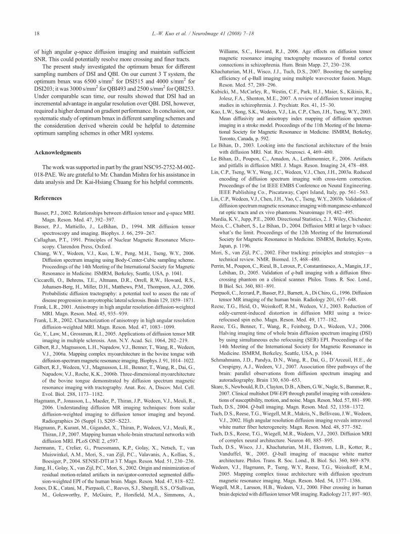
The present study investigated the optimum bmax for differentsampling numbers of DSI and QBI. On our current 3 T system, theoptimum bmax was 6500 s/mm2 for DSI515 and 4000 s/mm2 forDSI203; it was 3000 s/mm2 for QBI493 and 2500 s/mm2 for QBI253.Under comparable scan time, our results showed that DSI had anincremental advantage in angular resolution over QBI. DSI, however,required a higher demand on gradient performance. In conclusion, oursystematic study of optimum bmax in different sampling schemes andthe consideration derived wherein could be helpful to determineoptimum sampling schemes in other MRI systems.
Acknowledgments
Theworkwas supported in part by the grant NSC95-2752-M-002-018-PAE.We are grateful toMr. ChandanMishra for his assistance indata analysis and Dr. Kai-Hsiang Chuang for his helpful comments.
References
Basser, P.J., 2002. Relationships between diffusion tensor and q-space MRI.Magn. Reson. Med. 47, 392–397.
Basser, P.J., Mattiello, J., LeBihan, D., 1994. MR diffusion tensorspectroscopy and imaging. Biophys. J. 66, 259–267.
Callaghan, P.T., 1991. Principles of Nuclear Magnetic Resonance Micro-scopy. Clarendon Press, Oxford.
Chiang, W.Y., Wedeen, V.J., Kuo, L.W., Peng, M.H., Tseng, W.Y., 2006.Diffusion spectrum imaging using Body-Center-Cubic sampling scheme.Proceedings of the 14th Meeting of the International Society for MagneticResonance in Medicine. ISMRM, Berkeley, Seattle, USA, p. 1041.
Ciccarelli, O., Behrens, T.E., Altmann, D.R., Orrell, R.W., Howard, R.S.,Johansen-Berg, H., Miller, D.H., Matthews, P.M., Thompson, A.J., 2006.Probabilistic diffusion tractography: a potential tool to assess the rate ofdisease progression in amyotrophic lateral sclerosis. Brain 129, 1859–1871.
Frank, L.R., 2001. Anisotropy in high angular resolution diffusion-weightedMRI. Magn. Reson. Med. 45, 935–939.
Frank, L.R., 2002. Characterization of anisotropy in high angular resolutiondiffusion-weighted MRI. Magn. Reson. Med. 47, 1083–1099.
Ge, Y., Law, M., Grossman, R.I., 2005. Applications of diffusion tensor MRimaging in multiple sclerosis. Ann. N.Y. Acad. Sci. 1064, 202–219.
Gilbert, R.J., Magnusson, L.H., Napadow, V.J., Benner, T.,Wang, R., Wedeen,V.J., 2006a. Mapping complex myoarchitecture in the bovine tongue withdiffusion-spectrummagnetic resonance imaging.Biophys. J. 91, 1014–1022.
Gilbert, R.J., Wedeen, V.J., Magnusson, L.H., Benner, T., Wang, R., Dai, G.,Napadow, V.J., Roche, K.K., 2006b. Three-dimensional myoarchitectureof the bovine tongue demonstrated by diffusion spectrum magneticresonance imaging with tractography. Anat. Rec. A, Discov. Mol. Cell.Evol. Biol. 288, 1173–1182.
Hagmann, P., Jonasson, L., Maeder, P., Thiran, J.P., Wedeen, V.J., Meuli, R.,2006. Understanding diffusion MR imaging techniques: from scalardiffusion-weighted imaging to diffusion tensor imaging and beyond.Radiographics 26 (Suppl 1), S205–S223.
Hagmann, P., Kurant, M., Gigandet, X., Thiran, P., Wedeen, V.J., Meuli, R.,Thiran, J.P., 2007. Mapping human whole-brain structural networks withdiffusion MRI. PLoS ONE 2, e597.
Jaermann, T., Crelier, G., Pruessmann, K.P., Golay, X., Netsch, T., vanMuiswinkel, A.M., Mori, S., van Zijl, P.C., Valavanis, A., Kollias, S.,Boesiger, P., 2004. SENSE-DTI at 3 T. Magn. Reson. Med. 51, 230–236.
Jiang, H., Golay, X., van Zijl, P.C., Mori, S., 2002. Origin and minimization ofresidual motion-related artifacts in navigator-corrected segmented diffu-sion-weighted EPI of the human brain. Magn. Reson. Med. 47, 818–822.
Jones, D.K., Catani, M., Pierpaoli, C., Reeves, S.J., Shergill, S.S., O'Sullivan,M., Golesworthy, P., McGuire, P., Horsfield, M.A., Simmons, A.,
Williams, S.C., Howard, R.J., 2006. Age effects on diffusion tensormagnetic resonance imaging tractography measures of frontal cortexconnections in schizophrenia. Hum. Brain Mapp. 27, 230–238.
Khachaturian, M.H., Wisco, J.J., Tuch, D.S., 2007. Boosting the samplingefficiency of q-Ball imaging using multiple wavevector fusion. Magn.Reson. Med. 57, 289–296.
Kubicki, M., McCarley, R., Westin, C.F., Park, H.J., Maier, S., Kikinis, R.,Jolesz, F.A., Shenton, M.E., 2007. A review of diffusion tensor imagingstudies in schizophrenia. J. Psychiatr. Res. 41, 15–30.
Kuo, L.W., Song, S.K., Wedeen, V.J., Lin, C.P., Chen, J.H., Tseng, W.Y., 2003.Mean diffusivity and anisotropy index mapping of diffusion spectrumimaging in a stroke model. Proceedings of the 11th Meeting of the Interna-tional Society for Magnetic Resonance in Medicine. ISMRM, Berkeley,Toronto, Canada, p. 592.
Le Bihan, D., 2003. Looking into the functional architecture of the brainwith diffusion MRI. Nat. Rev. Neurosci. 4, 469–480.
Le Bihan, D., Poupon, C., Amadon, A., Lethimonnier, F., 2006. Artifactsand pitfalls in diffusion MRI. J. Magn. Reson. Imaging 24, 478–488.
Lin, C.P., Tseng, W.Y., Weng, J.C., Wedeen, V.J., Chen, J.H., 2003a. Reducedencoding of diffusion spectrum imaging with cross-term correction.Proceedings of the 1st IEEE EMBS Conference on Neural Engineering.IEEE Publishing Co., Piscataway, Capri Island, Italy, pp. 561–563.
Lin, C.P., Wedeen, V.J., Chen, J.H., Yao, C., Tseng, W.Y., 2003b. Validation ofdiffusion spectrummagnetic resonance imagingwithmanganese-enhancedrat optic tracts and ex vivo phantoms. Neuroimage 19, 482–495.
Mardia, K.V., Jupp, P.E., 2000. Directional Statistics, 2. J. Wiley, Chichester.Meca, C., Chabert, S., Le Bihan, D., 2004. Diffusion MRI at large b values:
what's the limit. Proceedings of the 12th Meeting of the InternationalSociety for Magnetic Resonance in Medicine. ISMRM, Berkeley, Kyoto,Japan, p. 1196.
Mori, S., van Zijl, P.C., 2002. Fiber tracking: principles and strategies—atechnical review. NMR. Biomed. 15, 468–480.
Perrin, M., Poupon, C., Rieul, B., Leroux, P., Constantinesco, A., Mangin, J.F.,Lebihan, D., 2005. Validation of q-ball imaging with a diffusion fibre-crossing phantom on a clinical scanner. Philos. Trans. R. Soc. Lond.,B Biol. Sci. 360, 881–891.
Pierpaoli, C., Jezzard, P., Basser, P.J., Barnett, A., Di Chiro, G., 1996.Diffusiontensor MR imaging of the human brain. Radiology 201, 637–648.
Reese, T.G., Heid, O., Weisskoff, R.M., Wedeen, V.J., 2003. Reduction ofeddy-current-induced distortion in diffusion MRI using a twice-refocused spin echo. Magn. Reson. Med. 49, 177–182.
Reese, T.G., Benner, T., Wang, R., Feinberg, D.A., Wedeen, V.J., 2006.Halving imaging time of whole brain diffusion spectrum imaging (DSI)by using simultaneous echo refocusing (SER) EPI. Proceedings of the14th Meeting of the International Society for Magnetic Resonance inMedicine. ISMRM, Berkeley, Seattle, USA, p. 1044.
Schmahmann, J.D., Pandya, D.N., Wang, R., Dai, G., D'Arceuil, H.E., deCrespigny, A.J., Wedeen, V.J., 2007. Association fibre pathways of thebrain: parallel observations from diffusion spectrum imaging andautoradiography. Brain 130, 630–653.
Skare, S., Newbould, R.D., Clayton,D.B.,Albers, G.W., Nagle, S., Bammer, R.,2007. Clinical multishot DW-EPI through parallel imaging with considera-tions of susceptibility, motion, and noise. Magn. Reson. Med. 57, 881–890.
Tuch, D.S., 2004. Q-ball imaging. Magn. Reson. Med. 52, 1358–1372.Tuch, D.S., Reese, T.G.,Wiegell, M.R.,Makris, N., Belliveau, J.W.,Wedeen,
V.J., 2002. High angular resolution diffusion imaging reveals intravoxelwhite matter fiber heterogeneity. Magn. Reson. Med. 48, 577–582.
Tuch, D.S., Reese, T.G., Wiegell, M.R., Wedeen, V.J., 2003. Diffusion MRIof complex neural architecture. Neuron 40, 885–895.
Tuch, D.S., Wisco, J.J., Khachaturian, M.H., Ekstrom, L.B., Kotter, R.,Vanduffel, W., 2005. Q-ball imaging of macaque white matterarchitecture. Philos. Trans. R. Soc. Lond., B. Biol. Sci. 360, 869–879.
Wedeen, V.J., Hagmann, P., Tseng, W.Y., Reese, T.G., Weisskoff, R.M.,2005. Mapping complex tissue architecture with diffusion spectrummagnetic resonance imaging. Magn. Reson. Med. 54, 1377–1386.
Wiegell, M.R., Larsson, H.B., Wedeen, V.J., 2000. Fiber crossing in humanbrain depictedwith diffusion tensorMR imaging. Radiology 217, 897–903.
Related Documents