Biomedical Research and Therapy 2015, 2(2): 207-219 ISSN 2198-4093 www.bmrat.org Isolation and propagation of human breast cancer cells from primary tumour biopsies 207 ORIGINAL RESEARCH Optimization of culture medium for the isolation and propagation of human breast cancer cells from primary tumour biopsies Binh Thanh Vu 1 , Hanh Thi Le 1 , Nhan Lu-Chinh Phan 1 , Phuc Van Pham 1,2,* 1 Laboratory of Stem Cell Research and Application, University of Science, Vietnam National University, Ho Chi Minh city, Viet Nam. 2 Biology Faculty, University of Science, Vietnam National University, Ho Chi Minh city, Viet Nam. *Corresponding author: [email protected] Received: 15 December 2014 / Accepted: 20 January 2015 / Published online: 22 February 2015 © The Author(s) 2015. This article is published with open access by BioMedPress (BMP) Abstract— Breast cancer cells from patients hold an important role in antigen production for immunotherapy, drug testing, and cancer stem cell studies. To date, although many studies have been conducted to develop protocols for the isolation and culture of breast cancer cells from tumour biopsies, the efficiencies of these protocols remain low. This study aimed to identify a suitable medium for the isolation and propagation of primary breast cancer cells from breast tumour biopsies. Breast tumour biopsies were obtained from hospitals after all patients had given their written in- formed consent and were cultured according to the expanding tumour method in 3 different media: DMEM/F12 (Dulbecco's Modified Eagle Medium: Nutrient Mixture F-12) supplemented with 10% FBS (Fetal bovine serum) and 1% antibiotic-antimycotic (Medium D); Medium 171 supplemented with 1X MEGS (Mammary Epithelial Growth Supplement) and 1% antibiotic-antimycotic (Medium M); or a 1:1 mixture of Medium D and Medium M (Medium DB). The cell culture efficiency was evaluated by several criteria, including the time of cell appearance, cell morphol- ogy, capability of proliferation, cell surface marker expression, ALDH (Aldehyde dehydrogenases) activity, karyotype, and tumour formation capacity in immune-deficient mice. Notably, primary cancer cells cultured in Medium DB showed a high expression of breast cancer stem cell surface markers (including CD44 + CD24 - and CD49f + ), low ex- pression of stromal cell surface markers (CD90), high ALDH activity, an abnormal karyotype, and high tumour for- mation capacity in immune-deficient mice. These findings suggested that Medium DB was suitable to support the sur- vival and proliferation of primary breast cancer cells as well as to enrich breast cancer stem cells. Keywords— Breast cancer cell, breast cancer stem cell, culture medium, primary cancer cell, tumor biopsy. INTRODUCTION The successful primary culture of cancer cells from breast tumours has great significance for the creation of an original cell source to study breast cancer cells (BCC) biology and for the development of therapeutic strategies. Although many studies on cancer cell biol- ogy have been conducted using cancer cell lines (Gillet et al., 2011; Gillet et al., 2013; Lacroix and Leclercq, 2004; Neve et al., 2006), recent reports have highlight- ed that those cancer cell lines do not, for various rea- sons, consistently display original cell characteristics (Keller et al., 2010). Therefore, to ensure that a chosen cell culture model accurately reflects the biological characteristics of the cells of interest, the use of prima- ry cancer cells is essential. Numerous primary culture studies are underway to identify optimal conditions, with regard to efficiency and duration, for the acquisition of BCCs. While there are many factors that influence the primary culture process, the culture medium is the key parameter. In DOI 10.7603/s40730-015-000-

Welcome message from author
This document is posted to help you gain knowledge. Please leave a comment to let me know what you think about it! Share it to your friends and learn new things together.
Transcript

Biomedical Research and Therapy 2015, 2(2): 207-219 ISSN 2198-4093 www.bmrat.org
Isolation and propagation of human breast cancer cells from primary tumour biopsies
207
ORIGINAL RESEARCH
Optimization of culture medium for the isolation and propagation of human breast cancer cells from primary tumour biopsies
Binh Thanh Vu1, Hanh Thi Le1, Nhan Lu-Chinh Phan1, Phuc Van Pham1,2,*
1Laboratory of Stem Cell Research and Application, University of Science, Vietnam National University, Ho Chi Minh city, Viet Nam. 2Biology Faculty, University of Science, Vietnam National University, Ho Chi Minh city, Viet Nam. *Corresponding author: [email protected]
Received: 15 December 2014 / Accepted: 20 January 2015 / Published online: 22 February 2015 © The Author(s) 2015. This article is published with open access by BioMedPress (BMP)
Abstract— Breast cancer cells from patients hold an important role in antigen production for immunotherapy, drug testing, and cancer stem cell studies. To date, although many studies have been conducted to develop protocols for the isolation and culture of breast cancer cells from tumour biopsies, the efficiencies of these protocols remain low. This study aimed to identify a suitable medium for the isolation and propagation of primary breast cancer cells from breast tumour biopsies. Breast tumour biopsies were obtained from hospitals after all patients had given their written in-formed consent and were cultured according to the expanding tumour method in 3 different media: DMEM/F12 (Dulbecco's Modified Eagle Medium: Nutrient Mixture F-12) supplemented with 10% FBS (Fetal bovine serum) and 1% antibiotic-antimycotic (Medium D); Medium 171 supplemented with 1X MEGS (Mammary Epithelial Growth Supplement) and 1% antibiotic-antimycotic (Medium M); or a 1:1 mixture of Medium D and Medium M (Medium DB). The cell culture efficiency was evaluated by several criteria, including the time of cell appearance, cell morphol-ogy, capability of proliferation, cell surface marker expression, ALDH (Aldehyde dehydrogenases) activity, karyotype, and tumour formation capacity in immune-deficient mice. Notably, primary cancer cells cultured in Medium DB showed a high expression of breast cancer stem cell surface markers (including CD44+CD24- and CD49f+), low ex-pression of stromal cell surface markers (CD90), high ALDH activity, an abnormal karyotype, and high tumour for-mation capacity in immune-deficient mice. These findings suggested that Medium DB was suitable to support the sur-vival and proliferation of primary breast cancer cells as well as to enrich breast cancer stem cells. Keywords— Breast cancer cell, breast cancer stem cell, culture medium, primary cancer cell, tumor biopsy.
INTRODUCTION
The successful primary culture of cancer cells from
breast tumours has great significance for the creation
of an original cell source to study breast cancer cells
(BCC) biology and for the development of therapeutic
strategies. Although many studies on cancer cell biol-
ogy have been conducted using cancer cell lines (Gillet
et al., 2011; Gillet et al., 2013; Lacroix and Leclercq,
2004; Neve et al., 2006), recent reports have highlight-
ed that those cancer cell lines do not, for various rea-
sons, consistently display original cell characteristics
(Keller et al., 2010). Therefore, to ensure that a chosen
cell culture model accurately reflects the biological
characteristics of the cells of interest, the use of prima-
ry cancer cells is essential.
Numerous primary culture studies are underway to
identify optimal conditions, with regard to efficiency
and duration, for the acquisition of BCCs. While there
are many factors that influence the primary culture
process, the culture medium is the key parameter. In
DOI 10.7603/s40730-015-0006-0

Vu et al., 2015 Biomed Res Ther 2015, 2(2): 207-219
Isolation and propagation of human breast cancer cells from primary tumour biopsies
208
studies on HBC cells, culture conditions were initially
adopted from a number of similar studies on normal
HME cells that had identified important factors de-
termining their in vitro survival (Hammond et al.,
1984; Stampfer et al., 1981a; Stampfer et al., 1981b).
Regrettably, medium components for the propagation
of primary human breast cells (HBC) have not yet
been determined, as the biology of this cell type re-
mains largely unclear. Stampfer et al. (Stampfer, 1982;
Stampfer and Bartley, 1985; Stampfer et al., 1993) de-
veloped a variety of culture media for the growth of
human mammary epithelial (HME) cells; the original
medium consisted of several undefined components,
but was later refined to a hormone- and growth factor-
supplemented medium that supports proliferation of
HME cells over many in vitro passages. Band and
Sager (Band and Sager, 1989) showed that it was use-
ful to propagate HME cells extensively in a growth
factor- and hormone-supplemented medium that also
contained serum and pituitary extract. Subsequently,
Petersen and Van Deurs (Petersen and van Deurs,
1987) and Ethier et al. (Ethier et al., 1991) reported the
growth of normal HME cells in serum-free media in
the absence of pituitary extract or serum. Although
these culture media promoted the growth of normal
mammary epithelial cells, very few of them supported
the growth of BCCs (Bartek et al., 1985; Taylor-
Papadimitriou et al., 1989). Thus, the culture condi-
tions that are conducive to the rapid proliferation of
normal HME cells over many in vitro passages hardly
support the growth of BCCs (Wolman et al., 1985).
The use of a serum-free medium for the cultivation of
normal HME cells circumvents a number of problems
that are mainly related to the instability of serum
(Hammond et al., 1984; Smith et al., 1981). However,
in comparison with serum-containing medium, se-
rum-free medium also entails some disadvantages
such as the need for a complex mixture of highly pure
medium components and a reduced cell proliferation
rate. Mammary tumour cell lines have been isolated
and grown in standard medium (e.g. Dulbecco's Mod-
ified Eagle Medium/Nutrient Mixture F-12,
DMEM/F12) supplemented with 10% foetal bovine
serum (FBS) (Engel and Young, 1978; Smith et al.,
1987; Soule et al., 1973). Serum contains growth fac-
tors, which promote cell proliferation, as well as adhe-
sion factors and antitrypsin activity, which promote
cell attachment. However, it has recently been accept-
ed that tumours consist of highly heterogeneous cell
populations with respect to cellular morphology, pro-
liferative potential, genetic lesions, and treatment re-
sponse (Bomken et al., 2010; Marusyk and Polyak,
2010). The isolation of cells from breast tumours may
give rise to several different cell types; normal coun-
terparts from which the neoplastic cells arise, such as
connective-tissue fibroblasts, infiltrating immune cells,
vascular endothelial cells, and smooth muscle cells, as
well as other elements of the normal tissue can all sur-
vive explantation (Sung et al., 2007; Weber and Kuo,
2012; Yu et al., 2011). Therefore, breast cancer epitheli-
al cells in tumour biopsies are irrevocably overgrown
by fibroblasts in a medium supplemented with high
serum concentrations.
In any case, the success rate of a BCC culture is in-
creased greatly by using selective media that enrich
the population of BCCs, but prevent the rapid and
extensive growth of normal cells, including stromal
cells and normal HME cells. Ethier et al. found that the
addition of 5% FBS to medium supplemented with
insulin, hydrocortisone, EGF, cholera toxin, and pro-
gesterone stimulated rapid proliferation of breast can-
cer epithelial-like cells (Ethier et al., 1993). They also
found that a relatively simple medium, only supple-
mented with 5% FBS, insulin, and hydrocortisone, re-
sulted in the slow emergence of BCCs that ultimately
gave rise to BCC lines (Ethier et al., 1993).
To date, the essential characteristics of primary breast
cancer cells are still a matter of debate. In particular,
recent studies have shown that solid breast tumours
harbour a cell population with stem cell characteris-
tics, which is responsible for the formation and
maintenance of tumours, development of metastases,
and, eventually, patient mortality. These cells are
known as cancer stem cells (Al-Hajj et al., 2003;
Clarke, 2005). In this study, we aimed to develop a
standardized protocol for the isolation and propaga-
tion of HBC cells from primary tumour biopsies that
a) ensures that the isolated primary cells include
breast cancer stem cells and b) minimizes contamina-
tion with other cells.
MATERIAL AND METHODS
Culture medium
Medium D: DMEM/F12 (1:1, v/v) supplemented with
10% FBS and 1% antibiotics/antimycotics (100X) (all

Vu et al., 2015 Biomed Res Ther 2015, 2(2): 207-219
Isolation and propagation of human breast cancer cells from primary tumour biopsies
209
bought from GeneWorld, HCM, Vietnam). Medium
M: Medium 171 supplemented with 1% MEGS (100X),
and 1% antibiotics/antimycotics (all bought from Life
Techonologies, Carlsbad, CA). Medium DB: mixed by
medium D and medium M (1:1, v/v).
Karyotyping reagents
Hypotonic solution: KCl 0.075 M and sodium citrate
0.8 % (1:1, v/v); Fix solution (Carnoy’s solution):
Methanol and glacial acetic acid (1:1, v/v).
Animals
Immunodeficient athymic nude mouse 7–8 weeks old
(NU(NCr)-Foxn1nu) were purchased from Charles
Rivers (Sulzfeld, Germany), kept under pathogen-free
conditions, and handled in accordance with the insti-
tutional recommendations for experimentation.
Primary culture of HBC cells from malignant tumour
specimens
Ten tumour biopsies, all from different patients, were
collected after obtaining the patients’ consent and the
approval from the ethics committee and were trans-
ported to the laboratory on ice. Excess adipose tissue
was pared off the tumour samples, following which
the samples were sliced into small fragments (approx-
imately 1–2 mm2) by using a scalpel, while care was
taken not to tear the tissue. Twelve of these fragments
were seeded per T25 tissue culture flask. Groups of 3
flasks then received either Medium D, Medium M, or
Medium DB for cultivation of the cells. The flasks
were incubated at 37°C with 5% CO2 and monitored
daily to record the time of cell appearance, the cell
morphology, as well as the number of tissue frag-
ments with cell migration. The medium was replaced
every 3 days.
Primary cancer cell phenotyping by flow cytometry
Primary cells were analysed for surface marker ex-
pression by flow cytometry. CD44, CD24, and CD49f
were recorded to determine the percentage ratio of
breast cancer stem cells (BCSCs), and CD90 was used
to monitor any fibroblast contamination. After 4
weeks of continuous culture, primary cells were de-
tached using 0.25% trypsin/EDTA (GeneWorld, Ho
Chi Minh, Vietnam). A total of 1 × 10⁶ cells were
stained with anti-CD44-APC (allophycocyanin) and
anti-CD24-FITC (fluorescein isothiocyanate), anti-
CD49f-FITC, or anti-CD90-FITC. Then, cells were
washed twice with sheath fluid, and subsequently
analysed by a FACSCalibur flow cytometry instru-
ment (BD Biosciences, San Jose, CA). Ten thousand
events were acquired in triplicate and analysed using
CellQuest Pro software (BD Biosciences, San Jose, CA).
ALDEFLUOR stem cell identification assay
Following 4 weeks of continuous culture, primary
cells were harvested and subjected to the ALDEFLU-
OR assay, according to the manufacturer’s instructions
(Stemcell Technologies, Vancouver, BC, Canada).
Briefly, the primary cell suspension was divided into 2
tubes per sample, with 1 × 10⁶ cells per tube. Tube 1
served as the control and received ALDH reagent, fol-
lowed by ALDH DEAB reagent, whereas tube 2
served as sample and received only ALDH reagent.
The cells were stained with 5 μl of these reagents for
30 minutes in the cell incubator, and then washed
twice with sheath fluid. Finally, the samples were ana-
lysed by a FACSCalibur flow cytometry instrument
(BD Biosciences, San Jose, CA). Ten thousand events
were acquired in triplicate and analysed by CellQuest
Pro software (BD Biosciences, San Jose, CA).
Karyotyping
Well-proliferating primary cells were treated with col-
cemid at a concentration of 0.10 μg/ml for 3 hours.
Primary cells were harvested, and then used for kary-
otyping by following a previously published protocol.
Briefly, the single cell suspension was incubated in
hypotonic solution for 30 minutes at 37°C, and then
fixed at least 3 times in Carnoy’s solution, which in-
cluded an overnight fixative step. The fixed cell sus-
pension was dropped on well-prepared slides and
stained according to the G-Banding protocol. Sets of
chromosomes were analysed using Ikaros software
(MetaSystems, Altlussheim, Germany).
Tumourigenesis assay
Five experimental groups were defined to evaluate the
effects of the 3 different kinds of culture medium on
the tumourigenic potential of primary BCCs. Each
group comprised 3 mice. In group 1, primary cells cul-
tured in Medium D were injected into the mammary
fat pad at cell densities of 106, 105, 104, and 103 cells per
100 μl PBS by using the right and left sides of the same
mouse. Similarly, the mice in group 2 and 3 were in-
jected with primary cells cultured in Medium M and

Vu et al., 2015 Biomed Res Ther 2015, 2(2): 207-219
Isolation and propagation of human breast cancer cells from primary tumour biopsies
210
Medium DB, respectively, at the same cell densities.
Group 4 comprised control mice that were injected
with cells of the MCF-7 BCC line (ATCC, Manassas,
VA), whereas group 5 comprised control mice injected
with cells of the MDA-MB-231 BCC line (ATCC, Ma-
nassas, VA), again at the aforementioned cell densi-
ties. Two weeks post-injection, the tumour size was
measured and calculated using the equation: (length ×
width2)/2.
RESULTS
Primary culture of HBC cells from malignant tumour
specimens
For all 10 breast cancer biopsies, primary cells were
observed to spread out from the tumour fragments.
Primary cells began to migrate from tumours around
day 4, with the earliest migrating cells being recorded
at day 3 (Fig. 1A). The percentage ratio of samples
displaying cell migration was highest when cultured
in Medium D (3/10 samples at day 3 and 10/10 at day
4), followed by samples cultivated in Medium DB
(3/10 samples at day 3, 9/10 at day 4, and 10/10 at day
6), and Medium M (1/10 samples at day 3, 6/10 at day
4, and 10/10 at day 6) (Fig. 1B). Consistently, at the
early stages of the culture period, the largest propor-
tion of successfully cultured tumour fragments was
observed for Medium D, followed by Medium DB,
and finally Medium M. However, at the late stages of
the culture period, this trend was no longer significant
(Fig. 1C).
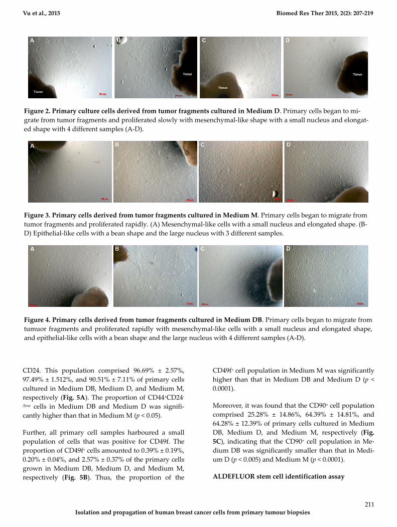
When investigating cell morphology, it was noted that
samples cultured in Medium D gave rise to primary
cells that uniformly exhibited a stromal-like, elongated
shape, containing a small nucleus, thus resembling
fibroblasts (Fig. 2). In contrast, almost all samples cul-
tured in either Medium M or Medium DB formed 2
differently shaped kinds of primary cells: epithelial-
like cells with a bean-like shape, having a large nucle-
us and mesenchymal-like cells with an elongated
shape, having a small nucleus (Fig. 3 and 4, respec-
tively).
During the 4-week cultivation period, it was visually
observed that the samples cultured in Medium D,
compared with those cultured in Medium DB and
Medium M, showed a markedly reduced proliferation
rate. While primary cells cultured in Medium D were
the earliest to migrate from tumour fragments, they
proliferated slowly and soon stopped dividing. This is
why some of these samples did not provide a suffi-
cient number of cells for further experiments. In con-
trast, primary cells cultured in either Medium DB or
Medium M proliferated rapidly, and almost all of
these samples provided a sufficient number of cells for
subsequent experiments.
Primary cancer cell surface marker analysis
Primary cells were analysed for the surface markers
CD44, CD24, and CD49f to identify the proportion of
BCSCs among the primary BCC populations. Fur-
thermore, the proportion of stromal cells present in
culture was determined by monitoring the expression
of CD90. Interestingly, nearly all primary cells were
positive for CD44 and negative or weakly positive for
Figure 1. Primary culture of from breast malignant tumors. (A) The time of primary cells began to migrate from tu-
mors in three different kinds of culture medium. (B) The ratio of successful culture samples in various culture media.
(C) The number of tumor fragments successfully cultured in three kinds of culture medium.
Vu et al., 2015 Biomed Res Ther 2015, 2(2): 207-219
Isolation and propagation of human breast cancer cells from primary tumour biopsies
211
CD24. This population comprised 96.69% ± 2.57%,
97.49% ± 1.512%, and 90.51% ± 7.11% of primary cells
cultured in Medium DB, Medium D, and Medium M,
respectively (Fig. 5A). The proportion of CD44+CD24-
/low cells in Medium DB and Medium D was signifi-
cantly higher than that in Medium M (p < 0.05).
Further, all primary cell samples harboured a small
population of cells that was positive for CD49f. The
proportion of CD49f+ cells amounted to 0.39% ± 0.19%,
0.20% ± 0.04%, and 2.57% ± 0.37% of the primary cells
grown in Medium DB, Medium D, and Medium M,
respectively (Fig. 5B). Thus, the proportion of the
CD49f+ cell population in Medium M was significantly
higher than that in Medium DB and Medium D (p <
0.0001).
Moreover, it was found that the CD90+ cell population
comprised 25.28% ± 14.86%, 64.39% ± 14.81%, and
64.28% ± 12.39% of primary cells cultured in Medium
DB, Medium D, and Medium M, respectively (Fig.
5C), indicating that the CD90+ cell population in Me-
dium DB was significantly smaller than that in Medi-
um D (p < 0.005) and Medium M (p < 0.0001).
ALDEFLUOR stem cell identification assay
Figure 2. Primary culture cells derived from tumor fragments cultured in Medium D. Primary cells began to mi-
grate from tumor fragments and proliferated slowly with mesenchymal-like shape with a small nucleus and elongat-
ed shape with 4 different samples (A-D).
Figure 3. Primary cells derived from tumor fragments cultured in Medium M. Primary cells began to migrate from
tumor fragments and proliferated rapidly. (A) Mesenchymal-like cells with a small nucleus and elongated shape. (B-
D) Epithelial-like cells with a bean shape and the large nucleus with 3 different samples.
Figure 4. Primary cells derived from tumor fragments cultured in Medium DB. Primary cells began to migrate from
tumuor fragments and proliferated rapidly with mesenchymal-like cells with a small nucleus and elongated shape,
and epithelial-like cells with a bean shape and the large nucleus with 4 different samples (A-D).

Vu et al., 2015 Biomed Res Ther 2015, 2(2): 207-219
Isolation and propagation of human breast cancer cells from primary tumour biopsies
212
All samples harboured a small population of cells that
displayed aldehyde dehydrogenase activity. The cell
population testing positive for ALDH activity repre-
sented 9.35% ± 3.64% and 2.28% ± 0.88% of the prima-
ry cells grown in Medium DB and Medium M, respec-
tively (Fig. 6). Thus, the size of the ALDH+ cell popula-
tion in Medium DB was larger than that in Medium M
(p < 0.05). Note that the number of cells derived from
primary culture in Medium D was insufficient for the
ALDEFLUOR assay.
Figure 6. ALDH assay to identify primary culture
cells expressing ALDH in the Medium DB and the
Medium M.
Karyotyping
Four rapidly proliferating samples were subjected to
karyotyping to determine which culture medium sup-
ported the growth of cancer cells, as defined by ab-
normal chromosome number. The chromosome num-
bers of primary cells derived from sample 1 ranged
between 45 and 48 (Fig. 7A, E). Specifically, cells cul-
tured in Medium D uniformly contained 46 chromo-
somes, whereas chromosome numbers ranged be-
tween 45 and 46 in cells grown in Medium M and be-
tween 45 and 48 in cells cultivated in Medium DB (Fig.
7A, E).
Primary cells derived from sample 2 contained be-
tween 44 and 46 chromosomes (Fig. 7B, F). Here, cells
cultured in Medium M displayed between 45 and 46
chromosomes, while Medium DB supported the
growth of cells with a wider range of chromosome
numbers, i.e. between 44 and 46 (Fig. 7B,F). The num-
ber of cells derived from the primary culture in Medi-
um D was insufficient for karyotyping. In primary
cells derived from sample 3, chromosome numbers
ranged between 44 and 46 (Fig. 7C,G). Cells cultured
in either Medium M or Medium DB contained be-
tween 44 and 46 chromosomes (Fig. 7C,G). Again, the
number of cells derived from the primary culture in
Medium D was insufficient for karyotyping. Primary
cells derived from sample 6 displayed chromosome
numbers between 44 and 47 (Fig. 7D,H). Specifically,
Figure 5. Expression of CD44, CD24, CD49f and CD90 in primary cells in 3 different media. (A) CD44/CD24 expres-
sion, (B) CD49f expression, (C) CD90 analysis.

Vu et al., 2015 Biomed Res Ther 2015, 2(2): 207-219
Isolation and propagation of human breast cancer cells from primary tumour biopsies
213
cells cultured in Medium D uniformly harboured 44
chromosomes, whereas chromosome numbers ranged
between 44 and 46 in cells cultured in Medium M, and
between 45 and 47 in cells cultured in Medium DB
(Fig. 7D,H).
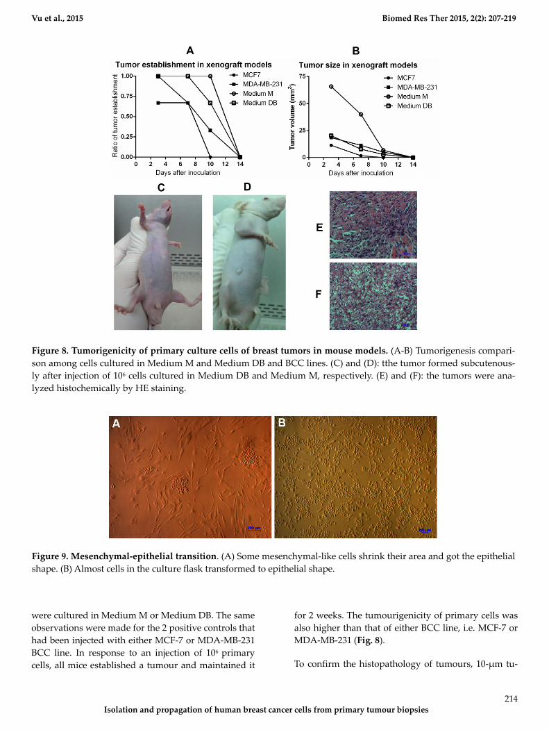
Tumourigenesis assay
In the tumourigenesis assay, the injection of 103, 104,
and 105 primary cells failed to cause tumour growth in
immunodeficient mice, regardless of whether they
Figure 7. Karyotype analysis of some samples. (A) Sample 1: The number of chromosomes ranged from 45 to 48; (B)
Sample 2: The number of chromosomes ranged from 44 to 46; (C) Sample 3: The number of chromosomes ranged from
45 to 46; (D) Sample 6: The number of chromosomes ranged from 44 to 47. The effect of culture medium on the growth
of selective primary cells with different chromosomes from sample 1 (E), sample 2 (F), sample 3 (G), sample 6 (H).
Vu et al., 2015 Biomed Res Ther 2015, 2(2): 207-219
Isolation and propagation of human breast cancer cells from primary tumour biopsies
214
were cultured in Medium M or Medium DB. The same
observations were made for the 2 positive controls that
had been injected with either MCF-7 or MDA-MB-231
BCC line. In response to an injection of 106 primary
cells, all mice established a tumour and maintained it
for 2 weeks. The tumourigenicity of primary cells was
also higher than that of either BCC line, i.e. MCF-7 or
MDA-MB-231 (Fig. 8).
To confirm the histopathology of tumours, 10-μm tu-
Figure 8. Tumorigenicity of primary culture cells of breast tumors in mouse models. (A-B) Tumorigenesis compari-
son among cells cultured in Medium M and Medium DB and BCC lines. (C) and (D): tthe tumor formed subcutenous-
ly after injection of 106 cells cultured in Medium DB and Medium M, respectively. (E) and (F): the tumors were ana-
lyzed histochemically by HE staining.
Figure 9. Mesenchymal-epithelial transition. (A) Some mesenchymal-like cells shrink their area and got the epithelial
shape. (B) Almost cells in the culture flask transformed to epithelial shape.

Vu et al., 2015 Biomed Res Ther 2015, 2(2): 207-219
Isolation and propagation of human breast cancer cells from primary tumour biopsies
215
mour sections were stained with haematoxylin-eosin
(HE). As shown in Fig. 8 E&F, tumours exhibited can-
cer cells with large nuclei; the tumours were estab-
lished from primary cells cultured in Medium DB and
Medium M.
Mesenchymal-epithelial transition (MET) and estab-
lishment of BCC lines
Together, these results indicated that Medium DB sur-passed the other media in supporting the growth of BCCs. Therefore, Medium DB was chosen to culture and maintain cells derived from malignant breast tumours. Primary cells also migrated from tumour fragments after 4–5 days of culture. In almost all samples, epithelial-like cells and mesenchymal-like cells appeared simultaneous-ly; however, all cells transformed in to mesenchymal-like shape after 1 month of continuous culture, including cells with epithelial phenotype previously. Interestingly, fol-lowing long-term culture (approximately 6 months), mesenchymal-shape cancer cells were observed to un-dergo back to the process of MET in culture.
Initially, some mesenchymal-like cells shrunk in size
and adopted an epithelial shape. Soon, neighbouring
cells also displayed this phenomenon (Fig. 9A). This
process continued and led to the formation of colonies
of epithelial cells that spread over the entire surface of
the culture, until all cells in the culture flask had
adopted an epithelial shape (Fig. 9B). The described
process occurred naturally without the use of any
stimulants, apart from the regular replacement of cul-
ture medium. These cells then proliferated rapidly and
formed cell lines. Hence, the BCC line described here-
in was successfully developed from malignant human
breast tumours via the explant culture method. These
cells exhibit all typical characteristics of BCC lines and
exhibit particular properties that they share with the
original tumour.
DISCUSSION
Breast tumours contain a combination of various
kinds of cells, including normal epithelial cells, stro-
mal cells, breast cancer cells, and breast cancer stem
cells. A suitable protocol for the isolation of BCCs
must not only provide for high cell growth efficiency,
but also for the establishment of cells exhibiting BCC
properties. From the information gathered from pre-
vious studies investigating single cell culture versus
explant tissue culture, this study employed expanding
tissue culture (data not shown). In this method, the
culture medium is the most decisive factor in the out-
growth of cells from tumour fragments, as well as in
the types of cells obtained. Based on existing literature
reports, 3 different kinds of media were chosen for use
in this study. M171 medium supplemented with
MEGS (Medium M) is a serum-free medium that sup-
ports the proliferation of normal human epithelial
mammary cells. In contrast, DMEM/F12 medium sup-
plemented with 10% FBS (Medium D) is a serum-
containing medium that supports the proliferation of
routine human BCC lines. For the purpose of this
study, these 2 kinds of medium were mixed in a ratio
of 1:1 to produce a third medium (Medium DB). Thus,
Medium DB contained 50% of each of the components
of Medium D and Medium M, and the serum concen-
tration was similarly reduced to 5%.
As detailed in the Results section, Medium D was not
suitable for the isolation of BCCs. Although in this
medium the cells migrated more rapidly than in Me-
dium M and Medium DB, they also showed a relative-
ly slow mitosis rate and therefore reduced prolifera-
tion. Hence, in nearly all samples, cells grown in Me-
dium D were not sufficiently high in number for use
in further evaluation. In contrast to Medium D, prima-
ry cells cultured in Medium M proliferated rapidly,
and the majority of samples cultured in Medium M
provided enough cells for additional experiments.
This observation can be explained by the fact that Me-
dium M contained a pool of hormones and growth
factors such as hydrocortisone, EGF, and insulin.
These factors are beneficial for the survival and prolif-
eration of breast tissue-derived cells. Primary cells
cultured in Medium DB proliferated most rapidly,
such that all samples provided cells that were suffi-
ciently high in number for use in further experiments.
These results are likely due to the components of Me-
dium DB, which included growth factors and hor-
mones from Medium M, as well as serum from Medi-
um D. However, it should be highlighted that the se-
rum concentration is reduced in comparison to com-
mon serum levels, and that this appears to be benefi-
cial with regard to the elimination of stromal cells.
Therefore, using Medium DB for primary tumour cell
culture results in the rapid proliferation of primary
cell populations.
Next, the existence of a CD44+CD24- population was
evaluated by flow cytometry in all primary cell cul-
tures. Nearly all primary cells grown in any of the 3

Vu et al., 2015 Biomed Res Ther 2015, 2(2): 207-219
Isolation and propagation of human breast cancer cells from primary tumour biopsies
216
culture media tested positive for CD44 and negative
for CD24, with the highest percentage of CD44+CD24-
cells occurring in Medium D and Medium DB. In a
previous study by Al-Hajj et al. (2003), primary cells
contained a sub-population of CD44+CD24- cells with
high tumourigenicity (Al-Hajj et al., 2003). However,
the possibility that the primary cells expressing
CD44+CD24- were not all BCSCs appears likely
(Ghebeh et al., 2013; Mannello, 2013). In a recent study,
Ghebeh et al. (2013) clearly demonstrated that both
normal breast tissue and breast cancer tissue har-
boured CD44+CD24- cells (Ghebeh et al., 2013). They
also suggested that BCSCs would be enriched in the
CD44+CD24- cells if they were combined with the
CD49f+ phenotype (Ghebeh et al., 2013). CD49f was
also determined as a marker of BCSCs in previous
studies (Meyer et al., 2010; Yu et al., 2012). Therefore,
in the next experiment, the existence of a CD49f+ cell
population in the primary cells was evaluated. The
results showed that primary cultures in Medium M
contained the highest percentage of CD49f+ cells, fol-
lowed by primary cultures in Medium DB and Medi-
um D. Hence, Medium M efficiently supported the
growth of cells with the CD49f+ phenotype; however,
compared with Medium D and Medium DB, Medium
M did not support the growth of CD44+CD24- cells.
Medium DB excellently promoted the growth of
CD44+CD24- cells, but little impact on the proliferation
of CD49f+ cells.
However, Medium D and Medium M also supported
stromal cell proliferation. Regarding CD90 expression,
more than 50% of the primary cells in Medium D and
Medium M tested positive for this marker, whereas
this population only accounted for about 25% of the
primary cells grown in Medium DB. These CD90+ cells
were considered as contaminant cells in the breast car-
cinoma primary culture (Araki et al., 2007; Haack-
Sorensen et al., 2008; Nakamura et al., 2006). In sum, a
marked contrast was observed with respect to the
proportion of contaminant cells versus cells with the
BCSC phenotype (CD44+CD24- and CD49f+) in prima-
ry cultures grown in Medium DB and Medium M.
Next, cellular ALDH expression was monitored to
evaluate culture efficiency. The ALDH enzyme has
important functions in the development of epithelial
homeostasis, and deregulation of this class of enzymes
has been implicated in multiple cancers (Marchitti et
al., 2008). The ALDEFLUOR assay is thought to be an
almost universal marker of stem cell activity in both
normal and cancer tissues (Corti et al., 2006; Hess et
al., 2004), including normal and malignant breast epi-
thelial stem cells (Ginestier et al., 2007). In this study,
the ALDH+ cell population was approximately 5 times
larger in Medium DB than in Medium M (9.35% ±
3.64% and 2.28% ± 0.88%, respectively). To sum up,
Medium DB, significantly more than the other 2 me-
dia, specifically promotes the growth of the breast
cancer stem cells that exist in malignant breast tu-
mours. To support this conclusion, karyotype analysis
revealed that nearly all cells in Medium DB exhibited
an abnormal karyotype, while in Medium M as well as
Medium D, primary cells contained both normal and
slightly abnormal karyotypes. All samples doing kar-
yotype derived from female breast cancer patients
whose tumors were diagnosed as primary tumors, i.e.
they had never undergone any previous treatment,
including chemotherapy or radiotherapy. Consequent-
ly, the number of chromosomes of primary cancer cells
was not so different than the normal chromosome
number, known as 46 chromosomes. This is consistent
with many studies of cancer cells primary culture, in-
cluding (Adeyinka et al., 2000; Bardi et al., 1993;
Brothman et al., 1990; Ferti et al., 2004; Stamouli et al.,
2004; Teixeira et al., 1995).
Following karyotyping, primary cells from Medium M
and Medium DB were used to induce tumours in
mice. The results showed that primary cells grown in
either Medium M or Medium DB, as well as other
BCC lines such as MCF-7 and MDA-MD-231 success-
fully caused tumours in mice when injected at a cell
density of 106 cells per mouse. Lower densities of pri-
mary cells or BCC lines failed to establish tumours in
mice. Mouse models that were used in the experi-
ments of examining the dose causing tumors were
athymic nude mice, whose immune system is partially
suppressed. Therefore, human cell transplantation,
primary cells or BCC lines, induced immune response
in mice, with the most powerful after 1 week. As a re-
sult, grafted cells including primary cells and BCC
lines existed only 2 weeks. However, the results also
showed the ability to establish tumors in mouse model
with primary cells cultured in Medium DB and Medi-
um M was as well as BCC lines. Tumour sections
stained with HE confirmed that the tumours con-
tained cancer cells with large nuclei; the tumours were
established from primary cells cultured in Medium
DB and Medium M.
Moreover, after long-term cultivation of approximate-
Vu et al., 2015 Biomed Res Ther 2015, 2(2): 207-219
Isolation and propagation of human breast cancer cells from primary tumour biopsies
217
ly 6 months in Medium DB, breast cancer cells were
observed to undergo the process of MET, i.e. altering
their mesenchymal phenotype back to the epithelial
phenotype. This process continued until epithelial cell
colonies were formed, and eventually all cells in the
culture flask transformed into the epithelial pheno-
type. At this point, cells proliferated more rapidly and
stably, in a manner similar to that observed in typical
BCC lines. Epithelial cancer cells acquire mesenchy-
mal features that provide a mechanism for tumour
cells to leave the primary tumour, resulting in the in-
duction of single cell and/or collective cell migration
(Drasin et al., 2011; Mani et al., 2008; May et al., 2011).
Overall, the carcinoma epithelial–mesenchymal transi-
tion (EMT) is defined by a loss of normal epithelial
architecture, which renders the epithelial tumour cells
phenotypically indistinguishable from fibroblasts. At
the molecular level, this is characterized by a down-
regulation of epithelial markers and an up-regulation
of mesenchymal markers, accompanied by an increase
in cell migration and invasion (Polyak and Weinberg,
2009; Scheel and Weinberg, 2011; Thiery et al., 2009).
However, the role of mesenchymal-epithelial transi-
tion (EMT) in cancer is complicated by the fact that in
the appropriate microenvironment, mesenchymal-like
cells likely undergo a reversion or MET, thereby per-
mitting colonization (Micalizzi et al., 2010; Wells et al.,
2008).
CONCLUSION
BCCs are essential biological tools for both research
and therapy. Breast tumours always contain a combi-
nation of different kinds of cells, including breast can-
cer cells. This study successfully established a simple
cell isolation procedure based on breast tumour-
explant cultivation in Medium DB, which is a 1:1 mix-
ture of DMEM/F12 supplemented with 10% FBS and
M171 supplemented with 1X MEGS. Cells isolated
using this procedure exhibited BCC properties such as
expression of BCSC markers (CD44+CD24-), expression
of ALDH, abnormal karyotype, and tumourigenic ca-
pability in mouse models. The findings of this study
served to establish a method that proved highly useful
for the isolation of BCCs from tumour biopsies as well
as for the enrichment of BCSCs.
ACKNOWLEDGMENT
This study was funded by Ministry of Science and
Technology under grant No. DTDL.2011-T/30,
Vietnam.
ABBREVIATIONS
ALDH: Aldehyde dehydrogenase, APC: Allophycocy-
anin, BCC: Breast cancer cell, BCSC: Breast cancer
stem cell, BPE: Bovine pituitary extract, DEAB:
Diethylaminobenzaldehyde, DMEM/F-12: Dulbecco's
Modified Eagle Medium/Nutrient Mixture F-12, EGF:
Epithelial growth factor, EMT: Epithelial-
mesenchymal transition, FBS: Fetal Bovine Serum ,
FCM: Flow cytometry, FITC: Fluorescein Isothiocya-
nate, HE: Hematoxylineosin, HBC: Human breast can-
cer cell, HME: Human mammary epithelial cells, Me-
dium D: DMEM/F12 supplemented with 10%FBS and
1% Antibiotics/antimycotic, Medium M: Medium 171
supplemented with MEGS and 1% Antibiot-
ics/antimycotic, Medium DB Medium D and Me-
dium M (1:1, v/v), MEGS: Mammary Epithelial
Growth Supplement, MET: Mesenchymal-epithelial
transition, NOD/SCID: Non-obese diabetic/severe
combined immune deficiency.
Competing interests
The authors declare that they have no competing in-
terests.
Open Access
This article is distributed under the terms of the Creative Com-
mons Attribution License (CC-BY 4.0) which permits any use,
distribution, and reproduction in any medium, provided the origi-
nal author(s) and the source are credited.
References Adeyinka, A., Kytola, S., Mertens, F., Pandis, N., and Larsson, C. (2000). Spectral karyotyping and chromosome banding studies of primary breast carcinomas and their lymph node metastases. International journal of molecular medicine 5, 235-240.

Vu et al., 2015 Biomed Res Ther 2015, 2(2): 207-219
Isolation and propagation of human breast cancer cells from primary tumour biopsies
218
Al-Hajj, M., Wicha, M.S., Benito-Hernandez, A., Morrison, S.J., and Clarke, M.F. (2003). Prospective identification of tumorigenic breast cancer cells. Proceedings of the National Academy of Sciences of the United States of America 100, 3983-3988. Araki, H., Yoshinaga, K., Boccuni, P., Zhao, Y., Hoffman, R., and Mahmud, N. (2007). Chromatin-modifying agents permit human hematopoietic stem cells to undergo multiple cell divisions while retaining their repopulating potential. Blood 109, 3570-3578. Band, V., and Sager, R. (1989). Distinctive traits of normal and tumor-derived human mammary epithelial cells expressed in a medium that supports long-term growth of both cell types. Proceedings of the National Academy of Sciences of the United States of America 86, 1249-1253. Bardi, G., Johansson, B., Pandis, N., Mandahl, N., Bak-Jensen, E., Andren-Sandberg, A., Mitelman, F., and Heim, S. (1993). Karyotypic abnormalities in tumours of the pancreas. British journal of cancer 67, 1106-1112. Bartek, J., Taylor-Papadimitriou, J., Miller, N., and Millis, R. (1985). Patterns of expression of keratin 19 as detected with monoclonal antibodies in human breast tissues and tumours. International journal of cancer Journal international du cancer 36, 299-306. Bomken, S., Fiser, K., Heidenreich, O., and Vormoor, J. (2010). Understanding the cancer stem cell. British journal of cancer 103, 439-445. Brothman, A.R., Peehl, D.M., Patel, A.M., and McNeal, J.E. (1990). Frequency and pattern of karyotypic abnormalities in human prostate cancer. Cancer research 50, 3795-3803. Clarke, M.F. (2005). A self-renewal assay for cancer stem cells. Cancer chemotherapy and pharmacology 56 Suppl 1, 64-68. Corti, S., Locatelli, F., Papadimitriou, D., Donadoni, C., Salani, S., Del Bo, R., Strazzer, S., Bresolin, N., and Comi, G.P. (2006). Identification of a primitive brain-derived neural stem cell population based on aldehyde dehydrogenase activity. Stem cells 24, 975-985. Drasin, D.J., Robin, T.P., and Ford, H.L. (2011). Breast cancer epithelial-to-mesenchymal transition: examining the functional consequences of plasticity. Breast cancer research : BCR 13, 226. Engel, L.W., and Young, N.A. (1978). Human breast carcinoma cells in continuous culture: a review. Cancer research 38, 4327-4339. Ethier, S.P., Mahacek, M.L., Gullick, W.J., Frank, T.S., and Weber, B.L. (1993). Differential isolation of normal luminal mammary epithelial cells and breast cancer cells from primary and metastatic sites using selective media. Cancer research 53, 627-635. Ethier, S.P., Summerfelt, R.M., Cundiff, K.C., and Asch, B.B. (1991). The influence of growth factors on the proliferative potential of normal and primary breast cancer-derived human breast epithelial cells. Breast cancer research and treatment 17, 221-230. Ferti, A.D., Stamouli, M.J., Panani, A.D., Raptis, S.A., and Young, B.D. (2004). Molecular cytogenetic analysis of breast cancer: a combined multicolor fluorescence in situ hybridization and G-banding study of uncultured tumor cells. Cancer genetics and cytogenetics 149, 28-37. Ghebeh, H., Sleiman, G.M., Manogaran, P.S., Al-Mazrou, A., Barhoush, E., Al-Mohanna, F.H., Tulbah, A., Al-Faqeeh, K., and Adra, C.N. (2013). Profiling of normal and malignant breast tissue show CD44high/CD24low phenotype as a predominant stem/progenitor marker when used in combination with Ep-CAM/CD49f markers. BMC cancer 13, 289. Gillet, J.P., Calcagno, A.M., Varma, S., Marino, M., Green, L.J., Vora, M.I., Patel, C., Orina, J.N., Eliseeva, T.A., Singal, V., et al. (2011). Redefining the relevance of established cancer cell lines to the study of mechanisms of clinical anti-cancer drug resistance. Proceedings of the National Academy of Sciences of the United States of America 108, 18708-18713. Gillet, J.P., Varma, S., and Gottesman, M.M. (2013). The clinical relevance of cancer cell lines. Journal of the National Cancer Institute 105, 452-458. Ginestier, C., Hur, M.H., Charafe-Jauffret, E., Monville, F., Dutcher, J., Brown, M., Jacquemier, J., Viens, P., Kleer, C.G., Liu, S., et al. (2007). ALDH1 is a marker of normal and malignant human mammary stem cells and a predictor of poor clinical outcome. Cell stem cell 1, 555-567.
Haack-Sorensen, M., Friis, T., Bindslev, L., Mortensen, S., Johnsen, H.E., and Kastrup, J. (2008). Comparison of different culture conditions for human mesenchymal stromal cells for clinical stem cell therapy. Scandinavian journal of clinical and laboratory investigation 68, 192-203. Hammond, S.L., Ham, R.G., and Stampfer, M.R. (1984). Serum-free growth of human mammary epithelial cells: rapid clonal growth in defined medium and extended serial passage with pituitary extract. Proceedings of the National Academy of Sciences of the United States of America 81, 5435-5439. Hess, D.A., Meyerrose, T.E., Wirthlin, L., Craft, T.P., Herrbrich, P.E., Creer, M.H., and Nolta, J.A. (2004). Functional characterization of highly purified human hematopoietic repopulating cells isolated according to aldehyde dehydrogenase activity. Blood 104, 1648-1655. Keller, P.J., Lin, A.F., Arendt, L.M., Klebba, I., Jones, A.D., Rudnick, J.A., DiMeo, T.A., Gilmore, H., Jefferson, D.M., Graham, R.A., et al. (2010). Mapping the cellular and molecular heterogeneity of normal and malignant breast tissues and cultured cell lines. Breast cancer research : BCR 12, R87. Lacroix, M., and Leclercq, G. (2004). Relevance of breast cancer cell lines as models for breast tumours: an update. Breast cancer research and treatment 83, 249-289. Mani, S.A., Guo, W., Liao, M.J., Eaton, E.N., Ayyanan, A., Zhou, A.Y., Brooks, M., Reinhard, F., Zhang, C.C., Shipitsin, M., et al. (2008). The epithelial-mesenchymal transition generates cells with properties of stem cells. Cell 133, 704-715. Mannello, F. (2013). Understanding breast cancer stem cell heterogeneity: time to move on to a new research paradigm. BMC medicine 11, 169. Marchitti, S.A., Brocker, C., Stagos, D., and Vasiliou, V. (2008). Non-P450 aldehyde oxidizing enzymes: the aldehyde dehydrogenase superfamily. Expert opinion on drug metabolism & toxicology 4, 697-720. Marusyk, A., and Polyak, K. (2010). Tumor heterogeneity: causes and consequences. Biochimica et biophysica acta 1805, 105-117. May, C.D., Sphyris, N., Evans, K.W., Werden, S.J., Guo, W., and Mani, S.A. (2011). Epithelial-mesenchymal transition and cancer stem cells: a dangerously dynamic duo in breast cancer progression. Breast cancer research : BCR 13, 202. Meyer, M.J., Fleming, J.M., Lin, A.F., Hussnain, S.A., Ginsburg, E., and Vonderhaar, B.K. (2010). CD44posCD49fhiCD133/2hi defines xenograft-initiating cells in estrogen receptor-negative breast cancer. Cancer research 70, 4624-4633. Micalizzi, D.S., Farabaugh, S.M., and Ford, H.L. (2010). Epithelial-mesenchymal transition in cancer: parallels between normal development and tumor progression. Journal of mammary gland biology and neoplasia 15, 117-134. Nakamura, Y., Muguruma, Y., Yahata, T., Miyatake, H., Sakai, D., Mochida, J., Hotta, T., and Ando, K. (2006). Expression of CD90 on keratinocyte stem/progenitor cells. The British journal of dermatology 154, 1062-1070. Neve, R.M., Chin, K., Fridlyand, J., Yeh, J., Baehner, F.L., Fevr, T., Clark, L., Bayani, N., Coppe, J.P., Tong, F., et al. (2006). A collection of breast cancer cell lines for the study of functionally distinct cancer subtypes. Cancer cell 10, 515-527. Petersen, O.W., and van Deurs, B. (1987). Preservation of defined phenotypic traits in short-term cultured human breast carcinoma derived epithelial cells. Cancer research 47, 856-866. Polyak, K., and Weinberg, R.A. (2009). Transitions between epithelial and mesenchymal states: acquisition of malignant and stem cell traits. Nature reviews Cancer 9, 265-273. Scheel, C., and Weinberg, R.A. (2011). Phenotypic plasticity and epithelial-mesenchymal transitions in cancer and normal stem cells? International journal of cancer Journal international du cancer 129, 2310-2314. Smith, H.S., Lan, S., Ceriani, R., Hackett, A.J., and Stampfer, M.R. (1981). Clonal proliferation of cultured nonmalignant and malignant human breast epithelia. Cancer research 41, 4637-4643. Smith, H.S., Wolman, S.R., Dairkee, S.H., Hancock, M.C., Lippman, M., Leff, A., and Hackett, A.J. (1987). Immortalization in culture:
Vu et al., 2015 Biomed Res Ther 2015, 2(2): 207-219
Isolation and propagation of human breast cancer cells from primary tumour biopsies
219
occurrence at a late stage in the progression of breast cancer. Journal of the National Cancer Institute 78, 611-615. Soule, H.D., Vazguez, J., Long, A., Albert, S., and Brennan, M. (1973). A human cell line from a pleural effusion derived from a breast carcinoma. Journal of the National Cancer Institute 51, 1409-1416. Stamouli, M.I., Panani, A.D., Ferti, A.D., Petraki, C., Oliver, R.T., Raptis, S.A., and Young, B.D. (2004). Detection of genetic alterations in primary bladder carcinoma with dual-color and multiplex fluorescence in situ hybridization. Cancer genetics and cytogenetics 149, 107-113. Stampfer, M.R. (1982). Cholera toxin stimulation of human mammary epithelial cells in culture. In vitro 18, 531-537. Stampfer, M.R., Bartholomew, J.C., Smith, H.S., and Bartley, J.C. (1981a). Metabolism of benzo[a]pyrene by human mammary epithelial cells: toxicity and DNA adduct formation. Proceedings of the National Academy of Sciences of the United States of America 78, 6251-6255. Stampfer, M.R., and Bartley, J.C. (1985). Induction of transformation and continuous cell lines from normal human mammary epithelial cells after exposure to benzo[a]pyrene. Proceedings of the National Academy of Sciences of the United States of America 82, 2394-2398. Stampfer, M.R., Vlodavsky, I., Smith, H.S., Ford, R., Becker, F.F., and Riggs, J. (1981b). Fibronectin production by human mammary cells. Journal of the National Cancer Institute 67, 253-261. Stampfer, M.R., Yaswen, P., Alhadeff, M., and Hosoda, J. (1993). TGF beta induction of extracellular matrix associated proteins in normal and transformed human mammary epithelial cells in culture is independent of growth effects. Journal of cellular physiology 155, 210-221. Sung, S.Y., Hsieh, C.L., Wu, D., Chung, L.W., and Johnstone, P.A. (2007). Tumor microenvironment promotes cancer progression, metastasis, and therapeutic resistance. Current problems in cancer 31, 36-100. Taylor-Papadimitriou, J., Stampfer, M., Bartek, J., Lewis, A., Boshell, M., Lane, E.B., and Leigh, I.M. (1989). Keratin expression in human mammary epithelial cells cultured from normal and malignant tissue: relation to in vivo phenotypes and influence of medium. Journal of cell science 94 ( Pt 3), 403-413. Teixeira, M.R., Pandis, N., Bardi, G., Andersen, J.A., Mitelman, F., and Heim, S. (1995). Clonal heterogeneity in breast cancer: karyotypic comparisons of multiple intra- and extra-tumorous samples from 3 patients. International journal of cancer Journal international du cancer 63, 63-68. Thiery, J.P., Acloque, H., Huang, R.Y., and Nieto, M.A. (2009). Epithelial-mesenchymal transitions in development and disease. Cell 139, 871-890. Weber, C.E., and Kuo, P.C. (2012). The tumor microenvironment. Surgical oncology 21, 172-177. Wells, A., Yates, C., and Shepard, C.R. (2008). E-cadherin as an indicator of mesenchymal to epithelial reverting transitions during the metastatic seeding of disseminated carcinomas. Clinical & experimental metastasis 25, 621-628. Wolman, S.R., Smith, H.S., Stampfer, M., and Hackett, A.J. (1985). Growth of diploid cells from breast cancers. Cancer genetics and cytogenetics 16, 49-64. Yu, H., Mouw, J.K., and Weaver, V.M. (2011). Forcing form and function: biomechanical regulation of tumor evolution. Trends in cell biology 21, 47-56. Yu, K.R., Yang, S.R., Jung, J.W., Kim, H., Ko, K., Han, D.W., Park, S.B., Choi, S.W., Kang, S.K., Scholer, H., et al. (2012). CD49f enhances multipotency and maintains stemness through the direct regulation of OCT4 and SOX2. Stem cells 30, 876-887.
Cite this article as:
Vu, B., Le, H., Phan, N. & Pham, P. (2015). Optimiza-
tion of culture medium for the isolation and propaga-
tion of human breast cancer cells from primary tu-
mour biopsies. Biomedical Research And Therapy, 2(2):
207-219.
Related Documents


