Optical Coherence Tomography: Optical Biopsy with a Short Photonic Needle? I.K. Jang, MD Massachusetts General Hospital Harvard Medical School The 3 rd Vulnerable Plaque Symposium Atlanta, March 16, 2002

Optical coherence tomography optical biopsy with a short photonic needle
Apr 14, 2017
Welcome message from author
This document is posted to help you gain knowledge. Please leave a comment to let me know what you think about it! Share it to your friends and learn new things together.
Transcript
Optical Coherence Tomography: Optical Biopsy with a Short
Photonic Needle?
I.K. Jang, MDMassachusetts General Hospital
Harvard Medical School
The 3rd Vulnerable Plaque SymposiumAtlanta, March 16, 2002
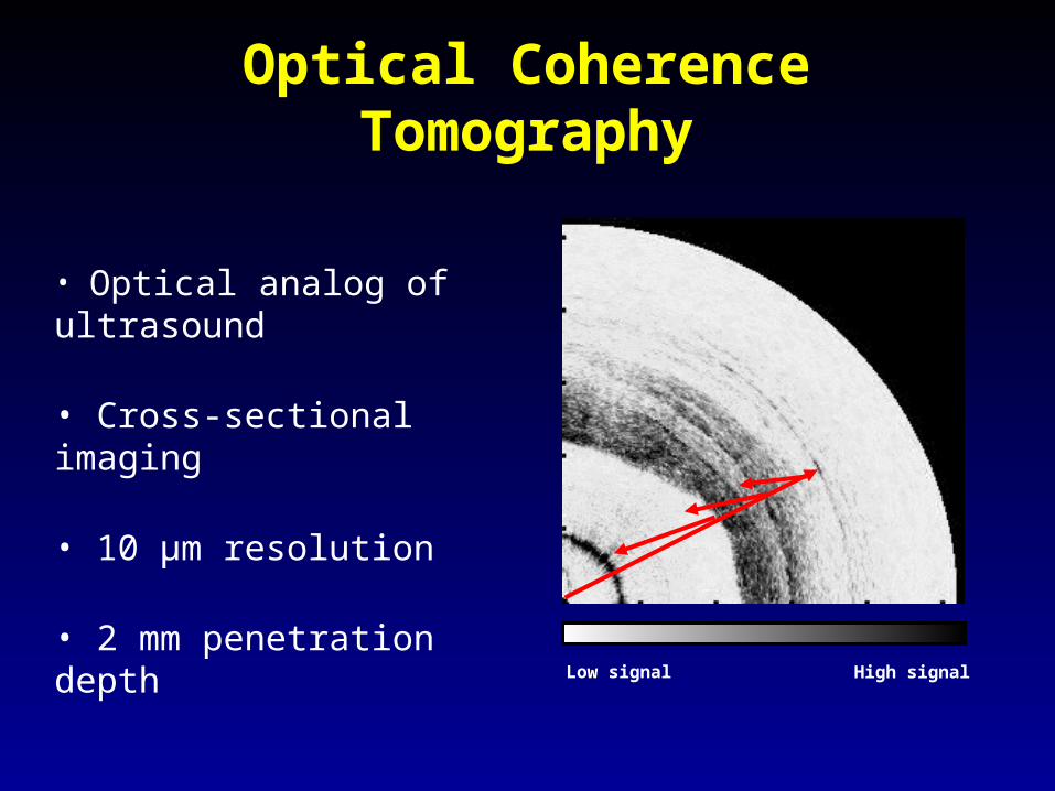
Optical Coherence Tomography
• Optical analog of ultrasound
• Cross-sectional imaging
• 10 µm resolution
• 2 mm penetration depth
Low signal High signal

MGH OCT System Technical Data
Optical wavelength :
Image acquisition rate :
Catheter:
Axial Resolution :
Transverse Resolution :
Data storage :
1300 nm
4-8 images / sec
3.0 F
10 m
25 m
Digital
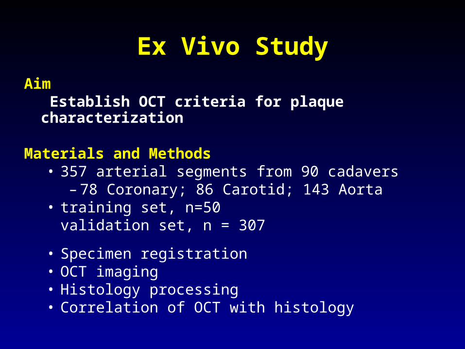
Ex Vivo StudyAim
Establish OCT criteria for plaque characterization
Materials and Methods• 357 arterial segments from 90 cadavers
– 78 Coronary; 86 Carotid; 143 Aorta• training set, n=50
validation set, n = 307
• Specimen registration• OCT imaging• Histology processing• Correlation of OCT with histology
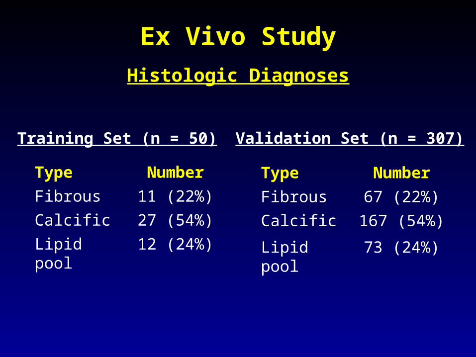
Ex Vivo Study
Type NumberFibrous 11 (22%)Calcific 27 (54%)Lipid pool 12 (24%)
Type NumberFibrous 67 (22%)Calcific 167 (54%)
Lipid pool 73 (24%)
Validation Set (n = 307)Training Set (n = 50)
Histologic Diagnoses

OCT Characteristics
lp
lp
Homogeneous,Signal-rich
Fibrous Lipid
Echolucent, Diffuse Borders
Echolucent, Sharp Borders
Calcific500 µm
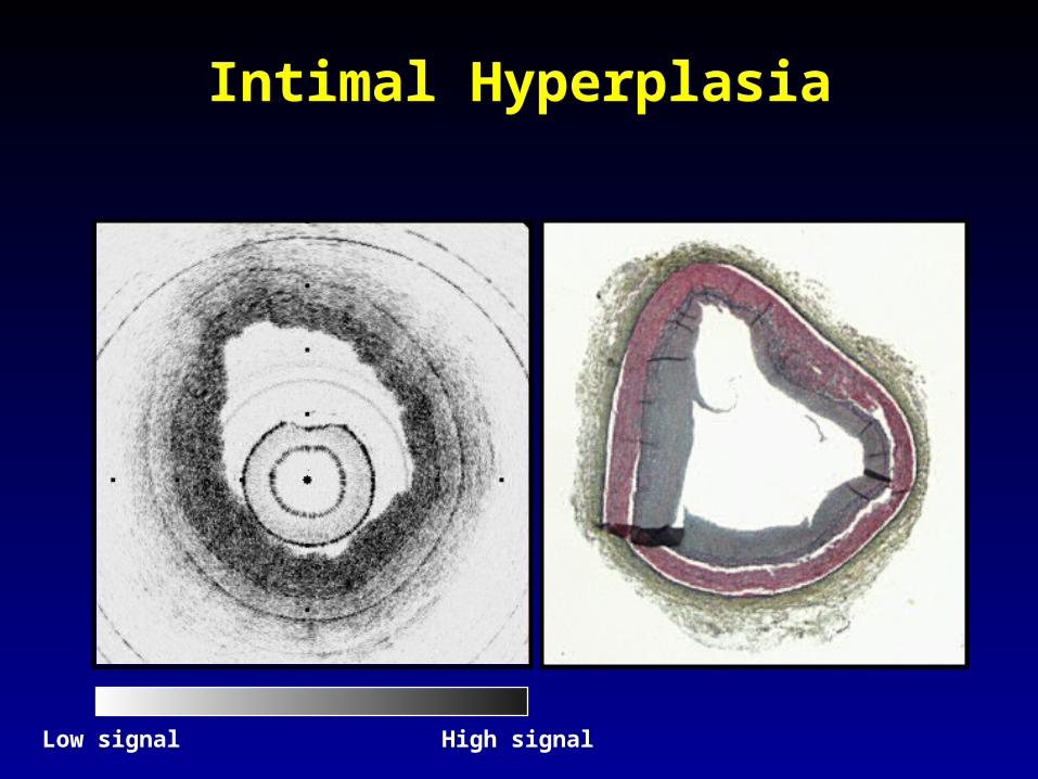
Intimal Hyperplasia
Low signal High signal

Fibrous Plaque
F
IMA
F
EELIEL
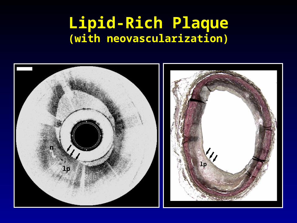
lp
n
lp
Lipid-Rich Plaque(with neovascularization)
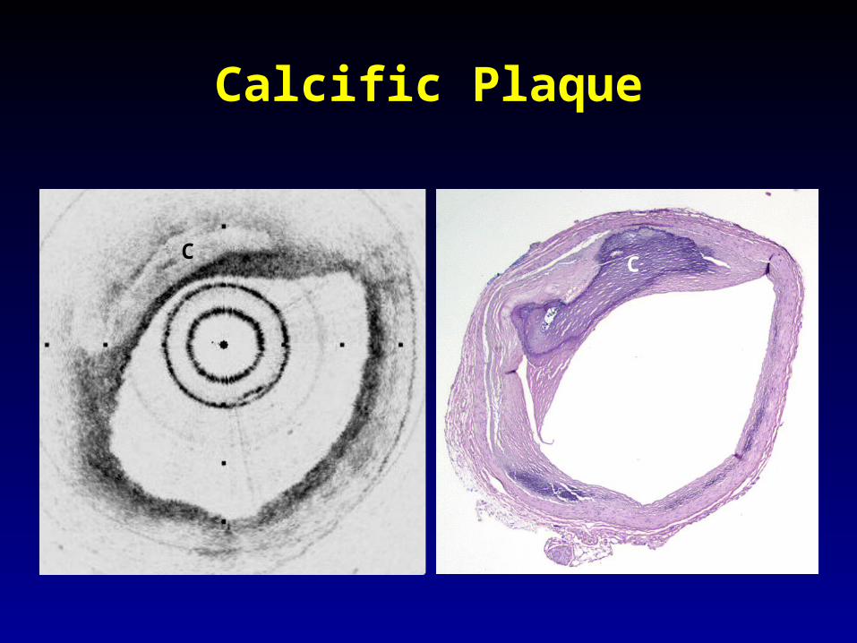
Calcific Plaque
C C

Ex Vivo Study Results
SENS .87 PPV .88SPEC .97 NPV .96
SENS .95 PPV 1.0SPEC 1.0 NPV .95
SENS .92 PPV .81SPEC .94 NPV .97
Accuracy Statistics
Fibrous
Calcific
Lipid pool
Interobserver = 0.88, Intraobserver = 0.91

Clinical Study 1
Feasibility and Safety:Pre and Post PCI
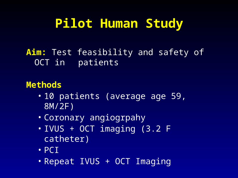
Pilot Human Study
Aim: Test feasibility and safety of OCT in patients
Methods• 10 patients (average age 59, 8M/2F)• Coronary angiogrpahy• IVUS + OCT imaging (3.2 F catheter)• PCI• Repeat IVUS + OCT Imaging

Fibrous Plaque
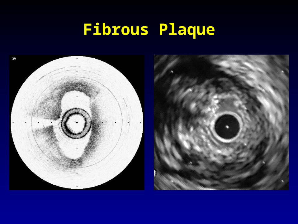
Fibrous Plaque

Lipid-rich Plaque

Lipid Rich Plaque
g
lplp
f
g
2x
ma
i
iel eel
f
OCT IVUS
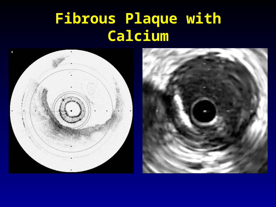
Fibrous Plaque with Calcium
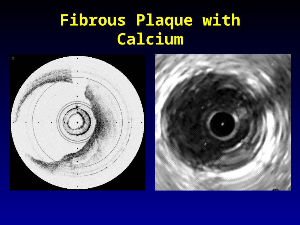
Fibrous Plaque with Calcium
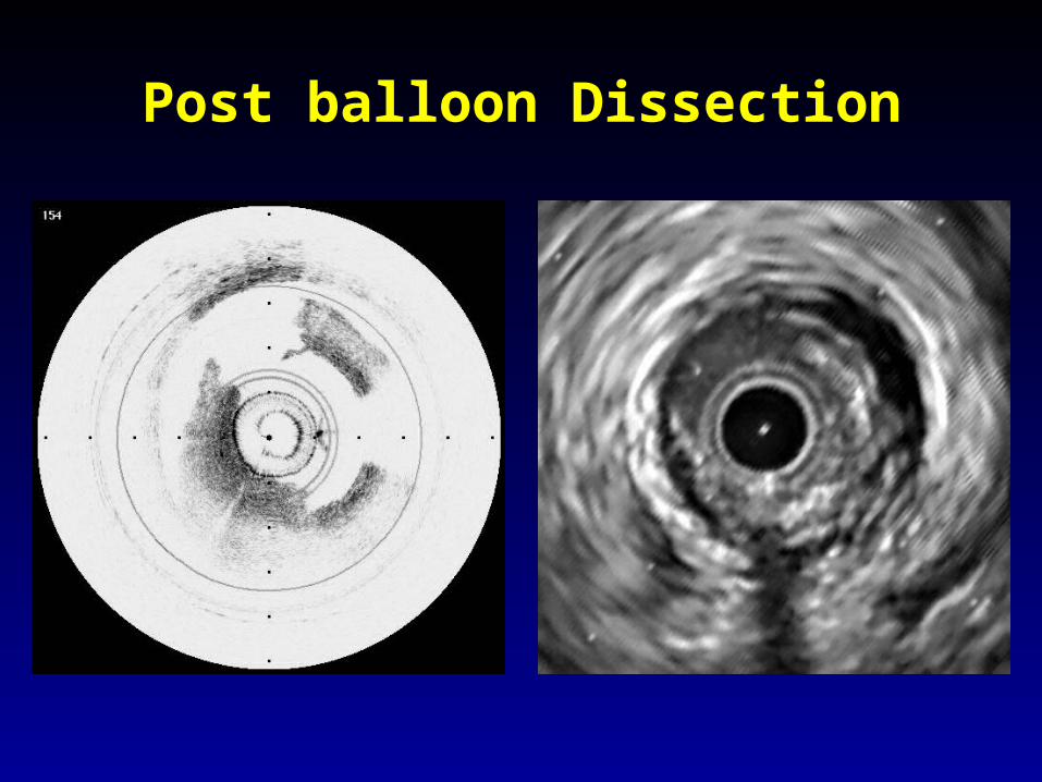
Post balloon Dissection
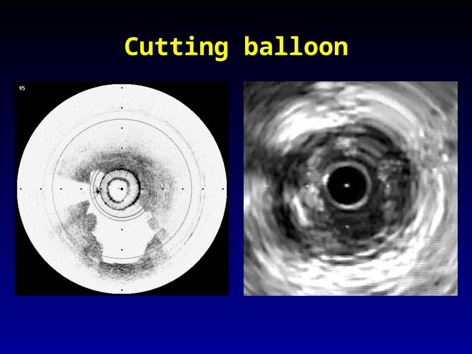
Cutting balloon

Stent
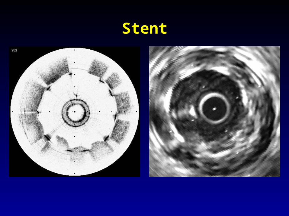
Stent
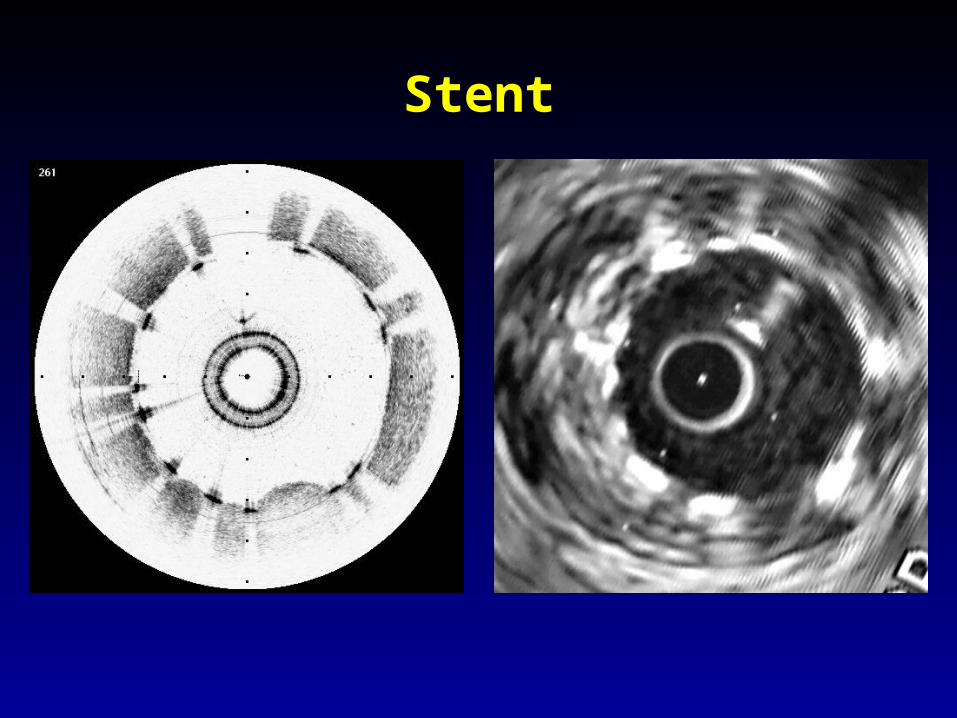
Stent
Pilot Human Study Results No OCT related complications
Variety of pathology imaged and compared with IVUS• 10 patients with 28 plaque segments• 8 dissections• 13 stent locations
Problems• Obstruction by blood• Motion artifacts

Acknowledgements
Massachusetts General HospitalCardiology DivisionH. Yabushita, B. MacNeill, H. Lowe, M. Hayashi, S. Clarke, E. Pomerantsev, D. DeJoseph, I.K. Jang
Wellman Laboratories of Photomedicine B.E. Bouma, M. Shishkov, C. Kauffman, N. Iftima, G.J. Tearney
Dept. of PathologyS. Houser, H.T. Aretz
CIMITJ. Muller, T. Brady, J. Rosen
Guidant CorporationD. Kilpatrick, J. Ellis, R. Jones, T. Linnemeier
Related Documents