10/8/2012 1 ESG Faried Mohammed Wagdy (MD) VISUAL FIELD By DR : Faried Mohammed Wagdy ( MD ) Menofia University (Egypt )

Welcome message from author
This document is posted to help you gain knowledge. Please leave a comment to let me know what you think about it! Share it to your friends and learn new things together.
Transcript

10/8/2012
1
ESG
Faried Mohammed Wagdy
(MD)
VISUAL FIELD
By
DR : Faried Mohammed Wagdy
( MD )
Menofia University
(Egypt )

10/8/2012
2

10/8/2012
3
Visual field loss out of proportion to
OCT in glaucoma

10/8/2012
4
On examination, her left eye had a visual acuity of
20/25 and the intraocular pressure was 19 mm Hg.
Indirect biomicroscopy (A) revealed moderate disc
cupping with mild thinning of the inferotemporal
neuroretinal rim.
Humphrey visual field testing (D) displayed a dense
superior arcuate and an early inferior arcuate scotoma that
were out of proportion to the disc findings.
10/8/2012
5

10/8/2012
6
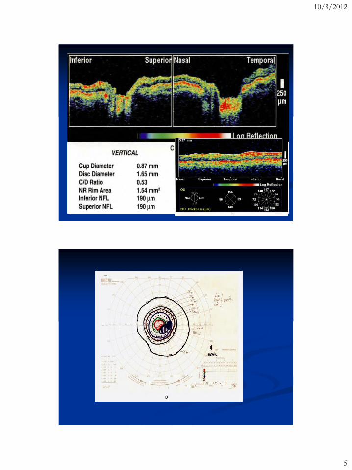
Inferior Seidel Scotoma due to Chorioretinal Scar
Dilated ophthalmoscopy (A) revealed marked cupping of the optic disc with an intact neuroretinal rim, and a chorioretinal scar in the superotemporal arcade.
A Goldmann visual field (D) displayed an inferior Seidel scotoma.
10/8/2012
7
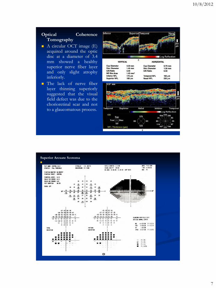
Optical Coherence Tomography
A circular OCT image (E) acquired around the optic disc at a diameter of 3.4 mm showed a healthy superior nerve fiber layer and only slight atrophy inferiorly.
The lack of nerve fiber layer thinning superiorly suggested that the visual field defect was due to the chorioretinal scar and not to a glaucomatous process.
Superior Arcuate Scotoma

10/8/2012
8
Optical Coherence Tomography
A 3.4 mm diameter circular tomogram (E) revealed atrophy of the retinal nerve fiber layer inferiorly. The thinning was most significant from 7:00 to 9:00, consistent with the superior visual defect observed clinically.

10/8/2012
9

10/8/2012
10
Optical Coherence
Tomography
A virtual absence of nerve
fibers was observed
inferotemporally at 5:00 in
the circular OCT tomogram
(j) corresponding to the area
of visual field loss.
A narrow, focal notch in the
nerve fiber layer was seen
superiorly, and did not
coincide with a clinically
detectable visual field defect.

10/8/2012
11
End-Stage Glaucoma
A 40-year-old Haitian man was taking pilocarpine 6% four times a day for primary open-angle glaucoma, until four days prior to examination, when he ran out of medication.
He had previously undergone laser trabeculoplasty in his right eye.
On examination of this eye, the visual acuity was 20/20 and the intraocular pressure was 27 mm Hg.
Gonioscopy revealed that the angle was open to the scleral spur, except inferiorly, where it was open to the ciliary body band.
Dilated ophthalmoscopy (A) showed a deep, excavated cup with an absence of neuroretinal rim.
A Humphrey visual field (C) displayed only a small remaining central island of vision

10/8/2012
12
Optical Coherence Tomography
Minimal nerve fiber tissue was observed on a 3.4 mm diameter circular OCT tomogram (D) around the optic disc.
The dramatic attenuation of the nerve fiber layer was consistent with this patient's degree of cupping and visual field loss.

10/8/2012
13
10/8/2012
14
Focal Defects in the Nerve Fiber Layer
Glaucoma may often cause focal regions of retinal nerve fiber layer (NFL) loss. These areas of NFL thinning can be difficult to detect by the traditional methods of ophthalmoscopy, stereoscopic biomicroscopy and optic nerve head photography, or evaluation of the red free NFL reflex.
The ability of OCT to profile the NFL in cross-section with high resolution is useful in the identification of focal or diffuse areas of NFL thinning.
A circular OCT tomogram acquired around the optic disc provides information on NFL thickness in a cylindrical cross-section surrounding the nerve head.
The normal variations in NFL thickness are
readily apparent in such a section, and focal or
diffuse NFL thinning may be identified by visual
inspection.
Alternatively, an automated computer image
processing algorithm may be used to
quantitatively measure the retinal and NFL
thickness from the circular tomograms,
providing an objective assessment of the size
and severity of the NFL loss.

10/8/2012
15
inferonasal step

10/8/2012
16
Dilated fundus examination showed a narrow
focal defect in the superotemporal nerve fiber
layer (A) and a thinning of the neuroretinal rim
temporally. A distinct inferonasal step was noted
on the Humphrey visual field (D) corresponding
to the nerve fiber layer defect observed
ophthalmoscopically
Optical Coherence Tomography
A circular OCT tomogram (E) taken at a diameter of 3.4 mm around the optic disc revealed a generally healthy nerve fiber layer with the exception of a focal area of thinning in the superotemporal nerve fiber layer (arrows), consistent with the defect observed clinically.
The thinning was primarily evident at the superficial margin of the nerve fiber layer, and reached a minimum thickness of 90 µ in the image.

10/8/2012
17
A Humphrey visual field
(D) revealed an inferior
arcuate scotoma
consistent with the
nerve fiber layer defect
and an early superior
nasal step as well.
Focal Nerve Fiber Layer Defect
Ophthalmoscopy (A) revealed a moderately cupped disc with an attenuated neuroretinal rim temporally.
The nerve fiber layer reflex (B) displayed a focal reduction in the superotemporal area.
10/8/2012
18
Optical Coherence Tomography
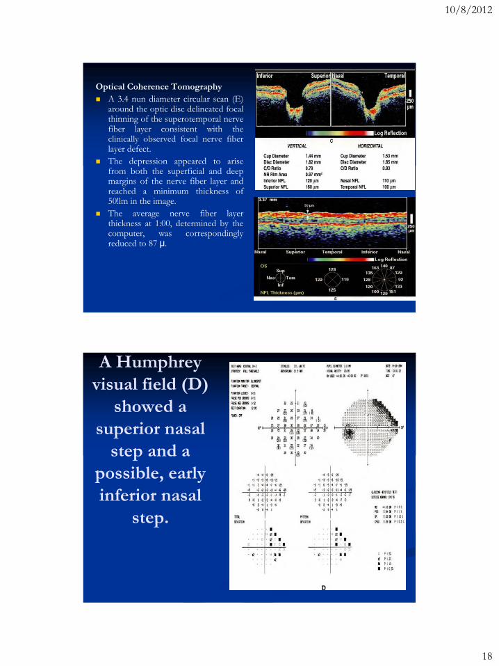
A 3.4 nun diameter circular scan (E) around the optic disc delineated focal thinning of the superotemporal nerve fiber layer consistent with the clinically observed focal nerve fiber layer defect.
The depression appeared to arise from both the superficial and deep margins of the nerve fiber layer and reached a minimum thickness of 50!lm in the image.
The average nerve fiber layer thickness at 1:00, determined by the computer, was correspondingly reduced to 87 µ.
A Humphrey
visual field (D)
showed a
superior nasal
step and a
possible, early
inferior nasal
step.

10/8/2012
19

10/8/2012
20
Message - -Field is important for follow up of
glaucomatous patients.
- - To be familiar with field essential to be
familiar with disc.
- - Quality of life may be not good with bad
fields

10/8/2012
21
Related Documents