Optical and morphological characterisation of a silver nanoparticle/fluorescent poly(phenylenethynylene) composite for optical biosensors Juan Carlos Ramos a, b , Antonio Ledezma a , Eduardo Arias a , Ivana Moggio a, * , Carlos Alberto Martı ´nez b , Felipe Castillon c a Centro de Investigacio ´n en Quı ´mica Aplicada, Boulevard Enrique Reyna 140, 25253 Saltillo, Mexico b Universidad Auto ´noma de Ciudad Jua ´rez, Henry Dunant 4016, C.P. 32310, Ciudad Jua ´rez, Mexico c Centro de Ciencia de la Materia Condensada de la UNAM, Apdo. Postal 2681, CP 22800 Ensenada, BC, Mexico Keywords: Poly(phenylenethynylene) Silver nanoparticles Nanocomposite Thin films Fungi abstract Thiol silver nanoparticles prepared by the phase transfer method have been mixed with a fluorescent poly(phenylenethynylene) sequenced with dithioester-diethylsulfide moieties in order to develop a nanocomposite for its possible application in optical biosensors for the detection and attack of fungi such as Paecilomyces variotii. Films have been prepared by dipping technique and characterized by AFM, XPS, UV-Visible and fluorescence spectroscopy. Optical Absorption properties of the nanocomposite are similar to those of the polymer with an absorption tail in the visible which supports the presence of silver nanoparticles. Despite the lack of fluorescence of the nanoparticles, the composite emits in the yellow green region and the intensity of the fluorescence of the nanocomposite film decreases after the immersion in the culture thus permitting the detection of the fungus by this technique. The fungus can be deposited on films of both the polymer and nanocomposite, nevertheless only in the latter case, an attack on mycelium is observed revealing the fungicidal effect of silver nanoparticles in the nanocomposite. Ó 2009 Elsevier Ltd. All rights reserved. 1. Introduction Bacteria’s are considered as the main pathogenic microorgan- isms and studies about novel detection methods are usually related to them. However, fungi are a major health problem especially for patients with immunodeficiency [1]. Fungal infections are often very progressive, moreover several saprophytic or plant fungi can become opportunistic pathogens for humans, especially for patients who are exposed to antibiotics or immunosuppressive drugs [2]. Contrary to mycoses due to Candida or Aspergillus niger , infections due to opportunistic fungi are more difficult to be detected as the pathogen microorganism is not expected to be found in humans. Impact of these infections can be relevant because the fungus can easily become a host in any organs of the human body such as intestine, appendix, urinary system, etc. For this reason, a rapid diagnostic method could decrease the propa- gation of the microorganism in the body of already affected patients as well as increasing the health security of hospitals avoiding a large diffusion of infections. Biosensors have appeared in the last years as a faster, cheaper and easier diagnostic technology with respect to classical analytical assays which require time for the culture growth and experience in handling and characterization of the microorganisms [3]. Among the different types of biosensors, optical biosensors present several advantages such as short response time, easy mode of detection, lack of electrical interfer- ence [4]. Conjugated polymers are optimal candidates as transducer elements for optical biosensors as they present specific and interesting optical properties due to the high p-electron delocalization through their backbone. Phenylenethynylenes are a class of semiconducting materials with interesting photo- and electroluminescent properties and their application in optical biosensors has been previously reported for the detection of Escherichia coli [5]. On the other hand, due to health impact of certain microorganisms, it could be useful to have a diagnostic method which permits not only to detect but also to attack and eliminate them. In this context, copper compounds are usually used in antifungal coating and recently this application was also proposed for polymer nanocomposites of copper nanoparticles [6]. The capacity of bulk silver compounds for attacking bacteria and fungi is well known [7–10]. In recent papers, the bactericide effect has also been demonstrated for silver nanoparticles for pathogen bacteria such as E. coli [11] and the HIV-1 virus [12]. As far as fungi are concerned, to the best of our knowledge there are no reports on the application of silver nanoparticles. In this respect, in present * Corresponding author. Tel.: þ52 844 4389830; fax: þ52 844 4389839. E-mail address: [email protected] (I. Moggio). Contents lists available at ScienceDirect Vacuum journal homepage: www.elsevier.com/locate/vacuum 0042-207X/$ – see front matter Ó 2009 Elsevier Ltd. All rights reserved. doi:10.1016/j.vacuum.2009.10.034 Vacuum 84 (2010) 1244–1249

Welcome message from author
This document is posted to help you gain knowledge. Please leave a comment to let me know what you think about it! Share it to your friends and learn new things together.
Transcript
lable at ScienceDirect
Vacuum 84 (2010) 1244–1249
Contents lists avai
Vacuum
journal homepage: www.elsevier .com/locate/vacuum
Optical and morphological characterisation of a silver nanoparticle/fluorescentpoly(phenylenethynylene) composite for optical biosensors
Juan Carlos Ramos a,b, Antonio Ledezma a, Eduardo Arias a, Ivana Moggio a,*, Carlos Alberto Martınez b,Felipe Castillon c
a Centro de Investigacion en Quımica Aplicada, Boulevard Enrique Reyna 140, 25253 Saltillo, Mexicob Universidad Autonoma de Ciudad Juarez, Henry Dunant 4016, C.P. 32310, Ciudad Juarez, Mexicoc Centro de Ciencia de la Materia Condensada de la UNAM, Apdo. Postal 2681, CP 22800 Ensenada, BC, Mexico
Keywords:Poly(phenylenethynylene)Silver nanoparticlesNanocompositeThin filmsFungi
* Corresponding author. Tel.: þ52 844 4389830; faE-mail address: [email protected] (I. Moggio).
0042-207X/$ – see front matter � 2009 Elsevier Ltd.doi:10.1016/j.vacuum.2009.10.034
a b s t r a c t
Thiol silver nanoparticles prepared by the phase transfer method have been mixed with a fluorescentpoly(phenylenethynylene) sequenced with dithioester-diethylsulfide moieties in order to developa nanocomposite for its possible application in optical biosensors for the detection and attack of fungisuch as Paecilomyces variotii. Films have been prepared by dipping technique and characterized by AFM,XPS, UV-Visible and fluorescence spectroscopy. Optical Absorption properties of the nanocomposite aresimilar to those of the polymer with an absorption tail in the visible which supports the presence ofsilver nanoparticles. Despite the lack of fluorescence of the nanoparticles, the composite emits in theyellow green region and the intensity of the fluorescence of the nanocomposite film decreases after theimmersion in the culture thus permitting the detection of the fungus by this technique. The fungus canbe deposited on films of both the polymer and nanocomposite, nevertheless only in the latter case, anattack on mycelium is observed revealing the fungicidal effect of silver nanoparticles in thenanocomposite.
� 2009 Elsevier Ltd. All rights reserved.
1. Introduction with respect to classical analytical assays which require time for the
Bacteria’s are considered as the main pathogenic microorgan-isms and studies about novel detection methods are usually relatedto them. However, fungi are a major health problem especially forpatients with immunodeficiency [1]. Fungal infections are oftenvery progressive, moreover several saprophytic or plant fungi canbecome opportunistic pathogens for humans, especially forpatients who are exposed to antibiotics or immunosuppressivedrugs [2]. Contrary to mycoses due to Candida or Aspergillus niger,infections due to opportunistic fungi are more difficult to bedetected as the pathogen microorganism is not expected to befound in humans. Impact of these infections can be relevantbecause the fungus can easily become a host in any organs of thehuman body such as intestine, appendix, urinary system, etc. Forthis reason, a rapid diagnostic method could decrease the propa-gation of the microorganism in the body of already affectedpatients as well as increasing the health security of hospitalsavoiding a large diffusion of infections. Biosensors have appeared inthe last years as a faster, cheaper and easier diagnostic technology
x: þ52 844 4389839.
All rights reserved.
culture growth and experience in handling and characterization ofthe microorganisms [3]. Among the different types of biosensors,optical biosensors present several advantages such as shortresponse time, easy mode of detection, lack of electrical interfer-ence [4]. Conjugated polymers are optimal candidates astransducer elements for optical biosensors as they present specificand interesting optical properties due to the high p-electrondelocalization through their backbone. Phenylenethynylenes area class of semiconducting materials with interesting photo- andelectroluminescent properties and their application in opticalbiosensors has been previously reported for the detection ofEscherichia coli [5]. On the other hand, due to health impact ofcertain microorganisms, it could be useful to have a diagnosticmethod which permits not only to detect but also to attack andeliminate them. In this context, copper compounds are usually usedin antifungal coating and recently this application was alsoproposed for polymer nanocomposites of copper nanoparticles [6].The capacity of bulk silver compounds for attacking bacteria andfungi is well known [7–10]. In recent papers, the bactericide effecthas also been demonstrated for silver nanoparticles for pathogenbacteria such as E. coli [11] and the HIV-1 virus [12]. As far as fungiare concerned, to the best of our knowledge there are no reports onthe application of silver nanoparticles. In this respect, in present
J.C. Ramos et al. / Vacuum 84 (2010) 1244–1249 1245
work we report the elaboration of a nanocomposite of silvernanoparticles with a fluorescent poly(phenylethynylene)sequenced with dithioester-diethylsulfide moieties, hereafternamed pPET3OC12-sqS to develop a fungicidal system; where thefungi can be easily detected by fluorescence and simultaneouslyattacked by silver nanoparticles. We studied P. variotii which,despite being usually considered a saprophytic fungus, maybecome opportunistic causing a disease in humans and animalscalled paecilomycosis [13].
2. Materials and methods
The synthesis and physicochemical characterization of pPEOC4and pPET3OC12-sqS (Fig. 1) have been reported in ref [14,15]. Silvernanoparticles were synthesized using the phase transfer methodreported first by Brust et al. for gold particles [16]. In a typicalsynthesis, 0.55 g of tetraoctylammonium bromide was dissolved in20 ml of toluene at room temperature under vigorous stirring.Then, 2 ml of a 0.1 M silver nitrate solution in distilled water wasadded to the reaction flask. One hour later, 0.3 ml of 1-dodeca-nethiol was added. Finally, 6.3 ml of 0.25 M sodium boronhydridesolution in distilled water was slowly added dropwise. After threehours, the reaction was stopped and the product was recoveredfrom organic phase by precipitation using cold ethanol. Nano-particles were re-dissolved in toluene and precipitated again incold ethanol and, finally, metal nanoparticles were dissolved andstored in toluene. The final concentration evaluated by dryinga known volume of the solution, was 2.2 mg/ml.
The nanocomposite is obtained by mixing the toluene solutionof nanoparticles with a 1 mg/ml CHCl3 solution of pPET3OC12-sqSin a relation 1:1 v/v for three hours under strong magnetic stirring.The yellow solution remained stable without aggregation orprecipitation after one year from its preparation. For the depositionof the films, Corning glass or quartz slides by SPI. Inc have beenused as substrates and treated with sulfochromic mixture asdescribed in published literature [17]. Then each lime has beenimmersed in the nanocomposite solution by dipping using a KSVLangmuir-Blodgett equipment at a speed of 68.55 mm/min for10 min followed by drying in air. A direct measurement of thethickness by profilometry was not possible. However, on the basisof the absorption peak due to the polymer and previously foundabsorption coefficient for pPET3OC12-sqS thin films [15], a thick-ness of around 10 nm can be estimated. As a sample control for themicrobiological assay, films of pPET3OC12-sqS and silver nano-particles have been prepared under the same conditions as those ofthe nanocomposite. For the microbiological tests, cultures onpotato dextrose agar of the fungus P. variotii have been incubatedfor 7 days to prepare a microorganism suspension in distilled watercontaining both spores and mycelium. Films were immersed in the
Fig. 1. Chemical structure of pPET3OC12-sqS and pPEOC4.
microorganism suspension for 20 min with the same procedureand equipment used for their elaboration. Then, these were drawnout and washed up to 6 times with distilled water. Each washingwas realized by dipping in distilled water for 2 min and water wasrenewed every time. Attack to the fungus was studied with a LeicaATC 2000 optical microscope and by laser scanning confocalmicroscopy (Zeiss Pascal 5). Also the microorganism suspensionhas been analyzed before and after the film immersion by opticalmicroscopy by casting few drops between two microscope slides.UV-Visible and emission spectra were recorded on a Shimadzu2401 spectrophotometer and a Perkin Elmer LS50B spectrofluo-rimeter, respectively. The excitation wavelength was chosen 10 nmunder the absorption lower energy peak. Quantum yields (q.y.)were obtained according to the procedure reported earlier [18] andquinine sulfate (q.y.¼ 0.54) was used as standard. The AFM analysiswas carried out on a Digital Dimension 3100 microscope in tappingmode at a rate of 0.3–0.5 Hz. TEM analysis was performed in a JEOL2010F microscope operating at 200 kV equipped with a Schottky-type field emission gun, an ultra-high resolution pole piece(Cs¼ 0.5 mm) and an energy dispersive X-ray spectrometer (EDS)unit. Samples for TEM were prepared by depositing a drop of theoriginal suspension on a lacey carbon coated Cu grid and allowed toevaporate. The SEM analysis was carried out on a SM510 TOPCONmicroscope.
The X-Ray photoelectron spectroscopy (XPS) analysis wascarried out on a modified laser ablation system, Riber LDM-32,using a Cameca Mac3 analyzer. The base pressure in the analysischamber was approximately 10�10 mbar. The X-ray Al Ka line at1486.6 eV was used for excitation. The binding energies werecalibrated with reference to Cu 2 p3/2 at 932.67 eV and Ag 3d5/2 at368.26 eV, respectively. The resolution attained with this set-up is1.1 eV measured on the C 1 s signal of a graphite target. Spectrawere collected by acquiring data at every 0.2 eV and the energyresolution was 0.8 eV. The wide-scan and core-level spectra for C1 s were obtained. A short-time scan of C 1 s was first acquired tocheck that no degradation occurs during the spectra acquisitiondue to X-ray exposure. Background subtraction was done using theTougaard method [19]. In addition, wide-scan spectra were gath-ered acquiring data at every 1.0 eV with an energy resolution of3 eV. Charging effect was corrected by shifting the binding energiesconsidering the C 1 s signal at 284.6 eV. A non-linear fit, usingGaussian curves, was performed maintaining the Full-Width atHalf-Maximum (FWHM) constant for all components in a particularspectrum.
3. Results and discussion
The poly(phenylethynylene) pPET3OC12-sqS, in its chemicalstructure presents the main conjugated chain sequenced withflexible dithioester-diethylsulfide segments. The conjugated back-bone imparts high fluorescence to the polymer, while the sulfuratoms of the flexible sequence are intended to promote the affinitywith silver. In Fig. 2a, the UV-Visible spectrum of the pPET3OC12-sqS/Ag nanocomposite in toluene is reported. Two bands, observedat 327 and 394 nm as found for the polymer solution [15], corre-spond to the electronic transitions of the dodecanoxy substitutedbenzene and to the p�p* electronic transitions of the conjugatedchains, respectively. Suspension of silver nanoparticles in tolueneexhibits a broad absorption band from 300 to 800 nm, witha maximum at 454 nm (as shown in inserted figure) due to theplasmon band. This absorption cannot be observed in the compositesuspension probably because it is masked by the stronger absorbingpolymer band. The absorption coefficient of pPET3OC12-sqS is infact, 50.7 gl�1 cm�1 for pPET3OC12-sqS [15] contra 0.33 gl�1 cm�1
for the suspension of silver nanoparticles. Nevertheless, the

Fig. 2. a) UV-Visible spectra of pPET3OC12-sqS/Ag nanoparticles in CHCl3/toluene 1:1(solid line), pPET3OC12-sqS in CHCl3 (dashed line) and Ag nanoparticles in toluene(dotted line). Inserted figure: magnification of the same spectra in the 450–600 nmregion. (b) Emission spectra of pPET3OC12-sqS/Ag nanoparticles in CHCl3/toluene 1:1(solid line) and pPET3OC12-sqS in CHCl3 (dashed line). Inserted figure: photo takenunder UV (lexc¼ 365 nm) irradiation. From left to right, pPET3OC12-sqS in CHCl3,
pPET3OC12-sqS/Ag nanoparticles in CHCl3/toluene 1:1 solution, silver nanoparticles intoluene and toluene. Notice that the bluish colour of the silver nanoparticlessuspension is due to toluene as evidenced by the comparison with the solventemission.
Fig. 3. a) UV-Visible spectra of films of pPET3OC12-sqS/Ag nanoparticles and pPE-T3OC12-sqS (inserted figure) before (solid lines) and after (dash lines) the immersionin P. variotii suspension. b) Emission spectra of film of pPET3OC12-sqS/Ag nano-particles before (solid lines) and after (dash lines) the immersion in P. variotiisuspension.
J.C. Ramos et al. / Vacuum 84 (2010) 1244–12491246
absorption tail found between 450 and 600 nm (see magnification ofthe 400–600 nm region in the inserted Fig. 2) indicates the presenceof the silver nanoparticles. As fluorescence (Fig. 2b) is concerned, thepPET3OC12-sqS/Ag nanoparticles suspension emits in the blue-green region (photo in the inserted Fig. 2) with an emissionmaximum at 454 nm coinciding with the emission of the polymer[15]. Despite of the lack of fluorescence of silver nanoparticles, thenanocomposite exhibits a quantum yield of 0.53, practically iden-tical to that found for the polymer alone [15] indicating that noenergy transfer occurs. This result together with the fact thatno energy shift is observed for the absorption or emission spectrapassing form the polymer to the nanocomposite samples, isconsistent with the hypothesis that the conjugated moiety of pPE-T3OC12-sqS is not involved in the assembly with silver.
The optical properties of the composite in thin films (Fig. 3) aresimilar to those already discussed for the solution. In the absorptionspectrum of pPET3OC12-sqS/Ag nanoparticles (Fig. 3a), two peaks
at 327 and 403 nm can be distinguished, in agreement with thespectrum of the pPET3OC12-sqS sample [15]. No evident featuresdue to silver are observed, as expected on the basis of the resultsdiscussed for the solutions and considering the low intensity of thespectrum. It is interesting to notice that after immersing the films inthe microorganism suspension (dashed lines), the spectrum for thepPET3OC12-sqS/Ag nanoparticles film presents the same absor-bance as the pristine sample. A slight broadening of the baselineobserved results from a certain loss of homogeneity. On thecontrary, for the spectrum of pPET3OC12-sqS film, a strongdecrease in the absorbance occurs. This behaviour is due to a partialdissolution of the polymer in water, as previously reported [15].This result indicates that the presence of the nanoparticles in thenanocomposite decreases the polymer solubility in water con-firming the formation of a composite through the interactions ofpolar groups of pPET3OC12-sqS with silver. As shown in Fig. 3b, thepPET3OC12-sqS/Ag nanoparticles film presents a broad fluores-cence band centered at around 481 nm, as the polymer film. Thefluorescence intensity decreases strongly as a consequence of thedetection of the fungus. This behaviour cannot be ascribed toa partial dissolution of the film according to what was previouslydiscussed and to the fact that no emission was found in themicroorganism suspension after the immersion of the film. Morelikely, the fungus which is deposited on the film produces
J.C. Ramos et al. / Vacuum 84 (2010) 1244–1249 1247
a quenching of the polymer emission. This result clearly indicatesthe possibility to use nanocomposite films for the detection of thismicroorganism.
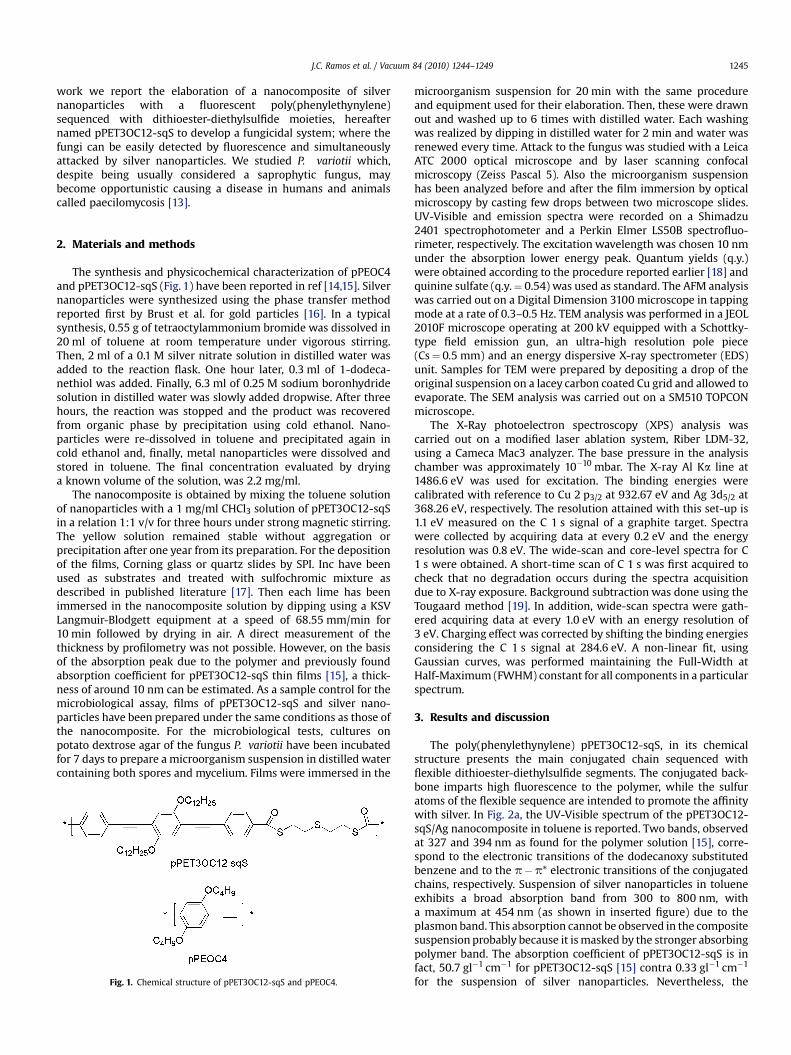
Preparation of the composite has indeed a technological rele-vance because this biosensor can be applied not only for thedetection but also for attacking the fungus as discussed below. InFig. 4, the optical micrographs relative to the microbiological testswith P. variotii are shown. The pristine culture presents sphericalspores which are typical of this fungus and mycelium. Both partsare transferred to the pPET3OC12-sqS film (top) as well as on thepPET3OC12-sqS/Ag nanoparticles film (bottom). The fact that thefungus can be transferred onto both samples indicates that pPE-T3OC12-sqS favours the affinity with the microorganism, probablybecause of the interactions of the flexible sequences with proteinspresent in the cellular membrane. In order to support thishypothesis, we performed the same test with polymer pPEOC4which does not contain polar groups in its chemical structure. Inthis case, no transfer was detected. The interesting point is that onlyfor the nanocomposite film, the mycelium presents morphologicaldifferences along its structure (dark and light segments as indicatedby the arrows in Fig. 4b) which could suggest cellular damage.
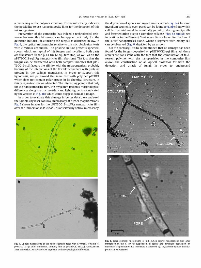
In order to evaluate this damage in better detail, we analyzedthe samples by laser confocal microscopy at higher magnifications.Fig. 5 shows images for the pPET3OC12-sqS/Ag nanoparticles filmafter the immersion in P. variotii. As observed by optical microscopy,
Fig. 4. Optical micrographs of the microorgamism tests with P. variotii. top) film ofpPET3OC12-sqS after immersion. bottom) film of pPET3OC12-sqS/Ag nanoparticlesafter immersion. Arrows indicate segments with morphological differences.
the deposition of spores and mycelium is evident (Fig. 5a). In somemycelium segments, even pores can be found (Fig. 5b) from whichcellular material could be eventually go out producing empty cellsand fragmentation due to a complete collapse (Figs. 5a and 5b, seeindications in the Figures). Similar results are found for the film ofthe silver nanoparticles alone, where a segment with empty cellcan be observed (Fig. 6, depicted by an arrow).
On the contrary, it is to be mentioned that no damage has beenfound for the fungus deposited on pPET3OC12-sqS films. All theseresults are consistent with the fact that the combination of fluo-rescent polymer with the nanoparticles in the composite filmallows the construction of an optical biosensor for both thedetection and attack of fungi. In order to understand
Fig. 5. Laser confocal micrographs of pPET3OC12-sqS/Ag nanoparticles film afterimmersion in the P. variotii suspension. a) spores and mycelium deposition- inmycelium, fragmentation due to collapse is observed, b) a mycelium fragment in whichpores can be observed.

Fig. 6. a) SEM image of a film of pure Ag nanoparticles after immersion in the P. variotiisuspension.
J.C. Ramos et al. / Vacuum 84 (2010) 1244–12491248
the mechanism of the attack, AFM, TEM and XPS studies on thenanocomposite samples were carried out. Silver nanoparticlesshow spherical morphology and an average diameter of 6.8 nm asfound by TEM (Fig. 7).
Unfortunately, the TEM characterization of the nanocompositewas not possible. The polymer forms a thin layer on the copper gridtrapping the particles. When the electron beam hits the film, thepolymer burns and the nanoparticles agglomerate. Morphologicalanalysis by AFM (Fig. 8a) reveals that the pPET3OC12-sqS/Agnanoparticles film presents a granular morphology analogously tothat found for pPET3OC12-sqS [15]. Isolated nanoparticles cannotbe distinguished even in nanometer size scans (Fig. 8b). A reason-able explanation could be that the nanoparticles are capped bya polymer layer. However, due to their very low dimension and lossof resolution of tapping images of nanometer size, this hypothesiscannot be completely demonstrated by this technique.
Fig. 7. TEM image of silver nanoparticles studied in this work. Inserted figure: particlesize distribution.
Fig. 8. AFM bi-dimensional images of the nanocomposite films in a) 50� 50 mm2 andb) 500� 500 nm2.
Fig. 9 shows the XPS spectra for the silver nanoparticles and thenanocomposite sample. The bonding nature and chemical states onthe surface of the films with silver nanoparticle sample (top) showsa major contribution in the Ag3d high resolution spectra of a peakcentred at 368.3 eV which is assigned to Ag 3d5/2 in Agþ form [20].The XPS Ag3d high resolution spectrum for the nanocomposite(bottom) shows no evidence of silver contribution. Taking intoaccount the limit in penetration depth of the X-rays, this resultcould be ascribed to the fact that the nanoparticles are covered bythe polymer, supporting the morphological characterization byAFM. Optical and morphological characterizations seem to indicatethat pPET3OC12-sqS is assembled over nanoparticles. Thishypothesis is reliable, considering the much greater size of thepolymer with respect to the nanoparticles and the fact that it isexpected to interact through the flexible groups partially withsilver (to make the nanocomposite) and to the fungus (to permit itsdeposition). However, microbiological tests indicate the attack ofthe fungus. A possible mechanism for the attack could involvea local direct contact of silver nanoparticles with the fungus i.e.certain facets of the silver nanoparticles are exposed on the surface.The evidence by laser scanning microscopy suggests that thiscontact occurs at specific points which could be related to the part

Fig. 9. XPS spectra of the Ag3d region for the silver nanoparticles (top) and thenanocomposite (bottom).
J.C. Ramos et al. / Vacuum 84 (2010) 1244–1249 1249
of nanocomposite film where the polymer layer does notcompletely cap all the particles. Nevertheless, the delivery of silverto the cultures and consequently its attack of the fungus cannot bediscarded as the amount of silver present in the nanocompositefilms is below the detection limit of the atomic absorptionmeasurements.
As a final remark, the importance of this work is that we havedemonstrated the viability of obtaining an optical biosensor for thedetection and attack of P. variotii. Nevertheless as the mycelium,where the attack was evident, is a growing part of the fungus whichcannot be quantified, a quantitative study of the device responsewas not possible for the moment.
4. Conclusions
In this work we report the development of a nanocomposite of silvernanoparticles with a fluorescent poly(phenylethynylene) sequenced
with dithioester-diethylsulfide moieties and microbiological tests withP. variotii. Optical and morphological characterizations of films of thenanocomposite suggest that the silver nanoparticles are covered by thepolymer. Nevertheless, the microbiological tests indicate an attack ofthe mycelium that could be due to the contact of some facets ofnanoparticles with the fungus. Despite the fact that attack is thuslimited, the importance of the work is that with this nanocomposite, itis possible to construct optical biosensors for the simultaneous detec-tion and attack of fungus. Future work will address quantifying theresponse by studying spore counting and spore viability. On the basis ofthose assays, changes in the nanocomposite composition or in the silvernanoparticles characteristics (size and capping agent) will be consid-ered, in order to increase the fungicidal properties of this biosensor.
Acknowledgements
This work was financially supported by CONACyT through theproject 51504-R and CIQA (project FD0003). Special acknowl-edgements to Antonio Diaz, for his technical assistance in XPSmeasurements, Esmeralda Saucedo for SEM analysis, SeleneSepulveda for TEM.
References
[1] Meunier F. Fungal infections in the compromised host. In: Rubin RH, editor.Clinical approach to infections in the compromised host. New York: PlenumPublishing; 1989. p. 193–220.
[2] Perfect JR, Schell WA. The new fungal opportunists are coming. Clin Infect Dis1996;22:S112–118.
[3] Liron Z, Bromberg A, Fisher M, editors. Novel approaches in biosensors and rapiddiagnostic assays. New York: Kluwer Academic/Plenum Publishers; 1999.
[4] Ligler FS, Taitt CAR, editors. Optical biosensors: present and future. Amster-dam: Elsevier Science BV; 2002.
[5] Disney MD, Zheng J, Swager TM, Seeberger PH. Detection of bacteria withcarbohydrate-functionalized fluorescent polymers. J Am Chem Soc2004;126:13343–6.
[6] Cioffi N, Torsi L, Ditaranto N, Tantillo G, Ghibelli L, Sabbatini L, et al. Coppernanoparticle/polymer composites with antifungal and bacteriostatic proper-ties. Chem Mater 2005;17:5255–62.
[7] Clement JL, Jarrett PS. Antibacterial silver. Met Based Drugs 1994;1:467–82.[8] George N, Faoagali J, Muller M. Silvazine (silver sulfadiazine and chlorhex-
idine) activity against 200 clinical isolates. Bur 1997;23:493–5.[9] Fox CL, Modak SM. Mechanism of silver sulfadiazine action on burn wound
infections. Antimicrobial Agents Chemother 1974;5:582–8.[10] Russell AD, Hugo WB. Antimicrobial activity and action of silver. Progr Med
Chem 1994;31:351–70.[11] Morones JR, Elechiguerra JL, Camacho A, Holt K, Kouri JB, Ramırez JT, et al. The
bactericidal effect of silver nanoparticles. Nanotechnology 2005;16:1–8.[12] Elechiguerra JL, Buro JL, Morones JR, Camacho-Bragado A, Gao X, Lara HH, et al.
Interactions of silver nanoparticles with HIV-1. J Nanobiotechnology 2005;3:6.[13] Brown AH, Smith G. The genus paecylomyces bainier and its perfect stage
byssochlamys westing. Trans Brit Mycol Soc 1957;40:17–89.[14] Wautelet P, Moroni M, Oswald L, Le Moigne J, Pham TA, Bigot JY, et al. Rigid rod
conjugated polymers for nonlinear optics. 2. Synthesis and characterization ofphenylene-ethynylene oligomers. Macromolecules 1996;29:446–55.
[15] Vazquez E, Esquivel Aguilar A, Moggio I, Arias E, Romero J, Barrientos H, et al.Immobilization of the enzyme b-lactamase by self-assembly on thin films ofa poly(phenyleneethynylene) sequenced with flexible segments containingsulfur atoms. Mat Sc Eng C 2007;27:787–93.
[16] Brust M, Walter M, Berthell D, Schiffrin DJ, Whyman R. Synthesis of thiol-derivatised gold nanoparticles in a two-phase liquid-liquid system. J Chem SocChem Commun 1994:801–2.
[17] Espinosa C, Moggio I, Arias-Marın E, Romero-Garcıa J, Cruz-Silva R, LeMoigne J, et al. Layer-by-layer assembled films of a rigid poly(phenyl-ethy-nylene) and alternate poly(phenyl-ethynylene)/poly(aniline). Synth Met2003;139:155–61.
[18] Morisaki Y, Ishida T, Chujo Y. Synthesis and properties of novel through-spacep-conjugated polymers based on poly(p-phenylenevinylene)s having a [2,2]paracyclophane skeleton in the main chain. Macromolecules 2002;35:7872–7.
[19] Tougaard S. Practical algorithm for background subtraction. Surf Sci1989;216:343–60.
[20] Li Xianfeng, Hao Xiufeng, Hui Na. Preparation of nanosilver particles intosulfonated poly(ether ether ketone) (S-PEEK) nanostructures by electro-spinning. Mater Lett 2006;61:421–6.
Related Documents


![Optical Biosensors [Online]-1 - Walter Scott, Jr. College of … · · 2008-12-171 Optical Biosensors First Semester Report Fall Semester 2008 by ... The yellow shows the gold electrodes](https://static.cupdf.com/doc/110x72/5ae44c567f8b9ae74a8eea87/optical-biosensors-online-1-walter-scott-jr-college-of-optical-biosensors.jpg)


