Nanomaterials 2020, 10, 2015; doi:10.3390/nano10102015 www.mdpi.com/journal/nanomaterials Review Opportunities for Persistent Luminescent Nanoparticles in Luminescence Imaging of Biological Systems and Photodynamic Therapy Douglas L. Fritzen 1 , Luidgi Giordano 1 , Lucas C. V. Rodrigues 1, * and Jorge H. S. K. Monteiro 2, * 1 Department of Fundamental Chemistry, Institute of Chemistry, University of São Paulo, São Paulo-SP 05508-000, Brazil; [email protected] (D.L.F.); [email protected] (L.G.) 2 Department of Chemistry, Humboldt State University, Arcata, CA 95521, USA * Correspondence: [email protected] (L.C.V.R.); [email protected] (J.H.S.K.M.) Received: 15 September 2020; Accepted: 7 October 2020; Published: 13 October 2020 Abstract: The use of luminescence in biological systems allows us to diagnose diseases and understand cellular processes. Persistent luminescent materials have emerged as an attractive system for application in luminescence imaging of biological systems; the afterglow emission grants background-free luminescence imaging, there is no need for continuous excitation to avoid tissue and cell damage due to the continuous light exposure, and they also circumvent the depth penetration issue caused by excitation in the UV-Vis. This review aims to provide a background in luminescence imaging of biological systems, persistent luminescence, and synthetic methods for obtaining persistent luminescent materials, and discuss selected examples of recent literature on the applications of persistent luminescent materials in luminescence imaging of biological systems and photodynamic therapy. Finally, the challenges and future directions, pointing to the development of compounds capable of executing multiple functions and light in regions where tissues and cells have low absorption, will be discussed. Keywords: persistent luminescence; luminescence imaging; theranostics; photodynamic therapy 1. Introduction Observation of cells and the different cellular components is a fascinating field that allows one to diagnose diseases and unravel biological processes [1–13]. The simplest way to observe cellular components is using a simple optical microscope and color staining [14]. This technique, pioneered by C. Golgi and S. Ramon y Cajal, is based on color change caused by a specific dye [14]. Specific interactions between dye and tissue or dye and cellular components are capable of revealing details about tissue structures and cell components using inexpensive techniques. Color staining is a straightforward technique capable of providing intricate details about tissues and cells. However, it relies on specific interactions between dyes and tissues or cell components; the dye needs to be washed out to warranty specificity and usually requires high concentrations to allow acceptable color contrasts. As an analogy, imagine that the yellow polymer, shown in Figure 1a, is a dye used in cell staining, and the grass represents a cell. The cell staining technique consists of simply placing the polymer onto the grass. As shown in Figure 1b, it is hard to spot the polymer first and takes a well- trained set of eyes to do it. Now imagine that the polymer is luminescent under UV light exposure. The white light illumination is turned off, and the sample is excited using an adequate excitation wavelength (Figure 1c). The use of luminescence grants the reader a clear picture of the polymer’s location in the grass with no interference or low interference from the cell background. Thus, luminescence imaging of biological systems is based on exciting a volume of a sample containing a luminescent compound using an adequate excitation source and wavelength and collecting the light emitted. Luminescence imaging is a sensitive technique that allows diagnosing diseases [15–17],

Welcome message from author
This document is posted to help you gain knowledge. Please leave a comment to let me know what you think about it! Share it to your friends and learn new things together.
Transcript

Nanomaterials 2020, 10, 2015; doi:10.3390/nano10102015 www.mdpi.com/journal/nanomaterials
Review
Opportunities for Persistent Luminescent Nanoparticles in Luminescence Imaging of Biological Systems and Photodynamic Therapy
Douglas L. Fritzen 1, Luidgi Giordano 1, Lucas C. V. Rodrigues 1,* and Jorge H. S. K. Monteiro 2,*
1 Department of Fundamental Chemistry, Institute of Chemistry, University of São Paulo,
São Paulo-SP 05508-000, Brazil; [email protected] (D.L.F.); [email protected] (L.G.) 2 Department of Chemistry, Humboldt State University, Arcata, CA 95521, USA
* Correspondence: [email protected] (L.C.V.R.); [email protected] (J.H.S.K.M.)
Received: 15 September 2020; Accepted: 7 October 2020; Published: 13 October 2020
Abstract: The use of luminescence in biological systems allows us to diagnose diseases and
understand cellular processes. Persistent luminescent materials have emerged as an attractive
system for application in luminescence imaging of biological systems; the afterglow emission grants
background-free luminescence imaging, there is no need for continuous excitation to avoid tissue
and cell damage due to the continuous light exposure, and they also circumvent the depth
penetration issue caused by excitation in the UV-Vis. This review aims to provide a background in
luminescence imaging of biological systems, persistent luminescence, and synthetic methods for
obtaining persistent luminescent materials, and discuss selected examples of recent literature on the
applications of persistent luminescent materials in luminescence imaging of biological systems and
photodynamic therapy. Finally, the challenges and future directions, pointing to the development
of compounds capable of executing multiple functions and light in regions where tissues and cells
have low absorption, will be discussed.
Keywords: persistent luminescence; luminescence imaging; theranostics; photodynamic therapy
1. Introduction
Observation of cells and the different cellular components is a fascinating field that allows one
to diagnose diseases and unravel biological processes [1–13]. The simplest way to observe cellular
components is using a simple optical microscope and color staining [14]. This technique, pioneered
by C. Golgi and S. Ramon y Cajal, is based on color change caused by a specific dye [14]. Specific
interactions between dye and tissue or dye and cellular components are capable of revealing details
about tissue structures and cell components using inexpensive techniques. Color staining is a
straightforward technique capable of providing intricate details about tissues and cells. However, it
relies on specific interactions between dyes and tissues or cell components; the dye needs to be
washed out to warranty specificity and usually requires high concentrations to allow acceptable color
contrasts. As an analogy, imagine that the yellow polymer, shown in Figure 1a, is a dye used in cell
staining, and the grass represents a cell. The cell staining technique consists of simply placing the
polymer onto the grass. As shown in Figure 1b, it is hard to spot the polymer first and takes a well-
trained set of eyes to do it. Now imagine that the polymer is luminescent under UV light exposure.
The white light illumination is turned off, and the sample is excited using an adequate excitation
wavelength (Figure 1c). The use of luminescence grants the reader a clear picture of the polymer’s
location in the grass with no interference or low interference from the cell background. Thus,
luminescence imaging of biological systems is based on exciting a volume of a sample containing a
luminescent compound using an adequate excitation source and wavelength and collecting the light
emitted. Luminescence imaging is a sensitive technique that allows diagnosing diseases [15–17],

Nanomaterials 2020, 10, 2015 2 of 38
reconstructing 3-D structures of tissues or cellular components [18,19], sensing chemical species [1–
3,12,20–38], and unraveling cellular processes [39–41]. One of the drawbacks of this technique is the
strong background emission intensity, especially in the blue and green regions of the electromagnetic
spectrum that are often higher than those of the luminescent compound.
Persistent luminescence (PeL) is a phenomenon where light is emitted for long periods, from
minutes to hours, after the excitation, resulting in a glow-in-the-dark phenomenon [42–54]. If we
come back to the analogy in Figure 1, now imagine that we use a material capable of luminescing
without a continuous excitation. Using the same analogy that the grass represents a cell, we will
achieve what is shown in Figure 1d. Because there is no continuous illumination, all the emission
background is eliminated, and we can locate where the luminescent compound is located. Thus, the
application of PeL materials eliminates the background emission and depth penetration problems,
resulted from the excitation wavelengths in the UV-Vis, commonly used in luminescence imaging of
biological systems [55–72].
(a) (b)
(c)
(d)
Figure 1. (a) Polymer under white light illumination. (b) Polymer dispersed in the grass under white
light, analog to the cell staining technique. (c) Polymer dispersed in the grass under UV light, analog
to the luminescence imaging technique. (d) Persistent luminescent material dispersed in the grass.
Theranostics corresponds to systems capable of simultaneously treat (therapy) and diagnose
(diagnostics) diseases. Recently, the research for non-invasive and tailored treatments have prompted
research in treatments that involves the generation of heat (photothermal therapy, PTT), reactive
oxygen species (photodynamic therapy, PDT) or gene therapy, to cite a few [43,73–86]. Photodynamic
therapy (PDT) is a non-invasive therapy based on the generation of singlet oxygen (1O2) and/or
reactive oxygen species (ROS). Cells and organisms are less likely to develop resistance to 1O2, making
PDT attractive for treating cancer [79]. Organic dyes such as porphyrins, chlorins, phthalocyanines,
and xanthenes are often used in PDT [87,88]. However, this class of compounds is prone to
photobleaching, they have low light-dark cytotoxicity ratios, and they are also known to form
aggregates that decrease the singlet oxygen generation efficiency as a function of the elapsed time,
and thus decreases the efficiency of the treatment [89]. PeL materials are known to generate light. The
possibility to use PeL materials in PDT is an exciting field that will render systems that do not to be
excited throughout the treatment.
Due to the broad range of applications and promising use in luminescence imaging of biological
systems, specific properties or specific applications of PeL materials have been reviewed over the
past years. However, past reviews were focused solely on use and advances of PeL in biological
systems [54,90], design and synthesis of PeL and their impact over the years [91]. Our work intends
to go deeper into PeL nanomaterials applied for luminescence imaging in biological systems, their
synthesis, and an extensive compilation of materials and methods for that specific application. Thus,
this review aims to provide a background in luminescence imaging of biological systems, PeL,
synthetic methods for obtaining PeL materials, and discuss selected examples of recent literature on
the applications of PeL materials in luminescence imaging of biological systems and photodynamic
therapy. The reader is referred to other reviews for detailed information about the persistent
luminescence phenomenon and materials exhibiting this phenomenon [49,91–93].
Nanomaterials 2020, 10, 2015 3 of 38
2. Luminescence Imaging
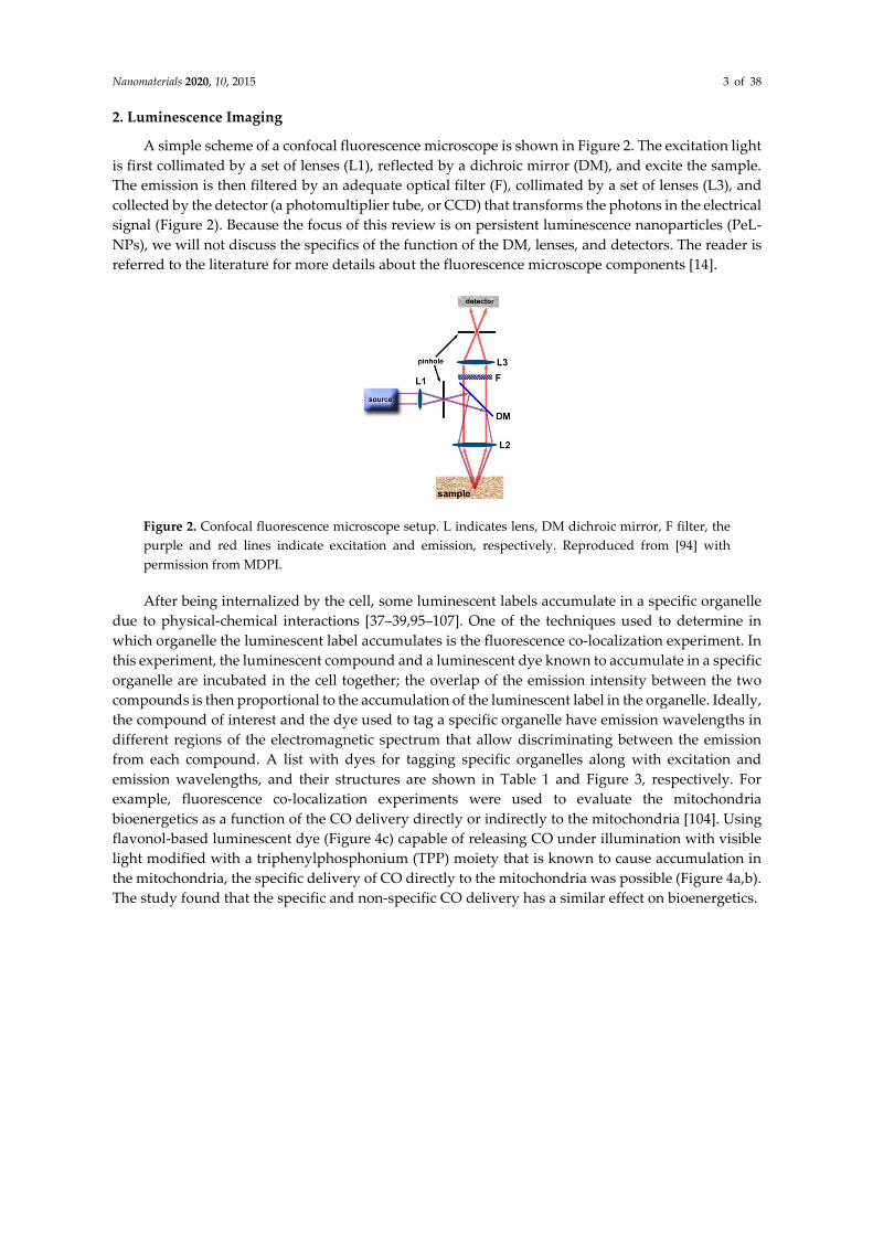
A simple scheme of a confocal fluorescence microscope is shown in Figure 2. The excitation light
is first collimated by a set of lenses (L1), reflected by a dichroic mirror (DM), and excite the sample.
The emission is then filtered by an adequate optical filter (F), collimated by a set of lenses (L3), and
collected by the detector (a photomultiplier tube, or CCD) that transforms the photons in the electrical
signal (Figure 2). Because the focus of this review is on persistent luminescence nanoparticles (PeL-
NPs), we will not discuss the specifics of the function of the DM, lenses, and detectors. The reader is
referred to the literature for more details about the fluorescence microscope components [14].
Figure 2. Confocal fluorescence microscope setup. L indicates lens, DM dichroic mirror, F filter, the
purple and red lines indicate excitation and emission, respectively. Reproduced from [94] with
permission from MDPI.
After being internalized by the cell, some luminescent labels accumulate in a specific organelle
due to physical-chemical interactions [37–39,95–107]. One of the techniques used to determine in
which organelle the luminescent label accumulates is the fluorescence co-localization experiment. In
this experiment, the luminescent compound and a luminescent dye known to accumulate in a specific
organelle are incubated in the cell together; the overlap of the emission intensity between the two
compounds is then proportional to the accumulation of the luminescent label in the organelle. Ideally,
the compound of interest and the dye used to tag a specific organelle have emission wavelengths in
different regions of the electromagnetic spectrum that allow discriminating between the emission
from each compound. A list with dyes for tagging specific organelles along with excitation and
emission wavelengths, and their structures are shown in Table 1 and Figure 3, respectively. For
example, fluorescence co-localization experiments were used to evaluate the mitochondria
bioenergetics as a function of the CO delivery directly or indirectly to the mitochondria [104]. Using
flavonol-based luminescent dye (Figure 4c) capable of releasing CO under illumination with visible
light modified with a triphenylphosphonium (TPP) moiety that is known to cause accumulation in
the mitochondria, the specific delivery of CO directly to the mitochondria was possible (Figure 4a,b).
The study found that the specific and non-specific CO delivery has a similar effect on bioenergetics.

Nanomaterials 2020, 10, 2015 4 of 38
Table 1. Commonly used dyes for fluorescence cell staining, organelle where the dye
accumulates, and excitation and emission wavelength peaks [108].
Dye Staining of λexc/nm λem/nm
Hoechst 33342 Nucleus 346 460
DAPI Nucleus 359 461
NBD C6-ceramide Golgi 466 536
DiO perchlorate Cell membrane and lipids 488 510
BODIPY FL Lipids 503 512
Rhodamine 123 Mitochondria 488 515
MitoTracker™ Green FM Mitochondria 490 516
LysoTracker™ Red DND-99 Lysosomes 577 590
λexc and λem are the excitation and emission wavelengths, respectively.
Figure 3. Structure of the most common dyes used for fluorescence cell staining.
(c)
Figure 4. Cellular luminescence imaging of A549 cells. (a) From left to right, red emission of
MitoTracker™ Red, green emission of the compound photoCORM-2, and overlay between the red
and green channels. (b) The emission intensity of the blue, green, and red emissions as a function of
the distance across the cell. (c) Structure of compound 2. The nucleus and mitochondria were stained
with Hoechst 33342 and MitoTracker™ Red, respectively. [Hoechst 33342] = [MitoTracker™ Red] =
300 nM, [2] = 25–100 μM. Reproduced from [104] with permission from the American Chemical
Society.
Another problem that arises, especially in the blue and green regions of the electromagnetic
where the emission intensity from cells and tissues is high, is a strong background emission that will
not allow the detection from the luminescent compound, especially when the compound has low

Nanomaterials 2020, 10, 2015 5 of 38
emission. Some solutions to avoid the interference from the cell or tissue emission are red shifting the
emission of the luminescent label to the red-NIR [13,109,110], use of two-photon absorption [111–
113], upconversion emission [114,115], or use of emission lifetime mapping. In this Review, we will
focus on the emission lifetime mapping measurement. The reader is redirected to the literature for a
detailed description of luminescent labels with emission in the red-NIR, two-photon absorption, and
upconversion materials [13,109–116].
The use of emission lifetime in cellular luminescence imaging is advantageous because it is
reproducible. The emission lifetime is a non-extensive and specific property of each compound,
allowing discrimination between the emission from the cell components and the luminescent label
[117,118]. Cell components and organic dyes usually show emission lifetimes in the nanoseconds
range, Table 2, which makes Fluorescence Lifetime Imaging Microscopy (FLIM) one of the most used
techniques [119–126]. Although FLIM is a technique that allows us to discriminate between the
emission lifetimes of the cell components and luminescent labels, there is not complete elimination
of the cell emission from the image. Longer emission lifetimes, in the range micro-millisecond, can
be achieved using transition metal complexes or lanthanide(III) compounds. These compounds show
unique spin forbidden and/or Laporte forbidden, in the case of the LnIII compounds, and are used in
Phosphorescence Lifetime Imaging Microscopy (PLIM) [117,127–132]. Emission lifetimes higher than
hundreds of nanoseconds allow complete elimination of the cell emission and yield a background-
free image. For example, the FLIM emission lifetime map of cockroach salivary ducts does not allow
to distinguish between cell components and the RuII complex (Figure 5a, left); the structure of the
complex is shown in Figure 5b) [117]. Due to the emission lifetime in the microsecond range, the RuII
complex the PLIM emission lifetime map can be obtained, providing a background-free image
(Figure 5a, right) [117].
Table 2. Excitation (λexc) and emission (λem) wavelengths peaks, and emission lifetimes (τ) for some of
the cell components and dyes used in cellular luminescence imaging [133–140].
Compound λexc/nm λem/nm τ/ns Reference
NAD(P)H free 340 470 0.3 [133]
Flavin mononucleotide 444 558 4.27–4.67 [134,135]
Collagen 280–350 370–440 ≤5.3 [133,136]
Riboflavin 420–500 520–750 4.12 [134]
Phenylalanine 258 280 7.5 [137]
Tyrosine 275 300 2.5 [138]
DAPI [a] 359 461 2.78 [139]
Rhodamine 123 [a] 488 515 3.97 [140]
[a]—in water.
(a)
(b)
Figure 5. (a) Emission lifetime map of cockroach salivary ducts stained with a RuII complex using
FLIM (left) or PLIM (right). (b) Structure of the RuII complex. Reproduced from [117].
At this point, the reader has been presented with the potentialities and challenges in the
luminescence imaging of biological systems. Although successful, luminescent organic dyes have

Nanomaterials 2020, 10, 2015 6 of 38
several downfalls for using in luminescence imaging of biological systems such as short emission
lifetime, small Stokes shift, and extensive photobleaching; all of those limitations leads to a not
complete elimination of the emission background, interference of the excitation source in the
imaging, and decrease of the emission intensity as a function of the time which does not allow for
experiments with an extended period of time, respectively. Materials with long emission lifetimes
such as lanthanide-doped nanoparticles, lanthanide complexes, and persistent luminescent materials
are an alternative to the organic dyes for obtaining high-quality luminescence imaging. In this review,
we will focus on persistent luminescent materials. The reader is directed to the literature for more
details about lanthanide-doped nanoparticles and lanthanide complexes applications in
luminescence imaging of biological systems [8,9,94].
3. Persistent Luminescence
Persistent luminescence (PeL) is a phenomenon where light is emitted for long periods of time,
from minutes to hours, after the excitation resulting in a glow-in-the-dark phenomenon. Matsuzawa
and co-workers were the first to report the SrAl2O4:Eu2+,Dy3+ green PeL emission that lasted >10 h,
after being charged by UV light [141]. Research in PeL has flourished since then, and several examples
based on doped/co-doped inorganic materials are found [49,91,92] with applications in emergency
signage, road signalization, luminous paintings, temperature and pressure sensing [91,142], and
cellular luminescence imaging [92], to cite a few.
3.1. PeL Mechanism
Despite the long emission duration shared characteristic, phosphorescence and PeL are entirely
different processes. While in phosphorescence, the long emission lifetime is caused by a spin-
forbidden transition, in PeL the long emission time is caused by the storage of energy in traps [93]
that are slowly promoted to the emitting levels. In these materials, the energy is stored by trapping
charge carriers (electrons and/or holes), and it is slowly released with the aid of thermal energy. Thus,
PeL is a particular case of thermostimulated luminescence [91] and is a defect dependent
phenomenon. Although simple, the PeL full mechanism took several years to be figured out. The
knowledge of trapping charge carriers (electrons and/or holes) in the defects for later thermal aid
release dates back from 1939 when Johnson proposed the electron storage process to explain the ZnS
PeL mechanism [143]. In 1945, Fonda observed that dopants and the crystalline phase influence the
duration and intensity of PeL [144]. More detailed mechanisms, based on quantitative positioning of
the energy levels and defects, appeared only in the 2000s with the works of Aitasalo and co-workers
[145], Clabau and co-workers [146], and Dorenbos [147]. Nowadays, the PeL mechanisms for
materials doped with Eu2+ or other similar emitters are very well established. This mechanism is
summarized in four steps; the first step, centered in the activator, involves the excitation of the
electrons (1), followed by trapping of the electrons into defects through the conducting band (CB)
(2a) or directly via tunneling (2b). The trapped electron is then thermally promoted (kT) to the
activator emitting levels via CB (3a), or via tunneling (3b), and finally decays radiatively, generating
the PeL (4) (Figure 6).

Nanomaterials 2020, 10, 2015 7 of 38
Figure 6. PeL simplified mechanism. VB is the valence band, CB conducting band, and kT is thermal
energy.
The mechanism described above is just a general one, and variations of the excitation and
trapping processes are known for different compositions. For example, in materials containing ions
like Eu2+, Tb3+, and Ti3+, excitation to the d metal orbitals is enough to allow electron trapping [148,149],
while in materials containing Eu3+ and Yb3+, only excitation to the charge transfer states allows the
energy storage [150,151]. In materials containing ions like Cr3+, Mn4+, and Sm3+ [152–154], the primary
excitation process that allows energy storage is the band gap excitation combined with energy
transfer processes. The different excitation processes can be related to the emitting centers’ redox
capacity since energy is stored by trapping electron or holes from the emitting center or the host. In
the case of Eu2+-doped materials, it was already proven by X-ray absorption or EPR spectroscopy that
in the charging process of persistent luminescence (process 1, Figure 6), Eu2+ is oxidized to Eu3+
[155,156].
The charge carriers trapping mechanism also changes for different compositions. Even if
thermoluminescence experiments are good to quantify the defect concentration and to estimate the
energy of the defects, there is no easy experiment to determine which charge carrier is participating
in the process. Based on the idea of the energy level positions, the proposed mechanisms suggest that
for most materials, like those doped with Ce3+, Eu2+, Tb3+, Cr3+, electron-trapping is the primary energy
storage process. However, for materials dependent on ligand-to-metal charge transfer excitation as
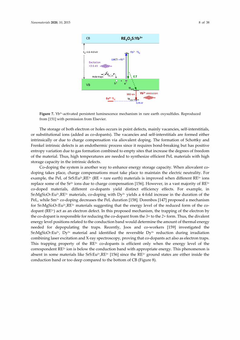
those doped with Eu3+ or Yb3+, hole trapping is the dominant energy storage process [150,151,157],
(Figure 7). The hole trapping mechanism is similar to the electron mechanism where the storage
happens under irradiation and the bleaching with thermal energy. However, the main differences
are the defect type (must be negative in order to store holes), its position (close to the valence band)
and finally, the excitation processes. The first excitation pathway is a band gap absorption followed
by the trapping of both electrons and holes, which may occur with several emitting centers [91]. The
second possible pathway is the charge-transfer excitation of a species followed by hole trapping
leading to a reversible photoreduction of the species [151]. In this case, a metastable reduced form of
the excited species is needed which is more probable when metals with low reduction potential are
present, for example, Eu3+ → Eu2+ and Yb3+ → Yb2+ pairs.
Nanomaterials 2020, 10, 2015 8 of 38
Figure 7. Yb3+-activated persistent luminescence mechanism in rare earth oxysulfides. Reproduced
from [151] with permission from Elsevier.
The storage of both electron or holes occurs in point defects, mainly vacancies, self-interstitials,
or substitutional ions (added as co-dopants). The vacancies and self-interstitials are formed either
intrinsically or due to charge compensation via aliovalent doping. The formation of Schottky and
Frenkel intrinsic defects is an endothermic process since it requires bond-breaking but has positive
entropy variation due to gas formation combined to empty sites that increase the degrees of freedom
of the material. Thus, high temperatures are needed to synthesize efficient PeL materials with high
storage capacity in the intrinsic defects.
Co-doping the system is another way to enhance energy storage capacity. When aliovalent co-
doping takes place, charge compensations must take place to maintain the electric neutrality. For
example, the PeL of SrS:Eu2+,RE3+ (RE = rare earth) materials is improved when different RE3+ ions
replace some of the Sr2+ ions due to charge compensation [156]. However, in a vast majority of RE3+
co-doped materials, different co-dopants yield distinct efficiency effects. For example, in
Sr2MgSi2O7:Eu2+,RE3+ materials, co-doping with Dy3+ yields a 4-fold increase in the duration of the
PeL, while Sm3+ co-doping decreases the PeL duration [158]. Dorenbos [147] proposed a mechanism
for Sr2MgSi2O7:Eu2+,RE3+ materials suggesting that the energy level of the reduced form of the co-
dopant (RE2+) act as an electron defect. In this proposed mechanism, the trapping of the electron by
the co-dopant is responsible for reducing the co-dopant from the 3+ to the 2+ form. Thus, the divalent
energy level positions related to the conduction band would determine the amount of thermal energy
needed for depopulating the traps. Recently, Joos and co-workers [159] investigated the
Sr2MgSi2O7:Eu2+, Dy3+ material and identified the reversible Dy3+ reduction during irradiation
combining laser excitation and X-ray spectroscopy, proving that co-dopants act also as electron traps.
This trapping property of the RE3+ co-dopants is efficient only when the energy level of the
correspondent RE2+ ion is below the conduction band with appropriate energy. This phenomenon is
absent in some materials like SrS:Eu2+,RE3+ [156] since the RE2+ ground states are either inside the
conduction band or too deep compared to the bottom of CB (Figure 8).

Nanomaterials 2020, 10, 2015 9 of 38
Figure 8. Host-referred 4f-electron binding energy curves and excited state energies of RE2+ and RE3+
ions in SrS. Reproduced from [156] with permission from The Royal Society of Chemistry.
Thus, efficient PeL materials design involves two parts, the presence of efficient activators and
the high concentration of charge carrier traps with proper depth. The blue-green PeL emitting
materials comprise the majority of the literature due to the low eye-sensitivity to longer wavelengths
when adapted to dark [160], and the lack of efficient red emitters (with allowed transitions) that
present efficient trapping [91]. Finally, there is the historical background, with most of the research
being done using Eu2+. Eu2+ is a traditional blue-green emitter, where red emission requires doping
in high crystalline field hosts or very covalent ones (due to the nephelauxetic effect). With a better
understanding of the PeL mechanism and increased demand for applications in luminescence
imaging of biological systems and solar energy harvesting, there is increased research on the design
of new red and NIR-emitting PeL materials [91], Table 3. For extensive details on all PeL materials
and different activators, the reader is advised other reviews [49,91,92,161]. In this review, we will
focus solely on a few examples of the most common activators.
Nanomaterials 2020, 10, 2015 10 of 38
Table 3. Examples of PeL materials containing different activators and their emission wavelengths
[47,49,150,151,153,154,162–184].
Activator Emission Wavelength References
Defects UV–NIR [154,162–166]
Eu2+ Blue–red [49,167,168]
Dy3+ Blue–red [169]
Gd3+ UV [170]
Eu3+ Red [150,167,171]
Tb3+ Green [171–173]
Sm3+ Red [154,174]
Er3+ Red–NIR [175,176]
Pr3+ Red–NIR [154,177,178]
Yb3+ NIR [151,179]
Cr3+ NIR [47,180]
Mn2+ Green, yellow or red [181]
Mn4+ NIR [153]
Bi3+ Blue or NIR [182,183]
Pb2+ UV [184]
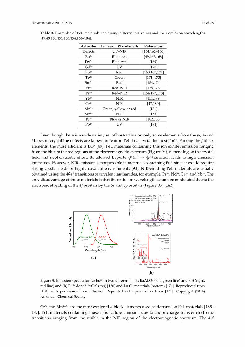
Even though there is a wide variety set of host-activator, only some elements from the p-, d- and
f-block or crystalline defects are known to feature PeL in a crystalline host [161]. Among the f-block
elements, the most efficient is Eu2+ [49]. PeL materials containing this ion exhibit emission ranging
from the blue to the red regions of the electromagnetic spectrum (Figure 9a), depending on the crystal
field and nephelauxetic effect. Its allowed Laporte 4f6 5d1 → 4f7 transition leads to high emission
intensities. However, NIR emission is not possible in materials containing Eu2+ since it would require
strong crystal fields or highly covalent environments [93]. NIR-emitting PeL materials are usually
obtained using the 4f-4f transitions of trivalent lanthanides, for example, Pr3+, Nd3+, Er3+, and Yb3+. The
only disadvantage of those materials is that the emission wavelength cannot be modulated due to the
electronic shielding of the 4f orbitals by the 5s and 5p orbitals (Figure 9b) [142].
(a)
(b)
Figure 9. Emission spectra for (a) Eu2+ in two different hosts BaAl2O4 (left, green line) and SrS (right,
red line) and (b) Eu3+ doped Y2O2S (top) [150] and Lu2O3 materials (bottom) [171]. Reproduced from
[150] with permission from Elsevier. Reprinted with permission from [171]. Copyright (2016)
American Chemical Society.
Cr3+ and Mn4+/2+ are the most explored d-block elements used as dopants on PeL materials [185–
187]. PeL materials containing those ions feature emission due to d-d or charge transfer electronic
transitions ranging from the visible to the NIR region of the electromagnetic spectrum. The d-d

Nanomaterials 2020, 10, 2015 11 of 38
electronic transitions are forbidden by the Laporte rule and dependent on the crystal field. Thus,
factors as coordination site symmetry, ligand field strength, and vibrionic coupling are essential for
relaxing the Laporte rule, resulting in increased emission rates. For example, the d-d Cr3+-centered
NIR emission in Cr3+-doped LaAlO3 perovskites and Cr3+-doped ZnGa2O4 spinels is due to the strong
crystalline field on the hosts mentioned above [180,188]. Bi3+ and Pb2+ are the most explored p-block
metals used as dopant in PeL materials due to their allowed metal-to-metal (MMCT), ligand-to-metal
(LMCT), or 6s2 → 6s1 6p1 electronic transitions [189].
3.2. Synthesis of PeL Nanomaterials
As aforementioned, a defined solid-state structure is a paramount factor in obtaining efficient
and long-lasting PeL materials. Due to the need of controlling and optimizing two different outputs,
the optical properties (i.e., high quality on excitation/emission spectra with a bright and long-lasting
afterglow emission), and the particle size control (i.e., narrow size distribution and controlled
morphology), synthesis of PeL materials are more challenging when compared to larger sized
nanoparticles. Factors such as optimization of (co-)dopants percentage on host, annealing
temperature range, heating exposure time, phase purity, amount of intrinsic defects are some of the
ones to be considered [190–194].
Even though there is a range of possibilities for PeL-NPs synthesis, up to now, there is not a
universal and flawless method for preparing PeL-NPs featuring intense light-emission, controlled
size distribution, and morphology of the NPs. The solid-state synthesis (ceramic synthesis) is the most
common method of obtaining a PeL material. The solids precursors are mixed and heated up to high
temperatures [195,196]. Bulk PeL materials based on aluminates [141,191,197–200], silicates [201–205],
and other compounds [47,49,206–208] have well-defined synthesis using this process. This method is
well-known, and the annealing step is necessary to yield crystal phase purity and enhance the
amount of defects in the structure. Alternatives synthesis, mostly wet-chemical methods, allows
better control of particle size and morphology; however, the low temperatures and shorter reducing
times yield materials with shorter PeL emission lifetime and/or a weak emission. Other preparation
methods such as combustion synthesis, sol–gel, co-precipitation, and hydrothermal are also widely
used for synthesizing PeL-NPs. Each of those methods has its particularities, and the ideal synthetic
parameters, temperature, heating rate, pressure, and concentration, involve extensive bench time
work and are dependent on each specific material.
3.2.1. Combustion Synthesis
Combustion synthesis (CS), or self-propagating heating synthesis, is a low energy consuming
method used to synthesize oxide ceramics that takes advantage of extremely exothermic reactions
between metal nitrates and organic fuels (typically urea, carbohydrazide, or glycine) [209–215]. In a
typical reaction, the synthesis occurs in a pre-heated muffle furnace, where the mixture of the nitrates
and the organic fuel is inserted. As the synthesis initiates, the fuel ignites, rupturing into flames, and
combustion takes place. The energy produced quickly heats the system (the temperature reaches
values > 1000 °C) and sustains the temperature for a period of over 60 s, which is long enough to
grow and crystallize the NP [209–215]. The final product is a fluffy, foamy powder with a large
surface area (Figure 10a). The advantages of the CS method are its short reaction time, and the heating
process tends to decrease undesired absorption of hydroxyl groups on the particle surface, which can
act as a luminescent quencher depending on the PeL phenomenon. Another advantage is the
extremely high temperatures achieved in short periods that reflect in increased concentration of
defects, improving the energy storage capability of the material as shown by Rodrigues and co-
workers for the blue-emitting material BaAl2O4:Eu2+,Dy3+ [191] (Figure 10b) and Qiu and co-workers
for the MAl2O4:Eu2+,Dy3+ (M = Sr2+, Ba2+ or Ca2+) material [215] (Figure 10c). On the downside, the
disadvantages of the CS method are the lack of reproducibility and difficulty in controlling the
process due to the unpredictable combustion step resulting in a broad range of NP sizes.

Nanomaterials 2020, 10, 2015 12 of 38
Figure 10. (a) Schematic flowchart of combustion synthesis (CS), and (b) SEM image of BaAl2O4:Eu2+,
Dy3+ prepared using the CS method. Reproduced from [191] with permission from Elsevier. (c) Picture
of the PeL emission of MAl2O4:Eu2+,RE3+ (M: (i) Ca2+, (ii) Sr2+ or (iii) Ba2+) prepared using the CS
method. Reproduced from [215] with permission from Elsevier.
3.2.2. Sol–Gel Synthesis
Sol–gel synthesis (SGS) is a wet chemical technique widely used to prepare inorganic polymers
and ceramics [216], including PeL materials. The sol–gel process is carried through a liquid solution,
that eventually transforms into a sol, and finally into a viscous colloidal gel state. The main steps in
SGS are the hydrolysis and/or condensation of molecular precursors (gelation agent), the formation
of a sol–gel aqueous solution, suspension and drying of the solids, and annealing (Figure 11a) [216–
222] Through the SGS technique is possible to produce a solid material from a homogenous solution.
The SGS allows precise and flexible control when using precise synthesis conditions (reaction time,
pH, temperature, the concentration of the precursors and surfactants, stirring, for example).
Furthermore, SGS offers a precursor-homogeneity and a useful method for controlling the particle
morphology and size. SGS is a widely used method for synthesizing aluminates and silicates based
PeL-NP. For example, SrAl2O4:Eu2+,Dy3+ (SAO:ED) NPs by sol–gel synthesis using a mixture of the
nitrate/acetate metals and citric acid as chelating agent [217]. The obtained SAO:ED NPs showed size
in the 20 nm range, with a lasting afterglow centered at 520 nm. Sr2MgSi2O7:Eu3+,Dy3+ PeL-NP with
an average size of 250 nm, were obtained using the SGS [220]. The advantage of the SGS for
synthesizing this material is the use of tetraethyl orthosilicate (TEOS) as a silicon source. TEOS
quickly goes through hydrolysis, which results in a viscous colloidal solution, reaching the required
gel-state and being a physical limitation for particle growth. A similar route using citric acid was
reported for synthesizing Zn2.94Ga1.96Ge2O10:Cr3+,Pr3+ NIR emitting PeL-NPs with persistent
luminescence that lasted for over 360 h (Figure 11e), and size in the range 30–60 nm (Figure 11b,c)
with good dispersibility in water (Figure 11d) allowing in vivo application (Figure 11f) [219].
Figure 11. (a) Schematic flowchart of SGS. (b) TEM, (c) high-resolution TEM, (d) excitation (blue
curve, left) emission at 700 nm) and emission (red curve, right) excitation at 254 nm) spectra of the
aqueous dispersion of the material, (e) afterglow emission collected at different times after turning
off UV excitation, and (f) in vivo NIR afterglow imaging. Material: Zn2.94Ga1.96Ge2O10:Cr3+,Pr3+, λexc =
254 nm and λem = 700 nm. Reprinted with permission from [219]. Copyright (2013) American Chemical
Society.

Nanomaterials 2020, 10, 2015 13 of 38
3.2.3. Co-precipitation Synthesis
Co-precipitation synthesis (CPS) is based on the control of particle growth based on the
solubility product constant of the precursors. This method relies on the solubility compatibility of
starting materials, relatively low reaction temperature, and shorter synthesis time. This is a simple
method where a saturated solution of soluble metals (most common are nitrates or acetates) is
precipitated by the addition of a precipitant agent (e.g., urea, sodium silicate, sodium bicarbonate,
for example) (Figure 12a). In general, the conditions that affect the CPS are the concentration of metals
solution, the concentration of precipitating agent solution, the slow controlled mixture between both
solutions, temperature when precipitating the solid and of the annealing process, and presence of
complexing agents like EDTA (which affects the kinetics) [223–228]. Using the CPS method, Wang
and co-workers synthesized water-dispersible nanocrystalline CaS:Eu2+,Sm3+,Mn2+ with 20–40 nm size
range (Figure 12b–d), efficient PeL that also showed up-conversion properties (Figure 12e) [227].
Figure 12. (a) Schematic flowchart of CPC. TEM images and (inset) histograms of the particle size
distribution of (b) CaS:Eu2+, Sm3+, Mn2+ and (c) functionalized CaS:Eu2+, Sm3+, Mn2+. (d) HRTEM of
CaS:Eu2+, Sm3+, Mn2+. The inset shows the SAED pattern. (e) Excitation (1 – green line, left), PeL
emission (2 – purple line, right), and up-conversion emission (3 – red line, right) spectra. The inset
shows photographs of CaS:Eu2+, Sm3+, Mn2+ under UV (left) and NIR excitation (right). λem = 610 nm,
λexc = 355 nm (PeL) or λexc = 980 nm (UC). Reproduced from [227] with permission from The Royal
Society of Chemistry.
3.2.4. Hydrothermal Synthesis
Hydrothermal Synthesis (HS) refers to a wet chemical technique were the precursors are sealed
and heated into reaction vessels (autoclaves). HS is carried out at high pressures, provided by the
autoclave reactor, where the synthesis between precursors is promoted. A typical NP synthesis using
the HS method occurs within a two-phase reaction medium, composed of two immiscible solutions,
an aqueous solution containing the metal precursors and an organic solvent (e.g., toluene) containing
a complexing or surfactant agent, like oleic acid, EDTA, or cetyltrimethylammonium bromide
(CTAB) for achieving control over the nanocrystalline size and morphologies. As the system heats up
and the pressure builds up, the solutions are perturbed, and the precipitation occurs at the liquid-
liquid surface. After that, the system is cooled down, and the precipitant is centrifuged. The solid is
then exposed to a high-temperature annealing treatment. This approach enables the synthesis of
highly crystalline nanomaterials under relatively mild conditions (Figure 13a). Concentration, pH,
annealing temperature, pressure, and reaction time are all factors that affect the HS [229–231]. For
example, synthesis of ZnGa2O4:Cr3+ using the HS led to monodisperse PeL-NP with size in the sub-
10 nm range (Figure 13b), and afterglow NIR emission (ca. 696 nm) (Figure 13c) longer than 40 min
[230]. Some examples of PeL materials and NP size, synthesis method, emission wavelength, and
afterglow duration are shown in Table 4.

Nanomaterials 2020, 10, 2015 14 of 38
Figure 13. (a) Schematic flowchart of HS. (b) TEM image of ZnGa2O4:Cr3+ dispersed in hexane
prepared via HS. (c) Excitation (black curve, left) and emission (red curve, right) spectra of the
ZnGa2O4:Cr3+ dispersed in hexane. The inset shows the photograph of the PeL emission of the NP
under 254 nm excitation (P = 6 W). λem = 696 nm, λexc = 254 nm. Reproduced from [230] with permission
from The Royal Society of Chemistry.
Table 4. Examples of PeL compounds, average size, synthesis method, emission wavelength (λexc),
and afterglow duration.
Compound Average
Size/nm
Synthesis
Method λem/nm Afterglow Reference
CaAl2O4: Eu2+, Nd3+ 70–80 co-precipitation 436 >360 s [228]
50 template 445 >2000 s [232]
CaAl2O4: Eu2+, La3+ 44 combustion 440 >800 s [213]
Sr2MgSi2O7:Eu2+,Dy3+ 20 combustion 457 >1800 s [233]
270 sol–gel 480 >1800 s [220]
BaAl2O4: Eu2+,Dy3+ 85–94 combustion 505 >20000 s [220]
CaS:Ce3+ 42 co-precipitation 507 >200 ms [225]
SrAl2O4:Eu2+,Dy3+,Tb3+ 50–80 combustion 513 >2700 s [213]
SrAl2O4: Eu2+,Dy3+
30 combustion 516 >1800 s [215]
20 sol–gel 520 >200 s [217]
50 co-precipitation 513 >2.5 h [224]
300 solvothermal 512 >100 s [234]
300 electrospinning 509 >200 s [235]
Zn2SiO4:Mn2+ 200 sol–gel 520 >20 ms [221]
BiPO4:Tb3+ 80–200 electrospinning 545 >15 ms [236]
BiPO4:Ce3+ 80–200 electrospinning 545 >15 ms [236]
CaMgSi2O6:Mn2+ 60–70 sol–gel 585 >1200 s [222]
SnO2:Eu2+ 50–100 solvothermal 588 >1000 s [237]
Ca2Si5N8:Eu2+,Tm3+ 5 laser ablation 610 >2000 s [238]
CaS:Eu2+,Sm3+,Mn2+ 30 co-precipitation 613 >30 min [227]
Y2O2S:Eu3+, Mg2+,Ti4+ 80–150 hydrothermal 627 >1000 s [229]
Y2O2S:Eu3+,Ca2+, Ti4+ 80–150 hydrothermal 627 >1000 s [229]
Y2O2S:Eu3+,Sr2+, Ti4+ 80–150 hydrothermal 627 >1000 s [229]
Y2O2S:Eu3+,Ba2+, Ti4+ 80–150 hydrothermal 627 >1000 s [229]
CaMgSi2O6:Eu2,
Pr3+,Mn2+ 100 template 660 >1 h [239]
ZnGa2O4:Cr3+ 8 hydrothermal
solvothermal
696
695
>3000 s
>120 min
[230]
[240]
Zn3Ga3Ge2O10:Cr3+,Pr3+ 30–60 Sol–gel 695 >360 h [219]
The background color on the λem column represents the emission color of the PeLNPs.

Nanomaterials 2020, 10, 2015 15 of 38
In addition to the aforementioned methods, other methodologies like the template method
[232,239,241], solvothermal method [228,234,237,240], electrospinning method [235,236], and laser
ablation/deposition techniques [238] are capable of producing PeL-NP. Nevertheless, there is still a
need for developing more controlled methodologies for preparing PeL.
4. Persistent Luminescence in Luminescence Imaging of Biological Systems
Due to its afterglow, PeL materials are desirable for luminescence imaging of biological systems
due to the possibility of obtaining high-quality images with non-interference from the background
[42,43,45,46,48,50–54,64,242,243]. When using PeL in luminescence imaging, two main approaches
are taken into account, materials with ultra-long persistent luminescence irradiated (or charged)
outside the organism or materials irradiated inside the organism that are reactivated with X-ray or
NIR radiation. Finally, detecting the persistent luminescence out of the biological system requires
emission in the red and NIR-emitting regions of the electromagnetic spectrum due to the low
absorption by tissues and cells in this region [66]. In this review, we will present the recent literature
on PeL used in cellular imaging, separating the materials as a function of the excitation source used
to produce the PeL phenomenon.
4.1. Excitation in the UV
UV radiation is the most common excitation source for PeL nanomaterials since most lattice, and
defects activators rely on high energy band gap and charge transfer transitions. Due to UV light’s
low penetrability in tissues and cells, UV activated PeL materials have to be activated before
incubation. Thus, exceptionally long afterglow is required from those materials, as the excitation is
hampered after in vivo injection. To optimize UV-excited PeL materials application in luminescence
imaging of biological systems, emission in the NIR is a must due to the low absorption of cells and
tissues in this region that leads to improved signal-to-noise ratio. Gallates and germanates doped
with Cr3+, a NIR activator, are frequently used in PeL imaging studies due to their optimal crystalline
field [47] and defect structure [244].
Maldiney and co-workers pioneered the use of NIR emitting PeL-NP in luminescence imaging
of biological systems [48]. Using the PEG-functionalized ZnGa2O4:Cr3+ spinel PeL-NP the authors
were able to obtain NIR-luminescence imaging of vascularization, tumors, and grafted cells, using
UV excitation for 2 min at 254 nm before injection with decent accumulation in the tumor [48]. In
follow-up work, the same research group improved the biocompatibility of the PeL-NPs by using
hydroxyapatite/β-tricalcium phosphate (HAp/β-TCP) doped with Eu2+/Eu3+, Mn2+, and Dy3+, which
exhibit efficient persistent luminescence for in vivo imaging after irradiation using UV excitation for
2 min at 254 nm (Figure 14) [245].
Figure 14. (a) In vivo imaging obtained at 5 and 10 min after the injection of the PeL-NPs. (b) Emission
intensity as a function of the time monitoring the whole body and liver during the first 10 min of
experiment. Pel-NPs: HAp/β-TCP doped Eu2+/Eu3+, Mn2+ and Dy3+. [PeL-NPs] = 0.8 mg/200 �L glucose.
Reproduced from [245] with permission from Elsevier.

Nanomaterials 2020, 10, 2015 16 of 38
Using the same material, ZnGa2O4:Cr3+, Zhou and co-workers expanded the applications of PeL
in luminescence imaging and demonstrated the application of biotinylated ZnGa2O4:Cr3+ PeL-NPs as
a background-free luminescent nano-bio probe for sensitive and specific detection of avidin in a
heterogeneous assay with a limit of detection of ~150 pM [240]. In the same year, Wang and co-
workers demonstrated that functionalization of ZnGa2O4:Cr3+ NPs with hyaluronic acid (HA) and
Gd2O3 yielded a multi-modal probe where high MRI contrast and high-quality NIR-PeL imaging
were obtained for in vivo systems using UV excitation, at 254 nm before injection [246].
Besides the exciting PeL possibilities in luminescence imaging of biological systems,
biocompatibility is still a challenge due to its low water solubility and low cell uptake. One of the
most used strategies to remediate those limitations is surface functionalization with PEG, liposomes,
or folic acid groups, which render improved water compatibility and cell uptake, respectively
[247,248]. Another strategy is the functionalization with water-soluble polymers or dendrimers [249].
For example, Zhang and co-workers used the polyamideamine (PAMAM) dendrimer grafted on
Zn1.25Ga1.5Ge0.25O4:0.5% Cr3+, 2.5% Yb3+, 0.25% Er3+ PeL-NPs surface for improved water solubility
[249]. The dendrimer not only improves the water solubility but also allows multiple points for
functionalization with other compounds. The PeL property was activated before the injection using
UV light at 254 nm for 10 min, and the system was successfully used for in vivo imaging [249]. The
use of the PAMAM allowed functionalization with Doxorubicin (DOX) via pH-sensitive hydrazine
bonds resulting in the release under acidic conditions, characteristic of cancer cells but not healthy
ones, resulting in decreased cell viability of HeLa cells and inhibition growth of tumors [249].
Although UV excitation of PeL-NPs before injection in biological systems has opened new
avenues and demonstrated the potential of these materials for application in luminescence imaging
of biological systems, it is not possible to activate these materials in vivo. That limits the applications
to PeL materials that have a long afterglow.
4.2. Excitation in Visible
The success of UV-charged PeL-NPs in luminescence imaging of biological systems stimulated
the development of PeL materials that could be activated in vivo or in vitro. Visible excitation in the
far-red region of the electromagnetic spectrum has high penetrability due to the low scattering by
cells and tissues. Thus, it is an alternative for expanding the use of PeL materials in luminescence
imaging of biological systems.
As described in Section 4.1 (vide supra), Maldiney and co-workers pioneered the use of NIR
emitting PeL NPs in luminescence imaging of biological systems using the system ZnGa2O4:Cr3+ [48].
This material can also be activated using an orange-red LED source [48,206]. The mechanism that
allows activation using an orange-red LED source was studied in detail by Bèssiere and co-workers
and is related to antisite defects in the first neighborhood of a Cr3+ ion and differs from the usual PeL
one (Figure 15) [244]. These defects are related to a swap between Zn2+ and Ga3+ sites in the crystal
structure where Zn2+ substitutes a nearby Ga3+ in the spinel’s octahedral site, and Ga3+ replaces Zn2+
in the spinel’s tetrahedral site. This exchange causes a local charge imbalance where the octahedral
and tetrahedral sites have negative and positive charges, respectively. The excitation of Cr3+ with
visible light (4A2 (t2g)3 → 4T2 (t2g)2(eg)1 transition) leaves a hole and an electron in the t2g and eg orbitals,
respectively forming an electron-hole pair. The nearby antisite defect pair drives the relaxation of Cr3+
back to the 4A2 ground state, storing the energy and rebalancing the charges of the defect. As a
consequence, the tetrahedral and octahedral sites become neutral. This process is reversed through
thermal energy, with Cr3+ going back to the 4T2 excited state and then relaxing to the 2E emitting state,
responsible for the persistent emission in ca. 700 nm.

Nanomaterials 2020, 10, 2015 17 of 38
Figure 15. Proposed mechanism of PeL in ZGO:Cr induced by excitation below 3.1 eV. CrN2 is
represented by its states (4T2, 4A2 or 2E). Blue and yellow spheres represent the two opposite charge
antisite defects. Steps: (a) optical excitation to the Cr3+ 4T2 excited level; (b) relaxation to the the Cr3+ 4A2 ground level, charge migration, and carriers trapping by neighboring antisite defects of opposite
charges; (c) thermal release of e--h+ pairs and trapping by Cr3+; (d) the Cr3+ 2E → 4A2 in the NIR.
Reprinted with permission from [244]. Copyright (2013) American Chemical Society.
The possibility of using visible-light for charging PeL materials opened-up new avenues and
expanded the number of PeL materials that could be used in luminescence imaging. For example, Shi
and co-workers used the HS method and ethylenediamine as a solvent to obtain ZnGa2O4:Cr3+,Eu3+
PeL-NPs with -NH2 groups at the surface that were subsequently used to decorate the NP surface
with either transacting activator of transduction peptide (TAT), or folic acid (FA). The first group,
TAT-decorated, was successfully uptaken by HepG2 (liver cancer) and H22 (hepatocellular
carcinoma) cells and was found to accumulate at the nuclei, while the FA-decorated NPs were
successfully used to selectively target tumoral cells both in vitro (HepG2 cell line) and in vivo (H22
tumor-bearing mouse). Even in vivo, these PeL-NPs could be re-activated using a 650 nm or 808 nm
LED, being excitation at 650 nm more effective [250]. In follow-up work, the same research group
used 5 nm NPs with the same composition to target MCF7 cells [251]. FA-functionalization is a
commonly used strategy for targeting cancer cells due to the overexpression of the folate receptor in
cancerous cells. Li, Yan, and co-workers showed that FA-functionalization of Zn1.25Ga1.5Ge0.25O4: Cr3+,
Yb3+, Er3+ PeL-NP were successfully used in luminescence imaging using a red LED source for in vivo
excitation [252].
Long term toxicity is still an issue for in vivo applications of NP systems [248]. Sun and co-
workers studied in detail the long-term toxicity of PEG-functionalized Zn1.1Ga1.8Sn0.1O4: Cr3+ PeL-NP.
The advantage of using PeL in those studies is that it allows tracking in real-time using luminescence
imaging without the constant need of a steady excitation source, allowing a detailed study of the
pathway inside the body. The PeL-NPs were monitored for 60 days after injection, with regular
tracking of the particles’ positions inside the body using the red excitation to recharge persistent
luminescence. The NPs were found to accumulate in the reticuloendothelial system (RES),
particularly lungs, liver spleen, and excretion through the digestive system. Histological, blood
biochemistry and hematological analyses found no difference between the treated and non-treated
mice [253].
Although the development of PeL-NPs with excitation in the visible was an improvement
compared to UV-excited ones, the useful excitation wavelengths for in vivo applications are limited
to the red and far-red wavelengths.
4.3. Excitation in the NIR
NIR excitation has attracted much attention due to its deeper penetration in the biological tissues
[55–72]. Usually, the up-conversion (UC) phenomenon, followed by energy transfer, is used to induce
persistent luminescence using NIR radiation [254]. In this case, it is challenging because it requires
efficient UC emission and efficient energy transfer. Stimulated emission, using NIR excitation, is an

Nanomaterials 2020, 10, 2015 18 of 38
alternative way to achieve PeL. In this process, NIR photons are used to bleach the populated traps
(usually after UV irradiation).
The use of NIR light as an excitation source to induce PeL was first demonstrated by Liu and co-
workers using Zn3Ga2GeO8 doped with Cr3+ and the UC pair Yb3+/Er3+ [255]. In this system, infrared
excitation (980 nm) is used to populate excited states of Er3+. Through an internal energy transfer, the
energy is transferred from Er3+ to Cr3+, and stored in defects in Cr3+ vicinities. Finally, with thermal
energy aid, the Cr3+ excited levels are populated, and the energy is released over a long period
through the Cr3+ characteristic emission. This phenomenon, named up-converted persistent
luminescence (UPCL), was also used as a strategy in PeL luminescence imaging [256,257]. Xue and
co-workers used the UPCL for demonstrating that PEG-functionalized Zn3Ga2GeO8:Cr,Yb,Er PeL-
NPs could be readily recharged in vivo using excitation at 980 nm (150 mW × cm−2 for 120 s) with no
efficiency loss after several cycles [256]. Conventional UC luminescence imaging was also possible
using this system, allowing the development of synergistic probes taking advantage of both
processes, UCPL and UC [256]. A multi-layered approach, composed of a self-assembled composite
made of both PeL-NPs (Zn1.1Ge1.8Ge0.1O4:0.5% Cr3+) and UCNPs (β-NaYbF4:0.5%Tm3+@NaYF4) was
proposed by Qiu and co-workers to ensure the efficiency of the UC, energy transfer, and PeL
processes (Figure 16) [257]. Under excitation at 980 nm, the Tm3+ excited electronic levels are
populated via an up-conversion energy transfer mechanism, followed by energy transfer to the PeL-
NP, and finally, PeL at 700 nm. This hybrid material was used for tracking lymph nodes in mice [257].
Figure 16. Energy diagram comparing the traditional UV charged PeL (left) and NIR-light-charged
UCPL (right) mechanisms. Reprinted with permission from [257]. Copyright (2017) American
Chemical Society.
Photostimulated emission is another way to obtain PeL using NIR excitation. In this process, the
first step is the same as the conventional PeL phenomenon. The difference is that, instead of using
thermal energy to bleach the traps, the system uses light energy to promote the charge carriers from
the traps to the emitting center, generating the luminescence. For example, Gao and co-workers used
the photostimulated luminescence of DSPE-PEG-biotin coated CaS:Eu2+,Sm3+ NPs for in vitro cellular
luminescence imaging of HeLa cells. PeL is obtained using a white LED to excite the material,
resulting in emission at ~650 nm. Excitation with NIR light is then used to produce photostimulated
luminescence in this material after the original excitation, increasing the number of photons released
while the light source is on [258].
4.4. Excitation in the X-ray
X-ray excitation has recently been proposed in the luminescence imaging of biological systems.
Although there is still a small number of articles reporting X-ray induced PeL, these materials are
promising for luminescence imaging [259–262]. The high penetrability of X-rays in cells and tissues
allows, virtually, imaging of any part of the body, making this radiation attractive for in vivo
applications. The high penetrability of the X-rays also allows recharging the PeL after hours, days, or
even weeks after the PeL material injection avoiding the dependence on afterglow duration. The use

Nanomaterials 2020, 10, 2015 19 of 38
of X-rays also opens up new avenues for combined luminescence imaging combined with X-ray
absorption imaging [263].
Xue and co-workers demonstrated X-rays’ high penetrability using the ZnGa2O4:Cr3+ PeL-NPs
and comparing the luminescence imaging using UV for charging the NPs before injection or in vivo
activation of the PeL using X-rays (Figure 17a) [262]. The use of X-rays not only allowed luminescence
imaging of deeper tissues, when compared to UV (Figure 17b), but also allows recharging the PeL in
vivo [262]. Strategies used to improve X-ray activated PeL materials usually involve doping or co-
doping with heavy atoms such as Tb3+ and Sm3+ [263,264]. Zheng and co-workers recently
demonstrated that X-ray activated MgGeO3:Mn2+,Yb3+,Li+ PeL-NPs have long afterglow and can emit
in the first and second biological windows for long-term luminescence imaging [265].
Figure 17. (a) Schematic diagram of in vivo PeL X-ray rechargeable luminescence imaging. (b)
Phantom imaging as a function of time or pork tissue thickness (0, 1, 3, 5, 10, and 20 mm) using the
PeL-NP ZnGa2O4:Cr3+. X-ray in vivo excitation for 5 min, at 45 kVp, or UV excitation prior to
incubation for 20 min, at 365 nm. Reprinted with permission from [262]. Copyright (2017) American
Chemical Society.
4.5. Photodynamic Therapy Using Persistent Luminescence
PDT is a non-invasive therapy based on the generation of 1O2 and reactive oxygen species (ROS).
The latter, generated through the interaction of the triplet level of a dye with ground state oxygen
(3O2) (Figure 18), is used to damage cancerous cells [79,87,266–274]. Cells and organisms are less likely
to develop resistance to 1O2, and it can therefore, be used successfully to treat cancer [79]. Organic
dyes such as porphyrins, chlorins, phthalocyanines, and xanthenes are often used in PDT [87,88].
However, this class of compounds is prone to photobleaching, have low light-dark cytotoxicity ratios,
and is also known to form aggregates that decrease the singlet oxygen generation efficiency as a
function of the elapsed time, and thus decreases the efficiency of the treatment [89]. Additionally, the
need for continuous in situ illumination causes damage to the skin and tissues.
Figure 18. Energy level diagram illustrating the formation of 1O2. A denotes absorption, ISC
intersystem crossing, S states with singlet and T states with triplet multiplicity.
The characteristic afterglow emission of PeL-NPs can be used as an internal light source in PDT
that would eliminate the need for continuous in situ illumination, avoiding skin and tissue damage,
and allowing the use of PDT in deep tissues. Curiously, the use of PeL in PDT is recent, and the first

Nanomaterials 2020, 10, 2015 20 of 38
examples were reported back in 2016 [275,276]. In those pioneer works, the proof-of-concept that PeL
could potentially be used in PDT was reported using ZnGa2O4:1% CrIII, 2% PrIII as the PeL-NP, and
the chemically bonded photosensitizer (PS) distyryl-BODIPY [275]. As noted by Akkaya and co-
workers, only a modest photocytotoxicity against HepG2 cells was observed due to the short PeL
emission lifetime in biological media. Re-charging the PeL is a strategy to repopulate the excited
states of the PeL-NP and restore the PeL [276–281]. Solubilizing in water and targeting the PeL-NPs
into cancer cells adds another challenge for in vivo PDT. Yan and co-workers proposed to study the
effect of a cancer cell membrane (CCM) shell in the tumor accumulation using the system
Zn1.25Ga1.5Ge0.25O4:0.5% CrIII, 2.5% YbIII, 0.25% ErIII as PeL-NP protected by a hollow SiO2 layer and
loaded with DOX [280]. The CCM inhibits premature leakage and also yields targeting capability for
metastases. As expected, the CCM shell’s presence yielded higher internalization than the system
without it [280]. Due to the high absorption of cells and tissues, the wavelength used to re-charge the
PeL-NP is within the biological window. Scherman, Richard, and co-workers reported that the PeL
of ZnGa2O4:CrIII can be restored using 808 nm excitation due to the UC excitation of the CrIII [48]. Yan
and co-workers incubated the system ZnGa2O4:CrIII – Si-Pc in HepG2 cells for 8 h, and re-charged the
PeL using 808 nm excitation pumps for 0, 3, 5, or 10 min that resulted in cell viability of almost 0 %
(concentration = 200 μg × mL−1) proving the potentialities of using PeL-NPs in efficient PDT [276].
Although NIR radiation has a deeper penetration than to UV or visible wavelengths [61,63], it still
cannot penetrate deeper tissues. X-ray radiation has unlimited penetrability, making this kind of
radiation attractive deep tissue treatment using X-ray activated PDT (XPDT) [260,261]. Low dose X-
ray radiation has been successfully used in PeL XPDT [282,283]. Yang, Li, and co-workers reported
the photocytotoxic activity of ZnGa2O4:0.5% CrIII, 0.5%WVI – ZnPcS4 in vitro against HeLa cells
(Figure 19) and in vivo [282]. In this case, doping with WVI enhances the X-ray cross-section
absorption, and continuous 1O2 generation is observed over at least 40 min using X-ray radiation (0.09
Gy × min−1) [282]. The use of X-ray radiation increased the cytotoxicity compared to excitation at 670
nm (Figure 19a).
Figure 19. (a) HeLa cell viability without light excitation (blue bar) and after 2 min of irradiation (red
bar). Luminescence imaging of HeLa cells treated with (b) PBS, (c) PBS + X-ray, (d) 150 μg mL−1
ZnGa2O4:0.5% CrIII, 0.5%WVI + X-ray, (e) 5 μg mL−1 ZnPcS4 + X-ray, (f) 5 μg mL−1 ZnPcS4 + LED, and
(g) 5 μg mL−1 ZnGa2O4:0.5% CrIII, 0.5%WVI–ZnPcS4 + X-ray. LED (λexc = 670 nm, P = 160 mW cm−2). (h)
HeLa cell viability without (pink bar) and after 2 min of X-ray irradiation (dark blue bar). The cells
were treated with 150 μg mL−1 ZnGa2O4:0.5% CrIII, 0.5%WVI + X-ray, 5 μg mL−1 ZnPcS4 + X-ray, 5 μg
mL−1 ZnPcS4 + LED, and 5 μg mL−1 ZnGa2O4:0.5% CrIII, 0.5%WVI – ZnPcS4 + X-ray. In the luminescence
images, Calcein AM (green fluorescence) and propidium iodide (red fluorescence) indicates the living
and dead cells, respectively. Reproduced from [282] with permission from Wiley.
Nanomaterials 2020, 10, 2015 21 of 38
As highlighted above, long-lasting PeL is one of the most critical requirements for using PeL-
NPs in PDT. One of the challenges is to develop less chemically aggressive synthetic routes that
damage the PeL-NPs surface, causing a decrease in the PeL emission lifetime. An additional challenge
for application in biological systems is the extensive emission quenching caused by the solvent.
Synthetic methodologies to achieve hydrogels, hollow silica interlayers or hollow cavities with
controllable size aim to achieve long-lasting PeL and improve cell biocompatibility [278–281]. For
example, tumor-injectable oleosol implants are obtained by dissolving the PeL-NPs in a mixture of
poly(lactic-co-glycolic acid)/N-methylpyrrolidone [279]. The injected oleosol quickly turns into a solid
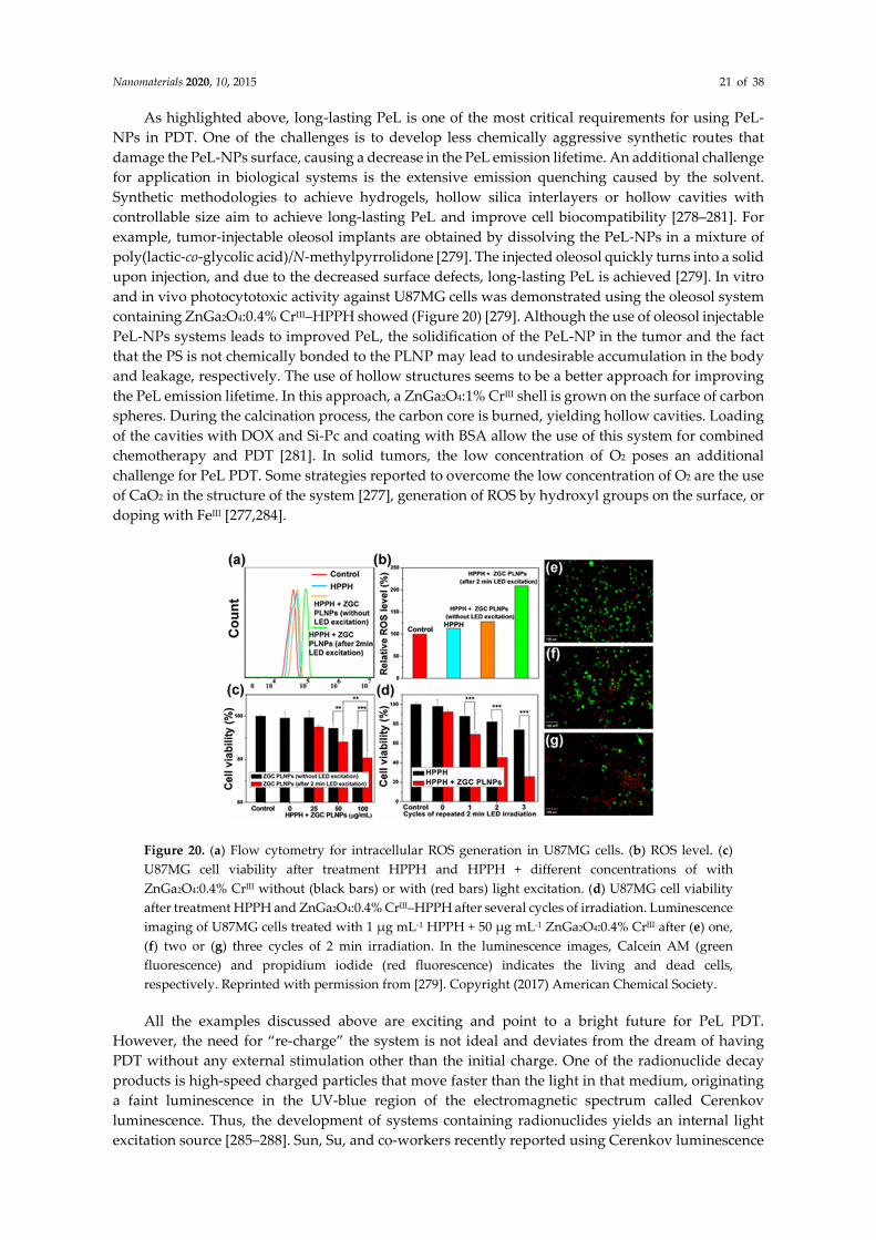
upon injection, and due to the decreased surface defects, long-lasting PeL is achieved [279]. In vitro
and in vivo photocytotoxic activity against U87MG cells was demonstrated using the oleosol system
containing ZnGa2O4:0.4% CrIII–HPPH showed (Figure 20) [279]. Although the use of oleosol injectable
PeL-NPs systems leads to improved PeL, the solidification of the PeL-NP in the tumor and the fact
that the PS is not chemically bonded to the PLNP may lead to undesirable accumulation in the body
and leakage, respectively. The use of hollow structures seems to be a better approach for improving
the PeL emission lifetime. In this approach, a ZnGa2O4:1% CrIII shell is grown on the surface of carbon
spheres. During the calcination process, the carbon core is burned, yielding hollow cavities. Loading
of the cavities with DOX and Si-Pc and coating with BSA allow the use of this system for combined
chemotherapy and PDT [281]. In solid tumors, the low concentration of O2 poses an additional
challenge for PeL PDT. Some strategies reported to overcome the low concentration of O2 are the use
of CaO2 in the structure of the system [277], generation of ROS by hydroxyl groups on the surface, or
doping with FeIII [277,284].
Figure 20. (a) Flow cytometry for intracellular ROS generation in U87MG cells. (b) ROS level. (c)
U87MG cell viability after treatment HPPH and HPPH + different concentrations of with
ZnGa2O4:0.4% CrIII without (black bars) or with (red bars) light excitation. (d) U87MG cell viability
after treatment HPPH and ZnGa2O4:0.4% CrIII–HPPH after several cycles of irradiation. Luminescence
imaging of U87MG cells treated with 1 μg mL-1 HPPH + 50 μg mL-1 ZnGa2O4:0.4% CrIII after (e) one,
(f) two or (g) three cycles of 2 min irradiation. In the luminescence images, Calcein AM (green
fluorescence) and propidium iodide (red fluorescence) indicates the living and dead cells,
respectively. Reprinted with permission from [279]. Copyright (2017) American Chemical Society.
All the examples discussed above are exciting and point to a bright future for PeL PDT.
However, the need for “re-charge” the system is not ideal and deviates from the dream of having
PDT without any external stimulation other than the initial charge. One of the radionuclide decay
products is high-speed charged particles that move faster than the light in that medium, originating
a faint luminescence in the UV-blue region of the electromagnetic spectrum called Cerenkov
luminescence. Thus, the development of systems containing radionuclides yields an internal light
excitation source [285–288]. Sun, Su, and co-workers recently reported using Cerenkov luminescence
Nanomaterials 2020, 10, 2015 22 of 38
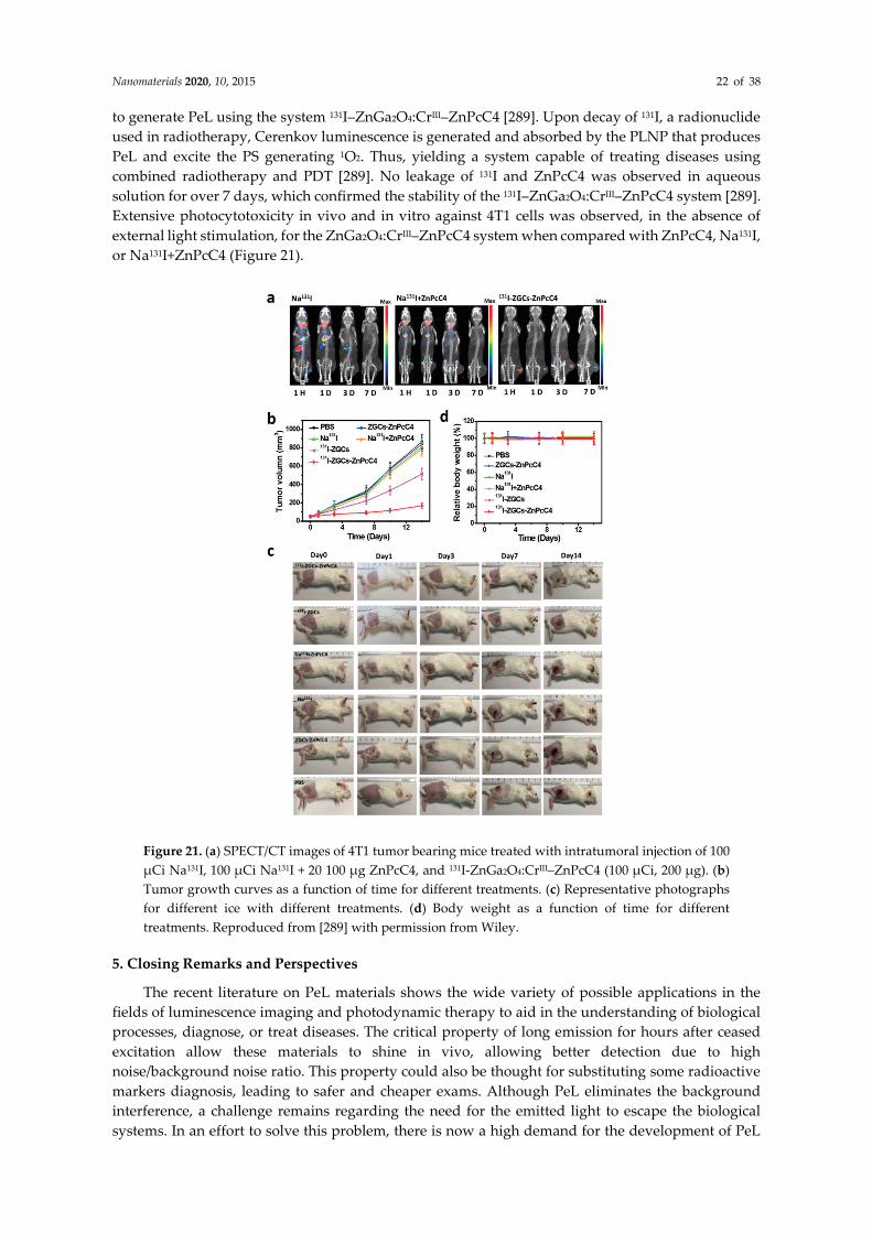
to generate PeL using the system 131I–ZnGa2O4:CrIII–ZnPcC4 [289]. Upon decay of 131I, a radionuclide
used in radiotherapy, Cerenkov luminescence is generated and absorbed by the PLNP that produces
PeL and excite the PS generating 1O2. Thus, yielding a system capable of treating diseases using
combined radiotherapy and PDT [289]. No leakage of 131I and ZnPcC4 was observed in aqueous
solution for over 7 days, which confirmed the stability of the 131I–ZnGa2O4:CrIII–ZnPcC4 system [289].
Extensive photocytotoxicity in vivo and in vitro against 4T1 cells was observed, in the absence of
external light stimulation, for the ZnGa2O4:CrIII–ZnPcC4 system when compared with ZnPcC4, Na131I,
or Na131I+ZnPcC4 (Figure 21).
Figure 21. (a) SPECT/CT images of 4T1 tumor bearing mice treated with intratumoral injection of 100
μCi Na131I, 100 μCi Na131I + 20 100 μg ZnPcC4, and 131I-ZnGa2O4:CrIII–ZnPcC4 (100 μCi, 200 μg). (b)
Tumor growth curves as a function of time for different treatments. (c) Representative photographs
for different ice with different treatments. (d) Body weight as a function of time for different
treatments. Reproduced from [289] with permission from Wiley.
5. Closing Remarks and Perspectives
The recent literature on PeL materials shows the wide variety of possible applications in the
fields of luminescence imaging and photodynamic therapy to aid in the understanding of biological
processes, diagnose, or treat diseases. The critical property of long emission for hours after ceased
excitation allow these materials to shine in vivo, allowing better detection due to high
noise/background noise ratio. This property could also be thought for substituting some radioactive
markers diagnosis, leading to safer and cheaper exams. Although PeL eliminates the background
interference, a challenge remains regarding the need for the emitted light to escape the biological
systems. In an effort to solve this problem, there is now a high demand for the development of PeL

Nanomaterials 2020, 10, 2015 23 of 38
materials that can be charged and emit in the NIR due to the high penetrability and low scattering of
this light. To accomplish this goal, it is still necessary to combine the different aspects presented in
this review: morphology control, long luminescence time, biocompatibility, and easy targeting.
The field of PeL-PDT is expected to have fast development in the coming years. The possibility
of achieving a treatment that requires light, namely PDT, without the need for continuous excitation,
is exciting and will advance non-invasive therapies. Achieving this goal will take first, the
development of PeL-PDT systems with optimized 1O2 efficiency, second, the use of light with higher
penetrability to allow deep tissue and in vivo treatment, and third, the development of Pel_NPs with
specific targeting abilities to yield high accumulation in the cancer cells. To the date, only a few
examples of PeL-PDT systems are known.
Author Contributions: Conceptualization, J.H.S.K.M. and L.C.V.R.; writing—original draft preparation, D.L.F,
L.G., L.C.V.R. and J.H.S.K.M.; writing—review and editing, D.L.F, L.G., L.C.V.R. and J.H.S.K.M.; supervision,
J.H.S.K.M. and L.C.V.R.; funding acquisition, J.H.S.K.M. and L.C.V.R. All authors contributed equally to this
work. All authors have read and agreed to the published version of the manuscript.
Funding: The Humboldt State University is gratefully acknowledged for financial support (start-up grant K1037
to JHSKM). The Brazilian National Council for Scientific and Technological agency—CNPq (grants 427312/2016-
7 to LCVR and 141252/2017-0 to LG) and the São Paulo Research Foundation-FAPESP (grants 2018/05280-5 to
LCVR and 2018/26282-6 to DLF) are also acknowledged for financial support.
Conflicts of Interest: The authors declare no conflict of interest.
References
1. Liu, L.; Zhang, H.; Song, D.; Wang, Z. An upconversion nanoparticle-based fluorescence resonance energy
transfer system for effectively sensing caspase-3 activity. Analyst 2018, 143, 761–767,
doi:10.1039/c7an01744h.
2. Liang, T.; Li, Z.; Wang, P.; Zhao, F.; Liu, J.; Liu, Z. Breaking Through the Signal-to-Background Limit of
Upconversion Nanoprobes Using a Target-Modulated Sensitizing Switch. J. Am. Chem. Soc. 2018, 140,
14696–14703, doi:10.1021/jacs.8b07329.
3. Hao, C.; Wu, X.; Sun, M.; Zhang, H.; Yuan, A.; Xu, L.; Xu, C.; Kuang, H. Chiral Core-Shell Upconversion
Nanoparticle@MOF Nanoassemblies for Quantification and Bioimaging of Reactive Oxygen Species in
Vivo. J. Am. Chem. Soc. 2019, 141, 19373–19378, doi:10.1021/jacs.9b09360.
4. Wang, H.; Zhao, W.; Liu, X.; Wang, S.; Wang, Y. BODIPY-Based Fluorescent Surfactant for Cell Membrane
Imaging and Photodynamic Therapy. ACS Appl. Bio Mater. 2020, 3, 593–601, doi:10.1021/acsabm.9b00977.
5. Zhou, J.; Liu, Z.; Li, F. Upconversion nanophosphors for small-animal imaging. Chem. Soc. Rev. 2012, 41,
1323–1349, doi:10.1039/c1cs15187h.
6. Lo, K.K.-W. Molecular Design of Bioorthogonal Probes and Imaging Reagents Derived from
Photofunctional Transition Metal Complexes. Acc. Chem. Res. 2020, 53, 32–44,
doi:10.1021/acs.accounts.9b00416.
7. Lin, S.; Pan, H.; Li, L.; Liao, R.; Yu, S.; Zhao, Q.; Sun, H.; Huang, W. AIPE-active platinum(ii) complexes
with tunable photophysical properties and their application in constructing thermosensitive probes used
for intracellular temperature imaging. J. Mater. Chem. C 2019, 7, 7893–7899, doi:10.1039/C9TC01905G.
8. Chen, G.Y.; Qju, H.L.; Prasad, P.N.; Chen, X.Y. Upconversion Nanoparticles: Design, Nanochemistry, and
Applications in Theranostics. Chem. Rev. 2014, 114, 5161–5214, doi:10.1021/cr400425h.
9. Wolfbeis, O.S. An overview of nanoparticles commonly used in fluorescent bioimaging. Chem. Soc. Rev.
2015, 44, 4743–4768, doi:10.1039/c4cs00392f.
10. Liu, G.; Jiang, F.; Chen, Y.; Yu, C.; Ding, B.; Shao, S.; Jia, M.; Ma, P.a.; Fu, Z.; Lin, J. Superior temperature
sensing of small-sized upconversion nanocrystals for simultaneous bioimaging and enhanced synergetic
therapy. Nanomed. Nanotechnol. Biol. Med. 2019, 24, 102135–102135, doi:10.1016/j.nano.2019.102135.
11. Gargas, D.J.; Chan, E.M.; Ostrowski, A.D.; Aloni, S.; Altoe, M.V.P.; Barnard, E.S.; Sanii, B.; Urban, J.J.;
Milliron, D.J.; Cohen, B.E.; et al. Engineering bright sub-10-nm upconverting nanocrystals for single-
molecule imaging. Nat. Nanotechnol. 2014, 9, 300–305, doi:10.1038/nnano.2014.29.
12. Zhao, M.; Wang, R.; Li, B.; Fan, Y.; Wu, Y.; Zhu, X.; Zhang, F. Precise In Vivo Inflammation Imaging Using
In Situ Responsive Cross-linking of Glutathione-Modified Ultra-Small NIR-II Lanthanide Nanoparticles.
Angew. Chem. (Int. Ed. Engl.) 2019, 58, 2050–2054, doi:10.1002/anie.201812878.
Nanomaterials 2020, 10, 2015 24 of 38
13. Chen, C.; Tian, R.; Zeng, Y.; Chu, C.; Liu, G. Activatable Fluorescence Probes for “Turn-On” and
Ratiometric Biosensing and Bioimaging: From NIR-I to NIR-II. Bioconjugate Chem. 2020, 31, 276–292,
doi:10.1021/acs.bioconjchem.9b00734.
14. Dobrucki, J.W.; Kubitscheck, U. Fluorescence Microscopy. In Fluorescence Microscopy: From Principles to
Biological Applications, 2nd ed.; Kubitscheck, U., Ed.; Wiley: Berlin, Germany, 2017; pp. 85–132.
15. Monteiro, J.; Machado, D.; de Hollanda, L.M.; Lancellotti, M.; Sigoli, F.A.; de Bettencourt-Dias, A. Selective
cytotoxicity and luminescence imaging of cancer cells with a dipicolinato-based Eu-III complex. Chem.
Commun. 2017, 53, 11818–11821, doi:10.1039/c7cc06753d.
16. Chauvin, A.S.; Comby, S.; Song, B.; Vandevyver, C.D.; Bünzli, J.-C.G. A versatile ditopic ligand system for
sensitizing the luminescence of bimetallic lanthanide bio-imaging probes. Chem. Eur. J. 2008, 14, 1726–1739,
doi:10.1002/chem.200701357.
17. Fernandez-Moreira, V.; Song, B.; Sivagnanam, V.; Chauvin, A.S.; Vandevyver, C.D.; Gijs, M.; Hemmila, I.;
Lehr, H.A.; Bünzli, J.-C.G. Bioconjugated lanthanide luminescent helicates as multilabels for lab-on-a-chip
detection of cancer biomarkers. Analyst 2010, 135, 42–52, doi:10.1039/b922124g.
18. Surender, E.M.; Comby, S.; Cavanagh, B.L.; Brennan, O.; Lee, T.C.; Gunnlaugsson, T. Two-Photon
Luminescent Bone Imaging Using Europium Nanoagents. Chem 2016, 1, 438–455,
doi:10.1016/j.chempr.2016.08.011.
19. Addisu, K.D.; Hsu, W.-H.; Hailemeskel, B.Z.; Andrgie, A.T.; Chou, H.-Y.; Yuh, C.-H.; Lai, J.-Y.; Tsai, H.-C.
Mixed Lanthanide Oxide Nanoparticles Coated with Alginate-Polydopamine as Multifunctional
Nanovehicles for Dual Modality: Targeted Imaging and Chemotherapy. ACS Biomater. Sci. Eng. 2019, 5,
5453–5469, doi:10.1021/acsbiomaterials.9b01226.
20. Chen, Z.; Chen, H.; Hu, H.; Yu, M.; Li, F.; Zhang, Q.; Zhou, Z.; Yi, T.; Huang, C. Versatile synthesis strategy
for carboxylic acid-functionalized upconverting nanophosphors as biological labels. J. Am. Chem. Soc. 2008,
130, 3023–3029, doi:10.1021/ja076151k.
21. Song, X.; Zhang, J.; Yue, Z.; Wang, Z.; Liu, Z.; Zhang, S. Dual-Activator Codoped Upconversion Nanoprobe
with Core-Multishell Structure for in Vitro and in Vivo Detection of Hydroxyl Radical. Anal. Chem. 2017,
89, 11021–11026, doi:10.1021/acs.analchem.7b02995.
22. Zhang, R.; Liang, L.; Meng, Q.; Zhao, J.; Ta, H.T.; Li, L.; Zhang, Z.; Sultanbawa, Y.; Xu, Z.P. Responsive
Upconversion Nanoprobe for Background-Free Hypochlorous Acid Detection and Bioimaging. Small
(Weinh. Der Bergstr. Ger.) 2019, 15, e1803712, doi:10.1002/smll.201803712.
23. Wang, F.; Qu, X.; Liu, D.; Ding, C.; Zhang, C.; Xian, Y. Upconversion nanoparticles-MoS2 nanoassembly as
a fluorescent turn-on probe for bioimaging of reactive oxygen species in living cells and zebrafish. Sens.
Actuators B Chem. 2018, 274, 180–187, doi:10.1016/j.snb.2018.07.125.
24. Yang, L.; Zhang, K.; Bi, S.; Zhu, J.-J. Dual-Acceptor-Based Upconversion Luminescence Nanosensor with
Enhanced Quenching Efficiency for in Situ Imaging and Quantification of MicroRNA in Living Cells. ACS
Appl. Mater. Interfaces 2019, 11, 38459–38466, doi:10.1021/acsami.9b12254.
25. Song, X.; Yue, Z.; Zhang, J.; Jiang, Y.; Wang, Z.; Zhang, S. Multicolor Upconversion Nanoprobes Based on
a Dual Luminescence Resonance Energy Transfer Assay for Simultaneous Detection and Bioimaging of
Ca2+ i and pHi in Living Cells. Chemistry 2018, 24, 6458–6463, doi:10.1002/chem.201800154.
26. Shi, Y.; Liu, Q.; Yuan, W.; Xue, M.; Feng, W.; Li, F. Dye-Assembled Upconversion Nanocomposite for
Luminescence Ratiometric in Vivo Bioimaging of Copper Ions. ACS Appl. Mater. Interfaces 2019, 11, 430–
436, doi:10.1021/acsami.8b19961.
27. Li, Z.; Liu, H.; Li, H.; Tsou, Y.-H.; Gao, Y.; Xu, X.; Du, W.; Wei, L.; Yu, M. Lysosome-targeting NIR
ratiometric luminecent upcoversion nanoprobe toward arginine. Sens. Actuators B Chem. 2019, 280, 94–101,
doi:10.1016/j.snb.2018.10.057.
28. Wang, N.; Yu, X.; Zhang, K.; Mirkin, C.A.; Li, J. Upconversion Nanoprobes for the Ratiometric Luminescent
Sensing of Nitric Oxide. J. Am. Chem. Soc. 2017, 139, 12354–12357, doi:10.1021/jacs.7b06059.
29. Tsukube, H.; Shinoda, S. Lanthanide complexes in molecular recognition and chirality sensing of biological
substrates. Chem. Rev. 2002, 102, 2389–2403, doi:10.1021/cr010450p.
30. Pandya, S.; Yu, J.; Parker, D. Engineering emissive europium and terbium complexes for molecular imaging
and sensing. Dalton Trans. (Camb. Engl. 2003) 2006, 2757–2766, doi:10.1039/b514637b.
31. Harbuzaru, B.V.; Corma, A.; Rey, F.; Jorda, J.L.; Ananias, D.; Carlos, L.D.; Rocha, J. A miniaturized linear
pH sensor based on a highly photoluminescent self-assembled europium(III) metal-organic framework.
Angew. Chem. (Int. Ed. Engl.) 2009, 48, 6476–6479, doi:10.1002/anie.200902045.

Nanomaterials 2020, 10, 2015 25 of 38
32. Gunnlaugsson, T.; Leonard, J.P. Responsive lanthanide luminescent cyclen complexes: From
switching/sensing to supramolecular architectures. Chem. Commun. (Camb. Engl.) 2005, 10.1039/b418196d,
3114–3131, doi:10.1039/b418196d.
33. Tan, H.; Liu, B.; Chen, Y. Lanthanide coordination polymer nanoparticles for sensing of mercury(II) by
photoinduced electron transfer. ACS Nano 2012, 6, 10505–10511, doi:10.1021/nn304469j.
34. Khullar, S.; Singh, S.; Das, P.; Mandal, S.K. Luminescent Lanthanide-Based Probes for the Detection of
Nitroaromatic Compounds in Water. ACS Omega 2019, 4, 5283–5292, doi:10.1021/acsomega.9b00223.
35. Wang, H.-F.; Ma, X.-F.; Zhu, Z.-H.; Zou, H.-H.; Liang, F.-P. Regulation of the Metal Center and
Coordinating Anion of Mononuclear Ln(III) Complexes to Promote an Efficient Luminescence Response to
Various Organic Solvents. Langmuir ACS J. Surf. Colloids 2020, 36, 1409–1417,
doi:10.1021/acs.langmuir.9b02990.
36. Hewitt, S.H.; Macey, G.; Mailhot, R.; Elsegood, M.R.J.; Duarte, F.; Kenwright, A.M.; Butler, S.J. Tuning the
anion binding properties of lanthanide receptors to discriminate nucleoside phosphates in a sensing array.
Chem. Sci. 2020, 11, 3619–3628, doi:10.1039/D0SC00343C.
37. Yang, Z.; Loh, K.Y.; Chu, Y.-T.; Feng, R.; Satyavolu, N.S.R.; Xiong, M.; Huynh, S.M.N.; Hwang, K.; Li, L.;
Xing, H.; et al. Optical Control of Metal Ion Probes in Cells and Zebrafish Using Highly Selective
DNAzymes Conjugated to Upconversion Nanoparticles. J. Am. Chem. Soc. 2018, 140, 17656–17665,
doi:10.1021/jacs.8b09867.
38. Li, X.; Zhao, H.; Ji, Y.; Yin, C.; Li, J.; Yang, Z.; Tang, Y.; Zhang, Q.; Fan, Q.; Huang, W. Lysosome-Assisted
Mitochondrial Targeting Nanoprobe Based on Dye-Modified Upconversion Nanophosphors for
Ratiometric Imaging of Mitochondrial Hydrogen Sulfide. ACS Appl. Mater. Interfaces 2018, 10, 39544–39556,
doi:10.1021/acsami.8b16818.
39. Tang, Z.; Song, B.; Zhang, W.; Guo, L.; Yuan, J. Precise Monitoring of Drug-Induced Kidney Injury Using
an Endoplasmic Reticulum-Targetable Ratiometric Time-Gated Luminescence Probe for Superoxide
Anions. Anal. Chem. 2019, 91, 14019–14028, doi:10.1021/acs.analchem.9b03602.
40. Ma, H.; Song, B.; Wang, Y.; Liu, C.; Wang, X.; Yuan, J. Development of organelle-targetable europium
complex probes for time-gated luminescence imaging of hypochlorous acid in live cells and animals. Dyes
Pigm. 2017, 140, 407–416, doi:10.1016/j.dyepig.2017.01.062.
41. Tang, Z.; Song, B.; Ma, H.; Luo, T.; Guo, L.; Yuan, J., Mitochondria Targetable Ratiometric Time-Gated
Luminescence Probe for Carbon Monoxide Based on Lanthanide Complexes. Anal. Chem. 2019, 91, 2939-
2946, doi:10.1021/acs.analchem.8b05127.
42. Liu, Y.H.; Wang, Y.H.; Jiang, K.; Sun, S.; Qian, S.H.; Wu, Q.P.; Lin, H. A persistent luminescence-based
label-free probe for the ultrasensitive detection of hemoglobin in human serum. Talanta 2020, 206,
doi:10.1016/j.talanta.2019.120206.
43. Wu, S.; Li, Y.; Zhang, R.; Fan, K.; Ding, W.; Xu, L.; Zhang, L. Persistent luminescence-polypyrrole
nanocomposite for dual-modal imaging and photothermal therapy of mammary cancer. Talanta 2021, 221,
121435, doi:10.1016/j.talanta.2020.121435.
44. Ding, S.; Guo, H.; Feng, P.; Ye, Q.; Wang, Y. A New Near-Infrared Long Persistent Luminescence Material
with Its Outstanding Persistent Luminescence Performance and Promising Multifunctional Application
Prospects. Adv. Opt. Mater. 2020, 8, 2000097, doi:10.1002/adom.202000097.
45. Shi, L.X.; Shao, J.J.; Jing, X.H.; Zheng, W.W.; Liu, H.; Zhao, Y. Autoluminescence-Free Dual Tumor Marker
Biosensing by Persistent Luminescence Nanostructures. ACS Sustain. Chem. Eng. 2020, 8, 686–694,
doi:10.1021/acssuschemeng.9b06621.
46. Wang, Z.H.; Liu, J.M.; Zhao, N.; Li, C.Y.; Lv, S.W.; Hu, Y.Z.; Lv, H.; Wang, D.; Wang, S. Cancer Cell
Macrophage Membrane Camouflaged Persistent Luminescent Nanoparticles for Imaging-Guided
Photothermal Therapy of Colorectal Cancer. ACS Appl. Nano Mater. 2020, 3, 7105–7118,
doi:10.1021/acsanm.0c01433.
47. Pan, Z.W.; Lu, Y.Y.; Liu, F. Sunlight-activated long-persistent luminescence in the near-infrared from Cr3+-
doped zinc gallogermanates. Nat. Mater. 2012, 11, 58–63, doi:10.1038/nmat3173.
48. Maldiney, T.; Bessière, A.; Seguin, J.; Teston, E.; Sharma, S.K.; Viana, B.; Bos, A.J.J.; Dorenbos, P.; Bessodes,
M.; Gourier, D.; et al. The in vivo activation of persistent nanophosphors for optical imaging of
vascularization, tumours and grafted cells. Nat. Mater. 2014, 13, 418–426, doi:10.1038/nmat3908.
49. Van den Eeckhout, K.; Smet, P.F.; Poelman, D. Persistent Luminescence in Eu2+-Doped Compounds: A
Review. Materials 2010, 3, 2536, doi:10.3390/ma3042536.

Nanomaterials 2020, 10, 2015 26 of 38
50. Lin, Q.S.; Li, Z.H.; Ji, C.H.; Yuan, Q. Electronic structure engineering and biomedical applications of low
energy-excited persistent luminescence nanoparticles. Nanoscale Adv. 2020, 2, 1380–1394,
doi:10.1039/c9na00817a.
51. Liu, J.H.; Lecuyer, T.; Seguin, J.; Mignet, N.; Scherman, D.; Viana, B.; Richard, C. Imaging and therapeutic
applications of persistent luminescence nanomaterials. Adv. Drug Deliv. Rev. 2019, 138, 193–210,
doi:10.1016/j.addr.2018.10.015.
52. Sun, S.K.; Wang, H.F.; Yan, X.P. Engineering Persistent Luminescence Nanoparticles for Biological
Applications: From Biosensing/Bioimaging to Theranostics. Acc. Chem. Res. 2018, 51, 1131–1143,
doi:10.1021/acs.accounts.7b00619.
53. Liang, L.; Chen, N.; Jia, Y.Y.; Ma, Q.Q.; Wang, J.; Yuan, Q.; Tan, W.H. Recent progress in engineering near-
infrared persistent luminescence nanoprobes for time-resolved biosensing/bioimaging. Nano Res. 2019, 12,
1279–1292, doi:10.1007/s12274-019-2343-6.
54. Tan, H.X.; Wang, T.Y.; Shao, Y.R.; Yu, C.Y.; Hu, L.D. Crucial Breakthrough of Functional Persistent
Luminescence Materials for Biomedical and Information Technological Applications. Front. Chem. 2019, 7,
387. doi:10.3389/fchem.2019.00387.
55. Li, Y.; Li, F.; Huang, Y.; Wu, H.; Wang, J.; Yang, J.; Xiao, Q.; Lin, H. Fe3+-codoped ultra-small NaGdF4:Nd3+
nanophosphors: Enhanced near-infrared luminescence, reduced particle size and bioimaging applications.
Rsc Adv. 2019, 9, 18070–18075, doi:10.1039/C9RA00798A.
56. Song, X.; Li, S.; Guo, H.; You, W.; Shang, X.; Li, R.; Tu, D.; Zheng, W.; Chen, Z.; Yang, H.; et al. Graphene-
Oxide-Modified Lanthanide Nanoprobes for Tumor-Targeted Visible/NIR-II Luminescence Imaging.
Angew. Chem. (Int. Ed. Engl.) 2019, 58, 18981–18986, doi:10.1002/anie.201909416.
57. Cao, C.; Liu, Q.; Shi, M.; Feng, W.; Li, F. Lanthanide-Doped Nanoparticles with Upconversion and
Downshifting Near-Infrared Luminescence for Bioimaging. Inorg. Chem. 2019, 58, 9351–9357,
doi:10.1021/acs.inorgchem.9b01071.
58. Wang, X.; Shi, J.; Li, P.; Zheng, S.; Sun, X.; Zhang, H. LuPO4:Nd3+ nanophosphors for dual-mode deep tissue
NIR-II luminescence/CT imaging. J. Lumin. 2019, 209, 420–426, doi:10.1016/j.jlumin.2019.02.028.
59. Feng, Y.; Xiao, Q.; Zhang, Y.; Li, F.; Li, Y.; Li, C.; Wang, Q.; Shi, L.; Lin, H. Neodymium-doped NaHoF4
nanoparticles as near-infrared luminescent/T2-weighted MR dual-modal imaging agents in vivo. J. Mater.
Chem. B 2017, 5, 504–510, doi:10.1039/C6TB01961G.
60. Wang, X.; Li, H.; Li, F.; Han, X.; Chen, G. Prussian blue-coated lanthanide-doped core/shell/shell
nanocrystals for NIR-II image-guided photothermal therapy. Nanoscale 2019, 11, 22079–22088,
doi:10.1039/c9nr07973d.
61. Barolet, D. Light-emitting diodes (LEDs) in dermatology. Semin. Cutan. Med. Surg. 2008, 27, 227–238,
doi:10.1016/j.sder.2008.08.003.
62. Hao, S.; Chen, G.; Yang, C.; Shao, W.; Wei, W.; Liu, Y.; Prasad, P.N. Nd3+-Sensitized multicolor
upconversion luminescence from a sandwiched core/shell/shell nanostructure. Nanoscale 2017, 9, 10633–
10638, doi:10.1039/c7nr02594g.
63. Ai, F.; Ju, Q.; Zhang, X.; Chen, X.; Wang, F.; Zhu, G. A core-shell-shell nanoplatform upconverting near-
infrared light at 808 nm for luminescence imaging and photodynamic therapy of cancer. Sci. Rep. 2015, 5,
10785, doi:10.1038/srep10785.
64. Jaque, D.; Richard, C.; Viana, B.; Soga, K.; Liu, X.G.; Sole, J.G. Inorganic nanoparticles for optical
bioimaging. Adv. Opt. Photonics 2016, 8, 1–103, doi:10.1364/aop.8.000001.
65. Smith, A.M.; Mancini, M.C.; Nie, S. BIOIMAGING Second window for in vivo imaging. Nat. Nanotechnol.
2009, 4, 710–711, doi:10.1038/nnano.2009.326.
66. Li, Y.; Li, X.; Xue, Z.; Jiang, M.; Zeng, S.; Hao, J. Second near-infrared emissive lanthanide complex for fast
renal-clearable in vivo optical bioimaging and tiny tumor detection. Biomaterials 2018, 169, 35–44,
doi:10.1016/j.biomaterials.2018.03.041.
67. Yang, Y.; Wang, P.; Lu, L.; Fan, Y.; Sun, C.; Fan, L.; Xu, C.; El-Toni, A.M.; Alhoshan, M.; Zhang, F. Small-
Molecule Lanthanide Complexes Probe for Second Near-Infrared Window Bioimaging. Anal. Chem. 2018,
90, 7946–7952, doi:10.1021/acs.analchem.8b00603.
68. Ning, Y.; Cheng, S.; Wang, J.-X.; Liu, Y.-W.; Feng, W.; Li, F.; Zhang, J.-L. Fluorescence lifetime imaging of
upper gastrointestinal pH in vivo with a lanthanide based near-infrared τ probe. Chem. Sci. 2019, 10, 4227–
4235, doi:10.1039/C9SC00220K.

Nanomaterials 2020, 10, 2015 27 of 38
69. Ren, T.; Xu, W.; Zhang, Q.; Zhang, X.; Wen, S.; Yi, H.; Yuan, L.; Zhang, X. Harvesting Hydrogen Bond
Network: Enhance the Anti-Solvatochromic Two-Photon Fluorescence for Cirrhosis Imaging. Angew. Chem.
Int. Ed. 2018, 57, 7473–7477, doi:10.1002/anie.201800293.
70. Agrawalla, B.K.; Lee, H.W.; Phue, W.H.; Raju, A.; Kim, J.J.; Kim, H.M.; Kang, N.Y.; Chang, Y.T. Two-Photon
Dye Cocktail for Dual-Color 3D Imaging of Pancreatic Beta and Alpha Cells in Live Islets. J. Am. Chem. Soc.
2017, 139, 3480–3487, doi:10.1021/jacs.6b12122.
71. Agrawalla, B.K.; Chandran, Y.; Phue, W.H.; Lee, S.C.; Jeong, Y.M.; Wan, S.Y.D.; Kang, N.Y.; Chang, Y.T.
Glucagon-Secreting Alpha Cell Selective Two-Photon Fluorescent Probe TP-alpha: For Live Pancreatic Islet
Imaging. J. Am. Chem. Soc. 2015, 137, 5355–5362, doi:10.1021/ja5115776.
72. Kumari, P.; Verma, S.K.; Mobin, S.M. Water soluble two-photon fluorescent organic probes for long-term
imaging of lysosomes in live cells and tumor spheroids. Chem. Commun. 2018, 54, 539–542,
doi:10.1039/c7cc07812a.
73. Hirsch, L.R.; Stafford, R.J.; Bankson, J.A.; Sershen, S.R.; Rivera, B.; Price, R.E.; Hazle, J.D.; Halas, N.J.; West,
J.L. Nanoshell-mediated near-infrared thermal therapy of tumors under magnetic resonance guidance.
Proc. Natl. Acad. Sci. USA 2003, 100, 13549–13554, doi:10.1073/pnas.2232479100.
74. Robinson, J.T.; Tabakman, S.M.; Liang, Y.Y.; Wang, H.L.; Casalongue, H.S.; Vinh, D.; Dai, H.J. Ultrasmall
Reduced Graphene Oxide with High Near-Infrared Absorbance for Photothermal Therapy. J. Am. Chem.
Soc. 2011, 133, 6825–6831, doi:10.1021/ja2010175.
75. Jaque, D.; Maestro, L.M.; del Rosal, B.; Haro-Gonzalez, P.; Benayas, A.; Plaza, J.L.; Rodriguez, E.M.; Sole,
J.G. Nanoparticles for photothermal therapies. Nanoscale 2014, 6, 9494–9530, doi:10.1039/c4nr00708e.
76. Zheng, B.-D.; He, Q.-X.; Li, X.; Yoon, J.; Huang, J.-D. Phthalocyanines as contrast agents for photothermal
therapy. Coord. Chem. Rev. 2021, 426, 213548, doi:10.1016/j.ccr.2020.213548.
77. Zeng, X.M.; Yan, S.Q.; Di, C.; Lei, M.C.; Chen, P.; Du, W.; Jin, Y.; Liu, B.F. “All-in-One” Silver Nanoprism
Platform for Targeted Tumor Theranostics. ACS Appl. Mater. Interfaces 2020, 12, 11329–11340,
doi:10.1021/acsami.9b21166.
78. Zhang, J.J.; Ning, L.L.; Huang, J.G.; Zhang, C.; Pu, K.Y. Activatable molecular agents for cancer theranostics.
Chem. Sci. 2020, 11, 618–630, doi:10.1039/c9sc05460j.
79. Castano, A.P.; Mroz, P.; Hamblin, M.R. Photodynamic therapy and anti-tumour immunity. Nat. Rev. Cancer
2006, 6, 535–545, doi:10.1038/nrc1894.
80. Cheng, P.H.; Pu, K.Y. Activatable Phototheranostic Materials for Imaging-Guided Cancer Therapy. ACS
Appl. Mater. Interfaces 2020, 12, 5286–5299, doi:10.1021/acsami.9b15064.
81. Celli, J.P.; Spring, B.Q.; Rizvi, I.; Evans, C.L.; Samkoe, K.S.; Verma, S.; Pogue, B.W.; Hasan, T. Imaging and
Photodynamic Therapy: Mechanisms, Monitoring, and Optimization. Chem. Rev. 2010, 110, 2795–2838,
doi:10.1021/cr900300p.
82. Lifshits, L.; Roque Iii, J.A.; Konda, P.; Monro, S.; Cole, H.D.; von Dohlen, D.; Kim, S.; Deep, G.; Thummel,
R.P.; Cameron, C.G.; et al. Near-infrared Absorbing Ru(II) Complexes Act as Immunoprotective
Photodynamic Therapy (PDT) Agents Against Aggressive Melanoma. Chem. Sci. 2020,
doi:10.1039/D0SC03875J.
83. Johnson, K.R.; Lombardi, V.C.; de Bettencourt-Dias, A. Photocytotoxicity of Oligothienyl-Functionalized
Chelates That Sensitize LnIII Luminescence and Generate 1O2. Chem. A Eur. J. 2020, n/a,
doi:10.1002/chem.202001568.
84. Herzog, R.W.; Frederickson, R.M. Special Issue Features State-of-the-Art in Clinical Gene Therapy. Mol.
Ther. 2020, 28, 1933, doi:10.1016/j.ymthe.2020.08.006.
85. Hu, L.; Fu, X.; Kong, G.; Yin, Y.; Meng, H.; Ke, G.; Zhang, X. DNAzyme-Gold Nanoparticle-based Probes
for Biosensing and Bioimaging. J. Mater. Chem. B 2020, 10.1039/D0TB01750G, doi:10.1039/D0TB01750G.
86. Li, L.; Tian, H.; He, J.; Zhang, M.; Li, Z.-G.; Ni, P. Fabrication of aminated poly(glycidyl methacrylate)s-
based polymers for co-delivery of anticancer drug and p53 gene. J. Mater. Chem. B 2020,
10.1039/D0TB01811B, doi:10.1039/D0TB01811B.
87. Li, X.; Lee, D.; Huang, J.-D.; Yoon, J. Phthalocyanine-Assembled Nanodots as Photosensitizers for Highly
Efficient Type I Photoreactions in Photodynamic Therapy. Angew. Chem. (Int. Ed. Engl.) 2018, 57, 9885–9890,
doi:10.1002/anie.201806551.
88. Fujishiro, R.; Sonoyama, H.; Ide, Y.; Fujimura, T.; Sasai, R.; Nagai, A.; Mori, S.; Kaufman, N.E.M.; Zhou, Z.;
Vicente, M.G.H.; et al. Synthesis, photodynamic activities, and cytotoxicity of new water-soluble cationic
Nanomaterials 2020, 10, 2015 28 of 38
gallium(III) and zinc(II) phthalocyanines. J. Inorg. Biochem. 2019, 192, 7–16,
doi:10.1016/j.jinorgbio.2018.11.013.
89. Toubia, I.; Nguyen, C.; Diring, S.; Ali, L.M.A.; Larue, L.; Aoun, R.; Frochot, C.; Gary-Bobo, M.; Kobeissi, M.;
Odobel, F. Synthesis and Anticancer Activity of Gold Porphyrin Linked to Malonate Diamine Platinum
Complexes. Inorg. Chem. 2019, 58, 12395–12406, doi:10.1021/acs.inorgchem.9b01981.
90. Wu, S.; Li, Y.; Ding, W.; Xu, L.; Ma, Y.; Zhang, L. Recent Advances of Persistent Luminescence
Nanoparticles in Bioapplications. Nano-Micro Lett. 2020, 12, 70, doi:10.1007/s40820-020-0404-8.
91. Xu, J.; Tanabe, S. Persistent luminescence instead of phosphorescence: History, mechanism, and
perspective. J. Lumin. 2019, 205, 581–620, doi:10.1016/j.jlumin.2018.09.047.
92. Van den Eeckhout, K.; Poelman, D.; Smet, P.F. Persistent Luminescence in Non-Eu2+-Doped Compounds:
A Review. Materials 2013, 6, 2789–2818, doi:10.3390/ma6072789.
93. Brito, H.F.; Holsa, J.; Laamanen, T.; Lastusaari, M.; Malkamaki, M.; Rodrigues, L.C.V. Persistent
luminescence mechanisms: Human imagination at work. Opt. Mater. Express 2012, 2, 371–381,
doi:10.1364/ome.2.000371.
94. Monteiro, J.H.S.K. Recent Advances in Luminescence Imaging of Biological Systems Using Lanthanide(III)
Luminescent Complexes. Molecules 2020, 25, 2089, doi:10.3390/molecules25092089.
95. Day, A.H.; Übler, M.H.; Best, H.L.; Lloyd-Evans, E.; Mart, R.J.; Fallis, I.A.; Allemann, R.K.; Al-Wattar,
E.A.H.; Keymer, N.I.; Buurma, N.J.; et al. Targeted cell imaging properties of a deep red luminescent
iridium(iii) complex conjugated with a c-Myc signal peptide. Chem. Sci. 2020, 11, 1599–1606,
doi:10.1039/C9SC05568A.
96. Ahmed, M.U.; Velkov, T.; Zhou, Q.T.; Fulcher, A.J.; Callaghan, J.; Zhou, F.; Chan, K.; Azad, M.A.K.; Li, J.
Intracellular localization of polymyxins in human alveolar epithelial cells. J. Antimicrob. Chemother. 2018,
74, 48–57, doi:10.1093/jac/dky409.
97. Zheng, Q.; Cheng, W.; Zhang, X.; Shao, R.; Li, Z. A pH-Induced Reversible Assembly System with
Resveratrol-Controllable Loading and Release for Enhanced Tumor-Targeting Chemotherapy. Nanoscale
Res. Lett. 2019, 14, 305, doi:10.1186/s11671-019-3139-z.
98. Yang, T.; Xu, L.; Liu, S.; Shen, Y.; Huang, L.; Zhang, L.; Ding, S.; Cheng, W. Amplified fluorescence imaging
of HER2 dimerization on cancer cells by using a co-localization triggered DNA nanoassembly. Microchim.
Acta 2019, 186, 439, doi:10.1007/s00604-019-3549-8.
99. Li, J.; Wei, Y.-J.; Yang, X.-L.; Wu, W.-X.; Zhang, M.-Q.; Li, M.-Y.; Hu, Z.-E.; Liu, Y.-H.; Wang, N.; Yu, X.-Q.
Rational Construction of a Mitochondrial Targeting, Fluorescent Self-Reporting Drug-Delivery Platform
for Combined Enhancement of Endogenous ROS Responsiveness. ACS Appl. Mater. Interfaces 2020, 12,
32432–32445, doi:10.1021/acsami.0c08336.
100. Xu, W.; Teoh, C.L.; Peng, J.; Su, D.; Yuan, L.; Chang, Y.-T. A mitochondria-targeted ratiometric fluorescent
probe to monitor endogenously generated sulfur dioxide derivatives in living cells. Biomaterials 2015, 56,
1–9, doi:10.1016/j.biomaterials.2015.03.038.
101. Xu, Z.; Zhang, M.-X.; Xu, Y.; Liu, S.H.; Zeng, L.; Chen, H.; Yin, J. The visualization of lysosomal and
mitochondrial glutathione via near-infrared fluorophore and in vivo imaging application. Sens. Actuators
B Chem. 2019, 290, 676–683, doi:10.1016/j.snb.2019.03.114.
102. Mayer, M.; Fey, K.; Heinze, E.; Wick, C.R.; Abboud, M.I.; Yeh, T.-L.; Tumber, A.; Orth, N.; Schley, G.;
Buchholz, B.; et al. A Fluorescent Benzo[g]isoquinoline-Based HIF Prolyl Hydroxylase Inhibitor for Cellular
Imaging. ChemMedChem 2019, 14, 94–99, doi:10.1002/cmdc.201800483.
103. Wang, Y.-S.; Tzeng, H.-T.; Tsai, C.-H.; Cheng, H.-C.; Lai, W.-W.; Liu, H.-S.; Wang, Y.-C. VAMP8, a vesicle-
SNARE required for RAB37-mediated exocytosis, possesses a tumor metastasis suppressor function. Cancer
Lett. 2018, 437, 79–88, doi:10.1016/j.canlet.2018.08.023.
104. Soboleva, T.; Esquer, H.J.; Anderson, S.N.; Berreau, L.M.; Benninghoff, A.D. Mitochondrial-Localized
Versus Cytosolic Intracellular CO-Releasing Organic PhotoCORMs: Evaluation of CO Effects Using
Bioenergetics. ACS Chem. Biol. 2018, 13, 2220–2228, doi:10.1021/acschembio.8b00387.
105. Xu, J.; Pan, J.; Jiang, X.; Qin, C.; Zeng, L.; Zhang, H.; Zhang, J.F. A mitochondria-targeted ratiometric
fluorescent probe for rapid, sensitive and specific detection of biological SO2 derivatives in living cells.
Biosens. Bioelectron. 2016, 77, 725–732, doi:10.1016/j.bios.2015.10.049.
106. Huth, U.S.; Schubert, R.; Peschka-Süss, R. Investigating the uptake and intracellular fate of pH-sensitive
liposomes by flow cytometry and spectral bio-imaging. J. Control. Release 2006, 110, 490–504,
doi:10.1016/j.jconrel.2005.10.018.

Nanomaterials 2020, 10, 2015 29 of 38
107. Sun, J.; Song, B.; Ye, Z.; Yuan, J. Mitochondria Targetable Time-Gated Luminescence Probe for Singlet
Oxygen Based on a beta-Diketonate-Europium Complex. Inorg. Chem. 2015, 54, 11660–11668,
doi:10.1021/acs.inorgchem.5b02458.
108. Suzuki, T.; Matsuzaki, T.; Hagiwara, H.; Aoki, T.; Takata, K. Recent Advances in Fluorescent Labeling
Techniques for Fluorescence Microscopy. Acta Histochem. Et Cytochem. 2007, 40, 131–137,
doi:10.1267/ahc.07023.
109. Wang, L.; Wang, J.; Xia, S.; Wang, X.; Yu, Y.; Zhou, H.; Liu, H. A FRET-based near-infrared ratiometric
fluorescent probe for detection of mitochondria biothiol. Talanta 2020, 219, 121296,
doi:10.1016/j.talanta.2020.121296.
110. Yuan, L.; Lin, W.Y.; Zheng, K.B.; He, L.W.; Huang, W.M. Far-red to near infrared analyte-responsive
fluorescent probes based on organic fluorophore platforms for fluorescence imaging. Chem. Soc. Rev. 2013,
42, 622–661, doi:10.1039/c2cs35313j.
111. Hamon, N.; Roux, A.; Beyler, M.; Mulatier, J.-C.; Andraud, C.; Nguyen, C.; Maynadier, M.; Bettache, N.;
Duperray, A.; Grichine, A.; et al. Pyclen-Based Ln(III) Complexes as Highly Luminescent Bioprobes for In
Vitro and In Vivo One- and Two-Photon Bioimaging Applications. J. Am. Chem. Soc. 2020, 142, 10184–10197,
doi:10.1021/jacs.0c03496.
112. Hamon, N.; Galland, M.; Le Fur, M.; Roux, A.; Duperray, A.; Grichine, A.; Andraud, C.; Le Guennic, B.;
Beyler, M.; Maury, O.; et al. Combining a pyclen framework with conjugated antenna for the design of
europium and samarium luminescent bioprobes. Chem. Commun. 2018, 54, 6173–6176,
doi:10.1039/c8cc02035c.
113. Bui, A.T.; Beyler, M.; Grichine, A.; Duperray, A.; Mulatier, J.-C.; Guyot, Y.; Andraud, C.; Tripier, R.;
Brasselet, S.; Maury, O. Near infrared two photon imaging using a bright cationic Yb(iii) bioprobe
spontaneously internalized into live cells. Chem. Commun. 2017, 53, 6005–6008, doi:10.1039/c7cc02835k.
114. Gautam, A.; Komal, P. Probable ideal size of Ln(3+)-based upconversion nanoparticles for single and
multimodal imaging. Coord. Chem. Rev. 2018, 376, 393–404, doi:10.1016/j.ccr.2018.08.008.
115. Zhu, X.H.; Zhang, J.; Liu, J.L.; Zhang, Y. Recent Progress of Rare-Earth Doped Upconversion Nanoparticles:
Synthesis, Optimization, and Applications. Adv. Sci. 2019, 6, doi:10.1002/advs.201901358.
116. Hemmer, E.; Acosta-Mora, P.; Mendez-Ramos, J.; Fischer, S. Optical nanoprobes for biomedical
applications: Shining a light on upconverting and near-infrared emitting nanoparticles for imaging,
thermal sensing, and photodynamic therapy. J. Mater. Chem. B 2017, 5, 4365–4392, doi:10.1039/c7tb00403f.
117. Jahn, K.; Buschmann, V.; Hille, C. Simultaneous Fluorescence and Phosphorescence Lifetime Imaging
Microscopy in Living Cells. Sci. Rep. 2015, 5, 14334-Article No.: 14334, doi:10.1038/srep14334.
118. Yang, W.; Srivastava, P.K.; Han, S.; Jing, L.; Tu, C.-C.; Chen, S.-L. Optomechanical Time-Gated Fluorescence
Imaging Using Long-Lived Silicon Quantum Dot Nanoparticles. Anal. Chem. 2019, 91, 5499–5503,
doi:10.1021/acs.analchem.9b00517.
119. Cao, S.; Li, H.; Liu, Y.; Wang, M.; Zhang, M.; Zhang, S.; Chen, J.; Xu, J.; Knutson, J.R.; Brand, L.
Dehydrogenase Binding Sites Abolish the “Dark” Fraction of NADH: Implication for Metabolic Sensing
via FLIM. J. Phys. Chem. B 2020, 124, 31, doi:10.1021/acs.jpcb.0c04835.
120. Straková, K.; López-Andarias, J.; Jiménez-Rojo, N.; Chambers, J.E.; Marciniak, S.J.; Riezman, H.; Sakai, N.;
Matile, S. HaloFlippers: A General Tool for the Fluorescence Imaging of Precisely Localized Membrane
Tension Changes in Living Cells. ACS Cent. Sci. 2020, 6, 1376–1385, doi:10.1021/acscentsci.0c00666.
121. Okkelman, I.; McGarrigle, R.; O’Carroll, S.; Carvajal Berrio, D.; Schenke-Layland, K.; Hynes, J.; Dmitriev,
R.I. Extracellular Ca2+-sensing fluorescent protein biosensor based on a collagen-binding domain. ACS
Appl. Bio Mater. 2020, 10.1021/acsabm.0c00649, doi:10.1021/acsabm.0c00649.
122. Bastiaens, P.I.H.; Squire, A. Fluorescence lifetime imaging microscopy: Spatial resolution of biochemical
processes in the cell. Trends Cell Biol. 1999, 9, 48–52, doi:10.1016/S0962-8924(98)01410-X.
123. Suhling, K.; French, P.M.W.; Phillips, D. Time-resolved fluorescence microscopy. Photochem. Photobiol. Sci.
2005, 4, 13–22, doi:10.1039/B412924P.
124. Lakowicz, J.R.; Szmacinski, H.; Nowaczyk, K.; Johnson, M.L. Fluorescence lifetime imaging of free and
protein-bound NADH. Proc. Natl. Acad. Sci. USA 1992, 89, 1271, doi:10.1073/pnas.89.4.1271.
125. Verveer, P.J.; Wouters, F.S.; Reynolds, A.R.; Bastiaens, P.I.H. Quantitative Imaging of Lateral ErbB1
Receptor Signal Propagation in the Plasma Membrane. Science 2000, 290, 1567,
doi:10.1126/science.290.5496.1567.

Nanomaterials 2020, 10, 2015 30 of 38
126. Gao, H.; Kam, C.; Chou, T.Y.; Wu, M.-Y.; Zhao, X.; Chen, S. A simple yet effective AIE-based fluorescent
nano-thermometer for temperature mapping in living cells using fluorescence lifetime imaging
microscopy. Nanoscale Horiz. 2020, 5, 488–494, doi:10.1039/C9NH00693A.
127. Kritchenkov, I.S.; Elistratova, A.A.; Sokolov, V.V.; Chelushkin, P.S.; Shirmanova, M.V.; Lukina, M.M.;
Dudenkova, V.V.; Shcheslavskiy, V.I.; Kalinina, S.; Reeß, K.; et al. A biocompatible phosphorescent Ir(iii)
oxygen sensor functionalized with oligo(ethylene glycol) groups: Synthesis, photophysics and application
in PLIM experiments. New J. Chem. 2020, 44, 10459–10471, doi:10.1039/D0NJ01405B.
128. Baggaley, E.; Gill, M.R.; Green, N.H.; Turton, D.; Sazanovich, I.V.; Botchway, S.W.; Smythe, C.; Haycock,
J.W.; Weinstein, J.A.; Thomas, J.A. Dinuclear Ruthenium(II) Complexes as Two-Photon, Time-Resolved
Emission Microscopy Probes for Cellular DNA. Angew. Chem. Int. Ed. 2014, 53, 3367–3371,
doi:10.1002/anie.201309427.
129. Baggaley, E.; Botchway, S.W.; Haycock, J.W.; Morris, H.; Sazanovich, I.V.; Williams, J.A.G.; Weinstein, J.A.
Long-lived metal complexes open up microsecond lifetime imaging microscopy under multiphoton
excitation: From FLIM to PLIM and beyond. Chem. Sci. 2014, 5, 879–886, doi:10.1039/C3SC51875B.
130. Chen, Z.; Zhang, K.Y.; Tong, X.; Liu, Y.; Hu, C.; Liu, S.; Yu, Q.; Zhao, Q.; Huang, W. Phosphorescent
Polymeric Thermometers for In Vitro and In Vivo Temperature Sensing with Minimized Background
Interference. Adv. Funct. Mater. 2016, 26, 4386–4396, doi:10.1002/adfm.201600706.
131. Solomatina, A.I.; Chelushkin, P.S.; Abakumova, T.O.; Zhemkov, V.A.; Kim, M.; Bezprozvanny, I.; Gurzhiy,
V.V.; Melnikov, A.S.; Anufrikov, Y.A.; Koshevoy, I.O.; et al. Reactions of Cyclometalated Platinum(II)
[Pt(N∧C)(PR3)Cl] Complexes with Imidazole and Imidazole-Containing Biomolecules: Fine-Tuning of
Reactivity and Photophysical Properties via Ligand Design. Inorg. Chem. 2019, 58, 204–217,
doi:10.1021/acs.inorgchem.8b02204.
132. Song, B.; Ye, Z.; Yang, Y.; Ma, H.; Zheng, X.; Jin, D.; Yuan, J. Background-free in-vivo Imaging of Vitamin
C using Time-gateable Responsive Probe. Sci. Rep. 2015, 5, 14194, doi:10.1038/srep14194.
133. Koenig, K. Clinical multiphoton tomography. J. Biophotonics 2008, 1, 13–23, doi:10.1002/jbio.200710022.
134. Koziol, B.; Markowicz, M.; Kruk, J.; Plytycz, B. Riboflavin as a source of autofluorescence in Eisenia fetida
coelomocytes. Photochem. Photobiol. 2006, 82, 570–573, doi:10.1562/2005-11-23-ra-738.
135. Grajek, H.; Gryczynski, I.; Bojarski, P.; Gryczynski, Z.; Bharill, S.; Kułak, L. Flavin mononucleotide
fluorescence intensity decay in concentrated aqueous solutions. Chem. Phys. Lett. 2007, 439, 151–156,
doi:10.1016/j.cplett.2007.03.042.
136. Maarek, J.-M.I.; Marcu, L.; Snyder, W.J.; Grundfest, W.S. Time-resolved fluorescence spectra of arterial
fluorescent compounds: Reconstruction with the Laguerre expansion technique. Photochem. Photobiol. 2000,
71, 178–187, doi:10.1562/0031-8655(2000)071<0178:trfsoa>2.0.co;2.
137. McGuinness, C.D.; Macmillan, A.M.; Sagoo, K.; McLoskey, D.; Birch, D.J.S. Excitation of fluorescence decay
using a 265nm pulsed light-emitting diode: Evidence for aqueous phenylalanine rotamers. Appl. Phys. Lett.
2006, 89, 063901, doi:10.1063/1.2245441.
138. Ashikawa, I.; Nishimura, Y.; Tsuboi, M.; Watanabe, K.; Iso, K. LIFETIME OF TYROSINE FLUORESCENCE
IN NUCLEOSOME CORE PARTICLES. J. Biochem. (Tokyo) 1982, 91, 2047–2056,
doi:10.1093/oxfordjournals.jbchem.a133898.
139. Mazzini, A.; Cavatorta, P.; Iori, M.; Favilla, R.; Sartor, G. THE BINDING OF 4’ 6 DIAMIDINO-2-
PHENYLINDOLE TO BOVINE SERUM ALBUMIN. Biophys. Chem. 1992, 42, 101–109, doi:10.1016/0301-
4622(92)80012-t.
140. Qianru, Y.; Michael, P.; Ahmed, A.H. Integrated biophotonics approach for noninvasive and multiscale
studies of biomolecular and cellular biophysics. J. Biomed. Opt. 2008, 13, 1–14, doi:10.1117/1.2952297.
141. Matsuzawa, T.; Aoki, Y.; Takeuchi, N.; Murayama, Y. A New Long Phosphorescent Phosphor with High
Brightness, SrAl2O4 : Eu2+, Dy3+. J. Electrochem. Soc. 1996, 143, 2670–2673, doi:10.1149/1.1837067.
142. Kersemans, M.; Michels, S.; Smet, P.; Paepegem, W.V. Seeing (ultra)sound in real-time through the
Acousto-PiezoLuminescent lens. In Proceedings of the Acoustics, Brisbane, Brisbane, Australia, 9–12
November 2016; p. 8.
143. Johnson, R.P. Luminescence of Sulphide and Silicate Phosphors. J. Opt. Soc. Am. 1939, 29, 387–391,
doi:10.1364/JOSA.29.000387.
144. Fonda, G.R. Factors Affecting Phosphorescence Decay of the Zinc Sulfide Phosphors. Trans. Electrochem.
Soc. 1945, 87, 339, doi:10.1149/1.3071650.

Nanomaterials 2020, 10, 2015 31 of 38
145. Aitasallo, T.; Holsa, J.; Jungner, H.; Lastusaari, M.; Niittykoski, J. Thermoluminescence study of persistent
luminescence materials: Eu2+- and R3+-doped calcium aluminates, CaAl2O4: Eu2+,R3+. J. Phys. Chem. B 2006,
110, 4589–4598, doi:10.1021/jp057185m.
146. Clabau, F.; Rocquefelte, X.; Le Mercier, T.; Deniard, P.; Jobic, S.; Whangbo, M.H. Formulation of
phosphorescence mechanisms in inorganic solids based on a new model of defect conglomeration. Chem.
Mater. 2006, 18, 3212–3220, doi:10.1021/cm052728q.
147. Dorenbos, P. Mechanism of persistent luminescence in Sr2MgSi2O7: Eu2+; Dy3+. Phys. Status Solidi B-Basic
Solid State Phys. 2005, 242, R7-R9, doi:10.1002/pssb.200409080.
148. Rodrigues, L.C.V.; Brito, H.F.; Holsa, J.; Lastusaari, M. Persistent luminescence behavior of materials doped
with Eu2+ and Tb3+. Opt. Mater. Express 2012, 2, 382–390, doi:10.1364/ome.2.000382.
149. Carvalho, J.M.; Rodrigues, L.C.V.; Holsa, J.; Lastusaari, M.; Nunes, L.A.O.; Felinto, M.; Malta, O.L.; Brito,
H.F. Influence of titanium and lutetium on the persistent luminescence of ZrO2. Opt. Mater. Express 2012, 2,
331–340, doi:10.1364/ome.2.000331.
150. Holsa, J.; Laamanen, T.; Lastusaari, M.; Malkamaki, M.; Niittykoski, J.; Zych, E. Effect of Mg2+ and Ti-IV
doping on the luminescence of Y2O2S:Eu3+. Opt. Mater. 2009, 31, 1791–1793,
doi:10.1016/j.optmat.2009.01.018.
151. Machado, I.P.; Pedroso, C.C.S.; de Carvalho, J.M.; Teixeira, V.D.; Rodrigues, L.C.V.; Brito, H.F. A new path
to design near-infrared persistent luminescence materials using Yb3+-doped rare earth oxysulfides. Scr.
Mater. 2019, 164, 57–61, doi:10.1016/j.scriptamat.2019.01.023.
152. Luo, H.D.; Dorenbos, P. The dual role of Cr3+ in trapping holes and electrons in lanthanide co-doped
GdAlO3 and LaAlO3. J. Mater. Chem. C 2018, 6, 4977–4984, doi:10.1039/c8tc01100a.
153. Li, S.Y.; Zhu, Q.; Li, X.D.; Sun, X.D.; Li, J.G. Near-infrared emitting microspheres of LaAlO3:Mn4+: Defects
engineering via Ge4+ doping for greatly enhanced luminescence and improved afterglow. J. Alloy. Compd.
2020, 827, doi:10.1016/j.jallcom.2020.154365.
154. Rodrigues, L.C.V.; Holsa, J.; Lastusaari, M.; Felinto, M.; Brito, H.F. Defect to R3+ energy transfer: Colour
tuning of persistent luminescence in CdSiO3. J. Mater. Chem. C 2014, 2, 1612–1618, doi:10.1039/c3tc31995d.
155. Korthout, K.; Van den Eeckhout, K.; Botterman, J.; Nikitenko, S.; Poelman, D.; Smet, P.F. Luminescence and
x-ray absorption measurements of persistent SrAl2O4:Eu,Dy powders: Evidence for valence state changes.
Phys. Rev. B 2011, 84, doi:10.1103/PhysRevB.84.085140.
156. dos Santos, D.O.A.; Giordano, L.; Barbará, M.A.S.G.; Portes, M.C.; Pedroso, C.C.S.; Teixeira, V.C.;
Lastusaari, M.; Rodrigues, L.C.V. Abnormal co-doping effect on the red persistent luminescence
SrS:Eu2+,RE3+ materials. Dalton Trans. 2020, doi:10.1039/D0DT01315C.
157. Luo, H.; Bos, A.J.J.; Dorenbos, P. Charge Carrier Trapping Processes in RE2O2S (RE = La, Gd, Y, and Lu). J.
Phys. Chem. C 2017, 121, 8760–8769, doi:10.1021/acs.jpcc.7b01577.
158. Lastusaari, M.; Jungner, H.; Kotlov, A.; Laamanen, T.; Rodrigues, L.C.V.; Brito, H.F.; Holsa, J.
Understanding Persistent Luminescence: Rare-Earth- and Eu2+ -doped Sr2MgSi2O7. Z. Fur Nat. Sect. B-A J.
Chem. Sci. 2014, 69, 171–182, doi:10.5560/znb.2014-3322.
159. Joos, J.J.; Korthout, K.; Amidani, L.; Glatzel, P.; Poelman, D.; Smet, P.F. Identification of Dy3+/Dy2+ as
Electron Trap in Persistent Phosphors. Phys. Rev. Lett. 2020, 125, doi:10.1103/PhysRevLett.125.033001.
160. Poelman, D.; Smet, P.F. Photometry in the dark: Time dependent visibility of low intensity light sources.
Opt. Express 2010, 18, 26293–26299, doi:10.1364/oe.18.026293.
161. Li, Y.; Gecevicius, M.; Qiu, J.R. Long persistent phosphors-from fundamentals to applications. Chem. Soc.
Rev. 2016, 45, 2090–2136, doi:10.1039/c5cs00582e.
162. Liang, Y.J.; Liu, F.; Chen, Y.F.; Wang, X.L.; Sun, K.N.; Pan, Z.W. Extending the applications for lanthanide
ions: Efficient emitters in short-wave infrared persistent luminescence. J. Mater. Chem. C 2017, 5, 6488–6492,
doi:10.1039/c7tc01436h.
163. Qiu, J.; Gaeta, A.L.; Hirao, K. Long-lasting phosphorescence in oxygen-deficient Ge-doped silica glasses at
room temperature. Chem. Phys. Lett. 2001, 333, 236–241, doi:10.1016/S0009-2614(00)01362-2.
164. Zhou, Z.S.; Jiang, K.F.; Chen, N.D.; Xie, Z.F.; Lei, B.F.; Zhuang, J.L.; Zhang, X.J.; Liu, Y.L.; Hu, C.F. Room
temperature long afterglow from boron oxide: A boric acid calcined product. Mater. Lett. 2020, 276,
doi:10.1016/j.matlet.2020.128226.
165. Ueda, J.; Hashimoto, A.; Tanabe, S. Orange Persistent Luminescence and Photodarkening Related to
Paramagnetic Defects of Nondoped CaO-Ga2O3-GeO2 Glass. J. Phys. Chem. C 2019, 123, 29946–29953,
doi:10.1021/acs.jpcc.9b07638.

Nanomaterials 2020, 10, 2015 32 of 38
166. Jiang, B.; Chi, F.F.; Wei, X.T.; Chen, Y.H.; Yin, M. A self-activated MgGa2O4 for persistent luminescence
phosphor. J. Appl. Phys. 2018, 124, doi:10.1063/1.5024771.
167. Lin, Y.; Nan, C.-W.; Cai, N.; Zhou, X.; Wang, H.; Chen, D. Anomalous afterglow from Y2O3-based
phosphor. J. Alloy. Compd. 2003, 361, 92–95, doi:10.1016/S0925-8388(03)00432-8.
168. Teng, Y.; Zhou, J.J.; Ma, Z.J.; Smedskjaer, M.M.; Qiu, J.R. Persistent Near Infrared Phosphorescence from
Rare Earth Ions Co-doped Strontium Aluminate Phosphors. J. Electrochem. Soc. 2011, 158, K17-K19,
doi:10.1149/1.3518767.
169. Huang, P.; He, X.Q.; Cui, C.; Wang, L. Synthesis and luminescence properties of Y2O2S:Dy3+, Mg2+, Ti4+
phosphors prepared by sol-gel process. Ceram. Int. 2014, 40, 2663–2668, doi:10.1016/j.ceramint.2013.10.058.
170. Yan, S.Y.; Liu, F.; Zhang, J.H.; Wang, X.J.; Liu, Y.C. Persistent Emission of Narrowband Ultraviolet-B Light
upon Blue-Light Illumination. Phys. Rev. Appl. 2020, 13, doi:10.1103/PhysRevApplied.13.044051.
171. Pedroso, C.C.S.; Carvalho, J.M.; Rodrigues, L.C.V.; Holsa, J.; Brito, H.F. Rapid and Energy-Saving
Microwave-Assisted Solid-State Synthesis of Pr3+-, Eu3+-, or Tb3+-Doped Lu2O3 Persistent Luminescence
Materials. ACS Appl. Mater. Interfaces 2016, 8, 19593–19604, doi:10.1021/acsami.6b04683.
172. Trojan-Piegza, J.; Niittykoski, J.; Holsa, J.; Zych, E. Thermoluminescence and kinetics of persistent
luminescence of vacuum-sintered Tb3+-doped and Tb3+, Ca2+-codoped LU2O3 materials. Chem. Mater. 2008,
20, 2252–2261, doi:10.1021/cm703060c.
173. Rodrigues, L.C.V.; Brito, H.F.; Holsa, J.; Stefani, R.; Felinto, M.; Lastusaari, M.; Laamanen, T.; Nunes, L.A.O.
Discovery of the Persistent Luminescence Mechanism of CdSiO3:Tb3+. J. Phys. Chem. C 2012, 116, 11232–
11240, doi:10.1021/jp212021k.
174. Singh, L.P.; Luwang, M.N.; Srivastava, S.K. Luminescence and photocatalytic studies of Sm3+ ion doped
SnO2 nanoparticles. New J. Chem. 2014, 38, 115–121, doi:10.1039/c3nj00759f.
175. Pihlgren, L.; Laihinen, T.; Rodrigues, L.C.V.; Carlson, S.; Eskola, K.O.; Kotlov, A.; Lastusaari, M.; Soukka,
T.; Brito, H.F.; Holsa, J. On the mechanism of persistent up-conversion luminescence in the ZrO2:Yb3+,Er3+
nanomaterials. Opt. Mater. 2014, 36, 1698–1704, doi:10.1016/j.optmat.2014.01.027.
176. Yu, N.Y.; Liu, F.; Li, X.F.; Pan, Z.W. Near infrared long-persistent phosphorescence in SrAl2O4:Eu2+, Dy3+,
Er3+ phosphors based on persistent energy transfer. Appl. Phys. Lett. 2009, 95, doi:10.1063/1.3272672.
177. Li, X.S.; Zhao, L.T. UV or blue light excited red persistent perovskite phosphor with millisecond lifetime
for use in AC-LEDs. Luminescence 2020, 35, 138–143, doi:10.1002/bio.3706.
178. Liang, Y.J.; Liu, F.; Chen, Y.F.; Wang, X.L.; Sun, K.N.; Pan, Z.W. Red/near-infrared/short-wave infrared
multi-band persistent luminescence in Pr3+-doped persistent phosphors. Dalton Trans. 2017, 46, 11149–
11153, doi:10.1039/c7dt02271a.
179. Liang, Y.J.; Liu, F.; Chen, Y.F.; Wang, X.J.; Sun, K.N.; Pan, Z.W. New function of the Yb3+ ion as an efficient
emitter of persistent luminescence in the short-wave infrared. Light-Sci. Appl. 2016, 5,
doi:10.1038/lsa.2016.124.
180. Katayama, Y.; Kobayashi, H.; Tanabe, S. Deep-red persistent luminescence in Cr3+-doped LaAlO3
perovskite phosphor for in vivo imaging. Appl. Phys. Express 2015, 8, doi:10.7567/apex.8.012102.
181. Ming, C.G.; Pei, M.T.; Song, F.; Ren, X.B.; Cai, Y.X.; Wang, G.Z.; Yuan, F.Y.; Qin, Y.T.; An, L.Q. Adjustable
emission color in Mn2+-doped Li2O-CaO-Al2O3-SiO2/P2O5 glass ceramics. J. Non-Cryst. Solids 2018, 492, 146–
149, doi:10.1016/j.jnoncrysol.2018.04.032.
182. Jia, D.D.; Zhu, J.; Wu, B.Q. Trapping centers in CaS: Bi3+ and CaS: En(2+),Tm3+. J. Electrochem. Soc. 2000, 147,
386–389, doi:10.1149/1.1393205.
183. Zou, Z.H.; Wu, C.; Li, X.D.; Zhang, J.C.; Li, H.H.; Wang, D.Y.; Wang, Y.H. Near-infrared persistent
luminescence of Yb3+ in perovskite phosphor. Opt. Lett. 2017, 42, 4510–4512, doi:10.1364/ol.42.004510.
184. Fu, J. Orange- and Violet-Emitting Long-Lasting Phosphors. J. Am. Ceram. Soc. 2002, 85, 255–257,
doi:10.1111/j.1151-2916.2002.tb00075.x.
185. De Guzman, G.N.A.; Fang, M.H.; Liang, C.H.; Bao, Z.; Hu, S.F.; Liu, R.S. Near-infrared phosphors and their
full potential: A review on practical applications and future perspectives. J. Lumin. 2020, 219,
doi:10.1016/j.jlumin.2019.116944.
186. Zhou, Q.; Dolgov, L.; Srivastava, A.M.; Zhou, L.; Wang, Z.L.; Shi, J.X.; Dramicanin, M.D.; Brik, M.G.; Wu,
M.M. Mn2+ and Mn4+ red phosphors: Synthesis, luminescence and applications in WLEDs. A review. J.
Mater. Chem. C 2018, 6, 2652–2671, doi:10.1039/c8tc00251g.
187. Adachi, S. Review-Mn4+ vs Cr3+: A Comparative Study as Activator Ions in Red and Deep Red-Emitting
Phosphors. ECS J. Solid State Sci. Technol. 2020, 9, doi:10.1149/2162-8777/ab6ea6.

Nanomaterials 2020, 10, 2015 33 of 38
188. Chan, M.H.; Huang, W.T.; Wang, J.; Liu, R.S.; Hsiao, M. Next-Generation Cancer-Specific Hybrid
Theranostic Nanomaterials: MAGE-A3 NIR Persistent Luminescence Nanoparticles Conjugated to Afatinib
for In Situ Suppression of Lung Adenocarcinoma Growth and Metastasis. Adv. Sci. 2020, 7,
doi:10.1002/advs.201903741.
189. Boutinaud, P. On the spectroscopy of Bi3+ in d(10) post-transition metal oxides. J. Lumin. 2020, 223,
doi:10.1016/j.jlumin.2020.117219.
190. Bonturim, E.; Merizio, L.G.; dos Reis, R.; Brito, H.F.; Rodrigues, L.C.V.; Felinto, M. Persistent luminescence
of inorganic nanophosphors prepared by wet-chemical synthesis. J. Alloy. Compd. 2018, 732, 705–715,
doi:10.1016/j.jallcom.2017.10.219.
191. Rodrigues, L.C.V.; Stefani, R.; Brito, H.F.; Felinto, M.; Holsa, J.; Lastusaari, M.; Laamanen, T.; Malkamaki,
M. Thermoluminescence and synchrotron radiation studies on the persistent luminescence of
BaAl2O4/Eu2+,Dy3+. J. Solid State Chem. 2010, 183, 2365–2371, doi:10.1016/j.jssc.2010.07.044.
192. Carvalho, J.M.; Rodrigues, L.C.V.; Felinto, M.; Nunes, L.A.O.; Holsa, J.; Brito, H.F. Structure-property
relationship of luminescent zirconia nanomaterials obtained by sol-gel method. J. Mater. Sci. 2015, 50, 873–
881, doi:10.1007/s10853-014-8648-7.
193. Li, L.Y.; Castaing, V.; Rytz, D.; Sontakke, A.D.; Katayama, Y.; Tanabe, S.; Peng, M.Y.; Viana, B. Tunable trap
depth for persistent luminescence by cationic substitution in Pr3+:K1-xNaxNbO3 perovskites. J. Am. Ceram.
Soc. 2019, 102, 2629–2639, doi:10.1111/jace.16116.
194. Du, J.R.; Poelman, D. Facile Synthesis of Mn4+-Activated Double Perovskite Germanate Phosphors with
Near-Infrared Persistent Luminescence. Nanomaterials 2019, 9, 1759, doi:10.3390/nano9121759.
195. Trojan-Piegza, J.; Zych, E.; Holsa, J.; Niittykoski, J. Spectroscopic Properties of Persistent Luminescence
Phosphors: Lu2O3:Tb3+,M2+ (M = Ca, Sr, Ba). J. Phys. Chem. C 2009, 113, 20493–20498, doi:10.1021/jp906127k.
196. Liu, Y.L.; Kuang, J.Y.; Lei, B.F.; Shi, C.S. Color-control of long-lasting phosphorescence (LLP) through rare
earth ion-doped cadmium metasilicate phosphors. J. Mater. Chem. 2005, 15, 4025–4031,
doi:10.1039/b507774e.
197. Stefani, R.; Rodrigues, L.C.V.; Carvalho, C.A.A.; Felinto, M.; Brito, H.F.; Lastusaari, M.; Holsa, J. Persistent
luminescence of Eu2+ and Dy3+ doped barium aluminate (BaAl2O4:Eu2+,Dy3+) materials. Opt. Mater. 2009, 31,
1815–1818, doi:10.1016/j.optmat.2008.12.035.
198. Katsumata, T.; Nabae, T.; Sasajima, K.; Komuro, S.; Morikawa, T. ChemInform Abstract: Effects of
Composition on the Long Phosphorescent SrAl2O4:Eu2+, Dy3+ Phosphor Crystals. ChemInform 1997, 28,
doi:10.1002/chin.199752005.
199. Hölsä, J.; Jungner, H.; Lastusaari, M.; Niittykoski, J. Persistent luminescence of Eu2+ doped alkaline earth
aluminates, MAl2O4:Eu2+. J. Alloy. Compd. 2001, 323-324, 326-330, doi:10.1016/S0925-8388(01)01084-2.
200. Babu, J.K.; Rao, B.S.; Suresh, K.; Sridhar, M.; Murthy, K.V.R. 3Photoluminescence study of activator ions
(Eu, Tb) co-doped in different host environments (CaO, CaSiO3, CaAl2O4 and CaSiAl2O6). Mater. Today:
Proc. 2019, 18, 2530–2539, doi:10.1016/j.matpr.2019.07.110.
201. Lin, Y.H.; Tang, Z.L.; Zhang, Z.T.; Wang, X.X.; Zhang, J.Y. Preparation of a new long afterglow blue-
emitting Sr2MgSi2O7-based photoluminescent phosphor. J. Mater. Sci. Lett. 2001, 20, 1505–1506,
doi:10.1023/a:1017930630889.
202. Lin, Y.H.; Nan, C.W.; Zhou, X.S.; Wu, J.B.; Wang, H.F.; Chen, D.P.; Xu, S.M. Preparation and
characterization of long afterglow M2MgSi2O7-based (M:Ca, Sr, Ba) photoluminescent phosphors. Mater.
Chem. Phys. 2003, 82, 860–863, doi:10.1016/j.matchemphys.2003.07.015.
203. Aitasalo, T.; Holsa, J.; Kirm, M.; Laamanen, T.; Lastusaari, M.; Niittykoski, J.; Raud, J.; Valtonen, R.
Persistent luminescence and synchrotron radiation study of the Ca2MgSi2O7: Eu2+, R3+ materials. Radiat.
Meas. 2007, 42, 644–647, doi:10.1016/j.radmeas.2007.01.058.
204. Hai, O.; Yang, E.L.; Wei, B.; Ren, Q.; Wu, X.L.; Zhu, J.F. The trap control in the long afterglow luminescent
material (Ca,Sr)(2)MgSi2O7:Eu2+,Dy3+. J. Solid State Chem. 2020, 283, doi:10.1016/j.jssc.2020.121174.
205. Aitasalo, T.; Hreniak, D.; Holsa, J.; Laamanen, T.; Lastusaari, M.; Niittykoski, J.; Pelle, F.; Strek, W. Persistent
luminescence of Ba2MgSi2O7: Eu2+. J. Lumin. 2007, 122, 110–112, doi:10.1016/j.jlumin.2006.01.112.
206. Bessiere, A.; Jacquart, S.; Priolkar, K.; Lecointre, A.; Viana, B.; Gourier, D. ZnGa2O4:Cr3+: A new red long-
lasting phosphor with high brightness. Opt. Express 2011, 19, 10131–10137, doi:10.1364/oe.19.010131.
207. Pang, R.; Li, C.Y.; Shi, L.L.; Su, Q. A novel blue-emitting long-lasting proyphosphate phosphor
Sr2P2O7:Eu2+, Y3+. J. Phys. Chem. Solids 2009, 70, 303–306, doi:10.1016/j.jpcs.2008.10.016.

Nanomaterials 2020, 10, 2015 34 of 38
208. Van den Eeckhout, K.; Smet, P.F.; Poelman, D. Persistent luminescence in rare-earth codoped
Ca2Si5N8:Eu2+. J. Lumin. 2009, 129, 1140–1143, doi:10.1016/j.jlumin.2009.05.007.
209. Santacruz-Gomez, K.; Melendrez, R.; Gil-Tolano, M.I.; Jimenez, J.A.; Makale, M.T.; Barboza-Flores, M.;
Castaneda, B.; Soto-Puebla, D.; Pedroza-Montero, M.; McKittrick, J.; et al. Thermally stimulated
luminescence and persistent luminescence of beta-irradiated YAG:Pr3+ nanophosphors produced by
combustion synthesis. Radiat. Meas. 2016, 94, 35–40, doi:10.1016/j.radmeas.2016.09.001.
210. Yu, X.B.; Zhou, C.L.; He, X.H.; Peng, Z.F.; Yang, S.P. The influence of some processing conditions on
luminescence of SrAl2O4: Eu2+ nanoparticles produced by combustion method. Mater. Lett. 2004, 58, 1087–
1091, doi:10.1016/j.matlet.2003.08.022.
211. Aruna, S.T.; Mukasyan, A.S. Combustion synthesis and nanomaterials. Curr. Opin. Solid State Mater. Sci.
2008, 12, 44–50, doi:10.1016/j.cossms.2008.12.002.
212. Ekambaram, S.; Patil, K.C.; Maaza, M. Synthesis of lamp phosphors: Facile combustion approach. J. Alloy.
Compd. 2005, 393, 81–92, doi:10.1016/j.jallcom.2004.10.015.
213. Song, H.J.; Chen, D.H. Combustion synthesis and luminescence properties of SrAl2O4: Eu2+,Dy3+,Tb3+
phosphor. Luminescence 2007, 22, 554–558, doi:10.1002/bio.1000.
214. McKittrick, J.; Shea, L.E.; Bacalski, C.F.; Bosze, E.J. The influence of processing parameters on luminescent
oxides produced by combustion synthesis. Displays 1999, 19, 169–172, doi:10.1016/S0141-9382(98)00046-8.
215. Qiu, Z.F.; Zhou, Y.Y.; Lu, M.K.; Zhang, A.Y.; Ma, Q.A. Combustion synthesis of long-persistent luminescent
MAl2O4: Eu2+, R3+ (M = Sr, Ba, Ca, R = Dy, Nd and La) nanoparticles and luminescence mechanism research.
Acta Mater. 2007, 55, 2615–2620, doi:10.1016/j.actamat.2006.12.018.
216. Danks, A.E.; Hall, S.R.; Schnepp, Z. The evolution of ‘sol-gel’ chemistry as a technique for materials
synthesis. Mater. Horiz. 2016, 3, 91–112, doi:10.1039/c5mh00260e.
217. Duan, X.X.; Huang, S.H.; You, F.T.; Xu, Z.; Teng, F.; Yi, L.X. Electrooptical characteristics of nanoscale and
bulk long persistent phosphor SrAl2O4: Eu, Dy. J. Exp. Nanosci. 2009, 4, 169–176,
doi:10.1080/17458080902912313.
218. Maia, A.S.; Stefani, R.; Kodaira, C.A.; Felinto, M.; Teotonio, E.E.S.; Brito, H.F. Luminescent nanoparticles of
MgAl2O4:Eu, Dy prepared by citrate sol-gel method. Opt. Mater. 2008, 31, 440–444,
doi:10.1016/j.optmat.2008.06.017.
219. Abdukayum, A.; Chen, J.T.; Zhao, Q.; Yan, X.P. Functional Near Infrared-Emitting Cr3+/Pr3+ Co-Doped Zinc
Gallogermanate Persistent Luminescent Nanoparticles with Superlong Afterglow for in Vivo Targeted
Bioimaging. J. Am. Chem. Soc. 2013, 135, 14125–14133, doi:10.1021/ja404243v.
220. Homayoni, H.; Ma, L.; Zhang, J.Y.; Sahi, S.K.; Rashidi, L.H.; Bui, B.; Chen, W. Synthesis and conjugation of
Sr2MgSi2O7:Eu2+, Dy3+ water soluble afterglow nanoparticles for photodynamic activation. Photodiagnosis
Photodyn. Ther. 2016, 16, 90–99, doi:10.1016/j.pdpdt.2016.08.012.
221. Milde, M.; Dembski, S.; Osvet, A.; Batentschuk, M.; Winnacker, A.; Sextl, G. Polymer-assisted sol-gel
process for the preparation of photostimulable core/shell structured SiO2/Zn2SiO4:Mn2+ particles. Mater.
Chem. Phys. 2014, 148, 1055–1063, doi:10.1016/j.matchemphys.2014.09.017.
222. Bessiere, A.; Lecointre, A.; Priolkar, K.R.; Gourier, D. Role of crystal defects in red long-lasting
phosphorescence of CaMgSi2O6:Mn diopsides. J. Mater. Chem. 2012, 22, 19039–19046,
doi:10.1039/c2jm32953k.
223. Shan, W.F.; Wu, L.M.; Tao, N.Z.; Chen, Y.W.; Guo, D.C. Optimization method for green SrAl2O4:Eu2+,Dy3+
phosphors synthesized via co-precipitation route assisted by microwave irradiation using orthogonal
experimental design. Ceram. Int. 2015, 41, 15034–15040, doi:10.1016/j.ceramint.2015.08.050.
224. Cheng, B.C.; Liu, H.J.; Fang, M.; Xiao, Y.H.; Lei, S.J.; Zhang, L.D. Long-persistent phosphorescent
SrAl2O4:Eu2+, Dy3+ nanotubes. Chem. Commun. 2009, 10.1039/b818057a, 944-946, doi:10.1039/b818057a.
225. Kumar, V.; Pitale, S.S.; Mishra, V.; Nagpure, I.M.; Biggs, M.M.; Ntwaeaborwa, O.M.; Swart, H.C.
Luminescence investigations of Ce3+ doped CaS nanophosphors. J. Alloy. Compd. 2010, 492, L8-L12,
doi:10.1016/j.jallcom.2009.11.076.
226. Chang, C.K.; Xu, J.; Jiang, L.; Mao, D.L.; Ying, W.J. Luminescence of long-lasting CaAl2O4: Eu2+,Nd3+
phosphor by co-precipitation method. Mater. Chem. Phys. 2006, 98, 509–513,
doi:10.1016/j.matchemphys.2005.09.069.
227. Wang, J.K.; He, N.; Zhu, Y.L.; An, Z.B.; Chen, P.; Grimes, C.A.; Nie, Z.; Cai, Q.Y. Highly-luminescent
Eu,Sm,Mn-doped CaS up/down conversion nano-particles: Application to ultra-sensitive latent fingerprint
detection and in vivo bioimaging. Chem. Commun. 2018, 54, 591–594, doi:10.1039/c7cc07790d.

Nanomaterials 2020, 10, 2015 35 of 38
228. Xue, Z.; Deng, S.; Liu, Y.; Lei, B.; Xiao, Y.; Zheng, M. Synthesis and luminescence properties of
SrAl2O4:Eu2+,Dy3+ hollow microspheres via a solvothermal co-precipitation method. J. Rare Earths 2013, 31,
241–246, doi:10.1016/S1002-0721(12)60265-8.
229. Liu, D.; Cui, C.; Huang, P.; Wang, L.; Jiang, G.W. Luminescent properties of red long-lasting phosphor
Y2O2S:Eu3+, M2+ (M = Mg, Ca, Sr, Ba), Ti4+ nanotubes via hydrothermal method. J. Alloy. Compd. 2014, 583,
530–534, doi:10.1016/j.jallcom.2013.08.196.
230. Srivastava, B.B.; Kuang, A.X.; Mao, Y.B. Persistent luminescent sub-10 nm Cr doped ZnGa2O4 nanoparticles
by a biphasic synthesis route. Chem. Commun. 2015, 51, 7372–7375, doi:10.1039/c5cc00377f.
231. Li, Z.J.; Zhang, Y.W.; Wu, X.; Huang, L.; Li, D.S.; Fan, W.; Han, G. Direct Aqueous-Phase Synthesis of Sub-
10 nm “Luminous Pearls” with Enhanced in Vivo Renewable Near-Infrared Persistent Luminescence. J.
Am. Chem. Soc. 2015, 137, 5304–5307, doi:10.1021/jacs.5b00872.
232. Xin, S.Y.; Wang, Y.H.; Dong, P.Y.; Zeng, W.; Zhang, J. Preparation, characterization, and luminescent
properties of CaAl2O4:Eu2+, Nd3+ nanofibers using core-sheath CaAl2O4:Eu2+, Nd3+/carbon nanofibers as
templates. J. Mater. Chem. C 2013, 1, 8156–8160, doi:10.1039/c3tc31356e.
233. Xu, Y.C.; Chen, D.H. Combustion synthesis and photoluminescence of Sr2MgSi2O7:Eu,Dy long lasting
phosphor nanoparticles. Ceram. Int. 2008, 34, 2117–2120, doi:10.1016/j.ceramint.2007.08.012.
234. Xue, Z.P.; Deng, S.Q.; Liu, Y.L. Synthesis and luminescence properties of SrAl2O4:Eu2+,Dy3+ nanosheets.
Phys. B-Condens. Matter 2012, 407, 3808–3812, doi:10.1016/j.physb.2012.05.065.
235. Cheng, Y.L.; Zhao, Y.; Zhang, Y.F.; Cao, X.Q. Preparation of SrAl2O4:Eu2+, Dy3+ fibers by electrospinning
combined with sol-gel process. J. Colloid Interface Sci. 2010, 344, 321–326, doi:10.1016/j.jcis.2009.12.044.
236. Yang, Y.G.; Liu, B.; Zhang, Y.Y.; Lv, X.S.; Wei, L.; Wang, X.P. Fabrication and luminescence of
BiPO4:Tb3+/Ce3+ nanofibers by electrospinning. Superlattices Microstruct. 2016, 90, 227–235,
doi:10.1016/j.spmi.2015.12.020.
237. Kong, J.T.; Zheng, W.; Liu, Y.S.; Li, R.F.; Ma, E.; Zhu, H.M.; Chen, X.Y. Persistent luminescence from Eu3+
in SnO2 nanoparticles. Nanoscale 2015, 7, 11048–11054, doi:10.1039/c5nr01961c.
238. Maldiney, T.; Sraiki, G.; Viana, B.; Gourier, D.; Richard, C.; Scherman, D.; Bessodes, M.; Van den Eeckhout,
K.; Poelman, D.; Smet, P.F. In vivo optical imaging with rare earth doped Ca2Si5N8 persistent luminescence
nanoparticles. Opt. Mater. Express 2012, 2, 261–268, doi:10.1364/ome.2.000261.
239. Li, Z.J.; Shi, J.P.; Zhang, H.W.; Sun, M. Highly controllable synthesis of near-infrared persistent
luminescence SiO2/CaMgSi2O6 composite nanospheres for imaging in vivo. Opt. Express 2014, 22, 10509–
10518, doi:10.1364/oe.22.010509.
240. Zhou, Z.H.; Zheng, W.; Kong, J.T.; Liu, Y.; Huang, P.; Zhou, S.Y.; Chen, Z.; Shi, J.L.; Chen, X.Y. Rechargeable
and LED-activated ZnGa2O4: Cr3+ near-infrared persistent luminescence nanoprobes for background-free
biodetection. Nanoscale 2017, 9, 6846–6853, doi:10.1039/c7nr01209h.
241. Li, Z.J.; Zhang, H.W.; Fu, H.X. Facile synthesis and morphology control of Zn2SiO4:Mn nanophosphors
using mesoporous silica nanoparticles as templates. J. Lumin. 2013, 135, 79–83,
doi:10.1016/j.jlumin.2012.10.036.
242. de Chermont, Q.L.; Chaneac, C.; Seguin, J.; Pelle, F.; Maitrejean, S.; Jolivet, J.P.; Gourier, D.; Bessodes, M.;
Scherman, D. Nanoprobes with near-infrared persistent luminescence for in vivo imaging. Proc. Natl. Acad.
Sci. USA 2007, 104, 9266–9271, doi:10.1073/pnas.0702427104.
243. Lecuyer, T.; Teston, E.; Ramirez-Garcia, G.; Maldiney, T.; Viana, B.; Seguin, J.; Mignet, N.; Scherman, D.;
Richard, C. Chemically engineered persistent luminescence nanoprobes for bioimaging. Theranostics 2016,
6, 2488–2524, doi:10.7150/thno.16589.
244. Bessiere, A.; Sharma, S.K.; Basavaraju, N.; Priolkar, K.R.; Binet, L.; Viana, B.; Bos, A.J.J.; Maldiney, T.;
Richard, C.; Scherman, D.; et al. Storage of Visible Light for Long-Lasting Phosphorescence in Chromium-
Doped Zinc Gallate. Chem. Mater. 2014, 26, 1365–1373, doi:10.1021/cm403050q.
245. Rosticher, C.; Viana, B.; Maldiney, T.; Richard, C.; Chaneac, C. Persistent luminescence of Eu, Mn, Dy doped
calcium phosphates for in-vivo optical imaging. J. Lumin. 2016, 170, 460–466,
doi:10.1016/j.jlumin.2015.07.024.
246. Wang, Y.; Yang, C.X.; Yan, X.P. Hydrothermal and biomineralization synthesis of a dual-modal nanoprobe
for targeted near-infrared persistent luminescence and magnetic resonance imaging. Nanoscale 2017, 9,
9049–9055, doi:10.1039/c7nr02038d.

Nanomaterials 2020, 10, 2015 36 of 38
247. Chávez-García, D.; Juárez-Moreno, K.; Campos, C.H.; Alderete, J.B.; Hirata, G.A. Upconversion rare earth
nanoparticles functionalized with folic acid for bioimaging of MCF-7 breast cancer cells. J. Mater. Res. 2017,
33, 191–200, doi:10.1557/jmr.2017.463.
248. Li, D.; He, S.; Wu, Y.; Liu, J.; Liu, Q.; Chang, B.; Zhang, Q.; Xiang, Z.; Yuan, Y.; Jian, C.; et al. Excretable
Lanthanide Nanoparticle for Biomedical Imaging and Surgical Navigation in the Second Near-Infrared
Window. Adv. Sci. 2019, 6, 1902042–1902042, doi:10.1002/advs.201902042.
249. Zhang, H.-J.; Zhao, X.; Chen, L.-J.; Yang, C.-X.; Yan, X.-P. Dendrimer grafted persistent luminescent
nanoplatform for aptamer guided tumor imaging and acid-responsive drug delivery. Talanta 2020, 219,
121209, doi:10.1016/j.talanta.2020.121209.
250. Shi, J.P.; Sun, X.; Zhu, J.F.; Li, J.L.; Zhang, H. One-step synthesis of amino-functionalized ultrasmall near
infrared-emitting persistent luminescent nanoparticles for in vitro and in vivo bioimaging. Nanoscale 2016,
8, 9798–9804, doi:10.1039/c6nr00590j.
251. Li, J.L.; Shi, J.P.; Wang, C.C.; Li, P.H.; Yu, Z.F.; Zhang, H.W. Five-nanometer ZnSn2O4: Cr, Eu ultra-small
nanoparticles as new near infrared-emitting persistent luminescent nanoprobes for cellular and deep tissue
imaging at 800 nm. Nanoscale 2017, 9, 8631–8638, doi:10.1039/c7nr02468a.
252. Li, Y.J.; Yan, X.P. Synthesis of functionalized triple-doped zinc gallogermanate nanoparticles with
superlong near-infrared persistent luminescence for long-term orally administrated bioimaging. Nanoscale
2016, 8, 14965–14970, doi:10.1039/c6nr04950h.
253. Sun, X.; Shi, J.P.; Fu, X.Y.; Yang, Y.; Zhang, H.W. Long-term in vivo biodistribution and toxicity study of
functionalized near-infrared persistent luminescence nanoparticles. Sci. Rep. 2018, 8, doi:10.1038/s41598-
018-29019-z.
254. Li, Z.J.; Huang, L.; Zhang, Y.W.; Zhao, Y.; Yang, H.; Han, G. Near-infrared light activated persistent
luminescence nanoparticles via upconversion. Nano Res. 2017, 10, 1840–1846, doi:10.1007/s12274-017-1548-
9.
255. Liu, F.; Liang, Y.J.; Pan, Z.W. Detection of Up-converted Persistent Luminescence in the Near Infrared
Emitted by the Zn3Ga2GeO8: Cr3+, Yb3+, Er3+ Phosphor. Phys. Rev. Lett. 2014, 113,
doi:10.1103/PhysRevLett.113.177401.
256. Xue, Z.L.; Li, X.L.; Li, Y.B.; Jiang, M.Y.; Ren, G.Z.; Liu, H.R.; Zeng, S.J.; Hao, J.H. A 980 nm laser-activated
upconverted persistent probe for NIR-to-NIR rechargeable in vivo bioimaging. Nanoscale 2017, 9, 7276–
7283, doi:10.1039/c6nr09716b.
257. Qiu, X.C.; Zhu, X.J.; Xu, M.; Yuan, W.; Feng, W.; Li, F.Y. Hybrid Nanoclusters for Near-Infrared to Near-
Infrared Upconverted Persistent Luminescence Bioimaging. ACS Appl. Mater. Interfaces 2017, 9, 32583–
32590, doi:10.1021/acsami.7b10618.
258. Gao, Y.; Li, R.F.; Zheng, W.; Shang, X.Y.; Wei, J.J.; Zhang, M.R.; Xu, J.; You, W.W.; Chen, Z.; Chen, X.Y.
Broadband NIR photostimulated luminescence nanoprobes based on CaS:Eu2+, Sm3+ nanocrystals. Chem.
Sci. 2019, 10, 5452–5460, doi:10.1039/c9sc01321k.
259. González Mancebo, D.; Becerro, A.I.; Corral, A.; Moros, M.; Balcerzyk, M.; Fuente, J.M.d.l.; Ocaña, M.
Enhancing Luminescence and X-ray Absorption Capacity of Eu3+:LaF3 Nanoparticles by Bi3+ Codoping.
ACS Omega 2019, 4, 765–774, doi:10.1021/acsomega.8b03160.
260. Hsu, C.-C.; Lin, S.-L.; Chang, C.A. Lanthanide-Doped Core-Shell-Shell Nanocomposite for Dual
Photodynamic Therapy and Luminescence Imaging by a Single X-ray Excitation Source. ACS Appl. Mater.
Interfaces 2018, 10, 7859–7870, doi:10.1021/acsami.8b00015.
261. Zhong, X.; Wang, X.; Zhan, G.; Tang, Y.A.; Yao, Y.; Dong, Z.; Hou, L.; Zhao, H.; Zeng, S.; Hu, J.; et al.
NaCeF4:Gd,Tb Scintillator as an X-ray Responsive Photosensitizer for Multimodal Imaging-Guided
Synchronous Radio/Radiodynamic Therapy. Nano Lett. 2019, 19, 8234–8244,
doi:10.1021/acs.nanolett.9b03682.
262. Xue, Z.L.; Li, X.L.; Li, Y.B.; Jiang, M.Y.; Liu, H.R.; Zeng, S.J.; Hao, J.H. X-ray-Activated Near-Infrared
Persistent Luminescent Probe for Deep-Tissue and Renewable in Vivo Bioimaging. ACS Appl. Mater.
Interfaces 2017, 9, 22132–22142, doi:10.1021/acsami.7b03802.
263. Li, X.L.; Xue, Z.L.; Jiang, M.Y.; Li, Y.B.; Zeng, S.J.; Liu, H.R. Soft X-ray activated NaYF4:Gd/Tb scintillating
nanorods for in vivo dual-modal X-ray/X-ray-induced optical bioimaging. Nanoscale 2018, 10, 342–350,
doi:10.1039/c7nr02926h.
Nanomaterials 2020, 10, 2015 37 of 38
264. Hu, Y.; Li, X.X.; Wang, X.; Li, Y.Q.; Li, T.Y.; Kang, H.X.; Zhang, H.W.; Yang, Y.M. Greatly enhanced
persistent luminescence of YPO4: Sm3+ phosphors via Tb3+ incorporation for in vivo imaging. Opt. Express
2020, 28, 2649–2660, doi:10.1364/oe.384678.
265. Zheng, S.H.; Shi, J.P.; Fu, X.Y.; Wang, C.C.; Sun, X.; Chen, C.J.; Zhuang, Y.X.; Zou, X.Y.; Li, Y.C.; Zhang,
H.W. X-ray recharged long afterglow luminescent nanoparticles MgGeO3:Mn2+,Yb3+,Li(+)in the first and
second biological windows for long-term bioimaging. Nanoscale 2020, 12, 14037–14046,
doi:10.1039/c9nr10622g.
266. Lan, G.; Ni, K.; Xu, Z.; Veroneau, S.S.; Song, Y.; Lin, W. Nanoscale Metal-Organic Framework Overcomes
Hypoxia for Photodynamic Therapy Primed Cancer Immunotherapy. J. Am. Chem. Soc. 2018, 140, 5670–
5673, doi:10.1021/jacs.8b01072.
267. Ai, X.; Ho, C.J.H.; Aw, J.; Attia, A.B.E.; Mu, J.; Wang, Y.; Wang, X.; Wang, Y.; Liu, X.; Chen, H.; et al. In vivo
covalent cross-linking of photon-converted rare-earth nanostructures for tumour localization and
theranostics. Nat. Commun. 2016, 7, 10432, doi:10.1038/ncomms10432.
268. Mi, Y.; Cheng, H.-B.; Chu, H.; Zhao, J.; Yu, M.; Gu, Z.; Zhao, Y.; Li, L. A photochromic upconversion
nanoarchitecture: Towards activatable bioimaging and dual NIR light-programmed singlet oxygen
generation. Chem. Sci. 2019, 10, 10231–10239, doi:10.1039/C9SC03524A.
269. Li, Y.; Tang, J.; Pan, D.-X.; Sun, L.-D.; Chen, C.; Liu, Y.; Wang, Y.-F.; Shi, S.; Yan, C.-H. A Versatile Imaging
and Therapeutic Platform Based on Dual-Band Luminescent Lanthanide Nanoparticles toward Tumor
Metastasis Inhibition. ACS Nano 2016, 10, 2766–2773, doi:10.1021/acsnano.5b07873.
270. Kanamori, T.; Sawamura, T.; Tanaka, T.; Sotokawa, I.; Mori, R.; Inada, K.; Ohkubo, A.; Ogura, S.-I.;
Murayama, Y.; Otsuji, E.; et al. Coating lanthanide nanoparticles with carbohydrate ligands elicits affinity
for HeLa and RAW264.7 cells, enhancing their photodamaging effect. Biorg. Med. Chem. 2017, 25, 743–749,
doi:10.1016/j.bmc.2016.11.050.
271. Song, D.; Chi, S.; Li, X.; Wang, C.; Li, Z.; Liu, Z. Upconversion System with Quantum Dots as Sensitizer:
Improved Photoluminescence and PDT Efficiency. ACS Appl. Mater. Interfaces 2019, 11, 41100–41108,
doi:10.1021/acsami.9b16237.
272. Kumar, B.; Rathnam, V.S.S.; Kundu, S.; Saxena, N.; Banerjee, I.; Giri, S. White-light-emitting NaYF4
Nanoplatform for NIR Upconversion-mediated Photodynamic Therapy and Bioimaging. ChemNanoMat
2018, 4, 583–595, doi:10.1002/cnma.201800096.
273. Sun, Q.; He, F.; Sun, C.; Wang, X.; Li, C.; Xu, J.; Yang, D.; Bi, H.; Gai, S.; Yang, P. Honeycomb-Satellite
Structured pH/H2O2-Responsive Degradable Nanoplatform for Efficient Photodynamic Therapy and
Multimodal Imaging. ACS Appl. Mater. Interfaces 2018, 10, 33901–33912, doi:10.1021/acsami.8b10207.
274. Jia, T.; Xu, J.; Dong, S.; He, F.; Zhong, C.; Yang, G.; Bi, H.; Xu, M.; Hu, Y.; Yang, D.; et al. Mesoporous cerium
oxide-coated upconversion nanoparticles for tumor-responsive chemo-photodynamic therapy and
bioimaging. Chem. Sci. 2019, 10, 8618–8633, doi:10.1039/C9SC01615E.
275. Ozdemir, T.; Lu, Y.-C.; Kolemen, S.; Tanriverdi-Ecik, E.; Akkaya, E.U. Generation of Singlet Oxygen by
Persistent Luminescent Nanoparticle–Photosensitizer Conjugates: A Proof of Principle for Photodynamic
Therapy without Light. ChemPhotoChem 2017, 1, 183–187, doi:10.1002/cptc.201600049.
276. Abdurahman, R.; Yang, C.-X.; Yan, X.-P. Conjugation of a photosensitizer to near infrared light renewable
persistent luminescence nanoparticles for photodynamic therapy. Chem. Commun. (Camb. Engl.) 2016, 52,
13303–13306.
277. Hu, L.; Wang, P.; Zhao, M.; Liu, L.; Zhou, L.; Li, B.; Albaqami, F.H.; El-Toni, A.M.; Li, X.; Xie, Y.; et al. Near-
infrared rechargeable “optical battery” implant for irradiation-free photodynamic therapy. Biomaterials
2018, 163, 154–162, doi:10.1016/j.biomaterials.2018.02.029.
278. Sun, S.-K.; Wu, J.-C.; Wang, H.; Zhou, L.; Zhang, C.; Cheng, R.; Kan, D.; Zhang, X.; Yu, C. Turning solid
into gel for high-efficient persistent luminescence-sensitized photodynamic therapy. Biomaterials 2019, 218,
doi:10.1016/j.biomaterials.2019.119328.
279. Fan, W.; Lu, N.; Xu, C.; Liu, Y.; Lin, J.; Wang, S.; Shen, Z.; Yang, Z.; Qu, J.; Wang, T.; et al. Enhanced
Afterglow Performance of Persistent Luminescence Implants for Efficient Repeatable Photodynamic
Therapy. Acs Nano 2017, 11, 5864–5872, doi:10.1021/acsnano.7b01505.
280. Li, Y.-J.; Yang, C.-X.; Yan, X.-P. Biomimetic Persistent Luminescent Nanoplatform for Autofluorescence-
Free Metastasis Tracking and Chemophotodynamic Therapy. Anal. Chem. 2018, 90, 4188–4195,
doi:10.1021/acs.analchem.8b00311.

Nanomaterials 2020, 10, 2015 38 of 38
281. Wang, J.; Li, J.; Yu, J.; Zhang, H.; Zhang, B. Large Hollow Cavity Luminous Nanoparticles with Near-
Infrared Persistent Luminescence and Tunable Sizes for Tumor Afterglow Imaging and Chemo-
/Photodynamic Therapies. ACS Nano 2018, 12, 4246–4258, doi:10.1021/acsnano.7b07606.
282. Song, L.; Li, P.-P.; Yang, W.; Lin, X.-H.; Liang, H.; Chen, X.-F.; Liu, G.; Li, J.; Yang, H.-H. Low-Dose X-ray
Activation of W(VI)-Doped Persistent Luminescence Nanoparticles for Deep-Tissue Photodynamic
Therapy. Adv. Funct. Mater. 2018, 28, 1707496, doi:10.1002/adfm.201707496.
283. Shi, T.; Sun, W.; Qin, R.; Li, D.; Feng, Y.; Chen, L.; Liu, G.; Chen, X.; Chen, H. X-Ray-Induced Persistent
Luminescence Promotes Ultrasensitive Imaging and Effective Inhibition of Orthotopic Hepatic Tumors.
Adv. Funct. Mater. 2020, 30, 2001166, doi:10.1002/adfm.202001166.
284. Wu, S.; Qiao, Z.; Li, Y.; Hu, S.; Ma, Y.; Wei, S.; Zhang, L. Persistent Luminescence Nanoplatform with
Fenton-like Catalytic Activity for Tumor Multimodal Imaging and Photoenhanced Combination Therapy.
ACS Appl. Mater. Interfaces 2020, 12, 25572–25580, doi:10.1021/acsami.0c04438.
285. Shaffer, T.M.; Pratt, E.C.; Grimm, J. Utilizing the power of Cerenkov light with nanotechnology. Nat.
Nanotechnol. 2017, 12, 106–117, doi:10.1038/nnano.2016.301.
286. Kamkaew, A.; Cheng, L.; Goel, S.; Valdovinos, H.F.; Barnhart, T.E.; Liu, Z.; Cai, W. Cerenkov Radiation
Induced Photodynamic Therapy Using Chlorin e6-Loaded Hollow Mesoporous Silica Nanoparticles. ACS
Appl. Mater. Interfaces 2016, 8, 26630–26637, doi:10.1021/acsami.6b10255.
287. Ni, D.; Ferreira, C.A.; Barnhart, T.E.; Quach, V.; Yu, B.; Jiang, D.; Wei, W.; Liu, H.; Engle, J.W.; Hu, P.; et al.
Magnetic Targeting of Nanotheranostics Enhances Cerenkov Radiation-Induced Photodynamic Therapy.
J. Am. Chem. Soc. 2018, 140, 14971–14979, doi:10.1021/jacs.8b09374.
288. Ferreira, C.A.; Ni, D.; Rosenkrans, Z.T.; Cai, W. Radionuclide-Activated Nanomaterials and Their
Biomedical Applications. Angew. Chem. Int. Ed. 2019, 58, 13232–13252, doi:10.1002/anie.201900594.
289. Wang, Q.; Liu, N.; Hou, Z.; Shi, J.; Su, X.; Sun, X. Radioiodinated Persistent Luminescence Nanoplatform
for Radiation-Induced Photodynamic Therapy and Radiotherapy. Adv. Healthc. Mater. 2020, e2000802,
doi:10.1002/adhm.202000802.
© 2020 by the authors. Licensee MDPI, Basel, Switzerland. This article is an open access
article distributed under the terms and conditions of the Creative Commons Attribution
(CC BY) license (http://creativecommons.org/licenses/by/4.0/).
Related Documents


![Application of luminescent Eu:Gd[sub 2]O[sub 3] nanoparticles to the visualization of protein micropatterns](https://static.cupdf.com/doc/110x72/6342eb419ec7b31d1509cea4/application-of-luminescent-eugdsub-2osub-3-nanoparticles-to-the-visualization.jpg)