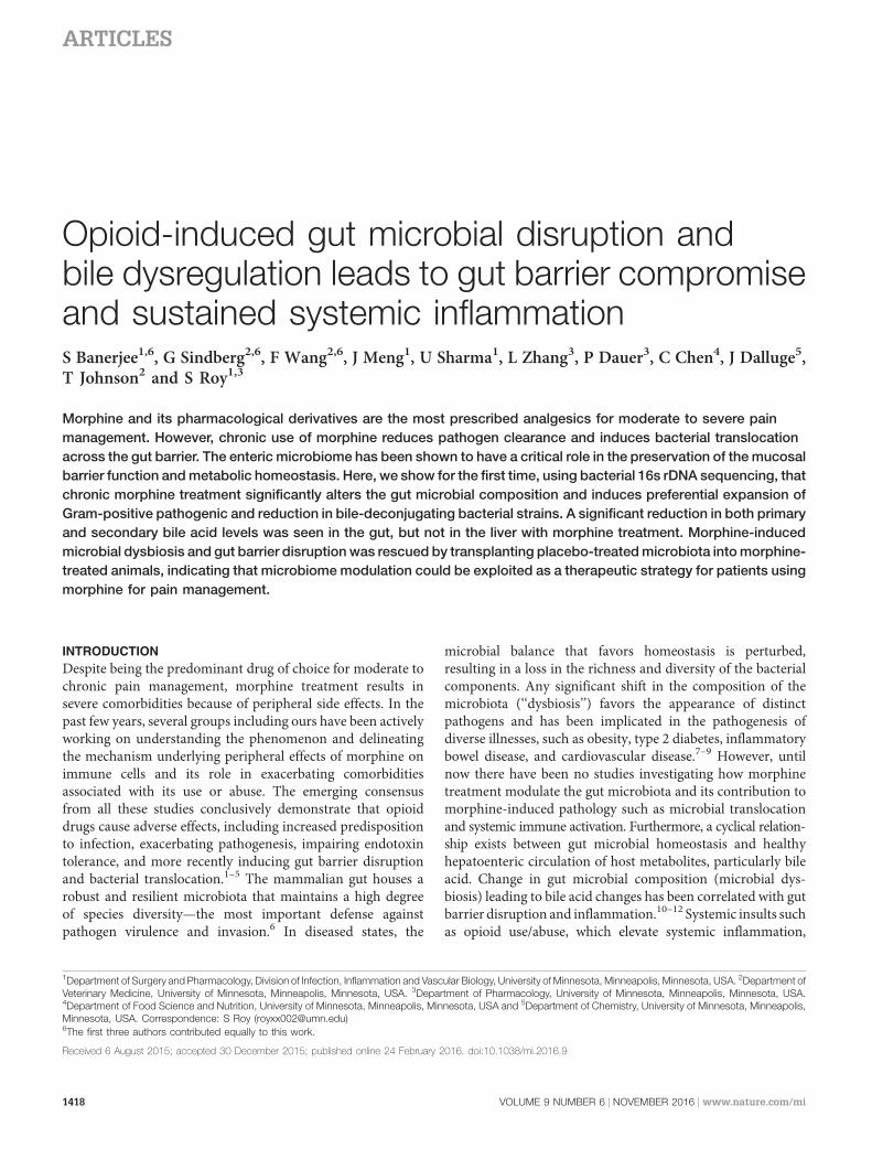
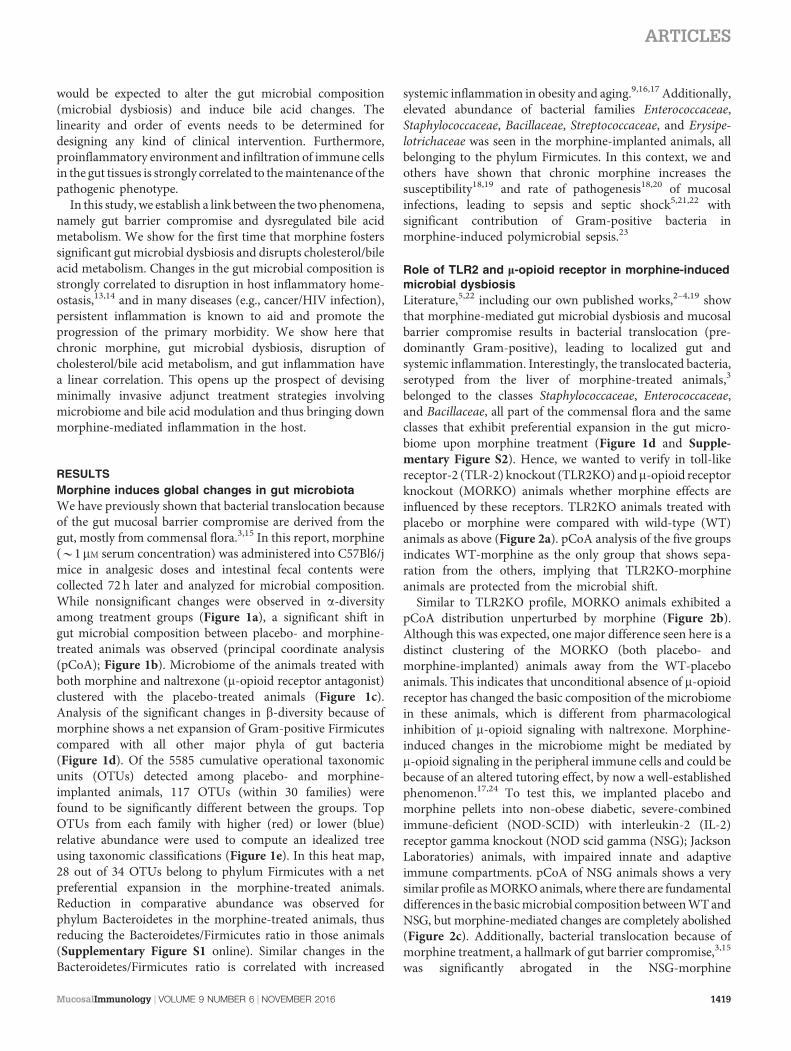
Opioid-induced gut microbial disruption and bile dysregulation leads to gut barrier compromise and sustained systemic inflammation S Banerjee 1,6 , G Sindberg 2,6 , F Wang 2,6 , J Meng 1 , U Sharma 1 , L Zhang 3 , P Dauer 3 , C Chen 4 , J Dalluge 5 , T Johnson 2 and S Roy 1,3 Morphine and its pharmacological derivatives are the most prescribed analgesics for moderate to severe pain management. However, chronic use of morphine reduces pathogen clearance and induces bacterial translocation across the gut barrier. The enteric microbiome has been shown to have a critical role in the preservation of the mucosal barrier function and metabolic homeostasis. Here, we show for the first time, using bacterial 16s rDNA sequencing, that chronic morphine treatment significantly alters the gut microbial composition and induces preferential expansion of Gram-positive pathogenic and reduction in bile-deconjugating bacterial strains. A significant reduction in both primary and secondary bile acid levels was seen in the gut, but not in the liver with morphine treatment. Morphine-induced microbial dysbiosis and gut barrier disruption was rescued by transplanting placebo-treated microbiota into morphine- treated animals, indicating that microbiome modulation could be exploited as a therapeutic strategy for patients using morphine for pain management. INTRODUCTION Despite being the predominant drug of choice for moderate to chronic pain management, morphine treatment results in severe comorbidities because of peripheral side effects. In the past few years, several groups including ours have been actively working on understanding the phenomenon and delineating the mechanism underlying peripheral effects of morphine on immune cells and its role in exacerbating comorbidities associated with its use or abuse. The emerging consensus from all these studies conclusively demonstrate that opioid drugs cause adverse effects, including increased predisposition to infection, exacerbating pathogenesis, impairing endotoxin tolerance, and more recently inducing gut barrier disruption and bacterial translocation. 1–5 The mammalian gut houses a robust and resilient microbiota that maintains a high degree of species diversity—the most important defense against pathogen virulence and invasion. 6 In diseased states, the microbial balance that favors homeostasis is perturbed, resulting in a loss in the richness and diversity of the bacterial components. Any significant shift in the composition of the microbiota (‘‘dysbiosis’’) favors the appearance of distinct pathogens and has been implicated in the pathogenesis of diverse illnesses, such as obesity, type 2 diabetes, inflammatory bowel disease, and cardiovascular disease. 7–9 However, until now there have been no studies investigating how morphine treatment modulate the gut microbiota and its contribution to morphine-induced pathology such as microbial translocation and systemic immune activation. Furthermore, a cyclical relation- ship exists between gut microbial homeostasis and healthy hepatoenteric circulation of host metabolites, particularly bile acid. Change in gut microbial composition (microbial dys- biosis) leading to bile acid changes has been correlated with gut barrier disruption and inflammation. 10–12 Systemic insults such as opioid use/abuse, which elevate systemic inflammation, 1 Department of Surgery and Pharmacology, Division of Infection, Inflammation and Vascular Biology, University of Minnesota, Minneapolis, Minnesota, USA. 2 Department of Veterinary Medicine, University of Minnesota, Minneapolis, Minnesota, USA. 3 Department of Pharmacology, University of Minnesota, Minneapolis, Minnesota, USA. 4 Department of Food Science and Nutrition, University of Minnesota, Minneapolis, Minnesota, USA and 5 Department of Chemistry, University of Minnesota, Minneapolis, Minnesota, USA. Correspondence: S Roy ([email protected]) 6 The first three authors contributed equally to this work. Received 6 August 2015; accepted 30 December 2015; published online 24 February 2016. doi:10.1038/mi.2016.9 ARTICLES 1418 VOLUME 9 NUMBER 6 | NOVEMBER 2016 | www.nature.com/mi

Welcome message from author
This document is posted to help you gain knowledge. Please leave a comment to let me know what you think about it! Share it to your friends and learn new things together.
Transcript
Opioid-induced gut microbial disruption andbile dysregulation leads to gut barrier compromiseand sustained systemic inflammationS Banerjee1,6, G Sindberg2,6, F Wang2,6, J Meng1, U Sharma1, L Zhang3, P Dauer3, C Chen4, J Dalluge5,T Johnson2 and S Roy1,3
Morphine and its pharmacological derivatives are the most prescribed analgesics for moderate to severe pain
management. However, chronic use of morphine reduces pathogen clearance and induces bacterial translocation
across the gut barrier. The enteric microbiome has been shown to have a critical role in the preservation of the mucosal
barrier function and metabolic homeostasis. Here, we show for the first time, using bacterial 16s rDNA sequencing, that
chronic morphine treatment significantly alters the gut microbial composition and induces preferential expansion of
Gram-positive pathogenic and reduction in bile-deconjugating bacterial strains. A significant reduction in both primary
and secondary bile acid levels was seen in the gut, but not in the liver with morphine treatment. Morphine-induced
microbial dysbiosis and gut barrier disruption was rescued by transplanting placebo-treated microbiota into morphine-
treated animals, indicating that microbiome modulation could be exploited as a therapeutic strategy for patients using
morphine for pain management.
INTRODUCTION
Despite being the predominant drug of choice for moderate tochronic pain management, morphine treatment results insevere comorbidities because of peripheral side effects. In thepast few years, several groups including ours have been activelyworking on understanding the phenomenon and delineatingthe mechanism underlying peripheral effects of morphine onimmune cells and its role in exacerbating comorbiditiesassociated with its use or abuse. The emerging consensusfrom all these studies conclusively demonstrate that opioiddrugs cause adverse effects, including increased predispositionto infection, exacerbating pathogenesis, impairing endotoxintolerance, and more recently inducing gut barrier disruptionand bacterial translocation.1–5 The mammalian gut houses arobust and resilient microbiota that maintains a high degreeof species diversity—the most important defense againstpathogen virulence and invasion.6 In diseased states, the
microbial balance that favors homeostasis is perturbed,resulting in a loss in the richness and diversity of the bacterialcomponents. Any significant shift in the composition of themicrobiota (‘‘dysbiosis’’) favors the appearance of distinctpathogens and has been implicated in the pathogenesis ofdiverse illnesses, such as obesity, type 2 diabetes, inflammatorybowel disease, and cardiovascular disease.7–9 However, untilnow there have been no studies investigating how morphinetreatment modulate the gut microbiota and its contribution tomorphine-induced pathology such as microbial translocationand systemic immune activation. Furthermore, a cyclical relation-ship exists between gut microbial homeostasis and healthyhepatoenteric circulation of host metabolites, particularly bileacid. Change in gut microbial composition (microbial dys-biosis) leading to bile acid changes has been correlated with gutbarrier disruption and inflammation.10–12 Systemic insults suchas opioid use/abuse, which elevate systemic inflammation,
1Department of Surgery and Pharmacology, Division of Infection, Inflammation and Vascular Biology, University of Minnesota, Minneapolis, Minnesota, USA. 2Department ofVeterinary Medicine, University of Minnesota, Minneapolis, Minnesota, USA. 3Department of Pharmacology, University of Minnesota, Minneapolis, Minnesota, USA.4Department of Food Science and Nutrition, University of Minnesota, Minneapolis, Minnesota, USA and 5Department of Chemistry, University of Minnesota, Minneapolis,Minnesota, USA. Correspondence: S Roy ([email protected])6The first three authors contributed equally to this work.
Received 6 August 2015; accepted 30 December 2015; published online 24 February 2016. doi:10.1038/mi.2016.9
ARTICLES
1418 VOLUME 9 NUMBER 6 | NOVEMBER 2016 |www.nature.com/mi
would be expected to alter the gut microbial composition(microbial dysbiosis) and induce bile acid changes. Thelinearity and order of events needs to be determined fordesigning any kind of clinical intervention. Furthermore,proinflammatory environment and infiltration of immune cellsin the gut tissues is strongly correlated to the maintenance of thepathogenic phenotype.
In this study, we establish a link between the two phenomena,namely gut barrier compromise and dysregulated bile acidmetabolism. We show for the first time that morphine fosterssignificant gut microbial dysbiosis and disrupts cholesterol/bileacid metabolism. Changes in the gut microbial composition isstrongly correlated to disruption in host inflammatory home-ostasis,13,14 and in many diseases (e.g., cancer/HIV infection),persistent inflammation is known to aid and promote theprogression of the primary morbidity. We show here thatchronic morphine, gut microbial dysbiosis, disruption ofcholesterol/bile acid metabolism, and gut inflammation havea linear correlation. This opens up the prospect of devisingminimally invasive adjunct treatment strategies involvingmicrobiome and bile acid modulation and thus bringing downmorphine-mediated inflammation in the host.
RESULTS
Morphine induces global changes in gut microbiota
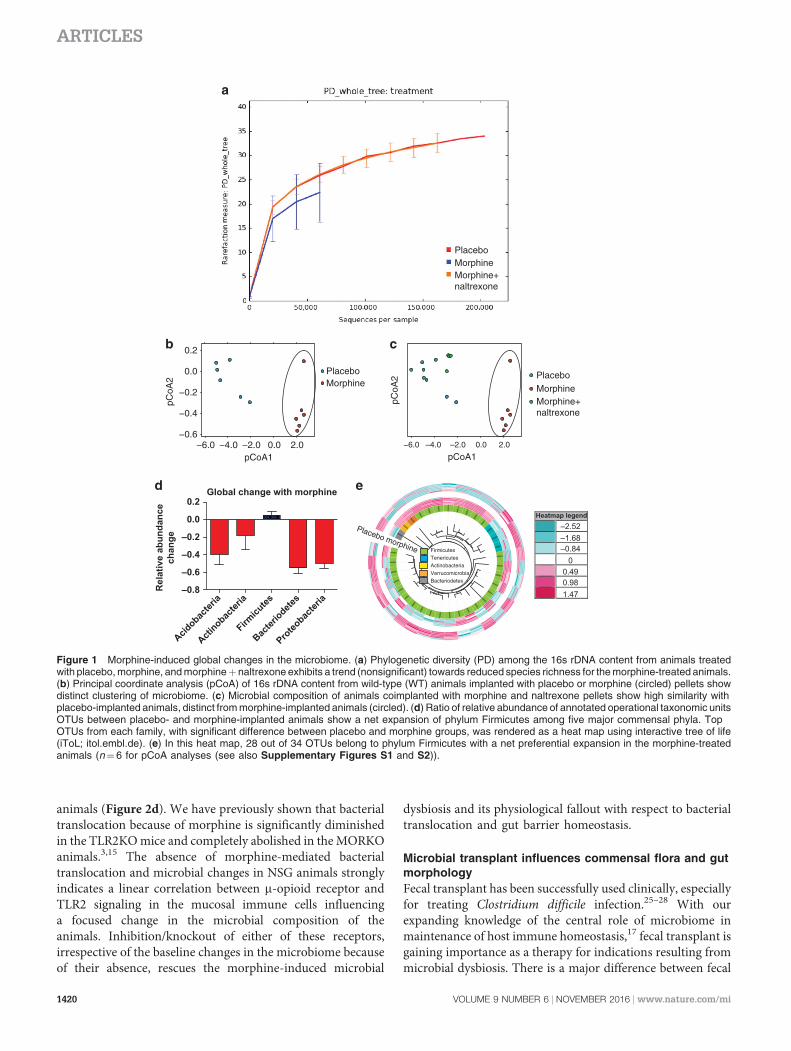
We have previously shown that bacterial translocation becauseof the gut mucosal barrier compromise are derived from thegut, mostly from commensal flora.3,15 In this report, morphine(B1mM serum concentration) was administered into C57Bl6/jmice in analgesic doses and intestinal fecal contents werecollected 72 h later and analyzed for microbial composition.While nonsignificant changes were observed in a-diversityamong treatment groups (Figure 1a), a significant shift ingut microbial composition between placebo- and morphine-treated animals was observed (principal coordinate analysis(pCoA); Figure 1b). Microbiome of the animals treated withboth morphine and naltrexone (m-opioid receptor antagonist)clustered with the placebo-treated animals (Figure 1c).Analysis of the significant changes in b-diversity because ofmorphine shows a net expansion of Gram-positive Firmicutescompared with all other major phyla of gut bacteria(Figure 1d). Of the 5585 cumulative operational taxonomicunits (OTUs) detected among placebo- and morphine-implanted animals, 117 OTUs (within 30 families) werefound to be significantly different between the groups. TopOTUs from each family with higher (red) or lower (blue)relative abundance were used to compute an idealized treeusing taxonomic classifications (Figure 1e). In this heat map,28 out of 34 OTUs belong to phylum Firmicutes with a netpreferential expansion in the morphine-treated animals.Reduction in comparative abundance was observed forphylum Bacteroidetes in the morphine-treated animals, thusreducing the Bacteroidetes/Firmicutes ratio in those animals(Supplementary Figure S1 online). Similar changes in theBacteroidetes/Firmicutes ratio is correlated with increased
systemic inflammation in obesity and aging.9,16,17 Additionally,elevated abundance of bacterial families Enterococcaceae,Staphylococcaceae, Bacillaceae, Streptococcaceae, and Erysipe-lotrichaceae was seen in the morphine-implanted animals, allbelonging to the phylum Firmicutes. In this context, we andothers have shown that chronic morphine increases thesusceptibility18,19 and rate of pathogenesis18,20 of mucosalinfections, leading to sepsis and septic shock5,21,22 withsignificant contribution of Gram-positive bacteria inmorphine-induced polymicrobial sepsis.23
Role of TLR2 and l-opioid receptor in morphine-inducedmicrobial dysbiosis
Literature,5,22 including our own published works,2–4,19 showthat morphine-mediated gut microbial dysbiosis and mucosalbarrier compromise results in bacterial translocation (pre-dominantly Gram-positive), leading to localized gut andsystemic inflammation. Interestingly, the translocated bacteria,serotyped from the liver of morphine-treated animals,3
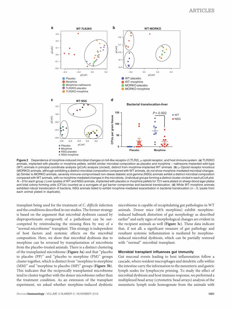
belonged to the classes Staphylococcaceae, Enterococcaceae,and Bacillaceae, all part of the commensal flora and the sameclasses that exhibit preferential expansion in the gut micro-biome upon morphine treatment (Figure 1d and Supple-mentary Figure S2). Hence, we wanted to verify in toll-likereceptor-2 (TLR-2) knockout (TLR2KO) andm-opioid receptorknockout (MORKO) animals whether morphine effects areinfluenced by these receptors. TLR2KO animals treated withplacebo or morphine were compared with wild-type (WT)animals as above (Figure 2a). pCoA analysis of the five groupsindicates WT-morphine as the only group that shows sepa-ration from the others, implying that TLR2KO-morphineanimals are protected from the microbial shift.
Similar to TLR2KO profile, MORKO animals exhibited apCoA distribution unperturbed by morphine (Figure 2b).Although this was expected, one major difference seen here is adistinct clustering of the MORKO (both placebo- andmorphine-implanted) animals away from the WT-placeboanimals. This indicates that unconditional absence of m-opioidreceptor has changed the basic composition of the microbiomein these animals, which is different from pharmacologicalinhibition of m-opioid signaling with naltrexone. Morphine-induced changes in the microbiome might be mediated bym-opioid signaling in the peripheral immune cells and could bebecause of an altered tutoring effect, by now a well-establishedphenomenon.17,24 To test this, we implanted placebo andmorphine pellets into non-obese diabetic, severe-combinedimmune-deficient (NOD-SCID) with interleukin-2 (IL-2)receptor gamma knockout (NOD scid gamma (NSG); JacksonLaboratories) animals, with impaired innate and adaptiveimmune compartments. pCoA of NSG animals shows a verysimilar profile as MORKO animals, where there are fundamentaldifferences in the basic microbial composition between WT andNSG, but morphine-mediated changes are completely abolished(Figure 2c). Additionally, bacterial translocation because ofmorphine treatment, a hallmark of gut barrier compromise,3,15
was significantly abrogated in the NSG-morphine
ARTICLES
MucosalImmunology | VOLUME 9 NUMBER 6 | NOVEMBER 2016 1419
animals (Figure 2d). We have previously shown that bacterialtranslocation because of morphine is significantly diminishedin the TLR2KO mice and completely abolished in the MORKOanimals.3,15 The absence of morphine-mediated bacterialtranslocation and microbial changes in NSG animals stronglyindicates a linear correlation between m-opioid receptor andTLR2 signaling in the mucosal immune cells influencinga focused change in the microbial composition of theanimals. Inhibition/knockout of either of these receptors,irrespective of the baseline changes in the microbiome becauseof their absence, rescues the morphine-induced microbial
dysbiosis and its physiological fallout with respect to bacterialtranslocation and gut barrier homeostasis.
Microbial transplant influences commensal flora and gutmorphology
Fecal transplant has been successfully used clinically, especiallyfor treating Clostridium difficile infection.25–28 With ourexpanding knowledge of the central role of microbiome inmaintenance of host immune homeostasis,17 fecal transplant isgaining importance as a therapy for indications resulting frommicrobial dysbiosis. There is a major difference between fecal
−0.6
−0.4
−0.2
0.0
0.2
PlaceboMorphine
pCoA1
pCoA
2 PlaceboMorphineMorphine+naltrexone
pCoA1
pCoA
2
FirmicutesTenericutesActinobacteriaVerrucomicrobiaBacteriodetes
Heatmap legend
PlaceboMorphineMorphine+naltrexone
−6.0 −4.0 −2.0 0.0 2.0
Rel
ativ
e ab
unda
nce
chan
ge
0.2
0.0
–0.2
–0.4
–0.6
–0.8
Proteo
bacter
ia
Bacter
iodetes
Firmicu
tes
Actinobac
teria
Acidobac
teria
Global change with morphine
Placebo morphine
−6.0 2.00.0–2.0–4.0
–2.52–1.68–0.84
00.490.981.47
Figure 1 Morphine-induced global changes in the microbiome. (a) Phylogenetic diversity (PD) among the 16s rDNA content from animals treatedwith placebo, morphine, and morphineþnaltrexone exhibits a trend (nonsignificant) towards reduced species richness for the morphine-treated animals.(b) Principal coordinate analysis (pCoA) of 16s rDNA content from wild-type (WT) animals implanted with placebo or morphine (circled) pellets showdistinct clustering of microbiome. (c) Microbial composition of animals coimplanted with morphine and naltrexone pellets show high similarity withplacebo-implanted animals, distinct from morphine-implanted animals (circled). (d) Ratio of relative abundance of annotated operational taxonomic unitsOTUs between placebo- and morphine-implanted animals show a net expansion of phylum Firmicutes among five major commensal phyla. TopOTUs from each family, with significant difference between placebo and morphine groups, was rendered as a heat map using interactive tree of life(iToL; itol.embl.de). (e) In this heat map, 28 out of 34 OTUs belong to phylum Firmicutes with a net preferential expansion in the morphine-treatedanimals (n¼6 for pCoA analyses (see also Supplementary Figures S1 and S2)).
ARTICLES
1420 VOLUME 9 NUMBER 6 | NOVEMBER 2016 |www.nature.com/mi
transplant being used for the treatment of C. difficile infectionand the conditions described in our studies. The former strategyis based on the argument that microbial dysbiosis caused bydisproportionate overgrowth of a pathobiont can be out-competed by reintroducing the missing flora by way of a‘‘normal microbiome’’ transplant. This strategy is independentof host factors and systemic effects on the microbialcomposition. Here, we show that microbial dysbiosis due tomorphine can be reversed by transplantation of microbiotafrom the placebo-treated animals. There is a distinct clusteringof the transplanted microbiome (Figure 3a) and that ‘‘placeboto placebo (PP)’’ and ‘‘placebo to morphine (PM)’’ groupscluster together, which is distinct from ‘‘morphine to morphine(MM)’’ and ‘‘morphine to placebo (MP)’’ groups (Figure 3b).This indicates that the reciprocally transplanted microbiometend to cluster together with the donor microbiome rather thanthe treatment condition. As an extension of the transplantexperiment, we asked whether morphine-induced dysbiotic
microbiome is capable of recapitulating gut pathologies in WTanimals. Donor mice (48 h morphine) exhibit morphine-induced hallmark distortion of gut morphology as describedearlier4 and early signs of morphological changes are evident inthe recipient animals as well (Figure 3c). These data indicatethat, if not all, a significant measure of gut pathology andresultant systemic inflammation is mediated by morphine-induced microbial dysbiosis, which can be partially restoredwith ‘‘normal’’ microbial transplant.
Microbial transplant influences gut immunity
Gut mucosal events leading to host inflammation follow acascade, where resident macrophages and dendritic cells withinthe intestine carry the information to the mesenteric and gastriclymph nodes for lymphocyte priming. To study the effect ofmicrobial dysbiosis and host immune response, we performed amultiplexed bead array (cytometric bead arrays) analysis of themesenteric lymph node homogenate from the animals with
c
a
b
−0.4 −0.2 0.0 0.2
−0.2
−0.1
0.0
0.1
0.2
0.3
PlaceboMorphineMorphine+naltrexoneTLR2KO-placeboTLR2KO-morphine
WT-TLR2KO
−0.2 0.0 0.2 0.4
−0.2
−0.1
0.0
0.1
0.2
WT-MORKO
WT-placeboWT-morphineMORKO-placeboMORKO-morphine
−0.6
−0.4
−0.2
0.0
0.2
−0.4 −0.2 0.0 0.2−0.6
PlaceboMorphine
pCoA1
pCoA
2
NSG-placeboNSG-morphine
WT-NSG
pCoA
2
pCoA
2
pCoA1 pCoA1
Placebo Morphine0
50
100
150C
FUs
per g
ram
tiss
ue
Bacterial translocation-liver
WTNSG
P= 0.002
d
Figure 2 Dependence of morphine-induced microbial changes on toll-like receptor-2 (TLR2), m-opioid receptor, and host immune system. (a) TLR2KOanimals, implanted with placebo or morphine pellets, exhibit similar microbial composition as placebo and morphineþnaltrexone implanted wild-type(WT) animals in principal coordinate analysis (pCoA) analysis (circled), distinct from morphine-implanted WT animals. (b) m-Opioid receptor knockout(MORKO) animals, although exhibiting a distinct microbial composition compared with WT animals, do not show morphine-mediated microbial changes.(c) Similar to MORKO animals, severely immune-compromised non-obese diabetic scid gamma (NSG) animals exhibit a distinct microbial compositioncompared with WT animals, with no morphine-mediated changes in the microbiota. (Individual groups forming a distinct cluster circled in each pCoA plot.N¼6 for each group.) Liver lysates of WT and NSG animals, implanted with placebo or morphine pellets for 72 h were plated on sheep-blood agar platesand total colony-forming units (CFUs) counted as a surrogate of gut barrier compromise and bacterial translocation. (d) While WT-morphine animalsexhibited robust translocation of bacteria, NSG animals failed to exhibit morphine-mediated exacerbation in bacterial translocation (n¼ 5; lysate fromeach animal plated in duplicate).
ARTICLES
MucosalImmunology | VOLUME 9 NUMBER 6 | NOVEMBER 2016 1421

fecal transplant from above. Among all cytokines analyzed,only IL-17 and IL-10 showed significant differences amonggroups. Elevated levels of IL-17 (Figure 4a) and attenuatedlevels of IL-10 (Supplementary Figure S3A) were seen in MM
compared with the rest of the four study groups, consistent withthe described role of IL-17 in host inflammation arising fromcompromised gut homeostasis, specifically in the context ofGram-positive infection.29–32 The reciprocally transplanted
Figure 3 Donor microbiome predominates treatment condition and influences gut physiology. (a) Reciprocal transplant of microbiota preservesdistinctness of microbial composition, which is closer to the donor microbiome, rather than the treatment condition (placebo or morphine alone; b). (c) Thisphysiologically translates to gut pathologies associated with morphine-induced dysbiotic microbiome, e.g., transplantation of morphine-induced dysbioticmicrobiome into healthy wild-type (WT) animals results in ‘‘morphine-like’’ diseased phenotype, very similar to morphine-implanted donor animals,whereas transplantation of ‘‘normal’’ microbiome into morphine-treated animals shows distinct improvement in the gut pathology. Arrows indicate sites ofgut injury.
a b
No treatment Recipient0
200
400
600
800
1,000
CFU
per
g ti
ssue
Liver
P= 0.0313
No treatment Donor Recipient0
50
100
150
pg m
l–1
IL-17-liver
P= 0.001 P= 0.0003
No treatment Donor Recipient0
50
100
150
IL-6-liverP= 0.0068
P= 0.0067 P= 0.0062
c
PPMM MP PM
0
50
100
150
200
250MLN-IL-17
pg m
l–1
**
####
d
pg m
l–1
Figure 4 Microbial transplant, immune response, and rescue. (a) Morphine treatment results in a robust interleukin-17 (IL-17) response in themesenteric lymph node (MLN) and transplantation of placebo microbiome rescues the inflammatory phenotype. (b) Reconstitution of dysbioticmicrobiome alone is sufficient to recapitulate most of the morphine-induced gut pathologies, including bacterial translocation across the intestinalmucosa. Systemic IL-17 response was only seen in (c) donor animals with morphine treatment and (d) both donors and recipients exhibit a systemicIL-6 response indicating proinflammatory environment in the host. In all cases (a–d), morphine-implanted animals receiving ‘‘normal’’ microbiotashow baseline levels of IL-17 (**Po0.05 between indicated groups; ##Po0.05 compared with morphine to morphine (MM)). Also see SupplementaryFigure S3.
ARTICLES
1422 VOLUME 9 NUMBER 6 | NOVEMBER 2016 |www.nature.com/mi
groups, MP and PM, maintained basal levels of the cytokines,comparable with PP. The lack of IL-17 or IL-10 response in MPor PM animals could be because of compensatory (averaging)effects between microbiome and host immune system. Alter-natively, in MP animals, transplanting dysbiotic microbiomemay change the general composition of the commensal floraand yet may not have resulted in severe mucosal barriercompromise to affect systemic immune response within 72 h.In PM animals, on the other hand, some measure of rescuebecause of microbial restoration may have resulted indiminished proinflammatory response.
Additionally, we set out to determine the overall systemicresponse to microbial dysbiosis by investigating the bloodcytokine levels in these animals. No significant differences werefound among the WT, TLR2KO, and MORKO animals underdifferent treatments (Supplementary Figure S3B–D). Asignificant decrease in IL-2 and IL-4 levels was observed inthe reciprocal fecal transplant animals compared with PP andMM, but no corresponding elevation in proinflammatorycytokines was observed (Supplementary Figure S3E). Thisindicates that the 72 h time point may not be sufficient to elicitproinflammatory biomarkers in the systemic circulation;however, localized inflammatory response is initiated withinthe intestine and mesenteric drainage in immunocompetentanimals. We have previously shown that morphine exacerbatesTLR-mediated inflammation,3,4,15 and basal levels of inflam-mation in the PM animals clearly exhibits protective effectbecause of the restoration of microbial homeostasis with fecaltransplant.
In the absence of systemic morphine, fecal transplant ofdysbiotic microbiome alone was sufficient to induce bacterialtranslocation to the liver (Figure 4b). Similar to MM animals inFigure 4a, fecal transplant induced IL-17 response in the liverof the donor mice, but not in the recipient animals (Figure 4c).There was a significant IL-6 response both in donor andrecipient animals (Figure 4d), indicating that IL-17-mediatedgut inflammation is contingent upon morphine-mediated hostimmunomodulation, whereas IL-6-mediated systemic inflam-mation is a result of bacterial translocation, common to bothdonors and recipient animals. This is consistent with ourobservation that IL-17 is an early response, whereas IL-6 is thesustained host response to morphine-mediated changes in guthomeostasis.4,12
Morphine-induced cholesterol/bile acid imbalance
Recent reports implicate increase in hydrophobic secondarybile acids (lithocholic acid and deoxycholic acid) and con-comitant decrease in hydrophilic secondary bile acids (urso-deoxycholic acid (UDCA)) in gut barrier disruption, bacterialtranslocation, and intestinal inflammation.13,14 As secondarybile acids are produced in entirety through gut microbialfermentation from primary bile acids (cholic acid andchenodeoxycholic acid), secondary bile imbalance is directlycorrelated, and associated with microbial dysbiosis.33 Exclu-sively within the host liver, primary bile acids are producedfrom cholesterol catabolism and is, in fact, one of the major
ways of eliminating excess cholesterol from the body.34 Thesecondary bile acids recirculate to the host liver through variousbile transporters and also signal through intestinal receptors,constituting the hepatoenteric bile circulation.11,35
To evaluate if morphine treatment induces metabolicchanges in the gut, WT and TLR2KO animals were implantedwith placebo or morphine pellet for 72 h as described above andthe fecal content was analyzed with mass spectrometry forchanges in major metabolites. Of 310 compounds tested, lipidmetabolites and especially bile acids (both primary andsecondary) exhibited significant changes because of morphine(Supplementary Figure S4). One of the major observationswas significantly high level of coprostanol in the morphine-implanted animals (Figure 5a). This could be because ofabnormal release of cholesterol into the gut or because ofselective expansion of microbes known for this conversion. Onthe other hand, concentrations of the host-derived primary(Figure 5b,c) and microbe-converted secondary bile acids(Figure 5d,e) were seen to diminish significantly in the feces ofmorphine-implanted animals compared with placebo andmorphineþ naltrexone-implanted animals. The morphine-mediated changes were completely abolished in the TLR2KOanimals (Figure 5f).
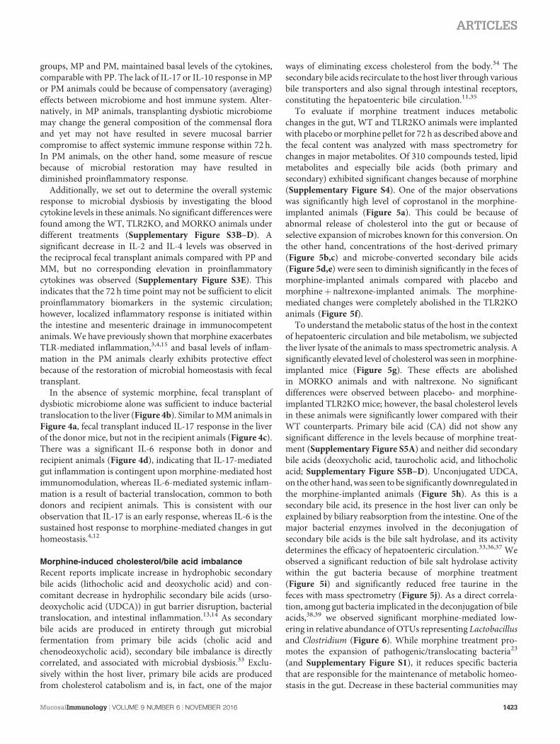
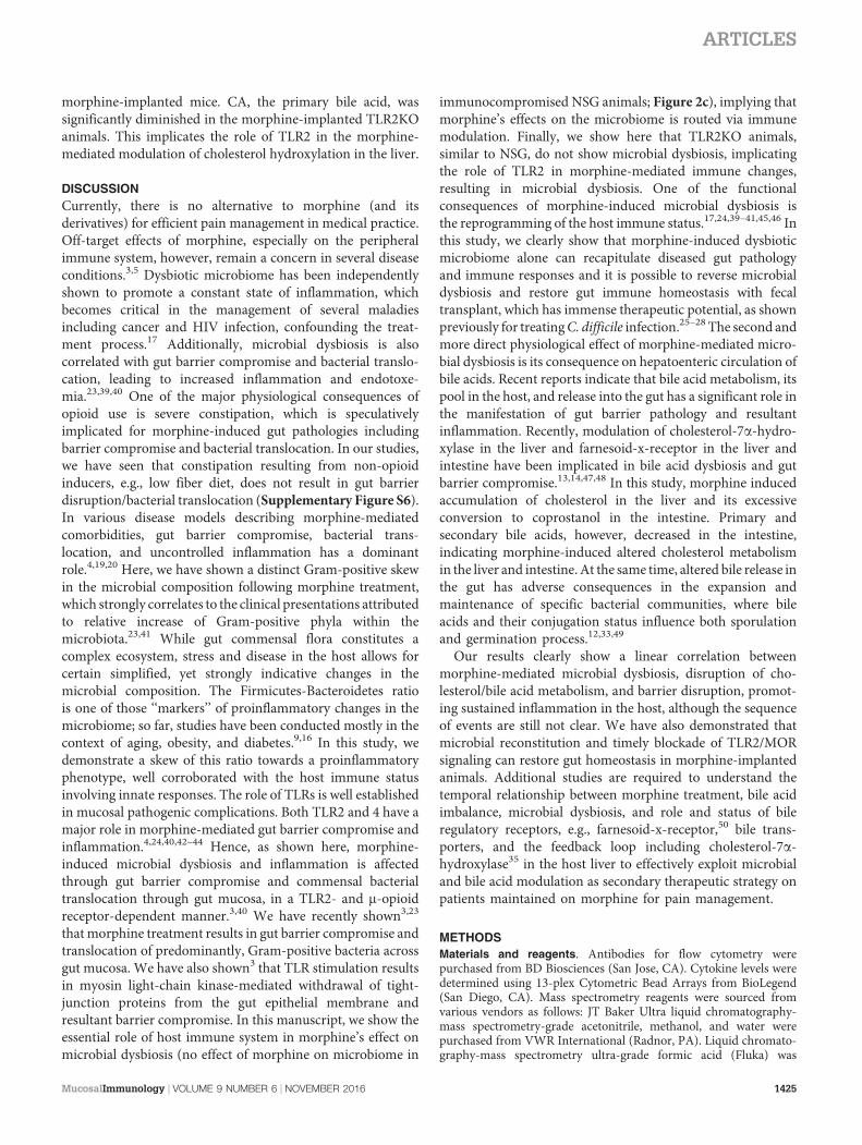
To understand the metabolic status of the host in the contextof hepatoenteric circulation and bile metabolism, we subjectedthe liver lysate of the animals to mass spectrometric analysis. Asignificantly elevated level of cholesterol was seen in morphine-implanted mice (Figure 5g). These effects are abolishedin MORKO animals and with naltrexone. No significantdifferences were observed between placebo- and morphine-implanted TLR2KO mice; however, the basal cholesterol levelsin these animals were significantly lower compared with theirWT counterparts. Primary bile acid (CA) did not show anysignificant difference in the levels because of morphine treat-ment (Supplementary Figure S5A) and neither did secondarybile acids (deoxycholic acid, taurocholic acid, and lithocholicacid; Supplementary Figure S5B–D). Unconjugated UDCA,on the other hand, was seen to be significantly downregulated inthe morphine-implanted animals (Figure 5h). As this is asecondary bile acid, its presence in the host liver can only beexplained by biliary reabsorption from the intestine. One of themajor bacterial enzymes involved in the deconjugation ofsecondary bile acids is the bile salt hydrolase, and its activitydetermines the efficacy of hepatoenteric circulation.33,36,37 Weobserved a significant reduction of bile salt hydrolase activitywithin the gut bacteria because of morphine treatment(Figure 5i) and significantly reduced free taurine in thefeces with mass spectrometry (Figure 5j). As a direct correla-tion, among gut bacteria implicated in the deconjugation of bileacids,38,39 we observed significant morphine-mediated low-ering in relative abundance of OTUs representing Lactobacillusand Clostridium (Figure 6). While morphine treatment pro-motes the expansion of pathogenic/translocating bacteria23
(and Supplementary Figure S1), it reduces specific bacteriathat are responsible for the maintenance of metabolic homeo-stasis in the gut. Decrease in these bacterial communities may
ARTICLES
MucosalImmunology | VOLUME 9 NUMBER 6 | NOVEMBER 2016 1423
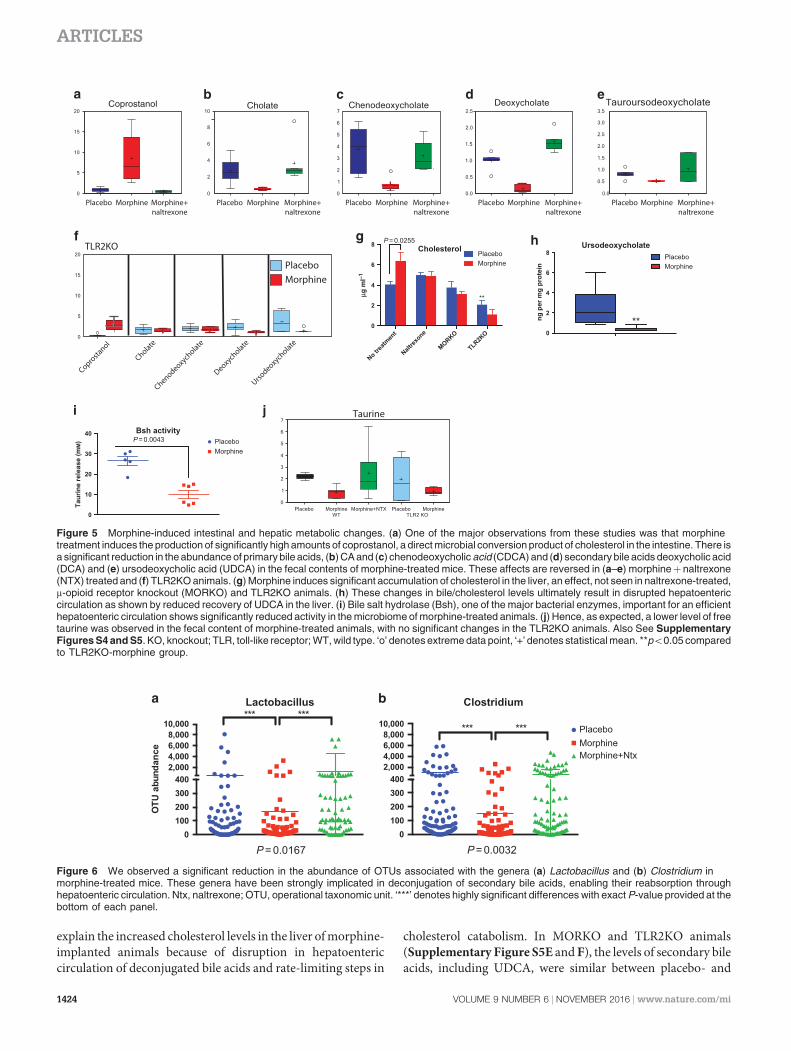
explain the increased cholesterol levels in the liver of morphine-implanted animals because of disruption in hepatoentericcirculation of deconjugated bile acids and rate-limiting steps in
cholesterol catabolism. In MORKO and TLR2KO animals(Supplementary Figure S5E and F), the levels of secondary bileacids, including UDCA, were similar between placebo- and
0100200300400
2,0004,0006,0008,000
10,000
OTU
abu
ndan
ce
Lactobacillus
P= 0.0167
*** ***
0100200300400
2,0004,0006,0008,000
10,000
Clostridium
PlaceboMorphineMorphine+Ntx
P= 0.0032
*** ***
a b
Figure 6 We observed a significant reduction in the abundance of OTUs associated with the genera (a) Lactobacillus and (b) Clostridium inmorphine-treated mice. These genera have been strongly implicated in deconjugation of secondary bile acids, enabling their reabsorption throughhepatoenteric circulation. Ntx, naltrexone; OTU, operational taxonomic unit. ‘***’ denotes highly significant differences with exact P-value provided at thebottom of each panel.
Placebo Morphine Morphine+naltrexone
b
Placebo Morphine Morphine+naltrexone
c
Placebo Morphine Morphine+naltrexone
a
MorphinePlacebo
TLR2KO
Coprostanol
Cholate
Chenodeoxycholate
Deoxycholate
Ursodeoxycholate
f
No trea
tmen
t
Naltrex
one
MORKO
TLR2KO
0
2
4
6
8
µg m
l–1
PlaceboMorphine
P= 0.0255
**
Cholesterolg
0
2
4
6
8
ng p
er m
g pr
otei
n
UrsodeoxycholatePlaceboMorphine
**
h
0
10
20
30
40
Taur
ine
rele
ase
(mM
)
Bsh activityPlaceboMorphine
P= 0.0043
i Taurinej
Placebo Morphine Morphine+naltrexone
e
Placebo Morphine Morphine+naltrexone
d
Figure 5 Morphine-induced intestinal and hepatic metabolic changes. (a) One of the major observations from these studies was that morphinetreatment induces the production of significantly high amounts of coprostanol, a direct microbial conversion product of cholesterol in the intestine. There isa significant reduction in the abundance of primary bile acids, (b) CA and (c) chenodeoxycholic acid (CDCA) and (d) secondary bile acids deoxycholic acid(DCA) and (e) ursodeoxycholic acid (UDCA) in the fecal contents of morphine-treated mice. These affects are reversed in (a–e) morphineþ naltrexone(NTX) treated and (f) TLR2KO animals. (g) Morphine induces significant accumulation of cholesterol in the liver, an effect, not seen in naltrexone-treated,m-opioid receptor knockout (MORKO) and TLR2KO animals. (h) These changes in bile/cholesterol levels ultimately result in disrupted hepatoentericcirculation as shown by reduced recovery of UDCA in the liver. (i) Bile salt hydrolase (Bsh), one of the major bacterial enzymes, important for an efficienthepatoenteric circulation shows significantly reduced activity in the microbiome of morphine-treated animals. (j) Hence, as expected, a lower level of freetaurine was observed in the fecal content of morphine-treated animals, with no significant changes in the TLR2KO animals. Also See SupplementaryFigures S4 and S5. KO, knockout; TLR, toll-like receptor; WT, wild type. ‘o’ denotes extreme data point, ‘+’ denotes statistical mean. **po0.05 comparedto TLR2KO-morphine group.
ARTICLES
1424 VOLUME 9 NUMBER 6 | NOVEMBER 2016 |www.nature.com/mi
morphine-implanted mice. CA, the primary bile acid, wassignificantly diminished in the morphine-implanted TLR2KOanimals. This implicates the role of TLR2 in the morphine-mediated modulation of cholesterol hydroxylation in the liver.
DISCUSSION
Currently, there is no alternative to morphine (and itsderivatives) for efficient pain management in medical practice.Off-target effects of morphine, especially on the peripheralimmune system, however, remain a concern in several diseaseconditions.3,5 Dysbiotic microbiome has been independentlyshown to promote a constant state of inflammation, whichbecomes critical in the management of several maladiesincluding cancer and HIV infection, confounding the treat-ment process.17 Additionally, microbial dysbiosis is alsocorrelated with gut barrier compromise and bacterial translo-cation, leading to increased inflammation and endotoxe-mia.23,39,40 One of the major physiological consequences ofopioid use is severe constipation, which is speculativelyimplicated for morphine-induced gut pathologies includingbarrier compromise and bacterial translocation. In our studies,we have seen that constipation resulting from non-opioidinducers, e.g., low fiber diet, does not result in gut barrierdisruption/bacterial translocation (Supplementary Figure S6).In various disease models describing morphine-mediatedcomorbidities, gut barrier compromise, bacterial trans-location, and uncontrolled inflammation has a dominantrole.4,19,20 Here, we have shown a distinct Gram-positive skewin the microbial composition following morphine treatment,which strongly correlates to the clinical presentations attributedto relative increase of Gram-positive phyla within themicrobiota.23,41 While gut commensal flora constitutes acomplex ecosystem, stress and disease in the host allows forcertain simplified, yet strongly indicative changes in themicrobial composition. The Firmicutes-Bacteroidetes ratiois one of those ‘‘markers’’ of proinflammatory changes in themicrobiome; so far, studies have been conducted mostly in thecontext of aging, obesity, and diabetes.9,16 In this study, wedemonstrate a skew of this ratio towards a proinflammatoryphenotype, well corroborated with the host immune statusinvolving innate responses. The role of TLRs is well establishedin mucosal pathogenic complications. Both TLR2 and 4 have amajor role in morphine-mediated gut barrier compromise andinflammation.4,24,40,42–44 Hence, as shown here, morphine-induced microbial dysbiosis and inflammation is affectedthrough gut barrier compromise and commensal bacterialtranslocation through gut mucosa, in a TLR2- and m-opioidreceptor-dependent manner.3,40 We have recently shown3,23
that morphine treatment results in gut barrier compromise andtranslocation of predominantly, Gram-positive bacteria acrossgut mucosa. We have also shown3 that TLR stimulation resultsin myosin light-chain kinase-mediated withdrawal of tight-junction proteins from the gut epithelial membrane andresultant barrier compromise. In this manuscript, we show theessential role of host immune system in morphine’s effect onmicrobial dysbiosis (no effect of morphine on microbiome in
immunocompromised NSG animals; Figure 2c), implying thatmorphine’s effects on the microbiome is routed via immunemodulation. Finally, we show here that TLR2KO animals,similar to NSG, do not show microbial dysbiosis, implicatingthe role of TLR2 in morphine-mediated immune changes,resulting in microbial dysbiosis. One of the functionalconsequences of morphine-induced microbial dysbiosis isthe reprogramming of the host immune status.17,24,39–41,45,46 Inthis study, we clearly show that morphine-induced dysbioticmicrobiome alone can recapitulate diseased gut pathologyand immune responses and it is possible to reverse microbialdysbiosis and restore gut immune homeostasis with fecaltransplant, which has immense therapeutic potential, as shownpreviously for treating C. difficile infection.25–28 The second andmore direct physiological effect of morphine-mediated micro-bial dysbiosis is its consequence on hepatoenteric circulation ofbile acids. Recent reports indicate that bile acid metabolism, itspool in the host, and release into the gut has a significant role inthe manifestation of gut barrier pathology and resultantinflammation. Recently, modulation of cholesterol-7a-hydro-xylase in the liver and farnesoid-x-receptor in the liver andintestine have been implicated in bile acid dysbiosis and gutbarrier compromise.13,14,47,48 In this study, morphine inducedaccumulation of cholesterol in the liver and its excessiveconversion to coprostanol in the intestine. Primary andsecondary bile acids, however, decreased in the intestine,indicating morphine-induced altered cholesterol metabolismin the liver and intestine. At the same time, altered bile release inthe gut has adverse consequences in the expansion andmaintenance of specific bacterial communities, where bileacids and their conjugation status influence both sporulationand germination process.12,33,49
Our results clearly show a linear correlation betweenmorphine-mediated microbial dysbiosis, disruption of cho-lesterol/bile acid metabolism, and barrier disruption, promot-ing sustained inflammation in the host, although the sequenceof events are still not clear. We have also demonstrated thatmicrobial reconstitution and timely blockade of TLR2/MORsignaling can restore gut homeostasis in morphine-implantedanimals. Additional studies are required to understand thetemporal relationship between morphine treatment, bile acidimbalance, microbial dysbiosis, and role and status of bileregulatory receptors, e.g., farnesoid-x-receptor,50 bile trans-porters, and the feedback loop including cholesterol-7a-hydroxylase35 in the host liver to effectively exploit microbialand bile acid modulation as secondary therapeutic strategy onpatients maintained on morphine for pain management.
METHODS
Materials and reagents. Antibodies for flow cytometry werepurchased from BD Biosciences (San Jose, CA). Cytokine levels weredetermined using 13-plex Cytometric Bead Arrays from BioLegend(San Diego, CA). Mass spectrometry reagents were sourced fromvarious vendors as follows: JT Baker Ultra liquid chromatography-mass spectrometry-grade acetonitrile, methanol, and water werepurchased from VWR International (Radnor, PA). Liquid chromato-graphy-mass spectrometry ultra-grade formic acid (Fluka) was
ARTICLES
MucosalImmunology | VOLUME 9 NUMBER 6 | NOVEMBER 2016 1425
purchased from Sigma-Aldrich (Saint Louis, MO). Internal standards2,2,4,4-D4 cholic acid, 2,2,4,4-D4 deoxycholic acid, 2,2,4,4-D4 cheno-deoxycholic acid, 2,2,4,4-D4 ursodeoxycholic acid, and 2,2,4,4-D4
lithocholic acid were purchased from Cambridge Isotope Laboratories(Andover, MA). 2,2,4,4-D4 taurocholic acid was purchased fromAlsaChim (Illkirch-Graffenstaden, France). Millipore AmiconUltrafree PTFE membrane centrifugal filters (0.2 mm) and MilliporeAmicon Ultra 0.5 ml 3,000 MWCO centrifugal filters were purchasedfrom Thermo Fisher Scientific (Waltham, MA).
Mice. C57BL/6 and NSG mice were purchased from JacksonLaboratories (Bar Harbor, ME). TLR2KO and MORKO mice werebred in-house. All animals were maintained in pathogen-free facilitiesand all procedures were approved by the University of MinnesotaInstitutional Animal Care and Use Committee. Typically, 8–10-week-old animals were used for our studies.
Placebo/morphine/naltrexone pellet implantation. Slow releasemorphine pellets (25 mg; B1 mM serum levels of morphine for 5–6days) and corresponding placebo or naltrexone pellets, as appropriate,were kindly provided by National Institute of Drug Abuse (NIDA,National Institutes of Health, Rockville, MD). The implantationprocedure involved 3% isoflurane-induced anesthesia, followed bymaking a small incision at the dorsal torso of the mice. The appropriatepellet was inserted into the small pocket created during incision andthe wound was closed using stainless-steel wound clips. The wholeprocess was carried out under aseptic conditions.
Fecal transplant. Two batches of C57Bl6/j (WT) animals (10 each)were implanted with placebo or morphine pellets as mentioned abovefor 24 or 48 h and their fecal contents collected and pooled. The fecalcontent was processed according to fecal microbial transplant pro-cedure described for human patients51 with modifications. Briefly, thefecal contents were suspended in phosphate-buffered saline(10 mg ml� 1; w v� 1), filtered through a 40 m mesh and centrifuged at6,000g for 20 min. The resultant microbial pellet was resuspended inhalf volume of chilled phosphate-buffered saline and aliquoted intovolumes for single thaw and use. Aliquotes destined for later use werereconstituted to 10% sterile glycerol (Sigma, St. Louis, MO) and storedat � 80 1C. A total of 32 WT mice were used for the transplantexperiment. Animals were implanted with placebo or morphine pellets(12 each) and the stored microbiota was administered (106 colony-forming units (CFUs) per dose) via oral gavage every 24 h three timesaccording to the following scheme: (a) placebo pellet-implantedanimals getting placebo microbiome (PP; n¼ 8), (b) placebopellet-implanted animals getting morphine microbiome (PM; n¼ 8),(c) morphine pellet-implanted animals getting placebo microbiome(MP; n¼ 8), and (d) morphine pellet-implanted animals gettingmorphine microbiome (MM; n¼ 8). Animals were killed 24 h afterthird transplant and fecal contents and tissues harvested for variousdownstream analyses as described.
Sequencing and 16S DNA analysis. Fecal content was collected fromthe gut region encompassing distal cecum and B1 in of the colon andfrozen on dry ice. The fecal matter was lysed using glass beads inMagnaLyser tissue disruptor (Roche, Indianapolis, IA) and total DNAwas isolated using Power-soil/Fecal DNA Isolation Kit (Mo-Bio,Carlsbad, CA) as per the manufacturer’s specifications. All sampleswas quantified via the Qubit Quant-iT dsDNA Broad-Range Kit(Invitrogen, Life Technologies, Grand Island, NY) to ensure that theymet minimum concentration and mass of DNA and submitted toeither Second Genome (San Fransisco, CA) or University of MinnesotaGenomic Center (Minnesota, MN) for microbiome analysis as follows:to enrich the sample for the bacterial 16S V4 rDNA region, DNA wasamplified using fusion primers designed against the surroundingconserved regions that are tailed with sequences to incorporateIllumina (San Diego, CA) flow cell adapters and indexing barcodes.Each sample was PCR amplified with two differently barcoded V4
fusion primers and were advanced for pooling and sequencing. Foreach sample, amplified products were concentrated using a solid-phasereversible immobilization method for the purification of PCR productsand quantified by electrophoresis using an Agilent (Santa Clara, CA)2100 Bioanalyzer. The pooled 16S V4-enriched, amplified, barcodedsamples were loaded into the MiSeq cartridge (Illumina Inc, San Diego,CA) reagent cartridge, and then onto the instrument along with theflow cell. After cluster formation on the MiSeq Instrument (Illumina,San Diego, CA), the amplicons were sequenced for 250 cycles withcustom primers designed for paired-end sequencing. Using QIIME,sequences were quality filtered and demultiplexed using exact matchesto the supplied DNA barcodes. The resulting sequences were thensearched against the Greengenes reference database of 16S sequences,and clustered at 97% by uclust (closed-reference OTU picking).Analysis fora- andb-diversity was carried out with standardized qiimeworkflow at the Minnesota Supercomputing Institute (University ofMinnesota, Minneapolis, MN). To compute the ‘‘global changes withmorphine’’ histogram (Figure 1d), relative abundance for all OTUsconstituting a single phylum in morphine-treated animals werenormalized with the same OTUs in the placebo animals, includingmismatched OTUs. The resulting ratio was analyzed using Prism(GraphPad, LaJolla, CA) software to understand morphine-mediatedperturbations in the five major phyla within the microbiome. The rawdata files for 16s rDNA sequencing have been deposited withArrayExpress with the accession numbers E-MTAB-3722 (nativeeffects of morphine) and E-MTAB-3723 (microbial transplant).
Mass spectrometry for gut and liver metabolites. Fecal metaboliteswere analyzed by Metabolon (Research Triangle Park, NC) using theirproprietary 310 named biochemicals screen. Based on the results,independent analysis of liver bile acids was performed at the Universityof Minnesota mass spectrometric facility as follows: samples foranalysis by ultra-performance liquid chromatography-tandem massspectrometry (MS/MS) were spiked with a fixed volume of theisotopically labeled bile acid internal standards described above. Toeach sample was added an equal volume of liquid chromatography-mass spectrometry-grade methanol. The samples were then cen-trifuged at 12,000 r.p.m. for 5 min and the supernatants removed fromthe proteinaceous pellet. These supernatants were centrifuged throughan Amicon Ultrafree low-binding hydrophilic polytetrafluoroethylenemembrane (0.2 mm) at 12,000 r.p.m. and the filtrates collected. Thesefiltrates were then centrifuged at 14,000g through an Amicon Ultra3 kDa molecular weight cutoff filtration column (Millipore, Billerica,MA) for 30 min, and the flow-through collected for ultra-performanceliquid chromatography-MS/MS analysis. A Waters Acquity UPLCcoupled to a Waters triple quadrupole mass spectrometer (AcquityTQD, Waters Corporation, Milford, MA) was used for separation anddetection of bile acids. A Waters CORTECS C18 2.1 mm� 100 mmcolumn (2.7 mm particles) at 40 1C was used during the following19 min gradient separation with A: water containing 0.1% formic acidand B: acetonitrile containing 0.1% formic acid, at a flow rate of0.6 ml min� 1: 35–40% B, 0–1.5 min; 40–50% B, 1.5–6.0 min; 50% B6.0–7.0 min; 50–97% B, 7.0–14.0 min; 97% B, 14.0–16.0 min; 97–35%B, 16.0–17.0 min; and 35% B, 17.0–19.0 min. By directly infusing eachof the bile acids and corresponding internal standards, cone voltagesand collision energies for each selected reaction monitoring transitionwere optimized. The transitions that produced the highest sensitivityfor the determination of each analyte were selected for quantification.Note that MS1 selected ion monitoring of the precursor ions specifiedlacked the sensitivity and selectivity of MS2 measurement of thefollowing precursor-to-precursor MS/MS transitions. Note also thatthe method of development and validation included identityverification via unique precursor-fragment MS/MS transitions foreach analyte. The following transitions were selected for quantitativeanalysis: lithocholic acid: 375.4–375.4; 2,2,4,4-D4 lithocholic acid:379.4–379.4; cheno-urso- and deoxycholic acids: 391.4–391.4; 2,2,4,4-D4
cheno-urso- and deoxycholic acids: 395.4–395.4; cholic acid: 407.3–407.3;
ARTICLES
1426 VOLUME 9 NUMBER 6 | NOVEMBER 2016 |www.nature.com/mi

2,2,4,4-D4 cholic acid: 411.3–411.3; taurocholic acid: 514.1–514.1;2,2,4,4-D4 taurocholic acid: 518.1–518.1. Dwell time for each transitionwas 0.05 s. For electrospray ionization-MS/MS in negative ionizationmode, parameters were as follows: capillary, 3.2 kV; cone, 70 V;extractor, 3 V; rf lens, 0.3 V; source temperature, 150 1C; desolvationtemperature, 500 1C; desolvation flow, 800 l h� 1; cone gas flow,20 l h� 1; low-mass resolution (Q1), 15 V; high-mass resolution (Q1),15 V; ion energy (Q1), 0.2 V; entrance, � 5 V; exit, 1 V; collisionenergy, 5 V; low-mass resolution (Q2), 15 V; high-mass resolution(Q2), 15 V; and ion energy (Q2), 3.5 V. For standardization, six levelsof calibration mixtures for each bile acid ranging from 0 to250 mg ml� 1 were prepared to achieve six different response ratios inthe mixtures. These solutions were then analyzed by UPLC-MS/MS,and the data were subjected to a linear least-squares analysis with theWaters Targetlynx software program (Waters Corporation). The peakarea ratios of analyte:internal standard were then used in conjunctionwith the calibration curves to determine the concentrations of bileacids in the samples.
Bile salt hydrolase assay. For microbial bile salt hydrolase assay,method described in Kumar et al.37 was used with modifications.Briefly, weight-matched fecal content from placebo and morphine-treated animals were resuspended in chilled phosphate-buffered salineand filtered sequentially through 100, 40, and 20 m mesh (BDBiosciences). The filtered suspension was centrifuged at 500 g(supernatant collected), 1000 g (supernatant collected), and, finally,10,000 g (pellet collected). The microbial pellets were resuspended in100 ml of 0.5 M citrate buffer and bacterial cells were disrupted using anultrasonic homogenizer. Bacterial cytoplasmic content was separatedfrom debris by centrifugation (20,000 g) and the supernatant was usedfor bile salt hydrolase assay. Bacterial cytoplasmic fraction wasincubated with or without 0.5 M tauro-deoxycholate (Fisher) at 37 1Cfor 30 min and release of free taurine was measured using 1% nin-hydrin at 570 nm. Bacterial cytoplasmic fraction, without tauro-deoxycholate incubation was used to determine free amino acids andused as a background for normalization.
Histology. Tissues were harvested and preserved in 10% for-maldehyde. Hematoxylin and eosin staining was performed by theComparative Pathology Shared Resource (CPSR and Bionet) at theUniversity of Minnesota and slides were imaged using a Leica DM5500B microscope (Leica biosystems, Buffalo Grove, IL). Representativeimages are shown.
Statistical analysis. Microbiome analysis: OTU tables were rarefied tothe sample containing the lowest number of sequences in each analysis.Qiime 1.8 was used to calculate a-diversity (alpha_rarefaction.py) andto summarize taxa (summarize_taxa_through_plots.py). pCoA wascarried out within this program using observation ID level. Heatmapswere generated using family level (L5) taxonomic data using R-basedPhyloseq or using Explicet as described. The Adonis test was used forfinding significant whole microbiome differences among discretecategorical or continuous variables. In this randomization/MonteCarlo permutation test, the samples were randomly reassigned to thevarious sample categories, and the mean normalized cross-categorydifferences from each permutation are compared with the true cross-category differences. The fraction of permutations with greaterdistinction among categories (larger cross-category differences)compared with that observed with the non-permuted data reported asthe P-value for the Adonis test. Cytokine concentrations and bile acidchanges from the plasma and liver is expressed as ±s.e.m. Significanceis defined as Po0.05 between groups in an unpaired Student’s t-test.Bacterial counts were reported as means of CFU and were analyzed bythe Mann–Whitney U-test (GraphPad Prism). For metabolite analysisby mass spectrometry, Welch’s two-sample t-test was used to identifybiochemicals that differed significantly between experimental groups.An estimate of the false discovery rate (q-value o0.10) was calculatedto take into account the multiple comparisons that normally occur in
metabolomic-based studies. Biochemical importance plot was obtainedusing random forest analysis (a statistical tool for biomarker selectionusing a supervised classification technique based on an ensembleof decision trees). In this study, the metabolic profiles of fecalsamples from five groups (WTþ placebo, WTþmorphine, WTþmorphineþNTX, TLR2KOþ placebo, and TLR2KOþmorphine)were compared among each other. Supplementary Figure S4A liststhe top 30 candidates based on importance to separating genotype/treatment. A predictive accuracy of 97% was observed based on keydifferences in lipid and bile metabolism.
SUPPLEMENTARY MATERIAL is linked to the online version of the paper
at http://www.nature.com/mi
ACKNOWLEDGMENTS
This work was supported, in part, by the NIH grants RO1 DA 12104, RO1 DA
022935, RO1 DA031202, K05DA033881, P50 DA 011806, 1R01DA034582,
1R21HL125021 (to SB), and 1R01DA037843 to SR and T32DA007097 (SR
and others).
AUTHOR CONTRIBUTIONS
S.R. conceptualized the project. S.B., T.J., and S.R. designed experiments.
S.B., G.S., F.W., J.J., U.S., L.Z., P.D., C.C., and J.D. did experiments.
S.B. and S.R. wrote the manuscript.
DISCLOSURE
The authors declared no conflict of interest.
& 2016 Society for Mucosal Immunology
REFERENCES1. Ninkovic, J. & Roy, S. Morphine decreases bacterial phagocytosis by
inhibiting actin polymerization through cAMP-, Rac-1-, and p38 MAPK-
dependent mechanisms. Am. J. Pathol. 180, 1068–1079 (2012).
2. Roy, S. et al. Opioid drug abuse and modulation of immune function:
consequences in the susceptibility to opportunistic infections. J. Neuro-
immune. Pharmacol. 6, 442–465 (2011).
3. Meng, J. et al. Morphine induces bacterial translocation in mice by
compromising intestinal barrier function in a TLR-dependent manner. PLoS
One 8, e54040 (2013).
4. Banerjee, S. et al. Morphine induced exacerbation of sepsis is mediated by
tempering endotoxin tolerance through modulation of miR-146a. Sci. Rep.
3, 1977 (2013).5. Hilburger, M.E. et al. Morphine induces sepsis in mice. J. Infect. Dis. 176,
183–188 (1997).
6. Turner, J.R. Intestinal mucosal barrier function in health and disease. Nat.
Rev. Immunol. 9, 799–809 (2009).
7. Fujimura, K. & Slusher, N. Role of the gut microbiota in defining human
health. Expert Rev. Anti 8, 435–454 (2010).
8. Raybould, H.E. Gut microbiota, epithelial function and derangements in
obesity. J. Physiol. 590, 441–446 (2012).
9. Power, S.E., O’Toole, P.W., Stanton, C., Ross, R.P. & Fitzgerald, G.F.
Intestinal microbiota, diet and health. Br. J. Nutr. 111, 387–402 (2014).
10. Bourzac, K. The bacterial tightrope. Nature 516, S14–S16 (2014).
11. Dawson, P.A. & Karpen, S.J. Intestinal transport and metabolism of bile
acids. J. Lipid Res. 55, 1085–1099 (2014).
12. Li, T. & Chiang, J.Y.L. Bile acid signaling in metabolic disease and drug
therapy. Pharmacol. Rev. 66, 948–983 (2014).
13. Stenman, L.K., Holma, R. & Korpela, R. High-fat-induced intestinal
permeability dysfunction associated with altered fecal bile acids. World J.
Gastroenterol. 18, 923–929 (2012).14. Stenman, L.K., Holma, R., Eggert, A. & Korpela, R. A novel mechanism for
gut barrier dysfunction by dietary fat: epithelial disruption by hydrophobic
bile acids. Am. J. Physiol. Gastrointest. Liver Physiol. 304, G227–G234
(2013).
15. Sindberg, G.M. et al. An infectious murine model for studying the systemic
effects of opioids on early HIV pathogenesis in the gut. J. Neuroimmune
Pharmacol. 10, 74–87 (2014).
ARTICLES
MucosalImmunology | VOLUME 9 NUMBER 6 | NOVEMBER 2016 1427
16. Mariat, D. et al. The Firmicutes/Bacteroidetes ratio of the human microbiota
changes with age. BMC Microbiol. 9, 123 (2009).
17. Jacobs, J.P. & Braun, J. Immune and genetic gardening of the intestinal
microbiome. FEBS Lett. 588, 4102–4111 (2014).
18. Breslow, J.M. et al. Potentiating effect of morphine on oral Salmonella
enterica serovar Typhimurium infection is m-opioid receptor-dependent.
Microb. Pathog. 49, 330–335 (2010).
19. Wang, J., Barke, R.a., Charboneau, R. & Roy, S. Morphine impairs host
innate immune response and increases susceptibility to Streptococcus
pneumoniae lung infection. J. Immunol. 174, 426–434 (2005).
20. El-Hage, N., Dever, S.M., Fitting, S., Ahmed, T. & Hauser, K.F. HIV-1
coinfection and morphine coexposure severely dysregulate hepatitis C
virus-induced hepatic proinflammatory cytokine release and free radical
production: increased pathogenesis coincides with uncoordinated host
defenses. J. Virol. 85, 11601–11614 (2011).
21. Babrowski, T. et al. Pseudomonas aeruginosa virulence expression is
directly activated by morphine and is capable of causing lethal gut-derived
sepsis in mice during chronic morphine administration. Ann. Surg. 255,
386–393 (2012).
22. Ocasio, F.M., Jiang, Y., House, S.D. & Chang, S.L. Chronic morphine
accelerates the progression of lipopolysaccharide-induced sepsis to
septic shock. J. Neuroimmunol. 149, 90–100 (2004).
23. Meng, J. et al. Opioid exacerbation of Gram-positive sepsis, induced by gut
microbial modulation, is rescued by IL-17A neutralization. Sci. Rep. 5,
10918 (2015).
24. Asselin, C. & Gendron, F.-P. Shuttling of information between the mucosal
and luminal environment drives intestinal homeostasis. FEBS Lett. 588,4148–4157 (2014).
25. Khoruts, A., Dicksved, J., Jansson, J.K. & Sadowsky, M.J. Changes in the
composition of the human fecal microbiome after bacteriotherapy for
recurrent. J. Clin. Gastroenterol. 44, 354–360 (2010).
26. Khoruts, A. & Sadowsky, M.J. Therapeutic transplantation of the distal gut
microbiota. Mucosal Immunol. 4, 4–7 (2011).
27. Kelly, C.R. et al. Fecal microbiota transplant for treatment of Clostridium
difficile infection in immunocompromised patients. Am. J. Gastroenterol.
109, 1065–1071 (2014).
28. Khoruts, A., Sadowsky, M.J. & Hamilton, M.J. Development of fecal
microbiota transplantation suitable for mainstream medicine. Clin.
Gastroenterol. Hepatol. 13, 246–250 (2014).
29. O’Connor, W., Zenewicz, L. & Flavell, A.R. The dual nature of T(H)17 cells:
shifting the focus to function. Nat. Immunol. 11, 471–476 (2010).
30. Meng, J., Li, D., Roy, S. & Wang, F. Morphine exacerbates experimental
sepsis by modulating IL17/IL22 response (IRC8P.488). J. Immunol. 192,
190.16–190.16 (2014).
31. Takahashi, N. et al. IL-17 produced by Paneth cells drives TNF-induced
shock. J. Exp. Med. 205, 1755–1761 (2008).32. Pappu, R., Rutz, S. & Ouyang, W. Regulation of epithelial immunity by IL-17
family cytokines. Trends Immunol. 33, 343–349 (2012).
33. Ridlon, J.M., Kang, D.J., Hylemon, P.B. & Bajaj, J.S. Bile acids and the gut
microbiome. Curr. Opin. Gastroenterol. 30, 332–338 (2014).
34. Bloch, K., Berg, B. & Rittenberg, D. The Biological Conversion of
Cholesterol to cholic acid. J. Biol. Chem. 149, 511–517 (1943).
35. Jones, M.L., Tomaro-Duchesneau, C. & Prakash, S. The gut microbiome,
probiotics, bile acids axis, and human health. Trends Microbiol. 22,
306–308 (2014).
36. Jones, B.V., Begley, M., Hill, C., Gahan, C.G.M. & Marchesi, J.R. Functional
and comparative metagenomic analysis of bile salt hydrolase activity in the
human gut microbiome. Proc. Natl. Acad. Sci. USA 105, 13580–13585
(2008).37. Kumar, R., Grover, S. & Batish, V.K. Hypocholesterolaemic effect of dietary
inclusion of two putative probiotic bile salt hydrolase-producing
Lactobacillus plantarum strains in Sprague–Dawley rats. Br. J. Nutr.
105, 561–573 (2011).
38. Cardona, M., Vanay, V., Midtveldt, T. & Norin, K.E. Probiotics in gnotobiotic
mice. Microb. Ecol. Health Dis. 12, 219–224 (2000).
39. Brestoff, J.R. & Artis, D. Commensal bacteria at the interface of host
metabolism and the immune system. Nat. Immunol. 14, 676–684 (2013).
40. Brenchley, J.M. & Douek, D.C. Microbial translocation across the GI tract.
Annu. Rev. Immunol. 30, 149–173 (2012).
41. Gomez-Hurtado, I. et al. Gut microbiota dysbiosis is associated with
inflammation and bacterial translocation in mice with CCl4-induced
fibrosis. PLoS One 6, e23037 (2011).
42. Akira, S., Uematsu, S. & Takeuchi, O. Pathogen recognition and innate
immunity. Cell 124, 783–801 (2006).
43. Ding, J. & Chang, T.L. TLR2 activation enhances HIV nuclear import and
infection through Tcell activation-independent and -dependent pathways.
J. Immunol. 188, 992–1001 (2012).44. Dutta, R. et al. Morphine modulation of toll-like receptors in microglial cells
potentiates neuropathogenesis in a HIV-1 model of coinfection with
pneumococcal pneumoniae. J. Neurosci. 32, 9917–9930 (2012).
45. Garsin, D.A. et al. A simple model host for identifying Gram-positive
virulence factors. Proc. Natl. Acad. Sci. USA 98, 10892–10897 (2001).
46. Hardy, H., Harris, J., Lyon, E., Beal, J. & Foey, A.D. Probiotics, prebiotics
and immunomodulation of gut mucosal defences: homeostasis and
immunopathology. Nutrients 5 (6), 1869–1912 (2013).
47. Renga, B. et al. The bile acid sensor FXR is required for immune-
regulatory activities of TLR-9 in intestinal inflammation. PLoS One 8 (1),
1–11 (2013).
48. Kakiyama, G. et al. Colonic inflammation and secondary bile acids in
alcoholic cirrhosis. Am. J. Physiol. Gastrointest. Liver Physiol. 306, 929–
937 (2014).
49. Weingarden, A.R. et al. Microbiota transplantation restores normal fecal
bile acid composition in recurrent Clostridium difficile infection. Am. J.
Physiol. Gastrointest. Liver Physiol. 306, G310–G319 (2014).
50. Shaik, F.B., Prasad, D.V.R. & Narala, V.R. Role of farnesoid� receptor in
inflammation and resolution. Inflamm. Res. 64, 9–20 (2015).51. Hamilton, M.J., Weingarden, A.R., Sadowsky, M.J. & Khoruts, A.
Standardized frozen preparation for transplantation of fecal microbiota
for recurrent Clostridium difficile infection. Am. J. Gastroenterol. 107,
761–767 (2012).
ARTICLES
1428 VOLUME 9 NUMBER 6 | NOVEMBER 2016 |www.nature.com/mi
Related Documents