DISEASES OF AQUATIC ORGANISMS Dis Aquat Org Vol. 138: 111–120, 2020 https://doi.org/10.3354/dao03453 Published online February 27 1. INTRODUCTION Concern about the impact of parasite infections in aquaculture has increased in recent years. In addition to visible mortality episodes and increased running costs, estimates of the world annual grow- out loss in finfish farming due to parasites range from 1-10% of harvest, with an annual cost of up to US$ 9.58 billion (Shinn et al. 2015a). This impact could be much higher in certain areas and produc- tion systems (Shinn et al. 2015b). In Mediterranean fish farming, one of the major parasitic diseases is enteromyxosis, caused by Entero- myxum leei (Palenzuela 2006). This myxozoan para- site infects the intestinal tract of fish and occasionally associated organs like gallbladder and liver. In con- trast to a complex, heteroxenous life cycle as is known for about 50 myxozoan species, spontaneous fish-to-fish transmission has been demonstrated for 2 species in the genus: E. leei and E. scophthalmi (Redondo et al. 2002, Sitjà-Bobadilla et al. 2007). The invasion of the intestine by these parasites often trig- © The authors 2020. Open Access under Creative Commons by Attribution Licence. Use, distribution and reproduction are un- restricted. Authors and original publication must be credited. Publisher: Inter-Research · www.int-res.com *Corresponding author: [email protected] Effect of a functional feed additive on mitigation of experimentally induced gilthead sea bream Sparus aurata enteromyxosis O. Palenzuela 1, *, R. Del Pozo 1 , M. C. Piazzon 1 , M. M. Isern-Subich 2 , S. Ceulemans 2 , P. Coutteau 2 , A. Sitjà-Bobadilla 1 1 Fish Pathology Group, Institute of Aquaculture Torre de la Sal (IATS-CSIC), 12595 Castellón, Spain 2 Adisseo, 9200 Dendermonde, Belgium ABSTRACT: In gilthead sea bream Sparus aurata, infection by Enteromyxum leei produces a cachectic syndrome with anorexia, weight loss, severe epaxial muscle atrophy and, eventually, death. Currently, there are neither vaccines nor effective prescription medicines to control this infection. Nutraceutical approaches are raising interest in the aquaculture industry, responding to the lack of therapeutic tools for the management of insidious chronic losses due to parasites. In this study, the effect of a commercially available health-promoting feed additive (SANACORE ® GM) at 2 different doses was tested in comparison with a basal diet without the additive during a laboratory-controlled challenge with E. leei. Group performance and biometrical values were mon- itored, and an in-depth parasitological diagnosis, quantification of parasite loads and histopatho- logical examination were carried out at the end of the trial. Supplemented diets mitigated the anorexia and growth arrestment observed in challenged fish fed the basal diet. This mitigation was maximum in the highest dose group, whose growth performance was not different from that of unchallenged controls. Treated groups also presented lower prevalence of infection and a lower parasite load, although the differences in the mean intensity of infection were not statistically sig- nificant. Although the decrease in parasite levels was similar with both doses of additive tested, the pathogeny of the infection was mostly suppressed with the higher dose, while only mitigated with the lower dose. The mechanisms involved in the effects obtained remain to be investigated, but the results point to a modulation of the immunopathological response to the infection. KEY WORDS: Parasites · Aquaculture · Enteromyxum leei · Functional feed additives · Treatments · Sparus aurata · SANACORE · Myxozoa OPEN PEN ACCESS CCESS

Welcome message from author
This document is posted to help you gain knowledge. Please leave a comment to let me know what you think about it! Share it to your friends and learn new things together.
Transcript

DISEASES OF AQUATIC ORGANISMSDis Aquat Org
Vol. 138: 111–120, 2020https://doi.org/10.3354/dao03453
Published online February 27
1. INTRODUCTION
Concern about the impact of parasite infectionsin aquaculture has increased in recent years. Inaddition to visible mortality episodes and increasedrunning costs, estimates of the world annual grow-out loss in finfish farming due to parasites rangefrom 1−10% of harvest, with an annual cost of upto US$ 9.58 billion (Shinn et al. 2015a). This impactcould be much higher in certain areas and produc-tion systems (Shinn et al. 2015b).
In Mediterranean fish farming, one of the majorparasitic diseases is enteromyxosis, caused by Entero -myxum leei (Palenzuela 2006). This myxozoan para-site infects the intestinal tract of fish and occasionallyassociated organs like gallbladder and liver. In con-trast to a complex, heteroxenous life cycle as isknown for about 50 myxozoan species, spontaneousfish-to-fish transmission has been demonstrated for2 species in the genus: E. leei and E. scophthalmi(Redondo et al. 2002, Sitjà-Bobadilla et al. 2007). Theinvasion of the intestine by these parasites often trig-
© The authors 2020. Open Access under Creative Commons byAttribution Licence. Use, distribution and reproduction are un -restricted. Authors and original publication must be credited.
Publisher: Inter-Research · www.int-res.com
*Corresponding author: [email protected]
Effect of a functional feed additive on mitigationof experimentally induced gilthead sea bream
Sparus aurata enteromyxosis
O. Palenzuela1,*, R. Del Pozo1, M. C. Piazzon1, M. M. Isern-Subich2, S. Ceulemans2, P. Coutteau2, A. Sitjà-Bobadilla1
1Fish Pathology Group, Institute of Aquaculture Torre de la Sal (IATS−CSIC), 12595 Castellón, Spain2Adisseo, 9200 Dendermonde, Belgium
ABSTRACT: In gilthead sea bream Sparus aurata, infection by Enteromyxum leei produces acachectic syndrome with anorexia, weight loss, severe epaxial muscle atrophy and, eventually,death. Currently, there are neither vaccines nor effective prescription medicines to control thisinfection. Nutraceutical approaches are raising interest in the aquaculture industry, responding tothe lack of therapeutic tools for the management of insidious chronic losses due to parasites. In thisstudy, the effect of a commercially available health-promoting feed additive (SANACORE® GM)at 2 different doses was tested in comparison with a basal diet without the additive during alaboratory-controlled challenge with E. leei. Group performance and biometrical values were mon-itored, and an in-depth parasitological diagnosis, quantification of parasite loads and histopatho-logical examination were carried out at the end of the trial. Supplemented diets mitigated theanorexia and growth arrestment observed in challenged fish fed the basal diet. This mitigationwas maximum in the highest dose group, whose growth performance was not different from thatof unchallenged controls. Treated groups also presented lower prevalence of infection and a lowerparasite load, although the differences in the mean intensity of infection were not statistically sig-nificant. Although the decrease in parasite levels was similar with both doses of additive tested,the pathogeny of the infection was mostly suppressed with the higher dose, while only mitigatedwith the lower dose. The mechanisms involved in the effects obtained remain to be investigated,but the results point to a modulation of the immunopathological response to the infection.
KEY WORDS: Parasites · Aquaculture · Enteromyxum leei · Functional feed additives · Treatments · Sparus aurata · SANACORE · Myxozoa
OPENPEN ACCESSCCESS

Dis Aquat Org 138: 111–120, 2020
gers a catarrhal response, releasing epithelium castscontaining developing parasite stages which easilyinfect other fish per os (Redondo et al. 2004, Sitjà-Bobadilla & Palenzuela 2012). Transmission in aqua-culture setups is favoured by fish behaviour such asdirect pecking on rectal prolapses (H. Yokoyamapers. comm.) and cannibalism (Padrós et al. 2001).
E. leei has a wide host- and geographical range,including economically important aquacultured spe-cies in the Mediterranean, Atlantic, Asian and SouthAmerican regions (Sitjà-Bobadilla & Palenzuela 2012,O. Palenzula & A. Sitjà-Bobadilla unpubl. data). Out-breaks have also been re ported in exhibition aquar -iums in Europe and North America (Padrós et al.2001, Katharios et al. 2014, Hyatt et al. 2018). The vir-ulence and mortality in different hosts is variableand is largely affected by species’ susceptibility andrearing conditions. In gilthead sea bream Sparusaurata (hereafter GSB), entero myxosis follows achronic course leading to a cachectic syndrome withanorexia, anaemia, weight loss, severe epaxial mus-cle atrophy and, eventually, death (Sitjà-Bobadilla &Palenzuela 2012). Direct mortality is most often mod-erate, whereas the serious economic impact ofenteromyxosis in these facilities is largely due toarrested growth and inability of the fish to reachcommercial size during late on-growing culturestages. Other species (e.g. sharpsnout sea breamDiplodus puntazzo or red sea bream Pagrus major)are much more susceptible to enteromyxosis andcan suffer mass mortality a few weeks after beingseeded in sea cages (Palenzuela 2006).
Some anti-coccidial combinations such as ampro-lium and salinomycin have been proved partiallyeffective against E. leei (Golomazon et al. 2006, Hyattet al. 2018). In addition, promising efficacy of someplant-derived essential oils and compounds againstE. leei and other myxozoans has been reported(Yiagnisis et al. 2016, reviewed by Wunderlich et al.2017). However, authorised prescription medicinalproducts or successful treatment protocols are notavailable for aquaculture, and control measures arelimited to the avoidance of risk factors, early diagno-sis and good farm management practices. Therefore,the farming industry needs other solutions to min-imise the impact of infection and has recently turnedto consider functional feed additives. In livestockfarming, health-promoting feed additives are a cru-cial component of effective disease prevention strate-gies, and a wide range of products with differentmodes of actions are currently offered, includingyeast extracts, phytobiotics, probiotics, pre biotics,organic acids and their derivatives (Gaggìa et al.
2010, Thormar 2012, Suresh et al. 2017, Patra et al.2019). In aquaculture, natural compounds with anti -biotic activity can work directly on the parasites,whereas immunomodulators and other host-centricapproaches can exert multiple beneficial effects(Coutteau et al. 2011, Coutteau & Goossens 2014,Coutteau 2015, Vallejos-Vidal et al. 2016). However,the effectiveness of these strategies for a given dis-ease and production model is difficult to predict andvalidate under controlled conditions. Since many ofthese nutraceutical strategies target the gut as a pri-mary focus for health, enteromyxosis in GSB consti-tutes an excellent model to evaluate their potential.
The objective of this work was to test the effect of acommercially available aquaculture functional feedadditive, SANACORE® GM (Adisseo), on an experi-mental infection trial with E. leei. This compound ismarketed as a broad-spectrum, health-promotingadditive consisting of a mixture of organic acids,inactivated yeast and yeast extracts (Saccharomycescerevisiae) with herbal extracts on a mineral carrier.
2. MATERIALS AND METHODS
2.1. Experimental design and sampling procedure
The trial was run at the indoor experimental facili-ties of the Institute of Aquaculture Torre de la Sal(IATS; 40° 08’ 16’’ N, 0° 09’ 55’’ E), using fiberglasstanks in an open flow-through seawater system withnatural photoperiod. Water temperature ranged from18−26.5°C throughout the experiment. Naïve GSBfingerlings were obtained from a local hatchery at 4 gand grown to an average weight of 12.9 g beforestarting the feeding trial. They were allocated into90 l tanks (25 fish tank−1) and acclimated to a basaldiet for 11 d until feeding with experimental dietsstarted. The experimental design included 3 chal-lenged groups receiving diets differing in the inclu-sion levels of SANACORE. Group A received a basalcontrol feed, representative of a commercial feed for-mulation, without additive (Diet A), Group B thesame feed with 0.2 % of SANACORE (Diet B) andGroup C with 0.4% SANACORE (Diet C). Thesegroups were distributed in 2 replicate tanks treat-ment−1. An additional non-challenged control group(CTRL) was stocked in 2 additional tanks and re -ceived Diet A throughout the trial. See Appendix fora list of diet ingredients. Fish were fed manually adlibitum twice a day on weekdays and with automaticfeeders on weekends during the whole experiment.Daily food intake was recorded and the performance
112
Palenzuela et al.: Nutraceuticals for enteromyxosis
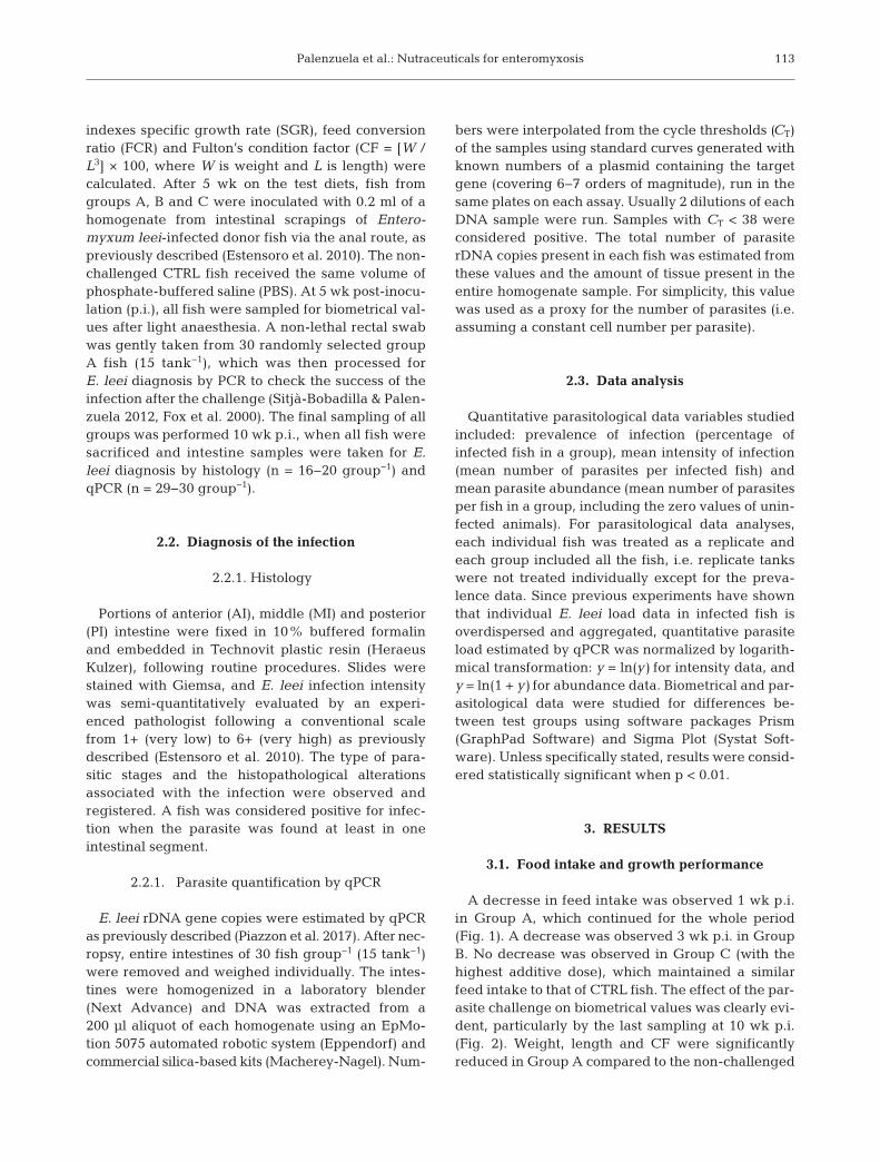
indexes specific growth rate (SGR), feed conversionratio (FCR) and Fulton’s condition factor (CF = [W /L3] × 100, where W is weight and L is length) werecalculated. After 5 wk on the test diets, fish fromgroups A, B and C were inoculated with 0.2 ml of ahomogenate from intestinal scrapings of Entero -myxum leei-infected donor fish via the anal route, aspreviously described (Estensoro et al. 2010). The non-challenged CTRL fish received the same volume ofphosphate-buffered saline (PBS). At 5 wk post-inocu-lation (p.i.), all fish were sampled for biometrical val-ues after light anaesthesia. A non-lethal rectal swabwas gently taken from 30 randomly selected groupA fish (15 tank−1), which was then processed forE. leei diagnosis by PCR to check the success of theinfection after the challenge (Sitjà-Bobadilla & Palen-zuela 2012, Fox et al. 2000). The final sampling of allgroups was performed 10 wk p.i., when all fish weresacrificed and intestine samples were taken for E.leei diagnosis by histology (n = 16−20 group−1) andqPCR (n = 29−30 group−1).
2.2. Diagnosis of the infection
2.2.1. Histology
Portions of anterior (AI), middle (MI) and posterior(PI) intestine were fixed in 10% buffered formalinand embedded in Technovit plastic resin (HeraeusKulzer), following routine procedures. Slides werestained with Giemsa, and E. leei infection intensitywas semi-quantitatively evaluated by an experi-enced pathologist following a conventional scalefrom 1+ (very low) to 6+ (very high) as previouslydescribed (Estensoro et al. 2010). The type of para-sitic stages and the histopathological alterationsassociated with the infection were observed andregistered. A fish was considered positive for infec-tion when the parasite was found at least in oneintestinal segment.
2.2.1. Parasite quantification by qPCR
E. leei rDNA gene copies were estimated by qPCRas previously described (Piazzon et al. 2017). After nec -ropsy, entire intestines of 30 fish group−1 (15 tank−1)were removed and weighed individually. The intes-tines were homogenized in a laboratory blender(Next Advance) and DNA was extracted from a200 µl aliquot of each homogenate using an EpMo-tion 5075 automated robotic system (Eppendorf) andcom mercial silica-based kits (Macherey-Nagel). Num-
bers were interpolated from the cycle thresholds (CT)of the samples using standard curves generated withknown numbers of a plasmid containing the targetgene (covering 6−7 orders of magnitude), run in thesame plates on each assay. Usually 2 dilutions of eachDNA sample were run. Samples with CT < 38 wereconsidered positive. The total number of parasiterDNA copies present in each fish was estimated fromthese values and the amount of tissue present in theentire homogenate sample. For simplicity, this valuewas used as a proxy for the number of parasites (i.e.assuming a constant cell number per parasite).
2.3. Data analysis
Quantitative parasitological data variables studiedincluded: prevalence of infection (percentage ofinfected fish in a group), mean intensity of infection(mean number of parasites per infected fish) andmean parasite abundance (mean number of parasitesper fish in a group, including the zero values of unin-fected animals). For parasitological data analyses,each in dividual fish was treated as a replicate andeach group included all the fish, i.e. replicate tankswere not treated individually except for the preva-lence data. Since previous experiments have shownthat individual E. leei load data in infected fish isoverdispersed and aggregated, quantitative parasiteload es timated by qPCR was normalized by logarith-mical transformation: y = ln(y) for intensity data, andy = ln(1 + y) for abundance data. Biometrical and par-asitological data were studied for differences be -tween test groups using software packages Prism(GraphPad Software) and Sigma Plot (Systat Soft-ware). Unless specifically stated, results were consid-ered statistically significant when p < 0.01.
3. RESULTS
3.1. Food intake and growth performance
A decresse in feed intake was observed 1 wk p.i.in Group A, which continued for the whole period(Fig. 1). A decrease was observed 3 wk p.i. in GroupB. No decrease was observed in Group C (with thehighest additive dose), which maintained a similarfeed intake to that of CTRL fish. The effect of the par-asite challenge on biometrical values was clearly evi-dent, particularly by the last sampling at 10 wk p.i.(Fig. 2). Weight, length and CF were significantlyreduced in Group A compared to the non-challenged
113
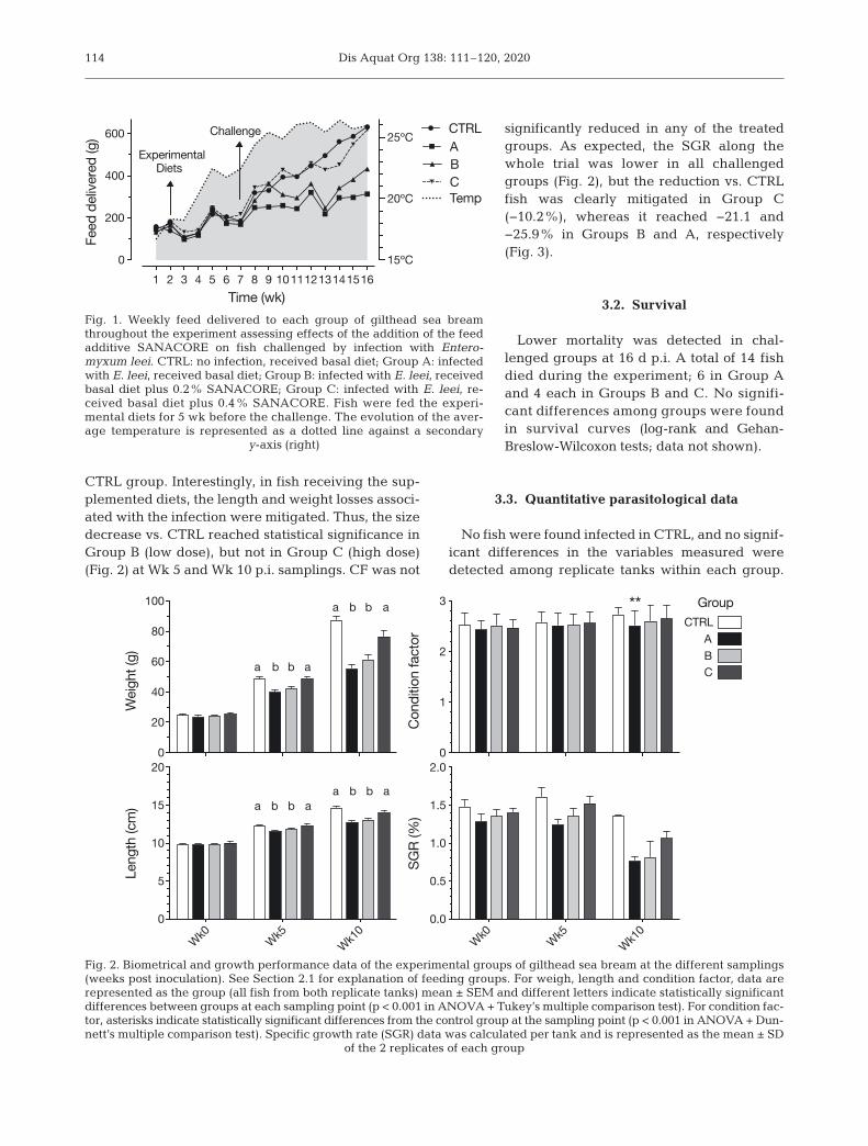
Dis Aquat Org 138: 111–120, 2020114
CTRL group. Interestingly, in fish receiving the sup-plemented diets, the length and weight losses associ-ated with the infection were mitigated. Thus, the sizedecrease vs. CTRL reached statistical significance inGroup B (low dose), but not in Group C (high dose)(Fig. 2) at Wk 5 and Wk 10 p.i. samplings. CF was not
significantly reduced in any of the treatedgroups. As expected, the SGR along thewhole trial was lower in all challengedgroups (Fig. 2), but the re duc tion vs. CTRLfish was clearly miti gated in Group C(−10.2%), whereas it reached −21.1 and−25.9% in Groups B and A, respectively(Fig. 3).
3.2. Survival
Lower mortality was detected in chal-lenged groups at 16 d p.i. A total of 14 fishdied during the experiment; 6 in Group Aand 4 each in Groups B and C. No signifi-cant differences among groups were foundin survival curves (log-rank and Gehan-Breslow-Wilcoxon tests; data not shown).
3.3. Quantitative parasitological data
No fish were found infected in CTRL, and no signif-icant differences in the variables measured weredetected among replicate tanks within each group.
1 2 3 4 5 6 7 8 9 101112131415160
200
400
600
15ºC
20ºC
25ºC
Time (wk)
Feed
del
ivere
d (g
) CTRLABC
Challenge
Temp
ExperimentalDiets
Fig. 1. Weekly feed delivered to each group of gilthead sea breamthroughout the experiment assessing effects of the addition of the feedadditive SANACORE on fish challenged by infection with Entero -myxum leei. CTRL: no infection, received basal diet; Group A: infectedwith E. leei, received basal diet; Group B: infected with E. leei, receivedbasal diet plus 0.2% SANACORE; Group C: infected with E. leei, re-ceived basal diet plus 0.4% SANACORE. Fish were fed the experi-mental diets for 5 wk before the challenge. The evolution of the aver-age temperature is represented as a dotted line against a secondary
y-axis (right)
0
20
40
60
80
100
Wei
ght (
g)
a ab b
a ab b
0
1
2
3
Cond
ition
fact
or
**
Wk0 Wk5Wk1
0Wk0 Wk5
Wk10
0.0
0.5
1.0
1.5
2.0
SGR
(%)
CTRLGroup
ABC
0
5
10
15
20
Leng
th (c
m) a ab ba ab b
Fig. 2. Biometrical and growth performance data of the experimental groups of gilthead sea bream at the different samplings(weeks post inoculation). See Section 2.1 for explanation of feeding groups. For weigh, length and condition factor, data arerepresented as the group (all fish from both replicate tanks) mean ± SEM and different letters indicate statistically significantdifferences between groups at each sampling point (p < 0.001 in ANOVA + Tukey’s multiple comparison test). For condition fac-tor, asterisks indicate statistically significant differences from the control group at the sampling point (p < 0.001 in ANOVA + Dun-nett’s multiple comparison test). Specific growth rate (SGR) data was calculated per tank and is represented as the mean ± SD
of the 2 replicates of each group

Palenzuela et al.: Nutraceuticals for enteromyxosis
At the intermediate non-lethal PCR check (5 wk p.i.),50% of Group A fish were found positive for E. leei.At the end of the trial, qPCR showed prevalence ofinfection was significantly higher in Group A (72.4%)than in the supplemented feed groups (Group B:44.8%; Group C: 46.6%) (Cochran-Armitage contin-gency test for trend, p < 0.05) (Fig. 4A). Furthermore,the differences in the mean prevalence of infectionin supplemented tanks (45.8%) vs. the unsupple-mented ones (72.4%) was also significant (p < 0.05)in a t-test (data not shown). The mean and medianintensity of infection was lower in both Group B(mean = 12.72; median = 11.32) and Group C (mean =13.33; median = 12.65) than in the untreated Group A(mean = 14.33; median = 14.67), although these dif-ferences did not reach statistical significance (1-way
ANOVA; Fig. 4B). The mean parasite abundance val-ues showed the differences between treated and un -treated groups very clearly. Both supplemented dietsresulted in significantly lower mean abundance val-ues (Group B: 5.70; Group C = 6.22) compared togroup A (10.38) (ANOVA, p = 0.029). The mean par-asite abundance data from both (pooled) treatedgroups (5.97) was also significantly different fromthat of Group A (t-test, p = 0.005). Median abundancevalues for both Groups B and C were 0, signifi-cantly lower than that of Group A (median = 13.85,Kruskal- Wallis test, p = 0.017; Fig. 4C).
3.4. Histopathological data
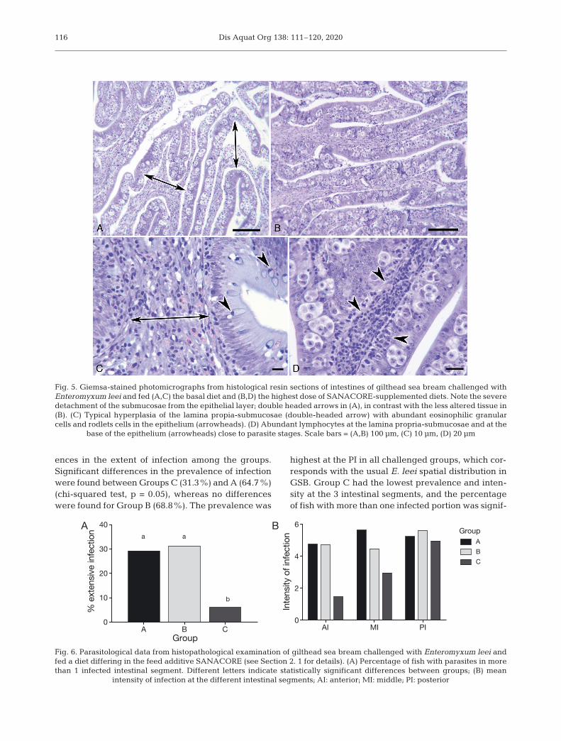
Challenged fish fed the basal diet (Group A)showed typical histopathology induced by the para-site invasion: detachment of the epithelium contain-ing parasites from the lamina propria-submucosae(Fig. 5A) and hyperplasic submucosae infiltratedwith numerous eosinophilic granular cells (Fig. 5C).A high number of rodlet cells was commonly presentin the epithelium. By contrast, in Group C, the intes-tine usually presented a lower degree of detachment,even with a high intensity of infection (Fig. 5B). Thesubmucosa was not remarkably hypertrophied butpresented abundant lymphocytes, which were alsoinfiltrated at the base of the epithelial layer and inclose contact with parasite stages (Fig. 5D). Micro-scopic examination of the 3 intestinal segments of asubsample of experimental fish also showed differ-
115
–40
–30
–20
–10
0
Decr
ease
from
CTR
L (%
)
Food
int
ake
Weight
SGR
ABC
Fig. 3. Percentage of decrease of performance indicators ofgilthead sea bream in challenged groups vs. non-challengedcontrol at the end of the experiment (10 wk post-inoculation
with Enteromyxum leei)
A B C0
20
40
60
80
100
Prev
alen
ce (%
)
A B C0
5
10
15
20
E. l
eei r
DNA
copi
es fi
sh–1
A B
Group
GroupC
0
5
10
15
para
s. rD
NA c
opie
s fis
h–1
a
b b
A B
C
Fig. 4. Quantitative parasitological data of the experi-mental groups at final sampling (10 weeks post-inocula-tion) (see Fig. 1 for details). (A) Mean prevalence of infec-tion in challenged groups; (B) mean intensity of infection;each dot represents the intensity value of an infected in-dividual; (C) mean parasite abundance. Data in (B) and(C) are normalized by logarithmic transformation as de-tailed in Section 2.3. Different lowercase letters indicatestatistically significant differences (p < 0.05) between
groups. Error bars: SD (A) or 95% CI (B,C)
Dis Aquat Org 138: 111–120, 2020
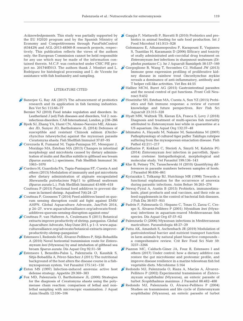
ences in the extent of infection among the groups.Significant differences in the prevalence of infectionwere found be tween Groups C (31.3%) and A (64.7%)(chi-squared test, p = 0.05), whereas no differenceswere found for Group B (68.8%). The prevalence was
highest at the PI in all challenged groups, which cor-responds with the usual E. leei spatial distribution inGSB. Group C had the lowest prevalence and inten-sity at the 3 intestinal segments, and the percentageof fish with more than one infected portion was signif-
116
Fig. 5. Giemsa-stained photomicrographs from histological resin sections of intestines of gilthead sea bream challenged withEnteromyxum leei and fed (A,C) the basal diet and (B,D) the highest dose of SANACORE-supplemented diets. Note the severedetachment of the submucosae from the epithelial layer; double headed arrows in (A), in contrast with the less altered tissue in(B). (C) Typical hyperplasia of the lamina propia-submucosae (double-headed arrow) with abundant eosinophilic granularcells and rodlets cells in the epithelium (arrowheads). (D) Abundant lymphocytes at the lamina propria-submucosae and at the
base of the epithelium (arrowheads) close to parasite stages. Scale bars = (A,B) 100 µm, (C) 10 µm, (D) 20 µm
A B C0
10
20
30
40
% e
xten
sive
infe
ctio
n a a
b
BA
Group
Group
C
AI MI PI0
2
4
6
Inte
nsity
of i
nfec
tion
A B
Fig. 6. Parasitological data from histopathological examination of gilthead sea bream challenged with Enteromyxum leei andfed a diet differing in the feed additive SANACORE (see Section 2. 1 for details). (A) Percentage of fish with parasites in morethan 1 infected intestinal segment. Different letters indicate statistically significant differences between groups; (B) mean
intensity of infection at the different intestinal segments; AI: anterior; MI: middle; PI: posterior

Palenzuela et al.: Nutraceuticals for enteromyxosis
icantly lower in Group C (chi-squared test, p < 0.05),but not in Group B (Fig. 6A). The mean intensity ofinfection by intestinal segment was lowest in GroupC, especially in the AI, though these differences werenot statistically significant (Fig. 6B).
4. DISCUSSION
Enteromyxum leei infections can cause severemortality in several cultured fish species such assharpsnout sea bream, red sea bream, fugu Taki -fugu rubripes and Japanese flounder Paralichthysoliva ceus (Yasuda et al. 2005, Palenzuela 2006,Rigos & Katharios 2010). The most obvious effectsof enteromyxosis in GSB include anorexia, weightloss and arrested growth (Palenzuela 2006, Sitjà-Bobadilla & Palenzuela 2012). In the current labora-tory challenge with E. leei, the parasite effectivelyinduced these clinical signs in GSB fed a standardcommercial diet (Group A). This group exhibited adramatic decrease in feed intake (−32.7%), weight(−36.2%) and SGR (−25.9%) compared to the unchal-lenged CTRL group after 10 wk p.i. At 5 wk p.i., 50%of Group A tested positive for the parasite in a non-lethal PCR, and 72.4% were infected at the final sam-pling at wk 10 wk p.i. This indicates exposure to ahigh infection pressure, further magnified by thehigh water temperatures sustained during the trial.
Anorexia is a complex pathophysiological responseto some parasitic infections, and it is widespreadamong livestock including fish (Symons 1985, Bernier2010). While it may seem counterproductive for theenergetic demands of a successful fight against in -fection, there are several hypotheses to explain thisphenomenon as a conciliation of immune and meta-bolic needs (Exton 1997, Kyriazakis et al. 1998). Ingastrointestinal infections, direct alterations of the en-teric nervous system also needs to be considered (Hal-liez & Buret 2015). In turbot Psetta maxima with en -tero myxosis, down-regulation of the neuropeptideY-receptor NPY2R, a main regulator of appetite (Zou& Secombes 2011), has been reported in early stagesof the infection (Ronza et al. 2016). This observation isin agreement with the early onset of anorexia inGroup A in the current trial, which was observed asearly as 1 wk p.i. and persisted until the the end of theexperiment, reaching 32.7% reduction in food intakecompared to the CTRL group. Diets supplementedwith SANACORE mitigated this decrease to theextent that Group C (high dose) did not differ signifi-cantly from the CTRL group with respect to food in-take. In Group B, anorexia started later and was not as
acute as in Group A (−21.63%). It remains to be inves-tigated if this reduction in the primary pathophysio-logical response to infection is related to a protectiverole of the additive at the early stages of parasite inva-sion or to other factors. In a previous E. leei infectionusing parasite-containing effluent water, food intakedecrease ranged between 22.1 and 15.4% in groupsfed different diets (Estensoro et al. 2011). Althoughthese values are not as low as in the untreated groupin the current experiment, this is most likely related todifferences in infection routes, temperatures and in-fective pressure between both studies.
Weight loss and arrested growth are obvious conse-quences of anorexia and of the diversion of energy in-vested in mounting an immune response, but they arealso largely caused by direct structural damage to theintestine epithelium and interference of the parasiteswith gastrointestinal function (Estensoro et al. 2011,Sitjà-Bobadilla & Palenzuela 2012). In E. leei-infectedfugu, dehydration and osmoregulatory failure, to-gether with impaired intestinal absorption, were themain contributors to a rapid weight loss observed ininfected fish (Ishimatsu et al. 2007). In the current ex-periment, the evolution of biometrical data clearly ev-idenced the clinical effect of the infection in chal-lenged groups, in consonance with field studies on E.leei-infected stocks and with the results of previouslaboratory challenge studies with this parasite (e.g.Estensoro et al. 2011, Piazzon et al. 2017). As deducedfrom the results of this trial, supplemented diets canpartially alleviate these effects, as Group C reachedsimilar performance levels as CTRL. The effect inGroup B, however, was moderate and fish still per -formed worse than in the CTRL group, reaching sta-tistical significance in all the biometrical variablesmeasured except CF. Interestingly, the PCR resultsshowed that both treated groups presented similarand lower prevalence values than Group A. Thus, thedifferent pathological effects be tween treated groupscannot be attributed exclusively to prevalence; othervariables, like the intensity of infection, must be con-sidered. However, the differences in the meannumber of parasites per infected fish in the differentgroups did not reach statistical significance. Althoughmean and median in tensity values in Group A werehigher than in both treated groups, the differencesbetween the latter were modest (indeed, Group B pre-sented values slightly lower than Group C).
Regarding the prevalence of infection and the meanintensity of infection, neither parameter could direct lyexplain the differences in the pathological effect ofthe challenge found between both treated groups.The observed differences in the mean intensity of in-
117

Dis Aquat Org 138: 111–120, 2020
fection and in the extent of infection along the intes-tinal tract could suggest delayed invasion or variabledegrees of ‘resistance’ to the multiplication of the par-asite once it breached the host barriers. Since all fishwere inoculated simultaneously with a single dose,these differences could indicate slightly improvedability to constrain parasite development or to elimi-nate parasites more efficiently. It must be noted, how-ever, that the overdispersion of intensity valuesmakes it very difficult to compare this variable be-tween groups, even with normalized data. This is acommon phenomenon in quantitative parasitology(Rózsa et al. 2000, Klar et al. 2010), further exacer-bated by the type of parasite studied in this work (i.e.a rapidly multiplying pluricellular microorganism).
Mean parasite abundance data takes into consider-ation the mean parasite quantities in all the fish fromthe group, including uninfected animals, thus merg-ing prevalence and intensity data in a single vari-able. This parameter showed very significant differ-ences between treated and non-treated groups, butfailed to prove substantial differences between bothtreated groups or to correlate with the differentialpathogeny observed with the 2 doses of additive. Theobservation that fish receiving the higher dose couldcope with relatively high individual parasite loads,which generated more severe pathophysiologicaleffects in other groups, is remarkable. Variability ofinfection-associated immunopathology has beenwidely recognized in many serious myxozoan in fec -tions, including Enteromyxum spp., Ceratonovashasta and Tetracapsuloides bryosalmonae (Gorgo -glione et al. 2013, Bjork et al. 2014, Sitjà-Bobadilla etal. 2015). In this scenario, the benefits of the additiveat the higher dose could be interpreted as being duein part to an immunomodulatory action. This inter-pretation is further supported by the differences inthe pathological alterations and cellular immune re -sponse observed in the intestine of the experimentalgroups. A conspicuous hyperplasia and eosinophilicinfiltration of the submucosae, as well as abundantrodlet cells were commonly observed in Group A fish,which is normally associated with extensive de -tachment of the epithelial layer from the submucosae.In contrast, even with comparable levels of parasitein tensity, fish from Group C generally displayed betteroverall epithelium topology and a more moderatecellular immune re sponse, which mainly consisted oflymphocyte infiltration.
The effect of feed supplements on growth perform-ance, immune response and disease resistance hasbeen previously studied in fish, but mainly in relationto bacterial pathogens (Newaj-Fyzul & Austin 2015,
Hoseinifar et al. 2015, Ringø & Song 2016). In GSB,different prebiotic and probiotic ingredients havebeen shown to enhance immune status, change in -testinal morphology (Cerezuela et al. 2013) and gutmicrobiota (Cordero et al. 2015, Piazzon et al. 2017).However, the efficacy of probiotics against ecto- andendo-parasites seems very limited (e.g. reviewedby Banerjee & Ray 2017). Although the commercialinterest of dietary additives for fish parasite control ishigh, their efficacy remains largely unexplored, andthe benefits for a given fish disease model are diffi-cult to predict. In a recent study, the effect of sodiumbutyrate on the infection and growth performance ofGSB was studied with experimentally induced entero -myxosis (Piazzon et al. 2017). Interestingly, the histo -pathological study also showed a statistically signifi-cant influence of diet on the prevalence of infectionin the AI. Similar to the current study, the number offish with more than one infected intestinal segmentwas significantly higher in the diet group showingthe worst growth performance. Butyrate did not de -crease the infection levels significantly, but it miti-gated the growth arrestment and alleviated the dis-ease outcome.
5. CONCLUSIONS
Quantitative parasitological data on prevalence, in-tensity and abundance, as well as histopathologicalstudies on the extent of infection, confirmed the effectof SANACORE-supplemented diets in reducing in-fection level and severity. Both GSB groups receivingthe functional feed additive showed lower prevalenceand intensity of infection compared to the challengedgroup not receiving the feed additive. Although theadditive proved to have a significant effect on the re-duction of infection pre valence and parasite abun-dance at both doses tested, the maximum benefits interms of pathogenesis re duction were obtained in thehigh-dose group. Since the negative impact in GSBproduction through enteromyxosis is mostly related tothe symptons of the disease, this additive appears tobe a promising tool for management of the disease inGSB aquaculture.
It cannot be ignored that the current trial wasbased on a laboratory-controlled experimental infec-tion with homogeneous and time-controlled expo-sure to the parasite and under stocking and feedingregimens unlike those of farmed fish. Further studiesin more complex farming settings are necessary todetermine the feeding regime and dosage of the sup-plement that could offer these benefits.
118
Palenzuela et al.: Nutraceuticals for enteromyxosis
Acknowledgements. This study was partially supported bythe EU H2020 program and by the Spanish Ministry ofEconomy and Competitiveness through ParaFishControl(634429) and AGL-2013-48560-R research projects, respec-tively. This publication reflects the views of the authorsonly; the European Commission cannot be held responsiblefor any use which may be made of the information con-tained therein. M.C.P. was contracted under CSIC PIE pro-ject no. 201740E013. The authors thank J. Monfort and L.Rodríguez for histological processing and I. de Vicente forassistance with fish husbandry and sampling.
LITERATURE CITED
Banerjee G, Ray AK (2017) The advancement of probioticsresearch and its application in fish farming industries.Res Vet Sci 115: 66−77
Bernier NJ (2010) Food intake regulation and disorders. In: Leatherland J (ed) Fish diseases and disorders, Vol 2: non-infectious disorders. CAB International, London, p 238−266
Bjork SJ, Zhang YA, Hurst CN, Alonso-Naveiro ME, Alexan-der JD, Sunyer JO, Bartholomew JL (2014) De fenses ofsusceptible and resistant Chinook salmon (Oncho -rhynchus tshawytscha) against the myxozoan parasiteCeratomyxa shasta. Fish Shellfish Immunol 37: 87−95
Cerezuela R, Fumanal M, Tapia-Paniagua ST, Meseguer J,Moriñigo MA, Esteban MA (2013) Changes in intestinalmorphology and microbiota caused by dietary adminis-tration of inulin and Bacillus subtilis in gilthead sea bream(Sparus aurata L.) specimens. Fish Shellfish Immunol 34: 1063−1070
Cordero H, Guardiola FA, Tapia-Paniagua ST, Cuesta A andothers (2015) Modulation of immunity and gut microbiotaafter dietary administration of alginate encapsulatedShewanella putrefaciens Pdp11 to gilthead seabream(Sparus aurata L.). Fish Shellfish Immunol 45: 608−618
Coutteau P (2015) Functional feed additives to prevent dis-ease in farmed shrimp. Aquafeed 7(4):24–27
Coutteau P, Goossens T (2014) Feed additives based on quo-rum sensing disruption could aid fight against EMS/AHPN. Global Aquaculture Advocate, Jan/Feb 2014,p 24−27. www.aquaculturealliance.org/ advocate/ feed-additives- quorum-sensing-disruption-against-ems/
Coutteau P, van Halteren A, Ceulemans S (2011) Botanicalextracts improve productivity of shrimp, pangasius. GlobalAquaculture Advocate, May/June 2011, p 1−3. www. aqua-culturealliance.org/advocate/botanical-extracts- improve-productivity-shrimp-pangasius/
Estensoro I, Redondo MJ, Álvarez-Pellitero P, Sitjà-BobadillaA (2010) Novel horizontal transmission route for Entero -myxum leei (Myxozoa) by anal intubation of gilthead seabream Sparus aurata. Dis Aquat Org 92: 51−58
Estensoro I, Benedito-Palos L, Palenzuela O, Kaushik S,Sitjà-Bobadilla A, Pérez-Sánchez J (2011) The nutritionalbackground of the host alters the disease course in a fish-myxosporean system. Vet Parasitol 175: 141−150
Exton MS (1997) Infection-induced anorexia: active hostdefense strategy. Appetite 29: 369−383
Fox MD, Palenzuela O, Bartholomew JL (2000) Strategiesfor the diagnosis of Ceratomyxa shasta using the poly-merase chain reaction: comparison of lethal and non-lethal sampling with microscopic examination. J AquatAnim Health 12: 100−106
Gaggìa F, Mattarelli P, Biavatti B (2010) Probiotics and pre-biotics in animal feeding for safe food production. Int JFood Microbiol 141: S15−S28
Golomazou E, Athanassopoulou F, Karagouni E, VagianouS, Tsantilas H, Karamanis D (2006) Efficacy and toxicityof orally administrated anti-coccidial drug treatment onEnteromyxum leei infections in sharpsnout seabream (Di -plo dus puntazzo C.). Isr J Aquacult Bamidgeh 58: 157−169
Gorgoglione B, Wang T, Secombes CJ, Holland JW (2013)Immune gene expression profiling of proliferative kid-ney disease in rainbow trout Oncorhynchus mykissreveals a dominance of anti-inflammatory, antibody andT helper cell-like activities. Vet Res 44: 55
Halliez MCM, Buret AG (2015) Gastrointestinal parasitesand the neural control of gut functions. Front Cell Neu-rosci 9: 452
Hoseinifar SH, Esteban MA, Cuesta A, Sun YZ (2015) Prebi-otics and fish immune response: a review of currentknowledge and future perspectives. Rev Fish SciAquacult 23: 315−328
Hyatt MW, Waltzek TB, Kieran EA, Frasca S, Lovy J (2018)Diagnosis and treatment of multi-species fish mortalityattributed to Enteromyxum leei while in quarantine at aUS aquarium. Dis Aquat Org 132: 37−48
Ishimatsu A, Hayashi M, Nakane M, Sameshima M (2007)Pathophysiology of cultured tiger puffer Takifugu rubripessuffering from the myxosporean emaciation disease. FishPathol 42: 211−217
Katharios P, Kokkari C, Sterioti A, Smyrli M, Kalatzis PG(2014) Enteromyxum leei infection in parrotfish, Spari-soma cretense: histopathological, morphological andmolecular study. Vet Parasitol 199: 136−143
Klar B, Petney TN, Taraschewski H (2010) Quantifying dif-ferences in parasite numbers between samples of hosts.J Parasitol 96: 856−861
Kyriazakis I, Tolkamp BJ, Hutchings MR (1998) Towards afunctional explanation for the occurrence of anorexiaduring parasitic infections. Anim Behav 56: 265−274
Newaj-Fyzul A, Austin B (2015) Probiotics, immunostimu-lants, plant products and oral vaccines, and their role asfeed supplements in the control of bacterial fish diseases.J Fish Dis 38: 937−955
Padrós F, Palenzuela O, Hispano C, Tosas O, Zarza C, Cre-spo S, Álvarez-Pellitero P (2001) Myxidium leei (Myxo-zoa) infections in aquarium-reared Mediterranean fishspecies. Dis Aquat Org 47: 57−62
Palenzuela O (2006) Myxozoan infections in Mediterraneanmariculture. Parassitologia 48: 27−29
Patra AK, Amasheh S, Aschenbach JR (2019) Modulation ofgastrointestinal barrier and nutrient transport functionin farm animals by natural plant bioactive compounds -a com prehensive review. Crit Rev Food Sci Nutr 59: 3237− 3266
Piazzon MC, Calduch-Giner JA, Fouz B, Estensoro I andothers (2017) Under control: how a dietary additive canrestore the gut microbiome and proteomic profile, andimprove disease resilience in a marine teleostean fish fedvegetable diets. Microbiome 5: 164
Redondo MJ, Palenzuela O, Riaza A, Macías A, Álvarez-Pellitero P (2002) Experimental transmission of Enter o -myxum scophthalmi (Myxozoa), an enteric parasite ofturbot Scophthalmus maximus. J Parasitol 88: 482−488
Redondo MJ, Palenzuela O, Álvarez-Pellitero P (2004)Studies on transmission and life cycle of Enteromyxumscophthalmi (Myxozoa), an enteric parasite of turbot
119

Dis Aquat Org 138: 111–120, 2020
Scophthalmus maximus. Folia Parasitol (Praha) 51: 188−198
Rigos G, Katharios P (2010) Pathological obstacles of newly-introduced fish species in Mediterranean mariculture: areview. Rev Fish Biol Fish 20: 47−70
Ringø E, Song SK (2016) Application of dietary supplements(synbiotics and probiotics in combination with plant prod-ucts and β-glucans) in aquaculture. Aquacult Nutr 22: 4−24
Ronza P, Robledo D, Bermúdez R, Losada AP and others(2016) RNA-seq analysis of early enteromyxosis in turbot(Scophthalmus maximus): new insights into parasiteinvasion and immune evasion strategies. Int J Parasitol46: 507−517
Rózsa L, Reiczigel J, Majoros G (2000) Quantifying parasitesin samples of hosts. J Parasitol 86: 228−232
Shinn A, Pratoomyot J, Bron JE, Paladini G, Brooker EE,Brooker AJ (2015a) Economic impacts of aquatic parasiteson global finfish production. Global Aquaculture Advo -cate, Sep/Oct 2015, p 82−84. www.aquaculturealliance.org/advocate/economic-impacts-of-aquatic- parasites-on-global-finfish-production/
Shinn AP, Pratoomyot J, Bron JE, Paladini G, Brooker EE,Brooker AJ (2015b) Economic costs of protistan andmetazoan parasites to global mariculture. Parasitology142: 196−270
Sitjà-Bobadilla A, Palenzuela O (2012) Enteromyxum spe-cies. In: Woo PTK, Buchmann K (eds) Fish parasites: pathobiology and protection. CAB International, Walling -ford, p 163−176
Sitjà-Bobadilla A, Diamant A, Palenzuela O, Álvarez-Pellitero P (2007) Effect of host factors and experimentalconditions on the horizontal transmission of Entero myxumleei (Myxozoa) to gilthead sea bream, Sparus aurata L.,and European sea bass, Dicentrarchus labrax (L.). J FishDis 30: 243−250
Sitjà-Bobadilla A, Schmidt-Posthaus H, Wahli T, HollandJW, Secombes CJ (2015) Fish immune responses to myx-ozoa. In: Okamura B, Gruhl A, Bartholomew J (eds) Myx-ozoan evolution, ecology and development. SpringerInternational Publishing, Cham, p 253−280
Suresh G, Das RK, Brar SK, Rouissi T, Ramirez AA, Chorfi Y,Godbout S (2017) Alternatives to antibiotics in poultry feed: molecular perspectives. Crit Rev Microbiol 44: 318−335
Symons LEA (1985) Anorexia: occurrence, pathophysiology,and possible causes in parasitic infections. Adv Parasitol24: 103−133
Thormar H (2012) Patented non-antibiotic agents as ani-mal feed additives. Recent Pat Food Nutr Agric 4: 155−168
Vallejos-Vidal E, Reyes-López F, Teles M, MacKenzie S(2016) The response of fish to immunostimulant diets.Fish Shellfish Immunol 56: 34−69
Wunderlich AC, Zica E, Ayres V, Guimarães AC, Takeara R(2017) Plant-derived compounds as an alternative treat-ment against parasites in fish farming: a review. In: Khater HF, Govindarajan M, Benelli G (eds) Naturalremedies in the fight against parasites. IntechOpen, doi: 10.5772/67668
Yasuda H, Ooyama T, Nakamura A, Iwata K, Palenzuela O,Yokoyama H (2005) Occurrence of the myxosporeanemaciation disease caused by Enteromyxum leei in cul-tured Japanese flounder Paralichthys olivaceus. FishPathol 40: 175−180
Yiagnisis M, Kolygas M, Athanassopoulou F (2016) Effectsof oregano essential oil on Enteromyxum leei (myxozoa)in fection on gilthead sea bream (Sparus aurata L.). Posterpresented at Aquaculture Europe 2016, 20−23 Septem-ber 2016, Edinburgh
Zou J, Secombes CJ (2011) Teleost fish interferons and theirrole in immunity. Dev Comp Immunol 35: 1376−1387
120
Diet A Diet B Diet C(no additive) (low dose) (high dose)
Crude protein/ 45/20 45/20 45/20fat proportion
LT fishmeal 15 15 15Poultry by-product 12 12 12meal
Soybean meal 25 25 25Soybean protein 10 10 10concentrate
Corn gluten 8 8 8Wheat gluten 3.4 3.4 3.4Wheat flour 10.5 10.5 10.5Soybean oil 9.7 9.7 9.7Fish oil 6 6 6Amino acids – − –Vitamins and − – –mineral premix
SANACORE 0 +0.2 +0.4
Appendix. Crude composition of the experimental feeds (in-gredients in %) provided to gilthead sea bream; differentgroups received diets differing in amount of the additive
SANACORE
Editorial responsibility: Dieter Steinhagen, Hannover, Germany
Submitted: August 28, 2019; Accepted: January 27, 2020Proofs received from author(s): February 24, 2020
Related Documents