The Open Inflammation Journal, 2009, 2, 9-21 9 1875-0419/09 2009 Bentham Open Open Access Submandibular Salivary Gland Endocrine Secretions and Systemic Pathophysiological Responses Ronald Mathison * Department of Physiology and Pharmacology, Faculty of Medicine, University of Calgary, 3330 Hospital Drive NW, Calgary, Alberta, T2N 4N1, Canada Abstract: Saliva is an exocrine secretion with the fluid and its components being dispensed into ducts. However, a sig- nificant component of salivary gland secretions is found in blood indicating endocrine secretion. The growth factors and hormones secreted into the blood by salivary glands manifest their actions in the face of stressful and often inflammatory stimuli, such as injury, trauma and infections, and they do not participate overtly in the regulation of the resting homeo- static steady state. This type of regulation is called allostasis, which encompasses the processes involved in maintaining systems balance in response to persistent changes, challenges, insults and injury. Viewing salivary gland endocrine secre- tions as having important roles in regulating responses to systemic stressors by modulating allostatic load provides a new perspective for understanding the role of salivary glands in modulation of systemic disease and pathology. INTRODUCTION Saliva is not one of the popular body fluids, and is only taken seriously when it disappears with ensuing difficulties in speaking and eating, necessary visits to the dentist for treatment cavities, periodontal disease, and sores in the soft tissues of the mouth. The functions of saliva, aside as a di- gestion aid, are thus revealed – lubrication, protection, de- fence and wound healing. The good and the bad of modern medicine have made research on saliva and salivary glands both needed and re- spected. Endocrine disorders, stress, anxiety, depression, and nutritional deficiencies decrease saliva flow, radiotherapy for head and neck cancers are now recognized to permanently damage salivary glands, and many medications - antihyper- tensives, antidepressants, analgesics, tranquilizers, diuretics, antihistames and other drugs [1] leave patients with a dry mouth. Further interest in saliva has grown with its use for non-invasive diagnostic purposes for both oral and systemic diseases and the assessment of health status, treatment out- come, disease onset and progression [2]. New developments in proteomics of saliva [3] and gene transfer technologies applied to the salivary glands [4] will facilitate, respectively and cooperatively, discovery of novel therapeutic targets, development of biomarkers with diagnostic and/or prognos- tic value, and open up avenues to treat irreversibly damaged salivary glands and deliver protein therapeutics locally to the oral cavity, gastrointestinal tract or into the bloodstream for systemic deliver [4-6]. These new developments will also facilitate an under- standing of the role of the salivary glands and their secre- tions in systemic health and disease. Some systemic diseases *Address correspondence to this author at the Department of Physiology and Pharmacology, Faculty of Medicine, University of Calgary, 3330 Hos- pital Drive NW, Calgary, Alberta, T2N 4N1, Canada; E-mails: [email protected], [email protected] are associated with poor oral health [7], and as will be dis- cussed subsequently, the proteins and peptides synthesized in the salivary glands that are secreted into the blood have sub- tle, but significant impact on systemic response to a variety of stresses and pathologies. The functions of these salivary endocrine factors and their involvement in the regulation of systemic response to stressors are interpreted within the con- cept of allostasis and allostatic load. SALIVA AND THE SALIVARY GLANDS Saliva, an exocrine secretion of the salivary glands, con- sists of water, electrolytes, enzymes, immunoglobulins, mu- cosal glycoproteins and numerous antimicrobial proteins, enzymes, growth factors and regulatory peptides. These components aid in the sensation of taste, digestion of starch and lipids and are responsible in the maintenance of oral health by protecting tissues with lubrication and buffering properties, contributing to the physical-chemical integrity of tooth enamel, and preventing adhesion of and colonization by microorganisms. Saliva is produced by major (parotid, submandibular, and sublingual) and minor (located throughout the oral cavity) salivary glands. The salivary glands are composed primarily of ductal and acinar cells, with the latter consisting of serous cells that produce a water saliva (e.g. parotid), or mucous cells (e.g. sublingual and minor salivary glands) which yield a secretion rich in immunoglobulins and mucus. The sub- mandibular glands have both serous and mucous acinar cells. The ductal cells line the salivary ducts leading from the aci- nar cells to the oral cavity and modify salivary composition by absorbing sodium chloride, and by adding proteins, growth factors and potassium. Salivary secretion is under autonomic nervous system control [8] with parasympathetic nerves activating the muscarinic receptors responsible for fluid secretion and the sympathetic nerves through stimula- tion of adrenergic receptors regulating protein secretion.

Welcome message from author
This document is posted to help you gain knowledge. Please leave a comment to let me know what you think about it! Share it to your friends and learn new things together.
Transcript

The Open Inflammation Journal, 2009, 2, 9-21 9
1875-0419/09 2009 Bentham Open
Open Access
Submandibular Salivary Gland Endocrine Secretions and Systemic Pathophysiological Responses
Ronald Mathison*
Department of Physiology and Pharmacology, Faculty of Medicine, University of Calgary, 3330 Hospital Drive NW,
Calgary, Alberta, T2N 4N1, Canada
Abstract: Saliva is an exocrine secretion with the fluid and its components being dispensed into ducts. However, a sig-
nificant component of salivary gland secretions is found in blood indicating endocrine secretion. The growth factors and
hormones secreted into the blood by salivary glands manifest their actions in the face of stressful and often inflammatory
stimuli, such as injury, trauma and infections, and they do not participate overtly in the regulation of the resting homeo-
static steady state. This type of regulation is called allostasis, which encompasses the processes involved in maintaining
systems balance in response to persistent changes, challenges, insults and injury. Viewing salivary gland endocrine secre-
tions as having important roles in regulating responses to systemic stressors by modulating allostatic load provides a new
perspective for understanding the role of salivary glands in modulation of systemic disease and pathology.
INTRODUCTION
Saliva is not one of the popular body fluids, and is only taken seriously when it disappears with ensuing difficulties in speaking and eating, necessary visits to the dentist for treatment cavities, periodontal disease, and sores in the soft tissues of the mouth. The functions of saliva, aside as a di-gestion aid, are thus revealed – lubrication, protection, de-fence and wound healing.
The good and the bad of modern medicine have made research on saliva and salivary glands both needed and re-spected. Endocrine disorders, stress, anxiety, depression, and nutritional deficiencies decrease saliva flow, radiotherapy for head and neck cancers are now recognized to permanently damage salivary glands, and many medications - antihyper-tensives, antidepressants, analgesics, tranquilizers, diuretics, antihistames and other drugs [1] leave patients with a dry mouth. Further interest in saliva has grown with its use for non-invasive diagnostic purposes for both oral and systemic diseases and the assessment of health status, treatment out-come, disease onset and progression [2]. New developments in proteomics of saliva [3] and gene transfer technologies applied to the salivary glands [4] will facilitate, respectively and cooperatively, discovery of novel therapeutic targets, development of biomarkers with diagnostic and/or prognos-tic value, and open up avenues to treat irreversibly damaged salivary glands and deliver protein therapeutics locally to the oral cavity, gastrointestinal tract or into the bloodstream for systemic deliver [4-6].
These new developments will also facilitate an under-standing of the role of the salivary glands and their secre-tions in systemic health and disease. Some systemic diseases
*Address correspondence to this author at the Department of Physiology
and Pharmacology, Faculty of Medicine, University of Calgary, 3330 Hos-
pital Drive NW, Calgary, Alberta, T2N 4N1, Canada;
E-mails: [email protected], [email protected]
are associated with poor oral health [7], and as will be dis-cussed subsequently, the proteins and peptides synthesized in the salivary glands that are secreted into the blood have sub-tle, but significant impact on systemic response to a variety of stresses and pathologies. The functions of these salivary endocrine factors and their involvement in the regulation of systemic response to stressors are interpreted within the con-cept of allostasis and allostatic load.
SALIVA AND THE SALIVARY GLANDS
Saliva, an exocrine secretion of the salivary glands, con-sists of water, electrolytes, enzymes, immunoglobulins, mu-cosal glycoproteins and numerous antimicrobial proteins, enzymes, growth factors and regulatory peptides. These components aid in the sensation of taste, digestion of starch and lipids and are responsible in the maintenance of oral health by protecting tissues with lubrication and buffering properties, contributing to the physical-chemical integrity of tooth enamel, and preventing adhesion of and colonization by microorganisms.
Saliva is produced by major (parotid, submandibular, and sublingual) and minor (located throughout the oral cavity) salivary glands. The salivary glands are composed primarily of ductal and acinar cells, with the latter consisting of serous cells that produce a water saliva (e.g. parotid), or mucous cells (e.g. sublingual and minor salivary glands) which yield a secretion rich in immunoglobulins and mucus. The sub-mandibular glands have both serous and mucous acinar cells. The ductal cells line the salivary ducts leading from the aci-nar cells to the oral cavity and modify salivary composition by absorbing sodium chloride, and by adding proteins, growth factors and potassium. Salivary secretion is under autonomic nervous system control [8] with parasympathetic nerves activating the muscarinic receptors responsible for fluid secretion and the sympathetic nerves through stimula-tion of adrenergic receptors regulating protein secretion.

10 The Open Inflammation Journal, 2009, Volume 2 Ronald Mathison
Relationship to Systemic Health
The relationship between salivary glands and systemic health is complex, involving several bidirectional interacting factors. Currently, a great deal of evidence suggests that poor oral health can contribute to systemic disease, and the oppo-site is equally true. Chronic inflammatory autoimmune dis-order of the salivary glands yields compromised secretory function, which impacts on peripheral systems. Systemic autoimmune diseases also can modify salivary gland func-tion and oral health. The relationships are complex, and only recently have attempts been made to unravel and reveal causal relationships.
Periodontal Disease and Systemic Health
Chronic inflammatory periodontal diseases are among the most prevalent chronic infections in humans, and many investigators have established a significant, albeit modest, positive association between periodontal disease and cardio-vascular disease, which includes atherosclerosis, myocardial infarction and stroke. In addition, epidemiological associa-tions have been made between periodontal diseases and chronic diseases such as diabetes, respiratory diseases and osteoporosis [9]. Nonetheless, the association between periodontal disease and systemic disease is not always ac-cepted, and the call has been made for prospective studies to evaluate this relationship with a rigorous assessment of both clinical endpoints and surrogate markers of risk [10]. Despite the uncertainty it is recognized that ``oral infection may rep-resent a significant risk-factor for systemic diseases, and hence the control of oral disease is essential in the prevention and management of these systemic conditions`` [10]. A clear definition of biological mechanism is still required and sev-eral hypotheses have been presented: 1) contribution to sys-temic inflammation with increased circulating cytokines and mediators; 2) cross-reactivity or molecular mimicry between bacterial antigens and self-antigens; and 3) direct infection of the blood vessel walls by periodontal organisms that escape into the circulation [9, 11]. Another mechanism could in-volve modification by oral infections of the secretion of sali-vary factors (Table 1) that impact on systemic health. This mechanism has received little attention and is evaluated herein.
Systemic Diseases Associated with Hyposalivation and/or Xerostomia
Broadly speaking salivary dysfunction manifests as either hypersalivation (sialorrhea) or hyposalivation. Xerostomia, which refers to a subjective experience of mouth dryness, is often used interchangeably, but incorrectly, with hyposaliva-tion. Patients with hyposalivation are not necessarily xerostomic, and may have normal salivary flow rates. None-theless, salivary gland hypofunction is a common and pri-mary symptom of xerostomia. Hypersalivation may be caused by drugs having cholinergic effects, and is often re-lated to neurological disorders such as Parkinson’s disease, amyotrophic lateral sclerosis (ALS) and developmental dis-abilities (e.g., cerebral palsy; Down syndrome, fragile X, autism).
Hyposalivation can be induced by medications, chemo- and radiotherapy, and graft-versus-host disease (GVHD) that cause significant oral pathology with mucosal infections
dental caries, and difficulties in tasting, eating, swallowing, and speaking. These pathologies also manifest in patients with a variety of systemic diseases including – SS (Sjögren's syndrome), rheumatoid arthritis, juvenile idiopathic (rheu-matoid) arthritis, systemic lupus erythematosus (an inflam-matory connective tissue disease), systemic sclerosis (sce-loderma), primary bilary cirrhosis (an autoimmune disease of the liver), sarcidosis (a multisystem granulomatous disorder), infections with human immunodeficiency virus (HIV), her-pes virus, hepatitis C, ectodermal dysplasia, chronic pan-creatitis and depression [7].
Table 1. Some Salivary Gland Growth Factors, Enzymes and Peptides that Impact on Systemic Health
Factor, Enzyme or Peptide Abbreviation
Basic fibroblast growth factor bFGF
Brain-derived neurotrophic factor BNDF
Epidermal growth factor EGF
Hepatocyte growth factor HGF
Insulin-like growth factors I and II IGF-I & IGFII
Kallikrein
Nerve growth factor NGF
Platelet derived growth factor PDGF
Submandibular gland peptide-T (TDIFEGG) SGPT
Sialorphin (QHNPR)
Submandibular rat-1 SMR1
Renin
Transforming growth factor alpha TGF
Transforming growth factor beta TGF
Vascular endothelial growth factor VEGF
The mechanisms responsible for hyposalivation include [7]: 1) neurotransmitter receptor dysfunctions, as seen with the presence of IgG antibodies M3 and M1 mAChRs in pa-tients with SS [12, 13], and lack of stimulation of nitric ox-ide synthase activation in the submandibular glands by vasoactive intestinal peptide (VIP) in non-obese diabetic (NOD) mice [14]; 2) alterations of fluid composition and electrolytes, as occurs in SS patients with reduced concentra-tion of nitrite, a cytocidal and cytostatic agent in human sa-liva that kills several oral pathogens [15]; 3) DNA damage (radiation therapy); 4) immune dysregulation associated with salivary gland parenchymal destruction, as with GVHD [16] and SS [17]; and 5) a combinations of these mechanisms.
SJÖGREN'S SYNDROME - A SALIVARY GLAND DISEASE WITH SYSTEMIC MANIFESTATIONS
Sjögren’s syndrome is a slowly progressive chronic in-
flammatory autoimmune disorder associated with autoim-mune destruction of the exocrine glands that occurs almost exclusively
in women (>90%). SS may be either primary,
accounting for approximately 50% of the cases, or secondary occurring in association with other connective tissue dis-eases, most commonly rheumatoid arthritis or systemic lupus

Salivary Gland Hormones and Allostatic Load The Open Inflammation Journal, 2009, Volume 2 11
erythematosus. This disease is characterized by failure of exocrine secretion from salivary and lacrimal glands. The manifesting features of SS are severe dryness of the mouth (xerostomia) and eyes (keratoconjunctivitis sicca) with many patients developing extraglandular manifestations features involving the kidney, liver, lungs, thyroid and the gastroin-testinal tract [18]. Myalgias (muscle pain) and arthralgias (joint pain), and variable alterations in the sensory and auto-nomic nervous system have been reported [19-21].
The respiratory manifestations of SS include interstitial pneumonitis, "small airways disease" and pleuritis, with one-third of the patients exhibiting chronic, dry non-productive cough and dyspnoea [22]. Close to 60% of the patients with primary SS have bronchial hyperresponsiveness (BHR) to methacholine [22-24], but atopy (allergic reaction involving immunoglobulin E) is not overly expressed. Although the mechanism for increased BHR has not been extensively in-vestigated some precipitating causes could be dryness of the respiratory mucosa, epithelial damage or increased inflam-matory cells in the airway mucosa, as there appears to be an involvement of neutrophils, mast cells and T-lymphocytes [22, 25]. This cellular profile is different from that seen with atopic asthma, because corticosteroids, which effectively alleviate BHR and respiratory symptoms in asthmatic pa-tients, are in ineffective in SS [26]. In addition, SS patients do not exhibit BHR to adenosine 5'‐monophosphate (AMP) as do atopic subjects [27].
Because there are many exocrine glands in the gastroin-testinal tract SS can involve any part of the gut, including the salivary glands, mouth, oesophagus, stomach, pancreas, he-patobiliary tree, and large and small bowel [28]. Mucosal atrophy can also be seen throughout the entire length of the oesophagus [29]. Chronic atrophic gastritis [30], duodenal ulcers [31] and mucosa-associated lymphoid tissue lympho-mas within the gastrointestinal tract [28] are problematic in SS. Nearly 25% and 50% of SS patients have, respectively, autoimmune pancreatitis [32] and abnormal liver function [33].
Potential mechanisms for SS-associated dysfunctions include [19]: 1) T-cell infiltration and destruction of gan-glions and nerves, 2) cytokine-induced inhibition of neu-ropeptide secretion from nerve endings, 3) immune complex-mediated inflammation, and 4) pathogenic autoantibodies, possibly an autoimmune epithelitis [18]. There is currently no cure for SS, and treatment is mainly palliative with in-tense oral hygiene, prevention and treatment of oral infec-tions, use of saliva substitutes, and local and systematic stimulation of salivary secretion. Cholinergic agents are the cornerstone of current pharmacotherapy, with corticoster-oids, cyclophoshamide, and nucleoside analogues being used only in patients with severe extraglandular manifestations of the disease [34].
A significant problem with studying SS and linking cause to symptoms is the silent asymptomatic phase of the disease initiation and development. Several recent advances may help resolve this issue. Mouse models of human SS, such as the NOD and the MRL/lpr mice, first used, respectively as models for type I diabetes and systemic lupus erythematosus [35, 36], develop SS- like lesions in their salivary and lacri-mal
glands. Also, proteomic analysis may offer scope for
treatment modalities. With SS patients the expression of two
trophic factors, EGF and TGF , are diminished in labial salivary glands [37], and tear fluid EGF concentration de-creases as levels of inflammatory cytokines in the conjuncti-val epithelium increase [38]. Saliva from SS patients reveals a set of differentially expressed proteins relative to controls that relate to active and chronic inflammation (e.g. ß2-microglobulin, the small subunit of the MHC class I mole-cule; cyclophilin, involved in immune-mediated endothelial activation and dysfunction; and Calgranulin B, a member of S100 family of calcium binding proteins present in both acute and chronic inflammation), while some others are in-volved in oxidative stress injury (e.g. glutathione S-transferase, involved in limiting oxidative injury; lipocalin, the lipocalin A precursor, a physiological scavenger of po-tentially harmful molecules derived from lipid peroxidation; and epidermal fatty acid binding protein (E-FABP) [39, 40]).
To date no studies have evaluated endocrine secretion of hormones or growth factors from salivary glands in patients with SS or sialadenitis. The following discussion of endo-crine secretions from salivary glands of rodents in modulat-ing systemic diseases and pathologies suggests that this as-pect may need consideration for a better understanding of the systemic (non-autoimmune) complications of SS.
Endocrine Secretion from Salivary Glands
The salivary glands are generally considered as exocrine glands that dispense their protein and fluid externally into a lumen or a duct. However, investigations dating from 60 years ago suggested an unorthodox view that salivary and other exocrine glands, such as the pancreas, are capable of endocrine secretion, dispensing their secretions internally, i.e. directly into the blood stream. It has been suggested that these glands be called “duacrine” glands [41]. With both types of secretion the secreted proteins originate in the trans-Golgi
network and are, for the most part, stored at high con-
centrations in dense core secretory granules [42], and leave
the cells via the exocytotic (regulated) pathway. Granule movement for endocrine secretion is to the blood-facing ba-solateral
membrane surface, whereas with exocrine secretion
the granules move towards the duct-facing apical membrane.
Salivary glands, of both rodents and humans, synthesize and release several biologically active peptides and hor-mones, including EGF, nerve growth factor (NGF), trans-forming growth factor-alpha and -beta (TGF- and TGF- ), hepatocyte growth factor (HGF), insulin, insulin-like growth factors I and II (IGF-I, IGFII), and basic fibroblast growth factor (bFGF) [43] (Table 1). The physiological roles of these factors in the oral cavity are not completely under-stood, although they generally seem to be involved in sali-vary gland morphogenesis, wound healing and tissue regen-eration [44, 45], and are important factors in the etiology of oral and glandular inflammation and malignancies [46-48] (Fig. 1). Secretion from rodent submandibular glands into the blood has been demonstrated for glucagon [49], EGF [50], NGF [51], renin [52], kallikrein [53] and sialorphin [54-56]. Endocrine secretion from the parotid has been shown for parotin, a protein complex originally extracted from bovine parotid glands [57], and amylase [58]. These biologically active polypeptides modify a variety of func-tions including growth and differentiation, enzymatic con-trol, homeostatic regulation, and adaptation to stress [59-61].
12 The Open Inflammation Journal, 2009, Volume 2 Ronald Mathison
Recent Identified Salivary Gland Peptide Hormones
A recent addition to the salivary gland family of regula-tory peptides are the peptides derived from the 146 amino acid prohorome SMR1 (submandibular rat-1), which is a product of the variable coding sequence-a1 (submandibular rat-1) gene [62]. SMR1 is predominantly expressed in the acinar cells of the submandibular gland of male rats and in the prostate [63, 64], and is sex-linked with differential ex-pression occurring essentially through transcriptional and/or post-transcriptional regulation exerted by androgens [61].
Proteolytic processing of the SMR1 prohormone [54] yields small peptide hormones, two of which have received the most attention (Fig. 2). The pentapeptide (QHNPR, sia-lorphin) is secreted into the bloodstream of male rats in re-sponse to acute stress and adrenergic stimulation, and is se-lectively taken up by peripheral targets [54, 56, 61]. Sialor-phin is functionally related to the peptide QRFSR, an inhibi-tor of two enkephalin-catabolizing ectoenzymes - human neutral ecto-endopeptidase, hNEP (EC 3.4.24.11), and hu-man ecto-aminopeptidase, hAP-N (EC 3.4.11.2) [65]. The relationship between actions on ectoenzymes, and the ability of sialorphin to modulate the adaptive balance between exci-tatory and inhibitory mechanisms serving appropriate male rat sexual response [66] has not been established.
The C-terminal of SMR1 prohormone also contains a biologically active heptapeptide, submandibular gland pep-tide-T (SGPT; TDIFEGG; aa 138-144) [67-70]. From se-quential alanine substitutions in SGPT, the tripeptide FEG
and the dipeptide FE were identified as minimal sequences that reduced the severity of allergic reactions [68, 71], al-though it is not known if these peptide fragments of SMR1 prohormone are endogenous to the salivary glands. The D-isomer of FEG (D-phe-D-glu-Gly; feG) mimics that actions of feG [72]. The SMR1-derived peptide hormones will be discussed in more detail below.
Submandibular Glands Peptides and Systemic Pathology
Endotoxemia
Submandibular glands are required for processes in-volved in liver cell proliferation. If these glands are removed liver regeneration after partial hepatectomy is compromised [73, 74], and the livers respond to a carcinogenic agent with fewer adenomatous nodules and carcinomas than control mice [75]. The liver of sialoadenectomized mice appears normal and have normal liver glycogen and plasma glucose concentration after immobilization, but after an aggressive encounter the hepatocytes are reduced in number but have an increased volume [76]. From these and other observations the concept of a submandibular gland-liver axis in rodents has been proposed [76]. Support for this concept was ob-tained when Buira and coworkers showed that sialoadenec-tomy induced transient apoptotic cell death, an increase in DNA synthesis with an increase in cell volume, but cell divi-sion was unaltered. These changes did not affect liver cell responses to stress (immobilization, aggressive encounter and fasting), but sialoadenectomy of mice receiving a non-lethal dose of bacterial lipopolysaccharide (LPS) combined
Fig. (1). Exocrine and Endocrine Secretion from Salivary Glands. Several functions of organs, tissues and cells regulated by exocrine and
endocrine secretion of peptides and growth factors from salivary glands. A decrease or loss of salivary gland endocrine secretion results in
inappropriate or pathological responses of these tissues to various stressors.

Salivary Gland Hormones and Allostatic Load The Open Inflammation Journal, 2009, Volume 2 13
with D-galactosamine, resulted in increased plasma alanine aminotransferase and aspartate aminotransferase, and liver myeloperoxidase (MPO) activities [76]. These effects of sialoadenectomy on the endotoxic reaction are a conse-quence of an inadequate cytokine production by the liver and a reduced corticosteroid release from adrenal glands [77].
A disturbance of the submandibular gland-liver axis in-duces an adaptive response that preserves the metabolic function of the liver but renders it more sensitive to bacterial endotoxins [77]. Although EGF is apparently the salivary gland factor contributing to liver regeneration [78], this growth factor does not contribute to endotoxin-induced he-patotoxicity [77].
Removal of the submandibular glands also increases the hypotensive responses to endotoxin [67], and a putative can-didate for the protective factor released from these glands is the carboxyl-terminus amidated derivative of the C-terminal peptides of SMR1 protein (i.e. TDIFEGGGK; Fig. 2). The peptide mimic feG(CONH2), but not feG-COOH, reduces the severity of endotoxic hypotension, endotoxin-induced per-turbation of intestinal motility [79], and reduces adhesion of leukocytes to extravascular tissue [80]. However, the effect of feG(CONH2) on endotoxin hepatic damage remains to be evaluated. One would expect that TDIFEGGGK is acted upon by a carboxypeptidase B-like enzyme removing a basic residue (Lys (K)), and then further action by peptidylglycine alpha-amidating mono-oxygenase (PAM), which has two catalytic domains working sequentially on glycine-extended peptides [81] would generate an alpha-amidated peptide (e.g. TDIFEGG-(CONH2). feG(CONH2) acts as a mimic of alpha-amidated SGPT, which when released by endocrine secretion into the blood stream acts as a regulator of endotoxic events. PAM has been identified in salivary secretions in rats [82], and this type of enzymatic processing is common and used for full activation of more than 45 other bioactive peptides including substance P, gastrin, oxytocin, and adrenocortico-tropic hormone [81, 83].
Allergic Reactions
Denervation of the salivary glands by removal of the su-perior cervical ganglia was found to reduce the severity of pulmonary inflammation initiated by systemic anaphylaxis [84]. Although sialoadenectomy did not modify the severity of the pulmonary inflammation concurrent sialoadenectomy and decentralization abolished the protective effect of decen-tralization [85]. From these observations we postulated that cervical sympathetic nerves tonically inhibit release of anti-inflammatory factors from submandibular glands, and the heptapeptide SGPT was subsequently isolated from rat sali-vary glands [67]. The tripeptide FEG, found in the C-terminal sequence of SGPT (TDIFEGG) and its metaboli-cally stable D-isomeric analogue, feG, exerts anti-allergic activities in mice, rats, sheep, cats and dogs [72, 86, 87]. feG also acts on isolated human leukocytes [88] indicating that receptor sites for this peptide are present in all species stud-ied to date. feG, as a potent inhibitor of IgE-mediated aller-gic reactions, reduces the amplitude of intestinal [72, 89], and bronchiolar smooth muscle contractions [90], bron-choconstriction, late-phase hypersensitivity reactions [87, 90] and pulmonary inflammation [86, 90].
The anti-inflammatory actions of feG involve:
1. modulation of leukocyte adhesion by actions on M 2 integrin, with a possible interaction with the
low affinity Fc RIII receptor (CD16) [91];
2. prevention of inflammation-induced up-regulation of 4 1 integrin (VLA4; CD49d) on circulating neutro-
phils [92], and
3. reduced generation of reactive oxygen species by neutrophils by a mechanism involving the regulation of protein kinase C (PKC) [92]. This latter action of feG also accounts for its ability to reduce the severity of neutrophil-mediated tissue damage following spi-nal cord injury [93].
Fig. (2). The SMR1 (Submandibular Rat-1 Prohormone) precursor protein is a prohormone that contains sialorphin near the N-terminal, and
SGPT (submandibular gland peptide T) near the C-terminal. Sialorphin is a neutral endopeptidase inhibitor and also relaxes corporal smooth
muscle. The peptide FEG(COOH) possesses biological activity in allergic and neutrophil-mediated inflammation, whereas its carboxyamide
derivative (FEG(CONH2) is effective against endotoxic reactions. The D-amino acid derivatives of feG and feG(NH2) are biologically ac-
tive, although they are not present endogenously in the salivary glands.

14 The Open Inflammation Journal, 2009, Volume 2 Ronald Mathison
Gastrointestinal Ulcers
A variety of factors play a role in the development of stomach or duodenal ulcers. These include: Helicobacter pylori, smoking, caffeine, alcohol, stress, nonsteroidal anti-inflammatory drugs (NSAIDs). The link between gastric ulcers and salivary glands occurred with the discovery of EGF [94, 95], a 53 amino acid protein with three in-tramolecular disulfide bonds, that is involved cell growth, proliferation and differentiation, and plays a role in onco-genesis and wound healing. Early on it was discovered that EGF, then also known as uragastrone, inhibited gastric acid secretion [96] and promoted the growth of the oxyntic (acid producing) mucosa of the stomach [97]. Since EGF was iso-lated originally from the submandibular glands of mice [95], and given its effects on the gastric mucosa it was noticed that removal of these glands exacerbated ethanol-induced gastric ulcers [98]. The protection afforded by the submandibular glands against ulcer formation is seen with a large number of ulcerogenic agents and situations, such as acetic acid, dex-amethasone, cysteamine, indomethacin and water-immersion restraint stress. In rats with their submandibular glands re-moved the increased susceptibility to ulcerogenic agents is of long duration, lasting up to 200 days [99]. These glands help maintain mucosal integrity by promoting blood flow and growth of the mucosal coat by stimulating synthesis of mucin, and enzymes favourable to ulcer healing (e.g. nitric oxide synthase and cyclo-oxygenases) [97, 100, 101]. The protective effects of the submandibular gland on the stomach extend beyond healing of ulcers as these glands are required for the induction and maintenance of optimal immunity against Helicobactor pylori, an ulcer causing bacteria [102]. On the negative side, the presence of the submandibular glands promotes the growth of xenografted tumours [103].
A large number of cytokines and growth factors (EGF, PDGF, HGF, TGF , VEGF, angiopoietins) are involved in gastrointestinal maintenance and ulcer healing, and the rela-tive importance of salivary gland EGF for this healing in humans has not been ascertained. Initial claims that salivary gland factors promote mucosal healing in gastroesophageal reflux disease have not been confirmed [104]. The multifac-torial nature of ulceration, and the difficulties in selective manipulation of salivary function in humans impose con-straints in discerning whether a deficiency in one or more salivary growth factors might be a contributing factor to ul-cer prevention in the human gastrointestinal tract. Nonethe-less, some insight into the role of salivary glands in gastroin-testinal ulceration may be gained by considering patients under intensive medical care such as those undergoing haemodialysis, which has significant acute effects on both salivary secretion rate and protein concentrations in saliva [105]. Haemodialysis patients are prone to developing peptic ulcers and have significant periodontal disease. Two studies have shown that these patients have decreased amounts of salivary EGF [106] and HGF [107]. The decreased bioactiv-ity, but not total amounts measured by HPLC, of salivary EGF [106] illustrates that a measurement of factor bioactiv-ity rather than just amounts by immunological or chemical assays may eliminate a confounding variable in these types of studies.
Pancreatitis
The salivary glands and pancreas have many histological and functional similarities. Salivary gland function is fre-quently impaired with chronic pancreatitis of various etiolo-gies, including autoimmune and idiopathic chronic pan-creatitis [108, 109]. An immune response directed against the pancreatic ductal system also adversely affects the sali-vary ducts [108].
Acute pancreatitis is an inflammatory disease of the pan-creas. Common symptoms are acute abdominal pain with diagnositic confirmation made with increased concentrations of serum amylase and lipase. In most patients (~80%) pan-creatic injury is mild without complications and treatment is supportive. However, local and systemic complications asso-ciated with severe disease are a medical emergency [110], as systemic inflammatory response syndrome (SIRS) often de-velop to manifest in life-threatening acute respiratory dis-tress syndrome (ARDS) [111].
In experimental studies, salivary gland removal did not affect significantly the histological signs of pancreatitis in-duced by caerulein (10 μg/kg/h for 5 h), but resulted in an enhanced reduction in pancreatic blood flow and DNA syn-thesis and an increase in plasma interleukin-1 [112]. EGF is generally selected as the salivary gland factor contributing to adverse effects of salivary gland removal, and indeed exoge-nous EGF enhanced pancreatic recovery after caerulein-induced pancreatitis, and prevented the pathological changes associated with salivary gland removal [112]. Salivary gland removal does not affect pancreatic blood flow in normal healthy animals [113], which is consistent with other obser-vations that salivary glands do not participate in maintaining normal systemic homeostasis, but rather participate in reduc-ing provoked responses. In keeping with these conclusions are the observations that prophylactic treatment of rats with the salivary gland-derived peptide mimetic, feG, signifi-cantly reduced the increases in plasma amylase (45%), pan-creatic histology (30%), myeloperoxidase - a measure of neutrophil infiltration (80%) and ICAM-1 mRNA expression (50%) caused by caerulein-induced pancreatitis, but did not modify these parameters in normal animals [114]. The re-duction of neutrophil infiltration associated with pancreatic injury reflects the inhibitory effects of feG on neutrophil migration and activation seen in IgE-mediated hypersensitiv-ity reactions [80, 88, 91, 92] and spinal cord injury [93].
Thus, several salivary gland factors (EGF and FEG-analogues) appear to contribute to pancreatic tissue protec-tion in response to injury by maintaining blood flow, pre-venting necrotic damage and reducing activation of inflam-matory cascades.
Stressors
Numerous studies have established that the submandibu-lar glands participate in stress responses such as aggressive behaviour, exposure to ether fumes, water immersion and restraint stress. These stressors cause the endocrine release from submandibular glands of a variety of factors, including renin [115], EGF [116], NGF [117], BNDF (brain-derived

Salivary Gland Hormones and Allostatic Load The Open Inflammation Journal, 2009, Volume 2 15
neurotrophic factor) [118], and sialorphin [61]. Rats with their submandibular glands removed show an increased se-verity of the gastric ulceration induced by water immersion and restraint stress [119].
The biological role of these submandibular factors in the
response to stress is poorly understood, but several salient suggestions have been made. NGF possesses anti-inflammatory actions [120], and probably participates in the repair and remodelling of damaged tissue following acute and/or chronic stressful events by promoting repair of dam-
aged nerve cells [121], and improving the healing of cutane-ous ulcers [122, 123]. Similarly, stress-induced release of EGF from salivary glands may accelerate wound healing, and provide protection of the gastrointestinal tract from ul-cerogenesis [124].
This discussion on stressors and the preceding sections on aggravated pathology in sialoadenectomized animals to inflammatory stimuli (endotoxin and allergens) and ulcero-genic stimuli emphasize that salivary glands and their endo-crine secretions ``might participate in integrative re-
Fig. (3). Allostasis and Allostatic Load. The top diagram illustrates a homeostatic process whereby a stimulus causing an imbalance (1)
produces a change (2) in a variable that is detected by receptor. This change or input signal (3) is sent to a control center along an afferent
pathway, and after integration an output signal (4) is sent to an effector along an efferent pathway. The effector feeds back to modulate
stimulus intensity (5) and returns variable to normal and homeostasis is re-established. The lower diagram shows that with stress or allostatic
load (the cumulative response to ongoing demands for physiological change) an adaptation occurs such that a modified state develops (al-
lostasis) with the establishment of an allostatic balance. The adaptive changes result in modified signalling (1), detection (2), transmission ((3
& 4), controller or effector (5) systems such that imbalance is not corrected properly (6), since the allostatic state is unable to respond or
gives an inappropriate response to an additional or new stressors.
16 The Open Inflammation Journal, 2009, Volume 2 Ronald Mathison
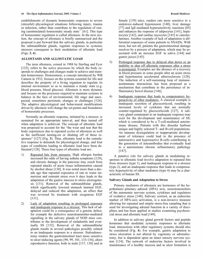
establishments of dynamic homeostatic responses to severe (stressful) physiological situations following injury, trauma or infection, rather than contribute to the regulation of rest-ing (unstimulated) homeostatic steady state`` [61]. This type of homeostatic regulation is called allostasis. In the next sec-tion, the concept of allostasis is briefly summarized and the proposal is put forward that the salivary glands, in particular the submandibular glands, regulate responses to systemic stressors consequent to their modulation of allostatic load (Figs. 3, 4).
ALLOSTASIS AND ALLOSTATIC LOAD
The term allostasis, coined in 1988 by Sterling and Eyer [125], refers to the active processes by which the body re-sponds to challenges and changes made to achieve and main-tain homeostasis. Homeostasis, a concept introduced by WB Cannon in 1932, focuses on the systems essential for life and describes the property of a living organism to regulate its internal environment so as to maintain a steady state (e.g. blood pressure, blood glucose). Allostasis is more dynamic and focuses on the processes required to maintain systems in balance in the face of environmental, life stage and unex-pected, sometimes persistent, changes or challenges [126]. The adaptive physiological and behavioural modifications driven by allostasis will change the set-points and boundaries of homeostatic processes.
Normally an allostatic response, initiated by a stressor, is sustained for an appropriate interval, and then turned off when adaptation is achieved. The concept of allostatic load was proposed to account for the “the wear and tear that the body experiences due to repeated cycles of allostasis as well as the inefficient turning-on or shutting off of these re-sponses” [127] (Fig. 3). Thus, allostatic load is the cumula-tive response to demands for physiological change, and four types of conditions leading to allostatic load have been de-lineated [128]. These four types of allostatic load are:
1) Repeated hits from stressors. High allergen burden increased the odds of having asthma symptoms [129], and chronic damage in the pancreas may result from repeated attacks of acute tissue inflammation caused by alcohol abuse [130]. It was noted more than a dec-ade ago that repeated exposures of rats to water im-mersion and restraint stress over 6 days leads to the adaptation of the gastric mucosa to stress ulcerogene-sis [131]. Removal of the submandibular glands, which significantly lowered stomach luminal EGF, delayed and reduced this adaptation, an effect that was reversed by treatment with exogenous EGF [131].
2) Lack of adaptation resulting in prolonged exposure and inadequate response to a stressor. This lack of ad-aptation could be a consequence of a genetic defect, for example the defective neurotransmitter-mediated signalling in the salivary glands of NOD mice con-tributes to the development of sialadenitis and even-tually SS [132]. Removal of the submandibular glands results in several pathologies possibly related to an inadequate response to a stressor. Sialoadenec-tomy renders the gastrointestinal tract more sensitive to ulcer-inducing agents [98, 99, 101, 133-136], alters reproductive function, both in male [137, 138] and in
female [139] mice, renders rats more sensitive to a endotoxic-induced hypotension [140], liver damage [77] and IgE-mediated hypersensitivity reactions [85] and enhances the response of adipocytes [141], hepa-tocytes [142], and cardiac myocytes [143] to catecho-lamines. Another example of lack of adaptation is dif-ferential responses of some patients to NSAIDs. With most, but not all, patients the gastrointestinal damage resolves by a process of adaptation, which may be as-sociated with an increase EGF in saliva [144] and gastric juices [145].
3) Prolonged response due to delayed shut down or an
inability to shut off allostatic responses after a stress is
terminated. Examples are the absence of a recovery
in blood pressure in some people after an acute stress and hypertension accelerated atherosclerosis [128]. The induction of a self-sustaining loop of immune-nonimmune interactions has been proposed as a mechanism that contribute to the persistence of in-flammatory bowel disease [146].
4) Inadequate response that leads to compensatory hy-peractivity of other mediators. A classical example is inadequate secretion of glucocorticoid, resulting in increased levels of cytokines that are normally counter-regulated by glucocorticoids [127]. A sali-vary gland counterpart to an inadequate response may exist for the development and maintenance of SS, which is considered to be a T-cell-mediated autoim-mune disease. Patients with this disorder harbour unique and highly selected T- and B-cell populations. An immune dysregulation or inappropriate develop-ment of tolerance could lead to the presence of autoreactive and hyporeactive B-cells contributing to the generation of autoantibodies that eventually lead to a autoimmune chronic inflammatory pathology [147].
A putative role for salivary glands in modulating re-sponses to allostatic load involve adaptation to repeated hits from stressors (type 1), and inadequate response to a stressor (type 2), and an inadequate response that leads to compensa-tory hyperactivity of other mediators (type 4) may be a char-acteristic of human SS.
Salivary Glands and Adaptation to Stress
Primary mediators of allostasis are hormones of the hy-pothalamo–pituitary–adrenal (HPA) axis, neurotransmitters of the autonomic nervous system, cytokines and regulators of oxidative stress [148]. Salivary cortisol, as an endocrine marker of HPA-axis activation, is a non-invasive measure allowing for repeated and simple stress-free sampling that is used for investigating adrenal function in a variety of disci-plines and has been applied in studies examining psychoso-cial stress and allostatic load [149].
In addition to salivary gland growth factors and peptide hormones that modulate systemic responses to allostatic load, interactions with other regulatory systems should also be considered (Fig. 4). For example, gastric adaptation to stress ulceration is not altered by vagotomy or adrenalec-tomy, but is dependent upon an intact sensory nervous sys-tem [124]. The network of endocrine factors involved in maintenance of a healthy mucosa and in ulcer formation is
Salivary Gland Hormones and Allostatic Load The Open Inflammation Journal, 2009, Volume 2 17
extensive. Testosterone, produced by the testis, increases the severity of mouth and stomach ulceration in both sham-operated and sialoadenectomized rats [150]. Pinealectomy augments water immersion and restraint stress-induced ul-cerous lesions, and these lesions exhibit a circadian rhythm with an increase in the day and attenuation at night, reflect-ing diurnal changes in melatonin synthesis [151]. Integrative input from the central nervous system is also involved. Ex-posing rats to either chronic constant light or repeated im-mobilization stress, respectively, either inhibits [152] or ac-tivates [153] the neural sympathetic component to the sali-vary glands. The sympathetic innervation of the salivary glands also modulates systemic responses such as mast cell mediated TNF -dependent cytotoxicity [154], acute hy-potensive responses to endotoxin [140], and pulmonary in-flammation following antigen challenge [85], probably through the regulation of endocrine secretion of factors that allow adjustment to allostatic load.
Further studies and evaluations from the perspective of the concept of allostatic load will determine the validity of this hypothesis, and possibly contribute to understanding the role of salivary glands in modulation of systemic disease and pathology.
ABBREVIATIONS
ARDS = Acute respiratory distress syndrome
ALS = Amyotrophic lateral sclerosis
AMP = Adenosine 5' monophosphate
bFGF = Basic fibroblast growth factor
BHR = Bronchial hyperresponsiveness
BNDF = Brain-derived neurotrophic factor
CD16 = Low affinity Fc RIII receptor
EGF = Epidermal growth factor
E-FABP = Epidermal fatty acid binding protein
GVHD = Graft-versus-host disease
HGF = Hepatocyte growth factor
HPA = Hypothalamo–pituitary–adrenal
hHSP60 = Human heat-shock protein 60
HIV = Human immunodeficiency virus
ICAM-1 = Inter-cellular adhesion molecule 1
Fig. (4). Salivary Glands and Allostasis. Left side of figure: Stressors or allostatic load (1) lead to altered state with less than optimal ho-
meostasis – the allostatic state. Endocrine secretion of submandibular gland factors (2) modulates the reactivity of system, and help re-
establish the homeostatic state (3). Right side of figure:. However, if endocrine secretion is reduced from salivary glands the appropriate
adjustments required to re-establish the homeostatic state cannot be made and the allostatic state persists (4) with pathological consequences.

18 The Open Inflammation Journal, 2009, Volume 2 Ronald Mathison
IGF-I & IGFII = Insulin-like growth factors I and II
LPS = Lipopolysaccharide
MPO = Myeloperoxidase
MHC = Major histocompatibility complex
mAChRs = Muscarinic acetylcholine receptors
NGF = Nerve growth factor
NOD = Non-obese diabetic
NSAIDs = Nonsteroidal anti-inflammatory drugs
PAM = Peptidylglycine alpha-amidating mono-oxygenase
PDGF = Platelet derived growth factor
PKC = Protein kinase C
SS = Sjögren's syndrome
SGPT = Submandibular gland peptide-T
SIRS = Systemic inflammatory response syndrome
SMR1 = Submandibular rat-1
TGF & TGF = Transforming growth factor alpha & beta
VEGF = Vascular endothelial growth factor
VIP = Intestinal peptide (VIP)
VLA4 = 4 1 integrin or CD49d
ACKNOWLEDGEMENTS
The financial support of Allergen NCE Inc. is appreci-ated.
REFERENCES
[1] Llena-Puy C. The role of saliva in maintaining oral health and as an aid to diagnosis. Med Oral Patol Oral Cir Bucal 2006; 11: E449-55.
[2] Segal A, Wong DT. Salivary diagnostics: enhancing disease detec-tion and making medicine better. Eur J Dent Educ 2008; 12 (Suppl
1): 22-9. [3] Messana I, Inzitari R, Fanali C, Cabras T, Castagnola M. Facts and
artifacts in proteomics of body fluids. What proteomics of saliva is telling us? J Sep Sci 2008; 31: 1948-63.
[4] Cotrim AP, Mineshiba F, Sugito T, Samuni Y, Baum BJ. Salivary gland gene therapy. Dent Clin North Am 2006; 50: 157-73.
[5] Baum BJ, Voutetakis A, Wang J. Salivary glands: novel target sites for gene therapeutics. Trends Mol Med 2004; 10: 585-90.
[6] Voutetakis A, Bossis I, Kok MR, et al. Salivary glands as a poten-tial gene transfer target for gene therapeutics of some monogenetic
endocrine disorders. J Endocrinol 2005; 185: 363-72. [7] von Bültzingslöwen I, Sollecito TP, Fox PC, et al. Salivary dys-
function associated with systemic diseases: systematic review and clinical management recommendations. Oral Surg Oral Med Oral
Pathol Oral Radiol Endod 2007; 103; S57 e1-15. [8] Proctor GB, Carpenter GH. Regulation of salivary gland function
by autonomic nerves. Auton Neurosci 2007; 133: 3-18. [9] Seymour GJ, Ford PJ, Cullinan MP, Leishman S, Yamazaki K.
Relationship between periodontal infections and systemic disease. Clin Microbiol Infect 2007; 13 (Suppl 4): 3-10.
[10] Williams RC, Barnett AH, Claffey N, et al. The potential impact of periodontal disease on general health: a consensus view. Curr Med
Res Opin 2008; 24: 1635-43. [11] Rautemaa R, Lauhio A, Cullinan MP, Seymour GJ. Oral infections
and systemic disease--an emerging problem in medicine. Clin Mi-crobiol Infect 2007; 13: 1041-7.
[12] Bacman S, Sterin-Borda L, Camusso JJ, Arana R, Hubscher O, Borda E. Circulating antibodies against rat parotid gland M3 mus-
carinic receptors in primary Sjogren's syndrome. Clin Exp Immu-
nol 1996; 104: 454-9. [13] Leiros CP, Rosignoli F, Genaro AM, Sales ME, Sterin-Borda L,
Santiago Borda E. Differential activation of nitric oxide synthase through muscarinic acetylcholine receptors in rat salivary glands. J
Auton Nerv Syst 2000; 79: 99-107. [14] Roca V, Rosignoli F, Calafat M, Leiros CP. Lack of nitric oxide-
mediated regulation of amylase secretion stimulated by VIP in pa-rotid glands of NOD mice. Int Immunopharmacol 2004; 4: 1837-
44. [15] Xia DS, Liu Y, Zhang CM, Yang SH, Wang SL. Antimicrobial
effect of acidified nitrate and nitrite on six common oral pathogens in vitro. Chin Med J 2006; 119: 1904-9.
[16] Nagler RM, Sherman Y, Nagler A. Histopathological study of the human submandibular gland in graft versus host disease. J Clin
Pathol 1999; 52: 395-7. [17] Manganelli P, Fietta P. Apoptosis and Sjogren syndrome. Semin
Arthritis Rheum 2003; 33: 49-65. [18] Mitsias DI, Kapsogeorgou EK, Moutsopoulos HM. Sjogren's syn-
drome: why autoimmune epithelitis? Oral Dis 2006; 12: 523-32. [19] Cai FZ, Lester S, Lu T, et al. Mild autonomic dysfunction in pri-
mary Sjogren's syndrome: a controlled study. Arthritis Res Ther 2008; 10: R31.
[20] Mellgren SI, Goransson LG, Omdal R. Primary Sjogren's syndrome associated neuropathy. Can J Neurol Sci 2007; 34: 280-7.
[21] Tzioufas AG, Voulgarelis M. Update on Sjogren's syndrome auto-immune epithelitis: from classification to increased neoplasias.
Best Pract Res Clin Rheumatol 2007; 21: 989-1010. [22] Gudbjornsson B, Hedenstrom H, Stalenheim G, Hallgren R. Bron-
chial hyperresponsiveness to methacholine in patients with primary Sjogren's syndrome. Ann Rheum Dis 1991; 50: 36-40.
[23] Ludviksdottir D, Janson C, Hogman M, et al. Increased nitric oxide in expired air in patients with Sjogren's syndrome. BHR study
group. Bronchial hyperresponsiveness. Eur Respir J 1999; 13: 739-43.
[24] Potena A, La Corte R, Fabbri LM, Papi A, Trotta F, Ciaccia A. Increased bronchial responsiveness in primary and secondary
Sjogren's syndrome. Eur Respir J 1990; 3: 548-53. [25] Amin K, Ludviksdottir D, Janson C, et al. Inflammation and struc-
tural changes in the airways of patients with primary Sjogren's syn-drome. Respir Med 2001; 95: 904-10.
[26] Stalenheim G, Gudbjornsson B. Anti-inflammatory drugs do not alleviate bronchial hyperreactivity in Sjogren's syndrome. Allergy
1997; 52: 423-7. [27] Ludviksdottir D, Janson C, Bjornsson E, et al. Different airway
responsiveness profiles in atopic asthma, nonatopic asthma, and Sjogren's syndrome. BHR Study Group. Bronchial hyperrespon-
siveness. Allergy 2000; 55: 259-65. [28] Schneider A, Merikhi A, Frank BB. Autoimmune disorders: gastro-
intestinal manifestations and endoscopic findings. Gastrointest En-dosc Clin N Am 2006; 16: 133-51.
[29] Volter F, Fain O, Mathieu E, Thomas M. Esophageal function and Sjogren's syndrome. Dig Dis Sci 2004; 49: 248-53.
[30] Ostuni PA, Germana B, Di Mario F, et al. Gastric involvement in primary Sjogren's syndrome. Clin Exp Rheumatol 1993; 11: 21-5.
[31] Biagini MR, Milani S, Fedi P, Calabro A, Tedesco A, Surrenti C. Duodenal ulcer and Sjogren's syndrome in patients with primary
biliary cirrhosis: a casual association? Am J Gastroenterol 1991; 86: 1190-3.
[32] Hayakawa T, Naruse S, Kitagawa M, Kondo T. Clinical aspects of autoimmune pancreatitis in Sjogren's syndrome. JOP 2001; 2: 88-
92. [33] Kaplan MJ, Ike RW. The liver is a common non-exocrine target in
primary Sjogren's syndrome: a retrospective review. BMC Gastro-enterol 2002; 2: 21.
[34] Mavragani CP, Moutsopoulos HM. Conventional therapy of Sjogren's syndrome. Clin Rev Allergy Immunol 2007; 32: 284-91.
[35] Moore PA, Bounous DI, Kaswan RL, Humphreys-Beher MG. Histologic examination of the NOD-mouse lacrimal glands, a po-
tential model for idiopathic autoimmune dacryoadenitis in Sjogren's syndrome. Lab Anim Sci 1996; 46: 125-8.
[36] Murphy ED. Lymphoproliferation (lpr) and other single-locus models for murine lupus. In: Gershwin ME, Merchant B Eds. De-
fects in Laboratory Animals. Plenum, New York 1981; 143-76. [37] Koski H, Konttinen YT, Hietanen J, Tervo T, Malmstrom M. Epi-
dermal growth factor, transforming growth factor-alpha, and epi-

Salivary Gland Hormones and Allostatic Load The Open Inflammation Journal, 2009, Volume 2 19
dermal growth factor receptor in labial salivary glands in Sjogren's
syndrome. J Rheumatol 1997; 24: 1930-5. [38] Pflugfelder SC, Jones D, Ji Z, Afonso A, Monroy D. Altered cyto-
kine balance in the tear fluid and conjunctiva of patients with Sjogren's syndrome keratoconjunctivitis sicca. Curr Eye Res 1999;
19: 201-11. [39] Giusti L, Baldini C, Bazzichi L, et al. Proteome analysis of whole
saliva: a new tool for rheumatic diseases--the example of Sjogren's syndrome. Proteomics 2007; 7: 1634-43.
[40] Ryu OH, Atkinson JC, Hoehn GT, Illei GG, Hart TC. Identification of parotid salivary biomarkers in Sjogren's syndrome by surface-
enhanced laser desorption/ionization time-of-flight mass spec-trometry and two-dimensional difference gel electrophoresis.
Rheumatology (Oxford) 2006; 45: 1077-86. [41] Isenman L, Liebow C, Rothman S. The endocrine secretion of
mammalian digestive enzymes by exocrine glands. Am J Physiol 1999; 276: E223-32.
[42] Kelly RB. Pathways of protein secretion in eukaryotes. Science 1985; 230: 25-32.
[43] Zelles T, Purushotham KR, Macauley SP, Oxford GE, Humphreys-Beher MG. Saliva and growth factors: the fountain of youth resides
in us all. J Dent Res 1995; 74: 1826-32. [44] Kagami H, Wang S, Hai B. Restoring the function of salivary
glands. Oral Dis 2008; 14: 15-24. [45] Patel VN, Rebustini IT, Hoffman MP. Salivary gland branching
morphogenesis. Differentiation 2006; 74: 349-64. [46] Tsukinoki K, Yasuda M, Asano S, et al. Association of hepatocyte
growth factor expression with salivary gland tumor differentiation. Pathol Int 2003; 53: 815-22.
[47] Werner H, Katz J. The emerging role of the insulin-like growth factors in oral biology. J Dent Res 2004; 83: 832-6.
[48] Teymoortash A, Tiemann M, Schrader C, Hartmann O, Werner JA. Transforming growth factor beta in chronic obstructive sialadenitis
of human submandibular gland. Arch Oral Biol 2003; 48: 111-6. [49] Lawrence AM, Tan S, Hojvat S, Kirsteins L. Salivary gland hyper-
glycemic factor: an extrapancreatic source of glucagon-like mate-rial. Science 1977; 195: 70-2.
[50] Nexo E, Olsen PS, Poulsen K. Exocrine and endocrine secretion of renin and epidermal growth factor from the mouse submandibular
glands. Regul Pept 1984; 8: 327-34. [51] Murphy RA, Saide JD, Blanchard MH, Young M. Nerve growth
factor in mouse serum and saliva: role of the submandibular gland. Proc Natl Acad Sci USA 1977; 74: 2330-3.
[52] Pedersen EB, Poulsen K. Vast release of submaxillary mouse renin to saliva after stimulation with cholinergic, beta-adrenergic but es-
pecially alpha-adrenergic agonists. Acta Endocrinol (Copenh) 1982; 99: 636-40.
[53] Berg T, Johansen L, Poulsen K. Exocrine and endocrine release of kallikrein after reflex-induced salivary secretion. Acta Physiol
Scand 1990; 139: 29-37. [54] Rougeot C, Rosinski-Chupin I, Njamkepo E, Rougeon F. Selective
processing of submandibular rat 1 protein at dibasic cleavage sites. Salivary and bloodstream secretion products. Eur J Biochem 1994;
219: 765-73. [55] Rougeot C, Rosinski-Chupin I, Rougeon F. Novel genes and hor-
mones in salivary glands: from the gene for the submandibular rat1 protein (SMR1) precursor to receptor sites for SMR1 mature pep-
tides. Biomed Rev 1998; 9: 17-32. [56] Rougeot C, Vienet R, Cardona A, Le Doledec L, Grognet JM,
Rougeon F. Targets for SMR1-pentapeptide suggest a link between the circulating peptide and mineral transport. Am J Physiol 1997;
273: R1309-20. [57] Ogata T. The internal secretion of salivary gland. Endocrinol Jpn
1955; 2: 247-61. [58] Proctor GB, Asking B, Garrett JR. Effects of secretory nerve stimu-
lation on the movement of rat parotid amylase into the circulation. Arch Oral Biol 1989; 34: 609-13.
[59] Barka T. Biologically active polypeptides in submandibular glands. J Histochem Cytochem 1980; 28: 836-59.
[60] Mori M, Takai Y, Kunikata M. Review : biologically active pep-tides in the submandibular gland. Role of the granular convoluted
tubule. Acta Histochem Cytochem 1992; 25: 325-41. [61] Rougeot C, Rosinski-Chupin I, Mathison R, Rougeon F. Rodent
submandibular gland peptide hormones and other biologically ac-tive peptides. Peptides 2000; 21: 443-55.
[62] Rosinski-Chupin I, Tronik D, Rougeon F. High level of accumula-
tion of a mRNA coding for a precursor-like protein in the submax-illary gland of male rats. Proc Natl Acad Sci USA 1988; 85: 8553-
7. [63] Rosinski-Chupin I, Rougeot C, Courty Y, Rougeon F. Localization
of mRNAs of two androgen-dependent proteins, SMR1 and SMR2, by in situ hybridization reveals sexual differences in acinar cells of
rat submandibular gland. J Histochem Cytochem 1993; 41: 1645-9. [64] Rosinski-Chupin I, Huaulme JF, Rougeot C, Rougeon F. The tran-
scriptional response to androgens of the rat VCSA1 gene is ampli-fied by both binary and graded mechanisms. Endocrinology 2001;
142: 4550-9. [65] Wisner A, Dufour E, Messaoudi M, et al. Human Opiorphin, a
natural antinociceptive modulator of opioid-dependent pathways. Proc Natl Acad Sci USA 2006; 103: 17979-84.
[66] Messaoudi M, Desor D, Nejdi A, Rougeot C. The endogenous androgen-regulated sialorphin modulates male rat sexual behavior.
Horm Behav 2004; 46: 684-91. [67] Mathison R. Anti-inflammatory peptides. Patent US 7153835,
December 26, 2006. [68] Mathison R, Davison JS, Befus D, Moore G. Peptides for treatment
of inflammation and shock. Patent US 6852697, February 8, 2005. [69] Mathison R, Davison JS, Befus D, Moore G. Treating allergic
reactions and inflammatory responses with tri-and dipeptides. Pat-ent US 7094760, July 1, 2003.
[70] Mathison R, Metwally E. Treating allergic reactions and inflamma-tory responses with tri-and dipeptides. Patent US 6586403, July 1,
2003. [71] Mathison RD, Befus AD, Davison JS. A novel submandibular
gland peptide protects against endotoxic and anaphylactic shock. Am J Physiol 1997; 273: R1017-23.
[72] Mathison R, Lo P, Moore G, Scott B, Davison JS. Attenuation of intestinal and cardiovascular anaphylaxis by the salivary gland
tripeptide FEG and its D-isomeric analog feG. Peptides 1998; 19: 1037-42.
[73] Jones DE Jr, Tran-Patterson R, Cui DM, Davin D, Estell KP, Miller DM. Epidermal growth factor secreted from the salivary gland is
necessary for liver regeneration. Am J Physiol 1995; 268: G872-8. [74] Lambotte L, Saliez A, Triest S, et al. Effect of sialoadenectomy
and epidermal growth factor administration on liver regeneration after partial hepatectomy. Hepatology 1997; 25: 607-12.
[75] Yamamoto R, Iishi H, Tatsuta M, Tsuji M, Terada N. Inhibitory effect of sialoadenectomy on hepatocellular tumourigenesis in male
mice induced by 3'-methyl-4-dimethylaminoazobenzene. Virchows Arch 1994; 425: 79-82.
[76] Buira I, Poch E, Sanchez O, et al. Sialoadenectomy alters liver cell turn-over and function in mice. J Cell Physiol 2004; 198: 12-21.
[77] Sanchez O, Almagro A, Viladrich M, Ramirez I, Soley M. Siaload-enectomy enhances hepatic injury induced by lipopolysaccha-
ride/galactosamine in mice. Liver Int 2008; 28: 878-88. [78] Noguchi S, Ohba Y, Oka T. Influence of epidermal growth factor
on liver regeneration after partial hepatectomy in mice. J Endocri-nol 1991; 128: 425-31.
[79] Mathison R, Lo P, Tan D, Scott B, Davison JS. The tripeptide feG reduces endotoxin-provoked perturbation of intestinal motility and
inflammation. Neurogastroenterol Motil 2001; 13: 599-603. [80] Mathison R, Woodman R, Davison JS. Regulation of leukocyte
adhesion to heart by the tripeptides feG and feG(NH2). Can J Physiol Pharmacol 2001; 79: 785-92.
[81] Merkler DJ. C-terminal amidated peptides: production by the in vitro enzymatic amidation of glycine-extended peptides and the
importance of the amide to bioactivity. Enzyme Microb Technol 1994; 16: 450-56.
[82] Kato T, Hanaoka K, Ochiai O, et al. Secretion of peptidylglycine alpha-amidating monooxygenase (PAM) from rat salivary glands.
Neurochem Int 1992; 21: 153-8. [83] Bolkenius FN, Ganzhorn AJ. Peptidylglycine alpha-amidating
mono-oxygenase: neuropeptide amidation as a target for drug de-sign. Gen Pharmacol 1998; 31: 655-9.
[84] Ramaswamy K, Mathison R, Carter L, et al. Marked antiinflamma-tory effects of decentralization of the superior cervical ganglia. J
Exp Med 1990; 172: 1819-30. [85] Mathison R, Hogan A, Helmer D, et al. Role for the submandibular
gland in modulating pulmonary inflammation following induction of systemic anaphylaxis. Brain Behav Immun 1992; 6: 117-29.

20 The Open Inflammation Journal, 2009, Volume 2 Ronald Mathison
[86] Declue AE, Schooley EK, Reinero CR. FEG-COOH tripeptide
attenuates allergen-induced eosinophilic airway inflammation in a model of feline asthma. J Vet Intern Med 2007; 21.
[87] Mathison R, Davison JS, Befus AD, Abraham WM. The tripeptide feG inhibits asthmatic reactions in sheep. In: Monduzzi E Eds. Im-
munology 2004, Medimond International Proceedings, Montreal 2004; 515-9.
[88] Mathison RD, Befus AD, Davison JS, Woodman RC. Modulation of neutrophil function by the tripeptide feG. BMC Immunol 2003;
4: 3. [89] Metwally E, Pires JM, Moore GJ, Befus DA, Davison JS, Mathison
R. Submandibular gland tripeptide FEG (Phe-Glu-Gly) and ana-logues: keys to structure determination. Peptides 2002; 23: 193-9.
[90] Dery RE, Ulanova M, Puttagunta L, et al. Frontline: Inhibition of allergen-induced pulmonary inflammation by the tripeptide feG: a
mimetic of a neuro-endocrine pathway. Eur J Immunol 2004; 34: 3315-25.
[91] Mathison RD, Christie E, Davison JS. The tripeptide feG inhibits leukocyte adhesion. J Inflamm 2008; 5: 6.
[92] Mathison RD, Davison JS. The tripeptide feG regulates the produc-tion of intracellular reactive oxygen species by neutrophils. J In-
flamm 2006; 3: 9. [93] Bao F, John SM, Chen Y, Mathison RD, Weaver LC. The tripep-
tide phenylalanine-(d) glutamate-(d) glycine modulates leukocyte infiltration and oxidative damage in rat injured spinal cord. Neuro-
science 2006; 140: 1011-22. [94] Cohen S, Elliott GA. The stimulation of epidermal keratinization
by a protein isolated from the submaxillary gland of the mouse. J Invest Dermatol 1963; 40: 1-5.
[95] Cohen S. Isolation of a mouse submaxillary gland protein acceler-ating incisor eruption and eyelid opening in the new-born animal. J
Biol Chem 1962; 237: 1555-62. [96] Gregory H. Isolation and structure of urogastrone and its relation-
ship to epidermal growth factor. Nature 1975; 257: 325-7. [97] Skinner KA, Soper BD, Tepperman BL. Effect of sialoadenectomy
and salivary gland extracts on gastrointestinal mucosal growth and gastrin levels in the rat. J Physiol 1984; 351: 1-12.
[98] Leitch GJ. Role of the salivary glands in protecting the stomach against ethanol. Alcohol Alcohol 1985; 20: 305-11.
[99] Olsen PS, Poulsen SS, Therkelsen K, Nexo E. Effect of siaload-enectomy and synthetic human urogastrone on healing of chronic
gastric ulcers in rats. Gut 1986; 27: 1443-9. [100] Sarosiek J, Bilski J, Murty VL, Slomiany A, Slomiany BL. Role of
salivary epidermal growth factor in the maintenance of physico-chemical characteristics of oral and gastric mucosal mucus coat.
Biochem Biophys Res Commun 1988; 152: 1421-7. [101] Tepperman BL, Soper BD, Morris GP. Effect of sialoadenectomy
on adaptive cytoprotection in the rat. Gastroenterology 1989; 97: 123-9.
[102] Shirai Y, Wakatsuki Y, Kusumoto T, et al. Induction and mainte-nance of immune effector cells in the gastric tissue of mice orally
immunized to Helicobacter pylori requires salivary glands. Gastro-enterology 2000; 118: 749-59.
[103] Okuda T, Onda M, Tokunaga A, Sugisaki Y. Stimulatory effect of EGF and inhibitory effect of sialoadenectomy on growth of an EGF
receptor-hyperproducing human gastric cancer xenograft in nude mice. Surg Today 1994; 24: 725-33.
[104] Kongara KR, Soffer EE. Saliva and esophageal protection. Am J Gastroenterol 1999; 94: 1446-52.
[105] Bots CP, Brand HS, Veerman EC, et al. Acute effects of hemo-dialysis on salivary flow rate and composition. Clin Nephrol 2007;
67: 25-31. [106] Guh JY, Chen HC, Chuang LY, Yang CY, Tsai JH, Lai YH. Sig-
nificance of salivary epidermal growth factor in peptic ulcer dis-ease in hemodialysis patients. Nephron 2001; 87: 134-8.
[107] Wilczynska-Borawska M, Borawski J, Kovalchuk O, Chyczewski L, Mysliwiec M, Stokowska W. Unexpected effect of haemodialy-
sis on salivary hepatocyte growth factor. Nephrol Dial Transplant 2005; 20: 2869-70.
[108] Kamisawa T, Tu Y, Egawa N, Sakaki N, Inokuma S, Kamata N. Salivary gland involvement in chronic pancreatitis of various eti-
ologies. Am J Gastroenterol 2003; 98: 323-6. [109] Kamisawa T, Tu Y, Sasaki R, Egawa N, Kamata N, Sasaki T. The
relationship of salivary gland function to elevated serum IgG4 in autoimmune pancreatitis. Intern Med 2007; 46: 435-9.
[110] Frossard JL, Steer ML, Pastor CM. Acute pancreatitis. Lancet
2008; 371: 143-52. [111] Sekimoto M, Takada T, Kawarada Y, et al. JPN Guidelines for the
management of acute pancreatitis: epidemiology, etiology, natural history, and outcome predictors in acute pancreatitis. J Hepatobil-
iary Pancreat Surg 2006; 13: 10-24. [112] Dembinski A, Warzecha Z, Konturek PC, et al. Epidermal growth
factor accelerates pancreatic recovery after caerulein-induced pan-creatitis. Eur J Pharmacol 2000; 398: 159-68.
[113] Warzecha Z, Dembinski A, Konturek PC, Ceranowicz P, Konturek SJ. Epidermal growth factor protects against pancreatic damage in
cerulein-induced pancreatitis. Digestion 1999; 60: 314-23. [114] Rifai Y, Elder AS, Carati CJ, et al. The tripeptide analog feG ame-
liorates severity of acute pancreatitis in a caerulein mouse model. Am J Physiol Gastrointest Liver Physiol 2008; 294: G1094-9.
[115] Bing J, Poulsen K. In mice aggressive behavior provokes vast increase in plasma renin concentration, causing only slight, if any,
increase in blood pressure. Acta Physiol Scand 1979; 105: 64-72. [116] Nexo E, Hollenberg MD, Bing J. Aggressive behavior in mice
provokes a marked increase in both plasma epidermal growth fac-tor and renin. Acta Physiol Scand 1981; 111: 367-71.
[117] Aloe L, Alleva E, Bohm A, Levi-Montalcini R. Aggressive behav-ior induces release of nerve growth factor from mouse salivary
gland into the bloodstream. Proc Natl Acad Sci USA 1986; 83: 6184-7.
[118] Tsukinoki K, Saruta J, Sasaguri K, et al. Immobilization stress induces BDNF in rat submandibular glands. J Dent Res 2006; 85:
844-8. [119] Konturek SJ, Brzozowski T, Konturek PK, Majka J, Dembinski A.
Role of salivary glands and epidermal growth factor (EGF) in gas-tric secretion and mucosal integrity in rats exposed to stress. Regul
Pept 1991; 32: 203-15. [120] Aloe L, Bracci-Laudiero L, Bonini S, Manni L. The expanding role
of nerve growth factor: from neurotrophic activity to immunologic diseases. Allergy 1997; 52: 883-94.
[121] Eichelman B. Aggressive behavior: from laboratory to clinic. Quo vadit? Arch Gen Psychiatry 1992; 49: 488-92.
[122] Bernabei R, Landi F, Bonini S, et al. Effect of topical application of nerve-growth factor on pressure ulcers. Lancet 1999; 354: 307.
[123] Aloe L, Tirassa P, Lambiase A. The topical application of nerve growth factor as a pharmacological tool for human corneal and skin
ulcers. Pharmacol Res 2008; 57: 253-8. [124] Brzozowski T, Konturek SJ, Pytko-Polonczyk J, Warzecha Z.
Gastric adaptation to stress: role of sensory nerves, salivary glands, and adrenal glands. Scand J Gastroenterol 1995; 30: 6-16.
[125] Sterling P, Eyer J. Allostasis, a new paradigm to explain arousal pathology. In: Fisher S, Reason J, Eds. Handbook of Life Stress,
Cognition and Health, New York: John Wiley & Sons 1988; 629-49.
[126] McEwen BS, Wingfield JC. The concept of allostasis in biology and biomedicine. Horm Behav 2003; 43: 2-15.
[127] McEwen BS. Allostasis and allostatic load: implications for neuro-psychopharmacology. Neuropsychopharmacology 2000; 22: 108-
24. [128] McEwen BS. Protective and damaging effects of stress mediators.
N Engl J Med 1998; 338: 171-9. [129] Salo PM, Arbes SJ Jr, Crockett PW, Thorne PS, Cohn RD, Zeldin
DC. Exposure to multiple indoor allergens in US homes and its re-lationship to asthma. J Allergy Clin Immunol 2008; 121: 678-84
e672. [130] Vonlaufen A, Wilson JS, Pirola RC, Apte MV. Role of alcohol
metabolism in chronic pancreatitis. Alcohol Res Health 2007; 30: 48-54.
[131] Konturek SJ, Brzozowski T, Majka J, Drozdowicz D, Stachura J. Adaptation of the gastric mucosa to stress. Role of prostaglandin
and epidermal growth factor. Scand J Gastroenterol Suppl 1992; 193: 39-45.
[132] Rosignoli F, Roca V, Meiss R, Leceta J, Gomariz RP, Perez Leiros C. Defective signalling in salivary glands precedes the autoimmune
response in the non-obese diabetic mouse model of sialadenitis. Clin Exp Immunol 2005; 142: 411-18.
[133] Kohut A, Mahelova O, Mojzis J, Mirossay L. Effect of siaload-enectomy on stomach lesions induced by indomethacin and ethanol
in relation to gastric vascular permeability, the gastrin level and HCl secretion in rats. Physiol Res 1992; 41: 381-6.
Salivary Gland Hormones and Allostatic Load The Open Inflammation Journal, 2009, Volume 2 21
[134] Konturek SJ, Brozozowski T, Bielanski W, Warzecha Z,
Drozdowicz D. Epidermal growth factor in the gastroprotective and ulcer-healing actions of sucralfate in rats. Am J Med 1989; 86: 32-
7. [135] Konturek SJ, Dembinski A, Warzecha Z, Bielanski W, Brzozowski
T, Drozdowicz D. Epidermal growth factor (EGF) in the gastropro-tective and ulcer healing actions of colloidal bismuth subcitrate
(De-Nol) in rats. Gut 1988; 29: 894-902. [136] Wingren U, Brown TH, Watkins BM, Larson GM. Delayed gastric
ulcer healing after extirpation of submandibular glands is sex-dependent. Scand J Gastroenterol 1989; 24: 1102-6.
[137] Tsutsumi O, Kurachi H, Oka T. A physiological role of epidermal growth factor in male reproductive function. Science 1986; 233:
975-7. [138] Reyes AB, Wakasugi N. Long-term influence of sialoadenectomy
on reproductive performance of male mice. J Reprod Fertil 1995; 105: 279-85.
[139] Okamoto S, Oka T. Evidence for physiological function of epider-mal growth factor: pregestational sialoadenectomy of mice de-
creases milk production and increases offspring mortality during lactation period. Proc Natl Acad Sci USA 1984; 81: 6059-63.
[140] Mathison R, Befus D, Davison JS. Removal of the submandibular glands increases the acute hypotensive response to endotoxin. Circ
Shock 1993; 39: 52-8. [141] Tebar F, Ramirez I, Soley M. Epidermal growth factor modulates
the lipolytic action of catecholamines in rat adipocytes. Involve-ment of a Gi protein. J Biol Chem 1993; 268: 17199-204.
[142] Grau M, Soley M, Ramirez I. Interaction between adrenaline and epidermal growth factor in the control of liver glycogenolysis in
mouse. Endocrinol 1997; 138: 2601-9. [143] Lorita J, Escalona N, Faraudo S, Soley M, Ramirez I. Effects of
epidermal growth factor on epinephrine-stimulated heart function in rodents. Am J Physiol Heart Circ Physiol 2002; 283: H1887-95.
[144] Lipscomb GR, Painter J, Riley S, Gillott T, Rees W. Role of acid and salivary epidermal growth factor in gastric mucosal adaptation
to naproxen in man. Scand J Gastroenterol 1998; 33: 790-4.
[145] Kelly SM, Jenner JR, Dickinson RJ, Hunter JO. Increased gastric
juice epidermal growth factor after non-steroidal anti-inflammatory drug ingestion. Gut 1994; 35: 611-4.
[146] Vogel JD, West GA, Danese S, et al. CD40-mediated immune-nonimmune cell interactions induce mucosal fibroblast chemokines
leading to T-cell transmigration. Gastroenterology 2004; 126: 63-80.
[147] Delaleu N, Jonsson R, Koller MM. Sjogren's syndrome. Eur J Oral Sci 2005; 113: 101-13.
[148] McEwen BS. Central effects of stress hormones in health and dis-ease: Understanding the protective and damaging effects of stress
and stress mediators. Eur J Pharmacol 2008; 583: 174-85. [149] Turner-Cobb JM. Psychological and stress hormone correlates in
early life: a key to HPA-axis dysregulation and normalisation. Stress 2005; 8: 47-57.
[150] Machowska A, Brzozowski T, Sliwowski Z, et al. Gastric secre-tion, proinflammatory cytokines and epidermal growth factor
(EGF) in the delayed healing of lingual and gastric ulcerations by testosterone. Inflammopharmacology 2008; 16: 40-7.
[151] Brzozowski T, Zwirska-Korczala K, Konturek PC, et al. Role of circadian rhythm and endogenous melatonin in pathogenesis of
acute gastric bleeding erosions induced by stress. J Physiol Phar-macol 2007; 58(Suppl 6): 53-64.
[152] Gallara RV, Bellavia SL, Serova LL, Sabban EL. Environmental light conditions alter gene expression of rat catecholamine biosyn-
thetic enzymes and Neuropeptide Y: differential effect in superior cervical ganglia and adrenal gland. Brain Res Mol Brain Res 2004;
124: 152-8. [153] Nankova B, Kvetnansky R, Hiremagalur B, Sabban B, Rusnak M,
Sabban EL. Immobilization stress elevates gene expression for catecholamine biosynthetic enzymes and some neuropeptides in rat
sympathetic ganglia: effects of adrenocorticotropin and glucocorti-coids. Endocrinology 1996; 137: 5597-604.
[154] Bissonnette EY, Mathison R, Carter L, Davison JS, Befus AD. Decentralization of the superior cervical ganglia inhibits mast cell
mediated TNF alpha-dependent cytotoxicity. 1. Potential role of salivary glands. Brain Behav Immun 1993; 7: 293-300.
Received: January 5, 2009 Revised: April 6, 2009 Accepted: April 14, 2009
© Ronald Mathison; Licensee Bentham Open.
This is an open access article licensed under the terms of the Creative Commons Attribution Non-Commercial License (http://creativecommons.org/licenses/by-nc/ 3.0/) which permits unrestricted, non-commercial use, distribution and reproduction in any medium, provided the work is properly cited.
Related Documents