Cronicon OPEN ACCESS EC MICROBIOLOGY EC MICROBIOLOGY Research Article Novel Brain Compartment Tapeworm Larvae-Possible Environmental Initiators of Demyelination in Multiple Sclerosis Alan B MacDonald* Molecular Interrogation Research, Naples, Florida, USA *Corresponding Author: Alan B MacDonald, Molecular Interrogation Research, Naples, Florida, USA. Received: October 08, 2020; Published: April 29, 2021 Abstract Multiple Sclerosis is a cause unknown neurodegenerative disease. Myelin sites in brain, spinal cord and eye are at risk for injury over the course of the illness. Peripheral nerve myelin injuries are absent. The epidemiology of multiple sclerosis leads investigators to scrutinize environmental variables as possible triggers for the disease. This report describes ten patients whose autopsy cerebro- spinal fluids revealed unsuspected microscopic scale cestode larval parasites of unknown species which were acquired in the United States from environmental food stuff ingestions contaminated with cestode eggs. Keywords: Cestode; Parasites; Cerebrospinal; Autopsy; Multiple Sclerosis Citation: Alan B MacDonald. “Novel Brain Compartment Tapeworm Larvae-Possible Environmental Initiators of Demyelination in Multiple Sclerosis”. EC Microbiology 17.5 (2021): 77-85. Introduction In the patients reported here, a thirty-year retrospective examination of autopsy cerebrospinal fluids from 10 MS patients was com- menced to search for possible medically cryptic microscopic antemortem infections of brain fluids which might have been undiagnosed during life. Methods and Patients Ten Multiple Sclerosis patient donors to a medical school brain bank were the subjects of this study. The dates of autopsy were be- tween 1984 - 2014. The institutional review board of the brain bank granted permission for tissue release for research purposes to the principal investigator. Patient confidentiality was enforced by extensive redaction of all individual patient identifiers. No medical records from the donor patients were available for release. Snap frozen cerebrospinal fluids obtained at the original autopsy were assigned a unique accession number and maintained at minus 80 degrees Centigrade in the brain bank. All brain solid tissue was processed for de- tailed neuropathological examination according to the institution protocols to ratify the category of multiple sclerosis and to exclude any additional brain pathologies and medical conditions outside of the domain of demyelinating disease. Thawed cerebrospinal fluid from ten patients was, for the first time, examined in this time capsule research study. All frozen fluids had been untouched since initial autopsy harvest. The justification for the permanent omission of pathologist examinations of autopsy

Welcome message from author
This document is posted to help you gain knowledge. Please leave a comment to let me know what you think about it! Share it to your friends and learn new things together.
Transcript

CroniconO P E N A C C E S S EC MICROBIOLOGYEC MICROBIOLOGY
Research Article
Novel Brain Compartment Tapeworm Larvae-Possible Environmental Initiators of Demyelination in Multiple Sclerosis
Alan B MacDonald*
Molecular Interrogation Research, Naples, Florida, USA
*Corresponding Author: Alan B MacDonald, Molecular Interrogation Research, Naples, Florida, USA.
Received: October 08, 2020; Published: April 29, 2021
Abstract
Multiple Sclerosis is a cause unknown neurodegenerative disease. Myelin sites in brain, spinal cord and eye are at risk for injury over the course of the illness. Peripheral nerve myelin injuries are absent. The epidemiology of multiple sclerosis leads investigators to scrutinize environmental variables as possible triggers for the disease. This report describes ten patients whose autopsy cerebro-spinal fluids revealed unsuspected microscopic scale cestode larval parasites of unknown species which were acquired in the United States from environmental food stuff ingestions contaminated with cestode eggs.
Keywords: Cestode; Parasites; Cerebrospinal; Autopsy; Multiple Sclerosis
Citation: Alan B MacDonald. “Novel Brain Compartment Tapeworm Larvae-Possible Environmental Initiators of Demyelination in Multiple Sclerosis”. EC Microbiology 17.5 (2021): 77-85.
Introduction
In the patients reported here, a thirty-year retrospective examination of autopsy cerebrospinal fluids from 10 MS patients was com-menced to search for possible medically cryptic microscopic antemortem infections of brain fluids which might have been undiagnosed during life.
Methods and Patients
Ten Multiple Sclerosis patient donors to a medical school brain bank were the subjects of this study. The dates of autopsy were be-tween 1984 - 2014. The institutional review board of the brain bank granted permission for tissue release for research purposes to the principal investigator. Patient confidentiality was enforced by extensive redaction of all individual patient identifiers. No medical records from the donor patients were available for release. Snap frozen cerebrospinal fluids obtained at the original autopsy were assigned a unique accession number and maintained at minus 80 degrees Centigrade in the brain bank. All brain solid tissue was processed for de-tailed neuropathological examination according to the institution protocols to ratify the category of multiple sclerosis and to exclude any additional brain pathologies and medical conditions outside of the domain of demyelinating disease.
Thawed cerebrospinal fluid from ten patients was, for the first time, examined in this time capsule research study. All frozen fluids had been untouched since initial autopsy harvest. The justification for the permanent omission of pathologist examinations of autopsy

Citation: Alan B MacDonald. “Novel Brain Compartment Tapeworm Larvae-Possible Environmental Initiators of Demyelination in Multiple Sclerosis”. EC Microbiology 17.5 (2021): 77-85.
Novel Brain Compartment Tapeworm Larvae-Possible Environmental Initiators of Demyelination in Multiple Sclerosis
78
cerebrospinal fluids was the declaration by international expert panels in multiple sclerosis practice, that microscopy of multiple sclerosis cerebrospinal fluids is always without any value, either antemortem of postmortem [1].
Thin films of thawed CSF in ordinary clean glass microscope slides were manually prepared with sterile pipette transfer of 100 micro-liters to create a “puddle” in the center of the slide, which was then air dried, heat annealed, and post fixed with 70% isopropyl alcohol. Microscopic examinations of thin films included: A: unstained films (phase contrast condenser optics with polarized white light), B: Hematoxylin and eosin stained films (Abbe condenser optics with polarized white light), C: Calcofluor white stain (1% in distilled H2O) in single wavelength near ultraviolet light at 390 nanometers provided by a 5 watt LED flashlight illumination through a Darkfield con-denser. D: Digital photography of microscopic studies captured images with a five-megapixel CMOS camera (Omax Inc.) mounted on one of the binoculars of a simple compound light microscope (Omax Inc.)
Results
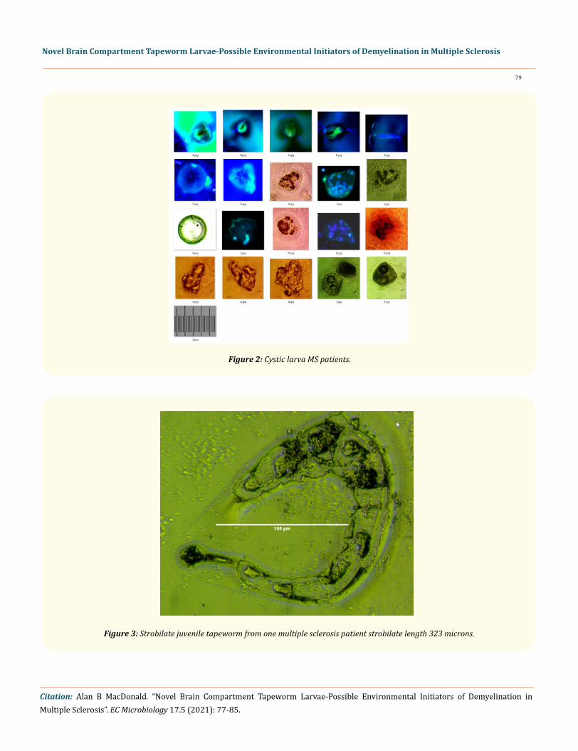
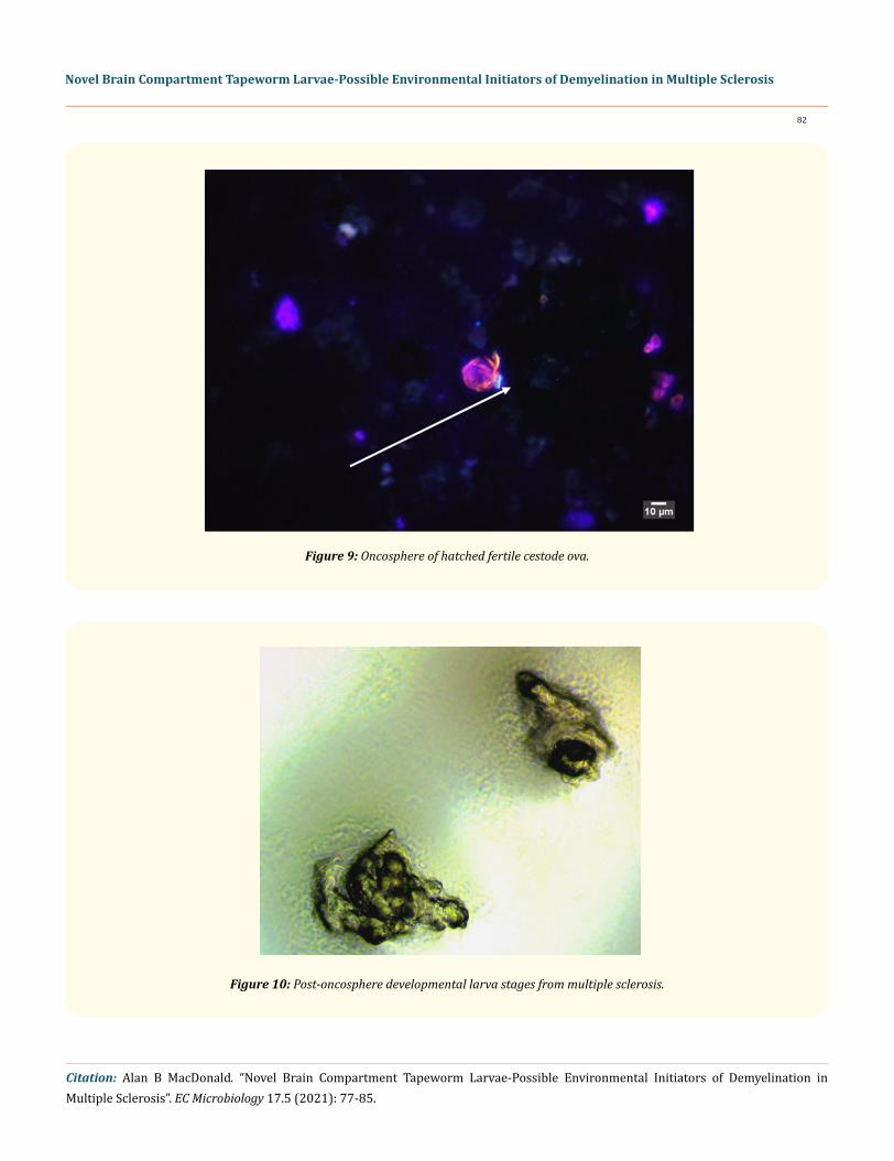
Larval cestodes measuring between 10 microns and 300 microns were identified in ten of ten patient cerebrospinal fluids. Various morphological developmental stages were noted in all cases. Cystic larvae multiple stages of maturation were found in all patients (Fig-ure). Strobilate juvenile tapeworms were found in two patients (Figure). Exfoliated tegument strips were found in two patients. (Figure) Racemose membrane units devoid of protoscolices were found in five patients. (Figure) Calcareous corpuscles were found in three pa-tients presenting as membrane bound corpuscles (Figure) and as liberated free corpuscles (Figure). Cestode hooklets were found in ten patients (Figure) and each patient demonstrated unique hooklets unlike those of any other patient in this group of ten, and unlike any hooklets of Taenia species or of echinococcal species of tapeworms. Oncospheres were visualized in two patients (Figure). Post-onco-sphere non-cystic and non-cavitary developmental stages were seen in ten patients (Figure). No ova were identified in any patient. Mor-phology of cestode larvae in the ten patients demonstrated a Coenurus pattern (Figure) [2] whereby cystic larval forms were endowed with at least two protoscolices and variably could contain eight protoscolices of unequal size attached to the inner cell membrane of the cysts. No larvae demonstrated either a brood capsule pattern of echinococcal larvae [3] or a monocephalic protoscolex pattern of Taenia species Cysticercal larvae [4]. Where tegument was observed in the surface of intact larvae each larval form demonstrated a thin tegument layer. None of the larvae demonstrated laminated pattern thick tegument typical of echinococcal hydatid cysts.
Figure 1: Coenurus cyst (Centers for Disease Control Website) [2].
Citation: Alan B MacDonald. “Novel Brain Compartment Tapeworm Larvae-Possible Environmental Initiators of Demyelination in Multiple Sclerosis”. EC Microbiology 17.5 (2021): 77-85.
Novel Brain Compartment Tapeworm Larvae-Possible Environmental Initiators of Demyelination in Multiple Sclerosis
79
Figure 2: Cystic larva MS patients.
Figure 3: Strobilate juvenile tapeworm from one multiple sclerosis patient strobilate length 323 microns.

Citation: Alan B MacDonald. “Novel Brain Compartment Tapeworm Larvae-Possible Environmental Initiators of Demyelination in Multiple Sclerosis”. EC Microbiology 17.5 (2021): 77-85.
Novel Brain Compartment Tapeworm Larvae-Possible Environmental Initiators of Demyelination in Multiple Sclerosis
80
Figure 4: Tegument (thin green stripe between white arrows) from multiple sclerosis.
Figure 5: Racemose membranes of larval cestode in multiple sclerosis (No scolex, no protoscolex found in the membranes).
Figure 6: Calcareous corpuscles in membrane tissue from multiple sclerosis CSF.

Citation: Alan B MacDonald. “Novel Brain Compartment Tapeworm Larvae-Possible Environmental Initiators of Demyelination in Multiple Sclerosis”. EC Microbiology 17.5 (2021): 77-85.
Novel Brain Compartment Tapeworm Larvae-Possible Environmental Initiators of Demyelination in Multiple Sclerosis
81
Figure 7: Free calcareous corpuscles - multiple sclerosis spinal fluids.
Figure 8: Hooklets of larval cestodes from MS Spinal fluids.
Citation: Alan B MacDonald. “Novel Brain Compartment Tapeworm Larvae-Possible Environmental Initiators of Demyelination in Multiple Sclerosis”. EC Microbiology 17.5 (2021): 77-85.
Novel Brain Compartment Tapeworm Larvae-Possible Environmental Initiators of Demyelination in Multiple Sclerosis
82
Figure 9: Oncosphere of hatched fertile cestode ova.
Figure 10: Post-oncosphere developmental larva stages from multiple sclerosis.

Citation: Alan B MacDonald. “Novel Brain Compartment Tapeworm Larvae-Possible Environmental Initiators of Demyelination in Multiple Sclerosis”. EC Microbiology 17.5 (2021): 77-85.
Novel Brain Compartment Tapeworm Larvae-Possible Environmental Initiators of Demyelination in Multiple Sclerosis
83
Discussion
Brain parasites have never been reported in multiple sclerosis. The dimensions of the parasites in cerebrospinal fluids from ten MS patients range from ten-micron diameter oncospheres, to a maximum dimension of 320 microns for the length of the largest juvenile stro-bilate (segmented) worm. By comparison, the dimensions of the smallest known human cestodes are recorded in Echinococcus species whose strobilate adult forms measure between 1000 microns to 3000 microns in length. Taenia solium species brain parasites present as
Figure 11: Versteria species Calcospherules in membrane (Human Eye) CDC image EID journal; https://doi.org/10.3201/eid2507.190223.

Citation: Alan B MacDonald. “Novel Brain Compartment Tapeworm Larvae-Possible Environmental Initiators of Demyelination in Multiple Sclerosis”. EC Microbiology 17.5 (2021): 77-85.
Novel Brain Compartment Tapeworm Larvae-Possible Environmental Initiators of Demyelination in Multiple Sclerosis
84
centimeter scale cysts (10,000 microns) in human brain. Recently a novel human brain and tissue parasitic cestode species (Versteria sp.) [5] have been defined based on its novel DNA profile in cestode mitochondrial DNA (mtDNA). The size dimensions of human parasitic Ver-steria species are not known, but similar Versteria like cestodes in animal models reportedly demonstrate very minute cysts, hooklets and strobilates which approximate the size dimensions of the unknown species agents in the patients in this report. It is not known whether coenurus type cysts are produced by larval Versteria species. Insufficient DNA is present on thin film glass slides from this patient group to permit DNA studies in pursuit of molecular typing of these novel parasites.
Animal cases of cerebral coenurus infestation with brain demyelination adjacent to sites of parasite deposits have been documented in sheep [6], goat [7] and other primates [8]. Coenurosis induced brain demyelination in the animal world offers an attractive conceptual model for further study to reconcile with human coenurosis associated demyelination of white matter sites in the ten multiple sclerosis patients described here.
Conclusion
Ten non-immunosuppressed patients with multiple sclerosis demonstrated microscopic diagnosable coenurus type cestodes in “dwarf-micro-cystic” scale in autopsy cerebrospinal fluids dating from year 1984 to 2014. In all cases, these parasites were invisible to the naked eye of board-certified medical school faculty neuropathologists and were not detected in full neuropathology microscopic ex-aminations associated with a complete brain autopsy. Brain invasive larval cestode infestations in human intermediate hosts are always preceded by inadvertent ingestion of fertile tapeworm eggs. Versteria species cestodes offer one attractive conceptual bridge to compre-hend the new public health implications of micron scale novel cestode human brain pathogens. Because of the lack of a common parasite morphology among ten patients presented here, the pleomorphisms of the brain invasive parasites, suggest that a possible plural species parasites might be discovered with advanced DNA sequencing (Deep Next Generation sequencing) of DNA extractions from multiple scle-rosis autopsy brain tissues. Additional studies of the mechanisms of demyelination in brains of animals parasitized with coenurus larvae will hopefully add perspective to the broad category of Coenurus cestode category induced myelin pathologies in mammalian hosts.
Acknowledgements
The Brain Tissue Center at the University of Colorado School of Medicine, Aurora, Colorado Is supported in part by the National Multiple Sclerosis Society (USA). Alan B. MacDonald received donations from a GoFundMe Charity for the purchase of a phase contrast condenser attachment for the compound microscope which he purchased with his own personal funds. No additional funding support was received.
Conflict of Interest Statement
The author declares that he has no conflicts of interest.
Bibliography
1. Thompson BJ., et al. “Diagnosis of Multiple Sclerosis: 2017 revisions of the McDonald Criteria”. The Lancet Neurology 17.2 (2017): 162-173.
2. Centers for Disease Control, Coenurosis.
3. Centers for Disease Control, Echinococcosis.
4. Centers for Disease Control, Cysticercosis.
Citation: Alan B MacDonald. “Novel Brain Compartment Tapeworm Larvae-Possible Environmental Initiators of Demyelination in Multiple Sclerosis”. EC Microbiology 17.5 (2021): 77-85.
Novel Brain Compartment Tapeworm Larvae-Possible Environmental Initiators of Demyelination in Multiple Sclerosis
85
Volume 17 Issue 5 May 2021©All rights reserved by Alan B MacDonald.
5. Centers for Disease Control, EID article.
6. Kish GHF., et al. “Clinical and Morphological characteristics of enzoonotic occurrence of acute coenurosis (Coenurus cerebralis) in a sheep herd”. Journal of Parasitic Disease 39.2 (2015): 280-283.
7. Shivasharanappa N., et al. “Neuropathological lesions of clinical and subclinical Coenurosis (Coenurus cerebralis) in organized goat farms in India”. Acta Parasitologica 62.2 (2017): 482-487.
8. Deplazes P., et al. “Wildlife-transmitted Taenia and Versteria cysticercosis and Coenurosis in humans and other primates”. The Inter-national Journal for Parasitology 9 (2019): 342-358.
Related Documents