© 2013 Arunachalam et al, publisher and licensee Dove Medical Press Ltd. This is an Open Access article which permits unrestricted noncommercial use, provided the original work is properly cited. International Journal of Nanomedicine 2013:8 1307–1315 International Journal of Nanomedicine One-step green synthesis and characterization of leaf extract-mediated biocompatible silver and gold nanoparticles from Memecylon umbellatum Kantha D Arunachalam Sathesh Kumar Annamalai Shanmugasundaram Hari Center for Environmental Nuclear Research, Directorate of Research, SRM University, Chennai, Tamil Nadu, India Correspondence: Kantha D Arunachalam Center for Environmental Nuclear Research, Directorate of Research, SRM University, Chennai, Tamil Nadu 603203, India Tel +91 44 2741 7144 Fax +91 44 2741 7146 Email [email protected] Abstract: In this experiment, green-synthesized silver and gold nanoparticles were produced rapidly by treating silver and gold ions with an extract of Memecylon umbellatum leaf. The reaction process was simple and easy to handle, and was monitored using ultraviolet-visible spectroscopy. The effect of the phytochemicals present in M. umbellatum, including saponins, phenolic compounds, phytosterols, and quinones, on formation of stable silver and gold nanoparticles was investigated by Fourier-transform infrared spectroscopy. The morphology and crystalline phase of the nanoparticles were determined by transmission electron microscopy and energy-dispersive x-ray spectroscopy. The results indicate that the saponins, phytosterols, and phenolic compounds present in the plant extract play a major role in formation of silver and gold nanoparticles in their respective ions in solution. The characteristics of the nanoparticles formed suggest application of silver and gold nanoparticles as chemical sensors in the future. Given the simple and eco-friendly approach for synthesis, these nanoparticles could easily be commercialized for large-scale production. Keywords: green synthesis, phytochemicals, saponins, nanoparticles, transmission electron microscopy Introduction Silver nanoparticles are widely used in industry and have an inhibitory effect on a number of microorganisms. They have also been used in the manufacture of ointments and creams to prevent infection of burns and wounds. 1 Organic solvents have been used for green synthesis of nanoparticles, with water in particular being an important solvent for bioreduction. 2 Green synthesis is an alternative harmless and environment-friendly method for producing nanoparticles. 3 Triangular-shaped and spherical-shaped gold nanoparticles have been used for detection of DNA and as an immunoassay system for antibodies. 4 The use of nanoparticles as potential drug carriers in the treatment of cancer has also been reported. Nanoparticles can be prepared using a variety of chemicals and physical methods, including chemical reduction, 5–7 photochemical reduction, 8–11 electrochemical reduction, 12,13 and heat vaporization. 14,15 The reagents can be inorganic compounds, such as sodium/potassium borohydrate, hydrazine, and salts of tartrate, or organic compounds, like sodium citrate, ascorbic acid, and amino acids, which are capable of being oxidized. Because noble metal nanoparticles are now widely used in areas of human contact, 16 there is a growing need to develop environmentally friendly processes that do not use toxic chemicals in their synthesis. A quest for an environmentally Dovepress submit your manuscript | www.dovepress.com Dovepress 1307 ORIGINAL RESEARCH open access to scientific and medical research Open Access Full Text Article http://dx.doi.org/10.2147/IJN.S36670

Welcome message from author
This document is posted to help you gain knowledge. Please leave a comment to let me know what you think about it! Share it to your friends and learn new things together.
Transcript
© 2013 Arunachalam et al, publisher and licensee Dove Medical Press Ltd. This is an Open Access article which permits unrestricted noncommercial use, provided the original work is properly cited.
International Journal of Nanomedicine 2013:8 1307–1315
International Journal of Nanomedicine
One-step green synthesis and characterization of leaf extract-mediated biocompatible silver and gold nanoparticles from Memecylon umbellatum
Kantha D ArunachalamSathesh Kumar AnnamalaiShanmugasundaram HariCenter for Environmental Nuclear Research, Directorate of Research, SRM University, Chennai, Tamil Nadu, India
Correspondence: Kantha D Arunachalam Center for Environmental Nuclear Research, Directorate of Research, SRM University, Chennai, Tamil Nadu 603203, India Tel +91 44 2741 7144 Fax +91 44 2741 7146 Email [email protected]
Abstract: In this experiment, green-synthesized silver and gold nanoparticles were produced
rapidly by treating silver and gold ions with an extract of Memecylon umbellatum leaf. The
reaction process was simple and easy to handle, and was monitored using ultraviolet-visible
spectroscopy. The effect of the phytochemicals present in M. umbellatum, including saponins,
phenolic compounds, phytosterols, and quinones, on formation of stable silver and gold
nanoparticles was investigated by Fourier-transform infrared spectroscopy. The morphology and
crystalline phase of the nanoparticles were determined by transmission electron microscopy
and energy-dispersive x-ray spectroscopy. The results indicate that the saponins, phytosterols,
and phenolic compounds present in the plant extract play a major role in formation of silver and
gold nanoparticles in their respective ions in solution. The characteristics of the nanoparticles
formed suggest application of silver and gold nanoparticles as chemical sensors in the future.
Given the simple and eco-friendly approach for synthesis, these nanoparticles could easily be
commercialized for large-scale production.
Keywords: green synthesis, phytochemicals, saponins, nanoparticles, transmission electron
microscopy
IntroductionSilver nanoparticles are widely used in industry and have an inhibitory effect on a
number of microorganisms. They have also been used in the manufacture of ointments
and creams to prevent infection of burns and wounds.1 Organic solvents have been used
for green synthesis of nanoparticles, with water in particular being an important solvent
for bioreduction.2 Green synthesis is an alternative harmless and environment-friendly
method for producing nanoparticles.3 Triangular-shaped and spherical-shaped gold
nanoparticles have been used for detection of DNA and as an immunoassay system
for antibodies.4 The use of nanoparticles as potential drug carriers in the treatment of
cancer has also been reported.
Nanoparticles can be prepared using a variety of chemicals and physical methods,
including chemical reduction,5–7 photochemical reduction,8–11 electrochemical
reduction,12,13 and heat vaporization.14,15 The reagents can be inorganic compounds,
such as sodium/potassium borohydrate, hydrazine, and salts of tartrate, or organic
compounds, like sodium citrate, ascorbic acid, and amino acids, which are capable of
being oxidized. Because noble metal nanoparticles are now widely used in areas of
human contact,16 there is a growing need to develop environmentally friendly processes
that do not use toxic chemicals in their synthesis. A quest for an environmentally
Dovepress
submit your manuscript | www.dovepress.com
Dovepress 1307
O R I g I N A L R E S E A R C H
open access to scientific and medical research
Open Access Full Text Article
http://dx.doi.org/10.2147/IJN.S36670
International Journal of Nanomedicine 2013:8
sustainable synthetic process has led to several biomimetic
approaches, one of the fundamental processes of which
involves bioreduction.
To overcome the problem of toxicity in synthesis,
plants have come to have a major role in the synthesis of
nanoparticles. The different-shaped polyol and water-soluble
heterocyclic components of plant biomolecules have both
protective and reductive activity, and are mainly responsible
for the reduction of silver and gold ions using chemical and
radiation methods. In recent years, extracellular silver and
gold nanoparticles were synthesized using various plant-
mediated extracts as a reducing agent.
Memecylon umbellatum is a well known herbal drug in
India. Its leaves are used for their astringent, antispasmodic,
antitumor, and neuroleptic activities, as well as to treat
leucorrhoea and gonorrhea. A lotion prepared from its
leaves is used to treat eye troubles,17 and the leaves and bark
can be applied to bruises.18 M. umbellatum belongs to the
Melastomataceae family, which is the seventh largest family
of flowering plants and originates from India.
To date, there is no report of synthesis of silver and
gold nanoparticles utilizing an aqueous leaf extract of
M. umbellatum. In this experiment, M. umbellatum extract
in a concentrated aqueous solution of silver nitrate and
chloroauric acid resulted in reduction of silver and gold ions
and formation of silver and gold nanoparticles. These green-
synthesized nanoparticles were examined by ultraviolet-
visible spectroscopy, transmission electron microscopy
(TEM), energy dispersive X-ray analysis (EDAX), and
Fourier transform infrared (FTIR) spectroscopy to determine
their size and shape.
Materials and methodsCollection of plantsThe M. umbellatum plant material was collected from the
Potheri forest in Tamil Nadu, India The herbarium was pre-
pared, submitted, and authenticated by Dr Jayaraman, Madras
Christian College, Tambaram, Chennai, Tamil Nadu.
Preparation of aqueous extractThe leaves of the plants collected were washed and air-dried
in the shade at room temperature for at least two weeks, cut
into small pieces, powdered in a mixer, and sieved using a
20 µm mesh sieve to obtain a uniform size range for further
study. Next, 20.0 g of the sieved leaf powder was added to
100 mL of sterile distilled water in a 500 mL Erlenmeyer
flask and boiled for 5 minutes. The flasks were kept under
continuous dark conditions at 30°C. The extract was filtered
and stored at 4°C for further experiments.
Phytochemical activityQualitative phytochemical analysis of the M. umbellatum
extract was performed using the methodology described
by Parekh and Chanda19 for determining the presence of
alkaloids (Mayer’s, Wagner’s, Dragendorff ’s reagents),
flavonoids (Shinoda alkaline reagent), phenolic compounds
(lead acetate, alkaline reagent test), triterpenes (Liberman-
Burchard test), saponins (foam test), and tannins (gelatin).20–23
The results of these tests were expressed qualitatively as
positive (+) or negative (−).
Antimicrobial activityAntimicrobial tests were done by the disc-diffusion method
(Murray et al, 1995) using 100 µL of suspension containing
108 colony-forming units per mL of bacteria on Mueller
Hinton agar. Six-millimeter diameter discs were impregnated
with 10 µL of the extract (300 µg per disc) at a concentration
of 30 mg/mL and placed on agar plates inoculated with
common clinically encountered pathogens. Negative controls
were prepared using the same solvents as those used to
dissolve the plant extracts. Ofloxacin (10 µg per disc),
ciprofloxacin (10 µg per disc), and netilmicin (30 µg per
disc) were used as positive reference standards to determine
the sensitivity of one strain per isolate for each microbial
species tested. The inoculated plates were incubated at 37°C
for 24 hours to obtain the bacterial strains. Antimicrobial
activity was evaluated in duplicate by measuring the zone
of inhibition for the test organisms.22 The antimicrobial
activity of M. umbellatum extract was evaluated for Bacillus
subtilis, Streptococcus pneumoniae, Staphylococcus
aureus, Salmonella typhimurium, Klebsiella aerogenes, and
Escherichia coli.
One-step green synthesis of silver and gold nanoparticles using leaf extractSilver nitrate and chloroauric acid were purchased from
Sigma-Aldrich (St Louis, MO), and the aqueous leaf extract
of M. umbellatum was used for the bioreduction process.
To synthesize nanoparticles from M. umbellatum, 5, 10,
and 15 mL of the aqueous leaf extract were carefully added
to 10 mL of 1 mM aqueous silver nitrate and chloroauric
acid solution in 250 mL Erlenmeyer flasks. The flasks
containing the extract were incubated on a rotary shaker at
150 rpm in dark conditions. A change in color of the colloidal
submit your manuscript | www.dovepress.com
Dovepress
Dovepress
1308
Arunachalam et al
International Journal of Nanomedicine 2013:8
solutions occurred, confirming green synthesis of silver and
gold nanoparticles.24
Ultraviolet-visible absorbance spectroscopyBioreduction of the nanoparticles by silver nitrate and
chloroauric acid was monitored using a Series 3000 double
beam ultraviolet-visible spectrophotometer (Thermo Fisher
Scientific, Waltham, MA, USA) with slit widths of 0.5, 1.0,
2.0, and 5.0 nm. The samples used for analysis were diluted
with 2 mL of deionized water and subsequently measured
in the ultraviolet-visible spectrum at regular time intervals.25
Deionized water was used for background correction of all
ultraviolet-visible spectra. Samples were loaded into a 1 cm
path length quartz cuvette for investigation. The ultraviolet-
visible spectra were fitted to Gaussian curves correcting for
a cubic background for full-width at half maximum and
wavelength of maximum absorbance measurements. All
Gaussian fits to the ultraviolet-visible spectra had goodness
of fit values (χ2 approximately 1) indicating accurate curve
analysis.26 The ultraviolet-visible spectrophotometric readings
were recorded at a scanning speed of 0.5 nm intervals. The
spectrum was scanned from 200 nm to 800 nm.
Transmission electron microscopyThirty minutes after reaction, the biomass had settled at the
bottom of the conical flasks, and samples of the suspended
precipitate were taken for observation by TEM. Samples of
the green-synthesized silver and gold nanoparticles were
prepared by placing a drop of the suspension on carbon-coated
copper grids, allowing the films on the TEM grids to stand
for two minutes, removing the excess solution with blotting
paper, and letting the grid dry prior to measurement. TEM
observations were performed using an H7600 instrument
(Hitachi, Tokyo, Japan) operated at an accelerating voltage
of 100 kV. The size distribution of the nanoparticles was
estimated based on TEM images using Sigma Scan Pro
software version 4.01.003 (SPSS Inc, Chicago, IL).27
FTIR spectroscopyA carefully weighed quantity of green-synthesized silver and
gold nanoparticle powder was subjected to FTIR analysis.
The bioreduced chloroauric acid and silver nitrate solutions
were centrifuged at 10,000 rpm for 15 minutes, and the pellet
was washed three times with 20 mL of deionized water.
The resulting purified suspension was completely dried,
ground with KBr pellets, and analyzed by FTIR. A total of
512 scans were recorded in order to obtain a good signal to
noise ratio.28
EDAX spectroscopyEnergy-dispersive X-ray spectroscopes exploit the photon
nature of light. In the X-ray range, the energy of a single
photon is just sufficient to produce a measurable X-ray
voltage pulse, and the output of an ultralow noise preamplifier
connected to the low noise constitutes a statistical measure
of the corresponding quantum energy. By digitally recording
and counting a large number of such pulses within a so-called
multichannel analyzer, a complete image of the X-ray
spectrum is built up almost simultaneously. This digital
quantum counting technique makes energy-dispersive X-ray
spectroscopy very reliable. A semiconductor material is used
to detect the X-rays together with processing electronics
to analyze the spectrum. The gold and silver solutions
reduced by M. umbellatum extract were dried, drop-coated
onto carbon film, and tested using an EDAX spectrometer
(S-3400N, Hitachi).
Results and discussionPhytochemical screeningThe aqueous extract of M. umbellatum was evaluated for the
presence of various phytoconstituents by performing a series
of qualitative chemical tests. Phytochemicals in the aqueous
extract of M. umbellatum are shown in Table 1, and include
primarily saponins, phytosterols, phenolic compounds,
quinones, carbohydrates, and protein. The aqueous
M. umbellatum leaf extract was found to have a high saponin
content, suggesting that saponins are the most favorable
starting material for preparation of these nanoparticles.
The pharmacological activity of saponin and its multiple
medicinal properties have also been reported for other plant
species. Early studies by Raut et al25 demonstrated that
Table 1 Preliminary phytochemical investigation of aqueous extract of Memecylon umbellatum
Phytochemicals Aqueous extract
Alkaloids −Phytosterol +Saponin +Phenolic compounds +Catechins −Flavonol glycosides −Quinones +Phlobatannins −Steroids −Notes: +, presence; −, absence.
submit your manuscript | www.dovepress.com
Dovepress
Dovepress
1309
One-step green synthesis of nanoparticles using M. umbellatum extract
International Journal of Nanomedicine 2013:8
nanoparticles formed using saponin from an aqueous extract
of Panax ginseng have strong activity in upregulation of the
immune response in mice. Phytosterol-based nanoparticles
form stable suspensions of submicron particles, which are
not soluble in water.29 Production of nanoparticles at room
temperature aided by tautomerization of membrane-bound
quinones has also been reported.30
Antimicrobial activityThe antimicrobial activity of the aqueous M. umbellatum
extracts was investigated, with their potency assessed
quantitatively by the zone of inhibition and zone diameter
using Pseudomonas aeruginosa, Streptococcus pyogenes,
Enterococcus faecalis, Proteus vulgaris, Proteus mirabilis,
Citrobacter freundii, and E. coli. The calculated zones of
inhibition are shown in Table 2.
Ultraviolet-visible spectrometryUltraviolet-visible spectrometry was used to examine the
size and shape of the nanoparticles in aqueous suspension.
The aqueous plant extracts acted as reducing and capping
agents for the nanoparticles. Reduction of silver ions by
combinations of biomolecules found in these extracts,
including enzymes/proteins, amino acids, polysaccharides,
and vitamins, is environmentally benign but chemically
complex. However, the mechanism is widely accepted
for green synthesis of silver and gold nanoparticles in the
presence of the nitrate reductase enzyme. Aqueous silver
nitrate (10 mL, 1 mM) was used to investigate the effect
of including different amounts of the dried biomass on
the extent of bioreduction and uniformity of the target
products. The amount of dried biomass was found to play a
critical role in the size distribution of silver nanoparticles.
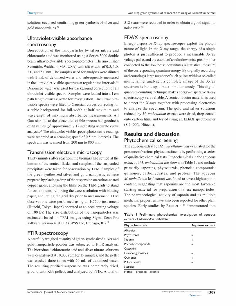
Figure 1 shows the absorption spectra for silver nanoparticles
produced at different time intervals, revealing that these
nanoparticles were produced within one hour of the silver
ions coming in contact with the biomass. After addition of
the biomass to the solution of silver nitrate, the solution
changed from colorless to brown color in about one hour,
with the final color deepening on increasing the amount
of dried biomass. Evolution of the absorbance spectra
emanating from the silver nanoparticles over time showed
increasingly sharp absorbance at around 440 nm with
increasing reaction time.
Saponins and phenolic compounds present in plant
extracts bind to nanoparticles via either free amine groups
or cysteine residues in proteins.31 Saponins, phenolic
compounds, and quinones from M. umbellatum capped the
silver nanoparticles, thereby stabilizing them. To conclude,
water-soluble fractions comprised of saponins, phenolic
compounds, and quinones in the aqueous plant extract helped
in the bioreduction of silver ions.
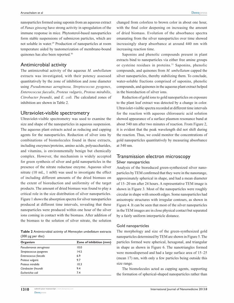
Reduction of gold ions to gold nanoparticles on exposure
to the plant leaf extract was detected by a change in color.
Ultraviolet-visible spectra recorded at different time intervals
for the reaction with aqueous chloroauric acid solution
showed appearance of a surface plasmon resonance band at
about 540 nm after two minutes of reaction. From Figure 2,
it is evident that the peak wavelength did not shift during
the reaction. Thus, we could monitor the concentrations of
gold nanoparticles quantitatively by measuring absorbance
at 540 nm.
Transmission electron microscopySilver nanoparticlesAnalysis of the bioreduced green-synthesized silver nano-
particles by TEM confirmed that they were in the nanorange,
approximately spherical in shape, and had a mean diameter
of 15–20 nm after 24 hours. A representative TEM image is
shown in Figure 3. Most of the nanoparticles were roughly
circular in shape with smooth edges. Some nanoparticles had
anisotropic structures with irregular contours, as shown in
Figure 4. It can be seen that most of the silver nanoparticles
in the TEM images are in close physical contact but separated
by a fairly uniform interparticle distance.
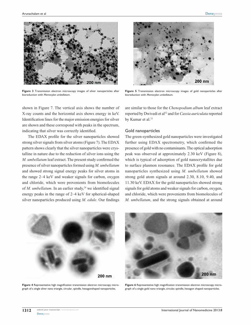
gold nanoparticlesThe morphology and size of the green-synthesized gold
nanoparticles determined by TEM are shown in Figure 5. The
particles formed were spherical, hexagonal, and triangular
in shape as shown in Figure 6. The nanotriangles formed
were monodispersed and had a large surface area of 15–25
(mean 17) nm, with only a few particles being outside this
size range.
The biomolecules acted as capping agents, supporting
the formation of spherical-shaped nanoparticles rather than
Table 2 Antimicrobial activity of Memecylon umbellatum extracts (300 µg per disc)
Organism Zone of inhibition (mm)
Pseudomonas aeruginosa 10.0Streptococcus pyogenes 14.5Enterococcus faecalis 6.9Proteus vulgaris 9.7Proteus mirabilis 10.3Citrobacter freundii 9.4Escherichia coli 7.4
submit your manuscript | www.dovepress.com
Dovepress
Dovepress
1310
Arunachalam et al
International Journal of Nanomedicine 2013:8
the formation of triangular or hexagonal nanostructures.
High magnification of TEM images recorded during our
earlier research showed that biologically synthesized gold
nanoparticles at the end of reaction with Memecylon edule
leaf extract were also predominantly triangular, circular, and
hexagonal in morphology. A large quantity of circular gold
nanoparticles with thin smooth ends on the exterior of the
nanoparticles was seen in the TEM images (Figure 6). Small
amounts of the extract could reduce the chloroaurate ions,
but did not protect most of the semispherical nanoparticles
from aggregating because of the inability of biomolecules
to act as protecting agents, and this was clearly observed in
the TEM images.
Energy-dispersive X-ray spectroscopySilver nanoparticlesEDAX spectroscopy conf irmed the presence of an
elemental silver signal from the silver nanoparticles, as
Figure 1 Absorption spectra of silver nanoparticles after bioreduction of Memecylon umbellatum aqueous.
Ad
sorb
ance
Wavelength (nm)
1.0
0.9
0.8
0.7
0.6
0.5
0.4
0.3
0.2
0.10.06
200 250 300 350 400 450 500 550 600 650 700 750 800
1 hour2 hour3 hour4 hour5 hour
7 hour8 hour
6 hour
Figure 2 Absorption spectra of gold nanoparticles after bio reduction with 0.5 mL of Memecylon umbellatum aqueous extract.
Ad
sorb
ance
Wavelength (nm)
1.0
0.9
0.8
0.7
0.6
0.5
0.4
0.3
0.2
0.15200 250 300 350 400 450 500 550 600 650 700 750 800
1 hour2 hour3 hour4 hour5 hour
7 hour8 hour
6 hour
submit your manuscript | www.dovepress.com
Dovepress
Dovepress
1311
One-step green synthesis of nanoparticles using M. umbellatum extract
International Journal of Nanomedicine 2013:8
are similar to those for the Chenopodium album leaf extract
reported by Dwivedi et al32 and for Cassia auriculata reported
by Kumar et al.33
gold nanoparticlesThe green-synthesized gold nanoparticles were investigated
further using EDAX spectrometry, which confirmed the
presence of gold with no contaminants. The optical adsorption
peak was observed at approximately 2.30 keV (Figure 8),
which is typical of adsorption of gold nanocrystallites due
to surface plasmon resonance. The EDAX profile for gold
nanoparticles synthesized using M. umbellatum showed
strong gold atom signals at around 2.30, 8.10, 9.40, and
11.30 keV. EDAX for the gold nanoparticles showed strong
signals for gold atoms and weaker signals for carbon, oxygen,
and chloride, which were provenients from biomolecules of
M. umbellatum, and the strong signals obtained at around
Figure 3 Transmission electron microscopy images of silver nanoparticles after bioreduction with Memecylon umbellatum.
200 nm
Figure 4 Representative high magnification transmission electron microscopy micro-graph of a single silver nano triangle, circular, spindle, hexagonshaped nanoparticles.
Figure 5 Transmission electron microscopy images of gold nanoparticles after bioreduction with Memecylon umbellatum.
Figure 6 Representative high magnification transmission electron microscopy micro-graph of a single gold nano triangle, circular, spindle, hexagon shaped nanoparticles.
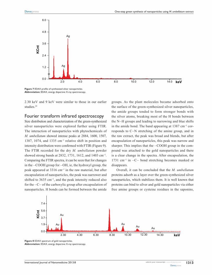
shown in Figure 7. The vertical axis shows the number of
X-ray counts and the horizontal axis shows energy in keV.
Identification lines for the major emission energies for silver
are shown and these correspond with peaks in the spectrum,
indicating that silver was correctly identified.
The EDAX profile for the silver nanoparticles showed
strong silver signals from silver atoms (Figure 7). The EDAX
pattern shows clearly that the silver nanoparticles were crys-
talline in nature due to the reduction of silver ions using the
M. umbellatum leaf extract. The present study confirmed the
presence of silver nanoparticles formed using M. umbellatum
and showed strong signal energy peaks for silver atoms in
the range 2–4 keV and weaker signals for carbon, oxygen
and chloride, which were provenients from biomolecules
of M. umbellatum. In an earlier study,24 we identified signal
energy peaks in the range of 2–4 keV for spherical-shaped
silver nanoparticles produced using M. edule. Our findings
submit your manuscript | www.dovepress.com
Dovepress
Dovepress
1312
Arunachalam et al
International Journal of Nanomedicine 2013:8
2.30 keV and 9 keV were similar to those in our earlier
studies.24
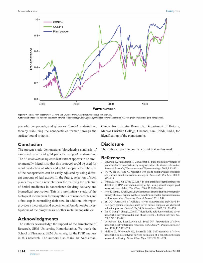
Fourier transform infrared spectroscopySize distribution and characterization of the green-synthesized
silver nanoparticles were explored further using FTIR.
The interaction of nanoparticles with phytochemicals of
M. umbellatum showed intense peaks at 2884, 1600, 1507,
1387, 1074, and 1335 cm−1 relative shift in position and
intensity distribution were confirmed with FTIR (Figure 9).
The FTIR recorded for the dry M. umbellatum powder
showed strong bands at 2832, 1731, 1612, and 1403 cm−1.
Comparing the FTIR spectra, it can be seen that for changes
in the –COOH group for –OH, ie, the hydroxyl group, the
peak appeared at 3316 cm−1 in the raw material, but after
encapsulation of nanoparticles, the peak was narrower and
shifted to 3635 cm−1, and the peak intensity reduced also
for the –C– of the carboxylic group after encapsulation of
nanoparticles. H bonds can be formed between the amide
groups. As the plant molecules became adsorbed onto
the surface of the green-synthesized silver nanoparticles,
the amide groups tended to form stronger bonds with
the silver atoms, breaking most of the H bonds between
the N–H groups and leading to narrowing and blue shifts
in the amide bond. The band appearing at 1387 cm−1 cor-
responds to C–N stretching of the amine group, and in
the raw extract, the peak was broad and blends, but after
encapsulation of nanoparticles, this peak was narrow and
sharper. This implies that the –COOH group in the com-
pound was attached to the gold nanoparticles and there
is a clear change in the spectra. After encapsulation, the
1731 cm−1 in –C– bond stretching becomes masked or
disappears.
Overall, it can be concluded that the M. umbellatum
proteins adsorb as a layer over the green-synthesized silver
nanoparticles, which stabilizes them. It is well known that
proteins can bind to silver and gold nanoparticles via either
free amine groups or cysteine residues in the saponins,
6.0
4.8
3.6
2.4
1.2
0.02.0
Ag
AgK
Cn
t
4.0 6.0 8.0 10.0 12.0 14.0 keV
0
Figure 7 EDAX profile of synthesized silver nanoparticles.Abbreviation: EDAX, energy dispersive X-ray spectroscopy.
7.4
Au
KC
nt
AuAu
AuAu
2.30 4.30 6.30 8.30 10.30 12.30 14.30 keV
5.6
3.7
1.9
0.0
Figure 8 EDAX spectrum of gold nanoparticles.Abbreviation: EDAX, energy dispersive X-ray spectroscopy.
submit your manuscript | www.dovepress.com
Dovepress
Dovepress
1313
One-step green synthesis of nanoparticles using M. umbellatum extract
International Journal of Nanomedicine 2013:8
phenolic compounds, and quinones from M. umbellatum,
thereby stabilizing the nanoparticles formed through the
surface-bound proteins.
ConclusionThe present study demonstrates bioreductive synthesis of
nanosized silver and gold particles using M. umbellatum.
The M. umbellatum aqueous leaf extract appears to be envi-
ronmentally friendly, so that this protocol could be used for
rapid production of silver and gold nanoparticles. The size
of the nanoparticles can be easily adjusted by using differ-
ent amounts of leaf extract. In the future, selection of such
plants may create a new platform for realizing the potential
of herbal medicines in nanoscience for drug delivery and
biomedical application. This is a preliminary study of the
biological mechanism for biosynthesis of nanoparticles and
a first step in controlling their size. In addition, this report
provides a theoretical and experimental foundation for inves-
tigations of the biosynthesis of other metal nanoparticles.
AcknowledgmentsThe authors acknowledge the support of the Directorate of
Research, SRM University, Kattankulathur. We thank the
School of Pharmacy, SRM University, for the FTIR analysis
in this research. The authors also thank Dr Narasiman,
Centre for Floristic Research, Department of Botany,
Madras Christian College, Chennai, Tamil Nadu, India, for
identification of the plant sample.
DisclosureThe authors report no conflicts of interest in this work.
References1. Satyavani K, Ramanathan T, Gurudeeban S. Plant-mediated synthesis of
biomedical silver nanoparticles by using leaf extract of Citrullus colocynthis. Research Journal of Nanoscience and Nanotechnology. 2011;1:95–101.
2. Wu W, He Q, Jiang C. Magnetic iron oxide nanoparticles: synthesis and surface functionalization strategies. Nanoscale Res Lett. 200;3: 397–415.
3. Wang Z, Hu J, Jin Y, Yao X, Lia J. In situ amplified chemiluminescent detection of DNA and immunoassay of IgG using special-shaped gold nanoparticles as label. Clin Chem. 2006;52:1958–1961.
4. Hojo K, Hara A, Kitai H, et al. Development of a method for environmentally friendly chemical peptide synthesis in water using water-dispersible amino acid nanoparticles. Chemistry Central Journal. 2011;5:49.
5. Yu DG. Formation of colloidal silver nanoparticles stabilized by Na+-poly(gamma-glutamic acid)-silver nitrate complex via chemical reduction process. Colloids Surf B Biointerfaces. 2007;59:171–178.
6. Tan Y, Wang Y, Jiang L, Zhu D. Thiosalicylic acid-functionalized silver nanoparticles synthesized in one-phase system. J Colloid Interface Sci. 2002;249:336–345.
7. Vorobyova SA, Lesnikovich AI, Sobal NS. Preparation of silver nanoparticles by interphase reduction. Colloids Surf A Physicochem Eng Asp. 1999;152:375–379.
8. Mallick K, Witcombb MJ, Scurrella MS. Self-assembly of silver nanoparticles in a polymer solvent: formation of a nanochain through nanoscale soldering. Mater Chem Phys. 2005;90:221–224.
1.0GSNP’s
Tra
nsm
itta
nce
Wave number
GGNP’s
Plant powder
0.8
0.6
0.4
0.2
0.0
4000 3000 2000 1000
Figure 9 Typical FTIR spectrum of gSNP’s and ggNP’s from M. umbellatum aqueous leaf extracts.Abbreviations: FTIR, Fourier transform infrared spectroscopy; gSNP, green synthesized silver nanoparticle; ggNP, green synthesized gold nanoparticle.
submit your manuscript | www.dovepress.com
Dovepress
Dovepress
1314
Arunachalam et al
International Journal of Nanomedicine
Publish your work in this journal
Submit your manuscript here: http://www.dovepress.com/international-journal-of-nanomedicine-journal
The International Journal of Nanomedicine is an international, peer-reviewed journal focusing on the application of nanotechnology in diagnostics, therapeutics, and drug delivery systems throughout the biomedical field. This journal is indexed on PubMed Central, MedLine, CAS, SciSearch®, Current Contents®/Clinical Medicine,
Journal Citation Reports/Science Edition, EMBase, Scopus and the Elsevier Bibliographic databases. The manuscript management system is completely online and includes a very quick and fair peer-review system, which is all easy to use. Visit http://www.dovepress.com/ testimonials.php to read real quotes from published authors.
International Journal of Nanomedicine 2013:8
9. Keki A, Torok J, Deak G, et al. Silver nanoparticles by PAMAM-assisted photochemical reduction of Ag+. J Colloid Interface Sci. 2000;229: 550–553.
10. Pileni MP. Fabrication and physical properties of self-organized silver nanocrystals. Pure Appl Chem. 2000;72:53–65.
11. Sun Y-P, Atorngitjawat P, Meziani MJ. Preparation of silver nanoparticles via rapid expansion of water in carbon dioxide microemulsion into reductant solution. Langmuir. 2001;17:5707–5710.
12. Liu YC, Lin LH. New pathway for the synthesis of ultrafine silver nanoparticles from bulk silver substrates in aqueous solutions by sono electrochemical methods. Electrochem Commun. 2004;6:1163–1168.
13. Sandmann G, Dietz H, Plieth W. Preparation of silver nanoparticles on ITO surfaces by a double-pulse method. J Electroanal Chem. 2000;491: 78–86.
14. Bae CH, Nam SH, Park SM. Formation of silver nanoparticles by laser ablation of a silver target in NaCl solution. Appl Surf Sci. 2002;197–198: 628–634.
15. Smetanaa AB, Klabunde KJ, Sorensen CM. Synthesis of spherical silver nanoparticles by digestive ripening, stabilization with various agents, and their 3-D and 2-D super-lattice formation. J Colloid Interface Sci. 2005;284:521–526.
16. Song JY, Kim BS. Biological synthesis of bimetallic Au/Ag nanoparticles using Persimmon (Diopyros kaki) leaf extract. Korean J Chem Eng. 2008;25:808–811.
17. The Wealth of India. A Dictionary of Indian Raw Materials and Industrial Products. New Delhi, India: National Institute of Science Communication and Information Resources; 1998.
18. Dhar ML, Dhar MM, Dhawan BN, Mehrotra BN, Ray C. Screening of Indian plants for biological activity: Part 1. Indian J Exp Biol. 1968;6: 232–247.
19. Parekh J, Chanda SV. In vitro antimicrobial activity and phytochemical analysis of some Indian medicinal plants. Turk J Biol. 2008;31: 53–58.
20. Harborne JB. Phytochemical Methods, 2nd ed. London, UK: Chapman and Hall Ltd; 1976.
21. Dey PM, Harborne JB. Methods in Plant Biochemistry. London, UK: Academic Press; 1987.
22. Subhashini S, Arunachalam KA, Kumar A. Preclinical studies on the phytochemical, antimicrobial, and wound healing properties of Indigofera aspalathoides leaves. J Pharm Res. 2011;4:3206–3211.
23. Arunachalam KA, Subhashini S, Annamalai SK. Wound healing and antigenotoxic activities of Aegle marmelos with relation to its antioxidant properties. J Pharm Res. 2012;5:1492–1502.
24. Elavazhagan T, Arunachalam KA. Memecylon edule leaf extract mediated green synthesis of silver and gold nanoparticles. Int J Nanomedicine. 2011;6:1265–1278.
25. Raut RW, Lakkakula JR, Kolekar NS, Mendhulkar VD, Kashid SB. Photosynthesis of silver nanoparticles using Gliricidia sepium (Jacq). Curr Nanosci. 2009;5:117–122.
26. Von White G II, Kerscher K, Brown RM, et al. Green synthesis of robust, biocompatible silver nanoparticles using garlic extract. Journal of Nanomaterials. 2012;2012:730–746.
27. Li C-M, Robertson IM, Jenkins ML, Hutchison JL, Doole RC. In situ TEM observation of the nucleation and growth of silver oxide nanoparticles. Micron. 2005;36:9–15.
28. Narayanan B, Sakthivel N. Coriander leaf mediated biosynthesis of gold nanoparticles. Mater Lett. 2008;62:4588–4590.
29. Türk M, Lietzow R. Stabilized nanoparticles of phytosterol by rapid expansion from supercritical solution into aqueous solution. AAPS Pharm Sci Tech. 2004;5:36–45.
30. Jha AK, Prasadb K, Prasad K. A green low-cost biosynthesis of Sb2O
3
nanoparticles. Biochem Eng J. 2009;43:303–306. 31. Shankar SS, Ahmad A, Sastry M. Geranium leaf assisted biosynthesis
of silver nanoparticles. Biotechnol Prog. 2003;19:1627–1631. 32. Dwivedi AD, Gopal K. Biosynthesis of silver and gold nanoparticles
using Chenopodium album leaf extract. Colloids Surf A Physicochem Eng Asp. 2010;369:27–33.
33. Kumar VG, et al. Facile green synthesis of gold nanoparticles using leaf extract of antidiabetic potent Cassia auriculata. Colloids Surf B Biointerfaces. 2011;87:159–163.
submit your manuscript | www.dovepress.com
Dovepress
Dovepress
Dovepress
1315
One-step green synthesis of nanoparticles using M. umbellatum extract
Related Documents