ONE-LUNG VENTILATION(OLV): TECHNIQUE AND ANAESTHETIC CONSIDERATIONS DR SHADAB

Welcome message from author
This document is posted to help you gain knowledge. Please leave a comment to let me know what you think about it! Share it to your friends and learn new things together.
Transcript

ONE-LUNG VENTILATION(OLV):
TECHNIQUE AND ANAESTHETIC
CONSIDERATIONS
DR SHADAB
INTRODUCTION
One-lung ventilation, OLV, means separation of the two lungs and each lung functioning independently by preparation of the airway
It is the intentional collapse of a lung on the operative side of the patient which facilitates most thoracic procedures.
Requires much skill of the anesthesia team because of
• Difficult to place lung isolation equipment• Ability to overcome hypoxic pulmonary
vasoconstriction• Patient population is comparably “sicker”

• OLV provides:• Protection of healthy lung from infected/bleeding one• Diversion of ventilation from damaged airway or lung• Improved exposure of surgical field
• OLV causes:• More manipulation of airway, more damage • Significant physiologic change and easily development of
hypoxemia

• Dependent Lung or Down Lung– The lung that is ventilated
• Non-dependent Lung or Up Lung– The lung that is collapsed to facilitate
the surgery
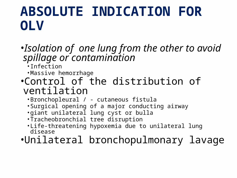
ABSOLUTE INDICATION FOR OLV
•Isolation of one lung from the other to avoid spillage or contamination
• Infection • Massive hemorrhage
•Control of the distribution of ventilation• Bronchopleural / - cutaneous fistula• Surgical opening of a major conducting airway• giant unilateral lung cyst or bulla• Tracheobronchial tree disruption• Life-threatening hypoxemia due to unilateral lung disease
•Unilateral bronchopulmonary lavage
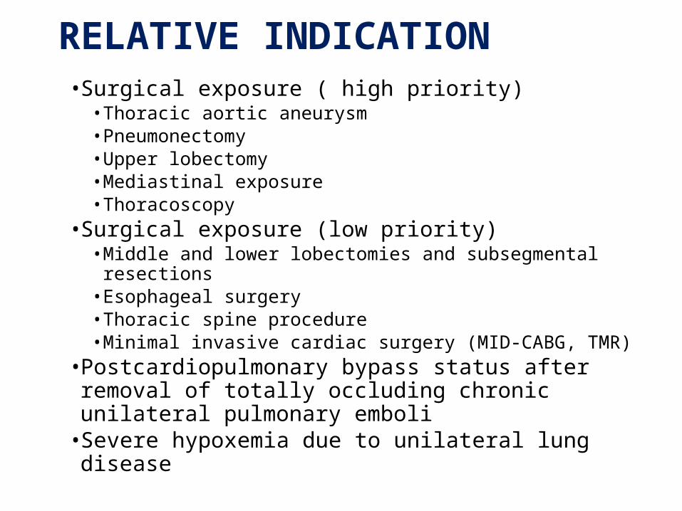
RELATIVE INDICATION • Surgical exposure ( high priority)
• Thoracic aortic aneurysm• Pneumonectomy• Upper lobectomy• Mediastinal exposure• Thoracoscopy
• Surgical exposure (low priority)• Middle and lower lobectomies and subsegmental resections• Esophageal surgery• Thoracic spine procedure• Minimal invasive cardiac surgery (MID-CABG, TMR)
• Postcardiopulmonary bypass status after removal of totally occluding chronic unilateral pulmonary emboli
• Severe hypoxemia due to unilateral lung disease

OLV is achieved by either;
-Double lumen ETT (DLT)
-Bronchial blocker
-Endobronchial tubeDouble-lumen endotracheal tube, DLTSingle-lumen ET with a built-in bronchial blocker, Univent TubeSingle-lumen ET with an isolated bronchial blockerArndt (wire-guided) endobronchial blocker setBalloon-tipped luminal cathetersEndobronchial intubation of a single-lumen ET
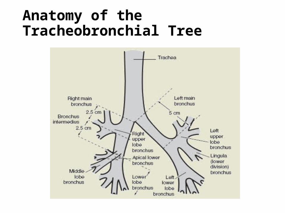
Anatomy of the Tracheobronchial Tree
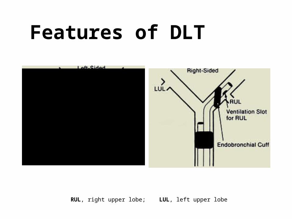
Features of DLT
RUL, right upper lobe; LUL, left upper lobe
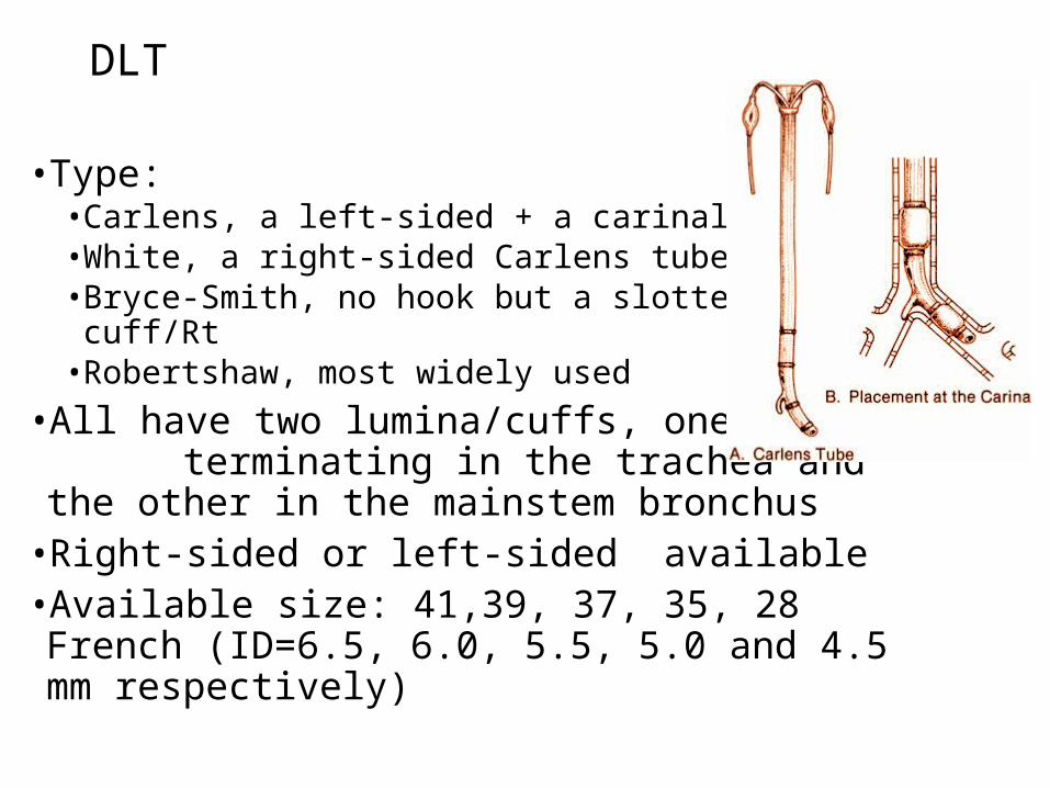
DLT
• Type:• Carlens, a left-sided + a carinal hook • White, a right-sided Carlens tube• Bryce-Smith, no hook but a slotted cuff/Rt • Robertshaw, most widely used
• All have two lumina/cuffs, one terminating in the trachea and the other in the mainstem bronchus
• Right-sided or left-sided available• Available size: 41,39, 37, 35, 28 French (ID=6.5,
6.0, 5.5, 5.0 and 4.5 mm respectively)

Left DLT…• Most commonly used• The bronchial lumen is longer, and a simple round opening and symmetric cuff
Better margin of safety than Rt DLT• Easy to apply suction and/or CPAP to either lung• Easy to deflate lung• Lower bronchial cuff volumes and pressures• Can be used
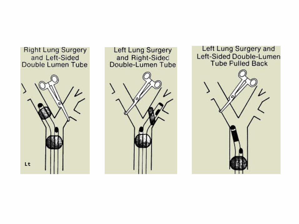
• Left lung isolation: clamp bronchial + ventilate/ tracheal lumen• Right lung isolation: clamp tracheal + ventilate/bronchial lumen

…Left DLT
• More difficult to insert (size and curve, cuff)• Risk of tube change and airway damage if kept in
position for post-op ventilation• Contraindication:
• Presence of lesion along DLT pathway• Difficult/impossible conventional direct vision intubation• Critically ill patients with single lumen tube in situ who
cannot tolerate even a short period of off mechanical ventilation
• Full stomach or high risk of aspiration• Patients, too small (<25-35kg) or too young (< 8-12 yrs)

Right DLT: bronchoscopic view

Carlens DLT Robertshaw DLT

Different types of DLT
Carlens White Bryce Smith
Robertshaw
lumen hook + + - -side Lt Rt Lt & Rt Lt & Rt

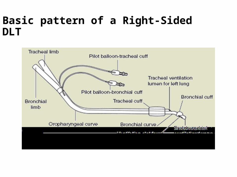
Basic pattern of a Right-Sided DLT

Rt Lt
Lt

passage of the left-sided DLT

guide for Length and Size of DLT
Length of tube , For 170 cm height, tube depth of 29 cm For every 10 cm height change , 1 cm depth change
Patient characteristics Tube size (Fr gauge) Tracheal width (mm):
18161514
41393735
Patient height4’ 6”-5’5”5’5”-5’10”5’11”-6’4”
35-3737-3939-41
Patient age (year)13-14
12108
3532
28 (lt only) 26 (lt only)

Check Position of Lt -DLT
Checklist for tracheal placementa. inflate tracheal cuffb. ventilate rapidly by handc. check that both lungs are being ventilatedd. If not, withdraw 2-3 cm & repeat
Checklist for Lt side a. inflate Lt cuff > 2ml b. ventilate and check bilateral breath sounds c. clamp Rt tube d. check unilateral (Lt) breath sounds
Checklist for Rt side a. clamp Lt tube b. check unilateral (Rt) breath sounds
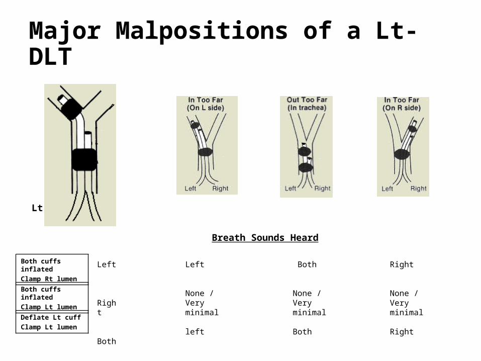
Major Malpositions of a Lt- DLT
Both cuffs inflatedClamp Rt lumen
Both cuffs inflatedClamp Lt lumen
Deflate Lt cuffClamp Lt lumen
Left
None / Very minimal
left
Left
Right
Both
Both
None / Very minimal
Both
Right
None / Very minimal
Right
Breath Sounds Heard
Lt

DLT Placement
• Prepare and check tube• Ensure cuff inflates and deflates
• Lubricate tube • Insert tube with distal concave curvature facing anteriorly
• Remove stylet once through the vocal cords
• Rotate tube 90 degrees (in direction of desired lung)
• Advancement of tube ceases when resistance is encountered. Average lip line is 29 ± 2 cm.
• *If a carinal hook is present, must watch hook go through cords to avoid trauma to them.

DLT Placement
• Check for placement by auscultation• Inflate tracheal cuff- expect equal lung ventilation• Clamp the white side (marked "tracheal" for left-sided
tube) and remove cap from the connector• Expect some left sided ventilation through bronchial lumen,
and some air leak past bronchial cuff, which is not yet inflated• Slowly inflate bronchial cuff until minimal or no leak is
heard at uncapped right connector• Go slow- it only requires 1-3 cc of gas and bronchial rupture is
a risk • Remove the clamp and replace the cap on the tracheal
side• Check that both lungs are ventilated• Selectively clamp each side, and expect visible chest
movement and audible breath sounds only on the right when left is clamped, and vice versa
DLT Placement
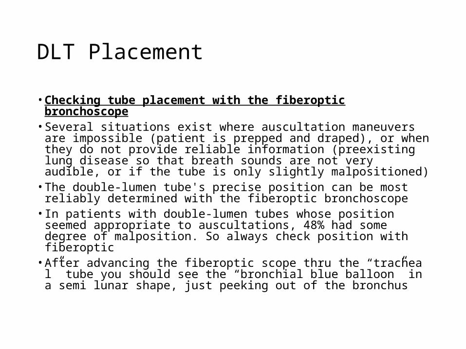
• Checking tube placement with the fiberoptic bronchoscope
• Several situations exist where auscultation maneuvers are impossible (patient is prepped and draped), or when they do not provide reliable information (preexisting lung disease so that breath sounds are not very audible, or if the tube is only slightly malpositioned)
• The double-lumen tube's precise position can be most reliably determined with the fiberoptic bronchoscope
• In patients with double-lumen tubes whose position seemed appropriate to auscultations, 48% had some degree of malposition. So always check position with fiberoptic
• After advancing the fiberoptic scope thru the “tracheal” tube you should see the “bronchial blue balloon” in a semi lunar shape, just peeking out of the bronchus

DLT Placement

To ensure correct position of DLT clinically :
breath sounds are- normal (not diminished) &- follow the expected unilateral pattern with unilateral clamping
the chest rises and falls in accordance with the breath sounds
the ventilated lung feels reasonably compliant
no leaks are present
respiratory gas moisture appears and disappears with each tidal ventilation
N.B even if the DLT is thought to be properly positioned by clinical signs, subsequent FOB may reveal an incidence of malposition ( 38 -78 %)

FOB picture of Lt - DLT
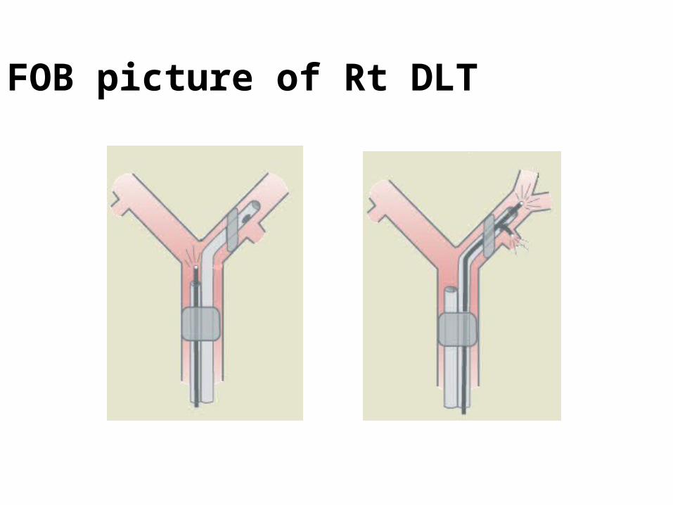
FOB picture of Rt DLT
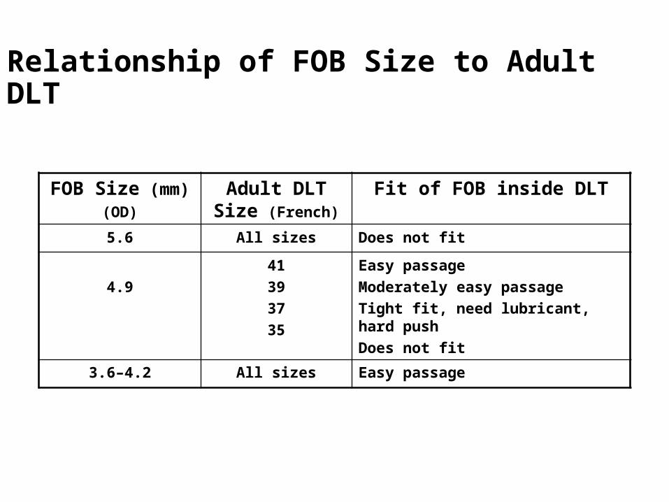
Relationship of FOB Size to Adult DLT
FOB Size (mm)(OD)
Adult DLT Size (French)
Fit of FOB inside DLT
5.6 All sizes Does not fit
4.941393735
Easy passageModerately easy passageTight fit, need lubricant, hard pushDoes not fit
3.6–4.2 All sizes Easy passage

Other Methods to Check DLT Position
Chest radiograph ; may be more useful than conventional auscultation and clamping in some patients, but it is always less precise than FOB. The DLT must have radiopaque markers at the end of Rt and Lt lumina.
Comparison of capnography;waveform and ETCO2 from each lumen may reveal a marked discrepancy (different degree of ventilation).
Surgeon ;
may be able to palpate, redirect or assist in changing DLT position from within the chest (by deflecting the DLT away from the wrong lung, etc..).

Adequacy for Sealing (air Bubble test )
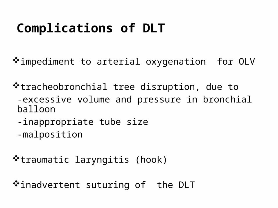
Complications of DLT
impediment to arterial oxygenation for OLV
tracheobronchial tree disruption, due to -excessive volume and pressure in bronchial balloon -inappropriate tube size-malposition
traumatic laryngitis (hook)
inadvertent suturing of the DLT

to avoid Tracheobronchial tree Disruption ;
1. Be cautious in patients with bronchial wall abnormalities.
2. Pick an appropriately sized tube.
3. Be sure that tube is not malpositioned ; Use FOB.
4. Avoid overinflation of endobronchial cuff.
5. Deflate endobronchial cuff during turning.
6. Inflate endobronchial cuff slowly.
7. Inflate endobronchial cuff with inspired gases.
8. Do not allow tube to move during turning.
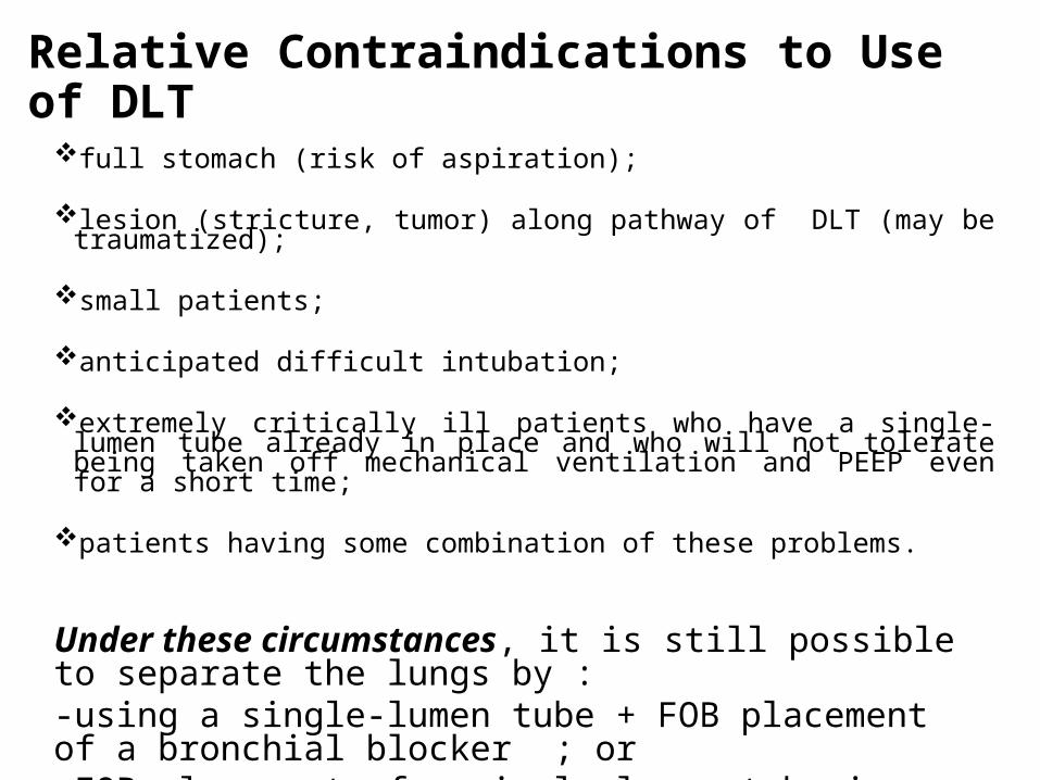
Relative Contraindications to Use of DLT
full stomach (risk of aspiration);
lesion (stricture, tumor) along pathway of DLT (may be traumatized);
small patients;
anticipated difficult intubation; extremely critically ill patients who have a single-lumen tube already in place and
who will not tolerate being taken off mechanical ventilation and PEEP even for a short time;
patients having some combination of these problems.
Under these circumstances, it is still possible to separate the lungs by : -using a single-lumen tube + FOB placement of a bronchial blocker ; or -FOB placement of a single-lumen tube in a main stem bronchus.

Advantages
Relatively easy to placeAllow conversion back and forth from OLV to two-
lung ventilation Allow suctioning of both lungs individuallyAllow CPAP to be applied to the non-dependent
lungAllow PEEP to be applied to the dependent lungAbility to ventilate around scope in the tube

Another indication for DLT: Reexpansion pulmonary edema
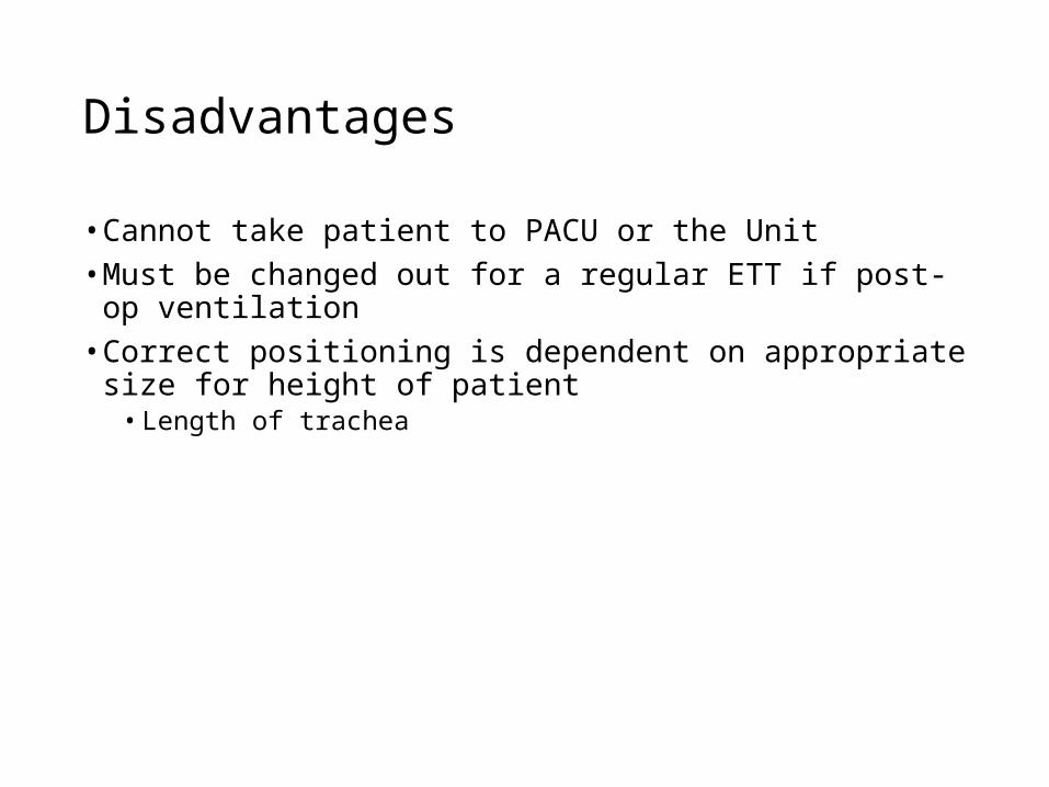
Disadvantages
• Cannot take patient to PACU or the Unit• Must be changed out for a regular ETT if post-op
ventilation• Correct positioning is dependent on appropriate size for
height of patient• Length of trachea

Bronchial Blockers (With Single-Lumen Endotracheal Tubes)
Lung separation can be effectively achieved with the use of a single-lumen endotracheal tube and a FOB placed bronchial blocker.
Often necessary in children as DLTs are too large to be used in them. The smallest DLT available is a left-sided 26 Fr tube, which may be used in patients 8 -12 years old and weighing 25 -35 kg.
Balloon-tipped luminal catheters have the advantage of allowing suctioning and injection of oxygen down the central lumen.

Types of bronchial blockers
Univent bronchial blocker system
Arndt endobronchial blocker
Cohen Flexitip Endobronchial Blocker
BB independent of a single-lumen tube

Univent bronchial blocker system

Univent Tubes
Univent Tubes
• Endotracheal intubation can be performed in the conventional manner, just like a single lumen endotracheal tube
• One-lung ventilation can be achieved by placement of the blocker to either the left or right lung, or to lung segments
• Insufflation and CPAP can be achieved through the lumen of the blocker shaft
• Blocked lung can be collapsed by aspirating air through the lumen of the blocker shaft
• The blocker can be retracted into its pocket to facilitate post-operative ventilation
• Improved "torque control" bronchial blocker:- Easier to direct by twisting than previous nylon catheter- High torque control malleable shaft for smooth intubation- Flexible blocker shaft with softer open lumen tip- Latex-free
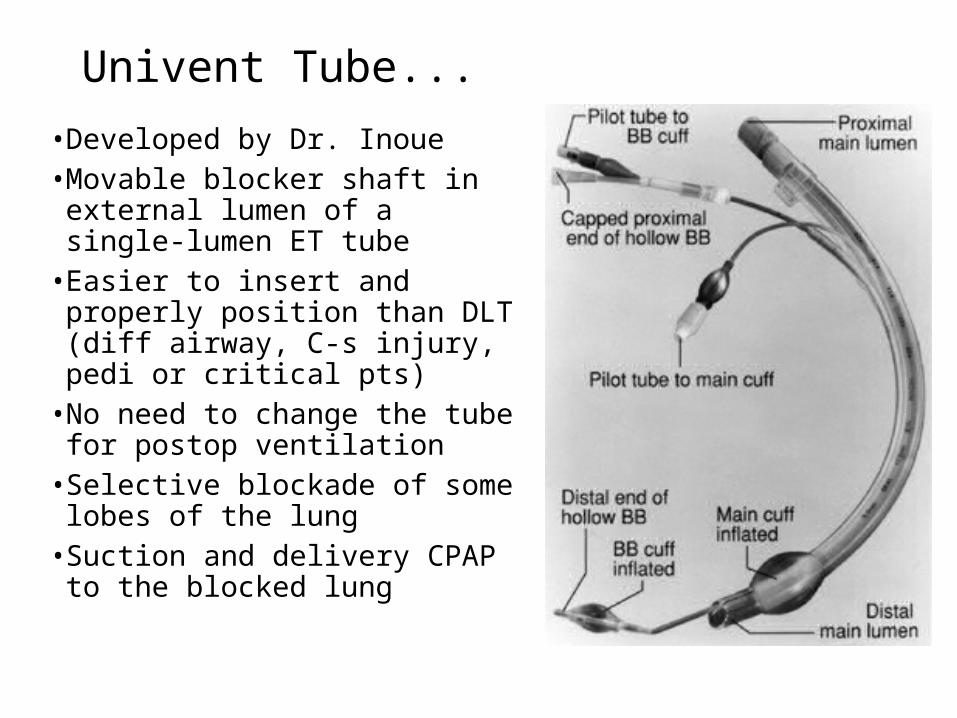
Univent Tube...
• Developed by Dr. Inoue• Movable blocker shaft in external
lumen of a single-lumen ET tube• Easier to insert and properly
position than DLT (diff airway, C-s injury, pedi or critical pts)
• No need to change the tube for postop ventilation
• Selective blockade of some lobes of the lung
• Suction and delivery CPAP to the blocked lung
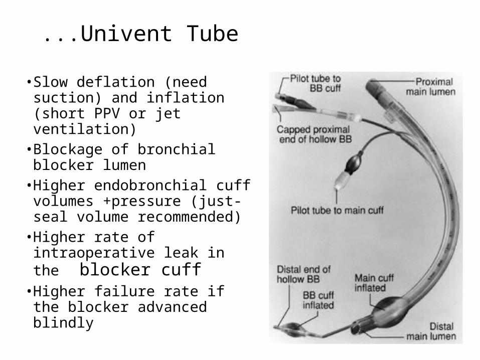
...Univent Tube
• Slow deflation (need suction) and inflation (short PPV or jet ventilation)
• Blockage of bronchial blocker lumen
• Higher endobronchial cuff volumes +pressure (just-seal volume recommended)
• Higher rate of intraoperative leak in the blocker cuff
• Higher failure rate if the blocker advanced blindly

Univent Tube

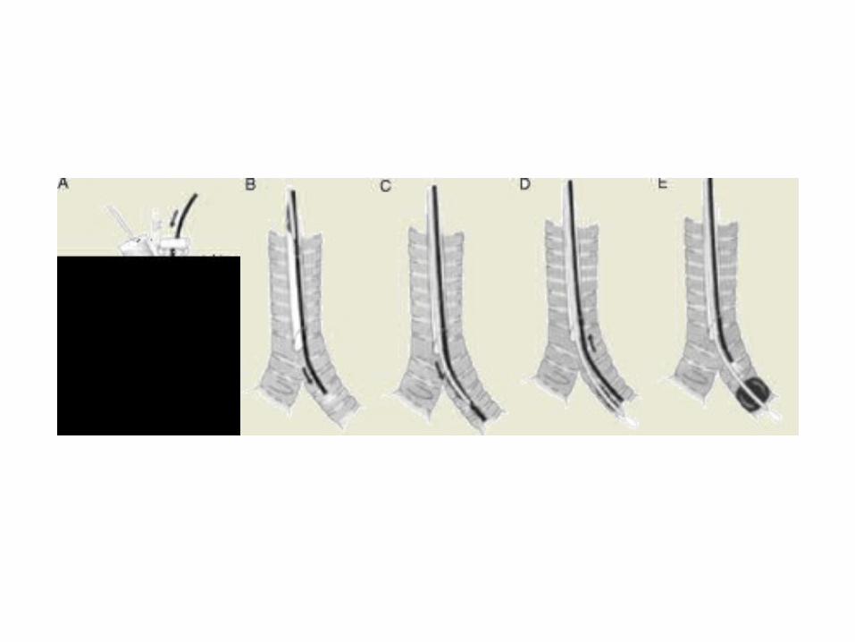
steps of FOB-aided method of positioning the Univent bronchial blocker in lt main stem bronchus
One- or two-lung ventilation is achieved simply by inflating or deflating, respectively, the bronchial blocker balloon

Indications for Wire-Guided Endobronchial Blockers vs. DLT• Critically ill patients• Rapid sequence induction• Known and unknown difficult airway• Postoperative intubation• Small adult and pediatric patients• Obese adults

Advantages of the Univent Bronchial Blocker Tube ( Relative to DLT )
1. Easier to insert and properly position.
2. Can be properly positioned during continuous ventilation and in the lateral decubitus position.
3. No need to change the tube when turning from the supine to prone position or for postoperative mechanical ventilation.
4. Selective blockade of some lobes of each lung.
5. Possible to apply CPAP to nonventilated operative lung.
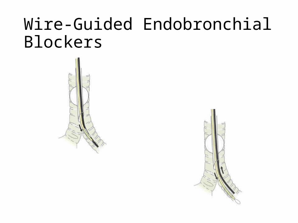
Advantages• Quickly and precisely navigate the airway • The guide wire loop couples the pediatric fiberoptic
bronchoscope and the wire-guided endobronchial blocker• yet both remain able to move independently of each other and
the pediatric fiberoptic bronchoscope may navigate the airway independent of its role in carrying the endobronchial blocker
• The pediatric bronchoscope acts as a guide, allowing the endobronchial blocker to be advanced over it into the correct position
• In addition, the wire-guided endobronchial blocker allows one-lung ventilation with a single-lumen endotracheal tube
• Thus, one-lung ventilation is not dependent on installing a special device in the airway, such as a double-lumen tube or a Univent endotracheal tube
• Allows one-lung ventilation in the critically ill patient in whom reintubation may be difficult or impossible and in patients with a known difficult airway requiring fiberoptic intubation with a conventional endotracheal tube
• Unnecessary to convert from a conventional double-lumen endotracheal tube to a single-lumen tube at the end of surgery
Disadvantages
• Satisfactory bronchial seal and lung separation are sometimes difficult to achieve
• The “blocked” lung collapses slowly (and sometimes incompletely)
• The balloon may become dislodged during surgery and enter the trachea proper, causing a complete airway obstruction
• In situations of acute increases in airway pressure, the endobronchial blocker balloon should be immediately deflated and the blocker re-advanced
• It will then re-enter the correct segment (as the tip remains in the correct bronchus and only the proximal balloon portion has entered the trachea)
• In this case, a pediatric fiberoptic bronchoscope should be re-introduced into the airway and the balloon re-positioned
• In order to prevent barotrauma, the initial balloon inflation volume should not be exceeded
• It is important that the balloon be fully deflated when not in use and only be re-inflated with the same volume used during positioning and bronchoscopy.
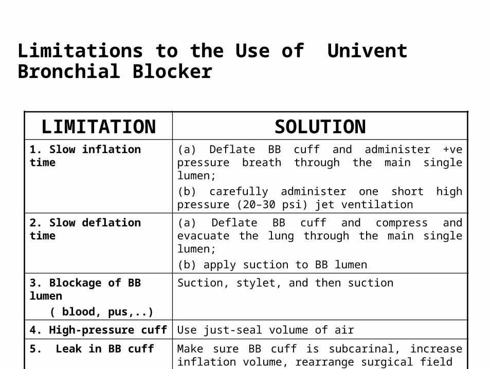
Limitations to the Use of Univent Bronchial Blocker
LIMITATION SOLUTION1. Slow inflation time (a) Deflate BB cuff and administer +ve pressure
breath through the main single lumen;(b) carefully administer one short high pressure (20–30 psi) jet ventilation
2. Slow deflation time (a) Deflate BB cuff and compress and evacuate the lung through the main single lumen; (b) apply suction to BB lumen
3. Blockage of BB lumen ( blood, pus,..)
Suction, stylet, and then suction
4. High-pressure cuff Use just-seal volume of air
5. Leak in BB cuff Make sure BB cuff is subcarinal, increase inflation volume, rearrange surgical field

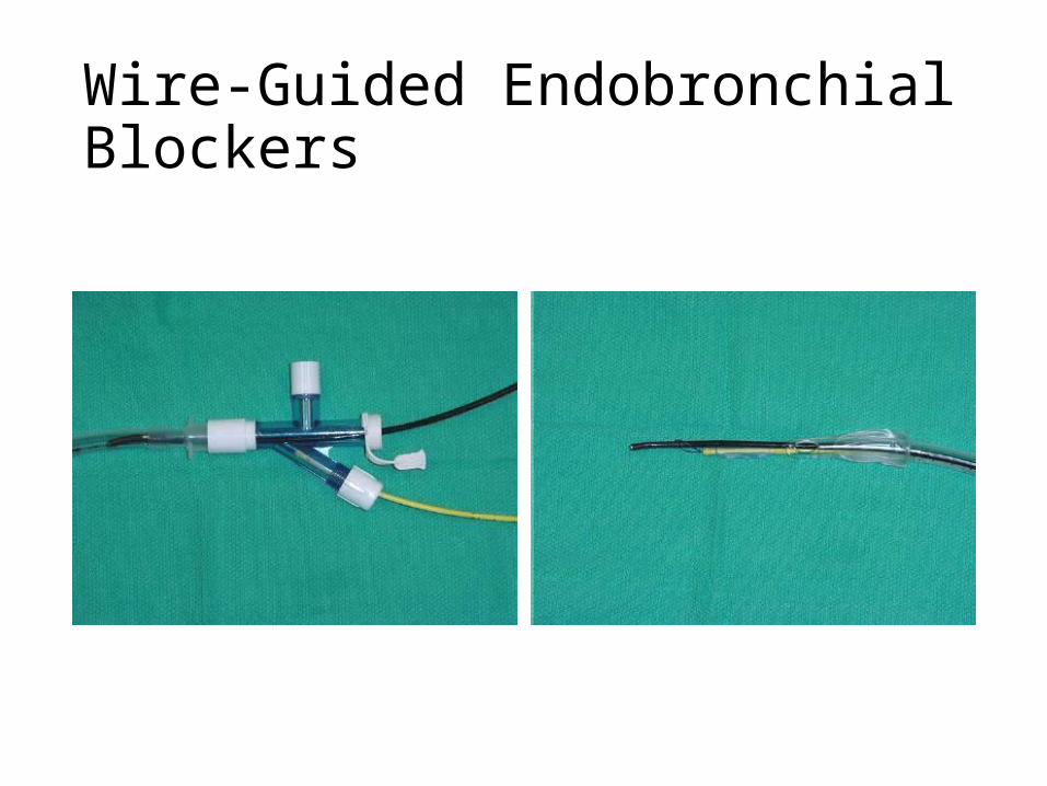
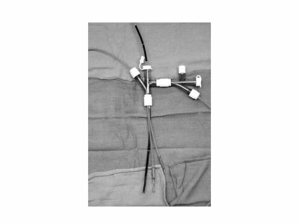
Arndt endobronchial blocker[Wire guided Endobronchial Blocker (WEB)]
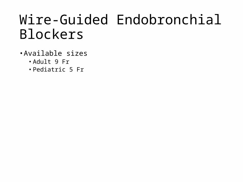
Wire-Guided Endobronchial Blockers• Available sizes
• Adult 9 Fr• Pediatric 5 Fr

Comparison of Various Tube Diameters
Wire-Guided Endobronchial Blockers

Wire-Guided Endobronchial Blockers
Wire-Guided Endobronchial Blockers

Wire-Guided Endobronchial Blockers

Fogarty Embolectomy Catheters

Fogarty Embolectomy Catheter
• Single-lumen balloon tipped catheter with a removable stylet
• In the parallel fashion, the Fogarty catheter is inserted prior to intubation
• In the co-axial fashion, the Fogarty catheter is placed through the endotracheal tube
• Both techniques require fiberoptic bronchoscopy to direct the Fogarty catheter into the correct pulmonary segment
• Once the catheter is in place, the balloon is inflated, sealing the airway
• Clinical limitations to the Fogarty technique• Difficult to direct and cannot be coupled to a fiberoptic bronchoscope• No accessory lumen for either removal of gas from the blocked segment
or insufflation of oxygen to reverse hypoxemia• Ventilate w/ 100% O2 prior to balloon inflation to aid in gas removal

Cohen Flexitip Endobronchial Blocker
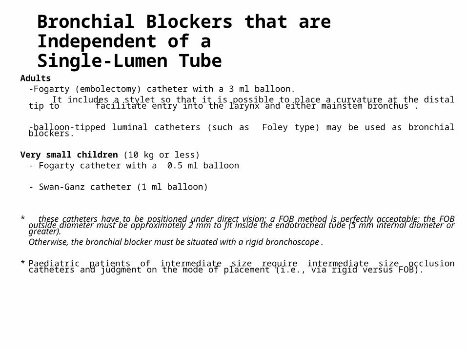
Bronchial Blockers that are Independent of a Single-Lumen Tube
Adults-Fogarty (embolectomy) catheter with a 3 ml balloon.
It includes a stylet so that it is possible to place a curvature at the distal tip to facilitate entry into the larynx and either mainstem bronchus .
-balloon-tipped luminal catheters (such as Foley type) may be used as bronchial blockers. Very small children (10 kg or less)
- Fogarty catheter with a 0.5 ml balloon
- Swan-Ganz catheter (1 ml balloon)
* these catheters have to be positioned under direct vision; a FOB method is perfectly acceptable; the FOB outside diameter must be approximately 2 mm to fit inside the endotracheal tube (3 mm internal diameter or greater). Otherwise, the bronchial blocker must be situated with a rigid bronchoscope.
* Paediatric patients of intermediate size require intermediate size occlusion catheters and judgment on the mode of placement (i.e., via rigid versus FOB).
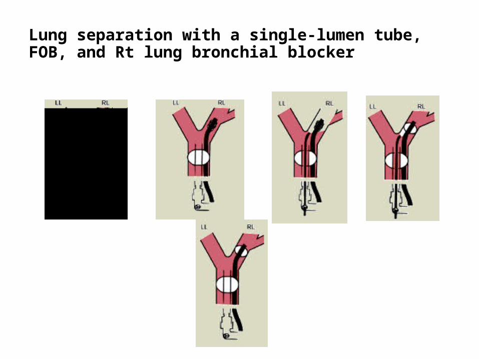
Lung separation with a single-lumen tube, FOB, and Rt lung bronchial blocker
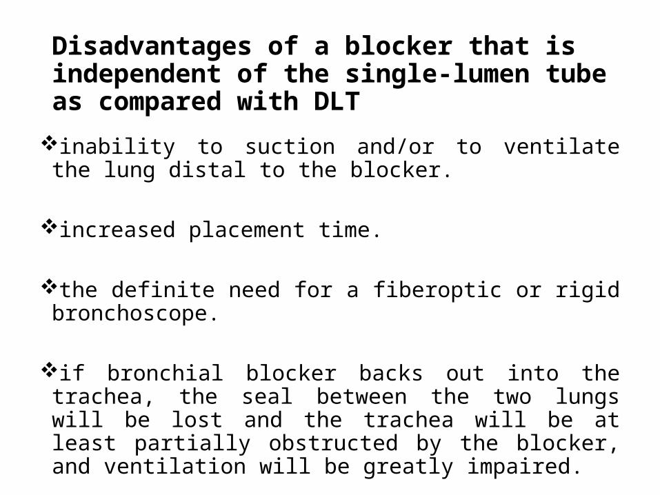
Disadvantages of a blocker that is independent of the single-lumen tube as compared with DLT
inability to suction and/or to ventilate the lung distal to the blocker.
increased placement time. the definite need for a fiberoptic or rigid bronchoscope. if bronchial blocker backs out into the trachea, the seal
between the two lungs will be lost and the trachea will be at least partially obstructed by the blocker, and ventilation will be greatly impaired.

Endobronchial Intubation with Single-Lumen Tubes
In adults, is often the easiest, quickest way for lung separation in patients presenting with haemoptysis , either
-blind, or -FOB , or-guidance by surgeon from within chest
In children it may be the simplest way to achieve OLV
Disadvantages-inability to do suctioning or ventilation of operative side.-difficult positioning bronchial cuff with inadequate ventilation of
Rt upper lobe after Rt endobronchial intubation.
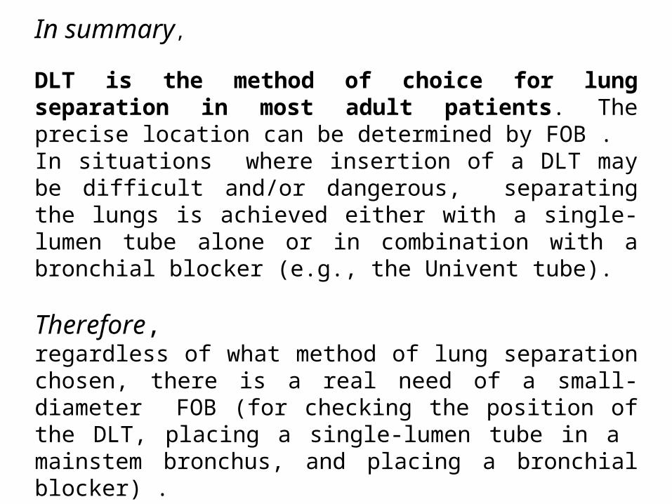
In summary,
DLT is the method of choice for lung separation in most adult patients. The precise location can be determined by FOB . In situations where insertion of a DLT may be difficult and/or dangerous, separating the lungs is achieved either with a single-lumen tube alone or in combination with a bronchial blocker (e.g., the Univent tube).
Therefore, regardless of what method of lung separation chosen, there is a real need of a small-diameter FOB (for checking the position of the DLT, placing a single-lumen tube in a mainstem bronchus, and placing a bronchial blocker) .
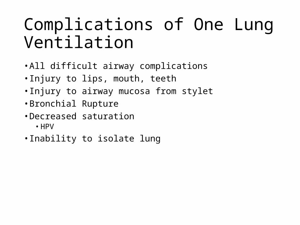
Complications of One Lung Ventilation• All difficult airway complications• Injury to lips, mouth, teeth• Injury to airway mucosa from stylet• Bronchial Rupture• Decreased saturation
• HPV
• Inability to isolate lung

Complications - Bronchial Rupture

Comparing Up Right & Lateral Decubitus Position
04/15/23 HSNZ KT 73

• Distribution of blood flow and ventilation is similar to that in the upright position but turned by 90 degrees.
• Blood flow and ventilation to the dependent lung are significantly greater than to the nondependent lung.
• Good V/Q matching at the level of the dependent lung results in adequate oxygenation in the awake patient breathing spontaneously.
• In Lateral Decubitus Position (LDP), ordinarily less Zone 1- due to vertical hydrostatic gradient is less in LDP than upright.
• % of Blood flow to lungs according to position; In upright/Supine-Rt 55% Lt 45%; In LDP Rt NDL 45% Lt DL 55%; In LDP Lt NDL 35% RT DL 65%
04/15/23 HSNZ KT 74
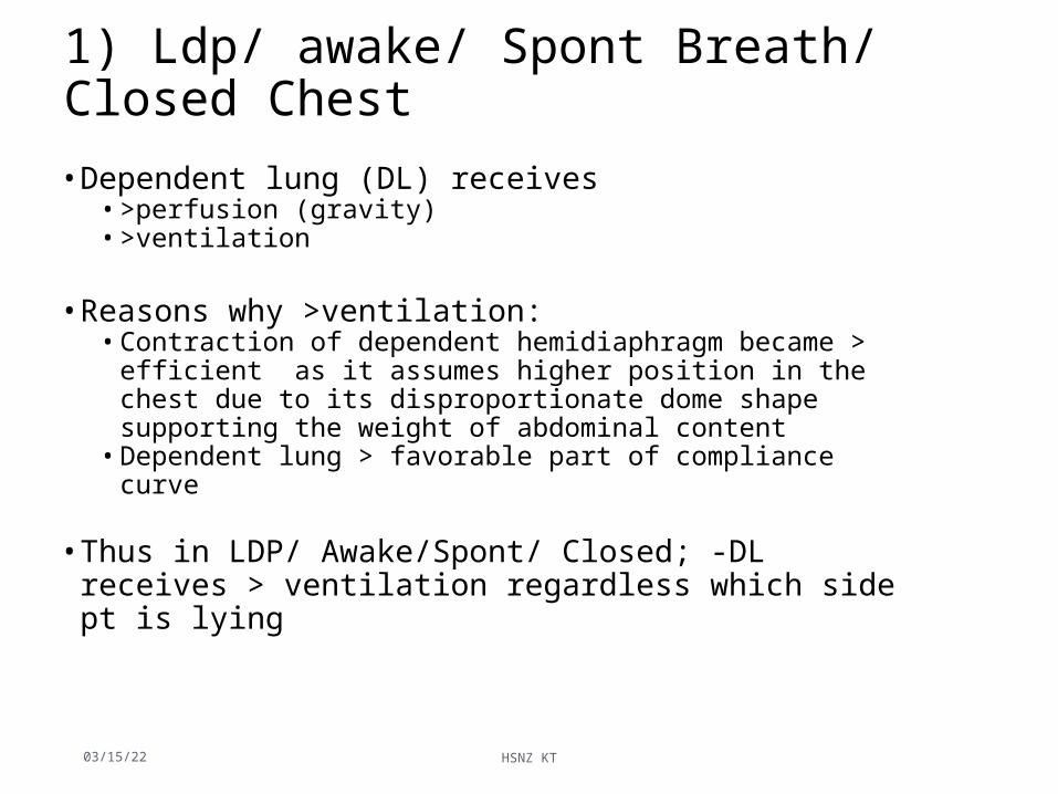
1) Ldp/ awake/ Spont Breath/ Closed Chest
• Dependent lung (DL) receives• >perfusion (gravity)• >ventilation
• Reasons why >ventilation:• Contraction of dependent hemidiaphragm became > efficient as it
assumes higher position in the chest due to its disproportionate dome shape supporting the weight of abdominal content
• Dependent lung > favorable part of compliance curve
• Thus in LDP/ Awake/Spont/ Closed; -DL receives > ventilation regardless which side pt is lying
04/15/23 HSNZ KT 75

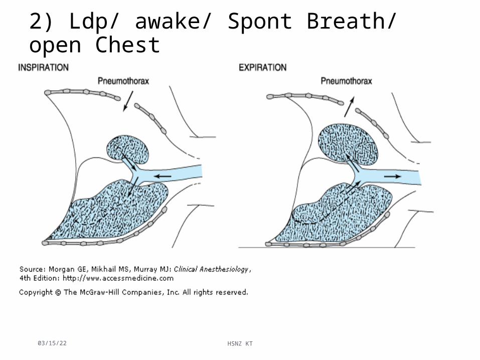
2) Ldp/ awake/ Spont Breath/ open Chest
2 complications 1.Mediastinal shift, occurring during inspiration.
Negative pressure more in intact hemithorax cause the mediastinum to move vertically downward and push into the dependent hemithorax.
• create circulatory & reflex changes, result in a clinical picture similar to that of shock and respiratory distress.
• Eg. Thoracoscopy LA, pt may need intubated immediately, with initiation of positive-pressure ventilation
04/15/23 HSNZ KT 76
Ldp/ awake/ Spont Breath/ open Chest
2. Paradoxical breathing:• During inspiration, movement of gas from the exposed
lung into the intact lung and movement of air from the environment into the open hemithorax cause collapse of the exposed lung.
• During expiration, the reverse occurs, and the exposed lung expands
04/15/23 HSNZ KT 77
2) Ldp/ awake/ Spont Breath/ open Chest
04/15/23 HSNZ KT 78
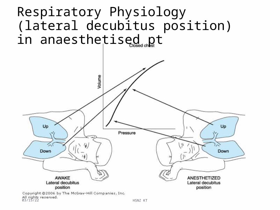
Respiratory Physiology (lateral decubitus position) in anaesthetised pt
04/15/23 HSNZ KT 79

Factors affecting respiratory physiology in lateral decubitus positionThe changes further accentuated by several factors:1)Induction of anesthesia2)Initiation of mechanical ventilation3)Use of neuromuscular blockade4)Opening the chest/pleural space5)Surgical Retraction/ Compression6)Pressure by mediastinum/ Abdominal content
• Perfusion continue to favor dependent lung (Due to gravitational effect)
• Ventilation favor the less perfused lung.• End result is V/Q mismatch(shunt) giving rise to hypoxemia.
04/15/23 HSNZ KT 80

Induction of Anaesthesia
• Reduce FRC• Non dependent lung moves to favorable part of compliance• Dependent lung moves to less compliance• Result in > ventilation in nondependent lung than dependent• But perfusion still favor the dependent lung (gravitational effect)• Thus V/Q mismatch occur causing hypoxia
04/15/23 HSNZ KT 81

Other factors involved• Positive Pressure Ventilation (PPV in mechanical ventilation)
favors ND lung as it is > compliant • Use of neuromuscular blockade- causing paralysis of the
diaphragm. Allowing abdominal to push the dependent hemidiaphram & impede further ventilation of DL
• Suboptimal positioning (usage of sand bag to maintain pt in LDP) further restrict movement of DL
• Opening of NDL cause increase compliance of NDL, as the lungs less restricted. This further attenuates differences of compliance between two lungs.
04/15/23 HSNZ KT 82

3) Ldp/ Anaesthetized / Spont Breath/ Closed Chest
• In awake/ anaesthetised- distribution of pulmonary blood flow influenced by gravitational effect
• But Induction of GAC cause significant changes in distribution of ventilation
• Reasons:• Ventilation favors NDL due to• GAC reduce both lungs FRC (both loss of volume)
• Effect of muscle relaxation- paralysis of both hemidiaphragm. The curve effect of diaphragm gives no Advantages
• Pressure effect by medialstinal structure- rest on dependent lung physically impedes DL.
04/15/23 HSNZ KT 83

3) Ldp/ Anaesthetized / Spont Breath/ Closed Chest
• Weight of abdominal contents pushing cephalad against diaphragm (greatest effect to DL)- physically impedes DL expansion and reduce FRCEffect more prominent if paralyzed
• Suboptimal positioning- fails to provide room for DL expansion; considerable compressing DL
• Opening chest/ pleural space (pneumothorax) further increase ventilation to NDL as it is no longer restricted
04/15/23 HSNZ KT 84
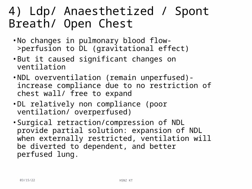
4) Ldp/ Anaesthetized / Spont Breath/ Open Chest • No changes in pulmonary blood flow- >perfusion to DL
(gravitational effect)• But it caused significant changes on ventilation• NDL overventilation (remain unperfused)- increase compliance
due to no restriction of chest wall/ free to expand• DL relatively non compliance (poor ventilation/ overperfused)• Surgical retraction/compression of NDL provide partial solution:
expansion of NDL when externally restricted, ventilation will be diverted to dependent, and better perfused lung.
04/15/23 HSNZ KT 85

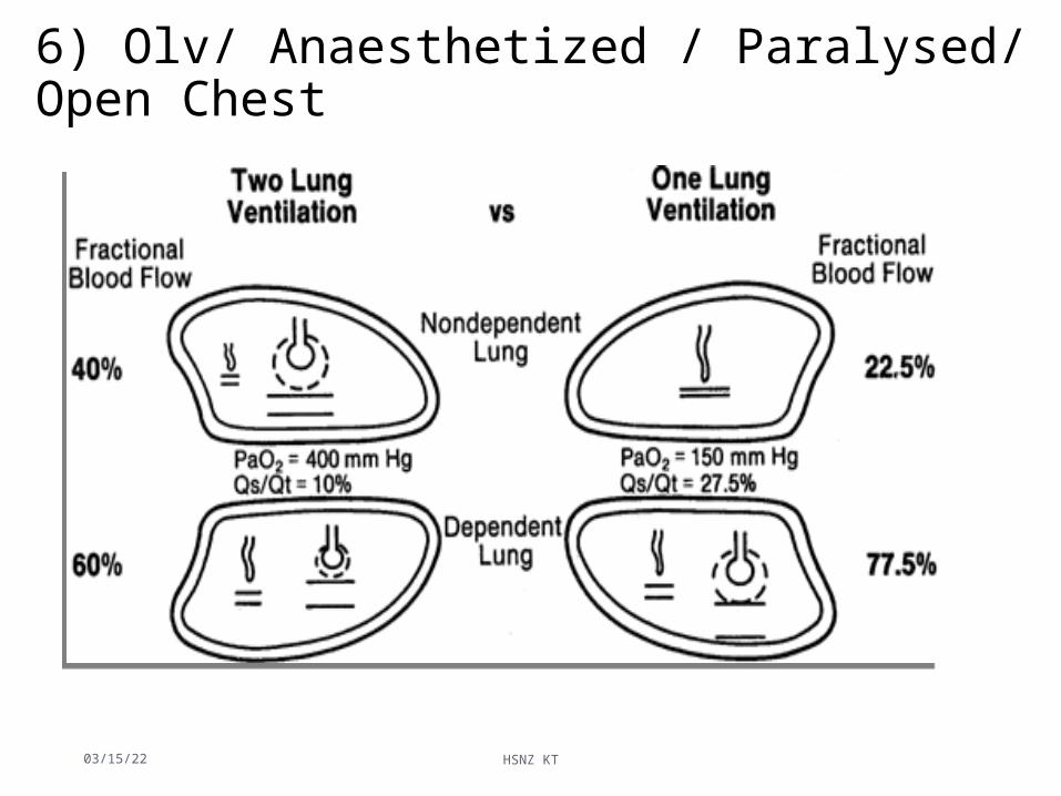
6) Olv/ Anaesthetized / Paralysed/ Open Chest
• Dependent lung is no longer on the steep (compliant) portion of the volume–pressure curve because of reduced lung volume and FRC.
# create a low V®/Q® ratio and a large P(A-a)O2 gradient.
04/15/23 HSNZ KT 86
6) Olv/ Anaesthetized / Paralysed/ Open Chest
04/15/23 HSNZ KT 87

Summary of V-Q relationships in the anesthetized, open-chest and paralyzed patients in LDP
04/15/23 HSNZ KT 88

SUMMARY OF V/Q RELATIONSHIP IN AWAKE & ANAESTHETISED PT
Awake/Closed Anaesthetised
Closed Open
V/Q V Q V Q V Q
NDL
DL
04/15/23 HSNZ KT
89

Summary of V-Q relationships in the anesthetized, open-chest and paralyzed patients in LDP
04/15/23 HSNZ KT 90

Physiology of OLV
• The principle physiologic change of OLV is the redistribution of lung perfusion between the ventilated (dependent) and blocked (nondependent) lung
• Many factors contribute to the lung perfusion, the major determinants of them are hypoxic pulmonary vasoconstriction (HPV) and gravity.
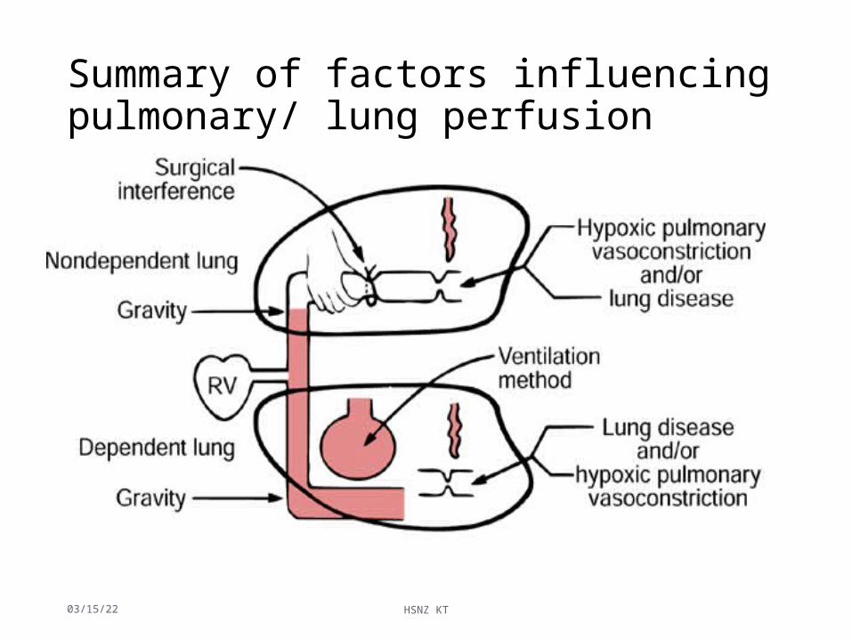
Summary of factors influencing pulmonary/ lung perfusion
04/15/23 HSNZ KT 92
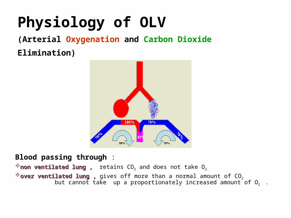
Physiology of OLV (Arterial Oxygenation and Carbon Dioxide Elimination)
Blood passing through :non ventilated lung ,non ventilated lung , retains CO2 and does not take O2. over ventilated lung ,over ventilated lung , gives off more than a normal amount of CO2 but cannot take up a
proportionately increased amount of O2 .

Thus, during one-lung ventilation
more decreased oxygenation than during two-lung
ventilation in LDP due to an obligatory Rt-Lt
transpulmonary shunt through the nonventilated
nondependent lung. Consequently, lower PaO2 &
larger P(A-a)O2
usually carbon dioxide elimination is not a
problem; but retention of CO2 by blood traversing
the nonventilated lung slightly exceeds the
increased elimination of CO2 from blood traversing
the ventilated lung, and the PaCO2 will usually
slowly increase and P(A-a)CO2 decreases .

Two-lung ventilation versus OLV
during OLV, the nonventilated lung has some blood flow and therefore has an obligatory shunt, which is not present during two-lung ventilation & is the most important reason for increased P(A-a)O2.

Blood Flow distribution during OLV
The major determinants of blood flow distribution between both lungs :
•gravity,
•amount of lung disease,
•magnitude HPV,
•surgical interference nondependent ,
•ventilation mode dependent
Blood Flow Distribution During OLV , cont….
Lung condition (amount of lung disease)
*severely diseased nondependent lung, may have a fixed reduction in blood flow preoperatively and its collapse may not cause much increase in shunt.
*increases in PVR in dependent ventilated lung decreases its ability to accept redistributed blood from the hypoxic lung. This may occur in case of :
-decreasing FIO2 in the dependent lung . -decreasing temperature .
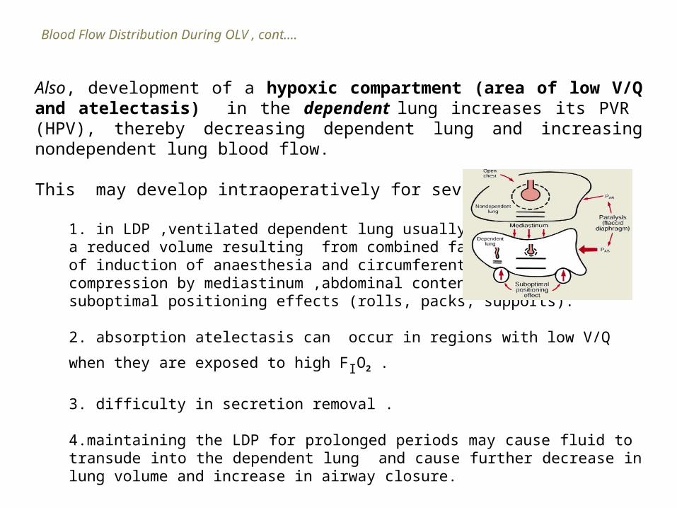
Also, development of a hypoxic compartment (area of low V/Q and atelectasis) in the dependent lung increases its PVR (HPV), thereby decreasing dependent lung and increasing nondependent lung blood flow.
This may develop intraoperatively for several reasons:
1. in LDP ,ventilated dependent lung usually hasa reduced volume resulting from combined factorsof induction of anaesthesia and circumferentialcompression by mediastinum ,abdominal contents, and suboptimal positioning effects (rolls, packs, supports).
2. absorption atelectasis can occur in regions with low V/Q when they are
exposed to high FIO2 .
3. difficulty in secretion removal .
4.maintaining the LDP for prolonged periods may cause fluid to transude into the dependent lung and cause further decrease in lung volume and increase in airway closure.
Blood Flow Distribution During OLV , cont….
Blood Flow Distribution During OLV , cont.
Surgical interference(compression ,retraction and ligation of pulmonary vessels during pulmonary resection) of the nondependent lung may further passively reduce its blood flow.
Mode of ventilation of dependent lung•If hyperventilated PaCO2 HPV•Excessive AWP (PEEP or VT ) dependent PVR and
nondependent lung blood flow.•FIO2 -VD in dependent lung, augmenting HPV in
nondependent lung -but ,may cause absorption atelectasis in
regions that have low V/Q ratios

Hypoxic pulmonary vasoconstriction (hpv)• HPV, a local response of pulmonary vascular smooth muscle
(PVSM), decreases blood flow to the area of lung where a low alveolar oxygen pressure is sensed.
• Intrinsic response of lung, no neuronal control, immediately present in transplanted lung.
• The mechanism of HPV is not completely understood. Vasoactive substances released by hypoxia or hypoxia itself (K+ channel) cause pulmonary artery smooth muscle contraction.
• All pulmonary arteries and veins vasoconstric in response to hypoxia, but greatest effect is to small pumonary arteriesm(200mm)
04/15/23 HSNZ KT 100

Hypoxic pulmonary vasoconstriction (hpv)
• HPV aids in keeping a normal V/Q relationship by diversion of blood from underventilated areas, responsible for the most lung perfusion redistribution in OLV.
• HPV is graded and limited, of greatest benefit when 30% to 70% of the lung is made hypoxic.
• But effective only when there are normoxic areas of the lung available to receive the diverted blood flow
04/15/23 HSNZ KT 101
Blood Flow Distribution During OLV , cont.
Magnitude of HPV
• HPV is an autoregulatory mechanism that protects the PaO2 by decreasing the amount of shunt flow that can occur through hypoxic lung as it diverts blood flow from the atelectatic lung toward the remaining normoxic or hyperoxic ventilated lung.
• HPV is of little importance When ;-very little of the lung is hypoxic (near 0%) because shunt will be small. -most of the lung is hypoxic (near 100%) there is no significant normoxic region to which the hypoxic region can divert flow.
• Of great importance if the percentage of hypoxic lung is intermediate ( 30 and 70%), which is the case during OLV

Factors that might determine the amount of regional HPV
Blood Flow Distribution During OLV , cont.

Factors that might determine the amount of regional HPV , cont.
1. Distribution of the alveolar hypoxia is probably not a determinant of the amount of HPV; all regions of the lung respond to alveolar hypoxia with vasoconstriction.
2. Atelectasis, most of blood flow reduction in acutely atelectatic lung is due to HPV and none of it to passive mechanical factors (such as vessel tortuosity).
3. Vasodilator drugs, most of them inhibit regional HPV
4. Anaesthetic drugs
5. Pulmonary vascular pressure, HPV response is-maximal at normal PVP and -decreased at either high or low PVP.
6. Pv¯O2 , HPV response also is -maximal when Pv¯O2 is normal and -decreased by either high or low Pv¯O2.
7. FIO2 selectively decreasing the FIO2 in the normoxic compartment causes an increase in normoxic lung vascular tone,
thereby decreasing blood flow diversion from hypoxic to normoxic lung.
8. Vasoconstrictor drugs constrict normoxic lung vessels preferentially, thereby disproportionately increasing normoxic lung PVR causing decrease normoxic lung blood flow and increase atelectatic lung blood flow.
9. PaCO2 , hypocapnia inhibits & hypercapnia directly enhances regional HPV. 10. PEEP

Other Causes of Hypoxaemia During OLV
Failure of the oxygen supply.
Gross hypoventilation of the dependent lung.
Blockage of the dependent lung airway lumen e.g. by secretions Malposition of the DLT Decrease of Pv¯O2 (decreased cardiac output, increased oxygen
consumption [excessive sympathetic nervous system stimulation, hyperthermia, shivering])
Transfusion of blood may cause pulmonary dysfunction attributed to the
action of isoantibodies against leukocytes, which causes cellular aggregation, microvascular occlusion, and capillary leakage.

Hypoxic pulmonary vasoconstriction
• HPV is a physiological response of the lung to alveolar hypoxia, which redistributes pulmonary blood flow from areas of low oxygen partial pressure to areas of high oxygen availability.
• The mechanism of HPV is not completely understood. Vasoactive substances released by hypoxia or hypoxia itself (activating K+, Ca++ and TRP channels) cause pulmonary artery smooth muscle contraction
HPV
• HPV aids in keeping a normal V/Q relationship by diversion of blood from underventilated areas, responsible for the most lung perfusion redistribution in OLV
• HPV is graded and limited, of greatest benefit when 30% to 70% of the lung is made hypoxic.
• HPV is effective only when there are normoxic areas of the lung available to receive the diverted blood flow

Factors affecting regional HPV
• HPV is inhibited directly by volatile anesthetics (not N20), vasodilators (NTG, SNP, NO, dobutamine, many ß2-agonist), increased PVR (MS, MI, PE) and hypocapnia
• HPV is indirectly inhibited by PEEP; vasoconstrictor drugs (epinephrine, norepinephrine, phenylephrine, dopamine) constrict normoxic lung vessels preferentially

Gravity and V-Q
• Upright LDP

Shunt and OLV
• Physiological (postpulmonary) shunt• About 2-5% CO,• Accounting for normal A-aD02, 10-15 mmHg• Including drainages from
• Thebesian veins of the heart• The pulmonary bronchial veins• Mediastinal and pleural veins
• Transpulmonary shunt increased due to continued perfusion of the atelectatic lung and A-aD02 may increase
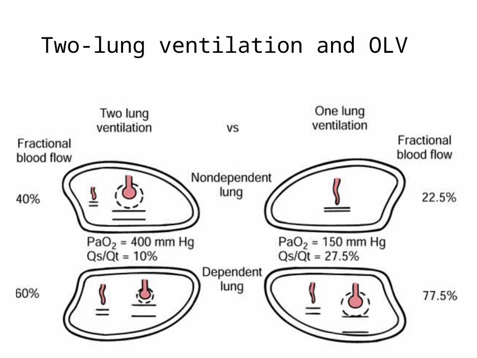
Two-lung ventilation and OLV
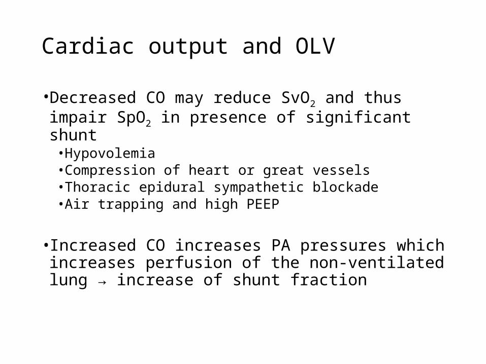
Cardiac output and OLV
• Decreased CO may reduce SvO2 and thus impair SpO2 in presence of significant shunt
• Hypovolemia• Compression of heart or great vessels• Thoracic epidural sympathetic blockade• Air trapping and high PEEP
• Increased CO increases PA pressures which increases perfusion of the non-ventilated lung → increase of shunt fraction
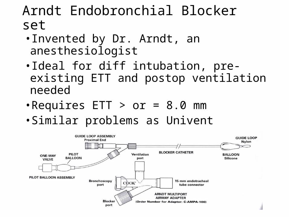
Arndt Endobronchial Blocker set
• Invented by Dr. Arndt, an anesthesiologist• Ideal for diff intubation, pre-existing ETT and postop
ventilation needed • Requires ETT > or = 8.0 mm• Similar problems as Univent• Inability to suction or ventilate the blocked lung

Other methods of OLV
• Single-lumen ETT with a balloon-tipped catheter• Including Fogarty embolectomy catheter, Magill or Foley,
and Swan-Ganz catheter (children < 10 kg)• Not reliable and may be more time-consuming • Inability to suction or ventilate the blocked lung
• Endobronchial intubation of single-lumen ETT• The easiest and quickest way of separating one lung from
the other bleeding one, esp. from left lung• More often used for pedi patients• More likely to cause serious hypoxemia or severe
bronchial damage
Management of OLV...
• Maintain two-lung ventilation as long as possible• Start OLV with 100% O2 then start backing off the FiO2 if
saturations are OK• Manual ventilation for the first few minutes of OLV to
get a sense of pulmonary compliance / resistance• Be attentive to inspiratory pressures and tidal volumes
and adjust the ventilator to optimize oxygenation and alveolar ventilation, with minimal barotrauma
• Look at the surgical field to see if the non-dependent lung is collapsed

...Management of OLV
• Tidal volume = 8-10 ml/kg • Adjust RR (increasing 20-30%) to keep PaCO2 = 40 mmHg• No PEEP (or very low PEEP, < 5 cm H2O)• Continuous monitoring of oxygenation and ventilation (SpO2, ABG and ET
CO2)
Other causes of hypoxemia in OLV
• Mechanical failure of O2 supply or airway blockade• Hypoventilation• Resorption of residual O2 from the clamped lung• Factors that decrease SvO2 (CO, O2 consumption)

Management of hypoxemia during OLV
• FiO2 = 1.0• Manual ventilation• Check DLT position with FOB• Check hemodynamic status• CPAP (5-10 cm H2O, 5 L/min) to nondependent lung, most effective• PEEP (5-10 cm H2O) to dependent lung, least effective• Intermittent two-lung ventilation• Clamp pulmonary artery
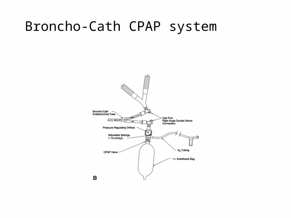
Broncho-Cath CPAP system

Patient Monitoring Considerations
• Direct arterial catheterization (a-line)• essential for nearly all thoracic cases• Allows for beat-to-beat blood pressure analysis • Sampling for determination of ABG
• Central venous pressure monitoring (central line)• essential for measuring right atrial and right ventricular
pressures• Useful in monitoring:
• large volume shifts • hypovolemia • need for vasoactive drugs
• Pulmonary artery catheterization• left sided filling pressures, cardiac output • Calculation of derived hemodynamic and respiratory parameters
and clinical use of Starling curve• Most PA catheters (more than 90%) float to and locate in the
right lung due to increased pulmonary blood flow• Create inaccurate reading for R thoracotomies

Patient Monitoring ConsiderationsOxygenation and Ventilation
Monitoring inspired oxygen Sampling of arterial blood for ABGsPulse oximetry
Transcutaneous oxygen tensionfor neonates
Qualitative signschest expansionobservation of reservoir bagauscultation of breath sounds
EtCO2 measurement, capnograph

Case Setup for DLT & OLV
•MSMAID•Preferred blade and handle•Airway – Have standard supplies & assortment of sizes for DLT or other OLV choice equipment
•Fiberoptic cart•Hemostats or clamps to clamp off lumens of the tube
•Suction!!

Ventilatory Management of OLV
• Conventional Ventilatory Management
• Differential Lung Ventilation Management
• High-Frequency Ventilation Management
• Low-Flow Apnoeic Ventilation (Apnoeic Insufflation)
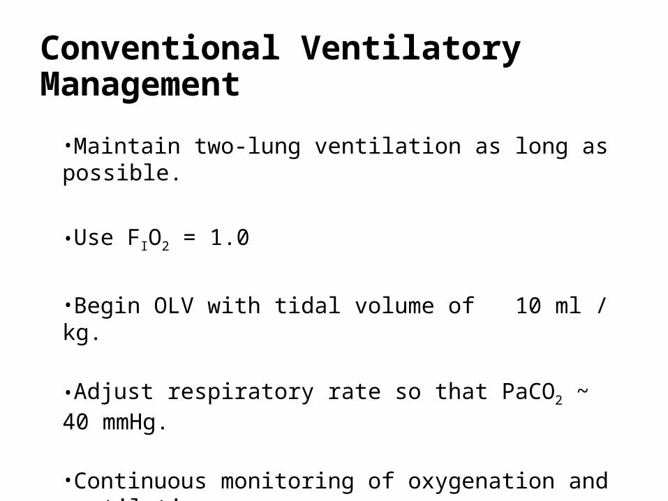
Conventional Ventilatory Management
•Maintain two-lung ventilation as long as possible.
•Use FIO2 = 1.0
•Begin OLV with tidal volume of 10 ml / kg.
•Adjust respiratory rate so that PaCO2 ~ 40 mmHg.
•Continuous monitoring of oxygenation and ventilation.

Differential Lung Ventilation Management
Intermittent Inflation of the Nondependent Operative Lung may be expected to increase PaO2 for a variable period of time.
Selective Dependent Lung PEEP
Selective Nondependent Lung CPAP (without tidal ventilation)
Differential Lung PEEP/CPAP

Selective PEEP to dependent lung improves
V/Q but also increases PVR in it ; this diverts
blood and increases shunt flow through, the
nonventilated lung.
Dependent lung is ventilated but compressed by : Mediastinum , Diaphragm P ( rolls, packs, shoulder supports) .The nondependent lung is nonventilated , and blood flow through it is a shunt flow.
Selective CPAP to nondependent lung
permits oxygen uptake from it ; Even if CPAP causes a
rise in PVR and diverts blood to dependent lung,
the diverted blood flow can still participate in gas
exchange in the ventilated dependent lung that greatly
increases PaO2
Differential lung CPAP (nondependent) /PEEP (dependent), wherever blood goes, both lungs can participate in O2 uptake. With this pattern, PaO2 can be restored to levels near those achieved by two-lung ventilation.

CPAP is created by the free flow of oxygen into the lung versus the restricted outflow of oxygen from the lung by the pressure relief valve.
The three essential components of a nondependent lung CPAP system
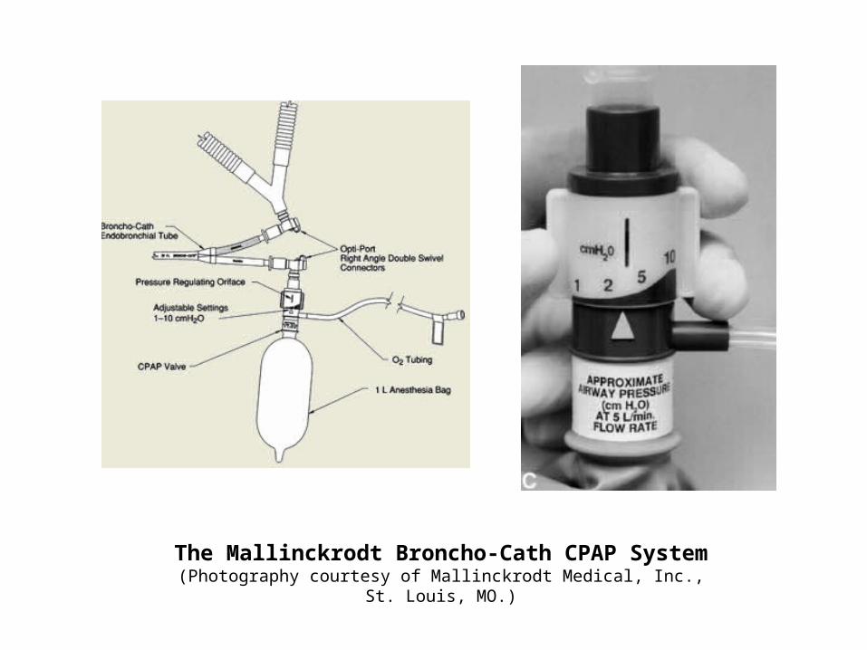
The Mallinckrodt Broncho-Cath CPAP System(Photography courtesy of Mallinckrodt Medical, Inc., St. Louis, MO.)
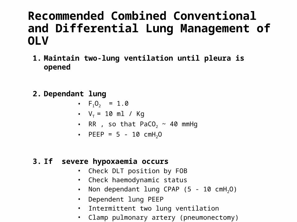
Recommended Combined Conventional and Differential Lung Management of OLV
1. Maintain two-lung ventilation until pleura is opened
2. Dependant lung• FIO2 = 1.0
• VT = 10 ml / Kg
• RR , so that PaCO2 ~ 40 mmHg
• PEEP = 5 - 10 cmH2O
3. If severe hypoxaemia occurs • Check DLT position by FOB• Check haemodynamic status• Non dependant lung CPAP (5 - 10 cmH2O)
• Dependent lung PEEP• Intermittent two lung ventilation• Clamp pulmonary artery (pneumonectomy)

High-Frequency Ventilation (HFV) Management
HFV delivers , very small VT (<2 ml/kg) at high rates (60 - 2,400 breaths/min)
So,• can be delivered through very small catheters
• it decreases PAWP
So,
it may be uniquely useful in facilitating the performance of thoracic surgery inthe following three ways;
-Use in Major Conducting Airway Surgery -Use in Bronchopleural Fistula -Use in Minimizing Movement of the Operative Field
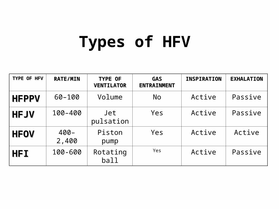
TYPE OF TYPE OF HFVHFV
RATE/MINRATE/MIN TYPE OF TYPE OF VENTILATORVENTILATOR
GAS GAS ENTRAINMENENTRAINMEN
TT
INSPIRATIOINSPIRATIONN
EXHALATIOEXHALATIONN
HFPPHFPPVV
60–100 Volume No Active Passive
HFJVHFJV 100–400 Jet pulsation
Yes Active Passive
HFOVHFOV 400–2,400
Piston pump
Yes Active Active
HFIHFI 100-600 Rotating ball
Yes Active Passive
Types of HFV

Low-Flow Apnoeic Ventilation (Apnoeic Insufflation)
• If ventilation is stopped during administration of 100 % O2 and airway is left connected to a fresh gas supply, O2 will be drawn into the lung by mass movement to replace the diffused O2 . There is usually no difficulty in maintaining an adequate PaO2 (especially if 5–10 cmH2O of CPAP is used) at least for 20 minutes .
• If flow of O2 is relatively low (<0.1 L/kg/min) almost all CO2 produced is
retained, and PaCO2 rises approximately 6 mmHg in the 1st minute and then 3 - 4 mmHg each minute thereafter .
• Safe period < 10 min
• arterial oxygen saturation monitoring via pulse oximetry is mandatory.
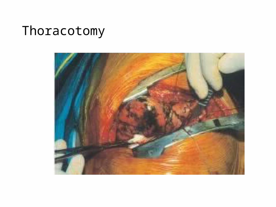
Thoracotomy
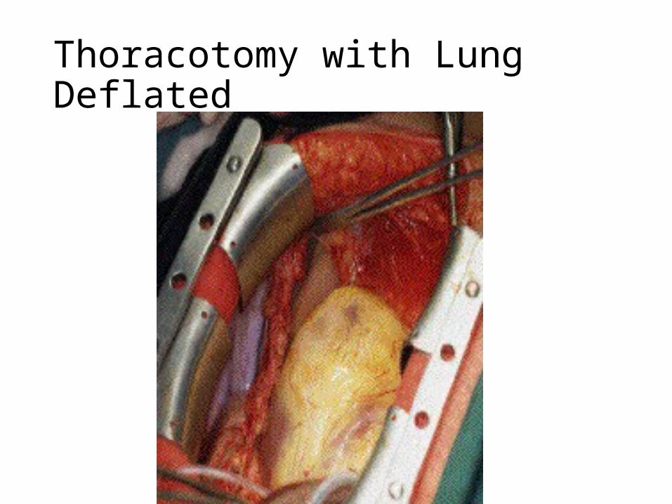
Thoracotomy with Lung Deflated
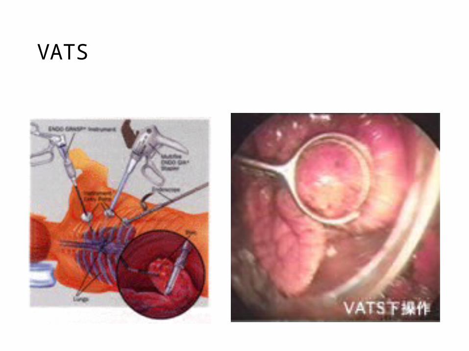
VATS

VATS
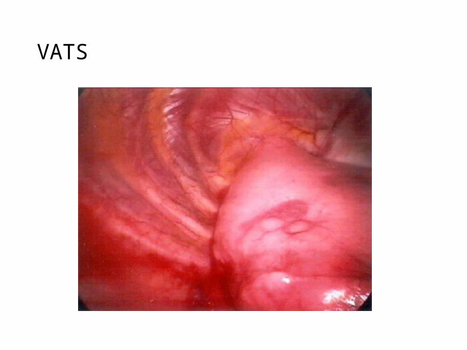
VATS

Summary
• OLV widely used in cardiothoracic surgery• Many methods can be used for OLV. Optimal
methods depends on indication, patient factors, equipment, skills and level of training
• FOB is the key equipment for OLV • Principle physiologic change of OLV is the
redistribution of pulmonary blood flow to keep an appropriate V/Q match
• Management of OLV is a challenge for the anesthesiologist, requiring knowledge, skill, vigilance, experience, and practice
Related Documents