Introduction Autism spectrum disorders (ASDs) form a heterogeneous group of neurodevelopmental disorders defined behaviourally by three core disturbances: marked deficits in interpersonal social interaction, disrupted verbal and non-verbal communication, and restricted repetitive and stereotyped patterns of behaviour and interests [1, 2]. The ASD phenotype includes the classical or typical autistic One carbon metabolism disturbances and the C677T MTHFR gene polymorphism in children with autism spectrum disorders Sergiu P. Pa¸ sca a, b , Eleonora Dronca c , Tamás Kaucsár a , Elena C. Cr˘ aciun d , Em˜ oke Endreffy e , Beatrix K. Ferencz f , Felicia Iftene g , Ileana Benga h , Rodica Cornean c , Ruma Banerjee i , Maria Dronca a, * a Department of Medical Biochemistry, Faculty of Medicine, Iuliu Ha¸ tieganu University of Medicine and Pharmacy, Cluj-Napoca, Romania b Center for Cognitive and Neural Studies (Coneural), Cluj-Napoca, Romania c Department of Genetics, Faculty of Medicine, Iuliu Ha¸ tieganu University of Medicine and Pharmacy, Cluj-Napoca, Romania d Department of Pharmaceutical Biochemistry and Clinical Laboratory, Faculty of Pharmacy, Iuliu Ha¸ tieganu University of Medicine and Pharmacy, Cluj-Napoca, Romania e Department of Pediatrics, Albert Szent-Györgyi Medical Center, University of Szeged, Hungary f Molecular Biology Center, Institute of Interdisciplinary Research of Babe¸ s-Bolyai University, Cluj-Napoca, Romania g Department of Child Psychiatry, Faculty of Medicine, Iuliu Ha¸ tieganu University of Medicine and Pharmacy, Cluj-Napoca, Romania h Department of Child Neurology, Faculty of Medicine, Iuliu Ha¸ tieganu University of Medicine and Pharmacy, Cluj-Napoca, Romania i Department of Biological Chemistry, University of Michigan, Ann Arbor, MI, USA Received: June 3, 2008; Accepted: August 1, 2008 Abstract Autism spectrum disorders (ASDs), which include the prototypic autistic disorder (AD), Asperger’s syndrome (AS) and pervasive devel- opmental disorders not otherwise specified (PDD-NOS), are complex neurodevelopmental conditions of unknown aetiology. The current study investigated the metabolites in the methionine cycle, the transsulphuration pathway, folate, vitamin B12 and the C677T polymor- phism of the MTHFR gene in three groups of children diagnosed with AD (n 15), AS (n 5) and PDD-NOS (n 19) and their age- and sex-matched controls (n 25). No metabolic disturbances were seen in the AS patients, while in the AD and PDD-NOS groups, lower plasma levels of methionine (P 0.01 and P 0.03, respectively) and -aminobutyrate were observed (P 0.01 and P 0.001, respectively). Only in the AD group, plasma cysteine (P 0.02) and total blood glutathione (P 0.02) were found to be reduced. Although there was a trend towards lower levels of serine, glycine, N, N-dimethylglycine in AD patients, the plasma levels of these metabolites as well as the levels of homocysteine and cystathionine were not statistically different in any of the ASDs groups. The serum levels of vitamin B12 and folate were in the normal range. The results of the MTHFR gene analysis showed a normal distribution of the C677T polymorphism in children with ASDs, but the frequency of the 677T allele was slightly more prevalent in AD patients. Our study indicates a possible role for the alterations in one carbon metabolism in the pathophysiology of ASDs and provides, for the first time, preliminary evidence for metabolic and genetic differences between clinical subtypes of ASDs. Keywords: autism • methionine cycle • transsulphuration • vitamins • MTHFR J. Cell. Mol. Med. Vol 13, No 10, 2009 pp. 4229-4238 *Correspondence to: Dr. Maria DRONCA, Department of Medical Biochemistry, Faculty of Medicine, Iuliu Ha¸ tieganu University of Medicine and Pharmacy, 6 Pasteur Street, Cluj-Napoca, CJ 400349 Romania. Tel.: (0040-766)530-042 Fax: (0040-364)-105-924 E-mail: [email protected] © 2008 The Authors Journal compilation © 2009 Foundation for Cellular and Molecular Medicine/Blackwell Publishing Ltd doi: 10.1111/j.1582-4934.2008.00463.x

Welcome message from author
This document is posted to help you gain knowledge. Please leave a comment to let me know what you think about it! Share it to your friends and learn new things together.
Transcript
Introduction
Autism spectrum disorders (ASDs) form a heterogeneous group ofneurodevelopmental disorders defined behaviourally by three coredisturbances: marked deficits in interpersonal social interaction,disrupted verbal and non-verbal communication, and restrictedrepetitive and stereotyped patterns of behaviour and interests [1, 2]. The ASD phenotype includes the classical or typical autistic
One carbon metabolism disturbances and the C677T MTHFR
gene polymorphism in children with autism spectrum disorders
Sergiu P. Pasca a, b, Eleonora Dronca c, Tamás Kaucsár a, Elena C. Craciun d, Emoke Endreffy e, BeatrixK. Ferencz f, Felicia Iftene g, Ileana Benga h, Rodica Cornean c, Ruma Banerjee i, Maria Dronca a, *
a Department of Medical Biochemistry, Faculty of Medicine, Iuliu Hatieganu University of Medicine and Pharmacy, Cluj-Napoca, Romania
b Center for Cognitive and Neural Studies (Coneural), Cluj-Napoca, Romaniac Department of Genetics, Faculty of Medicine, Iuliu Hatieganu University of Medicine and Pharmacy, Cluj-Napoca, Romania
d Department of Pharmaceutical Biochemistry and Clinical Laboratory, Faculty of Pharmacy, Iuliu Hatieganu University of Medicineand Pharmacy, Cluj-Napoca, Romania
e Department of Pediatrics, Albert Szent-Györgyi Medical Center, University of Szeged, Hungaryf Molecular Biology Center, Institute of Interdisciplinary Research of Babes-Bolyai University, Cluj-Napoca, Romania
g Department of Child Psychiatry, Faculty of Medicine, Iuliu Hatieganu University of Medicine and Pharmacy, Cluj-Napoca, Romania
h Department of Child Neurology, Faculty of Medicine, Iuliu Hatieganu University of Medicine and Pharmacy, Cluj-Napoca, Romania
i Department of Biological Chemistry, University of Michigan, Ann Arbor, MI, USA
Received: June 3, 2008; Accepted: August 1, 2008
Abstract
Autism spectrum disorders (ASDs), which include the prototypic autistic disorder (AD), Asperger’s syndrome (AS) and pervasive devel-opmental disorders not otherwise specified (PDD-NOS), are complex neurodevelopmental conditions of unknown aetiology. The currentstudy investigated the metabolites in the methionine cycle, the transsulphuration pathway, folate, vitamin B12 and the C677T polymor-phism of the MTHFR gene in three groups of children diagnosed with AD (n � 15), AS (n � 5) and PDD-NOS (n � 19) and their age-and sex-matched controls (n � 25). No metabolic disturbances were seen in the AS patients, while in the AD and PDD-NOS groups,lower plasma levels of methionine (P � 0.01 and P � 0.03, respectively) and �-aminobutyrate were observed (P � 0.01 and P � 0.001,respectively). Only in the AD group, plasma cysteine (P � 0.02) and total blood glutathione (P � 0.02) were found to be reduced.Although there was a trend towards lower levels of serine, glycine, N, N-dimethylglycine in AD patients, the plasma levels of thesemetabolites as well as the levels of homocysteine and cystathionine were not statistically different in any of the ASDs groups. The serumlevels of vitamin B12 and folate were in the normal range. The results of the MTHFR gene analysis showed a normal distribution of theC677T polymorphism in children with ASDs, but the frequency of the 677T allele was slightly more prevalent in AD patients. Our studyindicates a possible role for the alterations in one carbon metabolism in the pathophysiology of ASDs and provides, for the first time,preliminary evidence for metabolic and genetic differences between clinical subtypes of ASDs.
Keywords: autism • methionine cycle • transsulphuration • vitamins • MTHFR
J. Cell. Mol. Med. Vol 13, No 10, 2009 pp. 4229-4238
*Correspondence to: Dr. Maria DRONCA, Department of Medical Biochemistry, Faculty of Medicine, Iuliu Hatieganu University of Medicine and Pharmacy, 6 Pasteur Street, Cluj-Napoca, CJ 400349 Romania.Tel.: �(0040-766)530-042Fax: �(0040-364)-105-924E-mail: [email protected]
© 2008 The AuthorsJournal compilation © 2009 Foundation for Cellular and Molecular Medicine/Blackwell Publishing Ltd
doi:10.1111/j.1582-4934.2008.00463.x
4230
disorder (AD), Asperger syndrome (AS) characterized by no gener-al delay in language or cognitive development and pervasive devel-opmental disorders not otherwise specified (PDD-NOS), which is amilder condition that includes some, but not all, of the symptomsassociated with classic autism. Once considered a rare clinical enti-ty, autism is now considered common, with the most recent preva-lence estimate around 1 in 150 [3, 4]. In the scientific literature,there is varying support for a wide spectrum of hypotheses regardingthe causes of autism: from studies showing that genes play agreater role in the risk for autism than in any other common neu-ropsychiatric disorder [5] to studies implicating disruptive environ-mental factors during neurodevelopment in genetically susceptibleindividuals [6]. Nevertheless, it is becoming increasingly obviousthat a single cause or unifying theory is unlikely to account for whatis now better referred to as ‘the autisms’ [7, 8]. What is bothintriguing and frustrating for researchers investigating the patho-physiological basis of this neurodevelopmental group of disordersis that no specific biomarker for autism has been identified yet inorder to improve the reliability of behavioural diagnosis.
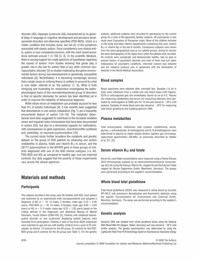
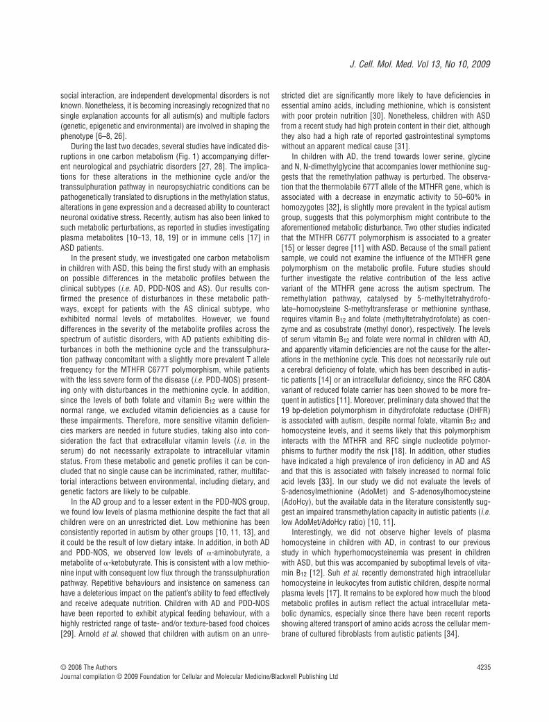
While inborn errors of metabolism can probably account for lessthan 5% of autistic individuals [9], it has recently been suggestedthat disturbances in one carbon metabolism (Fig. 1) are a frequentlyencountered feature of autism [10–19]. The metabolic distur-bances have been suggested to contribute to the increased oxidativestress and impaired redox homeostasis that have been documentedin autism [20], but also to a diminished capacity for methylation,with consequences on gene expression, neurotransmitter synthesisand, potentially, on neuronal synchronization [16].
The current study further broadens the metabolic and geneticdataset on the group of ASD patients by assessing one carbonmetabolites in plasma, folate and vitamin B12 in serum, and theC677T polymorphism in the MTHFR gene in three groups of chil-dren diagnosed with one of the ASD clinical subtypes (i.e. AD,PDD-NOS and AS) as compared to healthy age- and sex-matchedcontrols. Our data suggest that the severity of these impairmentsvary across the clinical spectrum.
Materials and methods
Participants
The subjects enrolled in this study were 39 children with ASD. Each patientwas examined by an experienced child neuropsychiatrist and assigned adiagnosis of AD (n � 15; 13 males, 2 females; mean age: 5.10 � 0.45years), PDD-NOS (n � 19; 13 males, 6 females; mean age: 8.83 � 0.84years) or AS (n � 5; 5 males; mean age: 9.23 � 1.82 years) based on thecriteria defined in the Diagnostic and Statistical Manual of MentalDisorders, Fourth Edition (DSM-IVR) [2]. Patients with childhood disinte-grative disorder or rare syndromes displaying autistic features wereexcluded from participation. Patients in each of the three ASDs subgroupswere matched by age and sex with healthy children from a pool of 25 indi-viduals, as follows: 13 controls for the AD group, 22 controls for the PDD-NOS group and 8 controls for the AS group (see Table 1). For the genetic
analysis, additional subjects were recruited for genotyping for the controlgroup for a total of 80 apparently healthy subjects. All participants in thisstudy were Caucasians of Romanian origin. None of the children includedin this study had taken vitamin supplements (containing folic acid, vitaminB12 or vitamin B6) in the last 6 months. Comparison subjects were drawnfrom the same geographical area as our patient groups, aiming to recoverthe same demographics of the region from where the patients were recruited.All controls were somatically and behaviourally healthy, had no past orpresent history of psychiatric disorder and none of them had ever takenmedications for psychiatric conditions. Informed consent was obtainedand the research protocol was in agreement with the Declaration ofHelsinki of the World Medical Association.
Blood samples
Blood specimens were obtained after overnight fast. Samples (~6 ml intotal) were withdrawn from a cubital vein into blood tubes (with heparin,EDTA or anticoagulant gel) and immediately stored on ice at 4�C. Plasma(for measuring metabolites) and serum (for measuring vitamins) were sep-arated by centrifugation at 3000 rpm for 10 min and stored at �20�C untilanalysis. Samples of whole blood were also stored at �20�C for measuringtotal blood glutathione and isolating genomic DNA.
Plasma metabolites
Total homocysteine, methionine, total cysteine, cystathionine, serine,glycine, �-aminobutyrate, N-methylglycine and N, N-dimethylglycine weredetermined in plasma by stable isotope dilution capillary gas chromatog-raphy/mass spectrometry (GS-MS), as previously described by Stabler et al. [21, 22].
Serum vitamin B12 and folate
Serum B12 and folate concentrations were measured using a Roche Elecsys2010 immunoassay analyser by an electrochemiluminiscence immunoas-say (ECLIA) using the Elecsys Vitamin B12 reagent kit and the Elecsys Folatereagent kit (Roche Diagnostics Gmbh, Mannheim, Germany). The assayswere performed according to the supplier’s recommendation.
Whole blood total glutathione
Total blood glutathione (tGSH) was measured in whole blood by isocraticRP-HPLC with precolumn derivatization and fluorimetric detection usingthe specific Chromsystems kit (Instruments and Chemical Gmbh,München, Germany). The assay was performed according to the supplier’srecommendation.
Genetic analysis
Genomic DNA was isolated from whole peripheral blood using the QIAampDNA Blood Mini Kit (Qiagen, Hilden, Germany) and was stored at �20�C untilfurther analysis. The genetic polymorphism was determined by using theLightCycler Real-Time PCR technology based on fluorescence resonance energy
© 2008 The AuthorsJournal compilation © 2009 Foundation for Cellular and Molecular Medicine/Blackwell Publishing Ltd
4231
transfer. MTHFR C677T polymorphism analysis was performed according tothe method described by Aslanidis et al. [23], using the LightCyclerTM high-speed thermal cycler (Hoffmann-LaRoche, Basel, Swizerland) and theLightCyclerTM DNA Master HybProbe kit (Roche Molecular Biochemicals,Mannheim, Germany). Melting curve analysis allowed identification of theMTHFR 677 polymorphism according to the following melting point tempera-tures: 63.1�C for the C allele and 55.2�C for the T allele.
Statistical analysis
The data are presented as means � standard error of the mean (S.E.M.).The normal distribution of continuous data was checked using theKolmogorov-Smirnov test. For normally distributed variables, Student’s
t-test was used for comparisons between the three ASD groups and theirage- and sex-matched controls. When normality was not present and equalvariance could not be assumed, the Mann–Whitney Rank-Sum Test wasapplied. Additionally, the effect size (Cohen’s d based on sample size) forvariables with a significant group mean difference was computed.Spearman’s rho test was used to evaluate the correlation between differentmetabolites. For the polymorphism under study, genotypic and allelic fre-quencies were calculated. The distribution of genotypes in all groups wastested for deviation from the Hardy–Weinberg equilibrium. The Pearson’schi-square (�2) test and Fisher’s exact test were applied to assess differ-ences in the genotype and allelic (respectively) distribution betweengroups of patients and controls. A P-value of less than 0.05 was consid-ered statistically significant. The SPSS software was used for performingthe analysis.
© 2008 The AuthorsJournal compilation © 2009 Foundation for Cellular and Molecular Medicine/Blackwell Publishing Ltd
Fig. 1 Overview of the one carbon metabolism pathway. Abbreviations: S-adenosylhomocysteine (AdoHcy); S-adenosylmethionine (AdoMet); betainehomocysteine methyltransferase (BHMT); cystathionine -synthase (CBS); cystathionine -lyase (CHT); dihydrofolate (DHF); dimethylglycine dehydro-genase (DMGDH); methionine synthase (MS), methylenetetrahydrofolate reductase (MTHFR); tetrahydrofolate (THF).
J. Cell. Mol. Med. Vol 13, No 10, 2009
4232
Results
Metabolic analyses
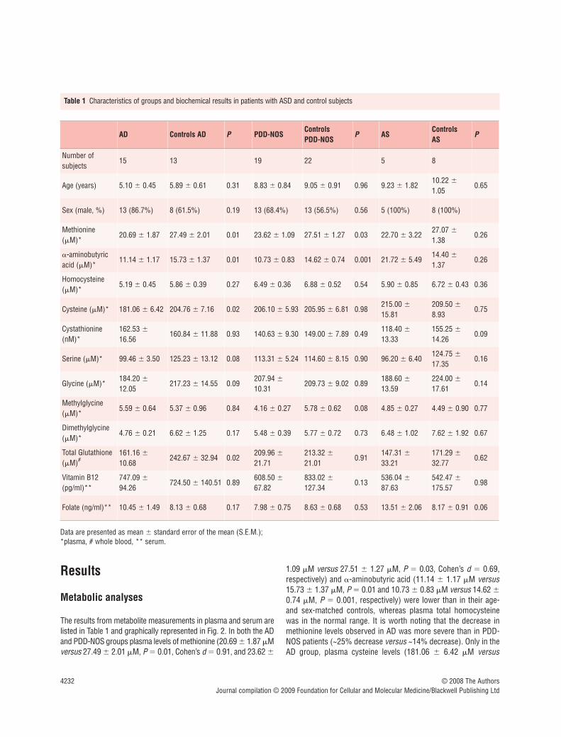
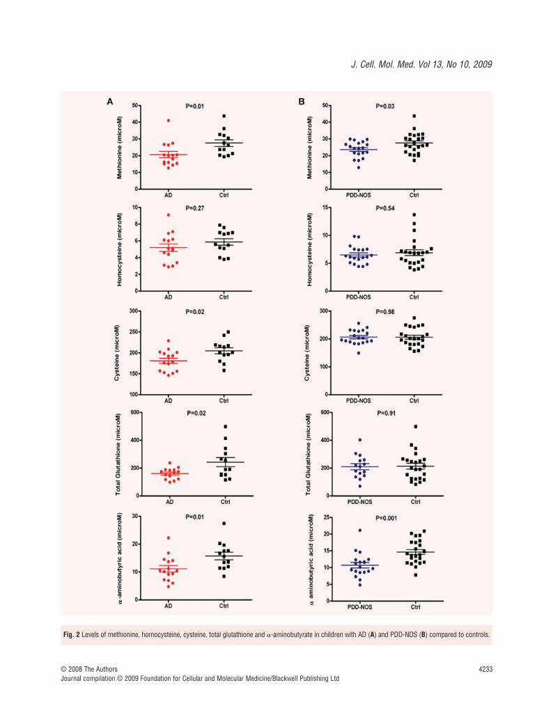
The results from metabolite measurements in plasma and serum arelisted in Table 1 and graphically represented in Fig. 2. In both the ADand PDD-NOS groups plasma levels of methionine (20.69 � 1.87 �Mversus 27.49 � 2.01 �M, P � 0.01, Cohen’s d � 0.91, and 23.62 �
1.09 �M versus 27.51 � 1.27 �M, P � 0.03, Cohen’s d � 0.69,respectively) and �-aminobutyric acid (11.14 � 1.17 �M versus15.73 � 1.37 �M, P � 0.01 and 10.73 � 0.83 �M versus 14.62 �0.74 �M, P � 0.001, respectively) were lower than in their age-and sex-matched controls, whereas plasma total homocysteinewas in the normal range. It is worth noting that the decrease inmethionine levels observed in AD was more severe than in PDD-NOS patients (~25% decrease versus ~14% decrease). Only in theAD group, plasma cysteine levels (181.06 � 6.42 �M versus
© 2008 The AuthorsJournal compilation © 2009 Foundation for Cellular and Molecular Medicine/Blackwell Publishing Ltd
AD Controls AD P PDD-NOSControls PDD-NOS
P ASControls AS
P
Number of subjects
15 13 19 22 5 8
Age (years) 5.10 � 0.45 5.89 � 0.61 0.31 8.83 � 0.84 9.05 � 0.91 0.96 9.23 � 1.8210.22 �
1.050.65
Sex (male, %) 13 (86.7%) 8 (61.5%) 0.19 13 (68.4%) 13 (56.5%) 0.56 5 (100%) 8 (100%)
Methionine(�M)*
20.69 � 1.87 27.49 � 2.01 0.01 23.62 � 1.09 27.51 � 1.27 0.03 22.70 � 3.2227.07 �
1.380.26
�-aminobutyricacid (�M)*
11.14 � 1.17 15.73 � 1.37 0.01 10.73 � 0.83 14.62 � 0.74 0.001 21.72 � 5.4914.40 �
1.370.26
Homocysteine(�M)*
5.19 � 0.45 5.86 � 0.39 0.27 6.49 � 0.36 6.88 � 0.52 0.54 5.90 � 0.85 6.72 � 0.43 0.36
Cysteine (�M)* 181.06 � 6.42 204.76 � 7.16 0.02 206.10 � 5.93 205.95 � 6.81 0.98215.00 �
15.81209.50 �
8.930.75
Cystathionine(nM)*
162.53 �
16.56160.84 � 11.88 0.93 140.63 � 9.30 149.00 � 7.89 0.49
118.40 �
13.33155.25 �
14.260.09
Serine (�M)* 99.46 � 3.50 125.23 � 13.12 0.08 113.31 � 5.24 114.60 � 8.15 0.90 96.20 � 6.40124.75 �
17.350.16
Glycine (�M)*184.20 �
12.05217.23 � 14.55 0.09
207.94 �
10.31209.73 � 9.02 0.89
188.60 �
13.59224.00 �
17.610.14
Methylglycine(�M)*
5.59 � 0.64 5.37 � 0.96 0.84 4.16 � 0.27 5.78 � 0.62 0.08 4.85 � 0.27 4.49 � 0.90 0.77
Dimethylglycine(�M)*
4.76 � 0.21 6.62 � 1.25 0.17 5.48 � 0.39 5.77 � 0.72 0.73 6.48 � 1.02 7.62 � 1.92 0.67
Total Glutathione(�M)#
161.16 �
10.68242.67 � 32.94 0.02
209.96 �
21.71213.32 �
21.010.91
147.31 �
33.21171.29 �
32.770.62
Vitamin B12(pg/ml)**
747.09 �
94.26724.50 � 140.51 0.89
608.50 �
67.82833.02 �
127.340.13
536.04 �
87.63542.47 �
175.570.98
Folate (ng/ml)** 10.45 � 1.49 8.13 � 0.68 0.17 7.98 � 0.75 8.63 � 0.68 0.53 13.51 � 2.06 8.17 � 0.91 0.06
Table 1 Characteristics of groups and biochemical results in patients with ASD and control subjects
Data are presented as mean � standard error of the mean (S.E.M.);*plasma, # whole blood, ** serum.
J. Cell. Mol. Med. Vol 13, No 10, 2009
4233© 2008 The AuthorsJournal compilation © 2009 Foundation for Cellular and Molecular Medicine/Blackwell Publishing Ltd
Fig. 2 Levels of methionine, homocysteine, cysteine, total glutathione and �-aminobutyrate in children with AD (A) and PDD-NOS (B) compared to controls.
4234
204.76 � 7.16 �M, P � 0.02; Cohen’s d � 0.91) and total bloodglutathione (161.16 � 10.68 �M versus 242.67 � 32.94 �M, P � 0.02; Cohen’s d � 0.89) were found to be lower. In contrast,the plasma levels of other amino acids, such as cystathionine, serine, glycine, N-methylglycine and N, N-dimethylglycine did notdiffer significantly from those of normal controls in any of the ASDgroups, although there was a trend towards lower levels of serine,glycine and N, N-dimethylglycine in the AD patients (Table 1). Thelevels of serum vitamin B12 were in the normal range of values inall ASDs groups; only 3 children with AD had levels below 250 pg/ml, but all subjects with a PDD-NOS diagnosis had levelsabove this suboptimal value. Serum folate levels were also in thenormal range, except for mildly higher levels in AS which reached a borderline statistical significance (13.51 � 2.06 ng/ml versus8.17 � 0.91 ng/ml, P � 0.06). However, in the context of thesmall group size, this apparent difference in B12 levels needs to betreated with caution.
We also examined the correlations between metabolite levels inthe three groups of ASDs patients. In AD patients we found a pos-itive correlation between tHcy levels and cysteine (rho � 0.54, P � 0.04), cystathionine (rho � 0.56, P � 0.03) and total bloodglutathione (rho � 0.48, P � 0.08); methionine was negativelycorrelated with cysteine, but statistical significance was notreached (rho � �0.30, P � 0.27). In the AD group, we alsoobserved a negative correlation between levels of N, N-dimethyl-glycine and cysteine (rho � �0.55, P � 0.03) and a positive cor-relation between N-methylglycine and serum folate (rho � 0.44, P � 0.09). Only in the PDD-NOS group a strong positive correla-tion was noticed between methionine and �-aminobutyric acid(rho � 0.66, P � 0.002).
Genotyping results
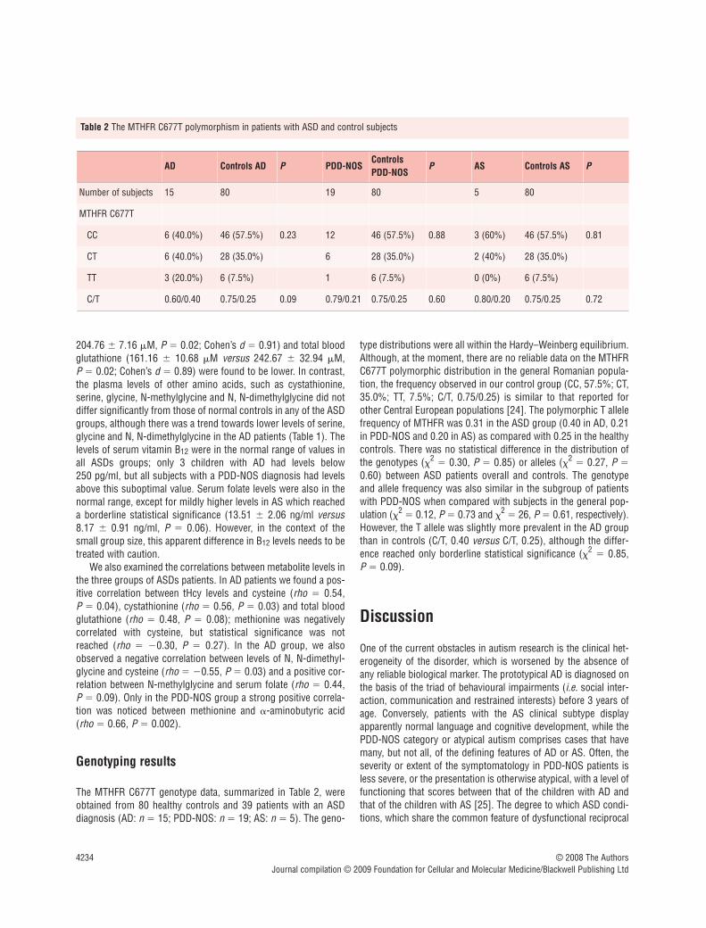
The MTHFR C677T genotype data, summarized in Table 2, wereobtained from 80 healthy controls and 39 patients with an ASDdiagnosis (AD: n � 15; PDD-NOS: n � 19; AS: n � 5). The geno-
type distributions were all within the Hardy–Weinberg equilibrium.Although, at the moment, there are no reliable data on the MTHFRC677T polymorphic distribution in the general Romanian popula-tion, the frequency observed in our control group (CC, 57.5%; CT,35.0%; TT, 7.5%; C/T, 0.75/0.25) is similar to that reported forother Central European populations [24]. The polymorphic T allelefrequency of MTHFR was 0.31 in the ASD group (0.40 in AD, 0.21in PDD-NOS and 0.20 in AS) as compared with 0.25 in the healthycontrols. There was no statistical difference in the distribution ofthe genotypes (�2
� 0.30, P � 0.85) or alleles (�2� 0.27, P �
0.60) between ASD patients overall and controls. The genotypeand allele frequency was also similar in the subgroup of patientswith PDD-NOS when compared with subjects in the general pop-ulation (�2
� 0.12, P � 0.73 and �2� 26, P � 0.61, respectively).
However, the T allele was slightly more prevalent in the AD groupthan in controls (C/T, 0.40 versus C/T, 0.25), although the differ-ence reached only borderline statistical significance (�2
� 0.85, P � 0.09).
Discussion
One of the current obstacles in autism research is the clinical het-erogeneity of the disorder, which is worsened by the absence ofany reliable biological marker. The prototypical AD is diagnosed onthe basis of the triad of behavioural impairments (i.e. social inter-action, communication and restrained interests) before 3 years ofage. Conversely, patients with the AS clinical subtype displayapparently normal language and cognitive development, while thePDD-NOS category or atypical autism comprises cases that havemany, but not all, of the defining features of AD or AS. Often, theseverity or extent of the symptomatology in PDD-NOS patients isless severe, or the presentation is otherwise atypical, with a level offunctioning that scores between that of the children with AD andthat of the children with AS [25]. The degree to which ASD condi-tions, which share the common feature of dysfunctional reciprocal
© 2008 The AuthorsJournal compilation © 2009 Foundation for Cellular and Molecular Medicine/Blackwell Publishing Ltd
AD Controls AD P PDD-NOSControlsPDD-NOS
P AS Controls AS P
Number of subjects 15 80 19 80 5 80
MTHFR C677T
CC 6 (40.0%) 46 (57.5%) 0.23 12 46 (57.5%) 0.88 3 (60%) 46 (57.5%) 0.81
CT 6 (40.0%) 28 (35.0%) 6 28 (35.0%) 2 (40%) 28 (35.0%)
TT 3 (20.0%) 6 (7.5%) 1 6 (7.5%) 0 (0%) 6 (7.5%)
C/T 0.60/0.40 0.75/0.25 0.09 0.79/0.21 0.75/0.25 0.60 0.80/0.20 0.75/0.25 0.72
Table 2 The MTHFR C677T polymorphism in patients with ASD and control subjects
J. Cell. Mol. Med. Vol 13, No 10, 2009
4235
social interaction, are independent developmental disorders is notknown. Nonetheless, it is becoming increasingly recognized that nosingle explanation accounts for all autism(s) and multiple factors(genetic, epigenetic and environmental) are involved in shaping thephenotype [6–8, 26].
During the last two decades, several studies have indicated dis-ruptions in one carbon metabolism (Fig. 1) accompanying differ-ent neurological and psychiatric disorders [27, 28]. The implica-tions for these alterations in the methionine cycle and/or thetranssulphuration pathway in neuropsychiatric conditions can bepathogenetically translated to disruptions in the methylation status,alterations in gene expression and a decreased ability to counteractneuronal oxidative stress. Recently, autism has also been linked tosuch metabolic perturbations, as reported in studies investigatingplasma metabolites [10–13, 18, 19] or in immune cells [17] inASD patients.
In the present study, we investigated one carbon metabolismin children with ASD, this being the first study with an emphasison possible differences in the metabolic profiles between theclinical subtypes (i.e. AD, PDD-NOS and AS). Our results con-firmed the presence of disturbances in these metabolic path-ways, except for patients with the AS clinical subtype, whoexhibited normal levels of metabolites. However, we found differences in the severity of the metabolite profiles across thespectrum of autistic disorders, with AD patients exhibiting dis-turbances in both the methionine cycle and the transsulphura-tion pathway concomitant with a slightly more prevalent T allelefrequency for the MTHFR C677T polymorphism, while patientswith the less severe form of the disease (i.e. PDD-NOS) present-ing only with disturbances in the methionine cycle. In addition,since the levels of both folate and vitamin B12 were within thenormal range, we excluded vitamin deficiencies as a cause forthese impairments. Therefore, more sensitive vitamin deficien-cies markers are needed in future studies, taking also into con-sideration the fact that extracellular vitamin levels (i.e. in theserum) do not necessarily extrapolate to intracellular vitaminstatus. From these metabolic and genetic profiles it can be con-cluded that no single cause can be incriminated, rather, multifac-torial interactions between environmental, including dietary, andgenetic factors are likely to be culpable.
In the AD group and to a lesser extent in the PDD-NOS group,we found low levels of plasma methionine despite the fact that allchildren were on an unrestricted diet. Low methionine has beenconsistently reported in autism by other groups [10, 11, 13], andit could be the result of low dietary intake. In addition, in both ADand PDD-NOS, we observed low levels of �-aminobutyrate, ametabolite of �-ketobutyrate. This is consistent with a low methio-nine input with consequent low flux through the transsulphurationpathway. Repetitive behaviours and insistence on sameness canhave a deleterious impact on the patient’s ability to feed effectivelyand receive adequate nutrition. Children with AD and PDD-NOShave been reported to exhibit atypical feeding behaviour, with ahighly restricted range of taste- and/or texture-based food choices[29]. Arnold et al. showed that children with autism on an unre-
stricted diet are significantly more likely to have deficiencies inessential amino acids, including methionine, which is consistentwith poor protein nutrition [30]. Nonetheless, children with ASDfrom a recent study had high protein content in their diet, althoughthey also had a high rate of reported gastrointestinal symptomswithout an apparent medical cause [31].
In children with AD, the trend towards lower serine, glycineand N, N-dimethylglycine that accompanies lower methionine sug-gests that the remethylation pathway is perturbed. The observa-tion that the thermolabile 677T allele of the MTHFR gene, which isassociated with a decrease in enzymatic activity to 50–60% inhomozygotes [32], is slightly more prevalent in the typical autismgroup, suggests that this polymorphism might contribute to theaforementioned metabolic disturbance. Two other studies indicatedthat the MTHFR C677T polymorphism is associated to a greater[15] or lesser degree [11] with ASD. Because of the small patientsample, we could not examine the influence of the MTHFR genepolymorphism on the metabolic profile. Future studies should further investigate the relative contribution of the less active variant of the MTHFR gene across the autism spectrum. Theremethylation pathway, catalysed by 5-methyltetrahydrofo-late–homocysteine S-methyltransferase or methionine synthase,requires vitamin B12 and folate (methyltetrahydrofolate) as coen-zyme and as cosubstrate (methyl donor), respectively. The levelsof serum vitamin B12 and folate were normal in children with AD,and apparently vitamin deficiencies are not the cause for the alter-ations in the methionine cycle. This does not necessarily rule outa cerebral deficiency of folate, which has been described in autis-tic patients [14] or an intracellular deficiency, since the RFC C80Avariant of reduced folate carrier has been showed to be more fre-quent in autistics [11]. Moreover, preliminary data showed that the19 bp-deletion polymorphism in dihydrofolate reductase (DHFR)is associated with autism, despite normal folate, vitamin B12 andhomocysteine levels, and it seems likely that this polymorphisminteracts with the MTHFR and RFC single nucleotide polymor-phisms to further modify the risk [18]. In addition, other studieshave indicated a high prevalence of iron deficiency in AD and ASand that this is associated with falsely increased to normal folicacid levels [33]. In our study we did not evaluate the levels of S-adenosylmethionine (AdoMet) and S-adenosylhomocysteine(AdoHcy), but the available data in the literature consistently sug-gest an impaired transmethylation capacity in autistic patients (i.e.low AdoMet/AdoHcy ratio) [10, 11].
Interestingly, we did not observe higher levels of plasmahomocysteine in children with AD, in contrast to our previousstudy in which hyperhomocysteinemia was present in childrenwith ASD, but this was accompanied by suboptimal levels of vita-min B12 [12]. Suh et al. recently demonstrated high intracellularhomocysteine in leukocytes from autistic children, despite normalplasma levels [17]. It remains to be explored how much the bloodmetabolic profiles in autism reflect the actual intracellular meta-bolic dynamics, especially since there have been recent reportsshowing altered transport of amino acids across the cellular mem-brane of cultured fibroblasts from autistic patients [34].
© 2008 The AuthorsJournal compilation © 2009 Foundation for Cellular and Molecular Medicine/Blackwell Publishing Ltd
4236
What is particularly novel about our results is the fact that onlychildren with typical autism (i.e. AD), but not PDD-NOS, haveimpairments in the transsulphuration pathway. More specifically,only children with AD had lower levels of plasma cysteine and totalblood glutathione. Interestingly, the levels of cystathionine werewithin the normal range. Low levels of cysteine and glutathioneaccompanied by higher levels of cystathionine have also beendescribed in the ASD group investigated by James et al. [11], andit could indicate a defect at the level of cystathionine gamma-lyase(CTH), a pyridoxal 5�-phosphate dependent enzyme that convertscystathionine to cysteine in the transsulphuration pathway.Another recent investigation reported that leukocytes from autisticchildren exhibit decreased intracellular cysteine and total glu-tathione, and unlike the findings in plasma, show a significant(80%) increase in intracellular homocysteine [17]. While in severalstudies children with ASD exhibited higher levels of vitamin B6
than the control subjects [35–37], the activity of pyridoxal kinaseand the levels of pyridoxal 5�-phosphate had been measured andfound to be generally much lower than levels in control subjects[37]. It has also been suggested that the reduced flux throughtranssulphuration in autism, with a consequent diminished glu-tathione pool, could also be due to the high levels of androgensdescribed in these patients [13, 38] since testosterone modulatesthe activity of CBS [39, 40]. The low levels of cysteine and glu-tathione, the decrease in the ratio of reduced to oxidized glu-tathione in plasma and the decreased ability to form sulphatedmetabolites in autistic patients [41] suggest that antioxidantdefences are compromised in these patients [20]. Glutathione isalso very important to heavy metal detoxification [42], and thevariations in glutathione abnormalities across the ASD suggeststhe possibility that it might be functionally significant since it wasshown that mercury-associated urinary porphyrin levels increasewith increasing severity of ASDs [43–45]. Finally, it is important todiscuss the limitations of our study. First, the number of subjectswas relatively small and, unfortunately, no quantitative standard-ized scale for autism has been applied. Second, being a retrospec-tive study it does not provide information about the dynamics ofmetabolite changes associated with different stages of develop-ment and/or in response to different dietary supplements (i.e.betaine, folic acid, vitamin B6, vitamin B12). By the same token, aninteresting aspect of this study is that it represents the first inves-
tigation of one carbon metabolism in children with different sub-types of ASD who are free of vitamin supplements.
In conclusion, our results indicate disturbances in both themethionine cycle and the transsulphuration pathway in childrenwith AD, which are most likely multifactorial in origin.Interestingly, children with milder clinical forms of ASD displaymilder metabolic perturbations. To date, only a few studies havespecifically reported biological correlates of different clinical sub-types in the autistic spectrum (e.g. [46, 47]). The methylationdeficit resulting from an altered methionine cycle supports thepreviously suggested contribution of epigenetic mechanisms tothe pathogenesis of autism [26]. Aside from this ‘metabolic repro-gramming’ of gene expression, dysfunctionalities in the synthesisof neurotransmitters and phospholipids would also result [28,48]. Genomic imprinting is the classic example of regulation ofgene expression via epigenetic modifications that leads to parentof origin-specific gene expression. From this perspective, the dis-turbances in the methionine cycle and the ensuing methylationdeficit in AD patients support the imprinting hypothesis for thedevelopment of autism, which emphasizes the imbalances in braindevelopment due to enhanced effects of paternally expressedimprinted genes [49]. In addition, disturbances in the methioninecycle have been hypothesized to contribute to alterations in neu-ronal synchronization in autism [16, 50]. On the other hand, thelow levels of glutathione, resulting from the decreased flowthrough transsulphuration in AD, is indicative of an imbalance inthe redox homeostasis, and links oxidative stress with membranelipid abnormalities, inflammation and aberrant immune responsethat have been observed in autism [20].
Acknowledgements
This work was supported by grants from the National Institutes ofHealth (DK64959 to R.B.), funding from the Jonty Foundation (to R.B.)and by a CEEX grant (83/2006) received from the Romanian Academyof Medical Sciences (to M.D.) The authors acknowledge the assaysdone in the Hematology Division laboratories of Robert Allen and SallyStabler at the University of Colorado Health Sciences Center in Denver,Colorado.
© 2008 The AuthorsJournal compilation © 2009 Foundation for Cellular and Molecular Medicine/Blackwell Publishing Ltd
References
1. Volkmar FR, Pauls D. Autism. Lancet.2003; 362: 1133–41.
2. American Psychiatric Association.Diagnostic and Statistical Manual-Text Revision. 4th ed. Washington, DC: American Psychiatric Association;2000.
3. Autism and Developmental DisabilitiesMonitoring Network Surveillance Year2002 Principal Investigators, Centers for
Disease Control and Prevention.Prevalence of autism spectrum disorders-autism and developmental disabilitiesmonitoring network, 14 sites, UnitedStates, 2002. MMWR Surveill Summ.2007; 56: 12–28.
4. Baird G, Simonoff E, Pickles A, et al.Prevalence of disorders of the autism spectrum in a population cohort of children in South Thames: the Special Needs
and Autism Project (SNAP). Lancet. 2006;368: 210–5.
5. Gupta AR, State MW. Recent advances inthe genetics of autism. Biol Psychiatry.2007; 61: 429–37.
6. Persico AM, Bourgeron T. Searching for ways out of the autism maze: genetic, epigenetic and environmentalclues. Trends Neurosci. 2006; 29: 349–58.
J. Cell. Mol. Med. Vol 13, No 10, 2009
4237© 2008 The AuthorsJournal compilation © 2009 Foundation for Cellular and Molecular Medicine/Blackwell Publishing Ltd
7. Geschwind DH, Levitt P. Autism spectrumdisorders: developmental disconnectionsyndromes. Curr Opin Neurobiol. 2007;17: 103–11.
8. Happe F, Ronald A, Plomin R. Time togive up on a single explanation for autism.Nat Neurosci. 2006; 9: 1218–20.
9. Manzi B, Loizzo AL, Giana G, et al.Autism and metabolic diseases. J ChildNeurol. 2008; 23: 307–14.
10. James SJ, Cutler P, Melnyk S, et al.Metabolic biomarkers of increased oxida-tive stress and impaired methylationcapacity in children with autism. Am J ClinNutr. 2004; 80: 1611–7.
11. James SJ, Melnyk S, Jernigan S, et al.Metabolic endophenotype and relatedgenotypes are associated with oxidativestress in children with autism. Am J MedGenet B Neuropsychiatr Genet. 2006; 141:947–56.
12. Pasca SP, Nemes B, Vlase L, et al. Highlevels of homocysteine and low serumparaoxonase 1 arylesterase activity in chil-dren with autism. Life Sci. 2006; 78:2244–8.
13. Geier DA, Geier MR. A clinical and labora-tory evaluation of methionine cycle-transsulfuration and androgen pathwaymarkers in children with autistic disorders.Horm Res. 2006; 66: 182–8.
14. Moretti P, Peters SU, Del Gaudio D, et al.Brief report: autistic symptoms, develop-mental regression, mental retardation,epilepsy, and dyskinesias in CNS folatedeficiency. J Autism Dev Disord. 2008; 38:1170–7.
15. Boris M, Goldblatt A, Galanko J, et al. Association of MTHFR gene variantswith autism. J Am Phys Surg. 2004; 9:106–8.
16. Deth R, Muratore C, Benzecry J, et al.How environmental and genetic factorscombine to cause autism: A redox/ methylation hypothesis. Neurotoxicology.2008; 29: 190–201.
17. Suh J, Walsh W, McGinnis W, et al.Altered sulfur amino acid metabolism inimmune cells of children diagnosed withautism. Am J Biochem Biotech. 2008; 4:105–13.
18. Adams M, Lucock M, Stuart J, et al.Preliminary evidence for involvement ofthe folate gene polymorphism 19bp dele-tion-DHFR in occurrence of autism.Neurosci Lett. 2007; 422: 24–9.
19. Geier DA, Kern JK, Garver CR, et al. Aprospective study of transsulfuration bio-markers in autistic disorders. NeurochemRes. 2009; 34: 394.
20. Chauhan A, Chauhan V. Oxidative stress inautism. Pathophysiology. 2006; 13:171–81.
21. Stabler SP, Lindenbaum J, Savage DG, et al. Elevation of serum cystathionine lev-els in patients with cobalamin and folatedeficiency. Blood. 1993; 81: 3404–13.
22. Stabler SP, Marcell PD, Podell ER, et al.Elevation of total homocysteine in theserum of patients with cobalamin or folatedeficiency detected by capillary gas chro-matography-mass spectrometry. J ClinInvest. 1988; 81: 466–74.
23. Aslanidis C, Nauck M, Schmitz G. High-speed prothrombin G– A 20210 andmethylenetetrahydrofolate reductaseC– T 677 mutation detection using real-time fluorescence PCR and melting curves.Biotechniques. 1999; 27: 234–6, 8.
24. Moore LE, Hung R, Karami S, et al. Folatemetabolism genes, vegetable intake andrenal cancer risk in central Europe. Int JCancer. 2008; 122: 1710–5.
25. Walker DR, Thompson A, ZwaigenbaumL, et al. Specifying PDD-NOS: a compari-son of PDD-NOS, Asperger syndrome, and autism. J Am Acad Child AdolescPsychiatry. 2004; 43: 172–80.
26. Schanen NC. Epigenetics of autism spec-trum disorders. Hum Mol Genet. 2006; 2:R138–50.
27. Sugden C. One-carbon metabolism in psy-chiatric illness. Nutr Res Rev. 2006; 19:117–36.
28. Muskiet FA, Kemperman RF. Folate andlong-chain polyunsaturated fatty acids inpsychiatric disease. J Nutr Biochem. 2006;17: 717–27.
29. Ahearn WH, Castine T, Nault K, et al. Anassessment of food acceptance in childrenwith autism or pervasive developmentaldisorder-not otherwise specified. J AutismDev Disord. 2001; 31: 505–11.
30. Arnold GL, Hyman SL, Mooney RA, et al.Plasma amino acids profiles in childrenwith autism: potential risk of nutritionaldeficiencies. J Autism Dev Disord. 2003;33: 449–54.
31. Levy SE, Souders MC, Ittenbach RF, et al. Relationship of dietary intake to gastrointestinal symptoms in children with autistic spectrum disorders. BiolPsychiatry. 2007; 61: 492–7.
32. Rozen R. Genetic predisposition to hyper-homocysteinemia: deficiency of methyl-enetetrahydrofolate reductase (MTHFR).Thromb Haemost. 1997; 78: 523–6.
33. Latif A, Heinz P, Cook R. Iron deficiency inautism and Asperger syndrome. Autism.2002; 6: 103–14.
34. Fernell E, Karagiannakis A, Edman G, et al. Aberrant amino acid transport infibroblasts from children with autism.Neurosci Lett. 2007; 418: 82–6.
35. Siva Sankar DV. Plasma levels of folates,riboflavin, vitamin B6, and ascorbate inseverely disturbed children. J Autism DevDisord. 1979; 9: 73–82.
36. Adams JB, George F, Audhya T.Abnormally high plasma levels of vitaminB6 in children with autism not taking sup-plements compared to controls not takingsupplements. J Altern Complement Med.2006; 12: 59–63.
37. Adams JB, Holloway C. Pilot study of amoderate dose multivitamin/mineral sup-plement for children with autistic spectrumdisorder. J Altern Complement Med. 2004;10: 1033–9.
38. Geier DA, Geier MR. A prospectiveassessment of androgen levels in patientswith autistic spectrum disorders: biochem-ical underpinnings and suggested thera-pies. Neuro Endocrinol Lett. 2007; 28:565–73.
39. Prudova A, Albin M, Bauman Z, et al.Testosterone regulation of homocysteinemetabolism modulates redox status inhuman prostate cancer cells. AntioxidRedox Signal. 2007; 9: 1875–81.
40. Vitvitsky V, Prudova A, Stabler S, et al.Testosterone regulation of renal cystathio-nine beta-synthase: implications for sex-dependent differences in plasma homocys-teine levels. Am J Physiol Renal Physiol.2007; 293: F594–600.
41. Alberti A, Pirrone P, Elia M, et al.Sulphation deficit in “low-functioning”autistic children: a pilot study. BiolPsychiatry. 1999; 46: 420–4.
42. Clarkson TW, Nordberg GF, Sager PR.Reproductive and developmental toxicityof metals. Scand J Work Environ Health.1985; 11: 145–54.
43. Nataf R, Skorupka C, Amet L, et al.Porphyrinuria in childhood autistic disorder: implications for environmentaltoxicity. Toxicol Appl Pharmacol. 2006;214: 99–108.
44. Geier DA, Geier MR. A prospective studyof mercury toxicity biomarkers in autisticspectrum disorders. J Toxicol EnvironHealth A. 2007; 70: 1723–30.
45. Geier DA, Geier MR. A prospectiveassessment of porphyrins in autistic disor-ders: a potential marker for heavy metalexposure. Neurotox Res. 2006; 10: 57–64.
46. Boger-Megiddo I, Shaw DW, FriedmanSD, et al. Corpus callosum morphometricsin young children with autism spectrum
4238 © 2008 The AuthorsJournal compilation © 2009 Foundation for Cellular and Molecular Medicine/Blackwell Publishing Ltd
disorder. J Autism Dev Disord. 2006; 36:733–9.
47. Connolly AM, Chez M, Streif EM, et al. Brain-derived neurotrophic factorand autoantibodies to neural antigens insera of children with autistic spectrum disorders, Landau-Kleffner syndrome, and
epilepsy. Biol Psychiatry. 2006; 59:354–63.
48. Van den Veyver IB. Genetic effects ofmethylation diets. Annu Rev Nutr. 2002;22: 255–82.
49. Badcock C, Crespi B. Imbalanced genomic imprinting in brain development:
an evolutionary basis for the aetiology of autism. J Evol Biol. 2006; 19: 1007–32.
50. Uhlhaas PJ, Singer W. What do disturbances in neural synchrony tell usabout autism? Biol Psychiatry. 2007; 62:190–1.
Related Documents