Role of HER2/neu in tumor progression and therapy S. Ménard*, P. Casalini, M. Campiglio, S. M. Pupa and E. Tagliabue Molecular Targeting Unit, Department of Experimental Oncology, Istituto Nazionale Tumori, Via Venezian 1, 20133 Milano (Italy), Fax: +39 02 23903073, e-mail: [email protected] Abstract. HER2 (human epidermal growth factor recep- tor-2; also known as erbB2) and its relatives HER1 (epi- dermal growth factor receptor; EGFR), HER3 and HER4 belong to the HER family of receptor tyrosine kinases. In normal cells, activation of this receptor tyrosine kinase family triggers a rich network of signaling pathways that control normal cell growth, differentiation, motility and adhesion in several cell lineages. The first tumor studied for an alteration of the HER2 oncogene is breast carci- noma, and so far the majority of studies have been per- formed on this oncotype. Although involvement of HER2 as a cause of human cell transformation needs to be fur- ther investigated, overexpression of the HER2 oncogene in human breast carcinomas has been associated with a CMLS, Cell. Mol. Life Sci. 61 (2004) 2965–2978 1420-682X/04/232965-14 DOI 10.1007/s00018-004-4277-7 © Birkhäuser Verlag, Basel, 2004 CMLS Cellular and Molecular Life Sciences more aggressive course of disease. It has been suggested that this association depends on HER2-driven prolifera- tion, vessel formation and/or invasiveness; however, poor prognosis may not be directly related to the presence of the oncoprotein on the cell membrane but instead to the breast carcinoma subset identified by HER2 overexpres- sion and characterized by a peculiar gene expression pro- file, as recently identified. HER2-positive tumors were recently shown to benefit from anthracyclin treatment and to be resistant to endocrine therapy. Despite the fact that many pathways interacting with HER2 are still not fully understood, this tyrosine kinase receptor is, to date, a promising molecule for targeted therapy. Key words. HER2; biological role; prognostic value; predictive value; therapy. Description of the normal counterpart and of its normal function HER2 (human epidermal growth factor receptor-2; also known as erbB2) and its related HER1 (epidermal growth factor receptor; EGFR), HER3 and HER4 belong to the HER family of receptor tyrosine kinases. Individual fea- tures of this group of receptors include the ligand-less HER2 receptor and the kinase-dead HER3 receptors [1–4]. A large family of ligands (reviewed in [5]) induces receptor dimerization, with each ligand favoring some dimeric combinations over others in a specific hierarchi- cal order, although a marked preference for HER2 as a dimer partner has been described [6, 7]. Expression patterns of HER receptors and their ligands, as well as targeted inactivation of components of the HER signaling network, have highlighted the importance of short-range ligand-receptor interactions, especially in mid-gestation processes. Apparently, the network is in- volved primarily in two types of interactions: (i) mes- * Corresponding author. enchyme-epithelia cross-talk and (ii) neuronal effects on target cells, including muscle, astroglia, oligodendro- cytes and Schwann cells. NRGs (neuregulins, a family of HER3 and HER4 ligands), for example, are synthesized by mesenchymal or neuronal cells and influence adjacent epithelial or non-neuronal cells, respectively, with respect to their differentiation, proliferation and migration. This may explain the crucial role of the HER receptor family in development of the cardiovascular system, nervous system, mammary gland and probably others. An essential role for the HER receptor family in mid-ges- tation development was indicated by embryonic lethality of HER2- [8], HER4- [9] and NRG-deficient mice at around day 10 post-fertilization due to aberrant cardiac and peripheral nervous system development. The trabec- ulae, fingerlike extensions of the ventricular myocar- dium, fail to develop in these mice, and the resulting mu- tant heart is characterized by irregular beat, an enlarged common ventricle and reduced blood flow. HER3 knock- out mice have less severe heart defects and consequently survive several days longer (to around embryonic day 13.5), displaying normal heart trabeculation but defective

Welcome message from author
This document is posted to help you gain knowledge. Please leave a comment to let me know what you think about it! Share it to your friends and learn new things together.
Transcript
Role of HER2/neu in tumor progression and therapyS. Ménard*, P. Casalini, M. Campiglio, S. M. Pupa and E. Tagliabue
Molecular Targeting Unit, Department of Experimental Oncology, Istituto Nazionale Tumori, Via Venezian 1, 20133 Milano (Italy), Fax: +39 02 23903073, e-mail: [email protected]
Abstract. HER2 (human epidermal growth factor recep-tor-2; also known as erbB2) and its relatives HER1 (epi-dermal growth factor receptor; EGFR), HER3 and HER4belong to the HER family of receptor tyrosine kinases. Innormal cells, activation of this receptor tyrosine kinasefamily triggers a rich network of signaling pathways thatcontrol normal cell growth, differentiation, motility andadhesion in several cell lineages. The first tumor studiedfor an alteration of the HER2 oncogene is breast carci-noma, and so far the majority of studies have been per-formed on this oncotype. Although involvement of HER2as a cause of human cell transformation needs to be fur-ther investigated, overexpression of the HER2 oncogenein human breast carcinomas has been associated with a
CMLS, Cell. Mol. Life Sci. 61 (2004) 2965–29781420-682X/04/232965-14DOI 10.1007/s00018-004-4277-7© Birkhäuser Verlag, Basel, 2004
CMLS Cellular and Molecular Life Sciences
more aggressive course of disease. It has been suggestedthat this association depends on HER2-driven prolifera-tion, vessel formation and/or invasiveness; however, poorprognosis may not be directly related to the presence ofthe oncoprotein on the cell membrane but instead to thebreast carcinoma subset identified by HER2 overexpres-sion and characterized by a peculiar gene expression pro-file, as recently identified. HER2-positive tumors wererecently shown to benefit from anthracyclin treatmentand to be resistant to endocrine therapy. Despite the factthat many pathways interacting with HER2 are still notfully understood, this tyrosine kinase receptor is, to date,a promising molecule for targeted therapy.
Key words. HER2; biological role; prognostic value; predictive value; therapy.
Description of the normal counterpart and of its normal function
HER2 (human epidermal growth factor receptor-2; alsoknown as erbB2) and its related HER1 (epidermal growthfactor receptor; EGFR), HER3 and HER4 belong to theHER family of receptor tyrosine kinases. Individual fea-tures of this group of receptors include the ligand-lessHER2 receptor and the kinase-dead HER3 receptors[1–4]. A large family of ligands (reviewed in [5]) inducesreceptor dimerization, with each ligand favoring somedimeric combinations over others in a specific hierarchi-cal order, although a marked preference for HER2 as adimer partner has been described [6, 7]. Expression patterns of HER receptors and their ligands,as well as targeted inactivation of components of the HERsignaling network, have highlighted the importance ofshort-range ligand-receptor interactions, especially inmid-gestation processes. Apparently, the network is in-volved primarily in two types of interactions: (i) mes-
* Corresponding author.
enchyme-epithelia cross-talk and (ii) neuronal effects ontarget cells, including muscle, astroglia, oligodendro-cytes and Schwann cells. NRGs (neuregulins, a family ofHER3 and HER4 ligands), for example, are synthesizedby mesenchymal or neuronal cells and influence adjacentepithelial or non-neuronal cells, respectively, with respectto their differentiation, proliferation and migration. Thismay explain the crucial role of the HER receptor familyin development of the cardiovascular system, nervoussystem, mammary gland and probably others.An essential role for the HER receptor family in mid-ges-tation development was indicated by embryonic lethalityof HER2- [8], HER4- [9] and NRG-deficient mice ataround day 10 post-fertilization due to aberrant cardiacand peripheral nervous system development. The trabec-ulae, fingerlike extensions of the ventricular myocar-dium, fail to develop in these mice, and the resulting mu-tant heart is characterized by irregular beat, an enlargedcommon ventricle and reduced blood flow. HER3 knock-out mice have less severe heart defects and consequentlysurvive several days longer (to around embryonic day13.5), displaying normal heart trabeculation but defective
valve formation. The role of HER4 has been further es-tablished in HER4 knockout mice by re-expressingHER4 under the regulatory control of the cardiac a-myosin heavy chain (aMHC) promoter [10, 11]. Cardiac-rescued MHC-HER4 HER4–/– mice are viable but displayabnormalities in the central nervous system and mam-mary gland. The mid-gestation cardiac defect in HER2knockout animals has been circumvented through the useof a conditional mutagenesis approach. Conditional mu-tagenesis of the HER2 gene in murine ventricular car-diomyocytes [12] revealed development of severe dilatedcardiomyopathy, with signs of cardiac dysfunction gener-ally appearing by the second postnatal month. Based onthese findings, the authors concluded that signaling fromthe HER2 receptor, which is enriched in T-tubules in car-diomyocytes, is crucial for adult heart function. In light ofthe adverse cardiac side effects observed in breast cancerpatients treated with the monoclonal anti-HER2 antibodytrastuzumab [13], an improved understanding of the mol-ecular mechanisms by which HER2 regulates heart func-tion is especially important.NRGs exert many functions in neural development. Rolesfor NRG-1-HER signaling in neural development havebeen demonstrated in mice carrying an HER3-null muta-tion [14], as well as by selective mutations of NRG-1ectodomains [15, 16], by MHC-HER-specific expression[10, 11] and by inducible HER2 ablation in Schwann cells[17]. In all of these mice, peripheral motorneuron axonsdefasciculate as they enter the muscle mass and fail toform mature neuromuscular junctions. Aberrant cranialnerve architecture and increased numbers of large in-terneurons within the cerebellum have been demon-strated in MHC-HER4 HER4–/– mice, while loss of NRG-1-HER signaling led to hypoplasia of the sympatheticganglion chain and the neural crest-derived portion of thecranial sensory ganglia [14, 18–20]. Moreover, the NRG-1-HER mutants were completely devoid of Schwann cellsin peripheral nerves at late development stages [15, 21].Thus, NRG-1 signaling through the HER2/HER3 het-erodimer is required for normal Schwann cell develop-ment. The role of HER2 signaling in later development ofthe Schwann cell lineage was also analyzed using condi-tional mutagenesis [17]. In that study, the conditionalHER2 mutants displayed peripheral nerve hypomyelina-tion associated with neuropathy, a phenotype reminiscentof the pathology in patients with Charcot-Marie-Toothdisease [22]. Thus HER2 is the first signaling moleculefor which a role in control of Schwann cell myelinationhas been demonstrated in vivo. Moreover, Kim et al. havevery recently demonstrated that HER2 signaling is alsocritical for oligodendrocyte differentiation in vivo [23].To date however, no data have been reported on nervoussystem toxicity in patients treated with anti-HER2-tar-geted therapy. Nevertheless, patients treated long termshould be carefully monitored for potential side effects,
2966 S. Ménard et al. HER2
since a possible role for HER2 in mature nervous systemtissue cannot be excluded.In contrast to the embryonic lethality caused by HER2 in-activation, mice carrying a naturally occurring germ-linemutation in the kinase domain of EGFR knownas Waved-2 (hypomorphic allele with severely reduced catalytic ac-tivity) are completely viable and display only epithelialdefects, such as a wavy hair phenotype. Mutant mice dis-play impaired epithelial development in several organs,resulting in phenotypes ranging from peri-implantationdeath to live progeny with abnormalities in multiple or-gans, such as liver and skin, depending on the geneticbackground [24, 25]. The importance of the HER receptor family and ligandsin human mammary carcinoma has evoked keen interestin the normal functions of these receptors in the mam-mary gland, an organ that undergoes considerable postna-tal development. Analyses of HER family ligands inmammary development have revealed a complicated pic-ture. RNAs encoding the majority of the HER-specificgrowth factors such as AR (anphiregulin), BTC (betacel-lulin), HB-EGF (heparin-binding EGF), EPR (epireg-ulin), EGF (epidermal growth factor), NRG1 and TGFa(tumor growth factor a) are all present, each with aunique temporal pattern of transcriptional regulation dur-ing the normal course of mammary development, matu-ration and involution [26]. HERs play several normal non-oncogenic roles in regu-lating growth, differentiation, apoptosis and/or remodel-ing in normal mammary glands. These receptors are dif-ferentially expressed in mammary epithelial and/or stro-mal cells during various stages of development. In themouse virgin gland, HER1 and HER2 colocalize in allmajor cell types during ductal morphogenesis but local-ize differentially in the mature gland. EGFR and HER2are preferentially expressed in lactating ducts and alveoli,and HER3 and HER4 are more pronounced in alveoli[26]. Interestingly, a switch from HER3 to HER4 expres-sion was observed in the developing mammary gland,suggesting that the two receptors play different roles inmammary morphogenesis. Activated EGFR and HER2are highly expressed in extracts of mammary glands col-lected at puberty, suggesting a prominent role of these re-ceptors at this stage of development, while both are ex-pressed to only a minor extent in mammary glands inlate-stage pregnancy and in lactation [27]. By contrast,HER3 and HER4 are active in mammary glands mostlyduring pregnancy and lactation [28]. With respect to thesignaling pathways activated by HER2, Niemann et al.,[29] demonstrated that formation of branched tubules re-lies on a pathway involving PI-3K, whereas alveolar mor-phogenesis requires MAPK. Mammary functions of HER2 have also been assessedusing cytoplasmic, truncated dominant-negative form ofthe receptor, under the control of the mouse mammary
tumor virus (MMTV) promoter. Transgenic animals ex-pressing dominant-negative MMTV-driven truncatedHER2 have significant defects in mammary developmentlate in gestation and early postpartum, with failure ofalveolar expansion [30]. Ductal development occurs inthese animals, but they have lactation problems, andmammary glands early postpartum show an immaturephenotype more typical of late pregnancy [11]. Thesedata do not reveal any indispensable role for a particularmember of the family in mammary gland development.Moreover, the cross-talk between HERs and steroid hor-mone receptors in mammary gland development remainsto be established. It seems very likely that these two re-ceptor types act synergistically and that inhibition of bothpathways is required for complete ablation of mammarygland development.
What are the types of cancer related to the describedtyrosine kinase?
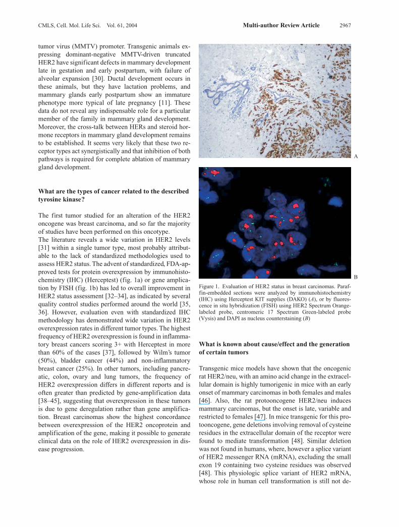
The first tumor studied for an alteration of the HER2oncogene was breast carcinoma, and so far the majorityof studies have been performed on this oncotype. The literature reveals a wide variation in HER2 levels[31] within a single tumor type, most probably attribut-able to the lack of standardized methodologies used to assess HER2 status. The advent of standardized, FDA-ap-proved tests for protein overexpression by immunohisto-chemistry (IHC) (Herceptest) (fig. 1a) or gene amplica-tion by FISH (fig. 1b) has led to overall improvement inHER2 status assessment [32–34], as indicated by severalquality control studies performed around the world [35,36]. However, evaluation even with standardized IHCmethodology has demonstrated wide variation in HER2overexpression rates in different tumor types. The highestfrequency of HER2 overexpression is found in inflamma-tory breast cancers scoring 3+ with Herceptest in morethan 60% of the cases [37], followed by Wilm’s tumor(50%), bladder cancer (44%) and non-inflammatorybreast cancer (25%). In other tumors, including pancre-atic, colon, ovary and lung tumors, the frequency ofHER2 overexpression differs in different reports and isoften greater than predicted by gene-amplification data[38–45], suggesting that overexpression in these tumorsis due to gene deregulation rather than gene amplifica-tion. Breast carcinomas show the highest concordancebetween overexpression of the HER2 oncoprotein andamplification of the gene, making it possible to generateclinical data on the role of HER2 overexpression in dis-ease progression.
CMLS, Cell. Mol. Life Sci. Vol. 61, 2004 Multi-author Review Article 2967
Figure 1. Evaluation of HER2 status in breast carcinomas. Paraf-fin-embedded sections were analyzed by immunohistochemistry(IHC) using Herceptest KIT supplies (DAKO) (A), or by fluores-cence in situ hybridization (FISH) using HER2 Spectrum Orange-labeled probe, centromeric 17 Spectrum Green-labeled probe (Vysis) and DAPI as nucleus counterstaining (B)
B
A
What is known about cause/effect and the generationof certain tumors
Transgenic mice models have shown that the oncogenicrat HER2/neu, with an amino acid change in the extracel-lular domain is highly tumorigenic in mice with an earlyonset of mammary carcinomas in both females and males[46]. Also, the rat protooncogene HER2/neu inducesmammary carcinomas, but the onset is late, variable andrestricted to females [47]. In mice transgenic for this pro-tooncogene, gene deletions involving removal of cysteineresidues in the extracellular domain of the receptor werefound to mediate transformation [48]. Similar deletionwas not found in humans, where, however a splice variantof HER2 messenger RNA (mRNA), excluding the smallexon 19 containing two cysteine residues was observed[48]. This physiologic splice variant of HER2 mRNA,whose role in human cell transformation is still not de-
fined, represents about 5% of all HER2 mRNA, and it hasbeen shown to transform in vitro cells with high effi-ciency due to its constitutive activation occurring follow-ing spontaneous dimerization (fig. 2). Despite the involvement of HER2 in cancer, its role inhuman cell transformation needs to be further investi-gated. Recent data have shown that HER2-positivebreast carcinomas represent a particular tumor groupwith peculiar behavior. Various clinical studies haveevaluated the relationship between HER2 and breast can-cer outcome, and most have shown that women withHER2-positive tumors have a poorer prognosis thanwomen with HER2-negative tumors [49–51]. However,while the prognostic value of HER2 amplification/over-expression in node-positive patients has been widelydemonstrated, there is no consensus on its value in node-negative cases [52–55]. Although a few studies on smallseries have shown some prognostic impact of HER2 pos-itivity in node-negative patients, others, including ourstudy of a large cohort of node-negative cases, argueagainst a prognostic role for HER2 in this patient subset[56]. Moreover, in a recent microarray analysis to iden-tify genes associated with poor prognosis (poor signa-ture) and good prognosis (good signature) in node-nega-tive patients, HER2 was not included in the 70 prognos-tic genes identified [57]. In any event, the prognostic impact of HER2 positivity isrelated only to the first 3–4 years after surgery, as indi-cated by the peak of early recurrences [56]. The reasonsfor early recurrences in HER2-positive tumors have beensuggested to rest in events occurring at time of surgery.Indeed, growth factors released during wound healing[58, 59] have been shown to preferentially stimulate thegrowth of HER2-positive tumors [60, 61]. These growthfactors are more likely to have a stimulatory effect in pa-tients with disseminated micrometastasis (node-positivepatients) of an HER2-positive tumor, which might alsoexplain the prognostic impact of HER2 according tonodal status. There is considerable interest in biologic markers able topredict the response of cancer patients to therapy. HER2overexpression has been implicated as a potential indica-tor of responsiveness to doxorubicin. Indeed, the study bythe Cancer and Acute Leukemia Group B (CALGB) ofnode-positive patients randomly allocated to three doselevels of CAF (cyclophosphamide, doxorubicin and fluo-rouracil) [62], as well as the study by Paik et al. [63] fromthe National Surgical Adjuvant Breast and Bowel Project(NSABP) examining the effect of doxorubicin in node-positive patients, and others [64] indicated that adminis-tration of doxorubicin was of significant benefit inHER2-positive tumors but without any beneficial effectin HER2-negative tumors. Contrary to expectations based on most previous studies,HER2-positive tumors were recently shown to benefit
from cyclophosphamide, methotrexate and fluorouracil(CMF) treatment [65, 66]; however, addition of doxoru-bicin to the CMF regimen further improved survival onlyin patients with HER2-positive tumors [67]. HER2 posi-tivity has been related to endocrine therapy unrespon-siveness, even in hormone receptor-positive patients [68,69]. The recent observation that the level of expression ofestrogen receptor is inversely correlated with HER2 ex-pression [70], together with clinical data indicating thatonly high estrogen receptor-expressing tumors are sensi-tive to the anti-estrogen reagent tamoxifen [71], likely ex-plain the tamoxifen resistance of HER2-positive tumors.
Is the decribed tyrosine kinase structurally altered or overexpressed in neoplastic cells? Molecular process for such alteration
In breast carcinomas, gene amplification is the key eventfor HER2 overexpression, whereas no mutation or ge-netic recombination has been described in human tumors,as opposed to experimental tumors in HER2/neu trans-genic mice. A new subset classification of breast carcinomas has re-cently been proposed based on the gene expression pro-file revealed by microarray analysis [72, 73]. HER2 over-expression, which is associated with overexpression ofother genes that are probably co-amplified in the sameamplicon, together with a series of other co-expressedgenes, has been reported to identify a subset of tumorscharacterized by the lack of expression of genes associ-ated with hormone receptor signaling pathways; high-level expression of a cluster of genes associated with pro-liferation; and expression of keratins associated with un-differentiated stem cells [74]. These findings suggest apattern of gene expression peculiar to HER2-expressingtumors, and raise the possibility that all the clinical fea-tures thus far associated with HER2, e.g., prognosis, pre-diction of response to therapy, might actually be related tothe biological behaviors of the subset and not directly tothe presence of the oncoprotein. For example, sensitivityto anthracyclins might be related to the growth character-istics of the HER2-expressing subset rather than to theHER2 receptor pathway itself, as also suggested by invitro studies [75].
What is known about the signal transduction pathways through which the tyrosine kinase exerts itsoncogenic activity?
The diversification of HERs family during the evolutionfrom one receptor/ligand in worm to four receptors/mul-tiple ligands in mammals has created a complex networkable to activate distinct signaling molecules downstream
2968 S. Ménard et al. HER2

CMLS, Cell. Mol. Life Sci. Vol. 61, 2004 Multi-author Review Article 2969
Figure 2. Schematic representation of the aberrant splicing event leading to the deletion of exon 19 (A). Western blot analysis of solubleextracts from NIH3T3 cells transfected with wild-type (WT#1 and 14) or splice (SPL#44 and 46) HER2 electrophoresed under non-re-ducing conditions, using monoclonal antibody against the intracellular domain of HER2 (B). Proliferation of the same transfected clones;MOCK represents NIH 3T3 cells transfected with empty vector (C).
of each receptor, providing a high degree of signaling di-versity [76–78]. In normal tissue, the ligand-less HER2functions only as a heterodimer with a ligand-bound re-ceptor of the family, resulting the most prevalent dimersin several human tissues; HER2-containing heterodimergenerates a stronger intracellular signal than that origi-nating from other complexes (reviewed in [5]). In normalcells, activation of this receptor tyrosine kinase familytriggers a rich network of signaling pathways that controlnormal cell growth, differentiation, motility and adhe-sion in several cell lineages [8]. The formation of het-erodimers and the ensuing activation are temporary andspatially controlled in normal cells and tissue, but dereg-ulation of this network has been reported in cancer cells,where increased expression levels of HER2 (e.g., inbreast cancer) [79, 80] or HER1 (e.g., in lung cancer)[81] and the presence of an autocrine secretory loop ofligands or a paracrine growth induced by ligands se-creted by adjacent stroma cells provide a growth advan-tage. Indeed, overexpression of HER2, which occurs inabout 30% of breast and ovarian cancers [82], activatesthe PI3K/Akt ‘survival’ pathway, whose action favorscell proliferation apparently by inhibiting apoptosis [80,83–85]. The ‘survival’ signal is normally coupled to theactivation of the mitogenic signal involving MAPK-pathway recruitment. Both pathways mediate control oncell cycle progression by regulating effectors such as cy-clin D1 and CDK inhibitors (p27 and p21) and by pro-moting ubiquitinilation of p53 [86–88]. Increased HER2expression in cancer enhances and prolongs signalingfrom both the PI3K/Akt and MAPK pathways [89, 90]and induced deregulation of cell cycle checkpoints, as-sociating upregulation of this receptor to the malignantphenotype. Hyperactivation of the PI3K/Akt pathway,observed in tumors upregulating HER2, and the geneticdefect of molecules along apoptotic pathways prevent tu-mor cell apoptosis in these cancers. Recent proof thatHER2 induction of proliferation in a normal mammarycellular context requires cooperation with survival-pro-moting factors supports the idea that the type of signaloriginating from overexpressed HER2 depends on addi-tional defects in cell cycle control and suppressor geneactivity [91]. In normal tissues, signaling by the HERfamily is controlled by several mechanisms that ensurethe appropriate tuning of signals: dephosphorylation ofreceptors, dissociation of ligand receptor, downregula-tion of receptors by endocytosis that can target thesemolecules either to lysosomal degradation (promotingsignal attenuation and decreas in mitogenicity) or recep-tor recycling to the plasma membrane (resulting in po-tentiation of receptor signaling). Hyperactivation ofHER2 in cancer cells slowed down the major part ofthese processes (reviewed in [92]). New negative regula-tory pathways of the HER family are now emerging,such as the receptor-associated late transducer (RALT)
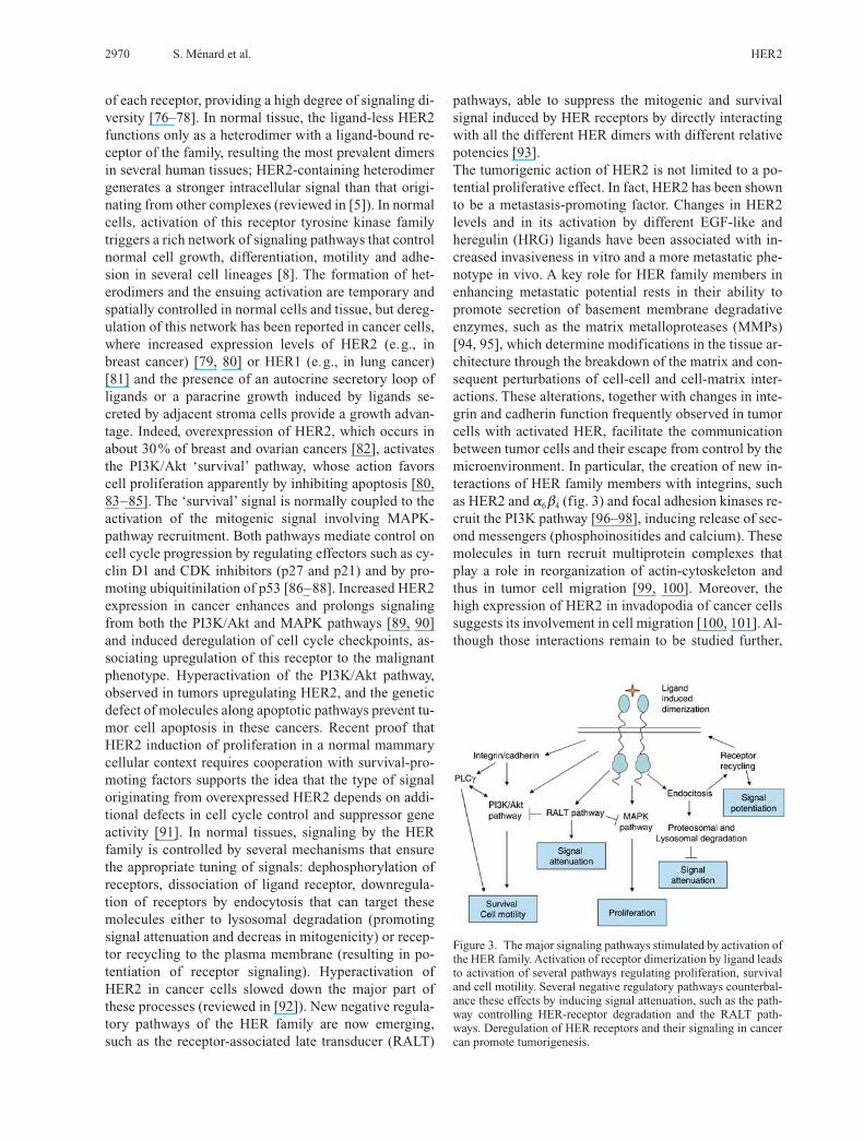
pathways, able to suppress the mitogenic and survivalsignal induced by HER receptors by directly interactingwith all the different HER dimers with different relativepotencies [93]. The tumorigenic action of HER2 is not limited to a po-tential proliferative effect. In fact, HER2 has been shownto be a metastasis-promoting factor. Changes in HER2levels and in its activation by different EGF-like andheregulin (HRG) ligands have been associated with in-creased invasiveness in vitro and a more metastatic phe-notype in vivo. A key role for HER family members inenhancing metastatic potential rests in their ability topromote secretion of basement membrane degradativeenzymes, such as the matrix metalloproteases (MMPs)[94, 95], which determine modifications in the tissue ar-chitecture through the breakdown of the matrix and con-sequent perturbations of cell-cell and cell-matrix inter-actions. These alterations, together with changes in inte-grin and cadherin function frequently observed in tumorcells with activated HER, facilitate the communicationbetween tumor cells and their escape from control by themicroenvironment. In particular, the creation of new in-teractions of HER family members with integrins, suchas HER2 and a6b4 (fig. 3) and focal adhesion kinases re-cruit the PI3K pathway [96–98], inducing release of sec-ond messengers (phosphoinositides and calcium). Thesemolecules in turn recruit multiprotein complexes thatplay a role in reorganization of actin-cytoskeleton andthus in tumor cell migration [99, 100]. Moreover, thehigh expression of HER2 in invadopodia of cancer cellssuggests its involvement in cell migration [100, 101]. Al-though those interactions remain to be studied further,
2970 S. Ménard et al. HER2
Figure 3. The major signaling pathways stimulated by activation ofthe HER family. Activation of receptor dimerization by ligand leadsto activation of several pathways regulating proliferation, survivaland cell motility. Several negative regulatory pathways counterbal-ance these effects by inducing signal attenuation, such as the path-way controlling HER-receptor degradation and the RALT path-ways. Deregulation of HER receptors and their signaling in cancercan promote tumorigenesis.
the current body of information points to a predominantrole for HER2 in motogenic and mitogenic signaling,whereas HER signaling appears to control the proteoly-sis of the matrix, mainly by inducing MMPs, and inva-sion [94, 99, 100]. By upregulating specific MMPs andangiogenic factors, such as VEGF [102], and by potenti-ating the adhesion of tumor cells to endothelial cells, ac-tivated HER members enhance angiogenesis and vascu-lar invasion.
Position of the tyrosine kinase early or late
HER2 amplification and overexpression is an early eventin breast cancerogenesis. Indeed, while it has never beenfound alterated in breast atypical hyperplasia, ‘ductal car-cinoma in situ’ (DCIS), the pre-invasive lesion for breastcancer, is found to display HER2 overexpression in50–60% of the cases (fig. 4), whereas the frequency de-creases to 25% in invasive carcinomas. A possible expla-nation for this apparent discrepancy, considering a role ofHER2 overexpression in increased invasiveness, is thatDCIS is the pre-invasive lesion principally for HER2-positive invasive carcinomas, while the HER2-negativeones might derive directly from atypical hyperplasia.
Specific drugs available, description of results
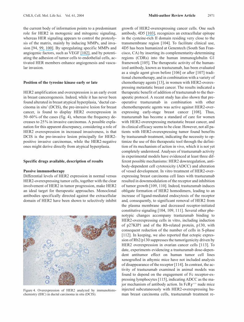
Passive immunotherapyDifferential levels of HER2 expression in normal versusHER2-overexpressing tumor cells, together with the clearinvolvement of HER2 in tumor progression, make HER2an ideal target for therapeutic approaches. Monoclonalantibodies specifically directed against the extracellulardomain of HER2 have been shown to selectively inhibit
growth of HER2-overexpressing cancer cells. One suchantibody, 4D5 [103], recognizes an extracellular epitopein the cysteine-rich II domain residing very close to thetransmembrane region [104]. To facilitate clinical use,4D5 has been humanized at Genentech (South San Fran-cisco, CA) by inserting its complementarity-determiningregions (CDRs) into the human immunoglobulin G1framework [105]. The therapeutic activity of the human-ized antibody, known as trastuzumab, has been evaluatedas a single agent given before [106] or after [107] tradi-tional chemotherapy, and in combination with a variety ofchemotherapy agents [13], in women with HER2-overex-pressing metastatic breast cancer. The results indicated atherapeutic benefit of addition of trastuzumab to the ther-apeutic protocol. A recent study has also shown that pre-operative trastuzumab in combination with otherchemotherapeutic agents was active against HER2-over-expressing early-stage breast cancer [108]. Thus,trastuzumab has become a standard of care for womenwith HER2-overexpressing metastatic breast cancer, andits clinical efficacy seems to be clear. However, not all pa-tients with HER2-overexpressing tumor found benefitsby trastuzumab treatment, indicating the necessity to op-timize the use of this therapeutic tool through the defini-tion of its mechanism of action in vivo, which it is not yetcompletely understood. Analyses of trastuzumab activityin experimental models have evidenced at least three dif-ferent possible mechanisms: HER2 downregulation, anti-body-dependent cell cytotoxicity (ADCC) and alterationof vessel development. In vitro treatment of HER2-over-expressing breast carcinoma cell lines with trastuzumabresulted in downmodulation of the receptor and inhibitionof tumor growth [109, 110]. Indeed, trastuzumab inducesobligate formation of HER2 homodimers, leading to anincrease of ligand-mediated endocytosis of the receptorand, consequently, to significant removal of HER2 fromthe plasma membrane and decreased receptor-initiatedconstitutive signaling [104, 109, 111]. Several other phe-notypic changes accompany trastazumab binding toHER2-overexpressing cells in vitro, including inductionof p27KIP1 and of the Rb-related protein, p130, withconsequent reduction of the number of cells in S-phase[112]. In keeping, we also reported that ectopic expres-sion of Rb2/p130 suppresses the tumorigenicity driven byHER2 overexpression in ovarian cancer cells [113]. Todate, experiments evidencing a trastuzumab dose-depen-dent antitumor effect on human tumor cell linesxenografted in athymic mice have not included analysisof disappearance of the receptor [114]. In contrast, the ac-tivity of trastuzumab examined in animal models wasfound to depend on the engagement of Fc receptor-ex-pressing lymphocytes [115], indicating ADCC as the ma-jor mechanism of antibody action. In FcRg +/+ nude miceinjected subcutaneously with HER2-overexpressing hu-man breast carcinoma cells, trastuzumab treatment re-
CMLS, Cell. Mol. Life Sci. Vol. 61, 2004 Multi-author Review Article 2971
Figure 4. Overexpression of HER2 analyzed by immunohisto-chemistry (IHC) in ductal carcinoma in situ (DCIS).
sulted in near-complete (96%) inhibition of tumorgrowth. This protective effect was reduced more than50% after disrupting the antibody’s ability to engage cel-lular Fcg receptor or in antibody-treated FcRg –/– mice. Arecent study in a preclinical model consisting of immun-odeficient SCID mice transplanted with human breastcarcinoma cells overexpressing HER2 showed thattrastuzumab treatment inducing a 30% reduction of tu-mor volume was accompanied with a reduction in vesselvolume, introducing a new mechanism of action for thisantibody [116]. Indeed, HER2 signaling is known to con-trol the expression of pro- and anti-angiogenic factors, in-cluding VEGF [102, 117], TGFa [118] and TSP-1 [116].However, this finding does not necessarily excludeADCC, since in a model such as the SCID mouse, whichis profoundly deficient in T and NK cells, inhibition ofHER2-driven pathways by the anti-HER2 antibody maycontribute significantly to anti-tumor efficacy. The anti-tumor activity through vessel regression, together withthe low protection exerted by trastuzumab found in miceknocked out for the Fc receptor, suggest that trastuzumabcan utilize different mechanisms in vivo. Clearly, the precise delineation of trastuzumab’s mecha-nism of action in patients is essential for the design ofmore successful antibody treatment protocols in breastcarcinoma patients. If, in fact, the major mechanism isADCC, the combination of trastuzumab with immuno-suppressive chemotherapeutic drugs requires optimaltiming of trastuzumab delivery to enable rescue of ADCCeffectors such as NK cells after chemotherapy. In thatcontext, clinical studies have demonstrated a synergisticaction between trastuzumab and the drug taxane [119],which induces suppression of adaptive immunity, but se-lectively increases NK activity [120]. To improve thera-peutic activity, stimulation of NK activity might also beconsidered, utilizing cytokines or ‘danger signals’ such asunmethylated CpG-oligodeoxynucleotides, which en-hance innate immunity [121].
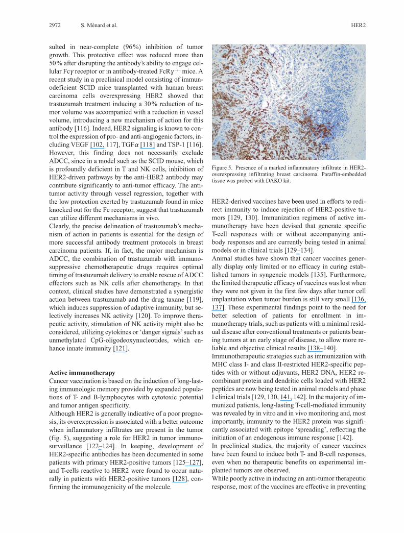
Active immunotherapyCancer vaccination is based on the induction of long-last-ing immunologic memory provided by expanded popula-tions of T- and B-lymphocytes with cytotoxic potentialand tumor antigen specificity.Although HER2 is generally indicative of a poor progno-sis, its overexpression is associated with a better outcomewhen inflammatory infiltrates are present in the tumor(fig. 5), suggesting a role for HER2 in tumor immuno-surveillance [122–124]. In keeping, development ofHER2-specific antibodies has been documented in somepatients with primary HER2-positive tumors [125–127],and T-cells reactive to HER2 were found to occur natu-rally in patients with HER2-positive tumors [128], con-firming the immunogenicity of the molecule.
HER2-derived vaccines have been used in efforts to redi-rect immunity to induce rejection of HER2-positive tu-mors [129, 130]. Immunization regimens of active im-munotherapy have been devised that generate specific T-cell responses with or without accompanying anti-body responses and are currently being tested in animalmodels or in clinical trials [129–134]. Animal studies have shown that cancer vaccines gener-ally display only limited or no efficacy in curing estab-lished tumors in syngeneic models [135]. Furthermore,the limited therapeutic efficacy of vaccines was lost whenthey were not given in the first few days after tumor cellimplantation when tumor burden is still very small [136,137]. These experimental findings point to the need forbetter selection of patients for enrollment in im-munotherapy trials, such as patients with a minimal resid-ual disease after conventional treatments or patients bear-ing tumors at an early stage of disease, to allow more re-liable and objective clinical results [138–140]. Immunotherapeutic strategies such as immunization withMHC class I- and class II-restricted HER2-specific pep-tides with or without adjuvants, HER2 DNA, HER2 re-combinant protein and dendritic cells loaded with HER2peptides are now being tested in animal models and phaseI clinical trials [129, 130, 141, 142]. In the majority of im-munized patients, long-lasting T-cell-mediated immunitywas revealed by in vitro and in vivo monitoring and, mostimportantly, immunity to the HER2 protein was signifi-cantly associated with epitope ‘spreading’, reflecting theinitiation of an endogenous immune response [142].In preclinical studies, the majority of cancer vaccineshave been found to induce both T- and B-cell responses,even when no therapeutic benefits on experimental im-planted tumors are observed. While poorly active in inducing an anti-tumor therapeuticresponse, most of the vaccines are effective in preventing
2972 S. Ménard et al. HER2
Figure 5. Presence of a marked inflammatory infiltrate in HER2-overexpressing infiltrating breast carcinoma. Paraffin-embeddedtissue was probed with DAKO kit.
spontaneous tumor occurrence [139]. Most data on thepreventive potential of HER2 vaccines have come fromstudies in genetically predisposed animals transgenicallyexpressing the rat HER2/neu proto-oncogene or its mu-tated activating form, and which are tolerant to self neu[143]. Different vaccination strategies include ratHER2/neu-positive allogeneic cells alone [144] or incombination with interleukin-12 [133]; the extracellulardomain of the rat oncoprotein [145] or peptides derivedfrom it [146]; and DNA plasmids encoding different frag-ments of HER2 or the full-length gene [131, 134, 147]. Immunological interventions targeting the HER2 protein,which is expressed at basal levels on adult normal tissues,have raised safety concerns with respect to the potentialinduction of autoimmune toxicity [148]; however, at pre-sent, no pertinent data in tumor-bearing hosts have beenobtained [130, 149].While therapeutic and prophylactic cancer vaccines mayoffer the advantage of inducing the development of bothT- and B-cell-mediated long-term immunity, unlike theapparently short-lived therapeutic effects of passive ther-apies, an optimal vaccine formulation with objective ther-apeutic benefits remains to be defined. This goal awaitsan improved understanding of the molecular mechanismsunderlying the immunological escape of HER2-positivetumors.
Anti-receptor therapyAs previously reported, signals induced by HER2-con-taining heterodimers have the strongest biological activ-ity with respect to the other HERs dimers [5], and highexpression of HER2 in tumors leads to amplification ofthe signaling cascade induced by the EGF-family ligands[92, 150]. Since HER2 is also the preferred partner ofHER1, inhibition of HER1 activity has been proposed asan avenue to block HER2 activity. The recent availabilityof tyrosine kinase inhibitors (TKIs) that specificallyblock the phosphorylation and function of HER1 has pro-vided a powerful new tool for therapy of tumors whoseproliferation depends on HER1 or HER2. One such in-hibitor is ZD1839 (Iressa), an orally active, selectiveHER1 TKI that induces a reversible inhibition or a delayin the growth of cancer cell lines and human tumorxenografts expressing high levels of HER1 [151]. Fol-lowing studies showed that tumors expressing low levelsof HER1 or overexpressing HER2 in presence of nor-mal/low HER1 are also sensitive to ZD1839 [152–155],whereas resistance to this TKI has been observed in celllines with a high to low level of EGFR, suggesting thatexpression of the target receptor is not a marker of sensi-tivity to ZD1839 [154, 155]. In addition, treatment ofbreast carcinoma cell lines expressing both HER2 andhormone receptors with ZD1839 was found to restore theinhibitory activity of tamoxifen [156], suggesting the
usefulness of TKI-mediated disruption of HER2 signal-ing in abrogating the anti-estrogen resistance observed inHER2-positive breast tumors. The precise mechanismthrough which Iressa exerts its antitumor effect remainsunclear. The first step results in the inhibition of theEGFR tyrosine-kinase activity, leading to a blockage ofthe activation pathways that mediate EGFR functions.The failure of ZD1839 with conventional chemothera-peutic agents in phase III trials on unselected NSCLChave highlighted the lack of acknowledge of the ZD1839mechanism of action as well as the absence of reliablemarkers of response to this drug (clinical endpoints). Elu-cidation of these points is mandatory to further design ofnew schedules of treatment with target-specific and con-ventional chemotherapies. Despite the abundance of studies on the biology of HER2and its receptor family, many pathways interacting withHER2 are still not fully understood or exploited for ther-apeutic purposes. However, HER2 is, to date, one of themost promising molecules for targeted therapy.
Acknowledgements. Partially supported by Associazione Italianaper la Ricerca sul Cancro and by Cariplo.
1 Plowman G. D., Green J. M., Culouscou J.-M., Carlton G. W.,Rothwell V. M. and Buckley S. (1993) Heregulin induces ty-rosine phosphorylation of HER4/p180erbB4. Nature 366:473–475
2 Coussens L., Yang-Feng T. L., Liao Y.-C., Chen E., Gray A.,McGrath J. et al. (1985) Tyrosine kinase receptor with exten-sive homology to EGF receptor shares chromosomal locationwith neu oncogene. Science 230: 1132–1139
3 Kraus M. H., Issing W., Miki T., Popescu N. C. and AaronsonS. A. (1989) Isolation and characterization of erbB3, a thirdmember of the erbB/epidermal growth factor receptor family:evidence for overexpression in a subset of human mammarytumors. Proc. Natl. Acad. Sci. USA 86: 9193–9197
4 Ullrich A., Coussens L., Hayflick J. S., Dull T. J., Gray A., TamA. W. et al. (1984) Human epidermal growth factor receptorcDNA sequence and aberrant expression of the amplifiedgene in A431 epidermoid carcinoma cells. Nature 309:418–425
5 Yarden Y. (2001) The EGFR family and its ligands in humancancer. signalling mechanisms and therapeutic opportunities.Eur. J. Cancer 37: S3–S8
6 Graus-Porta D., Beerli R. R., Daly J. M. and Hynes N. E.(1997) ErbB-2, the preferred heterodimerization partner of allErbB receptor, is a mediator of lateral signaling. EMBO J. 16:1647–1655
7 Tzahar E., Waterman H., Chen X., Levkowitz G., Karuna-garan D., Lavi S. et al. (1996) A hierarchinal network of inter-receptor interactions determines signal transduction by neudifferentiation factor/neuregulin and epidermal growth factor.Mol. Cell. Biol. 16: 5276–5287
8 Lee K.-F., Simon H., Chen H., Bates B., Hung M.-C. andHauser C. (1995) Requirement for neuregulin receptor erbB2in neural and cardiac development. Nature 378: 394–398
9 Gassmann M., Casagranda F., Orioli D., Simon H., Lai C.,Klein R. et al. (1995) Aberrant neural and cardiac develop-ment in mice lacking the ErbB4 neuregulin receptor. Nature378: 390–394
CMLS, Cell. Mol. Life Sci. Vol. 61, 2004 Multi-author Review Article 2973
10 Morris J. K., Lin W., Hauser C., Marchuk Y., Getman D. andLee K. F. (1999) Rescue of the cardiac defect in ErbB2 mutantmice reveals essential roles of ErbB2 in peripheral nervoussystem development. Neuron 23: 273–283
11 Tidcombe H., Jackson-Fisher A., Mathers K., Stern D. F.,Gassmann M. and Golding J. P. (2003) Neural and mammarygland defects in ErbB4 knockout mice genetically rescuedfrom embryonic lethality. Proc. Natl. Acad. Sci. USA 100:8281–8286
12 Ozcelik C., Erdmann B., Pilz B., Wettschureck N., Britsch S.,Hubner N. et al. (2002) Conditional mutation of the ErbB2(HER2) receptor in cardiomyocytes leads to dilated car-diomyopathy. Proc. Natl. Acad. Sci. USA 99: 8880–8885
13 Slamon D. J., Leyland-Jones B., Shak S., Fuchs H., Paton V.,Bajamonde A. et al. (2001) Use of chemotherapy plus a mon-oclonal antibody against HER2 for metastatic breast cancerthat overexpresses HER2. N. Engl. J. Med. 344: 783–792
14 Riethmacher D., Sonnenberg-Riethmacher E., Brinkmann V.,Yamaai T., Lewin G. R. and Birchmeier C. (1997) Severe neu-ropathies in mice with targeted mutations in the ErbB3 recep-tor. Nature 389: 725–730
15 Wolpowitz D., Mason T. B., Dietrich P., Mendelsohn M., Tal-mage D. A. and Role L. W. (2000) Cysteine-rich domain iso-forms of the neuregulin-1 gene are required for maintenanceof peripheral synapses. Neuron 25: 79–91
16 Meyer D., Yamaai T., Garratt A., Riethmacher-Sonnenberg E.,Kane D., Theill L. E. et al. (1997) Isoform-specific expressionand function of neuregulin. Development 124: 3575–3586
17 Garratt A. N., Voiculescu O., Topilko P., Charnay P. and Birch-meier C. (2000) A dual role of erbB2 in myelination and in ex-pansion of the schwann cell precursor pool [In Process Cita-tion]. J. Cell Biol. 148: 1035–1046
18 Britsch S., Li L., Kirchhoff S., Theuring F., Brinkmann V.,Birchmeier C. and Riethmacher D. (1998) The ErbB2 andErbB3 receptors and their ligand, neuregulin-1, are essentialfor development of the sympathetic nervous system. GenesDev. 12: 1825–1836
19 Erickson S. L., O’Shea K. S., Ghaboosi N., Loverro L., FrantzG., Bauer M. et al. (1997) ErbB3 is required for normal cere-bellar and cardiac development: a comparison with ErbB2-and heregulin-deficient mice. Development 124: 4999–5011
20 Kramer R., Bucay N., Kane D. J., Martin L. E., Tarpley J. E.and Theill L. E. (1996) Neuregulins with an Ig-like domainare essential for mouse myocardial and neuronal development.Proc. Natl. Acad. Sci. USA 93: 4833–4838
21 Woldeyesus M. T., Britsch S., Riethmacher D., Xu L., Son-nenberg-Riethmacher E., Abou-Rebyeh F. et al. (1999) Pe-ripheral nervous system defects in erbB2 mutants followinggenetic rescue of heart development. Genes Dev. 13:2538–2548
22 Benstead T. J. and Grant I. A. (2001) Progress in clinical neu-rosciences: Charcot-Marie-Tooth disease and related inheritedperipheral neuropathies. Can. J. Neurol. Sci. 28: 199–214
23 Kim J. Y., Sun Q., Oglesbee M. and Yoon S. O. (2003) The roleof ErbB2 signaling in the onset of terminal differentiation ofoligodendrocytes in vivo. J. Neurosci. 23: 5561–5571
24 Luetteke N. C., Phillips H. K., Qiu T. H., Copeland N. G., EarpH. S., Jenkins N. A. et al. (1994) The mouse waved-2 pheno-type results from a point mutation in the EGF receptor tyro-sine kinase. Genes Dev. 8: 399–413
25 Fowler K. J., Walker F., Alexander W., Hibbs M. L., Nice E. C.,Bohmer R. M. et al. (1995) A mutation in the epidermalgrowth factor receptor in waved-2 mice has a profound effecton receptor biochemistry that results in impaired lactation.Proc. Natl. Acad. Sci. USA 92: 1465–1469
26 Schroeder J. A. and Lee D. C. (1998) Dynamic expression andactivation of ERBB receptors in the developing mouse mam-mary gland. Cell Growth Differ. 9: 451–464
27 Sebastian J., Richards R. G., Walker M. P., Wiesen J. F., WerbZ., Derynck R. et al.. (1998) Activation and function of theepidermal growth factor receptor and erbB-2 during mam-mary gland morphogenesis. Cell Growth Differ. 9: 777–785
28 Yang Y., Spitzer E., Meyer D., Sachs M., Niemann C., Hart-mann G. et al. (1995) Sequential requirement of hepatocytegrowth factor and neuregulin in the morphogenesis and dif-ferentiation of the mammary gland. J. Cell Biol. 131: 215–226
29 Niemann C., Brinkmann V., Spitzer E., Hartmann G., SachsM., Naundorf H. et al. (1998) Reconstitution of mammarygland development In vitro: requirement of c- met and c-erbB2 signaling for branching and alveolar morphogenesis [InProcess Citation]. J. Cell Biol. 143: 533–545
30 Jones F. E. and Stern D. F. (1999) Expression of dominant-negative ErbB2 in the mammary gland of transgenic mice re-veals a role in lobuloalveolar development and lactation.Oncogene 18: 3481–3490
31 Ménard S., Casalini P., Campiglio M., Pupa S. M., Agresti R.and Tagliabue E. (2001) HER2 overexpression in various tu-mor types, focusing on its relationship to the development ofinvasive breast cancer. Ann. Oncol. 12: S15–S19
32 Gancberg D., Jarvinen T., Di Leo A., Rouas G., Cardoso F.,Paesmans M. et al. (2002) Evaluation of HER2/NEU proteinexpression in breast cancer by immunohistochemistry: an interlaboratory study assessing the reproducibility ofHER2/NEU testing. Breast Cancer Res. Treat. 74: 113–120
33 Rhodes A., Jasani B., Anderson E., Dodson A. R. and BalatonA. J. (2002) Evaluation of HER2/neu immunohistochemicalassay sensitivity and scoring on formalin-fixed and paraffin-processed cell lines and breast tumors: a comparative study in-volving results from laboratories in 21 countries. Am. J. Clin.Pathol. 118: 408–417
34 Paik S., Bryant J., Tan-Chiu E., Romond E., Hiller W., Park K.et al. (2002) Real-world performance of HER2 testing-na-tional surgical adjuvant breast and bowel project experience.J. Natl. Cancer Inst. 94: 852–854
35 Cell Markers and Cytogenetics Committees College of American Pathologists (2002) Clinical laboratory assays forHER2/neu amplification and overexpression: quality assur-ance, standardization, and proficiency testing. Arch. Pathol.Lab. Med. 126: 803–808
36 Roche P. C., Suman V. J., Jenkins R. B., Davidson N. E., Mar-tino S., Kaufman P. A. et al. (2002) Concordance between lo-cal and central laboratory HER2 testing in the breast inter-group trial N9831. J. Natl. Cancer Inst. 94: 855–857
37 Tagliabue E., Pilotti S., Gianni A. M., Ménard S. and ColnaghiM. I. (1998) Target molecules for immunotherapy of inflam-matory breast carcinomas. Eur. J. Cancer 34: 1982–1983
38 Nakamura H., Saji H., Ogata A., Hosaka M., Hagiwara M.,Kawasaki N. et al. (2003) Correlation between encoded pro-tein overexpression and copy number of the HER2 gene withsurvival in non-small cell lung cancer. Int. J. Cancer 103:61–66
39 Goebel S. U., Iwamoto M., Raffeld M., Gibril F., Hou W., Ser-rano J. et al. (2002) Her-2/neu expression and gene amplifica-tion in gastrinomas: correlations with tumor biology, growthand aggressiveness. Cancer Res. 62: 3702–3710
40 Yan J., Fang Y., Huang B. J., Liang Q. W., Wu Q. L. and ZengY. X. (2002) Absence of evidence for HER2 amplification innasopharyngeal carcinoma. Cancer Genet. Cytogenet. 132:116–119
41 Latif Z., Watters A. D., Bartlett J. M., Underwood M. A. andAitchison M. (2002) Gene amplification and overexpressionof HER2 in renal cell carcinoma. BJU Int. 89: 5–9
42 Khan A. J., King B. L., Smith B. D., Smith G. L., DiGiovannaM. P., Carter D. et al. (2002) Characterization of theHER2/neu oncogene by immunohistochemical and fluores-cence in situ hybridization analysis in oral and oropharyngealsquamous cell carcinoma. Clin. Cancer Res. 8: 540–548
2974 S. Ménard et al. HER2
43 Soule S., Baldridge L., Kirkpatrick K., Cheng L., Gilbert J. L.,Smith L. R. et al. (2002) HER2/neu expression in germ cell tu-mours. J. Clin. Pathol. 55: 656–658
44 Geddert H., Zeriouh M., Wolter M., Heise J. W., Gabbert H. E.and Sarbia M. (2002) Gene amplification and protein overex-pression of c-erb-b2 in Barrett carcinoma and its precursor le-sions. Am. J. Clin. Pathol. 118: 60–66
45 Hirsch F. R., Varella-Garcia M., Franklin W. A., Veve R., ChenL., Helfrich B. et al. (2002) Evaluation of HER2/neu gene am-plification and protein expression in non-small cell lung car-cinomas. Br. J. Cancer 86: 1449–1456
46 Siegel P. M. and Muller W. J. (1996) Mutations affecting con-served cysteine residues within the extracellular domain ofNeu promote receptor dimerization and activation. Proc. Natl.Acad. Sci USA 93: 8878–8883
47 Guy C. T., Webster M. A., Schaller M., Parsons T. J., CardiffR. D. and Muller W. J. (1992) Expression of the neu pro-tooncogene in the mammary epithelium of transgenic mice in-duces metastatic disease. Proc. Natl. Acad. Sci. USA 89:10578–10582
48 Siegel P. M., Ryan E. D., Cardiff R. D. and Muller W. J. (1999)Elevated expression of activated forms of Neu/ErbB-2 andErb–3 are involved in the induction of mammary tumors intransgenic mice: implicatins for human breast cancer. EMBOJ. 18: 2149–2164
49 Ménard S., Forti S., Castiglioni F., Agresti R. and Balsari A.(2001) HER2 as a prognostic factor in breast cancer. Oncol-ogy 61: 67–72
50 Masood S. and Bui M. M. (2002) Prognostic and predictivevalue of HER2/neu oncogene in breast cancer. Microsc. Res.Tech. 59: 102–108
51 Ménard S., Tagliabue E., Campiglio M. and Pupa S. M. (2000)Role of HER2 gene overexpression in breast carcinoma. J.Cell Physiol. 182: 150–162
52 Press M. F., Bernstein L., Thomas P. A., Meisner L. F., Zhou J.Y., Ma Y. L. et al. (1997) HER2/neu gene amplification char-acterized by fluorescence in situ hybridization: poor progno-sis in node-negative breast carcinomas. J. Clin. Oncol. 15:2894–2904
53 Cooke T., Reeves J., Lannigan A. and Stanton P. (2001) Thevalue of the human epidermal growth factor receptor-2(HER2) as a prognostic marker. Eur. J. Cancer 37: 3–10
54 Volpi A., De Paola F., Nanni O., Granato A. M., Bajorko P.,Becciolini A. et al. (2000) Prognostic significance of biologicmarkers in node-negative breast cancer patients: a prospectivestudy. Breast Cancer Res. Treat. 63: 181–192
55 Ross J. S., Muraca P. J. and Jaffe D. (1998) Multivariate analy-sis of prognostic factors in lymph node negative breast cancer.Mod. Pathol. 11: 26a
56 Ménard S., Balsari A., Casalini P., Tagliabue E., CampiglioM., Bufalino R. et al. (2002) HER2-positive breast carcino-mas as a particular subset with peculiar clinical behaviors.Clin. Cancer Res. 8: 520–525
57 Van’T Veer L. J., Dai H., Van de Vijver M. J., He Y. D., Hart A.A., Mao M. et al. (2002) Gene expression profiling predictsclinical outcome of breast cancer. Nature 415: 530–536
58 Murthy S. M., Goldschmidt R. A., Rao L. N., Ammirati M.,Buchmann T. and Scanlon E. F. (1989) The influence of surgi-cal trauma on experimental metastasis. Cancer 64: 2035–2044
59 Sieweke M. H. and Bissell M. J. (1994) The tumor-promotingeffect of wounding: a possible role for TGF-beta-induced stro-mal alterations. Crit Rev. Oncog. 5: 297–311
60 Tagliabue E., Agresti R., Ghirelli C., Morelli D. and MénardS. (2001) Letter to the editor. The early relapse of pre-menopausal patients after surgery for node-positive breastcarcinoma. Breast Cancer Res. Treat. 70: 155–156
61 Tagliabue E., Agresti R., Carcangiu M. L., Ghirelli C., MorelliD., Campiglio M. et al. (2003) Role of HER2 in wound-in-duced breast carcinoma proliferation. Lancet 362: 527–533
62 Thor A. D., Berry D. A., Budman D. R., Muss H. B., Kute T.,Henderson I. C. et al. (1998) erbB-2, p53 and efficacy of ad-juvant therapy in lymph node- positive breast cancer. J. Natl.Cancer Inst. 90: 1346–1360
63 Paik S. M., Bryant J., Park C. H., Fisher B., Tan-Chiu E.,Hyams D. et al. (1998) erbB-2 and response to doxorubicin in patients with axillary lymph node-positive, hormone recep-tor-negative breast cancer. J. Natl. Cancer Inst. 90: 1361–1370
64 Di Leo A., Gancberg D., Larsimont D., Tanner M., Jarvinen T.,Rouas G. et al. (2002) HER2 amplification and topoisomeraseIIalpha gene aberrations as predictive markers in node-posi-tive breast cancer patients randomly treated either with an anthracycline-based therapy or with cyclophosphamide,methotrexate, and 5-fluorouracil. Clin. Cancer Res. 8: 1107–1116
65 Ozinsky A., Underhill D. M., Fontenot J. D., Hajjar A. M.,Smith K. D., Wilson C. B. et al. (2000) The repertoire for pat-tern recognition of pathogens by the innate immune system isdefined by cooperation between toll-like receptors. Proc. Natl.Acad. Sci. USA 97: 13766–13771
66 Ménard S., Valagussa P., Pilotti S., Gianni L., Biganzoli E.,Boracchi P. et al. (2001) Response to Cyclophosphamide,Methotrexate, and Fluorouracil in lymph node-positive breastcancer according to HER2 overexpression and other tumor biologic variables. J. Clin. Oncol. 19: 329–335
67 Moliterni A., Ménard S., Valagussa P., Biganzoli E., BoracchiP., Balsari A. et al. (2003) HER2 overexpression and doxoru-bicin in the adjuvant chemotherapy of resectable breast can-cer. J. Clin. Oncol. 21: 458–462
68 Grunt T. W., Saceda M., Martin M. B., Lupu R., Dittrich E.,Krupitza G. et al. (1995) Bidirectional interactions betweenthe estrogen receptor and the c-erbB-2 signaling pathways:Heregulin inhibits estrogenic effects in breast cancer cells. Int.J. Cancer 63: 560–567
69 Elledge R. M., Green S., Ciocca D., Pugh R., Allred D. C.,Clark G. M. et al. (1998) HER2 expression and response to ta-moxifen in estrogen receptor-positive breast cancer: a south-west oncologty group study. Clin. Cancer Res. 4: 7–12
70 Konecny G., Pauletti G., Pegram M., Untch M., Dandekar S.,Aguilar Z. et al. (2003) Quantitative association betweenHER2/neu and steroid hormone receptors in hormone recep-tor-positive primary breast cancer. J. Natl. Cancer Inst. 95:142–153
71 Early Breast Cancer Trialists’ Collaborative Group (1998) Ta-moxifen for early breast cancer: an overview of the ran-domised trials. Lancet 351: 1451–1467
72 Perou C. M., Sorlie T., Eisen M. B., van de Rijn M., Jeffrey S.S., Rees C. A. et al. (2000) Molecular portraits of humanbreast tumours. Nature 406: 747–752
73 Sorlie T., Perou C. M., Tibshirani R., Aas T., Geisler S.,Johnsen H. et al. (2001) Gene expression patterns of breastcarcinomas distinguish tumor subclasses with clinical impli-cations. Proc. Natl. Acad. Sci. USA 98: 10869–10874
74 Bocker W., Moll R., Poremba C., Holland R., Van Diest P. J.,Dervan P. et al. (2002) Common adult stem cells in the humanbreast give rise to glandular and myoepithelial cell lineages: anew cell biological concept. Lab. Invest. 82: 737–746
75 Campiglio M., Somenzi G., Olgiati C., Beretta G., Balsari A.,Zaffaroni N. et al. (2003) Role of proliferation in HER2 statuspredicted response to doxorubicin. Int. J. Cancer 105:568–573
76 Aroian R. V., Koga M., Mendel J. E., Ohshima Y. and Stern-berg P. W. (1990) The let-23 gene necessary for Caenorhabdi-tis elegans vulval induction encodes a tyrosine kinase of theEGF receptor subfamily. Nature 348: 693–699
77 Wasserman J. D. and Freeman M. (1998) An autoregulatorycascade of EGF receptor signaling patterns the Drosophilaegg. Cell 95: 355–364
CMLS, Cell. Mol. Life Sci. Vol. 61, 2004 Multi-author Review Article 2975
78 Stein R. A. and Staros J. V. (2000) Evolutionary analysis of theErbB receptor and ligand families. J Mol. Evol. 50: 397–412
79 Pierce J. H., Arnstein P., DiMarco E., Artrip J., Kraus M. H.,Lonardo F. et al. (1991) Oncogenic potential of erbB-2 in human mammary epithelial cells. Oncogene 6: 1189–1194
80 Liu Y., El-Ashry D., Chen D., Ding I. Y. F. and Kern F. G.(1995) MCF–7 breast cancer cells overexpressing transfectedc- erbB-2 have an in vitro growth advantage in estrogen-depleted conditions and reduced estrogen-dependence and tamoxifen-sensitivity in vivo. Breast Cancer Res. Treat. 34:97–117
81 Franklin W. A., Veve R., Hirsch F. R., Helfrich B. A. and BunnP. A. Jr (2002) Epidermal growth factor receptor family inlung cancer and premalignancy. Semin. Oncol. 29: 3–14
82 Slamon D. J., Godolphin W., Jones L. A., Holt J. A., Wong S.C., Keith D. E. et al. (1989) Studies of the HER2/neu proto-oncogene in human breast and ovarian cancer. Science 244:707–712
83 Zhou B. P. and Hung M. C. (2002) Novel targets of Akt,p21(Cipl/WAF1), and MDM2. Semin. Oncol. 29: 62–70
84 Di Fiore P. P., Pierce J. H., Kraus M. H., Segatto O., King C.R. and Aaronson S. A. (1987) erbB-2 is a potent oncogenewhen overexpressed in NIH/3T3 cells. Science 237: 178–182
85 Muthuswamy S. K., Li D., Lelievre S., Bissell M. J. andBrugge J. S. (2001) ErbB2, but not ErbB1, reinitiates prolifer-ation and induces luminal repopulation in epithelial acini. Nat.Cell Biol. 3: 785–792
86 Lane H. A., Beuvink I., Motoyama A. B., Daly J. M., Neve R.M. and Hynes N. E. (2000) ErbB2 potentiates breast tumorproliferation through modulation of p27(Kip1)-Cdk2 com-plex formation: receptor overexpression does not determinegrowth dependency. Mol. Cell Biol. 20: 3210–3223
87 Zhou B. P., Liao Y., Xia W., Spohn B., Lee M. H. and Hung M.C. (2001) Cytoplasmic localization of p21Cip1/WAF1 byAkt-induced phosphorylation in HER2/neu-overexpressingcells. Nat. Cell Biol. 3: 245–252
88 Zhou B. P., Liao Y., Xia W., Zou Y., Spohn B. and Hung M. C.(2001) HER2/neu induces p53 ubiquitination via Akt-medi-ated MDM2 phosphorylation. Nat. Cell Biol. 3: 973–982
89 Karunagaran D., Tzahar E., Beerli R. R., Chen X. M., Graus-Porta D., Ratzkin B. J. et al. (1996) ErbB-2 is a common aux-iliary subunit of NDF and EGF receptors: implications forbreast cancer. EMBO J. 15: 254–264
90 Waterman H., Alroy I., Strano S., Seger R. and Yarden Y.(1999) The C-terminus of the kinase-defective neuregulin re-ceptor ErbB–3 confers mitogenic superiority and dictates en-docytic routing. EMBO J. 18: 3348–3358
91 Debnath J., Mills K. R., Collins N. L., Reginato M. J.,Muthuswamy S. K. and Brugge J. S. (2002) The role of apop-tosis in creating and maintaining luminal space within normaland oncogene-expressing mammary acini. Cell 111: 29–40
92 Harari D. and Yarden Y. (2000) Molecular mechanisms under-lying ErbB2/HER2 action in breast cancer. Oncogene 19:6102–6114
93 Anastasi S., Fiorentino L., Fiorini M., Fraioli R., Sala G.,Castellani L. et al. (2003) Feedback inhibition by RALT con-trols signal output by the ErbB network. Oncogene 22:4221–4234
94 O-charoenrat P., Rhys-Evans P., Court W. J., Box G. M. andEccles S. A. (1999) Differential modulation of proliferation,matrix metalloproteinase expression and invasion of humanhead and neck squamous carcinoma cells by c-erbB ligands.Clin. Exp. Met. 17: 631–639
95 Tan M., Yao J. and Yu D. H. (1997) Overexpression of the c-erbB-2 gene enhanced intrinsic metastasis potential in humanbreast cancer cells without increasing their transformationabilities. Cancer Res. 57: 1199–1205
96 Campiglio M., Tagliabue E., Uppugunduri S., Pellegrini R.,Martignone S., Ménard S. et al. (1994) Colocalization of the
p185HER2 oncoprotein and integrin a6b4 in Calu-3 lung carci-noma cells. J. Cell. Biochem. 55: 409–418
97 Tagliabue E., Ardini E., Pellegrini R., Campiglio M., BufalinoR., Jeschke M. et al. (1996) Laminin activates the p185HER2oncoprotein and mediates growth inhibition of breast carci-noma cells. Br. J. Cancer 74: 1427–1433
98 Gambaletta D., Marchetti A., Benedetti L., Mercurio A. M.,Sacchi A. and Falcioni R. (2000) Cooperative signaling be-tween alpha(6)beta(4) integrin and ErbB-2 receptor is re-quired to promote phosphatidylinositol 3-kinase-dependentinvasion. J. Biol. Chem. 275: 10604–10610
99 Feldner J. C. and Brandt B. H. (2002) Cancer cell motility – onthe road from c-erbB-2 receptor steered signaling to actin re-organization. Exp. Cell Res. 272: 93–108
100 Dittmar T., Husemann A., Schewe Y., Nofer J. R., NiggemannB., Zanker K. S. et al. (2002) Induction of cancer cell migra-tion by epidermal growth factor is initiated by specific phos-phorylation of tyrosine 1248 of c-erbB-2 receptor via EGFR.FASEB J. 16: 1823–1825
101 Eccles S. A. (2000) Cell biology of lymphatic metastasis. Thepotential role of c-erbB oncogene signalling. Recent ResultsCancer Res. 157: 41–54
102 Kerbel R. S., Viloria-Petit A., Klement G. and Rak J. (2000)‘Accidental’ anti-angiogenic drugs. Anti-oncogene directedsignal transduction inhibitors and conventional chemothera-peutic agents as examples. Eur. J. Cancer 36: 1248–1257
103 Fendly B. M., Winget M., Hudziak R. M., Lipari M. T., NapierM. A. and Ullrich A. (1990) Characterization of murine mon-oclonal antibodies reactive to either the human epidermalgrowth factor receptor or HER2/neu gene product. CancerRes. 50: 1550–1558
104 De Santes K., Slamon D., Anderson S. K., Shepard M., FendlyB., Maneval D. et al. (1992) Radiolabeled antibody targetingof the HER2/neu oncoprotein. Cancer Res. 52: 1916–1923
105 Carter P., Presta L., Gorman C. M., Ridgway J. B., Henner D.,Wong W. L. et al. (1992) Humanization of an anti-p185HER2 an-tibody for human cancer therapy. Proc. Natl. Acad. Sci. USA89: 4285–4289
106 Vogel C. L., Cobleigh M. A., Tripathy D., Gutheil J. C., HarrisL. N., Fehrenbacher L. et al. (2002) Efficacy and safety oftrastuzumab as a single agent in first-line treatment of HER2-overexpressing metastatic breast cancer. J. Clin. Oncol. 20:719–726
107 Cobleigh M. A., Vogel C. L., Tripathy D., Robert N. J., SchollS., Fehrenbacher L. et al. (1999) Multinational study of the ef-ficacy and safety of humanized anti-HER2 monoclonal anti-body in women who have HER2-overexpressing metastaticbreast cancer that has progressed after chemotherapy formetastatic disease. J. Clin. Oncol. 17: 2639–2648
108 Burstein H. J., Harris L. N., Gelman R., Lester S. C., Nunes R.A., Kaelin C. M. et al. (2003) Preoperative therapy withtrastuzumab and paclitaxel followed by sequential adjuvantdoxorubicin/cyclophosphamide for HER2 overexpressingstage II or III breast cancer: a pilot study. J. Clin. Oncol. 21:46–53
109 Hudziak R. M., Lewis G. D., Winget M., Fendly B. M., Shep-ard H. M. and Ullrich A. (1989) p185HER2 monoclonal anti-body has antiproliferative effects in vitro and sensitizes hu-man breast tumor cells to tumor necrosis factor. Mol. Cell.Biol. 9: 1165–1172
110 Hieken T. J., Mehta R. R. and Shilkaitis A. (1996) HER2/neuand p53 expression in breast cancer: valid prognostic markerswhen assessed by direct immunoassay, but not by immunohis-tochemistry. Proc. Annu. Meet. Am. Soc. Clin. Oncol. 15: 113
111 Sarup J. C., Johnson R. M., King K. L., Fendly B. M., LipariM. T., Napier M. A. et al. (1991) Characterization of an anti-p185HER2 monoclonal antibody that stimulates receptorfunction and inhibits tumor cell growth. Growth Regul. 1:72–82
2976 S. Ménard et al. HER2
112 Sliwkowski M. X., Lofgren J. A., Lewis G. D., Hotaling T. E.,Fendly B. M. and Fox J. A. (1999) Nonclinical studies ad-dressing the mechanism of action of trastuzumab (Herceptin).Semin. Oncol. 26: 60–70
113 Pupa S. M., Howard C. M., Invernizzi A. M., De Vecchi R.,Giani C., Claudio P. P. et al. (1999) Ectopic expression of pRB2/p130 suppresses the tumorigenicity of c-erbB-2-overexpressing SkOv3 tumor cell line. Oncogene 18: 651–656
114 Shepard H. M., Lewis G. D., Sarup J. C., Fendly B. M.,Maneval D., Mordenti J. et al. (1991) Monoclonal antibodytherapy of human cancer: taking the HER2 protooncogene tothe clinic. J. Clin. Immunol. 11: 117–127
115 Clynes R. A., Towers T. L., Presta L. G. and Ravetch J. V.(2000) Inhibitory Fc receptors modulate in vivo cytoxicityagainst tumor targets. Nat. Med. 6: 443–446
116 Izumi Y., Xu L., di Tomaso E., Fukumura D. and Jain R. K.(2002) Tumour biology: herceptin acts as an anti-angiogeniccocktail. Nature 416: 279–280
117 Allgayer H., Babic R., Gruetzner K. U., Tarabichi A., Schild-berg F. W. and Heiss M. M. (2000) c-erbB-2 is of independentprognostic relevance in gastric cancer and is associated withthe expression of tumor-associated protease systems. J. Clin.Oncol. 18: 2201–2209
118 Nicolini G., Miloso M., Moroni M. C., Beguinot L. and ScottoL. (1996) Post-transcriptional control regulates transforminggrowth factor alpha in the human carcinoma KB cell line. J.Biol. Chem. 271: 30290–30296
119 Sato Y., Roman M., Tighe H., Lee D., Corr M., Nguyen M. D.et al. (1996) Immunostimulatory DNA sequences necessaryfor effective intradermal gene immunization. Science 19:352–354
120 Tsavaris N., Kosmas C., Vadiaka M., Kanelopoulos P. andBoulamatsis D. (2002) Immune changes in patients with ad-vanced breast cancer undergoing chemotherapy with taxanes.Br. J. Cancer 87: 21–27
121 Sfondrini L., Besusso D., Rumio C., Rodolfo M., Ménard S.and Balsari A. (2002) Prevention of spontaneous mammaryadenocarcinoma in HER2/neu transgenic mice by foreignDNA. FASEB J. 16: 1749–1754
122 Rilke F., Colnaghi M. I., Cascinelli N., Andreola S., BaldiniM. T., Bufalino R. et al. (1991) Prognostic significance ofHER2/neu expression in breast cancer and its relationship toother prognostic factors. Int. J. Cancer 49: 44–49
123 Pupa S. M., Bufalino R., Invernizzi A. M., Andreola S., RilkeF., Lombardi L. et al. (1996) Macrophage infiltrate and prog-nosis in c-erbB-2-overexpressing breast carcinomas. J. Clin.Oncol. 14: 85–94
124 Ménard S., Pupa S. M., Casalini P., Rilke F. and Colnaghi M.I. (1996) Relationship of histological grade, c-erbB-2 expres-sion and inflammatory infiltrate to prognosis in carcinoma ofthe breast – in reply. J. Clin. Oncol. 14: 2407
125 Pupa S. M., Ménard S., Andreola S. and Colnaghi M. I. (1993)Antibody response against the c-erbB-2 oncoprotein in breastcarcinoma patients. Cancer Res. 53: 5864–5866
126 Disis M. L., Calenoff E., McLaughlin G., Murphy A. E., ChenW., Groner B. et al. (1994) Existent T-cell and antibody im-munity to HER2/neu protein in patients with breast cancer.Cancer Res. 54: 16–20
127 Disis M. L., Pupa S. M., Gralow J. R., Dittadi R., Ménard S.and Cheever M. A. (1997) High titer HER2/neu protein spe-cific antibody immunity can be detected in patients with earlystage breast cancer. J. Clin. Oncol. 15: 3363–3367
128 Disis M. L. and Cheever M. A. (1997) HER2/neu protein: atarget for antigen-specific immunotherapy of human cancer.Adv. Cancer Res. 71: 343–371
129 Foy T. M., Fanger G. R., Hand S., Gerard C., Bruck C. andCheever M. A. (2002) Designing HER2 vaccines. Semin. On-col. 29: 53–61
130 Kiessling R., Weil W. Z., Herrmann F., Lindencrona J. A.,Choudhury A., Kono K. et al. (2002) Cellular immunity to theHer-2/neu protooncogene. Adv. Cancer Res. 85: 101–144
131 Rovero S., Amici A., Carlo E. D., Bei R., Nanni P., QuaglinoE. et al. (2000) DNA vaccination against rat her-2/Neu p185more effectively inhibits carcinogenesis than transplantablecarcinomas in transgenic BALB/c mice. J. Immunol. 165:5133–5142
132 Di Carlo E., Rovero S., Boggio K., Quaglino E., Amici A.,Smorlesi A. et al. (2001) Inhibition of mammary carcinogen-esis by systemic interleukin 12 or p185neu DNA vaccinationin Her-2/neu transgenic BALB/c mice. Clin. Cancer Res. 7:830s–837s
133 Nanni P., Nicoletti G., De Giovanni C., Landuzzi L., Di CarloE., Cavallo F. et al. (2001) Combined allogeneic tumor cellvaccination and systemic interleukin 12 prevents mammarycarcinogenesis in HER2/neu transgenic mice. J. Exp. Med.194: 1–12
134 Quaglino E., Iezzi M., Mastini C., Amici A., Pericle F., DiCarlo E. et al. (2004) Electroporated DNA vaccine clearsaway multifocal mammary carcinomas in HER2/neu trans-genic mice. Cancer Res. 64: 2858–2864
135 Lollini P. L. and Forni G. (2002) Antitumor vaccines: is it pos-sible to prevent a tumor? Cancer Immunol. Immunother. 51:409–416
136 Nanni P., Forni G. and Lollini P. L. (1999) Cytokine gene ther-apy: hopes and pitfalls. Ann. Oncol. 10: 261–266
137 Cavallo F., Di Carlo E., Butera M., Verrua R., Colombo M. P.,Musiani P. et al. (1999) Immune events associated with thecure of established tumors and spontaneous metastases by lo-cal and systemic interleukin 12. Cancer Res. 59: 414–421
138 Bocchia M., Bronte V., Colombo M. P., De Vincentiis A., DiNicola M., Forni G. et al. (2000) Antitumor vaccination:where we stand. Haematologica 85: 1172–1206
139 Finn O. J. and Forni G. (2002) Prophylactic cancer vaccines.Curr. Opin. Immunol. 14: 172–177
140 Jager E., Jager D. and Knuth A. (2002) Clinical cancer vaccinetrials. Curr. Opin. Immunol. 14: 178–182
141 Knutson K. L., Schiffman K. and Disis M. L. (2001) Immu-nization with a HER2/neu helper peptide vaccine generatesHER2/neu CD8 T-cell immunity in cancer patients. J. Clin. In-vest. 107: 477–484
142 Disis M. L., Gooley T. A., Rinn K., Davis D., Piepkorn M.,Cheever M. A. et al. (2002) Generation of T-cell immunity tothe HER2/neu protein after active immunization withHER2/neu peptide-based vaccines. J. Clin. Oncol. 20:2624–2632
143 De Vecchi R., Pupa S. M., Ménard S. and Lollini P.-L. (1999)Correspondence re: Y. Chen et al., DNA vaccines encodingfull-length or truncated neu induce protective immunityagainst neu-expressing mammary tumors. Cancer Res. 58:1965–1971, 1998. Cancer Res. 59: 4471–4472
144 Cefai D., Morrison B. W., Sckell A., Favre L., Balli M., Leu-nig M. et al. (1999) Targeting HER2/neu for active-specificimmunotherapy in a mouse model of spontaneous breast can-cer. Int. J. Cancer 83: 393–400
145 Esserman L. J., Lopez T., Montes R., Bald L. N., Fendly B. M.and Campbell M. J. (1999) Vaccination with the extracellulardomain of p185neu prevents mammary tumor development inneu transgenic mice. Cancer Immunol. Immunother. 47:337–342
146 Dakappagari N. K., Douglas D. B., Triozzi P. L., Stevens V. C.and Kaumaya P. T. (2000) Prevention of mammary tumorswith a chimeric HER2 B-cell epitope peptide vaccine. CancerRes. 60: 3782–3789
147 Pupa S. M., Invernizzi A. M., Forti S., Nanni P., Lollini P. L.,Meazza R. et al. (2001) Prevention of neu-expressing breasttumor development in mice transgenic for rat proto-neu byDNA vaccination. Gene Ther. 8: 75–79
CMLS, Cell. Mol. Life Sci. Vol. 61, 2004 Multi-author Review Article 2977
148 Press M. F., Cordon-Cardo C. and Slamon D. J. (1990) Ex-pression of the HER2/neu proto-oncogene in normal humanadult and fetal tissues. Oncogene 5: 593–962
149 Ohashi P. S. and DeFranco A. L. (2002) Making and breakingtolerance. Curr. Opin. Immunol. 14: 744–759
150 Prenzel N., Fischer O. M., Streit S., Hart S. and Ullrich A.(2001) The epidermal growth factor receptor family as a cen-tral element for cellular signal transduction and diversifica-tion. Endocr. Relat. Cancer 8: 11–31
151 Sirotnak F. M., Zakowski M. F., Miller V. A., Scher H. I. andKris M. G. (2000) Efficacy of cytotoxic agents against humantumor xenografts is markedly enhanced by coadministrationof ZD1839 (Iressa), an inhibitor of EGFR tyrosine kinase.Clin. Cancer Res. 6: 4885–4892
152 McCormick S. R., Lillemoe T. J., Beneke J., Schrauth J. andReinartz J. (2002) HER2 assessment by immunohistochemi-cal analysis and fluorescence in situ hybridization: compari-son of HercepTest and PathVysion commercial assays. Am. J.Clin. Pathol. 117: 935–943
153 Normanno N., Campiglio M., De Luca A., Somenzi G.,Maiello M., Ciardiello F. et al. (2002) Cooperative inhibitoryeffect of ZD1839 (‘iressa’) in combination with trastuzumab(‘herceptin’) on human breast cancer cell growth. Ann. Oncol.13: 65–72
154 Moasser M. M., Basso A., Averbuch S. D. and Rosen N.(2001) The tyrosine kinase inhibitor zd1839 (‘iressa’) inhibitsher2-driven signaling and suppresses the growth of her2-over-expressing tumor cells. Cancer Res. 61: 7184–7188
155 Campiglio M., Locatelli A., Olgiati C., Normanno N.,Somenzi G., Viganò L. et al. (2004) Inhibition of proliferationand induction of apoptosis in breast cancer cells by the epi-dermal growth factor receptor (EGFR) tyrosine kinase in-hibitor ZD1839 (‘Iressa’) is independent of EGFR expressionlevel. J. Cell Physiol. 198: 259–268
156 Witters L., Engle L. and Lipton A. (2002) Restoration of es-trogen responsiveness by blocking the HER2/neu pathway.Oncol. Rep. 9: 1163–1166
2978 S. Ménard et al. HER2
Related Documents