REVIEW ARTICLE Cancer Metabolism Oncogenic pathways and the electron transport chain: a dangeROS liaison Vittoria Raimondi 1 , Francesco Ciccarese 1 and Vincenzo Ciminale 1,2 Driver mutations in oncogenic pathways, rewiring of cellular metabolism and altered ROS homoeostasis are intimately connected hallmarks of cancer. Electrons derived from different metabolic processes are channelled into the mitochondrial electron transport chain (ETC) to fuel the oxidative phosphorylation process. Electrons leaking from the ETC can prematurely react with oxygen, resulting in the generation of reactive oxygen species (ROS). Several signalling pathways are affected by ROS, which act as second messengers controlling cell proliferation and survival. On the other hand, oncogenic pathways hijack the ETC, enhancing its ROS- producing capacity by increasing electron flow or by impinging on the structure and organisation of the ETC. In this review, we focus on the ETC as a source of ROS and its modulation by oncogenic pathways, which generates a vicious cycle that resets ROS levels to a higher homoeostatic set point, sustaining the cancer cell phenotype. British Journal of Cancer (2020) 122:168–181; https://doi.org/10.1038/s41416-019-0651-y BACKGROUND Altered reactive oxygen species (ROS) homoeostasis is emerging as an important hallmark of the cancer cell phenotype. These alterations are consistent with the “ROS rheostat” theory, 1 which states that, depending on their homoeostatic set point, ROS can control different signal transduction pathways, thus acting as either tumour promoters or tumour suppressors. At low/medium levels, ROS enhance mitogenic signalling and cell survival, and may contribute to genetic instability. 2 It is thus not surprising that most tumours exhibit a higher ROS set point compared with their healthy counterparts. On the other hand, excessive ROS levels result in extensive macromolecular damage and engage cell death pathways. It should be noted, however, that ROS-producing pathways are not the only determinants of the ROS rheostat, and cancer cells commonly increase the fuelling of antioxidant pathways and may cope with oxidative damage by upregulating repair systems. 3 Mitochondria are an important source of ROS production through the activity of the electron transport chain (ETC). 4 Although there are also many other important sources of ROS generation (e.g. NADPH oxidases [NOX]), 5 ETC-derived ROS are pivotal regulators of cell fate, given the central role of mitochondria in life and death. Electron transfer through the ETC is a tight and efficient molecular machine. Nevertheless, it is estimated that in normal conditions, about 0.2–2% of the electrons that pass through the ETC complexes leak from the system and reduce O 2 , generating superoxide (O 2 •– ), 6,7 which is rapidly converted by superoxide dismutases (SOD) into hydrogen peroxide (H 2 O 2 ), the most important ROS acting as a “second messenger” in signal transduction due to its relatively long half-life and diffusion across membranes via the aquaporin channels. 8 The hydroxyl radical ( • OH) is a highly damaging ROS with an extremely short half-life that is generated from H 2 O 2 in the presence of iron or copper through the Fenton reaction. O 2 •– can also interact with nitric oxide (NO), generating the reactive nitrogen species (RNS) peroxynitrite (ONOO - ), which controls signalling molecules through the nitration of tyrosine residues. 9 For the role of RNS in cancer we refer the reader to recent excellent reviews. 10,11 It is important to note that many tumours presenting mutations in ETC components show a strong propensity to produce ROS, 12 supporting the crucial role of this machinery in the modulation of the cancer cell phenotype. Several signal transduction pathways that control cell turnover are known to be ROS-sensitive (reviewed in 13–15 ). Through the reversible oxidation of cysteine residues to sulfenic acid (SO – ) 16 and disulfide bonds, 17 ROS modulate the activity of redox- sensitive proteins by controlling several aspects of the cancer phenotype. The oxidation of phosphatase and tensin homolog (PTEN) results in AKT-mediated cell survival and proliferation, 18 while the oxidation of prolyl hydroxylases leads to hypoxia- inducible factor 1α (HIF-1α) stabilisation, 19 resulting in a profound metabolic rewiring of cancer cells. Notably, O 2 •– may inhibit apoptosis in cancer cells, accounting for resistance to Fas- mediated cell death. 20 In this regard, decreasing the levels of O 2 •– restores apoptosis in cancer cells overexpressing the anti- apoptotic protein BCL-2 21 (see below). In addition, increased mitochondrial ROS levels drive “mitohorm- esis”, a condition of mild mitochondrial stress that favours pro- survival pathways through the activation of mammalian target of rapamycin (mTOR) complex 1 (mTORC1) signalling, an alteration that is frequently associated with poor clinical outcome of cancer patients. 22 Notably, the mitochondria-targeted O 2 •– scavenger www.nature.com/bjc Received: 27 June 2019 Revised: 30 October 2019 Accepted: 5 November 2019 Published online: 10 December 2019 1 Veneto Institute of Oncology IOV – IRCCS, Padua, Italy and 2 Department of Surgery, Oncology and Gastroenterology, University of Padua, Padua, Italy Correspondence: Vincenzo Ciminale ([email protected]) These authors contributed equally: Vittoria Raimondi, Francesco Ciccarese © The Author(s), under exclusive licence to Cancer Research UK 2019

Welcome message from author
This document is posted to help you gain knowledge. Please leave a comment to let me know what you think about it! Share it to your friends and learn new things together.
Transcript
REVIEW ARTICLECancer Metabolism
Oncogenic pathways and the electron transport chain: adangeROS liaisonVittoria Raimondi1, Francesco Ciccarese1 and Vincenzo Ciminale1,2
Driver mutations in oncogenic pathways, rewiring of cellular metabolism and altered ROS homoeostasis are intimately connectedhallmarks of cancer. Electrons derived from different metabolic processes are channelled into the mitochondrial electron transportchain (ETC) to fuel the oxidative phosphorylation process. Electrons leaking from the ETC can prematurely react with oxygen,resulting in the generation of reactive oxygen species (ROS). Several signalling pathways are affected by ROS, which act as secondmessengers controlling cell proliferation and survival. On the other hand, oncogenic pathways hijack the ETC, enhancing its ROS-producing capacity by increasing electron flow or by impinging on the structure and organisation of the ETC. In this review, wefocus on the ETC as a source of ROS and its modulation by oncogenic pathways, which generates a vicious cycle that resets ROSlevels to a higher homoeostatic set point, sustaining the cancer cell phenotype.
British Journal of Cancer (2020) 122:168–181; https://doi.org/10.1038/s41416-019-0651-y
BACKGROUNDAltered reactive oxygen species (ROS) homoeostasis is emerging asan important hallmark of the cancer cell phenotype. Thesealterations are consistent with the “ROS rheostat” theory,1 whichstates that, depending on their homoeostatic set point, ROS cancontrol different signal transduction pathways, thus acting as eithertumour promoters or tumour suppressors. At low/medium levels,ROS enhance mitogenic signalling and cell survival, and maycontribute to genetic instability.2 It is thus not surprising that mosttumours exhibit a higher ROS set point compared with their healthycounterparts. On the other hand, excessive ROS levels result inextensive macromolecular damage and engage cell death pathways.It should be noted, however, that ROS-producing pathways are
not the only determinants of the ROS rheostat, and cancer cellscommonly increase the fuelling of antioxidant pathways and maycope with oxidative damage by upregulating repair systems.3
Mitochondria are an important source of ROS productionthrough the activity of the electron transport chain (ETC).4
Although there are also many other important sources of ROSgeneration (e.g. NADPH oxidases [NOX]),5 ETC-derived ROS arepivotal regulators of cell fate, given the central role ofmitochondria in life and death.Electron transfer through the ETC is a tight and efficient
molecular machine. Nevertheless, it is estimated that in normalconditions, about 0.2–2% of the electrons that pass through theETC complexes leak from the system and reduce O2, generatingsuperoxide (O2
•–),6,7 which is rapidly converted by superoxidedismutases (SOD) into hydrogen peroxide (H2O2), the mostimportant ROS acting as a “second messenger” in signaltransduction due to its relatively long half-life and diffusion acrossmembranes via the aquaporin channels.8 The hydroxyl radical
(•OH) is a highly damaging ROS with an extremely short half-lifethat is generated from H2O2 in the presence of iron or copperthrough the Fenton reaction. O2
•– can also interact with nitricoxide (NO), generating the reactive nitrogen species (RNS)peroxynitrite (ONOO−), which controls signalling moleculesthrough the nitration of tyrosine residues.9 For the role of RNSin cancer we refer the reader to recent excellent reviews.10,11
It is important to note that many tumours presenting mutationsin ETC components show a strong propensity to produce ROS,12
supporting the crucial role of this machinery in the modulation ofthe cancer cell phenotype.Several signal transduction pathways that control cell turnover
are known to be ROS-sensitive (reviewed in 13–15). Through thereversible oxidation of cysteine residues to sulfenic acid (SO–)16
and disulfide bonds,17 ROS modulate the activity of redox-sensitive proteins by controlling several aspects of the cancerphenotype. The oxidation of phosphatase and tensin homolog(PTEN) results in AKT-mediated cell survival and proliferation,18
while the oxidation of prolyl hydroxylases leads to hypoxia-inducible factor 1α (HIF-1α) stabilisation,19 resulting in a profoundmetabolic rewiring of cancer cells. Notably, O2
•– may inhibitapoptosis in cancer cells, accounting for resistance to Fas-mediated cell death.20 In this regard, decreasing the levels ofO2
•– restores apoptosis in cancer cells overexpressing the anti-apoptotic protein BCL-221 (see below).In addition, increased mitochondrial ROS levels drive “mitohorm-
esis”, a condition of mild mitochondrial stress that favours pro-survival pathways through the activation of mammalian target ofrapamycin (mTOR) complex 1 (mTORC1) signalling, an alteration thatis frequently associated with poor clinical outcome of cancerpatients.22 Notably, the mitochondria-targeted O2
•– scavenger
www.nature.com/bjc
Received: 27 June 2019 Revised: 30 October 2019 Accepted: 5 November 2019Published online: 10 December 2019
1Veneto Institute of Oncology IOV – IRCCS, Padua, Italy and 2Department of Surgery, Oncology and Gastroenterology, University of Padua, Padua, ItalyCorrespondence: Vincenzo Ciminale ([email protected])These authors contributed equally: Vittoria Raimondi, Francesco Ciccarese
© The Author(s), under exclusive licence to Cancer Research UK 2019
mitoTEMPO inhibits tumour cell migration and metastasis in mice,23
corroborating a crucial role for ETC-derived O2•– in tumorigenesis.
In this review, we focus on the ETC as a source of ROS, itsalterations in cancer cells and its modulation by oncogenicpathways, highlighting the connection between ETC-derived ROSand the cancer cell phenotype.
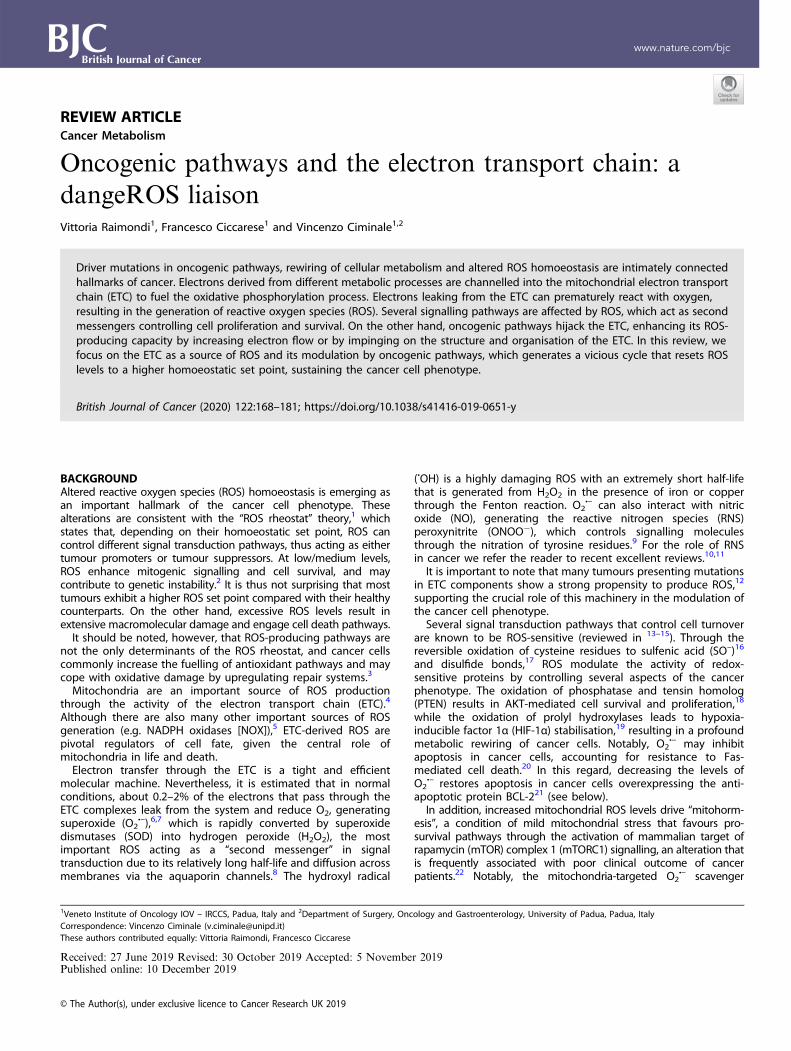
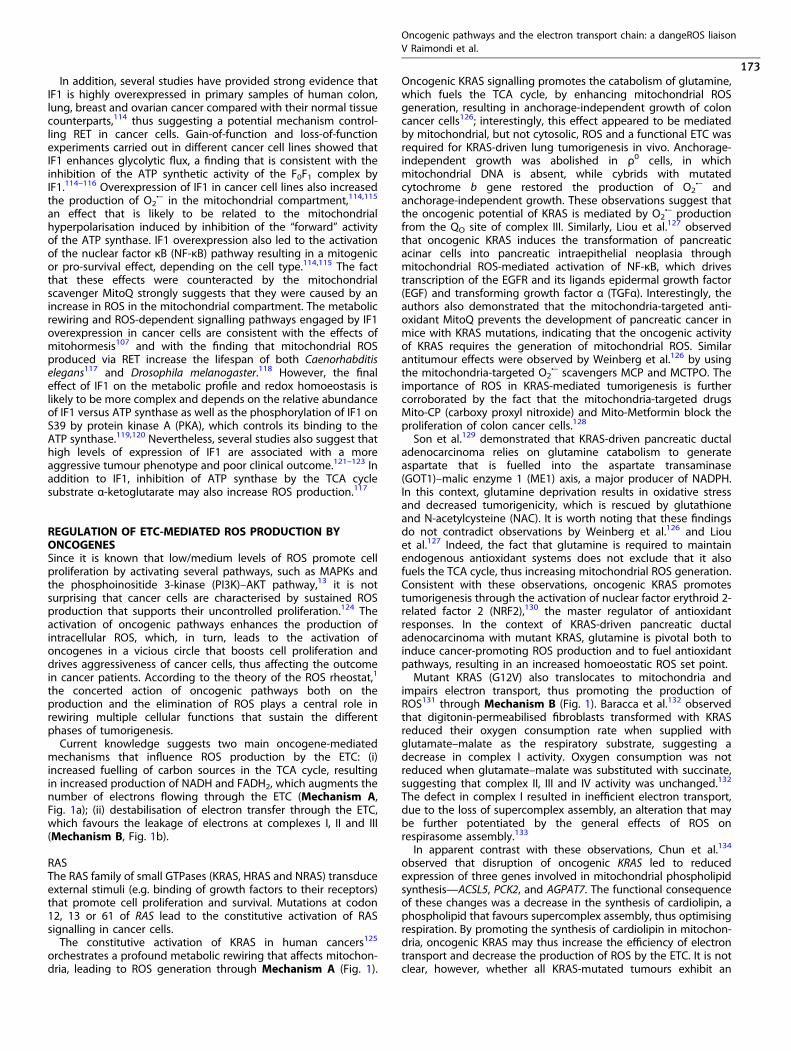
SITES OF ROS PRODUCTION IN THE MITOCHONDRIAL ETCFunctional electron transport provides the bioenergetic fuellingnecessary to sustain tumour initiation, growth and dissemination.However, defects in the ETC may also favour tumorigenesis.The ETC is responsible for the transfer of electrons from reduced
nicotinamide adenine dinucleotide (NADH) and flavin adeninedinucleotide (FADH2), produced in the tricarboxylic acid (TCA)cycle, to oxygen, across complexes I, II, III and IV. This electron flowprovides energy for proton translocation, thus generating atransmembrane potential (Δψm) that drives ATP synthesis by theATP synthase complex (Fig. 1). The proton gradient may be eitherdissipated or increased by the ATP synthase complex whenworking in its ATP-producing or ATP-consuming mode, respec-tively. In mammals, complexes I and III have been identified as themost relevant sites of ROS production within the ETC.24,25
Complex I (or NADH-ubiquinone oxidoreductase, Fig. 1) oxidisesNADH to NAD+ and transfers electrons to the carrier ubiquinone(UbQ, also known as coenzyme Q10), resulting in its reduction toubiquinol (UbQH2) and the translocation of protons across theinner mitochondrial membrane (IMM).26 During this process,single-electron reduction of O2 can occur, leading to thegeneration of O2
•–, which is converted into H2O2 by manganesesuperoxide dismutase (MnSOD). H2O2 can diffuse across themembrane and be reduced to H2O by mitochondrial andcytoplasmic peroxiredoxins, catalases, thioredoxin peroxidasesand glutathione peroxidases.27 Mammalian complex I contains44 subunits, seven of which (ND1, ND2, ND3, ND4, ND4L, ND5 andND6) are coded by the mitochondrial genome, and the others bythe nuclear genome.28 ROS can be generated when the electronsare transferred from NADH to UbQ through the flavin (IF) and theUbQ-reducing (IQ) sites in complex I.Mutations in complex I components may result in increased
production of O2•–, which may sustain ROS-dependent oncogenic
pathways and induce damage of mitochondrial DNA.29 In additionto affecting supercomplex assembly (see below), defects inND2 subunit are known to promote tumorigenesis and metastasisin breast, pancreatic and oral cancers, and head and neckcarcinomas.30 Similar phenotypes were also reported for muta-tions of the ND6 subunit in lung cancer,31 and ND4 in acutemyeloid leukaemia32 and in glioblastoma.33 These mutations areprobably associated with increased ROS generation, which in turnselects for increased fuelling of scavenging pathways andenhanced production of NADPH by the folate pathway, resultingin the promotion of metastatic dissemination.34 Wheaton et al.35
also demonstrated the importance of complex I in cancerprogression by using its inhibitor metformin, which reducedtumorigenesis in vitro and in vivo. Metformin acts upstream of theROS-producing IF site, inhibiting ROS generation from complex Iand reducing electron flow to complex III, thus decreasing theproduction of ROS from it. In hypoxic conditions, NADH depletionand supercomplex disassembly (see below) hamper complex Iactivity, and ROS generation occurs mainly from complex III.36
However, during hypoxia, complex I may produce ROS by reverseelectron transfer (RET, see below). In contrast, the inhibition ofcomplex I at the IQ site by rotenone increases ROS generation andinduces apoptosis of breast cancer cells through the activation ofthe c-Jun N-terminal kinase (JNK) and p38 pathways.37
Complex III (or ubiquinol–cytochromec oxidoreductase, Fig. 1)transfers the electrons received by UbQH2 from complex I andcomplex II to cytochrome c and couples this reaction with proton
translocation across the IMM.38 Complex III is a dimer containingthree highly conserved core subunits: cytochrome b (containingtwo haem groups—bH and bL), cytochrome c1 and the Rieskeiron–sulfur protein (ISP, with an Fe–S cluster). Eight additionalsubunits in the complex are required for its assembly, stability andregulation.39 The reaction catalysed by complex III, known as the“Q-cycle”,40 involves two different binding sites on cytochrome b,QO for UbQH2 and QI for UbQ.In the IIIQO site, electrons may leak and interact with O2, by
producing O2•– both in the matrix and the intermembrane space
(IMS), where it is converted into H2O2 by SOD.41,42 H2O2 can crossthe outer mitochondrial membrane and reach the cytoplasm,where it can act as a signalling molecule. Several studies reportedthat the inhibition of QI site with antimycin A blocks QO–QI
electron transfer, resulting in increased electron leak andconsequent ROS generation at the IIIQO site.43,44 Although it isreported that the inhibition of binding of UbQH2 to the QO site bystigmatellin and myxothiazol prevents the production of ROS incomplex III,43 it is plausible that a block in electron transfer acrosscomplex III could induce RET (see below) and ROS production.In the context of cancer progression, it was shown that
ubiquinol–cytochromec reductase core protein II (UQCR2), animportant subunit of complex III, is upregulated in humantumours, including lung adenocarcinoma and breast cancer.45
Interestingly, UQCR2 negatively regulates p53 levels by inducingits degradation through the interaction with PHB, a p53chaperone. This blunts p53/p21-mediated cell-cycle arrest andsenescence, thus promoting tumorigenesis. Although ROS gener-ated from complex III might stabilise p53, the overexpression ofUQCR2 can revert this effect, by supporting tumour cell growthand dissemination. The role of UQCR2 in tumorigenesis issupported by a study by Shang et al.,46 who demonstrated apositive correlation between UQCR2 overexpression, tumourprogression and poor prognosis in colorectal cancer. Based onthese findings, the authors proposed UQCR2 as a prognosticbiomarker and therapeutic target.A similar role is attributable to ubiquinol–cytochromec reduc-
tase hinge (UQCRH), another subunit of complex III that isoverexpressed in lung adenocarcinoma47 and in hepatocellularcarcinoma.48 UQCRH regulates electron transfer from cytochromec1 to cytochrome c, and its upregulation results in enhanced ROSproduction.47 In hepatocellular carcinoma, UQCRH overexpressionis accompanied by the upregulation of two other subunits ofcomplex III, the previously described UQCR2 and UQCRB. Parket al.48 observed that high expression of these proteins correlateswith poor prognosis, suggesting that they may serve as usefulcancer outcome biomarkers.These observations suggest that inhibitors of ETC complexes
could present a new opportunity for cancer therapy. Consistentwith this notion, complex III inhibitors, such as myxothiazol andantimycin A, block the reoxidation of UbQH2, by impairing theUbQ-dependent processes in breast cancer cells.49 It is thus likelythat inhibition of complex III might revert electron flow, thusinducing massive ROS generation from complex I and leading tocell death.Although complex III was previously considered to play a
prominent role in ROS production, Liu et al.50 proposed that theFMN group in complex I is the leading site in the ETC responsiblefor generating H2O2 from succinate (through RET, see below) andmalate/glutamate.Small amounts of ROS are also produced in the flavin-reducing
site (IIF) within complex II (or succinate–ubiquinone oxidoreduc-tase, Fig. 1),51 a heterotetramer composed of two hydrophilicsubunits (SDHA, SDHB) and two hydrophobic subunits (SDHC,SDHD).52 The SDHA catalytic domain contains the prostheticgroup FAD, which is reduced to FADH2 as a consequence of theoxidation of succinate to fumarate, thus generating electrons thatare mobilised to the SDHB subunit. Here, three Fe–S clusters
Oncogenic pathways and the electron transport chain: a dangeROS liaisonV Raimondi et al.
169
1234567890();,:
NADHNAD+
UbQ
UbQ
Cyt c
FMN
QO
QI
O2 H2OFADH2
FADH2
IIF
Succinate
Fatty
acids
FumarateO2
O2
O2
O2
b
IV
O2 -
O2 -
O2 -
O2 -
GP
DH
PR
OD
H
DH
OD
H
ET
F:Q
O
UbQ
Proline
Dihydroorotate
FADH2 FADH2
FADH2
G3P
RAS
PI3K
BCR/ABL
IMS
MM
Ca2+
IF1ATP
synthaseATP
synthase
NADHNAD+
Succinate
Fumarate
Cyt c
O2H2O
Malate
ADP ATP
c1
O2
Glutamine
Glutamate
OAA
MnSOD
Citrate
Isocitrate
α-KG
TCA cycle
QI
QO
FMN
UbQ
O2
O2 Pyruvate
Acetyl-CoA
Lactate
CuA
a3 CuB
O2
FADH2
IIF
H2O2
RAS
Glu
tam
inol
ysis
MYC
MCL1
AKT
MICU1
MICU2
Ca2+
Ca2+
mTORa
MC
U
MC
U
Increased TCA activity
O2 -
O2 -O2
-
O2 -
III
III
IVa
UbQ
UbQ
NADH
IMS
MM
ATPsynthase
ATPsynthase
Increased mitochondrial
mass
Electron flow/leakage
RET(electrons/UbQ transport)
Activation/repressionby oncogenes
Phosphorylation
ROS — producing sites
Prosthetic groups
III
c1
CuA
a
CuBa3
I II
Δψm
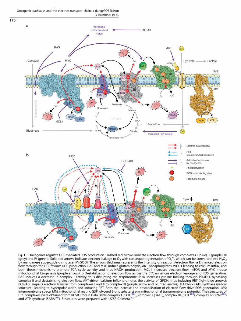
Fig. 1 Oncogenes regulate ETC-mediated ROS production. Dashed red arrows indicate electron flow through complexes I (blue), II (purple), III(grey) and IV (green). Solid red arrows indicate electron leakage to O2 with consequent generation of O2
•–, which can be converted into H2O2by manganese superoxide dismutase (MnSOD). The arrows thickness represents the intensity of reactions/electron flux. a Enhanced electronflow through the ETC favours ROS production. RAS and MYC induce glutaminolysis, AKT phosphorylates MICU1 leading to calcium influx, andboth these mechanisms promote TCA cycle activity and thus NADH production. MCL1 increases electron flow. mTOR and MYC inducemitochondrial biogenesis (purple arrows). b Destabilisation of electron flow across the ETC enhances electron leakage and ROS generation.RAS induces a decrease in complex I activity, thus disrupting the respirasome; PI3K increases proline fuelling through PRODH, bypassingcomplex II and destabilising electron flow; AKT-driven calcium influx promotes the activity of GPDH, thus inducing RET (light-blue arrows);BCR/ABL impairs electron transfer from complexes I and II to complex III (purple arrow and blunted arrows). IF1 blocks ATP synthase (yellowstructure), leading to hyperpolarisation and inducing RET. Both the increase and destabilisation of electron flow drive ROS generation. IMS:intermembrane space. MM: mitochondrial matrix. G3P: glycerol 3-phosphate. Δψm: mitochondrial transmembrane potential. The structures ofETC complexes were obtained from RCSB Protein Data Bank: complex I (5XTD204), complex II (3AEF), complex III (5XTE204), complex IV (5Z62205)and ATP synthase (5ARA206). Structures were prepared with UCSF Chimera.207
Oncogenic pathways and the electron transport chain: a dangeROS liaisonV Raimondi et al.
170
promote electron transfer to the SDHC and SDHD subunitsassociated with the haem and UbQ prosthetic groups. Haemfavours the reduction of UbQ to UbQH2, which shuttles fromcomplex II to complex III, where it is re-oxidised before returningto complex II.53 Although complex II is not involved in thegeneration of the transmembrane potential, it has an importantrole in transferring electrons, bypassing complex I.54 Electronsleaked by FADH2 in the IIF site can contribute to ROS generation,although this event can be decreased when the binding of malateor succinate blocks the site’s access to O2.
55 Many diseases relatedto mutations in complex II proteins are characterised by increasedROS production from the IIF site. An example is represented by theloss of complex II function, which occurs in hereditaryparaganglioma–pheochromocytoma, renal cell carcinoma andgastrointestinal stromal tumours. These mutations corrupt com-plex II’s electron transport activity, leading to succinate accumula-tion, increased ROS generation and decreased ATP productionthrough oxidative phosphorylation (OXPHOS).56,57 The frequentloss of complex II subunit expression in cancer (in particular SDHB)suggests that these subunits might have tumour-suppressorfunctions; indeed, the accumulation of succinate activates theHIF-1α oncogenic pathway.58
On the other hand, complex II is overexpressed in haemato-poietic stem and progenitor cells compared with differentiatedcells. In this cell system, a high ratio between complex II and ATPsynthase maintains an elevated Δψm, whose dissipation compro-mises the self-renewal potential of stem cells. Moreover, highexpression of complex II in haematopoietic stem cells is associatedwith low ROS generation.59 The knowledge that cancer stem cellsalso show decreased levels of ROS60 may suggest that a highcomplex II/ATP synthase ratio could also preserve the cancerstem-cell reservoir.The last enzyme of the ETC, complex IV (or cytochromec oxidase,
Fig. 1), catalyses the reduction of O2 to H2O. In mammals, it is a 14-subunit complex containing three mitochondrial-encoded subunits(I, II and III) and 11 nuclear-encoded regulatory subunits. Two haemgroups, cytochromes a and a3, and two copper centres, CuA andCuB,
61 together with a binding site for cytochrome c constitute theelectron transport machinery of complex IV. Four electrons fromfour cytochromec molecules are transferred to the CuA centre andthen to the cytochrome a and the a3-CuB binuclear centre, byreducing Fe3+ and Cu2+ to Fe2+ and Cu+. These reactions areaccompanied by the passage of four protons across the IMM andthe reduction of O2 into two molecules of H2O, in a very rapidreaction that limits the generation of reactive intermediates.62
Although it is not directly involved in ROS production, complex IVactivity affects the overall electron flow, with an impact on theelectron leakage by previous complexes, and co-operates withoncogenes, such as BCL-2 (see below), to support tumorigenesis.Mutations in subunits I and II of complex IV have been reported inepithelial ovarian cancer, prostate cancers and acute myeloidleukaemia. Interestingly, the expression of complex IV is enhancedby p53, and the frequent co-mutation of complex IV and TP53 inacute myeloid leukaemia patients correlates with worse prog-nosis,63 probably due to increased damage of mitochondrial DNAand mitochondrial dysfunction.
ORGANISATION OF THE ETCSeveral studies of the organisation of the respiratory complexeshave revealed the crucial role of the accessory assembly factors inETC function and in preventing ROS production resulting fromdisassembled OXPHOS subunits.64 The “plasticity model”, pro-posed by Schagger and Pfeiffer,65 describes the coexistence ofboth independent and associated complexes. In particular,complexes I, III and IV (less frequently, complex II) assemble insupercomplexes.66,67 While complex I is mainly found associatedin supercomplexes, 70% of complex III and 15% of complex IV
units are associated with supercomplexes.68 In higher eukaryotes,the most frequent supercomplex, known as the respirasome, iscomposed of one complex I unit, two complex III units and onecomplex IV unit (I1III2IV1).69 Although the mechanism governingthe assembly of the respirasome is not well understood,66,70 thereis evidence that complexes III and IV are able to formautonomously, while mature complex I exists only in therespirasome71 and affects its correct assembly. Several studiesshowed that mitochondrial disorders that reduce the abundanceof complex III or IV are often combined with an impairment incomplex I expression.72–75 Supercomplex assembly optimises thestructural proximity of UbQ with complexes I and III76 and thechannelling of electrons to UbQ and cytochrome c, thusenhancing the efficiency of electron transfer among thecomplexes and reducing ROS generation.However, mitochondrial remodelling during apoptosis or in
hypoxia-induced acidification of the mitochondrial matrix impairsthe assembly of supercomplexes.77,78 Supercomplex assembly isalso connected to OMA1-dependent remodelling of the mito-chondrial cristae.79 The plasticity of ETC organisation can also fine-tune the production of ROS, and consequently, the activity of ROS-sensitive pathways,80 and influences the adaptation of cancer cellsto hypoxia.The balance between supercomplexes and free complexes can
influence cellular metabolism. Although assembly of complex I insupercomplexes promotes electron transport through NADHderived from glucose metabolism, free complex I favours electrontransport through FAD-linked pathways81 (see below). Indeed, byswitching the ETC organisation between free complexes andsupercomplexes, cancer cells can tailor their metabolism todifferent microenvironment conditions.In several cancer types, loss of supercomplex organisation may
favour a metabolic switch towards the Warburg effect pheno-type.82 Consistent with the central role of complex I inrespirasome assembly, cancer cells with mutations in ND2 exhibita glycolytic metabolic profile; interestingly, these cancer cells alsoexhibited increased metastatic potential.30 In addition, the down-regulation of NDUFS1, another complex I subunit, is selected byantiangiogenic therapy in ovarian cancer, leading to a stableglycolytic phenotype and increased aggressiveness.83
Although increased ROS levels and metabolic rewiring causedby the loss of supercomplex organisation promote tumorigenesis,some evidence suggests that supercomplex assembly could alsobe enhanced by oncogenes, thus limiting ROS generation by theETC. For instance, it has been reported that HER2 can translocateto the inner side of the IMM,84 where it could promote increasedassembly of supercomplexes. Interestingly, Rohlenova et al.85
observed that mitochondrial-targeted tamoxifen (MitoTam), whichdisrupts supercomplex assembly, impairs electron flow fromcomplex I to complex III, thus increasing ROS generation and celldeath in HER2-overexpressing breast cancer cells. It is noteworthythat as the electron flow from FAD-linked enzymes to complex IIIis not affected by MitoTam treatment, hypoxic adaptation couldprotect cancer cells from its cytotoxic effect. In addition, KRAScontributes to protect against ROS overload through its involve-ment in the biosynthetic pathway of cardiolipin (see below), anIMM-specific phospholipid. Cardiolipin is sequestered by super-complexes, being protected from degradation, and in turn,stabilises supercomplexes, thus decreasing the electron leakage.86
ETC-ASSOCIATED ENZYMESMetabolic enzymes that link oxidation of their substrates to thereduction of UbQ represent additional ROS-producing sitesconnected to the ETC (Fig. 1B).Proline dehydrogenase (PRODH) is an IMM enzyme responsible
for proline catabolism that transfers electrons through FADH2 toUbQ.87 Proline-derived electrons can leak out to O2, thus
Oncogenic pathways and the electron transport chain: a dangeROS liaisonV Raimondi et al.
171
generating O2•–. The overexpression of PRODH leads to decreased
ETC efficiency and increased ROS generation, due to directcompetition for UbQ between PRODH and complex II. This effectmay be counteracted by succinate,87 due to its higher efficiency inreducing the UbQ pool. Depending on the cell context, PRODHmay act as either a tumour suppressor or an oncogenic factor.88 Incancer cells, by generating ROS, PRODH may trigger apoptosis andsuppress mitogenic pathways (e.g. those triggered by theepidermal growth factor receptor [EGFR] and Wnt–β-catenin).89
Moreover, p53 directly upregulates PRODH, which, in turn, inducesROS, DNA damage and promotes cellular senescence.90 Consistentwith these findings, many tumour types show downregulation ofPRODH expression.89 In prostate cancer, CMYC negatively regulatesPRODH expression via miR23b*, thus promoting tumorigenesis andtumour progression.91 All these effects can be reverted byantioxidants.88 However, under specific metabolic conditions,PRODH can take on a pro-survival role; indeed, glucose deprivationand/or hypoxia upregulate PRODH through AMP-activated proteinkinase (AMPK), independent of HIF-1α/2α, leading to ROSgeneration, and consequently, to the activation of autophagy,which, in this context, exerts a pro-survival role. Proline catabolismthrough PRODH is preferentially used to produce ATP underglucose starvation but not in hypoxia.92 These observationsindicate that PRODH could modulate ETC activity based onnutrient availability and oxygen tension. Moreover, PRODH over-expression was observed in breast cancer metastases, comparedwith primary tumour samples, supporting a role for PRODH inmetastasis formation.93
Mitochondrial glycerol-3-phosphate dehydrogenase (GPDH orGPD2), a component of the glycerophosphate shuttle, isassociated with the ETC on the outer side of the IMM. Severalfunctions have been described for the glycerophosphate shuttle,the most important being the metabolism of glycerol 3-phos-phate, which connects glycolysis, lipogenesis and OXPHOS.GPDH also has an important role in producing ROS by directlyleaking electrons and by inducing RET.94 In this regard, a panel ofprostate-cancer-derived cell lines was shown to possess upregu-lated GPDH activity compared with healthy prostate epithelialcells.95 Furthermore, highly proliferating, undifferentiated cancersdisplay higher levels of GPDH activity than more differentiatedcancers with low proliferative capacity.96
Mitochondrial dihydro-orotate dehydrogenase (DHODH) is aFAD-linked enzyme that mediates the oxidation of dihydro-orotateto orotate, which fuels electrons in the UbQ pool and thus maycontribute to O2
•– production at the QO site of complex III.97 AsDHODH is a key component of the pyrimidine biosynthesispathway, which is required for DNA replication, it has an essentialrole in cancer cell growth.98 Indeed, the oncogenic mitogen-activated protein kinase (MAPK), KRAS and mTOR pathwaysincrease DHODH fuelling by inducing de novo pyrimidinebiosynthesis, while MYC increases the expression of DHODH.99
Although no mutations in DHODH have been reported in cancer,malignant cells are dependent on its enzymatic activity to sustainpyrimidine synthesis.100 In fact, inhibition of DHODH throughspecific antagonists decreases cell growth in many cancer cell lines,in particular acute myeloid leukaemia, and high expression ofDHODH correlates with higher-grade gliomas,98 suggesting thatDHODH inhibition may be a therapeutic strategy to induce tumourcell differentiation. However, it is important to underline that thecatalytic activity of DHODH is strictly connected with a functionalETC, which influences the availability of UbQ. The role of DHODH inROS production is still controversial, as its inhibition can increase ordecrease ROS generation in a context-dependent manner.98
Electron transfer flavoprotein:ubiquinone oxidoreductase (ETF:QO) is an IMM protein that contains FAD and an Fe–S cluster.Together with electron transfer flavoprotein (ETF), ETF:QO catalysesthe transfer of electrons derived from β-oxidation of fatty acids tothe UbQ pool. However, the impairment of electron transfer from
FADH2 to the QO site may lead to O2•– formation.101 ETF:QO activity
depends on functional complexes I and III, as they share theacceptor UbQ pool. However, as described above, ETF-mediatedelectron transfer represents the preferential way to fuel the ETCunder chronic hypoxia, as the activity of complexes I and II isimpaired. Under low oxygen tension, the activation of HIF-1αinduces a metabolic rewiring towards glycolysis and the use ofglutamine as a carbon source to synthesise fatty acids,102 which arecatabolised through β-oxidation to sustain electron flow throughthe ETF system, thus maintaining ETC function and Δψm. Indeed,the inhibition of fatty acid β-oxidation by etomoxir proved to be aneffective strategy to overcome hypoxia-mediated resistance toradiotherapy in cancer cells,103 suggesting an important role forETF:QO in cancer survival under hypoxia. Consistent with thishypothesis, a work by Schuetz et al.104 reported that ETF:QO isoverexpressed in renal carcinoma and oncocytoma, supporting itsputative involvement in tumorigenesis.
REVERSE ELECTRON TRANSPORT (RET)In addition to the canonical “forward” electron flow through thecomplexes of the ETC described above, electrons may betransferred from UbQH2 back to complex I, with the generationof NADH from NAD+. This process, which is termed RET, producesa significant amount of ROS. Although RET is well characterisedin vitro, its relevance in vivo has long been controversial, and onlyrecently has experimental evidence started to accumulate insupport of its role in cell physiology and pathologic processes.105
Several studies indicated that the key conditions that lead toRET are accumulation of reduced UbQ and high Δψm. Electronsderiving from complex II, PRODH, GPDH, ETF:QO and DHODH maydrive over-reduction of UbQ.106 Furthermore, all conditionsinhibiting electron transport downstream of complex III alsoincrease reduction of the UbQ pool. High mitochondrial trans-membrane potential may result from increased H+ extrusion bythe ETC, by the ATP synthase working in its “reverse” ATP-consuming mode, or through the inhibition of the “forward”depolarising/ATP-synthesising mode of action of the ATPsynthase. To this effect, an important role may be played byATPase inhibitory factor 1 (IF1), which, in its active dimeric form,inhibits both the ATP hydrolase and synthase activities of thecomplex.107 Acidification of the mitochondrial matrix favoursinhibition of the ATP hydrolase function by IF1.108,109 Thismechanism plays an important role in hypoxia by preventingATP depletion by the “reverse” mode of function of the ATPsynthase (reviewed by Campanella et al.110).Most studies investigating the relevance of RET in human
pathology have been focused on the tissue damage occurringduring ischaemia/reperfusion in heart attack and stroke. Chou-chani et al.106 recently showed that ischaemic tissues accumulatesuccinate, which, during reperfusion, is oxidised by complex II,giving rise to RET that produces high levels of ROS, which causeextensive macromolecular damage and trigger cell death. Con-sistent with this notion, inhibitors of complex I or complex II (e.g.rotenone and dimethyl-malonate, respectively), as well asantioxidants, protect the heart from ischaemia/reperfusion-mediated tissue damage.106,111
Although the role of RET in cancer has not been directlyinvestigated, it is now clear that oxygen tension in the tumourmicroenvironment is subjected to ample temporal and spatialfluctuations as a result of the chaotic organisation of theneoangiogenic process.112 The resulting imbalance betweenoxygen supply and demand suggests that the tumour tissue isconstantly subjected to recurrent ischaemia/reperfusion cycles inwhich RET might play a major role in altering ROS homoeostasis.ROS accumulation in hypoxia/reperfusion could also be attribu-table to Ca2+ overload and alterations in ETC supercomplexorganisation.113
Oncogenic pathways and the electron transport chain: a dangeROS liaisonV Raimondi et al.
172
In addition, several studies have provided strong evidence thatIF1 is highly overexpressed in primary samples of human colon,lung, breast and ovarian cancer compared with their normal tissuecounterparts,114 thus suggesting a potential mechanism control-ling RET in cancer cells. Gain-of-function and loss-of-functionexperiments carried out in different cancer cell lines showed thatIF1 enhances glycolytic flux, a finding that is consistent with theinhibition of the ATP synthetic activity of the F0F1 complex byIF1.114–116 Overexpression of IF1 in cancer cell lines also increasedthe production of O2
•– in the mitochondrial compartment,114,115
an effect that is likely to be related to the mitochondrialhyperpolarisation induced by inhibition of the “forward” activityof the ATP synthase. IF1 overexpression also led to the activationof the nuclear factor κB (NF-κB) pathway resulting in a mitogenicor pro-survival effect, depending on the cell type.114,115 The factthat these effects were counteracted by the mitochondrialscavenger MitoQ strongly suggests that they were caused by anincrease in ROS in the mitochondrial compartment. The metabolicrewiring and ROS-dependent signalling pathways engaged by IF1overexpression in cancer cells are consistent with the effects ofmitohormesis107 and with the finding that mitochondrial ROSproduced via RET increase the lifespan of both Caenorhabditiselegans117 and Drosophila melanogaster.118 However, the finaleffect of IF1 on the metabolic profile and redox homoeostasis islikely to be more complex and depends on the relative abundanceof IF1 versus ATP synthase as well as the phosphorylation of IF1 onS39 by protein kinase A (PKA), which controls its binding to theATP synthase.119,120 Nevertheless, several studies also suggest thathigh levels of expression of IF1 are associated with a moreaggressive tumour phenotype and poor clinical outcome.121–123 Inaddition to IF1, inhibition of ATP synthase by the TCA cyclesubstrate α-ketoglutarate may also increase ROS production.117
REGULATION OF ETC-MEDIATED ROS PRODUCTION BYONCOGENESSince it is known that low/medium levels of ROS promote cellproliferation by activating several pathways, such as MAPKs andthe phosphoinositide 3-kinase (PI3K)–AKT pathway,13 it is notsurprising that cancer cells are characterised by sustained ROSproduction that supports their uncontrolled proliferation.124 Theactivation of oncogenic pathways enhances the production ofintracellular ROS, which, in turn, leads to the activation ofoncogenes in a vicious circle that boosts cell proliferation anddrives aggressiveness of cancer cells, thus affecting the outcomein cancer patients. According to the theory of the ROS rheostat,1
the concerted action of oncogenic pathways both on theproduction and the elimination of ROS plays a central role inrewiring multiple cellular functions that sustain the differentphases of tumorigenesis.Current knowledge suggests two main oncogene-mediated
mechanisms that influence ROS production by the ETC: (i)increased fuelling of carbon sources in the TCA cycle, resultingin increased production of NADH and FADH2, which augments thenumber of electrons flowing through the ETC (Mechanism A,Fig. 1a); (ii) destabilisation of electron transfer through the ETC,which favours the leakage of electrons at complexes I, II and III(Mechanism B, Fig. 1b).
RASThe RAS family of small GTPases (KRAS, HRAS and NRAS) transduceexternal stimuli (e.g. binding of growth factors to their receptors)that promote cell proliferation and survival. Mutations at codon12, 13 or 61 of RAS lead to the constitutive activation of RASsignalling in cancer cells.The constitutive activation of KRAS in human cancers125
orchestrates a profound metabolic rewiring that affects mitochon-dria, leading to ROS generation through Mechanism A (Fig. 1).
Oncogenic KRAS signalling promotes the catabolism of glutamine,which fuels the TCA cycle, by enhancing mitochondrial ROSgeneration, resulting in anchorage-independent growth of coloncancer cells126; interestingly, this effect appeared to be mediatedby mitochondrial, but not cytosolic, ROS and a functional ETC wasrequired for KRAS-driven lung tumorigenesis in vivo. Anchorage-independent growth was abolished in ρ0 cells, in whichmitochondrial DNA is absent, while cybrids with mutatedcytochrome b gene restored the production of O2
•– andanchorage-independent growth. These observations suggest thatthe oncogenic potential of KRAS is mediated by O2
•– productionfrom the QO site of complex III. Similarly, Liou et al.127 observedthat oncogenic KRAS induces the transformation of pancreaticacinar cells into pancreatic intraepithelial neoplasia throughmitochondrial ROS-mediated activation of NF-κB, which drivestranscription of the EGFR and its ligands epidermal growth factor(EGF) and transforming growth factor α (TGFα). Interestingly, theauthors also demonstrated that the mitochondria-targeted anti-oxidant MitoQ prevents the development of pancreatic cancer inmice with KRAS mutations, indicating that the oncogenic activityof KRAS requires the generation of mitochondrial ROS. Similarantitumour effects were observed by Weinberg et al.126 by usingthe mitochondria-targeted O2
•– scavengers MCP and MCTPO. Theimportance of ROS in KRAS-mediated tumorigenesis is furthercorroborated by the fact that the mitochondria-targeted drugsMito-CP (carboxy proxyl nitroxide) and Mito-Metformin block theproliferation of colon cancer cells.128
Son et al.129 demonstrated that KRAS-driven pancreatic ductaladenocarcinoma relies on glutamine catabolism to generateaspartate that is fuelled into the aspartate transaminase(GOT1)–malic enzyme 1 (ME1) axis, a major producer of NADPH.In this context, glutamine deprivation results in oxidative stressand decreased tumorigenicity, which is rescued by glutathioneand N-acetylcysteine (NAC). It is worth noting that these findingsdo not contradict observations by Weinberg et al.126 and Liouet al.127 Indeed, the fact that glutamine is required to maintainendogenous antioxidant systems does not exclude that it alsofuels the TCA cycle, thus increasing mitochondrial ROS generation.Consistent with these observations, oncogenic KRAS promotestumorigenesis through the activation of nuclear factor erythroid 2-related factor 2 (NRF2),130 the master regulator of antioxidantresponses. In the context of KRAS-driven pancreatic ductaladenocarcinoma with mutant KRAS, glutamine is pivotal both toinduce cancer-promoting ROS production and to fuel antioxidantpathways, resulting in an increased homoeostatic ROS set point.Mutant KRAS (G12V) also translocates to mitochondria and
impairs electron transport, thus promoting the production ofROS131 through Mechanism B (Fig. 1). Baracca et al.132 observedthat digitonin-permeabilised fibroblasts transformed with KRASreduced their oxygen consumption rate when supplied withglutamate–malate as the respiratory substrate, suggesting adecrease in complex I activity. Oxygen consumption was notreduced when glutamate–malate was substituted with succinate,suggesting that complex II, III and IV activity was unchanged.132
The defect in complex I resulted in inefficient electron transport,due to the loss of supercomplex assembly, an alteration that maybe further potentiated by the general effects of ROS onrespirasome assembly.133
In apparent contrast with these observations, Chun et al.134
observed that disruption of oncogenic KRAS led to reducedexpression of three genes involved in mitochondrial phospholipidsynthesis—ACSL5, PCK2, and AGPAT7. The functional consequenceof these changes was a decrease in the synthesis of cardiolipin, aphospholipid that favours supercomplex assembly, thus optimisingrespiration. By promoting the synthesis of cardiolipin in mitochon-dria, oncogenic KRAS may thus increase the efficiency of electrontransport and decrease the production of ROS by the ETC. It is notclear, however, whether all KRAS-mutated tumours exhibit an
Oncogenic pathways and the electron transport chain: a dangeROS liaisonV Raimondi et al.
173
increase in cardiolipin levels in mitochondria. These apparentlyparadoxical effects may be explained by the fact that the studiesby Weinberg et al. 126 and Baracca et al. 132 used healthy cellstransfected with a construct coding for mutant KRAS, while Chunet al.134 used KRAS-mutated colon cancer cells (HCT116) in whichmutated KRAS was removed by knockout. In the latter case, theimpact of oncogenic KRAS on ETC activity was studied in thecontext of a heavily mutated genetic landscape (4,288 mutationsare reported in COSMIC for HCT116), in which the acquisition ofoncogenic KRAS could have a protective role by promoting thesynthesis of cardiolipin and reducing the generation of ROS fuelledby other oncogenes. Instead, introducing mutant KRAS in normalcells permitted a more direct investigation of the effects of mutantKRAS on the ETC. Further investigations will be needed to establishthe role of oncogenic KRAS signalling on supercomplex assemblyand respiratory efficiency and its possible impact on cancer.Recent studies suggest that the effects of KRAS on redox
homoeostasis are required for maintaining the cancer phenotypeand may thus represent attractive therapeutic targets for KRAS-driven cancers, which still represent a clinical challenge due to thelack of effective therapies. In this regard, Shaw et al.135 demon-strated that induction of oxidative stress through the smallmolecule lanperisone kills mouse embryonic fibroblasts trans-fected with mutant KRAS and restrains their growth in vivo.Interestingly, Iskandar et al.136 observed that hyperactivation ofmutant KRAS with the small molecule C1 leads to the activation ofthe PI3K–AKT pathway, which enhances ROS generation (seebelow), leading to mitochondrial dysfunction, cell death andblockade of tumours with mutant KRAS. These effects are bluntedby NAC, indicating that the generation of ROS through theKRAS–AKT axis is necessary to mediate C1 cytotoxicity, corrobor-ating the feasibility of a ROS-based anticancer strategy to targetKRAS-driven tumours.
MYCThe MYC family of transcription factors (CMYC, LMYC and NMYC)controls cell proliferation and apoptosis by regulating a largenumber of RNA polymerase I-, II- and III-dependent genes.137,138
MYC amplification is commonly observed in neuroblastoma and inbreast, ovary, prostate and uterine cancers, while the CMYC-immunoglobulin translocation is a hallmark of Burkitt’slymphoma.139
Like RAS, MYC induces complex metabolic rewiring in cancercells, which is achieved through the stimulation of glycolysis,140
mitochondrial biogenesis141 and glutaminolysis.142 Li et al.141
demonstrated that inducible expression of MYC in the B-cell lineP493-6 increases mitochondrial mass and enhances the oxygenconsumption rate, an indicator of ETC activity. These effects are inpart mediated by the induction of the mitochondrial transcriptionfactor A (TFAM) by MYC,141 thus possibly driving ROS productionvia enhanced electron flow through the ETC.Observations by Wise et al.142 in glioma cell lines indicated
that MYC controls a transcriptional programme that promotesthe catabolism of glutamine as a carbon source to fuel the TCAcycle, thus sustaining ROS production by the ETC. Vafa et al.143
observed that overexpression of CMYC in human fibroblastsinduced an increase in ROS levels, which correlated with theformation of foci of DNA damage; the antioxidant NAC reducedboth of these effects. As MYC drove cell-cycle entry andproliferation even if DNA was damaged, these observationssuggest a mechanism of MYC-induced genomic instability andselection for p53 loss (a frequent alteration in CMYC-driventumours144), both of which fuel clonal evolution andtumour progression. The role of ROS production followingMYC amplification is supported by the fact that exogenousantioxidants (vitamin C and Tiron) inhibited transformation ofMYC-overexpressing NIH/3T3 fibroblasts.145 Moreover, bluntingROS through mitochondria-targeted vitamin E blocked the
proliferation and induced cell death in osteogenic sarcomacells.146
In chemotherapy-resistant triple-negative breast cancer, theupregulation of MYC with the anti-apoptotic protein MCL1 selectsfor a stem-cell phenotype that is dependent on mitochondrialrespiration.147 Accumulation of MCL1 in the mitochondrial matrixincreases the ability of complexes I, II and IV to transfer electrons.The concerted action of MYC on mitochondrial mass and MCL1 onthe ETC resulted in HIF-1α stabilisation, selection of cancer stemcells and resistance to chemotherapy.147 However, as in the case forKRAS, the effects of MYC on ROS homoeostasis are a matter ofbalance, as MYC may also upregulate mitochondrial peroxiredoxin 3to protect cells from ROS in hypoxia.148 Moreover, NADPHproduction through serine and one-carbon metabolism protectshypoxic breast cancer stem cells from oxidative stress.149 Thisevidence suggests that ROS trigger the selection of cancer stem cellsthrough the upregulation of increased antioxidant defences, whichis consistent with the lower ROS set point of cancer stem cells.60
MYC may also act through Mechanism B by upregulating theexpression of several mitochondrial nuclear-encoded proteins,resulting in an imbalance between ETC subunits coded by thenuclear and mitochondrial genomes that leads to the generationof misassembled respiratory complexes.150 Interestingly, Herrmannet al.151 observed a strong correlation between the progressionfrom normal prostate epithelium to invasive prostate carcinomawith imbalanced nuclear-encoded versus mitochondrion-encodedsubunits of complex IV. This suggests that misassembledrespiratory complexes promote tumour progression. MYC alsoaffects ROS production by impinging on cancer cell metabolism. Inparticular, as mentioned above, MYC promotes the expression of aplethora of genes involved in nucleotide synthesis, comprisingDHODH,99 thus triggering the generation of ROS by destabilisingthe electron flow across the ETC.MYC overexpression is associated with an increased prolifera-
tion rate in breast cancer,152 and MYC amplification in luminal Abreast cancer is associated with poor survival and resistance toendocrine therapy.153 Although these studies did not evaluatewhether ROS are involved in the aggressiveness of MYC-drivenbreast cancer, it is known that high ROS levels are associated withresistance to endocrine therapy.154 The impact of MYC-driven ROSproduction on the clinical outcome of cancer needs to be furtherstudied in the future. Unfortunately, MYC is still an undruggabletarget. However, the dependency of MYC-driven tumours onNADPH production through serine metabolism and one-carbonmetabolism implies that inhibition of these pathways could be aneffective anticancer strategy for patients affected by thesemalignancies.
PI3K–AKT–mTORConsistent with their role in apoptosis suppression, cell prolifera-tion, metabolism and anabolic reactions, the interconnectedPI3K–AKT and mTOR pathways are hyperactivated in almost 40%of all human cancers.155–157
As the PI3K–AKT–mTOR pathway plays a pivotal role in theinduction of the Warburg effect158 and in the inhibition ofautophagy,159 cancers with hyperactivation of this pathwayaccumulate dysfunctional ROS-producing mitochondria that arenot eliminated by autophagy.The metabolism of non-essential amino acids is an important
feature of metabolic rewiring in cancer cells and in highlyproliferating cells. In fact, cancer cells metabolise non-essentialamino acids to obtain nucleotides and lipids, preserve redoxhomoeostasis and control epigenetic regulation.160 Among the 11non-essential amino acids, glutamine, serine and proline playimportant roles in tumorigenesis. Proline synthesis mediated byΔ1-pyrroline-5-carboxylate (P5C) reductases (PYCRs) and prolinedegradation through PRODH constitute a “proline cycle” betweenthe cytosol and mitochondria.88 In EGFR-mutated non-small-cell
Oncogenic pathways and the electron transport chain: a dangeROS liaisonV Raimondi et al.
174
lung cancer, constitutive downstream activation of the PI3Kpathway drives proline synthesis, which fuels EGFR-regulatedproline oxidation.161 The activity of PRODH decreases theefficiency of mitochondrial electron transport, driving theproduction of ROS from the QO site of complex III throughMechanism B. These findings suggest that proline metabolismcould play an important role in non-small-cell lung cancer withEGFR mutations.Although the PI3K–AKT–mTOR pathway is associated with the
induction of aerobic glycolysis, Marchi et al.162 provided amechanism through which AKT could mediate ROS generationthrough Mechanism A. These authors observed thatmitochondria-localised AKT phosphorylates MICU1, a regulatorysubunit of the mitochondrial calcium uniporter (MCU), resulting inthe destabilisation of the MICU1–MICU2 heterodimer, thus leadingto increased calcium influx in mitochondria.162 Decreased expres-sion of MICU1 following its phosphorylation was associated withincreased levels of mitochondrial ROS and enhanced in vivogrowth of cancer cells. Mitochondrial calcium overload couldaccount for increased ROS generation through mitochondrialdysfunction, which, however, may trigger permeability-transition-pore-mediated cell death.163 Alternatively, increased calcium levelsin mitochondria could drive ROS production by stimulatingenzymes of the TCA cycle and OXPHOS, thus accelerating oxygenconsumption and generation of ROS by the ETC164 throughMechanism A. Moreover, mitochondrial calcium also increases theactivity of GPDH, producing ROS by direct leaking of electrons andby inducing RET towards complex II and complex I94 (MechanismB). The activity of the PI3K–AKT–mTOR pathway is controlled bythe tumour suppressor PTEN, which is inactivated in several humancancer types. Observations by Marchi et al.162 highlight the centralrole of PTEN status in the AKT-mediated effect on MICU1.Interestingly, ROS inactivate PTEN through oxidation, thus favour-ing activation of AKT that phosphorylates MICU1 leading tocalcium uptake in mitochondria, and calcium drives the productionof ROS by the ETC, thus establishing a vicious circle sustainingactivation of AKT and boosting tumour progression. In line withthis scenario, scavenging mitochondrial O2
•– through MitoTEMPOblunted the activation of AKT, reverted the Warburg effect andinduced death in melanoma cells.165 Moreover, Variar et al.166
observed that the mitochondria-targeted antioxidant Mito-CPenhances apoptotic cell death in a Burkitt’s lymphoma cell lineby decreasing AKT activation and HIF-1α stabilisation underhypoxia, suggesting the possibility to block hypoxic adaptationin cancer cells by decreasing ROS generated by the ETC.In a study of breast cancer cells, Jin et al.167 recently
demonstrated that PI3K–AKT-mediated inactivation of glycogensynthase kinase-3β (GSK-3β) through phosphorylation induces anabnormal activity of complexes I and III, thus altering electron flowand enhancing ROS production through Mechanism B. Theresulting ROS released in the tumour microenvironment impairedthe cytotoxic activity of NK cells by oxidising a serine residue inthe initiation factor eIF2B, leading to downregulation of NKG2Dand its ligands.167 These results provide evidence for an AKT/ROS-mediated mechanism to inhibit innate immune response in thetumour microenvironment. Interestingly, inactivation of GSK-3βalso leads to stabilisation of CMYC,168 which can further enhancegeneration of ROS by the ETC. It remains to be elucidated whetherthe inhibition of GSK-3β by AKT requires ROS-mediated PTENinactivation.mTOR is a central kinase that integrates energy sensing and
anabolic pathways, co-ordinating protein synthesis and cellgrowth.169 Synthesis of novel cellular components is an energy-consuming process; thus, it is not surprising that mTOR promotesmitochondrial metabolism by indirectly increasing the levels ofnuclear-encoded mitochondrial proteins. For instance, mTORpromotes the formation of functional complexes between thetranscription factor yin-yang 1 (YY1) and its cofactor peroxisome-
proliferator-activated receptor coactivator 1α (PGC-1α), whichdrives expression of many genes encoding mitochondrial proteins,including cytochrome c,170 resulting in increased mitochondrialrespiration and ROS production through Mechanism A. Moreover,mTOR co-operates with oestrogen-related receptor α to promotethe transcription of genes involved in OXPHOS and in the TCAcycle.171 Furthermore, through the inactivation of 4E-BP proteins,mTOR upregulates nuclear-encoded subunits of respiratorycomplex I and ATP synthase, thus increasing mitochondrialrespiration.172 Goo et al.173 observed that, in the context of PTENinactivation, hyperactivated AKT is associated with phosphoryla-tion of 4E-BP1, increased activity of complexes I, III and IV andaugmented oxygen consumption. These results suggest thatmTOR could enhance mitochondrial respiration, and hence, ROSproduction through Mechanism A. Hyperactivation of thePI3K–AKT–mTOR pathway could also result in an imbalancebetween nuclear-encoded and mitochondrial-encoded subunitsof the respiratory complexes, as observed for MYC, leading to theproduction of misassembled complexes, loss of supercomplexesand increased ROS production through Mechanism B.Given its central role in co-ordinating metabolism, mTOR is at
the crossroads between anabolic pathways and mitogenicsignalling of cancer cells. mTORC1 phosphorylates S6K1, whichactivates carbamoyl-phosphate synthetase 2, aspartate transcar-bamoylase, dihydro-orotase (CAD) through phosphorylation onS1859. CAD is a rate-limiting enzyme as it catalyses the first threesteps of de novo pyrimidine synthesis, by generating dihydro-orotate from glutamine,174 thus fuelling DHODH, which producesROS. Notably, the oxidation of dihydro-orotate to orotateintegrates nucleotide synthesis, which is required to sustain theuncontrolled proliferation of cancer cells, with ROS production bythe ETC, which drives cancer initiation and progression. BesidesmTOR, KRAS, MYC, AKT and other oncogenes also converge onpyrimidine synthesis. Interestingly, Hail et al.175 observed thatinhibition of DHODH through terifluonomide decreases mitochon-drial ROS levels and has a cytostatic effect in prostatecancer cells.Future studies should be aimed at investigating whether adecreased nucleotide pool or decreased ROS levels may accountfor the anticancer activity of DHODH inhibitors, as well as the roleof DHODH-produced ROS in cancer development.The hyperactivation of the PI3K–AKT–mTOR pathway is a
marker of poor prognosis in several human cancers, such asoesophageal squamous cell carcinoma176 and breast cancer.177 Yuet al.178 recently observed that pterostilbene, an antioxidantcompound primarily found in blueberries, slows down theprogression of mantle cell lymphoma by targetingthe PI3K–AKT–mTOR pathway. However, pterostilbene alsodirectly affects apoptosis and the cell cycle, thus rendering theinterpretation of these results more complex; the impact of ROSon cancer in the context of PI3K–AKT–mTOR hyperactivation thusdeserves further investigation.
BCR/ABLThe t(9,22) translocation that is pathognomonic of chronicmyeloid leukaemia gives rise to the Philadelphia chromosomeand to the chimaeric gene BCR/ABL, coding for a constitutivelyactive tyrosine kinase that transduces mitogenic and anti-apoptotic signals to cancer cells.179
The BCR/ABL fusion protein promotes the production of ROS bythe ETC partly through the activation of the PI3K–mTOR pathway.Kim et al.180 observed that glucose metabolism is involved in thegeneration of ROS in BCR/ABL-transformed cells. Treatment with2-deoxyglucose, the BCR/ABL inhibitor imatinib mesylate orrotenone reduced the production of ROS. The same effect wasobtained with wortmannin and rapamycin, which inhibit PI3K andmTORC1, respectively, indicating the tight connections amongBCR/ABL, PI3K, mTOR, glucose metabolism and ROS production bythe ETC through Mechanism A. The decrease in ROS levels
Oncogenic pathways and the electron transport chain: a dangeROS liaisonV Raimondi et al.
175
following rotenone treatment suggests the involvement of RET(Mechanism B) in ROS generation by BCR/ABL. Direct action ofBCR/ABL on the activity of the ETC has also been observed.181
Nieborowska-Skorska et al.181 observed a sharp decrease inelectron flow rates between complexes I and II and between IIand III, with an increase in O2
•– production through Mechanism B,in BCR/ABL-expressing myeloid precursors. This ROS productionwas decreased by the mitochondria-targeted antioxidant MitoQand sustained by complex III, as demonstrated by the rescue effectof the complex III inhibitors myxothiazol, stigmatellin andantimycin A. The small GTPase Rac2 was found to promote ROSgeneration by complex III, as Rac2 knockout substantially reducedmitochondrial O2
•– levels and oxidative stress in BCR/ABL-expressing cells.181 The authors noted that this mechanismunderlies ROS production by the ETC in leukaemia cells with avariety of genetic alterations (FLT3–ITD, TEL–ABL1, TEL–JAK2, TEL/PDGFβR, TEL–TRKC, BCR–FGFR1 and mutated JAK2). Theseobservations indicate that different types of leukaemia cells maypromote genomic instability and progression through theactivation of Rac2, which interferes with electron transport fromcomplexes I to III and II to III, ultimately inducing leakage ofelectrons from complex III and O2
•– generation.
BCL-2The B-cell lymphoma-2 (BCL-2) family includes several proteinsthat counteract intrinsic apoptosis by binding pro-apoptoticproteins along the IMM.182 Many tumour types, including breastcancer, prostate cancer, B-cell lymphomas and colorectal adeno-carcinomas, display BCL-2 overexpression.182,183 Several lines ofevidence suggest that the upregulation of BCL-2, besides directlyblocking apoptosis, creates a pro-oxidant state that promotes cellsurvival. Chen and Pervaiz184,185 showed that the overexpressionof BCL-2 in leukaemia cells regulates mitochondrial respiration byaffecting ETC activity through direct interaction of BCL-2 with thecomplex IV subunits Va and Vb, by promoting correct assembly ofthe complex in the IMM and thus upregulating its activity. Basedon these findings, the authors suggested that BCL-2 increases ETCactivity and O2
•– production, leading to a pro-oxidant milieu thatfavours cell survival. Interestingly, during hypoxia, in the presenceof BCL-2, the relative abundance of the subunit Va is higher thanthat of subunit Vb, leading to reduced complex IV activity andmitochondrial respiration, which, in this context, maintains themitochondrial redox state unchanged, thus preventing oxidativestress that would otherwise favour cell death.185 This scenario issupported by the observation that decreasing levels of O2
•– triggerapoptosis in BCL-2-overexpressing cancer cells21 and suggeststhat the balance between O2
•– and H2O2 could modulate cancercell survival through O2
•–-mediated inhibition of apoptosis.186
Furthermore, the establishment of a mild pro-oxidant milieuprevents the dephosphorylation of BCL-2 on S70 residue, thusimproving BCL-2 binding with BAX and cell survival.187
CONCLUDING REMARKSIn the 1920s, Otto Warburg postulated defective mitochondrialrespiration as the cause of cancer.188 Although the discovery ofgenetic alterations in oncogenes and tumour-suppressor geneschanged our perspective on cancer pathogenesis, many recentstudies identified pro-tumorigenic pathways that affect ROSgeneration by the ETC, thus integrating genetic and bioenergeticalterations of cancer cells in a unified scenario.The ETC is the principal source of mitochondrial ROS. Activated
RAS, MYC, PI3K–AKT–mTOR and BCR/ABL are examples ofoncogenic pathways that affect the ETC to enhance ROSproduction. Oncogenes may control this process through twomain mechanisms: increasing TCA cycle fuelling and mitochon-drial mass (here defined as Mechanism A); increasing electronleakage from the ETC by either altering its organisation (e.g.
disrupting the respirasome) or promoting metabolism throughETC-associated enzymes (defined as Mechanism B).It is worth noting that different oncogenic pathways co-operate to
produce the cancer phenotype. In fact, KRAS, MYC andPI3K–AKT–mTOR constitute an interconnected network that fine-tunes the generation of ROS by the ETC. mTORC2 hyperpho-sphorylates AKT, thus enhancing ROS production through theabove-mentioned mechanisms and MYC protein levels through theinhibition of GSK-3β. MYC, in turn, can blunt the production of ROStriggered by mTORC1, thus curbing ROS overload in cancer cells. Inthis regard, Hartleben et al.189 observed that MYC-driven upregula-tion of tuberous sclerosis complex 1 (TSC1) in Burkitt’s lymphomacells represses the activation of mTORC1, thus limiting theproduction of ROS by the ETC. In contrast, oncogenic KRAS inducesa metabolic shift resulting in enhanced glycolytic flux and lactateproduction that inhibits the interaction of TSC2 with Rheb, thusleading to mTORC1 activation.190 The observation that oncogenicKRAS promotes MYC protein stability191 further complicates thisscenario. This complex network of interactions may act as a rheostatto optimise a ROS set point that exploits the tumour-promotingactivity of ROS while negating their anticancer effects.One of the most intriguing features of cancer cells is their
metabolic plasticity. The recently described hybrid metabolicphenotype, in which the Warburg effect and OXPHOS coexist,provides cancer cells with the ability to adapt their metabolism todifferent microenvironments.192 Mitochondrial ROS play a centralrole in this process, through the stabilisation of HIF-1α, whichpromotes glycolysis, and AMPK, which promotes OXPHOS andfatty acid β-oxidation. It is worth noting that the hybrid metabolicphenotype is promoted by MYC and by fatty acid β-oxidation. Theimportance of β-oxidation suggests a crucial role of the ETC-associated enzyme ETF:QO in tumorigenesis.Although the mitogenic role of ROS suggests that dietary
antioxidants could prove beneficial in cancer prevention,193
experimental and clinical evidence indicates that they favourcancer progression194–196 and impair the efficacy of chemother-apy and radiotherapy.197 Consistent with this notion, non-small-cell lung cancer cells with loss-of-function mutations in thetumour-suppressor liver kinase 1 (LKB1) are more sensitive tooxidative stress.198,199 Indeed, LKB1-mutated patients respondbetter than wild-type patients to therapies with platinum-basedanticancer drugs,200 which are potent inducers of ROS.201 Thecontrasting effect of untargeted versus mitochondria-targetedantioxidants on tumorigenesis, although not generalisable to alltumour models, further corroborates the concept that tumourprogression requires the generation of mitochondrial ROS and thelimitation of cytosolic ROS, to maximise mitogenic signalling andavoid oxidative damage. In this regard, the inhibition ofendogenous antioxidant systems in order to increase ROS levelsabove the toxic threshold represents a promising effectivestrategy to selectively kill cancer cells.202,203
The fact that the described oncogenes are deregulated in a highproportion of extremely aggressive cancers, such as non-small-celllung cancer, pancreatic ductal adenocarcinoma and triple-negative breast cancer, supports a pivotal role for mitochondrialROS as determinants of the clinical outcome.Finally, although well characterised in the context of ischaemia/
reperfusion models, the role of RET in cancer is still poorlyunderstood and deserves thorough studies. The fact that twocommon alterations of cancer cells, i.e. transient intra-tumourhypoxia and IF1 overexpression, are potent inducers of RETprovides a strong rationale for a crucial role for RET in the rewiringof redox homoeostasis in cancer.
ACKNOWLEDGEMENTSWe are grateful to Donna Mia D’Agostino for paper revising and helpful discussion.We thank Alessandro Grinzato for helping with the figure. Molecular graphics of ETC
Oncogenic pathways and the electron transport chain: a dangeROS liaisonV Raimondi et al.
176
complexes were performed with UCSF Chimera, developed by the Resource forBiocomputing, Visualization and Informatics at the University of California, SanFrancisco, with support from NIH P41-GM103311.
AUTHOR CONTRIBUTIONSV.R. and F.C. wrote the paper and prepared the figure, V.C. prepared the paragraphon RET and revised the final version of the paper.
ADDITIONAL INFORMATIONEthics approval and consent to participate Not applicable.
Consent to publish Not applicable.
Data availability Not applicable.
Competing interests The authors declare no competing interests.
Funding information V.C. is supported by the Associazione Italiana per la Ricerca sulCancro (Italian Association for Cancer Research)—IG 17794.
Note This work is published under the standard license to publish agreement. After12 months the work will become freely available and the license terms will switch toa Creative Commons Attribution 4.0 International (CC BY 4.0).
Publisher’s note Springer Nature remains neutral with regard to jurisdictional claimsin published maps and institutional affiliations.
REFERENCES1. Maryanovich, M. & Gross, A. A. ROS rheostat for cell fate regulation. Trends Cell
Biol. 23, 129–134 (2013).2. Yalcin, S., Marinkovic, D., Mungamuri, S. K., Zhang, X., Tong, W., Sellers, R. et al.
ROS-mediated amplification of AKT/mTOR signalling pathway leads to myelo-proliferative syndrome in Foxo3(-/-) mice. Embo J. 29, 4118–4131 (2010).
3. Scott, T. L., Rangaswamy, S., Wicker, C. A. & Izumi, T. Repair of oxidative DNAdamage and cancer: recent progress in DNA base excision repair. Antioxid.Redox Signal 20, 708–726 (2014).
4. Zhao, R. Z., Jiang, S., Zhang, L. & Yu, Z. B. Mitochondrial electron transport chain,ROS generation and uncoupling (Review). Int. J. Mol. Med. 44, 3–15 (2019).
5. Meitzler, J. L., Antony, S., Wu, Y., Juhasz, A., Liu, H., Jiang, G. et al. NADPHoxidases: a perspective on reactive oxygen species production in tumor biology.Antioxid. Redox Signal 20, 2873–2889 (2014).
6. Turrens, J. F. Mitochondrial formation of reactive oxygen species. J. Physiol. 552(Pt 2), 335–344 (2003).
7. Cadenas, E. & Davies, K. J. Mitochondrial free radical generation, oxidative stress,and aging. Free Radic. Biol. Med 29, 222–230 (2000).
8. Lennicke, C., Rahn, J., Lichtenfels, R., Wessjohann, L. A. & Seliger, B. Hydrogenperoxide - production, fate and role in redox signaling of tumor cells. CellCommun. Signal 13, 39 (2015).
9. Liaudet, L., Vassalli, G. & Pacher, P. Role of peroxynitrite in the redox regulationof cell signal transduction pathways. Front. Biosci. 14: 4809–4814 (2009).
10. Kruk, J. & Aboul-Enein, H. Y. Reactive oxygen and nitrogen species in carcino-genesis: implications of oxidative stress on the progression and development ofseveral cancer types. Mini Rev. Med Chem. 17, 904–919 (2017).
11. Moldogazieva, N. T., Lutsenko, S. V. & Terentiev, A. A. Reactive oxygen andnitrogen species-induced protein modifications: implication in carcinogenesisand anticancer therapy. Cancer Res 78, 6040–6047 (2018).
12. Porporato, P. E., Filigheddu, N., Pedro, J. M. B., Kroemer, G. & Galluzzi, L. Mito-chondrial metabolism and cancer. Cell Res 28, 265–280 (2018).
13. Zhang, J., Wang, X., Vikash, V., Ye, Q., Wu, D., Liu, Y. et al. ROS and ROS-mediatedcellular signaling. Oxid. Med Cell Longev. 2016, 4350965 (2016).
14. Cavallari, I., Scattolin, G., Silic-Benussi, M., Raimondi, V., D’Agostino, D. M. &Ciminale, V. Mitochondrial proteins coded by human tumor viruses. Front.Microbiol. 9, 81 (2018).
15. Mortezaee, K., Salehi, E., Mirtavoos-Mahyari, H., Motevaseli, E., Najafi, M., Far-hood, B. et al. Mechanisms of apoptosis modulation by curcumin: Implicationsfor cancer therapy. J. Cell Physiol. 234, 12537–12550 (2019).
16. Sena, L. A. & Chandel, N. S. Physiological roles of mitochondrial reactive oxygenspecies. Mol. Cell 48, 158–167 (2012).
17. Cremers, C. M. & Jakob, U. Oxidant Sensing by Reversible Disulfide Bond For-mation*. J. Biol. Chem. 288, 26489–26496 (2013).
18. Connor, K. M., Subbaram, S., Regan, K. J., Nelson, K. K., Mazurkiewicz, J. E.,Bartholomew, P. J. et al. Mitochondrial H2O2 regulates the angiogenic pheno-type via PTEN oxidation. J. Biol. Chem. 280, 16916–16924 (2005).
19. Chandel, N. S., McClintock, D. S., Feliciano, C. E., Wood, T. M., Melendez, J. A.,Rodriguez, A. M. et al. Reactive oxygen species generated at mitochondrialcomplex III stabilize hypoxia-inducible factor-1alpha during hypoxia: amechanism of O2 sensing. J. Biol. Chem. 275, 25130–25138 (2000).
20. Clement, M. V. & Stamenkovic, I. Superoxide anion is a natural inhibitor of FAS-mediated cell death. Embo J. 15, 216–225 (1996).
21. Clement, M. V., Hirpara, J. L. & Pervaiz, S. Decrease in intracellular superoxidesensitizes Bcl-2-overexpressing tumor cells to receptor and drug-inducedapoptosis independent of the mitochondria. Cell Death Differ. 10, 1273–1285(2003).
22. Kenny, T. C., Craig, A. J., Villanueva, A. & Germain, D. MitohormEsis Primes TumorInvasion And Metastasis. Cell Rep. 27, 2292–2303.e2296 (2019).
23. Porporato, P. E., Payen, V. L., Perez-Escuredo, J., De Saedeleer, C. J., Danhier, P.,Copetti, T. et al. A mitochondrial switch promotes tumor metastasis. Cell Rep. 8,754–766 (2014).
24. Kowaltowski, A. J., de Souza-Pinto, N. C., Castilho, R. F. & Vercesi, A. E.Mitochondria and reactive oxygen species. Free Radic. Biol. Med 47, 333–343(2009).
25. Brand, M. D. The sites and topology of mitochondrial superoxide production.Exp. Gerontol. 45, 466–472 (2010).
26. Sazanov, L. A. The mechanism of coupling between electron transfer andproton translocation in respiratory complex I. J. Bioenerg. Biomembr. 46,247–253 (2014).
27. Azadmanesh, J. & Borgstahl, G. E. O. A review of the catalytic mechanism ofhuman manganese superoxide dismutase. Antioxidants (Basel) https://doi.org/10.3390/antiox7020025 (2018).
28. Giachin, G., Bouverot, R., Acajjaoui, S., Pantalone, S. & Soler-Lopez, M. Dynamicsof human mitochondrial complex I assembly: implications for neurodegenera-tive diseases. Front Mol. Biosci. 3, 43 (2016).
29. Tan, A. S., Baty, J. W. & Berridge, M. V. The role of mitochondrial electrontransport in tumorigenesis and metastasis. Biochim Biophys. Acta 1840,1454–1463 (2014).
30. Marco-Brualla, J., Al-Wasaby, S., Soler, R., Romanos, E., Conde, B., Justo-Mendez,R. et al. Mutations in the ND2 subunit of mitochondrial complex I are sufficientto confer increased tumorigenic and metastatic potential to cancer cells. Can-cers (Basel) https://doi.org/10.3390/cancers11071027 (2019).
31. Ishikawa, K., Takenaga, K., Akimoto, M., Koshikawa, N., Yamaguchi, A., Imanishi,H. et al. ROS-generating mitochondrial DNA mutations can regulate tumor cellmetastasis. Science 320, 661–664 (2008).
32. Qiao, C., Zhou, C., Zhang, S., Guo, R., Zhang, F., Qian, S. et al. [Analysis of ND4gene mutations in acute myelogenous leukemia]. Zhonghua Xue Ye Xue Za Zhi35, 708–712 (2014).
33. Yeung, K. Y., Dickinson, A., Donoghue, J. F., Polekhina, G., White, S. J., Gram-matopoulos, D. K. et al. The identification of mitochondrial DNA variants inglioblastoma multiforme. Acta Neuropathol. Commun. 2, 1 (2014).
34. Piskounova, E., Agathocleous, M., Murphy, M. M., Hu, Z., Huddlestun, S. E., Zhao,Z. et al. Oxidative stress inhibits distant metastasis by human melanoma cells.Nature 527, 186–191 (2015).
35. Wheaton, W. W., Weinberg, S. E., Hamanaka, R. B., Soberanes, S., Sullivan, L. B.,Anso, E. et al. Metformin inhibits mitochondrial complex I of cancer cells toreduce tumorigenesis. Elife 3, e02242 (2014).
36. Bell, E. L., Klimova, T. A., Eisenbart, J., Moraes, C. T., Murphy, M. P., Budinger, G. R.et al. The Qo site of the mitochondrial complex III is required for the trans-duction of hypoxic signaling via reactive oxygen species production. J. Cell Biol.177, 1029–1036 (2007).
37. Deng, Y. T., Huang, H. C. & Lin, J. K. Rotenone induces apoptosis in MCF-7 humanbreast cancer cell-mediated ROS through JNK and p38 signaling. Mol. Carcinog.49, 141–151 (2010).
38. Trumpower, B. L. & Gennis, R. B. Energy transduction by cytochrome complexesin mitochondrial and bacterial respiration: the enzymology of coupling electrontransfer reactions to transmembrane proton translocation. Annu Rev. Biochem63, 675–716 (1994).
39. Saraste, M. Oxidative phosphorylation at the fin de siecle. Science 283,1488–1493 (1999).
40. Mitchell, P. The protonmotive Q cycle: a general formulation. FEBS Lett. 59,137–139 (1975).
41. Turrens, J. F., Alexandre, A. & Lehninger, A. L. Ubisemiquinone is the electrondonor for superoxide formation by complex III of heart mitochondria. Arch.Biochem Biophys. 237, 408–414 (1985).
Oncogenic pathways and the electron transport chain: a dangeROS liaisonV Raimondi et al.
177
42. Richter, C., Gogvadze, V., Laffranchi, R., Schlapbach, R., Schweizer, M., Suter, M.et al. Oxidants in mitochondria: from physiology to diseases. Biochim Biophys.Acta 1271, 67–74 (1995).
43. Quinlan, C. L., Gerencser, A. A., Treberg, J. R. & Brand, M. D. The mechanism ofsuperoxide production by the antimycin-inhibited mitochondrial Q-cycle. J. Biol.Chem. 286, 31361–31372 (2011).
44. Erecinska, M. & Wilson, D. F. The effect of antimycin A on cytochromes b561,b566, and their relationship to ubiquinone and the iron-sulfer centers S-1 (+N-2) and S-3. Arch. Biochem Biophys. 174, 143–157 (1976).
45. Han, Y., Wu, P., Wang, Z., Zhang, Z., Sun, S., Liu, J. et al. Ubiquinol-cytochrome Creductase core protein II promotes tumorigenesis by facilitating p53 degrada-tion. EBioMedicine 40, 92–105 (2019).
46. Shang, Y., Zhang, F., Li, D., Li, C., Li, H., Jiang, Y. et al. Overexpression of UQCRC2is correlated with tumor progression and poor prognosis in colorectal cancer.Pathol. Res Pr. 214, 1613–1620 (2018).
47. Gao, F., Liu, Q., Li, G., Dong, F., Qiu, M., Lv, X. et al. Identification of ubiquinolcytochrome c reductase hinge (UQCRH) as a potential diagnostic biomarker forlung adenocarcinoma. Open Biol. https://doi.org/10.1098/rsob.150256 (2016)
48. Park, E. R., Kim, S. B., Lee, J. S., Kim, Y. H., Lee, D. H., Cho, E. H. et al. Themitochondrial hinge protein, UQCRH, is a novel prognostic factor for hepato-cellular carcinoma. Cancer Med. 6, 749–760 (2017).
49. Goncalves, R. L., Rothschild, D. E., Quinlan, C. L., Scott, G. K., Benz, C. C. & Brand,M. D. Sources of superoxide/H2O2 during mitochondrial proline oxidation.Redox Biol. 2, 901–909 (2014).
50. Liu, Y., Fiskum, G. & Schubert, D. Generation of reactive oxygen species by themitochondrial electron transport chain. J. Neurochem 80, 780–787 (2002).
51. Scheffler, I. E. A century of mitochondrial research: achievements and per-spectives. Mitochondrion 1, 3–31 (2001).
52. Kluckova, K., Bezawork-Geleta, A., Rohlena, J., Dong, L. & Neuzil, J. Mitochondrialcomplex II, a novel target for anti-cancer agents. Biochim. Biophys. Acta. 1827,552–564 (2013).
53. Neuzil, J., Dong, L. F., Rohlena, J., Truksa, J. & Ralph, S. J. Classification of mito-cans, anti-cancer drugs acting on mitochondria. Mitochondrion 13, 199–208(2013).
54. Lancaster, C. R. Succinate:quinone oxidoreductases: an overview. Biochim. Bio-phys. Acta. 1553, 1–6 (2002).
55. Bezawork-Geleta, A., Rohlena, J., Dong, L., Pacak, K. & Neuzil, J. Mitochondrialcomplex II: at the crossroads. Trends Biochem Sci. 42, 312–325 (2017).
56. Hoekstra, A. S. & Bayley, J. P. The role of complex II in disease. Biochim. Biophys.Acta. 1827, 543–551 (2013).
57. Cecchini, G. Respiratory complex II: role in cellular physiology and disease.Biochim. Biophys. Acta. 1827, 541–542 (2013).
58. Tseng, P. L., Wu, W. H., Hu, T. H., Chen, C. W., Cheng, H. C., Li, C. F. et al. Decreasedsuccinate dehydrogenase B in human hepatocellular carcinoma acceleratestumor malignancy by inducing the Warburg effect. Sci. Rep. 8, 3081 (2018).
59. Morganti, C., Bonora, M. & Ito, K. Electron transport chain complex II sustainshigh mitochondrial membrane potential in hematopoietic stem and progenitorcells. Stem Cell Res. 40, 101573 (2019).
60. Diehn, M., Cho, R. W., Lobo, N. A., Kalisky, T., Dorie, M. J., Kulp, A. N. et al.Association of reactive oxygen species levels and radioresistance in cancer stemcells. Nature 458, 780–783 (2009).
61. Tsukihara, T., Aoyama, H., Yamashita, E., Tomizaki, T., Yamaguchi, H., Shinzawa-Itoh, K. et al. Structures of metal sites of oxidized bovine heart cytochrome coxidase at 2.8 A. Science 269, 1069–1074 (1995).
62. Alnajjar, K. S., Hosler, J. & Prochaska, L. Role of the N-terminus of subunit III inproton uptake in cytochrome c oxidase of Rhodobacter sphaeroides. Biochem-istry 53, 496–504 (2014).
63. Wu, S., Akhtari, M. & Alachkar, H. Characterization of mutations in the mito-chondrial encoded electron transport chain complexes in acute myeloid leu-kemia. Sci. Rep. 8, 13301 (2018).
64. Khalimonchuk, O., Bird, A. & Winge, D. R. Evidence for a pro-oxidant inter-mediate in the assembly of cytochrome oxidase. J. Biol. Chem. 282,17442–17449 (2007).
65. Schagger, H. & Pfeiffer, K. Supercomplexes in the respiratory chains of yeast andmammalian mitochondria. Embo J. 19, 1777–1783 (2000).
66. Acin-Perez, R., Fernandez-Silva, P., Peleato, M. L., Perez-Martos, A. & Enriquez, J.A. Respiratory active mitochondrial supercomplexes. Mol. Cell 32, 529–539(2008).
67. Schonfeld, P., Wieckowski, M. R., Lebiedzinska, M. & Wojtczak, L. Mitochondrialfatty acid oxidation and oxidative stress: lack of reverse electron transfer-associated production of reactive oxygen species. Biochim. Biophys. Acta. 1797,929–938 (2010).
68. Schagger, H. & Pfeiffer, K. The ratio of oxidative phosphorylation complexes I-Vin bovine heart mitochondria and the composition of respiratory chain super-complexes. J. Biol. Chem. 276, 37861–37867 (2001).
69. Schafer, E., Seelert, H., Reifschneider, N. H., Krause, F., Dencher, N. A. & Vonck, J.Architecture of active mammalian respiratory chain supercomplexes. J. Biol.Chem. 281, 15370–15375 (2006).
70. Guerrero-Castillo, S., Baertling, F., Kownatzki, D., Wessels, H. J., Arnold, S., Brandt,U. et al. The assembly pathway of mitochondrial respiratory chain complex I. CellMetab. 25, 128–139 (2017).
71. Moreno-Lastres, D., Fontanesi, F., Garcia-Consuegra, I., Martin, M. A., Arenas, J.,Barrientos, A. et al. Mitochondrial complex I plays an essential role in humanrespirasome assembly. Cell Metab. 15, 324–335 (2012).
72. Bruno, C., Santorelli, F. M., Assereto, S., Tonoli, E., Tessa, A., Traverso, M. et al.Progressive exercise intolerance associated with a new muscle-restricted non-sense mutation (G142X) in the mitochondrial cytochrome b gene. Muscle Nerve28, 508–511 (2003).
73. D’Aurelio, M., Gajewski, C. D., Lenaz, G. & Manfredi, G. Respiratory chainsupercomplexes set the threshold for respiration defects in human mtDNAmutant cybrids. Hum. Mol. Genet 15, 2157–2169 (2006).
74. Vempati, U. D., Han, X. & Moraes, C. T. Lack of cytochrome c in mouse fibroblastsdisrupts assembly/stability of respiratory complexes I and IV. J. Biol. Chem. 284,4383–4391 (2009).
75. Diaz, F., Fukui, H., Garcia, S. & Moraes, C. T. Cytochrome c oxidase is required forthe assembly/stability of respiratory complex I in mouse fibroblasts. Mol. CellBiol. 26, 4872–4881 (2006).
76. Althoff, T., Mills, D. J., Popot, J. L. & Kuhlbrandt, W. Arrangement of electrontransport chain components in bovine mitochondrial supercomplex I1III2IV1.Embo J. 30, 4652–4664 (2011).
77. Cogliati, S., Lorenzi, I., Rigoni, G., Caicci, F. & Soriano, M. E. Regulation of mito-chondrial electron transport chain assembly. J. Mol. Biol. 430, 4849–4873 (2018).
78. Ramirez-Aguilar, S. J., Keuthe, M., Rocha, M., Fedyaev, V. V., Kramp, K., Gupta, K. J.et al. The composition of plant mitochondrial supercomplexes changes withoxygen availability. J. Biol. Chem. 286, 43045–43053 (2011).
79. Bohovych, I., Fernandez, M. R., Rahn, J. J., Stackley, K. D., Bestman, J. E., Ana-ndhan, A. et al. Metalloprotease OMA1 fine-tunes mitochondrial bioenergeticfunction and respiratory supercomplex stability. Sci. Rep. 5, 13989 (2015).
80. Whelan, S. P. & Zuckerbraun, B. S. Mitochondrial signaling: forwards, backwards,and in between. Oxid. Med. Cell Longev. 2013, 351613 (2013).
81. Lenaz, G., Tioli, G., Falasca, A. I. & Genova, M. L. Complex I function in mito-chondrial supercomplexes. Biochim Biophys. Acta 1857, 991–1000 (2016).
82. Sun, D., Li, B., Qiu, R., Fang, H. & Lyu, J. Cell type-specific modulation ofrespiratory chain supercomplex organization. Int. J. Mol. Sci. https://doi.org/10.3390/ijms17060926 (2016)
83. Curtarello, M., Zulato, E., Nardo, G., Valtorta, S., Guzzo, G., Rossi, E. et al. VEGF-targeted therapy stably modulates the glycolytic phenotype of tumor cells.Cancer Res. 75, 120–133 (2015).
84. Ding, Y., Liu, Z., Desai, S., Zhao, Y., Liu, H., Pannell, L. K. et al. Receptor tyrosinekinase ErbB2 translocates into mitochondria and regulates cellular metabolism.Nat. Commun. 3, 1271 (2012).
85. Rohlenova, K., Sachaphibulkij, K., Stursa, J., Bezawork-Geleta, A., Blecha, J., Endaya,B. et al. Selective disruption of respiratory supercomplexes as a new strategy tosuppress Her2high breast cancer. Antioxid. Redox Signal 26, 84–103 (2017).
86. Milenkovic, D., Blaza, J. N., Larsson, N. G. & Hirst, J. The enigma of the respiratorychain supercomplex. Cell Metab. 25, 765–776 (2017).
87. Hancock, C. N., Liu, W., Alvord, W. G. & Phang, J. M. Co-regulation of mito-chondrial respiration by proline dehydrogenase/oxidase and succinate. AminoAcids 48, 859–872 (2016).
88. Liu, W. & Phang, J. M. Proline dehydrogenase (oxidase) in cancer. Biofactors 38,398–406 (2012).
89. Liu, Y., Borchert, G. L., Donald, S. P., Diwan, B. A., Anver, M. & Phang, J. M. Prolineoxidase functions as a mitochondrial tumor suppressor in human cancers.Cancer Res 69, 6414–6422 (2009).
90. Nagano, T., Nakashima, A., Onishi, K., Kawai, K., Awai, Y., Kinugasa, M. et al.Proline dehydrogenase promotes senescence through the generation of reac-tive oxygen species. J. Cell Sci. 130, 1413–1420 (2017).
91. Liu, W., Le, A., Hancock, C., Lane, A. N., Dang, C. V., Fan, T. W. et al. Repro-gramming of proline and glutamine metabolism contributes to the proliferativeand metabolic responses regulated by oncogenic transcription factor c-MYC.Proc. Natl Acad. Sci. USA 109, 8983–8988 (2012).
92. Liu, W., Glunde, K., Bhujwalla, Z. M., Raman, V., Sharma, A. & Phang, J. M. Prolineoxidase promotes tumor cell survival in hypoxic tumor microenvironments.Cancer Res 72, 3677–3686 (2012).
93. Elia, I., Broekaert, D., Christen, S., Boon, R., Radaelli, E., Orth, M. F. et al. Prolinemetabolism supports metastasis formation and could be inhibited to selectivelytarget metastasizing cancer cells. Nat. Commun. 8, 15267 (2017).
94. Mracek, T., Drahota, Z. & Houstek, J. The function and the role of the mito-chondrial glycerol-3-phosphate dehydrogenase in mammalian tissues. BiochimBiophys. Acta 1827, 401–410 (2013).
Oncogenic pathways and the electron transport chain: a dangeROS liaisonV Raimondi et al.
178
95. Singh, G. Mitochondrial FAD-linked glycerol-3-phosphate dehydrogenase: atarget for cancer therapeutics. Pharmaceuticals (Basel) 7, 192–206 (2014).
96. Trachootham, D., Alexandre, J. & Huang, P. Targeting cancer cells by ROS-mediated mechanisms: a radical therapeutic approach? Nat. Rev. Drug Disco. 8,579–591 (2009).
97. Hey-Mogensen, M., Goncalves, R. L., Orr, A. L. & Brand, M. D. Production ofsuperoxide/H2O2 by dihydroorotate dehydrogenase in rat skeletal musclemitochondria. Free Radic. Biol. Med 72, 149–155 (2014).
98. Madak, J. T., Bankhead, A. 3rd, Cuthbertson, C. R., Showalter, H. D. & Neamati, N.Revisiting the role of dihydroorotate dehydrogenase as a therapeutic target forcancer. Pharm. Ther. 195, 111–131 (2019).
99. Liu, Y. C., Li, F., Handler, J., Huang, C. R., Xiang, Y., Neretti, N. et al. Globalregulation of nucleotide biosynthetic genes by c-Myc. PLoS ONE 3, e2722 (2008).
100. Sykes, D. B. The emergence of dihydroorotate dehydrogenase (DHODH) as atherapeutic target in acute myeloid leukemia. Expert Opin. Ther. Targets 22,893–898 (2018).
101. Watmough, N. J. & Frerman, F. E. The electron transfer flavoprotein: ubiquinoneoxidoreductases. Biochim Biophys. Acta 1797, 1910–1916 (2010).
102. Metallo, C. M., Gameiro, P. A., Bell, E. L., Mattaini, K. R., Yang, J., Hiller, K. et al.Reductive glutamine metabolism by IDH1 mediates lipogenesis under hypoxia.Nature; 481, 380–384 (2001).
103. Dheeraj, A., Agarwal, C., Schlaepfer, I. R., Raben, D., Singh, R., Agarwal, R. et al. Anovel approach to target hypoxic cancer cells via combining beta-oxidationinhibitor etomoxir with radiation. Hypoxia (Auckl.) 6, 23–33 (2018).
104. Schuetz, A. N., Yin-Goen, Q., Amin, M. B., Moreno, C. S., Cohen, C., Hornsby, C. D.et al. Molecular classification of renal tumors by gene expression profiling. J.Mol. Diagn. 7, 206–218 (2005).
105. Scialo, F., Fernandez-Ayala, D. J. & Sanz, A. Role of mitochondrial reverse elec-tron transport in ROS signaling: potential roles in health and disease. Front.Physiol. 8, 428 (2017).
106. Chouchani, E. T., Pell, V. R., Gaude, E., Aksentijevic, D., Sundier, S. Y. Robb, E. L.et al. Ischaemic accumulation of succinate controls reperfusion injury throughmitochondrial ROS. Nature 515, 431–435 (2014).
107. Esparza-Molto, P. B., Nuevo-Tapioles, C. & Cuezva, J. M. Regulation of the H(+)-ATPsynthase by IF1: a role in mitohormesis. Cell Mol. Life Sci. 74, 2151–2166 (2017).
108. Garcia-Bermudez, J. & Cuezva, J. M. The ATPase Inhibitory Factor 1 (IF1): Amaster regulator of energy metabolism and of cell survival. Biochim. Biophys.Acta. 1857, 1167–1182 (2016).
109. Cabezon, E., Butler, P. J., Runswick, M. J. & Walker, J. E. Modulation of theoligomerization state of the bovine F1-ATPase inhibitor protein, IF1, by pH. J.Biol. Chem. 275, 25460–25464 (2000).
110. Campanella, M., Parker, N., Tan, C. H., Hall, A. M. & Duchen, M. R. IF(1): setting thepace of the F(1)F(o)-ATP synthase. Trends Biochem Sci. 34, 343–350 (2009).
111. Brand, M. D., Goncalves, R. L., Orr, A. L., Vargas, L., Gerencser, A. A., Borch Jensen,M. et al. Suppressors of superoxide-H2O2 production at site IQ of mitochondrialcomplex I protect against stem cell hyperplasia and ischemia-reperfusion injury.Cell Metab. 24, 582–592 (2016).
112. Hardee, M. E., Dewhirst, M. W., Agarwal, N. & Sorg, B. S. Novel imaging providesnew insights into mechanisms of oxygen transport in tumors. Curr. Mol. Med. 9,435–441 (2009).
113. Jang, S. & Javadov, S. Association between ROS production, swelling and therespirasome integrity in cardiac mitochondria. Arch. Biochem. Biophys. 630, 1–8(2017).
114. Sanchez-Arago, M., Formentini, L., Martinez-Reyes, I., Garcia-Bermudez, J., San-tacatterina, F., Sanchez-Cenizo, L. et al. Expression, regulation and clinical rele-vance of the ATPase inhibitory factor 1 in human cancers. Oncogenesis 2, e46(2013).
115. Formentini, L., Sanchez-Arago, M., Sanchez-Cenizo, L. & Cuezva, J. M. Themitochondrial ATPase inhibitory factor 1 triggers a ROS-mediated retrogradeprosurvival and proliferative response. Mol. Cell 45, 731–742 (2012).
116. Formentini, L., Martinez-Reyes, I. & Cuezva, J. M. The mitochondrial bioenergeticcapacity of carcinomas. IUBMB Life 62, 554–560 (2010).
117. Chin, R. M., Fu, X., Pai, M. Y., Vergnes, L., Hwang, H., Deng, G. et al. The metabolitealpha-ketoglutarate extends lifespan by inhibiting ATP synthase and TOR.Nature 510, 397–401 (2014).
118. Scialo, F., Sriram, A., Fernandez-Ayala, D., Gubina, N., Lohmus, M., Nelson, G. et al.Mitochondrial ROS produced via reverse electron transport extend animal life-span. Cell Metab. 23, 725–734 (2016).
119. Esparza-Molto, P. B., Nuevo-Tapioles, C., Chamorro, M., Najera, L., Torresano, L.,Santacatterina, F. et al. Tissue-specific expression and post-transcriptional reg-ulation of the ATPase inhibitory factor 1 (IF1) in human and mouse tissues. Fasebj. 33, 1836–1851 (2019).
120. Garcia-Bermudez, J., Sanchez-Arago, M., Soldevilla, B., Del Arco, A., Nuevo-Tapioles, C. & Cuezva, J. M. PKA phosphorylates the ATPase inhibitory factor 1
and inactivates its capacity to bind and inhibit the mitochondrial H(+)-ATPsynthase. Cell Rep. 12, 2143–2155 (2015).
121. Song, R., Song, H., Liang, Y., Yin, D., Zhang, H., Zheng, T. et al. Reciprocal acti-vation between ATPase inhibitory factor 1 and NF-kappaB drives hepatocellularcarcinoma angiogenesis and metastasis. Hepatology 60, 1659–1673 (2014).
122. Yin, T., Lu, L., Xiong, Z., Wei, S. & Cui, D. ATPase inhibitory factor 1 is a prognosticmarker and contributes to proliferation and invasion of human gastric cancercells. Biomed. Pharmacother. 70, 90–96 (2015).
123. Wu, J., Shan, Q., Li, P., Wu, Y., Xie, J. & Wang, X. ATPase inhibitory factor 1 is apotential prognostic marker for the migration and invasion of glioma. Oncol.Lett. 10, 2075–2080 (2015).
124. Ogrunc, M., Di Micco, R., Liontos, M., Bombardelli, L., Mione, M., Fumagalli, M.et al. Oncogene-induced reactive oxygen species fuel hyperproliferation andDNA damage response activation. Cell Death Differ. 21, 998–1012 (2014).
125. Simanshu, D. K., Nissley, D. V. & McCormick, F. RAS proteins and their regulatorsin human disease. Cell 170, 17–33 (2017).
126. Weinberg, F., Hamanaka, R., Wheaton, W. W., Weinberg, S., Joseph, J., Lopez, M.et al. Mitochondrial metabolism and ROS generation are essential for Kras-mediated tumorigenicity. Proc. Natl Acad. Sci. USA 107, 8788–8793 (2010).
127. Liou, G. Y., Doppler, H., DelGiorno, K. E., Zhang, L., Leitges, M., Crawford, H. C.et al. Mutant KRas-Induced mitochondrial oxidative stress in acinar cells upre-gulates EGFR Signaling to Drive formation of pancreatic precancerous lesions.Cell Rep. 14, 2325–2336 (2016).
128. Boyle, K. A., Van Wickle, J., Hill, R. B., Marchese, A., Kalyanaraman, B., Dwinell, M.B. Mitochondria-targeted drugs stimulate mitophagy and abrogate colon cancercell proliferation. J. Biol. Chem. 293, 14891–14904 (2018).
129. Son, J., Lyssiotis, C. A., Ying, H., Wang, X., Hua, S., Ligorio, M. et al. Glutaminesupports pancreatic cancer growth through a KRAS-regulated metabolic path-way. Nature 496, 101–105 (2013).
130. DeNicola, G. M., Karreth, F. A., Humpton, T. J., Gopinathan, A., Wei, C., Frese, K.et al. Oncogene-induced Nrf2 transcription promotes ROS detoxification andtumorigenesis. Nature 475, 106–109 (2011).
131. Hu, Y., Lu, W., Chen, G., Wang, P., Chen, Z., Zhou, Y. et al. K-ras(G12V) transfor-mation leads to mitochondrial dysfunction and a metabolic switch from oxi-dative phosphorylation to glycolysis. Cell Res. 22, 399–412 (2012).
132. Baracca, A., Chiaradonna, F., Sgarbi, G., Solaini, G., Alberghina, L. & Lenaz, G.Mitochondrial Complex I decrease is responsible for bioenergetic dysfunction inK-ras transformed cells. Biochim. Biophys. Acta. 1797, 314–323 (2010).
133. Lenaz, G. & Genova, M. L. Structural and functional organization of the mito-chondrial respiratory chain: a dynamic super-assembly. Int J. Biochem Cell Biol.41, 1750–1772 (2009).
134. Chun, S. Y., Johnson, C., Washburn, J. G., Cruz-Correa, M. R., Dang, D. T. & Dang, L.H. Oncogenic KRAS modulates mitochondrial metabolism in human coloncancer cells by inducing HIF-1alpha and HIF-2alpha target genes. Mol. Cancer 9,293 (2010).
135. Shaw, A. T., Winslow, M. M., Magendantz, M., Ouyang, C., Dowdle, J.,Subramanian, A. et al. Selective killing of K-ras mutant cancer cells by smallmolecule inducers of oxidative stress. Proc. Natl Acad. Sci. USA 108, 8773–8778(2011).
136. Iskandar, K., Rezlan, M., Yadav, S. K., Foo, C. H., Sethi, G. Qiang, Y. et al. Syntheticlethality of a novel small molecule against mutant KRAS-expressing cancer cellsinvolves AKT-dependent ROS production. Antioxid. Redox Signal 24, 781–794(2016).
137. Lutz, W., Leon, J. & Eilers, M. Contributions of Myc to tumorigenesis. Biochim.Biophys. Acta. 1602, 61–71 (2002).
138. Oskarsson, T. & Trumpp, A. The Myc trilogy: lord of RNA polymerases. Nat. CellBiol. 7, 215–217 (2005).
139. Kalkat, M., De Melo, J., Hickman, K. A., Lourenco, C., Redel, C., Resetca, D. et al.MYC deregulation in primary human cancers. Genes (Basel) https://doi.org/10.3390/genes8060151 (2017).
140. Kim, J. W., Zeller, K. I., Wang, Y., Jegga, A. G., Aronow, B. J., O’Donnell, K. A. et al.Evaluation of myc E-box phylogenetic footprints in glycolytic genes by chro-matin immunoprecipitation assays. Mol. Cell Biol. 24, 5923–5936 (2004).
141. Li, F., Wang, Y., Zeller, K. I., Potter, J. J., Wonsey, D. R., O’Donnell, K. A. et al. Mycstimulates nuclearly encoded mitochondrial genes and mitochondrial biogen-esis. Mol. Cell Biol. 25, 6225–6234 (2005).
142. Wise, D. R., DeBerardinis, R. J., Mancuso, A., Sayed, N., Zhang, X. Y., Pfeiffer, H. K.et al. Myc regulates a transcriptional program that stimulates mitochondrialglutaminolysis and leads to glutamine addiction. Proc. Natl Acad. Sci. USA 105,18782–18787 (2008).
143. Vafa, O., Wade, M., Kern, S., Beeche, M., Pandita, T. K. Hampton, G. M. et al. c-Myccan induce DNA damage, increase reactive oxygen species, and mitigate p53function: a mechanism for oncogene-induced genetic instability. Mol. Cell 9,1031–1044 (2002).
Oncogenic pathways and the electron transport chain: a dangeROS liaisonV Raimondi et al.
179
144. Eischen, C. M., Weber, J. D., Roussel, M. F., Sherr, C. J. & Cleveland, J. L. Disruptionof the ARF-Mdm2-p53 tumor suppressor pathway in Myc-induced lymphoma-genesis. Genes Dev. 13, 2658–2669 (1999).
145. CS, K., Carcamo, J. M. & Golde, D. W. Antioxidants prevent oxidative DNAdamage and cellular transformation elicited by the over-expression of c-MYC.Mutat. Res 593, 64–79 (2006).
146. Anso, E., Mullen, A. R., Felsher, D. W., Matés, J. M., DeBerardinis, R. J. & Chandel,N. S. Metabolic changes in cancer cells upon suppression of MYC. Cancer Metab.1, 7 (2013).
147. Lee, K. M., Giltnane, J. M., Balko, J. M., Schwarz, L. J., Guerrero-Zotano, A. L.,Hutchinson, K. E. et al. MYC and MCL1 cooperatively promote chemotherapy-resistant breast cancer stem cells via regulation of mitochondrial oxidativephosphorylation. Cell Metab. 26, 633–647.e637 (2017).
148. Wonsey, D. R., Zeller, K. I. & Dang, C. V. The c-Myc target gene PRDX3 is requiredfor mitochondrial homeostasis and neoplastic transformation. Proc. Natl Acad.Sci. USA 99, 6649–6654 (2002).
149. Samanta, D. & Semenza, G. L. Serine synthesis helps hypoxic cancer stem cellsregulate redox. Cancer Res. 76, 6458–6462 (2016).
150. Morrish, F., Giedt, C. & Hockenbery, D. c-MYC apoptotic function is mediated byNRF-1 target genes. Genes Dev. 17, 240–255 (2003).
151. Herrmann, P. C., Gillespie, J. W., Charboneau, L., Bichsel, V. E., Paweletz, C. P.,Calvert, V. S. et al. Mitochondrial proteome: altered cytochrome c oxidasesubunit levels in prostate cancer. Proteomics 3, 1801–1810 (2003).
152. Dueck, A. C., Reinholz, M. M., Geiger, X. J., Tenner, K., Ballman, K., Jenkins, R. B.et al. Impact of c-MYC protein expression on outcome of patients with early-stage HER2+ breast cancer treated with adjuvant trastuzumab NCCTG (alliance)N9831. Clin. Cancer Res 19, 5798–5807 (2013).
153. Green, A. R., Aleskandarany, M. A., Agarwal, D., Elsheikh, S., Nolan, C. C., Diez-Rodriguez, M. et al. MYC functions are specific in biological subtypes of breastcancer and confers resistance to endocrine therapy in luminal tumours. Br. J.Cancer 114, 917–928 (2016).
154. Penney, R. B. & Roy, D. Thioredoxin-mediated redox regulation of resistance toendocrine therapy in breast cancer. Biochim Biophys. Acta 1836, 60–79 (2013).
155. Datta, S. R., Brunet, A. & Greenberg, M. E. Cellular survival: a play in three Akts.Genes Dev. 13, 2905–2927 (1999).
156. Shaw, R. J. & Cantley, L. C. Ras, PI(3)K and mTOR signalling controls tumour cellgrowth. Nature 441, 424–430 (2006).
157. Millis, S. Z., Ikeda, S., Reddy, S., Gatalica, Z. & Kurzrock, R. Landscape ofphosphatidylinositol-3-kinase pathway alterations across 19784 diverse solidtumors. JAMA Oncol. 2, 1565–1573 (2016).
158. Courtnay, R., Ngo, D. C., Malik, N., Ververis, K., Tortorella, S. M. & Karagiannis, T. C.Cancer metabolism and the Warburg effect: the role of HIF-1 and PI3K. Mol. Biol.Rep. 42, 841–851 (2015).
159. Li, X., Hu, X., Wang, J., Xu, W., Yi, C., Ma, R. et al. Inhibition of autophagy viaactivation of PI3K/Akt/mTOR pathway contributes to the protection of hesper-idin against myocardial ischemia/reperfusion injury. Int J. Mol. Med 42,1917–1924 (2018).
160. Choi, B. H. & Coloff, J. L. The diverse functions of non-essential amino acids incancer. Cancers (Basel) https://doi.org/10.3390/cancers11050675 (2019).
161. Angulo, B., Suarez-Gauthier, A., Lopez-Rios, F., Medina, P. P., Conde, E., Tang, M.et al. Expression signatures in lung cancer reveal a profile for EGFR-mutanttumours and identify selective PIK3CA overexpression by gene amplification. J.Pathol. 214, 347–356 (2008).
162. Marchi, S., Corricelli, M., Branchini, A., Vitto, V. A. M., Missiroli, S., Morciano, G. et al.Akt-mediated phosphorylation of MICU1 regulates mitochondrial Ca(2+) levelsand tumor growth. Embo J. https://doi.org/10.15252/embj.201899435 (2019)
163. Hajnóczky, G., Csordás, G., Das, S., Garcia-Perez, C., Saotome, M., Roy, S. S. et al.Mitochondrial calcium signalling and cell death: approaches for assessing therole of mitochondrial Ca2+ uptake in apoptosis. Cell Calcium 40, 553–560(2006).
164. Brookes, P. S., Yoon, Y., Robotham, J. L., Anders, M. W., Sheu, S. S. Calcium, ATP,and ROS: a mitochondrial love-hate triangle. Am. J. Physiol. Cell Physiol. 287,C817–C833 (2004).
165. Nazarewicz, R. R., Dikalova, A., Bikineyeva, A., Ivanov, S., Kirilyuk, I. A., Grigor’ev, I.A. et al. Does scavenging of mitochondrial superoxide attenuate cancer pro-survival signaling pathways? Antioxid. Redox Signal 19, 344–349 (2013).
166. Variar, G., Pant, T., Singh, A., Ravichandran, A., Swami, S., Kalyanaraman, B. et al.Effect of mitochondrially targeted carboxy proxyl nitroxide on Akt-mediatedsurvival in Daudi cells: Significance of a dual mode of action. PLoS ONE 12,e0174546 (2017).
167. Jin, F., Wu, Z., Hu, X., Zhang, J., Gao, Z., Han, X. et al. The PI3K/Akt/GSK-3beta/ROS/eIF2B pathway promotes breast cancer growth and metastasis via sup-pression of NK cell cytotoxicity and tumor cell susceptibility. Cancer Biol. Med 16,38–54 (2019).
168. Gregory, M. A., Qi, Y. & Hann, S. R. Phosphorylation by glycogen synthase kinase-3 controls c-myc proteolysis and subnuclear localization. J. Biol. Chem. 278,51606–51612 (2003).
169. Laplante, M. & Sabatini, D. M. mTOR signaling in growth control and disease. Cell149, 274–293 (2012).
170. Cunningham, J. T., Rodgers, J. T., Arlow, D. H., Vazquez, F., Mootha, V. K. &Puigserver, P. mTOR controls mitochondrial oxidative function through a YY1-PGC-1alpha transcriptional complex. Nature 450, 736–740 (2007).
171. Chaveroux, C., Eichner, L. J., Dufour, C. R., Shatnawi, A., Khoutorsky, A., Bourque,G. et al. Molecular and genetic crosstalks between mTOR and ERRalpha are keydeterminants of rapamycin-induced nonalcoholic fatty liver. Cell Metab. 17,586–598 (2013).
172. Morita, M., Gravel, S. P., Chenard, V., Sikstrom, K., Zheng, L., Alain, T. et al.mTORC1 controls mitochondrial activity and biogenesis through 4E-BP-dependent translational regulation. Cell Metab. 18, 698–711 (2013).
173. Goo, C. K., Lim, H. Y., Ho, Q. S., Too, H. P., Clement, M. V. & Wong, K. P. PTEN/Aktsignaling controls mitochondrial respiratory capacity through 4E-BP1. PLoS ONE7, e45806 (2012).
174. Ben-Sahra, I., Howell, J. J., Asara, J. M. & Manning, B. D. Stimulation of de novopyrimidine synthesis by growth signaling through mTOR and S6K1. Science 339,1323–1328 (2013).
175. Hail, N. Jr., Chen, P. & Bushman, L. R. Teriflunomide (leflunomide) promotescytostatic, antioxidant, and apoptotic effects in transformed prostate epithelialcells: evidence supporting a role for teriflunomide in prostate cancer chemo-prevention. Neoplasia 12, 464–475 (2010).
176. Wu, N., Du, Z., Zhu, Y., Song, Y., Pang, L. & Chen, Z. The expression and prog-nostic impact of the PI3K/AKT/mTOR signaling pathway in advanced esopha-geal squamous cell carcinoma. Technol. Cancer. Res. Treat. 17,1533033818758772 (2018).
177. Sobhani, N., Roviello, G., Corona, S., Scaltriti, M., Ianza, A., Bortul, M. et al. Theprognostic value of PI3K mutational status in breast cancer: a meta-analysis. J.Cell Biochem 119, 4287–4292 (2018).
178. Yu, D., Zhang, Y., Chen, G., Xie, Y., Xu, Z. Chang, S. et al. Targeting the PI3K/Akt/mTOR signaling pathway by pterostilbene attenuates mantle cell lymphomaprogression. Acta Biochim Biophys Sin (Shanghai) 50, 782–792 (2018).
179. Salesse, S. & Verfaillie, C. M. BCR/ABL: from molecular mechanisms of leukemiainduction to treatment of chronic myelogenous leukemia. Oncogene 21,8547–8559 (2002).
180. Kim, J. H., Chu, S. C., Gramlich, J. L., Pride, Y. B., Babendreier, E., Chauhan, D. et al.Activation of the PI3K/mTOR pathway by BCR-ABL contributes to increasedproduction of reactive oxygen species. Blood 105, 1717–1723 (2005).
181. Nieborowska-Skorska, M., Kopinski, P. K., Ray, R., Hoser, G., Ngaba, D. Flis, S. et al.Rac2-MRC-cIII-generated ROS cause genomic instability in chronic myeloidleukemia stem cells and primitive progenitors. Blood 119, 4253–4263 (2012).
182. Campbell, K. J. & Tait, S. W. G. Targeting BCL-2 regulated apoptosis in cancer.Open Biol. https://doi.org/10.1098/rsob.180002 (2018).
183. Suvarna, V., Singh, V. & Murahari, M. Current overview on the clinical update ofBcl-2 anti-apoptotic inhibitors for cancer therapy. Eur. J. Pharm. 862, 172655(2019).
184. Chen, Z. X. & Pervaiz, S. Bcl-2 induces pro-oxidant state by engaging mito-chondrial respiration in tumor cells. Cell Death Differ. 14, 1617–1627 (2007).
185. Chen, Z. X. & Pervaiz, S. Involvement of cytochrome c oxidase subunits Va andVb in the regulation of cancer cell metabolism by Bcl-2. Cell Death Differ. 17,408–420 (2010).
186. Pervaiz, S. & Clement, M. V. Superoxide anion: oncogenic reactive oxygenspecies? Int J. Biochem Cell Biol. 39, 1297–1304 (2007).
187. Low, I. C., Loh, T., Huang, Y., Virshup, D. M. & Pervaiz, S. Ser70 phosphorylation ofBcl-2 by selective tyrosine nitration of PP2A-B56delta stabilizes its antiapoptoticactivity. Blood 124, 2223–2234 (2014).
188. Brand, R. A. Biographical sketch: Otto Heinrich Warburg, PhD, MD. Clin. Orthop.Relat. Res 468, 2831–2832 (2010).
189. Hartleben, G., Muller, C., Kramer, A., Schimmel, H., Zidek, L. M., Dornblut, C. et al.Tuberous sclerosis complex is required for tumor maintenance in MYC-drivenBurkitt’s lymphoma. Embo J. https://doi.org/10.15252/embj.201798589 (2018)
190. Byun, H. O., Jung, H. J., Seo, Y. H., Lee, Y. K., Hwang, S. C., Hwang, E. S. et al. GSK3inactivation is involved in mitochondrial complex IV defect in transforminggrowth factor (TGF) beta1-induced senescence. Exp. Cell Res 318, 1808–1819(2012).
191. Vaseva, A. V., Blake, D. R., Gilbert, T. S. K., Ng, S., Hostetter, G., Azam, S. H. et al.KRAS Suppression-Induced Degradation of MYC Is Antagonized by a MEK5-ERK5Compensatory Mechanism. Cancer Cell 34, 807–822.e807 (2018).
192. Jia, D., Lu, M., Jung, K. H., Park, J. H., Yu, L., Onuchic, J. N. et al. Elucidating cancermetabolic plasticity by coupling gene regulation with metabolic pathways. Proc.Natl. Acad. Sci. USA 116, 3909–3918 (2019).
Oncogenic pathways and the electron transport chain: a dangeROS liaisonV Raimondi et al.
180
193. Ma, H., Das, T., Pereira, S., Yang, Z., Zhao, M., Mukerji, P. et al. Efficacy of dietaryantioxidants combined with a chemotherapeutic agent on human colon cancerprogression in a fluorescent orthotopic mouse model. Anticancer Res 29,2421–2426 (2009).
194. Sayin, V. I., Ibrahim, M. X., Larsson, E., Nilsson, J. A., Lindahl, P. & Bergo, M. O.Antioxidants accelerate lung cancer progression in mice. Sci. Transl. Med 6,221ra215 (2014).
195. Harris, I. S., Treloar, A. E., Inoue, S., Sasaki, M., Gorrini, C., Lee, K. C. et al. Glu-tathione and thioredoxin antioxidant pathways synergize to drive cancerinitiation and progression. Cancer Cell 27, 211–222 (2015).
196. Sotgia, F., Martinez-Outschoorn, U. E., Lisanti, M. P. Mitochondrial oxidativestress drives tumor progression and metastasis: should we use antioxidants as akey component of cancer treatment and prevention? BMC Med 9, 62 (2011).
197. Jung, A. Y., Cai, X., Thoene, K., Obi, N., Jaskulski, S., Behrens, S. et al. Antioxidantsupplementation and breast cancer prognosis in postmenopausal womenundergoing chemotherapy and radiation therapy. Am. J. Clin. Nutr. 109, 69–78(2019).
198. Zulato, E., Ciccarese, F., Agnusdei, V., Pinazza, M., Nardo, G., Iorio, E. et al. LKB1loss is associated with glutathione deficiency under oxidative stress and sen-sitivity of cancer cells to cytotoxic drugs and gamma-irradiation. Biochem Pharm.156, 479–490 (2018).
199. Zulato, E., Ciccarese, F., Nardo, G., Pinazza, M., Agnusdei, V., Silic-Benussi, M. et al.Involvement of NADPH Oxidase 1 in Liver Kinase B1-Mediated Effects on TumorAngiogenesis and Growth. Front Oncol. 8, 195 (2018).
200. Bonanno, L., De Paoli, A., Zulato, E., Esposito, G., Calabrese, F., Favaretto, A. et al.LKB1 expression correlates with increased survival in patients with advancednon-small cell lung cancer treated with chemotherapy and bevacizumab. Clin.Cancer Res 23, 3316–3324 (2017).
201. Casares, C., Ramirez-Camacho, R., Trinidad, A., Roldan, A., Jorge, E. & Garcia-Berrocal, J. R. Reactive oxygen species in apoptosis induced by cisplatin: reviewof physiopathological mechanisms in animal models. Eur. Arch. Otorhinolar-yngol. 269, 2455–2459 (2012).
202. Ciccarese, F. & Ciminale, V. Escaping death: mitochondrial redox homeostasis incancer cells. Front Oncol. 7, 117 (2017).
203. Silic-Benussi, M., Scattolin, G., Cavallari, I., Minuzzo, S., Del Bianco, P., Frances-cato, S. et al. Selective killing of human T-ALL cells: an integrated approachtargeting redox homeostasis and the OMA1/OPA1 axis. Cell Death Dis. 9, 822(2018).
204. Guo, R., Zong, S., Wu, M., Gu, J. & Yang, M. Architecture of human mitochondrialrespiratory megacomplex I2III2IV2. Cell 170, 1247–1257.e1212 (2017).
205. Zong, S., Wu, M., Gu, J., Liu, T., Guo, R. & Yang, M. Structure of the intact 14-subunit human cytochrome c oxidase. Cell Res 28, 1026–1034 (2018).
206. Zhou, A., Rohou, A., Schep, D. G., Bason, J. V., Montgomery, M. G., Walker, J. E.et al. Structure and conformational states of the bovine mitochondrial ATPsynthase by cryo-EM. Elife 4, e10180 (2015).
207. Pettersen, E. F., Goddard, T. D., Huang, C. C., Couch, G. S., Greenblatt, D. M.,Meng, E. C. et al. UCSF Chimera-a visualization system for exploratory researchand analysis. J. Comput Chem. 25, 1605–1612 (2004).
Oncogenic pathways and the electron transport chain: a dangeROS liaisonV Raimondi et al.
181
Related Documents


![REVIEW Open Access The modulation of apoptosis by oncogenic … · 2017. 8. 25. · transmissible oncogenic pathogen [4], and in 1932, Shope and Hurst demonstrated the oncogenic activity](https://static.cupdf.com/doc/110x72/60a5adee03abc344316eb0df/review-open-access-the-modulation-of-apoptosis-by-oncogenic-2017-8-25-transmissible.jpg)