Oncogenic HRAS activates epithelial-mesenchyme transition and confers stemness to p53-deficient urothelial cells to drive muscle invasion of basal subtype carcinomas Feng He 1,4 , Jonathan Melamed 2 , Moon-shong Tang 3 , Chuanshu Huang 3 and Xue-Ru Wu 1,2,4,* Departments of 1 Urology, 2 Pathology and 3 Environmental Medicine, New York University School of Medicine, New York, NY 10016; 4 Veterans Affairs New York Harbor Healthcare System, Manhattan Campus, New York, NY 10010 Running title: Molecular drivers of muscle-invasive bladder cancer Key Words: oncogenic HRAS, p53, epithelial-mesenchymal transition, progenitor cells, invasive urothelial carcinoma subtype The authors declare that there are no conflicts of interest in the work reported in this manuscript. *Send editorial correspondence to: Dr. Xue-Ru Wu Department of Urology New York University School of Medicine Veterans Affairs Medical Center in Manhattan 423 E23 Street, 18 th Floor, Room 18064 South New York, New York 10010 Tel: 212-951-5429 Fax: 212-951-5424 Email: [email protected] March 12, 2015 on May 29, 2016. © 2015 American Association for Cancer Research. cancerres.aacrjournals.org Downloaded from Author manuscripts have been peer reviewed and accepted for publication but have not yet been edited. Author Manuscript Published OnlineFirst on March 20, 2015; DOI: 10.1158/0008-5472.CAN-14-3067

Welcome message from author
This document is posted to help you gain knowledge. Please leave a comment to let me know what you think about it! Share it to your friends and learn new things together.
Transcript

Oncogenic HRAS activates epithelial-mesenchyme transition and confers stemness to p53-deficient urothelial cells to drive muscle invasion of
basal subtype carcinomas
Feng He1,4, Jonathan Melamed2, Moon-shong Tang3, Chuanshu Huang3 and Xue-Ru Wu1,2,4,*
Departments of 1Urology, 2Pathology and 3Environmental Medicine, New York University School of Medicine, New York, NY 10016; 4Veterans Affairs New York Harbor Healthcare
System, Manhattan Campus, New York, NY 10010
Running title: Molecular drivers of muscle-invasive bladder cancer
Key Words: oncogenic HRAS, p53, epithelial-mesenchymal transition, progenitor cells, invasive urothelial carcinoma subtype
The authors declare that there are no conflicts of interest in the work reported in this manuscript.
*Send editorial correspondence to: Dr. Xue-Ru Wu
Department of Urology New York University School of Medicine
Veterans Affairs Medical Center in Manhattan 423 E23 Street, 18th Floor, Room 18064 South
New York, New York 10010 Tel: 212-951-5429 Fax: 212-951-5424
Email: [email protected]
March 12, 2015
on May 29, 2016. © 2015 American Association for Cancer Research. cancerres.aacrjournals.org Downloaded from
Author manuscripts have been peer reviewed and accepted for publication but have not yet been edited. Author Manuscript Published OnlineFirst on March 20, 2015; DOI: 10.1158/0008-5472.CAN-14-3067

2
Abstract
Muscle-invasive urothelial carcinomas of the bladder (MIUCB) exhibit frequent receptor
tyrosine kinase alterations but the precise nature of their contributions to tumor pathophysiology
is unclear. Using mutant HRAS (HRAS*) as an oncogenic prototype, we obtained evidence in
transgenic mice that RTK/RAS pathway activation in urothelial cells causes hyperplasia that
neither progresses to frank carcinoma nor regresses to normal urothelium through a period of one
year. This persistent hyperplastic state appeared to result from an equilibrium between pro-
mitogenic factors and compensatory tumor barriers in the p19-MDM2-p53-p21 axis and a
prolonged G2 arrest. Conditional inactivation of p53 in urothelial cells of transgenic mice
expressing HRAS* resulted in carcinoma-in-situ and basal-subtype MIUCB with focal squamous
differentiation resembling the human counterpart. The transcriptome of microdissected MIUCB
was enriched in genes that drive epithelial-mesenchyme transition, the upregulation of which is
associated with urothelial cells expressing multiple progenitor/stem cell markers. Taken together,
our results provide evidence for RTK/RAS pathway activation and p53 deficiency as a
combinatorial theranostic biomarker which may inform the progression and treatment of
urothelial carcinoma.
on May 29, 2016. © 2015 American Association for Cancer Research. cancerres.aacrjournals.org Downloaded from
Author manuscripts have been peer reviewed and accepted for publication but have not yet been edited. Author Manuscript Published OnlineFirst on March 20, 2015; DOI: 10.1158/0008-5472.CAN-14-3067

3
Introduction
Muscle-invasive urothelial carcinoma of the bladder (MIUCB) is amongst the most
aggressive and deadliest cancers (1). Due to its high risk of progression to metastatic stages,
MIUCB often calls for multi-agent neoadjuvant chemotherapy followed by radical cystectomy or
adjuvant chemotherapy after the surgery or radiotherapy concomitant with systemic
chemotherapy (2,3). Despite such debilitating therapies, over 50% of MIUCB advance to local
and distant metastasis at which point the 5-year survival rates are only about 30% and 5%,
respectively (1).
A significant recent development is the recognition that MIUCB is not a single disease
entity but comprises distinct subtypes distinguishable by combinatorial molecular signatures and
divergent clinical outcomes (4-11). While the exact number, interrelationship and spectra of the
molecular signatures between different subtypes from different studies remain to be delineated, a
consensus is emerging pointing to at least two major subtypes: luminal and basal. The luminal
subtype bears features of the luminal umbrella cells of normal urothelium, e.g., high-levels of
uroplakins, cytokeratin 20 and E-cadherin (4-6,9-11). Mutations of fibroblast growth factor 3
(FGFR3) and tuberous sclerosis 1 (TSC1) are prevalent along with alterations involving many
other genes. The basal subtype, on the other hand, expresses abundant proteins associated with
the basal cells of normal urothelium, such as cytokeratins 14, 5 and 6B as well as markers
signifying increased stemness and epithelial-mesenchymal (EMT) transition a (e.g., high CD44,
TWIST1/2, SNAI2, ZEB2, VIM and N-cadherin and low E-cadherin and claudin) (4-6,9,10).
Focal squamous differentiation is common in this subtype and, as suspected, the basal subtype is
much more aggressive and correlates with more advanced stage and poorer prognosis than is the
luminal subtype (4,8,10). Notably, the frequency of p53 mutations that characterize MIUCB in
on May 29, 2016. © 2015 American Association for Cancer Research. cancerres.aacrjournals.org Downloaded from
Author manuscripts have been peer reviewed and accepted for publication but have not yet been edited. Author Manuscript Published OnlineFirst on March 20, 2015; DOI: 10.1158/0008-5472.CAN-14-3067

4
general does not differ significantly between the two major subtypes, although one study found
RB1 pathway alterations to be more prevalent in the basal subtype than in the luminal subtype
(5).
Notwithstanding the recent progress in subtyping MIUCB, several critical issues remain.
First and foremost, are different subtypes of MIUCB caused by distinct genetic drivers? Thus far,
most sub-classification studies are based on expression signatures including those of uroplakins,
cytokeratins and cadherins (9-11), which are not genetic tumor drivers but phenotypic
consequences of urothelial differentiation vis-à-vis de-differentiation. Those making use of gene
mutations for sub-classification often involve multiple alterations (8) whose relationship with a
particular subtype remains correlative. A definitive cause-consequence effect between a
minimum essential set of genetic drivers and a given subtype requires experimental verification
using biologically relevant systems. Such biological studies are important because defining the
genetic driver(s) could not only simplify the subtyping of MIUCB and reduce the number of
prognosticators, but also narrow down druggable targets for precise therapeutic intervention (12).
Second, do different subtypes of MIUCB progress via divergent phenotypic pathways?
Clinicopathological studies have long held that MIUCB can (i) arise de novo (i.e., without a
defined precursor), (ii) progress from flat, carcinoma-in-situ (CIS) precursor lesions, or (iii)
progress from high-grade, non-invasive papillary urothelial carcinomas (13-16). It is crucially
important to determine whether some of the MIUCB subtypes are actually a result of tumor
progression from a particular premalignant lesion, so that specific strategies can be devised to
predict and prevent progression. Third, do different MIUCB subtypes originate from different
normal urothelial cell types? Normal urothelium can be divided into at least three different
compartments: basal, intermediate and luminal (17). While all urothelial carcinomas were
on May 29, 2016. © 2015 American Association for Cancer Research. cancerres.aacrjournals.org Downloaded from
Author manuscripts have been peer reviewed and accepted for publication but have not yet been edited. Author Manuscript Published OnlineFirst on March 20, 2015; DOI: 10.1158/0008-5472.CAN-14-3067

5
previously thought to derive from the normal urothelial stem cells residing in the basal zone,
recent studies suggest otherwise (16,18). In particular, chemical carcinogenesis employing a
bladder-specific carcinogen, N-butyl-N-(4-hydroxybutyl)nitrosamine (BBN), coupled with
lineage tracing, suggests that low-grade non-invasive and high-grade MIUCB originate from
intermediate and basal compartments, respectively (19,20). It remains an open question,
however, as to whether the different subtypes within MIUCB can also originate from different
normal urothelial subtypes. Finally, are different MIUCB subtypes molecularly and
phenotypically static or are they quite dynamic and interchangeable reflecting different stages of
de-dedifferentiation and tumor progression? In other words, could the luminal subtype de-
differentiate and transition into the basal subtype during the course of tumor progression?
Conversely, could the basal subtype re-gain the ability to differentiate into the luminal subtype
thus becoming less aggressive subsequent to radio- and/or chemo-therapy?
To begin to tackle some of these questions, we took an in-depth look of the effects of
HRAS activation and p53 deficiency using a blend of in vitro and in vivo approaches. Activation
of the RTK/RAS pathway and inactivation of the p53 pathway, events that were previously
thought to define low-grade non-invasive and high-grade MIUCB, respectively (13,21,22), were
recently found in whole-genome analyses to be equally prevalent in high-grade MIUCB (72%
with RTK/RAS activation and 76% with p53 pathway activation) (23). This suggests that
alterations affecting both signaling pathways could overlap, simply by chance, in at least 50% of
the MIUCB. One scenario is that this overlap is merely due to genetic drifting of two common
events that do not necessarily cross-talk and are of no consequence to tumorigenesis. Another
scenario is that these two events functionally converge as a result of selective pressure in tumor
cells and that they collaborate or even synergize to exert a tumor-driving role leading to the
on May 29, 2016. © 2015 American Association for Cancer Research. cancerres.aacrjournals.org Downloaded from
Author manuscripts have been peer reviewed and accepted for publication but have not yet been edited. Author Manuscript Published OnlineFirst on March 20, 2015; DOI: 10.1158/0008-5472.CAN-14-3067

6
formation of MIUCB. In this study, we examine these two competing hypotheses and our results
have important implications on the molecular pathogenesis of MIUCB and shed light on how
some of the MIUCB subtypes can be better managed clinically.
on May 29, 2016. © 2015 American Association for Cancer Research. cancerres.aacrjournals.org Downloaded from
Author manuscripts have been peer reviewed and accepted for publication but have not yet been edited. Author Manuscript Published OnlineFirst on March 20, 2015; DOI: 10.1158/0008-5472.CAN-14-3067

7
Materials and Methods
Transgenic, Knockout and Compound Mice
The transgenic mouse line, Upk2-HRAS*, harbored a single-copy transgene comprising a
3.6-kB murine uroplakin II promoter (UPII) and a constitutively active HRAS gene (24). The
urothelial expression level of the HRAS* in this low-copy Upk2-HRAS* line is equivalent to
that of the endogenous wild-type Ras, as evidenced by Real-time PCR and Western blotting (24).
The second transgenic line, Upk2-cre harbored a transgene comprising the UPII and a 1.4-kB cre
recombinase gene (25). The third transgenic line harbored a “floxed” p53 allele (e.g., p53LOX)
where loxP sites were inserted in introns 4 and 6, allowing deletion of exons 5 and 6 upon cre
expression (26). The identity of Upk2-HRAS* and Upk2-cre were verified by Southern blotting
and that of p53LOX by genomic PCR. Intercrosses were carried out among these three lines with
additional crosses among their offspring, yielding a number of genotypes, from which four major
genotypes were chosen for phenotypic characterization: (i) Upk2-cre (as negative control), (ii)
Upk2-HRAS*/WT, (iii) Upk2-cre/p53LOX/LOX and (iv) Upk2-HRAS*/WT/Upk2-cre/p53LOX/LOX. All
animal experiments were approved by Institutional Animal Care and Use Committee.
Laser-capture Microdissection (LCM) and Expression Arrays
Since urinary bladders of Upk2-cre mice exhibited normal urothelia and those of Upk2-
HRAS*/WT/Upk2-cre/p53LOX/LOX compound mice exhibited CIS and muscle-invasive lesions,
these bladders were used for cross-sectioning and LCM. Briefly, 30 μm-thick frozen sections
were lightly stained with hemotoxylin and the aforementioned lesions were dissected out using
Leica LMD6000 Laser Micro-Dissection System. Total RNAs were extracted using RNeasy
Micro Kit (Qiagen, Valencia, CA) and the RNA quality was verified by HPLC. Microarray was
carried out with Affymetrix 3’ IVT mouse expression arrays at our in-house facility (GEO
on May 29, 2016. © 2015 American Association for Cancer Research. cancerres.aacrjournals.org Downloaded from
Author manuscripts have been peer reviewed and accepted for publication but have not yet been edited. Author Manuscript Published OnlineFirst on March 20, 2015; DOI: 10.1158/0008-5472.CAN-14-3067
8
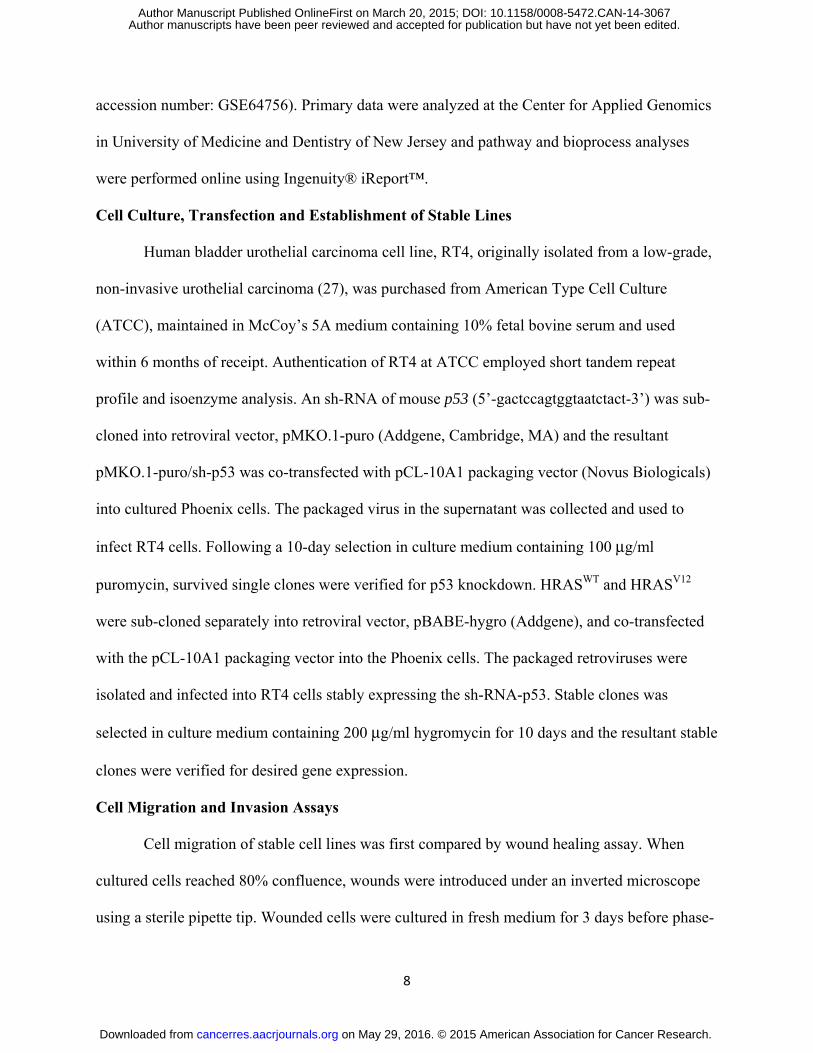
accession number: GSE64756). Primary data were analyzed at the Center for Applied Genomics
in University of Medicine and Dentistry of New Jersey and pathway and bioprocess analyses
were performed online using Ingenuity® iReport™.
Cell Culture, Transfection and Establishment of Stable Lines
Human bladder urothelial carcinoma cell line, RT4, originally isolated from a low-grade,
non-invasive urothelial carcinoma (27), was purchased from American Type Cell Culture
(ATCC), maintained in McCoy’s 5A medium containing 10% fetal bovine serum and used
within 6 months of receipt. Authentication of RT4 at ATCC employed short tandem repeat
profile and isoenzyme analysis. An sh-RNA of mouse p53 (5’-gactccagtggtaatctact-3’) was sub-
cloned into retroviral vector, pMKO.1-puro (Addgene, Cambridge, MA) and the resultant
pMKO.1-puro/sh-p53 was co-transfected with pCL-10A1 packaging vector (Novus Biologicals)
into cultured Phoenix cells. The packaged virus in the supernatant was collected and used to
infect RT4 cells. Following a 10-day selection in culture medium containing 100 μg/ml
puromycin, survived single clones were verified for p53 knockdown. HRASWT and HRASV12
were sub-cloned separately into retroviral vector, pBABE-hygro (Addgene), and co-transfected
with the pCL-10A1 packaging vector into the Phoenix cells. The packaged retroviruses were
isolated and infected into RT4 cells stably expressing the sh-RNA-p53. Stable clones was
selected in culture medium containing 200 μg/ml hygromycin for 10 days and the resultant stable
clones were verified for desired gene expression.
Cell Migration and Invasion Assays
Cell migration of stable cell lines was first compared by wound healing assay. When
cultured cells reached 80% confluence, wounds were introduced under an inverted microscope
using a sterile pipette tip. Wounded cells were cultured in fresh medium for 3 days before phase-
on May 29, 2016. © 2015 American Association for Cancer Research. cancerres.aacrjournals.org Downloaded from
Author manuscripts have been peer reviewed and accepted for publication but have not yet been edited. Author Manuscript Published OnlineFirst on March 20, 2015; DOI: 10.1158/0008-5472.CAN-14-3067

9
contrast images were recorded. For invasion assay, BioCoat Matrigel Invasion Chamber (BD
Biosciences, San Jose, CA) was employed. Briefly, stable clones (2.5 x 10⁴ cells) were seeded in
24-well chambers (in triplicate) containing 20 ng/ml 12-O-tetradecanoylphorbol-13-acetate.
After incubation for 72 h, the non-invading cells atop the membrane were removed by scrapping
and, the invading cells underneath the membrane were visualized using Diff-Quik stain and
counted in five high-power (200 x) microscopic fields (one-center and four-peripheral).
Cell Proliferation Assay
Stably transfected cells (2×10³/well) were cultured for 48 h and quantified by 3-(4,5-
dimethylthiazol-2-yl)-2,5-diphenyltetrazoliumbromide) method (Bio-Rad, Hercules, CA).
Cell Cycle Analysis
Urinary bladders were inverted to expose the mucosa. After incubation in a solution
containing 1 mg/ml dispase at 4°C overnight, the urothelial cells were gently scraped off and
digested with a solution containing 0.25% Trypsin-EDTA at 37°C for 30 min. The cells were
washed in PBS by centrifugation at 800 g for 5 min and filtered through a 100 μm pore-size
filter, fixed with pre-cooled 70% ethanol at 4°C and stained with 40 μg/ml propidium iodide
containing 100 μg/ml RNase. Cell sorting was carried out using Facscan (Beckman) and the data
were analyzed using ModiFit 3.2 (Verity Software House).
Quantitative Real-time PCR
Total RNA was isolated from bladder urothelia using RNeasy Mini Kit (Qiagen) and 2 μg
of it was used for cDNA synthesis using High Capacity cDNA Reverse Transcription Kit
(Applied Biosystem, Grand Island, NY). Real-time PCR was carried out with 7500 System
(Applied Biosystems) under 95°C for 15’ for the first cycle; 95°C for 15”, 58°C for 20” and
72°C for 30” for 50 cycles, and 72°C for 5’ for the last cycle. PCR products were quantified by
on May 29, 2016. © 2015 American Association for Cancer Research. cancerres.aacrjournals.org Downloaded from
Author manuscripts have been peer reviewed and accepted for publication but have not yet been edited. Author Manuscript Published OnlineFirst on March 20, 2015; DOI: 10.1158/0008-5472.CAN-14-3067

10
direct SYBR Green incorporation, with the relative abundance expressed as ratios to β-actin. The
primers were: p19ARF-forward: gtcgcaggttcttggtcact, p19ARF-reverse: cgaatctgcaccgtagttga;
p53-forward: agagaccgccgtacagaaga, p53-reverse: ctgtagcatgggcatccttt; p21-forward:
cggtggaactttgacttcgt, p21-reverse: cagggcagaggaagtactgg.
Western Blotting, Histological, Immunohistochemical and Immunofluorescent Staining
Total proteins from mouse urothelia or cultured RT4 cells were dissolved in a lysis buffer
(10% SDS, 20 mM Tris/HCl (pH7.5), 50 mM NaCl, 5 mM β-mercaptoethanol and a mixture of
protease inhibitors). After SDS-PAGE, the proteins were transferred onto PVDF membrane and
reacted consecutively with primary (Supplemental Table S1) and peroxidase-conjugated
secondary antibodies.
Freshly dissected urinary bladders were fixed in PBS-buffered 10% formalin and
embedded routinely in paraffin. Four μm thick sections were stained with H&E for histological
examination. For immunohistochemistry, de-paraffinized sections were microwaved in a citrate
buffer (pH 6.0) for 20 min to unmask the antigens and then incubated with primary
(Supplemental Table S1) and secondary antibodies conjugated with horseradish peroxidase.
Statistical Analysis
Student’s t test (two-tailed) was used to evaluate the statistical significance between
Upk2-HRAS* mice and wild-type (Upk2-cre) mice in urothelial expression of p53 pathway
components and between different groups of stably transfected cultured cells in their
proliferation and invasion rates, with P value <0.05 considered statistically significant.
on May 29, 2016. © 2015 American Association for Cancer Research. cancerres.aacrjournals.org Downloaded from
Author manuscripts have been peer reviewed and accepted for publication but have not yet been edited. Author Manuscript Published OnlineFirst on March 20, 2015; DOI: 10.1158/0008-5472.CAN-14-3067

11
Results
Oncogenic HRAS*-induced Persistent Urothelial Hyperplasia Results from an Equilibrium
between Mitogenic Signals and Anti-tumor Defenses
A highly reproducible phenotype in transgenic mice bearing a single copy of oncogenic
HRAS* under the control of the UPII promoter (Upk2-HRAS*) was the persistent urothelial
hyperplasia (24). Compared to normal urothelium from the wild-type littermates (Fig. 1A/a), the
hyperplastic lesions of the Upk2-HRAS* mice appeared as highly thickened, nonetheless well-
differentiated urothelia with excellent polarity (Fig. 1A/b-d) and retention of superficial umbrella
cells (arrows in Fig. 1A/c and 1A/d); and they started around 2 months of age and persisted
through 12 months, without progressing, in grade or stage, to full-fledged urothelial tumor or
reverting to normal urothelium. To understand the molecular underpinning of this phenomenon,
we examined the cell-cycle status and found that at the steady state there was a significant
reduction of G0/G1 urothelial cells and increase of G2 cells in Upk2-HRAS* transgenic mice (12
months of age), as compared with the wild-type controls (Fig. 1B, upper panel). This
corresponded well with elevated mitogenic signals including phosphorylated ERK and AKT
(both T308 and S473) in the transgenic mice (Fig. 1B lower panel; Supplemental Fig. S1).
However, S-phase cells were not significantly higher (Fig. 1B), suggesting that the DNA
synthesis was held in check and that a prolonged G2 arrest existed, possibly due to concurrent
induction of growth inhibitors/tumor suppressors (28). Of the tumor-suppressive pathways
surveyed, that of p53, including p19, p53 and p21, exhibited marked upregulation on mRNA
(Fig. 1C, left panel) and protein (Fig 1C, right panel) levels. Such overt upregulation was not
observed in pRB family proteins (e.g., pRB, p107 and p130). Interestingly, factors key to
promoting G2/M transition such as CDC2 and CYCLIN B1 were significantly down-regulated in
on May 29, 2016. © 2015 American Association for Cancer Research. cancerres.aacrjournals.org Downloaded from
Author manuscripts have been peer reviewed and accepted for publication but have not yet been edited. Author Manuscript Published OnlineFirst on March 20, 2015; DOI: 10.1158/0008-5472.CAN-14-3067
12
the hyperplastic lesions of the Upk2-HRAS* mice (Fig. 1D), a phenomenon observed in non-
urothelial cells with an upregulated p53 pathway (28). Our results suggest that oncogenic
HRAS*-triggered proliferative forces are counter-balanced by anti-proliferative forces,
especially by the p53 signaling axis, thus reaching an equilibrium and resulting in a non-
progressive and non-regressive state of persistent urothelial hyperplasia, that is quite different
from oncogenic RAS-induced premature senescence and apoptosis in primary-cultured cells
(29).
Removal of p53 Confers Invasive Property to Cultured Non-invasive Urothelial Tumor
Cells Expressing Oncogenic HRAS*
To determine whether the tumor-barrier effects of p53 upregulation by oncogenic
HRAS* in urothelial cells were coincidental or causative, we introduced oncogenic HRAS*
along with sh-RNA of p53 into cultured RT4 cells, which were originally derived from a human
low-grade, superficial papillary bladder tumor (27) and lacked RAS or p53 mutation/deletion
(30). Enforced expression of oncogenic HRAS* in RT4 elicited a marked upregulation of p53
and p21 (Fig. 2A). Knockdown of p53 or that along with the expression of a wild-type HRAS
enhanced cell proliferation (Supplemental Fig. S2), but only slightly increased cell migration and
invasion (Fig. 2C). In contrast, knocking down p53 and expressing an oncogenic HRAS*
resulted in a dramatic increase of cell migration and invasion of RT4 cells (Fig. 2C and 2D).
Thus, p53 deficiency and RAS activation appear to be synergistic in conferring the invasive
property to human urothelial tumor cells and triggering the conversion of non-invasive human
urothelial tumor cells into invasive ones.
Conditional Compound Mice Expressing Oncogenic HRAS* and Lacking p53 Develop
High-grade, Muscle-invasive Urothelial Carcinoma
on May 29, 2016. © 2015 American Association for Cancer Research. cancerres.aacrjournals.org Downloaded from
Author manuscripts have been peer reviewed and accepted for publication but have not yet been edited. Author Manuscript Published OnlineFirst on March 20, 2015; DOI: 10.1158/0008-5472.CAN-14-3067

13
To further define the interactive effects between oncogenic HRAS* and p53 deficiency in
vivo, we developed compound mice by ablating p53 from urothelial cells expressing oncogenic
HRAS*. To do so, we cross-bred three independent mouse lines: Upk2-HRAS* (24), Upk2-cre
(in which the UPII drives the expression of a cre recombinase in urothelium) (25) and floxed p53
(in which the exons of 5 and 6 were flanked two loxP sites) (Fig. 3A) (26). We chose four
resultant genotypes for phenotypic analyses: (i) Upk2-cre (as negative controls), (ii) Upk2-
HRAS*/WT, (iii) Upk2-cre/p53LOX/LOX and (iv) Upk2-HRAS*/WT/Upk2-cre/p53LOX/LOX (Fig. 3B
and 3C). These four groups were followed for 16-months and, upon histopathological
examination, the Upk2-cre and Upk2-cre/p53LOX/LOX lines exhibited normal urothelia, and the
Upk2-HRAS*/WT line exhibited urothelial hyperplasia, as expected, throughout the 16-month
observation (Fig. 3D). In stark contrast, the compound line expressing oncogenic HRAS* and
lacking p53 developed exclusively high-grade bladder tumors in the form of carcinoma-in-situ
(CIS) and muscle-invasive tumors (Fig. 3C and 3D and Fig. 4A-C, 4E-G). The invasive tumors
arose as early as 6 months of age and, by 16 months, a majority (60%) of the mice harbored
muscle-invasive bladder tumors (Fig. 3C). The CIS lesions were relatively flat with
microinvasive lesions in adjacent lamina propria (Fig. 4B and 4C). The microinvasive and
muscle-invasive lesions were confirmed by cytokeratin 5 staining (Fig. 4D, 4H and 4I). These
lesions bear strong resemblance to those found in human patients with muscle-invasive urothelial
carcinoma, and lend strong support to the sequence of urothelial tumor progression from CIS to
invasive tumors (13,14,31,32). Finally, focal squamous differentiation within the muscle-
invasive lesions was common as evidenced by H&E staining and by immunohistochemical
staining using antibodies against keratin 1 and TRIM29 (Supplemental Fig. S3), markers for
identifying squamous components in human muscle-invasive urothelial carcinomas (33).
on May 29, 2016. © 2015 American Association for Cancer Research. cancerres.aacrjournals.org Downloaded from
Author manuscripts have been peer reviewed and accepted for publication but have not yet been edited. Author Manuscript Published OnlineFirst on March 20, 2015; DOI: 10.1158/0008-5472.CAN-14-3067
14
Epithelial-mesenchymal Transition Signifies CIS-invasive Tumor Conversion
That compound mice urothelially expressing oncogenic HRAS* and lacking p53
developed CIS and then invasive lesions also provided a unique opportunity for us to utilize
these well-defined lesions to identify the molecular events that underlie this poorly defined
progression step. Toward this end, we performed laser-capture microdissection of normal
urothelia from the Upk2-cre mice, and CIS and muscle-invasive lesions from Upk2-
HRAS*/WT/Upk2-cre/p53LOX/LOX compound mice. After high-quality mRNAs were isolated from
freshly dissected lesions, the cDNAs were hybridized to oligonucleotide arrays representing all
mouse genes (Affymatrix). Of differentially upregulated genes, those functioning in the
epithelial-mesenchymal transition (EMT) dominate the muscle-invasive lesions when CIS
lesions were used as a reference (Table 1). Three groups of genes were particularly worth noting:
(i) transcription factors that drive EMT (e.g., twist homolog 1 (TWIST), zinc finger E-box
binding homeobox 2 (ZEB2) and ZEB1); (ii) matrix-degrading enzymes (e.g., matrix
metallopeptidases 13, 3, 2 and 9); and (iii) extracellular matrix components (e.g., collagen (type
I, III and IV), versican, vimentin, and fibronectin) (Table 1). Not surprisingly, those upregulated
in the muscle-invasive tumor/CIS comparison were also upregulated in the muscle-invasive
tumor/normal urothelium comparison. However, few of those upregulated in muscle-invasive
tumors were also upregulated in the CIS lesions, indicating that the EMT genes are primarily
switched on during muscle invasion. The only genes that showed more than 2-fold increase in
CIS over normal urothelium were MMP13 and platelet-derived growth factor receptor,
suggesting their potential role(s) in CIS formation. Antibody staining confirmed that MMP2, 3, 9
and 13 were all over-expressed almost exclusively in the muscle-invasive lesions, with MMPs 2
on May 29, 2016. © 2015 American Association for Cancer Research. cancerres.aacrjournals.org Downloaded from
Author manuscripts have been peer reviewed and accepted for publication but have not yet been edited. Author Manuscript Published OnlineFirst on March 20, 2015; DOI: 10.1158/0008-5472.CAN-14-3067

15
and 9 primarily associated with tumor cells and MMP3 and 13 in both tumor cells and matrix
(Fig. 5).
EMT Occurs in Urothelial Carcinoma Progenitor/Stem Cells
To explore whether overexpression of EMT drivers occurred in more differentiated
urothelial cells or in progenitor cells thus potentially playing a role in invasive tumor initiation,
we performed immunohistochemical and double- and triple-immunofluorescent localization.
Both ZEB1 and ZEB2 were strongly labeled in the nuclei of the invasive tumors cells of Upk2-
HRAS*/WT/Upk2-cre/p53LOX/LOX compound mice, but were barely detectable in normal urothelia
of Upk2-cre or Upk2-cre/p53LOX/LOX mice and were only weakly labeled in Upk2-HRAS* mice
(Fig. 6A and 6B). Upon triple fluorescent staining, ZEB2 was found to be associated with cells
specifically expressing CD44, a urothelial/carcinoma progenitor cell marker (Fig. 6C)
(18,19,34). Interestingly, these ZEB2- and CD44-positive cells had a marked decrease of E-
cadherin, an epithelial marker (35), and a marked increase of vimentin, a mesenchymal cell
marker (Fig. 6C) (36). Double staining of ZEB2 with keratin 14, another urothelial progenitor
cell marker (34), again showed excellent co-localization (Fig. 6D). Whereas normal-appearing
urothelial regions showed K14-positive cells which lacked ZEB2 labeling, areas with tumor
morphology showed strong co-expression of ZEB2 and K14 (Fig. 6D). Furthermore, areas with
leading edge of invasion showed strong ZEB2 and K14 co-expression (Fig. D). Interestingly,
invasive tumors cells of the Upk2-HRAS*/WT/Upk2-cre/p53LOX/LOX compound mice, but not the
non-invasive cells of the single transgenic mice, strongly co-expressed in the nuclei activated
AKT, activated β−catenin and ZEB2 (Supplemental Fig. S4), suggesting this signaling pathway
as the underlying cause of EMT activation. These results establish that urothelial tumor
progenitor cells in our compound transgenic mice expressing oncogenic HRAS* and lacking p53
on May 29, 2016. © 2015 American Association for Cancer Research. cancerres.aacrjournals.org Downloaded from
Author manuscripts have been peer reviewed and accepted for publication but have not yet been edited. Author Manuscript Published OnlineFirst on March 20, 2015; DOI: 10.1158/0008-5472.CAN-14-3067

16
strongly express EMT drivers and their expression may play a central role in initiating muscle-
invasive urothelial carcinoma. Finally, in contrast to the expansion of K14-positive cells in the
muscle-invasive lesions, cells positive for keratin 20, a marker expressed in urothelial superficial
umbrella cells and used for terminal differentiation of normal urothelium (37), were completely
absent from the muscle-invasive lesions (Supplemental Fig. S5). These results, together with our
observation of focal squamous differentiation of the muscle-invasive lesions, strongly indicate
that the muscle-invasive urothelial carcinoma of the bladder that we observed in our Upk2-
HRAS*/Upk2-cre/p53LOX/LOX mice belong to the “basal-subtype” recently classified in patients
(4-6,8-10).
on May 29, 2016. © 2015 American Association for Cancer Research. cancerres.aacrjournals.org Downloaded from
Author manuscripts have been peer reviewed and accepted for publication but have not yet been edited. Author Manuscript Published OnlineFirst on March 20, 2015; DOI: 10.1158/0008-5472.CAN-14-3067

17
Discussion
The recent expansion of whole-genome and whole-exome sequencing into a broad range
of human cancers has yielded unprecedented details about somatic gene mutations, making it
possible to classify cancers in genomic terms and to devise target-specific, precision therapies
(38). Urothelial carcinoma of the bladder (UCB) is no exception. In a landmark paper (23), The
Cancer Genome Atlas (TCGA) Research Network reported a comprehensive, multiplatform
analysis of 131 high-grade, muscle-invasive urothelial carcinomas of the bladder (MIUCB) on
their somatic mutation, DNA copy number, messenger and micro-RNA expression, protein and
phosphorylated protein expression and DNA methylation. Of the several surprises from that
report, one relates to the high frequency of alterations in the RTK/RAS/PIK3K signaling axis.
Up to 72% of the high-grade MIUCB harbored activation mutations in the FGFR3, EGFR,
ERBB2, ERBB3, HRAS/NRAS and PIK3CA or inactivating mutations in NF1, PTEN, INPP4B,
STK11, TSC1 and TSC2 (23). This is surprising because alterations in this pathway were
previously assigned primarily to low-grade, non-invasive UCB and to predict low risk of
progression and favorable clinical outcome (13,21,22) – a concept supported by independent
studies using genetically engineered mice. For instance, urothelial expression of an FGFR3
mutant (K644E) that constitutively activates the tyrosine kinase of FGFR3, either alone or in
combination with KRAS and β-catenin mutations or with PTEN deletion, in transgenic mice
failed to elicit any urothelial carcinoma (39). Similarly, urothelial overexpression of an
epidermal growth factor receptor (EGFR) in our transgenic mice induced proliferation but not
tumor formation even after an exhaustively long (28-month) follow-up (40). Furthermore,
urothelium-specific expression in our transgenic mice of oncogenic HRAS* at a level
comparable to the endogenous RAS elicited urothelial hyperplasia that only occasionally
on May 29, 2016. © 2015 American Association for Cancer Research. cancerres.aacrjournals.org Downloaded from
Author manuscripts have been peer reviewed and accepted for publication but have not yet been edited. Author Manuscript Published OnlineFirst on March 20, 2015; DOI: 10.1158/0008-5472.CAN-14-3067

18
progressed to low-grade, papillary non-invasive UCB in aged mice (>12 months) (24). High-
grade MIUCB was never observed in any of these RTK/RAS-pathway-activated mouse models
(24,39,40). The fact that gene mutations that activate the RTK/RAS pathway are highly prevalent
in human high-grade MIUCB from the TCGA study (23) raises an important question as to
whether these mutations are tumor “drivers” or “passengers” and whether the mutations require
additional genetic alterations to be tumorigenic.
Our present study provides experimental evidence establishing that RAS activation per se
is non-tumorigenic in urothelial cells in vivo due, in large part, to a compensatory tumor barrier
that RAS elicits in the p53 tumor suppressor pathway (Figs. 1, 2 and Supplemental Fig. S1).
Although p53 deficiency by itself is also non-tumorigenic, it is highly synergistic with RAS
activation, and these two alterations together are necessary and sufficient to initiate high-grade,
carcinoma-in-situ (CIS) and MIUCB (Figs. 3 and 4). Of note, the MIUCB we observed in our
double transgenic mice expressing oncogenic Ha-RAS and lacking p53 bear strong resemblance
to the basal-subtype of MIUCB recently classified in patients (4-11) in their (i) high expression
of basal cell markers such as K5, K14 and CD44 (Figs. 4 and 6 and Supplemental Fig. S5), (ii)
low or lack of expression of luminal cell markers such as E-cadherin and K20 (Fig. 6 and
Supplemental Fig. S5), (iii) focal squamous differentiation (Supplemental Fig. S3), and (iv) high
expression of EMT transcription factors (Twist, ZEB1 and ZEB2) (Table 1; Fig. 6), EMT
markers (vimentin, MMPs 2, 3, 9 and 13) (Table 1; Figs. 5 and 6) and extracellular matrix
components (collagen, versican and fibronectin) (Table 1). Our study therefore functionally
defines RAS pathway activation and p53 deficiency as the highly synergistic co-drivers for the
basal-subtype MIUCB, and it has several significant implications. First, as has been
demonstrated in other cancer types, tumor drivers (as opposed to the passengers) are more
on May 29, 2016. © 2015 American Association for Cancer Research. cancerres.aacrjournals.org Downloaded from
Author manuscripts have been peer reviewed and accepted for publication but have not yet been edited. Author Manuscript Published OnlineFirst on March 20, 2015; DOI: 10.1158/0008-5472.CAN-14-3067

19
reliable biomarkers for cancer sub-classification and prediction of chemotherapeutic response
and clinical outcome (12). RAS pathway activation together with p53 deficiency could
potentially serve as a new biomarker set for the genetic identification of the basal-subtype of
MIUCB that may be associated with an unfavorable prognosis, hence requiring aggressive
therapeutic modalities. Second, our study reveals a previously unrecognized molecular cross-talk
between RAS and p53 pathways in converting low-grade non-invasive urothelial lesions (e.g.,
hyperplasia and low-grade papillary) into becoming high-grade non-invasive (e.g., high-grade
papillary and CIS) and invasive ones (e.g., MIUCB). Not only did we demonstrate such a
relationship in transgenic mice (Figs. 3-6; Supplemental Figs. S3-S5), we also showed that
introducing oncongenic HRAS* and knocking down p53 in cultured RT4 cells confer invasive
properties to these otherwise non-invasive human UCB cells (Fig. 2). It has been suggested,
based on clinical longitudinal studies, that approximately 25% of the low-grade, non-invasive
UCB can eventually progress in grade and/or stage to muscle invasion (41,42). This occurs in an
unpredictable manner that necessitates lifelong, vigilant follow-up by repeated cystoscopy and
biopsy - a main cause for morbidity, time lost from work and high medical expenses. Thus far,
no biomarker exists that can reliably predict the risk of progression of non-invasive UCB to the
invasive stage (43,44). Perhaps it is not surprising that p53 alterations are not very predictive of
UCB progression (45), based on data from genetically engineered mice indicating the lack of
tumorigenicity by p53 deficiency alone (Fig. 3; (26,46,47)). It is possible, however, that a
combination of RAS pathway activation and p53 deficiency, as we demonstrated here, are better
biomarkers for UCB surveillance and prediction of tumor progression. It is worth noting that
ablation of both PTEN and p53 in mouse urothelia also led to MIUCB (48), consistent with the
fact that PTEN acts in the RAS pathway and PTEN inactivation is functionally akin to RAS
on May 29, 2016. © 2015 American Association for Cancer Research. cancerres.aacrjournals.org Downloaded from
Author manuscripts have been peer reviewed and accepted for publication but have not yet been edited. Author Manuscript Published OnlineFirst on March 20, 2015; DOI: 10.1158/0008-5472.CAN-14-3067

20
activation. Third, because RAS activation is a co-driver of the basal-type MIUCB, inhibition of
this pathway might be of significant value in treating and/or preventing the progression of this
MIUCB subtype. The fact that the basal-type MIUCB in humans is often resistant to the existing
chemotherapeutics (4) makes RAS pathway inhibition a particularly attractive avenue to explore.
Since suppressing activated RAS per se remains challenging (49), it is likely that effectors of
RAS will have to be targeted and that inhibition of more than one signaling branch (e.g., PI3K-
AKT as well as MAPK) is required to achieve satisfactory results (50). Finally, the development
of a new transgenic mouse model that consistently develops the basal-type MIUCB provides a
novel in vivo platform for dissecting the evolutionary steps and the potential cross-talks among
the different MIUCB subtypes and for testing subtype-specific diagnostic, preventive and
therapeutic strategies. Clearly, many of these ideas require clinical validation studies before they
can be translated to the bedside.
From a mechanistic standpoint, RAS activation and p53 deficiency could synergize on
several fronts to affect cellular processes that govern urothelial tumorigenesis and progression.
As shown recently, RAS activation increases the replicative pressure on urothelial cells, causing
them to undergo DNA damage (51). Under normal circumstances, i.e., when p53 pathway is
intact, urothelial cells can sense DNA damage and upregulate p19Arf which in turn upregulates
p53 and downstream effectors such as p21 (Fig. 1). This helps restrain G1/S and G2/M transition
and allow time for DNA damage repair to take place. When p53 pathway is defective, however,
cell cycle progression proceeds with amplification of the damaged DNA, setting a stage for
malignant transformation. Another level of interaction is the collaborative nature of RAS
activation and p53 deficiency on cell motility. Activated RAS is a strong enhancer of cell
motility (52), whereas a functional p53 is a potent cell motility inhibitor (53). As we showed in
on May 29, 2016. © 2015 American Association for Cancer Research. cancerres.aacrjournals.org Downloaded from
Author manuscripts have been peer reviewed and accepted for publication but have not yet been edited. Author Manuscript Published OnlineFirst on March 20, 2015; DOI: 10.1158/0008-5472.CAN-14-3067

21
our in vitro assay, activated RAS or p53 knock down alone only had a marginal increase on cell
motility, but combining these two events resulted in a marked increase of cell motility and
triggered invasion (Fig. 2). Finally, as with other epithelial cells, RAS activation and p53
deficiency are both strong promoters of EMT (54,55). MAPK and AKT pathways, both shown to
be prominently activated in our transgenic mice (Fig. 1; Supplemental Fig. S1), can activate
factors such as β-catenin that drive EMT (Table 1; Supplemental Fig. S4). While normal p53
negatively regulates this process, p53 deficiency fuels EMT and sets the tumor cell invasion in
motion (Figs. 2-6; Supplemental Fig. S3-S5). There is mounting evidence suggesting that EMT
can lead to drug resistance (35). Since EMT enhances the stemness and the plasticity of
urothelial cells, it may also fuel the trans-differentiation of some of the urothelial progenitor cells
toward the squamous lineage and squamous differentiation, another potential cause of drug
resistance. In this regard, inhibiting RAS effectors that drive EMT and/or inhibiting EMT
effectors such as MMPs may play a critical role in reducing chemoresistance that has been
observed in the basal-type MIUCB (4). Because EMT is highly activated in progenitor/stem cells
that give rise to the basal-type MIUCB (Fig. 6 and Supplemental Fig. S5), its suppression may
present a unique opportunity for controlling the root cause of tumor cell expansion and invasion.
In summary, the data presented in this paper provide the first experimental evidence
demonstrating that the loss of p53 is critical in allowing hyperplastic urothelial cells in vivo to
bypass G2 arrest induced by activated HRAS and proceed to tumor formation; that RAS pathway
activation and p53 pathway inactivation together confer invasive properties to non-invasive
urothelial tumor cells and these two synergistic events are necessary and sufficient to convert
carcinoma in situ to basal-subtype, muscle-invasive urothelial carcinoma of the bladder; and that
activation of EMT and increased stemness in urothelial progenitor cells are crucial epigenetic
on May 29, 2016. © 2015 American Association for Cancer Research. cancerres.aacrjournals.org Downloaded from
Author manuscripts have been peer reviewed and accepted for publication but have not yet been edited. Author Manuscript Published OnlineFirst on March 20, 2015; DOI: 10.1158/0008-5472.CAN-14-3067

22
events for invasive tumorigenesis. Our data also strongly suggest that increased urothelial
plasticity due to EMT may underlie urothelial transdifferentiation to the squamous lineage,
leading to focal squamous differentiation in urothelial carcinomas. From a clinical standpoint,
combined RAS pathway activation and p53 pathway inactivation, events highly prevalent in
human urothelial carcinomas as evidenced by whole-genome analyses, may serve as a new
biomarker set to predict urothelial carcinoma progression, and inhibition of receptor tyrosine
kinase/RAS pathway components may be used as therapeutic targets for basal-subtype, muscle-
invasive urothelial carcinomas that are resistant to conventional chemotherapeutics.
on May 29, 2016. © 2015 American Association for Cancer Research. cancerres.aacrjournals.org Downloaded from
Author manuscripts have been peer reviewed and accepted for publication but have not yet been edited. Author Manuscript Published OnlineFirst on March 20, 2015; DOI: 10.1158/0008-5472.CAN-14-3067

23
Acknowledgments
This work was supported in part by grants from the United States National Institutes of
Health (P01 CA165980) and Veterans Affairs Office of Research and Development (Biomedical
Laboratory Research and Development Service; 1I01BX002049) and a grant-in-aid from the
Goldstein Fund for Urological Research of the New York University School of Medicine.
on May 29, 2016. © 2015 American Association for Cancer Research. cancerres.aacrjournals.org Downloaded from
Author manuscripts have been peer reviewed and accepted for publication but have not yet been edited. Author Manuscript Published OnlineFirst on March 20, 2015; DOI: 10.1158/0008-5472.CAN-14-3067

24
References
1. Lotan Y, Kamat AM, Porter MP, Robinson VL, Shore N, Jewett M, et al. Key concerns
about the current state of bladder cancer: a position paper from the Bladder Cancer Think
Tank, the Bladder Cancer Advocacy Network, and the Society of Urologic Oncology.
Cancer 2009;115(18):4096-103.
2. Dancik GM, Theodorescu D. Pharmacogenomics in bladder cancer. Urologic oncology
2014;32(1):16-22.
3. Meeks JJ, Bellmunt J, Bochner BH, Clarke NW, Daneshmand S, Galsky MD, et al. A
systematic review of neoadjuvant and adjuvant chemotherapy for muscle-invasive
bladder cancer. European urology 2012;62(3):523-33.
4. Choi W, Porten S, Kim S, Willis D, Plimack ER, Hoffman-Censits J, et al. Identification
of distinct basal and luminal subtypes of muscle-invasive bladder cancer with different
sensitivities to frontline chemotherapy. Cancer cell 2014;25(2):152-65.
5. Damrauer JS, Hoadley KA, Chism DD, Fan C, Tiganelli CJ, Wobker SE, et al. Intrinsic
subtypes of high-grade bladder cancer reflect the hallmarks of breast cancer biology.
Proceedings of the National Academy of Sciences of the United States of America
2014;111(8):3110-5.
6. Hurst CD, Knowles MA. Molecular subtyping of invasive bladder cancer: time to divide
and rule? Cancer cell 2014;25(2):135-6.
7. Riester M, Taylor JM, Feifer A, Koppie T, Rosenberg JE, Downey RJ, et al. Combination
of a novel gene expression signature with a clinical nomogram improves the prediction of
survival in high-risk bladder cancer. Clinical cancer research : an official journal of the
American Association for Cancer Research 2012;18(5):1323-33.
on May 29, 2016. © 2015 American Association for Cancer Research. cancerres.aacrjournals.org Downloaded from
Author manuscripts have been peer reviewed and accepted for publication but have not yet been edited. Author Manuscript Published OnlineFirst on March 20, 2015; DOI: 10.1158/0008-5472.CAN-14-3067

25
8. Hoadley KA, Yau C, Wolf DM, Cherniack AD, Tamborero D, Ng S, et al. Multiplatform
Analysis of 12 Cancer Types Reveals Molecular Classification within and across Tissues
of Origin. Cell 2014;158(4):929-44.
9. Volkmer JP, Sahoo D, Chin RK, Ho PL, Tang C, Kurtova AV, et al. Three differentiation
states risk-stratify bladder cancer into distinct subtypes. Proceedings of the National
Academy of Sciences of the United States of America 2012;109(6):2078-83.
10. Sjodahl G, Lovgren K, Lauss M, Patschan O, Gudjonsson S, Chebil G, et al. Toward a
molecular pathologic classification of urothelial carcinoma. The American journal of
pathology 2013;183(3):681-91.
11. Huang HY, Shariat SF, Sun TT, Lepor H, Shapiro E, Hsieh JT, et al. Persistent uroplakin
expression in advanced urothelial carcinomas: implications in urothelial tumor
progression and clinical outcome. Human pathology 2007.
12. Livshits G, Lowe SW. Accelerating cancer modeling with RNAi and nongermline
genetically engineered mouse models. Cold Spring Harbor protocols 2013;2013(11).
13. Wu XR. Urothelial tumorigenesis: a tale of divergent pathways. Nat Rev Cancer
2005;5(9):713-25.
14. Dinney CP, McConkey DJ, Millikan RE, Wu X, Bar-Eli M, Adam L, et al. Focus on
bladder cancer. Cancer cell 2004;6(2):111-6.
15. Grossman HB. Superficial bladder cancer: decreasing the risk of recurrence. Oncology
(Huntingt) 1996;10(11):1617-24; discussion 24, 27-8.
16. Castillo-Martin M, Domingo-Domenech J, Karni-Schmidt O, Matos T, Cordon-Cardo C.
Molecular pathways of urothelial development and bladder tumorigenesis. Urologic
oncology 2010;28(4):401-8.
on May 29, 2016. © 2015 American Association for Cancer Research. cancerres.aacrjournals.org Downloaded from
Author manuscripts have been peer reviewed and accepted for publication but have not yet been edited. Author Manuscript Published OnlineFirst on March 20, 2015; DOI: 10.1158/0008-5472.CAN-14-3067

26
17. Wu XR, Kong XP, Pellicer A, Kreibich G, Sun TT. Uroplakins in urothelial biology,
function, and disease. Kidney Int 2009;75:1153-65.
18. Gandhi D, Molotkov A, Batourina E, Schneider K, Dan H, Reiley M, et al. Retinoid
signaling in progenitors controls specification and regeneration of the urothelium.
Developmental cell 2013;26(5):469-82.
19. Van Batavia J, Yamany T, Molotkov A, Dan H, Mansukhani M, Batourina E, et al.
Bladder cancers arise from distinct urothelial sub-populations. Nature cell biology 2014.
20. Shin K, Lim A, Odegaard JI, Honeycutt JD, Kawano S, Hsieh MH, et al. Cellular origin
of bladder neoplasia and tissue dynamics of its progression to invasive carcinoma. Nature
cell biology 2014;16(5):469-78.
21. Wolff EM, Liang G, Jones PA. Mechanisms of Disease: genetic and epigenetic
alterations that drive bladder cancer. Nat Clin Pract Urol 2005;2(10):502-10.
22. van Rhijn BW, van der Kwast TH, Vis AN, Kirkels WJ, Boeve ER, Jobsis AC, et al.
FGFR3 and P53 characterize alternative genetic pathways in the pathogenesis of
urothelial cell carcinoma. Cancer research 2004;64(6):1911-4.
23. Network TCGAR. Comprehensive molecular characterization of urothelial bladder
carcinoma. Nature 2014;507(7492):315-22.
24. Mo L, Zheng X, Huang HY, Shapiro E, Lepor H, Cordon-Cardo C, et al. Hyperactivation
of Ha-ras oncogene, but not Ink4a/Arf deficiency, triggers bladder tumorigenesis. J Clin
Invest 2007;117(2):314-25.
25. Mo L, Cheng J, Lee EY, Sun TT, Wu XR. Gene deletion in urothelium by specific
expression of Cre recombinase. Am J Physiol Renal Physiol 2005;289(3):F562-8.
on May 29, 2016. © 2015 American Association for Cancer Research. cancerres.aacrjournals.org Downloaded from
Author manuscripts have been peer reviewed and accepted for publication but have not yet been edited. Author Manuscript Published OnlineFirst on March 20, 2015; DOI: 10.1158/0008-5472.CAN-14-3067

27
26. He F, Mo L, Zheng XY, Hu C, Lepor H, Lee EY, et al. Deficiency of pRb family proteins
and p53 in invasive urothelial tumorigenesis. Cancer research 2009;69(24):9413-21.
27. Rigby CC, Franks LM. A human tissue culture cell line from a transitional cell tumour of
the urinary bladder: growth, chromosone pattern and ultrastructure. British journal of
cancer 1970;24(4):746-54.
28. Taylor WR, Stark GR. Regulation of the G2/M transition by p53. Oncogene
2001;20(15):1803-15.
29. Lin AW, Barradas M, Stone JC, van Aelst L, Serrano M, Lowe SW. Premature
senescence involving p53 and p16 is activated in response to constitutive MEK/MAPK
mitogenic signaling. Genes Dev 1998;12(19):3008-19.
30. DeGraff DJ, Robinson VL, Shah JB, Brandt WD, Sonpavde G, Kang Y, et al. Current
preclinical models for the advancement of translational bladder cancer research.
Molecular cancer therapeutics 2013;12(2):121-30.
31. Cohen SM, Ohnishi T, Clark NM, He J, Arnold LL. Investigations of rodent urinary
bladder carcinogens: collection, processing, and evaluation of urine and bladders. Toxicol
Pathol 2007;35(3):337-47.
32. Dalbagni G, Presti J, Reuter V, Fair WR, Cordon-Cardo C. Genetic alterations in bladder
cancer. Lancet 1993;342(8869):469-71.
33. Huang W, Williamson SR, Rao Q, Lopez-Beltran A, Montironi R, Eble JN, et al. Novel
markers of squamous differentiation in the urinary bladder. Human pathology
2013;44(10):1989-97.
34. Chan KS, Espinosa I, Chao M, Wong D, Ailles L, Diehn M, et al. Identification,
molecular characterization, clinical prognosis, and therapeutic targeting of human bladder
on May 29, 2016. © 2015 American Association for Cancer Research. cancerres.aacrjournals.org Downloaded from
Author manuscripts have been peer reviewed and accepted for publication but have not yet been edited. Author Manuscript Published OnlineFirst on March 20, 2015; DOI: 10.1158/0008-5472.CAN-14-3067

28
tumor-initiating cells. Proceedings of the National Academy of Sciences of the United
States of America 2009;106(33):14016-21.
35. McConkey DJ, Choi W, Marquis L, Martin F, Williams MB, Shah J, et al. Role of
epithelial-to-mesenchymal transition (EMT) in drug sensitivity and metastasis in bladder
cancer. Cancer metastasis reviews 2009;28(3-4):335-44.
36. Kokkinos MI, Wafai R, Wong MK, Newgreen DF, Thompson EW, Waltham M.
Vimentin and epithelial-mesenchymal transition in human breast cancer--observations in
vitro and in vivo. Cells, tissues, organs 2007;185(1-3):191-203.
37. Lopez-Beltran A, Montironi R, Vidal A, Scarpelli M, Cheng L. Urothelial dysplasia of
the bladder: diagnostic features and clinical significance. Analytical and quantitative
cytopathology and histopathology 2013;35(3):121-9.
38. Kandoth C, McLellan MD, Vandin F, Ye K, Niu B, Lu C, et al. Mutational landscape and
significance across 12 major cancer types. Nature 2013;502(7471):333-9.
39. Ahmad I, Singh LB, Foth M, Morris CA, Taketo MM, Wu XR, et al. K-Ras and beta-
catenin mutations cooperate with Fgfr3 mutations in mice to promote tumorigenesis in
the skin and lung, but not in the bladder. Disease models & mechanisms 2011;4(4):548-
55.
40. Cheng J, Huang H, Zhang ZT, Shapiro E, Pellicer A, Sun TT, et al. Overexpression of
epidermal growth factor receptor in urothelium elicits urothelial hyperplasia and
promotes bladder tumor growth. Cancer research 2002;62(14):4157-63.
41. Knowles MA. Molecular subtypes of bladder cancer: Jekyll and Hyde or chalk and
cheese? Carcinogenesis 2006;27(3):361-73.
on May 29, 2016. © 2015 American Association for Cancer Research. cancerres.aacrjournals.org Downloaded from
Author manuscripts have been peer reviewed and accepted for publication but have not yet been edited. Author Manuscript Published OnlineFirst on March 20, 2015; DOI: 10.1158/0008-5472.CAN-14-3067
29
42. Tanaka M, Grossman HB. Tumor suppressor genes of bladder cancer and potential for
gene therapy. Adv Exp Med Biol 2003;539(Pt A):185-91.
43. Xylinas E, Kluth LA, Lotan Y, Daneshmand S, Rieken M, Karakiewicz PI, et al. Blood-
and tissue-based biomarkers for prediction of outcomes in urothelial carcinoma of the
bladder. Urologic oncology 2014;32(3):230-42.
44. Shirodkar SP, Lokeshwar VB. Potential new urinary markers in the early detection of
bladder cancer. Current opinion in urology 2009;19(5):488-93.
45. Stadler WM, Lerner SP, Groshen S, Stein JP, Shi SR, Raghavan D, et al. Phase III study
of molecularly targeted adjuvant therapy in locally advanced urothelial cancer of the
bladder based on p53 status. Journal of clinical oncology : official journal of the
American Society of Clinical Oncology 2011;29(25):3443-9.
46. Cheng J, Huang H, Pak J, Shapiro E, Sun TT, Cordon-Cardo C, et al. Allelic loss of p53
gene is associated with genesis and maintenance, but not invasion, of mouse carcinoma in
situ of the bladder. Cancer research 2003;63(1):179-85.
47. Gao J, Huang HY, Pak J, Cheng J, Zhang ZT, Shapiro E, et al. p53 deficiency provokes
urothelial proliferation and synergizes with activated Ha-ras in promoting urothelial
tumorigenesis. Oncogene 2004;23(3):687-96.
48. Puzio-Kuter AM, Castillo-Martin M, Kinkade CW, Wang X, Shen TH, Matos T, et al.
Inactivation of p53 and Pten promotes invasive bladder cancer. Genes Dev
2009;23(6):675-80.
49. Stephen AG, Esposito D, Bagni RK, McCormick F. Dragging ras back in the ring.
Cancer cell 2014;25(3):272-81.
on May 29, 2016. © 2015 American Association for Cancer Research. cancerres.aacrjournals.org Downloaded from
Author manuscripts have been peer reviewed and accepted for publication but have not yet been edited. Author Manuscript Published OnlineFirst on March 20, 2015; DOI: 10.1158/0008-5472.CAN-14-3067

30
50. Zhou H, Huang HY, Shapiro E, Lepor H, Huang WC, Mohammadi M, et al. Urothelial
tumor initiation requires deregulation of multiple signaling pathways: implications in
target-based therapies. Carcinogenesis 2012;33(4):770-80.
51. Evangelou K, Bartkova J, Kotsinas A, Pateras IS, Liontos M, Velimezi G, et al. The
DNA damage checkpoint precedes activation of ARF in response to escalating oncogenic
stress during tumorigenesis. Cell death and differentiation 2013;20(11):1485-97.
52. Baker NM, Yee Chow H, Chernoff J, Der CJ. Molecular Pathways: Targeting RAC-p21-
Activated Serine-Threonine Kinase Signaling in RAS-Driven Cancers. Clinical cancer
research : an official journal of the American Association for Cancer Research
2014;20(18):4740-6.
53. Muller PA, Vousden KH, Norman JC. p53 and its mutants in tumor cell migration and
invasion. The Journal of cell biology 2011;192(2):209-18.
54. Turley EA, Veiseh M, Radisky DC, Bissell MJ. Mechanisms of disease: epithelial-
mesenchymal transition--does cellular plasticity fuel neoplastic progression? Nature
clinical practice Oncology 2008;5(5):280-90.
55. Ansieau S, Courtois-Cox S, Morel AP, Puisieux A. Failsafe program escape and EMT: a
deleterious partnership. Seminars in cancer biology 2011;21(6):392-6.
on May 29, 2016. © 2015 American Association for Cancer Research. cancerres.aacrjournals.org Downloaded from
Author manuscripts have been peer reviewed and accepted for publication but have not yet been edited. Author Manuscript Published OnlineFirst on March 20, 2015; DOI: 10.1158/0008-5472.CAN-14-3067

31
Table Legend
Table 1. Differential expression of genes important for epithelial-mesenchymal transition
(EMT) between muscle-invasive urothelial carcinoma (MIUCB), carcinoma-in-situ (CIS) and
normal urothelium. Laser-capture, microdissected tissues were subject to mRNA
extraction/cDNA synthesis and expression array analysis (see Methods; GEO accession number:
GSE64756). Fold changes in three pair-wise comparisons are shown, with ranking from the
highest to the lowest (>2 fold) expression in consecutive order for the MIUCB/CIS comparison
chosen for practical purposes (see text).
on May 29, 2016. © 2015 American Association for Cancer Research. cancerres.aacrjournals.org Downloaded from
Author manuscripts have been peer reviewed and accepted for publication but have not yet been edited. Author Manuscript Published OnlineFirst on March 20, 2015; DOI: 10.1158/0008-5472.CAN-14-3067

32
Figure Legends
Figure 1. Simultaneous induction of pro- and anti-mitogenic signals in transgenic mice
expressing oncogenic HRAS* in urothelia, leading to persistent non-progressive hyperplasia.
Urothelia from Upk2-HRAS*/WT transgenic mice and wild-type littermates were subject to (A)
histopathology, (B, upper panel) flow cytometry, (B, lower panel) Western blotting of key RAS-
downstream effectors, (C, left panel) Real-time PCR of p53 pathway components, (C, right
panel) Western blotting of key tumor suppressors, and (D, upper panel) Western blotting and (D,
lower panel) immunohistochemical staining of promoters of G2/M transition CDC2 and
CYCLIN B1. Note that, the WT mouse (12-months old (A-a)) exhibited normal urothelium,
whereas the Upk2-HRAS*/WT transgenic mice (A-b, A-c, and A-d, 4-, 8- and 12-months,
respectively) exhibited urothelial hyperplasia. Note the reduced G0/G1 and increased G2 phase
(B, upper panel) and elevated pERK and pAKT (B, lower panel) in the transgenic mouse
urothelia as compared with the WT controls. Note the overexpression of p19, p53 and p21 on
both mRNA (C, left panel) and protein level (C, right panel) and largely unchanged pRB family
proteins in the transgenic mice. Also note the reduced levels of CDC2 and CYCLIN B1 in the
Upk2-HRAS*/WT transgenic mice (D). Magnification: 200 x in (A) and (D).
Figure 2. Combined effects of oncogenic HRAS* and p53 deficiency on cultured urothelial
cells. (A, left panel) Western blotting showing that cultured RT4 cells stably transfected with
FLAG-tagged HRAS* overexpress p53 and p21, compared with vector-only transfected cells.
(B) Western blotting showing that RT4 cells stably transfected with sh-RNA of p53 had marked
decrease of p53 itself and p21. RT4 cells stably transfected with Sh-RNA of green fluorescent
protein (GFP) served as a negative control; NT stands for no transfection. (C) Cell invasion assay
using Matrigel (invasive cells counted per 5 high-powered (200 x) fields microscopically),
on May 29, 2016. © 2015 American Association for Cancer Research. cancerres.aacrjournals.org Downloaded from
Author manuscripts have been peer reviewed and accepted for publication but have not yet been edited. Author Manuscript Published OnlineFirst on March 20, 2015; DOI: 10.1158/0008-5472.CAN-14-3067

33
showing a very small increase of invasive cells in p53-knockdown cells, a moderate increase in
p53-knockdown/WT-HRAS co-expressing cells and a marked increase in p53-
knockdown/HRAS* co-expressing cells. (D) Microscopic images of wound healing (upper
panel) and Matrigel-invasion (lower panel) experiments showing marked increase of both cell
migration and invasion in cells co-expressing HRAS* and p53-shRNA.
Figure 3. Inactivation of p53 in urothelial cells of transgenic mice expressing oncogenic
HRAS*. (A) Three transgenic lines for intercrossing contained transgene of uroplakin II
promoter (Upk2) driving cre recombinase (Line 1); floxed p53 where exons 5 and 6 were flanked
by loxP sites (Line 2); and Upk2 driving oncogenic HRAS*. (B) Offspring of two representative
crosses were genotyped by Southern blotting of restriction-digested tail genomic DNA (upper
panels) using a mouse Upk2 probe, revealing the 5.4-kB Upk2–cre transgene fragment, the 1.4-
kB Upk2-HRAS* fragment and the 1.1-kB endogenous Upk2 fragment (endo-Upk2). The same
DNA samples were subject to PCR using primers specifically detecting the first loxP site of the
p53 mutant allele. Asterisks denote the four major genotypes chosen for additional analyses (also
see C). (C) The rate of the four major genotypes free of high-grade muscle-invasive urothelial
carcinoma (tumor-free rate). Note that only mice expressing oncogenic HRAS* as well as
lacking p53 in urothelia developed invasive urothelial carcinoma. (D) Representative H&E
images of the four genotypes (all 8 months old) (see text). Magnification: 200 x.
Figure 4. Morphological features of urothelial lesions in compound transgenic mice expressing
oncogenic HRAS* and lacking p53. Urinary bladders of transgenic mice expressing oncogenic
HRAS* and lacking p53 (8-12 months old) were stained by H&E (A-C; E-G) or anti-keratin 5
(D, H and I). Note the high-grade lesions resembling carcinoma-in-situ (A-C) with lamina
propria invasion (B and D, arrows) and muscle-invasive lesions (E-G) that were strongly labeled
on May 29, 2016. © 2015 American Association for Cancer Research. cancerres.aacrjournals.org Downloaded from
Author manuscripts have been peer reviewed and accepted for publication but have not yet been edited. Author Manuscript Published OnlineFirst on March 20, 2015; DOI: 10.1158/0008-5472.CAN-14-3067

34
(brown) by anti-keratin 5 (D, H and I). L denotes lumen. S in (E-F) denotes smooth muscle.
Magnification: 200 x for all panels.
Figure. 5. Upregulation of matrix metalloproteinases in muscle-invasive urothelial carcinoma
cells. Immunochemical staining using anti-MMP antibodies followed by hematoxylin counter
staining was performed on bladder tissues from age-matched (8-months) Upk2-cre, Upk2-
HRAS*, Upk2-cre/p53lox/lox and Upk2-HRAS*/Upk2-cre/p53lox/lox mice. Note the significant
upregulation of MMP2, MMP3, MMP9 and MMP13 in the muscle-invasive urothelial carcinoma
cells of transgenic mice expressing the oncogenic HRAS* and deficient for p53. MMP3 and
MMP13 were also detected strongly in some matrix cells. Magnification: 200x for all panels.
Figure 6. Detection of transcriptional factors driving EMT in urothelial progenitor cells. (A and
B) Urinary bladders from age-matched (8-months) Upk2-cre, Upk2-HRAS*/WT, Upk2-cre/p53lox/lox
and Upk2-HRAS*/WT/Upk2-cre/p53lox/lox mice were immunohistochemically stained with anti-
ZEB1 (A) and anti-ZEB2 (B) and counter-stained by hemotoxylin. Note the marked upregulation
of both proteins almost exclusively in the muscle-invasive lesions of the Upk2-HRAS*/WT/Upk2-
cre/p53lox/lox mice. (C) Urinary bladders from Upk2-HRAS*/WT and Upk2-HRAS*/WT/Upk2-
cre/p53lox/lox mice were triple-stained using immuofluorescent method with anti-E-cadherin (E-
cad), -ZEB2 and -CD44 (left two panels) or with anti-vimentin, -ZEB2 and -CD44 (right two
panels). DAPI was used to visualize the nuclei. Note the marked down-regulation of E-cadherin
and dramatic up-regulation of ZEB2 in CD44-positive cells in the muscle-invasive lesions of the
Upk2-HRAS*/WT/Upk2-cre/p53lox/lox mice. Also note the co-localization of vimentin, ZEB2 and
CD44 in the invasive tumor cells (far-right panel, arrows). (D) Urinary bladders from Upk2-
HRAS*/WT, Upk2-cre/p53lox/lox and Upk2-HRAS*/WT/Upk2-cre/p53lox/lox mice were subject to
immunofluorescent staining with anti-ZEB2 and -keratin 14 antibodies, with DAPI as counter-
on May 29, 2016. © 2015 American Association for Cancer Research. cancerres.aacrjournals.org Downloaded from
Author manuscripts have been peer reviewed and accepted for publication but have not yet been edited. Author Manuscript Published OnlineFirst on March 20, 2015; DOI: 10.1158/0008-5472.CAN-14-3067

35
staining to visualize the nuclei. Note the lack of ZEB2 staining in K14-positive cells in Upk2-
HRAS*/WT and Upk2-cre/p53lox/lox mice and the strong staining of ZEB2 in K14-positive cells in
Upk2-HRAS*/WT/Upk2-cre/p53lox/lox mice (middle panel, dashed circle). Dashed box illustrates an
area of normal-appearing urothelium showing the lack of ZEB2 staining. Also note that the
leading edge of an early invasive lesion in Upk2-HRAS*/WT/Upk2-cre/p53lox/lox mice had marked
upregulation of ZEB2 in K14-positive cells (right panel). Magnification: 200x for all panels.
on May 29, 2016. © 2015 American Association for Cancer Research. cancerres.aacrjournals.org Downloaded from
Author manuscripts have been peer reviewed and accepted for publication but have not yet been edited. Author Manuscript Published OnlineFirst on March 20, 2015; DOI: 10.1158/0008-5472.CAN-14-3067

Table 1
on May 29, 2016. © 2015 American Association for Cancer Research. cancerres.aacrjournals.org Downloaded from
Author manuscripts have been peer reviewed and accepted for publication but have not yet been edited. Author Manuscript Published OnlineFirst on March 20, 2015; DOI: 10.1158/0008-5472.CAN-14-3067

b-ACTIN
p53
p21
pRB
p107
p130
MDM2
WT Upk2-HRAS*/WT
A
b
c d
C D
1 2 3 4 5 6
a
WT
Upk2-HRAS*/WT
0
2
4
6
8
10
p53 p21
Ratio v
s b
-actin
B
p19
p19
% o
f C
ells
WT Upk2-HRAS*/WT
WT
Upk2-HRAS*/WT
0
20
40
80
100
S G2 G0/G1
pERK
pAKT (T308)
pAKT (S473)
ERK
AKT
b-ACTIN
*
* *
Fig. 1
CDC2
WT Upk2-HRAS*/WT
CYCLIN B1
b-ACTIN
WT Upk2-HRAS*/WT
L
L L
L
on May 29, 2016. © 2015 American Association for Cancer Research. cancerres.aacrjournals.org Downloaded from
Author manuscripts have been peer reviewed and accepted for publication but have not yet been edited. Author Manuscript Published OnlineFirst on March 20, 2015; DOI: 10.1158/0008-5472.CAN-14-3067

Vector p53-sh
HRASWT
p53-sh
HRAS*
p53-sh
HRAS* Vector
FLAG
p53
p21
b-ACTIN
NT GFP p53-sh
Vector p53-sh HRASWT
p53-sh HRAS*
p53-sh
0
20
40
60
80
100
120
140
Cell
num
ber
per
5 f
ield
s
A B C
D
Fig. 2 on May 29, 2016. © 2015 American Association for Cancer Research. cancerres.aacrjournals.org Downloaded from
Author manuscripts have been peer reviewed and accepted for publication but have not yet been edited. Author Manuscript Published OnlineFirst on March 20, 2015; DOI: 10.1158/0008-5472.CAN-14-3067

4 6 7
Upk2 cre Line 1
Line 2 5
Upk2 HRAS*/WT Line 3
A
B
C
Upk2-cre
Upk2-HRAS*
Endo-Upk2
p53LOX
p53WT
1 2 3 4 5 6 7 8 9 10
* * * * 20
40
60
80
100
Age (months)
Upk2-cre Upk2-HRAS*/WT
Upk2-cre/p53lox/lox
Upk2-cre/p53lox/lox
Tum
or-
free r
ate
(%
)
Upk2-HRAS*/WT
0 2 4 6 8 10 12 14 16 0
Fig. 3
D Upk2-cre Upk2-HRAS*/WT Upk2-cre/p53lox/lox Upk2-cre/p53lox/lox
Upk2-HRAS*/WT
p53
on May 29, 2016. © 2015 American Association for Cancer Research. cancerres.aacrjournals.org Downloaded from
Author manuscripts have been peer reviewed and accepted for publication but have not yet been edited. Author Manuscript Published OnlineFirst on March 20, 2015; DOI: 10.1158/0008-5472.CAN-14-3067

L L
L
L
A B C
D
G H I
E F
S
S
S
S
Fig. 4 on May 29, 2016. © 2015 American Association for Cancer Research. cancerres.aacrjournals.org Downloaded from
Author manuscripts have been peer reviewed and accepted for publication but have not yet been edited. Author Manuscript Published OnlineFirst on March 20, 2015; DOI: 10.1158/0008-5472.CAN-14-3067

Fig. 5
MMP2
MMP3
MMP9
MMP13
Upk2-HRAS*/WT WT Upk2-cre/p53lox/lox Upk2-cre/p53lox/lox
Upk2-HRAS*/WT
on May 29, 2016. © 2015 American Association for Cancer Research. cancerres.aacrjournals.org Downloaded from
Author manuscripts have been peer reviewed and accepted for publication but have not yet been edited. Author Manuscript Published OnlineFirst on March 20, 2015; DOI: 10.1158/0008-5472.CAN-14-3067

A
B
C
D
ZEB2 K14 Fig. 6
Upk2-HRAS*/WT Upk2-cre Upk2-cre/p53lox/lox Upk2-cre/p53lox/lox
Upk2-HRAS*/WT
Upk2-HRAS*/WT Upk2-cre/p53lox/lox
Upk2-HRAS*/WT
Upk2-HRAS*/WT Upk2-cre/p53lox/lox
Upk2-HRAS*/WT
Upk2-HRAS*/WT Upk2-cre/p53lox/lox
Upk2-HRAS*/WT
Upk2-cre/p53lox/lox
Upk2-HRAS*/WT
Upk2-cre/p53lox/lox
on May 29, 2016. © 2015 American Association for Cancer Research. cancerres.aacrjournals.org Downloaded from
Author manuscripts have been peer reviewed and accepted for publication but have not yet been edited. Author Manuscript Published OnlineFirst on March 20, 2015; DOI: 10.1158/0008-5472.CAN-14-3067

Published OnlineFirst March 20, 2015.Cancer Res Feng He, Jonathan Melamed, Moon-shong Tang, et al. muscle invasion of basal subtype carcinomasand confers stemness to p53-deficient urothelial cells to drive Oncogenic HRAS activates epithelial-mesenchyme transition
Updated version
10.1158/0008-5472.CAN-14-3067doi:
Access the most recent version of this article at:
Material
Supplementary
http://cancerres.aacrjournals.org/content/suppl/2015/03/21/0008-5472.CAN-14-3067.DC1.html
Access the most recent supplemental material at:
Manuscript
Authoredited. Author manuscripts have been peer reviewed and accepted for publication but have not yet been
E-mail alerts related to this article or journal.Sign up to receive free email-alerts
Subscriptions
Reprints and
To order reprints of this article or to subscribe to the journal, contact the AACR Publications
Permissions
To request permission to re-use all or part of this article, contact the AACR Publications
on May 29, 2016. © 2015 American Association for Cancer Research. cancerres.aacrjournals.org Downloaded from
Author manuscripts have been peer reviewed and accepted for publication but have not yet been edited. Author Manuscript Published OnlineFirst on March 20, 2015; DOI: 10.1158/0008-5472.CAN-14-3067
Related Documents