Instructions for use Title On the Larval Development of Some Hermit Crabs from Hokkaido, Japan, Reared Under Laboratory Conditions (Decapoda : Anomura) (With 33 Text-figures and 7 Tables) Author(s) QUINTANA, Rodolfo; IWATA, Fumio Citation 北海道大學理學部紀要, 25(1), 25-85 Issue Date 1987-10 Doc URL http://hdl.handle.net/2115/27702 Type bulletin (article) File Information 25(1)_P25-85.pdf Hokkaido University Collection of Scholarly and Academic Papers : HUSCAP

Welcome message from author
This document is posted to help you gain knowledge. Please leave a comment to let me know what you think about it! Share it to your friends and learn new things together.
Transcript
Instructions for use
Title On the Larval Development of Some Hermit Crabs from Hokkaido, Japan, Reared Under Laboratory Conditions(Decapoda : Anomura) (With 33 Text-figures and 7 Tables)
Author(s) QUINTANA, Rodolfo; IWATA, Fumio
Citation 北海道大學理學部紀要, 25(1), 25-85
Issue Date 1987-10
Doc URL http://hdl.handle.net/2115/27702
Type bulletin (article)
File Information 25(1)_P25-85.pdf
Hokkaido University Collection of Scholarly and Academic Papers : HUSCAP
On the Larval Development of Some Hermit
Crabs from Hokkaido, Japan, Reared
Under Laboratory Conditions
(Decapoda: Anomura)
By
Rodolfo Quintana and Fumio Iwata
Zoological Institute, Faculty of Science, Hokkaido University,
Sapporo 060, Japan.
(With 33 Text-figures and 7 Tables)
Introduction
Descriptive accounts of larvae of a number of Diogenidae and Paguridae species from different geographic regions have been given -among others-, by MacDonald, Pike and Williamson, (1957); Pike and Williamson (1960); Provenzano (1963; 1968a ); Makarov (1967); Roberts (1970; 1973); Biffar and Provenzano (1972); Nyblade and McLaughlin (1975); Hong (1981), but our knowledge of larvae of species of both families from Japan is still deficient.
The list of anomuran crabs inhabiting the coasts of Hokkaido, northern Japan includes approximately 15 species of hermit crabs (Igarashi, 1970; Miyake, 1982), for which only some reports have been published on their larval stages, so that the larvae of several of these species (especially those of the genus Paguristes and approximately the 50% of the Pagurus species) remain so far unknown.
Kurata (1964) described the larvae of several Pagurus species from the coasts of Hokkaido. In his carefully constructed plankton study, using mainly character-
1) Contribution No.1 from the Oshoro Marine Biological Station, Faculty of Science, Hokkaido University.
2) Contribution No. 49 from the Usujiri Fisheries Laboratory, Faculty of Fisheries, Hokkaido University.
Jour. Fac. Sci. Hokkaido Univ. Ser. VI, Zool. 25 (1): 25-85 (1987)
25
26 R. Quintana and F. Iwata
istics of the carapace, antenna and telson, Kurata was able to distinguish 17 types of zoeas and 6 of megalopas of the genus Pagurus, but some of his "species" still remain undetermined to specific level. Larvae of three additional Pagurus species were later described by Hong (1969; 1981) and Lee and Hong (1970) from cultures of species from Korean waters, which are also common species in Japan (see Konishi and Quintana (1987) for a summary of the characteristics of Kurata's zoeal species and other Japanese zoeas recognized at present in the genus Pagurus).
Similarly, Kurata (1968b ) described the complete larval development of the diogenid hermit crab Paguristes digitalis (Stimpson) from Sagami Bay, Arasaki, being this the first larval account of a Paguristes species. A more recent account is that of Baba and Fukuda (1985) on the development of the allied diogenid Diogenes nitidimanus Terao from southern Japan.
The most recent larval reports of Pagurus species from Hokkaido are those of Quintana and Konishi (1986) on the prezoeal stage of three Pagurus species, and Konishi and Quintana (1987) on the complete larval development of P. brachiomastus (Thallwitz).
As an attempt to contribute to the study of larval stages of anomuran crabs from Hokkaido, the present report gives information on the rearing experiments, as well as detailed descriptions and illustrations of larval and postlarval stages of five hermit crabs (one Paguristes and four Pagurus species) reared in the laboratory from the eggs.
Material and Methods
Collection
The ovigerous females were collected from several localities in Hokkaido, during the period May 1985 to June 1986, as indicated in Table 1. Three methods were used for collecting adults specimens: by SCUBA diving (Paguristes ortman-
Table 1. Collection of oVIgerous females and experimental conditions of rearing anomuran larvae from Hokkaido.
date and site of date of laboratory conditions Species collection hatch out temp. (,C) sal. (ppt)
Pag. ortmanni 17 Aug. '85, Oshoro Aug. 19 18 app.32
P. ochotensis 7 Jun. '86, Oshoro Jun. 9 18 app.32
P. trigonocheirus 27 Oct. '85, Usujiri Dec. 12 9-10 n. r.
P. middendorffii 6 May '85, Muroran May 13 13-14 32.16
P. brachiomastus 1st Jun. '85, Shikabe Jun. 10 18 32.3-33.1
Pag., Paguristes; P., Pagurus; n. r., not recorded.
Larval development of hermit crabs, Hokkaido 27
ni, Pagurus ochotensis and P. brachiomastus), obtaining them from local fishermen (Pagurus trigonocheirus and Elassochirus cavimanus), and by littoral collections during low tide hours (Pagurus middendO'i:ifii).
Rearing methods
Selected ovigerous females (i.e., with egg mass in advanced development) were placed into glass-containers (3 1) filled with sea water brought from the respective sites of collection, which was briefly filtered; containers also were provided with coarse sand and stones. When hatching took place, the newly hatched larvae were transferred to artificial sea water (J AMARIN® : J amarin Laboratory, Osaka, Japan), and provided with Artemia salina nauplii since the same day of hatching when possible. Only healthy, actively swimming larvae were selected for the rearing experiments. Glass vessels of different sizes were used, each gently aerated, and placed into a controlled temperature unit; as the development continued, gradual isolation of the originally group-reared larvae was carried to avoid the cannibalism. Larvae were reared in a simulated daynight regime of 16: 8 hours. The water temperature and salinity recorded in the different rearings are indicated in Table 1. Daily controls, including counting of larvae (alive, dead) and exuviae (when present), change of sea water and food were carried out every morning. A detail of these daily observations for each species is tabulated in Tables 2-5. In order to avoid eventual contamination of rearings, the possible presence of dead larvae was also checked every day, on evening; these were not used for further observations.
Larvae of each stage and their respective exuviae were fixed in a solution of sea water-formaldehyde (5%) for further measurements and dissections.
Scanning electron microscopy (SEM).
Prezoeas, zoeal stages and early postlarval stages selected for SEM observations were fixed in ice-chilled Karnovsky's solution. Prezoeas were fixed few seconds (10-15) after release from the egg mass, due to their notably short duration, and other larval stages, from 10-20 hours after molting, in order to permit the carapace becomes sufficiently hardened. Specimens were rinsed in distilled water after fixation, and further dehydrated through a graded ethanol series. After the critical point drying, some specimens (3-5) were dissected under stereo microscope, and both complete larvae (4-6) and parts were mounted into metal stubs, and finally sputter-coated with gold (4-6 min). Observations were made in a JEOL, JSM-T20 at gun potential of 20 kV.
The descriptions of larvae are based on the dissections of at least five specimens of each stage, previously stained with methylene blue, and the illustrations were made to scale with the aid of a camera lucida. The descriptions of each stage are primarily based on notes made at the time of observation of the larval appendages, and further, supplemented with figures prepared. Due to illustrations are auto-explanatory, only relevant characters will be noted on descriptions.
28 R. Quintana and F. Iwata
D
5~3 2 jh I _ \J 3 Mxp , ' I
4
G
H
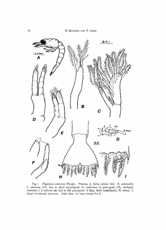
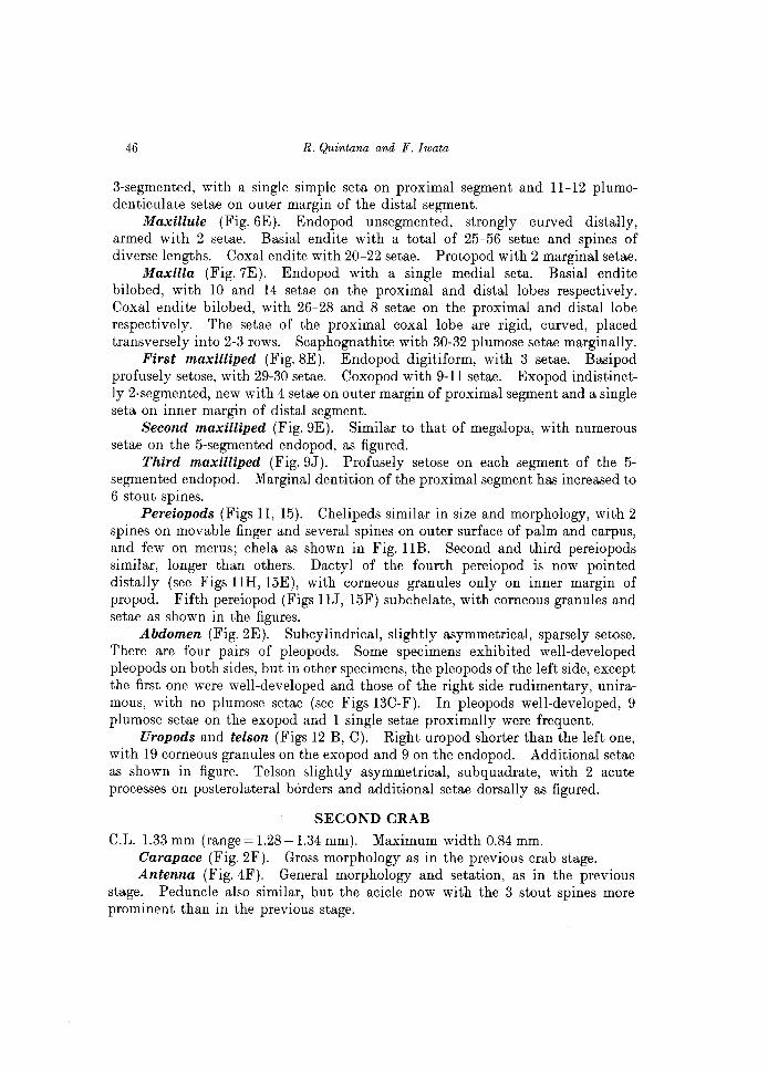
Fig. 1. Paguristes ortmanni Miyake. Prezoea. A, larva, lateral view; B, antennule; C, antenna; D-F, first to third maxillipeds; G, rudiments of pereiopods (Ch, cheliped; numerals 2-5 indicate the 2nd to 5th pereiopods; 3 Mxp, third maxilliped); H, telson; I, detail of telsonal processes. Scale bars=O.l mm, except for A.
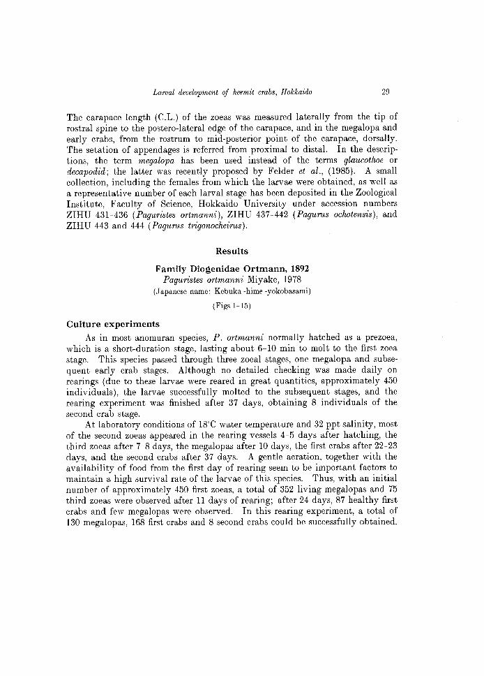
Larval development of hermit crabs, Hokkaido 29
The carapace length (C.L.) of the zoeas was measured laterally from the tip of rostral spine to the postero-Iateral edge of the carapace, and in the megalopa and early crabs, from the rostrum to mid-posterior point of the carapace, dorsally. The setation of appendages is referred from proximal to distal. In the descriptions, the term megalopa has been used instead of the terms glaucothoe or decapodid; the latter was recently proposed by Felder et al., (1985). A small collection, including the females from which the larvae were obtained, as well as a representative number of each larval stage has been deposited in the Zoological Institute, Faculty of Science, Hokkaido University under accession numbers ZIHU 431~436 (Paguristes ortmanni), ZIHU 437~442 (Pagurus ochotensis), and ZIHU 443 and 444 (Pagurus trigonocheirus).
Results
Family Diogenidae Ortmann, 1892 Paguristes ortmanni Miyake, 1978
(Japanese name: Kebuka -hime -yokobasami)
(Figs 1-15)
Culture experiments
As in most anomuran species, P. ortmanni normally hatched as a prezoea, which is a short-duration stage, lasting about 6~1O min to molt to the first zoe a stage. This species passed through three zoeal stages, one megalopa and subsequent early crab stages. Although no detailed checking was made daily on rearings (due to these larvae were reared in great quantities, approximately 450 individuals), the larvae successfully molted to the subsequent stages, and the rearing experiment was finished after 37 days, obtaining 8 individuals of the second crab stage.
At laboratory conditions of 18°C water temperature and 32 ppt salinity, most of the second zoeas appeared in the rearing vessels 4~5 days after hatching, the third zoeas after 7~8 days, the megalopas after 10 days, the first crabs after 22~23 days, and the second crabs after 37 days. A gentle aeration, together with the availability of food from the first day of rearing seem to be important factors to maintain a high survival rate of the larvae of this species. Thus, with an initial number of approximately 450 first zoeas, a total of 352 living megalopas and 75 third zoeas 'were observed after 11 days of rearing; after 24 days, 87 healthy first crabs and few megalopas were observed. In this rearing experiment, a total of 130 megalopas, 168 first crabs and 8 second crabs could be successfully obtained.
30
E
R. Quintana and F. Iwata
, A-E,
.O.2mm. a
B
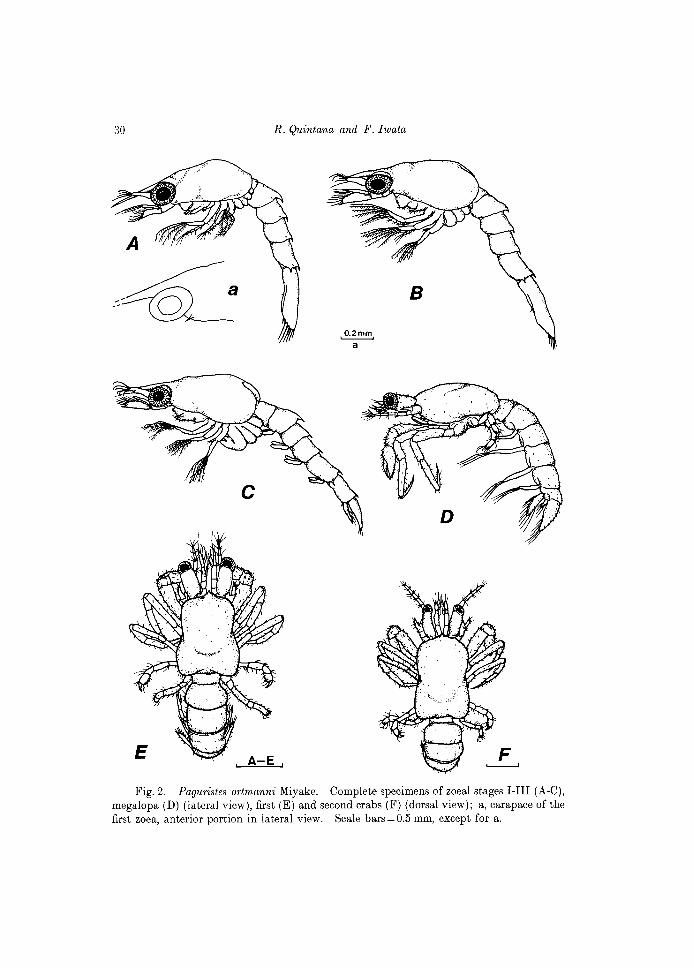
Fig. 2. Paguristes ortmanni Miyake. Complete specimens of zoeal stages I-III (A-C), megalopa (D) (lateral view), first (E) and second crabs (F) (dorsal view); a, carapace of the first zoea, anterior portion in lateral view. Scale bars=O.5 mm, except for a.
Larval development of hermit crabs, Hokkaido 31
~ a,c
'--__ ....,j,D,E
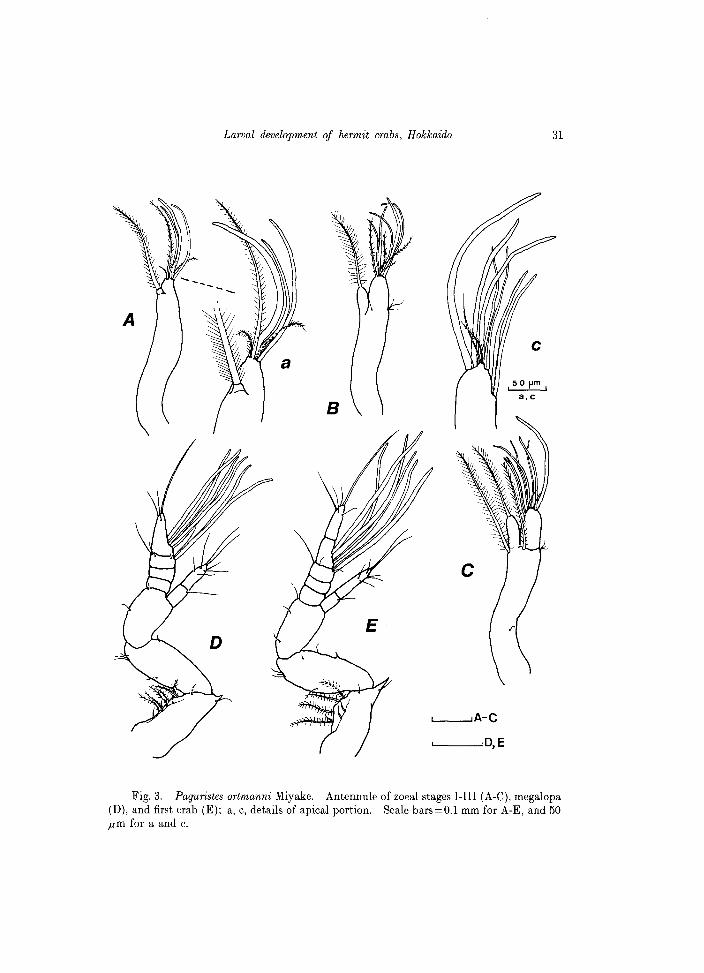
Fig, 3. Paguristes ortmanni Miyake. Antennule of zoeal stages I-III (A-C), megalopa (D), and first crab (E); a, c, details of apical portion. Scale bars=O.l mm for A-E, and 50 ,urn for a and c.
32 R. Quintana and F. Iwata
Description of the larval stages.
PREZOEA
C.L.O.90mm Body surrounded with a very thin, thransparent cuticle which protrudes as
long expansions on antennules, antennae and the tel son (see Fig. 1). Other appendages i.e. mandibles, maxillules, maxillae and maxillipeds are nonfunctional at this stage. The presence of the third maxilliped and the rudiments of pereiopods is evident at this early stage (see Fig. IG). First to third maxillipeds with no natatory plumose setae. Carapace oblong, unarmed, with no frontal spine; posterolateral borders rounded. Abdomen composed of 5 somites, each unarmed, except the fifth one, with a tiny posterolateral spine.
Antennule (Fig. IB). With 3 distal and 1 subterminal projections, all except one of the distal fringed with fine setules on margins.
Antenna (Fig. Ie). Endopod elongate, bearing 3 setulose projections. Exopod broadly oval, with 10 projections, all except the short proximal one firinged with fine setules marginally.
Telson (Fig. IH, I). Broadly triangular, notched on mid point of posterior margin and with 7 projections on each furca; all except the outermost process fringed marginally with fine setules.
FIRST ZOEA
C.L. 1.45 mm (range = 1.43-1.4 7 mm) Carapace (Figs 2A, a). Frontal spine (=rostrum) distally pointed, moder
ately long, reaching to tip of antennules. Anterolateral margins with 1 tiny spine; posterolateral margins rounded. Surface glabrous. Anterior half of carapace with a middorsal carina, moderately developed.
Antennule (Fig. 3A). Uniramous, elongate, bearing terminally 3 aesthetascs and 3 setulose setae; 1 seta sub terminally, reaching approximately to tips of aesthetascs.
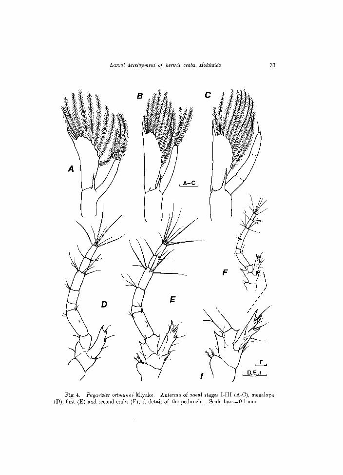
Antenna (Fig.4A). Endopod slightly less than 1/2 length of scale, bearing 3 terminal setae, each profusely setulose, one of which is shorter than others. Exopod ( = scale) almost straight along outer margin, distolaterally pointed; inner margin moderately rounded, fringed with 10 setae, each profusely setulose. Distal seta as long as the distal spine of the scale. A stout spine, finely serrate along both margins emerges distally from the peduncle.
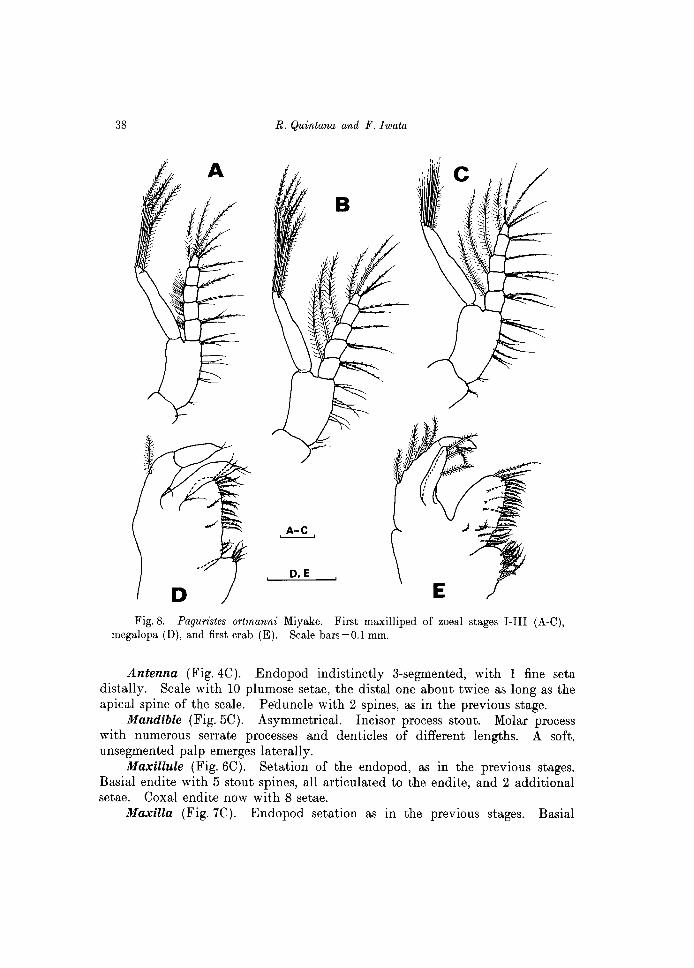
Mandible (Figs 5A; 15A). Slightly asymmetrical. Incisor process acute, strong. Molar process with numerous small denticles and marginal teeth. No palp present at this stage.
Maxillule (Fig. 6A). Endopod 3-segmented, with 1 short seta on the proximal segment, 1 long seta on the mid segment and 3 on the distal segment. Basial endite with 2 strong, rigid spines, not articulated, each with spinules on distal half, and 2 additional seta. Coxal endite with 7 seta.
Larval development of hermit crabs, Hokkaido 33
Fig. 4. Paguristes ortmanni Miyake. Antenna of zoeal stages I-III (A-C), megalopa (D), first (E) and second crabs (F); f, detail of the peduncle. Scale bars=O.l mm.
34
A
R. Quintana and F. Iwata
D
L--.........JB,C
'--_-"D,E
Fig. 5. Paguristes ortmanni Miyake. Mandible of zoeal stages I-III (A-C), megalopa (D), and first crab (E). C shows both mandibles in a same specimen. Scale bars=50 j.lm.
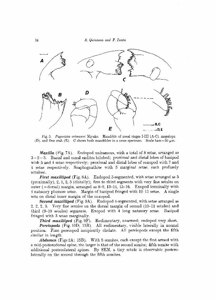
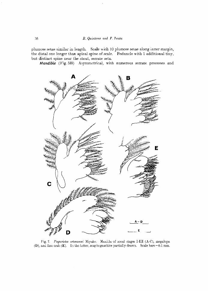
Maxilla (Fig. 7 A). Endopod uniramous, with a total of 8 setae, arranged as 3 + 2 + 3. Basial and coxal endites bilobed; proximal and distal lobes of basipod with 5 and 4 setae respectively; proximal and distal lobes of coxopod with 7 and 4 setae respectively. Scaphognathite with 5 marginal setae, each profusely setulose.
First maxilliped (Fig. 8A). Endopod 5-segmented, with setae arranged as 3 (proximally), 2, 1,2,5 (distally); first to third segments with very fine setules on outer (=dorsal) margin, arranged as 8-9, 13-14, 15-16. Exopod terminally with 4 natatory plumose setae. Margin of basipod fringed with 10-11 setae. A single seta on distal inner margin of the coxopod.
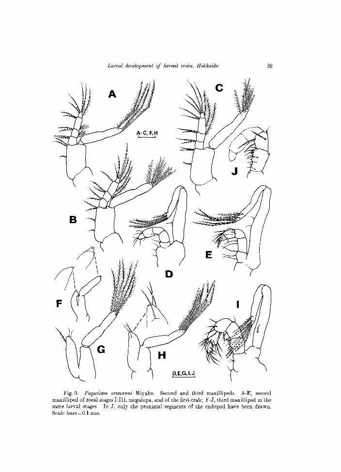
Second maxilliped (Fig. 9A). Endopod 4-segmented, with setae arranged as 2, 2, 2, 5. Very fine setules on the dorsal margin of second (10-11 setules) and third (9-10 setules) segments. Exopod with 4 long natatory setae. Basipod fringed with 3 setae margin~lly.
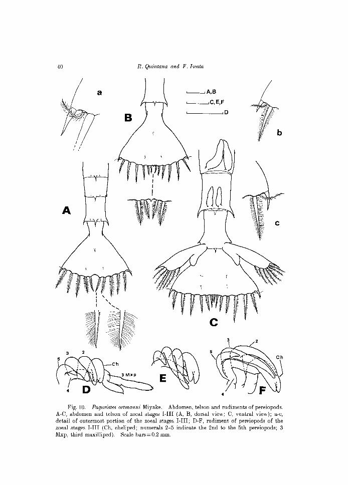
Third maxilliped (Fig. 9F). Rudimentary, unarmed; endopod very short. Pereiopods (Fig. 10D; 15B). All rudimentary, visible laterally in normal
position. First pereiopod incipiently chelate. All pereiopods except the fifth similar in length.
Abdomen (Figs 2A; 15B). With 5 somites, each except the first armed with a mid-posterodorsal spine, the larger is that of the second somite; fifth somite with additional posterolateral spines. By SEM, a tiny setule is observable posterolaterally on the second through the fifth somites.
Larval development of hermit crabs, Hokkaido 35
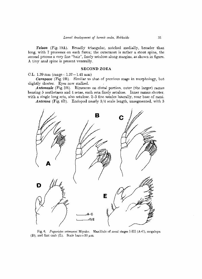
Telson (Fig. lOA). Broadly triangular, notched medially, broader than long, with 7 processes on each furca; the outermost is rather a stout spine, the second process a very fine "hair", finely setulose along margins, as shown in figure. A tiny anal spine is present ventrally.
SECOND ZOEA
C.L. 1.39 mm (range = 1.37 -1.41 mm) Carapace (Fig.2B). Similar to that of previous stage in morphology, but
slightly shorter. Eyes now stalked. Antennule (Fig. 3B). Biramous on distal portion, outer (the larger) ramus
bearing 5 aesthetascs and 4 setae, each seta finely setulose. Inner ramus shorter, with a single long seta, also setulose. 2-3 fine setules laterally, near base of rami.
Antenna (Fig.4B). Endopod nearly 3/4 scale length, unsegmented, with 3
B
L--JA-C
'--_---', D, E
Fig. 6. Paguristes ortmanni Miyake. Maxillule of zoeal stages I-III (A-C), rnegalopa (D), and first crab (E). Scale bars=50 ,urn.
36 R. Quintana and F. Iwata
plumose setae similar in length. Scale with 10 plumose setae along inner margin, the distal one longer than apical spine of scale. Peduncle with 1 additional tiny, but distinct spine near the stout, serrate seta.
Mandible (Fig. 5B) Asymmetrical, with numerous serrate processes and
Fig. 7. Paguristes ortmanni Miyake. Maxilla of zoeal stages I-III (A-C), megalopa (D), and first crab (E). In the latter, scaphognathite partially drawn. Scale bars=O.l mm.
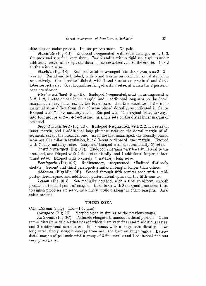
Larval development of hermit crabs, Hokkaido 37
denticles on molar process. Incisor process stout. No palp. Maxillule (Fig. 6B). Endopod 3-segmented, with setae arranged as 1, 1, 3,
the proximal seta fine, very short. Basial endite with 4 rigid stout spines and 2 additional setae; all except the distal spine are articulated to the endite. Coxal endite with 7 setae.
Maxilla (Fig. 7B). Endopod setation arranged into three groups as 3+2+ 3 setae. Basial endite bilobed, with 5 and 4 setae on proximal and distal lobes respectively. Coxal endite bilobed, with 7 and 4 setae on proximal and distal lobes respectively. Scaphognathite fringed with 7 setae, of which the 2 posterior ones are shorter.
First maxilliped (Fig. 8B). Endopod 5-segmented, setation arrangement as 3, 2, 1, 2, 4 setae on the inner margin, and 1 additional long seta on the dorsal margin of all segments, except the fourth one. The fine structure of the inner marginal setae differs from that of setae placed dorsally, as indicated in figure. Exopod with 7 long, natatory setae. Basipod with 11 marginal setae, arranged into four groups as 2 + 3 + 3 + 3 setae. A single seta on the distal inner margin of coxopod.
Second maxilliped (Fig. 9B). Endopod 4-segmented, with 2, 2, 1, 4 setae on inner margin, and 1 additional long plumose setae on the dorsal margin of all segments except the proximal one. As in the first maxilliped, the dorsally placed setae are all similar in setulation, but different to those of inner margin. Exopod with 7 long, natatory setae. Margin of basipod with 4, (occasionally 3) setae.
Third maxilliped (Fig. 9G). Endopod emerging very basally, lateral to the protopod, and fringed with 2 fine setae distally, and 1 additional longer, subterminal setae. Exopod with 6 (rarely 7) natatory, long setae.
Pereiopods (Fig. WE). Rudimentary, unsegmented. Cheliped distinctly chelate. Second and third pereiopods similar in length, longer than others.
Abdomen (Figs 2B; lOB). Second through fifth somites each with a midposterodorsal spine, and additional posterolateral spines on the fifth somite.
Telson (Fig. lOB). Not medially notched, with a tiny spiniform, smooth process on the mid point of margin. Each furca with 8 marginal processes; third to eighth processes are setae, each finely setulose along the entire margins. Anal spine present.
THIRD ZOEA
C.L. 1.55 mm (range = 1.52-1.56 mm) Carapace (Fig. 2C). Morphologically similar to the previous stages. Antennule (Fig. 3C). Peduncle elongate, biramous on distal portion. Outer
ramus distally with 5 aesthetascs (of which 2 are very fine) and 2 additional setae, and 2 subterminal aesthetascs. Inner ramus with a single seta distally. Two long setae, finely setulose emerge from near the base on inner ramus. Laterodistal margin of peduncle with a group of 3 fine setules and 1 additional fine seta very proximally.
38 R. Quintana and F. Iwata
A-C '------'
D,E
Fig. 8. Paguristes ortmanni Miyake. First maxilliped of zoeal stages I-III (A-C), megalopa (D), and first crab (E). Scale bars=O.l mm.
Antenna (Fig.4C). Endopod indistinctly 3-segmented, with 1 fine seta distally. Scale with 10 plumose setae, the distal one about twice as long as the apical spine of the scale. Peduncle with 2 spines, as in the previous stage.
Mandible (Fig.5C). Asymmetrical. Incisor process stout. Molar process with numerous serrate processes and denticles of different lengths. A soft, unsegmented palp emerges laterally.
Maxillule (Fig. 6C). Setation of the endopod, as in the previous stages. Basial endite with 5 stout spines, all articulated to the endite, and 2 additional setae. Coxal endite now with 8 setae.
Maxilla (Fig.7C). Endopod setation as in the previous stages. Basial
Larval development of hermit crabs, Hokkaido 39
,D.E.G.I,J,
Fig. 9. Paguristes ortmanni Miyake. Second and third maxillipeds. A-E, second maxilliped of zoeal stages I-III, megalopa, and of the first crab; F-J, third maxilliped in the same larval stages. In J, only the proximal segments of the endopod have been drawn. Scale bars = 0.1 mm.
40
A
3 2
a
, , ,
B
R. Quintana and F. Iwata
'-------...J A, B
'--_---" C, E,F
'--___ ---" D
it Fig. 10. Paguristes ortmanni Miyake. Abdomen, telson and rudiments of pereiopods.
A-C, abdomen and telson of zoeal stages I-III (A, B, dorsal view; C, ventral view); a-c, detail of outermost portion of the zoeal stages I-III; D-F, rudiment of pereiopods of the zoeal stages I-III (Ch, cheliped; numerals 2-5 indicate the 2nd to the 5th pereiopods; 3 Mxp, third maxilliped). Scale bars=O.2 mm.
Larval development of hermit crabs, Hokkaido 41
endite bilobed, with 5 setae on each lobe. Coxal endite bilobed, with 8+4 setae. Scaphognathite with a posterior, naked lobe, and 9 marginal setae, each finely setulose.
First maxilliped (Fig. 8C). Endopod 5-segmented, setation as 3, 3, 1, 2, 4 setae on inner margin, and 1 additional long seta on the dorsal margin of all segments, except the fourth one. Exopod with 7 long, natatory setae. Basipod with 11 setae on margin, arranged as 2, 3, 3, 3 setae. A single seta on distal inner margin of coxopod.
Second maxilliped (Fig. 9C). Endopod 4-segmented, with 2,2,2,4 setae on inner margin, and 1 additional long seta on the dorsal margin of all segments, except the proximal one. Exopod with 8 long natatory setae. Basipod with 3 setae on margin.
Third maxilliped (Fig. 9H). Endopod with 2 terminal setae and 1 subterminal simple seta. Exopod with 7 long natatory setae. Pereiopods (Fig. lOF). More developed but still unsegmented.
Abdomen (Fig.3C; lOC). Now with 6 segments, all except the first somite with a mid-posterodorsal spine. Fifth and sixth somites with posterolateral spines, those of the sixth segment tiny but distinct. Rudimentary uniramous pleopods present ventrally on somites 2-5. Uropods on the sixth somite; exopod wi th a stout disto-Iateral spine and 7 (occasionally 8) plumose setae on distal half along inner margin; endopod unarmed.
Telson (Fig. lOC). Posterior margin almost straight, with 9 processes on each furca (better to refer as 9+9). Third to ninth processes are setae, each profusely setulose along margins. Dorsal surface of tel son with 4 setae on the central area.
MEGALOPA
C.L. 0.95 mm (range=0.83-0.98 mm) Carapace (Fig.2D; 15C). Sparsely covered with minute setae. Front
short, not pointed distally. Anterolateral margins of carapace with 12 setae, and posterolateral margins with 6 setae. No ocular spines basally.
Antennule (Fig. 3D). Peduncle 3-segmented, basal segment with 1 spine disto-Iaterally and additional long setae on outer margin. Outer 4-segmented flagellum with aesthetascs on the third and distal segments, 6 aesthetascs in total, and additional setae as shown in figure. Inner flagellum 2-segmented, with 1 and 7 setae on the proximal and distal segments respectively.
Antenna (Fig. 4D). Peduncle indistinctly 4-segmented, with scale (= acicle) well-developed, with 3 spines and 3 setae laterally; second segment of peduncle with a small spine disto-Iaterally. Flagellum with 7 segments, distal one very short, all except the second segment with setae, which are progressively longer through apical segments; setal formula as 5, 0, 2, 6, 3, 4, 3 setae.
Mandible (Fig.5D). Markedly reduced in size. Mandibular plate slightly concave, edge rounded marginally. Palp 3-segmented, with 4 setae on distal
42 R. Quintana and F. Iwata
c
~ \
J I
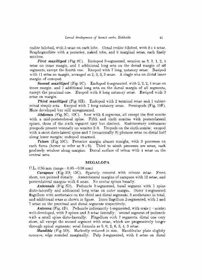
Fig.l1. Paguristes ortmanni Miyake. Pereiopods of the postlarval stages. A, right cheliped of the megalopa; B, right chela of the first crab; C, left cheliped of the second crab; c, detail of distal portion of fingers; D, second pereiopod of the megalopa; ; d, detail of dactyl; E, third pereiopod of the megalopa; F, third pereiopod of the first crab; G, fourth pereiopod of the megalopa; g, detail of distal segments; H, fourth pereiopod of the first crab; h, detail of the dactyl; I, fifth pereiopod of the megalopa; J, fifth pereiopod of the first crab; j, detail of distal segments. Scale bars=O.2 mm, except for B.

Larval development of hermit crabs, Hokkaido 43
segment. Maxillule (Fig.6D). Endopod digiti form, unsegmented, markedly fleeted,
with 1 short apical seta. Basial endite broadly oval, with a total of 17 spines and setae. Coxal endite with 3 simple setae and 4 minute spines, not pointed distally.
Maxilla (Fig. 7D). Endopod reduced to a digitiform process, with 2 subterminal setae. Basial endite with Sand 7 setae on proximal and distal lobes respectively. Coxal endite with 5 and 4 setae on proximal and distal lobes respectively. Scaphognathite greatly developed, with 30-31 marginal setae and few, additional setae on surface.
First maxilliped (Fig. SD). Markedly modified from that of zoeal stages. Endopod unsegmented, with 4-5 fine setae. Basipod broadly produced, armed
*
,O.2mm I
B
~A
'--_----I' C, D
Fig. 12. Paguristes ortmanni Miyake. Uropods and telson of the megalopa (A) and first crab (B), dorsal view (lateral setation of exopod of left uropod (A) has been omitted for clarity); C, detail of the telson of .the first crab; D, last abdominal somite and telson of the second crab. Asterisk in A indicates a detail of postero-lateral margin of the megalopal telson. Scale bars=O.1 mm, except for B.
44 R. Quintana and F. Iwata
with a total of 21-22 setae, most placed along margin. Coxopod with 5 terminal setae. Exopod 2-segmented, with a single plumose seta on proximal segment and 2 fine setae on distal segment.
Second maxilliped (Fig.9D). Endopod now 5-segmented, shorter than the proximal segment of the exopod, with 1, 1, 0, 3, 4 setae. Exopod 2-segmented, distal segment markedly flected, armed with 6-7 long plumose setae.
Third maxilliped (Fig. 91). Markedly modified from that of zoeal stages. Endopod 5-segmented, with numerous setae as indicated in figure; basal segment with 3 spines on inner margin. Exopod 2-segmented, distal segment strongly flected, armed with 7 long plumose setae.
Pereiopods (Figs 11, 15C). Setose, evenly covered with minute spinules. Chelipeds (Fig. 11A) almost symmetrical, fingers crossing at tips; one minute spine on movable finger, 3 on outer margin of palm and carpus, and 2 minute spines on inner margin of merus. Second and third pereiopods similar in length, markedly longer than chelipeds; dactyl longer than propod, pointed distally, setation as illustrated (Figs 11 D, E). Fourth and fifth pereiopods (Figs 11 G, I) very short, dactyls rudimentary, not pointed distally; corneous granules on ventral margins of dactyl and propod. Fifth pereiopod distinctly subchelate.
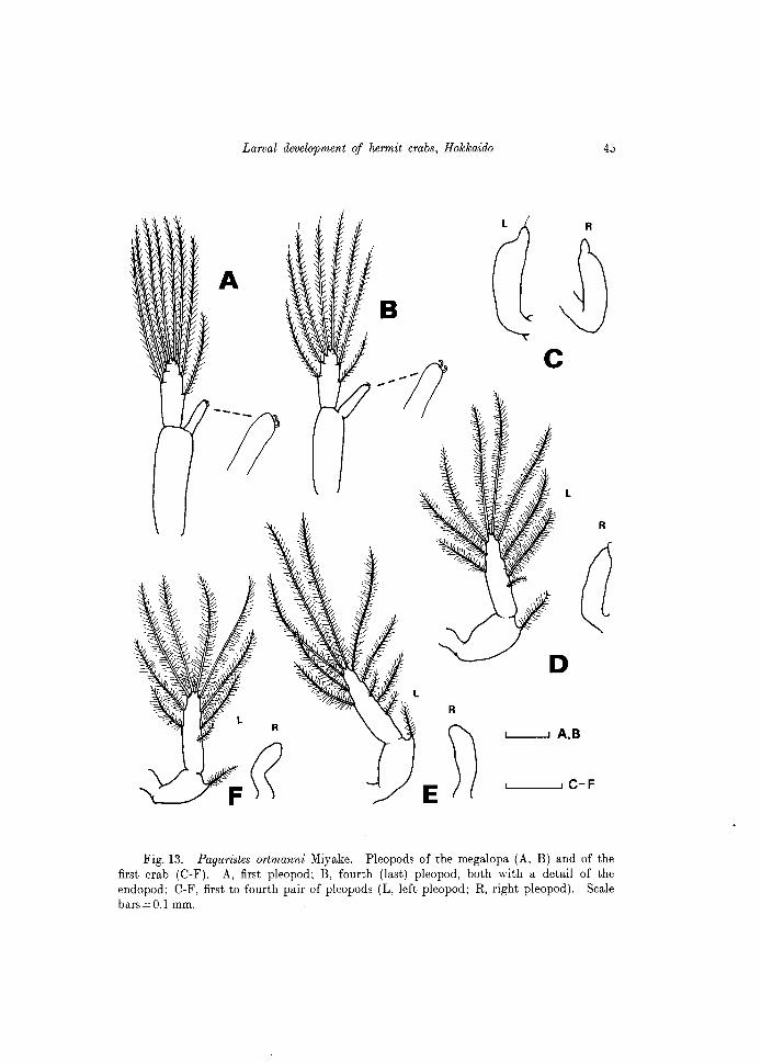
Abdomen (Fig.2D). Subcylindrical, 6-segmented; second to fifth segments each with a pair of well-developed pleopods; sixth segment with uropods. Pleopods each with 8 long plumose setae on exopod, and 2 hooked setae on endopod distally (Figs 13 A, B).
Uropods and telson (Fig. 12A). Uropods symmetrical, probably the right one only slightly reduced. Exopod with 16 marginal plumose setae, 6-8 simple marginal setae, placed alternately among the plumose setae, and 16-18 corneous granules. Endopod with 5 marginal plumose setae and 7-8 (occasionally 9) corneous granules. Telson symmetrical, longer than broad, posterior margin rounded, with 10 plumose setae and a group of 4 simple, short setae posterolaterally. Additional setae on uropods and telson as illustrated.
FIRST CRAB
C.L. 0.79 mm (range =0.78-0.83 mm). Maximum width 0.66 mm. Carapace (Figs 2E, 15D). Surface sparsely covered with minute setae,
moderate transverse depression dorsally. Front short. Posterolateral margins of carapace fringed with 8-10 short setae. Ocular peduncles long, slightly less than 1/2 C.L.; no ocular spines.
Antennule (Fig 3E). Although the outer flagellum is now 5-segmented, the number of aesthetascs (=6) is unchanged from the megalopa stage.
Antenna (Fig. 4E). Acicle with 3 stout lateral spines. Flagellum composed of 7 segments, the proximal longest and the distal segment shortest; all except the second segment armed with setae which are progressively longer through distal segments.
Mandible (Fig.5E). Cutting edge obtuse, margins almost straight. Palp
Larval development of hermit crabs, Hokkaido 45
R
c
L
R
L--J A,B
L..-_---', c- F
Fig. 13. Paguristes ortmanni Miyake. Pleopods of the megalopa (A, B) and of the first crab (C-F). A, first pleopod; B, fourth (last) pleopod, both with a detail of the endopod; C-F, first to fourth pair of pleopods (L, left pleopod; R, right pleopod). Scale bars = 0.1 mm.
46 R. Quintana and F. Iwata
3-segmented, with a single simple seta on proximal segment and 11-12 plumodenticulate setae on outer margin of the distal segment.
Maxillule (Fig.6E). Endopod unsegmented, strongly curved distally, armed with 2 setae. Basial endite with a total of 25-56 setae and spines of diverse lengths. Coxal endite with 20-22 setae. Protopod with 2 marginal setae.
Maxilla (Fig.7E). Endopod with a single medial seta. Basial endite bilobed, with 10 and 14 setae on the proximal and distal lobes respectively. Coxal endite bilobed, with 26-28 and 8 setae on the proximal and distal lobe respectively. The setae of the proximal coxal lobe are rigid, curved, placed transversely into 2-3 rows. Scaphognathite with 30-32 plumose setae marginally.
First maxilliped (Fig.8E). Endopod digitiform, with 3 setae. Basipod profusely setose, with 29-30 setae. Coxopod with 9-11 setae. Exopod indistinctly 2-segmented, new with 4 setae on outer margin of proximal segment and a single seta on inner margin of distal segment.
Second maxilliped (Fig.9E). Similar to that of megalopa, with numerous setae on the 5-segmented endopod, as figured.
Third maxilliped (Fig.9J). Profusely setose on each segment of the 5-segmented endopod. Marginal dentition of the proximal segment has increased to 6 stout spines.
Pereiopods (Figs 11, 15). Chelipeds similar in size and morphology, with 2 spines on movable finger and several spines on outer surface of palm and carpus, and few on merus; chela as shown in Fig. lIB. Second and third pereiopods similar, longer than others. Dactyl of the fourth pereiopod is now pointed distally (see Figs llR, 15E), with corneous granules only on inner margin of propod. Fifth pereiopod (Figs llJ, 15F) subchelate, with corneous granules and setae as shown in the figures.
Abdomen (Fig.2E). Subcylindrical, slightly asymmetrical, sparsely setose. There are four pairs of pleopods. Some specimens exhibited well-developed pleopods on both sides, but in other specimens, the pleopods of the left side, except the first one were well-developed and those of the right side rudimentary, uniramous, with no plumose setae (see Figs 13C-F). In pleopods well-developed, 9 plumose setae on the exopod and 1 single setae proximally were frequent.
Uropods and telson (Figs 12 B, C). Right uropod shorter than the left one, with 19 corneous granules on the exopod and 9 on the endopod. Additional setae as shown in figure. Telson slightly asymmetrical, subquadrate, with 2 acute processes on posterolateral borders and additional setae dorsally as figured.
SECOND CRAB
C.L. 1.33 mm (range = 1.28-1.34 mm). Maximum width 0.84 mm. Carapace (Fig. 2F). Gross morphology as in the previous crab stage. Antenna (Fig.4F). General morphology and setation, as in the previous
stage. Peduncle also similar, but the acicle now with the 3 stout spines more prominent than in the previous stage.
Larval development of hermit crabs, Hokkaido 47
n
b4
n n .. .. .. .. .. .. L~ft .. .. ..
Right Left Right Left
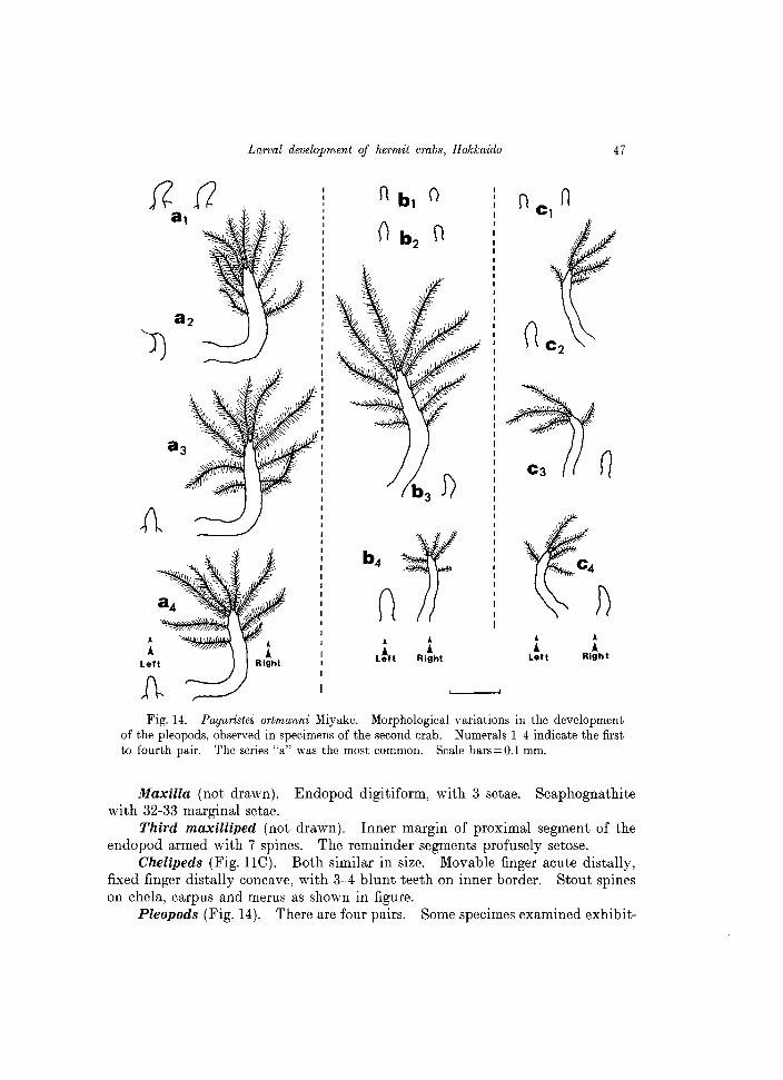
~ Fig. 14. Paguristes ortmanni Miyake. Morphological variations in the development
of the pleopods, observed in specimens of the second crab. Numerals 1-4 indicate the first to fourth pair. The series "a" was the most common. Scale bars=O.1 mm.
Maxilla (not drawn). Endopod digitiform, with 3 setae. Scaphognathite with 32-33 marginal setae.
Third maxilliped (not drawn). Inner margin of proximal segment of the endopod armed with 7 spines. The remainder segments profusely setose.
Chelipeds (Fig. lle). Both similar in size. Movable finger acute distally, fixed finger distally concave, with 3-4 blunt teeth on inner border. Stout spines on chela, carpus and merus as shown in figure.
Pleopods (Fig. 14). There are four pairs. Some specimes examined exhibit-
48 R. Quintana and F. Iwata
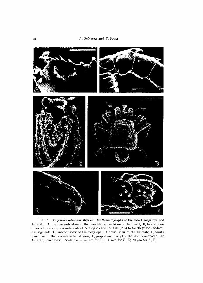
Fig. 15. Paguristes ortmanni Miyake. SEM-micrographs of the zoe a I, megalopa and 1st crab. A, high magnification of the mandibular dentition of the zoea I; B, lateral view of zoea I, showing the rudiments of pereiopods and the first (left) to fourth (rigth) abdominal segments; C, anterior view of the megalopa; D, dorsal view of the 1st crab; E, fourth pereiopod of the 1st crab, external view; F, propod and dactyl of the fifth pereiopod of the 1st crab, inner view. Scale bars=0.5 mm for D; 100 mm for B, E; 50 pm for A, F.
Larval development of hermit crabs, Hokkaido 49
ed well-developed pleopods (except the first one) only on the right side, and the setation was 9-10 plumose setae. But in other specimens, only the last pleopod of the right side was developed, bearing 6 setae; similarly, only the third pleopod of the left side was large, well-developed, bearing 10 plumose setae and 1 additional fine seta. Another range of variation, less frequent is shown in Fig. 14 together with the two above mentioned. In all of them, invariably the first pair was markedly reduced, as a single soft protuberance, difficult to remove from the respective abdominal somite.
Remarks: Al though not longer commented in the section Discussion of the present paper, this interesting ontogeny and changes in the morphology of pleopods in the early crab stages of Paguristes ortmanni have also been recently observed in the juvenile development of the allied diogenid crab, Clibanarius sclopetarius (Herbst) (see Brossi-Garcia, 1987 for a discussion of ontogeny of pleopods in the latter species and other diogenid crabs).
Family Pa~uridae Latreille, 1802 Pagurus middendmfJii Brandt, 1851 (Japanese name: Tenaga-honyadokari)
(Fig. 16)
Culture experiments
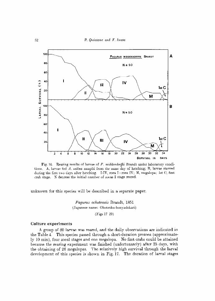
Two groups of 50 larvae were reared under the same laboratory conditions, as indicated in Table 1, and a gentle aeration was provided on each of the glass vessels used. One group was starved during the first two days and the another group fed A. salina nauplii since the first day after hatching. The daily checking for both rearing experiments is indicated in Tables 2 and 3. The initial conditions seem to have important effects in the subsequent molts and survival of larvae obtained, as shown comparatively in Fig. 16. The initial starving condition has a detrimental effect on the larval development.
Under "favorable" conditions, larvae of P. middendorffii invariably hatch as a prezoea, lasting approximately 10 min before molting to the first zoea. Subsequent stages include four zoeal stages, one megalopa, and the first crab.
Including the results observed in a third (=extra) group of 50 larvae, the megalopa of this species appeared 24-26 days after hatching, and the first crab was attained after 32-34 days of rearing.
Description of the larval sta~es
Kurata (1964) described the first zoea of P. middendorffii obtained both from eggs and plankton, and the zoeas II -IV from the plankton of Hokkaido. The prezoea of this species was recently described by Quintana and Konishi (1986). The postlarval stages obtained, i.e. the megalopa and the first crab, which are
50 R. Quintana and F. Iwata
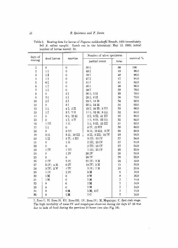
Table 2. Rearing data for larvae of Pagurus middendorffii Brandt, 1851 immediately fed A. salina nauplii. Hatch out in the laboratory: May 13, 1985; initial number of larvae reared: 50.
days of Number of alive specimens survival % dead larvae exuvIae rearing partial count total
1 0 0 50 I 50 100
2 1 I 0 49 I 49 98.0 3 1 I 0 48 I 48 96.0 4 1 I 0 47 I 47 94.0
5 61 0 411 41 82.0
6 1 I 0 40 I 40 80.0 7 11 0 39 I 39 78.0
8 0 31 36 I; 3 II 39 78.0
9 31 51 28 I; 8 II 36 72.0
10 21 31 23 I; 1111 34 68.0 11 0 31 20 I; 14 II 34 68.0 12 1 I 4 I; 2 II 15 I; 16 II; 2 III 33 66.0 13 1 I 3 I; 7 II 11 I; 12 II; 9 III 32 64.0 14 0 9 I; 12 II 2 I; 9 II; 21 III 32 64.0
15 0 1 I; 1 II 1 I; 9 II; 22 III 32 64.0 16 1 III III 1 I; 8 II; 22 III 31 62.0 17 1 I 0 8 II; 22 III 30 60.0 18 0 3 III 8 II; 19 III; 3 IV 30 60.0 19 211 3 II; 19 III 3 II; 3 III; 22 IV 28 56.0 20 III 2 II; 2 III 3 III; 24 IV 27 54.0 21 0 III 2 III; 25 IV 27 54.0 22 0 0 2 III; 25 IV 27 54.0 23 lIV 1 III 1 III; 25 IV 26 52.0 24 0 lIV 26 IV 26 52.0 25 0 0 26 IV 26 52.0 26 2IV 3IV 21 IV; 3 M 24 48.0 27 5 IV; 4 M 6IV 10 IV; 5M 15 30.0 28 3 IV; 2 M 4IV 3 IV; 7 M 10 20.0 29 lIV 2IV 9M 9 18.0 30 1M 0 8M 8 16.0 31 1M 0 7M 7 14.0 32 0 0 7M 7 14.0
33 0 0 7M 7 14.0 34 0 6M 1 M; 6C 7 14.0 35 0 1M 7C 7 14.0
I, Zoea I; II, Zoea II; III, Zoea III; IV, Zoea IV; M, Megalopa; C, first crab stage. The high mortality of zoeas IV and megalopas observed during the days 27-28 was due to lack of food during the previous 24 hours (see also Fig. 16).
Larval development of hermit crabs, Hokkaido
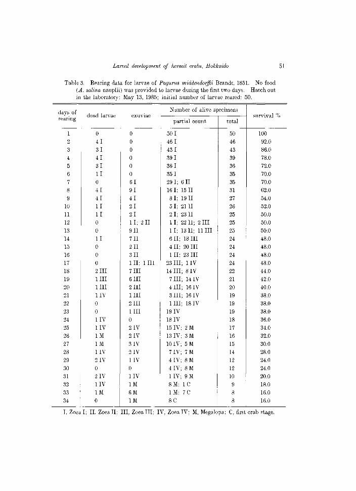
Table 3. Rearing data for larvae of Pagurus middendorffii Brandt, 1851. No food (A. salina nauplii) was provided to larvae during the first two days. Hawh out in the laboratory: May 13, 1985; initial number of larvae reared: 50.
Number of alive specimens
51
days of dead larvae exuvIae survival % rearing partial count total
1 0 0 50 I 50 100
2 4 I 0 46 I 46 92.0
3 31 0 43 I 43 86.0
4 4 I 0 39 I 39 78.0
5 31 0 36 I 36 72.0
6 1 I 0 35 I 35 70.0
7 0 61 29 I; 6 II 35 70.0
8 41 91 16 I; 15 II 31 62.0
9 41 41 8 I; 19 II 27 54.0
10 1 I 21 5 I; 21 II 26 52.0
11 1 I 21 2 I; 23 II 25 50.0
12 0 11; 2 II 1 I; 22 II; 2 III 25 50.0
13 0 9II 1 I; 13 II; 11 III 25 50.0
14 1 I 7II 6 II; 18 III 24 48.0
15 0 2II 4 II; 20 III 24 48.0
16 0 3II 1 II; 23 III 24 48.0
17 0 1 II; 1 III 23 III; IIV 24 48.0
18 2 III 7 III 14 III; 8 IV 22 44.0
19 1 III 6 III 7 III; 14 IV 21 42.0
20 1 III 2 III 4 III; 161V 20 40.0
21 IIV 1 III 3 III; 16 IV 19 38.0
22 0 2 III 1 III; 181V 19 38.0
23 0 1 III 19 IV 19 38.0
24 IIV 0 18 IV 18 36.0
25 IIV 21V 15 IV; 2 M 17 34.0
26 1M 21V 131V; 3M 16 32.0
27 1M 31V 10 IV; 5M 15 30.0
28 11V 21V 7 IV; 7 M 14 28.0
29 21V IIV 4 IV; 8 M 12 24.0
30 0 0 4 IV; 8 M 12 24.0
31 21V IIV 1 IV; 9 M 10 20.0
32 IIV 1M 8M; 1 e 9 18.0
33 1M 6M 1 M; 7 e 8 16.0
34 0 1M 8e 8 16.0
I, Zoea I; II, Zoea II; III, Zoea III; IV, Zoea IV; M, Megalopa; e, first crab stage.
52
100
80
60
40
~ 20 > > 0:: ::>
I/J .... 100 < > 0:: ..: 80 ...
60
40
20
2 4 6 8 10
R. Quintana and F. Iwata
PAGURUS MIDDENDORFFII BRANDT A
N= 50
B
N= 50
1st C
M ,r\-. . . . .
12 14 16 18 20 22 24 26 28 30 32 34
SURVIVAL IN DAYS
Fig. 16. Rearing results of larvae of P. middendorffii Brandt under laboratory conditions. A, larvae fed A. salina nauplii from the same day of hatching; B, larvae starved during the first two days after hatching. I-IV, zoe a 1- zoea IV; M, megalopa; 1st C, first crab stage. N denotes the initial number of zoeas I stage reared.
unknown for this species will be described in a separate paper.
Culture experiments
Pagurus ochotensis Brandt, 1851 (Japanese name: Ohotsuku-honyadokari)
(Figs 17-29)
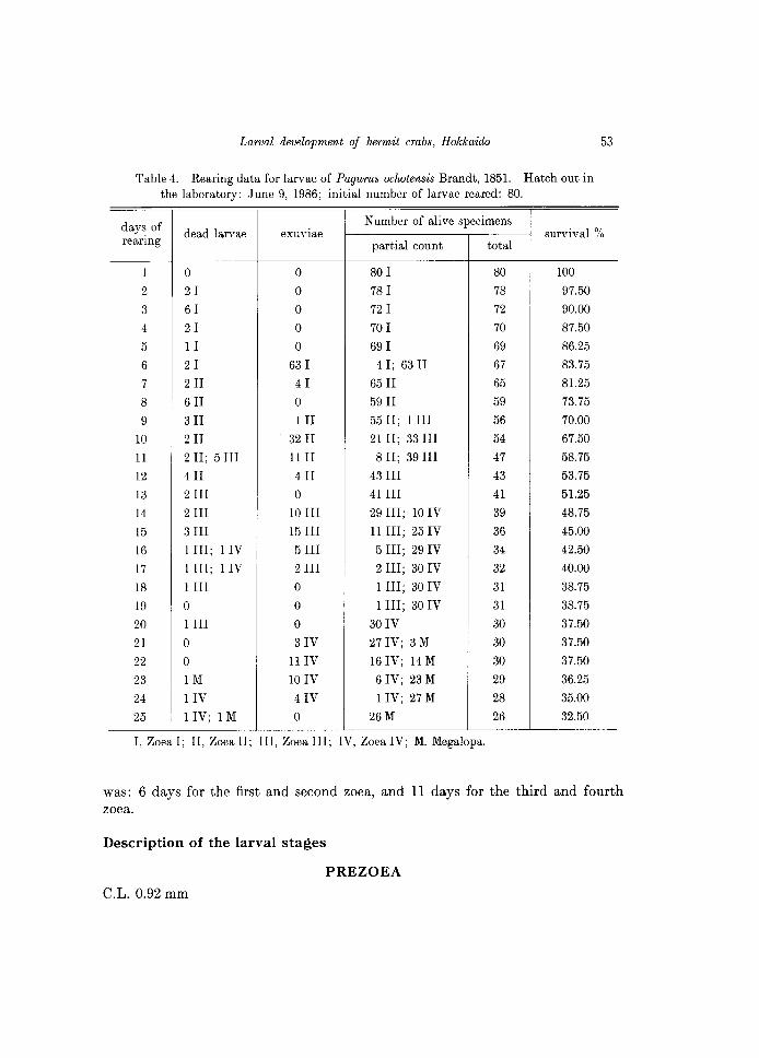
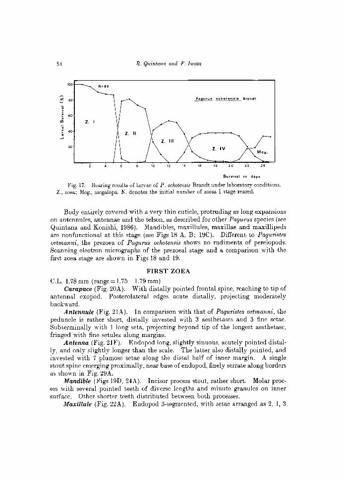
A group of 80 larvae was reared, and the daily observations are indicated in the Table 4. This species passed through a short-duration prezoea (approximately lO min), four zoeal stages and one megalopa. No first crabs could be attained because the rearing experiment was finished (unfortunately) after 25 days, with the obtaining of 26 megalopas. The relatively high survival through the larval development of this species is shown in Fig. 17. The duration of larval stages
Larval development of hermit crabs, Hokkaido 53
Table 4. Rearing data for larvae of Pagurus ochotensis Brandt, 1851. Hatch out in the laboratory: June 9, 1986; initial number of larvae reared: 80.
days of Number of alive specimens dead larvae exuvIae survival %
rearing partial count total
1 0 0 80 I 80 100
2 21 0 78 I 78 97.50
3 61 0 72 I 72 90.00
4 21 0 70 I 70 87.50
5 11 0 69 I 69 86.25
6 2 I 63 I 4 I; 63 II 67 83.75
7 2II 41 65 II 65 81.25
8 6II 0 59 II 59 73.75
9 3II III 55 II; 1 III 56 70.00
10 2II 32 II 21 II; 33 III 54 67.50
11 2 II; 5 III 11 II 8 II; 39 III 47 58.75
12 4II 4II 43 III 43 53.75
13 2 III 0 41 III 41 51.25
14 2 III 10 III 29 III; 10 IV 39 48.75
15 3 III 15 III 11 III; 25 IV 36 45.00
16 1 III; 1 IV 5 III 5 III; 29 IV 34 42.50
17 1 III; 1 IV 2 III 2 III; 30 IV 32 40.00
18 1 III 0 1 III; 30 IV 31 38.75
19 0 0 1 III; 30 IV 31 38.75
20 1 III 0 30 IV 30 37.50
21 0 3IV 27IV; 3M 30 37.50
22 0 11 IV 16IV; 14M 30 37.50
23 1M 10 IV 6 IV; 23 M 29 36.25
24 lIV 4IV 1 IV; 27 M 28 35.00
25 1 IV; 1 M 0 26M 26 32.50
I, Zoea I; II, Zoea II; III, Zoea III; IV, Zoea IV; M. Megalopa.
was: 6 days for the first and second zoea, and 11 days for the third and fourth zoea.
Description of the larval stages
PREZOEA C.L. 0.92 mm
54 R. Quintana and F. Iwata
100 N'SO
~
~ 80 . > .;;
60
en Z. I
: 40 . ....J
20
4 12 24
Survival In days
Fig. 17. Rearing results of larvae of P. oclwtensis Brandt under laboratory conditions. Z., zoea; Meg., megalopa. N. denotes the initial number of zoeas I stage reared.
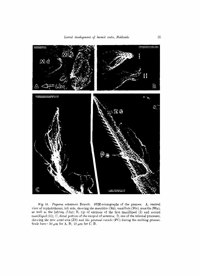
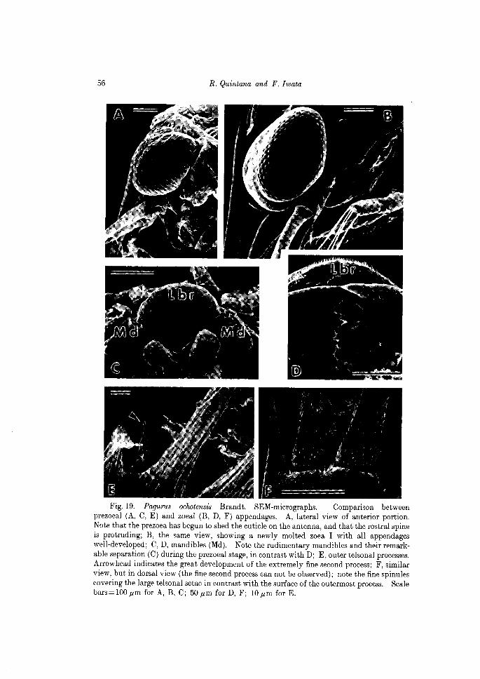
Body entirely covered with a very thin cuticle, protruding as long expansions on antennules, antennae and the telson, as described for other Pagurus species (see Quintana and Konishi, 1986). Mandibles, maxillules, maxillae and maxillipeds are nonfunctional at this stage (see Figs 18 A, B; 19C). Different to Paguristes ortmanni, the prezoea of Pagurus ochotensis shows no rudiments of pereiopods. Scanning electron micrographs of the prezoeal stage and a comparison with the first zoe a stage are shown in Figs 18 and 19.
FIRST ZOEA
C.L. 1.78 mm (range = 1.75-1.79 mm) Carapace (Fig. 20A). With distally pointed frontal spine, reaching to tip of
antennal exopod. Posterolateral edges acute distally, projecting moderately backward.
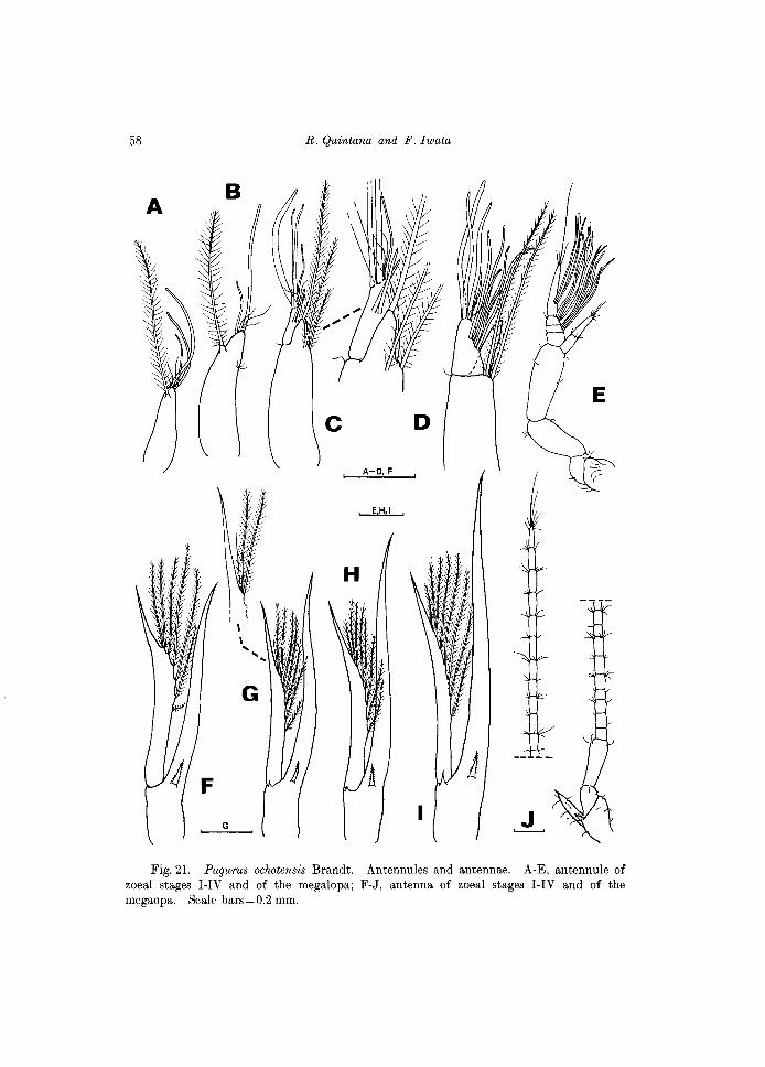
Antennule (Fig. 21A). In comparison with that of Paguristes ortmanni, the peduncle is rather short, distally invested with 3 aesthetascs and 3 fine setae. Subterminally with 1 long seta, projecting beyond tip of the longest aesthetasc, fringed with fine setules along margins.
Antenna (Fig. 21F). Endopod long, slightly sinuous, acutely pointed distally, and only slightly longer than the scale. The latter also distally pointed, and invested with 7 plumose setae along the distal half of inner margin. A single stout spine emerging proximally, near base of endopod, finely serrate along borders as shown in Fig. 29A.
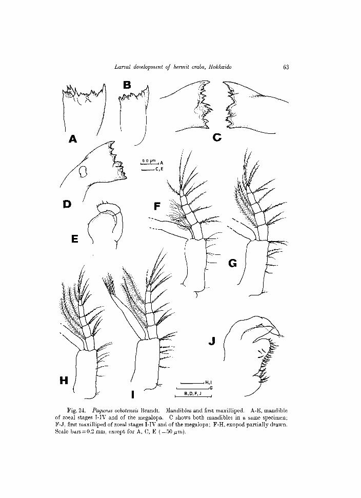
Mandible (Figs 19D, 24A). Incisor process stout, rather short. Molar process with several pointed teeth of diverse lengths and minute granules on inner surface. Other shorter teeth distributed between both processes.
Maxillule (Fig. 22A). Endopod 3-segmented, with setae arranged as 2, 1, 3
Larval development of hermit crabs, Hokkaido 55
Fig. 18. Pagurus oclwtensis Brandt. SEM-micrographs of the prezoea. A, ventral view of cephalothorax, left side, showing the mandible (Md), maxillule (Mle), maxilla (MIa), as well as the labrum (Lbr); B, tip of exopods of the first maxilliped (I) and second maxilliped (II); C, distal portion of the exopod of antenna; D, one of the telsonal processes, showing the new zoeal seta (ZS) and the prezoeal cuticle (PC) during the molting process. Scale bars=50.um for A, B; lO.um for C, D.
56 R. Quintana and F. Iwata
Fig. 19. Pagurus ochotensis Brandt. SEM-micrographs. Comparison between prezoeal (A, C, E) and zoeal (B, D, F) appendages. A, lateral view of anterior portion. Note that the prezoea has begun to shed the cuticle on the antenna, and that the rostral spine is protruding; B, the same view, showing a newly molted zoe a I with all appendages well-developed; C, D, mandibles (Md). Note the rudimentary mandibles and their remarkable separation (C) during the prezoeal stage, in contrast with D; E, outer telsonal processes. Arrowhead indicates the great development of the extremely fine second process; F, similar view, but in dorsal view (the fine second process can not be observed); note the fine spinules covering the large telsonal setae in contrast with the surface of the outermost process. Scale bars = 100,um for A, B, C; 50,um for D, F; lO,um for E.
Larval development of hermit crabs, Hokkaido 57
e
\!~. 11'.1 1;(.;\
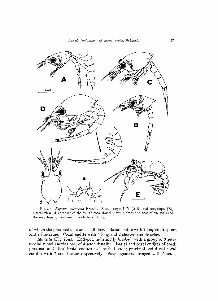
Fig. 20. Pagurus ochotensis Brandt. Zoeal stages I-IV (A-D) and megalopa (E), lateral view; d, carapace of the fourth zoea, dorsal view; e, front and base of eye stalks of the megalopa, dorsal view. Scale bars = 1 mm.
of which the proximal ones are small, fine. Basial endite with 2 long stout spines and 2 fine setae. Coxal endite with 5 long and 2 shorter, simple setae.
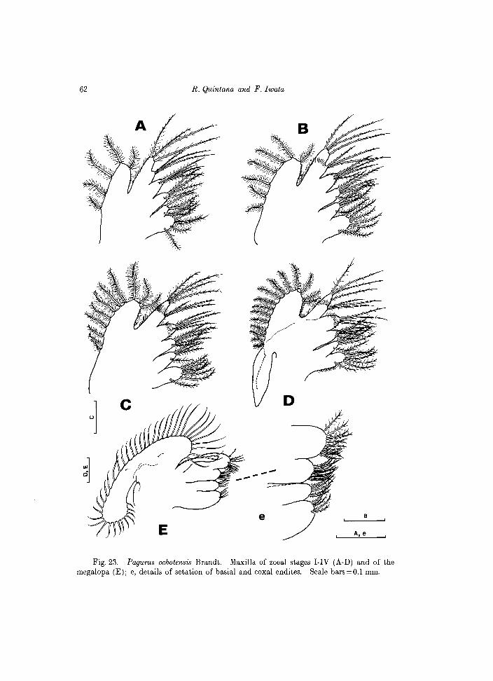
Maxilla (Fig. 23A). Endopod indistinctly bilobed, with a group of 3 setae medially and another one, of 4 setae distally. Basial and coxal endites bilobed; proximal and distal basial endites each with 4 setae; proximal and distal coxal endites with 7 and 4 setae respectively. Scaphognathite fringed with 5 setae,
58 R. Quintana and F. Iwata
Fig.21. Pagurus ochotensis Brandt. Antennules and antennae. A-E, antennule of zoeal stages I-IV and of the megalopa; F-J, antenna of zoeal stages I-IV and of the megaopa. Scale bars = 0.2 mm.
Larval development of hermit crabs, Hokkaido 59
each profusely fringed with fine setules as shown in Fig. 29B. First maxilliped (Fig. 24F). Endopod 5-segmented, with setal arrangement
as 3, 2, 1, 2, 5. One seta of the distal group is dorsal in position. First through third segments invested with very fine, long setules on outer (= dorsal) margin as 5-6, 8, and 5-6 setules respectively. Exopod with 4 long, plumose, natatory setae. Basipod with 9 setae on inner margin.
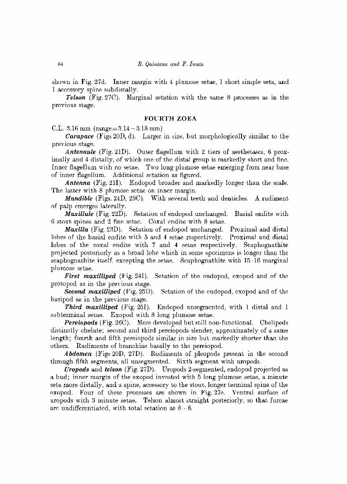
Second maxilliped (Fig. 25A). Endopod 4-segmented, with 2 setae on first to third segments, and 5 setae on distal segment of which one is dorsal in position. Very fine, long setules on the dorsal margin of the second and third segments, arranged as 3-4 and 5-6 respectively. Basipod with 3 setae, the distal pair very similar to those present on first through third segments.
Third maxilliped (Fig. 25F). Rudimentary, uniramous, unarmed. Pereiopods. No rudiments of pereiopods at this stage. Abdomen (Figs 20A, 27A). Composed of 5 segments and the telson. Poste
rior margin of each segment with spines as figured; only in the fifth segment, the posterolateral spines are long, approximately as long as 1/2 of the length of the segment. Each segment with an additional pair of fine setules dorsally.
Telson (Fig. 27 A). Elongate, approximately 1.5 times its posterior width, slightly notched medially. Anal spine present ventrally. Posterior margin of each furca invested with 7 processes. Outermost process is a stout spine, articulated to telson as shown in Fig. 19F; the second process is a fine seta, fringed with fine setules; third through seventh processes are long setae, each evenly covered with minute spinules on surface and long setules along borders, as shown in Fig. 19F.
SECOND ZOEA
C.L. 2.14 mm (range=2.13-2.17 mm) Carapace (Fig.20B). Rostral spine projected anteriorly beyond tip of
antennae. As in the previous stage, posterolateral edges pointed distally, short. Eyes are now stalked.
Antennule (Fig. 21B). With 2 aesthetascs, markedly different in length and 3 fine setae distally, and 1 single subterminal seta, not reaching tip of longest aesthetasc; 3 minute setules on outer margin.
Antenna (Fig. 21G). Endopod longer than the scale. The latter with 7 plumose setae and 1 apical, fine seta, none reaching tip of the acute distal spine of scale. An additional simple spine basally is added to the stout serrate spine, which was also present in the previous stage.
Mandible (Fig. 24B). Incisor process more developed than in the previous stage, with accessory dentition on its inner border. Molar process with several short, but acute teeth and minute granules on inner margin.
Maxillule (Fig. 22B). Setation of the 3-segmented endopod unchanged. Basial endite now with 4 long, stout spines, 2 of which are articulated to the endite, and 2 fine setae. Coxal endite with 7 setae.

60 R. Quintana and F. Iwata
A
A
B
E Fig. 22. Pagurus ochotensis Brandt. Maxillule of zoeal stages I-IV (A-D) and the
megalopa (E); e, detail of the basial endite. Scale bars 0.1 = mm, except for e.
Maxilla (Fig 23B). Endopod unchanged in setation. Basial endite bilobed, with 5 and 4 setae on the proximal and distal lobes respectively. Coxal endite bilobed, with 7 and 4 setae on the proximal and distal lobes respectively. Scaphognathite fringed with 7 setae, each profusely setulose.
First maxilliped (Fig. 24G). Endopod 5-segmented, with setation arranged as 3, 2, 1, 2, 4 setae on the inner margin, and 1 additional long setae on the dorsal margin of all segments, except the fourth one. Exopod with 7 long natatory setae. Basipod with 9-10 setae marginally.
Second maxilliped (Fig. 25B). Endopod 4 segmented, with 2, 2, 2, 4 setae on inner margin, and 1 additional long plumose seta on the dorsal margin of all
Larval development of hermit crabs, Hokkaido 61
segments, except the proximal one. Exopod with 7 long, natatory setae. Basipod with 3 setae marginally.
Third maxilliped (Fig. 25G). Endopod as a bud, with a single apical seta, Exopod with 6 long natatory setae.
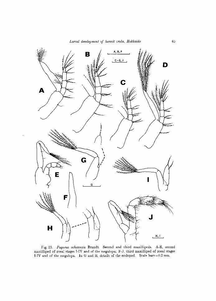
Pereiopods (Figs 20B, 26A). Visible laterally beneath the carapace. Rudimentary buds, first one not chelate.
Abdomen (Figs 20B, 27B). Oomposed of 5 segments and the telson. Posterolateral and posterodorsal spines of the second through fourth segment very short, but well-developed in the fifth segment, especially the lateral ones. A pair of fine setules dorsally only on the fifth segment.
Telson (Fig. 27B). Except the outermost 2 processes, each furca with 6 setae on the posterior margin.
THIRD ZOEA
O.L. 2.75 mm (range==2.73-2.77 mm) Carapace (Fig. 200). Similar in morphology as the previous stage. Antennule (Fig. 210). An incipient inner lobe and an outer, larger lobe are
differentiated at this stage. Outer lobe with 4 aesthetascs, the longest wide, the shortest very fine, and 2 short aesthetascs subterminally. Inner lobe with a single plumose seta apically. Additional 2 plumose setae placed near base of inner lobe. A single, minute seta on outer margin.
Antenna (Fig. 21H). Endopod markedly longer than scale. The latter with 8 setae on inner margin, all plumose. A serrate spine and a shorter, simple spine are placed basally.
Mandible (Fig. 240). Slightly asymmetrical, each with several teeth on both incisor and molar processes. Teeth of the right mandible longer and more acute distally. No bud of palp at this stage.
Maxillule (Fig. 220). Setation unchanged from the previous stage. Maxilla (Fig. 230). Setation as in the previous stage, except m the
scaphognathite which is fringed now with 11 plumose setae. First maxilliped (Fig. 24H). Setation of the 5-segmented endopod un
changed. Exopod with 8 long plumose setae. Basipod with 10 setae on inner margm.
Second maxilliped (Fig. 250). Setation of endopod and basipod as in the previous stage. Exopod with 8 long plumose setae.
Third maxilliped (Fig. 25H). Endopod more developed but still unsegmented, with 1 apical seta. Exopod with 7 long natatory setae distally.
Pereiopods (Fig. 26B). Rudimentary, first incipiently chelate distally. Fourth and fifth pereiopods markedly shorter than others.
Abdomen (Figs 200,270). Now composed of 6 segments and the telson. No rudiments of pleopods, but the sixth segment with uropods, which are slightly longer than 1/2 of the telson length, excepting the setae. Uropods uniramous, incipiently produced on proximal 1/3 of the inner margin, and pointed distally as
62 R. Quintana and F. Iwata
------
e B
A,e
Fig. 23. Pagurus ochotensis Brandt. Maxilla of zoeal stages I-IV (A-D) and of the megalopa (E); e, details of setation of basial and coxal endites. Scale bars=O.l mm.
Larval development of hermit crabs, Hokkaido 63
c ~A '-----' C,E
o F
-'-
E
J
'--_---" H,I '--___ -"G
B,D,F,J
Fig. 24. Pagurus oclwtensis Brandt. Mandibles and first maxilliped. A-E, mandible of zoeal stages I-IV and of the megalopa. C shows both mandibles in a same specimen; F-J, first maxilliped of zoeal stages I-IV and of the megalopa; F-R, exopod partially drawn. Scale bars=0.2 mm, except for A, C, E (=50 ,urn).
64 R. Quintana and F. Iwata
shown in Fig. 27d. Inner margin with 4 plumose setae, 1 short simple seta, and 1 accessory spine sub distally.
Telson (Fig. 27C). Marginal setation with the same 8 processes as in the previous stage.
FOURTH ZOEA
C.L. 3.16 mm (range=3.14-3.18 mm) Carapace (Figs 20D, d). Larger in size, but morphologically similar to the
previous stage. Antennule (Fig. 21D). Outer flagellum with 2 tiers of aesthetascs, 6 prox
imally and 4 distally, of which one of the distal group is markedly short and fine. Inner flagellum with no setae. Two long plumose setae emerging from near base of inner flagellum. Additional setation as figured.
Antenna (Fig. 2II). Endopod broader and markedly longer than the scale. The latter with 8 plumose setae on inner margin.
Mandible (Figs. 24D, 29C). With several teeth and denticles. A rudiment of palp emerges laterally.
Maxillule (Fig. 22D). Setation of endopod unchanged. Basial endite with 6 stout spines and 2 fine setae. Coxal endite with 8 setae.
Maxilla (Fig. 23D). Setation of endopod unchanged. Proximal and distal lobes of the basial endite with 5 and 4 setae respectively. Proximal and distal lobes of the coxal endite with 7 and 4 setae respectively. Scaphognathite projected posteriorly as a broad lobe which in some specimens is longer than the scaphognathite itself, excepting the setae. Scaphognathite with 15-16 marginal plumose setae.
First maxilliped (Fig. 241). Setation of the endopod, exopod and of the protopod as in the previous stage.
Second maxilliped (Fig. 25D). Setation of the endopod, exopod and of the basipod as in the previous stage.
Third maxilliped (Fig. 251). Endopod unsegmented, with 1 distal and 1 subterminal setae. Exopod with 8 long plumose setae.
Pereiopods (Fig. 26C). More developed but still non-functional. Chelipeds distinctly chelate; second and third pereiopods slender, approximately of a same length; fourth and fifth pereiopods similar in size but markedly shorter than the others. Rudiments of branchiae basally to the pereiopod.
Abdomen (Figs 20D, 27D). Rudiments of pleopods present in the second through fifth segments, all unsegmented. Sixth segment with uropods.
Uropods and telson (Fig. 27D). Uropods 2-segmented, endopod projected as a bud; inner margin of the exopod invested with 5 long plumose setae, a minute seta more distally, and a spine, accessory to the stout, longer terminal spine of the exopod. Four of these processes are shown in Fig. 27e. Ventral surface of uropods with 3 minute setae. Telson almost straight posteriorly, so that furcae are undifferentiated, with total setation as 8+8.
Larval d(Jl)elopment of hermit crabs, Hokkaido 65
Fig. 25. Pagurus ochotensis Brandt. Second and third maxillipeds. A-E, second maxilliped of zoeal stages I-IV and of the megalopa; F-J, third maxilliped of zoeal stages I-IV and of the megalopa. In G and H, details of the endopod. Scale bars=O.2 mm.
66
~ . /~
A
R. Quintana and F. Iwata
k-~ ..... ....... 0,
, A,G,H , O.2mm
Fig. 26. Pagurus ochotensis Brandt. Pereiopods. A-C, rudiment of pereiopods of the zoeal stages II-IV; D-H, pereiopods of the megalopa; D, right cheliped; D" left chela; Dz, right chela; E, second periopod; F, third pereiopod; G, fourth pereiopod (E-G, each with a detail of distal portion); H, fifth pereiopod. Scale bars, as indicated.
Larval development of hermit crabs, Hokkaido
MEGALOPA
C.L. 2.0 mm (range = 1.91-2.14 mm)
67
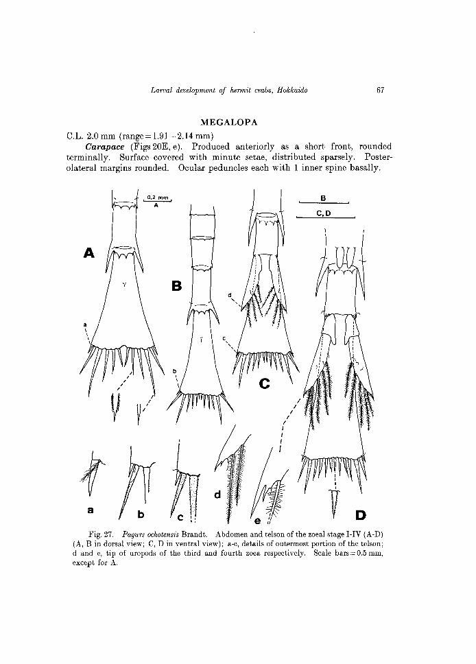
Carapace (Figs 20E, e). Produced anteriorly as a short front, rounded terminally. Surface covered with minute setae, distributed sparsely. Posterolateral margins rounded. Ocular peduncles each with 1 inner spine basally.
A
a
0.2 mm
A
/
~/
B
C,D
m V D
Fig. 27. Pagurs oclwtensis Brandt. Abdomen and telson of the zoeal stage I-IV (A-D) (A, B in dorsal view; C, D in ventral view); a-c, details of outermost portion of the telson; d and e, tip of uropods of the third and fourth zoea respectively. Scale bars = 0.5 mm, except for A.
68 R. Quintana and F. Iwata
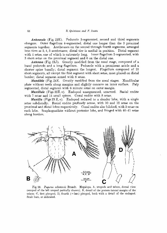
Antennule (Fig. 21E). Peduncle 3-segmented, second and third segments elongate. Outer flagellum 4-segmented, distal one longer than the 3 proximal segments together. Aesthetascs on the second through fourth segments, arranged into tiers as 5,4,3 aesthetascs; distal tier is medial in position. Distal segment with 4 setae, one of which is extremely long. Inner flagellum 2-segmented, with 3 short setae on the proximal segment and 6 on the distal one.
Antenna (Fig.2IJ). Greatly modified from the zoeal stage, composed of a basal peduncle and a long flagellum. Peduncle with a prominent acicle and a shorter spine basally; distal segment the longest. Flagellum composed of 21 short segments, all except the first segment with short setae, most placed on distal border; distal segment armed with 8 setae.
Mandible (Fig. 24E. Greatly modified from the zoeal stages. Mandibular plate without teeth along margins and slightly concave on inner surface. Palp segmented, distal segment with 6 minute setae on outer margin.
Maxillule (Figs 22E, e). Endopod unsegmented, unarmed. Basial endite with 7 setae and 14 small spines. Coxal endite with 8 setae.
Maxilla (Figs 23 E, e). Endopod reduced to a slender lobe, with a single setae sub distally. Basial endite profusely setose, with 10 and 12 setae on the proximal and distal lobes respectively. Coxal endite also bilobed, with 5 setae on each lobe. Scaphognathite without posterior lobe, and fringed with 40-41 setae along borders.
B c
\ \ \ \
I I I
ill) Fig. 28. Pagurus oclwtensis Brandt. Megalopa. A, uropods and telson, dorsal view
(exopod of the left uropod partially drawn); B, detail of the postero-lateral margin of the telson; C, first pleopod; D, fourth (= last) pleopod, both with a detail of the endopod. Scale bars, as indicated.
Larval dl'!lJelopment of hermit crabs, Hokkaido 69
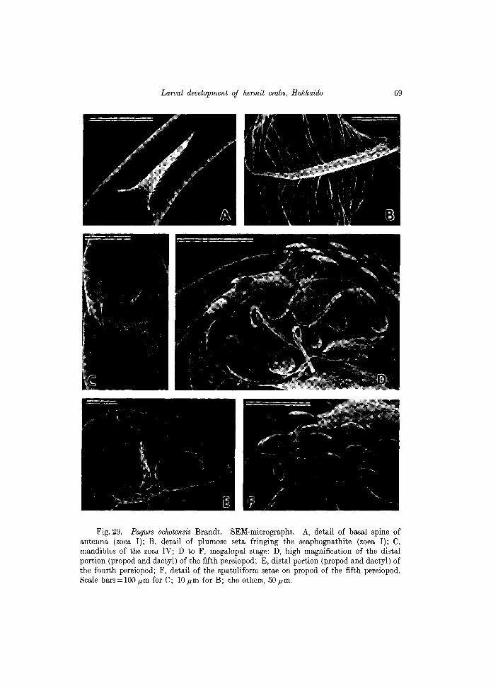
Fig. 29. Pagurs ochotensis Brandt. SEM-micrographs. A, detail of basal spine of antenna (zoea I); B, detail of plumose seta fringing the scaphognathite (zoea I); C, mandibles of the zoea IV; D to F, megalopal stage: D, high magnification of the distal portion (propod and dactyl) of the fifth pereiopod; E, distal portion (propod and dactyl) of the fourth pereiopod; F, detail of the spatuliform setae on propod of the fifth pereiopod. Scale bars = 100 jlm for C; lOjlm for B; the others, 50jlm.
70 R. Quintana and F. Iwata
First maxilliped (Fig. 24J). Markedly modified from the zoeal maxillipeds. Endopod digitiform, unsegmented. Basial endite broad, with 21 short setae. Coxal endite with 5 setae. Exopod indistinctly segmented, with 3 apical setae.
Second maxilliped (Fig. 25E). Endopod 4-segmented, with 3 setae on the third and fourth segments. Exopod 2-segmented, distal segment strongly fleeted. Proximal segment with 2 setae, and distal with 6 plumose setae.
Third maxilliped (Fig. 25J). Endopod greatly developed, profusely setose on each segment, specially on the third, fourth and fifth segments; first segment with 1 spine on inner border. Exopod with 6 plumose on the distal segment, which is strongly fleeted.
Pereiopods (Fig. 26). All functional, well-developed. Right cheliped slightly larger than the left one; cutting borders of chelae minutely serrate, an shown in Fig. 26D. Second and third pereiopods similar in size and setation, with long dactyl (see Figs 26E, F). Fourth and fifth pereiopods greatly reduced. Fourth pereiopod (Figs 26G, g) with short propod and dactyl; pro pod with 6-7 short, spatuliform setae on ventro-distal margin; dactyl pointed distally, with setae and 3 spinules on ventro-distal margin (see also Fig. 29E). Fifth pereiopod (Fig. 26H) subchelate, dactyl markedly reduced; propod elongate, with 4 very long, distally curved setae, several shorter setae and spatuliform setae; dactyl invested with setae, spines and few (2-3) spatuliform setae. The mentioned "spatuliform setae" are called corneous granules for other authors (sometimes "bulbous corneous granules"), but as observed by SEM, it is evident that they are not granules but rather short, spatula-like processes, all curved anteriorly, as shown in Figs. 29D, F.
Abdomen (Fig.20E). Composed of 6 segments and the telson, subcylindrical, sparsely setose, with pleopods on the second through fifth segments. First to fourth pleopods with 11, 10 (occasionally 11), 9 and 8 long, plumose setae on the respective exopod, and with 3, 3, 2-3, 2 hooked setae on the respective endopod (see Figs 28C, D).
Uropods and telson (Figs 28A, B). Uropods with a prominent exopod and reduced endopod; exopod fringed with 12-13 long plumose setae, 7-8 simple setae, distributed alternately among the plumose setae, and 12-13 short, spatuliform setae; endopod with 5 spatuliform setae and 4 simple setae. Telson subquadrate, as long as 1.4 times its width; posterior border slightly rounded, invested with 8 plumose setae; posterolateral edges as shown in Fig. 28B.
Pagurus trigonocheirus (Stimpson, 1858) (Japanese name: Mitsukado-honyadokari)
(Figs 30, 31)
Rearing results and remarks
Few prezoeas and individuals of the first zoea of this speCIes could be
Larval development of hermit crabs, Hokkaido 71
obtained from a single ovigerous female which spent more than one month in the laboratory. No larvae developed beyond first zoea.
Adults of this species and of another hermit crab, Elassochirus cavimanus are accompanying fauna in the traditional catches of edible prawns Pandalus borealis and P. nipponensis in the area, and the fishermen usually throw out these hermit crabs because are not commercially important. Local fishermen catch prawns at Funka Bay, off the coast of Usujiri, at depths of 230-280 m by using conical traps; such a devices are locally called "ebi-kago". The season for capturing prawns is restricted from May to late-October (occasionally to early-November), so the obtaining of ovigerous females of Pagurus trigonocheirus is also restricted to this period.
The temperature at approximately 220 m deep is about 9-10"C in I ateOctober. The over density of captured adults of this species (more than 300 individuals in each occasion), oxygen deficiency, and the abrupt change of water temperature when transferred to large tanks at the Usujiri Marine Biological Station were obviously adverse conditions, resulting in many dead specimens, even before checking the presence of ovigerous females. Only in late-October some females were found among the caught material, but in a very immature condition, so the ontogenic development was observed in the laboratory, at water
O.5mm A
IO.2mm , B
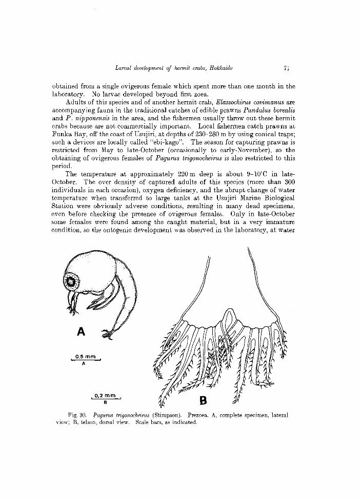
Fig. 30. Pagurus trigonocheirus (Stimpson). Prezoea. A, complete specimen, lateral view; B, telson, dorsal view. Scale bars, as indicated.
72 R. Quintana and F. Iwata
temperature of lOoC. The only remaining ovigerous female died after 30 days after the collection, and a portion of egg mass was immediately isolated to be reared with a treatment of antibiotics (500 mg Eritromicin+50.000 units of Penicilim G, dissolved in 6 1 of artificial sea water). Eggs with evident fungal infestation were eliminated. The hatch out took place after 46 days, obtaining 15 prezoeas and 18 first zoeas.
Description of the larval stages
PREZOEA
C.L. 0.93 mm Carapace (Fig.30A). Globose, with no rostral neither posterior spines,
surrounded with a thin, transparent cuticle, protruding as long expansions on antennules, antennae and the telson. Other appendages are rudimentary and non-functional at this stage.
Telson (Fig. 30B). With 6 long cuticular expansions on each furca. Each telsonal process fringed with fine setules along margins.
FIRST ZOEA
All the appendages, except the first and second maxillipeds are shown III
detail in Fig. 31. C.L. 2.48 mm (range=2.42-2.61 mm)
Carapace (Fig. 31A). Frontal spine reaching to tip of antennal scale, and longer than the posterolateral spines. No rudiments of pereiopods are visible laterally beneath the carapace.
Antennule (Fig. 31B). Uniramous, with 2 subequal aesthetascs and 2-3 additional fine setae distally, and 1 long subterminal seta, reaching beyond tip of longer aesthetascs. The subterminal seta fringed with sparse setules on the entire length.
Antenna (Fig. 3lC). Endopod elongate, slightly shorter than the exopod (= scale), bifid at tip. Scale distally pointed, fringed with 8 pulmose setae and a additional minute seta on inner margin; distal setae reaching to tip of endopod. A stout spine, finely serrate on margins emerges from near base of scale.
Mandible (Fig. 31D). Incisor process pointed distally, with accessory smaller teeth. Molar process with minute denticles marginally.
Maxillule (Fig. 31E). Endopod 3-segmented, with a tiny, fine seta on the proximal segment, 1 long seta on the second and 3 long setae on the distal segment. Basial endite with 2 strong, rigid spines and 2 fine setae. The stout spines not articulated to the endite, and each one armed with minute spinules distally. Coxal endite with 6 setae.
Maxilla (Fig. 31F). Endopod uniramous, with a group of 3 setae medially and 4 distally. Basial and coxal endites bilobed; proximal and distal basial lobes with 5 and 4 setae respectively; proximal and distal coxal lobes with 6 and
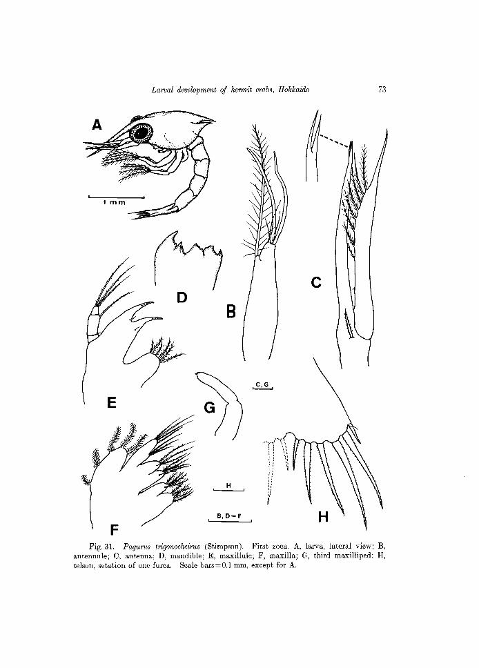
E
Larval development of hermit crabs, Hokkaido
B
H
B, 0- F
:. ': :: " :: ': I
73
Fig.31. Pagurus trigonocheirus (Stimpson). First zoea. A, larva, lateral view; B, antennule; C, antenna; D, mandible; E, maxillule; F, maxilla; G, third maxilliped; H, telson, setation of one furca. Scale bars=O.l mm, except for A.
74 R. Quintana and F. Iwata
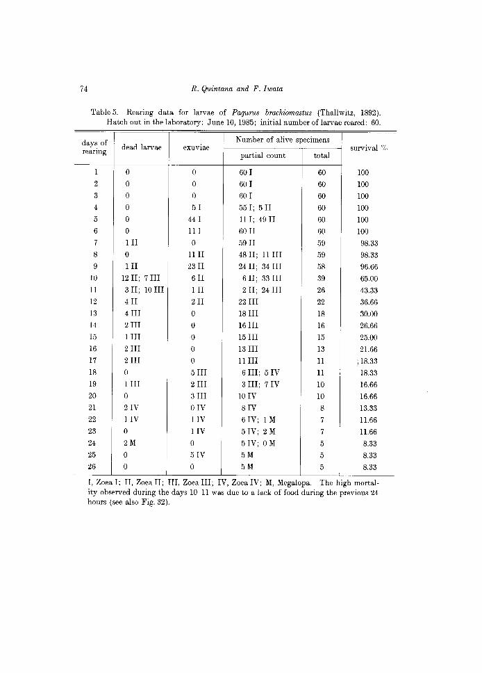
Table 5. Rearing data for larvae of Pagurus brachiomastus (Thallwitz, 1892). Hatch out in the laboratory: June 10, 1985; initial number oflarvae reared: 60.
days of dead larvae eXUVIae Number of alive specimens
survival % rearing partial count total
1 0 0 60 I 60 100
2 0 0 60 I 60 100
3 0 0 60 I 60 100
4 0 51 55 I; 5 II 60 100
5 0 44 I II I; 49 II 60 100
6 0 III 60 II 60 100
7 III 0 59 II 59 98.33
8 0 llll 48 II; II III 59 98.33
9 III 23 II 24 II; 34 III 58 96.66
10 1211; 7III 611 6 II; 33 III 39 65.00
II 3 II; 10 III III 2 II; 24 III 26 43.33
12 411 211 22 III 22 36.66
13 4 III 0 18 III 18 30.00
14 2 III 0 16 III 16 26.66
15 1 III 0 15 III 15 25.00
16 2 III 0 13 III 13 21.66
17 2 III 0 llIII II /18.33
IS 0 5 III 6 III; 5 IV II 18.33
19 1 III 2 III 3 III; 7 IV 10 16.66
20 0 3 III 10 IV 10 16.66
21 21V OIV SIV 8 13.33
22 IIV IIV 6 IV; 1 M 7 11.66
23 0 IIV 5 IV; 2 M 7 1l.66
24 2M 0 5 IV; 0 M 5 8.33
25 0 51V 5M 5 8.33
26 0 0 5M 5 S.33
I, Zoea I; II, Zoe a II; III, Zoea III; IV, Zoea IV; M, Megalopa. The high mortality observed during the days 1O-11 was due to a lack of food during the previous 24 hours (see also Fig. 32).
Larval development of hermit crabs, Hokkaido 75
4 setae respectively. Third maxilliped (Figs. 31A, G). Visible laterally beneath the carapace.
Uniramous, rudimentary, unarmed distally. Abdomen (Fig. 31A). Composed of 5 segments plus the telson. Second
through fifth segments with small spines posterodorsally; fifth segment with long posterolateral spines and shorter posterior spines. No pleopod buds at this stage.
Telson (Fig. 31R). Elongate, with 7 processes on posterior margin of each furca. Outermost processes are a stout, smooth spine, articulated to the telson and a very fine seta (called "hair-like process"), shorter than the outermost spine. Third through seventh processes are long setae, each fringed with minute setules along margins.
Pagurus brachiomastus (Thallwitz, 1892) (Japanese name: Ikubi-honyadokari)
(Fig. 32)
Culture experiments
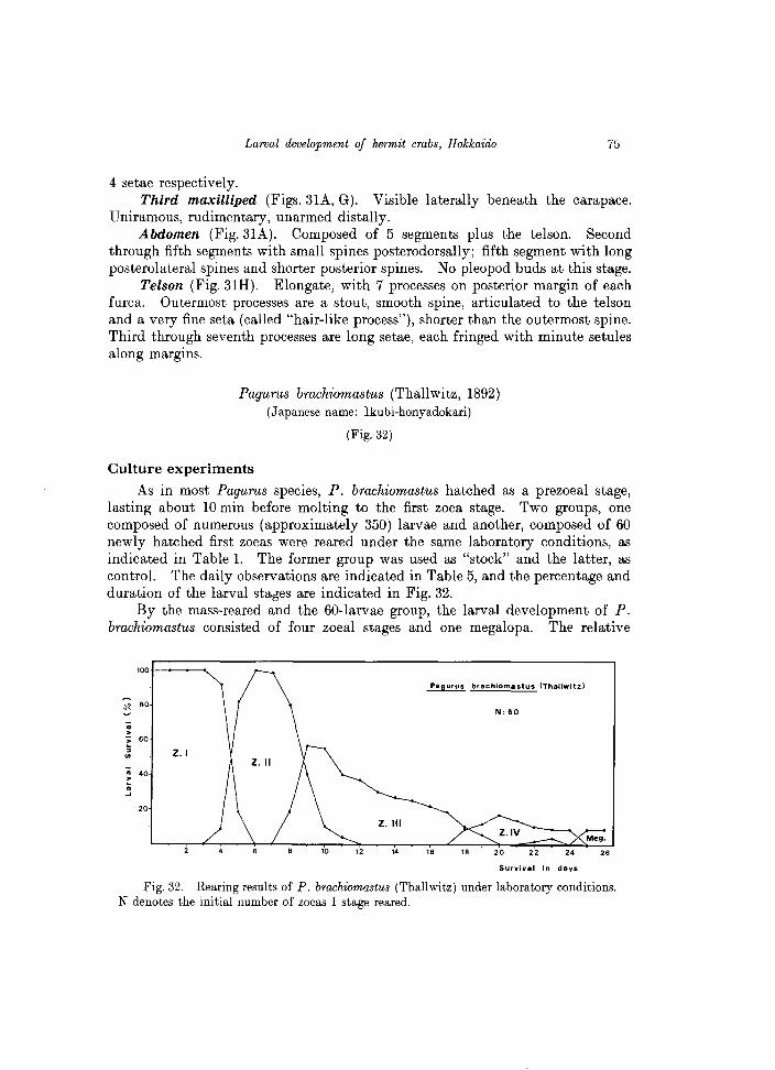
As in most Pagurus species, P. brachiomastus hatched as a prezoeal stage, lasting about 10 min before molting to the first zoea stage. Two groups, one composed of numerous (approximately 350) larvae and another, composed of 60 newly hatched first zoeas were reared under the same laboratory conditions, as indicated in Table 1. The former group was used as "stock" and the latter, as control. The daily observations are indicated in Table 5, and the percentage and duration of the larval sta6es are indicated in Fig. 32.
By the mass-reared and the 60-larvae group, the larval development of P. brachiomastus consisted of four zoeal stages and one megalopa. The relative
100
Pagurus brachiomastu5 (Thallwitz)
~ 80
: · 60 ;; Z.I en
;; 40 · • .... 20
z. III
14 16
Survival in days
Fig. 32. Rearing results of P. brachiomastus (Thallwitz) under laboratory conditions. N denotes the initial number of zoeas I stage reared.
76 R. Quintana and F. Iwata
duration of each zoeal stage was: 4-5 days for the first zoea, 8 days for the second zoea, 11-12 days for the third zoea, and 7-8 days for the fourth zoea. The first individuals of the megalopa stage appeared in the rearing vessels 22-23 days after hatching. The rearing experiment of the 60 -larvae group was finished after 26 days, with the obtaining of a total of 5 megalopas; other additional megalopas were obtained from the "stock" rearing.
Descriptions
The prezoea of this and allied species was recently described by Quintana and Konishi (1986) based on the present material. More recently, the complete larval development based on laboratory-reared larvae of material from Akkeshi, eastern Hokkaido has been described by Konishi and Quintana (1987).
Remarks
Elassochirus cavimanus (Miers, 1879)
(Japanese name: Gotoh-honyadokari)
As Pagurus trigonocheirus, this species is also captured as accompanying fauna in the "ebi-kago" traps at Funka Bay, off Usujiri at depths of 230-280 m. E. cavimanus is distributed in depths between 170-400 m (Miyake, 1982). On each of the three occasions to obtain ovigerous females (October-November 1985), a total of 300-380 adults specimens were examined at the Usujiri Mar. BioI. Station, but most individuals were males, and the few females did not bear eggs. Probably, ovigerous females are present in the area during the months out of the short season for penaeoid prawns fishing. This species apparently can better tolerate the abrupt changes of water temperature and pressure than P. trigonocheirus, and the captured adults seemed healthy. Apparently, no larvae of the genus Elassochirus have yet been described.
Discussion
la. On the family Diogenidae and the genus Paguristes.
In the literature (except that of Japan), there are published accounts on larvae for only five Paguristes, genus which contains more than 100 species: P. oculatus (see Bourdillon-Casanova, 1960; Pike and Williamson, 1960), P. turgidus (see Hart, 1937,), P. sericeus (see Rice and Provenzano, 1965), P. abbreviatus (see Dechance, 1963), and P. spinipes (see Provenzano, 1978). The genus Paguristes in Japan comprises 12 species (Miyake, 1982), and the only published larval account is that of Kurata (1968b ) on P. digitalis. Other reports on larvae of allied Japanese species within the family Diogenidae are also scarce; the most recent study is that of Baba and Fukuda (1985) on Diogenes nitidimanus.
Larval development of hermit crabs, Hokkaido 77
1 b. Larval development
Including the genus Paguristes, MacDonald, Pike and Williamson (1957) reported that possibly there is no free prezoea in the genera Diogenes, Dardanus and Clibanarius (members of the old subfamily Diogeninae, until to erect the superfamily Coenobitoidea by the mentioned authors (p. 256). Nevertheless, we think that the prezoeal stage has been probably overlooked due to its short duration. Provenzano (1978) noted that some larvae of Paguristes spinipes Milne-Edwards hatched as prezoeas, but no descriptions neither illustrations were provided. Therefore, it seems probable that the present descriptions and observations on the prezoea stage of Paguristes ortmanni are the first account on this respect.
The prezoeas (as also in most of Pagurus species) had a duration of 6-10 min before molting to the first zoea stage, and showed certain advanced characters such as a biramous third maxilliped and rudiments of the pereiopods (see Fig. 1). As observed in Paguristes digitalis (Stimpson) (see Kurata, 1968b ), P. spinipes Milne-Edwards (see Provenzano, 1978), and in our P. ortmanni, the Paguristes species normally have an abbreviated development consisting of three zoeal stages and a megalopa (see also Pike and Williamson, 1960), but a direct development has been reported by Dechance (1963) in Paguristes abbreviatus Dechance. In contrast, a prolonged development comprising five (occasionally six) zoeal stages has been reported in another diogenid, Petrochirus diogenes (L.) (see Provenzano, 1968a ). Kurata (1968a ) reported a larval development of 7-8 zoeas for the diogenid Dardanus arrosor (Herbst). This indicates that the larval development in the diogenidae hermit crabs is highly variable and therefore, the number of zoeal stages (as stated by Provenzano, op. cit.) seems to be of little systematic significance.
Most of Paguristes species with non-direct development attain the megalopa stage approximately within one week after hatching. At 18°C, P. ortmanni larvae reached the megalopa stage after 10 days, and the first crab stage after 22-23 days. An exception to this relatively short larval duration is that of P. spinipes as reported by Provenzano (1978); the megalopa stage was attained after 3-4 weeks, and the first crab after 6-7 weeks at laboratory conditions of 20°C.
The anterolateral carapacial spines and the short posterolateral spines of the fifth segment of zoeal stages, suggested by Rice and Provenzano (1965) and later by Provenzano (1978) as two characters of possible generic significance, are also exhibited by the zoeas of Paguristes digitalis (see Kurata, 1968b ) and the present P. ortmanni zoeas.
Kurata described the zoeal outermost telsonal process of P. digitalis as fused to the telson, but in P. ortmmanni this is evidently separate from the telson, so we think this character was probably overlooked in the Kurata's material. By the other hands, a detailed comparison of megalopal characters as listed Table 7 between P. ortmanni and P. digitalis (the only two Japanese Paguristes species so
78 R. Quintana and F. Iwata
far described) is not possible, except for the telsonal setation, due to Kurata illustrated only the antenna and the megalopa in dorsal view.
The morphological characteristics of the megalopa of P. ortmanni are consistent with a previous suggestion of Kurata (1968b ) that his megalopa "G-I" described in 1964 (p.44) as a pagurid species apparently corresponds to P.
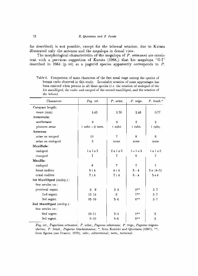
Table 6. Comparison of main characters of the first zoeal stage among the species of hermit crabs observed in this study. Invariable setation of some appendages has been omitted when present in all these species (i.e. the setation of endopod of the 1st maxilliped, the endo- and exopod of the second maxilliped, and the setation of the telson).
Characters Pag. ort. P. oclwt. P. trigo. P. brach.'
Carapace length:
mean (mm) 1.45 1.78 2.48 0.77
Antennule:
aesthetascs 3 3 2 2
plumose setae 1 subt. + 3 term. 1 subt. 1 subt. 1 subt.
Antenna:
setae on exopod 10 7 8 6
setae on endopod 3 none none none
Maxillule:
endopod 1+1+3 2+1+3 1+1+3 1+1+3
coxopod 7 7 6 7
Maxilla:
endopod 8 7 7 7
basal endites 5+4 4+4 5+4 5+ (4-5)
coxal endites 7+4 7+4 6+4 5+4
1st Maxilliped (endop.) :
fine setules on :
proximal segmt. 8- 9 5-6 5" 5-7
2nd segmt. 13-14 8 7" 5-7
3rd segmt. 15-16 5-6 6" 5-7
2nd Maxilliped (endop.) :
fine setules on :
2nd segmt. 10-11 3-4 4" 5
3rd segmt. 9-10 5-6 5" 6
Pag. ort., Paguristes ortmanni; P. oclwt., Pagurus oclwtensis; P. trigo., Pagurus trigonocheirus; P. brach., Pagurus brachiomastus; " from Konishi and Quintana (1987); ., from figures (see Ivanov, 1979); subt., subterminal; term., terminal.
Larval drwelopment of hermit crabs, Hokkaido 79
ortmanni, especially by the morphologies of the antennal acicle (3 stout spines and 3 setae on outer margin) and the telson, which are markedly different to those of other megalopas he attributed as belonging to Paguridae.
2a. On the family Paguridae and the genus Pagurus. The extensive literature on the larval development of pagurids crabs (see
Introduction) includes descriptions and illustrations of many species of Pagurus, genus with approximately 200 species (cf. Nyblade and McLaughlin, 1975). The larval affinities and relationships between Pagurus and allied genera within the vast family Paguridae have been discussed in detail by Provenzano (1968b ) and Nyblade and McLaughlin (1975), including larval descriptions of the genera Pylopaguropsis, Munidopagurus, Xylopagurus, Lithopagurus, Tomopaguroides, Tomopaguropsis, Acanthopagurus, Anapagurus, Cestopagurus, Orthopagurus, Porcellanopagurus, Spiropagurus, and Labidopagurus.
With regard to the pagurid crabs of Japan, and as indicated by Konishi and Quintana (1987), there are 51 species of the family Paguridae, but published descriptions are restricted to approximately 12 per cent in these species.
2b. Larval development
Evidence of a prezoea stage, of short duration was recorded in the larval development of both Pagurus ochotensis and P. trigonocheirus, confirming previous observations on this genus (for review see Quintana and Konishi, 1986). On the basis of the larval development of the present four Pagurus species, i.e. P. middendorffii, P. ochotensis, P. trigonocheirus, and P. brachiomastus, the number of zoeal stages of most species of this genus seems to be four, as pointed out by Pike and Williamson (1960) and later by Roberts (1970).
The relatively long posterolateral spines of the carapace in all four zoeal stages of Labidochirus splendescens was suggested by Nyblade and McLaughlin (1975) as a diagnostic character which distinguishes this species from other described pagurids, but this character is also present at least in the first zoeal stage of our Pagurus trigonocheirus (see Fig. 31A). The zoeal characters of the latter species are basically the same as described by Ivanov (1979), and the slight differences in carapace length are obviously not important.
The character of a apically bifid antennal endopod in P. trigonocheirus (see Fig. 31 C) seems to be present in a few species among the Japanese pagurids (see comparative table by Konishi and Quintana, 1987). Other characters among the present material are compared in Table 6.
In P. ochotensis and P. brachiomastus, the first individuals which successfully molted to the megalopa stage appeared in the cultures approximately 21-22 days after hatching, and after 26 days in P. middendorffii. The total duration of the zoeal phase was 24 days in the two former species and 28 days (32 days in the initially starved zoeas) in the latter species. The difference of a relatively long larval duration in the "normal" rearing of P. middendorffii seems to be associated
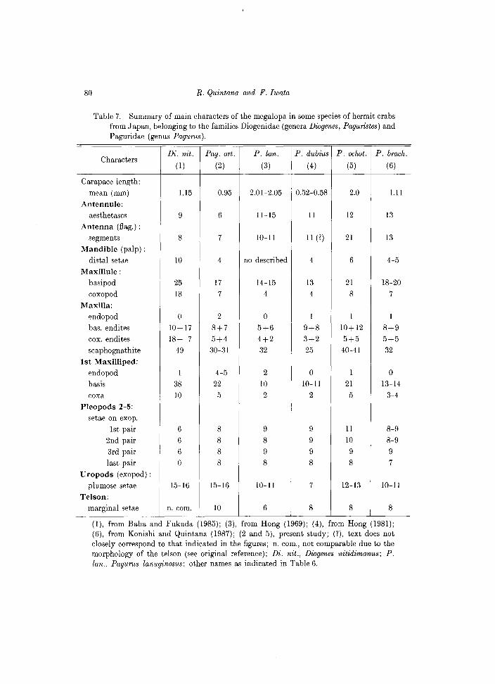
80 R. Quintana and F. Iwata
Table 7. Summary of main characters of the megalopa in some species of hermit crabs from Japan, belonging to the families Diogenidae (genera Diogenes, Paguristes) and Paguridae (genus Pagurus).
Characters Di. nit. Pag. art. P. lan. P. dubius P. oclwt. P. brach.
(1) (2) (3) (4) (5) (6)
Carapace length:
mean (mm) 1.15 0.95 2.01~2.05 0.52~0.58 2.0 1.11 Antennule:
aesthetascs 9 6 11~15 11 12 13
Antenna (flag.) :
segments 8 7 10~11 11 (?) 21 13
Mandible (palp) :
distal setae 10 4 no described 4 6 4~5
Maxillule: basipod 25 17 14~15 13 21 18~20
coxopod 18 7 4 4 8 7
Maxilla: endopod 0 2 0 1 1 1
bas. endites 10+17 8+7 5+6 9+8 10+12 8+9 cox. endites 18+ 7 5+4 4+2 3+2 5+5 5+5 scaphognathite 49 30~31 32 25 40~41 32
1st Maxilliped: endopod 1 4~5 2 0 1 0
basis 38 22 10 1O~11 21 13~14
coxa 10 5 2 2 5 3~4
Pleopods 2~5: setae on exop.
1st pair 6 8 9 9 11 8~9
2nd pair 6 8 8 9 10 8~9
3rd pair 6 8 9 9 9 9 last pair 0 8 8 8 8 7
Uropods (exopod) :
plumose setae 15~16 15~16 1O~11 7 12~13 1O~11
Telson: marginal setae n. com. 10 6 8 8 8
(1), from Baba and Fukuda (1985); (3), from Hong (1969); (4), from Hong (1981); (6), from Konishi and Quintana (1987); (2 and 5), present study; (1), text does not closely correspond to that indicated in the figures; n. com., not comparable due to the morphology of the telson (see original reference); Di. nit., Diogenes nitidimanus; P. lan., Pagurus lanuginosus; other names as indicated in Table 6.
Larval development of hermit crabs, Hokkaido 81
to the low temperature used (13-14"C). Probably this larval period is also prolonged in natural conditions since ovigerous females were collected in earlyMay, when temperature of shallow waters was approximately 7-9"C.
Paguristes and Pagurus: Larval characteristics
Larval characteristics of the genus Pagurus and other allied hermit crabs of the superfamily Coenobitoidea (which includes the Diogenidae) have been reviewed in some detail by MacDonald, Pike and Williamson (1957), Pike and Williamson (1960) and Provenzano (1968) (see also Provenzano, 1963 for a review of megalopa characters of Dardanus and allied genera), and as mentioned previously, larval characters of Pagurus and allied pagurid genera have been reviewed by Nyblade and McLaughlin (1975), so these will be not mention again in this discussion.
On the basis of larval features observed in this study, obvious differences can be easily found in both zoeas and megalopas of these two genera of hermit crabs (see Tables 6 and 7). Despite these differences, some common characters are also present. Thus, the mandibular palp and the naked posterior lobe of the maxillary scaphognathite appear in the last zoeal stage, i.e. Zoea III stage in Paguristes ortmanni and Zoea IV stage in Pagurus species. After molting to the megalopa, the scaphognathite is fringed with many plumose setae along the entire margins, and the endopod is markedly slender and reduced in setation (see Figs 7 and 23). The significance of the reduction of maxillary endites and the developmental patterns of the scaphognathite have been discussed in detail by Van Dover (1982) and Van Dover et al., (1982).
No attempts were made to asses the behavior of megalopas of Paguristes ortmanni and of Pagurus ochotensis to select and entry a suitable gastropod shell. This subject has been previously reviewed by Reese (1962) and later by Roberts (1971) for megalopas of Pagurus longicarpus Say. Shell entry occurs during the megalopa stage within 24-48 hours, being this time shorter after molting to the first crab instar. Nevertheless, a complex behavioral mechanism seems to be associated to the successful entry and use of a shell in the megalopas and young crabs of seven species of hermit crabs (Hazlett and Provenzano, 1965). In the experiments of Roberts (1971), the mortality of megalopas was unaffected by shell presence or absence. Nevertheless, Bookhout (1964) reported a relatively high mortality in megalopas of Pagurus bernhardus when deprived of a shell. Roberts (op. cit.) attributed these differences to the fact that his megalopas were reared individually, and that in the group-reared megalopas of Bookout (1964), the higher mortality was also affected by aggressive behavior and cannibalism. Recently Dawirs (1982) has evaluated comparatively the effects -among others- of single and group rearing of larvae of Pagurus bernhardus and of Carcinus maenas. In our experiments, although the initial number of Pagurus Zoeas I was high (50-80), the subsequent later zoeas were isolated in small groups, and as they molted to the megalopa, the newly raised individuals were reared in smaller groups,
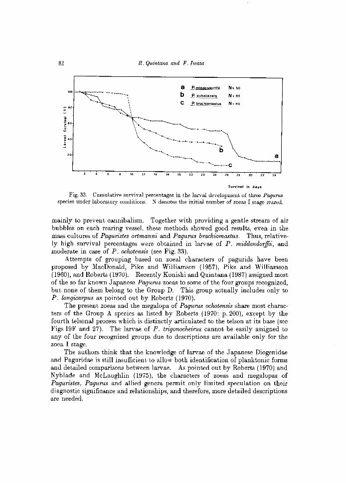
82 R. Quintana and F. Iwata
a 1". middendorlfll
b ...E.. ochotensis N,80
c P. brachlomastus
10 12 14 16 18 20 22 24 26 28 30 32 34
Survival in days
Fig. 33. Cumulative survival percentages in the larval development of three Pagurus species under laboratory conditions. N denotes the initial number of zoeas I stage reared.
mainly to prevent cannibalism. Together with providing a gentle stream of air bubbles on each rearing vessel, these methods showed good results, even in the mass cultures of Paguristes ortmanni and Pagurus brachiomastus. Thus, relatively high survival percentages were obtained in larvae of P. middend01:iJii, and moderate in case of P. ochotensis (see Fig. 33).
Attempts of grouping based on zoeal characters of pagurids have been proposed by MacDonald, Pike and Williamson (1957), Pike and Williamson (1960), and Roberts (1970). Recently Konishi and Quintana (1987) assigned most of the so far known Japanese Pagurus zoeas to some of the four groups recognized, but none of them belong to the Group D. This group actually includes only to P. longicarpus as pointed out by Roberts (1970).
The present zoeas and the megalopa of Pagurus ochotensis share most characters of the Group A species as listed by Roberts (1970: p.200), except by the fourth telsonal process which is distinctly articulated to the telson at its base (see Figs 19F and 27). The larvae of P. trigonocheirus cannot be easily assigned to any of the four recognized groups due to descriptions are available only for the zoea I stage.
The authors think that the knowledge of larvae of the Japanese Diogenidae and Paguridae is still insufficient to allow both identification of planktonic forms and detailed comparisons between larvae. As pointed out by Roberts (1970) and Nyblade and McLaughlin (1975), the characters of zoeas and megalopas of Paguristes, Pagurus and allied genera permit only limited speculation on their diagnostic significance and relationships, and therefore, more detailed descriptions are needed.
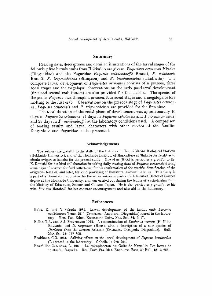
Larval development of hermit crabs, Hokkaido 83
Summary
Rearing data, descriptions and detailed illustrations of the larval stages of the following five hermit crabs from Hokkaido are given: Paguristes ortmanni Miyake (Diogenidae) and the Paguridae Pagurus middendoiffii Brandt, P. oclwtensis Brandt, P. trigonocheirus (Stimpson) and P. brachiomastus (Thallwitz). The complete larval development of Paguristes ortmanni consists of a prezoea, three zoeal stages and the megalopa; observations on the early postlarval development (first and second crab instars) are also provided for this species. The species of the genus Pagurus pass through a prezoea, four zoeal stages and a megalopa before molting to the first crab. Observations on the prezoea stage of Paguristes ortmanni, Pagurus oclwtensis and P. trigonocheirus are provided for the first time.
The total duration of the zoeal phase of development was approximately 10 days in Paguristes ortmanni, 24 days in Pagurus oclwtensis and P. brachiomastus, and 28 days in P. middendoiffii at the laboratory conditions used. A comparison of rearing results and larval characters with other species of the families Diogenidae and Paguridae is also presented.
Acknowledgements
The authors are grateful to the staffs of the Oshoro and Usujiri Marine Biological Stations (Hokkaido University), and of the Hokkaido Institute of Mariculture at Shikabe for facilities to obtain ovigerous females for the present study. One of us (R.Q.) is particularly grateful to Dr. K. Konishi for his kind collaboration in taking daily rearing data of Pagurus ochotensis during some days of absence for field collections, for his confirmation of the specific identification of the ovigerous females, and later, for kind providing of literature inaccessible to us. This study is a part of a Dissertation submitted by the senior author in partial fulfillment of Doctor of Science degree at the Hokkaido University, and was carried out during the tenure of a scholarship from the Ministry of Education, Science and Culture, Japan. He is also particularly grateful to his wife, Viviana Maraboli, for her constant encouragement and also aid in the laboratory.
References
Baba, K. and Y. Fukuda 1985. Larval development of the hermit crab Diogenes nitidimanus Terao, 1913 (Crustacea: Anomura: Diogenidae) reared in the laboratory. Mem. Fac. Educ., Kumamoto Univ., Nat. Sci., 34: 5-17.
Biffar, T.A. and A.J. Provenzano 1972. A reexamination of Dardanus venosus (H. Milne Edwards) and D. imperator (Miers), with a description of a new species of Dardanus from the western Atlantic (Crustacea, Decapoda, Diogenidae). Bull. Mar. Sci. 22: 777-805.
Bookhout, C.G. 1964. Salinity effects on the larval development of Pagurus bernhardus (L.) reared in the laboratory. Ophelia 1: 275-294.
Bourdillon-Casanova, L. 1960. Le meroplancton du Golfe de Marseille: Les larves de crustaces decapodes. Rec. Trav. Sta. Mar. Endoume, Fasc. 30 Bull. 18: 2-286.
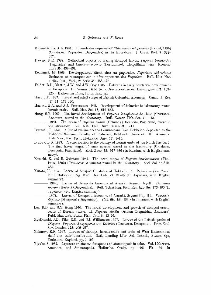
84 R. Quintana and F. Iwata
Brossi-Garcia, A.L. 1987. Juvenile development of Clibanarius sclopetarius (Herbst, 1796) (Crustacea: Paguridea: Diogenidae) in the laboratory. J. Crust. BioI. 7: 338-357.
Dawirs, RR 1982. Methodical aspects of rearing decapod larvae, Pagurus bernhardus (Paguridae) and Carcinus maenas (Portunidae). Helgolander wiss. Meeresunters 35: 439-464.
Dechance, M. 1963. Developpement direct chez un paguridae, Paguristes abbreviatus Dechance, et remarques sur Ie developpement des Paguristes. Bull. Mus. Nat. d'Hist. Nat., Paris, 2e Serie 35: 488-495.
Felder, D.L., Martin, J.W. and J.W. Goy 1985. Patterns in early postlarval development of Decapods. In: Wenner, A.M. (ed.), Crustacean Issues: Larval growth 2: 163-225. Balkemma Press, Rotterdam, pp.
Hart, J.F. 1937. Larval and adult stages of British Columbia Anomura. Canad. J. Res. (D) 15: 179-220.
Hazlett, B.A. and A.J. Provenzano 1965. Development of behavior in laboratory reared hermit crabs. Bull. Mar. Sci. 15; 616-633.
Hong, S.Y. 1969. The larval development of Pagurus lanuginosus de Haan (Crustacea, Anomura) reared in the laboratory. Bull. Korean Fish. Soc. 2: 1-15.
---~ 1981. The larvae of Pagurus dubius (Ortman) (Decapoda, Paguridae) reared in the laboratory. Bull. Natl. Fish. Univ. Busan 21: 1-11.
Igarashi, T. 1970. A list of marine decapod crustaceans from Hokkaido, deposited at the Fisheries Museum, Faculty of Fisheries, Hokkaido University II. Anomura. Fish. Mus., Fac. Fish., Hokkaido Univ. 12: 1-15.
Ivanov, B.G. 1979. A contribution to the biology of hermit crabs of the North Pacific. 2. The first larval stages of some species reared in the laboratory (Crustacea, Decapoda, Paguridae). Zool. Zhur. 58: 977-986 (In Russian, with English summary).
Konishi, K. and R Quintana 1987. The larval stages of Pagurus brachiomastus (ThalIwitz, 1892) (Crustacea: Anomura) reared in the laboratory. Zoo I. Sci. 4: 349-365.
Kurata, H. 1964. Larvae of decapod Crustacea of Hokkaido. 5. Paguridae (Anomura). Bull. Hokkaido Reg. Fish. Res. Lab. 29: 24-48 (In Japanese, with English summary).
---~ 1968.. Larvae of Decapoda Anomura of Arasaki, Sagami Bay-II. Dardanus arrosor (Herbst) (Diogenidae). Bull. Tokai Reg. Fish. Res. Lab. 56: 173-180 (In Japanese, with English summary).
---~ 1968b • Larvae of Decapoda Anomura of Arasaki, Sagami Bay-III. Paguristes digitalis (Stimpson) (Diogenidae). Ibid, 56: 181-186. (In Japanese, with English summary).
Lee, B.D. and S.Y. Hong 1970. The larval development and growth of decapod crustaceans of Korean waters. II. Pagurus similis Ortman (Paguridae, Anomura). Publ. Mar. Lab. Pusan Fish. CoIl. 3: 13-26.
MacDonald, J.D., Pike, RB. and D.1. Williamson 1957. Larvae of the British species of Diogenes, Pagurus, Anapagurus and Lithodes (Crustacea, Decapoda). Proc. Zoo I. Soc. London 128: 209-257.
Makarov, RR 1967. Larvae of shrimps, hermit-crabs and crabs of West Kamchatkan shelf and their distribution. Natl. Lending Libr. Sci. Tchnol., Boston Spa, Yorkshire, England, pp. 1-199.
Miyake, S. 1982. Japanese crustacean decapods and stomatopods in color. Vol. I Macrura, Anomura, and Stomatopoda. Hoikusha, Osaka, pp.I-261. PIs. 1-56 (In
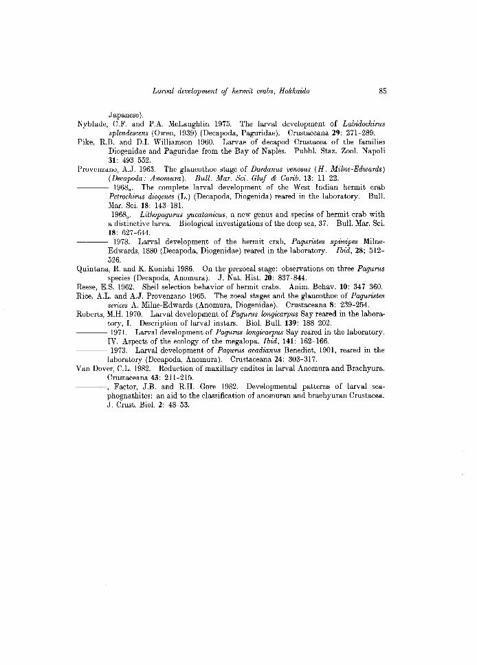
Larval development of hermit crabs, Hokkaido 85
Japanese). Nyblade, C.F. and P.A. McLaughlin 1975. The larval development of Labidochirus
splendescens (Owen, 1939) (Decapoda, Paguridae). Crustaceana 29: 271-289. Pike, R.B. and D.1. Williamson 1960. Larvae of decapod Crustacea of the families
Diogenidae and Paguridae from the Bay of Naples. Pubbl. Staz. Zoo I. Napoli 31: 493-552.
Provenzano, A.J. 1963. The glaucothoe stage of Dardanus venosus (H. Milne-Edwards) (Decapoda: Anomura). Bull. Mar. Sci. Gluf & Carib. 13: 11-22.
1968a • The complete larval development of the West Indian hermit crab Petrochirus diogenes (L.) (Decapoda, Diogenida) reared in the laboratory. Bull. Mar. Sci. 18: 143-181.
~--- 1968b • Lithopagurus yucatanicus, a new genus and species of hermit crab with a distinctive larva. Biological investigations of the deep sea, 37. Bull. Mar. Sci. 18: 627-644.
1978. Larval development of the hermit crab, Paguristes spinipes MilneEdwards, 1880 (Decapoda, Diogenidae) reared in the laboratory. Ibid, 28; 512-526.
Quintana, R. and K. Konishi 1986. On the prezoeal stage: observations on three Pagurus species (Decapoda, Anomura). J. Nat. Hist. 20: 837-844.
Reese, E.S. 1962. Shell selection behavior of hermit crabs. Anim. Behav. 10: 347-360. Rice, A.L. and A.J. Provenzano 1965. The zoeal stages and the glaucothoe of Paguristes
serices A. Milne-Edwards (Anomura, Diogenidae). Crustaceana 8: 239-254. Roberts, M.H. 1970. Larval development of Pagurus longicarpus Say reared in the labora
tory, I. Description of larval instars. BioI. Bull. 139: 188-202. ~--- 1971. Larval development of Pagurus longicarpus Say reared in the laboratory.
IV. Aspects of the ecology of the megalopa. Ibid, 141: 162-166. ---- 1973. Larval development of Pagurus acadianus Benedict, 1901, reared in the
laboratory (Decapoda, Anomura). Crustaceana 24: 303-317. Van Dover, C.L. 1982. Reduction of maxillary endites in larval Anomura and Brachyura.
Crustaceana 43: 211-215. ~~~-, Factor, J.B. and R.H. Gore 1982. Developmental patterns of larval sca
phognathites: an aid to the classification of anomuran and brachyuran Crustacea. J. Crust. BioI. 2: 48-53.
Related Documents