O n the Evidence of Crosslinking in Methyl Pendent PBZT Fiber VINAY R. MEHTA,*,' SATISH KUMAR,+.' M. B. POLK,' D. L. VANDERHART,' F. E. ARNOLD: and T. D. DANC4 'School of Textile and Fiber Engineering, Georgia Institute of Technology, Atlanta, Georgia 30332-0295; *Polymers Division, National Institute of Standards and Technology, Gaithersburg, Maryland 20899; 3WL/MLBP, Wright Patterson Air Force Base, Dayton, Ohio 45433; 4University of Dayton Research Institute, Dayton, Ohio 45469 SYNOPSIS In order to influence the compressive strength of the rigid rod polymeric fibers, methyl pendent poly (p-phenylene benzobisthiazole) fibers have been heat treated in the 400 to 550°C temperature range in air and in nitrogen for varying times to achieve intermolecular crosslinking. These fibers have been examined using Fourier transform infrared (FTIR) spectroscopy, 13C solid-state nuclear magnetic resonance (NMR) swelling behavior, and scanning electron microscopy. 13C NMR has also been carried out on solutions of as-spun fibers. Fibers heat-treated at 400°C, both in nitrogen and in air, up to heat-treatment times of 60 min are insoluble in 99% chlorosulfonic acid, however no direct evidence of crosslinking has been obtained for these fibers using spectroscopic techniques, suggesting that in these fibers the degree of crosslinking must be very low. Evidence that methyl groups are precursors to certain crosslinks was first seen via a weak methylene resonance in 13C solid-state NMR, corresponding to about 2% of the original methyl intensity, in a sample heat-treated at 45OoC in air. Fibers heat-treated in nitrogen at 550°C for 10 minutes do not exhibit any swelling in chlorosulfonic acid, are brittle, have lost most methyl groups; however, some CH, groups form. In this fiber, the carbon intensity for the CH, group in the 13C solid- state NMR is 18% of the intensity for the CH3 group in the as-spun fiber. The fibers heat- treated at 400 and 450°C show a fibrillar morphology, while the fibrillar morphology is not observed in the fibers heat-treated at 550°C in nitrogen for 10 min. Based on this work, it is our judgment that if heat treatment of this material is to improve compressive strength, the heat treatment protocol of time and temperature will probably be critical and the highest temperatures of exposure will probably lie in the 450 to 550°C range. 0 1996 John Wiley & Sons, Inc. Keywords: crosslinking poly(p-phenylene benzobis thiazole) PBZT methyl pendant PBZT 13C solid state NMR I NTRO DUCT10 N In order to influence the compressive strength of the rigid-rod polymeric fibers such as poly(p-phenylene benzobisthiazole) (PBZT), molecular crosslinking has been one of the approaches attempted. In these sys- tems, a decrease in the fiber solubility has been taken as evidence of crosslinking.'-6 In the halogenated * Present address: Research & Technology,Allied Signal Inc., Morristown, NJ 07962. Journal of Polymer Science: Part B: Polymer Physics, Val. 34,1881-1891 (1996) 0 1996 John Wiley & Sons, Inc. CCC 0887-6266/96/111881- 11 To whom correspondenceshould be addressed. PBZT ~ystern,~ loss of halogen and the associated loss in fiber solubility was taken as the evidence of crosslinking. Dang et al.7showed, using infrared (IR) spectroscopy, the existence of a "pseudo-ladder" structure in the dihydroxy pendent PBZT system due to strong intramolecular hydrogen bonding between the hydroxy groups and the neighboring nitrogen at- oms of the benzobisthiazole units. Decreased solu- bility for PBZT has also been attributed to enhanced order achieved on heat treatment: The compressive strengths of most of these fibrous systems were in the 200-400 MPa range, with a few values being as low as 150 MPa and few values as high as 700 MPa. For 1881

Welcome message from author
This document is posted to help you gain knowledge. Please leave a comment to let me know what you think about it! Share it to your friends and learn new things together.
Transcript
On the Evidence of Crosslinking in Methyl Pendent PBZT Fiber
VINAY R. MEHTA,*,' SATISH KUMAR,+.' M. B. POLK,' D. L. VANDERHART,' F. E. ARNOLD: and T. D. D A N C 4
'School of Textile and Fiber Engineering, Georgia Institute of Technology, Atlanta, Georgia 30332-0295; *Polymers Division, National Institute of Standards and Technology, Gaithersburg, Maryland 20899; 3WL/MLBP, Wright Patterson Air Force Base, Dayton, Oh io 45433; 4University of Dayton Research Institute, Dayton, Ohio 45469
SYNOPSIS
In order to influence the compressive strength of the rigid rod polymeric fibers, methyl pendent poly (p-phenylene benzobisthiazole) fibers have been heat treated in the 400 to 550°C temperature range in air and in nitrogen for varying times to achieve intermolecular crosslinking. These fibers have been examined using Fourier transform infrared (FTIR) spectroscopy, 13C solid-state nuclear magnetic resonance (NMR) swelling behavior, and scanning electron microscopy. 13C NMR has also been carried out on solutions of as-spun fibers. Fibers heat-treated a t 400°C, both in nitrogen and in air, up to heat-treatment times of 60 min are insoluble in 99% chlorosulfonic acid, however no direct evidence of crosslinking has been obtained for these fibers using spectroscopic techniques, suggesting that in these fibers the degree of crosslinking must be very low. Evidence that methyl groups are precursors to certain crosslinks was first seen via a weak methylene resonance in 13C solid-state NMR, corresponding to about 2% of the original methyl intensity, in a sample heat-treated at 45OoC in air. Fibers heat-treated in nitrogen at 550°C for 10 minutes do not exhibit any swelling in chlorosulfonic acid, are brittle, have lost most methyl groups; however, some CH, groups form. In this fiber, the carbon intensity for the CH, group in the 13C solid- state NMR is 18% of the intensity for the CH3 group in the as-spun fiber. The fibers heat- treated at 400 and 450°C show a fibrillar morphology, while the fibrillar morphology is not observed in the fibers heat-treated a t 550°C in nitrogen for 10 min. Based on this work, it is our judgment that if heat treatment of this material is to improve compressive strength, the heat treatment protocol of time and temperature will probably be critical and the highest temperatures of exposure will probably lie in the 450 to 550°C range. 0 1996 John Wiley & Sons, Inc. Keywords: crosslinking poly(p-phenylene benzobis thiazole) PBZT methyl pendant PBZT 13C solid state NMR
I NTRO DUCT10 N
In order to influence the compressive strength of the rigid-rod polymeric fibers such as poly(p-phenylene benzobisthiazole) (PBZT), molecular crosslinking has been one of the approaches attempted. In these sys- tems, a decrease in the fiber solubility has been taken as evidence of crosslinking.'-6 In the halogenated
* Present address: Research & Technology, Allied Signal Inc., Morristown, NJ 07962.
Journal of Polymer Science: Part B: Polymer Physics, Val. 34,1881-1891 (1996) 0 1996 John Wiley & Sons, Inc. CCC 0887-6266/96/111881- 11
To whom correspondence should be addressed.
PBZT ~ys te rn ,~ loss of halogen and the associated loss in fiber solubility was taken as the evidence of crosslinking. Dang et al.7 showed, using infrared (IR) spectroscopy, the existence of a "pseudo-ladder" structure in the dihydroxy pendent PBZT system due to strong intramolecular hydrogen bonding between the hydroxy groups and the neighboring nitrogen at- oms of the benzobisthiazole units. Decreased solu- bility for PBZT has also been attributed to enhanced order achieved on heat treatment: The compressive strengths of most of these fibrous systems were in the 200-400 MPa range, with a few values being as low as 150 MPa and few values as high as 700 MPa. For
1881
1882 MEHTA ET AL.
a general discussion on compressive strength of bulk glassy polymers refer to ref. 9 and on compressive strength of high-performance fibers refer to ref. 10.
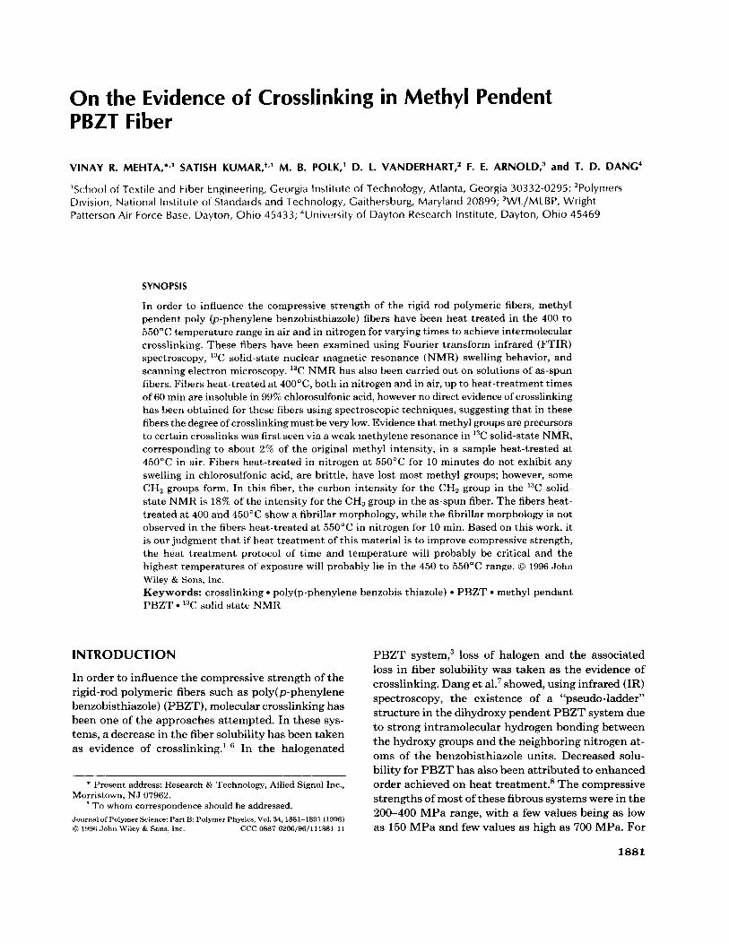
Sweeny3 has studied halogenated PPTA and ha- logenated PBZT fibers as a function of heat treat- ment time and temperature. Loss of halogen and fiber insolubility have been considered as the evi- dence of crosslinking. In halogenated PPTA fibers, with up to 100% removal of the halogen groups, no compressive strength improvement was observed as compared to the control KevlarTM 29 fiber. This has been attributed to two factors: (1) possible chain scission, and (2) the possibility of the disruption of the hydrogen bonding as a result of crosslinking. To circumvent these problems, further work was done on halogenated PBZT, in the hope of carrying out the reaction shown in Figure 1. The question be- comes whether all the radicals created by the re- moval of the halogen will form covalently cross- linked bonds as shown in Figure 1, or whether only a fraction of the radicals are in geometrically acces- sible positions participating in crosslinking reac- tions. The fibers which have lost more than about 25% of the halogens, are reported to exhibit brittle failure. Below 25% halogen loss, the fibers exhibit fibrillar behavior.
Santhosh et al.5 pointed out that the fibers of di- methyl phenoxy PBZT heat-treated a t 480°C ex- hibited brittle behavior. However, it must be pointed out that even these brittle fibers exhibited fibrilla- tion and axial splitting, which was more severe than that observed in PBZT. No comments were made on the fibrillation/axial splitting aspects of the brit- tle fibers in Sweeny's work: but the presumption appears to be that fibers failing in a brittle manner do not fibrillate.
We have examined crosslinking in heat-treated methyl pendent PBZT (MePBZT). This system, with crosslinking via - CH2 - or - CH2CH2 - linkages, offers a different crosslink structure than that obtained via the crosslinking of halogenated PBZT. Heat-treatment has been carried out in ni- trogen as well as in air. Analysis is presented based on visual swelling, thermogravimetric analysis (TGA), Fourier transform infrared (FTIR) spec- troscopy, and solution and solid-state NMR.
EXPERIMENTAL"
The MePBZT fiber was spun from a 10 wt % solu- tion in polyphosphoric acid, (polymer intrinsic vis- cosity = 24 dL/g) a t 90-110°C using the dry-jet wet-spinning technique.I2 The fiber was washed in
I V
Figure 1. PBZT.3
Proposed crosslinking reaction in halogenated
running distilled water for 3 to 5 days and dried in a vacuum oven for 12 h a t 180°C. This fiber is re- ferred to as "as-spun MePBZT" or abbreviated as "AS MePBZT." AS MePBZT fiber was heat-treated in a glass petri dish in a ceramic furnace. Heat- treatment conditions are listed in Table I. Forced air or nitrogen flow was maintained during the entire period of heat treatment. All samples heat-treated in nitrogen at 400°C retain their copper color, while the samples heat treated in air are dark brown or black. The sample heated in nitrogen up to 550°C and then held showed a grayish black color. For comparison, PBZT samples, vacuum dried for 12 h a t 180°C were also heat-treated a t 400°C in air and in nitrogen for 60 min.
'H and I3C solution NMR spectra were obtained on the Varian XL 400 NMR spectrometer. For so- lution "C NMR, 1.5 wt % of the as-spun methyl pendent PBZT or as-spun PBZT fiber was dissolved in chlorosulfonic acid and for solution proton NMR, 0.7 wt % polymer was used with deuterated trifluo- romethane sulfonic acid as the solvent. Thermo- gravimetric analysis was carried out on the Seiko thermal analysis system at 10"C/min in air as well as in nitrogen. In both cases, the samples were heated up to 400°C or up to 550°C and then were held a t that temperature for 60 min. FTIR spectra of the fibers were obtained in the transmission ge- ometry on the flattened fibers, using a Perkin Elmer 1600 series instrument with a microscope attach- ment and a mercury-cadmium-telluride (MCT) de- tector. Scanning electron microscopy was carried out using a Hitachi S-800 microscope, on samples which were coated with an approximately 40 nm thick Au coating.
Solid-state NMR( 13C) spectra were acquired in a standard way13 on a noncommercial spectrometer
CROSSLINKING IN PBZT FIBER 1883
Table I. Heat Treatment Conditions and Solubility Behavior of Various Samples
Temp Sample ("C)" Air/N2
Time (min)
Color of the Sample Solubilityb after H T
PBZT PBZT MePBZT MePBZT MePBZT MePBZT MePBZT MePBZT
400 400 400 400 400 400 450 550
Air Nitrogen Nitrogen Nitrogen Air Air Air Nitrogen
60 60 10 60 10 60 10
lO"/min from room temperature to 550°C and held a t 550°C for 10 min
Soluble Soluble Insoluble Insoluble Insoluble Insoluble Insoluble Insoluble
Dark Brown/Black Copper Copper Copper Dark Brown Dark Brown/Black Dark Brown Grayish Black
The samples a t 400 and 450°C were heat-treated in the preheated oven for the stated time period. Solubility in 99% chlorosulfonic acid monitored for more than 30 days.
operating at 2.35 T (25.2 MHz) . The probe is also noncommercial, and incorporates a 7 mm magic- angle spinning (MAS) rotor and stator system manufactured by Doty Scientific Inc. MAS fre- quencies were uniformly set to 4.0 KHz in order to avoid the overlap of centerbands and spinning side- bands; rf levels for cross-polarization ( C P ) and de- coupling corresponded to nutation frequencies of 65 KHz for protons and 69 KHz for the 13C nuclei. The CP time was 1.5 ms for all spectra. Given that the samples studied have both protonated and nonpro- tonated carbons, the latter associated with slower CP rates, the choice of CP time is an attempt to establish conditions where the relative intensities of all signals will reflect, to an accuracy exceeding 90%, the relative populations of these corresponding carbons. The latter statement is made assuming that the proton rotating frame relaxation times, Tl,H, are long compared to 1.5 ms; this was the usual case for these samples. We note that at 4.0 KHz MAS, the nonprotonated carbon resonances are exceed- ingly sensitive to the rf matching conditions, and while we took great care in setting our rf levels, slight variations in the relative intensities of protonated and nonprotonated carbons could be seen in the spectra of different samples. In interpreting these spectra, we have been careful not to attribute these collective changes in relative intensities to changes in the chemistry of the samples. Generally, 5,000 to 15,000 scans were taken. Delays between acquisi- tions were usually 6 s.
Chemical shifts were predicted using a C-13 NMR module ( ChemWindow) from Soft Shell Interna- tional Limited.I4 Agreement between predicted and
observed solution-state shifts is characterized by a standard deviation of 5.5 ppm for this Wolfgang Ro- bien database.15 This means that a deviation of up to 10-11 ppm can occur, especially when comparing predicted values to solid-state shifts, since the latter are subject to a wider variety of influence^'^,'^ in- cluding local chain packing, frozen conformations, and the disposition of local aromatic rings capable of producing ring-current shifts."
RESULTS AND DISCUSSION
Swelling Study
As-spun MePBZT fibers dissolved in 99% chloro- sulfonic acid (CSA) within 3 to 5 min. However, none of the heat-treated methyl pendent PBZT ( H T MePBZT) fibers dissolved, even after 40 days in 99% chlorosulfonic acid. These fibers exhibited varying degrees of swelling depending upon the heat treatment time and environment, as shown below:
MePBZT heat-treated at 400°C
(in 99% chlorosulfonic acid for 40 days)
Most Swelling - Least Swelling
1 min/N, % 10 min/N2 > 10 min/air
> 1 h/N2 > 1 h/air
By comparison, the PBZT samples heat-treated a t 400°C in air and in nitrogen for 1 h dissolved in 99% chlorosulfonic acid. The degree of swelling for
1884 MEHTA ET AL.
I / I SYTC NI
O Y J s s 6 m n 100
Time (Minute)
wo, 1 LOB
I (b' 102.8
h se 01 Y
100 5 -
; n.0
m 25 50 n 100
Time (Minute)
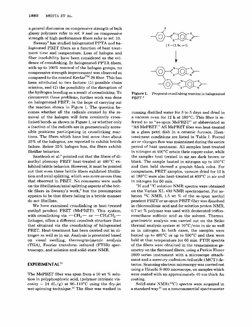
Figure 2. trogen. (a) Methyl PBZT, (b) PBZT.
Thermogravimetric analysis in air and in ni-
the methyl PBZT samples decreased with the in- creased heat-treatment time, and the samples heat- treated in air exhibited a lower degree of swelling as compared to the samples heat-treated in nitrogen. The sample heat-treated in nitrogen a t 550°C did not exhibit any swelling.
Thermogravimetric Analysis
Figure 2 shows the TGA plots for AS-MePBZT and as-spun PBZT in air and in nitrogen. In all cases there is one to two percent initial weight loss, which is attributed to the residual moisture and the residual polyphosphoric acid. The As-MePBZT sample did not lose weight when held a t 400°C for 60 min in nitrogen and lost about 1% weight in air. If the weight loss in methyl PBZT in air is due to the loss of CH3 groups, then it represents an 18% loss of methyl groups. However, such a loss of methyl groups was not confirmed from the solid-state NMR spectrum of the 400°C heat-treated sample. On the other hand, PBZT did not show any weight loss when held for 1 h a t 400°C either in air or in nitro-
gen. TGA of MePBZT in nitrogen suggest about 2% weight loss in heating up to 550°C and an additional 1.4% weight loss when held at 550°C €or 30 min. The 1.4% weight loss corresponds to a loss of about 25% of the pendent methyl groups.
Infrared Spectroscopy
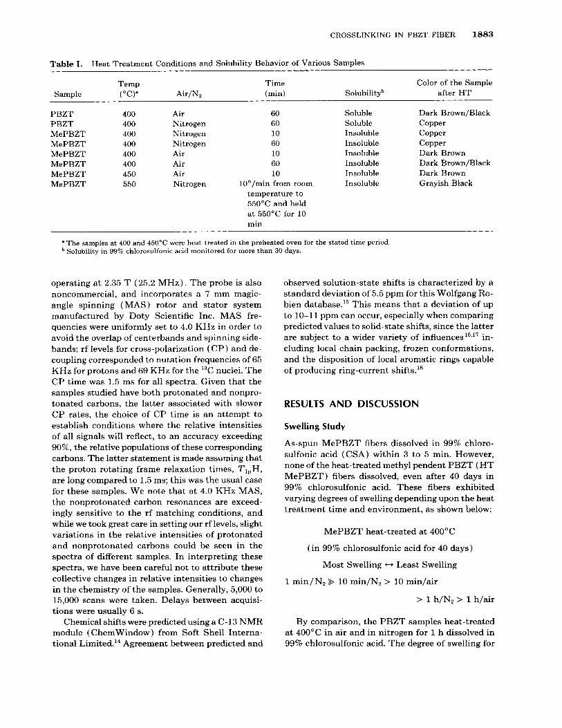
In Figure 3 the IR spectrum of AS-MePBZT is shown along with the IR spectra of samples heat- treated a t 40OoC/60 minlair, 40OoC/60 min/ni- trogen, and 550"C/ 10 min/nitrogen. As-spun and heat-treated samples show medium to weak absorp- tion bands in the aliphatic C - H stretch region of 3000-2800 cm-'. One can interpret these bands as arising from CH2 and/or CH, stretching vibrations; however, the aliphatic groups detected by solution and the solid-state NMR (presented later) include only methyls for the as-spun and the 400°C heat- treated sample. For the 55O0C/1O min/nitrogen sample, a significantly smaller number of aliphatic groups, dominated by methylenes, remain. At the same time, the multiplicity of IR absorptions in this region for this sample is similar to that for the as-
Wavenumber (an-1)
Figure 3. Fourier transform infrared spectra of (a) as- spun methyl PBZT, (b) heat-treated methyl PBZT/ 4OO0C/60 min/air, (c) heat-treated methyl PBZT/nitro- gen/lO°C/min to 55OOC and held at 550 for 10 min, and (d) heat-treated methyl PBZT/40O0C/60 minutes/nitro- gen. Note the loss of water in the samples heat-treated at 4OO0C, judging by the reduction in the broad band near 3400 cm-'. Also note the increase in relative intensity in the carbonyl region (1650-1800 cm-') in (b) versus (d) as a result of heat treating in air.
CROSSLINKING IN PBZT FIBER 1885
1 4 12 . O 8 6 4 2 PPM 0
(b)
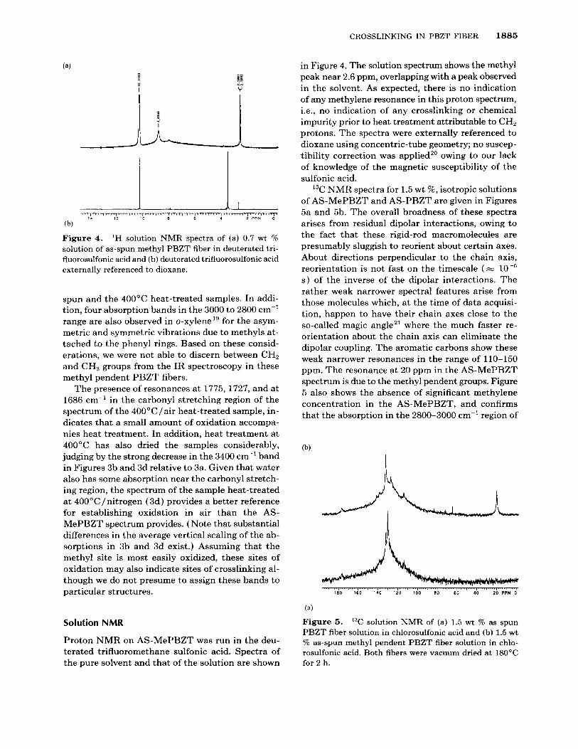
Figure 4. 'H solution NMR spectra of (a) 0.7 wt % solution of as-spun methyl PBZT fiber in deuterated tri- fluorosulfonic acid and (b) deuterated trifluorosulfonic acid externally referenced to dioxane.
spun and the 400°C heat-treated samples. In addi- tion, four absorption bands in the 3000 to 2800 cm-' range are also observed in o-xylenel' for the asym- metric and symmetric vibrations due to methyls at- tached to the phenyl rings. Based on these consid- erations, we were not able to discern between CH, and CH, groups from the IR spectroscopy in these methyl pendent PBZT fibers.
The presence of resonances at 1775,1727, and at 1686 cm-' in the carbonyl stretching region of the spectrum of the 400"C/air heat-treated sample, in- dicates that a small amount of oxidation accompa- nies heat treatment. In addition, heat treatment at 400°C has also dried the samples considerably, judging by the strong decrease in the 3400 cm-' band in Figures 3b and 3d relative to 3a. Given that water also has some absorption near the carbonyl stretch- ing region, the spectrum of the sample heat-treated at 400"C/nitrogen (3d) provides a better reference for establishing oxidation in air than the AS- MePBZT spectrum provides. ( Note that substantial differences in the average vertical scaling of the ab- sorptions in 3b and 3d exist.) Assuming that the methyl site is most easily oxidized, these sites of oxidation may also indicate sites of crosslinking al- though we do not presume to assign these bands to particular structures.
Solution NMR
Proton NMR on AS-MePBZT was run in the deu- terated trifluoromethane sulfonic acid. Spectra of the pure solvent and that of the solution are shown
in Figure 4. The solution spectrum shows the methyl peak near 2.6 ppm, overlapping with a peak observed in the solvent. As expected, there is no indication of any methylene resonance in this proton spectrum, i.e., no indication of any crosslinking or chemical impurity prior to heat treatment attributable to CH2 protons. The spectra were externally referenced to dioxane using concentric-tube geometry; no suscep- tibility correction was applied,' owing to our lack of knowledge of the magnetic susceptibility of the sulfonic acid.
13C NMR spectra for 1.5 wt %, isotropic solutions of AS-MePBZT and AS-PBZT are given in Figures 5a and 5b. The overall broadness of these spectra arises from residual dipolar interactions, owing to the fact that these rigid-rod macromolecules are presumably sluggish to reorient about certain axes. About directions perpendicular to the chain axis, reorientation is not fast on the timescale (= s ) of the inverse of the dipolar interactions. The rather weak narrower spectral features arise from those molecules which, at the time of data acquisi- tion, happen to have their chain axes close to the so-called magic angle21 where the much faster re- orientation about the chain axis can eliminate the dipolar coupling. The aromatic carbons show these weak narrower resonances in the range of 110-150 ppm. The resonance at 20 ppm in the AS-MePBZT spectrum is due to the methyl pendent groups. Figure 5 also shows the absence of significant methylene concentration in the AS-MePBZT, and confirms that the absorption in the 2800-3000 cm-' region of
i,
(4 Figure 5. solution NMR of (a) 1.5 wt % as spun PBZT fiber solution in chlorosulfonic acid and (b) 1.5 wt % as-spun methyl pendent PBZT fiber solution in chlo- rosulfonic acid. Both fibers were vacuum dried at 180°C for 2 h.
1886 MEHTA ET AL.
b d fl,fZ,f4
'1
$80 $60 '140 $20 $00 b0 k 0 $0 bo PPm
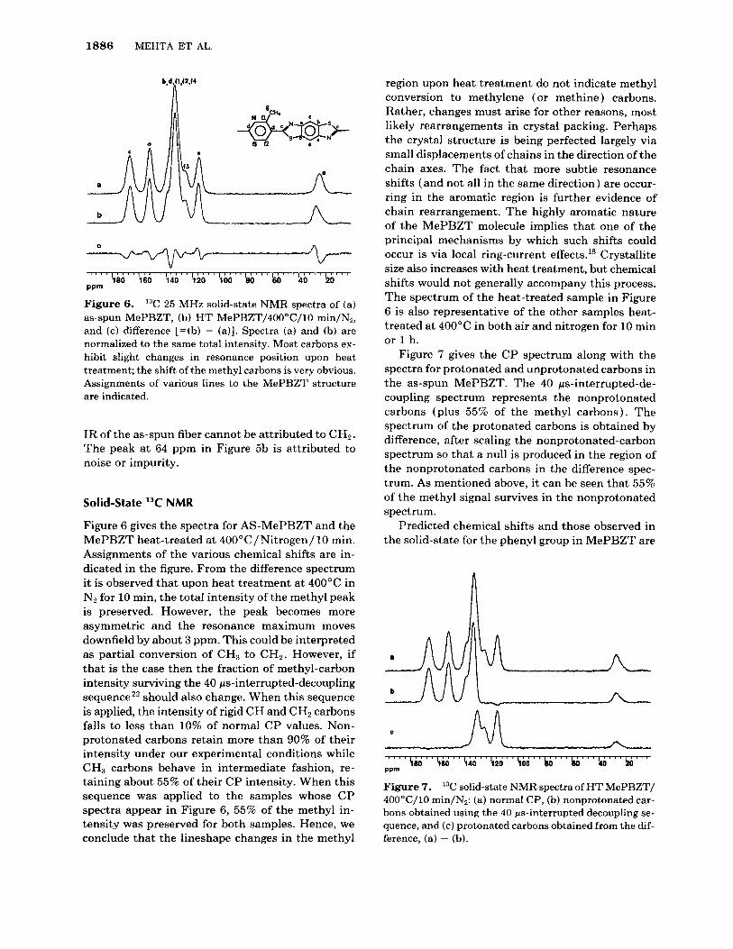
Figure 6. '% 25 MHz solid-state NMR spectra of (a) as-spun MePBZT, (b) H T MePBZT/40O0C/10 min/N,, and (c) difference [=(b) - (a)]. Spectra (a) and (b) are normalized to the same total intensity. Most carbons ex- hibit slight changes in resonance position upon heat treatment; the shift of the methyl carbons is very obvious. Assignments of various lines to the MePBZT structure are indicated.
IR of the as-spun fiber cannot be attributed to CHz. The peak at 64 ppm in Figure 5b is attributed to noise or impurity.
Solid-state 13C NMR
Figure 6 gives the spectra for AS-MePBZT and the MePBZT heat-treated at 400"C/Nitrogen/ 10 min. Assignments of the various chemical shifts are in- dicated in the figure. From the difference spectrum it is observed that upon heat treatment at 400°C in Nz for 10 min, the total intensity of the methyl peak is preserved. However, the peak becomes more asymmetric and the resonance maximum moves downfield by about 3 ppm. This could be interpreted as partial conversion of CH3 to CH2. However, if that is the case then the fraction of methyl-carbon intensity surviving the 40 ps-interrupted-decoupling sequence2' should also change. When this sequence is applied, the intensity of rigid CH and CH, carbons falls to less than 10% of normal CP values. Non- protonated carbons retain more than 90% of their intensity under our experimental conditions while CH3 carbons behave in intermediate fashion, re- taining about 55% of their CP intensity. When this sequence was applied to the samples whose CP spectra appear in Figure 6, 55% of the methyl in- tensity was preserved for both samples. Hence, we conclude that the lineshape changes in the methyl
region upon heat treatment do not indicate methyl conversion to methylene (or methine) carbons. Rather, changes must arise for other reasons, most likely rearrangements in crystal packing. Perhaps the crystal structure is being perfected largely via small displacements of chains in the direction of the chain axes. The fact that more subtle resonance shifts (and not all in the same direction ) are occur- ring in the aromatic region is further evidence of chain rearrangement. The highly aromatic nature of the MePBZT molecule implies that one of the principal mechanisms by which such shifts could occur is via local ring-current effects." Crystallite size also increases with heat treatment, but chemical shifts would not generally accompany this process. The spectrum of the heat-treated sample in Figure 6 is also representative of the other samples heat- treated at 400°C in both air and nitrogen for 10 min or 1 h.
Figure 7 gives the CP spectrum along with the spectra for protonated and unprotonated carbons in the as-spun MePBZT. The 40 ps-interrupted-de- coupling spectrum represents the nonprotonated carbons (plus 55% of the methyl carbons). The spectrum of the protonated carbons is obtained by difference, after scaling the nonprotonated-carbon spectrum so that a null is produced in the region of the nonprotonated carbons in the difference spec- trum. As mentioned above, it can be seen that 55% of the methyl signal survives in the nonprotonated spectrum.
Predicted chemical shifts and those observed in the solid-state for the phenyl group in MePBZT are
c
Figure 7. 13C solid-state NMR spectra of H T MePBZT/ 4OO0C/10 min/N,: (a) normal CP, (b) nonprotonated car- bons obtained using the 40 ps-interrupted decoupling se- quence, and (c) protonated carbons obtained from the dif- ference, (a) - (b).
CROSSLINKING IN PBZT FIBER 1887
given in the second data column of Table 11, together with similar predicted shifts for other structures considered later. The basis for the chemical shift assignments in Figure 6 for the MePBZT will now be discussed, given that contributions to these shifts from solid-state effects create some possible uncer- tainty in the assignments. First consider the pro- tonated spectrum of Figure 7. The higher field line (116 ppm) is assigned to the protonated benzobis- thiazole carbons; these occur at the same shift in PBZT, where they are assigned unambiguously, in contrast to the more numerous protonated phenyl carbons at about 128 ppm. The protonated-carbon resonances of MePBZT located at 125 and 131 ppm, by default, belong to the phenyl moiety. The 125 ppm resonance is assigned to the position para to the methyl-substituted carbon, in keeping with ad- ditivity rules for aromatic sub~titution.'~ The non- protonated resonance at 139 ppm is assigned to the methyl-substituted carbon, consistent with its rel- ative intensity as well as these substituent effects. Therefore, the chain substituted phenyl carbons are at 134 ppm since that is the only resonance position left, given that the assignment of the 167 and 152 ppm resonances, as given in Figure 6, is not in doubt. Note that the position of the methyl-resonance maximum, 27.3 ppm, is almost 8 ppm downfield from its value in solution.
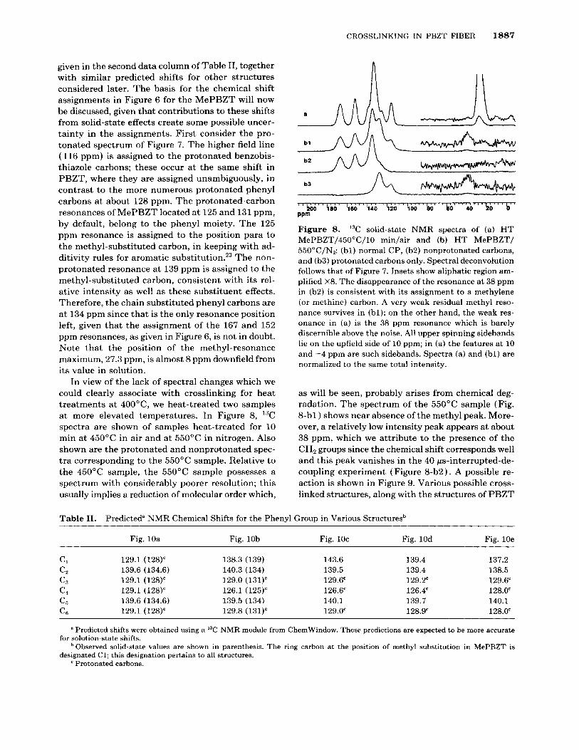
In view of the lack of spectral changes which we could clearly associate with crosslinking for heat treatments at 400"C, we heat-treated two samples at more elevated temperatures. In Figure 8, 13C spectra are shown of samples heat-treated for 10 min at 450°C in air and at 550°C in nitrogen. Also shown are the protonated and nonprotonated spec- tra corresponding to the 550°C sample. Relative to the 450°C sample, the 550°C sample possesses a spectrum with considerably poorer resolution; this usually implies a reduction of molecular order which,
b3
b 0 $80 $60 T O $20 $00 b0 b0 h b b PPm
Figure 8. I3C solid-state NMR spectra of (a) H T MePBZT/45O0C/10 min/air and (b) HT MePBZT/ 55OoC/N2: (bl) normal CP, (b2) nonprotonated carbons, and (b3) protonated carbons only. Spectral deconvolution follows that of Figure 7. Insets show aliphatic region am- plified X8. The disappearance of the resonance at 38 ppm in (b2) is consistent with its assignment to a methylene (or methine) carbon. A very weak residual methyl reso- nance survives in (bl); on the other hand, the weak res- onance in (a) is the 38 ppm resonance which is barely discernible above the noise. All upper spinning sidebands lie on the upfield side of 10 ppm; in (a) the features a t 10 and -4 ppm are such sidebands. Spectra (a) and (bl) are normalized to the same total intensity.
as will be seen, probably arises from chemical deg- radation. The spectrum of the 550°C sample (Fig. 8-bl ) shows near absence of the methyl peak. More- over, a relatively low intensity peak appears at about 38 ppm, which we attribute to the presence of the CH2 groups since the chemical shift corresponds well and this peak vanishes in the 40 ps-interrupted-de- coupling experiment (Figure 8-b2). A possible re- action is shown in Figure 9. Various possible cross- linked structures, along with the structures of PBZT
Table 11. Predicted" NMR Chemical Shifts for the Phenyl Group in Various Structuresb
Fig. 10a Fig. 10b Fig. 1Oc Fig. 10d Fig. 10e
CI 129.1 (128)' 138.3 (139) 143.6 139.4 137.2 C* 139.6 (134.6) 140.3 (134) 139.5 139.4 138.5 c3 129.1 (128)' 129.0 (131)' 129.6' 129.2' 129.6' c4 129.1 (128)' 126.1 (125)' 126.6' 126.4' 128.0' c5 139.6 (134.6) 139.5 (134) 140.1 139.7 140.1 C6 129.1 (128)' 129.8 (131)' 129.0' 128.9' 128.0'
a Predicted shifts were obtained using a 13C NMR module from ChemWindow. These predictions are expected to be more accurate
Observed solid-state values are shown in parenthesis. The ring carbon a t the position of methyl substitution in MePBZT is for solution-state shifts.
designated C1; this designation pertains to all structures. ' Protonated carbons.
1888 MEHTA E T AL.
I V
-Q+;ms* N t + CH,
Figure 9. PBZT heat-treated in nitrogen.
Proposed crosslinking reaction in methyl
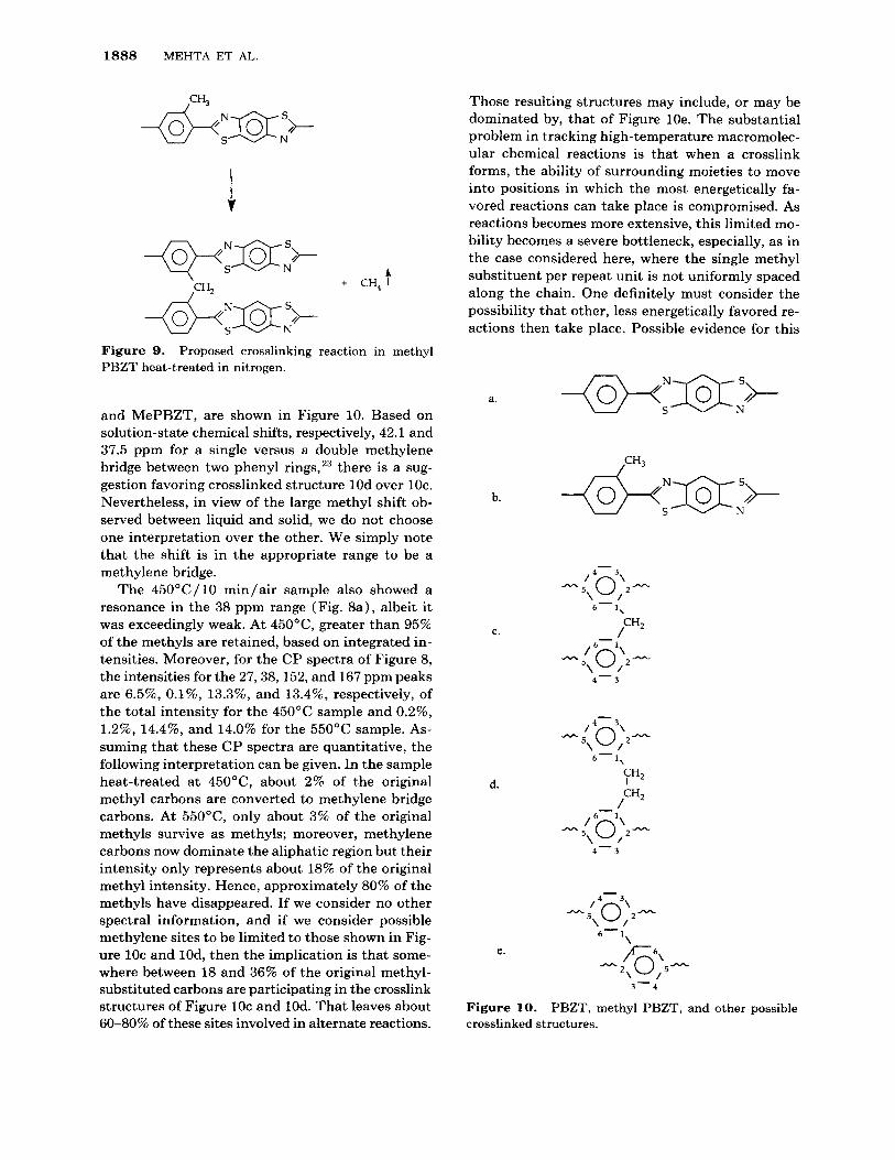
and MePBZT, are shown in Figure 10. Based on solution-state chemical shifts, respectively, 42.1 and 37.5 ppm for a single versus a double methylene bridge between two phenyl rings,23 there is a sug- gestion favoring crosslinked structure 10d over 1Oc. Nevertheless, in view of the large methyl shift ob- served between liquid and solid, we do not choose one interpretation over the other. We simply note that the shift is in the appropriate range to be a methylene bridge.
The 45O0C/1O min/air sample also showed a resonance in the 38 ppm range (Fig. 8a) , albeit it was exceedingly weak. At 450"C, greater than 95% of the methyls are retained, based on integrated in- tensities. Moreover, for the CP spectra of Figure 8, the intensities for the 27,38,152, and 167 ppm peaks are 6.596, 0.1%, 13.3%, and 13.4%, respectively, of the total intensity for the 450°C sample and 0.2%, 1.296, 14.4%, and 14.0% for the 55OoC sample. As- suming that these CP spectra are quantitative, the following interpretation can be given. In the sample heat-treated at 450"C, about 2% of the original methyl carbons are converted to methylene bridge carbons. At 550"C, only about 3% of the original methyls survive as methyls; moreover, methylene carbons now dominate the aliphatic region but their intensity only represents about 18% of the original methyl intensity. Hence, approximately 80% of the methyls have disappeared. If we consider no other spectral information, and if we consider possible methylene sites to be limited to those shown in Fig- ure 1Oc and 10d, then the implication is that some- where between 18 and 36% of the original methyl- substituted carbons are participating in the crosslink structures of Figure 1Oc and 10d. That leaves about 60-80% of these sites involved in alternate reactions.
Those resulting structures may include, or may be dominated by, that of Figure 10e. The substantial problem in tracking high-temperature macromolec- ular chemical reactions is that when a crosslink forms, the ability of surrounding moieties to move into positions in which the most energetically fa- vored reactions can take place is compromised. As reactions becomes more extensive, this limited mo- bility becomes a severe bottleneck, especially, as in the case considered here, where the single methyl substituent per repeat unit is not uniformly spaced along the chain. One definitely must consider the possibility that other, less energetically favored re- actions then take place. Possible evidence for this
a.
b.
C.
d.
e .
-(9+N333+ S N
,CH3
-
6-1,
/CH2 -
/ 6 - 5 \ 0 ; 2 -
4 - 3
- / 4
- 5 \ 0 ; 2 - . . 6-1,
/ 6 - 5 \ 0)-
4 - 3
3 - - 4
Figure 10. crosslinked structures.
PBZT, methyl PBZT, and other possible
CROSSLINKING IN PBZT FIBER 1889
Temperature ("C)
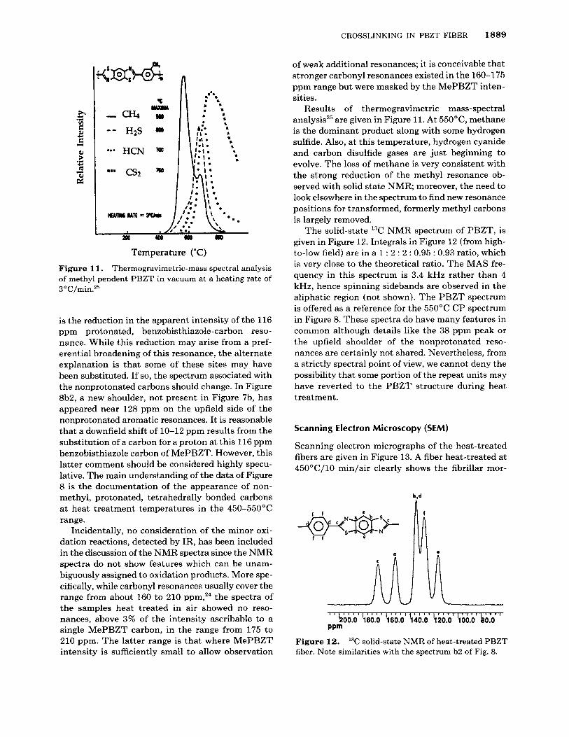
Figure 11. Thermogravimetric-mass spectral analysis of methyl pendent PBZT in vacuum at a heating rate of 3 O C /min.2s
is the reduction in the apparent intensity of the 116 ppm protonated, benzobisthiazole-carbon reso- nance. While this reduction may arise from a pref- erential broadening of this resonance, the alternate explanation is that some of these sites may have been substituted. If so, the spectrum associated with the nonprotonated carbons should change. In Figure 8b2, a new shoulder, not present in Figure 7b, has appeared near 128 ppm on the upfield side of the nonprotonated aromatic resonances. It is reasonable that a downfield shift of 10-12 ppm results from the substitution of a carbon for a proton at this 116 ppm benzobisthiazole carbon of MePBZT. However, this latter comment should be considered highly specu- lative. The main understanding of the data of Figure 8 is the documentation of the appearance of non- methyl, protonated, tetrahedrally bonded carbons at heat treatment temperatures in the 450-550°C range.
Incidentally, no consideration of the minor oxi- dation reactions, detected by IR, has been included in the discussion of the NMR spectra since the NMR spectra do not show features which can be unam- biguously assigned to oxidation products. More spe- cifically, while carbonyl resonances usually cover the range from about 160 to 210 ~ p m , ' ~ the spectra of the samples heat treated in air showed no reso- nances, above 3% of the intensity ascribable to a single MePBZT carbon, in the range from 175 to 210 ppm. The latter range is that where MePBZT intensity is sufficiently small to allow observation
of weak additional resonances; it is conceivable that stronger carbonyl resonances existed in the 160-175 ppm range but were masked by the MePBZT inten- sities.
Results of thermogravimetric mass-spectral analysis25 are given in Figure 11. At 550"C, methane is the dominant product along with some hydrogen sulfide. Also, at this temperature, hydrogen cyanide and carbon disulfide gases are just beginning to evolve. The loss of methane is very consistent with the strong reduction of the methyl resonance ob- served with solid state NMR; moreover, the need to look elsewhere in the spectrum to find new resonance positions for transformed, formerly methyl carbons is largely removed.
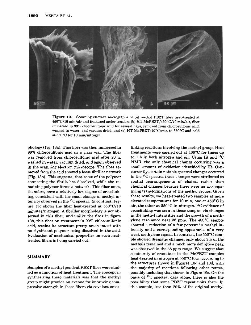
The solid-state "C NMR spectrum of PBZT, is given in Figure 12. Integrals in Figure 12 (from high- to-low field) are in a 1 : 2 : 2 : 0.95 : 0.93 ratio, which is very close to the theoretical ratio. The MAS fre- quency in this spectrum is 3.4 kHz rather than 4 kHz, hence spinning sidebands are observed in the aliphatic region (not shown). The PBZT spectrum is offered as a reference for the 550°C CP spectrum in Figure 8. These spectra do have many features in common although details like the 38 ppm peak or the upfield shoulder of the nonprotonated reso- nances are certainly not shared. Nevertheless, from a strictly spectral point of view, we cannot deny the possibility that some portion of the repeat units may have reverted to the PBZT structure during heat treatment.
Scanning Electron Microscopy (SEM)
Scanning electron micrographs of the heat-treated fibers are given in Figure 13. A fiber heat-treated at 45OoC/10 min/air clearly shows the fibrillar mor-
1 , I , , , , I , , I , , , , , I , I , , , ( , , , , 1 , 1
1200.0 $00.0 $60.0 $40.0 $20.0 $00.0 b0.Q PPm
Figure 12. fiber. Note similarities with the spectrum b2 of Fig. 8.
13C solid-state NMR of heat-treated PBZT
1890 MEHTA ET AL.
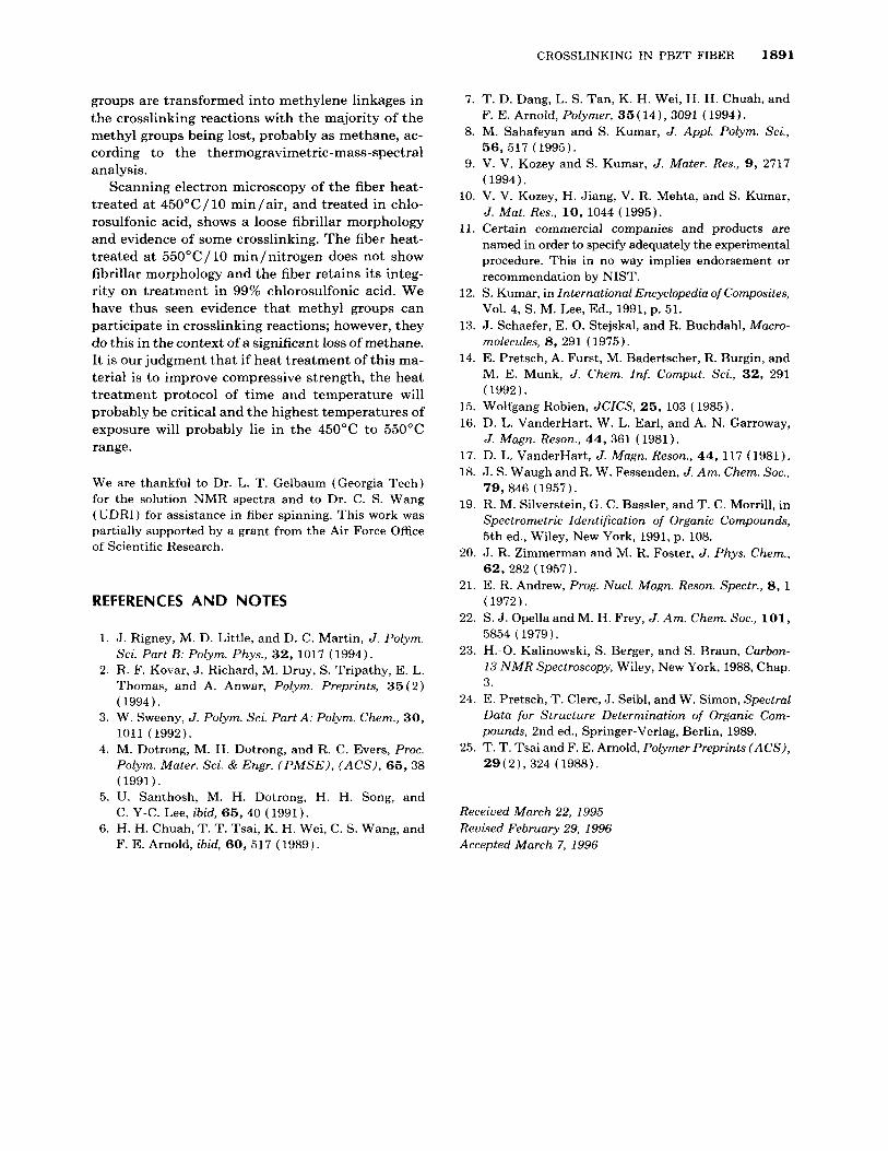
Figure 13. Scanning electron micrographs of (a) methyl PBZT fiber heat-treated at 450"C/10 min/air and fractured under tension, (b) HT MePBZT/45O0C/10 min/air, fiber immersed in 99% chlorosulfonic acid for several days, removed from chlorosulfonic acid, washed in water, and vacuum dried, and (c) HT MePBZT/lO"C/min to 550°C and held at 550°C for 10 min/nitrogen.
phology (Fig. 13a). This fiber was then immersed in 99% chlorosulfonic acid in a glass vial. The fiber was removed from chlorosulfonic acid after 20 h, washed in water, vacuum dried, and again observed in the scanning electron microscope. The fiber re- moved from the acid showed a loose fibrillar network (Fig. 13b). This suggests, that some of the polymer connecting the fibrils has dissolved, while the re- maining polymer forms a network. This fiber must, therefore, have a relatively low degree of crosslink- ing, consistent with the small change in methyl in- tensity observed in the 13C spectra. In contrast, Fig- ure 13c shows the fiber heat-treated at 55OoC/10 minutes/nitrogen. A fibrillar morphology is not ob- served in this fiber, and unlike the fiber in figure 13b, this fiber on treatment in 99% chlorosulfonic acid, retains its structure pretty much intact with no significant polymer being dissolved in the acid. Evaluation of mechanical properties on such heat- treated fibers is being carried out.
SUMMARY
Samples of a methyl pendent PBZT fiber were stud- ied as a function of heat treatment. The concept in synthesizing these materials was that the methyl group might provide an avenue for improving com- pressive strength in these fibers via covalent cross-
linking reactions involving the methyl group. Heat treatments were carried out a t 4OO0C for times up to 1 h in both nitrogen and air. Using IR and I3C NMR, the only chemical change occurring was a small amount of oxidation identified by IR. Con- currently, certain notable spectral changes occurred in the 13C spectra; these changes were attributed to spatial rearrangements of chains, rather than chemical changes because there were no accompa- nying transformations of the methyl groups. Given these results, we heat-treated two samples at more elevated temperatures for 10 min, one at 450°C in air, the other at 550°C in nitrogen. 13C evidence of crosslinking was seen in these samples via changes in the methyl intensities and the growth of a meth- ylene resonance near 38 ppm. The 450°C sample showed a reduction of a few percent in methyl in- tensity and a corresponding appearance of a very weak methylene signal. In contrast, the 550°C sam- ple showed dramatic changes; only about 3% of the methyls remained and a much more definitive peak was observed in the 38 ppm range. We suggest that a minority of crosslinks in the MePBZT samples heat-treated in nitrogen at 550°C form according to the structures shown in Figures 1Oc and 10d, with the majority of reactions following other routes, possibly including that shown in Figure 10e. On the basis of 13C spectral data alone, there is also the possibility that some PBZT repeat units form. In this sample, less than 20% of the original methyl
CROSSLINKING IN PBZT FIBER 1891
groups are transformed into methylene linkages in the crosslinking reactions with the majority of the methyl groups being lost, probably as methane, ac- cording to the thermogravimetric-mass-spectral analysis.
Scanning electron microscopy of the fiber heat- treated a t 450°C/10 minlair, and treated in chlo- rosulfonic acid, shows a loose fibrillar morphology and evidence of some crosslinking. The fiber heat- treated a t 550"C/ 10 minlnitrogen does not show fibrillar morphology and the fiber retains its integ- rity on treatment in 99% chlorosulfonic acid. We have thus seen evidence that methyl groups can participate in crosslinking reactions; however, they do this in the context of a significant loss of methane. It is our judgment that if heat treatment of this ma- terial is to improve compressive strength, the heat treatment protocol of time and temperature will probably be critical and the highest temperatures of exposure will probably lie in the 450°C to 550°C range.
We are thankful to Dr. L. T. Gelbaum (Georgia Tech) for the solution NMR spectra and to Dr. C. S. Wang (UDRI) for assistance in fiber spinning. This work was partially supported by a grant from the Air Force Office of Scientific Research.
REFERENCES AND NOTES
1. J. Rigney, M. D. Little, and D. C. Martin, J . Polym. Sci. Part B: Polym. Phys., 32, 1017 ( 1994).
2. R. F. Kovar, J. Richard, M. Druy, S. Tripathy, E. L. Thomas, and A. Anwar, Polym. Preprints, 35(2) ( 1994).
3. W. Sweeny, J . Polym. Sci. Part A: Polym. Chem., 30, 1011 (1992).
4. M. Dotrong, M. H. Dotrong, and R. C. Evers, Proc. Polym. Mater. Sci. & Engr. ( P M S E ) , (ACS) , 65, 38 ( 1991 ).
5. U. Santhosh, M. H. Dotrong, H. H. Song, and C. Y-C. Lee, ibid, 65, 40 (1991).
6. H. H. Chuah, T. T. Tsai, K. H. Wei, C. S. Wang, and F. E. Arnold, ibid, 60, 517 ( 1989).
7. T. D. Dang, L. S. Tan, K. H. Wei, H. H. Chuah, and F. E. Arnold, Polymer, 35 ( 14) , 3091 ( 1994).
8. M. Sahafeyan and S. Kumar, J. Appl. Polym. Sci., 56, 517 (1995).
9. V. V. Kozey and S. Kumar, J . Mater. Res., 9, 2717 (1994).
10. V. V. Kozey, H. Jiang, V. R. Mehta, and S. Kumar, J . Mat. Res., 10, 1044 (1995).
11. Certain commercial companies and products are named in order to specify adequately the experimental procedure. This in no way implies endorsement or recommendation by NIST.
12. S. Kumar, in International Encyclopedia of Composites, Vol. 4, S. M. Lee, Ed., 1991, p. 51.
13. J. Schaefer, E. 0. Stejskal, and R. Buchdahl, Macro- molecules, 8, 291 ( 1975).
14. E. Pretsch, A. Furst, M. Badertscher, R. Burgin, and M. E. Munk, J . Chem. Inf. Comput. Sci., 32, 291 (1992).
15. Wolfgang Robien, JCICS, 25, 103 ( 1985). 16. D. L. VanderHart, W. L. Earl, and A. N. Garroway,
17. D. L. VanderHart, J . Magn. Reson., 44, 117 (1981). 18. J. S. Waugh and R. W. Fessenden, J . Am. Chem. SOC.,
79 ,846 ( 1957). 19. R. M. Silverstein, G. C. Bassler, and T. C. Morrill, in
Spectrometric Identification of Organic Compounds, 5th ed., Wiley, New York, 1991, p. 108.
20. J. R. Zimmerman and M. R. Foster, J . Phys. Chem., 62, 282 ( 1957).
21. E. R. Andrew, Prog. Nucl. Magn. Reson. Spectr., 8, 1 (1972).
22. S. J . OpellaandM. H. Frey, J . Am. Chem. SOC., 101, 5854 (1979).
23. H . - 0 . Kalinowski, S. Berger, and S. Braun, Carbon- 13 N M R Spectroscopy, Wiley, New York, 1988, Chap.
J . Magn. Reson., 44, 361 (1981).
24. E. Pretsch, T. Clerc, J. Seibl, and W. Simon, Spectral Data for Structure Determination of Organic Com- pounds, 2nd ed., Springer-Verlag, Berlin, 1989.
25. T. T. Tsai and F. E. Arnold, Polymer Preprints (ACS), 2 9 ( 2 ) , 324 (1988).
Received March 22, 1995 Revised February 29, 1996 Accepted March 7, 1996
Related Documents


