Master in Forensic Sciences University of Porto On the effects of preservation, blade angle and intra- and inter-individual differences on the identification of tool class characteristics retained on human costal cartilage in cut marks analysis Katerina Puentes Porto, 2011

Welcome message from author
This document is posted to help you gain knowledge. Please leave a comment to let me know what you think about it! Share it to your friends and learn new things together.
Transcript
Master in Forensic Sciences
University of Porto
On the effects of preservation, blade angle and intra-
and inter-individual differences on the identification of
tool class characteristics retained on human costal
cartilage in cut marks analysis
Katerina Puentes
Porto, 2011

ORIGINAL ARTICLE
A Dissertation presented for the
Master in Forensic Sciences Degree
On the effects of preservation, blade angle and intra- and inter-individual differences on
the identification of tool class characteristics retained on human costal cartilage in cut
marks analysis
Post-graduate student: Katerina Puentes, MD
Adviser: Hugo Filipe Violante Cardoso, PhD (Faculty of Medicine - University of Porto)


AKNOWLEDGMENTS
To Steven A. Symes, who was the beginning of everything:
THANK YOU Steve…
To my beloved family, Marigula, Sofia, Karina and Martijn,
who were the engine behind my strength to walk this path…
To my adviser, Prof. Hugo Cardoso, for his guidance,
patience and understanding…
To Dr. Luis Coelho, for his priceless support and help, and
above all, for his friendship during this process…
To Mr. Amílcar Freitas da Rocha, for his precious
collaboration with the graphic aspects of this work…
To Mrs. Elisa Duarte, for her invaluable help with the
statistical analysis along the process…
To Prof. Agostinho Santos and Prof. Teresa Magalhães for
their continuous support and help…

INDEX
I. Abstract ....................................................................................................... 1
II. Introduction ................................................................................................... 3
III. Materials and Methods ................................................................................. 9
IV. Results ...................................................................................................... 17
V. Discussion .................................................................................................. 21
VI. Conclusions ............................................................................................... 27
VII. References ............................................................................................... 29
VIII. Appendix 1 .............................................................................................. 31

1
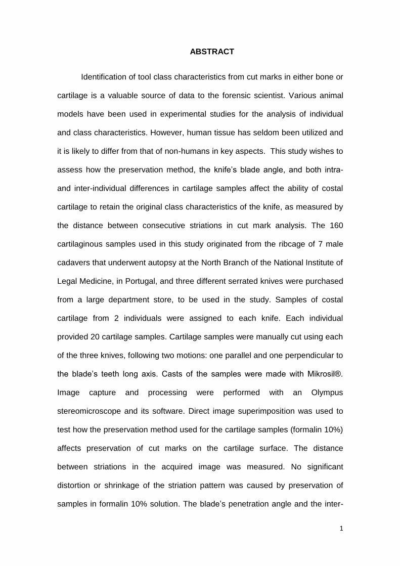
ABSTRACT
Identification of tool class characteristics from cut marks in either bone or
cartilage is a valuable source of data to the forensic scientist. Various animal
models have been used in experimental studies for the analysis of individual
and class characteristics. However, human tissue has seldom been utilized and
it is likely to differ from that of non-humans in key aspects. This study wishes to
assess how the preservation method, the knife’s blade angle, and both intra-
and inter-individual differences in cartilage samples affect the ability of costal
cartilage to retain the original class characteristics of the knife, as measured by
the distance between consecutive striations in cut mark analysis. The 160
cartilaginous samples used in this study originated from the ribcage of 7 male
cadavers that underwent autopsy at the North Branch of the National Institute of
Legal Medicine, in Portugal, and three different serrated knives were purchased
from a large department store, to be used in the study. Samples of costal
cartilage from 2 individuals were assigned to each knife. Each individual
provided 20 cartilage samples. Cartilage samples were manually cut using each
of the three knives, following two motions: one parallel and one perpendicular to
the blade’s teeth long axis. Casts of the samples were made with Mikrosil®.
Image capture and processing were performed with an Olympus
stereomicroscope and its software. Direct image superimposition was used to
test how the preservation method used for the cartilage samples (formalin 10%)
affects preservation of cut marks on the cartilage surface. The distance
between striations in the acquired image was measured. No significant
distortion or shrinkage of the striation pattern was caused by preservation of
samples in formalin 10% solution. The blade’s penetration angle and the inter-
2
individual differences were shown to affect the identification of the tool class
characteristics from the striation pattern observed in a kerf wall, although this
fact seem to be related only to the degree of calcification of the costal cartilage.
Intra-individual differences do not seem to be relevant enough as to affect in a
significant way the identification of the tool class characteristics from the
striation pattern observed in a kerf wall, for the same knife following the same
motion. The degree of calcification of the cartilage is a source of great variation
regarding the interpretation of striations pattern in cartilage.
3
INTRODUCTION
Cutting lesions are defined, from a medico-legal point of view, as those in
which the length is greater than the depth of the wound [1]. Cutting lesions are
also tool marks on human tissue and the American Association of Firearm and
Tool Mark Examiners defines a tool mark as a mark produced when a tool, or
object, is placed against another object and force enough is applied to the tool
or object, so that it leaves an impression [2]. Cut wounds involve the use of an
instrument whose action is exerted through at least one sharp edge, and the
instruments most frequently used are knives and saws, although cleavers, axes
and swords among others, can also be used. Nevertheless, the last three types
of weapon more frequently exert their action through a cutting (sharp) and
striking (blunt) combined mechanism. Injuries caused by sharp instruments or
mechanisms can result from interpersonal violence, both ante-mortem and peri-
mortem, which can cause serious damage or even the death of the victim. This
type of trauma can also result from acts of post-mortem mutilation or
dismemberment, often following a homicide.
The great value for forensic doctors, as well as for other forensic
scientists, of the analysis of cut marks and particularly the identification of the
features of marks caused by sharp instruments in either bone or cartilage,
regardless of whether they are peri-mortem or post-mortem inflicted criminal
acts can be twofold [3, 4]. Firstly, the potential information which can be
extracted through the detailed characterization of these tool marks, allows the
recognition of the weapon/instrument class that was used and, secondly, in
special circumstances, even identify the specific individual blade that created
the cut mark. Consequently, the proper documentation and analysis of knife and
4
saw marks do have the potential to contribute significantly to the interpretation
of the criminal acts involved [3,4].
Forensic scientists are usually asked to determine the type of weapon
used to produce a sharp force trauma defect and sometimes, even to match the
defect to a specific weapon. Tool class characteristics are characteristics that
are common to all tools that are of the same “type” (i.e. shape: a W-shaped kerf
suggests a cross-cut saw, a U-shaped kerf suggests a rip-cut saw, while a V-
shape kerf suggests a beveled blade – knife -) as opposed to individual
characteristics which are characteristics that are unique to one particular tool.
The “type” of weapon used is determined through the tool class characteristics
which do not establish tool mark uniqueness. These also include, mean
distance between teeth in serrated knives or instruments.
The great variety of sharp instruments that may eventually be used in
acts of violence is such that sometimes the tool marks that they produce have
very few common characteristics. Even the same instrument, depending on how
it is used, may cause different types of injuries. Such is the case of a knife,
which can cause stab wounds (in which the depth is greater than the length of
the wound), or merely cut or incised wounds if used only through a “sliding”
movement of the blade.
The identification of distinctive features in tool marks reveals particular
complexities [5]. The difficulty and complexity of classifying knife wounds are
demonstrated by examining a single knife wound to the chest affecting two
consecutive ribs [4] where the examination of the two concurrently cut ribs
would produce two different morphological results. While the rib defect
5
attributed to the spine of the knife cannot be classified conclusively as a wound
created by a knife, the incised wound in the other rib can due to its class
characteristics (i.e., V-shaped kerf). Thus, it is the proximity of the wounds that
conservatively suggests a single weapon, making possible for this knife’s blade
to be described as a single beveled-edge knife.
Although knife wounds are second only to ballistic injuries as the major
cause of violent death in homicides, knife wound analysis has also received
little attention in forensic investigation. Frustration and confusion often arises
with regard to analysis and examination of tool marks while common
misconceptions regarding the analysis procedures and the disagreement
between forensic scientists show the need for a standardized protocol for
analysis of tool marks in bone and cartilage so as to meet the current most
demanding evidentiary standards [6].
Several experimental studies have been conducted in cut mark analysis
in order to extrapolate the results and allow its application in the forensic
context [2,4,7,8,9,10,11,12,13]. Some of these experimental studies have
focused on macroscopic or microscopic perspective of the cut mark analysis,
while others used both perspectives. Microscopic observations usually showed
a greater ability to reliably identify the class of the instrument used to produce
the tool mark, i.e., the gross appearance of some of the tool marks made with a
machete in bone was similar to the tool marks produced by a knife, and only the
microscopic appearance of a set of striations in each tool mark allowed the
distinction between the instruments [13]. However, a review of the published
literature showed that a careful macroscopic observation and detailed
6
description of the observed tool marks is considered essential before their
microscopic analysis.
Although various animal models are widely used in experimental studies
of trauma analysis, the human osseous and cartilaginous tissue is significantly
different from the non-human. In particular, it clearly differs from the animal
models most commonly used, namely porcine and bovine. These species are
fast-growing animals when compared to humans, and that fact is reflected in the
microscopic structure of the cartilage. Porcine cartilage has an increased cell
density when compared to human cartilage as well as a higher proteoglycans’
concentration, thus being denser. This higher density is reflected in a different
collagen network architecture which influences its role as a modulator of the
tissue’s mechanical properties [14]. Moreover, the animal pieces used in
experimental studies are commercially obtained and often belong to young
individuals, in which the cartilaginous tissue structure reflects a relatively
immature stage of development and therefore its non-adult biomechanical
characteristics, further compromising the results of a study of this nature. Due to
all these facts, it becomes challenging to interpret the results acquired from
animal models in order to extrapolate them to a human model.
Despite an increasing interest in tool mark analysis, attempts at it have
become a dismal scientific endeavor. Many forensic anthropologists and
pathologists have performed multiple saw and knife mark analyses in
experimental studies using mostly animal models [15,16,17,18] in an attempt to
extrapolate the results to the human model. Due to the previously explained
differences between the human and non-human cartilage structure, it is
7
believed to be of great interest and pertinence to use human cartilaginous
tissue in an experimental model study.
In the literature, whenever a preservation method was used for the
cartilage samples, it was a 10% formalin solution. It is believed to be so due to
the systematic use of this solution for tissue samples’ preservation in a medical
examiner’s general context. Formalin is known to act on tissue through its
dehydration, nonetheless, no study attempting to establish whether this
dehydration caused any shrinkage or distortion of the tool marks under
examination is known to the author.
It is also noticeable from the literature, that cut marks were produced
holding the blade perpendicular to the sample, or in a 90º angle to the sample.
Nonetheless, none of the studies refer if and how the researchers ensured that
the blade, while penetrating into the sample, stayed “perpendicular” or at a
“90º”. Thus, the assessment of the influence of the variation in the blade’s
penetration angle would be as important in a forensic context as the evaluation
of the striations produced while trying to keep the blade in a specific angle
during its penetration into the cartilage sample.
The present research was designed in order to address the issue of the
misidentification of a blade when differentiating cut marks on cartilage produced
by differently serrated blades. The goals of this study are 1) determining how
the preservation method used for the costal cartilage samples (formalin 10%)
affects preservation of cut marks, 2) asses how the blade’s penetration angle,
as well as the inter- and intra-individual differences, affect the identification of
the tool class characteristics from the striation pattern observed in a kerf wall.

8
This study wishes to assess how the preservation of cartilage, the knife’s blade
angle, and both intra- and inter-individual differences in cartilage samples affect
the ability of costal cartilage to retain the original class characteristics of the
knife, as measured by the distance between consecutive striations in cut mark
analysis.
9
MATERIAL AND METHODS
The 160 cartilaginous samples used in this study originated from the
ribcage of 7 male cadavers, with ages between 20 and 60 years that underwent
autopsy at the North Branch of the National Institute of Legal Medicine, in
Portugal. Death from traumatic causes was an exclusion criterion, as well as
any history of bone or connective tissue disease.
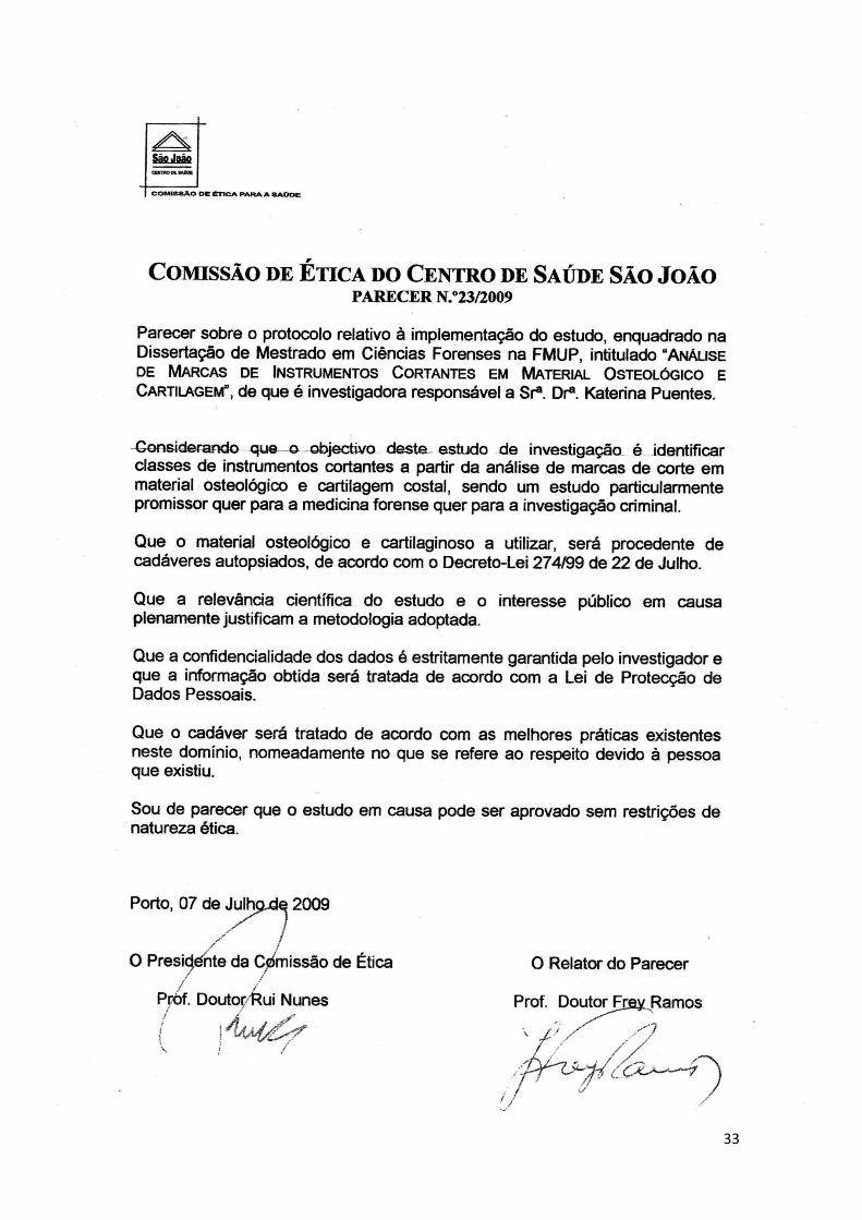
To ensure that the study would be carried out in accordance with ethical
rules, the project was submitted for ethical approval to the regional Ethical
Commission (Ethical Commission of the São João Health Center). Ethical
approval was granted (Appendix 1), provided that the National Registry for Non-
Donors (RENNDA) would be checked for each cadaver previous to the
samples’ collection, thus ensuring this collection would be performed in
accordance with Act 274/99 (July 22nd) which regulates the use of cadavers for
teaching purposes and scientific research.
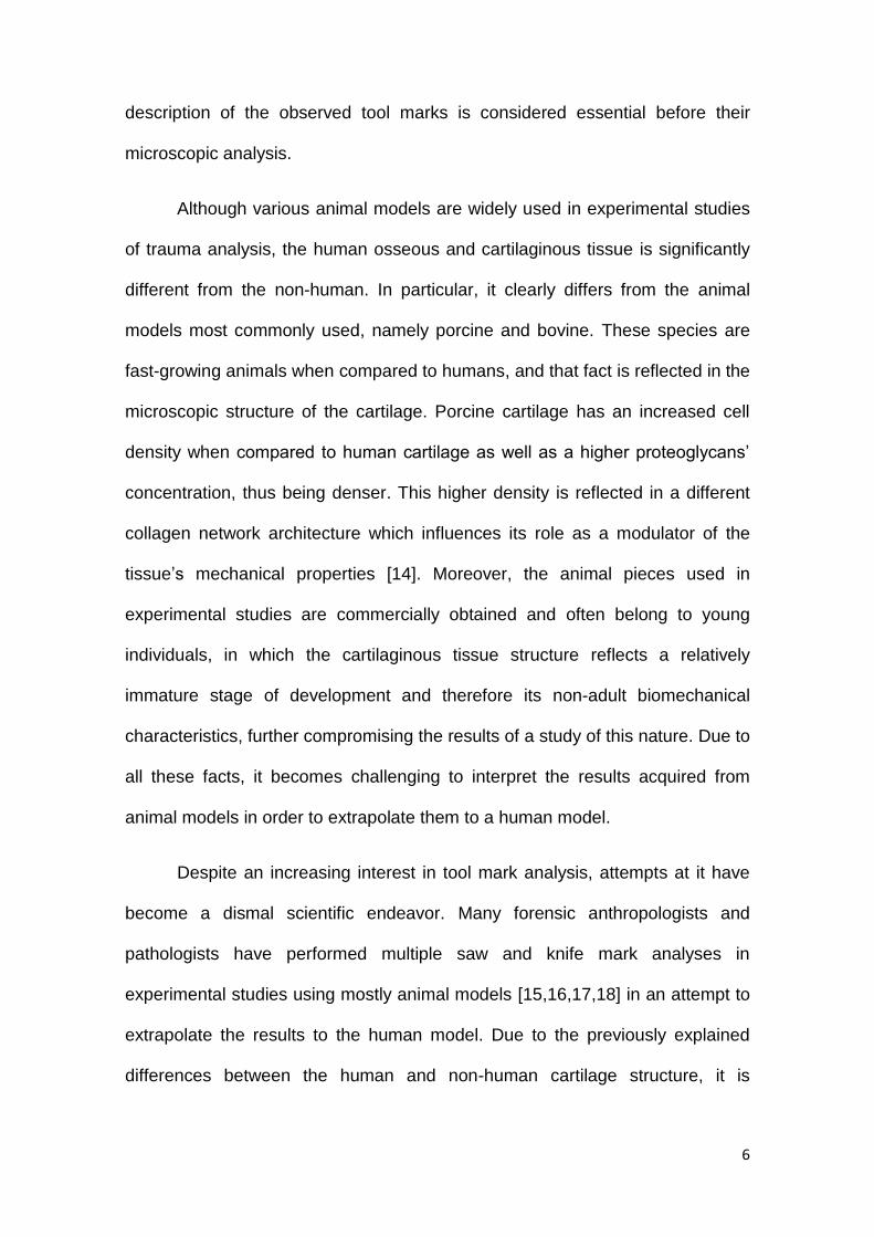
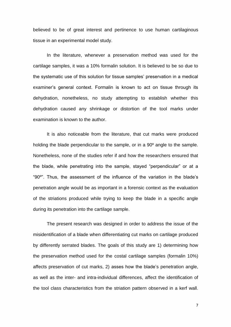
Three different serrated knives (Fig. 1) were purchased from a large
department store. Knife standard terminology with respect to knife blade
anatomy is described according to [18]. Knife 1 was a straight spine, left
grounded, mixed pattern finely serrated knife (Fig. 2). Knife 2 was a straight
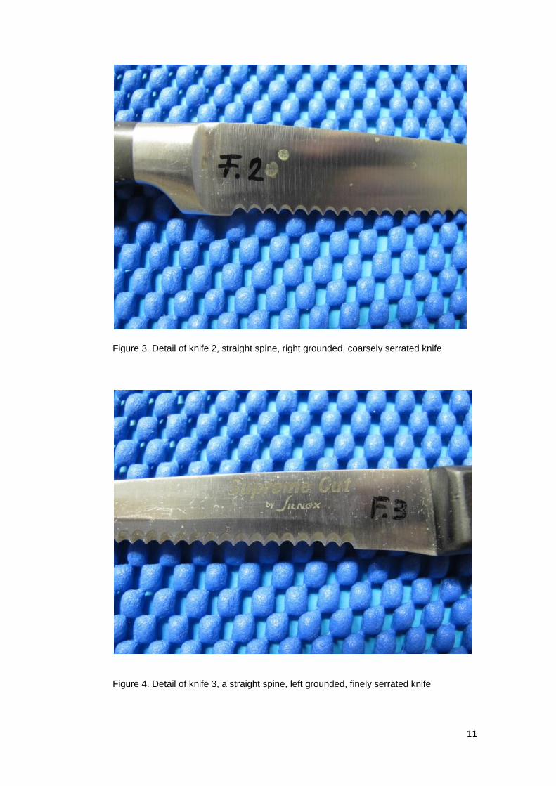
spine, right grounded, coarsely serrated knife (Fig. 3). Knife 3 was a straight
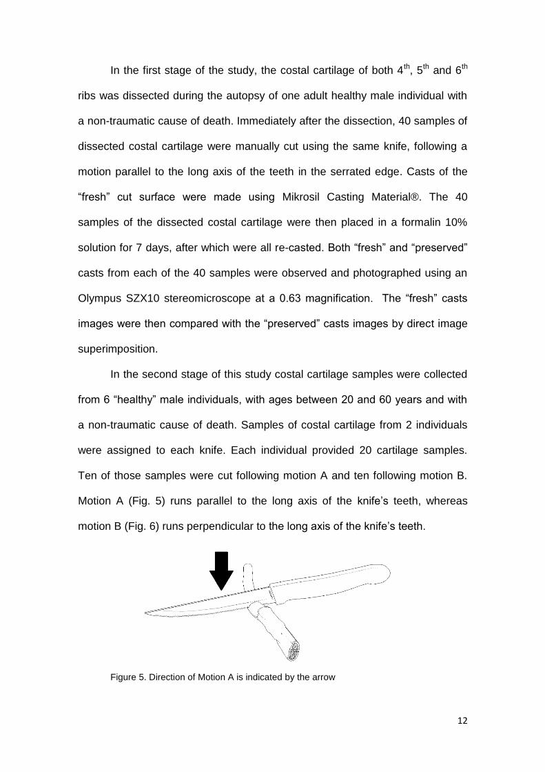
spine, left grounded, finely serrated knife (Fig. 4). None of the knives was used
previously to the study.
10
Figure 1. Photograph of the three knives used in this study
Figure 2. Detail of knife 1, a straight spine, left grounded, mixed pattern finely serrated
knife
11
Figure 3. Detail of knife 2, straight spine, right grounded, coarsely serrated knife
Figure 4. Detail of knife 3, a straight spine, left grounded, finely serrated knife
12
In the first stage of the study, the costal cartilage of both 4th, 5th and 6th
ribs was dissected during the autopsy of one adult healthy male individual with
a non-traumatic cause of death. Immediately after the dissection, 40 samples of
dissected costal cartilage were manually cut using the same knife, following a
motion parallel to the long axis of the teeth in the serrated edge. Casts of the
“fresh” cut surface were made using Mikrosil Casting Material®. The 40
samples of the dissected costal cartilage were then placed in a formalin 10%
solution for 7 days, after which were all re-casted. Both “fresh” and “preserved”
casts from each of the 40 samples were observed and photographed using an
Olympus SZX10 stereomicroscope at a 0.63 magnification. The “fresh” casts
images were then compared with the “preserved” casts images by direct image
superimposition.
In the second stage of this study costal cartilage samples were collected
from 6 “healthy” male individuals, with ages between 20 and 60 years and with
a non-traumatic cause of death. Samples of costal cartilage from 2 individuals
were assigned to each knife. Each individual provided 20 cartilage samples.
Ten of those samples were cut following motion A and ten following motion B.
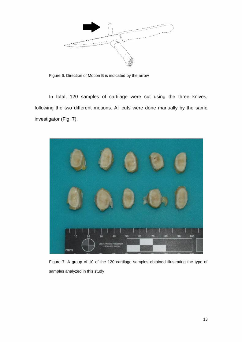
Motion A (Fig. 5) runs parallel to the long axis of the knife’s teeth, whereas
motion B (Fig. 6) runs perpendicular to the long axis of the knife’s teeth.
Figure 5. Direction of Motion A is indicated by the arrow
13
Figure 6. Direction of Motion B is indicated by the arrow
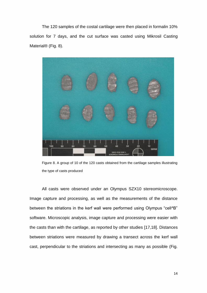
In total, 120 samples of cartilage were cut using the three knives,
following the two different motions. All cuts were done manually by the same
investigator (Fig. 7).
Figure 7. A group of 10 of the 120 cartilage samples obtained illustrating the type of
samples analyzed in this study
14
The 120 samples of the costal cartilage were then placed in formalin 10%
solution for 7 days, and the cut surface was casted using Mikrosil Casting
Material® (Fig. 8).
Figure 8. A group of 10 of the 120 casts obtained from the cartilage samples illustrating
the type of casts produced
All casts were observed under an Olympus SZX10 stereomicroscope.
Image capture and processing, as well as the measurements of the distance
between the striations in the kerf wall were performed using Olympus “cell^B”
software. Microscopic analysis, image capture and processing were easier with
the casts than with the cartilage, as reported by other studies [17,18]. Distances
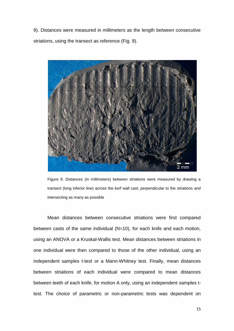
between striations were measured by drawing a transect across the kerf wall
cast, perpendicular to the striations and intersecting as many as possible (Fig.
15
9). Distances were measured in millimeters as the length between consecutive
striations, using the transect as reference (Fig. 9).
Figure 9. Distances (in millimeters) between striations were measured by drawing a
transect (long inferior line) across the kerf wall cast, perpendicular to the striations and
intersecting as many as possible
Mean distances between consecutive striations were first compared
between casts of the same individual (N=10), for each knife and each motion,
using an ANOVA or a Kruskal-Wallis test. Mean distances between striations in
one individual were then compared to those of the other individual, using an
independent samples t-test or a Mann-Whitney test. Finally, mean distances
between striations of each individual were compared to mean distances
between teeth of each knife, for motion A only, using an independent samples t-
test. The choice of parametric or non-parametric tests was dependent on

16
assumptions of normality and heteroscedasticity of data. In some occasions,
non-equal variances t-tests were used. Given that cuts produced by motion B
will not reflect the actual distances between the knife’s teeth, the proportion in
the decrease of the distance between consecutive striations was calculated for
these samples. Statistical analysis of data was performed using SPSS 17.0.
17
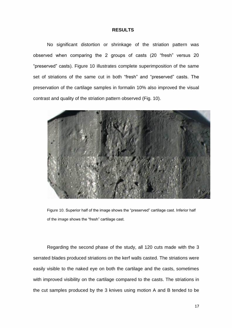
RESULTS
No significant distortion or shrinkage of the striation pattern was
observed when comparing the 2 groups of casts (20 “fresh” versus 20
“preserved” casts). Figure 10 illustrates complete superimposition of the same
set of striations of the same cut in both “fresh” and “preserved” casts. The
preservation of the cartilage samples in formalin 10% also improved the visual
contrast and quality of the striation pattern observed (Fig. 10).
Figure 10. Superior half of the image shows the “preserved” cartilage cast. Inferior half
of the image shows the “fresh” cartilage cast.
Regarding the second phase of the study, all 120 cuts made with the 3
serrated blades produced striations on the kerf walls casted. The striations were
easily visible to the naked eye on both the cartilage and the casts, sometimes
with improved visibility on the cartilage compared to the casts. The striations in
the cut samples produced by the 3 knives using motion A and B tended to be
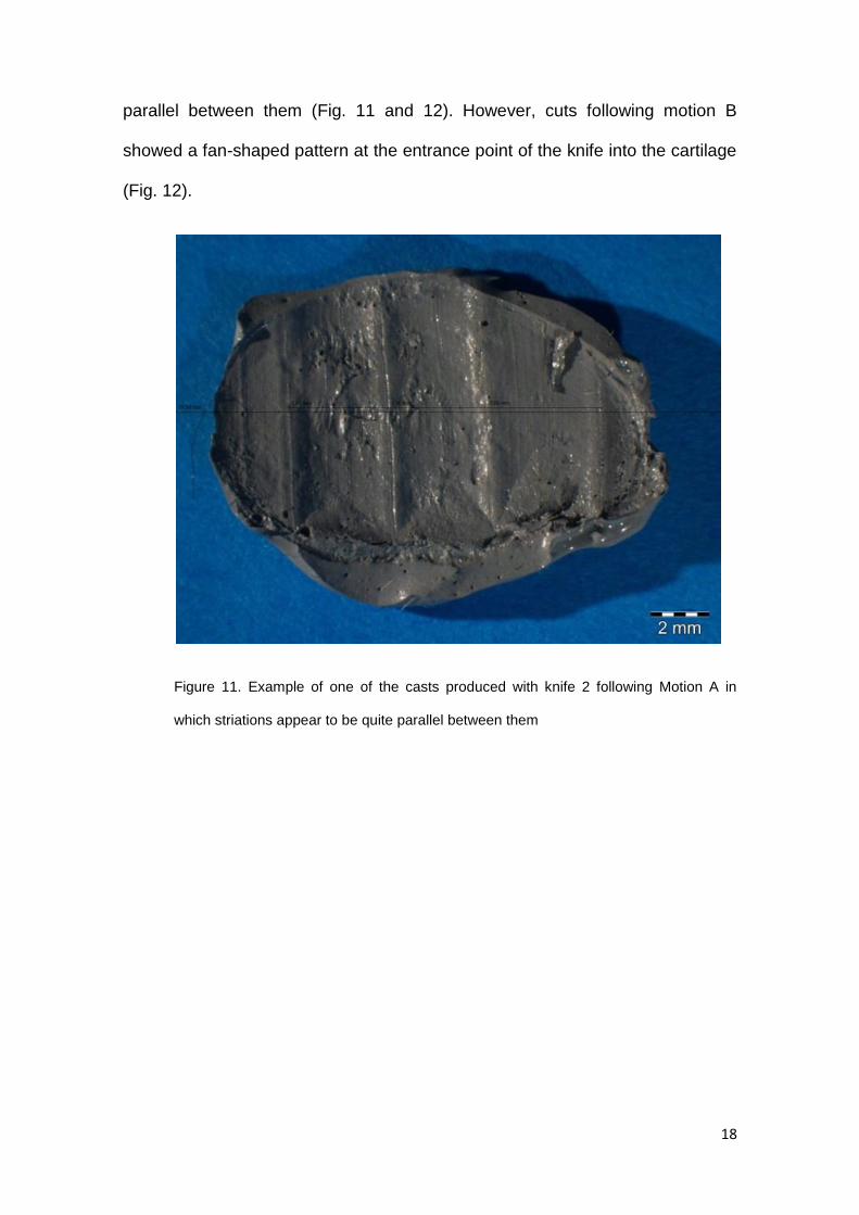
18
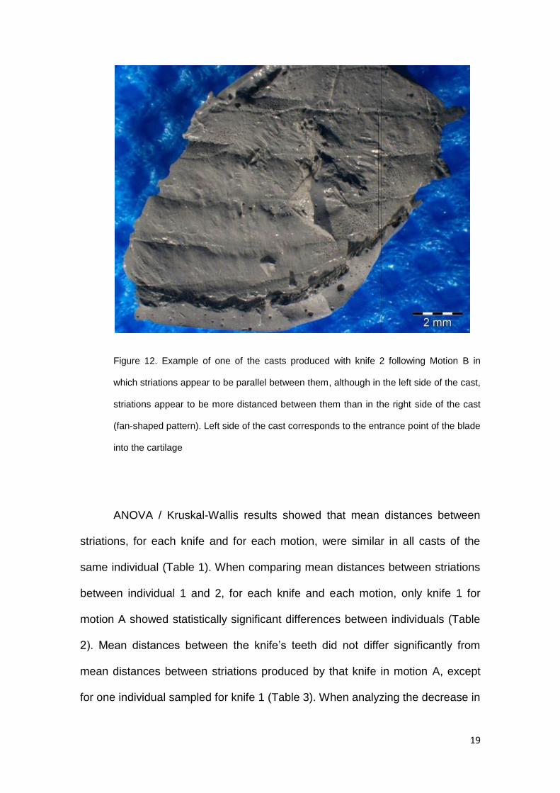
parallel between them (Fig. 11 and 12). However, cuts following motion B
showed a fan-shaped pattern at the entrance point of the knife into the cartilage
(Fig. 12).
Figure 11. Example of one of the casts produced with knife 2 following Motion A in
which striations appear to be quite parallel between them
19
Figure 12. Example of one of the casts produced with knife 2 following Motion B in
which striations appear to be parallel between them, although in the left side of the cast,
striations appear to be more distanced between them than in the right side of the cast
(fan-shaped pattern). Left side of the cast corresponds to the entrance point of the blade
into the cartilage
ANOVA / Kruskal-Wallis results showed that mean distances between
striations, for each knife and for each motion, were similar in all casts of the
same individual (Table 1). When comparing mean distances between striations
between individual 1 and 2, for each knife and each motion, only knife 1 for
motion A showed statistically significant differences between individuals (Table
2). Mean distances between the knife’s teeth did not differ significantly from
mean distances between striations produced by that knife in motion A, except
for one individual sampled for knife 1 (Table 3). When analyzing the decrease in
20
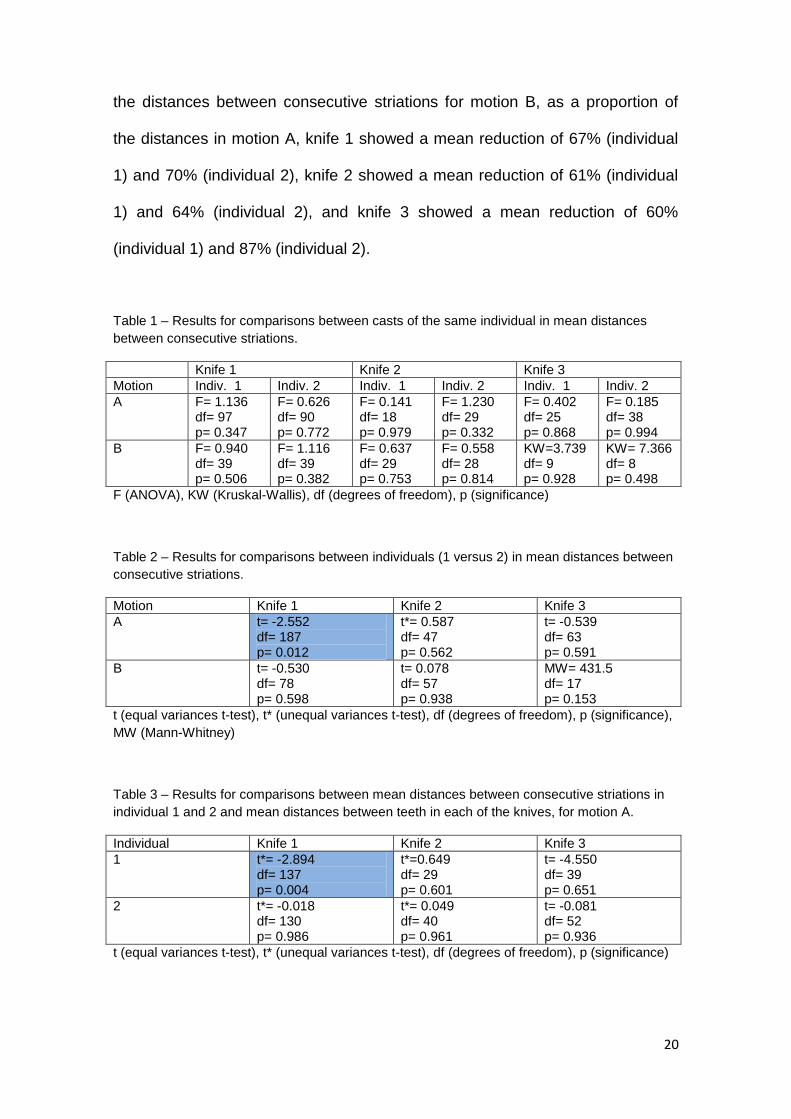
the distances between consecutive striations for motion B, as a proportion of
the distances in motion A, knife 1 showed a mean reduction of 67% (individual
1) and 70% (individual 2), knife 2 showed a mean reduction of 61% (individual
1) and 64% (individual 2), and knife 3 showed a mean reduction of 60%
(individual 1) and 87% (individual 2).
Table 1 – Results for comparisons between casts of the same individual in mean distances
between consecutive striations.
Knife 1 Knife 2 Knife 3
Motion Indiv. 1 Indiv. 2 Indiv. 1 Indiv. 2 Indiv. 1 Indiv. 2
A F= 1.136 df= 97 p= 0.347
F= 0.626 df= 90 p= 0.772
F= 0.141 df= 18 p= 0.979
F= 1.230 df= 29 p= 0.332
F= 0.402 df= 25 p= 0.868
F= 0.185 df= 38 p= 0.994
B F= 0.940 df= 39 p= 0.506
F= 1.116 df= 39 p= 0.382
F= 0.637 df= 29 p= 0.753
F= 0.558 df= 28 p= 0.814
KW=3.739 df= 9 p= 0.928
KW= 7.366 df= 8 p= 0.498
F (ANOVA), KW (Kruskal-Wallis), df (degrees of freedom), p (significance)
Table 2 – Results for comparisons between individuals (1 versus 2) in mean distances between
consecutive striations.
Motion Knife 1 Knife 2 Knife 3
A t= -2.552 df= 187 p= 0.012
t*= 0.587 df= 47 p= 0.562
t= -0.539 df= 63 p= 0.591
B t= -0.530 df= 78 p= 0.598
t= 0.078 df= 57 p= 0.938
MW= 431.5 df= 17 p= 0.153
t (equal variances t-test), t* (unequal variances t-test), df (degrees of freedom), p (significance),
MW (Mann-Whitney)
Table 3 – Results for comparisons between mean distances between consecutive striations in
individual 1 and 2 and mean distances between teeth in each of the knives, for motion A.
Individual Knife 1 Knife 2 Knife 3
1 t*= -2.894 df= 137 p= 0.004
t*=0.649 df= 29 p= 0.601
t= -4.550 df= 39 p= 0.651
2 t*= -0.018 df= 130 p= 0.986
t*= 0.049 df= 40 p= 0.961
t= -0.081 df= 52 p= 0.936
t (equal variances t-test), t* (unequal variances t-test), df (degrees of freedom), p (significance)
21
DISCUSSION
Assessing the influence of formalin preservation on the striation pattern,
direct and complete image superimposition, when comparing the 2 groups of
casts (20 “fresh” versus 20 “preserved” casts) showed that no significant
distortion or shrinkage of the striation pattern occurred by preservation of the
cartilage samples in a 10% formalin % solution for 7 days. The preservation of
the cartilage samples in a 10% formalin solution also improved the visual
contrast and quality of the striation pattern observed, being this improvement
most probably an effect of the dehydrating proprieties of the formalin. Taking
into consideration that the sharp force trauma analysis SWGANTH
recommendations state that “cartilage should be analyzed for the presence of
sharp force defects before removal of soft tissues”, this dehydrating proprieties
of the formalin solution appear to be very useful in order to facilitate the
examination.
Analysis of the factors influencing cut marks showed that mean distances
between striations, for each knife and for each motion, are similar in all casts of
the same individual. This means that the same knife, in the same cutting
motion, cutting different cartilage samples in the same individual, produces a
similar and repeatable striation pattern, which can be measured consistently. In
addition to intra-individual consistency in striation pattern, results also show that
this is also the case for inter-individual comparisons. There was only one
exception when comparing distances between individual 1 and 2, for knife 1 and
motion A, where these individuals differed. Since only individual 1 showed
differences between the distances measured in the casts and the distances
between the knife’s teeth, this suggested a possible problem with this
22
individual’s costal cartilage samples. In fact, this individual is the oldest of the 6
cadavers utilized in the second phase of the study (60 years-old) and showed,
by far, the most calcified cartilage of the entire sample. Although the degree of
calcification of each cartilage sample was never quantified, a qualitative
assessment was sufficient to reveal important differences between subjects. In
comparison, mean distances between striations produced by knife 2 and 3 did
not seem to differ from distances between the knife’s teeth. When analyzing the
decrease in the distances between consecutive striations for motion B, as a
proportion of the distances in motion A for the same knife and the same
individual, the greatest reduction noticed was in individual 2 for knife 3 (an 87%
decrease). This was the younger individual (20 years-old) of the 6 cadavers
used in the second phase of the study, and coincidentally it was also the
individual who provided the least calcified cartilage samples. In all three knifes
utilized, a reduction of 60-70% in mean distances between striations can be
expected, when the blade is progressing on a plane that is perpendicular to that
of the teeth’s axis.
Striations produced following cutting motion B, using all three knifes,
showed a consistent fan-shaped striation pattern, which was always observed
at the entrance point of the knife, and it seems to be a defining morphological
feature of these casts. It was also noticed that the more calcified the cartilage
was, the more angular was the curve of the striations at the base of the fan-
shaped pattern before the striations become parallel. This pattern is also
described in a study using porcine cartilage by Pounder, 2011, and it is
explained by the fact that the knife used had a drop point blade and unlike a
straight spine blade, the drop point blade cannot enter the cartilage in a simple
23
vertical manner and the blade moves sideways until the drop point has passed
through the firm cartilage. This explanation, however, does not seem to justify
the presence of a fan-shaped pattern in the striations produced by the 3 knives
in this study, following motion B, since all knives had straight spine blades. It
was rather noticed that this fan-shaped pattern was related to a change in the
penetration angle of the knife while progressing into the cartilage. The
morphological differences (more or less angular) in the fan-shaped pattern were
also noticed to be related to the degree of calcification of the cartilage, since the
more calcified the cartilage was, the more “unsteady” was the blade’s
penetration in the cartilage, thus producing a more angular fan-shaped pattern.
One of the main factors affecting the ability of cartilage to retain the
class characteristics of the knife seems to be the amount of calcification. This
was rather clear when comparing the more calcified cartilages of individual 1
with that of individual 2, under knife 1 and motion A, as well as when comparing
the reduction in striations of individual 1 with that of individual 2, under knife 3
and motion B. The more calcified the cartilage is, the more “resistance” seems
to offer to the passage of the blade making its penetration into the cartilage
“unsteady”. On the other hand, the less calcified the cartilage is, the less
“resistance” seems to offer to the passage of the blade, making its penetration
into the cartilage much more “steady”. Other experimental studies conducted in
this area using non-human cartilage samples [15,16,17,18] from young age
commercially acquired animals eliminate this variation introduced by the
influence of calcified cartilaginous tissue from mature and older individuals.
Consequently, this study cautions against the fact that cartilage from
24
commercially acquired animals may not reflect human cartilage in numerous
ways, in addition to the obvious human/non-human divide.
There are some potential limitations to this study, one of which is the
variations eventually introduced by the investigator while manually producing
the cuts. Although a mechanically controlled machine may eliminate this source
of error, it has little to resemble actual cut marks in a real forensic scenario.
Sample size also be other important limitations, as the study was only able to
compare between two different individuals, across the three different knives.
The number and diversity in class of knives may be another limitation, but these
only call attention for future research. In particular, this study’s research design
did not assess the influence of different degrees of calcification in the striation
pattern in detail. Future research is needed to falsify, confirm or complete the
results in this study.
The present study wished to contribute with new data concerning the
analysis of cut marks on human costal cartilage. Results suggest that the class
characteristics of the knife can be retained rather faithfully in costal cartilage
when the cut is parallel to the teeth’s axis, with exceptions resulting from the
use of calcified cartilage. However, in an actual forensic case there may be
situations when the expert knows the penetrating angle and other when he/she
does not. Depending on the class of the knife utilized, striations produced by a
perpendicular cutting motion of a certain knife may mimic the striations
produced by a parallel cutting motion of another knife with a much smaller
distance between the teeth’s axis. This is an unavoidable fact. However, results
in this study also suggest that whenever the cutting motion is not parallel to the
teeth’s axis, a fan-shaped pattern, with striations at a greater or lesser angle,

25
can be identified at the point of the blade’s entry. The exact angle at which the
blade actually penetrated is, unfortunately, impossible to determine at the
moment.

26
27
CONCLUSIONS
This study wished to assess how certain variables influence the ability of
human cartilage to retain the class characteristic of the blade in sharp force
trauma. Cartilage samples preservation in formalin 10% solution not only do not
caused significant distortion or shrinkage of the striation pattern, but improved
the visual contrast and quality of the striation pattern observed. With important
exceptions, this study was able to show that cartilage is able to retain the class
characteristic of the blade (mean distance between teeth) used to cut it, quite
faithfully, when the cutting motion is parallel to that of the axis of the knife’s
teeth. However, The blade’s penetration angle and inter-individual differences in
costal cartilage affect the identification of the tool class characteristics from the
striation pattern observed in a kerf wall, although this fact seem to be intimately
related to the degree of calcification of the costal cartilage of the individual
under analysis.

28
29
REFERENCES
1. DiMaio VJ, DiMaio D: Forensic Pathology, CRC SERIES In Practical
Aspects of Criminal and Forensic Investigations, 2001.
2. Bartelink EJ, Wiersema JM, Demaree RS: Quantitative analysis of sharp-
force trauma: an application of scanning electron microscopy in forensic
anthropology, J Forensic Sci, 46(6):1288-93, 2001.
3. Reichs K: Forensic Osteology: Advances in the Identification of Human
Remains, 580 pages, 1998.
4. Symes SA, Williams JA, Murray EA, Hoffman JM, Holland TD, Saul JM,
Saul FP, Pope EJ: 21. Taphonomic Context of Sharp-Force Trauma in
Suspected Cases of Human Mutilation and Dismemberment. In: Haglund
WD,Sorg MH, Advances in Forensic Taphonomy: Method, Theory and
Archaeological Perspectives, CRC Press LLC, Boca Raton, pp. 2002.
5. Ciallella C, Caringi C, Aromatario M: Wounds inflicted by survival-knives,
Forensic Sci Int, 126(1):82-7, 2002.
6. Scientific Working Group for Forensic Anthropology (SWGANTH).
Trauma Analysis. 2011.
7. Bello SM, Soligo C: A new method for the quantitative analysis of
cutmark micromorphology, Journal of Archaeological Science, 35(1542-
52, 2008.
8. Blumenschine RJ, Marean CW, Capaldo SD: Blind Tests of Inter-analyst
Correspondence and Accuracy in the Identification of Cut Marks,
Percussion Marks, and Carnivore Tooth Marks on Bone Surfaces,
Journal of Archaeological Science, 23(493–507, 1996.
9. Humphrey JH, Hutchinson DL: Macroscopic characteristics of hacking
trauma, J Forensic Sci, 46(2):228-33, 2001.
10. Lewis JE: Identifying sword marks on bone: criteria for distinguishing
between cut marks made by different classes of bladed weapons,
Journal of Archaeological Science, 35(2001-8, 2008.
11. Saville PA, Hainsworth SV, Rutty GN: Cutting crime: the analysis of the
"uniqueness" of saw marks on bone, Int J Legal Med, 121(5):349-57,
2007.
12. Thompson TJ, Inglis J: Differentiation of serrated and non-serrated
blades from stab marks in bone, Int J Legal Med, 123(2):129-35, 2009.
13. Tucker BK, Hutchinson DL, Gilliland MF, Charles TM, Daniel HJ, Wolfe
LD: Microscopic characteristics of hacking trauma, J Forensic Sci,
46(2):234-40, 2001.
14. Rieppo J, Halmesmaki EP, Siitonen U, Laasanen MS, Toyras J, Kiviranta
I, Hyttinen MM, Jurvelin JS, Helminen HJ: Histological Differences of
Human, Bovine and Porcine Cartilage. 49th Annual Meeting of the
Orthopaedic Research Society, New Orleans, Louisiana. U.S.A., 2003.

30
15. Crowder C, Rainwater CW, Fridie JS: Microscopic Analysis of Sharp
Force Trauma in Bone and Cartilage: A Validation Study. National
Institute of Justice Award No. 2009-DN-BX-K238. 2011
16. Love JC, Symes SA: Understanding rib fracture patterns: incomplete and
buckle fractures, J Forensic Sci, 49(6):1153-8, 2004.
17. Pounder DJ, Cormack L, Broadbent E, Millar J: Class characteristics of
serrated knife stabs to cartilage, Am J Forensic Med Pathol, 32(2):157-
60, 2011.
18. Pounder DJ, Reeder FD: Striation patterns in serrated blade stabs to
cartilage, Forensic Sci Int, 208(1-3):91-4, 2011.

31
APPENDIX 1

32
33

34
Related Documents


