ON CRANIOFACIAL MICROSOMIA SHAPE AND SURGERY BRITT PLUIJMERS

Welcome message from author
This document is posted to help you gain knowledge. Please leave a comment to let me know what you think about it! Share it to your friends and learn new things together.
Transcript

curriculum vitae
Britt Pluijmers was born on the 16th of November 1987 in Cardi� (UK), and spent part ofher childhood in Belgium before moving to the Netherlands. She graduated from LibanonLyceum high school in Rotterdam in 2005.In 2006 she started medical training at the Erasmus University Hospital in Rotterdam(EMC). During her studies she was a board member of the Education committee EMC, anddid voluntary work in the Palestinian Territories.In her third year, under direct supervision of dr. Koudstaal, she started research at thedepartment of Oral and Maxillofacial surgery at the EMC led by em. prof. dr. van der Wal.Her Master thesis was carried out under the guidance of prof. Dunaway, at the GreatOrmond Street Hospital in London (UK). During her internship at the department of Oraland Maxillofacial surgery EMC, she started this thesis, with dr. Koudstaal as co-promotorand prof. dr. Wolvius as promotor.In 2013, she graduated Cum Laude from medical school, and commenced her dentistrystudies at Radboud University, Nijmegen, from which she graduated in 2016.Simultaneously, she was a board member of Promeras, the PhD committee of theErasmus MC. In 2015 she started her Oral- and Maxillofacial surgery training (undersupervision of prof. dr. Wolvius) at the Erasmus Medical Center in Rotterdam and the St.Elisabeth Hospital in Tilburg (under supervision of J.P.O. Scheerlinck). She was a memberof the organizational committee of the JOD NWHHT in 2017.At present, she is in her senior year of her training, and a member of the implementationcommittee for the new curriculum for Oral- and Maxillofacial surgery residents.
Britt is married to Coen Iordens and proud mother to Michiel. She has 3 awesomebrothers.
ON CRANIOFACIAL MICROSOMIASHAPE AND SURGERY
BRITT PLUIJMERS
ON
CRAN
IOFA
CIAL M
ICROSO
MIA
SHA
PE AN
D SU
RGERY
B.I. PLUIJM
ERS
Britt Pluijmers Cover v6.indd 1-3 24-7-2019 16:56:17
Uitnodiging
voor het bijwonen van de openbare verdediging van het
proefschrift
On Craniofacial MicrosomiaShape and Surgery
door Britt Pluijmers
Op woensdag 11 september 2019 om 11:30
in de Prof. Andries Queridozaal van het Onderwijscentrum van het Erasmus Medisch Centrum Wytemaweg 80 te Rotterdam
Na a� oop van de promotie bent u van harte welkom op de receptie
Britt PluijmersMathenesserlaan 371a
3023 GD Rotterdam0647480499
ParanimfenSuzanne Deetman
Melvyn [email protected]
Britt Pluijmers Cover v6.indd 4 24-7-2019 16:56:36
Uitnodiging
voor het bijwonen van de openbare verdediging van het
proefschrift
On Craniofacial MicrosomiaShape and Surgery
door Britt Pluijmers
Op woensdag 11 september 2019 om 11:30
in de Prof. Andries Queridozaal van het Onderwijscentrum van het Erasmus Medisch Centrum Wytemaweg 80 te Rotterdam
Na a� oop van de promotie bent u van harte welkom op de receptie
Britt PluijmersMathenesserlaan 371a
3023 GD Rotterdam0647480499
ParanimfenSuzanne Deetman
Melvyn [email protected]
Britt Pluijmers Cover v6.indd 4 24-7-2019 16:56:36Naamloos-2 1-4 24-7-2019 17:05:31

On Craniofacial MicrosomiaShape and Surgery
Britt Irene Pluijmers
PSM 20190527 Proefschrift Britt Pluijmers.indd 1 24-07-19 15:04

© 2019, Britt Irene Iordens-Pluijmers
ISBN: 978-94-6380-426-4
Artwork cover: Margot AnnuschekLay-out: RON Graphic Power, www.ron.nuPrinting: ProefschriftMaken || www.proefschriftmaken.nl
Financial support for the printing and distribution of this thesis was kindly supported by:Afdeling Mondziekten, Kaak- en Aangezichtschirurgie Erasmus Medisch CentrumErasmus Medisch CentrumNederlandse Vereniging voor Mondziekten, Kaak- en Aangezichtschirurgie
All rights reserved. No parts of this publication may be reported or transmitted, in any of form or by any means, without the permission of the author.
Geistlich Bio-Oss® en Geistlich Bio-Gide®
PSM 20190527 Proefschrift Britt Pluijmers.indd 2 24-07-19 15:04

Thesis
to obtain the degree of Doctor from theErasmus University Rotterdam
by command of therector magnificus
Prof.dr. R.C.M.E. Engels
and in accordance with the decision of the Doctorate Board.The public defence shall be held on
Wednesday 11th of September 2019 at 11:30 hrs
byBritt Irene Pluijmers
born in Cardiff, Wales (UK)
On Craniofacial Microsomiashape and surgery
Craniofaciale microsomievorm en chirurgie
Erasmus University Rotterdam
PSM 20190527 Proefschrift Britt Pluijmers.indd 3 24-07-19 15:04

Doctoral Committee:
Promotor: Prof. dr. E.B. Wolvius
Other members: Prof. D.J. Dunaway Prof. dr. G.J. Kleinrensink Prof. dr. I.M.J. Mathijssen
Copromotor: Dr. M.J. Koudstaal
Paranymphs: S. Deetman M.B.D. Pluijmers
PSM 20190527 Proefschrift Britt Pluijmers.indd 4 24-07-19 15:04

“On wednesdays we wear pink”
PSM 20190527 Proefschrift Britt Pluijmers.indd 5 24-07-19 15:04

PSM 20190527 Proefschrift Britt Pluijmers.indd 6 24-07-19 15:04

Contents
Part I General introduction 11
Part II Population 27
Chapter 1 Craniofacial and extracraniofacial anomalies in craniofacial microsomia: A multicenter study of 755 patients 29
Part III Shape 49
Chapter 2 Characterizing the skull base in craniofacial microsomia using principal component analysis 51
Chapter 3 Using principal component analysis to describe the midfacial deformities in patients with craniofacial microsomia 71
Chapter 4 Describing the mandible in patients with craniofacial microsomia based on principal component analysis and thin-plate-spline video analysis. 93
Chapter 5 Is there a difference in orbital volume between affected and unaffected sides in patients with unilateral craniofacial microsomia 109
Part IV Surgery 119
Chapter 6 Mandibular reconstruction in the growing patient with unilateral craniofacial microsomia: a systematic review 121
Chapter 7 Surgical correction of the midface in craniofacial microsomia. 143 Part 1: a systematic review
PSM 20190527 Proefschrift Britt Pluijmers.indd 7 24-07-19 15:04

PSM 20190527 Proefschrift Britt Pluijmers.indd 8 24-07-19 15:04

Chapter 8 Surgical correction of the midface in craniofacial microsomia. 163 Part 2: Is the maxillary canting and its surgical correction in patients with CFM correlated to the mandibular deformity?
Chapter 9 Surgical correction of craniofacial microsomia: Evaluation of interventions in 565 Patients at three major Craniofacial units 175
Part V General discussion 195
Part VI Summaries 205Summary 206Samenvatting 209
Part VII Appendices 213Publications 214Presentations 217PhD Portfolio 218Dankwoord 220
PSM 20190527 Proefschrift Britt Pluijmers.indd 9 24-07-19 15:04

PAR
TIPSM 20190527 Proefschrift Britt Pluijmers.indd 10 24-07-19 15:04

I
General introduction
PSM 20190527 Proefschrift Britt Pluijmers.indd 11 24-07-19 15:04

General introduction
12
General introduction
Craniofacial microsomia (CFM) is the second most common congenital craniofacial malformation following cleft lip and palate.1,2 In 1952, Goldenhar characterized the disorder as a triad of accessory tragus, mandibular hypoplasia and epibulbar dermoid.3 Other names for the disorder are ‘otomandibular dysostosis’ and ‘first and second branchial arch syndrome’.4,5 A term often found in genetics literature is ‘oculo-auriculo-vertebral syndrome’ (OAVS) as proposed by Gorlin.6 However, in the surgical field, hemifacial microsomia and nowadays craniofacial microsomia is most commonly used.
The deformity is characterized by predominantly asymmetrical hypoplasia of struc-tures derived from the first and second pharyngeal arches, leading to a distinct scoliosis of the facial skeleton. The first pharyngeal arch gives rise to the mandible, maxilla, zygoma, trigeminal nerve, muscles of mastication, and the inner ear and a part of the external ear, whereas the second pharyngeal arch gives rise to the facial nerve, stapes, styloid process, portions of the hyoid bone, facial musculature, and the majority of the external ear.7 Other anomalies seen in patients with CFM include malformations of the vertebrae, cervical spine, cardiorespiratory system, urogenital system, limbs, central nervous system and gastrointestinal system. Most often reported are skeletal, cardiac and renal anomalies.
CFM is most often regarded as a unilateral malformation; however, facial structures have been reported to be involved bilaterally in 10% of cases.8-10 Previous studies suggested that, in most cases, the contralateral side is abnormal as well, although not truly hypoplastic.10-15
The etiology of CFM has not yet been clarified. Well-known hypotheses are local haemorrhage of the stapedial artery16 and disturbed migration of cranial neural crest cells.17,18 Several possible genes, proteins and or pathway signalling disregulations have been suggested including BPAX1, Foxi3 and loss of Hedgehog signaling.16-19 However, an increased risk is found in a history of multiple pregnancies, second-trimester vaginal bleeding and risk factors associated with poverty.4 Leading to the believe that the etiology might include genetic and non-genetic factors, in line with an oligogenic or even a multifactorial etiology.7 Although CFM usually occurs sporadically, familial cases compatible with autosomal dominant and autosomal recessive patterns of inheritance have been described.
Patients with CFM are phenotypically heterogeneous; their dysmorphologies range from minor to severe. Therefore, a comprehensive classification is needed to describe the severity of the different anomalies to ensure clear communication among physicians in various specialties and researchers. The Pruzansky classification was the first of such systems, which was later subcategorized by Kaban et al.20,21 (fig. 1 & 2) This schema focuses only on mandibular hypoplasia. The Orbit, Mandible, Ear, Nerve, Soft tissue (O.M.E.N.S.) classification, proposed by Vento et al., includes the malformations of the five major craniofacial regions.22 To encompass the extracraniofacial anomalies, the acronym was
PSM 20190527 Proefschrift Britt Pluijmers.indd 12 24-07-19 15:04

General introduction
13
I
expanded to the O.M.E.N.S-plus.23 (fig. 3) The most recent derivative of the O.M.E.N.S-plus is the pictorial Phenotypic Assessment Tool-Craniofacial Microsomia (PAT-CFM) by Birgfeld et al.24 (fig. 4) The PAT-CFM also includes scoring of both the mandible on radiography as on medical photography, cleft lip, macrostomia and an additional detailed assessment of minor deformities such as epibulbar dermoids and skin and ear tags.
Several studies provided insight into the etiology, prognosis and treatment of CFM by assessment of correlations between the degree of mandibular hypoplasia and the other anatomic variables in the O.M.E.N.S.-plus18,22,23,25-28 A correlation between the degree of mandibular hypoplasia and the other anatomic dysmorphologies is observed in all studies, especially the correlation between the degree of mandibular hypoplasia and orbital deformity.18,22,26-28 Tuin et al. concluded that structures derived from the first pharyngeal arch are associated in their respective degree of severity, as are the structures derived mainly from the second pharyngeal arch.18 But they are not found to be related to one another, except for the significant correlation between soft-tissue and nerve involvement.18 Furthermore, there are studies of possible association between the O.M.E.N.S score and the likelihood of coexistent extracraniofacial anomalies. 18,22,23,25-30
Previous studies on this condition, included a relatively small number of patients, varying from 65 to 100. One exception is an analysis of 259 patients; however, this study only documented the prevalence of OAVS at birth. These numbers might explain the differences in distribution of the O.M.E.N.S. score and the reported correlations and associations.18,22,23,25-30
Fig 1. Pruzanksy ClassificationS. Pruzansky. Not all dwarfed mandibles are alike. Birth Defects Orig Artic Ser 1969; 5: 120-9.
Fig 2. Pruzanksy-Kaban ClassificationKaban LB, Moses MH, Mulliken JB. Surgical cor-rection of hemifacial microsomia in the growing child. Plast Reconstr Surg. 1988 Jul;82(1):9-19.
PSM 20190527 Proefschrift Britt Pluijmers.indd 13 24-07-19 15:04

General introduction
14
Shape analysisThe advent of accurate and detailed three-dimensional scanning techniques has enabled the collection of large amounts of very detailed data about the deformities seen in CFM. Implicitly, great challenges in finding a way of analysing this data appear that usefully describes the deformity and has the potential for guiding surgical correction of the deformity. Traditional morphometric techniques rely on the analysis of distances and angles between specific landmarks. These techniques have been useful in describing specific relationships such as the normal antero-posterior relationship of the maxilla and mandible, but they are unable to deal with the complex relationships between the large numbers of landmarks required to describe a skull.
Cephalometric descriptions look at the relation between two or more points. (fig. 5) The analyses presented in this thesis utilises geometric, morphometric techniques which study the differences in the cartesian spatial coordinates of specific landmarks on the skull. (fig. 6)
Principal component analysis (PCA) of this information will allow the significant dif-ferences between any two skulls to be described in mathematical terms. PCA is a way to reduce the data description into a smaller number of relevant variables, ‘the principal components’, without reduction of the data itself. The principal components are calculated from the eigenvectors of the covariance matrix of the data set.31 These eigenvectors align with the main axes of variation within the data set and thereby reduce the dimensionality of the data. By measuring the differences between a group of normal and CFM skulls in this way, a mathematical model can be produced which defines the specifics for CFM.
Fig 3. O.M.E.N.S. ClassificationVento AR, LaBrie RA, Mulliken JB. The O.M.E.N.S. classification of hemifacial microsomia. Cleft Palate Craniofac J 1991; 28(1): 68-76.
PSM 20190527 Proefschrift Britt Pluijmers.indd 14 24-07-19 15:04
General introduction
15
I
Fig. 4 PAT-CFMBirgfeld CB, Luquetti DV, Gougoutas AJ, et al. A phenotypic assessment tool for craniofacial microsomia. Plastic and reconstructive surgery 2011; 127(1): 313-20.
PSM 20190527 Proefschrift Britt Pluijmers.indd 15 24-07-19 15:04

General introduction
16
With PCA it is possible to look at the deformities of the CFM skull as a whole. PCA has previously been used in the analysis of craniofacial shapes in anthropological studies and has also been shown to be useful in characterizing hard tissue deformities of Apert, Crouzon, and Pfeiffer skulls.32-34
Having established this information mathematically, it needs to be presented in a graphical form to make it useful as a clinically useful tool. A surgeon, for example, wants to know what type of osteotomy should be performed and which movements would be required to achieve the desired changes. Warping using thin plate splines as an interplant between the landmarks, is an established technique that allows graphical representation of the changes described in a three-dimensional image, and therefore allows a holistic description of CFM.35
Like bending a thin sheet of metal, every movement of a particular point, will create movement in the whole shape. (fig. 7) Thus, it can greatly aid to visualize a normalised skull of a CFM patient.35
Geometric morphometrics and mathematical modelling techniques have been used to analyse complex shapes and are now being used in facial analysis.36-41 As mentioned above, PCA is a way to reduce the data description into a smaller number of relevant variables, ‘the principal components’, without reduction of the data itself. The principal components are calculated from the eigenvectors of the covariance matrix of the data set.31 These eigenvectors align with the main axes of variation within the data set and thereby reduce the dimensionality of the data. PCA allows comparison between complex shapes by identifying the most variable shape changes (principal components) within a
Fig 5. Classic cephalometric analysis of a patient with a unilateral presentation and a right-sided Pruzansky-Kaban type III mandible.
Fig 6. Specific landmarks on the skull of a CFM patient with a mild phenotype.
PSM 20190527 Proefschrift Britt Pluijmers.indd 16 24-07-19 15:04

General introduction
17
I
population.31 (fig. 8) This analysis is done using a Point Distribution Model (PDM). A PDM is a model which describes the mean shape and the allowed variability within a population.
In order to compare biological shapes, landmarks are required to be placed on biologically homologous points. Not only should there be enough landmarks to represent the specific shape, it must be done in a repeatable and reliable fashion. In practise the most reliable and repeatable landmarks tend to be intersections of sutures, foramina and recognisable ridges.37,40 (fig. 6) The PDMs describe the variation between the spatial relationships of landmarks.42 After placing the landmarks, the software documents the
Fig 8. A graphic representation of variations within a population. The first principal component describes the largest variation within the population. The second principal components describe the second largest variation.
Fig 7. Metal sheet bending: the movement of a particular point, will create movement in the whole shape
PSM 20190527 Proefschrift Britt Pluijmers.indd 17 24-07-19 15:04

General introduction
18
Cartesian coordinates of each landmark. A shape defined by a series of landmarks can be represented by one point in a multidimensional space. (fig. 8) The shape difference of the principal components is calculated from the eigenvectors of the covariance matrix.
A vector has to be created which will allow us to see how a CFM patient’s skull might have appeared, would they not have CFM. Warping software using thin plate splines as an interplant between the landmarks facilitates the visualisation of a biological shape change as a deformation. It allows the creation of a new shape based on an original shape and the model’s corresponding coordinates (fig. 9).
SurgeryAs mentioned, the phenotypical expression of CFM has a broad spectrum. Several treat-ment strategies have been proposed over time.43,44 (fig. 10) However, there is no uniform internationally acclaimed treatment algorithm.
Orbital malformations can include epibulbair dermoids, eyelid coloboma, orbital dystopia, and micro- or anophthalmus.22,23,45
Hypoplasia of the jaw may vary from a normally shaped but smaller sized mandible to an abnormally shaped mandible with absence of the condyle and ramus leading not only to functional problems such as a malocclusion, airway problems or ankylosis; but also a distinct facial scoliosis/asymmetry.12,44
External ear problems, occurring in the majority of patients with CFM, ranges from microtia to anotia with atresia of the auditory canal. 22,23,45 Another aspect frequently seen in patients with CFM is the presence of preauricular or facial tags and/or pits with or without cartilage remnants.
Fig 9. Warping between a normal mandible and its own CFM shape, using the CFM vector.
PSM 20190527 Proefschrift Britt Pluijmers.indd 18 24-07-19 15:04

General introduction
19
I
Furthermore, soft-tissue problems due to muscle and/or fat underdevelopment or atrophy are described. Macrostomia (Tessier 7 cleft) can be part of the phenotype of CFM. Finally, facial nerve palsy of either a part of or all branches is observed in 10-45% CFM patients.46
Due to the variable presentation of CFM many treatment options for the various anomalies are possible and sometimes indicated. 12,47,48 For the correction of the skeletal viscerocranium, including maxilla, zygoma and orbital bones; distraction osteogenesis (DO), grafts and osteotomies such as unilateral box osteotomies or le Fort I osteotomies are viable options reported. 12,49-55
Surgical techniques for the correction of the, predominantly unilateral, malformation of the mandible are: DO, autologous bonegrafts, alloplastic grafts and osteotomies such as bilateral sagittal split osteotomies. 43,56-64
Another challenge a surgeon may encounter is the reconstruction of the deformed ear. Ear epithesis belong to the non-surgical therapeutic options. Surgical correction may vary between reshaping the existent cartilage and creation of a neo auricle with the help of alloplastic materials or autologous cartilage often in combination with temporal flaps.65-72
Disfiguring preauricular and/or facial skin tags are frequently removed in the first years of a patient’s life. Other soft tissues defects, in need of a surgical approach early in life to enhance feeding, are clefts of the lip and/or commissure (macrostomia).12,46
Fig 10. Illustration of longitudinal assessments and common interventions for children with CFM.
Heike C, Hing A, Aspinall C, Bartlett S, Birgfeld C, Drake A, Pimenta L, Sie K, Urata M, Vivaldi D, Luquetti D.
2013. Clinical care in craniofacial microsomia: A review of current management recommendations and
opportunities to advance research. Am J Med Genet Part C Semin Med Genet 163C:271–282.
PSM 20190527 Proefschrift Britt Pluijmers.indd 19 24-07-19 15:04

General introduction
20
Further, as a result of the lack of soft tissues, patients often require (additional) soft tissue reconstruction besides the bony reconstruction.73 Many types of patient-tailored surgeries are carried out ranging from free fat transfers such as lipofilling to microsurgical free tissue transfers to restore the facial contour.12,16,73-79
Although facial nerve palsy of either one or several to all branches can be observed in CFM patients, little is published on the techniques and outcomes of the surgical reconstruction of the facial nerve.
AimsAs mentioned earlier: previous studies on this condition, included a relatively small num-ber of patients, varying from 65 to 154.18,22,26,28 Leaving not only controversies regarding the differences in distribution of the phenotype i.e. PAT-CFM score but also on treatment options and optimal timing of surgery.
In order to study a large group of patients with CFM, a multicenter collaboration including the craniofacial units of Rotterdam, London and Boston was initiated.
The overall aim of this thesis is to analyze a large population of patients with CFM with regards to shape i.e. the craniofacial phenotype of CFM and the surgery to correct the craniofacial deformity. Therefore, the following research questions were formulated:
1. Which phenotypes do we see in a large cohort of patients with CFM, can specific types of patients be found?
2. How do the different components of the PAT-CFM correlate with each other, including extra-craniofacial features?
3. What is the variance in the anatomy of the deformation between the affected and non-affected sides in patients with CFM; and what are the differences between CFM patients and the normal population?
4. Are geometric morphometrics in combination with principal component analysis a useful tool in the characterization the deformity.
5. Which types of surgery, to correct the seen asymmetry and/or deformity in CFM, can CFM patients encounter?
6. What is the optimal treatment strategy for patients with CFM?
Thesis outlineIn part II the database is presented. (chapter 1) An analysis of patients with CFM with regard to severity, laterality and gender ratio as well as possible correlations among the different components of the PAT-CFM, including cleft lip and palate, and extracraniofacial anomalies is done. Furthermore, we investigated whether certain combinations of anomalies occur more frequently than others by using PCA, which might provide more insight into the embryologic processes that cause CFM.
Part III describes the shape analysis studies. (chapter 2-5) In these studies we set out to mathematically describe the multivariate differences between a set of normal and CFM
PSM 20190527 Proefschrift Britt Pluijmers.indd 20 24-07-19 15:04

General introduction
21
I
skulls using PCA and to present these differences visually in a way that can guide the clinician in planning correction of the deformity. (Chapter 2-4) Furthermore, difference in orbital volume between affected and unaffected sides in patients with unilateral CFM have been analysed. (chapter 5)
Part IV addresses the surgical corrections of CFM. (chapter 6-9) Studies on surgical corrections of patients with CFM until now, entail small cohorts. The studies are restricted to expert opinions, with significant differences on not only the optimal treatment modality but also on the indication of surgery and the optimal timing of surgery. Two systematic reviews describe the current knowledge with regards to mandibular and maxillary reconstructions (chapter 6 and 7). Chapter 8 describes the relation of the maxillary can-ting and mandibular hypoplasia and its relation to surgical intervention. In chapter 9 a large retrospective study is described. The purpose of this retrospective study was to evaluate the type of surgical corrections of the craniofacial anomaly in patients with CFM. Additional objectives were to evaluate the timing of the procedures and the total number of surgical corrections performed. Lastly, the number of surgical procedures in correlation to the severity, including a unilateral versus bilateral phenotype, was evaluated.
Finally, part V and VI are respectively the general discussion and (Dutch) summary. In the general discussion the possible answers to the thesis’ questions are provided. The limitations and strengths are discussed as well as the clinical implications. Furthermore, suggestions for future studies are presented.
PSM 20190527 Proefschrift Britt Pluijmers.indd 21 24-07-19 15:04
General introduction
22
References
1. Grabb WC. The first and second branchial arch syndrome. Plast Reconstr Surg 1965; 36(5): 485-508.2. Poswillo D. The aetiology and pathogenesis of craniofacial deformity. Development 1988; 103
Suppl: 207-12.3. M. G. Associations malformatives de l’oeil et de l’oreille : en particulier le syndrome dermoïde
épibulbaire-appendices auriculaires-fistula auris congenita et ses relations avec la dysostose mandibulo-faciale. J of Genet Hum 1952; (1): 243-82.
4. Stark RB, Saunders DE. The first branchial syndrome. The oral-mandibular-auricular syndrome. Plast Reconstr Surg Transplant Bull 1962; 29: 229-39.
5. Francois J, Haustrate L. [Colobomatous anomalies of the eye and first arch syndrome]. Ann Ocul (Paris) 1954; 187(4): 340-68.
6. Gorlin RJ, Jue KL, Jacobsen U, Goldschmidt E. Oculoauriculovertebral Dysplasia. J Pediatr 1963; 63: 991-9.
7. Moore KL. The developing human, clinically oriented embryology. 5th ed. Philadelphia; 2011.8. Posnick JC, Tiwana PS, Costello BJ. Treacher Collins syndrome: comprehensive evaluation and
treatment. Oral Maxillofac Surg Clin North Am 2004; 16(4): 503-23.9. Ross RB. Lateral facial dysplasia (first and second branchial arch syndrome, hemifacial microsomia).
Birth defects original article series 1975; 11(7): 51-9.10. Caron C, Pluijmers BI, Wolvius EB, et al. Craniofacial and extracraniofacial anomalies in craniofacial
microsomia: a multicenter study of 755 patients’. J Craniomaxillofac Surg 2017; 45(8): 1302-10.11. Ongkosuwito EM, van Neck JW, Wattel E, van Adrichem LN, Kuijpers-Jagtman AM. Craniofacial
morphology in unilateral hemifacial microsomia. The British journal of oral & maxillofacial surgery 2013; 51(8): 902-7.
12. Birgfeld CB, Heike C. Craniofacial microsomia. Semin Plast Surg 2012; 26(2): 91-104.13. Maas B, Pluijmers BI, Knoops PGM, Ruff C, Koudstaal MJ, Dunaway D. Using principal component
analysis to describe the midfacial deformities in patients with craniofacial microsomia. J Craniomaxillofac Surg 2018.
14. Kaya O, Pluijmers BI, Staal F, et al. Describing the mandible in patients with craniofacial microsomia based on principal component analysis and thin plate spline video analysis. Int J Oral Maxillofac Surg 2018.
15. Schaal SC, Ruff C, Pluijmers BI, et al. Characterizing the skull base in craniofacial microsomia using principal component analysis. Int J Oral Maxillofac Surg 2017; 46(12): 1656-63.
16. Poswillo D. Hemorrhage in development of the face. Birth defects original article series 1975; 11(7): 61-81.
17. Johnston MC, Bronsky PT. Prenatal craniofacial development: new insights on normal and abnormal mechanisms. Crit Rev Oral Biol Med 1995; 6(4): 368-422.
18. Tuin AJ, Tahiri Y, Paine KM, Paliga JT, Taylor JA, Bartlett SP. Clarifying the relationships among the different features of the OMENS+ classification in craniofacial microsomia. Plast Reconstr Surg 2015; 135(1): 149e-56e.
19. Converse JM, Coccaro PJ, Becker M, Wood-Smith D. On hemifacial microsomia. The first and second branchial arch syndrome. Plast Reconstr Surg 1973; 51(3): 268-79.
20. S. P. Not all dwarfed mandibles are alike. Birth defects original article series 1969; 5: 120-9.21. Kaban LB, Moses MH, Mulliken JB. Correction of hemifacial microsomia in the growing child: a
follow-up study. The Cleft palate journal 1986; 23 Suppl 1: 50-2.22. Vento AR, LaBrie RA, Mulliken JB. The O.M.E.N.S. classification of hemifacial microsomia. Cleft Palate
Craniofac J 1991; 28(1): 68-76; discussion 7.23. Horgan JE, Padwa BL, LaBrie RA, Mulliken JB. OMENS-Plus: analysis of craniofacial and extracraniofacial
anomalies in hemifacial microsomia. Cleft Palate Craniofac J 1995; 32(5): 405-12.24. Birgfeld CB, Luquetti DV, Gougoutas AJ, et al. A phenotypic assessment tool for craniofacial
microsomia. Plast Reconstr Surg 2011; 127(1): 313-20.25. Rollnick BR, Kaye CI, Nagatoshi K, Hauck W, Martin AO. Oculoauriculovertebral dysplasia and
variants: phenotypic characteristics of 294 patients. Am J Med Genet 1987; 26(2): 361-75.
PSM 20190527 Proefschrift Britt Pluijmers.indd 22 24-07-19 15:04
General introduction
23
I
26. Poon CC, Meara JG, Heggie AA. Hemifacial microsomia: use of the OMENS-Plus classification at the Royal Children’s Hospital of Melbourne. Plast Reconstr Surg 2003; 111(3): 1011-8.
27. Barisic I, Odak L, Loane M, et al. Prevalence, prenatal diagnosis and clinical features of oculo-auriculo-vertebral spectrum: a registry-based study in Europe. Eur J Hum Genet 2014; 22(8): 1026-33.
28. Park JU, Do TH, Kwon GY, Choi TH, Kim S. Statistical analysis using the OMENS classification in Oriental patients with hemifacial microsomia: a comparative analysis with Western centers. Ann Plast Surg 2014; 72(1): 50-5.
29. Renkema RW, Caron C, Mathijssen IMJ, et al. Vertebral anomalies in craniofacial microsomia: a systematic review. Int J Oral Maxillofac Surg 2017; 46(10): 1319-29.
30. Renkema RW, Caron C, Wolvius EB, et al. Central nervous system anomalies in craniofacial microsomia: a systematic review. Int J Oral Maxillofac Surg 2018; 47(1): 27-34.
31. IT J. Principal component analysis, second edition. New York, United States:: Springer; 2002.32. Pluijmers BI, Ponniah AJ, Ruff C, Dunaway D. Using principal component analysis to describe the
Apert skull deformity and simulate its correction. J Plast Reconstr Aesthet Surg 2012; 65(12): 1750-2.33. Crombag GA, Verdoorn MH, Nikkhah D, Ponniah AJ, Ruff C, Dunaway D. Assessing the corrective
effects of facial bipartition distraction in Apert syndrome using geometric morphometrics. J Plast Reconstr Aesthet Surg 2014; 67(6): e151-61.
34. Visser R, Ruff CF, Angullia F, et al. Evaluating the Efficacy of Monobloc Distraction in the Crouzon-Pfeiffer Craniofacial Deformity Using Geometric Morphometrics. Plast Reconstr Surg 2017; 139(2): 477e-87e.
35. Bookstein FL. Principal warps: thin-plate splines and the decomposition of deformations. IEEE Transactions on Pattern Analysis and Machine Intelligence 1989; 11(6): 567 - 85.
36. Bookstein FL. Describing a craniofacial anomaly: finite elements and the biometrics of landmark locations. Am J Phys Anthropol 1987; 74(4): 495-509.
37. Bookstein FL. Landmark methods for forms without landmarks: morphometrics of group differences in outline shape. Med Image Anal 1997; 1(3): 225-43.
38. FL B. Principal warps: thin-plate splines and the decomposition of deformations. IEEE Transactions on Pattern Analysis and Machine Intelligence 1989; 11(6): 567-85.
39. Flores RL, Deluccia N, Grayson BH, Oliker A, McCarthy JG. Creating a virtual surgical atlas of craniofacial procedures: Part I. Three-dimensional digital models of craniofacial deformities. Plast Reconstr Surg 2010; 126(6): 2084-92.
40. Bookstein FL. Shape and the information in medical images: a decade of the morphometric synthesis. Computer Vis Image Underst 1997; 66(2): 97-118.
41. O’Higgins P. The study of morphological variation in the hominid fossil record: biology, landmarks and geometry. Journal of anatomy 2000; 197 ( Pt 1): 103-20.
42. Cootes T.F. TCJ, Cooper D.H., Graham J. . Training Models of Shape from Sets of Examples. In: Hogg B, editor. Proceedings of the British Machine Vision Conference. Leeds: Springer; 1992.
43. Wan DC, Taub PJ, Allam KA, et al. Distraction osteogenesis of costocartilaginous rib grafts and treatment algorithm for severely hypoplastic mandibles. Plast Reconstr Surg 2011; 127(5): 2005-13.
44. Heike CL, Hing AV, Aspinall CA, et al. Clinical care in craniofacial microsomia: a review of current management recommendations and opportunities to advance research. Am J Med Genet C Semin Med Genet 2013; 163C(4): 271-82.
45. Heike CL, Luquetti DV, Hing AV. Craniofacial Microsomia Overview. 1993.46. Cline JM, Hicks KE, Patel KG. Characterization of facial paresis in hemifacial microsomia. Otolaryngol
Head Neck Surg 2014; 150(2): 188-93.47. Pluijmers BI, Caron CJ, Dunaway DJ, Wolvius EB, Koudstaal MJ. Mandibular reconstruction in the
growing patient with unilateral craniofacial microsomia: a systematic review. Int J Oral Maxillofac Surg 2014; 43(3): 286-95.
48. Nagy K, Kuijpers-Jagtman AM, Mommaerts MY. No evidence for long-term effectiveness of early osteodistraction in hemifacial microsomia. Plast Reconstr Surg 2009; 124(6): 2061-71.
49. Munro IR. Treatment of craniofacial microsomia. Clin Plast Surg 1987; 14(1): 177-86.
PSM 20190527 Proefschrift Britt Pluijmers.indd 23 24-07-19 15:04

General introduction
24
50. Nakajima H, Sakamoto Y, Tamada I, Ogata H, Kishi K, Sakamoto T. Maxillary-driven simultaneous maxillo-mandibular distraction for hemifacial microsomia. J Craniomaxillofac Surg 2011; 39(8): 549-53.
51. Yamauchi K, Kanno T, Ariyoshi W, Funaki K, Takahashi T. Use of an alveolar distraction device for repositioning the maxillary segment to correct asymmetry of the maxillomandibular complex. J Oral Maxillofac Surg 2005; 63(9): 1398-401.
52. Vu HL, Panchal J, Levine N. Combined simultaneous distraction osteogenesis of the maxilla and mandible using a single distraction device in hemifacial microsomia. J Craniofac Surg 2001; 12(3): 253-8.
53. Sant’Anna EF, Lau GW, Marquezan M, de Souza Araujo MT, Polley JW, Figueroa AA. Combined maxillary and mandibular distraction osteogenesis in patients with hemifacial microsomia. AM J ORTHOD DENTOFACIAL ORTHOP 2015; 147(5): 566-77.
54. Kim JT, Ng SW, Kim YH. Application of various compositions of thoracodorsal perforator flap for craniofacial contour deformities. J Plast Reconstr Aesthet Surg 2011; 64(7): 902-10.
55. Cohen SR, Rutrick RE, Burstein FD. Distraction osteogenesis of the human craniofacial skeleton: initial experience with new distraction system. J Craniofac Surg 1995; 6(5): 368-74.
56. Padwa BL, Mulliken JB, Maghen A, Kaban LB. Midfacial growth after costochondral graft construction of the mandibular ramus in hemifacial microsomia. J Oral Maxillofac Surg 1998; 56(2): 122-7; discussion 7-8.
57. Kaban LB, Padwa BL, Mulliken JB. Surgical correction of mandibular hypoplasia in hemifacial microsomia: the case for treatment in early childhood. J Oral Maxillofac Surg 1998; 56(5): 628-38.
58. Guo L, Ferraro NF, Padwa BL, Kaban LB, Upton J. Vascularized fibular graft for pediatric mandibular reconstruction. Plast Reconstr Surg 2008; 121(6): 2095-105.
59. Andrade NN, Raikwar K. Medpor in maxillofacial deformities: report of three cases. J Maxillofac Oral Surg 2009; 8(2): 192-5.
60. Kaban LB, Moses MH, Mulliken JB. Surgical correction of hemifacial microsomia in the growing child. Plast Reconstr Surg 1988; 82(1): 9-19.
61. Ohtani J, Hoffman WY, Vargervik K, Oberoi S. Team management and treatment outcomes for patients with hemifacial microsomia. Am J Orthod Dentofacial Orthop 2012; 141(4 Suppl): S74-81.
62. Meazzini MC, Mazzoleni F, Bozzetti A, Brusati R. Comparison of mandibular vertical growth in hemifacial microsomia patients treated with early distraction or not treated: follow up till the completion of growth. J Craniomaxillofac Surg 2012; 40(2): 105-11.
63. McCarthy JG, Schreiber J, Karp N, Thorne CH, Grayson BH. Lengthening the human mandible by gradual distraction. Plast Reconstr Surg 1992; 89(1): 1-8; discussion 9-10.
64. Huisinga-Fischer CE, Vaandrager JM, Prahl-Andersen B. Longitudinal results of mandibular distraction osteogenesis in hemifacial microsomia. J Craniofac Surg 2003; 14(6): 924-33.
65. Nagata S. Modification of the stages in total reconstruction of the auricle: Part IV. Ear elevation for the constructed auricle. Plast Reconstr Surg 1994; 93(2): 254-66; discussion 67-8.
66. Nagata S. Modification of the stages in total reconstruction of the auricle: Part III. Grafting the three-dimensional costal cartilage framework for small concha-type microtia. Plast Reconstr Surg 1994; 93(2): 243-53; discussion 67-8.
67. Nagata S. Modification of the stages in total reconstruction of the auricle: Part II. Grafting the three-dimensional costal cartilage framework for concha-type microtia. Plast Reconstr Surg 1994; 93(2): 231-42; discussion 67-8.
68. Nagata S. Modification of the stages in total reconstruction of the auricle: Part I. Grafting the three-dimensional costal cartilage framework for lobule-type microtia. Plast Reconstr Surg 1994; 93(2): 221-30; discussion 67-8.
69. Brent B. Microtia repair with rib cartilage grafts: a review of personal experience with 1000 cases. Clin Plast Surg 2002; 29(2): 257-71, vii.
70. Brent B. Ear reconstruction with an expansile framework of autogenous rib cartilage. Plast Reconstr Surg 1974; 53(6): 619-28.
71. Reinisch JF, Lewin S. Ear reconstruction using a porous polyethylene framework and temporoparietal fascia flap. Facial Plast Surg 2009; 25(3): 181-9.
PSM 20190527 Proefschrift Britt Pluijmers.indd 24 24-07-19 15:04
General introduction
25
I
72. Pan B, Jiang H, Guo D, Huang C, Hu S, Zhuang H. Microtia: ear reconstruction using tissue expander and autogenous costal cartilage. J Plast Reconstr Aesthet Surg 2008; 61 Suppl 1: S98-103.
73. Tanna N, Wan DC, Kawamoto HK, Bradley JP. Craniofacial microsomia soft-tissue reconstruction comparison: inframammary extended circumflex scapular flap versus serial fat grafting. Plast Reconstr Surg 2011; 127(2): 802-11.
74. Inigo F, Jimenez-Murat Y, Arroyo O, Fernandez M, Ysunza A. Restoration of facial contour in Romberg’s disease and hemifacial microsomia: experience with 118 cases. Microsurgery 2000; 20(4): 167-72.
75. Longaker MT, Siebert JW. Microsurgical correction of facial contour in congenital craniofacial malformations: the marriage of hard and soft tissue. Plast Reconstr Surg 1996; 98(6): 942-50.
76. Siebert JW, Anson G, Longaker MT. Microsurgical correction of facial asymmetry in 60 consecutive cases. Plast Reconstr Surg 1996; 97(2): 354-63.
77. Saadeh PB, Chang CC, Warren SM, Reavey P, McCarthy JG, Siebert JW. Microsurgical correction of facial contour deformities in patients with craniofacial malformations: a 15-year experience. Plast Reconstr Surg 2008; 121(6): 368e-78e.
78. Cobb AR, Koudstaal MJ, Bulstrode NW, Lloyd TW, Dunaway DJ. Free groin flap in hemifacial volume reconstruction. The British journal of oral & maxillofacial surgery 2013; 51(4): 301-6.
79. Wang X, Chen J, Zhang Y, Yang Q. Associated balancing surgical treatments of hemifacial microsomia. J Craniofac Surg 2010; 21(5): 1456-9.
PSM 20190527 Proefschrift Britt Pluijmers.indd 25 24-07-19 15:04

PAR
TIPSM 20190527 Proefschrift Britt Pluijmers.indd 26 24-07-19 15:04

II
Population
PSM 20190527 Proefschrift Britt Pluijmers.indd 27 24-07-19 15:04

CH
AP
TER
CH
AP
TER
PSM 20190527 Proefschrift Britt Pluijmers.indd 28 24-07-19 15:04

1
Craniofacial and extracraniofacial anomalies in craniofacial microsomia:
A multicenter study of 755 patients
Cornelia J.J.M. Caron* and Britt I. Pluijmers*, Eppo B. Wolvius, Caspar .W.N. Looman, Neil Bulstrode, Robert D. Evans, Peter Ayliffe,
John B. Mulliken, David J. Dunaway, Bonnie L. Padwa, Maarten J. Koudstaal.
*both authors contributed equally to this paper
Journal of Cranio-Maxillo-Facial Surgery 2017 Aug;45(8):1302-1310. doi:10.1016/j.jcms.2017.06.001. Epub 2017 Jun 8.
PSM 20190527 Proefschrift Britt Pluijmers.indd 29 24-07-19 15:04

Chapter 1
30
ABSTRACT
Aims Craniofacial microsomia (CFM) is a congenital malformation of structures derived from the first and second pharyngeal arches leading to underdevelopment of the face. How-ever, besides the craniofacial underdevelopment, extracraniofacial anomalies including cardiac, renal and skeletal malformation have been described. The aim of this study is to analyse a large population of patients with regard to demographics, typical phenotypes including craniofacial and extracraniofacial anomalies, and the correlations between the different variables of this condition.
Material and methods A retrospective study was conducted in patients diagnosed with CFM with available clinical and/or radiographic images. All charts were reviewed for information on demographic, radiographic and diagnostic criteria. The presence of cleft lip/palate and extracraniofacial anomalies were noted. Pearson correlation tests and principal component analysis was performed on the phenotypic variables.
Results A total of 755 patients were included. The male-to-female ratio and right-to-left ratio were both 1.2:1. A correlation was found among Pruzansky-Kaban, orbit and soft tissue. Similar correlations were found between ear and nerve. There was no strong correlation between phenotype and extracraniofacial anomalies. Nevertheless, extracraniofacial anomalies were more frequently seen than in the ‘normal’ population. Patients with bilateral invol vement had a more severe phenotype and a higher incidence of extracraniofacial anomalies and cleft lip/palate.
Conclusion Outcomes were similar to those of other smaller cohorts. Structures derived from the first pharyngeal arch and the second pharyngeal arch were correlated with degree of severity. Extracraniofacial anomalies were positively correlated with CFM. The findings show that bilaterally affected patients are more severely affected and should be approached more comprehensively.
PSM 20190527 Proefschrift Britt Pluijmers.indd 30 24-07-19 15:04

Craniofacial and extracraniofacial anomalies in craniofacial microsomia
31
1
Introduction
Craniofacial microsomia (CFM) is generally considered to be the second most common congenital craniofacial malformation following cleft lip and palate.1,2 Goldenhar characterized the disorder as a triad of accessory tragus, mandibular hypoplasia and epibulbar dermoid.3 Later, the disorder was called ‘otomandibular dysostosis’ and ‘first and second branchial arch syndrome’.4,5 Gorlin et al. called this condition ‘oculo-auriculo-vertebral syndrome’ (OAVS), a term often found in genetics literature.6 However, in the surgical field, CFM is nowadays most often used.
Any structure derived from the first and second pharyngeal arches can be affected, leading to a phenotype predominantly characterized by asymmetrical hypoplasia of the facial skeleton. Although several theories have been proposed, the exact aetiology has not yet been clarified. The well-known hypotheses are local haemorrhage of the stapedial artery7 and disturbed migration of cranial neural crest cells8,9, leading to asymmetrical development of structures derived from the first and second pharyngeal arches.5,10
The first pharyngeal arch gives rise to the mandible, maxilla, zygoma, trigeminal nerve, muscles of mastication, and a part of the external ear, whereas the second pharyngeal arch gives rise to the facial nerve, stapes, styloid process, portions of the hyoid bone, facial musculature, and the majority of the external ear.11 CFM is most often regarded as a unilateral malformation; however the facial structures have been reported to be involved bilaterally in 10% of cases.12,13 Previous studies suggested that, in most cases, the contralateral side is abnormal as well, although not truly hypoplastic.14
Patients with CFM are phenotypically heterogeneous; their dysmorphologies range from minor to severe. Therefore, a comprehensive classification is needed to describe the severity of the different anomalies to ensure clear communication among physicians in various specialties and researchers. The Pruzansky classification was the first such system, which was later subcategorized by Kaban et al.15,16 This schema focuses only on mandibular hypoplasia. The Orbit, Mandible, Ear, Nerve, Soft tissue (O.M.E.N.S.), proposed by Vento et al., includes the five major malformations in craniofacial regions.17
Other anomalies seen in patients with CFM include malformations of the vertebrae, cervical spine, cardiorespiratory system, urogenital system, limbs, central nervous sys-tem and gastrointestinal system. Most often reported are skeletal, cardiac and renal anomalies.18
To encompass the extracraniofacial anomalies, the acronym was expanded to the O.M.E.N.S-plus.19 The most recent derivative of the O.M.E.N.S-plus is the pictorial Phenotypic Assessment Tool-Craniofacial Microsomia (PAT-CFM) by.20 The PAT-CFM also includes scoring of both the mandible on radiography as on medical photography, cleft lip, macrostomia and an additional detailed assessment of minor deformities such as epibulbar dermoids and skin and ear tags.
PSM 20190527 Proefschrift Britt Pluijmers.indd 31 24-07-19 15:04
Chapter 1
32
Several studies provided insight into the aetiology, prognosis and treatment of CFM by assessment of correlations between the degree of mandibular hypoplasia and the other anatomic variables in the O.M.E.N.S.-plus.9,17,19,21-24 A correlation between the degree of mandibular hypoplasia and the other anatomic dysmorphologies is observed in all studies, especially the correlation between the degree of mandibular hypoplasia and orbital deformity.9,17,21-23 Tuin et al. concluded that structures derived from the first pharyngeal arch are associated in degree of severity, as are the structures derived mainly from the second pharyngeal arch.15 Furthermore, there are studies of possible association between the O.M.E.N.S score and the likelihood of coexistent extracraniofacial anomalies.9,17,19,21-24
None of the previous studies on this topic used principal component analysis (PCA) to correlate multiple variables at the same time. PCA is a way to reduce the data description into a smaller amount of relevant variables, without reduction of the data themselves.25-27 Previous studies on this condition, included a relatively small number of patients, varying from 65 to 100. One exception is an analysis of 259 patients; however, this study documented the prevalence of OAVS at birth. These numbers might explain the differences in distribution of the O.M.E.N.S. score and the reported correlations and associations.9,17,19,21-24 To study a large group of patients with CFM, we initiated a multicenter collaboration including the craniofacial units of Rotterdam, London and Boston.
The aim of this study is to analyse the largest population of patients with CFM with regard to severity, laterality and gender ratio as well as possible correlations among the different components of the PAT-CFM, including cleft lip and palate, and extracraniofacial anomalies. Furthermore, we investigated whether certain combinations of anomalies occur more frequently than others by using PCA, which might provide more insight into the embryologic processes that cause CFM.
Materials and methods
This retrospective study was conducted in a population diagnosed with CFM at the Craniofacial Units of Erasmus MC, Rotterdam, The Netherlands; Great Ormond Street Hospital in London, UK; and Boston Children’s Hospital in Boston Massachusetts, USA. This study was approved by the Institutional Review Boards (Rotterdam: MEC-2013-575; London: 14 DS25; Boston: X05-08-058).
We identified patients diagnosed with CFM presented at one of the units from January 1980 until January 2016. Patients were included only if medical photography and/or radiography of the face and medical history were available. Patients with isolated microtia, i.e., without mandibular hypoplasia on radiologic images, and patients diagnosed with other craniofacial syndromes that include craniofacial hypoplasia (e.g., Treacher Collins syndrome) were excluded. All charts were reviewed for information on demographic, radiographic and diagnostic criteria.
PSM 20190527 Proefschrift Britt Pluijmers.indd 32 24-07-19 15:04

Craniofacial and extracraniofacial anomalies in craniofacial microsomia
33
1
The severity of the deformity was scored in patients with the help of O.M.E.N.S.-plus or PAT-CFM. The orbit (O) is based on the size and position: scores ranging from O0 to O4. The mandible was scored on both, photography (M0-M3) and radiography (Pruzansky-Kaban Type I-Type III). Type I mandibles are smaller in size with normal dimensions and position of the condyle and ramus. Type IIA mandibles are smaller in size with decreased overall dimensions, but normal position, of the condyle and ramus. Type IIB mandibles are smaller in size with decreased overall dimensions of the condyle and ramus, furthermore the temporo- mandibular joint (TMJ) is malformed and displaced. In the Type III mandible, the ramus, condyle and TMJ are absent. External auricular anomalies are graded from E0 to E4, i.e., normal ear to anotia. Facial nerve weakness is categorized from N0 to N4. Soft tissue deficiency varied from normal soft tissues, S0, to severe soft tissue deficiency, S3.
There were few records with photography that depicted facial nerve paresis (N0-N4); therefore, facial nerve function was taken from the chart or was not included. According to PAT-CFM, both a global and detailed assessment, i.e., cleft lip/palate, ophthalmic anomalies and presence of ear and/or skin tags, were performed.20 All medical charts were reviewed for extracraniofacial anomalies, i.e., cardiac, renal and vertebral/spine anomalies. Cardiac, renal and vertebral/spine anomalies were separately scored. When no information on a history of cardiac, renal and/or vertebral/spine anomalies was found, patients were categorized as having ‘no extracraniofacial anomaly’.
Statistical analysis was performed using IBM SPSS Statistics for Windows, version 21.0 (IBM Corp., Armonk, NY) and R Core Team (2016). R: a language and environment for statistical computing (R Foundation for Statistical Computing, Vienna, Austria; http://www. R-project.org/). Descriptive statistics were used to describe sex, laterality and diagnostic data. Pearson correlation coefficients were used to correlate the different components of the PAT-CFM and extracraniofacial anomalies.
PCA was used to measure the correlation between multiple variables and to detect clustering of the data, using the Ward method. The principal components are calculated from the eigen- vectors of the covariance matrix of the data set. These eigenvectors align with the main axes of variation within the data set and thereby reduce the redundancy of the data. Biplots based on the extraction of the data represent, as closely as possible, the correlation between multiple variables. Furthermore, hierarchal data clustering is used to distinguish phenotypic groups within the biplot. Within the biplots, clusters/combinations of anomalies were further analysed. In the calculations concerning correlations, i.e., Pearson correlation coefficients and PCA, bilateral cases were not included. All variables are ordinal and not numeric; we used PCA instead of correspondence analysis because of the small numbers.
PSM 20190527 Proefschrift Britt Pluijmers.indd 33 24-07-19 15:04

Chapter 1
34
Results
Study PopulationCraniofacial microsomia was diagnosed in 955 patients. Clinical pictures and/or radiographic images were available in 755 patients; these were included for further analysis. Facial structures were affected bilaterally in 86 patients (11,4%) and unilaterally in 669 patients (88,6%). In the unilateral cases, 371 patients were affected on the right side and 298 on the left side, with an overall left-to-right ratio of 1,2:1 as well. In total, 408 males (54%) and 347 females (46%) were included, with an overall male-to-female ratio of 1,2:1.
Pruzansky-Kaban classificationThe Pruzansky-Kaban classification was scored in 526 patients. Overall, Types I (26,2%) and IIA (26,6%) were most often diagnosed (Table 1). The Pruzansky-Kaban classification of the more severely affected side in patients with bilateral CFM was significantly more frequently scored as Type IIB or III compared to the mandibles of the unilaterally affected patients (Pearson’s X2(3) = 18,527, p < 0.001). However, the least affected side in patients with bilateral CFM did not significantly differ from the Pruzansky-Kaban classification compared to those in the unilaterally affected patients (Pearson’s X2(3) = 1,357, p 0.716). The most frequently seen combination of Pruzansky-Kaban classifications in patients with bilateral CFM was a Type III on both sides.
Global assessment of PAT-CFM in patients with unilateral CFMPAT-CFM was scored in 649 patients with unilateral CFM. Orbital involvement was present in 44,9%, of which most patients (16,1%) were scored as O1. In total 90,6% presented with a mandibular deformity visible on clinical photography. There was a positive correlation (r =0,608; p < 0.001; n = 253) between Pruzansky-Kaban classification and the M on photography. In most patients (40,9%), deviation of the chin was classified as M1. Auricular anomalies were present in 82,7% of the patients; E3 was scored in 64,1%. Like the mandible, deficiency in soft tissue was more often on the right side and was most often characterized as minimal (S1). Orbital displacement and size, and the involvement of the facial nerve were the variables in which ‘normality’, i.e., O0 and N0, was the most common score. Macrostomia was diagnosed in 21,5% of the unilaterally affected patients (Table 2).Facial nerve paresis was mentioned in the medical charts of 238 patients, but could not be assessed on photographs and was therefore classified as ‘unable’ in 431 patients. As preoperative photographs were unavailable in 20 patients, the PAT-CFM was determined on postoperative photographs (Table 3).
PSM 20190527 Proefschrift Britt Pluijmers.indd 34 24-07-19 15:04

Craniofacial and extracraniofacial anomalies in craniofacial microsomia
35
1
Table 1. Pruzansky-Kaban classification in patients with craniofacial microsomia.
Pruzansky-Kaban classification Right Left
Bilateral severe
Bilateral less severe Total
n 253 210 63 63 526 (100%)
Type I 78 51 9 17 138 (26,2%)
Type IIA 72 59 9 22 140 (26,6%)
Type IIB 57 51 20 12 128 (24,5%)
Type III 46 49 25 12 120 (22,6%)
Bilateral severe = most severely affected side; Bilateral less severe = less severely affected side.
Table 2. Phenotypic Assessment Tool-Craniofacial Microsomia of patients with unilateral cranio-facial microsomia.
PAT-CFM Right side Left Side Total Orbit
O0O1O2O3O4
360214
57463310
28113946434211
641353 (55,1%)103 (16,1%)89 (13,9%)75 (11,7%)21 (3,3%)
MandibleM0M1M2AM2BM3
23319
104613118
1781964472721
41138 (9,2%)
168 (40,9%)108 (26,3%)
58 (14,1%)39 (9,5%)
EarE0E1E2E3E4
345594247
1898
274484238
1397
619107 (17,3%)84 (13,6%)85 (13,3%)
328 (53,0%)15 (2,4%)
NerveN0N1N2N3
12970132917
109642018
7
238134 (56,3%)
33 (13,9%)47 (19,7%)24 (10,1%)
Soft tissueS0S1S2S3
35672
1648832
27844
1119924
634116 (18,3%)275 (43,4%)187 (29,5%)
56 (8,8%)
MacrostomiaYesNo
37182
289
29862
236
669144 (21,5%)525 (69,5%)
PAT-CFM=Phenotypic Assessment Tool-Craniofacial Microsomia
PSM 20190527 Proefschrift Britt Pluijmers.indd 35 24-07-19 15:04

Chapter 1
36
Global assessment of PAT-CFM in patients with bilateral CFMPAT-CFM was scored in 63 patients with bilateral involvement. The phenotype of these patients was diverse, and several combinations of the categories between the left and right side were found. When auricular deformities were present, most patients presented with an E3 anomaly on at least one side (Table 4).
None of the bilaterally affected patients had undergone previous operations on one or more anatomic variable of the PAT-CFM. In 38 patients, at least one anatomic variable of the PAT-CFM was scored as ‘unable’ and therefore could not be categorized (Table 5).
Detailed assessment of the PAT-CFMOphtalmic anomalies, i.e., epibulbar dermoid and colobomata were present in 13,4% of the patients. Epibulbar dermoids were present more often than colobomata. Ocular anomalies were significantly more commonly diagnosed in patients with bilateral CFM than in patients with unilateral CFM (Pearson X2(1) = 27,191, p < 0,001).
Ear and/or skin tags were diagnosed in a total of 311 patients (41,2%). Ear and/or skin tags were significantly more often diagnosed in patients with bilateral CFM than in patients with unilateral CFM (Pearson X2(1) = 16,825, p < 0,001) (Table 6).
Extracraniofacial anomalies and cleft lip/palate in patients with CFMExtracraniofacial anomalies included vertebral and/or spinal anomalies, cardiac anomalies and renal anomalies. Extracraniofacial anomalies were documented in 35,0% of patients, including both unilateral and bilateral involvement. Vertebral/spine anomalies were diagnosed in 26,1% of the 755 patients with CFM. Vertebral/spine anomalies were not only significantly more frequent in patients with a more severe mandibular hypoplasia (Pearson X2(3) = 10,604, p = 0,014), they were also significantly more often present in patients with bilateral CFM than in patients with unilateral anomalies (Pearson X2(1) = 10,735, p = 0,001).In total, 140 patients (18,5%) with CFM were diagnosed with a cardiac anomaly. Cardiac anomalies are not significantly more frequent in bilaterally affected patients than in unilaterally affected patients (Pearson X2(1) = 3,183, p = 0,074).
Table 3. Missing data of Phenotypic Assessment Tool-Craniofacial Microsomia in patients with unilateral craniofacial microsomia.
PAT-CFM Unable Surgery Total OrbitMandibleEarNerveSoft tissue
26253
32431
31
25
18-4
28258
50431
35
PAT-CFM = Phenotypic Assessment Tool-Craniofacial Microsomia Two patients had undergone surgery for all four of these variables.
PSM 20190527 Proefschrift Britt Pluijmers.indd 36 24-07-19 15:04

Craniofacial and extracraniofacial anomalies in craniofacial microsomia
37
1
Table 4. Phenotypic Assessment Tool-Craniofacial Microsomia in patients with bilateral craniofacial microsomia.
PAT-CFM Right side Left SideOrbit
O0O1O2O3O4
8155
7883
5236
5542
MandibleM0M1M2AM2BM3
587
1716117
54161315
64
EarE0E1E2E3E4
588
125
294
551510
920
1
NerveN0N1N2N3N4
2420
0310
2420
0211
Soft tissueS0S1S2S3
54122314
5
51181812
3
MacrostomiaYesNo
25 (39,7%)38 (60,3%)
PAT-CFM = Phenotypic Assessment Tool-Craniofacial Microsomia
Table 5. Missing data of Phenotypic Assessment Tool-Craniofacial Microsomia in patients with bilateral craniofacial microsomia.
PAT-CFM Unable
Right SideUnable
Left Side Total OrbitMandibleEarNerveSoft tissue
528286232
3432316235
396059
10467
PAT-CFM = Phenotypic Assessment Tool-Craniofacial Microsomia.
PSM 20190527 Proefschrift Britt Pluijmers.indd 37 24-07-19 15:04

Chapter 1
38
Renal anomalies were found in 10,5% of all patients, and were seen significantly more often in patients with bilateral CFM than in patients with unilateral CFM (Pearson X2(1) = 5,045, p = 0,025).
Of the 755 patients diagnosed with CFM, 120 patients (15,9%) were also diagnosed with cleft lip/palate. There was no significant correlation between the Pruzansky-Kaban classification and presence of cleft lip/palate (r = 0,084; p = 0,054; n = 525). Cleft lip/ palate was diagnosed significantly more often in patients with bilateral CFM than in patients with unilateral CFM (Pearson X2(1) = 10,431, p = 0.001) (Table 7). Once an extracraniofacial anomaly is found, there is a higher chance that it coexists with anomalies in other organ systems. For example, of the patients diagnosed with a cardiac anomaly 20,7% also had a renal anomaly and 50,7% had vertebral anomalies. No strong correlations were found among these variables (Table 8).
Correlations between affected anatomic variables in CFMA Pearson correlation test was performed for the unilateral cases to identify correlations between the severity of each individual variable of the PAT-CFM. The highest correlation
Table 6. Numbers of patients with and without epibulbar dermoid, coloboma and/or tags.
Detailed assessment Unilateral CFM Bilateral CFM TotalEye
Epibulbar dermoidColobomataEpibulbair dermoid and colobomataNo anomalies
66960
68
595
8621
33
59
75581
911
654
TagsEar - and skinNo anomalies
669258411
865333
755311444
CFM= Craniofacial Microsomia
Table 7. Extracraniofacial anomalies and cleft lip/palate in patients with CFM.
Unilateral CFM Bilateral CFM TotalExtracraniofacial anomaly
Cardiac anomalyYesNo
118551
2264
140 (18,5%)615 (81,5%)
Renal anomalyYesNo
64605
1571
79 (10,5%)676 (89,5%)
Vertebral anomalyYesNo
162507
3551
197 (26,1%)558 (73,9%)
Cleft lip/palate
YesNo
96573
2462
120 (15,9%)635 (84,1%)
CFM= Craniofacial Microsomia
PSM 20190527 Proefschrift Britt Pluijmers.indd 38 24-07-19 15:04
Craniofacial and extracraniofacial anomalies in craniofacial microsomia
39
1
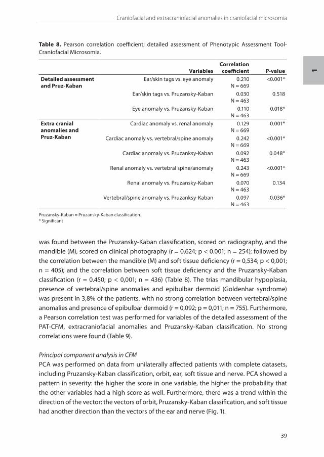
Table 8. Pearson correlation coefficient; detailed assessment of Phenotypic Assessment Tool-Craniofacial Microsomia.
Variables Correlation
coefficient P-valueDetailed assessment and Pruz-Kaban
Ear/skin tags vs. eye anomaly 0.210N = 669
<0.001*
Ear/skin tags vs. Pruzansky-Kaban 0.030N = 463
0.518
Eye anomaly vs. Pruzansky-Kaban 0.110N = 463
0.018*
Extra cranial anomalies and Pruz-Kaban
Cardiac anomaly vs. renal anomaly 0.129N = 669
0.001*
Cardiac anomaly vs. vertebral/spine anomaly 0.242N = 669
<0.001*
Cardiac anomaly vs. Pruzanksy-Kaban 0.092N = 463
0.048*
Renal anomaly vs. vertebral spine/anomaly 0.243N = 669
<0.001*
Renal anomaly vs. Pruzansky-Kaban 0.070N = 463
0.134
Vertebral/spine anomaly vs. Pruzanksy-Kaban 0.097N = 463
0.036*
Pruzansky-Kaban = Pruzansky-Kaban classification.* Significant
was found between the Pruzansky-Kaban classification, scored on radiography, and the mandible (M), scored on clinical photography (r = 0,624; p < 0.001; n = 254); followed by the correlation between the mandible (M) and soft tissue deficiency (r = 0,534; p < 0,001; n = 405); and the correlation between soft tissue deficiency and the Pruzansky-Kaban classification (r = 0.450; p < 0,001; n = 436) (Table 8). The trias mandibular hypoplasia, presence of vertebral/spine anomalies and epibulbar dermoid (Goldenhar syndrome) was present in 3,8% of the patients, with no strong correlation between vertebral/spine anomalies and presence of epibulbar dermoid (r = 0,092; p = 0,011; n = 755). Furthermore, a Pearson correlation test was performed for variables of the detailed assessment of the PAT-CFM, extracraniofacial anomalies and Pruzansky-Kaban classification. No strong correlations were found (Table 9).
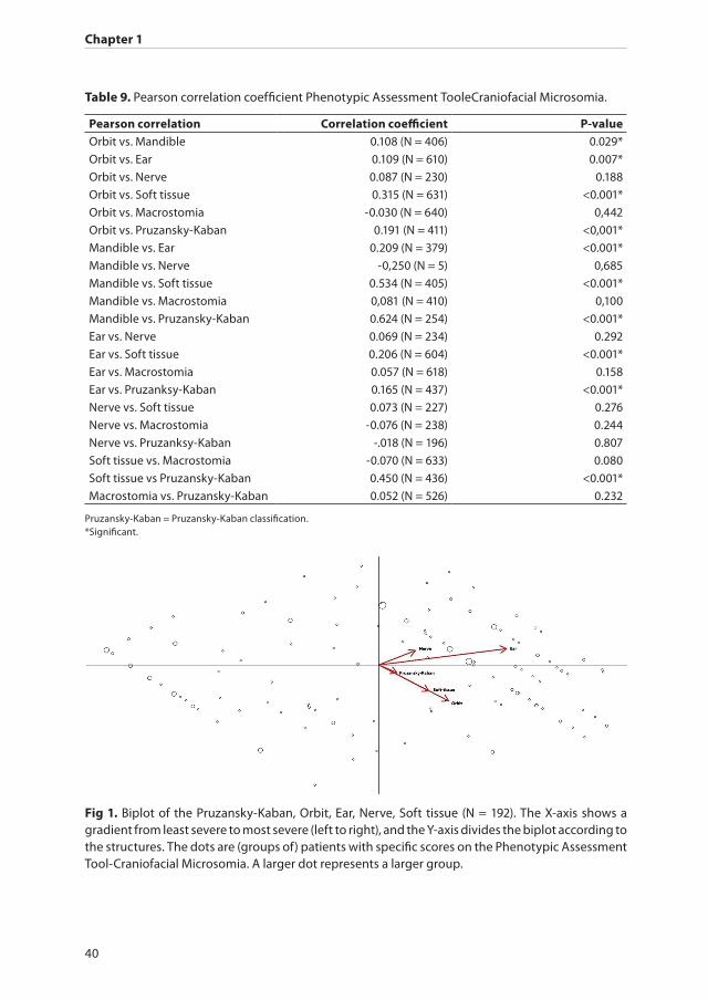
Principal component analysis in CFMPCA was performed on data from unilaterally affected patients with complete datasets, including Pruzansky-Kaban classification, orbit, ear, soft tissue and nerve. PCA showed a pattern in severity: the higher the score in one variable, the higher the probability that the other variables had a high score as well. Furthermore, there was a trend within the direction of the vector: the vectors of orbit, Pruzansky-Kaban classification, and soft tissue had another direction than the vectors of the ear and nerve (Fig. 1).
PSM 20190527 Proefschrift Britt Pluijmers.indd 39 24-07-19 15:04
Chapter 1
40
Table 9. Pearson correlation coefficient Phenotypic Assessment TooleCraniofacial Microsomia.
Pearson correlation Correlation coefficient P-valueOrbit vs. Mandible 0.108 (N = 406) 0.029*
Orbit vs. Ear 0.109 (N = 610) 0.007*
Orbit vs. Nerve 0.087 (N = 230) 0.188
Orbit vs. Soft tissue 0.315 (N = 631) <0.001*
Orbit vs. Macrostomia -0.030 (N = 640) 0,442
Orbit vs. Pruzansky-Kaban 0.191 (N = 411) <0,001*
Mandible vs. Ear 0.209 (N = 379) <0.001*
Mandible vs. Nerve -0,250 (N = 5) 0,685
Mandible vs. Soft tissue 0.534 (N = 405) <0.001*
Mandible vs. Macrostomia 0,081 (N = 410) 0,100
Mandible vs. Pruzansky-Kaban 0.624 (N = 254) <0.001*
Ear vs. Nerve 0.069 (N = 234) 0.292
Ear vs. Soft tissue 0.206 (N = 604) <0.001*
Ear vs. Macrostomia 0.057 (N = 618) 0.158
Ear vs. Pruzanksy-Kaban 0.165 (N = 437) <0.001*
Nerve vs. Soft tissue 0.073 (N = 227) 0.276
Nerve vs. Macrostomia -0.076 (N = 238) 0.244
Nerve vs. Pruzanksy-Kaban -.018 (N = 196) 0.807
Soft tissue vs. Macrostomia -0.070 (N = 633) 0.080
Soft tissue vs Pruzansky-Kaban 0.450 (N = 436) <0.001*
Macrostomia vs. Pruzansky-Kaban 0.052 (N = 526) 0.232
Pruzansky-Kaban = Pruzansky-Kaban classification.*Significant.
Fig 1. Biplot of the Pruzansky-Kaban, Orbit, Ear, Nerve, Soft tissue (N = 192). The X-axis shows a gradient from least severe to most severe (left to right), and the Y-axis divides the biplot according to the structures. The dots are (groups of) patients with specific scores on the Phenotypic Assessment Tool-Craniofacial Microsomia. A larger dot represents a larger group.
PSM 20190527 Proefschrift Britt Pluijmers.indd 40 24-07-19 15:04
Craniofacial and extracraniofacial anomalies in craniofacial microsomia
41
1
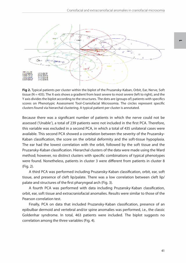
Because there was a significant number of patients in which the nerve could not be assessed (‘Unable’), a total of 239 patients were not included in the first PCA. Therefore, this variable was excluded in a second PCA, in which a total of 435 unilateral cases were available. This second PCA showed a correlation between the severity of the Pruzansky-Kaban classification, the score on the orbital deformity and the soft-tissue hypoplasia. The ear had the lowest correlation with the orbit, followed by the soft tissue and the Pruzansky-Kaban classification. Hierarchal clusters of the data were made using the Ward method; however, no distinct clusters with specific combinations of typical phenotypes were found. Nonetheless, patients in cluster 3 were different from patients in cluster 8 (Fig. 2).
A third PCA was performed including Pruzansky-Kaban classification, orbit, ear, soft tissue, and presence of cleft lip/palate. There was a low correlation between cleft lip/palate and structures of the first pharyngeal arch (Fig. 3).
A fourth PCA was performed with data including Pruzansky-Kaban classification, orbit, ear, soft tissue and extracraniofacial anomalies. Results were similar to those of the Pearson correlation test.
Finally, PCA on data that included Pruzansky-Kaban classification, presence of an epibulbar dermoid and vertebral and/or spine anomalies was performed, i.e., the classic Goldenhar syndrome. In total, 463 patients were included. The biplot suggests no correlation among the three variables (Fig. 4).
Fig 2. Typical patients per cluster within the biplot of the Pruzansky-Kaban, Orbit, Ear, Nerve, Soft tissue (N = 435). The X-axis shows a gradient from least severe to most severe (left to right), and the Y axis divides the biplot according to the structures. The dots are (groups of) patients with specifics scores on Phenotypic Assessment Tool-Craniofacial Microsomia. The circles represent specific clusters found via hierarchal clustering. A typical patient per cluster is annotated.
PSM 20190527 Proefschrift Britt Pluijmers.indd 41 24-07-19 15:04

Chapter 1
42
Fig. 4. Close-up of the biplot of the Pruzansky-Kaban, presence of epibulbar dermoid and vertebral/spine anomalies, i.e., Goldenhar syndrome (N = 463). The X-axis shows a gradient from least severe to most severe (left to right), and the Y-axis divides the biplot according to the structures. The dots are (groups of) patients with specific scores on the Pruzansky-Kaban classification and presence of epibulbar dermoid and vertebral/spine anomalies.
Fig 3. Close-up of the biplot of the Pruzansky-Kaban, Orbit, Ear, Soft-tissue and presence of cleft lip/palate (N = 435). The X-axis shows a gradient from least severe to most severe (left to right), and the Y-axis divides the biplot according to the structures. The dots are (groups of) patients with specifics scores on the Phenotypic Assessment Tool-Craniofacial Microsomia.
Discussion
Study populationBy combining the datasets of three major craniofacial units, it was possible to study 755 patients with CFM. In this study, patients were diagnosed solely with bilateral CFM when radiographic images showed bilateral mandibular hypoplasia. Diagnosis of bilateral CFM
PSM 20190527 Proefschrift Britt Pluijmers.indd 42 24-07-19 15:04

Craniofacial and extracraniofacial anomalies in craniofacial microsomia
43
1
was not influenced by external facial aspects, such as presence of ear and/or skin tags on both sides. In the literature, 2,5%-34% of patients with CFM are diagnosed with bilateral CFM. This wide range might be the result of selection bias or use of different selection criteria.28 In this study, 12% of the patients were diagnosed with bilateral CFM, which is slightly lower than the 13,6% (n = 977) found in the meta-analysis of Xu et al.29 A male-to- female ratio was found in our study (1,2:1) that was similar to the ratio in the meta-analysis (1,09:1 n = 908). Earlier studies showed similar results with right-to-left ratios varying from 1,2:1 to 1,8:1. 9,17,22,23
PAT-CFM and extracraniofacial anomalies and their correlationsUnlike in other studies, the Pruzansky-Kaban classification was equally divided in our cohort, whereas other studies found higher numbers among patients with Type I and IIA. Possibly, this might be due to selection bias, as patients with the most severe cases are referred to specialized craniofacial centers (Table 10).
Although there was a positive correlation between the score of the mandible on clinical photography (M) of the PAT-CFM and the Pruzansky-Kaban classification, based on radiography (r = 0,624; p < 0.001; n = 254), there was no strong correlation between these variables, and thus these should not be considered as interchangeable components of the PAT-CFM.
Several studies have shown an association between the outcome of the PAT-CFM and the presence of extracraniofacial anomalies. Syndromologists consider an anomaly to be ‘associated’ if it occurs in 10%-15% of the patients.6,19 Hennekam et al. described that an association is a pattern of anomalies, of which at least two are morphologic, that occur together more often than would be expected by chance, and in which a causal relationship has not been identified.30 Extracraniofacial anomalies were diagnosed in 10,5% to 26,1% of the patients with CFM in this study (which is higher than the incidence of 0,001%-1% in live births in the ‘healthy’ population).31-33 Statistical analysis showed weak and insignificant correlations among the tested variables; therefore, the term ‘association’ should be abandoned and replaced with ‘correlation’ when statistical analysis shows significant findings. Hennekam et al. state that the term ‘association’ is not durable but might be useful to motivate clinicians to evaluate patients for other, related anomalies.30
This study found a that structures derived from the first pharyngeal arch are correlated with degree of severity, as are the structures derived from the second pharyngeal arch. These results support the findings by Tuin et al., which reinforces the suggestion that the aetiology involves a disturbed migration of the (cranial) neural crest cells.9
Patients diagnosed with an extracraniofacial anomaly have a higher chance of having coexisting extracraniofacial anomalies in other organ systems, as noted by Rollnick and Kaye28, suggesting a similar pathogenesis of these anomalies.
‘Goldenhar syndrome’ is often applied to patients with mandibular hypoplasia, epibulbar dermoid and vertebral/spine anomalies; it is regarded by some as a variant
PSM 20190527 Proefschrift Britt Pluijmers.indd 43 24-07-19 15:04

Chapter 1
44
and is estimated to represent 10% of the patients with CFM.28 In this study, this trias was diagnosed only in 3,8% of the patients. There was a very weak positive correlation among the three variables. Analysis of statistical correlations in other studies also failed to substantiate a ‘Goldenhar’ variant as a distinct entity.9,17 The term ‘Goldenhar syndrome’ should therefore be discarded.
It was not possible to identify specific groups of patients with PCA, as all clusters overlapped with at least one other cluster, suggesting that CFM is a continuum of anomalies that coexist in all combinations and degrees of severity. However, many differences were found between patients affected unilaterally and bilaterally. We suggest that patients with unilateral or bilateral CFM should be approached more comprehensively.
Patients with bilateral CFM tend to be at the severe end of the spectrum and are also more often diagnosed with extracraniofacial anomalies and/or cleft lip/palate. These results might be explained by the embryogenesis and the default migration of (cranial) neural crest cells.
Table 10. Extended version of the table used in Park et al.22
Study Vento et al. Poon et al. Park et al. Tuin et al.
Caron, Pluijmers
et al.Total n of patients 154 65 100 105 755Orbit (%)
O0O1O2O3O4
814
150--
7712110--
532222
3--
721010
8--
55161412
3
P-K classification(%)M0M1M2aM2bM3
1140221710
930272311
0592118
2
1236191419
026,226,624,522,6
Ear (%)E0E1E2E3E4
34141933--
19342720--
17122348--
12181357--
17141353
2
Nerve (%)N0N1N2N3
538
1920
768
115
7946
11
617
266
(n = 283)56142010
Soft tissue (%)S0S1S2S3
55828
9
284523
4
24521410
234127
9
184330
9
P-K classification = Pruzansky-Kaban classification
PSM 20190527 Proefschrift Britt Pluijmers.indd 44 24-07-19 15:04
Craniofacial and extracraniofacial anomalies in craniofacial microsomia
45
1
Conclusion
A large cohort of patients with CFM is presented. Of 955 patients, data on 755 patients were available for in-depth analysis. The demographics showed outcomes similar to those of other cohorts. Using our strict criteria, 12% of the patients were affected bilaterally. Statistical analyses showed that the structures derived from the first pharyngeal arch correlated more with one another than with the structures derived from the second pharyngeal arch, and vice versa.
Extracraniofacial anomalies were positively, although not strongly, correlated with CFM. Further research is needed to determine a possible correlation is the pathogenesis.
Although phenotypically no specific groups of patients could be identified, patients with bilateral CFM were more severely affected than patients with unilateral CFM. Therefore, these bilaterally affected patients should be approached more comprehensively. Finally, even patients with a minor clinical presentation should be screened for extracraniofacial anomalies, including cardiac, renal, spinal and vertebral deformities.
Acknowledgements
The authors acknowledge M. Gribova, Y. Klazen, L. van de Lande, B. Maas, R. Renkema, and S. Schaal for their assistance with the databases at Great Ormond Street Hospital, London, UK, and Boston Children’s Hospital, Boston, Massachusetts, USA. This research did not receive any specific grant from funding agencies in the public, commercial, or not-for-profit sectors
PSM 20190527 Proefschrift Britt Pluijmers.indd 45 24-07-19 15:04
Chapter 1
46
References
1. Grabb WC. The first and second branchial arch syndrome. Plast Reconstr Surg 1965; 36(5): 485-508.2. Poswillo D. The aetiology and pathogenesis of craniofacial deformity. Development 1988; 103
Suppl: 207-12.3. M. G. Associations malformatives de l’oeil et de l’oreille : en particulier le syndrome dermoïde
épibulbaire-appendices auriculaires-fistula auris congenita et ses relations avec la dysostose mandibulo-faciale. J of Genet Hum 1952; (1): 243-82.
4. Francois J, Haustrate L. [Colobomatous anomalies of the eye and first arch syndrome] Anomalies colobomateuses du globe oculaire et syndrome du premier arc. Ann Ocul (Paris) 1954; 187(4): 340-68.
5. Stark RB, Saunders DE. The first branchial syndrome. The oral-mandibular-auricular syndrome. Plast Reconstr Surg Transplant Bull 1962; 29: 229-39.
6. Gorlin RJ, Jue KL, Jacobsen U, Goldschmidt E. Oculoauriculovertebral Dysplasia. J Pediatr 1963; 63: 991-9.
7. Poswillo D. Hemorrhage in development of the face. Birth Defects Orig Artic Ser 1975; 11(7): 61-81.8. Johnston MC, Bronsky PT. Prenatal craniofacial development: new insights on normal and abnormal
mechanisms. Crit Rev Oral Biol Med 1995; 6(4): 368-422.9. Tuin AJ, Tahiri Y, Paine KM, Paliga JT, Taylor JA, Bartlett SP. Clarifying the relationships among the
different features of the OMENS+ classification in craniofacial microsomia. Plast Reconstr Surg 2015; 135(1): 149e-56e.
10. Converse JM, Coccaro PJ, Becker M, Wood-Smith D. On hemifacial microsomia. The first and second branchial arch syndrome. Plast Reconstr Surg 1973; 51(3): 268-79.
11. Moore KL. The developing human, clinically oriented embryology. 5th ed. Philadelphia; 2011.12. Ross RB. Lateral facial dysplasia (first and second branchial arch syndrome, hemifacial microsomia).
Birth Defects Orig Artic Ser 1975; 11(7): 51-9.13. Posnick JC, Tiwana PS, Costello BJ. Treacher Collins syndrome: comprehensive evaluation and
treatment. Oral Maxillofac Surg Clin North Am 2004; 16(4): 503-23.14. Ongkosuwito EM, van Neck JW, Wattel E, van Adrichem LN, Kuijpers-Jagtman AM. Craniofacial
morphology in unilateral hemifacial microsomia. Br J Oral Maxillofac Surg 2013; 51(8): 902-7.15. S. P. Not all dwarfed mandibles are alike. Birth Defects Orig Artic Ser 1969; 5: 120-9.16. Kaban LB, Moses MH, Mulliken JB. Correction of hemifacial microsomia in the growing child: a
follow-up study. Cleft Palate J 1986; 23 Suppl 1: 50-2.17. Vento AR, LaBrie RA, Mulliken JB. The O.M.E.N.S. classification of hemifacial microsomia. Cleft Palate
Craniofac J 1991; 28(1): 68-76; discussion 7.18. Heike CL, Luquetti DV, Hing AV. Craniofacial Microsomia Overview. 1993.19. Horgan JE, Padwa BL, LaBrie RA, Mulliken JB. OMENS-Plus: analysis of craniofacial and extracraniofacial
anomalies in hemifacial microsomia. Cleft Palate Craniofac J 1995; 32(5): 405-12.20. Birgfeld CB, Luquetti DV, Gougoutas AJ, et al. A phenotypic assessment tool for craniofacial
microsomia. Plast Reconstr Surg 2011; 127(1): 313-20.21. Barisic I, Odak L, Loane M, et al. Prevalence, prenatal diagnosis and clinical features of oculo-
auriculo-vertebral spectrum: a registry-based study in Europe. Eur J Hum Genet 2014; 22(8): 1026-33.22. Park JU, Do TH, Kwon GY, Choi TH, Kim S. Statistical analysis using the OMENS classification in
Oriental patients with hemifacial microsomia: a comparative analysis with Western centers. Ann Plast Surg 2014; 72(1): 50-5.
23. Poon CC, Meara JG, Heggie AA. Hemifacial microsomia: use of the OMENS-Plus classification at the Royal Children’s Hospital of Melbourne. Plast Reconstr Surg 2003; 111(3): 1011-8.
24. Rollnick BR, Kaye CI, Nagatoshi K, Hauck W, Martin AO. Oculoauriculovertebral dysplasia and variants: phenotypic characteristics of 294 patients. Am J Med Genet 1987; 26(2): 361-75.
25. Jolliffe. Principal component analysis. second edition ed. New York: Springer; 2002.26. Nieuwboer A, De Weerdt W, Dom R, Lesaffre E. A frequency and correlation analysis of motor deficits
in Parkinson patients. Disabil Rehabil 1998; 20(4): 142-50.
PSM 20190527 Proefschrift Britt Pluijmers.indd 46 24-07-19 15:04

Craniofacial and extracraniofacial anomalies in craniofacial microsomia
47
1
27. Tongeren RJCtBOv. Data analysis in community and landscape ecology. 1 ed. Wageningen: Centre for Agricultural Publishing and Documentation; 1987.
28. Rollnick BR, Kaye CI. Hemifacial microsomia and variants: pedigree data. Am J Med Genet 1983; 15(2): 233-53.
29. Xu S, Zhang Z, Tang X, Yin L, Liu W, Shi L. The influence of gender and laterality on the incidence of hemifacial microsomia. J Craniofac Surg 2015; 26(2): 384-7.
30. Hennekam RC, Biesecker LG, Allanson JE, et al. Elements of Morphology: General Terms for Congenital Anomalies. Am J Med Genet A 2013; 161(11): 2726-33.
31. Alexander PG, Tuan RS. Role of environmental factors in axial skeletal dysmorphogenesis. Birth Defects Res C Embryo Today 2010; 90(2): 118-32.
32. Hoffman JI. Congenital heart disease: incidence and inheritance. Pediatr Clin North Am 1990; 37(1): 25-43.
33. Wiesel A, Queisser-Luft A, Clementi M, Bianca S, Stoll C, Group ES. Prenatal detection of congenital renal malformations by fetal ultrasonographic examination: an analysis of 709,030 births in 12 European countries. Eur J Med Genet 2005; 48(2): 131-44.
PSM 20190527 Proefschrift Britt Pluijmers.indd 47 24-07-19 15:04

PAR
TIPSM 20190527 Proefschrift Britt Pluijmers.indd 48 24-07-19 15:04

III
Shape
PSM 20190527 Proefschrift Britt Pluijmers.indd 49 24-07-19 15:04

CH
AP
TER
CH
AP
TER
PSM 20190527 Proefschrift Britt Pluijmers.indd 50 24-07-19 15:04

2
Characterizing the skull base in craniofacial microsomia using principal
component analysis
Sontje C. Schaal, Cliff Ruff, Britt I. Pluijmers, Erwin Pauws, Caspar W. N. Looman, Maarten J. Koudstaal, David J. Dunaway.
International Journal of Oral and Maxillofacial Surgery. 2017; 46: 1656–1663 http://dx.doi.org/10.1016/j.ijom.2017.07.008, Epub 2017 Jul 31.
PSM 20190527 Proefschrift Britt Pluijmers.indd 51 24-07-19 15:04

Chapter 2
52
ABSTRACT
AimsThe aim of this study was to compare the anatomical differences in the skull base between the affected and non-affected side in patients with craniofacial microsomia (CFM), and to compare the affected and non-affected sides with measurements from a normal population.
Material and Methods Three-dimensional computed tomography scans of 13 patients with unilateral CFM and 19 normal patients (age range 7–12 years) were marked manually with reliable homologous landmarks. Principal component analysis (PCA), as part of a point distribution model (PDM), was used to analyse the variability within the normal and preoperative CFM patient groups.
ResultsThrough analysis of the differences in the principal components calculated for the two groups, a model was created to describe the differences between CFM patients and normal age-matched controls. The PDMs were also used to describe the shape changes in the skull base between the cohorts and validated this model. Using thin- plate splines as a means of interpolation, videos were created to visualize the transformation from CFM skull to normal skull, and to display the variability in shape changes within the groups themselves.
ConclusionIn CFM cases, the skull base showed significant asymmetry. Anatomical areas around the glenoid fossa and mastoid process showed the most asymmetry and restriction of growth, suggesting a pathology involving the first and second pharyngeal arches.
PSM 20190527 Proefschrift Britt Pluijmers.indd 52 24-07-19 15:04

Characterizing the skull base using PCA
53
2
Introduction
Craniofacial microsomia (CFM) is the second most common congenital craniofacial anomaly after cleft lip and palate1. The prevalence is between 1 in 3000 and 1 in 5000 live births.2–4 CFM has a heterogeneous presentation, mainly characterized by hypoplasia in the auricular, mandibular, and maxillary anatomical regions.5–8 Many of the clinical features involve structures that arise from the first and second pharyngeal arches, thus involvement of the adjacent anatomical structures might also occur within this congenital craniofacial condition.4,9
The aetiology or underlying cause of CFM remains a subject of discussion in the literature. The different theories include a sporadic event, disturbed migration of the cranial neural crest cells, and a hereditary role in genetics.10–14 Another hypothesis is stape-dial artery disruption causing ischaemic necrosis to the anatomical features in the first and second pharyngeal arches.15
The varied phenotypic presentations of CFM may be due to the wide variety of structures that arise from the first and second pharyngeal arches.16–19 The Pruzansky–Kaban classification is the most commonly used classification system to describe mandibular deformity in CFM and was used in this study.20,21 The skull base has a close relationship with the facial skeleton, and the morphology of the skull base has an influence on facial asymmetry.22,23 CFM is mainly characterized by the facial asymmetry, and thus far only one study has evaluated the skull base.24 This previous study found that the skull base axis was not deviated compared to those of age-matched controls and that there was little difference in morphological measurements with increasing severity of CFM.24
The data contained within conventional three-dimensional computed tomography (3D-CT) scans can be utilized in mathematical techniques such as geometric morpho-metrics to analyse complex shapes. In this study, principal component analysis (PCA) was performed using manually landmarked 3D-CT scans to identify the global complex shape of different skulls. Using this analysis, the differences between the affected and unaffected populations could be visualized and described. This technique has been used successfully in the analysis of Apert syndrome and Crouzon–Pfeiffer syndrome.25–27
The aim of this study was to determine the anatomical differences in the skull base between the affected and non-affected sides in patients with CFM, and to determine the differences between the CFM patients and the normal population.
PSM 20190527 Proefschrift Britt Pluijmers.indd 53 24-07-19 15:04
Chapter 2
54
Materials and methods
Data collectionPatients diagnosed with unilateral CFM with suitable preoperative 3D-CT scans available, aged between 7 and 12 years, and without any history of craniofacial skeletal surgery were included. Patients were classified with the Pruzansky–Kaban system, and those with types 1–2B were included (Tables 1 and 2).
Patients classified as Pruzansky–Kaban type 3 were excluded since essential anatomical features are missing, making them inappropriate for the study analysis. Patients with bilateral CFM were also excluded, since the affected sides would nullify each other during the analysis. After applying the eligibility criteria, a total of 13 patients with unilateral CFM (eight right-sided and five left-sided CFM) who had preoperative 3D-CT scans were available for analysis. The control group comprised patients with an unaffected craniofacial skeleton, aged between 7 and 12 years (Table 3).
Nineteen normal patients were included in the study as the control group.
The 3D-CT scans were taken at Great Ormond Street Hospital using a 16-slice Siemens Somatom Sensation spiral CT scanner set to 0.75 collimation (Siemens Medical Solutions, Malvern, PA, USA) and at Erasmus MC using a 6-slice Siemens spiral CT scanner (Emotion 6; Siemens, Munich, Germany), with a fixed slice thickness of 0.8 mm. All scans were saved as DICOM files (Digital Imaging and Communications in Medicine) and were converted into a University College London (UCL) proprietary format. The files were loaded into 3D voxel-imaging software (Robin’s 3D; Robin Richards 2013). For all 3D-CT scans, a Hounsfield unit range of between 223 HU and 431 HU was chosen as the threshold for data imaging of the bony tissue surface. The mandible and the top of the cranium were separated and segmented off from the rest of the craniofacial skeleton to facilitate the accurate placement of the landmarks on the skull base surface.
LandmarksAn accurate and reliable set of homologous landmarks had to be identified for the comparison of the normal and CFM patient scans. To increase the reliability and repeatability of the landmarks, they were located at anatomical points of the skull base. An iterative process was used to test different landmark sets and to determine which distribution of landmarks best de- scribed the morphology of the skull base in normal and CFM patients. The landmarks were mainly placed around the anterior and middle skull base, as these areas are of surgical interest and are expected to be affected in CFM cases (Figs 1 and 2). Thus, a smaller number of landmarks were located on the posterior skull base.
PSM 20190527 Proefschrift Britt Pluijmers.indd 54 24-07-19 15:04

Characterizing the skull base using PCA
55
2
Table 1. The distribution of age, gender, disorder and affected side of the CFM population
Age in years Females Males CFM Right Left7 1 1 2 1 1
8 2 1 3 1 2
9 0 0 0 0 0
10 2 3 5 3 2
11 2 1 3 3 0
12 0 0 0 0 0
Total 7 6 13 8 5
CFM = Craniofacial Microsomia
Table 2. The distribution in age and Pruzansky-Kaban classification of the CFM population
Age in years 1 2A 2B Total7 0 2 0 2
8 0 3 0 3
9 0 0 0 0
10 1 1 3 5
11 0 1 2 3
12 0 0 0 0
Total 1 7 5 13
CFM = Craniofacial microsomia
Table 3. Distribution of age and gender in the normal population
Age in years Females Males Total7 2 0 2
8 1 1 2
9 6 2 8
10 3 0 3
11 1 1 2
12 2 0 2
Total 15 4 19
It was important that the set of landmarks used captured all key shape features of the skull base.
The landmark set used was developed and validated using thin-plate spline (TPS) warps and a visualization technique using false colours to represent differences between two skull shapes. A random normal scan was chosen and TPS were warped to the landmark coordinates of another randomly chosen normal target scan. This process brought the two sets of landmarks into alignment. TPS were used to interpolate between the landmarks in the warping process. Colour-coded images were then generated to show the remaining differences between the two scans, with the colour at each point on the image representing the distance of that point on the scan to the closest point on the
PSM 20190527 Proefschrift Britt Pluijmers.indd 55 24-07-19 15:04

Chapter 2
56
target scan (Fig. 3). If the landmarks were sufficiently distributed to capture the surface detail between landmarks, little to no difference would appear on the colour maps, and areas that were poorly described by the chosen landmarks would show up as different colours. For the CFM population, additional TPS warps were made to visualize colour maps. This process was repeated on the CFM scans to ensure that the chosen landmarks also described the CFM population, taking into account any further shape differences introduced by the anomaly (Fig. 4).
Fig 1. The set of 51 landmarks placed on a normal skull, in caudal and cranial view.
Fig 2. The set of 51 landmarks placed on a CFM skull, in caudal and cranial view.
PSM 20190527 Proefschrift Britt Pluijmers.indd 56 24-07-19 15:04

Characterizing the skull base using PCA
57
2
The final set used consisted of 51 homologous landmarks that were located on all normal and CFM scans (table 4).
Fig. 3. Colour-coded map of the warped normal predicted skull superimposed onto its actual counterpart (10-mm range). Cranial and caudal views are shown. The green and light blue areas indicate sufficient anatomical correspondence between the two scans.
Fig. 4. Colour-coded map of the warped CFM predicted skull superimposed onto its actual counterpart (10-mm range). Cranial and caudal views are shown. The green and light blue areas indicate sufficient anatomical correspondence between the two scans with the chosen landmarks.
PSM 20190527 Proefschrift Britt Pluijmers.indd 57 24-07-19 15:04

Chapter 2
58
Tab
le 4
. The
set
of 5
1 la
ndm
arks
on
the
skul
l bas
e us
ed in
this
stu
dy.
Lab
elLa
nd
mar
kD
efin
itio
nIn
tra-
obse
rver
rel
iab
ility
for
each
lan
dm
ark
SD n
orm
alSD
CFM
AIn
cisi
ve fo
ssa
Mos
t pos
tero
-infe
rior p
oint
0.27
80.
388
BRi
ght g
reat
er p
alat
ine
fora
men
Mos
t ant
erio
r poi
nt o
f the
righ
t gre
ater
pal
atin
e fo
ram
en0.
425
0.33
1C
Left
gre
ater
pal
atin
e fo
ram
enM
ost a
nter
ior p
oint
of t
he le
ft g
reat
er p
alat
ine
fora
men
0.26
80.
356
DPo
ster
ior n
asal
spi
neM
ost p
oste
rior p
oint
of t
he p
oste
rior n
asal
spi
ne0.
520.
855
EPo
ster
ior b
orde
r of v
omer
Mos
t pos
terio
r poi
nt o
f the
pos
terio
r bor
der o
f vom
er0.
271
0.60
8F
Righ
t pte
rygo
id h
amul
usM
ost s
uper
ior p
oint
of t
he ri
ght p
tery
goid
ham
ulus
0.29
0.22
GLe
ft p
tery
goid
ham
ulus
Mos
t sup
erio
r poi
nt o
f lef
t pte
rygo
id h
amul
us0.
203
0.33
8H
Righ
t lat
eral
pte
rygo
id p
late
Mos
t sup
erio
r poi
nt o
f the
infe
rior p
art o
f the
righ
t lat
eral
pte
rygo
id p
late
0.33
40.
874
ILe
ft la
tera
l pte
rygo
id p
late
Mos
t sup
erio
r poi
nt o
f the
infe
rior p
art o
f the
left
late
ral p
tery
goid
pla
te0.
584
1.8
JPh
aryn
geal
tub
ercl
eM
ost a
nter
omed
ial p
oint
(V-s
hap
e) o
f the
pha
ryng
eal t
uber
cle
0.99
70.
717
KRi
ght f
oram
en o
vale
Mos
t ant
erom
edia
l inf
erio
r poi
nt o
f the
righ
t for
amen
ova
le0.
558
0.26
8L
Left
fora
men
ova
leM
ost a
nter
omed
ial i
nfer
ior p
oint
of t
he le
ft fo
ram
en o
vale
0.61
90.
753
MRi
ght m
andi
bula
r fos
saTh
e m
iddl
e ce
ntre
poi
nt o
f the
righ
t man
dibu
lar f
ossa
0.83
60.
899
NLe
ft m
andi
bula
r fos
saTh
e m
iddl
e ce
ntre
poi
nt o
f the
left
man
dibu
lar f
ossa
1.01
41.
359
ORi
ght e
xter
nal a
cous
tic m
eatu
sTh
e ce
ntre
of t
he h
ighe
st p
oint
acc
ordi
ng to
the
Fran
kfor
t hor
izon
tal;
rig
ht p
orio
n0.
469
0.89
3
PLe
ft e
xter
nal a
cous
tic m
eatu
sTh
e ce
ntre
of t
he h
ighe
st p
oint
acc
ordi
ng to
the
Fran
kfor
t hor
izon
tal;
le
ft p
orio
n0.
381
2.33
2
QRi
ght m
asto
id p
roce
ssPo
int o
f max
imum
cur
vatu
re o
f the
righ
t mas
toid
pro
cess
1.33
10.
591
RLe
ft m
asto
id p
roce
ssPo
int o
f max
imum
cur
vatu
re o
f the
left
mas
toid
pro
cess
0.52
50.
547
SRi
ght v
agin
al p
roce
ss o
f the
ty
mpa
nic
por
tion;
tem
por
al b
one
The
mos
t sup
erio
r poi
nt o
f the
righ
t vag
inal
pro
cess
; ens
heat
hed
root
of
the
styl
oid
proc
ess
0.63
82.
248
TLe
ft v
agin
al p
roce
ss o
f the
ty
mpa
nic
por
tion;
tem
por
al b
one
The
mos
t sup
erio
r poi
nt o
f the
left
vag
inal
pro
cess
; ens
heat
hed
root
of
the
styl
oid
proc
ess
1.25
10.
5
URi
ght j
ugul
ar fo
ram
en/j
ugul
ar
proc
ess
Mos
t pos
tero
-sup
erio
r poi
nt o
f the
righ
t jug
ular
fora
men
0.42
30.
426
VLe
ft ju
gula
r for
amen
/jug
ular
pr
oces
sM
ost p
oste
ro-s
uper
ior p
oint
of t
he le
ft ju
gula
r for
amen
0.64
80.
408
WRi
ght c
arot
id c
anal
Mos
t ant
ero
-infe
rior p
oint
of t
he ri
ght c
arot
id c
anal
0.50
30.
824
PSM 20190527 Proefschrift Britt Pluijmers.indd 58 24-07-19 15:04
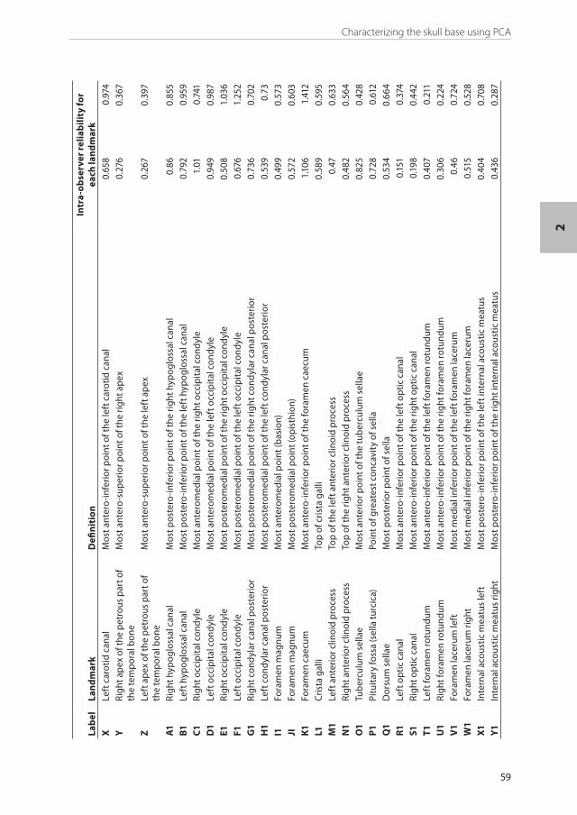
Characterizing the skull base using PCA
59
2
Lab
elLa
nd
mar
kD
efin
itio
nIn
tra-
obse
rver
rel
iab
ility
for
each
lan
dm
ark
XLe
ft c
arot
id c
anal
Mos
t ant
ero
-infe
rior p
oint
of t
he le
ft c
arot
id c
anal
0.65
80.
974
YRi
ght a
pex
of t
he p
etro
us p
art o
f th
e te
mp
oral
bon
eM
ost a
nter
o-s
uper
ior p
oint
of t
he ri
ght a
pex
0.27
60.
367
ZLe
ft a
pex
of t
he p
etro
us p
art o
f th
e te
mp
oral
bon
eM
ost a
nter
o-s
uper
ior p
oint
of t
he le
ft a
pex
0.26
70.
397
A1
Righ
t hyp
oglo
ssal
can
alM
ost p
oste
ro-in
ferio
r poi
nt o
f the
righ
t hyp
oglo
ssal
can
al0.
860.
855
B1Le
ft h
ypog
loss
al c
anal
Mos
t pos
tero
-infe
rior p
oint
of t
he le
ft h
ypog
loss
al c
anal
0.79
20.
959
C1Ri
ght o
ccip
ital
con
dyle
Mos
t ant
erom
edia
l poi
nt o
f the
righ
t occ
ipit
al c
ondy
le1.
010.
741
D1
Left
occ
ipit
al c
ondy
leM
ost a
nter
omed
ial p
oint
of t
he le
ft o
ccip
ital
con
dyle
0.94
90.
987
E1Ri
ght o
ccip
ital
con
dyle
Mos
t pos
tero
med
ial p
oint
of t
he ri
ght o
ccip
ital
con
dyle
0.50
81.
036
F1Le
ft o
ccip
ital
con
dyle
Mos
t pos
tero
med
ial p
oint
of t
he le
ft o
ccip
ital
con
dyle
0.67
61.
252
G1
Righ
t con
dyla
r can
al p
oste
rior
Mos
t pos
tero
med
ial p
oint
of t
he ri
ght c
ondy
lar c
anal
pos
terio
r0.
736
0.70
2H
1Le
ft c
ondy
lar c
anal
pos
terio
rM
ost p
oste
rom
edia
l poi
nt o
f the
left
con
dyla
r can
al p
oste
rior
0.53
90.
73I1
Fora
men
mag
num
Mos
t ant
erom
edia
l poi
nt (b
asio
n)0.
499
0.57
3JI
Fora
men
mag
num
Mos
t pos
tero
med
ial p
oint
(opi
sthi
on)
0.57
20.
603
K1Fo
ram
en c
aecu
mM
ost a
nter
o-in
ferio
r poi
nt o
f the
fora
men
cae
cum
1.10
61.
412
L1Cr
ista
gal
liTo
p of
cris
ta g
alli
0.58
90.
595
M1
Left
ant
erio
r clin
oid
proc
ess
Top
of th
e le
ft a
nter
ior c
linoi
d pr
oces
s0.
470.
633
N1
Righ
t ant
erio
r clin
oid
proc
ess
Top
of th
e rig
ht a
nter
ior c
linoi
d pr
oces
s0.
482
0.56
4O
1Tu
ber
culu
m s
ella
eM
ost a
nter
ior p
oint
of t
he tu
ber
culu
m s
ella
e0.
825
0.42
8P1
Pitu
itar
y fo
ssa
(sel
la tu
rcic
a)Po
int o
f gre
ates
t con
cavi
ty o
f sel
la0.
728
0.61
2Q
1D
orsu
m s
ella
eM
ost p
oste
rior p
oint
of s
ella
0.53
40.
664
R1Le
ft o
ptic
can
alM
ost a
nter
o-in
ferio
r poi
nt o
f the
left
opt
ic c
anal
0.15
10.
374
S1Ri
ght o
ptic
can
alM
ost a
nter
o-in
ferio
r poi
nt o
f the
righ
t opt
ic c
anal
0.19
80.
442
T1Le
ft fo
ram
en ro
tund
umM
ost a
nter
o-in
ferio
r poi
nt o
f the
left
fora
men
rotu
ndum
0.40
70.
211
U1
Righ
t for
amen
rotu
ndum
Mos
t ant
ero
-infe
rior p
oint
of t
he ri
ght f
oram
en ro
tund
um0.
306
0.22
4V
1Fo
ram
en la
ceru
m le
ftM
ost m
edia
l inf
erio
r poi
nt o
f the
left
fora
men
lace
rum
0.46
0.72
4W
1Fo
ram
en la
ceru
m ri
ght
Mos
t med
ial i
nfer
ior p
oint
of t
he ri
ght f
oram
en la
ceru
m0.
515
0.52
8X1
Inte
rnal
aco
ustic
mea
tus
left
Mos
t pos
tero
-infe
rior p
oint
of t
he le
ft in
tern
al a
cous
tic m
eatu
s0.
404
0.70
8Y1
Inte
rnal
aco
ustic
mea
tus
right
Mos
t pos
tero
-infe
rior p
oint
of t
he ri
ght i
nter
nal a
cous
tic m
eatu
s0.
436
0.28
7
PSM 20190527 Proefschrift Britt Pluijmers.indd 59 24-07-19 15:04

Chapter 2
60
Data analysisTo determine the repeatability of the landmarks, a normal skull and a CFM skull were chosen at random and landmarked 10 times with at least 48h between sessions to reduce memory bias. The mean and the standard deviation (SD) values were then calculated to demonstrate the reproducibility of the landmarking process; the results are shown in Table 4. For the normal and CFM bony tissue, a SD of <2 mm was defined as acceptable and a SD of <1 mm was defined as accurate.28,29
Left-sided CFM patients were mirrored to create a right-sided deformity to allow the analysis of the affected and unaffected sides within CFM as one uniform group. The mirroring of unilateral CFM was done under the assumption that the degree to which the sides (right and left) are affected is comparable. The landmark data were then analysed using a Point Distribution Model (PDM) software package. The PDM is a form of statistical shape or morphometric analysis whose function is to capture the variation seen in a group of related shapes statistically. It is a form of multivariate analysis that analyses the input or training shapes in a holistic manner by looking not just at how each point varies in isolation, but how each point on the shape co-varies with every other point. The PDM accomplishes this by representing each of the training shapes with a set of homologous landmarks, from which a mean shape is calculated. Each shape in the training set is then expressed in terms of its difference from this mean, and a table of how each point co-varies with respect to every other point is calculated for each shape and then summed over all of the shapes in the training set to form a covariance matrix, which represents how each landmark tends to vary in relation to every other landmark in the training set taken as a whole. 30,31
In this study, eigenvector analysis was applied to the covariance matrix to yield a set of eigenvectors and eigenvalues, in which each eigenvector represents a way, or direction, in which the landmarks tend to vary as a group, and the associated eigenvalue represents how much of this variation is present in the training set, or its variance. Each eigenvector can be thought of as a ‘mode of variation’ or way in which the overall shape varies within the training set. PCA can then be applied by ordering the eigenvectors, or modes of variation, in descending order of their eigenvalues, and retaining only the modes with the highest values, which represent the modes of variation that account for most of the variation seen in the training shapes. The final model consists of a mean shape and a set of modes (or principal components) of variation and their relative importance (the eigenvalues) in describing the variation seen in the training set. The modes of variation can be visualized by applying weighted amounts of the eigenvalues (i.e. 2±SD) of the eigenvectors to the mean shape and generating a video of the transformation between the shapes thus generated, using TPS as a means of interpolation between the landmarked points.
In summary, the method used was first to generate a set of homologous landmarks describing the areas of interest in the skull base, which were validated as sufficient for
PSM 20190527 Proefschrift Britt Pluijmers.indd 60 24-07-19 15:04

Characterizing the skull base using PCA
61
2
the task by warping and colour map comparisons (Figs 3 and 4). The resulting set of 51 landmarks was then considered to represent the shape of the important area of interest in the skull base as a whole, and the landmarks were then located on all of the normal and CFM scans. PDMs were then generated from these landmarks on the normal and CFM sets individually, and videos of the modes of variation produced were generated. Finally, a joint model was built in an attempt to cancel out the normal modes of variation from the CFM model to leave only the differences between the two training sets, and a video of the principal component of this difference applied to the mean shape of the normal was generated.
Linear measurementsAfter analysing the anatomical changes seen in the PDM model, linear measurements were made using Robins 3D. Additional landmarks were chosen based on anatomical and surgical interest, as well as those defined by Paliga et al.24, to measure the intermediate distances. The tuberculum sellae was chosen as the reference point for specific landmark measurements, as it has a central position in the skull base. Fourteen measurements were made on the affected and non-affected sides of the CFM and normal skull bases. Measurements were compared within and between these groups.Differences in the cranial base angle and cranial base length were tested statistically with a simple analysis of variance (ANOVA) test. To test the differences between the affected side of CFM patients, the unaffected side of CFM patients, and the normal cohort, a multi- level analysis was performed with the child as the random effect; the analysis between sides within the children was, therefore, performed using a paired t-test. By adding a normal cohort to the dataset, the dependencies within the cohort were accounted for. For all analyses, statistical significance was defined as a P-value of<0.05.
Results
All patients with CFM were clinically identified at Great Ormond Street Hospital, London, UK. The control group consisted of epileptic patients from Great Ormond Street Hospital and patients with other medical conditions scanned at Erasmus MC, Rotterdam, the Netherlands.
Landmark intra-observer reliabilityThe standard deviations were calculated for all 51 homologous landmarks (Table 4). All landmarks had an SD value below 2.4 mm.
In the normal cohort, five out of 51 landmarks had a SD between 1 mm and 1.4 mm. Forty-six landmarks had a SD threshold of <1 mm. The placement of the landmarks was highly accurate for 90%, and was within the 2 mm limit for 100%.
PSM 20190527 Proefschrift Britt Pluijmers.indd 61 24-07-19 15:04
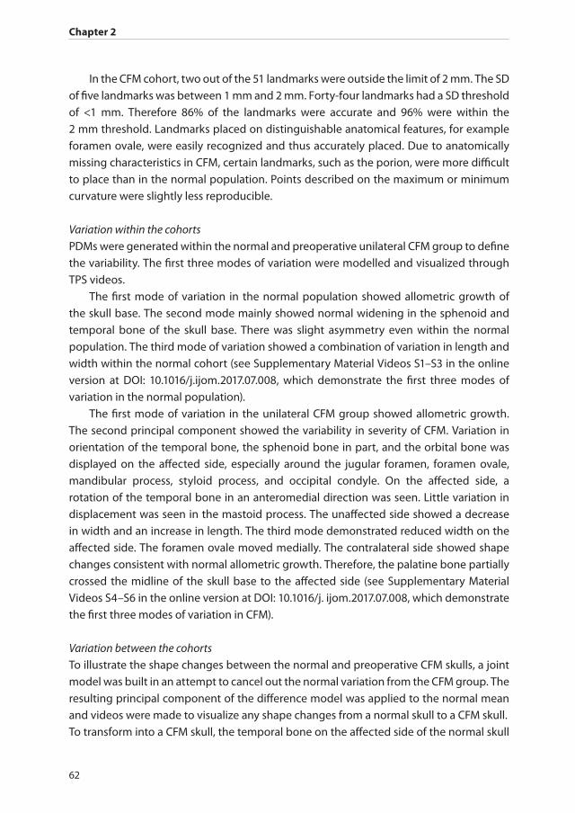
Chapter 2
62
In the CFM cohort, two out of the 51 landmarks were outside the limit of 2 mm. The SD of five landmarks was between 1 mm and 2 mm. Forty-four landmarks had a SD threshold of <1 mm. Therefore 86% of the landmarks were accurate and 96% were within the 2 mm threshold. Landmarks placed on distinguishable anatomical features, for example foramen ovale, were easily recognized and thus accurately placed. Due to anatomically missing characteristics in CFM, certain landmarks, such as the porion, were more difficult to place than in the normal population. Points described on the maximum or minimum curvature were slightly less reproducible.
Variation within the cohortsPDMs were generated within the normal and preoperative unilateral CFM group to define the variability. The first three modes of variation were modelled and visualized through TPS videos.
The first mode of variation in the normal population showed allometric growth of the skull base. The second mode mainly showed normal widening in the sphenoid and temporal bone of the skull base. There was slight asymmetry even within the normal population. The third mode of variation showed a combination of variation in length and width within the normal cohort (see Supplementary Material Videos S1–S3 in the online version at DOI: 10.1016/j.ijom.2017.07.008, which demonstrate the first three modes of variation in the normal population).
The first mode of variation in the unilateral CFM group showed allometric growth. The second principal component showed the variability in severity of CFM. Variation in orientation of the temporal bone, the sphenoid bone in part, and the orbital bone was displayed on the affected side, especially around the jugular foramen, foramen ovale, mandibular process, styloid process, and occipital condyle. On the affected side, a rotation of the temporal bone in an anteromedial direction was seen. Little variation in displacement was seen in the mastoid process. The unaffected side showed a decrease in width and an increase in length. The third mode demonstrated reduced width on the affected side. The foramen ovale moved medially. The contralateral side showed shape changes consistent with normal allometric growth. Therefore, the palatine bone partially crossed the midline of the skull base to the affected side (see Supplementary Material Videos S4–S6 in the online version at DOI: 10.1016/j. ijom.2017.07.008, which demonstrate the first three modes of variation in CFM).
Variation between the cohortsTo illustrate the shape changes between the normal and preoperative CFM skulls, a joint model was built in an attempt to cancel out the normal variation from the CFM group. The resulting principal component of the difference model was applied to the normal mean and videos were made to visualize any shape changes from a normal skull to a CFM skull.To transform into a CFM skull, the temporal bone on the affected side of the normal skull
PSM 20190527 Proefschrift Britt Pluijmers.indd 62 24-07-19 15:04

Characterizing the skull base using PCA
63
2
changed in medial direction and shortened in length. The mandibular fossa and mastoid process moved towards each other. There was also a medial and cranial displacement of the external acoustic meatus, styloid process, jugular foramen, and petrous part of the temporal bone. Posterolateral displacement of the maxilla and the palatine bone were shown. Overall, the midline of the skull base showed a slight rotation to the affected side. Thus, the relevant anatomical features on the temporal and sphenoid bone of the CFM skull moved closer together and the distance within became smaller than in a normal skull (see Supplementary Material Video S7 in the online version at DOI: 10.1016/j.ijom. 2017.07.008, which demonstrates the skull change from normal to CFM).
Linear measurementsNo significant difference was found between the CFM and normal patient cohorts in the cranial base angle, length of the anterior part of the cranial base, or total length of the cranial base (Tables 5 and 6).
Eight out of the 14 measurements varied significantly between the affected and unaffected side in the patients with CFM (Table 7). In the comparison between the affected side of CFM patients and the normal patients, 10 out of 14 landmark measurements showed a significant difference (Table 8). Comparison of the unaffected side of CFM patients to the normal cohort indicated no significant difference for 12 out of 14 measurements. Exceptions were the hypoglossal canal to the tuberculum sellae and the temporal bone to the tuberculum sellae, which varied significantly.
Table 5. Cranial base angle; mean ± standard deviation in degrees.
CFM (n=13) Normal (N=19) P-valueForamen caecum; tuberculum sellae and to opisthion
2,964 ± 1,855 2,675 ± 2,150 0,7
CFM = Craniofacial microsomia
Table 6. Cranial base length; mean ± standard deviation in millimetres.
CFM (n=13) Normal (N=19) P-valueForamen caecum to tuberculum sellae (anterior)
50,31 ± 1,996 40,07 ± 4,891 0,13
Tuberculum sellae to opisthion (posterior)
73,09 ± 3,137 76,38 ± 2,704 0,0035
Foramen caecum; tuberculum sellae and to opisthion
123,4 ± 3,072 124,5 ± 4,076 0,44
CFM = Craniofacial microsomia
PSM 20190527 Proefschrift Britt Pluijmers.indd 63 24-07-19 15:04
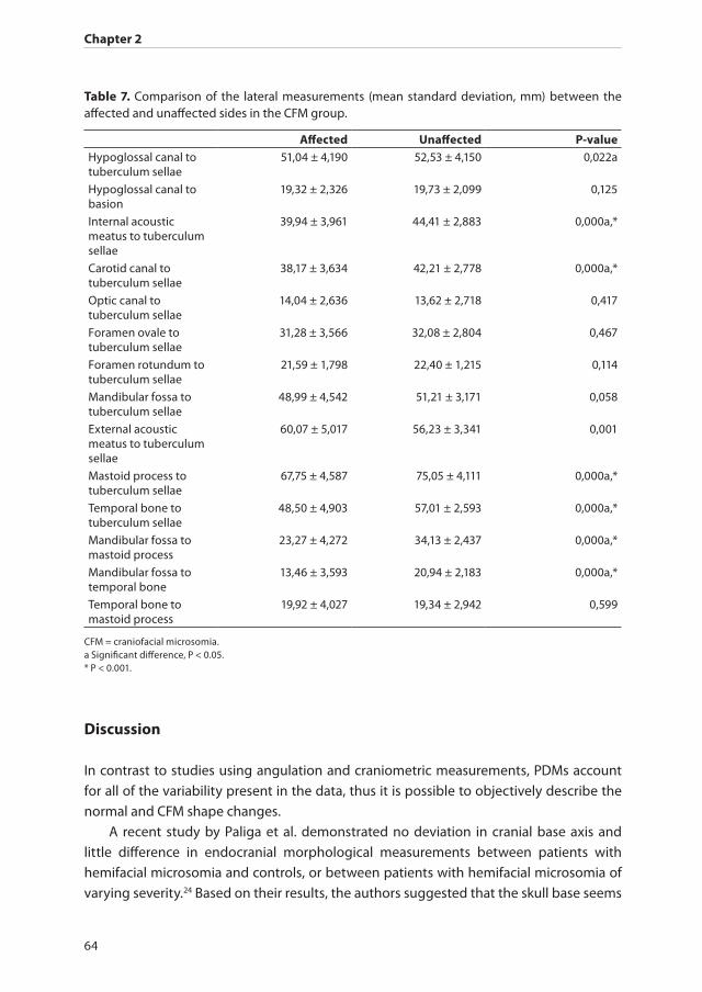
Chapter 2
64
Discussion
In contrast to studies using angulation and craniometric measurements, PDMs account for all of the variability present in the data, thus it is possible to objectively describe the normal and CFM shape changes.
A recent study by Paliga et al. demonstrated no deviation in cranial base axis and little difference in endocranial morphological measurements between patients with hemifacial microsomia and controls, or between patients with hemifacial microsomia of varying severity.24 Based on their results, the authors suggested that the skull base seems
Table 7. Comparison of the lateral measurements (mean standard deviation, mm) between the affected and unaffected sides in the CFM group.
Affected Unaffected P-valueHypoglossal canal to tuberculum sellae
51,04 ± 4,190 52,53 ± 4,150 0,022a
Hypoglossal canal to basion
19,32 ± 2,326 19,73 ± 2,099 0,125
Internal acoustic meatus to tuberculum sellae
39,94 ± 3,961 44,41 ± 2,883 0,000a,*
Carotid canal to tuberculum sellae
38,17 ± 3,634 42,21 ± 2,778 0,000a,*
Optic canal to tuberculum sellae
14,04 ± 2,636 13,62 ± 2,718 0,417
Foramen ovale to tuberculum sellae
31,28 ± 3,566 32,08 ± 2,804 0,467
Foramen rotundum to tuberculum sellae
21,59 ± 1,798 22,40 ± 1,215 0,114
Mandibular fossa to tuberculum sellae
48,99 ± 4,542 51,21 ± 3,171 0,058
External acoustic meatus to tuberculum sellae
60,07 ± 5,017 56,23 ± 3,341 0,001
Mastoid process to tuberculum sellae
67,75 ± 4,587 75,05 ± 4,111 0,000a,*
Temporal bone to tuberculum sellae
48,50 ± 4,903 57,01 ± 2,593 0,000a,*
Mandibular fossa to mastoid process
23,27 ± 4,272 34,13 ± 2,437 0,000a,*
Mandibular fossa to temporal bone
13,46 ± 3,593 20,94 ± 2,183 0,000a,*
Temporal bone to mastoid process
19,92 ± 4,027 19,34 ± 2,942 0,599
CFM = craniofacial microsomia.a Significant difference, P < 0.05.* P < 0.001.
PSM 20190527 Proefschrift Britt Pluijmers.indd 64 24-07-19 15:04

Characterizing the skull base using PCA
65
2
to be spared in CFM and that the results were in agreement with the pathophysiology of the stapedial artery insult hypothesis of Poswillo with regard to the restriction of abnormalities to derivatives of the first and second pharyngeal arches in this area.15,16,24. A significant part of the skull base is derived from derivatives of the first and second arches (squamous temporal bone, glenoid fossa, root of zygoma, spine of sphenoid, and styloid process). It would be surprising to find these structures unaffected if first and second arch involvement in the pathogenesis of CFM is correct. Closer examination of the landmark set used by Paliga et al. showed that none of the landmarks was placed on first and second arch derivatives, and so it is not surprising that their study showed no significant
Table 8. Comparison of the lateral measurements (mean ± standard deviation, mm) between the normal group and the affected side in the CFM group and between the normal group and the unaffected side in the CFM group.
Normal Affected P-value Unaffected P-valueHypoglossal canal to tuberculum sellae
55,01 ± 2,414 51,04 ± 4,190 0,002a 52,53 ± 4,150 0,039a
Hypoglossal canal to basion
20,61 ± 5,091 19,32 ± 2,326 0,355 19,73 ± 2,099 0,524
Internal acoustic meatus to tuberculum sellae
45,65 ± 2,565 39,94 ± 3,961 0,000a,* 44,41 ± 2,883 0,237
Carotid canal to tuberculum sellae
44,07 ± 2,262 38,17 ± 3,634 0,000a,* 42,21 ± 2,778 0,054
Optic canal to tuberculum sellae
13,7 ± 2,316 14,04 ± 2,636 0,696 13,62 ± 2,718 0,925
Foramen ovale to tuberculum sellae
32,97 ± 2,413 31,28 ± 3,566 0,081 32,08 ± 2,804 0,357
Foramen rotundum to tuberculum sellae
22,29 ± 2,018 21,59 ± 1,798 0,277 22,40 ± 1,215 0,862
Mandibular fossa to tuberculum sellae
51,73 ± 1,897 48,99 ± 4,542 0,009a 51,21 ± 3,171 0,598
External acoustic meatus to tuberculum sellae
56,53 ± 2,553 60,07 ± 5,017 0,005a 56,23 ± 3,341 0,797
Mastoid process to tuberculum sellae
75,83 ± 3,951 67,75 ± 4,587 0,000a,* 75,05 ± 4,111 0,59
Temporal bone to tuberculum sellae
60,01 ± 2,935 48,50 ± 4,903 0,000a,* 57,01 ± 2,593 0,012a
Mandibular fossa to mastoid process
33,03 ± 3,726 23,27 ± 4,272 0,000a,* 34,13 ± 2,437 0,381
Mandibular fossa to temporal bone
22,05 ± 2,634 13,46 ± 3,593 0,000a,* 20,94 ± 2,183 0,248
Temporal bone to mastoid process
17,88 ± 2,033 19,92 ± 4,027 0,034a 19,34 ± 2,942 0,126
CFM, craniofacial microsomia.a Significant difference, P < 0.05.* P < 0.001
PSM 20190527 Proefschrift Britt Pluijmers.indd 65 24-07-19 15:04
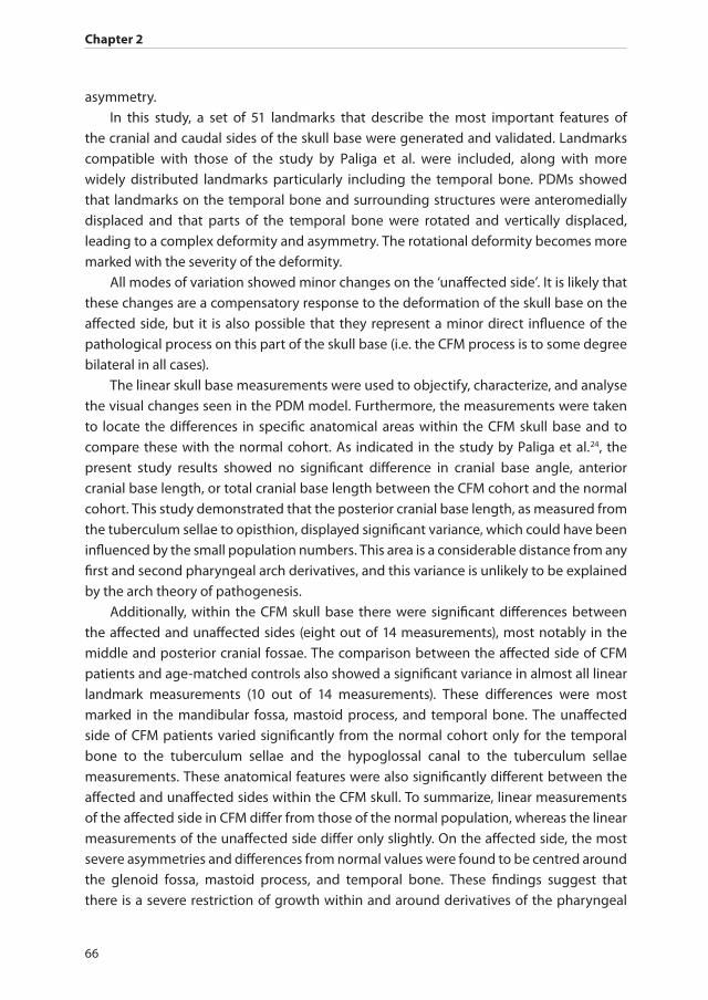
Chapter 2
66
asymmetry.In this study, a set of 51 landmarks that describe the most important features of
the cranial and caudal sides of the skull base were generated and validated. Landmarks compatible with those of the study by Paliga et al. were included, along with more widely distributed landmarks particularly including the temporal bone. PDMs showed that landmarks on the temporal bone and surrounding structures were anteromedially displaced and that parts of the temporal bone were rotated and vertically displaced, leading to a complex deformity and asymmetry. The rotational deformity becomes more marked with the severity of the deformity.
All modes of variation showed minor changes on the ‘unaffected side’. It is likely that these changes are a compensatory response to the deformation of the skull base on the affected side, but it is also possible that they represent a minor direct influence of the pathological process on this part of the skull base (i.e. the CFM process is to some degree bilateral in all cases).
The linear skull base measurements were used to objectify, characterize, and analyse the visual changes seen in the PDM model. Furthermore, the measurements were taken to locate the differences in specific anatomical areas within the CFM skull base and to compare these with the normal cohort. As indicated in the study by Paliga et al.24, the present study results showed no significant difference in cranial base angle, anterior cranial base length, or total cranial base length between the CFM cohort and the normal cohort. This study demonstrated that the posterior cranial base length, as measured from the tuberculum sellae to opisthion, displayed significant variance, which could have been influenced by the small population numbers. This area is a considerable distance from any first and second pharyngeal arch derivatives, and this variance is unlikely to be explained by the arch theory of pathogenesis.
Additionally, within the CFM skull base there were significant differences between the affected and unaffected sides (eight out of 14 measurements), most notably in the middle and posterior cranial fossae. The comparison between the affected side of CFM patients and age-matched controls also showed a significant variance in almost all linear landmark measurements (10 out of 14 measurements). These differences were most marked in the mandibular fossa, mastoid process, and temporal bone. The unaffected side of CFM patients varied significantly from the normal cohort only for the temporal bone to the tuberculum sellae and the hypoglossal canal to the tuberculum sellae measurements. These anatomical features were also significantly different between the affected and unaffected sides within the CFM skull. To summarize, linear measurements of the affected side in CFM differ from those of the normal population, whereas the linear measurements of the unaffected side differ only slightly. On the affected side, the most severe asymmetries and differences from normal values were found to be centred around the glenoid fossa, mastoid process, and temporal bone. These findings suggest that there is a severe restriction of growth within and around derivatives of the pharyngeal
PSM 20190527 Proefschrift Britt Pluijmers.indd 66 24-07-19 15:04
Characterizing the skull base using PCA
67
2
arches and it is likely that asymmetries seen elsewhere in the skull base are deformational changes in areas with normal growth ability but directly connected to the abnormal area. The linear measurements also imply that the skull base asymmetry can contribute to the facial asymmetry.
The results clearly show that the skull base is affected in CFM. Since the facial skeleton is in direct contact with the skull base, it is apparent that skull base asymmetry contributes to facial asymmetry in CFM. It is not possible to surgically correct many skull base asymmetries (e.g. the position of the temporomandibular joint or external auditory meatus), which implies that the actual asymmetry of CFM cannot be fully corrected and must therefore be masked by procedures on areas that can be surgically corrected.
A limitation of this study is the age range of the patients included: 7–12 years. Allometric growth is significant in this age range and this particularly affected the PDM analysis. Although PDM can help identify changes due to growth, many of the subtle anatomical differences caused by CFM may have been masked.
This study described significant asymmetry of the skull base in CFM. The most significant asymmetries and restrictions of growth were found to be centred around the glenoid fossa and mastoid process, suggesting involvement of the first and second pharyngeal arch derivatives in the pathogenesis. Distortion of the skull in this area is complex and is present in the vertical, horizontal, and antero-posterior planes, associated with a rotation of this part of the skull base. More minor abnormalities are present in other parts of the skull base, and are likely to be due to deformation resulting in abnormal growth in the region of the affected temporal bone.
PSM 20190527 Proefschrift Britt Pluijmers.indd 67 24-07-19 15:04

Chapter 2
68
References
1. Heike CL, Hing AV, Aspinall CA, Bartlett SP, Birgfeld CB, Drake AF, Pimenta LA, Sie KC, Urata MM, Vivaldi D, Luquetti DV. Clinical care in craniofacial microsomia: a review of current management recommen- dations and opportunities to advance re- search. Am J Med Genet Part C Semin Med Genet 2013;163C:271–82.
2. Poswillo D. The aetiology and pathogenesis of craniofacial deformity. Development 1988;103(Suppl):207–12.
3. Grabb WC. The first and second branchial arch syndrome. Plast Reconstr Surg 1965;36:485–508.4. Birgfeld C, Heike C. Craniofacial microso- mia. Semin Plast Surg 2012;26:91–104.5. Gougoutas AJ, Singh DJ, Low DW, Bartlett SP. Hemifacial macrosomia: clinical features
and pictographic representations of the OMENS classification system. Plast Reconstr Surg 2007;120:112e–120e.
6. Murray JE, Kaban LB, Mulliken JB. Analy- sis and treatment of hemifacial macrosomia. Plast Reconstr Surg 1984;74f:186–99.
7. Rune B, Selvik G, Sarna¨s KV, Jacobsson S. Growth in hemifacial macrosomia studied with the aid of roentgen stereophotogram- metry and metallic implants. Cleft Palate J 1981;18:128–46.
8. Ongkosuwito EM, Van Neck JW, Wattel E, Van Adrichem LN, Kuijpers-Jagtman AM. Craniofacial morphology in unilateral hemi- facial microsomia. Br J Oral Maxillofac Surg 2013;51:902–7.
9. Converse JM, Coccaro PJ, Becker M, Wood- Smith D. On hemifacial microsomia. The first and second branchial arch syndrome. Plast Reconstr Surg 1973;51:268–79.
10. Taysi K, Marsh JL, Wise DM. Familial hemifacial macrosomia. Cleft palate J 1983;20:47–53.11. Johnston MC, Bronsky PT. Prenatal cranio- facial development: new insights on normal and
abnormal mechanisms. Crit Rev Oral Biol Med 1995;6:368–422.12. Tuin AJ, Tahiri Y, Paine KM, Paliga JT, Taylor JA, Bartlett SP. Clarifying the rela- tionships among the
different features of the OMENS+ classification in craniofacial microsomia. Plast Reconstr Surg 2015;135:149e–56e.
13. Werler MM, Sheehan JE, Hayes C, Mitchell AA, Mulliken JB. Vasoactive exposures, vascular events and hemifacial microsomia. Birth Defects Res A Clin Mol Teratol 2004;70:389–95.
14. Werler MM, Sheehan JE, Hayes C, Padwa BL, Mitchell AA, Mulliken JB. Demograph- ic and reproductive factors associated with hemifacial microsomia. Cleft Palate Cranio- fac J 2004;41. 494–450.
15. Poswillo D. The pathogenesis of the first and second branchial arch syndrome. Oral Surg Oral Med Oral Pathol 1973;35:302–28.
16. Pruzansky S. Not all dwarfed mandibles are alike. Birth Defect Orig Artic Ser 1969;2:120–9.17. Kaban LB, Moses MH, Mulliken JB. Cor- rection of hemifacial microsomia in the growing child: a
follow-up study. Cleft Pal- ate J 1986;23(Suppl 1):50–2.18. Silvestri A, Natali G, Fadda MT. Dental agenesis in hemifacial microsomia. Pediat- ric Dent 1996;18:48–
51.19. Keogh IJ, Troulis MJ, Monroy AA, Eavey RD, Kaban LB. Isolated microtia as a marker for unsuspected
hemifacial microsomia. Arch Otolaryngol Head Neck Surg 2007;133:997–1001.20. Siebert JW, Longaker MT. Microsurgical correction of facial asymmetry in hemifacial microsomia.
Operative Techniques. Plast Reconstr Surg 1994;1:93.21. Heike CL, Luquetti DV, Hing AV, Hing AV. Craniofacial microsomia overview. In: Pagon RA, Adam MP,
Ardinger HH, Wallace SE, Amemiya A, Bean LJH, Bird TD, Led- better N, Mefford HCH, Smith RJH, Ste- phens K, editors. GeneReviews1 [Internet]. Seattle, WA: University of Washington; 1993–2016.
22. Choi JW, Ra YS, Hong SH. Use of distrac- tion osteogenesis to change endocranial mor- phology in unilateral coronal craniosynostosis patients. Plast Reconstr Surg 2010;126:995–1004.
23. Marsh JL, Gado MH, Vannier MW, Stevens WG. Osseous anatomy of unilateral coronal synostosis. Cleft Palate J 1986;23:87–100.
24. Paliga JT, Tahiri Y, Wink J, Bartlett SP, Taylor JA. Cranial base deviation in hemi- facial microsomia by craniometric analysis. J Craniofac Surg 2015;26:e61–e64.
25. Pluijmers BI, Ponniah AJ, Ruff C, Dunaway D. Using principal component analysis to describe the Apert skull deformity and sim- ulate its correction. J Plast Reconstr Aesthet Surg 2012;65:1750–2.
PSM 20190527 Proefschrift Britt Pluijmers.indd 68 24-07-19 15:04

Characterizing the skull base using PCA
69
2
26. Staal FC, Ponniah AJ, Angullia F, Ruff C, Koudstaal MJ, Dunaway D. Describing Crouzon and Pfeiffer syndrome based on principal component analysis. J Craniomax- illofac Surg 2015;43:528–36.
27. Crombag GA, Verdoorn MH, Nikkhah D, Ponniah AJ, Ruff C, Dunaway D. Assessing the corrective effects of facial bipartition distraction in Apert syndrome using geomet- ric morphometrics. J Plast Reconstr Aesthet Surg 2014;67:e151–61.
28. Farkas LG, Munro IR. Anthropometric facial proportions in medicine. Springfield, IL: Charles C, Thomas; 1987.
29. Farkas LG. Anthropometry of the head and face. Second edition. New York, NY: Raven Press; 1994.30. Cootes TF, Taylor CJ, Cooper DH, GrahamJ. Training models of shape from sets of examples. In: Hogg D, Boyle R, editors. BMVC92. London, UK:
Springer; 1992. p. 9–18.31. Jolliffe IT. Principal component analysis. Second edition. New York, NY: Springer; 2002.
PSM 20190527 Proefschrift Britt Pluijmers.indd 69 24-07-19 15:04

CH
AP
TER
PSM 20190527 Proefschrift Britt Pluijmers.indd 70 24-07-19 15:04

3
Using principal component analysis to describe the midfacial deformities
in patients with craniofacial microsomia
Britta D.P.J. Maas, Britt I. Pluijmers, Paul G.M. Knoops, Cliff Ruff, Maarten J. Koudstaal, David J. Dunaway
Journal of Craniomaxillofacial Surgery. 2018 Dec;46(12):2032-2041. doi: 10.1016/j.jcms.2018.09.019. Epub 2018 Sep 25.
PSM 20190527 Proefschrift Britt Pluijmers.indd 71 24-07-19 15:04
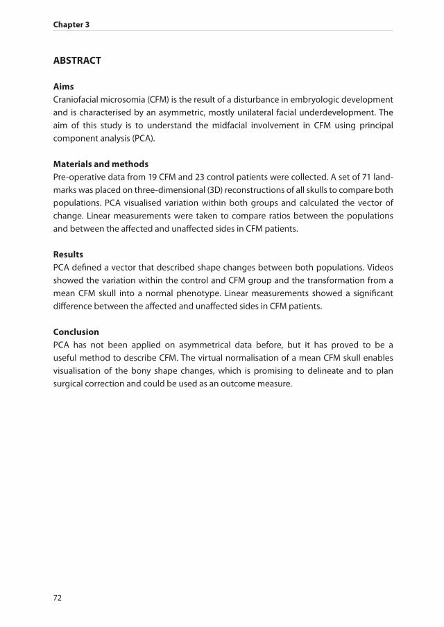
Chapter 3
72
ABSTRACT
AimsCraniofacial microsomia (CFM) is the result of a disturbance in embryologic development and is characterised by an asymmetric, mostly unilateral facial underdevelopment. The aim of this study is to understand the midfacial involvement in CFM using principal component analysis (PCA).
Materials and methods Pre-operative data from 19 CFM and 23 control patients were collected. A set of 71 land-marks was placed on three-dimensional (3D) reconstructions of all skulls to compare both populations. PCA visualised variation within both groups and calculated the vector of change. Linear measurements were taken to compare ratios between the populations and between the affected and unaffected sides in CFM patients.
Results PCA defined a vector that described shape changes between both populations. Videos showed the variation within the control and CFM group and the transformation from a mean CFM skull into a normal phenotype. Linear measurements showed a significant difference between the affected and unaffected sides in CFM patients.
Conclusion PCA has not been applied on asymmetrical data before, but it has proved to be a useful method to describe CFM. The virtual normalisation of a mean CFM skull enables visualisation of the bony shape changes, which is promising to delineate and to plan surgical correction and could be used as an outcome measure.
PSM 20190527 Proefschrift Britt Pluijmers.indd 72 24-07-19 15:04
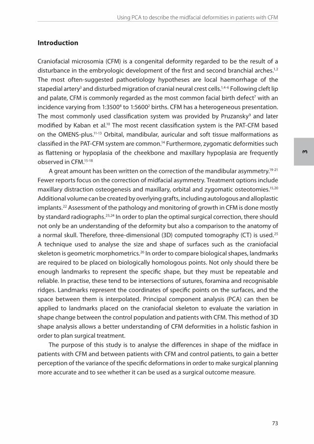
Using PCA to describe the midfacial deformities in patients with CFM
73
3
Introduction
Craniofacial microsomia (CFM) is a congenital deformity regarded to be the result of a disturbance in the embryologic development of the first and second branchial arches.1,2 The most often-suggested pathoetiology hypotheses are local haemorrhage of the stapedial artery3 and disturbed migration of cranial neural crest cells.1,4-6 Following cleft lip and palate, CFM is commonly regarded as the most common facial birth defect7 with an incidence varying from 1:35008 to 1:56002 births. CFM has a heterogeneous presentation. The most commonly used classification system was provided by Pruzansky9 and later modified by Kaban et al.10 The most recent classification system is the PAT-CFM based on the OMENS-plus.11-13 Orbital, mandibular, auricular and soft tissue malformations as classified in the PAT-CFM system are common.14 Furthermore, zygomatic deformities such as flattening or hypoplasia of the cheekbone and maxillary hypoplasia are frequently observed in CFM.15-18
A great amount has been written on the correction of the mandibular asymmetry.19-21 Fewer reports focus on the correction of midfacial asymmetry. Treatment options include maxillary distraction osteogenesis and maxillary, orbital and zygomatic osteotomies.15,20 Additional volume can be created by overlying grafts, including autologous and alloplastic implants.22 Assessment of the pathology and monitoring of growth in CFM is done mostly by standard radiographs.23,24 In order to plan the optimal surgical correction, there should not only be an understanding of the deformity but also a comparison to the anatomy of a normal skull. Therefore, three-dimensional (3D) computed tomography (CT) is used.25 A technique used to analyse the size and shape of surfaces such as the craniofacial skeleton is geometric morphometrics.26 In order to compare biological shapes, landmarks are required to be placed on biologically homologous points. Not only should there be enough landmarks to represent the specific shape, but they must be repeatable and reliable. In practise, these tend to be intersections of sutures, foramina and recognisable ridges. Landmarks represent the coordinates of specific points on the surfaces, and the space between them is interpolated. Principal component analysis (PCA) can then be applied to landmarks placed on the craniofacial skeleton to evaluate the variation in shape change between the control population and patients with CFM. This method of 3D shape analysis allows a better understanding of CFM deformities in a holistic fashion in order to plan surgical treatment.
The purpose of this study is to analyse the differences in shape of the midface in patients with CFM and between patients with CFM and control patients, to gain a better perception of the variance of the specific deformations in order to make surgical planning more accurate and to see whether it can be used as a surgical outcome measure.
PSM 20190527 Proefschrift Britt Pluijmers.indd 73 24-07-19 15:04
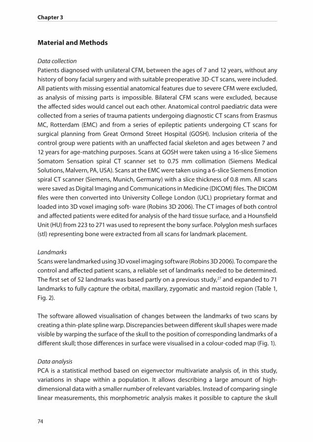
Chapter 3
74
Material and Methods
Data collectionPatients diagnosed with unilateral CFM, between the ages of 7 and 12 years, without any history of bony facial surgery and with suitable preoperative 3D-CT scans, were included. All patients with missing essential anatomical features due to severe CFM were excluded, as analysis of missing parts is impossible. Bilateral CFM scans were excluded, because the affected sides would cancel out each other. Anatomical control paediatric data were collected from a series of trauma patients undergoing diagnostic CT scans from Erasmus MC, Rotterdam (EMC) and from a series of epileptic patients undergoing CT scans for surgical planning from Great Ormond Street Hospital (GOSH). Inclusion criteria of the control group were patients with an unaffected facial skeleton and ages between 7 and 12 years for age-matching purposes. Scans at GOSH were taken using a 16-slice Siemens Somatom Sensation spiral CT scanner set to 0.75 mm collimation (Siemens Medical Solutions, Malvern, PA, USA). Scans at the EMC were taken using a 6-slice Siemens Emotion spiral CT scanner (Siemens, Munich, Germany) with a slice thickness of 0.8 mm. All scans were saved as Digital Imaging and Communications in Medicine (DICOM) files. The DICOM files were then converted into University College London (UCL) proprietary format and loaded into 3D voxel imaging soft- ware (Robins 3D 2006). The CT images of both control and affected patients were edited for analysis of the hard tissue surface, and a Hounsfield Unit (HU) from 223 to 271 was used to represent the bony surface. Polyglon mesh surfaces (stl) representing bone were extracted from all scans for landmark placement.
LandmarksScans were landmarked using 3D voxel imaging software (Robins 3D 2006). To compare the control and affected patient scans, a reliable set of landmarks needed to be determined. The first set of 52 landmarks was based partly on a previous study,27 and expanded to 71 landmarks to fully capture the orbital, maxillary, zygomatic and mastoid region (Table 1, Fig. 2).
The software allowed visualisation of changes between the landmarks of two scans by creating a thin-plate spline warp. Discrepancies between different skull shapes were made visible by warping the surface of the skull to the position of corresponding landmarks of a different skull; those differences in surface were visualised in a colour-coded map (Fig. 1).
Data analysisPCA is a statistical method based on eigenvector multivariate analysis of, in this study, variations in shape within a population. It allows describing a large amount of high-dimensional data with a smaller number of relevant variables. Instead of comparing single linear measurements, this morphometric analysis makes it possible to capture the skull
PSM 20190527 Proefschrift Britt Pluijmers.indd 74 24-07-19 15:04
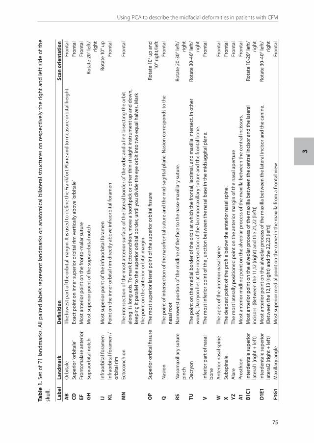
Using PCA to describe the midfacial deformities in patients with CFM
75
3
Tab
le 1
. Set
of 7
1 la
ndm
arks
. All
pai
red
lab
els
rep
rese
nt la
ndm
arks
on
anat
omic
al b
ilate
ral s
truc
ture
s on
res
pec
tive
ly t
he r
ight
and
left
sid
e of
the
sk
ull.
Lab
elLa
nd
mar
kD
efin
itio
nSc
an o
rien
tati
onA
BO
rbit
ale
The
low
est p
art o
f the
orb
ital
mar
gin
. It i
s us
ed to
defi
ne th
e Fr
ankf
ort P
lane
and
to m
easu
re o
rbit
al h
eigh
t.Fr
onta
l C
DSu
per
ior ‘
orbi
tale
’Ex
act p
oint
on
inne
r sup
erio
r orb
ital
rim
ver
tical
ly a
bov
e ‘o
rbit
ale’
Fron
tal
EFFr
onto
mal
are
ante
rior
Mos
t ant
erio
r poi
nt o
n th
e fr
onto
-mal
ar s
utur
eFr
onta
l G
HSu
prao
rbit
al n
otch
Mos
t sup
erio
r poi
nt o
f the
sup
raor
bita
l not
chRo
tate
20°
left
/rig
htIJ
Infr
aorb
ital
fora
men
Mos
t sup
erio
r poi
nt o
f the
infr
aorb
ital
fora
men
Rota
te 1
0° u
pK
LIn
frao
rbit
al fo
ram
en /
orbi
tal r
imPo
int o
n th
e in
ner o
rbit
al ri
m d
irect
ly a
bov
e in
frao
rbit
al fo
ram
enFr
onta
l
MN
Ecto
conc
hion
The
inte
rsec
tion
of th
e m
ost a
nter
ior s
urfa
ce o
f the
late
ral b
orde
r of t
he o
rbit
and
a lin
e bi
sect
ing
the
orbi
t al
ong
its
long
axi
s. T
o m
ark
Ecto
conc
hion
, mov
e a
toot
hpic
k or
oth
er th
in s
trai
ght i
nstr
umen
t up
and
dow
n,
keep
ing
it pa
ralle
l to
the
sup
erio
r orb
ital
bor
der,
until
you
div
ide
the
eye
orbi
t int
o tw
o eq
ual h
alve
s. M
ark
the
poi
nt o
n th
e an
terio
r orb
ital
mar
gin
Fron
tal
OP
Sup
erio
r orb
ital
fiss
ure
The
mos
t sup
erio
r lat
eral
poi
nt o
f the
sup
erio
r orb
ital
fiss
ure
Rota
te 1
0° u
p an
d 10
° rig
ht/l
eft
QN
asio
nTh
e p
oint
of i
nter
sect
ion
of th
e na
sofr
onta
l sut
ure
and
the
mid
-sag
itta
l pla
ne. N
asio
n co
rres
pon
ds to
the
nasa
l roo
tFr
onta
l
RS
Nas
omax
illar
y su
ture
pi
nch
Nar
row
est p
ortio
n of
the
mid
line
of th
e fa
ce to
the
naso
-max
illar
y su
ture
. Ro
tate
20-
30° l
eft/
right
TUD
acry
onTh
e p
oint
on
the
med
ial b
orde
r of t
he o
rbit
at w
hich
the
fron
tal,
lacr
imal
, and
max
illa
inte
rsec
t. In
oth
er
wor
ds, D
acry
on li
es a
t the
inte
rsec
tion
of th
e la
crim
omax
illar
y su
ture
and
the
fron
tal b
one.
Rota
te 3
0-40
° lef
t/rig
htV
Infe
rior p
art o
f nas
al
bon
e Th
e m
ost i
nfer
ior p
oint
of t
he ju
nctio
n b
etw
een
the
nasa
l bas
e in
the
mid
sagg
ital
pla
ne.
Fron
tal
WA
nter
ior n
asal
spi
neTh
e ap
ex o
f the
ant
erio
r nas
al s
pine
Fron
tal
XSu
bspi
nale
The
deep
est p
oint
of t
he p
rofil
e b
elow
the
ante
rior n
asal
spi
ne.
Fron
tal
YZ
Ala
reTh
e m
ost l
ater
ally
pos
ition
ed p
oint
on
the
ante
rior m
argi
n of
the
nasa
l ap
ertu
reFr
onta
lA
1Pr
osth
ion
Mos
t ant
erio
r mid
line
poi
nt o
n th
e al
veol
ar p
roce
ss o
f the
max
illa
bet
wee
n th
e ce
ntra
l inc
isso
rs.
Fron
tal
B1C1
Inte
rden
tale
sup
erio
r la
tera
l1 (r
ight
+ le
ft)
Mos
t ant
erio
r poi
nt o
n th
e al
veol
ar p
roce
ss o
f the
max
illa
bet
wee
n th
e ce
ntra
l inc
isor
and
the
late
ral
inci
sors
. (Be
twee
n th
e 11
,12
(rig
ht) a
nd th
e 21
,22
(left
))Ro
tate
10-
20° l
eft/
right
D1E
1In
terd
enta
le s
uper
ior
late
ral2
(rig
ht +
left
)M
ost a
nter
ior p
oint
on
the
alve
olar
pro
cess
of t
he m
axill
a b
etw
een
the
late
ral i
ncis
or a
nd th
e ca
nine
. (B
etw
een
the
12,1
3 (r
ight
) and
the
22,2
3 (le
ft))
Rota
te 3
0-40
° lef
t/rig
htF1
G1
Max
illar
y an
gle
Mos
t sup
erio
r med
ial p
oint
on
the
curv
e in
the
max
illa
from
a fr
onta
l vie
w
Fron
tal
PSM 20190527 Proefschrift Britt Pluijmers.indd 75 24-07-19 15:04

Chapter 3
76
Tab
le 1
. Con
tinu
ed
Lab
elLa
nd
mar
kD
efin
itio
nSc
an o
rien
tati
onH
1I1
Ecto
mol
are
On
the
max
illa:
pos
ition
ed a
t the
mos
t lat
eral
poi
nt o
n th
e la
tera
l sur
face
of t
he a
lveo
lar c
rest
. Fou
nd a
long
th
e se
cond
mol
ar o
n th
e m
axill
a.90
° lef
t/rig
ht a
nd
30-4
0° c
hin
upJ1
K1In
frao
rbit
al fo
ram
en to
al
veol
usM
iddl
e of
the
stra
ight
line
dow
n fr
om th
e in
frao
rbit
al fo
ram
en to
the
dent
o-a
lveo
lar j
unct
ion
Fron
tal
L1M
1In
frao
rbit
al fo
ram
en to
m
axill
ary
angl
eM
iddl
e of
the
stra
ight
line
from
the
infr
aorb
ital
fora
men
(GH
) to
the
max
illar
y an
gle
Fron
tal
N1O
1Zy
gom
axill
are
Inte
rsec
tion
of z
ygom
axill
lary
sut
ure
and
mos
t med
ial m
asse
ter m
uscl
e at
tach
men
t.Ro
tate
20-
40° l
eft/
right
and
10°
up
P1Q
1Zy
gom
atic
ang
leA
ngle
bet
wee
n zy
gom
atic
ridg
e an
d or
bita
l por
tion
of th
e zy
gom
aRo
tate
70-
90° l
eft/
right
R1S1
Gle
noid
foss
aM
ost s
uper
ior p
oint
of t
he g
leno
id fo
ssa
Rota
te 9
0° le
ft/
right
T1U
1Po
rion
Hig
hest
poi
nt o
n th
e ex
tern
al a
udito
ry m
eatu
sRo
tate
90°
left
/rig
ht a
nd 3
0° u
pV
1W1
Zygo
mat
icof
ront
al
sutu
reM
ost l
ater
al p
oint
on
the
fron
to-m
alar
sut
ure.
Rota
te 1
00-1
20°
left
/rig
htX1
Y1M
asto
idal
eTh
e in
ferio
r tip
of t
he m
asto
id p
roce
ssRo
tate
90°
chi
n up
Z1G
lab
ella
The
mos
t for
war
dly
proj
ectin
g p
oint
in th
e m
id-s
agit
tal p
lane
at t
he lo
wer
mar
gin
of th
e fr
onta
l bon
e, w
hich
lie
s ab
ove
the
nasa
l roo
t and
bet
wee
n th
e su
per
cilia
ry a
rche
s.
Fron
tal
A2B
2Fr
onto
tem
por
ale
Mos
t sup
erio
r poi
nt o
f sup
erio
r tem
por
al li
ne. (
top
of th
e la
tera
l orb
it b
one)
Rota
te 3
0-60
° lef
t/rig
htC
2D2
Orig
in z
ygom
aO
rigin
of z
ygom
atic
pro
cess
on
tem
por
al b
one.
In C
FM p
atie
nts
defin
e th
e or
igin
of t
he z
ygom
a on
the
skul
l w
here
it is
exp
ecte
d to
be.
Ro
tate
40-
60°
right
/lef
t and
10°
do
wn
E2F2
Zygo
mat
icof
acia
l fo
ram
enTh
e p
oint
on
the
zygo
mat
icof
acia
l for
amen
on
the
late
ral z
ygom
aRo
tate
20-
40° l
eft/
right
G2H
2Zy
gom
atic
otem
por
al
sutu
reTh
e lo
wes
t par
t of t
he z
ygom
atic
otem
por
al s
utur
e on
the
low
er p
art o
f the
zyg
omat
ic p
roce
ssRo
tate
90°
left
/rig
ht a
nd 1
0° u
pI2
Max
imum
cur
vatu
re
nasa
l bon
ePo
int w
ith m
axim
um c
urva
ture
in b
etw
een
nasi
on a
nd a
nter
ior n
asal
spi
neFr
onta
l
J2K
2In
ferio
r orb
ital
ridg
e/zy
gom
atic
o-m
axill
ary
sutu
re
Poin
t jus
t lat
eral
of t
he z
ygom
atic
omax
illar
y su
ture
infe
rior o
f the
orb
ital
ridg
eFr
onta
l
PSM 20190527 Proefschrift Britt Pluijmers.indd 76 24-07-19 15:04

Using PCA to describe the midfacial deformities in patients with CFM
77
3
Lab
elLa
nd
mar
kD
efin
itio
nSc
an o
rien
tati
onL2
M2
Max
imum
cur
vatu
re
mas
toid
The
max
imum
cur
vatu
re o
f the
ven
tral
sid
e of
the
mas
toid
from
a la
tera
l vie
wp
oint
Fron
tal a
nd 3
0° u
p
N2O
2N
asal
ridg
ePo
int w
ith m
axim
um c
urva
ture
on
the
nasa
l rid
ge o
n th
e m
axill
aRo
tate
20°
left
/rig
htP2
Q2
Infr
aorb
ital
fora
men
/na
sal b
one
Poin
t on
the
angl
e b
etw
een
max
illa
and
nasa
l bon
e on
a h
oriz
onta
l lin
e fr
om th
e in
frao
rbit
al fo
ram
enFr
onta
l
R2S
2M
axim
um c
urva
ture
la
tera
l asp
ect z
ygom
aM
axim
um c
urva
ture
of t
he z
ygom
a b
etw
een
zygo
max
illar
e an
d zy
gom
atic
otem
por
al s
utur
e fr
om a
late
ral
view
poi
nt
Rota
te 9
0° le
ft/
right
PSM 20190527 Proefschrift Britt Pluijmers.indd 77 24-07-19 15:04

Chapter 3
78
surface as a whole. A Point Distribution Model (PDM) is created of the landmarks. A PDM is a model that describes the mean shape and the allowed variability within a population. Eigenvectors are extracted from the landmark data, which are the principal components of variation in shape.26,28,29 The first principal component describes the largest variation within the population. The second and third principal components describe the second and third largest variations. The thin-plate splines (TPS) can interpolate changes between landmarks and are using minimum bending energy to estimate the surface between landmarks.26 This method was used to visualise the bone shape changes and to create videos that showed the variation in the control and affected population and between the populations.
Fig 1. Colour-coded map showing the predicted (warped) skull compared to its actual counterpart. A frontal view, 40o view and lateral view showing the positive and negative surface differences. Areas of light blue and green show good correspondence between the two scans, showing that the landmarks capture most of the skull shape.
Fig 2. Landmarks projected on the skull in frontal (left) and 40o (right) view, showing where the 71 landmarks from are placed on the skull.
PSM 20190527 Proefschrift Britt Pluijmers.indd 78 24-07-19 15:04

Using PCA to describe the midfacial deformities in patients with CFM
79
3
To define the repeatability of the landmarks, a randomly chosen control skull and affected skull were landmarked 10 times in different sittings, which allowed determination of the intra- observer reliability.
Since our cohort consisted of both right- and left-sided CFM patients, creating a representative unilateral CFM model was impossible. Therefore, all the paired landmarks of the left-sided skulls were flipped to the right side and registered to the same coordinate system. This enabled the creation of a right-sided CFM data set. To emphasize the outcomes of the videos, linear measurements of the skulls were taken. Using Robins 3D software, distances between coherent landmarks from the original landmark set were calculated and were defined as linear measurements to ratify the visual changes seen in the PCA model and in addition to the limitation of PCA. As it was impossible to define landmarks on partly missing anatomy, PCA was not able to describe the shape changes. Therefore, we used linear measurements from, for example, the origin of the zygoma to the zygomatic angle to describe changes in length of the expected zygomatic length. When videos did not show large differences, linear measurements were used to verify these assumptions. The left and right sides in and between CFM skulls and control skulls were compared, and ratios of the orbits were calculated for a better comparison of orbital size (Table 2).
The Wilcoxon signed-rank test was used to analyse differences within the CFM and control group. To make comparisons between the two groups, an independent two-tailed t-test for unequal sample sizes was used after determining the data to be normally distributed with the Lilliefors test. To allow comparison of the orbital ratios between the total amount of normal orbital ratios and right or left CFM ratios, the Mann-Whitney test was performed. A p-value of < 0.05 was chosen for the significance level.
Table 2. Linear measurements between landmarks. ‘*’: right (affected) and left (unaffected) side.
Measurements Distances between Landmarks Description
Orbital Width * E – T and F – U Frontomalare anterior to dacryon
Orbital Length *Orbital Ratio *
A – C and B – D Orbitale to superior orbitaleOrbital width/orbital length
Nasiozygomatic Length * Q – G2 and Q – H2 Nasion to zygomaticotemporal suture
Zygomatic Height * A – N1 and B – O1 Orbitale to zygomaxillare
Expected Zygomatic Length * C2 – P1 and D2 – Q1 Origin zygoma to zygomatic angle
Facial Width G2 – H2 Distance between zygomaticotemporal sutures
PSM 20190527 Proefschrift Britt Pluijmers.indd 79 24-07-19 15:04
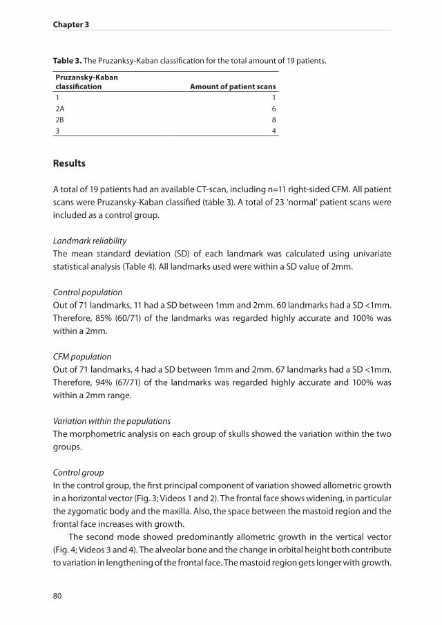
Chapter 3
80
Results
A total of 19 patients had an available CT-scan, including n=11 right-sided CFM. All patient scans were Pruzansky-Kaban classified (table 3). A total of 23 ‘normal’ patient scans were included as a control group.
Landmark reliabilityThe mean standard deviation (SD) of each landmark was calculated using univariate statistical analysis (Table 4). All landmarks used were within a SD value of 2mm.
Control populationOut of 71 landmarks, 11 had a SD between 1mm and 2mm. 60 landmarks had a SD <1mm. Therefore, 85% (60/71) of the landmarks was regarded highly accurate and 100% was within a 2mm.
CFM populationOut of 71 landmarks, 4 had a SD between 1mm and 2mm. 67 landmarks had a SD <1mm. Therefore, 94% (67/71) of the landmarks was regarded highly accurate and 100% was within a 2mm range.
Variation within the populationsThe morphometric analysis on each group of skulls showed the variation within the two groups.
Control groupIn the control group, the first principal component of variation showed allometric growth in a horizontal vector (Fig. 3; Videos 1 and 2). The frontal face shows widening, in particular the zygomatic body and the maxilla. Also, the space between the mastoid region and the frontal face increases with growth.
The second mode showed predominantly allometric growth in the vertical vector (Fig. 4; Videos 3 and 4). The alveolar bone and the change in orbital height both contribute to variation in lengthening of the frontal face. The mastoid region gets longer with growth.
Table 3. The Pruzanksy-Kaban classification for the total amount of 19 patients.
Pruzansky-Kaban classification Amount of patient scans1 1
2A 6
2B 8
3 4
PSM 20190527 Proefschrift Britt Pluijmers.indd 80 24-07-19 15:04

Using PCA to describe the midfacial deformities in patients with CFM
81
3
Table 4. The intra-observer reliability for normal skulls and CFM skulls for the landmarks used in this study.
Label SD Normals (mm) SD CFM (mm)A 0,476 0,641
B 0,607 0,955
C 0,561 0,745
D 0,647 0,967
E 0,291 0,373
F 0,507 0,23
G 0,68 0,409
H 0,438 0,446
I 0,391 0,399
J 0,375 0,396
K 0,328 0,301
L 0,355 0,468
M 0,356 0,527
N 0,577 0,592
O 0,711 0,438
P 1,2 0,342
Q 0,295 0,572
R 0,508 0,46
S 0,713 0,381
T 0,585 0,393
U 0,666 0,905
V 0,301 0,251
W 0,253 0,201
X 0,337 0,274
Y 0,513 0,617
Z 0,475 0,466
A1 0,316 0,28
B1 1,766 0,701
C1 0,879 0,227
D1 0,822 0,438
E1 0,642 0,731
F1 0,844 1,04
G1 0,663 0,611
H1 0,954 0,579
I1 1,422 0,711
J1 0,681 0,566
K1 0,782 0,801
L1 0,342 0,489
M1 0,454 0,329
N1 0,495 0,464
O1 0,578 0,354
P1 0,529 0,307
Q1 0,553 0,416
PSM 20190527 Proefschrift Britt Pluijmers.indd 81 24-07-19 15:04

Chapter 3
82
Supplementary video related to this article can be found at https://doi.org/10.1016/j.jcms.2018.09.019.
CFM populationThe first component of variation in the CFM population shows the variability in severity of CFM (Fig. 5; Videos 5 and 6). From the zygomaticotemporal suture, a down-bending zygoma is displayed on the affected side. The zygomatic body appears to be shorter and decreased in length. The orbit shows an increase in width and decrease in length, and the frontotemporal region moves down. The distance between the mastoid and the lateral dorsal zygomatic ridge decreases. The maxilla shows a decreased length.
The second mode of variation shows predominantly allometric growth (Videos 7 and 8). The affected side demonstrates a flattening of the lower part of the zygoma.
Label SD Normals (mm) SD CFM (mm)R1 1,041 0,996
S1 0,556 0,694
T1 0,646 1,08
U1 1 1,252
V1 0,432 0,376
W1 0,393 0,429
X1 0,356 0,293
Y1 0,315 0,659
Z1 1,301 0,779
A2 1,179 0,93
B2 1,061 0,992
C2 0,665 0,645
D2 0,362 0,786
E2 1,857 0,254
F2 1,856 0,335
G2 0,37 0,252
H2 0,642 0,352
I2 0,922 0,594
J2 0,594 0,538
K2 1,306 0,984
L2 0,603 0,872
M2 0,6 0,749
N2 0,603 0,779
O2 0,961 1,129
P2 0,636 0,593
Q2 0,741 0,47
R2 0,669 0,535
S2 0,816 0,36
Table 4. Continued
PSM 20190527 Proefschrift Britt Pluijmers.indd 82 24-07-19 15:04

Using PCA to describe the midfacial deformities in patients with CFM
83
3
Fig. 3. First principal component of the normal group in 40o left view, frontal view and 40o right view. Minus 2SD (top) and plus 2SD (bottom). Showing allometric growth in a horizontal vector within the age range of 7-12 years.
Fig. 4. Second principal component of the normal group in 40o left, frontal and 40o right. Minus 2SD (top) and plus 2SD (bottom). Showing allometric growth in a vertical vector.
PSM 20190527 Proefschrift Britt Pluijmers.indd 83 24-07-19 15:04

Chapter 3
84
Fig. 6. Third principal component of the CFM group in 40o left, frontal and 40o right. Minus 2SD (top) and plus 2SD (bottom). Showing the shape variation in an oblique vector. The maxilla shifts to the right and the zygomatic body moves upwards causing a smaller orbit.
Fig. 5. First principal component of the CFM group in 40o left, frontal and 40o right. Plus 2SD (top) and minus 2SD (bottom). Showing the shape variation on the affected (right) side. At the bottom: the zygoma bends down and the zygomatic body is shorter. The orbit is smaller and the frontotemporal region moves down.
PSM 20190527 Proefschrift Britt Pluijmers.indd 84 24-07-19 15:04
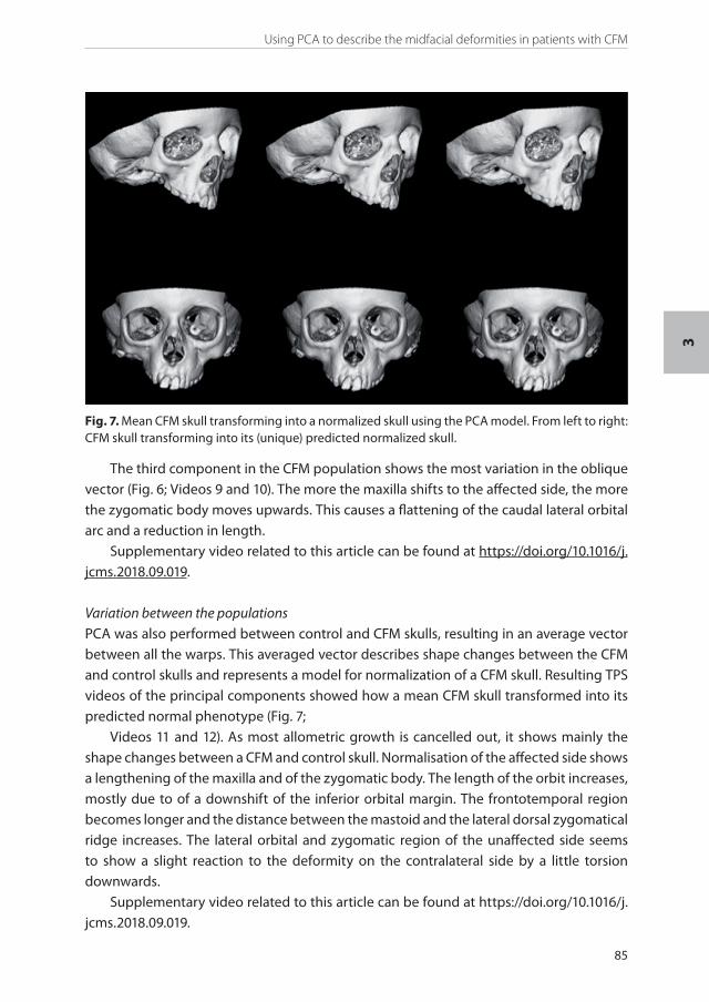
Using PCA to describe the midfacial deformities in patients with CFM
85
3
The third component in the CFM population shows the most variation in the oblique vector (Fig. 6; Videos 9 and 10). The more the maxilla shifts to the affected side, the more the zygomatic body moves upwards. This causes a flattening of the caudal lateral orbital arc and a reduction in length.
Supplementary video related to this article can be found at https://doi.org/10.1016/j.jcms.2018.09.019.
Variation between the populationsPCA was also performed between control and CFM skulls, resulting in an average vector between all the warps. This averaged vector describes shape changes between the CFM and control skulls and represents a model for normalization of a CFM skull. Resulting TPS videos of the principal components showed how a mean CFM skull transformed into its predicted normal phenotype (Fig. 7;
Videos 11 and 12). As most allometric growth is cancelled out, it shows mainly the shape changes between a CFM and control skull. Normalisation of the affected side shows a lengthening of the maxilla and of the zygomatic body. The length of the orbit increases, mostly due to of a downshift of the inferior orbital margin. The frontotemporal region becomes longer and the distance between the mastoid and the lateral dorsal zygomatical ridge increases. The lateral orbital and zygomatic region of the unaffected side seems to show a slight reaction to the deformity on the contralateral side by a little torsion downwards.
Supplementary video related to this article can be found at https://doi.org/10.1016/j.jcms.2018.09.019.
Fig. 7. Mean CFM skull transforming into a normalized skull using the PCA model. From left to right: CFM skull transforming into its (unique) predicted normalized skull.
PSM 20190527 Proefschrift Britt Pluijmers.indd 85 24-07-19 15:04

Chapter 3
86
Linear measurmentsEuclidean distances were calculated (Table 5) for a total of 13 measurements. When comparing the affected (right) side to the unaffected (left) side within the CFM population, it is evident the affected side is significantly different (Figs. 8-10) except for the nasiozygo-matic length (Fig. 11). No differences between the right and left side were found within the control population. Comparing both populations with each other, the affected (right) side in CFM was significantly different from the right side in the control group (Figs. 9 and 10). Both the right and left CFM orbital ratios were compared to the total of normal orbital ratios. This showed a significant different orbital ratio on the affected side in the CFM skulls (Fig. 8). There was no difference in facial width between the populations (Fig. 12).
Discussion
PCA allows a mathematical analysis of a unique skull as a whole rather than comparison of average values taken from samples of a control population. Earlier analysis of Noonan30, Apert31 and Crouzon and Pfeiffer27 has been done. These syndromes affect the skull symmetrically. This was the first time that PCA was performed on asymmetrical skulls with underdeveloped and missing parts.32 Previously our study group showed the skull base to be asymmetrical in patients with CFM.33 Defining a reliable set of landmarks was challenging, although analysing the intra-observer errors confirmed that the chosen landmarks are reproducible.
The PCA model showed the variation within the populations. The fact the mathematical model shows allometric growth in the first and second principal component of the control
Table 5. Measurements in the orbital and upper midfacial regions for normal and CFM skulls.
Measurements LandmarksNormal
Mean (mm) SD (mm)CFM
Mean (mm) SD (mm)Orbital Width Affected E-T 36.95 1.72 37.82 1.80
Orbital Width Unaffected F-U 36.49 1.72 36.70 1.54
Orbital Length Affected A-C 32.48 2.14 32.12 33.92
Orbital Length Unaffected B-D 33.04 2.00 33.92 1.88
Orbital Ratio Affected Width/Length 1.14 0.09 1.18 0.04
Orbital Ratio Unaffected Width/Length 1.11 0,08 1.08 0.05
Nasiozygomatic Length Affected Q-G2 79.95 4.09 80.18 4.59
Nasiozygomatic Length Unaffected
Q-H2 78.98 3.56 79.45 4.03
Zygomatic Height Affected A-N1 22.98 2.82 21.33 2.24
Zygomatic Height Unaffected B-O1 23.49 3.00 23.05 1.56
Exp Zygomatic Length Affected C2-P1 38.38 3.19 33.57 4.69
Exp Zygomatic Length Unaffected D2-Q1 38.57 2.96 37.85 4.89
Facial Width G2-H2 109.31 4.61 106.72 6.61
PSM 20190527 Proefschrift Britt Pluijmers.indd 86 24-07-19 15:04

Using PCA to describe the midfacial deformities in patients with CFM
87
3
Fig. 8. Comparison of the orbital ratios between the control group and the CFM group (right and left). * = p £ 0.05.
Fig 10. Comparison of the expected zygomatical length between right and left in the normal group and the CFM group. * = p≤0.05
Fig. 9. Comparison of the zygomatic height between right and left in the normal group and in the CFM group. * = p≤0.05
Fig 11. Comparison of the nasiozygomatic length between right and left in the normal group and the CFM group. * = p≤0.05
population means that the model appears to mirror growth in the control population. The largest variation in controls seems to be allometric growth; for the CFM population, this seems to be a visualisation of the spread in severity. Concurrent with other studies, we also noticed unequal orbital sizes15, a more hypoplastic zygoma17 and a decrease in height of the maxilla.34 Furthermore, we found a decreased length of the lateral part of the upper
0,9
1
1,1
1,2
1,3
Total Normal CFM Right CFM Left
Orb
ital r
atio
(w
idth
/leng
th)
Orbital Ratio
**
20
25
30
35
40
45
Right (affected) Left
Len
gth
(mm
)
Expected Zygomatical Length
CFMNormal
**
80
90
100
110
120
CFM Normal
Len
gth
(mm
)
Facial Width
15
17
19
21
23
25
27
29
Right (affected) Left
Hei
ght
(mm
)
Zygomatic Height
CFMNormal
*
*
60
65
70
75
80
85
90
Right (affected) Left
Len
gth
(mm
)
Nasiozygomatic Length
CFMNormal
Fig 12. Comparison of the nasiozygomatic length between right and left in the normal group and the CFM group. * = p≤0.05
PSM 20190527 Proefschrift Britt Pluijmers.indd 87 24-07-19 15:04
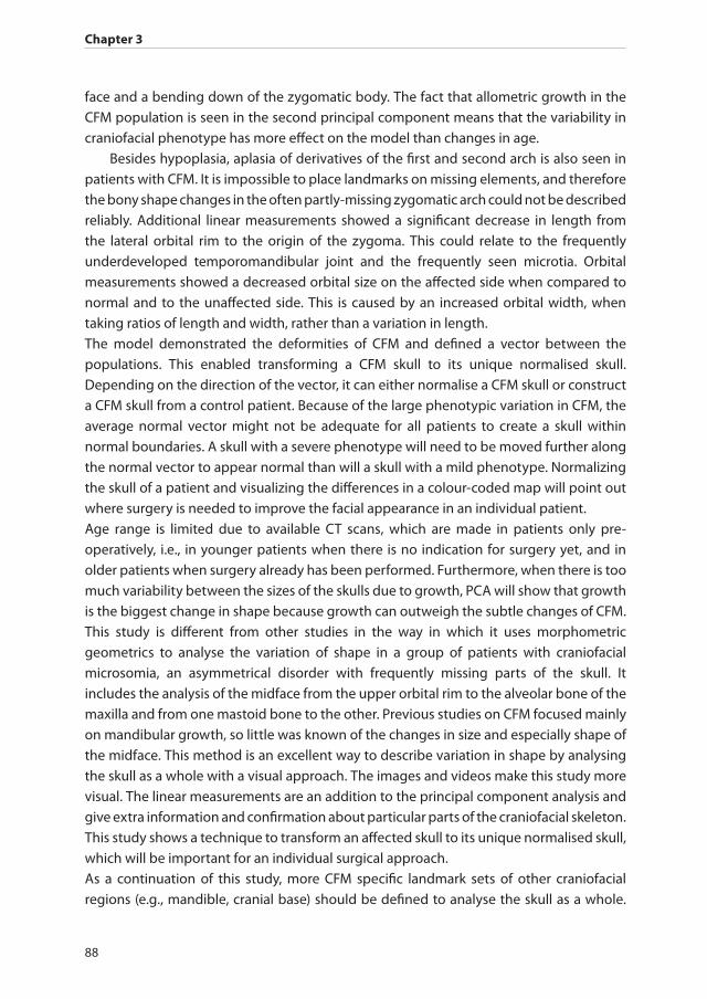
Chapter 3
88
face and a bending down of the zygomatic body. The fact that allometric growth in the CFM population is seen in the second principal component means that the variability in craniofacial phenotype has more effect on the model than changes in age.
Besides hypoplasia, aplasia of derivatives of the first and second arch is also seen in patients with CFM. It is impossible to place landmarks on missing elements, and therefore the bony shape changes in the often partly-missing zygomatic arch could not be described reliably. Additional linear measurements showed a significant decrease in length from the lateral orbital rim to the origin of the zygoma. This could relate to the frequently underdeveloped temporomandibular joint and the frequently seen microtia. Orbital measurements showed a decreased orbital size on the affected side when compared to normal and to the unaffected side. This is caused by an increased orbital width, when taking ratios of length and width, rather than a variation in length.The model demonstrated the deformities of CFM and defined a vector between the populations. This enabled transforming a CFM skull to its unique normalised skull. Depending on the direction of the vector, it can either normalise a CFM skull or construct a CFM skull from a control patient. Because of the large phenotypic variation in CFM, the average normal vector might not be adequate for all patients to create a skull within normal boundaries. A skull with a severe phenotype will need to be moved further along the normal vector to appear normal than will a skull with a mild phenotype. Normalizing the skull of a patient and visualizing the differences in a colour-coded map will point out where surgery is needed to improve the facial appearance in an individual patient.Age range is limited due to available CT scans, which are made in patients only pre-operatively, i.e., in younger patients when there is no indication for surgery yet, and in older patients when surgery already has been performed. Furthermore, when there is too much variability between the sizes of the skulls due to growth, PCA will show that growth is the biggest change in shape because growth can outweigh the subtle changes of CFM.This study is different from other studies in the way in which it uses morphometric geometrics to analyse the variation of shape in a group of patients with craniofacial microsomia, an asymmetrical disorder with frequently missing parts of the skull. It includes the analysis of the midface from the upper orbital rim to the alveolar bone of the maxilla and from one mastoid bone to the other. Previous studies on CFM focused mainly on mandibular growth, so little was known of the changes in size and especially shape of the midface. This method is an excellent way to describe variation in shape by analysing the skull as a whole with a visual approach. The images and videos make this study more visual. The linear measurements are an addition to the principal component analysis and give extra information and confirmation about particular parts of the craniofacial skeleton. This study shows a technique to transform an affected skull to its unique normalised skull, which will be important for an individual surgical approach.As a continuation of this study, more CFM specific landmark sets of other craniofacial regions (e.g., mandible, cranial base) should be defined to analyse the skull as a whole.
PSM 20190527 Proefschrift Britt Pluijmers.indd 88 24-07-19 15:04

Using PCA to describe the midfacial deformities in patients with CFM
89
3
Outcome of craniofacial surgery on CFM can be measured by comparing the normalised skull with the postoperative scan. Also, a more extensive study with collaboration between international craniofacial centres to increase the numbers of patients is recommended, which would increase the sensitivity of PCA.
ConclusionThe skull of a patient with craniofacial microsomia differs in many ways from a ‘normal’ skull. It was already known that orbital, zygomatic and maxillary sizes were different in the affected side of patients with CFM. The exact changes in shape, such as the increased orbital width, the bending down of the zygoma and the frontotemporal region were not yet described. These findings are helpful for a better understanding of the deformity. The developed vector between the populations, which is able to transform a CFM skull into its normalised skull, is a promising tool for reconstructive surgery and could be used as a surgical outcome measurement tool.
PSM 20190527 Proefschrift Britt Pluijmers.indd 89 24-07-19 15:04

Chapter 3
90
References
1. Converse JM, Coccaro PJ, Becker M, Wood-Smith D. On hemifacial microsomia. The first and second branchial arch syndrome. Plastic and reconstructive surgery 1973; 51(3): 268-79.
2. Grabb WC. The first and second branchial arch syndrome. Plastic and reconstructive surgery 1965; 36(5): 485-508.
3. Poswillo D. Hemorrhage in development of the face. Birth defects original article series 1975; 11(7): 61-81.
4. Johnston MC, Bronsky PT. Prenatal craniofacial development: new insights on normal and abnormal mechanisms. Crit Rev Oral Biol Med 1995; 6(4): 368-422.
5. Stark RB, Saunders DE. The first branchial syndrome. The oral-mandibular-auricular syndrome. Plast Reconstr Surg Transplant Bull 1962; 29: 229-39.
6. Tuin AJ, Tahiri Y, Paine KM, Paliga JT, Taylor JA, Bartlett SP. Clarifying the relationships among the different features of the OMENS+ classification in craniofacial microsomia. Plastic and reconstructive surgery 2015; 135(1): 149e-56e.
7. Murray JE, Kaban LB, Mulliken JB. Analysis and treatment of hemifacial microsomia. Plastic and reconstructive surgery 1984; 74(2): 186-99.
8. Poswillo D. Otomandibular deformity: pathogenesis as a guide to reconstruction. Journal of maxillofacial surgery 1974; 2(2-3): 64-72.
9. Pruzansky S. Not all dwarfed mandibles are alike. Birth defects original article series 1969; 5: 120-9.10. Kaban LB, Moses MH, Mulliken JB. Surgical correction of hemifacial microsomia in the growing child.
Plastic and reconstructive surgery 1988; 82(1): 9-19.11. Birgfeld CB, Luquetti DV, Gougoutas AJ, et al. A phenotypic assessment tool for craniofacial
microsomia. Plastic and reconstructive surgery 2011; 127(1): 313-20.12. Horgan JE, Padwa BL, LaBrie RA, Mulliken JB. OMENS-Plus: analysis of craniofacial and extracraniofacial
anomalies in hemifacial microsomia. Cleft Palate Craniofac J 1995; 32(5): 405-12.13. Vento AR, LaBrie RA, Mulliken JB. The O.M.E.N.S. classification of hemifacial microsomia. Cleft Palate
Craniofac J 1991; 28(1): 68-76; discussion 7.14. Caron C, Pluijmers BI, Wolvius EB, et al. Craniofacial and extracraniofacial anomalies in craniofacial
microsomia: a multicenter study of 755 patients’. J Craniomaxillofac Surg 2017; 45(8): 1302-10.15. Santler G, Karcher H, Mossbock R. Simultaneous orbital expansion and intraoral distraction
osteogenesis of upper and lower jaws in a patient with hemifacial microsomia. J Craniomaxillofac Surg 2003; 31(4): 228-33.
16. Keogh IJ, Troulis MJ, Monroy AA, Eavey RD, Kaban LB. Isolated microtia as a marker for unsuspected hemifacial microsomia. Arch Otolaryngol Head Neck Surg 2007; 133(10): 997-1001.
17. Cousley RR, Calvert ML. Current concepts in the understanding and management of hemifacial microsomia. Br J Plast Surg 1997; 50(7): 536-51.
18. Gorlin R CM, Hennekam RC. Branchial arch and oral-acral disorders. Syndromes of the Head and Neck. New York: Oxford University Press; 2001: 790–8.
19. Pluijmers BI, Caron CJ, Dunaway DJ, Wolvius EB, Koudstaal MJ. Mandibular reconstruction in the growing patient with unilateral craniofacial microsomia: a systematic review. Int J Oral Maxillofac Surg 2014; 43(3): 286-95.
20. McCarthy JG, Schreiber J, Karp N, Thorne CH, Grayson BH. Lengthening the human mandible by gradual distraction. Plastic and reconstructive surgery 1992; 89(1): 1-8; discussion 9-10.
21. Wan DC, Taub PJ, Allam KA, et al. Distraction osteogenesis of costocartilaginous rib grafts and treatment algorithm for severely hypoplastic mandibles. Plastic and reconstructive surgery 2011; 127(5): 2005-13.
22. Staal F, Pluijmers B, Wolvius E, Koudstaal M. Patient-Specific Implant for Residual Facial Asymmetry following Orthognathic Surgery in Unilateral Craniofacial Microsomia. Craniomaxillofac Trauma Reconstr 2016; 9(3): 264-7.
23. Ongkosuwito EM, van Neck JW, Wattel E, van Adrichem LN, Kuijpers-Jagtman AM. Craniofacial morphology in unilateral hemifacial microsomia. The British journal of oral & maxillofacial surgery 2013; 51(8): 902-7.
PSM 20190527 Proefschrift Britt Pluijmers.indd 90 24-07-19 15:04
Using PCA to describe the midfacial deformities in patients with CFM
91
3
24. Sant’Anna EF, Lau GW, Marquezan M, de Souza Araujo MT, Polley JW, Figueroa AA. Combined maxillary and mandibular distraction osteogenesis in patients with hemifacial microsomia. Am J Orthod Dentofacial Orthop 2015; 147(5): 566-77.
25. Shibazaki-Yorozuya R, Yamada A, Nagata S, Ueda K, Miller AJ, Maki K. Three-dimensional longitudinal changes in craniofacial growth in untreated hemifacial microsomia patients with cone-beam computed tomography. Am J Orthod Dentofacial Orthop 2014; 145(5): 579-94.
26. Bookstein FL. Shape and the information in medical images: a decade of the morphometric synthesis. Computer Vis Image Underst 1997; 66(2): 97-118.
27. Staal FC, Ponniah AJ, Angullia F, Ruff C, Koudstaal MJ, Dunaway D. Describing Crouzon and Pfeiffer syndrome based on principal component analysis. J Craniomaxillofac Surg 2015; 43(4): 528-36.
28. O’Higgins P. The study of morphological variation in the hominid fossil record: biology, landmarks and geometry. Journal of anatomy 2000; 197 ( Pt 1): 103-20.
29. Cootes T.F. TCJ, Cooper D.H., Graham J. . Training Models of Shape from Sets of Examples. In: Hogg B, editor. Proceedings of the British Machine Vision Conference. Leeds: Springer; 1992.
30. Hammond P, Hutton TJ, Allanson JE, et al. 3D analysis of facial morphology. Am J Med Genet A 2004; 126A(4): 339-48.
31. Pluijmers BI, Ponniah AJ, Ruff C, Dunaway D. Using principal component analysis to describe the Apert skull deformity and simulate its correction. J Plast Reconstr Aesthet Surg 2012; 65(12): 1750-2.
32. Netherway DJ, Abbott AH, Gulamhuseinwala N, et al. Three-dimensional computed tomography cephalometry of plagiocephaly: asymmetry and shape analysis. Cleft Palate Craniofac J 2006; 43(2): 201-10.
33. Schaal SC, Ruff C, Pluijmers BI, et al. Characterizing the skull base in craniofacial microsomia using principal component analysis. Int J Oral Maxillofac Surg 2017; 46(12): 1656-63.
34. Wink JD, Paliga JT, Tahiri Y, Goldstein JA, Taylor JA, Bartlett SP. Maxillary involvement in hemifacial microsomia: an objective three-dimensional analysis of the craniofacial skeleton. J Craniofac Surg 2014; 25(4): 1236-40.
PSM 20190527 Proefschrift Britt Pluijmers.indd 91 24-07-19 15:04

CH
AP
TER
PSM 20190527 Proefschrift Britt Pluijmers.indd 92 24-07-19 15:04

4
Describing the mandible in patients with craniofacial microsomia based on
principal component analysis and thin-plate-spline video analysis.
Onur Kaya* and Britt I. Pluijmers*, Femke Staal, Cliff Ruff, Bonnie L. Padwa, Maarten J. Koudstaal, David J. Dunaway.
*Both authors contributed equally to this paper.
International Journal of Ora and Maxillofac Surg. 2019 Mar;48(3):302-308. doi: 10.1016/j.ijom.2018.08.015. Epub 2018 Sep 15.
PSM 20190527 Proefschrift Britt Pluijmers.indd 93 24-07-19 15:04

Chapter 4
94
Abstract
AimsCraniofacial microsomia (CFM) is most often described as a unilateral malformation of derivatives of the first and second branchial arches. The mandible has been classified using several classification systems. However, all are based on two-dimensional imaging. The aim of this study was to mathematically describe the deformed mandible based on principal component analysis (PCA) in a three-dimensional way. This may aid in defining the flaws in existing surgical corrections of the mandible through the identification of the differences in shape compared with a normal mandible in a holistic view with the help of videos.
Material and MethodsForty-three homologous landmarks were defined to describe a mandible with CFM. Computed tomography scans of 22 patients and 30 controls were marked manually. The changes in shape between the mandibles were visualized using videos.
Results and conclusionsA lateral rotation with increase in posterior rotation of the condyle due to shortening of the condyle–gonial height and a longitudinal rotation with outward bending of the mandibular angle were noted on the affected side, as well as an inward bending of the angle on the unaffected side.
Due to the compensatory remodelling of the mandible on the unaffected side, one could suggest that CFM is never truly unilateral.
PSM 20190527 Proefschrift Britt Pluijmers.indd 94 24-07-19 15:04

Characterizing the mandible using PCA
95
4
Introduction
Craniofacial microsomia (CFM) is a complex congenital anomaly that is characterized by an underdevelopment of derivatives of the first and second branchial arches. It is estimated to occur in 1: 3,500 to 1: 20,000 live births, which makes it the second most common craniofacial disorder following cleft lip and palate.1-3 Although it is most often regarded as a unilateral malformation, the involvement of clinical features is bilateral in 10% of the cases.4-6 However, even in the bilateral cases asymmetry is commonly observed. Structures involved are among others: the mandible, orbit, zygoma, maxilla, ears, soft tissue and facial nerves. The phenotype can range from subtle facial asymmetry or microtia to severe underdevelopment of the structures mentioned above.
Mandibular hypoplasia is often the most striking deformity in CFM and occurs in 89-100% of affected patients.7 The heterogeneous presentation of CFM challenges the classification of deformity. With regards to the mandible, the severity of hypoplasia can be classified into the following four types based on the Pruzansky-Kaban classification. Type I: minimal hypoplasia of the mandible. Type IIa: abnormal shape and size in the mandibular ramus. Type IIb: hypoplastic mandibular ramus and temporomandibular joint (TMJ) abnormal in morphology and position. Type III: absent ramus, condyle, and TMJ.8,9 However, it must be noted that the Pruzansky-Kaban classification is based on 2-dimensional imaging, i.e. cephalograms and orthopantomograms, losing 3-dimensional information. Furthermore, currently the Pruzansky-Kaban classification tends to show inter- and intra-rater variability when evaluating affected mandibles.10
Geometric morphometrics (GM) is a mathematical modelling technique that uses homologous anatomical landmarks points to convert the skull into a geometric object with Cartesian coordinates.11 Homologous landmarks are necessary and need to be accurate and reliable to validate geometric morphometrics modelling. The landmarks should be reliable and easy to repeat, furthermore there should be enough landmarks to represent the specific shape. Intersections of sutures, foramina and recognisable ridges are often used.11,12 Landmarks represent the coordinates of specific points on the surfaces and the space between them is interpolated. Principal Component Analysis (PCA) can be applied to landmarks placed on the mandible; to evaluate the variation in shape change between mandibles within a CFM population and between a CFM and control population.
PCA has previously been used in the analysis of craniofacial shapes in anthropological studies and has also been shown useful in characterizing hard tissue deformities of Apert, Crouzon, Pfeiffer skulls and more recently to describe the CFM skull base.13-15 PCA and geometric morphometrics make it possible to visualize shape change in a 3-dimensional way. This could aid to the improvement of surgical planning of correction of craniofacial deformities by identifying the hiatus of the present surgical techniques.
PSM 20190527 Proefschrift Britt Pluijmers.indd 95 24-07-19 15:04

Chapter 4
96
The aim of this study was to get a better understanding of the variance in shape in CFM patients in order possibly aid to refine the current treatment modalities. At the same time, the efficacy of principal component analysis will be assessed when characterizing the mandibular deformity in CFM.
Material and methods
Data collectionThe craniofacial databases of three craniofacial units in three countries were used to identify CFM patients: Great Ormond Street Hospital, London, UK (GOSH), Erasmus Medical Centre, Rotterdam, the Netherlands (EMC), and Boston Children’s Hospital, Boston, USA (BCH).
CFM patients with a unilateral presentation, between the ages of 6 and 19 years, and of whom good quality preoperative 3D computed tomography (CT) scans were available, were included. Patients with a Pruzansky–Kaban type III mandible were excluded due to the lack of homologous points over the deformity.
An age-matched control group was recruited, which included the CT scans of patients with anatomically normal skulls. The data were collected from a series of trauma patients undergoing diagnostic CT scans at EMC and from a series of epileptic patients undergoing CT scans for surgical planning at GOSH. Inclusion criteria were patients with an unaffected facial skeleton, between the ages of 6 and 19 years.
The scans were obtained in DICOM format (Digital Imaging and Communications in Medicine) using a 16-slice Siemens Somatom Sensation spiral CT scanner set to 0.75-mm collimation (Siemens AG, Munich, Germany). DICOM data were reconstructed into polygon mesh surfaces (stereolithography, STL) demonstrating bone and then loaded into a mesh viewer (Robin 3D, 2015) for segmenting and landmarking.
In total 22 CFM patients were included: nine from GOSH in London, five from EMC in Rotterdam, and eight from BCH in Boston. A total of 30 controls were included. The CFM patients were divided into two age groups to lessen the dilution effects of allometric growth: 6–12 years (n = 13) and 13–19 years (n = 9). The controls were stratified into two corresponding groups: 6–12 years (n = 14) and 13–19 years (n = 16).
To guarantee unbiased shape analysis, all prevalent left-sided defects were mirrored before landmarking. The mandible was isolated from the rest of the craniofacial skeleton to gain access to all regions in order to correctly place the necessary landmarks (Fig. 1). The segmentation and landmarking was performed manually using Robin 3D software if delineation of the condyles and coronoid process was possible from the articulating glenoid fossa.
PSM 20190527 Proefschrift Britt Pluijmers.indd 96 24-07-19 15:04

Characterizing the mandible using PCA
97
4
LandmarksBased on anatomy and existing anthropometric points, 43 landmarks were considered (Table 1, Fig. 2). The landmarks were more densely located around the condyles in order to be able to fully capture these geometrically complex regions. The landmarks were validated with the help of colour map algorithms (Fig. 3). If the landmarks are distributed adequately over the mandibular surface and the morphology is captured sufficiently well, the colour map will show very little difference between surfaces. The aim was to ensure that this was the case, particularly in the regions known to be affected in CFM, such as the coronoid processes, condyles, and rami.
A random control and CFM mandible were chosen and landmarked 10 times in different sittings to determine the landmark repeatability and intra-observer reliability. The standard deviation (SD) was calculated to determine intra-observer reliability for all 43 landmarks. Finally, another random control mandible was chosen and landmarked 10 times in different sittings by two researchers based at different locations. Subsequently the inter-observer reliability for each landmark was determined by performing a t-test and reading the P-values.
Shape analysisPCA is able to quantify the differences in shape within a population by reducing the dimensionality of the shape data into lesser amounts of variables, called principal components or modes of variation. This analysis is done using a Point Distribution
Fig. 1. The mandible was isolated manually from the rest of the craniofacial skeleton if delineation of the condyles and coronoid process was possible from the articulating glenoid fossa.
PSM 20190527 Proefschrift Britt Pluijmers.indd 97 24-07-19 15:04

Chapter 4
98
Table 1. Landmarks.
Landmark Position DefinitionA Infradentale Most anteriosuperior point
on the labial crest of the mandibular alveolar process
B B-point Most posterior point of the bony curvature of the mandible below infradentale and above pogonion
C Pogonion Most anterior point on the mandible in the midline
D Menthon The most inferior point of the mandibular symphysis
E Right mental tubercle Most anterolateral aspect of the right mental tubercle
F Left mental tubercle Most anterolateral aspect of the left mental tubercle
G Right Digastric Fossa Greatest curvature right of the menthon
H Left Digastric Fossa Greatest curvature left of the menthon
I Right mental foramen Most anteromedial point of the right mental foramen
J Left mental foramen Most anteromedial point of the left mental foramen
K Right midmandibular point Most protruding part of the right ramus beneath molar 2
L Left midmandibular point Most protruding part of the left ramus beneath molar 2
M Right Retromolar Fossa Marker in the middle of the area behind the last molar
N Left Retromolar Fossa Marker in the middle of the area behind the last molar
O Right anterior ramus (negative) Point in the middle of the anterior border of the right ramus
P Left anterior ramus (negative) Point in the middle of the anterior border of the right ramus
Q Right mandibular notch Point of greatest concavity on the right mandibular notch
R Left mandibular notch Point of greatest concavity on the left mandibular notch
S Linguale Most posterior point on the posterior aspect of symphysis
T Right Linguale curve Greatest curvature right of the linguale point
U Left Linguale curve Greatest curvature left of the linguale point
V Right mylohyoid line Most posterior point on the right mylohyoid line
PSM 20190527 Proefschrift Britt Pluijmers.indd 98 24-07-19 15:04

Characterizing the mandible using PCA
99
4
Landmark Position DefinitionW Left mylohyoid line Most posterior point on the left
mylohyoid line
X Right lingula Most posterior point of right lingula
Y Left lingula Most posterior point of left lingula
Z Right gonion Point on the right mandibular angle representing the intersection of the lines of the posterior ramus and the inferior border of the mandible
A1 Left gonion Point on the left mandibular angle representing the intersection of the lines of the posterior ramus and the inferior border of the mandible
B1 Right coronion Most superior point on the right coronoid process
C1 Left coronion Most superior point on the left coronoid process
D1 Right pterygoid fovea Point of maximum curvature within the right pterygoid fovea
E1 Left pterygoid fovea Point of maximum curvature within the left pterygoid fovea
F1 Right condylion lateralis Most lateral aspect of the right condylar head
G1 Left condylion lateralis Most lateral aspect of the left condylar head
H1 Right condylion medialis Most medial aspect of the right condylar head
I1 Left condylion medialis Most medial aspect of the left condylar head
J1 Right condylion superioris Most superior aspect of the right condylar head
K1 Left condylion superioris Most superior aspect of the left condylar head
L1 Right condylion posterioris Most posterior aspect of the right condylar head
M1 Left condylion posterioris Most posterior aspect of the left condylar head
N1 Right condylion anterioris Most anterior aspect of the right condylar head
O1 Left condylion anterioris Most anterior aspect of the left condylar head
P1 Right medial condylar neck Point of most negative curvature beneath right condylion medialis
Q1 Left medial condylar neck Point of most negative curvature beneath left condylion medialis
PSM 20190527 Proefschrift Britt Pluijmers.indd 99 24-07-19 15:04

Chapter 4
100
Fig. 2. A control (left) and CFM mandible (right) with the 43 landmarks placed on the surfaces (Table 1).
Fig. 3. The colour map algorithm showing the difference in surfaces between randomly chosen control mandibles (left) and randomly chosen CFM mandibles (right). The colour maps use a range of 5 mm to demonstrate positive and negative surface differences. Light blue areas indicate good correspondence with less than 1 mm difference.
PSM 20190527 Proefschrift Britt Pluijmers.indd 100 24-07-19 15:04

Characterizing the mandible using PCA
101
4
Model (PDM). A PDM is a model, which describes the mean shape and the allowed variability within a population. PDMs describe the variation between the spatial relationships of the landmarks. After placing the landmarks, the software documented the Cartesian coordinates of each landmark. A shape defined by a series of landmarks can be represented by one point in a multidimensional space. The shape difference of the principal components is calculated from the eigenvectors of the covariance matrix.12 The resulting modes of variation representing the principal components are ordered such that the first mode describes the biggest difference within the population. Each following mode represents the remaining variability associated with shape variations decreasing in prevalence.
Thin-plate spline (TPS) warping uses minimum bending energy and interpolates changes between landmarks to estimate the surface between these points.11 This technique was able to visualise the changes by creating movies that showed variation within and among the different groups. To study the changes in shape, movies sequences were created by interpolating the mandibles over 32 frames. An average mandible for each group was warped to the landmarks of the mean CFM/normal mandible visualising shape change between the CFM and control population. Furthermore, the mean CFM/normal mandible was warped to -2SD and +2SD along each mode of variation visualising the shape change within the population.
ResultsPatient demographicsIn total 22 CFM patients were included from the London (n=9), Rotterdam (n=5) and Boston (n=8) databases and 30 controls were included.
The CFM patients were divided into two age groups to lessen the dilution effects of allometric growth: 6-12 years (n=13) and 13-19 years (n=9). The controls were stratified into two corresponding groups: 6-12 years (n=14) and 13-19 years (n=16).
LandmarksGenerally, the colour maps show good congruity between the surfaces indicating that the landmarks were sufficient in capturing the mandibular morphology of the CFM and control population. Essentially, the complex regions most affected in CFM such as the coronoid processes and condyles all show a difference of surface of not much more than 1 mm. The overall good level of congruity between surfaces, most importantly in the affected regions, justified that our set of 43 landmarks was usable for both the CFM and control populations.
None of the landmarks used exceeded a SD of more than 3 mm. Landmarks placed on anatomically distinct areas such as the mental foramen showed greater reliability than marks placed on areas such as curves for both populations.
PSM 20190527 Proefschrift Britt Pluijmers.indd 101 24-07-19 15:04

Chapter 4
102
For the control mandibles, 40 out of 43 landmarks were within the threshold of a SD less than 1 mm. Three of the used landmarks were between 1 mm and 2 mm. Therefore 93% of the landmarks were considered very reliable for the control population and none of the landmarks exceeded the 2 mm limit.
The CFM mandible reliability showed that 41 out of 43 landmarks were within the threshold of a SD less than 1 mm. One of the used landmarks was between 1 mm and 2 mm, and the other was between 2 mm and 3 mm. Thus, 95% of the landmarks were considered very reliable for the CFM population and only one landmark exceeded the 2 mm limit.
The inter-observer variability for the chosen control mandible showed a significant difference for 38 out of 43 landmarks with a P-value < 0.05. Between the two landmarkers, 88% of the landmarks were placed on significantly different areas with a smallest significant distance difference of 0.23 mm (placed on the left digastric fossa) and the largest significant distance difference being 3.98 mm (placed on the right lingual curve).
Shape analysisCFM patientsThe first mode of variation showed allometric growth in the younger population. For both the older group and younger group, smaller mandibles were associated with a receded chin and a shorter, more horizontal lateral body/ramus that bends inwards, with a smaller condylar process. The more severe phenotype was found to be associated with small size and a shape change at the chin and angle of the jaw. In the younger group, there was found to be no shape change to the unaffected side. In the older group, the unaffected side showed biplanar rotation of the body and coronoid process, with rotation and shortening of the condylar process associated with a less prominent angle and greater severity of the affected side. The rotation of the unaffected side compensated for the growth of the affected side.
In the second mode of variation, there was involvement of the unaffected side in both the younger and older age groups. A narrower jaw caused by greater rotation of the unaffected side and greater angulation of the lateral body on the affected side was seen. Similarly, there was rotation of the anterior (unaffected) mandibular body (Supplementary Material, video 1).
In the third mode of variation, lateral splaying of the ramus on the affected side associated with anterior rotation of the ramus on the unaffected side and a narrower chin was observed in the younger population. With regard to the older patients with CFM, again a rotation of the unaffected side and greater angulation of the lateral body on the affected side causing a narrow jaw overall was seen. However, the chin appeared not to be affected (Table 2).
PSM 20190527 Proefschrift Britt Pluijmers.indd 102 24-07-19 15:04

Characterizing the mandible using PCA
103
4
Control groupThe first mode of variation showed allometric growth in both age groups. In the older group, the videos showed lengthening of the condylar processes associated with promi-nence of the angle and an increased height of the body, with the jaw becoming squarer and taller.
The second mode of variation showed widening of the ramus from the midline in the youngest group. There was an increase in height of the anterior body, upper angulation of the lateral body, and descent of the chin. A longer chin was associated with a narrower jaw (angles nearer the midline) and vice versa, i.e., vertical growth. In the older group, a more pronounced chin cleft – a dent in the region of the symphysis – was associated with a narrow ramus towards the midline and forward placement of the coronoid processes.
For the younger population, the third mode of variation showed variations in chin shape from a sharper chin, associated with a longer body and more anterior coronoid process, to a round chin. Chin variation and jaw length dominated, similar to the second mode for the older controls. For the older population, a narrow jaw was associated with a longer chin (inferior descent) and taller coronoid processes. Chin and jaw width variation dominated, mimicking the second mode for the younger controls (Table 2).
Table 2. Shape variation within the craniofacial microsomia group and control group according to the mode of variation.
Mode of variation Craniofacial microsomia ControlsFirst mode 6–12 years: allometric growth
• Receded chin• Shorter, more horizontal and
inward bending body/ramus• Smaller condylar process13–19 years:• Rotation and shortening of
condylar process• Unaffected side: biplanar
rotation of body and coronoids on
Allometric growthBoth age groups:• More prominent body• Condylar lengthening
Second mode Both age groups:• Angulation of lateral body on
affected side• Rotation of anterior mandibular
body• Unaffected side: involvement,
causing a narrower jaw
6–12 years:• Ramus widening and vertical
growth13–19 years• Pronounced chin cleft
associated with narrow ramus and more anterior coronoids
Third mode 6–12 years:• Narrower chin• Lateral splaying of the ramus• Unaffected side: anterior
rotation13–19 years:• Angulation of lateral body• Unaffected side: rotation of
body
6–12 years:• Chin shape variation• Body shape variation13–19 years:• Chin shape variation• Body length variation
PSM 20190527 Proefschrift Britt Pluijmers.indd 103 24-07-19 15:04
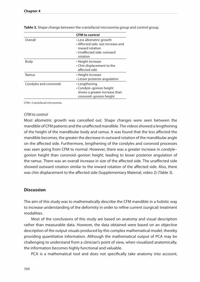
Chapter 4
104
CFM to controlMost allometric growth was cancelled out. Shape changes were seen between the mandible of CFM patients and the unaffected mandible. The videos showed a lengthening of the height of the mandibular body and ramus. It was found that the less affected the mandible becomes, the greater the decrease in outward rotation of the mandibular angle on the affected side. Furthermore, lengthening of the condyles and coronoid processes was seen going from CFM to normal. However, there was a greater increase in condyle–gonion height than coronoid–gonion height, leading to lesser posterior angulation of the ramus. There was an overall increase in size of the affected side. The unaffected side showed outward rotation similar to the inward rotation of the affected side. Also, there was chin displacement to the affected side (Supplementary Material, video 2) (Table 3).
Discussion
The aim of this study was to mathematically describe the CFM mandible in a holistic way to increase understanding of the deformity in order to refine current (surgical) treatment modalities.
Most of the conclusions of this study are based on anatomy and visual description rather than measurable data. However, the data obtained were based on an objective description of the output visuals produced by this complex mathematical model, thereby providing quantitative information. Although the mathematical output of PCA may be challenging to understand from a clinician’s point of view, when visualized anatomically, the information becomes highly functional and valuable.
PCA is a mathematical tool and does not specifically take anatomy into account;
Table 3. Shape change between the craniofacial microsomia group and control group.
CFM to controlOverall • Less allometric growth
• Affected side: size increase and inward rotation
• Unaffected side: outward rotation
Body • Height increase• Chin displacement to the
affected side
Ramus • Height increase• Lesser posterior angulation
Condyles and coronoids • Lengthening• Condyle–gonion height
shows a greater increase than coronoid–gonion height
CFM= Craniofacial microsomia
PSM 20190527 Proefschrift Britt Pluijmers.indd 104 24-07-19 15:04

Characterizing the mandible using PCA
105
4
therefore, it is crucial to validate the results using anatomical knowledge of the mandible. The first mode of variation in the control population largely showed allometric growth and the subsequent modes increasingly demonstrated normal variation. This is expected, because allometric growth is very likely to contribute more towards overall variation than subtler differences in morphology, as seen in the later modes. Therefore, by being in tandem with what is expected from the growing mandible, the modes of variation validate the PCA model and allow confident conclusions to be drawn from it. Based on earlier studies, three principal components were used.13-15 As stated above, the first principal component is mostly allometric growth, which was seen in both the control and CFM patient groups. Therefore, the second and third modes of variation are the most descriptive with regard to the actual change in morphology. After the third mode of variation, the morphology changes are too subtle to differ from the second and third.
The placement of the landmarks is a crucial but time-consuming process. Although the intra-observer reliability was good due to the learning curve, the assessment of inter-observer reliability showed a significant difference in distance ranging from 0.23 mm to 3.98 mm (P < 0.05) for 38 of the 43 points. Not surprisingly, the greatest inter-observer differences in distance were seen for the ‘smooth’ surfaces of the mandible and the smallest differences were seen for the anatomical landmarks such as the fossae and foramina.
The aetiology of CFM is possibly related to a disturbed migration of the neural crest, leading to underdevelopment of the structures of the first and second branchial arches.16 The mandible, a derivative of the first branchial arch, is one of the, if not the most reported feature of CFM.6,10,17 The mandibular hypoplasia and heterogeneous phenotype cause a surgical challenge to correct the asymmetry. Besides, as these videos demonstrate, although one side most often seems to be more dominant in CFM, the unaffected side tends to be affected due to compensatory remodelling of the mandible. As described earlier development in one region could affect the entire craniofacial skeleton.18-21 This would suggest that a patient with CFM is never truly affected unilateral. Along with this concept is the influence of the underdevelopment of the mandible with the other structures of the viscerocranium.
Recent work by Schaal et al. showed significant asymmetry of the skull base in CFM, with the most significant asymmetries and restrictions of growth centred around the glenoid fossa and mastoid process.13 Distortion of the skull in this area is complex and is present in the vertical, horizontal and antero-posterior planes associated with a rotation of this part of the skull base. This concept suggest that with the increase of the hypoplasia of the skull base an increase of distortion is seen, which has effect on the growth of structures of the same region such as the temporal bone possibly leading to posterior displacement of the condyle.16 Several studies already demonstrated that the maxilla and orbit become displaced secondary to the lack of mandibular growth on the affected side.18-21 Therefore, the present authors are currently working on a study that combines the different areas of the CFM skull: the mandible, maxilla/midface, and skull base.
PSM 20190527 Proefschrift Britt Pluijmers.indd 105 24-07-19 15:04

Chapter 4
106
Conclusion
In conclusion, shape analysis based on PCA allows a mathematical description of the shape of a deformed mandible in 3D. The holistic nature of thin plate spline warping, derived from the PCA, allows a review of the shape change, which helps in understanding the actual changes in shape and may possibly aid in the refinement of current (surgical) treatment strategies.
A CFM mandible differs from a normal mandible as a result of hypoplasia leading to a rotation towards the affected side; there is both a lateral rotation with increase of posterior rotation of the condyle due to shortening of the condyle–gonial height and a longitudinal rotation with outward bending of the mandibular angle on the affected side, as well as inward bending of the angle on the unaffected side. Due to the compensatory remodelling of the mandible on the unaffected side, one could suggest that CFM is never truly unilateral.
PSM 20190527 Proefschrift Britt Pluijmers.indd 106 24-07-19 15:04
Characterizing the mandible using PCA
107
4
References
1. Grabb WC. The first and second branchial arch syndrome. Plast Reconstr Surg 1965; 36(5): 485-508.2. Melnick M. The etiology of external ear malformations and its relation to abnormalities of the
middle ear, inner ear, and other organ systems. Birth defects original article series 1980; 16(4): 303-31.3. Poswillo D. Otomandibular deformity: pathogenesis as a guide to reconstruction. Journal of
maxillofacial surgery 1974; 2(2-3): 64-72.4. Posnick JC, Ruiz RL, Tiwana PS. Craniofacial dysostosis syndromes: stages of reconstruction. Oral and
maxillofacial surgery clinics of North America 2004; 16(4): 475-91.5. Ross RB. Lateral facial dysplasia (first and second branchial arch syndrome, hemifacial microsomia).
Birth defects original article series 1975; 11(7): 51-9.6. Caron C, Pluijmers BI, Wolvius EB, et al. Craniofacial and extracraniofacial anomalies in craniofacial
microsomia: a multicenter study of 755 patients’. J Craniomaxillofac Surg 2017; 45(8): 1302-10.7. Heike CL, Luquetti DV, Hing AV. Craniofacial Microsomia Overview. 1993.8. Pruzansky S. Not all dwarfed mandibles are alike. Birth defects original article series 1969; 5: 120-9.9. Kaban LB, Moses MH, Mulliken JB. Correction of hemifacial microsomia in the growing child: a
follow-up study. The Cleft palate journal 1986; 23 Suppl 1: 50-2.10. Birgfeld CB, Heike CL, Saltzman BS, Leroux BG, Evans KN, Luquetti DV. Reliable classification of facial
phenotypic variation in craniofacial microsomia: a comparison of physical exam and photographs. Head Face Med 2016; 12: 14.
11. Bookstein FL. Shape and the information in medical images: a decade of the morphometric synthesis. Computer Vis Image Underst 1997; 66(2): 97-118.
12. O’Higgins P. The study of morphological variation in the hominid fossil record: biology, landmarks and geometry. Journal of anatomy 2000; 197 ( Pt 1): 103-20.
13. Schaal SC, Ruff C, Pluijmers BI, et al. Characterizing the skull base in craniofacial microsomia using principal component analysis. Int J Oral Maxillofac Surg 2017; 46(12): 1656-63.
14. Staal FC, Ponniah AJ, Angullia F, Ruff C, Koudstaal MJ, Dunaway D. Describing Crouzon and Pfeiffer syndrome based on principal component analysis. J Craniomaxillofac Surg 2015; 43(4): 528-36.
15. Pluijmers BI, Ponniah AJ, Ruff C, Dunaway D. Using principal component analysis to describe the Apert skull deformity and simulate its correction. Journal of plastic, reconstructive & aesthetic surgery : JPRAS 2012; 65(12): 1750-2.
16. Sze RW, Paladin AM, Lee S, Cunningham ML. Hemifacial microsomia in pediatric patients: asymmetric abnormal development of the first and second branchial arches. AJR Am J Roentgenol 2002; 178(6): 1523-30.
17. Birgfeld CB, Heike C. Craniofacial microsomia. Semin Plast Surg 2012; 26(2): 91-104.18. Kaban LB. Mandibular asymmetry and the fourth dimension. J Craniofac Surg 2009; 20 Suppl 1: 622-
31.19. Kaban LB, Moses MH, Mulliken JB. Surgical correction of hemifacial microsomia in the growing child.
Plastic and reconstructive surgery 1988; 82(1): 9-19.20. Ongkosuwito EM, van Neck JW, Wattel E, van Adrichem LN, Kuijpers-Jagtman AM. Craniofacial
morphology in unilateral hemifacial microsomia. The British journal of oral & maxillofacial surgery 2013; 51(8): 902-7.
21. Rune B, Selvik G, Sarnas KV, Jacobsson S. Growth in hemifacial microsomia studied with the aid of roentgen stereophotogrammetry and metallic implants. The Cleft palate journal 1981; 18(2): 128-46.
PSM 20190527 Proefschrift Britt Pluijmers.indd 107 24-07-19 15:04

CH
AP
TER
PSM 20190527 Proefschrift Britt Pluijmers.indd 108 24-07-19 15:04

5
Is there a Difference in Orbital Volume Between Affected and Unaffected sides
In Patients with Unilateral Craniofacial Microsomia
Maria N. Gribova, Britt I. Pluijmers, Cory M. Resnick, Cornelia J. J. M. Caron, Alessandro Borghi, Maarten J. Koudstaal, Bonnie L. Padwa, DMD, MD
Journal of Oral Maxillofacial Surgery 2018 Dec;76(12):2625-2629. doi: 10.1016/j.joms.2018.04.038. Epub 2018 May31.
PSM 20190527 Proefschrift Britt Pluijmers.indd 109 24-07-19 15:04
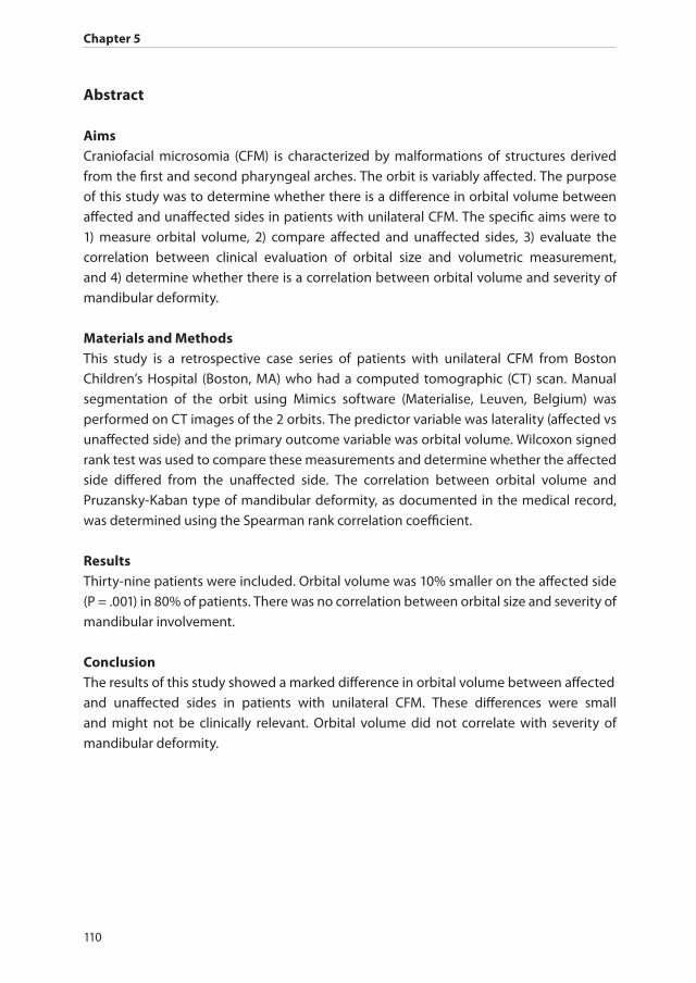
Chapter 5
110
Abstract
AimsCraniofacial microsomia (CFM) is characterized by malformations of structures derived from the first and second pharyngeal arches. The orbit is variably affected. The purpose of this study was to determine whether there is a difference in orbital volume between affected and unaffected sides in patients with unilateral CFM. The specific aims were to 1) measure orbital volume, 2) compare affected and unaffected sides, 3) evaluate the correlation between clinical evaluation of orbital size and volumetric measurement, and 4) determine whether there is a correlation between orbital volume and severity of mandibular deformity.
Materials and MethodsThis study is a retrospective case series of patients with unilateral CFM from Boston Children’s Hospital (Boston, MA) who had a computed tomographic (CT) scan. Manual segmentation of the orbit using Mimics software (Materialise, Leuven, Belgium) was performed on CT images of the 2 orbits. The predictor variable was laterality (affected vs unaffected side) and the primary outcome variable was orbital volume. Wilcoxon signed rank test was used to compare these measurements and determine whether the affected side differed from the unaffected side. The correlation between orbital volume and Pruzansky-Kaban type of mandibular deformity, as documented in the medical record, was determined using the Spearman rank correlation coefficient.
ResultsThirty-nine patients were included. Orbital volume was 10% smaller on the affected side (P = .001) in 80% of patients. There was no correlation between orbital size and severity of mandibular involvement.
Conclusion The results of this study showed a marked difference in orbital volume between affectedand unaffected sides in patients with unilateral CFM. These differences were small and might not be clinically relevant. Orbital volume did not correlate with severity of mandibular deformity.
PSM 20190527 Proefschrift Britt Pluijmers.indd 110 24-07-19 15:04

Orbital volume in craniofacial microsomia
111
5
Introduction
Craniofacial microsomia (CFM) is the most common facial anomaly following cleft lip and palate.1-3 The disorder is characterized by malformations of structures with an embryological origin in the first and second pharyngeal arches and the phenotypic presentation is variable.
The OMENS classification recognizes the most commonly affected facial structures in CFM including the Orbit, Mandible, Ear, Nerve, and Soft-tissues.4 This classification grades the orbital deformity by subjective evaluation of size and position.5 There is inconsistency in the reported incidence and severity of the orbital deformity as well as variability in the correlation between orbital involvement and the type of mandibular deformity.5,6 These differences may be due to the fact that previous studies used subjective clinical scores of orbital involvement rather than objective measurements of orbital size.
There is lack of correlation between the clinical appearance of the size of the orbit and the volume measured on CT scan.7 To the best of the authors’ knowledge, there are no reports in the literature that quantitatively measure orbital size in patients with CFM.
The purpose of this study was to determine whether there is a difference in orbital volume between affected and unaffected sides in patients with unilateral CFM. The authors hypothesize that the orbit on the affected side will be smaller than that on the unaffected side. The specific aims were to 1) measure orbital volume in patients with CFM 2) compare the volumes of the affected and unaffected sides 3) evaluate the correlation between the clinical evaluation of orbital size and the volumetric measurement and 4) determine whether there is a correlation between orbital volume and severity of mandibular deformity.
Materials and Methods
Study designThis is a retrospective study of patients with unilateral CFM who had CT images obtained as part of standard clinical care. This study was approved by the institutional review board of the committee on clinical investigation at Boston Children’s Hospital (Boston, MA; protocol number X05-08-058) and all research activities were conducted in accordance with the Declaration of Helsinki.
SampleThe study population was composed of all patients presenting for evaluation and management of CFM at Boston Children’s Hospital from 1950 through 2015. To be included in the study, patients had to have unilateral CFM and a CT scan including the 2 orbits. Patients were excluded if they had undergone a procedure in or around the orbit before
PSM 20190527 Proefschrift Britt Pluijmers.indd 111 24-07-19 15:04
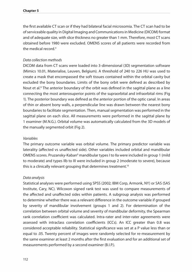
Chapter 5
112
the first available CT scan or if they had bilateral facial microsomia. The CT scan had to be of serviceable quality in Digital Imaging and Communications in Medicine (DICOM) format and of adequate size, with slice thickness no greater than 1 mm. Therefore, most CT scans obtained before 1980 were excluded. OMENS scores of all patients were recorded from the medical record.4
Data collection methodsDICOM data from CT scans were loaded into 3-dimensional (3D) segmentation software (Mimics 10.01, Materialise, Leuven, Belgium). A threshold of 240 to 226 HU was used to create a mask that encompassed the soft tissues contained within the orbital cavity but excluded the bony boundaries. Limits of the bony orbit were defined as described by Nout et al.8 The anterior boundary of the orbit was defined in the sagittal plane as a line connecting the most anterosuperior points of the supraorbital and infraorbital rims (Fig 1). The posterior boundary was defined as the anterior portion of the optic canal. In areas of thin or absent bony walls, a perpendicular line was drawn between the nearest bony boundaries to facilitate segmentation. Then, manual segmentation was performed in the sagittal plane on each slice. All measurements were performed in the sagittal plane by 1 examiner (M.N.G.). Orbital volume was automatically calculated from the 3D models of the manually segmented orbit (Fig 2).
VariablesThe primary outcome variable was orbital volume. The primary predictor variable was laterality (affected vs unaffected side). Other variables included orbital and mandibular OMENS scores. Pruzansky-Kaban9 mandibular types I to IIa were included in group 1 (mild to moderate) and types IIb to III were included in group 2 (moderate to severe), because this is a clinically relevant grouping that determines treatment.10
Data analysisStatistical analyses were performed using SPSS (2002; IBM Corp, Armonk, NY) or SAS (SAS Institute, Cary, NC). Wilcoxon signed rank test was used to compare measurements of the affected and unaffected sides within patients. A subgroup analysis was performed to determine whether there was a relevant difference in the outcome variable if grouped by severity of mandibular involvement (groups 1 and 2). For determination of the correlation between orbital volume and severity of mandibular deformity, the Spearman rank correlation coefficient was calculated. Intra-rater and inter-rater agreements were assessed with intraclass correlation coefficients (ICCs). An ICC greater than 0.8 was considered acceptable reliability. Statistical significance was set at a P value less than or equal to .05. Twenty percent of images were randomly selected for re-measurement by the same examiner at least 2 months after the first evaluation and for an additional set of measurements performed by a second examiner (B.I.P.).
PSM 20190527 Proefschrift Britt Pluijmers.indd 112 24-07-19 15:04
Orbital volume in craniofacial microsomia
113
5
Fig. 1. Sagittal computed tomogram displaying the anterior boundary of the orbit.
Fig. 2. Three-dimensional models of the orbit.
Results
PatientsOf 238 patients with unilateral CFM evaluated during the study period, 39 (23 male and 16 female; mean age, 11.5 yr; range, 1 to 44 yr) were included (Table 1). The remaining 199 patients were excluded because they had bilateral craniofacial microsomia, insufficient imaging data, or extensive surgical treatment of the orbit or associated bony structures. Sixteen patients composed group 1 (mild to moderate) and 23 composed group 2 (mode-rate to severe).
Orbital Volume For the entire sample, orbital volume on the affected side was 10 ± 41% smaller than on the unaffected side (mean affected 21501.47 ± 4840.65 mm3, mean unaffected 22467.42 ± 4179.66 mm3, p=0.001) (table 1). These differences were statistically significant in both sub-groups (group 1, p=0.021; group 2, p=0.0258) The affected side was smaller than the unaffected side in 80% of the sample.
In patients with clinically normal sized orbits (OMENS grade O0 and O2) CT volumetric measurements found that the affected side was 2 ± 6% smaller than the unaffected side (mean affected 22062.78± 4086.14 mm3, mean unaffected 22463.77 ± 4333.24 mm3, p=0.010). The affected side was smaller than the unaffected side in 82%.
There was a negative correlation between orbital volume and severity of the mandibular deformity (orbital volume decreased as the Pruzansky-Kaban severity increased), but this correlation was not statistically significant (p= 0.353).
PSM 20190527 Proefschrift Britt Pluijmers.indd 113 24-07-19 15:04
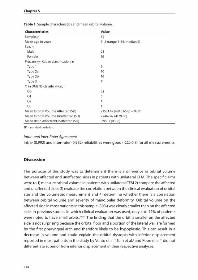
Chapter 5
114
Intra- and Inter-Rater AgreementIntra- (0.992) and inter-rater (0.982) reliabilities were good (ICC>0.8) for all measurements.
Discussion
The purpose of this study was to determine if there is a difference in orbital volume between affected and unaffected sides in patients with unilateral CFM. The specific aims were to 1) measure orbital volume in patients with unilateral CFM 2) compare the affected and unaffected sides 3) evaluate the correlation between the clinical evaluation of orbital size and the volumetric measurement and 4) determine whether there is a correlation between orbital volume and severity of mandibular deformity. Orbital volume on the affected side in most patients in this sample (80%) was clearly smaller than on the affected side. In previous studies in which clinical evaluation was used, only 4 to 12% of patients were noted to have small orbits.4,6,11 The finding that the orbit is smaller on the affected side is not surprising because the orbital floor and a portion of the lateral wall are formed by the first pharyngeal arch and therefore likely to be hypoplastic. This can result in a decrease in volume and could explain the orbital dystopia with inferior displacement reported in most patients in the study by Vento et al.4 Tuin et al.6 and Poon et al.11 did not differentiate superior from inferior displacement in their respective analyses.
Table 1. Sample characteristics and mean orbital volume.
Characteristics Value Sample, n 39
Mean age in years 11,5 (range 1-44, median 9)
Sex, n
Male 23
Female 16
Pruzansky- Kaban classification, n
Type 1 6
Type 2a 10
Type 2b 16
Type 3 7
O in OMENS classification, n
O0 32
O1 5
O2 1
O3 1
Mean Orbital Volume Affected (SD) 21501.47 (4840.65) p = 0.001
Mean Orbital Volume Unaffected (SD) 22467.42 (4179.66)
Mean Ratio Affected/Unaffected (SD) 0.9532 (0,133)
SD = standard deviation.
PSM 20190527 Proefschrift Britt Pluijmers.indd 114 24-07-19 15:04
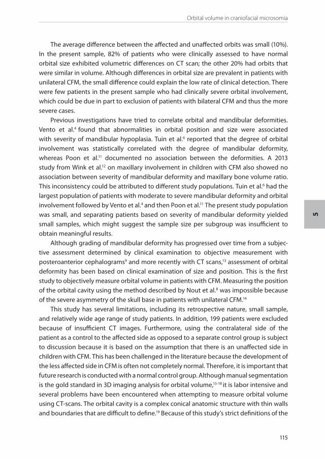
Orbital volume in craniofacial microsomia
115
5
The average difference between the affected and unaffected orbits was small (10%). In the present sample, 82% of patients who were clinically assessed to have normal orbital size exhibited volumetric differences on CT scan; the other 20% had orbits that were similar in volume. Although differences in orbital size are prevalent in patients with unilateral CFM, the small difference could explain the low rate of clinical detection. There were few patients in the present sample who had clinically severe orbital involvement, which could be due in part to exclusion of patients with bilateral CFM and thus the more severe cases.
Previous investigations have tried to correlate orbital and mandibular deformities. Vento et al.4 found that abnormalities in orbital position and size were associated with severity of mandibular hypoplasia. Tuin et al.6 reported that the degree of orbital involvement was statistically correlated with the degree of mandibular deformity, whereas Poon et al.11 documented no association between the deformities. A 2013 study from Wink et al.12 on maxillary involvement in children with CFM also showed no association between severity of mandibular deformity and maxillary bone volume ratio. This inconsistency could be attributed to different study populations. Tuin et al.6 had the largest population of patients with moderate to severe mandibular deformity and orbital involvement followed by Vento et al.4 and then Poon et al.11 The present study population was small, and separating patients based on severity of mandibular deformity yielded small samples, which might suggest the sample size per subgroup was insufficient to obtain meaningful results.
Although grading of mandibular deformity has progressed over time from a subjec-tive assessment determined by clinical examination to objective measurement with posteroanterior cephalograms9 and more recently with CT scans,13 assessment of orbital deformity has been based on clinical examination of size and position. This is the first study to objectively measure orbital volume in patients with CFM. Measuring the position of the orbital cavity using the method described by Nout et al.8 was impossible because of the severe asymmetry of the skull base in patients with unilateral CFM.14
This study has several limitations, including its retrospective nature, small sample, and relatively wide age range of study patients. In addition, 199 patients were excluded because of insufficient CT images. Furthermore, using the contralateral side of the patient as a control to the affected side as opposed to a separate control group is subject to discussion because it is based on the assumption that there is an unaffected side in children with CFM. This has been challenged in the literature because the development of the less affected side in CFM is often not completely normal. Therefore, it is important that future research is conducted with a normal control group. Although manual segmentation is the gold standard in 3D imaging analysis for orbital volume,15-18 it is labor intensive and several problems have been encountered when attempting to measure orbital volume using CT-scans. The orbital cavity is a complex conical anatomic structure with thin walls and boundaries that are difficult to define.19 Because of this study’s strict definitions of the
PSM 20190527 Proefschrift Britt Pluijmers.indd 115 24-07-19 15:04
Chapter 5
116
bony orbit and the use of previously validated methodology,8 there was good intra- and inter-observer agreement. However, given the nature of the images, it was impossible to blind the examiners to the primary predictor variable (affected vs unaffected side) and this could have introduced some bias. The present study would be improved by documentation of globe or periorbital tissue measurements and delineating the separate orbital bones and their individual contributions to the smaller orbital volume in patients with CFM.
The affected side exhibited a notably smaller volume in patients with unilateral CFM compared with the unaffected side. Although these differences were small and might not be clinically relevant, they add to a broader understanding of the subclinical spectrum of CFM.
Acknowledgments
The authors acknowledge the help provided by Prof Dr Eppo B. Wolvius from the Erasmus Medical Centre in Rotterdam in the development of this project and Kimberly Daniels, MS, Doctoral Candidate of the Drexel University Dornsife School of Public Health for her help with the statistics.
PSM 20190527 Proefschrift Britt Pluijmers.indd 116 24-07-19 15:04

Orbital volume in craniofacial microsomia
117
5
References
1. Grabb WC. The first and second branchial arch syndrome. Plastic and reconstructive surgery 1965; 36(5): 485-508.
2. Poswillo D. Otomandibular deformity: pathogenesis as a guide to reconstruction. Journal of maxillofacial surgery 1974; 2(2-3): 64-72.
3. Ross RB. Lateral facial dysplasia (first and second branchial arch syndrome, hemifacial microsomia). Birth defects original article series 1975; 11(7): 51-9.
4. Vento AR, LaBrie RA, Mulliken JB. The O.M.E.N.S. classification of hemifacial microsomia. The Cleft palate-craniofacial journal : official publication of the American Cleft Palate-Craniofacial Association 1991; 28(1): 68-76; discussion 7.
5. Horgan JE, Padwa BL, LaBrie RA, Mulliken JB. OMENS-Plus: analysis of craniofacial and extracranio-facial anomalies in hemifacial microsomia. The Cleft palate-craniofacial journal : official publication of the American Cleft Palate-Craniofacial Association 1995; 32(5): 405-12.
6. Tuin AJ, Tahiri Y, Paine KM, Paliga JT, Taylor JA, Bartlett SP. Clarifying the relationships among the different features of the OMENS+ classification in craniofacial microsomia. Plastic and reconstructive surgery 2015; 135(1): 149e-56e.
7. Raskin EM, Millman AL, Lubkin V, della Rocca RC, Lisman RD, Maher EA. Prediction of late enophthalmos by volumetric analysis of orbital fractures. Ophthalmic plastic and reconstructive surgery 1998; 14(1): 19-26.
8. Nout E, van Bezooijen JS, Koudstaal MJ, et al. Orbital change following Le Fort III advancement in syndromic craniosynostosis: quantitative evaluation of orbital volume, infra-orbital rim and globe position. Journal of cranio-maxillo-facial surgery : official publication of the European Association for Cranio-Maxillo-Facial Surgery 2012; 40(3): 223-8.
9. Kaban LB, Mulliken JB, Murray JE. Three-dimensional approach to analysis and treatment of hemifacial microsomia. The Cleft palate journal 1981; 18(2): 90-9.
10. Kaban LB, Moses MH, Mulliken JB. Surgical correction of hemifacial microsomia in the growing child. Plastic and reconstructive surgery 1988; 82(1): 9-19.
11. Poon CC, Meara JG, Heggie AA. Hemifacial microsomia: use of the OMENS-Plus classification at the Royal Children’s Hospital of Melbourne. Plastic and reconstructive surgery 2003; 111(3): 1011-8.
12. Wink JD, Paliga JT, Tahiri Y, Goldstein JA, Taylor JA, Bartlett SP. Maxillary involvement in hemifacial microsomia: an objective three-dimensional analysis of the craniofacial skeleton. The Journal of craniofacial surgery 2014; 25(4): 1236-40.
13. Swanson JW, Mitchell BT, Wink JA, Taylor JA, Bartlett SP. Surgical Classification of the Mandibular Deformity in Craniofacial Microsomia Using 3-Dimensional Computed Tomography. Plastic and reconstructive surgery Global open 2016; 4(1): e598.
14. Schaal SC, Ruff C, Pluijmers BI, et al. Characterizing the skull base in craniofacial microsomia using principal component analysis. International journal of oral and maxillofacial surgery 2017; 46(12): 1656-63.
15. Jansen J, Schreurs R, Dubois L, Maal TJ, Gooris PJ, Becking AG. Orbital volume analysis: validation of a semi-automatic software segmentation method. International journal of computer assisted radiology and surgery 2016; 11(1): 11-8.
16. Scolozzi P, Jaques B. Computer-aided volume measurement of posttraumatic orbits reconstructed with AO titanium mesh plates: accuracy and reliability. Ophthalmic plastic and reconstructive surgery 2008; 24(5): 383-9.
17. Smektala T, Nysjo J, Thor A, et al. Three-Dimensional Eyeball and Orbit Volume Modification After LeFort III Midface Distraction. The Journal of craniofacial surgery 2015; 26(5): 1652-5.
18. Deveci M, Ozturk S, Sengezer M, Pabuscu Y. Measurement of orbital volume by a 3-dimensional software program: an experimental study. Journal of oral and maxillofacial surgery : official journal of the American Association of Oral and Maxillofacial Surgeons 2000; 58(6): 645-8.
19. Wagner ME, Gellrich NC, Friese KI, et al. Model-based segmentation in orbital volume measurement with cone beam computed tomography and evaluation against current concepts. International journal of computer assisted radiology and surgery 2016; 11(1): 1-9.
PSM 20190527 Proefschrift Britt Pluijmers.indd 117 24-07-19 15:04

PAR
T
PSM 20190527 Proefschrift Britt Pluijmers.indd 118 24-07-19 15:04

IV
Surgery
PSM 20190527 Proefschrift Britt Pluijmers.indd 119 24-07-19 15:04

CH
AP
TER
PSM 20190527 Proefschrift Britt Pluijmers.indd 120 24-07-19 15:04

6
Mandibular reconstruction in the growing patient with unilateral
craniofacial microsomia: a systematic review
Britt I. Pluijmers, Cornelia J. J. M.Caron, David J. Dunaway, Eppo B. Wolvius, Maarten J. Koudstaal.
International Journal of Oral and Maxillofacial Surgery. 2014 Mar;43(3):286-95. doi: 10.1016/j.ijom.2013.11.001. Epub 2013 Dec 12.
PSM 20190527 Proefschrift Britt Pluijmers.indd 121 24-07-19 15:04

Chapter 6
122
Abstract
AimsThe purpose of this systematic review is to provide an overview of the surgical correction of the mandible in unilateral craniofacial microsomia (UCM) performed in the growing patient, and its long-term outcome and stability.
Material and methodsThe following databases were searched: PubMed, Embase, Cochrane, and Web of Science. Articles reporting prospective and retrospective studies of patients not older than 16 years (N=4) who had undergone surgical correction of a craniofacial microsomia spectrum condition using grafts, osteotomies, distraction, or combinations of these, were reviewed. The period of follow-up was selected to be 1 year. After inclusion, the articles were evaluated on short- and long-term outcomes, relapse, and any increase in asymmetry following treatment.
ResultsThirty of 1611 articles were included in the qualitative synthesis. Analysis of the surgical mandibular correction of UCM showed that the outcome is not so much treatment- dependent, but patient-dependent, i.e. deformity gradation-dependent. The type I– IIa Pruzansky–Kaban patient had the best results with regard to minimal relapse and/or minimal increase in asymmetry.
ConclusionSingle-stage correction of the asymmetry should be postponed until the permanent dentition stage. It can be concluded that in the treatment of the severely hypoplastic mandible, the patient will benefit from a multi-stage treatment protocol if indicated for functional or psychological problems.
PSM 20190527 Proefschrift Britt Pluijmers.indd 122 24-07-19 15:04

Mandibular reconstruction in the growing patient
123
6
Introduction
Deformities of the unilateral craniofacial microsomia (UCM) spectrum have proven difficult to treat in the growing patient, due to their heterogeneous presentation. The structures of the first and second branchial arches involved are the maxilla, zygoma, mandible, external and middle ear, facial and trigeminal nerves, muscles of mastication, and overlying soft tissues.1 A straightforward classification system is essential to improve our knowledge of the deformity. The most commonly used classification system is that provided by Pruzansky2 and later modified by Kaban et al.3 although other systems have been reported.4,5 The Pruzansky–Kaban system consists of four types. Type I is a small mandible with normal morphology. Type IIa is a mandibular ramus abnormal in both size and shape; type IIb is a mandibular ramus and temporomandibular joint (TMJ) abnormal in size, morphology, and location. A type III deformity consists of an absent ramus, condyle, and TMJ.
Correction of the asymmetric mandible by mandibular osteotomies and bone grafts has been performed in adults since 1928.6 The first report of surgery in children with UCM dates from 1941.2 Up to the 1980s, the overall tendency was for reconstruction of the severely hypoplastic mandible with autogenous grafts, mostly costochondral grafts; however rib, iliac crest, fibula, and temporal bone were also used.7,8 In the early 1990s, McCarthy et al. were the first to report successful lengthening by gradual distraction of the mandible.9 This changed the approach to craniofacial correction in a revolutionary way.10 However, two decades after the first report, it has become clear that distraction osteogenesis (DO) is not the ideal solution for every patient. Extensive work by Nagy et al. showed a lack of statistical evidence to support the use of DO before the permanent dentition stage as a single treatment modality.11 There have been increasing numbers of reports on multi stage correction of the facial deformity with the use of both DO and grafts.12-15
The purpose of this systematic review is to provide an overview of the surgical correction of the mandible in UCM in the growing patient, and its long-term outcome and stability.
Material and methods
The PRISMA statement16 was used as a guideline.
Search strategyThe following databases were searched: PubMed (until 18 October 2012), Embase (until 18 October 2012), Cochrane (until 18 October 2012), and Web of Science (until 18 October 2012).
PSM 20190527 Proefschrift Britt Pluijmers.indd 123 24-07-19 15:04

Chapter 6
124
We focused on search terms for the congenital deformity of interest and the surgical intervention. Both free text words and medical subject heading (MeSH) terms were used. The heading sequence (hemifacial microsom*[tw] OR hemi facial microsom*[tw] OR hemifacial hypoplas*[tw] OR Hemi facial hypo- plas*[tw] OR craniofacial microsom*[tw] OR facial asymmetr*[tw] OR face asym- metr*[tw] OR asymmetric fac*[tw] OR (goldenhar*[tw] OR otomandibular dys- ostosis[tw] OR oculoauricul*[tw] OR facioauricul*[tw] OR facio-auricul*[tw] OR oculo-auricul*[tw] OR OAV[tw] OR FAV[tw] OR ((lateral[tw] OR unilater- al[tw] OR hemilateral[tw]) AND (facial[tw] OR craniofacial[tw])) OR (branchial[tw] AND (arch[tw] OR arches[tw])) AND (syndrom*[tw] OR defect*[tw] OR anomal*[tw] OR dys- plas*))) AND (distract*[tw] OR osteodis- tract*[tw] OR osteotomy[mesh] OR osteotom*[tw] OR bone transplant*[tw] OR bone graft*[tw] OR bone auto- graft*[tw] OR bone allograft*[tw] OR osseous flap*[tw]) NOT (animals[mesh] NOT humans[mesh]) AND eng[la] was selected. Reference lists of included studies were hand-searched for additional studies of interest.
Inclusion criteriaArticles reporting prospective and retrospective studies of children and adolescents not older than 16 years of age (N≥4) who had undergone surgical correction of a craniofacial microsomia spectrum condition by grafts, osteotomies, DO, or a combination of these interventions, were included. The period of follow-up was selected to be ≥1 year.
Data extraction and analysisDuplicates were removed. Two authors reviewed the literature individually (BIP and CJJMC). Reports on heterogeneous patient groups and/or study groups with fewer than four patients were excluded due to possible bias and an expected low level of evidence.17 Articles that met the inclusion criteria or for which the abstract was lacking information, were obtained as full-text articles. Next, full- text articles were reviewed in accordance with the inclusion criteria. Only the data of patients with objective information were included in the quantitative assessment. If there were multiple publications by the same author (group), the author was contacted and the studies were combined.
Articles were graded on quality of evidence using the Oxford Centre for Evidence-Based Medicine (OCEBM) criteria.18 Data on the number of patients, classification, type of surgery, type of implanted material, average age during the intervention, average length of follow-up, number of relapses, increases in asymmetry, and the number of complications, where available, were tabulated.
Results
The initial search identified 2471 papers. Thirty articles met the inclusion criteria. 3,14,15,19-
45 (fig. 1, table1) To prevent outcome bias, a total of 173,19,21,22,25,27,30-35,38,40-43 articles had to
PSM 20190527 Proefschrift Britt Pluijmers.indd 124 24-07-19 15:04

Mandibular reconstruction in the growing patient
125
6be combined. An overlap in patient data was observed in six different combined article study groups. For example, Meazzini et al. have published three studies–in 2005, 2008, and 2012.19,21,25 The patients in these articles were likely to be the same patients in each independent study (which the author confirmed), therefore these three articles were combined and approached as one study. The most recent data of the combined articles were used; however additional information from the older articles was used to complete any missing information, if needed. All in all, a total of 19 studies were used for the analysis. All studies included met the OCEBM criteria for level 4 evidence as case series or case–control studies (Table 1).The age of patients ranged from 2 years till 15.3 years, with a mean age of 8.9 years (n=247). The mean follow-up time was 4.3 years (n=230). Two hundred and twenty-six patients had a follow-up time of at least 1 year post surgery. However, 8 studies had a follow-up time of more than 5 years.14,19,22,23,25,38,39,42,43,45
Fig. 1. Data extraction flowchart, according to the PRISMA statement.16
PSM 20190527 Proefschrift Britt Pluijmers.indd 125 24-07-19 15:04

Chapter 6
126
Table 1. Studies Meeting criteria for inclusion in current systematic review.
Citation OC
EBM
leve
l of e
vid
ence
Methodology Ther
apy
No
. of p
atie
nts
Mea
n a
ge
at s
urg
ery*
Mea
n fo
llow
-up
tim
e*
(no
of p
atie
nts
≥ 1
yea
r)
Wan et al. (2011)14 4 Retrospective CS DOGraftG+DO
4 27
16/27
7.49.9
13.9
13(31)
Gui et al. (2011)15 4 Retrospective CS G+DO 21 14.4 3 (17)
Santamaria et al. (2008)20 4 Retrospective CS GraftG+DO
102/10
7.2 3.8 (10)
Meazzini et al. (2012)19 4 Prospective CCS DO 14 5.9 11(14)
Meazzini et al. (2008)21 4 Prospective CCS DO 17 5.6 5(17)
Meazzini et al. (2005)25 4 Prospective CS DO 8 5.6 5.8(8)
Chow et al. (2008)22 4 Retrospective CS DO 4 9.5 7(4)
Trahar et al (2003)27 4 Prospective CS DO 6 10.4 2(6)
Shetye et al. (2006)23 4 Retrospective CS DO 12 4.8 7(12)
Scolozzi et al. (2006)24 4 Retrospective CS DO 5 8.2 2.2(5)
Baek and Kim (2005)26 4 Retrospective CS DO 19 6.4 2.7(19)
Huisinga-Fischer et al. (2003)28 4 Prospective CS DO 8 10.0 2.5(8)
Satoh et al. (2002)29 4 Prospective CS DO 10 9.2 1.3-3.3(10)
Rachmiel et al. (2001)31 4 Retrospective CS DO 11 6-12 1-2(11)
Rachmiel et al. (2000)32 4 Retrospective CS DO 22 6-14 1(22)
Kusnoto et al. (1999)36 4 Retrospective CS DO 7 2.4 1.5(7)
Huang et al. (1999)37 4 Prospective CS DO 5 Child 1(5)
Cerajewska and Singh (2002)30 4 Prospective CS Graft 14 9 2 (ND)
Hay et al. (2000)34 4 Prospective CS Graft 14 9 3 (14)
Hay and Singh (2000)33 4 Prospective CS Graft 15 10 3.5 (15)
Singh et al (1999)35 4 Prospective CS Graft 14 9 3 (14)
Padwa et al. (1998)38 4 Retrospective CS Graft 33 6.2 5.5(33)
Mulliken et al. (1989)40 4 Retrospective CS Graft 8 6.5 4.5(8)
Kaban et al. (1988)41 4 Retrospective CS E&R Graft
611
11.26.1
4.2(10)4.4(11)
Kaban et al. (1986)3 4 Retrospective CS E&R Graft
611
ND ND
≥1(17)≥1(17)
Munro et al. (1989)39 4 Retrospective CS Graft 22 9.6 1-9(22)
Ousterhout and Vargervik (1987)42 4 Prospective CS Graft 14 12.5 5.2(14)
Vargervik et al. (1986)43 4 Prospective CS Graft 14 12.5 5,2(14)
Ortiz-Monasterio (1982)44 4 Retrospective CS Graft 6 3.5-5 -3 (ND)
Converse et al. (1973)45 4 Retrospective CS Graft 12 8.5 3-12(12)
OCEBM: Center for Evidence-Based Medicine, CS: case series, CCS: case-control study, DO: distraction osteogenesis, G: graft ND: No data available, E&R: Elongation and rotation due osteotomy,*in years
PSM 20190527 Proefschrift Britt Pluijmers.indd 126 24-07-19 15:04
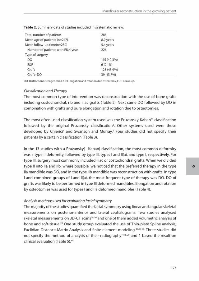
Mandibular reconstruction in the growing patient
127
6
Classification and TherapyThe most common type of intervention was reconstruction with the use of bone grafts including costochondral, rib and iliac grafts (Table 2). Next came DO followed by DO in combination with grafts and pure elongation and rotation due to osteotomies.
The most often used classification system used was the Pruzansky-Kaban41 classification followed by the original Pruzansky classification2. Other systems used were those developed by Chierici4 and Swanson and Murray.5 Four studies did not specify their patients by a certain classification (Table 3).
In the 13 studies with a Pruzansky(– Kaban) classification, the most common deformity was a type II deformity, followed by type III, types I and II(a), and type I, respectively. For type III, surgery most commonly included iliac or costochondral grafts. When we divided type II into IIa and IIb, where possible, we noticed that the preferred therapy in the type IIa mandible was DO, and in the type IIb mandible was reconstruction with grafts. In type I and combined groups of I and II(a), the most frequent type of therapy was DO. DO of grafts was likely to be performed in type III deformed mandibles. Elongation and rotation by osteotomies was used for types I and IIa deformed mandibles (Table 4).
Analysis methods used for evaluating facial symmetryThe majority of the studies quantified the facial symmetry using linear and angular skeletal measurements on posterior-anterior and lateral cephalograms. Two studies analysed skeletal measurements on 3D-CT scans28,36 and one of them added volumetric analysis of bone and soft-tissue.28 One study group evaluated the use of Thin-plate Spline analysis, Euclidian Distance Matrix Analysis and finite element modeling.30,33-35 Three studies did not specify the method of analysis of their radiography14,15,20 and 1 based the result on clinical evaluation (Table 5).44
Table 2. Summary data of studies included in systematic review.
Total number of patients 285
Mean age of patients (n=247) 8.9 years
Mean follow-up time(n=230) 5.4 years
Number of patients with FU≥1year 226
Type of surgery
DO 115 (40.3%)
E&R 6 (2.1%)
Graft 125 (43.9%)
Graft+DO 39 (13.7%)
DO: Distraction Osteogenesis, E&R: Elongation and rotation due osteotomy, FU: Follow-up.
PSM 20190527 Proefschrift Britt Pluijmers.indd 127 24-07-19 15:04
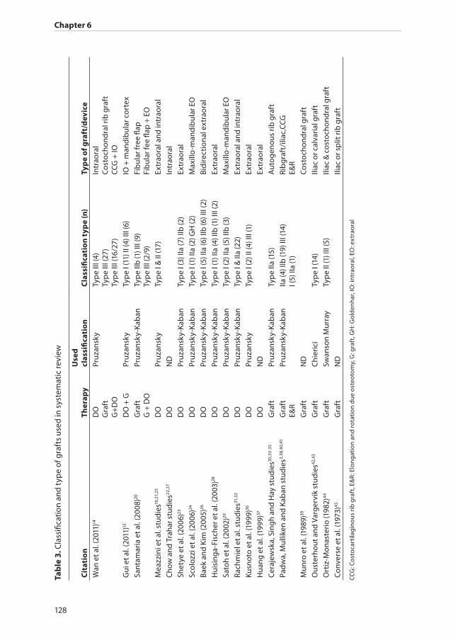
Chapter 6
128
Tab
le 3
. Cla
ssifi
cati
on a
nd t
ype
of g
raft
s us
ed in
sys
tem
atic
revi
ew
Cit
atio
nTh
erap
yU
sed
cl
assi
fica
tio
nC
lass
ifica
tio
n t
ype
(n)
Typ
e o
f gra
ft/d
evic
eW
an e
t al.
(201
1)14
D
OG
raft
G+
DO
Pruz
ansk
yTy
pe
III (4
) Ty
pe
III (2
7)
Typ
e III
(16/
27)
Intr
aora
l C
osto
chon
dra
l rib
gra
ftCC
G +
IO
Gui
et a
l. (2
011)
15
DO
+ G
Pruz
ansk
yTy
pe
I (11
) II (
4) II
I (6)
IO +
man
dib
ular
cor
tex
Sant
amar
ia e
t al.
(200
8)20
G
raft
G +
DO
Pruz
ansk
y-Ka
ban
Typ
e IIb
(1) I
II (9
) Ty
pe
III (2
/9)
Fib
ular
free
flap
Fi
bul
ar fe
e fla
p +
EO
Mea
zzin
i et a
l. st
udie
s19,2
1,25
DO
Pruz
ansk
yTy
pe
I & II
(17)
Extr
aora
l and
intr
aora
l
Cho
w a
nd T
raha
r stu
die
s22,2
7D
ON
DIn
trao
ral
Shet
ye e
t al.
(200
6)23
DO
Pruz
ansk
y-Ka
ban
Typ
e I (
3) II
a (7
) IIb
(2)
Extr
aora
l
Scol
ozzi
et a
l. (2
006)
24D
OPr
uzan
sky-
Kab
anTy
pe
I (1)
IIa
(2) G
H (2
)M
axill
o-m
and
ibul
ar E
O
Baek
and
Kim
(200
5)26
DO
Pruz
ansk
y-Ka
ban
Ty
pe
I (5)
IIa
(6) I
Ib (6
) III
(2)
Bid
irec
tion
al e
xtra
oral
Hui
sing
a-Fi
sche
r et a
l. (2
003)
28D
OPr
uzan
sky-
Kab
an
Typ
e I (
1) II
a (4
) IIb
(1) I
II (2
)Ex
trao
ral
Sato
h et
al.
(200
2)29
DO
Pruz
ansk
y-Ka
ban
Typ
e I (
2) II
a (5
) IIb
(3)
Max
illo
-man
dib
ular
EO
Rach
mie
l et a
l. st
udie
s31,3
2D
OPr
uzan
sky-
Kab
anTy
pe
I & II
a (2
2)Ex
trao
ral a
nd in
trao
ral
Kusn
oto
et a
l. (1
999)
36D
OPr
uzan
sky
Typ
e I (
2) II
(4) I
II (1
)Ex
trao
ral
Hua
ng e
t al.
(199
9)37
DO
ND
Extr
aora
l
Cer
ajew
ska,
Sin
gh a
nd H
ay s
tud
ies30
,33-
35
Gra
ftPr
uzan
sky-
Kab
anTy
pe
IIa (1
5)A
utog
enou
s ri
b g
raft
Pad
wa,
Mul
liken
and
Kab
an s
tud
ies3,
38,4
0,41
Gra
ftE&
RPr
uzan
sky-
Kab
anIIa
(4) I
Ib (1
9) II
I (14
) I (
5) II
a (1
)Ri
bgr
aft/
iliac
,CCG
E&
R
Mun
ro e
t al.
(198
9)39
Gra
ftN
DC
osto
chon
dra
l gra
ft
Ous
terh
out a
nd V
arg
ervi
k st
udie
s42,4
3G
raft
Chi
eric
iTy
pe
I (14
)Ili
ac o
r cal
vari
al g
raft
Ort
iz-M
onas
teri
o (1
982)
44
Gra
ftSw
anso
n M
urra
yTy
pe
II (1
) III
(5)
Iliac
& c
osto
chon
dra
l gra
ft
Con
vers
e et
al.
(197
3)45
Gra
ftN
DIli
ac o
r sp
lit ri
b g
raft
CCG
: Cos
toca
rtila
gin
ous
rib
gra
ft, E
&R:
Elo
ng
atio
n an
d ro
tati
on d
ue
oste
otom
y, G
: gra
ft, G
H: G
old
enh
ar, I
O: i
ntra
oral
, EO
: ext
raor
al
PSM 20190527 Proefschrift Britt Pluijmers.indd 128 24-07-19 15:04

Mandibular reconstruction in the growing patient
129
6
Surgical outcomeMost studies showed good results early post-operatively (Table 6). In the majority of DO and graft studies for Pruzansky(-Kaban) type II-III (Table 7 and 8) an increase of facial asymmetry was seen, due to relapse, bone resorption or as suggested19,21,22,25,27 due to the fact that the affected side could not keep up with the growth of the none affected side. The most stable results were seen in the milder deformity type I-IIa in osteotomy and DO studies. However, the combination of DO with grafts, proved to give the best long-term results for both mild and severe deformed mandibles (Pruzansky-Kaban I-III).14,15,20
ComplicationsNine studies failed to provide any information regarding complications (Table 9).
Among the minor complications described in other studies were: transient hypesthesia, nerve palsy (mandibular), skin infections, and hardware problems related to distraction devices. More severe complications reported were TMJ ankylosis (n=3), graft failure (n=7), fibrous nonunion (n=2), and costochondral graft overgrowth (n=4).
Table 4. Summary Pruzansky Kaban classification (n=220)
Type Therapy Percentage (n)Type I 13.6% (n=30)
DO (intraoral and extraoral) 6.3% (n=14)
Graft +DO (intraoral + mandibular cortex) 5% (n=11)
E&R 2.3% (n=5)
Type I & II (a) 17.7% (n=39)
DO (intraoral and extraoral) 17.7% (n=39)
Type II 38.1% (n=84)
DO (extraoral) 1.8% (n=4)
Graft +DO (intraoral + mandibular cortex) 1.8% (n=4)
Type IIa 20% (n=44)
DO (extraoral) 11%(n=24)
E&R 0.4%(n=1)
Graft (iliac/ribgraft) 8.6% (n=19)
Type IIb 14.5% (n=32)
DO (extraoral) 5.5% (n=12)
Graft (FFF or CCG) 9% (n=20)
Type III 30.6% (n=67)
DO (intraoral and extraoral) 5% (n=11)
Graft (FFF or CCG) 14.5%(n=32)
DO+ Graft (intraoral and extraoral + CCG+MC+FFF) 11% (n=24)
CCG: Costocartilaginous rib graft, DO: distraction osteogenesis, E&R: Elongation and rotation due osteotomy, FFF: Fibular Free Flap, MC: Mandibular cortex
PSM 20190527 Proefschrift Britt Pluijmers.indd 129 24-07-19 15:04

Chapter 6
130
Table 5. Evaluation of facial asymmetry
Citation Therapy Analysis methodWan et al. (2011)14 DO
GraftG+DO
Analysis (not specified) of panorex and CTscan, and clinical view
Gui et al. (2011)15 DO + G Analysis (not specified) of radiography and MP
Santamaria et al. (2008)20 GraftG + DO
Analysis (not specified) of OPG, PA& lateral cephalogram and clinical view
Meazzini et al. studies19,21,25 DO Ratio’s on OPG, Angular measurements on PA cephalogram and MP
Chow and Trahar studies22,27 DO Linear skeletal measurements on OPG, PA&lateral cephalogram and SMV
Shetye et al. (2006)23 DO Linear skeletal measurements on PA&lateral cephalogram
Scolozzi et al. (2006)24 DO Linear skeletal measurements on OPG, PA&lateral cephalogram and MP
Baek and Kim (2005)26 DO Linear and angular measurements on PA&lateral cephalogram
Huisinga-Fischer et al. (2003)28 DO Horizontal, vertical, angular and volumetric measurements on 3DCT
Satoh et al. (2002)29 DO Linear skeletal measurements on PA&lateral cephalogram
Rachmiel et al. studies31,32 DO Linear and angular skeletal measurements on OPG and PA cephalogram
Kusnoto et al. (1999)36 DO 3D skeletal measurements from PA&lateral cephalogram
Huang et al. (1999)37 DO Linear and angular skeletal measurements on OPG, PA&lateral cephalogram
Cerajewska, Singh and Hay studies30,33-35 Graft EDMA, TPS and FEM on PA&lateral cephalogram
Padwa, Mulliken and Kaban studies3,38,40,41
GraftE&R
Linear and angular skeletal measurements on OPG PA& lateral cephalogram
Munro et al. (1989)39 Graft Linear measurements on OPG PA& lateral cephalogram
Ousterhout and Vargervik studies42,43 Graft Linear measurements on OPG PA& lateral cephalogram
Ortiz-Monasterio (1982)44 Graft Clinical view
Converse et al. (1973)45 Graft Linear measurements on PA cephalogram
DO: distraction osteogenesis, E&R: Elongation and rotation due osteotomy, EDMA: Eucliean Distance Matrix Analysis, FEM: finite element modeling, G: graft MP: medical photography, OPG: Orthopantomogram, PA: Posterior-Anterior, SMV: submentovertex, TPS: Thin-plate spline analysis
PSM 20190527 Proefschrift Britt Pluijmers.indd 130 24-07-19 15:04

Mandibular reconstruction in the growing patient
131
6
Table 6. Short-term outcomes
Citation Therapy Short term outcomeWan et al. (2011)14 DO
Graft
G+DO
Mean distraction length 24,7 mm, 1/4 redistraction 6/27 ribgraft failuresuccessful regraft 16/27 undergrowth graft 16/27 successful distraction of ribgraft
Gui et al. (2011)15 DO + G 18/21 patient satisfaction 1/21 gonioplasty, 2/21 Medpor 100% satisfaction
Santamaria et al. (2008)20 Graft
G + DO
9/10 successful flap survival, 2/10 undergrowth graft 2/10 successful distraction of fibular graft
Meazzini et al. studies19,21,25 DO Extremely satisfying ratio affected: non affected side
Chow and Trahar studies22,27 DO Moderate improvement maxillary&ramus height and mandibular length
Shetye et al. (2006)23 DO Ramus height ratio affected: non affected 0,93 improvement occlusal cant
Scolozzi et al. (2006)24 DO 3/5 complete horizontalization occlusal plane, 2/5 2 degree cant
Baek and Kim (2005)26 DO I&IIa increase gonial angle, increase ramus, improvement occlusionIIb&III retrognathic mandible, less increase ramus, less improvement occlusion
Huisinga-Fischer et al. (2003)28 DO Increase of mandible length, increase soft tissue
Satoh et al. (2002)29 DO Correction of occlusal plane and chin deviation
Rachmiel et al. studies31,32 DO 17-21 mm bone elongation mandible
Kusnoto et al. (1999)36 DO Increase of body length>ramus height>total length of mandible
Huang et al. (1999)37 DO Increase of mandibular length
Cerajewska, Singh and Hay studies30,33-35 Graft Increase mandibular length body and ramus
Padwa, Mulliken and Kaban studies3,38,40,41
GraftE&R
Increase symmetry and levelling occlusal plane Increase symmetry and levelling occlusal plane
Munro et al. (1989)39 Graft 16/18 facial growth
Ousterhout and Vargervik studies42,43 Graft Increase symmetry
Ortiz-Monasterio (1982)44 Graft Overcorrection occlusal plane, increased symmetry
Converse et al. (1973)45 Graft Satisfactory occlusal plane
DO: distraction osteogenesis, E&R: Elongation and rotation due osteotomy, G: graft
PSM 20190527 Proefschrift Britt Pluijmers.indd 131 24-07-19 15:04

Chapter 6
132
Table 7. Long-term outcomes
Citation Therapy Long term outcomeRelapse and/or Increase asymmetry
Wan et al. (2011)14 DOGraftG+DO
Minimal relapse 2/27 overgrowth 3/16 bone resorption
4/27 increase asymmetry 9/27 increase asymmetry 3/16 relapse and increase asymmetry
Gui et al. (2011)15 DO + G 3D facial symmetry improvement
Minimal relapse
Santamaria et al. (2008)20
Graft
G + DO
6/8 partial 2/8 total, improvement occlusion, bigonial obliqueness, and menton deviation
2/2 partial improvement
3/8 increase asymmetry
No mention relapse
Meazzini et al. studies19,21,25
DO Symmetry obtained lost at completion at growth
Total relapse at end of growth
Chow and Trahar studies22,27
DO Outgrowth of normal side after 2 years
Relapse affected side
Shetye et al. (2006)23 DO No alteration in growth Relapse in ramus height, occlusal and bigonial canting
Scolozzi et al. (2006)24 DO Aesthetic improvement 60% horizontalization occlusal plane
No mention
Baek and Kim (2005)26 DO Pruzansky-kaban I&IIa stable Pruzansky-kaban IIb&III relapse
Huisinga-Fischer et al. (2003)28
DO After 1yr relapse in 50% of patients progressive in 3years
Relapse chin position & soft tissue volume
Satoh et al. (2002)29 DO Stable 2/10 slight occlusal change
Rachmiel et al. studies31,32 DO Decrease PM angle Slight decrease PM angle
Kusnoto et al. (1999)36 DO 5% decrease mandibular length
Slight decrease mandibular length
Huang et al. (1999)37 DO Stable, but more downward movement of chin instead of forward
No mention
Cerajewska, Singh and Hay studies30,33-35
Graft Bilateral growth ramus, decrease mandibular body length
Minimal relapse mandibular body
Padwa, Mulliken and Kaban studies3,38,40,41
Graft
E&R
42% succesfull <5° cant; 42% acceptable/fair result >5°<8° cant; 16% unsuccesfull >8° cant
Stable occlusion
50% outgrown by normal side
No mention
Munro et al. (1989)39 Graft 16/18 facial growth 2/18 overgrowth
2/18 increase asymmetry
PSM 20190527 Proefschrift Britt Pluijmers.indd 132 24-07-19 15:04

Mandibular reconstruction in the growing patient
133
6
Citation Therapy Long term outcomeRelapse and/or Increase asymmetry
Ousterhout and Vargervik studies42,43
Graft 11/14 stable Slight increase asymmetry
Ortiz-Monasterio (1982)44
Graft Slight decrease of overcorrection
No relapse or increase asymmetry
Converse et al. (1973)45 Graft Residual asymmetry No increase asymmetry noticed
DO: distraction osteogenesis, E&R: Elongation and rotation due osteotomy, G: graft, PM: protuberance menti
Table 8. Relapse and/or increase of asymmetry, differentiated by therapy and type
Therapy Type (n) Relapse and/or increase asymmetryDO (n=104)
Type I (n=14) Minimal relapse and increase asymmetry
Type I & II(a)(n=39) Till 1 year slight increase asymmetry, in 43% FU ≥5 years total relapse at end of growth
Type II (n=4) Slight decrease mandibular length
Type IIa (n=24) In 54% stable, in 46% relapse mandibular length, ramus height or increase occlusal cant
Type IIb (n=12) Relapse mandibular length, ramus height, increase occlusal cant and/or chinpoint deviation
Type III (n=11) Relapse mandibular length, ramus height, increase occlusal cant and chinpoint deviation
E&R (n=6)
Type I (n=5) Stable occlusion
Type IIa (n=1) Stable occlusion slight chinpoint deviation
Iliac/ribgraft (n=19)
Type IIa (n=19) Minimal relapse
Fibular Free Flap (n=8)
Type IIb (n=1) Minimal increase asymmetry
Type III (n=7) Increase asymmetry
Costocartilaginous rib graft (n=44)
In 75% increase asymmetry of which 6% increase due ribgraft overgrowth
Type IIb (n=19)
Type III(n=25)
MC graft +DO (n=21)
Type I (n=11) Minimal relapse
Type II (n=4) Minimal relapse
Type III (n=6) Minimal relapse
FFF + DO (n=2)
Type III (n=2) No relapse
CCG + DO (n=16)
Type III (n=16) In 12,5% relapse and increase asymmetry requiring reintervention
CCG: Costocartilaginous rib graft, DO: Distraction osteogenesis, E&R: Elongation and rotation, FFF: fibular free flap, MC: mandibular cortex
PSM 20190527 Proefschrift Britt Pluijmers.indd 133 24-07-19 15:04

Chapter 6
134
Table 9. Complications
Citation Therapy Complications reportedWan et al. (2011)14 DO
GraftG+DO
No data 6/27 primary ribgraft failures 2/27 ribgraft overgrowth Bone resorption 3/16, fibrous union 1/16, 1/16 celullitis, 1/16 hardware failure
Gui et al. (2011)15 DO + G 80,95 % transient hypesthesia lower lip
Santamaria et al. (2008)20 Graft
G + DO
1 failed free flap, 1 partial dehiscence donor site, 1 partial skin paddle necrosis No data
Meazzini et al. studies19,21,25 DO No data
Chow and Trahar studies22,27 DO No data
Shetye et al. (2006)23 DO No data
Scolozzi et al. (2006)24 DO Cutaneous infection, evident scarring
Baek and Kim (2005)26 DO No data
Huisinga-Fischer et al. (2003)28 DO Pin-track infection, tooth germ injury
Satoh et al. (2002)29 DO No data
Rachmiel et al. studies31,32 DO 2 openbite, 2 pintrack infection, 1 transient nV3 palsy, 1 nV3 palsy, 4 transient hypesthesia, 4 hardware failure, evident scarring
Kusnoto et al. (1999)36 DO No data
Huang et al. (1999)37 DO No data
Cerajewska, Singh and Hay studies30,33-35
Graft No data
Padwa, Mulliken and Kaban studies3,38,40,41
GraftE&R
1 fibrous union TMJ ankylosis No complications
Munro et al. (1989)39 Graft 5 post operative infections 3 TMJ ankylosis; 2 ribgraft overgrowth
Ousterhout and Vargervik studies42,43
Graft Infection, transient nerve palsy
Ortiz-Monasterio (1982)44 Graft No complications observed
Converse et al. (1973)45 Graft No data
DO: distraction osteogenesis, E&R: elongation and rotation due osteotomy, G: graft, TMJ: temporomandibular joint, nV3: mandibular nerve
Discussion
With reported incidence rates from 1:350046 to 1:560047 to 1:2000048 first- and second branchial arch deformities are believed to constitute the second most common facial birth defects after clefts. Despite this fact, the number of patients reported is low with a total of 285 patients in the past 40 years of documentation. This could be explained in part by the heterogeneous presentation of the malformation and the majority of patients having a rather mild asymmetry1 that does not warrant surgical correction. However, it might also indicate that the incidence rates could be lower than suggested. The small number of patients can also explain the lack of randomised controlled trials. The majority of studies
PSM 20190527 Proefschrift Britt Pluijmers.indd 134 24-07-19 15:04

Mandibular reconstruction in the growing patient
135
6
reported are case series, which in turn leads to low level of evidence-based medicine.18 Therefore, one should be cautious not to make firm conclusions when interpreting the information.
GraftsThe grafts used in the reported data included costochondral grafts, autogenous bone from the iliac crest, ribgrafts, temporal skull region and outer cortex of the unaffected side of the mandible. In patients with a Pruzansky I, grafts were only used in combination with DO.15 In the Pruzansky IIa, the reported grafts were used as interposed bone grafts to lengthen to mandible.3,30,33-35 However, in 50% of these cases the normal side outgrew the affected side despite the elongation. The more severely affected mandibles, classified as Pruzansky IIb and III, were generally in need of (re)construction of the tempomandibular joint and ramus.3,20,38-41 In these cases the most popular graft proved to be the costochondral graft, followed by the iliac crest or ribgraft and fibular free flap. It is believed that the costochondral graft will grow along with the normal side, which may explains it popularity.49 Unfortunately, the growth potential is unpredictable and can result in overgrowth, however; only 4 patients with overgrowth were found in this study.14,39 An increase of asymmetry mostly due to relapse e.g. bone resorption was seen in more than 50% of the patients treated with grafts, often resulting in additional surgical correction of which 18 patients were reported to have additional DO of the bone graft.14,20
Distraction OsteogenesisAfter the introduction of DO it became one of the preferred surgical interventions as it is considered to be a safe and effective technique in young patients.10,11,50 As stated above, the systematic review of studies performed between 2002 and 2008 by Nagy et al. did not show statistical evidence to support the use of early DO stage, as a single treatment modality in children. With additional recent and older studies, we too found no statistical evidence for such a recommendation. While some studies did not provide insight in the amount of relapse or increase of asymmetry24,37, most studies reported relapse of the distracted bone. Meazinni et al. even reported 100% relapse at the end of growth.19 These findings suggest that relapse and increase of asymmetry occurs less often in type I mandibles. Although it could easily be stated that in general any surgical correction of the type I mandible has better long-term stability results, as the reported studies of this systematic review show.
Elongation and RotationThe elongation and rotation surgery performed in low-grade affected mandibles demon-strates this tendency as well. The surgery includes a vertical or oblique osteotomy in the affected mandible followed by rotation of the mandible body in relation to ramus. Unfortunately this technique fails to be sufficient if larger distances have to be overcome, which generally is the case from Pruzansky IIa up.3
PSM 20190527 Proefschrift Britt Pluijmers.indd 135 24-07-19 15:04

Chapter 6
136
Distraction Osteogenesis and GraftsThis review shows that DO in combination with grafts has the most satisfying out-come.14,15,20 Gui et al included 21 patients in a prospective study15 in which the outer mandible cortex of the unaffected side was placed after DO of the affected side to add more volume and symmetry to the patients facial contour. In the other studies14,20 18 cases of DO in the graft were successfully performed. However, these interventions had to be done due to unsatisfactory result of the graft alone i.e. due to relapse or/and increase of asymmetry. In these cases it is fair to state that the second intervention was a solution for the disappointing outcome of the first intervention rather than being a strategically planned treatment. The long-term stability outcome does advocate a more multi-staged approach for the severely deformed mandible as in the cases reported.
For the severely affected patient, early surgery can be considered to prevent the negative psychosocial effects.51 Early correction of the hypoplastic mandible is advocated by those who believe in the progressive nature of the deformity and the need to reduce secondary deformity.9,41,52
Reservation should be adopted with regard to the early single-stage procedure. In particular, in low grade Pruzansky-Kaban mandibles (I and IIa) the psychosocial effects versus the risk of reintervention should carefully be considered. However, in the more severe hypoplastic mandible, the psychosocial effects could outweigh the possible risks. Single-stage reconstruction in the young patient with a severe Pruzansky IIb or Pruzansky III mandible is a challenge. Multi-stage reconstruction seems to be the way to go. Due to the heterogeneous patient characteristics, it seems difficult to provide a straightforward treatment protocol. Recently several authors have produced treatment algorithms based on their experience.14,53 Wan et al. for example, provided an algorithm for the severely hypoplastic mandible which starts at the age of 6 and follows the patient until skeletal maturity. The advantage of such treatment algorithms is individual fine-tuning in a standardised way.
At the Dutch Craniofacial Centre EMC single-stage procedures for the correction of asym-metry, if possible, are performed when the patient reached the permanent dentition stage around the age of 15. The Dutch Craniofacial Centre EMC advocates the use of (multi) centred protocols, which then can be compared, hopefully gaining more knowledge of the treatment of the deformity and even better patient care.
PSM 20190527 Proefschrift Britt Pluijmers.indd 136 24-07-19 15:04

Mandibular reconstruction in the growing patient
137
6
Conclusion
In conclusion, the systematic review of the surgical correction of the patient with UCM, shows that the outcome is not so much treatment dependent, but more patient i.e. deformity gradation dependent. The type I to IIa Pruzansky-Kaban patients demonstrated the best results concerning minimal relapse and/or minimal increase of asymmetry. However, one must bear to mind that the levels of evidence of the articles in this systematic review were OCEBM level 4. Single-stage correction of the asymmetry should be postponed until the permanent dentition stage or even until skeletal maturity. In the treatment of the severely hypoplastic mandibles, it could be concluded that the patients benefit from a multi-stage treatment protocol if indicated for functional or psychological problems.
Acknowledgements
The authors acknowledge W. Bramer, biomedical information specialist of Erasmus MC, for his assistance with the literature search.
PSM 20190527 Proefschrift Britt Pluijmers.indd 137 24-07-19 15:04

Chapter 6
138
References
1. Keogh IJ, Troulis MJ, Monroy AA, Eavey RD, Kaban LB. Isolated microtia as a marker for unsuspected hemifacial microsomia. Arch Otolaryngol Head Neck Surg 2007; 133(10): 997-1001.
2. Pruzansky S. Not all dwarfed mandibles are alike. Birth defects: Original article series 1969; 5(2): 120-9.3. Kaban LB, Moses MH, Mulliken JB. Correction of hemifacial microsomia in the growing child: a
follow-up study. Cleft Palate J 1986; 23 Suppl 1: 50-2.4. Chierici G. Radiologic assesment of facial asymmetry. In: E.P. Harvold KVGC, ed. Treatment of
Hemifacial Microsomia. New York: Alan R. Liss; 1983: 57-87.5. Murray JE, Swanson LT, Cohen M, Habal MB. Correction of midfacial deformities. Surg Clin North Am
1971; 51(2): 341-52.6. Limberg AA. New method of plastic lengthening of the mandible in unilateral micrognathism and
asymmetry of the face. JA Dent A 1928; 15: 581.7. Murray JE, Kaban LB, Mulliken JB. Analysis and treatment of hemifacial microsomia. Plast Reconstr
Surg 1984; 74(2): 186-99.8. Munro IR. One-stage reconstruction of the temporomandibular joint in hemifacial microsomia. Plast
Reconstr Surg 1980; 66(5): 699-710.9. McCarthy JG, Schreiber J, Karp N, Thorne CH, Grayson BH. Lengthening the human mandible by
gradual distraction. Plast Reconstr Surg 1992; 89(1): 1-8; discussion 9-10.10. McCarthy JG, Katzen JT, Hopper R, Grayson BH. The first decade of mandibular distraction: lessons
we have learned. Plast Reconstr Surg 2002; 110(7): 1704-13.11. Nagy K, Kuijpers-Jagtman AM, Mommaerts MY. No evidence for long-term effectiveness of early
osteodistraction in hemifacial microsomia. Plast Reconstr Surg 2009; 124(6): 2061-71.12. Corcoran J, Hubli EH, Salyer KE. Distraction osteogenesis of costochondral neomandibles: a clinical
experience. Plast Reconstr Surg 1997; 100(2): 311-5; discussion 6-7.13. Stelnicki EJ, Hollier L, Lee C, Lin WY, Grayson B, McCarthy JG. Distraction osteogenesis of
costochondral bone grafts in the mandible. Plast Reconstr Surg 2002; 109(3): 925-33; discussion 34-5.14. Wan DC, Taub PJ, Allam KA, et al. Distraction osteogenesis of costocartilaginous rib grafts and
treatment algorithm for severely hypoplastic mandibles. Plast Reconstr Surg 2011; 127(5): 2005-13.15. Gui L, Zhang Z, Zang M, et al. Restoration of facial symmetry in hemifacial microsomia with
mandibular outer cortex bone grafting combined with distraction osteogenesis. Plast Reconstr Surg 2011; 127(5): 1997-2004.
16. Moher D, Liberati A, Tetzlaff J, Altman DG. Preferred reporting items for systematic reviews and meta-analyses: the PRISMA statement. BMJ 2009; 339: b2535.
17. Altman DG. Statistics and ethics in medical research: III How large a sample? Br Med J 1980; 281(6251): 1336-8.
18. OCEBM LoEWg. The Oxford 2011 Levels of Evidence. 2011. Oxford Centre for Evidence-Based Medicine. http://www.cebm.net/index.aspx?o=5653.
19. Meazzini MC, Mazzoleni F, Bozzetti A, Brusati R. Comparison of mandibular vertical growth in hemifacial microsomia patients treated with early distraction or not treated: Follow up till the completion of growth. J Cranio-Maxillofac Surg 2012; 40(2): 105-11.
20. Santamaria E, Morales C, Taylor JA, Hay A, Ortiz-Monasterio F. Mandibular microsurgical reconstruction in patients with hemifacial microsomia. Plast Reconstr Surg 2008; 122(6): 1839-49.
21. Meazzini MC, Mazzoleni F, Bozzetti A, Brusati R. Does functional appliance treatment truly improve stability of mandibular vertical distraction osteogenesis in hemifacial microsomia? J Craniomaxillofac Surg 2008; 36(7): 384-9.
22. Chow A, Lee HF, Trahar M, Kawamoto H, Vastardis H, Ting K. Cephalometric evaluation of the craniofacial complex in patients treated with an intraoral distraction osteogenesis device: a long-term study. Am J Orthod Dentofacial Orthop 2008; 134(6): 724-31.
23. Shetye PR, Grayson BH, Mackool RJ, McCarthy JG. Long-term stability and growth following unilateral mandibular distraction in growing children with craniofacial microsomia. Plast Reconstr Surg 2006; 118(4): 985-95.
PSM 20190527 Proefschrift Britt Pluijmers.indd 138 24-07-19 15:04
Mandibular reconstruction in the growing patient
139
6
24. Scolozzi P, Herzog G, Jaques B. Simultaneous maxillo-mandibular distraction osteogenesis in hemifacial microsomia: a new technique using two distractors. Plast Reconstr Surg 2006; 117(5): 1530-41; discussion 42.
25. Meazzini MC, Mazzoleni F, Gabriele C, Bozzetti A. Mandibular distraction osteogenesis in hemifacial microsomia: long-term follow-up. J Craniomaxillofac Surg 2005; 33(6): 370-6.
26. Baek SH, Kim S. The determinants of successful distraction osteogenesis of the mandible in hemifacial microsomia from longitudinal results. J Craniofac Surg 2005; 16(4): 549-58.
27. Trahar M, Sheffield R, Kawamoto H, Lee HF, Ting K. Cephalometric evaluation of the craniofacial complex in patients treated with an intraoral distraction osteogenesis device: a preliminary report. Am J Orthod Dentofacial Orthop 2003; 124(6): 639-50.
28. Huisinga-Fischer CE, Vaandrager JM, Prahl-Andersen B. Longitudinal results of mandibular distraction osteogenesis in hemifacial microsomia. J Craniofac Surg 2003; 14(6): 924-33.
29. Satoh K, Suzuki T, Uemura T, Hosaka Y. Maxillo-mandibular distraction osteogenesis for hemifacial microsomia in children. Ann Plast Surg 2002; 49(6): 572-8.
30. Cerajewska TL, Singh GD. Morphometric analyses of the mandible in prepubertal craniofacial microsomia patients treated with an inverted-L osteotomy. Clin Anat 2002; 15(2): 100-7.
31. Rachmiel A, Manor R, Peled M, Laufer D. Intraoral distraction osteogenesis of the mandible in hemifacial microsomia. J Oral Maxillofac Surg 2001; 59(7): 728-33.
32. Rachmiel A, Aizenbud D, Eleftheriou S, Peled M, Laufer D. Extraoral vs. intraoral distraction osteogenesis in the treatment of hemifacial microsomia. Ann Plast Surg 2000; 45(4): 386-94.
33. Hay AD, Singh GD. Mandibular transformations in prepubertal patients following treatment for craniofacial microsomia: thin-plate spline analysis. Clin Anat 2000; 13(5): 361-72.
34. Hay AD, Ayoub AF, Moos KF, Singh GD. Euclidean distance matrix analysis of surgical changes in prepubertal craniofacial microsomia patients treated with an inverted L osteotomy. Cleft Palate Craniofac J 2000; 37(5): 497-502.
35. Singh GD, Hay AD. Morphometry of the mandible in prepubertal craniofacial microsomia patients following an inverted L osteotomy. Int J Adult Orthodon Orthognath Surg 1999; 14(3): 229-35.
36. Kusnoto B, Figueroa AA, Polley JW. A longitudinal three-dimensional evaluation of the growth pattern in hemifacial microsomia treated by mandibular distraction osteogenesis: a preliminary report. J Craniofac Surg 1999; 10(6): 480-6.
37. Huang CS, Ko WC, Lin WY, Liou EJ, Hong KF, Chen YR. Mandibular lengthening by distraction osteogenesis in children--a one-year follow-up study. Cleft Palate Craniofac J 1999; 36(3): 269-74.
38. Padwa BL, Mulliken JB, Maghen A, Kaban LB. Midfacial growth after costochondral graft construction of the mandibular ramus in hemifacial microsomia. J Oral Maxillofac Surg 1998; 56(2): 122-7; discussion 7-8.
39. Munro IR, Phillips JH, Griffin G. Growth after construction of the temporomandibular joint in children with hemifacial microsomia. Cleft Palate J 1989; 26(4): 303-11.
40. Mulliken JB, Ferraro NF, Vento AR. A retrospective analysis of growth of the constructed condyle-ramus in children with hemifacial microsomia. Cleft Palate J 1989; 26(4): 312-7.
41. Kaban LB, Moses MH, Mulliken JB. Surgical correction of hemifacial microsomia in the growing child. Plast Reconstr Surg 1988; 82(1): 9-19.
42. Ousterhout DK, Vargervik K. Surgical treatment of the jaw deformities in hemifacial microsomia. Aust N Z J Surg 1987; 57(2): 77-87.
43. Vargervik K, Ousterhout DK, Farias M. Factors affecting long-term results in hemifacial microsomia. Cleft Palate J 1986; 23 Suppl 1: 53-68.
44. Ortiz-Monasterio F. Early mandibular and maxillary osteotomies for the correction of hemifacial microsomia. A preliminary report. Clin Plast Surg 1982; 9(4): 509-17.
45. Converse JM, Horowitz SL, Coccaro PJ, Wood-Smith D. The corrective treatment of the skeletal asymmetry in hemifacial microsomia. Plast Reconstr Surg 1973; 52(3): 221-32.
46. Poswillo D. Otomandibular deformity: pathogenesis as a guide to reconstruction. Journal of maxillofacial surgery 1974; 2(2-3): 64-72.
PSM 20190527 Proefschrift Britt Pluijmers.indd 139 24-07-19 15:04

Chapter 6
140
47. Grabb WC. The first and second branchial arch syndrome. Plast Reconstr Surg 1965; 36(5): 485-508.48. Melnick M. The etiology of external ear malformations and its relation to abnormalities of the
middle ear, inner ear, and other organ systems. Birth Defects Orig Artic Ser 1980; 16(4): 303-31.49. Mercuri LG, Swift JQ. Considerations for the Use of Alloplastic Temporomandibular Joint
Replacement in the Growing Patient. Journal of Oral and Maxillofacial Surgery 2009; 67(9): 1979-90.50. Molina F. Mandibular distraction osteogenesis: a clinical experience of the last 17 years. J Craniofac
Surg 2009; 20 Suppl 2: 1794-800.51. Dufton LM, Speltz ML, Kelly JP, Leroux B, Collett BR, Werler MM. Psychosocial outcomes in children
with hemifacial microsomia. J Pediatr Psychol 2011; 36(7): 794-805.52. Kearns GJ, Padwa BL, Mulliken JB, Kaban LB. Progression of facial asymmetry in hemifacial
microsomia. Plast Reconstr Surg 2000; 105(2): 492-8.53. Ohtani J, Hoffman WY, Vargervik K, Oberoi S. Team management and treatment outcomes for
patients with hemifacial microsomia. Am J Orthod Dentofacial Orthop 2012; 141(4 SUPPL): S74-S81.
PSM 20190527 Proefschrift Britt Pluijmers.indd 140 24-07-19 15:04

Mandibular reconstruction in the growing patient
141
6
PSM 20190527 Proefschrift Britt Pluijmers.indd 141 24-07-19 15:04

CH
AP
TER
PSM 20190527 Proefschrift Britt Pluijmers.indd 142 24-07-19 15:04
7
Surgical correction of the Midface in Craniofacial Microsomia.
Part 1a systematic review
Lara S. van de Lande, Britt I. Pluijmers, Cornelia J.J.M. Caron, Eppo B. Wolvius, David J. Dunaway, Maarten J. Koudstaal, Bonnie L. Padwa.
Journal of Craniomaxillofacial Surgery. 2018 Sep;46(9):1427-1435. doi: 10.1016/j.jcms.2018.05.043. Epub 2018 May 25.
PSM 20190527 Proefschrift Britt Pluijmers.indd 143 24-07-19 15:04
Chapter 7
144
Abstract
AimsMandibular reconstruction in craniofacial microsomia (CFM) has been described and reviewed at length although final results are not always (aesthetically) satisfactory due to maxillo-mandibular asymmetry, for which optimal correction techniques remain unclear. The aim of this systematic review is to provide an overview of the surgical options for maxillary correction in patients with unilateral CFM.
Material and MethodsMEDLINE/Pubmed, Embase, Cochrane and Web of Science databases were searched up to April 15, 2017. Inclusion criteria were: studies reporting patients with unilateral CFM (n>4) who had maxillary correction (with/without simultaneous mandibular correction) with a minimal follow-up of 6 months. The outcome measures included type of treatment (including preceding facial procedures), type and severity of mandibular deformity (by Pruzansky-Kaban system: Types I/IIa/IIb/III), asymmetry analysis method, outcome (i.e. occlusion, canting, stability, esthetic result, facial symmetry), complications and additional treatment needed.
ResultsNine studies met the inclusion criteria. Analysis showed that Le Fort I + mandibular distraction osteogenesis (LeFort+MDO) and BiMaxillary osteotomy (BiMax) were used for treatment, as single or multiple-stage procedures. All studies reported aesthetic and functional improvement.
ConclusionTypes I/IIa benefited from LeFort+MDO; Type IIb from LeFort+MDO or BiMax; and Type III from BiMax (with 50% of cases having preceding mandibular procedures, including patient-fitted prosthesis) at a mean age of 20.2 years. Four studies recommended additional (esthetic) procedures.
PSM 20190527 Proefschrift Britt Pluijmers.indd 144 24-07-19 15:04
Surgical correction of the midface in CFM: a systematic review
145
7
Introduction
Craniofacial microsomia (CFM) is a congenital malformation of the derivatives of the first and second pharyngeal arches; primary involving the lower- and mid-face; resulting in a heterogeneous phenotype of facial asymmetry.1-3 A predominantly unilateral presentation is found in 88,6% of all patients. The right-left ratio in unilateral patients is reported 1,2:1; which is the same for the male-female ratio.3
The deformity is captured through several grading systems. The most recent grading system: the Phenotypic Assesment Tool – Craniofacial Microsomia (PAT-CFM)4 follows the Pruzansky-Kaban system in describing the malformation of the mandible. It consists of four types: Type I is a normally shaped but small mandible; Type IIa is a small and abnormally shaped mandibular ramus whereas Type IIb is a small, abnormally shaped and located mandibular ramus and temporo-mandibular joint (TMJ); and Type III describes an absent ramus, condyle and TMJ.5 Deviation of the mandible upwards and towards the affected side is observed in patients with CFM and is associated with canting of the occlusal plane,6 and facial asymmetry. A critical step in achieving better facial skeletal harmony is to restore the maxillo-mandibular symmetry.
Mandibular reconstruction in patients with CFM has been described and reviewed at length, showing that the outcome of treatment is not so much treatment-dependent, but patient and severity dependent.7 Some studies recommend the use of mandibular distraction osteogenesis (MDO) - with or without previous bone grafts - for correction of the mandibular deformity.7 However, the final results of MDO are not always satisfactory due to canting of the occlusal plane and complete three-dimensional facial and occlusal symmetry is not always obtained.8,9
The optimal choice of technique to correct the maxillo-mandibular asymmetry in patients with CFM remains unclear. Several techniques have been used to obtain a medial rotation and elongation, such as a Le Fort I osteotomy with simultaneous mandibular distraction osteogenesis (LeFort+MDO)10 and bimaxillary rotational osteotomies: a Le Fort I osteotomy with a bilateral sagittal split osteotomy of the mandible (BiMax).11
The aim of this study is to investigate and create an overview of the surgical treatments for surgical correction of the maxilla (with or without simultaneous correction of the mandible) to correct the asymmetry in patients with CFM. Secondary outcomes were relapse, number of corrections, timing of the procedures and complications.
Material and methods
The PRISMA statement12 was used as guideline for this structured review of the literature.
PSM 20190527 Proefschrift Britt Pluijmers.indd 145 24-07-19 15:04
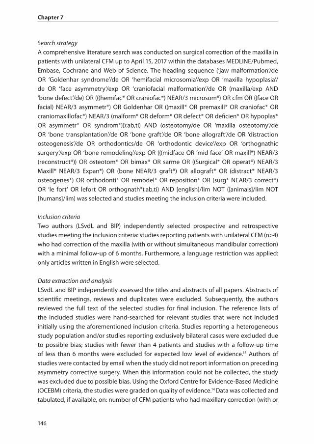
Chapter 7
146
Search strategyA comprehensive literature search was conducted on surgical correction of the maxilla in patients with unilateral CFM up to April 15, 2017 within the databases MEDLINE/Pubmed, Embase, Cochrane and Web of Science. The heading sequence (‘jaw malformation’/de OR ‘Goldenhar syndrome’/de OR ‘hemifacial microsomia’/exp OR ‘maxilla hypoplasia’/de OR ‘face asymmetry’/exp OR ‘craniofacial malformation’/de OR (maxilla/exp AND ‘bone defect’/de) OR (((hemifac* OR craniofac*) NEAR/3 microsom*) OR cfm OR ((face OR facial) NEAR/3 asymmetr*) OR Goldenhar OR ((maxill* OR premaxill* OR craniofac* OR craniomaxillofac*) NEAR/3 (malform* OR deform* OR defect* OR deficien* OR hypoplas* OR asymmetr* OR syndrom*))):ab,ti) AND (osteotomy/de OR ‘maxilla osteotomy’/de OR ‘bone transplantation’/de OR ‘bone graft’/de OR ‘bone allograft’/de OR ‘distraction osteogenesis’/de OR orthodontics/de OR ‘orthodontic device’/exp OR ‘orthognathic surgery’/exp OR ‘bone remodeling’/exp OR (((midface OR ‘mid face’ OR maxill*) NEAR/3 (reconstruct*)) OR osteotom* OR bimax* OR sarme OR ((Surgical* OR operat*) NEAR/3 Maxill* NEAR/3 Expan*) OR (bone NEAR/3 graft*) OR allograft* OR (distract* NEAR/3 osteogenes*) OR orthodonti* OR remodel* OR reposition* OR (surg* NEAR/3 correct*) OR ‘le fort’ OR lefort OR orthognath*):ab,ti) AND [english]/lim NOT ([animals]/lim NOT [humans]/lim) was selected and studies meeting the inclusion criteria were included.
Inclusion criteriaTwo authors (LSvdL and BIP) independently selected prospective and retrospective studies meeting the inclusion criteria: studies reporting patients with unilateral CFM (n>4) who had correction of the maxilla (with or without simultaneous mandibular correction) with a minimal follow-up of 6 months. Furthermore, a language restriction was applied: only articles written in English were selected.
Data extraction and analysisLSvdL and BIP independently assessed the titles and abstracts of all papers. Abstracts of scientific meetings, reviews and duplicates were excluded. Subsequently, the authors reviewed the full text of the selected studies for final inclusion. The reference lists of the included studies were hand-searched for relevant studies that were not included initially using the aforementioned inclusion criteria. Studies reporting a heterogeneous study population and/or studies reporting exclusively bilateral cases were excluded due to possible bias; studies with fewer than 4 patients and studies with a follow-up time of less than 6 months were excluded for expected low level of evidence.13 Authors of studies were contacted by email when the study did not report information on preceding asymmetry corrective surgery. When this information could not be collected, the study was excluded due to possible bias. Using the Oxford Centre for Evidence-Based Medicine (OCEBM) criteria, the studies were graded on quality of evidence.14 Data was collected and tabulated, if available, on: number of CFM patients who had maxillary correction (with or
PSM 20190527 Proefschrift Britt Pluijmers.indd 146 24-07-19 15:04

Surgical correction of the midface in CFM: a systematic review
147
7
without simultaneous mandibular correction), number of CFM patients with preceding mandibular surgery, type of preceding mandibular surgery, classification of mandibular type (by the Pruzansky-Kaban or Pruzansky system), type of surgical treatment to correct the maxilla (and mandible), mean age at time of surgical treatment, analysis method of the asymmetry and timing of the analysis, maxillary (and mandibular) movement, outcome (increase or decrease in asymmetry, including occlusal plane and residual cant, and patients’ satisfaction), follow-up length, number and kind of complications, and number and type of additional treatments performed or recommended.
Results
The literature search yielded 5,509 publications. Screening of reference lists of the included articles did not result in any additional articles. After applying the selection criteria, 179 publications were read in full text, of which 16 were initially included. Nine papers were excluded for lack of data on preceding mandibular correction, leaving 7 studies for inclusion (Fig. 1).15-21
Fig. 1. Flowchart of included articles.
PSM 20190527 Proefschrift Britt Pluijmers.indd 147 24-07-19 15:04
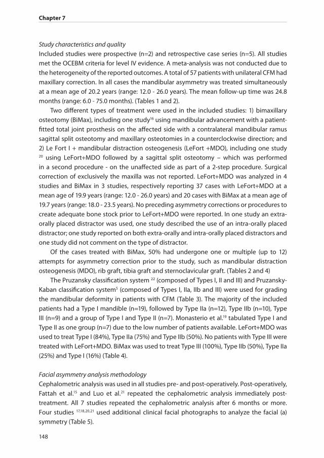
Chapter 7
148
Study characteristics and qualityIncluded studies were prospective (n=2) and retrospective case series (n=5). All studies met the OCEBM criteria for level IV evidence. A meta-analysis was not conducted due to the heterogeneity of the reported outcomes. A total of 57 patients with unilateral CFM had maxillary correction. In all cases the mandibular asymmetry was treated simultaneously at a mean age of 20.2 years (range: 12.0 - 26.0 years). The mean follow-up time was 24.8 months (range: 6.0 - 75.0 months). (Tables 1 and 2).
Two different types of treatment were used in the included studies: 1) bimaxillary osteotomy (BiMax), including one study16 using mandibular advancement with a patient-fitted total joint prosthesis on the affected side with a contralateral mandibular ramus sagittal split osteotomy and maxillary osteotomies in a counterclockwise direction; and 2) Le Fort I + mandibular distraction osteogenesis (LeFort +MDO), including one study 20 using LeFort+MDO followed by a sagittal split osteotomy – which was performed in a second procedure - on the unaffected side as part of a 2-step procedure. Surgical correction of exclusively the maxilla was not reported. LeFort+MDO was analyzed in 4 studies and BiMax in 3 studies, respectively reporting 37 cases with LeFort+MDO at a mean age of 19.9 years (range: 12.0 - 26.0 years) and 20 cases with BiMax at a mean age of 19.7 years (range: 18.0 - 23.5 years). No preceding asymmetry corrections or procedures to create adequate bone stock prior to LeFort+MDO were reported. In one study an extra-orally placed distractor was used, one study described the use of an intra-orally placed distractor; one study reported on both extra-orally and intra-orally placed distractors and one study did not comment on the type of distractor.
Of the cases treated with BiMax, 50% had undergone one or multiple (up to 12) attempts for asymmetry correction prior to the study, such as mandibular distraction osteogenesis (MDO), rib graft, tibia graft and sternoclavicular graft. (Tables 2 and 4)
The Pruzansky classification system 22 (composed of Types I, II and III) and Pruzansky-Kaban classification system5 (composed of Types I, IIa, IIb and III) were used for grading the mandibular deformity in patients with CFM (Table 3). The majority of the included patients had a Type I mandible (n=19), followed by Type IIa (n=12), Type IIb (n=10), Type III (n=9) and a group of Type I and Type II (n=7). Monasterio et al.19 tabulated Type I and Type II as one group (n=7) due to the low number of patients available. LeFort+MDO was used to treat Type I (84%), Type IIa (75%) and Type IIb (50%). No patients with Type III were treated with LeFort+MDO. BiMax was used to treat Type III (100%), Type IIb (50%), Type IIa (25%) and Type I (16%) (Table 4).
Facial asymmetry analysis methodologyCephalometric analysis was used in all studies pre- and post-operatively. Post-operatively, Fattah et al.15 and Luo et al.21 repeated the cephalometric analysis immediately post-treatment. All 7 studies repeated the cephalometric analysis after 6 months or more. Four studies 17,18,20,21 used additional clinical facial photographs to analyze the facial (a)symmetry (Table 5).
PSM 20190527 Proefschrift Britt Pluijmers.indd 148 24-07-19 15:04

Surgical correction of the midface in CFM: a systematic review
149
7
Tab
le 1
. Tre
atm
ent a
nd c
lass
ifica
tion
Cit
atio
nO
CEB
M le
vel
of e
vid
ence
Met
ho
do
log
yN
o. o
f pat
ien
tsSu
rgic
al
trea
tmen
t
No
. of
pre
ced
ing
m
and
ibu
lar
surg
ery
(%)
Mea
n a
ge
at
surg
ery,
yea
rs
Mea
n fo
llow
-u
p t
ime,
m
on
ths
Liu
et a
l. 20
1720
4Re
tros
pec
tive
CS
12Le
Fort
+M
DO
α0
(0%
)22
.443
.4
Luo
et a
l. 20
1621
4Re
tros
pec
tive
CS
7Le
Fort
+M
DO
0 (0
%)
13.7
58.8
Fatt
ah e
t al.2
01415
4Re
tros
pec
tive
CS
8Bi
Max
2 (2
5.0%
)18
.038
.4
Wol
ford
et a
l. 20
1216
4Pr
osp
ecti
ve C
S6
BiM
axβ
4 (6
7.0%
)23
.575
.0
Oht
ani e
t al.
2012
174
Retr
osp
ecti
ve C
S6
BiM
ax4
(66.
6%)
18.1
12.0
Bala
ji et
al.
2010
184
Retr
osp
ecti
ve C
S11
LeFo
rt+
MD
O0
(0%
)21
.86.
0
Mon
aste
rio
et a
l. 19
9719
4Pr
osp
ecti
ve C
S7
LeFo
rt+
MD
O0
(0%
)19
.07.
0
OC
EBM
: Oxf
ord
Cen
tre
for E
vid
ence
-Bas
ed M
edic
ine;
CS:
Cas
e Se
ries
; LeF
ort+
MD
O: L
e Fo
rt I
+ m
and
ibul
ar d
istr
acti
on o
steo
gen
esis
; BiM
ax: B
iMax
illar
y os
teot
omy.
α Fo
llow
ed b
y a
sag
itta
l sp
lit
oste
otom
y on
the
unaff
ecte
d si
de
as a
sec
ond
ary
surg
ery;
β Usi
ng
a p
atie
nt-fi
tted
tota
l joi
nt p
rost
hes
is a
nd
man
dib
ular
ad
van
cem
ent o
n aff
ecte
d si
de
Tab
le 2
. Ove
rvie
w o
f tre
atm
ent c
hara
cter
isti
cs
Trea
tmen
tN
o. o
f p
atie
nts
No
. of
stu
die
sM
ean
ag
e, y
ears
(r
ang
e)
Mea
n fo
llow
-up
ti
me,
mo
nth
s (r
ang
e)
No
. of p
rece
din
g
man
dib
ula
r su
rger
y(%
)Le
Fort
+M
DO
374
19.9
(12.
0-26
.0)
28.3
(6.0
-58.
8)0
(0%
)
BiM
ax20
319
.7(1
8.0-
23.5
)41
.5(1
2.0-
75.0
)10
(50%
)
Tota
l57
720
.2(1
2.0
-26.
0)24
.8(6
.0-7
5.0)
10 (1
7.5%
)
LeFo
rt+
MD
O: L
e Fo
rt I
+ m
and
ibul
ar d
istr
acti
on o
steo
gen
esis
; BiM
ax: B
iMax
illar
y os
teot
omy.
PSM 20190527 Proefschrift Britt Pluijmers.indd 149 24-07-19 15:04
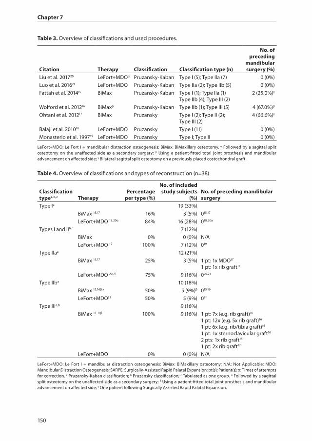
Chapter 7
150
Table 3. Overview of classifications and used procedures.
Citation Therapy Classification Classification type (n)
No. of preceding
mandibular surgery (%)
Liu et al. 201720 LeFort+MDOα Pruzansky-Kaban Type I (5); Type IIa (7) 0 (0%)
Luo et al. 201621 LeFort+MDO Pruzansky-Kaban Type IIa (2); Type IIb (5) 0 (0%)
Fattah et al. 201415 BiMax Pruzansky-Kaban Type I (1); Type IIa (1) Type IIb (4); Type III (2)
2 (25.0%)γ
Wolford et al. 201216 BiMaxβ Pruzansky-Kaban Type IIb (1); Type III (5) 4 (67.0%)β
Ohtani et al. 201217 BiMax Pruzansky Type I (2); Type II (2); Type III (2)
4 (66.6%)γ
Balaji et al. 201018 LeFort+MDO Pruzansky Type I (11) 0 (0%)
Monasterio et al. 199719 LeFort+MDO Pruzansky Type I; Type II 0 (0%)
LeFort+MDO: Le Fort I + mandibular distraction osteogenesis; BiMax: BiMaxillary osteotomy. α Followed by a sagittal split osteotomy on the unaffected side as a secondary surgery; β Using a patient-fitted total joint prosthesis and mandibular advancement on affected side; γ Bilateral sagittal split osteotomy on a previously placed costochondral graft.
Table 4. Overview of classifications and types of reconstruction (n=38)
Classification typea,b,c Therapy
Percentage per type (%)
No. of included study subjects
(%)No. of preceding mandibular surgery
Type Ia 19 (33%)
BiMax 15,17 16% 3 (5%) 015,17
LeFort+MDO 18,20α 84% 16 (28%) 018,20α
Types I and IIb,c 7 (12%)
BiMax 0% 0 (0%) N/A
LeFort+MDO 19 100% 7 (12%) 019
Type IIaa 12 (21%)
BiMax 15,17 25% 3 (5%) 1 pt: 1x MDO17
1 pt: 1x rib graft17
LeFort+MDO 20,21 75% 9 (16%) 020,21
Type IIba 10 (18%)
BiMax 15,16β,γ 50% 5 (9%)β 015,16
LeFort+MDO21 50% 5 (9%) 021
Type IIIa,b 9 (16%)
BiMax 15-17β 100% 9 (16%) 1 pt: 7x (e.g. rib graft)16
1 pt: 12x (e.g. 5x rib graft)16
1 pt: 6x (e.g. rib/tibia graft)16
1 pt: 1x sternoclavicular graft16
2 pts: 1x rib graft15
1 pt: 2x rib graft17
LeFort+MDO 0% 0 (0%) N/A
LeFort+MDO: Le Fort I + mandibular distraction osteogenesis; BiMax: BiMaxillary osteotomy; N/A: Not Applicable; MDO: Mandibular Distraction Osteogenesis; SARPE: Surgically-Assisted Rapid Palatal Expansion; pt(s): Patient(s); x: Times of attempts for correction. a Pruzansky-Kaban classification; b Pruzansky classification; c Tabulated as one group. α Followed by a sagittal split osteotomy on the unaffected side as a secondary surgery; β Using a patient-fitted total joint prosthesis and mandibular advancement on affected side; γ One patient following Surgically Assisted Rapid Palatal Expansion.
PSM 20190527 Proefschrift Britt Pluijmers.indd 150 24-07-19 15:04

Surgical correction of the midface in CFM: a systematic review
151
7
Outcome 0-7 days post-treatmentMost studies showed improved facial symmetry and aesthetically satisfactory outcomes, based on the surgeons’ and/or patients’ opinion.15,16,18,19 The maxillary downward movement varied from 0 to 7.5 mm with BiMax15,16 and from 4.0 to 7.0 mm with LeFort+MDO.19 When LeFort+MDO was used, Balaji et al., overcorrected with 2-3 mm on the predicted distraction length18 and Monasterio et al., reported predicted overcorrection, although the amount of overcorrection was not mentioned.19 There was no data available on overcorrection when BiMax was used. Balaji et al., reported mild overcorrection in the first week post-treatment. All studies showed improved leveling of the occlusal plane and four studies measured a slight residual cant of a maximum of 2.3 degrees (Table 6). Only one study reported relapse.15 Wolford et al. showed significant improvement on the following parameters with a subjective analysis: rating on pain, jaw movement, diet ability and level of total disability, however limitation in jaw function retained (Table 7).16
ComplicationsWolford et al., reported fibrosis of TMJ in a patient treated with BiMax, who also had a TMJ reconstruction.16 This patient had undergone 12 previous procedures prior to the definitive BiMax operation. A total of six patients had temporary lower lip paresis that resolved within 3-6 months without any treatment. Luo et al. reported two patients
Table 5. Overview of facial asymmetry analysis methodologies
Citation Therapy Analysis method TimingLiu et al. 201720 LeFort+MDOα Clinical photographs,
cephalometryPre-, and post-operative up to 36-56 months
Luo et al. 201621 LeFort+MDO Clinical photographs, Photographic panoramic, cephalometry
Pre-operative, immediate post-distraction and 6 months, 1, 2 and 4 years post-distraction.
Fattah et al. 201415 BiMax Cephalometry Pre-operative, immediate post-operative and >1 year post-operative
Wolford et al. 201216 BiMaxβ Cephalometry Pre-operative, 5 days and 76 months post-operative
Ohtani et al. 201217 BiMax Clinical photographs, cephalometry
Pre-operative and >1 year post-operative
Balaji et al. 201018 LeFort+MDO Clinical photographs, cephalometry, occlusal cant
Pre-, and 6 months post-distraction
Monasterio et al. 199719 LeFort+MDO Photographic panoramic, Cephalometry
Pre- and post-distraction and every 6 months post-distraction
LeFort+MDO: Le Fort I + mandibular distraction osteogenesis; BiMax: BiMaxillary osteotomy; α Followed by a sagittal split osteotomy on the unaffected side as a secondary surgery; β Using a patient-fitted total joint prosthesis and mandibular advancement on affected side.
PSM 20190527 Proefschrift Britt Pluijmers.indd 151 24-07-19 15:04
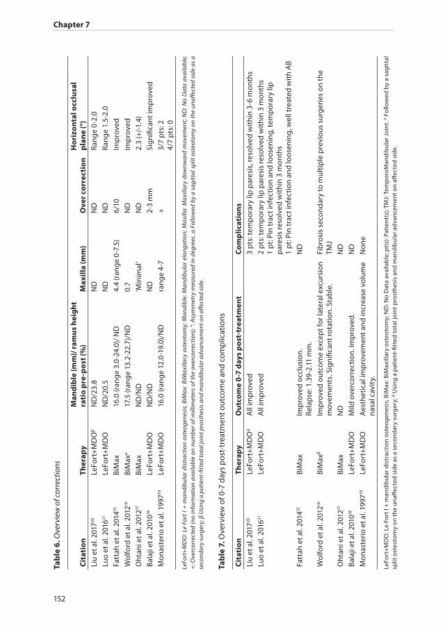
Chapter 7
152
Tab
le 6
. Ove
rvie
w o
f cor
rect
ions
Cit
atio
nTh
erap
yM
and
ible
(mm
)/ r
amu
s h
eig
ht
rati
o p
re-p
ost
(%)
Max
illa
(mm
)O
ver
corr
ecti
on
Ho
rizo
nta
l occ
lusa
l p
lan
e (°
)Li
u et
al.
2017
20Le
Fort
+M
DO
βN
D/2
3.8
ND
ND
Rang
e 0-
2.0
Luo
et a
l. 20
1621
LeFo
rt+
MD
ON
D/2
0.5
ND
ND
Rang
e 1.
5-2.
0
Fatt
ah e
t al.
2014
15Bi
Max
16.0
(ran
ge
3.0-
24.0
)/ N
D4.
4 (r
ang
e 0-
7.5)
6/10
Imp
rove
d
Wol
ford
et a
l. 20
1216
BiM
axα
17.5
(ran
ge
13.2
-22.
7)/N
D0.
7N
DIm
pro
ved
Oht
ani e
t al.
2012
17Bi
Max
ND
/ND
‘Min
imal
’N
D2.
3 (+
/-1.
4)
Bala
ji et
al.
2010
18Le
Fort
+M
DO
ND
/ND
ND
2-3
mm
Sign
ifica
nt im
pro
ved
Mon
aste
rio
et a
l. 19
9719
LeFo
rt+
MD
O16
.0 (r
ang
e 12
.0-1
9.0)
/ND
rang
e 4-
7+
3/7
pts
: 24/
7 p
ts: 0
LeFo
rt+
MD
O: L
e Fo
rt I
+ m
andi
bula
r di
stra
ctio
n os
teog
enes
is; B
iMax
: BiM
axill
ary
oste
otom
y; M
andi
ble:
Man
dibu
lar
elon
gatio
n; M
axill
a: M
axill
ary
dow
nwar
d m
ovem
ent;
ND
: No
Dat
a av
aila
ble;
+
: Ove
rcor
rect
ed (n
o in
form
atio
n av
aila
ble
on n
umbe
r of m
illim
eter
s of
the
over
corr
ectio
n); °
: Asy
mm
etry
mea
sure
d in
deg
rees
. α F
ollo
wed
by
a sa
gitt
al s
plit
oste
otom
y on
the
unaff
ecte
d si
de a
s a
seco
ndar
y su
rger
y; β
Usi
ng a
pat
ient
-fitt
ed to
tal j
oint
pro
sthe
sis a
nd m
andi
bula
r adv
ance
men
t on
affec
ted
side
.
Tab
le 7
. Ove
rvie
w o
f 0-7
day
s p
ost-
trea
tmen
t out
com
e an
d co
mp
licat
ions
Cit
atio
nTh
erap
yO
utc
om
e 0
-7 d
ays
po
st-t
reat
men
tC
om
plic
atio
ns
Liu
et a
l. 20
1720
LeFo
rt+
MD
Oα
All
imp
rove
d3
pts
: tem
por
ary
lip p
ares
is, r
esol
ved
wit
hin
3-6
mon
ths
Luo
et a
l. 20
1621
LeFo
rt+
MD
OA
ll im
pro
ved
2 p
ts: t
emp
orar
y lip
par
esis
reso
lved
wit
hin
3 m
onth
s1
pt:
Pin
trac
t inf
ecti
on a
nd lo
osen
ing
, tem
por
ary
lip
par
esis
reso
lved
wit
hin
3 m
onth
s1
pt:
Pin
trac
t inf
ecti
on a
nd lo
osen
ing
, wel
l tre
ated
wit
h A
B
Fatt
ah e
t al.
2014
15Bi
Max
Imp
rove
d oc
clus
ion.
Re
lap
se: 1
.39-
2.11
mm
.N
D
Wol
ford
et a
l. 20
1216
BiM
axβ
Imp
rove
d ou
tcom
e ex
cep
t for
late
ral e
xcur
sion
m
ovem
ents
. Sig
nific
ant r
otat
ion.
Sta
ble
.Fi
bro
sis
seco
ndar
y to
mul
tip
le p
revi
ous
surg
erie
s on
the
TMJ
Oht
ani e
t al.
2012
17Bi
Max
ND
ND
Bala
ji et
al.
2010
18Le
Fort
+M
DO
Mild
ove
rcor
rect
ion.
Imp
rove
d.
ND
Mon
aste
rio
et a
l. 19
9719
LeFo
rt+
MD
OA
esth
etic
al im
pro
vem
ent a
nd in
crea
se v
olum
e na
sal c
avit
y.N
one
LeFo
rt+
MD
O: L
e Fo
rt I
+ m
and
ibul
ar d
istr
acti
on o
steo
gen
esis
; BiM
ax: B
iMax
illar
y os
teot
omy;
ND
: No
Dat
a av
aila
ble
; pt(
s): P
atie
nt(s
); TM
J: T
emp
oroM
and
ibul
ar J
oint
. α Fo
llow
ed b
y a
sag
itta
l sp
lit o
steo
tom
y on
the
unaff
ecte
d si
de
as a
sec
ond
ary
surg
ery;
β Usi
ng
a p
atie
nt-fi
tted
tota
l joi
nt p
rost
hes
is a
nd
man
dib
ular
ad
van
cem
ent o
n aff
ecte
d si
de.
PSM 20190527 Proefschrift Britt Pluijmers.indd 152 24-07-19 15:04
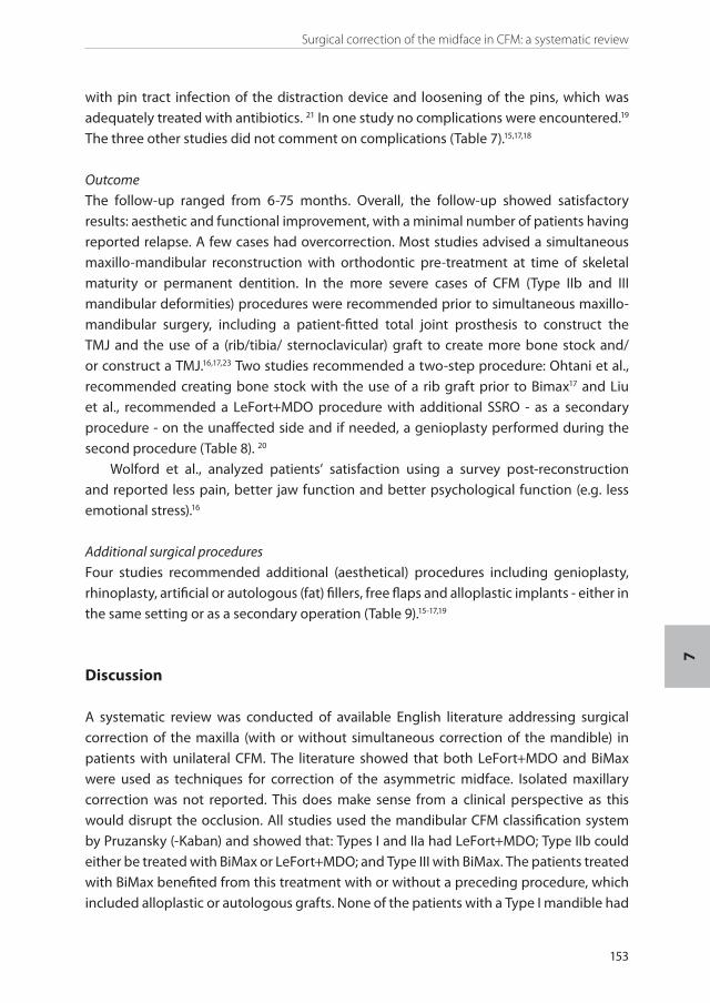
Surgical correction of the midface in CFM: a systematic review
153
7
with pin tract infection of the distraction device and loosening of the pins, which was adequately treated with antibiotics. 21 In one study no complications were encountered.19 The three other studies did not comment on complications (Table 7).15,17,18
OutcomeThe follow-up ranged from 6-75 months. Overall, the follow-up showed satisfactory results: aesthetic and functional improvement, with a minimal number of patients having reported relapse. A few cases had overcorrection. Most studies advised a simultaneous maxillo-mandibular reconstruction with orthodontic pre-treatment at time of skeletal maturity or permanent dentition. In the more severe cases of CFM (Type IIb and III mandibular deformities) procedures were recommended prior to simultaneous maxillo-mandibular surgery, including a patient-fitted total joint prosthesis to construct the TMJ and the use of a (rib/tibia/ sternoclavicular) graft to create more bone stock and/or construct a TMJ.16,17,23 Two studies recommended a two-step procedure: Ohtani et al., recommended creating bone stock with the use of a rib graft prior to Bimax17 and Liu et al., recommended a LeFort+MDO procedure with additional SSRO - as a secondary procedure - on the unaffected side and if needed, a genioplasty performed during the second procedure (Table 8). 20
Wolford et al., analyzed patients’ satisfaction using a survey post-reconstruction and reported less pain, better jaw function and better psychological function (e.g. less emotional stress).16
Additional surgical proceduresFour studies recommended additional (aesthetical) procedures including genioplasty, rhinoplasty, artificial or autologous (fat) fillers, free flaps and alloplastic implants - either in the same setting or as a secondary operation (Table 9).15-17,19
Discussion
A systematic review was conducted of available English literature addressing surgical correction of the maxilla (with or without simultaneous correction of the mandible) in patients with unilateral CFM. The literature showed that both LeFort+MDO and BiMax were used as techniques for correction of the asymmetric midface. Isolated maxillary correction was not reported. This does make sense from a clinical perspective as this would disrupt the occlusion. All studies used the mandibular CFM classification system by Pruzansky (-Kaban) and showed that: Types I and IIa had LeFort+MDO; Type IIb could either be treated with BiMax or LeFort+MDO; and Type III with BiMax. The patients treated with BiMax benefited from this treatment with or without a preceding procedure, which included alloplastic or autologous grafts. None of the patients with a Type I mandible had
PSM 20190527 Proefschrift Britt Pluijmers.indd 153 24-07-19 15:04

Chapter 7
154
Tab
le 8
. Ove
rvie
w o
f out
com
e an
d re
com
men
dat
ion
Cit
atio
nTh
erap
yFo
llow
-up
ti
me,
mo
nth
sO
utc
om
eC
on
clu
sio
n -
Rec
om
men
dat
ion
Liu
et a
l. 20
1720
LeFo
rt+
MD
Oα
43.4
Sign
ifica
nt im
pro
ved
occl
usio
n ca
nt, h
oriz
onta
l oc
clus
ion
pla
ne a
nd fa
cial
sym
met
ry.
Two
-ste
p p
roce
dur
e: L
eFor
t+M
DO
and
SSR
O a
s a
seco
ndar
y su
rger
y +
gen
iop
last
y, if
nee
ded
Luo
et a
l. 20
1621
LeFo
rt+
MD
O58
.8M
inim
al re
lap
se o
f cor
rect
ion
in fi
rst
6 m
onth
sSi
ngle
-sta
ge
surg
ery:
All
pat
ient
s sa
tisfi
ed a
t lat
est c
ontr
ol. N
o se
cond
ary
surg
erie
s.
Fatt
ah e
t al.
2014
15Bi
Max
38.4
3/10
imp
rove
d, 6
/10
over
corr
ecte
d,
1 w
orse
ned
. Min
imal
non
-sig
nific
ant r
elap
se.
Tim
ing
surg
ery:
Ske
leta
l mat
urit
y fo
r sta
ble
lo
ng-t
erm
resu
lts
whi
le m
inim
izin
g m
orb
idit
y an
d nu
mb
er o
f pro
ced
ures
.
Wol
ford
et a
l. 20
1216
BiM
axβ
75.0
Imp
rove
d in
cisa
l op
enin
g, s
tab
le m
axill
a-m
and
ibul
ar c
omp
lex,
no
chan
ges
in th
e m
and
ible
pos
itio
n. N
o re
lap
se.
Pati
ent-
fitte
d p
rost
hesi
s us
e +
ad
dit
iona
l se
cond
ary
pro
ced
ures
in c
omp
lex
case
s.
Oht
ani e
t al.
2012
17Bi
Max
12.0
Sign
ifica
nt im
pro
vem
ent f
acia
l sym
met
ry.
Exce
llent
cos
met
ic re
sult
s.Tw
o-s
tep
pro
ced
ure:
Ste
pw
ise
inte
rven
tion
s w
ith
orth
odon
tic
trea
tmen
t. Ri
bgr
aft i
n se
vere
ca
ses.
Tim
ing:
Ske
leta
l mat
urit
y.
Bala
ji et
al.
2010
18Le
Fort
+M
DO
6.0
2-3
mm
ove
rcor
rect
ion
wit
h st
able
, pre
dic
tab
le
resu
lts.
Sign
ifica
nt a
ngul
ar c
hang
e lip
and
occ
lusa
l ca
nt. 2
-3 m
m o
verc
orre
ctio
n gi
ves
a b
ette
r ae
sthe
tica
l out
com
e.
Mon
aste
rio
et a
l. 19
9719
LeFo
rt+
MD
O7.
0A
esth
etic
imp
rove
men
t.Ti
min
g su
rger
y: p
t >14
yea
rs
LeFo
rt+
MD
O: L
e Fo
rt I
+ m
and
ibul
ar d
istr
acti
on o
steo
gen
esis
, BiM
ax: B
iMax
illar
y os
teot
omy;
TM
J: T
emp
oroM
and
ibul
ar J
oint
; pt(
s): P
atie
nt(s
). α F
ollo
wed
by
a sa
git
tal s
plit
ost
eoto
my
on th
e un
affec
ted
sid
e as
a s
econ
dar
y su
rger
y; β U
sin
g a
pat
ient
-fitt
ed to
tal j
oint
pro
sth
esis
an
d m
and
ibul
ar a
dva
nce
men
t on
affec
ted
sid
e.
PSM 20190527 Proefschrift Britt Pluijmers.indd 154 24-07-19 15:04

Surgical correction of the midface in CFM: a systematic review
155
7
Tab
le 9
. Ove
rvie
w o
f sof
t tis
sue
imp
rove
men
ts.
Cit
atio
nTh
erap
ySo
ft t
issu
e im
pro
vem
ents
Liu
et a
l. 20
1720
LeFo
rt+
MD
Oα
Seco
nd
ary
gen
iop
last
y
Luo
et a
l. 20
1621
LeFo
rt+
MD
OFa
cial
fat g
raft
ing
, gen
iop
last
y, a
llop
last
ic a
ugm
enta
tion
Fatt
ah e
t al.
2014
15Bi
Max
Seco
nd
ary
gen
iop
last
y (n
=5)
, mal
ar a
nd
man
dib
ular
imp
lant
s (n
=2)
.
Wol
ford
et a
l. 20
1216
BiM
axβ
Ad
vice
: allo
pla
stic
/ aut
ogen
ous
imp
lant
s in
com
ple
x ca
ses.
Oht
ani e
t al.
2012
17Bi
Max
Fat a
ugm
enta
tion
(n=
3). I
f nee
ded
: gen
iop
last
y, rh
inop
last
y, m
uscl
e fla
ps
and
arti
ficia
l/ fa
t fille
rs.
Bala
ji et
al.
2010
18Le
Fort
+M
DO
N/A
Mon
aste
rio
et a
l. 19
9719
LeFo
rt+
MD
OA
dvi
ce: g
enio
pla
sty
(n=1
).
LeFo
rt+
MD
O: L
e Fo
rt I
+ m
and
ibul
ar d
istr
acti
on o
steo
gen
esis
, BiM
ax: B
iMax
illar
y os
teot
omy;
N/A
: Not
Ap
plic
able
. α Fo
llow
ed b
y a
sag
itta
l sp
lit o
steo
tom
y on
th
e un
affec
ted
sid
e as
a
seco
nd
ary
surg
ery;
β Usi
ng
a p
atie
nt-fi
tted
tota
l joi
nt p
rost
hes
is a
nd
man
dib
ular
ad
van
cem
ent o
n aff
ecte
d si
de.
PSM 20190527 Proefschrift Britt Pluijmers.indd 155 24-07-19 15:04

Chapter 7
156
a procedure preceding facial asymmetry correction, but the majority of the patients with Types IIa, IIb and III had asymmetry corrections prior to the study, including mandibular distraction osteogenesis (MDO), rib graft, tibia graft and sternoclavicular graft. The literature shows that BiMax is used in more severe cases and that the majority of these cases undergo (multiple) operations prior to the BiMax procedure, suggesting that patients with more severe CFM might benefit from a multi-stage treatment approach. Since no data was available on the timing of the corrective operations prior to BiMax, no conclusions can be drawn on the best timing for the first attempt at correction. However, some studies recommend postponing intervention, if possible, until skeletal maturity. The data from this study is in agreement with previous systematic reviews7,24 which suggest delaying mandibular reconstruction until the permanent dentition, or even skeletal maturity if there are no pressing functional and/or psychosocial problems(3). Several studies state that there is no evidence supporting the effectiveness of early asymmetry correction in patients with CFM.8 Nevertheless, there are definite indications for early surgery: functional impairments (i.e. airway, swallowing, mastication, speech and psychosocial) should dictate the timing of surgical intervention; and patients with respiratory obstruction (i.e. breathing issues and sleep apnea, feeding and speech difficulties and/or emotional distress)25 should be candidates for early interventions.26,27 However, even when delaying correction until permanent dentition, six out of seven studies in this systematic review recommended additional (aesthetic) surgery. Therefore, patients and their caregivers should be made aware that treating the deformities caused by CFM is a long process and that, in the long term, additional procedures may be needed.
Information on preceding corrective surgery in CFM patients is essential to analyze the outcome of the used treatment; a severely affected CFM patient with multiple attempts for correction might have a different outcome than a severely affected CFM patient without previous correction. Initially, 16 studies were included, however, nine studies were excluded for lack of data on preceding corrective surgery. Therefore, a limited number of patients were available for analyses.
Fifty percent of the patients treated with BiMax had undergone previous asymmetry correction. Unfortunately, as sparse information regarding the type of treatment prior to BiMax was reported, there was no baseline data on the degree of canting, (mal)occlusion and the number of millimeters of distraction or the length of graft used prior to BiMax. Therefore, it is unclear what degree of severity of asymmetry was present at the time of surgery. Furthermore, the degree of canting of the maxilla was not reported. It is unclear if the degree of maxillary canting is proportional to the severity of the mandibular asymmetry (graded by the Pruzansky(-Kaban) classification). Thus, it is impossible to compare the reported patients and their treatments. Therefore, we suggest a study designed to analyze the maxillary canting of patients with CFM. The authors of this study hypothesize a positive correlation of mandibular hypoplasia and the degree of maxillary canting. If there is a correlation between maxillary canting and the Pruzansky-Kaban
PSM 20190527 Proefschrift Britt Pluijmers.indd 156 24-07-19 15:04

Surgical correction of the midface in CFM: a systematic review
157
7
classification, a more profound recommendation for treatment could be provided for the correction of the maxillo-mandibular asymmetry.
The mean follow-up time was 28.3 months (range: 6.0-75.0 months). Most studies reported a satisfactory outcome in the long term, but the majority lacked statistical evidence. LeFort+MDO had a mean follow-up time of 9.2 months compared to a BiMax follow-up time of 45.9 months. These enormous differences in follow-up time make comparing between the two techniques impossible. No significant relapse was documented with either technique. Overcorrection of 2-3 millimeters resolved into a more symmetrical outcome in most cases. The occlusal cant was not always fully corrected as four studies showed a residual cant with a maximum of 2.31 degrees. However, Padwa et al., showed that an occlusal cant must be more than 4 degrees to be visually notable.28
Furthermore, the authors of this work advocate to aim for some canting in relation to the soft tissue asymmetry most often present due to soft tissue deficiency and facial nerve dysfunction. This means not completely correcting the skeletal asymmetry and leaving some occlusal cant, so, when the face is animating, the soft tissue cant is in line with the occlusal cant. Correcting the bony chin in these cases corrects for the passive asymmetrical face. This way, both in rest and during animation, the patients tend to have the best harmony between soft and hard tissues, thus resulting in the best achievable (esthetic) outcome.
It seems impossible to predict which patients would benefit from overcorrection and which patients would not. In a study on maxillo-mandibular reconstruction of class III malocclusion, 20% of the patients had a relapse of more than 2 millimeters, suggesting that an overcorrection of 2 millimeters is advisable.29 However, no data was available on the predictable factors for the need of overcorrection. Even though facial analyses were performed pre- and post-operative, specificity and sensitivity of these measurements may be questioned since there was no use of modern measurements30 such as 3D analysis.
None of the included studies used computer aided surgical planning for the correction of the maxillo-mandibular complex. The use of 3D computer-aided-design and computer-aided-manufacturing principles are described as an accurate and reliable method in the diagnosis, treatment planning, and designing of cutting guides that optimize surgical correction in a small number of patients with hemimandibular hyperplasia and Class III malocclusion 31,32 Moreover, as recent studies suggest that asymmetry occurs from skull base and on, the mandible and maxilla seen as a continuum part of this.3,33 It could aid in these complex cases in which the standardized cephalometry does not apply. A case report on the correction of a patient with CFM with the use of computer assisted orthognatic surgical protocol confirms the clinical feasibility.34 However, no large studies exist and more research is needed to confirm the advantages of computer aided surgical planning.
In the modern era, treatment benefits are based on multiple factors, including objective analyses such as 3D measurement techniques, skeletal and/or soft tissue
PSM 20190527 Proefschrift Britt Pluijmers.indd 157 24-07-19 15:04

Chapter 7
158
analysis, complications, opinion of the physician, and satisfaction of the patient (and his/her caregivers). However, only one study reported results based on the patient’s opinion. In the future, a study on quality of life and other outcome measurements could be useful to truly answer the results of the treatment and the needs of these patients. A global workgroup focusing on patient-reported outcomes has presented a minimal set of outcomes for CFM patients in 2015.35
The lack of information on aforementioned factors makes it impossible to truly answer the question of which treatment and timing would definitely benefit these patients to correct the maxilla and restore the facial harmony. Therefore, the authors started collecting retrospective data at four major craniofacial centers on all facial corrective treatments, their outcomes and complications, and initiated a study on 3D-measurement analyses and a study on maxillary canting in patients with CFM.
Conclusion
This systematic review shows that surgical correction of the maxilla was performed simultaneously with correction of the mandible in all reported patients at a mean age of 20.2 years in a total of 57 patients with unilateral CFM. Severity of the mandibular deformity was graded by the Pruzansky (-Kaban) classification and showed that Types I and IIa would benefit from LeFort+MDO; Type IIb from LeFort+MDO or BiMax; and Type III from BiMax (of which 50% of the cases had preceding mandibular surgery, including the use of a patient-fitted prosthesis), either as a one-step procedure, or as part of a two-step treatment algorithm. However, due to lack of data, no hard conclusions can be drawn on the ideal surgical treatment to correct the asymmetry and the timing in patients with CFM. Moreover, additional (aesthetical) procedures were frequently suggested to achieve the desired end-result.
Acknowledgements
We thank W. Bramer, biomedical information specialist of Erasmus MC, for his assistance with the literature search.
PSM 20190527 Proefschrift Britt Pluijmers.indd 158 24-07-19 15:04

Surgical correction of the midface in CFM: a systematic review
159
7
References
1. Molina F, Monasterio FO. Mandibular Elongation and Remodeling by Distraction - a Farewell to Major Osteotomies. Plastic and Reconstructive Surgery 1995; 96(4): 825-40.
2. Granstrom G, Jacobsson C. First and second branchial arch syndrome: aspects on the embryogenesis, elucidations, and rehabilitation using the osseointegration concept. Clin Implant Dent Relat Res 1999; 1(2): 59-69.
3. Caron C, Pluijmers BI, Wolvius EB, et al. Craniofacial and extracraniofacial anomalies in craniofacial macrosomia: A multicenter study of 755 patients. J Craniomaxillofac Surg 2017; 45(8): 1302-10.
4. Birgfeld CB, Luquetti DV, Gougoutas AJ, et al. A phenotypic assessment tool for craniofacial microsomia. Plast Reconstr Surg 2011; 127(1): 313-20.
5. Kaban LB, Moses MH, Mulliken JB. Correction of hemifacial microsomia in the growing child: a follow-up study. Cleft Palate J 1986; 23 Suppl 1: 50-2.
6. Grayson BH, Boral S, Eisig S, Kolber A, McCarthy JG. Unilateral craniofacial microsomia. Part I. Mandibular analysis. Am J Orthod 1983; 84(3): 225-30.
7. Pluijmers BI, Caron CJJM, Dunaway DJ, Wolvius EB, Koudstaal MJ. Mandibular reconstruction in the growing patient with unilateral craniofacial microsomia: A systematic review. Int J Oral Maxillofac Surg 2014; 43(3): 286-95.
8. Nagy K, Kuijpers-Jagtman AM. No Evidence for Long-Term Effectiveness of Early Osteodistraction in Hemifacial Microsomia ‘Outcomes Article’. Plastic and … 2009.
9. Polley JW, Figueroa AA. Distraction osteogenesis: Its application in severe mandibular deformities in hemifacial microsomia. J CRANIOFAC SURG 1997; 8(5): 422-30.
10. Molina F. Combined maxillary and mandibular distraction osteogenesis. Semin Orthod 1999; 5(1): 41-5.
11. Obwegeser HL. Surgical correction of small or retrodisplaced maxillae. The “dish-face” deformity. Plast Reconstr Surg 1969; 43(4): 351-65.
12. Moher D, Altman DG, Liberati A, Tetzlaff J. PRISMA statement. Epidemiology 2011; 22(1): 128-author.13. Altman DG. Statistics and ethics in medical research: III How large a sample? Br Med J 1980; 281(6251):
1336-8.14. Durieux N, Vandenput S, Pasleau F. [OCEBM levels of evidence system]Medecine factuelle: la hierarchisation des preuves par le Centre for Evidence-Based Medicine d’oOford.
Rev Med Liege 2013; 68(12): 644-9.15. Fattah AY, Caro C, Khechoyan DY, Tompson B, Forrest CR, Phillips JH. Cephalometric outcomes of
orthognathic surgery in hemifacial microsomia. J Craniofac Surg 2014; 25(5): 1734-9.16. Wolford LM, Bourland TC, Odrigues D, Perez DE, Limoeiro E. Successful reconstruction of nongrowing
hemifacial microsomia patients with unilateral temporomandibular joint total joint prosthesis and orthognathic surgery. J Oral Maxillofac Surg 2012; 70(12): 2835-53.
17. Ohtani J, Hoffman WY, Vargervik K, Oberoi S. Team management and treatment outcomes for patients with hemifacial microsomia. Am J Orthod Dentofacial Orthop 2012; 141(4 SUPPL): S74-S81.
18. Balaji SM. Change of lip and occlusal cant after simultaneous maxillary and mandibular distraction osteogenesis in hemifacial microsomia. J Maxillofac Oral Surg 2010; 9(4): 344-9.
19. Monasterio FO, Molina F, Andrade L, Rodriguez C, Arregui JS. Simultaneous mandibular and maxillary distraction in hemifacial microsomia in adults: Avoiding occlusal disasters. PLAST RECONSTR SURG 1997; 100(4): 852-61.
20. Liu H, Zhang X, Liu L, Chen Q, Shao J, Luo E. Combined Bimaxillary Distraction Osteogenesis Associated with Orthognathic Surgery for Hemifacial Microsomia in Adults. Aesthetic plastic surgery 2017.
21. Luo E, Yang SM, Du W, et al. Bimaxillary Orthognathic Approach to Correct Skeletal Facial Asymmetry of Hemifacial Microsomia in Adults. Aesthetic plastic surgery 2016; 40(3): 400-9.
22. Pruzansky S. Not all dwarfed mandibles are alike. Birth Defects Orig Artic Ser 1969; 5: 120-9.23. Bezrukov VM, Plotnikov NA, Gun’ko VI, Babayev TA, Nikitin AA. Surgical correction of deformation of
the facial skeleton in patients with hemifacial microsomia. ACTA CHIR PLAST 1988; 30(4): 202-14.
PSM 20190527 Proefschrift Britt Pluijmers.indd 159 24-07-19 15:04

Chapter 7
160
24. Mommaerts MY, Nagy K. Is early osteodistraction a solution for the ascending ramus compartment in hemifacial microsomia? A literature study. J Cranio-Maxillofac Surg 2002; 30(4): 201-7.
25. Murray JE, Kaban LB, Mulliken JB. Analysis and treatment of hemifacial microsomia. PLAST RECONSTR SURG 1984; 74(2): 186-99.
26. Wolford LM, Karras SC, Mehra P. Considerations for orthognathic surgery during growth, part 1: mandibular deformities. Am J Orthod Dentofacial Orthop 2001; 119(2): 95-101.
27. Wolford LM, Karras SC, Mehra P. Considerations for orthognathic surgery during growth, part 2: maxillary deformities. Am J Orthod Dentofacial Orthop 2001; 119(2): 102-5.
28. Padwa BL, Kaiser MO, Kaban LB. Occlusal cant in the frontal plane as a reflection of facial asymmetry. J ORAL MAXILLOFAC SURG 1997; 55(8): 811-7.
29. Proffit WR, Phillips C, Turvey TA. Stability after surgical-orthodontic corrective of skeletal Class III malocclusion. 3. Combined maxillary and mandibular procedures. Int J Adult Orthodon Orthognath Surg 1991; 6(4): 211-25.
30. Akhil G, Senthil Kumar KP, Raja S, Janardhanan K. Three-dimensional assessment of facial asymmetry: A systematic review. Journal of pharmacy & bioallied sciences 2015; 7(Suppl 2): S433-7.
31. Hatamleh MM, Yeung E, Osher J, Huppa C. Novel Treatment Planning of Hemimandibular Hyperplasia by the Use of Three-Dimensional Computer-Aided-Design and Computer-Aided-Manufacturing Technologies. J Craniofac Surg 2017; 28(3): 764-7.
32. Hatamleh M, Turner C, Bhamrah G, Mack G, Osher J. Improved Virtual Planning for Bimaxillary Orthognathic Surgery. J Craniofac Surg 2016; 27(6): e568-73.
33. Tuin AJ, Tahiri Y, Paine KM, Paliga JT, Taylor JA, Bartlett SP. Clarifying the relationships among the different features of the OMENS+ classification in craniofacial microsomia. Plast Reconstr Surg 2015; 135(1): 149e-56e.
34. Vale F, Scherzberg J, Cavaleiro J, et al. 3D virtual planning in orthognathic surgery and CAD/CAM surgical splints generation in one patient with craniofacial microsomia: a case report. Dental Press J Orthod 2016; 21(1): 89-100.
35. The International Consortium for Health Outcomes Measurement (ICHOM). n.d. http://www.ichom.org/medical-conditions/craniofacial-microsomia/ (accessed October 6 2017).
PSM 20190527 Proefschrift Britt Pluijmers.indd 160 24-07-19 15:04

Surgical correction of the midface in CFM: a systematic review
161
7
PSM 20190527 Proefschrift Britt Pluijmers.indd 161 24-07-19 15:04

CH
AP
TER
PSM 20190527 Proefschrift Britt Pluijmers.indd 162 24-07-19 15:04

8
Surgical correction of the Midface in Craniofacial Microsomia.
Part 2Is the maxillary canting and its surgical
correction in patients with CFM correlated to the mandibular deformity?
Britt I. Pluijmers* and Lara S. van de Lande*, Cornelia J.J.M. Caron, MD, Eppo B. Wolvius, David J. Dunaway, Bonnie L. Padwa, Maarten J. Koudstaal
*Both authors contributed equally tot his paper
Journal of Craniomaxillofacial Surgery. 2018 Sep;46(9):1436-1440. doi: 10.1016/j.jcms.2018.05.044. Epub 2018 May 24.
PSM 20190527 Proefschrift Britt Pluijmers.indd 163 24-07-19 15:04

Chapter 8
164
Abstract
AimsPatients with Craniofacial Microsomia (CFM) mandibles Types I/IIa benefit from combined LeFort 1 osteotomy and Mandibular Distraction Osteogenesis (LeFort+MDO); Type IIb from LeFort+MDO or Bimaxillary osteotomy (BiMax); and Type III from BiMax (with 50% of cases having preceding mandibular procedures, including patient-fitted prosthesis); as seen in Part 1. This leads to the question how maxillary and mandibular hypoplasia are correlated and influence the types of maxillary correction.
Material and MethodsA retrospective chart study was conducted including patients diagnosed with CFM from 2 large craniofacial units. Radiographic and clinical information were obtained. Unilateral affected patients with available (ConeBeam) CT-scan of the maxillary-mandibular complex, without treatment of the upper jaw prior to the CT-scan were included. A maxillary cant grading system was set up and evaluated. Pearson correlation coefficients were used to correlate the maxillary cant and the severity of the mandibular hypoplasia.
ResultsEighty-one patients were included of whom 39,5% had a Pruzansky-Kaban type III mandible and 42% a mild maxillary cant. There was a significant positive correlation between severity of the mandibular hypoplasia and the categorized canting (r=0,370; p<0,001; n=81). Twenty-four patients had maxillary surgery, mainly a BiMax.
ConclusionThere is a positive correlation between the severity of mandibular hypoplasia and maxillary cant. The severity of mandibular hypoplasia seems to dictate an intervention for both maxillary and mandibular surgery.
PSM 20190527 Proefschrift Britt Pluijmers.indd 164 24-07-19 15:04
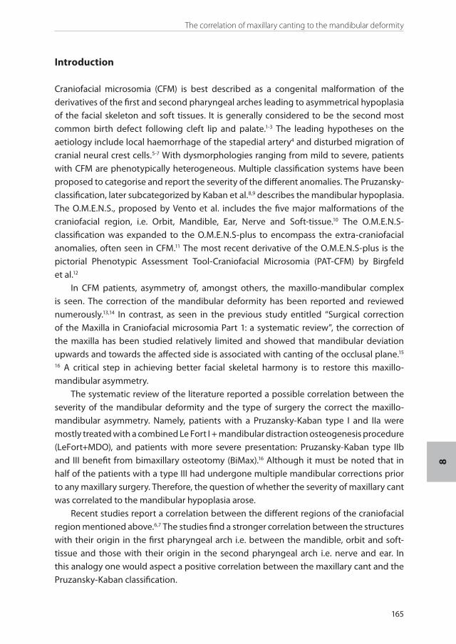
The correlation of maxillary canting to the mandibular deformity
165
8
Introduction
Craniofacial microsomia (CFM) is best described as a congenital malformation of the derivatives of the first and second pharyngeal arches leading to asymmetrical hypoplasia of the facial skeleton and soft tissues. It is generally considered to be the second most common birth defect following cleft lip and palate.1-3 The leading hypotheses on the aetiology include local haemorrhage of the stapedial artery4 and disturbed migration of cranial neural crest cells.5-7 With dysmorphologies ranging from mild to severe, patients with CFM are phenotypically heterogeneous. Multiple classification systems have been proposed to categorise and report the severity of the different anomalies. The Pruzansky-classification, later subcategorized by Kaban et al.8,9 describes the mandibular hypoplasia. The O.M.E.N.S., proposed by Vento et al. includes the five major malformations of the craniofacial region, i.e. Orbit, Mandible, Ear, Nerve and Soft-tissue.10 The O.M.E.N.S-classification was expanded to the O.M.E.N.S-plus to encompass the extra-craniofacial anomalies, often seen in CFM.11 The most recent derivative of the O.M.E.N.S-plus is the pictorial Phenotypic Assessment Tool-Craniofacial Microsomia (PAT-CFM) by Birgfeld et al.12
In CFM patients, asymmetry of, amongst others, the maxillo-mandibular complex is seen. The correction of the mandibular deformity has been reported and reviewed numerously.13,14 In contrast, as seen in the previous study entitled “Surgical correction of the Maxilla in Craniofacial microsomia Part 1: a systematic review”, the correction of the maxilla has been studied relatively limited and showed that mandibular deviation upwards and towards the affected side is associated with canting of the occlusal plane.15 16 A critical step in achieving better facial skeletal harmony is to restore this maxillo-mandibular asymmetry.
The systematic review of the literature reported a possible correlation between the severity of the mandibular deformity and the type of surgery the correct the maxillo-mandibular asymmetry. Namely, patients with a Pruzansky-Kaban type I and IIa were mostly treated with a combined Le Fort I + mandibular distraction osteogenesis procedure (LeFort+MDO), and patients with more severe presentation: Pruzansky-Kaban type IIb and III benefit from bimaxillary osteotomy (BiMax).16 Although it must be noted that in half of the patients with a type III had undergone multiple mandibular corrections prior to any maxillary surgery. Therefore, the question of whether the severity of maxillary cant was correlated to the mandibular hypoplasia arose.
Recent studies report a correlation between the different regions of the craniofacial region mentioned above.6,7 The studies find a stronger correlation between the structures with their origin in the first pharyngeal arch i.e. between the mandible, orbit and soft-tissue and those with their origin in the second pharyngeal arch i.e. nerve and ear. In this analogy one would aspect a positive correlation between the maxillary cant and the Pruzansky-Kaban classification.
PSM 20190527 Proefschrift Britt Pluijmers.indd 165 24-07-19 15:04

Chapter 8
166
In order to study a large group of patients with CFM, a multicenter collaboration was initiated between the craniofacial units of Rotterdam and Boston.
Following the systematic review of the literature, the purpose of this study was to evaluate a possible correlation between the maxillary cant and the mandibular hypoplasia, and to review the types of maxillary correction in our cohort of CFM patients.
Material and methods
With approval of both Institutional Review Boards (Rotterdam: File number MEC-2013-575; Boston: File number X05-08-058) a chart study was performed on all CFM patients presented at one of the units between January 1980 until January 2016. Unilateral affected patients with available (ConeBeam) CT-scan of the mandible and the medical history were included. CT-scans of patients who had had treatment of the upper jaw prior to the CT-scan were excluded. All charts were reviewed for information on radiographic and diagnostic criteria.
Since there are no standardized measurement tools to retrospectively evaluate the maxillary cant on CT-scan, a classification system was created for CT-scan reconstructions in the coronal plane, capturing 4 categories: normal, mild, moderate and severe. These categories were based on the degree of deviation of the line between the first molar level of the upper jaw and on the orbital rims- selected as landmarks – on a slice of the coronal plane of the CT-scan (Figures 1-4).
Two observers (BIP, LSvdL) scored the maxillary cant simultaneously 2 times with an interval of 2 weeks apart. The intrarater variability was calculated with the Intraclass Correlation Coefficient Reliability by internal consistency (Cronbach’s α) was examined. A correlation of >0,7 was set as acceptable; a correlation >0,8 was considered good and a correlation >0,9 was marked as excellent.
Descriptive statistics were used to describe affected side, age at time of surgery and diagnostic data. Pearson correlation coefficients were used to correlate the maxillary cant categorized and the severity of the mandibular hypoplasia as proposed by Pruzansky-Kaban.
Results
Of the 492 patients presented at both units, 81 patients with applicable CT-scans could be included for the analysis.
A total of 8 patients had a Pruzanksy-Kaban type I mandible, 11 had a type IIa, 29 were diagnosed with a type IIb and 32 patients had a type III mandible (Table 1). Overall, most patients had a mild (42%) to moderate (28,4%) cant. There was a significant positive
PSM 20190527 Proefschrift Britt Pluijmers.indd 166 24-07-19 15:04

The correlation of maxillary canting to the mandibular deformity
167
8
correlation between severity of the mandibular hypoplasia and the amount of canting of the maxilla (r=0,370; p<0,001; n=81) (Table 1). This correlation was seen in the first and in the second analysis. The intraclass variability was 0,868 (CI: 0,797-0,916) and the Cronbach’s α was 0,868 which is a good correlation.
Of the 81 patients presented, 24 patients (29,6%) had undergone maxillary surgery (Table 2 and 3). A BiMax was the most performed type of surgery (n=13), followed by a Le Fort1 osteotomy combined with a mandibular distraction (n=8) with the help of extra-orally placed distractors with a multivariate vector (in one case the vector control was not described), a Le Fort 1 osteotomy (n=2) and a Le Fort 1 osteotomy combined with placement of a costochondral graft to elongate the mandible.
Of those 24 surgical patients, 10 patients had preceding mandibular surgery. Six patients had preceding MDO, of whom 3 followed by LeFort+MDO, 2 followed by a BiMax
Fig. 1. Normal.
Fig. 3. Moderatie cant.
Fig. 2. Mild cant.
Fig. 4. Severe cant.
PSM 20190527 Proefschrift Britt Pluijmers.indd 167 24-07-19 15:04
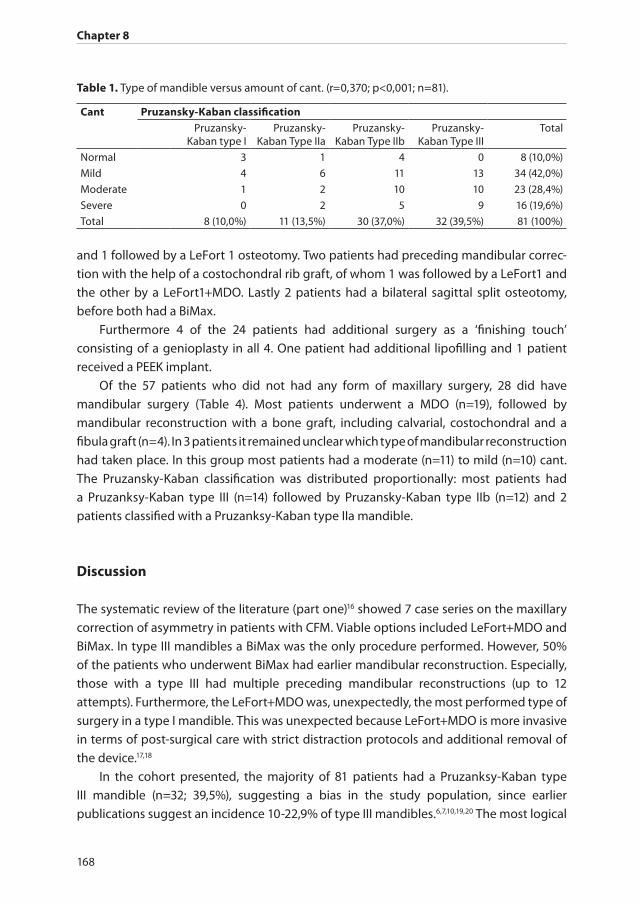
Chapter 8
168
and 1 followed by a LeFort 1 osteotomy. Two patients had preceding mandibular correc-tion with the help of a costochondral rib graft, of whom 1 was followed by a LeFort1 and the other by a LeFort1+MDO. Lastly 2 patients had a bilateral sagittal split osteotomy, before both had a BiMax.
Furthermore 4 of the 24 patients had additional surgery as a ‘finishing touch’ consisting of a genioplasty in all 4. One patient had additional lipofilling and 1 patient received a PEEK implant.
Of the 57 patients who did not had any form of maxillary surgery, 28 did have mandibular surgery (Table 4). Most patients underwent a MDO (n=19), followed by mandibular reconstruction with a bone graft, including calvarial, costochondral and a fibula graft (n=4). In 3 patients it remained unclear which type of mandibular reconstruction had taken place. In this group most patients had a moderate (n=11) to mild (n=10) cant. The Pruzansky-Kaban classification was distributed proportionally: most patients had a Pruzanksy-Kaban type III (n=14) followed by Pruzansky-Kaban type IIb (n=12) and 2 patients classified with a Pruzanksy-Kaban type IIa mandible.
Discussion
The systematic review of the literature (part one)16 showed 7 case series on the maxillary correction of asymmetry in patients with CFM. Viable options included LeFort+MDO and BiMax. In type III mandibles a BiMax was the only procedure performed. However, 50% of the patients who underwent BiMax had earlier mandibular reconstruction. Especially, those with a type III had multiple preceding mandibular reconstructions (up to 12 attempts). Furthermore, the LeFort+MDO was, unexpectedly, the most performed type of surgery in a type I mandible. This was unexpected because LeFort+MDO is more invasive in terms of post-surgical care with strict distraction protocols and additional removal of the device.17,18
In the cohort presented, the majority of 81 patients had a Pruzanksy-Kaban type III mandible (n=32; 39,5%), suggesting a bias in the study population, since earlier publications suggest an incidence 10-22,9% of type III mandibles.6,7,10,19,20 The most logical
Table 1. Type of mandible versus amount of cant. (r=0,370; p<0,001; n=81).
Cant Pruzansky-Kaban classificationPruzansky-
Kaban type IPruzansky-
Kaban Type IIaPruzansky-
Kaban Type IIbPruzansky-
Kaban Type IIITotal
Normal 3 1 4 0 8 (10,0%)
Mild 4 6 11 13 34 (42,0%)
Moderate 1 2 10 10 23 (28,4%)
Severe 0 2 5 9 16 (19,6%)
Total 8 (10,0%) 11 (13,5%) 30 (37,0%) 32 (39,5%) 81 (100%)
PSM 20190527 Proefschrift Britt Pluijmers.indd 168 24-07-19 15:04
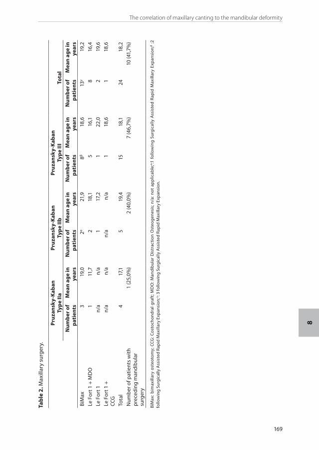
The correlation of maxillary canting to the mandibular deformity
169
8
Tab
le 2
. Max
illar
y su
rger
y.
Pru
zan
sky-
Kab
anTy
pe
IIa
Pru
zan
sky-
Kab
anTy
pe
IIb
Pru
zan
sky-
Kab
anTy
pe
III
Tota
lN
um
ber
of
pat
ien
tsM
ean
ag
e in
ye
ars
Nu
mb
er o
f p
atie
nts
Mea
n a
ge
in
year
sN
um
ber
of
pat
ien
tsM
ean
ag
e in
ye
ars
Nu
mb
er o
f p
atie
nts
Mea
n a
ge
in
year
sBi
Max
319
,02α
21,9
8β18
,613
γ19
,2
Le F
ort 1
+ M
DO
111
,72
18,1
516
,18
16,4
Le F
ort 1
n/a
n/a
117
,21
22,0
219
,6
Le F
ort 1
+CC
G
n/a
n/a
n/a
n/a
118
,61
18,6
Tota
l4
17,1
519
,415
18,1
2418
,2
Num
ber
of p
atie
nts
wit
h p
rece
din
g m
and
ibul
ar
surg
ery
1 (2
5,0%
)2
(40,
0%)
7 (4
6,7%
)10
(41,
7%)
BiM
ax: b
imax
illar
y os
teot
omy;
CCG
: Cos
toch
ond
ral g
raft
; MD
O: M
and
ibul
ar D
istr
acti
on O
steo
gen
esis
; n/a
: not
ap
plic
able
;α:1
fo
llow
ing
Surg
ical
ly A
ssis
ted
Rap
id M
axill
ary
Exp
ansi
on;β :2
fo
llow
ing
Surg
ical
ly A
ssis
ted
Rap
id M
axill
ary
Exp
ansi
on; γ
: 3 fo
llow
ing
Surg
ical
ly A
ssis
ted
Rap
id M
axill
ary
Exp
ansi
on.
PSM 20190527 Proefschrift Britt Pluijmers.indd 169 24-07-19 15:04
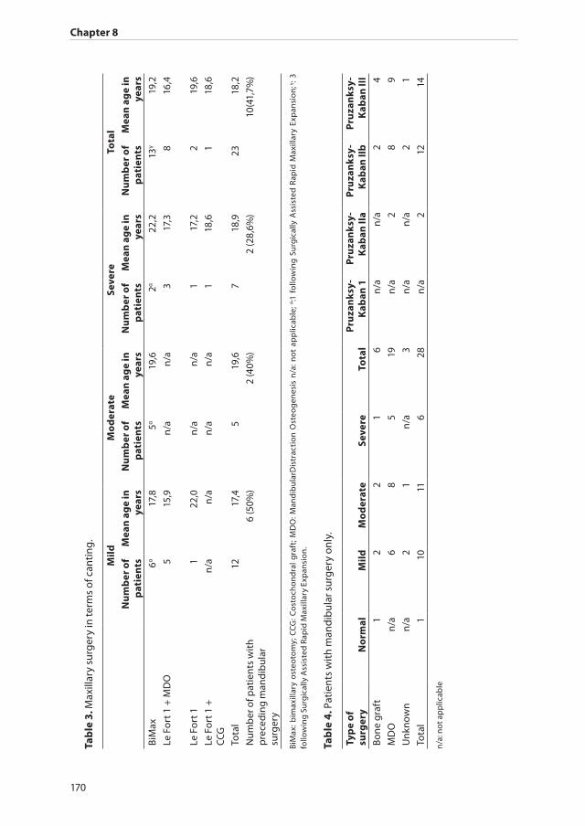
Chapter 8
170
Tab
le 3
. Max
illar
y su
rger
y in
term
s of
can
ting
.
Mild
Mo
der
ate
Seve
reTo
tal
Nu
mb
er o
f p
atie
nts
Mea
n a
ge
in
year
sN
um
ber
of
pat
ien
tsM
ean
ag
e in
ye
ars
Nu
mb
er o
f p
atie
nts
Mea
n a
ge
in
year
sN
um
ber
of
pat
ien
tsM
ean
ag
e in
ye
ars
BiM
ax6α
17,8
5α19
,62α
22,2
13γ
19,2
Le F
ort 1
+ M
DO
515
,9n
/an
/a3
17,3
816
,4
Le F
ort 1
122
,0n
/an
/a1
17,2
219
,6
Le F
ort 1
+CC
G
n/a
n/a
n/a
n/a
118
,61
18,6
Tota
l12
17,4
519
,67
18,9
2318
,2
Num
ber
of p
atie
nts
wit
h p
rece
din
g m
and
ibul
ar
surg
ery
6 (5
0%)
2 (4
0%)
2 (2
8,6%
)10
(41,
7%)
BiM
ax: b
imax
illar
y os
teot
omy;
CCG
: Cos
toch
ond
ral g
raft
; MD
O: M
and
ibul
arD
istr
acti
on O
steo
gen
esis
n/a
: not
ap
plic
able
; α:1
fo
llow
ing
Surg
ical
ly A
ssis
ted
Rap
id M
axill
ary
Exp
ansi
on; γ
: 3
follo
win
g Su
rgic
ally
Ass
iste
d Ra
pid
Max
illar
y Ex
pan
sion
.
Tab
le 4
. Pat
ient
s w
ith
man
dib
ular
sur
ger
y on
ly.
Typ
e o
f su
rger
yN
orm
alM
ildM
od
erat
eSe
vere
Tota
lP
ruza
nk
sy-
Kab
an 1
Pru
zan
ksy
-K
aban
IIa
Pru
zan
ksy
-K
aban
IIb
Pru
zan
ksy
-K
aban
III
Bone
gra
ft1
22
16
n/a
n/a
24
MD
On
/a6
85
19n
/a2
89
Unk
now
nn
/a2
1n
/a3
n/a
n/a
21
Tota
l1
1011
628
n/a
212
14
n/a
: not
ap
plic
able
PSM 20190527 Proefschrift Britt Pluijmers.indd 170 24-07-19 15:04
The correlation of maxillary canting to the mandibular deformity
171
8
explanation for this bias is the lack of indication for surgery and thus pre-operative scan in patients with a low type Pruzansky-Kaban.
There are no standardized measurement tools to retrospectively evaluate the maxillary cant. The used classification based on the first molars and orbital rim proved to have a good intra-rater variability. The authors acknowledge the subjectivity of the scale. Due to phenotypical heterogeneity of CFM patients and abnormality of development of landmark points such as the skullbase but also of the zygoma and the frontotemporal region; a more objective retrospective measurement tool is challenging to define.21
The severity of canting was mild in most of the patients (n=34). There was a positive relationship between the severity of mandibular hypoplasia and maxillary canting, supporting the theory of failed migration of neural crest cells in the first pharyngeal arch (r=0,370; p<0,001; n=81).6,7
In total, 24 patients had maxillary surgery. Due to the retrospective nature of the study it was not always clear from the charts if a patient had had orthodontic therapy. However, when reviewing the types of surgery, BiMax, LeFort+MDO, it is to be expected that nearly all patients had had pre-surgical orthodontic therapy. Most patients had a bimaxillary osteotomy (n=13). Of these 24 patients (41,7%) had a type of mandibular correction earlier in life. The majority (62,5%; n=15) had a type III mandible and a mild cant (50%; n=12). In the systematic review of the literature two types of treatments were seen: LeFort+MDO and BiMax. However, in this study Le Fort I and Le Fort I in combination with insertion of a costochondral graft (CCG) were also seen in a limited number of patients. The majority of the patients were treated with combined Le Fort 1 and mandibular distraction or BiMax. Half of the patients with a mild cant had had previous mandibular surgery. In the group with a severe cant, only one third had a preceding mandibular correction. Nevertheless, in total 8 of the 16 patients (50%) with a severe maxillary cant had had mandibulary surgery versus 16 of the 34 patients (47,1%) with a mild maxillary cant. Moreover, both maxillary and mandibulary surgery, seems to be ‘mandibulary driven’ i.e. the severity of mandibular hypoplasia seems to dictate intervention rather than a severe maxillary cant.
Furthermore, it must be kept in mind that the numbers in this retrospective study are low and the indications for surgery, other than the plausible restoration of facial harmony, were poorly documented. A future prospective study, as currently is set up between multiple centers with standardized outcome measurement tools should be conducted to identify the optimal treatment strategy.
Comparable with the systematic review, maxillary surgery was performed during the skeletal maturity with a mean age at time of surgery of 18,2 years old. Based on Part One and Part two correction of the maxillo-mandibular asymmetry it is suggested to wait until permanent dentition, if there are no definite indications for early surgery such as functional (e.g. Obstructive Sleep Apneoa) and/or psychological impairments.22-24
PSM 20190527 Proefschrift Britt Pluijmers.indd 171 24-07-19 15:04

Chapter 8
172
Conclusion
An additional study was performed on the correlation between the amount of maxillary canting and mandibular hypoplasia. Furthermore, types of surgical correction of the maxilla and mandible in patients with CFM were evaluated. There is a positive correlation between the severity of mandibular hypoplasia and canting of the maxilla. This outcome could support the failed migration of the (neural) crest hypothesis, aiding to further understanding of the pathoetiology of the deformity.
In contrast to the systematic review of the literature the most often performed type of maxillary surgery in the studied group was a Bimaxillary osteotomy. It must be noted that nearly half of these patients had mandibular surgery earlier in life. The majority of them had a more severe phenotype i.e. Pruzansky-Kaban type III mandible, but notably had a mild maxillary cant. However, 50% of the patients with a severe maxillary cant had had mandibulary surgery versus 47,1% with a mild maxillary cant; regardless of (simultaneous) maxillary surgery. Both maxillary and mandibulary surgery seems to be ‘mandibulary driven’ i.e. the severity of mandibular hypoplasia seems to dictate intervention.
PSM 20190527 Proefschrift Britt Pluijmers.indd 172 24-07-19 15:04
The correlation of maxillary canting to the mandibular deformity
173
8
References
1. Grabb WC. The first and second branchial arch syndrome. Plast Reconstr Surg 1965; 36(5): 485-508.2. Heike CL, Luquetti DV, Hing AV. Craniofacial Microsomia Overview. 1993.3. Birgfeld CB, Heike C. Craniofacial microsomia. Semin Plast Surg 2012; 26(2): 91-104.4. Poswillo D. Hemorrhage in development of the face. Birth Defects Orig Artic Ser 1975; 11(7): 61-81.5. Johnston MC, Bronsky PT. Prenatal craniofacial development: new insights on normal and abnormal
mechanisms. Crit Rev Oral Biol Med 1995; 6(4): 368-422.6. Tuin AJ, Tahiri Y, Paine KM, Paliga JT, Taylor JA, Bartlett SP. Clarifying the relationships among the
different features of the OMENS+ classification in craniofacial microsomia. Plast Reconstr Surg 2015; 135(1): 149e-56e.
7. Caron C, Pluijmers BI, Wolvius EB, et al. Craniofacial and extracraniofacial anomalies in craniofacial macrosomia: A multicenter study of 755 patients. J Craniomaxillofac Surg 2017; 45(8): 1302-10.
8. Kaban LB, Moses MH, Mulliken JB. Correction of hemifacial microsomia in the growing child: a follow-up study. Cleft Palate J 1986; 23 Suppl 1: 50-2.
9. Pruzansky S. Not all dwarfed mandibles are alike. Birth Defects Orig Artic Ser 1969; 5: 120-9.10. Vento AR, LaBrie RA, Mulliken JB. The O.M.E.N.S. classification of hemifacial microsomia. Cleft Palate
Craniofac J 1991; 28(1): 68-76; discussion 7.11. Horgan JE, Padwa BL, LaBrie RA, Mulliken JB. OMENS-Plus: analysis of craniofacial and extracraniofacial
anomalies in hemifacial microsomia. Cleft Palate Craniofac J 1995; 32(5): 405-12.12. Birgfeld CB, Luquetti DV, Gougoutas AJ, et al. A phenotypic assessment tool for craniofacial
microsomia. Plast Reconstr Surg 2011; 127(1): 313-20.13. Pluijmers BI, Caron CJ, Dunaway DJ, Wolvius EB, Koudstaal MJ. Mandibular reconstruction in the
growing patient with unilateral craniofacial microsomia: a systematic review. Int J Oral Maxillofac Surg 2014; 43(3): 286-95.
14. Nagy K, Kuijpers-Jagtman AM, Mommaerts MY. No evidence for long-term effectiveness of early osteodistraction in hemifacial microsomia. Plast Reconstr Surg 2009; 124(6): 2061-71.
15. Grayson BH, Boral S, Eisig S, Kolber A, McCarthy JG. Unilateral craniofacial microsomia. Part I. Mandibular analysis. Am J Orthod 1983; 84(3): 225-30.
16. van de Lande LS, Pluijmers BI, Caron C, et al. Surgical correction of the midface in craniofacial microsomia. Part 1: A systematic review. J Craniomaxillofac Surg 2018; 46(9): 1427-35.
17. McCarthy JG, Schreiber J, Karp N, Thorne CH, Grayson BH. Lengthening the human mandible by gradual distraction. Plast Reconstr Surg 1992; 89(1): 1-8; discussion 9-10.
18. McCarthy JG, Katzen JT, Hopper R, Grayson BH. The first decade of mandibular distraction: lessons we have learned. Plast Reconstr Surg 2002; 110(7): 1704-13.
19. Poon CC, Meara JG, Heggie AA. Hemifacial microsomia: use of the OMENS-Plus classification at the Royal Children’s Hospital of Melbourne. Plast Reconstr Surg 2003; 111(3): 1011-8.
20. Park JU, Do TH, Kwon GY, Choi TH, Kim S. Statistical analysis using the OMENS classification in Oriental patients with hemifacial microsomia: a comparative analysis with Western centers. Ann Plast Surg 2014; 72(1): 50-5.
21. Schaal SC, Ruff C, Pluijmers BI, et al. Characterizing the skull base in craniofacial microsomia using principal component analysis. Int J Oral Maxillofac Surg 2017.
22. Wolford LM, Karras SC, Mehra P. Considerations for orthognathic surgery during growth, part 2: maxillary deformities. Am J Orthod Dentofacial Orthop 2001; 119(2): 102-5.
23. Wolford LM, Karras SC, Mehra P. Considerations for orthognathic surgery during growth, part 1: mandibular deformities. Am J Orthod Dentofacial Orthop 2001; 119(2): 95-101.
24. Murray JE, Kaban LB, Mulliken JB. Analysis and treatment of hemifacial microsomia. Plast Reconstr Surg 1984; 74(2): 186-99.
PSM 20190527 Proefschrift Britt Pluijmers.indd 173 24-07-19 15:04

CH
AP
TER
PSM 20190527 Proefschrift Britt Pluijmers.indd 174 24-07-19 15:04

9
Surgical Correction of Craniofacial Microsomia: Evaluation of Interventions
in 565 Patients at Three Major Craniofacial Units
Britt I. Pluijmers, C.J.J.M. Caron, Lara S. van de Lande, Sontje Schaal, Irene M. Mathijssen, Eppo B. Wolvius, Neil Bulstrode, Robert D. Evans,
Bonnie L. Padwa, Maarten J. Koudstaal, David J. Dunaway.
Plast Reconstr Surg. 2019 May;143(5):1467-1476. doi: 10.1097/PRS.0000000000005554.
PSM 20190527 Proefschrift Britt Pluijmers.indd 175 24-07-19 15:04

Chapter 9
176
Abstract
AimsCraniofacial microsomia is characterized by an asymmetric, hypoplasia of derivatives of the first and second pharyngeal arch, leading to a variety of phenotypic presentations. Studies on surgical correction of patients with craniofacial microsomia have small cohorts, leaving controversial opinions on the optimal treatment modality, the indication for surgery and the optimal timing of surgery. The purpose of this study was to evaluate the types of, timing of, and total number of surgical corrections performed and the number of surgical procedures in correlation to the severity of the phenotype.
Patients and MethodsA retrospective chart study was conducted including patients diagnosed with craniofacial microsomia from three large craniofacial units. Demographic, radiographic, and clinical information was obtained, including type and number of surgical procedures and age at the time of surgery.
Results A total of 565 patients were included. In total, 443 (78.4 percent) of all patients underwent some form of surgery during their life, varying from skin tag removal to major craniofacial operations. The number of surgical interventions was higher with increasing severity of phenotype, bilateral presentation, and a younger age at the first intervention.
Conclusions Multiple surgical corrections are frequently seen in patients with a more severe or bilateral presentation. Furthermore, those who are treated earlier in life for correction of asymmetry of the mandible will undergo significantly more surgical procedures to correct the asymmetry later on, independent of the Pruzansky-Kaban type mandible. A prospective international multicenter study is designed with a uniform registration and outcome measurement tool to identify the optimal treatment strategy.
PSM 20190527 Proefschrift Britt Pluijmers.indd 176 24-07-19 15:04
Surgical correction of craniofacial microsomia
177
9
Introduction
Craniofacial microsomia is best described in its embryologic origin: a developmental defect of any structure of the first and/or second pharyngeal arch leading to a predominantly asymmetric, hypoplasia of their derivatives.1-3 The phenotypical presentation and therefore, the severity of the deformity can vary significantly.2,4-6 Furthermore bilateral involvement is reported in 2.5-34% of cases.7,8 Starting early in life, patients can encounter several functional and/or aesthetic problems. Orbital malformations can include epibulbair dermoids, eyelid coloboma, orbital dystopia, and micro- or anophthalmus.4,9,10 Furthermore, hypoplasia of the jaw may vary from a normally shaped but smaller sized mandible to an abnormally shaped mandible with absence of the condyle and ramus leading not only to functional problems such as a malocclusion, airway problems or ankylosis; but also, to a distinct facial scoliosis.8,11 External ear problems, occurring in the majority of patients with ra, ranges from microtia to anotia with atresia of the auditory canal. 4,9,10 Another aspect frequently seen in patients with craniofacial microsomia is the presence of preauricular or facial tags and/or pits with or without cartilage remnants. Besides, soft-tissue problems due to muscle and/or fat underdevelopment or atrophy are described. Macrostomia, (Tessier 7 cleft) can be part of the phenotype of craniofacial microsomia. Finally, facial nerve palsy of either a part of or all branches is observed in 10-45% craniofacial microsomia patients.12
Due to the wide phenotypic spectrum of craniofacial microsomia, an internationally accepted classification and/or grading system is essential for the communication between different centers with regards to patient care and research. One of the first grading systems used is the Pruzansky classification,13 later adjusted by Kaban et al.14,15 Vento et al. further extended the classification system with inclusion of classifications for Orbital asymmetry, Ear deformity, Nerve dysfunction and Soft-tissue deficiency, to the orbital, mandibular, ear, neural, and soft-tissue classification;10 modified by Horgan et al. to the orbital, mandibular, ear, neural, and soft-tissue -plus which covers additional extracranial features.4 The most recent derivative is the pictorial Phenotypic Assessment Tool-Craniofacial Microsomia by Birgfeld et al.5
Craniofacial microsomia is regarded to be the second most common congenital facial condition following cleft lip and palate.11 Because of the variable presentation of craniofacial microsomia a wide range of treatment options are available. 16-49 Studies on surgical treatment in craniofacial microsomia are limited to small cohorts and expert opinions, with significant differences. There is no consensus on indications, the optimal treatment modality and optimal timing of surgery.
In order to study a large group of patients with craniofacial microsomia, a collabora-tion between the craniofacial units of Erasmus University Medical Center (EMC) Rotterdam, Great Ormond street Hospital NHS Foundation Trust (GOSH) London and
PSM 20190527 Proefschrift Britt Pluijmers.indd 177 24-07-19 15:04
Chapter 9
178
Boston’s children’s Hospital (BCH) Boston, was initiated. Because of this collaboration it was possible to analyze a large data set of patients with craniofacial microsomia. The purpose of this retrospective study was to evaluate the type of surgical corrections of the craniofacial anomaly in patients with craniofacial microsomia. Additional objectives were to evaluate the timing of the procedures and the total number of surgical corrections performed. Lastly, the number of surgical procedures in correlation to the severity, including a unilateral versus bilateral phenotype, was evaluated.
Patients and Methods
Study designWith approval of the institutional medical ethics board of all three centers (i.e., Medical Ethics Review Committee Erasmus MC, file number MEC-2013-575; IRB Medical Ethics Review Committee Boston Children’s Hospital, file number X05-08-058; and the R&D Medical Ethics Review Committee, Great Ormond Street Hospital NHS Foundation Trust, file number 14 DS25), a retrospective cohort study was conducted. Patients’ charts from January of 1980 until January of 2016 of patients diagnosed with craniofacial microsomia were reviewed. Patients in whom adequate diagnosis based on the orbital, mandibular, ear, neural, and soft-tissue–plus classification or phenotypic assessment tool for craniofacial microsomia was not able to be determined were excluded. The Boston Children’s Hospital has a history of reporting the orbital, mandibular, ear, neural, and soft-tissue– plus classification in patients’ charts. At the Great Ormond Street Hospital and the Erasmus University Medical Center, all patients with both clinical and radiographic images were included and scored with the help of the phenotypic assessment tool for craniofacial microsomia by two trained researchers at each location. If there was any disagreement, the score was discussed if needed with a researcher from the other location, until agreement was achieved. Throughout time, different treatment strategies have been used at each center.
VariablesBaseline characteristics included sex and diagnosis. All surgical notes were reviewed; type and number of surgical procedures, indication, and age at the time of surgery were registered. Surgical procedures included any surgical procedure to correct a functional or aesthetic problem related to craniofacial microsomia (e.g., osteotomies, lipofilling, ear reconstructions). Removal of hardware (such as distraction devices) or surgical treatment to address complications such as infections or device failure was not counted as a surgical correction. Complications following surgery were annotated.
All information was anonymized before it was imported into IBM SPSS Version 24 (IBM Corp., Armonk, N.Y.) for analysis. Descriptive statistics were used to describe sex,
PSM 20190527 Proefschrift Britt Pluijmers.indd 178 24-07-19 15:04

Surgical correction of craniofacial microsomia
179
9
laterality, and diagnostic data. Pearson correlation coefficients were used to correlate the severity and laterality. An independent t test was performed to compare the mean age between intervention groups. A linear regression was calculated to predict the number of operations based on the age at first surgery and Pruzansky-Kaban type.
Results
Patient demographicsCraniofacial microsomia was diagnosed in 955 patients. A total of 565 patients met the inclusion criteria. (Table 1)
The male to female ratio was 1.2:1. A total of 496 (87,8%) patients had a unilateral presentation of craniofacial microsomia. The distribution between right and left-sided craniofacial microsomia was 1.2:1 (n=496) as well. (Table 1)
Most patients with unilateral craniofacial microsomia had a Pruzansky-Kaban mandible Type I (26.8%) or IIa (26.6%) followed by the Pruzanksy-Kaban IIb (23.2%) and III (15.9%). In 40 patients from the BCH the data on the mandible was inconclusive with notes in the charts doubting 2 different scores for example 1 or 2a. The Pruzansky-Kaban classification of the more severely affected side in patients with bilateral craniofacial microsomia was significantly more frequently scored as IIB or III compared to the Pruzansky-Kaban classification of the unilateral affected patients ([Pearson chi-square (3) = 26,227, p < 0.001]). (Tables 2 & 3) The classification of the nerve proved to be challenging in this retrospective cohort, we were unable to score the nerve function reliably.
Orbito-zygomatic-complexWith regards to the orbito-zygomatic-complex; 53 of the 565 patients had surgery involving these structures. Forty-nine patients had a unilateral presentation of craniofacial microsomia. More than half of the patients had an abnormal orbital size and/or displacement (Table 5). Most patients received an alloplastic malar implant (n=23), followed by bone grafts including costochondral (n=8), iliac (n=5) cavarial (n=5) and fibula (n=1) bone grafts. Eleven patients had a variety of orbito-zygomatic osteotomies. Only four patients had a bilateral presentation. Two received correction of the asymmetry by alloplastic malar implants. The other two bilateral patients received a bone graft of either a fibula graft or an iliac bone graft.
EyeIn total 55 patients with craniofacial microsomia underwent surgery to the eye. In most patients this consisted of removal of an epibulbar dermoid (Table 6).
PSM 20190527 Proefschrift Britt Pluijmers.indd 179 24-07-19 15:04
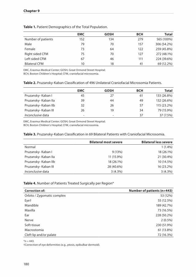
Chapter 9
180
Table 1. Patient Demographics of the Total Population.
EMC GOSH BCH TotalNumber of patients 152 134 279 565 (100%)
Male 79 70 157 306 (54.2%)
Female 73 64 122 259 (45.8%)
Right sided CFM 75 70 127 272 (48.1%)
Left sided CFM 67 46 111 224 (39.6%)
Bilateral CFM 10 18 41 69 (12.2%)
EMC, Erasmus Medical Center; GOSH, Great Ormond Street Hospital;BCH, Boston Children’s Hospital; CFM, craniofacial microsomia.
Table 2. Pruzansky-Kaban Classification of 496 Unilateral Craniofacial Microsomia Patients.
EMC GOSH BCH TotalPruzansky- Kaban I 45 27 61 133 (26.8%)
Pruzansky- Kaban IIa 39 44 49 132 (26.6%)
Pruzansky- Kaban IIb 32 26 57 115 (23.2%)
Pruzansky- Kaban III 26 19 34 79 (15.9%)
Inconclusive data - - 37 37 (7.5%)
EMC, Erasmus Medical Center; GOSH, Great Ormond Street Hospital;BCH, Boston Children’s Hospital; CFM, craniofacial microsomia.
Table 3. Pruzansky-Kaban Classification in 69 Bilateral Patients with Craniofacial Microsomia.
Bilateral most severe Bilateral less severeNormal 1 (1.4%)
Pruzansky- Kaban I 9 (13%) 18 (26.1%)
Pruzansky- Kaban IIa 11 (15.9%) 21 (30.4%)
Pruzansky- Kaban IIb 18 (26.1%) 10 (14.5%)
Pruzansky- Kaban III 28 (40.6%) 16 (23.2%)
Inconclusive data 3 (4.3%) 3 (4.3%)
Table 4. Number of Patients Treated Surgically per Region*
Correction of: Number of patients (n=443)Orbito / Zygomatic complex 53 (12%)
Eye† 55 (12.5%)
Mandible 189 (42.7%)
Maxilla 73 (16.5%)
Ear 228 (50.2%)
Nerve 2 (0.5%)
Soft-tissue 230 (51.9%)
Macrostomia 61 (13.8%)
Cleft lip and/or palate 72 (16.3%)
*n = 443.†Correction of eye deformities (e.g., ptosis, epibulbar dermoid).
PSM 20190527 Proefschrift Britt Pluijmers.indd 180 24-07-19 15:04

Surgical correction of craniofacial microsomia
181
9
MandibleOf the 443 patients who received a form of surgery to correct the deformity, 42.7 percent had a mandibular correction or reconstruction.
Most patients had a Pruzansky-Kaban type III mandible, followed by the type IIb, type IIa and type I. (Table 7) The patients with inconclusive data on the Pruzansky-Kaban classification were left out (n=17).
Unilateral craniofacial microsomiaSix patients with a Pruzansky-Kaban type I mandible underwent surgery, including an alloplastic graft, genioplasty, distraction osteogenesis, costochondral graft and osteo-tomy. Patients with a unilateral Pruzansky-Kaban type IIa most often received distraction osteogenesis to lengthen the underdeveloped mandible. In the group of patients with a unilateral Pruzansky-Kaban type IIa 10 of the 19 patients underwent multiple surgeries to correct the deformed mandible. Most often the additional surgery consisted of a genioplasty (n=5). In the unilateral Pruzansky-Kaban type IIb group, 26 patients underwent distraction osteogenesis followed by reconstruction of the mandible with the help of a costochondral graft (n=17). However, in 29 of 59 (49.2%) unilateral Pruzansky-Kaban type IIb patients, an additional type of surgery was carried out. Most patients (n=15) underwent additional osteotomies including bimaxillary osteotomies, unilateral or bilateral sagittal split osteotomies and genioplasties.
In the group with a unilateral Pruzanksy-Kaban type III, most patients underwent a reconstruction of the absent condyle with the help of a bone graft (n=30). Most often, a costochondral graft was used, followed by an iliac bone graft and calvarial bone graft. Additional surgery to correct the deformity in this group was performed in 61percent of the patients. Most of these patients had additional osteotomies including bimaxillary osteotomies, unilateral; or bilateral sagittal split osteotomies and genioplasties (n=13), followed by distraction osteogenesis of the bone graft (n=10).
Table 6. Eye Surgery.
Type of surgery Unilateral CFM (n=40) Bilateral CFM (n=15)Removal epibulbar dermoid 18 7
Exo/esotropia correction 9 1
Ptosis correction 8 2
Coloboma repair 6 4
Canthoplasty 3 5
Probing of nasal lacrimal duct 3 1
Prosthesis 2 2
Cornea correction 2
Dacrocystorhinostomy 1
Entropion correction 1
CFM, craniofacial microsomia.
PSM 20190527 Proefschrift Britt Pluijmers.indd 181 24-07-19 15:04
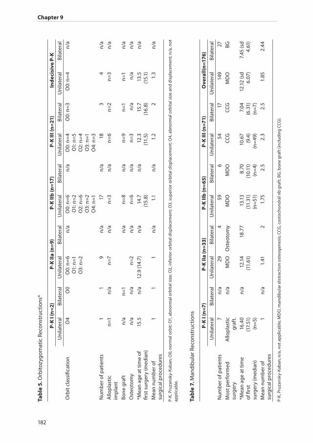
Chapter 9
182
Tab
le 5
. Orb
itozy
gom
atic
Rec
onst
ruct
ions
*
P-K
I (n
=2)
P-K
IIa
(n=
9)P
-K II
b (n
=17)
P-K
III (
n=
21)
Ind
ecis
ive
P-K
Uni
late
ral
Bila
tera
lU
nila
tera
lBi
late
ral
Uni
late
ral
Bila
tera
lU
nila
tera
lBi
late
ral
Uni
late
ral
Bila
tera
l
Orb
it c
lass
ifica
tion
O4
O0
O0:
n=
6O
1: n
=1O
3: n
=2
n/a
O0:
n=
6O
1: n
=2O
2: n
=6
O3:
n=2
O4:
n=1
n/a
O0:
n=
4O
1: n
=5
O2:
n=
4O
3: n
=1O
4: n
=3
O0:
n=
3O
0: n
=4
n/a
Num
ber
of p
atie
nts
11
9n
/a17
n/a
183
4n
/a
Allo
pla
stic
imp
lant
n=1
n/a
n=7
n/a
n=
3n
/an
=6
n=2
n=
3n
/a
Bone
gra
ftn
/an
=1n
/an
=8
n/a
n=
9n
=1n
=1n
/a
Ost
eoto
my
n/a
n/a
n=2
n/a
n=
6n
/an
=3
n/a
n/a
n/a
*Mea
n ag
e at
tim
e of
fir
st s
urg
ery
(med
ian)
15.5
n/a
12.9
(14.
7)n
/a14
.7(1
5.8)
n/a
12.3
(11.
5)15
.7(1
6.8)
13.5
(15.
1)n
/a
Mea
n nu
mb
er o
f su
rgic
al p
roce
dur
es1
11
n/a
1.1
n/a
1.2
21.
3n
/a
P-K
, Pru
zans
ky-K
aban
; O0,
nor
mal
orb
it; O
1, a
bn
orm
al o
rbit
al s
ize;
O2,
infe
rior
orb
ital
dis
pla
cem
ent;
O3,
sup
erio
r orb
ital
dis
pla
cem
ent;
O4,
ab
nor
mal
orb
ital
siz
e an
d d
isp
lace
men
t; n
/a, n
ot
app
licab
le.
Tab
le 7
. Man
dib
ular
Rec
onst
ruct
ions
P-K
I (n
=7)
P-K
IIa
(n=
33)
P-K
IIb
(n=
65)
P-K
III (
n=7
1)O
vera
ll(n
=176
)U
nila
tera
lBi
late
ral
Uni
late
ral
Bila
tera
lU
nila
tera
lBi
late
ral
Uni
late
ral
Bila
tera
lU
nila
tera
lBi
late
ral
Num
ber
of p
atie
nts
7n
/a29
459
654
1714
927
Mos
t per
form
ed
surg
ery
Allo
pla
stic
graf
t.n
/aM
DO
Ost
eoto
my
MD
OM
DO
CCG
CCG
MD
OBG
*Mea
n ag
e at
tim
e of
firs
t su
rger
y (m
edia
n)
16.4
0 (1
7.51
) (n
=5)
n/a
12.1
4(1
1.61
)18
.77
13.1
3(1
1.31
)(n
=51
)
8.70
(10.
11)
(n=
4)
10.6
7(9
.4)
(n=
49)
7.04
(6.3
1)(n
=7)
12.1
2 (s
d 6.
07)
7.45
(sd
4.61
)
Mea
n nu
mb
er o
f su
rgic
al p
roce
dur
es
1n
/a1.
412
1.75
2.5
2.3
2.5
1.85
2.44
P-K
, Pru
zans
ky-K
aban
; n/a
, not
ap
plic
able
; MD
O, m
and
ibul
ar d
istr
acti
on o
steo
gen
esis
; CCG
, cos
toch
ond
ral r
ib g
raft
; BG
, bon
e g
raft
(in
clu
din
g CC
G).
PSM 20190527 Proefschrift Britt Pluijmers.indd 182 24-07-19 15:04

Surgical correction of craniofacial microsomia
183
9
Bilateral craniofacial microsomiaThe same trend found in unilateral patients can be found in the bilateral group. Osteotomies are most often performed in the Pruzansky-Kaban IIa group (n=4) and distraction osteogenesis is performed most in the bilateral Pruzanksy-Kaban type IIb group (n=4). The need of reconstruction of the mandible using bone grafts is most frequently seen in the Pruzanksy-Kaban type III group (n=11). Furthermore, 75 percent of all bilateral patients who underwent surgery to correct the deformity needed additional surgery with frequencies ranging from 1 to 5 surgical procedures and an average of 2.44.
Number of surgical procedures.As mentioned above, in bilateral patients the average number of surgical procedures is 2.44, in comparison to unilateral patients who will undergo an average of 1.85 procedures [Pearson chi-square(5) = 16.037, p = 0.007]. The age of patients at their first mandibular procedure, who underwent 1-2 surgical procedures compared to those with 3 or more, drops from a mean of 12.18 years (n= 116) to 9.73 years (n=35) (t
(149)=2.11, p=0.036).
Finally, a linear regression was calculated to predict the number of surgeries based on the age at first surgery and Pruzansky-Kaban. The linear regression model indicated that for every year increase in age a lower number of operations was performed, independent of the Pruzanksy-Kaban type mandible (coefficients -0.033, SE 0.016, p=0.042).
MaxillaSeventy-three patients underwent correction of the maxilla. (Table 8) The interventions included bimaxillary osteotomies, with a “classic” bilateral sagittal split osteotomy of the mandible and a distraction osteogenesis of the hypoplastic mandible or bone graft with a unilateral sagittal split osteotomy on the “normal” side (Le Fort plus mandibular distraction osteogenesis procedure); and single Le Fort osteotomies with or without a preceding surgically assisted rapid maxillary expansion and recontouring of the maxilla. A surgically assisted rapid maxillary expansion followed by a Le Fort I osteotomy was not counted as an additional correction, because this is regarded as a preparative step.
In a total of 42 patients, classic bimaxillary osteotomies were performed, followed by a single Le Fort I procedure (n = 7) and a Le Fort plus mandibular distraction osteogenesis procedure (n = 5). In eight patients, a surgically assisted rapid maxillary expansion was performed preceding the definite osteotomy. All patients were treated at the end of puberty. Additional maxillary surgery to reach the desired facial symmetry was rarely seen.
EarA total of 228 patients (40.4 percent) underwent reconstruction of the ear. Of 10 patients, there were insufficient data on the type of surgery. These were left out of the analysis. In the normal to mild cases (E0 to E1), patients underwent otoplasty, most often
PSM 20190527 Proefschrift Britt Pluijmers.indd 183 24-07-19 15:04

Chapter 9
184
accompanied by other soft-tissue reconstruction in the area. (Table 9. A total of 37 unilateral E2 ears were corrected. Most ears were corrected with the help of a costochondral graft (n = 23) followed by a “regular” otoplasty (n = 14). One bilateral case had rib-graft reconstructions for both ears.
In the unilateral patients with type E3 ears, 120 patients underwent multistage reconstruction with the help of a costochondral graft. Three of these patients required a preceding tissue expander treatment. Nineteen patients had a “regular” otoplasty; however, in five cases, a reconstruction with the help of a costochondral graft was performed later. In one patient, a prosthetic ear was used. Furthermore, one patient’s ear was reconstructed with a free vascular skin graft. In the bilateral patients with type E3 ears, all patients underwent multistage reconstruction with a costochondral graft. Two patients needed a tissue expander before the definitive treatment. Three patients received a neoear for their anotia (E4). Two patients, one of whom was a bilateral patient, received a reconstructed ear using a costochondral graft. One unilateral patient received an implant-supported epithesis.
NerveOne patient underwent a cross-facial nerve grafting at age 15. Another patient received gold weights to correct the lagophthalmos caused by facial nerve impairment.
Soft TissueIn total, 230 patients underwent soft-tissue correction. In most cases (n = 139), it concerned the removal of a skin tag, dermoid or duct cyst, skin pits, or cartilage remnant in the embryologic pathway of the first or second pharyngeal arch. Most patients had incomplete data on the age at first treatment other than a notion of treatment during infancy. If we set these procedures apart, 91 patients underwent surgery involving soft tissue primarily. The most common procedure was lipofilling, with an average of 1.2 procedures per patient, followed by a type of free autologous tissue transfer. (Table 10)
CleftsMacrostomia correction was performed in 61 patients, of whom six also had a cleft lip and palate. Nine patients were treated for their cleft lip. A total of 24 patients had a cleft palate, which needed correction. Furthermore, 32 patients underwent cleft lip and palate surgery; eight of these patients had a bilateral presentation.
ComplicationsComplications were annotated in 5.7 percent of patients (n = 3) that underwent an orbitozygomatic complex reconstruction, all of which were (autologous) graft infections. One complication (1.8 percent) was registered in the eye correction: a corneal melt. In 63 patients that underwent mandibular reconstruction (33.3 percent), a complication
PSM 20190527 Proefschrift Britt Pluijmers.indd 184 24-07-19 15:04

Surgical correction of craniofacial microsomia
185
9
Tab
le 8
. Max
illar
y re
cons
truc
tion
s.
P-K
I (n
=1)
P-K
IIa
(n=1
4)P
-K II
b (n
=26
)P
-K II
I (n
=27
)O
vera
ll(n
=73)
Uni
late
ral
Bila
tera
lU
nila
tera
lBi
late
ral
Uni
late
ral
Bila
tera
lU
nila
tera
lBi
late
ral
Uni
late
ral
Bila
tera
l
Num
ber
of p
atie
nts
1n
/a12
224
221
663
*10
Mos
t per
form
ed
surg
ery
unkn
own
n/a
BiM
axBi
Max
αBi
Max
±Bi
Max
BiM
axα
BiM
axα
BiM
ax∞
BiM
ax±
*Mea
n ag
e at
tim
e of
fir
st s
urg
ery
(med
ian)
unkn
own
n/a
16.7
(18.
1)21
.318
.6(1
7.6)
16.8
16.3
(16.
7)17
.5(1
6.8)
17.2
(17.
2)18
.4 (1
6.8)
Mea
n nu
mb
er o
f su
rgic
al p
roce
dur
es
1n
/a1
11.
11
1.1
11.
11
BiM
ax: C
lass
ic b
imax
illar
y os
teot
omy;
n/a
: not
ap
plic
able
; P-K
: Pru
zans
ky-K
aban
; α
: 1 fo
llow
ing
surg
ical
ly a
ssis
ted
rap
id m
axill
ary
exp
ansi
on.
±:2
follo
win
g su
rgic
ally
ass
iste
d ra
pid
max
illar
y ex
pan
sion
.∞
:4 fo
llow
ing
surg
ical
ly a
ssis
ted
rap
id m
axill
ary
exp
ansi
on..
* 5
unila
tera
l pat
ient
s h
ad in
dec
isiv
e Pr
uza
nsky
-Kab
an c
lass
ifica
tion
s, a
ll of
wh
om h
ad a
bim
axill
ary
oste
otom
y.
Tab
le 9
. Ear
reco
nstr
ucti
ons.
E0: N
orm
al e
arE1
: All
par
ts p
rese
nt.
m
ild d
efo
rmit
y
E2: A
uri
cle
½- 2
/3 o
f p
red
icte
d s
ize
No
t all
par
ts p
rese
nt
E3: S
ever
ely
mal
form
ed, o
ften
p
ean
ut s
hap
edE4
: An
oti
a U
nila
tera
lBi
late
ral
Uni
late
ral
Bila
tera
lU
nila
tera
lBi
late
ral
Uni
late
ral
Bila
tera
lU
nila
tera
lBi
late
ral
Num
ber
of p
atie
nts
2n
/a9
n/a
371
144
172
1
Mos
t per
form
ed
surg
ery
Oto
pla
sty
n/a
Oto
pla
sty
n/a
Reco
n-
stru
ctio
n w
ith
CCG
Reco
n-
stru
ctio
n w
ith
CCG
Reco
n-
stru
ctio
n w
ith
CCG
Reco
n-
stru
ctio
n w
ith
CCG
n/a
Reco
n-
stru
ctio
n w
ith
CCG
Mea
n ag
e at
tim
e of
fir
st s
urg
ery
(med
ian)
10.7
(7.5
)n
/a6.
2(5
.6)
n/a
9.6
(9.5
)(n
=35
)
11.7
10.4
(10.
1)(n
=127
)
11.5
(11.
4)(n
=13)
30.4
(30.
4)n
/a
Mea
n nu
mb
er o
f su
rgic
al p
roce
dur
es1
n/a
1.4
n/a
2.4
(n=
36)
2.5
(per
ear
)2.
62.
7(p
er e
ar)
2.5
1
CCG
: Cos
toch
ond
ral g
raft
; n/a
: not
ap
plic
able
.
PSM 20190527 Proefschrift Britt Pluijmers.indd 185 24-07-19 15:04

Chapter 9
186
occurred. These complications included autologous graft infections (n=12), postoperative hypoesthesia (n=9), and ankylosis or graft ankylosis (both n=8). Furthermore, wound infection (n=6), infected osteosynthesis material and malunion/nonunion (n=5), malpositioning of the distraction device and overgrowth of the rib graft (n=3), infected Medpor (Porex Surgical, Newnan, Ga.) (n=2), and graft dehiscence and hypertrophic scars (n=1) were annotated. In the maxillary surgery group, three patients (4.1 percent) encountered a complication: one patient had a wound infection, one patient had a sinusitis, and one patient had hypoesthesia postoperatively. In 27 patients who underwent ear reconstruction (11.8 percent), notation of a complication was made in the charts. Most had a wound infection (n = 15), followed by an infection of the autologous graft and hypertrophic scars (n = 4), graft dehiscence (n = 3), and a malfunctioning of the hardware (n=1). In three of the 230 patients with a soft-tissue correction (1.3 percent), a notation was made of a complication: two wound infections and one infected autologous graft. There were no complications mentioned in the patients’ charts after the nerve reconstruction, the correction of the macrostomia, or the cleft lip and/or palate repair.
Discussion
Craniofacial microsomia has a heterogeneous presentation, demanding a multiangle approach.46-48 Studies on surgical correction of patients with CFM until now have small cohorts restricted to expert opinions, with significant differences on not only the optimal treatment modality but also indication of surgery and optimal timing of surgery. In this study the surgical corrections performed at three large centers were evaluated. Because of the retrospective nature of the study indications were not always stated and therefore
Table 10. Soft-Tissue Reconstructions.
Type of procedure Number of patients Mean age*Total 230 14.7 (n=91)
Skintag, dermoid or duct cyst, skin pits or cartilage remnant removal
139 Infancy
Lipofilling 57 14.7
Autologous tissue flap 19 16.7
Scar revision 15 9.6
Browlift 5 14
Botulinum toxin type A 6 9.5
Tissue reduction 11 16.8
Tissue expander 6 13.3
*n = 91.
PSM 20190527 Proefschrift Britt Pluijmers.indd 186 24-07-19 15:04

Surgical correction of craniofacial microsomia
187
9
left out of this study. Furthermore, the complications and treatment of complications were poorly documented. The presented data might be an under-reporting of the true numbers.
For the evaluation of the patients both the phenotypic assessment tool for craniofacial microsomia and the orbital, mandibular, ear, neural, and soft-tissue–plus classification were used.4 The authors are supportive of the phenotypic assessment tool for craniofacial microsomia, despite the tool’s convenience and ease of use, some data including nerve function, and radiographic orbit position were left out since it was impossible to gather these specific data from the historic charts.
This study finds a ratio of 1.2:1 for both the male-to-female ratio as for the right-to-left ratio, which represents a more right sided affected deformity. Some authors report a 3:2 predominance for the male gender and for a right-side presentation of the deformity, whereas other studies report a more equally distribution by sex and side.4,6,50-52 Furthermore 87.8 percent had a predominantly unilateral presentation compared to 2.5 to 34 percent reported in literature.53-55
Throughout the years, different treatment modalities and paradigms have been proposed for the correction of the asymmetry of the deformity.24,32,56,57 In this cohort 78.4 percent of the patients underwent surgery related to their craniofacial deformity.
The Pruzansky-Kaban type III mandible patient is the most challenging in terms of correction of the facial asymmetry.26,29,57 Patients with a unilateral type IIb mandible are in need of multiple operations in 49.1 percent. A unilateral type III mandible, however, would need multiple operations in 61 percent of the cases; however, the most challenging patients are the bilateral patients, who are in need of multiple surgical procedures in 83 percent (Pruzansky-Kaban type IIb) and 76 percent (Pruzansky- Kaban type III) of all cases.
With regard to mandibular surgery, age at the first surgical procedure is shown to have an influence on the number of surgical procedures needed throughout life. Patients, both unilateral and bilateral, who underwent three or more surgical corrections of the mandible were treated significantly earlier at a mean age of 9.73 years (n = 35), compared with those who underwent “only” one or two surgical procedures of the mandible, who were on average treated at 12.81 years (n = 116; t
149 = 2.11; p = 0.036). Also, the linear
regression model indicated that for every year decrease in age, the number of operations performed went up, independent of the Pruzansky-Kaban type mandible. In other words, those who are treated earlier in life for correction of asymmetry of the mandible will undergo more surgical procedures to correct the asymmetry, possibly suggesting that the operations might be responsible for impaired growth, which then increases the need for more operations. This reinforces the policy of correcting mandibular asymmetry at an older age unless there are significant functional problems (e.g., obstructive sleep apnea).28,58
Surgical interventions regarding the maxilla were most often single procedures. The treatment outcome, measured as the number of surgical procedures/the need of
PSM 20190527 Proefschrift Britt Pluijmers.indd 187 24-07-19 15:04

Chapter 9
188
additional surgery, might be better because of the skeletal maturity of the patients at the time of surgery; the mean age at the time of intervention was 17.2 to 18.4 years.
Half of all patients underwent ear reconstruction, with the Nagata two-step otoplasty most frequently performed. However, in 40.1 percent, the number of surgical procedures was more than two. Concurrent with the literature, patients on average underwent a total of 2.5 instead of two procedures, with one case requiring nine procedures because of skin necrosis or inflammation.59,60
Facial nerve weakness could possibly contribute to a negative surgical outcome in terms of patient satisfaction and quality of life. Cline et al. recently reviewed the literature concerning the prevalence of facial nerve palsy, which is found to range between 10 and 45 percent.12 In comparison with other surgical corrections, little has been written on facial nerve reanimation, especially with other surgical techniques, for the correction of asymmetry in craniofacial microsomia.61 However, reconstruction of the nerve is comprehensive as a result of agenesis and/or underdevelopment of the overlying struc-tures. 62 To evaluate the influence of the facial nerve in corrective surgery, it would be recommended to look into a grading system that includes both dynamic and static symmetry in patients.
Based on the data presented, we advocate postponing corrective surgery until a more mature age unless there are significant functional problems. This approach will most likely reduce the number of surgical interventions and burden of care for these patients and their caretakers.
Conclusion
Although craniofacial microsomia is considered to be the second most common craniofacial defect after cleft lip and palate,11 the numbers of patients in case series in the literature are low. We present the surgical data of 565 craniofacial microsomia patients. Most patients had a unilateral Pruzansky-Kaban type I (26.8 percent) or IIa (26.6 percent) mandible, followed by the Pruzansky-Kaban IIb (23.2 percent) and III (15.9 percent) mandible. In this large cohort, 78 percent of the patients underwent surgery related to their craniofacial deformity.
Concurrent with other studies, the most challenging patients in terms of correction of asymmetry have Pruzansky-Kaban type III mandibles. However, we found that another subgroup consists of bilateral craniofacial microsomia patients in whom one could assume the bilaterality would lead to more symmetry but who would face an even larger number of surgical procedures—on average 1.75 times more—than unilateral craniofacial microsomia patients. Furthermore, those who are treated earlier in life for correction of asymmetry of the mandible will undergo significantly more surgical procedures to correct the asymmetry later, independent of the Pruzansky-Kaban type mandible. Prospective
PSM 20190527 Proefschrift Britt Pluijmers.indd 188 24-07-19 15:04
Surgical correction of craniofacial microsomia
189
9
studies are essential and patient outcome measurements are needed to be able to truly compare patient outcomes between different treatment modalities to further improve care.
Currently, a prospective international multicenter study is designed with uniform registration and outcome measurement tools. To reduce the number of surgical inter-ventions, the clear indications for surgery should be defined.
Acknowledgment
The authors acknowledge M. Gribova, Y. Klazen, B. Maas, and R. Renkema, for their assistance with the databases at the Great Ormond Street Hospital, London, UK, and Boston Children’s Hospital, Boston, Massachusetts, USA.
PSM 20190527 Proefschrift Britt Pluijmers.indd 189 24-07-19 15:04
Chapter 9
190
References
1. Werler MM, Starr JR, Cloonan YK, Speltz ML. Hemifacial microsomia: from gestation to childhood. J Craniofac Surg 2009; 20 Suppl 1: 664-9.
2. Keogh IJ, Troulis MJ, Monroy AA, Eavey RD, Kaban LB. Isolated microtia as a marker for unsuspected hemifacial microsomia. Arch Otolaryngol Head Neck Surg 2007; 133(10): 997-1001.
3. Hartsfield JK. Review of the etiologic heterogeneity of the oculo-auriculo-vertebral spectrum (Hemifacial Microsomia). Orthod Craniofac Res 2007; 10(3): 121-8.
4. Horgan JE, Padwa BL, LaBrie RA, Mulliken JB. OMENS-Plus: analysis of craniofacial and extracraniofacial anomalies in hemifacial microsomia. Cleft Palate Craniofac J 1995; 32(5): 405-12.
5. Birgfeld CB, Luquetti DV, Gougoutas AJ, et al. A phenotypic assessment tool for craniofacial microsomia. Plast Reconstr Surg 2011; 127(1): 313-20.
6. Caron C, Pluijmers BI, Wolvius EB, et al. Craniofacial and extracraniofacial anomalies in craniofacial macrosomia: A multicenter study of 755 patients. J Craniomaxillofac Surg 2017; 45(8): 1302-10.
7. Kaban LB. Mandibular asymmetry and the fourth dimension. J Craniofac Surg 2009; 20 Suppl 1: 622-31.
8. Birgfeld CB, Heike C. Craniofacial microsomia. Semin Plast Surg 2012; 26(2): 91-104.9. Heike CL, Luquetti DV, Hing AV. Craniofacial Microsomia Overview. 1993.10. Vento AR, LaBrie RA, Mulliken JB. The O.M.E.N.S. classification of hemifacial microsomia. Cleft Palate
Craniofac J 1991; 28(1): 68-76; discussion 7.11. Heike CL, Hing AV, Aspinall CA, et al. Clinical care in craniofacial microsomia: a review of current
management recommendations and opportunities to advance research. Am J Med Genet C Semin Med Genet 2013; 163C(4): 271-82.
12. Cline JM, Hicks KE, Patel KG. Characterization of facial paresis in hemifacial microsomia. Otolaryngol Head Neck Surg 2014; 150(2): 188-93.
13. Pruzansky S. Not all dwarfed mandibles are alike. Birth Defects Orig Artic Ser 1969; 1969(1): 120-9.14. Kaban LB, Mulliken JB, Murray JE. Three-dimensional approach to analysis and treatment of
hemifacial microsomia. Cleft Palate J 1981; 18(2): 90-9.15. Kaban LB, Moses MH, Mulliken JB. Surgical correction of hemifacial microsomia in the growing child.
Plast Reconstr Surg 1988; 82(1): 9-19.16. Pluijmers BI, Caron CJ, Dunaway DJ, Wolvius EB, Koudstaal MJ. Mandibular reconstruction in the
growing patient with unilateral craniofacial microsomia: a systematic review. Int J Oral Maxillofac Surg 2014; 43(3): 286-95.
17. Nagy K, Kuijpers-Jagtman AM, Mommaerts MY. No evidence for long-term effectiveness of early osteodistraction in hemifacial microsomia. Plast Reconstr Surg 2009; 124(6): 2061-71.
18. Nakajima H, Sakamoto Y, Tamada I, Ogata H, Kishi K, Sakamoto T. Maxillary-driven simultaneous maxillo-mandibular distraction for hemifacial microsomia. J Craniomaxillofac Surg 2011; 39(8): 549-53.
19. Yamauchi K, Kanno T, Ariyoshi W, Funaki K, Takahashi T. Use of an alveolar distraction device for repositioning the maxillary segment to correct asymmetry of the maxillomandibular complex. J Oral Maxillofac Surg 2005; 63(9): 1398-401.
20. Vu HL, Panchal J, Levine N. Combined simultaneous distraction osteogenesis of the maxilla and mandible using a single distraction device in hemifacial microsomia. J Craniofac Surg 2001; 12(3): 253-8.
21. Sant’Anna EF, Lau GW, Marquezan M, de Souza Araujo MT, Polley JW, Figueroa AA. Combined maxillary and mandibular distraction osteogenesis in patients with hemifacial microsomia. AM J ORTHOD DENTOFACIAL ORTHOP 2015; 147(5): 566-77.
22. Kim JT, Ng SW, Kim YH. Application of various compositions of thoracodorsal perforator flap for craniofacial contour deformities. J Plast Reconstr Aesthet Surg 2011; 64(7): 902-10.
23. Cohen SR, Rutrick RE, Burstein FD. Distraction osteogenesis of the human craniofacial skeleton: initial experience with new distraction system. J Craniofac Surg 1995; 6(5): 368-74.
24. Munro IR. Treatment of craniofacial microsomia. Clin Plast Surg 1987; 14(1): 177-86.
PSM 20190527 Proefschrift Britt Pluijmers.indd 190 24-07-19 15:04
Surgical correction of craniofacial microsomia
191
9
25. Meazzini MC, Mazzoleni F, Bozzetti A, Brusati R. Comparison of mandibular vertical growth in hemifacial microsomia patients treated with early distraction or not treated: follow up till the completion of growth. J Craniomaxillofac Surg 2012; 40(2): 105-11.
26. Wan DC, Taub PJ, Allam KA, et al. Distraction osteogenesis of costocartilaginous rib grafts and treatment algorithm for severely hypoplastic mandibles. Plast Reconstr Surg 2011; 127(5): 2005-13.
27. McCarthy JG, Schreiber J, Karp N, Thorne CH, Grayson BH. Lengthening the human mandible by gradual distraction. Plast Reconstr Surg 1992; 89(1): 1-8; discussion 9-10.
28. Huisinga-Fischer CE, Vaandrager JM, Prahl-Andersen B. Longitudinal results of mandibular distraction osteogenesis in hemifacial microsomia. J Craniofac Surg 2003; 14(6): 924-33.
29. Padwa BL, Mulliken JB, Maghen A, Kaban LB. Midfacial growth after costochondral graft construction of the mandibular ramus in hemifacial microsomia. J Oral Maxillofac Surg 1998; 56(2): 122-7; discussion 7-8.
30. Guo L, Ferraro NF, Padwa BL, Kaban LB, Upton J. Vascularized fibular graft for pediatric mandibular reconstruction. Plast Reconstr Surg 2008; 121(6): 2095-105.
31. Andrade NN, Raikwar K. Medpor in maxillofacial deformities: report of three cases. J Maxillofac Oral Surg 2009; 8(2): 192-5.
32. Ohtani J, Hoffman WY, Vargervik K, Oberoi S. Team management and treatment outcomes for patients with hemifacial microsomia. Am J Orthod Dentofacial Orthop 2012; 141(4 Suppl): S74-81.
33. Nagata S. Modification of the stages in total reconstruction of the auricle: Part III. Grafting the three-dimensional costal cartilage framework for small concha-type microtia. Plast Reconstr Surg 1994; 93(2): 243-53; discussion 67-8.
34. Nagata S. Modification of the stages in total reconstruction of the auricle: Part II. Grafting the three-dimensional costal cartilage framework for concha-type microtia. Plast Reconstr Surg 1994; 93(2): 231-42; discussion 67-8.
35. Brent B. Microtia repair with rib cartilage grafts: a review of personal experience with 1000 cases. Clin Plast Surg 2002; 29(2): 257-71, vii.
36. Nagata S. Modification of the stages in total reconstruction of the auricle: Part IV. Ear elevation for the constructed auricle. Plast Reconstr Surg 1994; 93(2): 254-66; discussion 67-8.
37. Brent B. Ear reconstruction with an expansile framework of autogenous rib cartilage. Plast Reconstr Surg 1974; 53(6): 619-28.
38. Reinisch JF, Lewin S. Ear reconstruction using a porous polyethylene framework and temporoparietal fascia flap. Facial Plast Surg 2009; 25(3): 181-9.
39. Pan B, Jiang H, Guo D, Huang C, Hu S, Zhuang H. Microtia: ear reconstruction using tissue expander and autogenous costal cartilage. J Plast Reconstr Aesthet Surg 2008; 61 Suppl 1: S98-103.
40. Tanna N, Wan DC, Kawamoto HK, Bradley JP. Craniofacial microsomia soft-tissue reconstruction comparison: inframammary extended circumflex scapular flap versus serial fat grafting. Plast Reconstr Surg 2011; 127(2): 802-11.
41. Inigo F, Jimenez-Murat Y, Arroyo O, Fernandez M, Ysunza A. Restoration of facial contour in Romberg’s disease and hemifacial microsomia: experience with 118 cases. Microsurgery 2000; 20(4): 167-72.
42. Longaker MT, Siebert JW. Microsurgical correction of facial contour in congenital craniofacial malformations: the marriage of hard and soft tissue. Plast Reconstr Surg 1996; 98(6): 942-50.
43. Saadeh PB, Chang CC, Warren SM, Reavey P, McCarthy JG, Siebert JW. Microsurgical correction of facial contour deformities in patients with craniofacial malformations: a 15-year experience. Plast Reconstr Surg 2008; 121(6): 368e-78e.
44. Cobb AR, Koudstaal MJ, Bulstrode NW, Lloyd TW, Dunaway DJ. Free groin flap in hemifacial volume reconstruction. Br J Oral Maxillofac Surg 2013; 51(4): 301-6.
45. Wang X, Chen J, Zhang Y, Yang Q. Associated balancing surgical treatments of hemifacial microsomia. J Craniofac Surg 2010; 21(5): 1456-9.
46. Poswillo D. Otomandibular deformity: pathogenesis as a guide to reconstruction. Journal of maxillofacial surgery 1974; 2(2-3): 64-72.
PSM 20190527 Proefschrift Britt Pluijmers.indd 191 24-07-19 15:04

Chapter 9
192
47. Grabbe K. [The formation of glycocoll in the glyoxylate cycle of various bacteria] Die Glykokollbildung uber den Glyoxylat-Zyklus bei verschiedenen Bakterien. Zentralbl Bakteriol
Orig 1965; 198(1): 303-8.48. Melnick M. The etiology of external ear malformations and its relation to abnormalities of the
middle ear, inner ear, and other organ systems. Birth Defects Orig Artic Ser 1980; 16(4): 303-31.49. Fattah AY, Caro C, Khechoyan DY, Tompson B, Forrest CR, Phillips JH. Cephalometric outcomes of
orthognathic surgery in hemifacial microsomia. J Craniofac Surg 2014; 25(5): 1734-9.50. Gorlin R, Cohen M, Hennekam RC. Syndromes of the Head and Neck. 4th ed. New York: Oxford
University Press: 790-8.51. Cousley RR. A comparison of two classification systems for hemifacial microsomia. Br J Oral Maxillofac
Surg 1993; 31(2): 78-82.52. Poon CC, Meara JG, Heggie AA. Hemifacial microsomia: use of the OMENS-Plus classification at the
Royal Children’s Hospital of Melbourne. Plast Reconstr Surg 2003; 111(3): 1011-8.53. Xu S, Zhang Z, Tang X, Yin L, Liu W, Shi L. The influence of gender and laterality on the incidence of
hemifacial microsomia. J Craniofac Surg 2015; 26(2): 384-7.54. Rollnick BR, Kaye CI. Hemifacial microsomia and variants: pedigree data. Am J Med Genet 1983; 15(2):
233-53.55. Rollnick BR, Kaye CI, Nagatoshi K, Hauck W, Martin AO. Oculoauriculovertebral dysplasia and
variants: phenotypic characteristics of 294 patients. Am J Med Genet 1987; 26(2): 361-75.56. Farina R, Valladares S, Torrealba R, Nunez M, Uribe F. Orthognathic surgery in craniofacial microsomia:
treatment algorithm. Plast Reconstr Surg Glob Open 2015; 3(1): e294.57. Santamaria E, Morales C, Taylor JA, Hay A, Ortiz-Monasterio F. Mandibular microsurgical
reconstruction in patients with hemifacial microsomia. Plast Reconstr Surg 2008; 122(6): 1839-49.58. McCarthy JG, Katzen JT, Hopper R, Grayson BH. The first decade of mandibular distraction: lessons
we have learned. Plast Reconstr Surg 2002; 110(7): 1704-13.59. Walton RL, Beahm EK. Auricular reconstruction for microtia: Part II. Surgical techniques. Plast
Reconstr Surg 2002; 110(1): 234-49; quiz 50-1, 387.60. Zim SA. Microtia reconstruction: an update. Curr Opin Otolaryngol Head Neck Surg 2003; 11(4): 275-
81.61. Hammal Z, Cohn JF, Wallace ER, et al. Facial Expressiveness in Infants With and Without Craniofacial
Microsomia: Preliminary Findings. Cleft Palate Craniofac J 2018; 55(5): 711-20.62. MacQuillan A, Vesely M, Harrison D, Grobbelaar A. Reanimation options in patients with hemifacial
microsomia and marginal mandibular nerve palsy. Plast Reconstr Surg 2003; 112(7): 1962-3.
PSM 20190527 Proefschrift Britt Pluijmers.indd 192 24-07-19 15:04

Surgical correction of craniofacial microsomia
193
9
PSM 20190527 Proefschrift Britt Pluijmers.indd 193 24-07-19 15:04

PAR
T
PSM 20190527 Proefschrift Britt Pluijmers.indd 194 24-07-19 15:04

V
General discussion
PSM 20190527 Proefschrift Britt Pluijmers.indd 195 24-07-19 15:04

General discussion
196
General discussion
Craniofacial microsomia (CFM) is considered to be the second most common congenital malformation after cleft-lip-and palate. Even with reported incidences of 1:3000-1:5600, the number of patients in case-series and cohort studies remain low leading to controversies in, not only diagnosis and terminology, but also with regards to indication and timing of treatment and the optimal treatment modality.1-7
In order to study a large group of patients with CFM, a collaboration between the craniofacial units of Erasmus University Medical Center (EMC) Rotterdam, Great Ormond street Hospital NHS Foundation Trust (GOSH) London and Boston’s children’s Hospital (BCH) Boston, was initiated. Due to this collaboration it was possible to analyze a large data set of patients with CFM.
The overall aim of this thesis is to analyze a large population of patients with CFM with regards to shape, i.e. the craniofacial phenotype of CFM and the surgery to correct the craniofacial deformity.
Phenotypes and correlationsIn chapter 1 we looked into the phenotypic presentation of the deformity. Correlations between the different areas, including extra-craniofacial malformations were assessed and possible distinct variations were evaluated.7 The facial structures of the 755 included patients, were affected bilaterally in 86 patients (11,4%) and unilaterally in 669 patients (88,6%). Statistical analyses showed that the structures derived from the first pharyngeal arch (e.g. mandible, maxilla, zygoma, trigeminal nerve, muscles of mastication, and a part of the external ear) correlate more with one another than with the structures derived from the second pharyngeal arch (e.g. the facial nerve, the stapes, styloid process, portions of the hyoid bone, facial musculature, and the majority of the external ear) and vice versa. This would contribute to the hypothesis that CFM in fact occurs due to a disturbed migration of (neural) crest cells stemming from these respective pharyngeal arches.3,8
Extracraniofacial manifestations including vertebral, renal, heart, central nervous system, lung, and gastrointestinal defects may also occur.9-12 In our study extracraniofacial malformations were positively, however not strongly, correlated with CFM. As Rollnick et al. noted earlier, patients diagnosed with an extracraniofacial anomaly have a higher chance to have a coexisting extracraniofacial anomaly in other organ systems.13 It is believed that “Goldenhar syndrome”, a trias of craniofacial hypoplasia, epibulbar dermoids and vertebral anomalies, is a subset of CFM.10 However, in our population only 3,8% of all patients had the distinct features of “Goldenhar syndrome”. Moreover, no statistical correlations could be found between them. The term “Goldenhar syndrome” should therefore be discarded.14,15
Although phenotypically no specific groups of patients could be identified, patients with bilateral CFM were more severely affected than patients with unilateral CFM. They
PSM 20190527 Proefschrift Britt Pluijmers.indd 196 24-07-19 15:04

General discussion
197
V
frequently scored as Type IIB or III compared to the mandibles of the unilaterally affected patients (Pearson’s χ2(3)=18,527, p=<0.001). Therefore, these bilaterally affected patients should be approached more comprehensively and even those with a minor clinical presentation should be screened for extracraniofacial anomalies, such as cardiac, renal, spinal and vertebral deformities. Heike et al. suggests renal ultrasound screening at the time of diagnosis of CFM and examination for signs of cardiac anomalies with referral of a child with an abnormal exam or concerns based on history to a cardiologist. Furthermore, a routine cervical spine film at the age of 3 is incorporated in their protocol as well as annual clinical evaluation for scoliosis.16
Anatomy and shape varianceGeometric morphometric studies, describing different areas of a CFM skull, have been performed in chapter 2-4.17-19 A significant part of the skull base is derived from derivatives of the first and second arches (squamous temporal bone, glenoid fossa, root of zygoma, spine of sphenoid, and styloid process). The analysis of the Point Distribution Model in chapter 2 showed distinct asymmetry of the cranial base.17 The differences were most marked in the mandibular fossa, mastoid process, and temporal bone. Since the facial skeleton is in direct contact with the skull base, it is apparent that skull base asymmetry contributes to facial asymmetry in CFM. It is technically not possible or indicated from a clinical standpoint to surgically correct skull base asymmetries (e.g. the position of the temporomandibular joint or external auditory meatus), which implies that the actual asymmetry of CFM cannot be fully corrected and must therefore be masked by procedures on areas that can be surgically corrected. In chapter 3, we focused on the midface.19 It includes the analysis from the upper orbital rim to the alveolar bone of the maxilla and from one mastoid bone to the other. The largest variation in controls seemed to be allometric growth, the physiological growth any child will go through. For the CFM population, the biggest variation was the spread in severity. Concurrent with other studies, there was a noticeable unequal orbital size, a more hypoplastic zygoma and a decrease in height of the maxilla.20-22 Furthermore, we found a decreased length of the lateral part of the upper face and a downward bending of the zygomatic body. The fact that allometric growth in the CFM population was seen in the second principal component, i.e. the second most important variation of shape, means that the variability in craniofacial phenotype is more important, than changes in age. However, with regards to the mandible (chapter 4), allometric growth did seem to be a part of the first principal component in the younger age group (6-12 years). A CFM mandible differed from a normal mandible as a result of hypoplasia leading to a rotation towards the affected side; there was both a lateral rotation with increase of posterior rotation of the condyle due to shortening of the condyle–gonial height and a longitudinal rotation with outward bending of the mandibular angle on the affected side, as well as inward bending of the angle on the unaffected side. As described above the most significant asymmetries and restrictions of
PSM 20190527 Proefschrift Britt Pluijmers.indd 197 24-07-19 15:04

General discussion
198
growth of the skull base were centered around the glenoid fossa and mastoid process. Distortion of the skull in this area is complex and was present in the vertical, horizontal, and antero-posterior planes, associated with a rotation of this part of the skull base. This concept suggests that with the increase in hypoplasia of the skull base, an increase in distortion is seen, which has an effect on the growth of structures in the same region, such as the temporal bone. This may lead to a posterior displacement of the condyle. Several studies have already demonstrated that the maxilla and orbit become displaced secondary to the lack of mandibular growth on the affected side.5,23-25 Furthermore, all 3 studies showed compensatory remodeling on the unaffected side, which suggests that CFM is never truly unilateral.
In conclusion, shape analysis based on PCA allows a mathematical description of the shape of a CFM patient in 3D. The holistic nature of thin plate spline warping, derived from the PCA, allows a review of the shape change, which helps in understanding the actual changes in shape and may possibly aid in the refinement of current (surgical) treatment strategies. Although it must be considered that the process of developing a set of landmarks and afterward training to reliably landmark, takes a considerable amount of time. Nonetheless (semi-) automatic landmark systems are available for the “normal” population. Recently de Jong et al. and Gül et al. used an algorithm which automatically landmarks 3D skulls. 26,27 These pipelines enable analysis of large datasets giving insight in normal facial growth in normal population compared to congenitally disturbed facial growth.
In the final chapter of part III (chapter 5), we focused on the differences of the orbital volume among unilateral patients. In previous studies (n = 65-154), in which clinical evaluation was used, only 4 to 12% of patients were noted to have small orbits. 8,14,28 This study found a significant smaller orbital volume on the affected side in 80% of the sample. The mean difference found was 10%. This could explain why the difference is clinically challenging to objectify. From clinical standpoint, often minor to moderate orbital asymmetries remain untreated. However, if the orbits are truly asymmetric and clinically esthetical disturbing, a surgical correction can be considered. This bespoke, the impact of surgery including a transcranial approach must be discussed. So far, no clinical guidelines or classification systems for determining orbital asymmetry exist to guide the surgeons and their patients in the decision-making process.
Surgical corrections and treatment strategies
Two systematic reviews have been done in Part IV: one concerning mandibular corrections in the growing patient (chapter 6) and one concerning midface corrections (chapter 7). Both studies found low numbers of included patients, respectively 285 and 57.
PSM 20190527 Proefschrift Britt Pluijmers.indd 198 24-07-19 15:04
General discussion
199
V
Additionally, both systematic reviews advocate to postpone early surgery to prevent multiple reinterventions. Although, in the more severe hypoplastic mandible, airway or psychosocial problems, may outweigh the possible risk of a reintervention. Besides, both systematic reviews showed a higher number of corrections in the more severe hypoplastic mandible. Therefor a single-stage mandibular reconstruction in a young patient with a severe Pruzansky IIb or Pruzansky III seems challenging; and a (tailormade) multi-stage treatment algorithm at a more mature age would be preferable. The systematic review of the surgical correction of the mandible shows that the outcome is not so much treatment-dependent, but more patient-dependent, i.e. deformity dependent.
The assessment of the literature with regards to the maxillary correction arose some questions. Particularly how the severity of the mandibular deformity and the type of surgery to correct the maxillo-mandibular asymmetry were related. Patients with a Pruzansky-Kaban type I and IIa were mostly treated with a combined Le Fort I + mandibular distraction osteogenesis procedure (LeFort + MDO), and patients with more severe presentation: Pruzansky-Kaban type IIb and III benefited from bimaxillary osteotomy (BiMax). This was unexpected because LeFort + MDO is more invasive in terms of post-surgical care with strict distraction protocols and additional removal of the distraction devices.29,30 In chapter 8 a study was performed to evaluate a possible correlation between the maxillary cant and the mandibular hypoplasia and to review the types of maxillary correction in our cohort of CFM patients. There are no standardized measurement tools to retrospectively evaluate the maxillary cant. The classification system used in this study was based on the first molars and orbital rim and proved to have a good intra-rater variability. However, it does remain a subjective scale. Due to phenotypical heterogeneity of CFM patients and abnormality of development of landmark points such as the skullbase but also of the zygoma and the frontotemporal region, a more objective retrospective measurement tool is challenging to define. Most patients included (39,5%) had a mild cant and a severe Pruzansky-Kaban type mandible (type III). Furthermore, a correlation was found with regards to severity of the mandibular hypoplasia and the canting of the upper jaw. The majority of the patients were treated with combined Le Fort 1 and mandibular distraction or BiMax. Half of the patients with a mild cant had had previous mandibular surgery. In the group with a severe cant, only one third had a preceding mandibular correction. Nevertheless, in total 8 of the 16 patients (50%) with a severe maxillary cant had had mandibular surgery versus 16 of the 34 patients (47.1%) with a mild maxillary cant. Moreover, both maxillary and mandibular surgery seem to be ‘mandibular driven’ i.e. the severity of mandibular hypoplasia seems to dictate intervention rather than a severe maxillary cant.
Finally, in chapter 9, an overview of the types of surgical corrections of the craniofacial anomaly in a large group of patients with CFM (n=565) is given. In this cohort, 78,4% of the patients underwent surgery related to their craniofacial deformity. Patients, both unilateral and bilateral, whom had 3 or more surgical corrections of the mandible were
PSM 20190527 Proefschrift Britt Pluijmers.indd 199 24-07-19 15:04

General discussion
200
significantly treated earlier at a mean age of to 9,7 years (n=35); than those who ‘only’ underwent 1-2 surgical procedures of the mandible. They were on average treated at 12,8 years (n= 116). (t(149)=2,11, p=0,036). Also, the linear regression model indicated that for every year decrease in age, the number of surgeries performed went up, independently of the Pruzanksy-Kaban type mandible. In other words, those who are treated earlier in life for correction of asymmetry of the mandible will undergo more surgical procedures to correct the asymmetry. (Coefficients -0,033, SE 0,016, p=0,042). This reinforces the policy to correct mandibular asymmetry at an older age unless there are significant functional problems (e.g. OSA).30,31 Concurrent with other studies the most challenging patients in terms of correction of asymmetry have Pruzansky-Kaban type III mandible. However, we found that another subgroup is the bilateral CFM patient who, although one could assume the bilaterality would lead to more symmetry, face even a larger number of surgical procedures, on average 1,75 times more, than the unilateral CFM patient.
Strengths and weaknesses of the included studies
The major strength of this study is the large number of patients included. Up to now, it is the largest database with data on phenotype and treatment modalities. Previous studies on this condition, included a relatively small number of patients, varying from 65 to 154. Exceptions are an analysis of 259 patients; however, this study only documented the prevalence of OAVS at birth; and a large genome wide association study, but this study only focused on the genetic variances.3,8,14,28,32-37
Furthermore, the studies with regards to shape analysis were the first to mathematically describe the changes of the mandible, midface and skull base. They were able to visualize the shape change in a holistic manner through the thin-plate-spline videos.
However, the cohort studies remain chart studies with a retrospective nature. The PAT-CFM score was done from clinical pictures and the actual indications of surgery were not always stated and therefore left out of this study.
Although a large number of patients could be included. The number of adequate CT-scans of unoperated patients would be preferably higher for the shape analysis studies. Moreover, not only hypoplasia, but aplasia of derivatives of the first and second arch is also seen in patients with CFM. Since it is impossible to place landmarks on missing elements, the more severely affected patients had to be left out in the landmark studies.
Unanswered questions and recommendations for future research
The etiology of the deformity remains uncertain. CFM is best known as a first and second pharyngeal arch syndrome. However, the positive correlation with extracraniofacial features suggests that the deformity expands the pharyngeal arches and that the etiology
PSM 20190527 Proefschrift Britt Pluijmers.indd 200 24-07-19 15:04

General discussion
201
V
should be found beyond local disruptions, i.e. local hemorrhage. Further research with regards to the etiology would be recommended; such as whole-genome sequencing in families or twins; or such as an international genome wide association study.
Second, if the landmark process of the geometric morphometric studies would be simplified, it could be a promising tool for reconstructive surgery and could be used as a surgical outcome measurement tool within individual patients.
Third, due to the lack of information on the indications of surgery, a question still not fully answered would be the optimal treatment strategy. Multicentre prospective cohort studies are essential and patient outcome measurements are needed to truly be able to compare patient outcomes between different treatment modalities to further improve care. These studies should not only include clinical parameters such as sleep questionnaires or facial profile measurements; but should include psychometrics, sociometrics, anxiety and depression, health-related QoL, and family stress questionnaires. The ICHOM Standard Set for Craniofacial microsomia is recommended to better understand how to improve the lives and actual needs of these patients.38
Clinical recommendations
The aim of this thesis was to aid the general understanding of craniofacial microsomia and improve care for these patients.
Based on the studies the (clinical) recommendations would be: – All patients with CFM should be screened for extracraniofacial anomalies,
especially patients with a Pruzansky-Kaban type IIb or III mandible or a bilateral phenotype.
– If one extracraniofacial asymmetry is found the chances of having additional extracraniofacial deformities increase
– Type IIb, III and bilaterally affected patients have the highest risks of having additional extracraniofacial and craniofacial deformities.
– The skull base is indeed affected by the anomaly and this should be considered in the treatment planning
– A unilateral CFM face is not truly unilateral due to compensatory remodeling. Care should be taken in the surgical planning since the unaffected side is “probably” hypoplastic as well.
– Postpone surgical treatment for the asymmetry as long as possible if no functional problems occur.
PSM 20190527 Proefschrift Britt Pluijmers.indd 201 24-07-19 15:04

General discussion
202
References
1. Werler MM, Starr JR, Cloonan YK, Speltz ML. Hemifacial microsomia: from gestation to childhood. J Craniofac Surg 2009; 20 Suppl 1: 664-9.
2. Keogh IJ, Troulis MJ, Monroy AA, Eavey RD, Kaban LB. Isolated microtia as a marker for unsuspected hemifacial microsomia. Arch Otolaryngol Head Neck Surg 2007; 133(10): 997-1001.
3. Zhang YB, Hu J, Zhang J, et al. Genome-wide association study identifies multiple susceptibility loci for craniofacial microsomia. Nat Commun 2016; 7: 10605.
4. Hartsfield JK. Review of the etiologic heterogeneity of the oculo-auriculo-vertebral spectrum (Hemifacial Microsomia). Orthod Craniofac Res 2007; 10(3): 121-8.
5. Kaban LB. Mandibular asymmetry and the fourth dimension. J Craniofac Surg 2009; 20 Suppl 1: 622-31.
6. Birgfeld CB, Heike C. Craniofacial microsomia. Semin Plast Surg 2012; 26(2): 91-104.7. Caron C, Pluijmers BI, Wolvius EB, et al. Craniofacial and extracraniofacial anomalies in craniofacial
microsomia: a multicenter study of 755 patients’. J Craniomaxillofac Surg 2017; 45(8): 1302-10.8. Tuin AJ, Tahiri Y, Paine KM, Paliga JT, Taylor JA, Bartlett SP. Clarifying the relationships among the
different features of the OMENS+ classification in craniofacial microsomia. Plast Reconstr Surg 2015; 135(1): 149e-56e.
9. Feingold M, Baum J. Goldenhar’s syndrome. Am J Dis Child 1978; 132(2): 136-8.10. Gorlin RJ, Jue KL, Jacobsen U, Goldschmidt E. Oculoauriculovertebral Dysplasia. J Pediatr 1963; 63:
991-9.11. Tsirikos AI, McMaster MJ. Goldenhar-associated conditions (hemifacial microsomia) and congenital
deformities of the spine. Spine (Phila Pa 1976) 2006; 31(13): E400-7.12. Stromland K, Miller M, Sjogreen L, et al. Oculo-auriculo-vertebral spectrum: associated anomalies,
functional deficits and possible developmental risk factors. Am J Med Genet A 2007; 143A(12): 1317-25.
13. Rollnick BR, Kaye CI. Hemifacial microsomia and variants: pedigree data. Am J Med Genet 1983; 15(2): 233-53.
14. Vento AR, LaBrie RA, Mulliken JB. The O.M.E.N.S. classification of hemifacial microsomia. Cleft Palate Craniofac J 1991; 28(1): 68-76; discussion 7.
15. Tuin J, Tahiri Y, Paliga JT, Taylor JA, Bartlett SP. Distinguishing Goldenhar Syndrome from Craniofacial Microsomia. J Craniofac Surg 2015; 26(6): 1887-92.
16. Heike CL, Hing AV, Aspinall CA, et al. Clinical care in craniofacial microsomia: a review of current management recommendations and opportunities to advance research. Am J Med Genet C Semin Med Genet 2013; 163C(4): 271-82.
17. Schaal SC, Ruff C, Pluijmers BI, et al. Characterizing the skull base in craniofacial microsomia using principal component analysis. International journal of oral and maxillofacial surgery 2017.
18. Kaya O, Pluijmers BI, Staal F, et al. Describing the mandible in patients with craniofacial microsomia based on principal component analysis and thin plate spline video analysis. International journal of oral and maxillofacial surgery 2018.
19. Maas B, Pluijmers BI, Knoops PGM, Ruff C, Koudstaal MJ, Dunaway D. Using principal component analysis to describe the midfacial deformities in patients with craniofacial microsomia. J Craniomaxillofac Surg 2018; 46(12): 2032-41.
20. Santler G, Karcher H, Mossbock R. Simultaneous orbital expansion and intraoral distraction osteogenesis of upper and lower jaws in a patient with hemifacial microsomia. J Craniomaxillofac Surg 2003; 31(4): 228-33.
21. Cousley RR, Calvert ML. Current concepts in the understanding and management of hemifacial microsomia. Br J Plast Surg 1997; 50(7): 536-51.
22. Wink JD, Paliga JT, Tahiri Y, Goldstein JA, Taylor JA, Bartlett SP. Maxillary involvement in hemifacial microsomia: an objective three-dimensional analysis of the craniofacial skeleton. J Craniofac Surg 2014; 25(4): 1236-40.
23. Kaban LB, Moses MH, Mulliken JB. Surgical correction of hemifacial microsomia in the growing child. Plast Reconstr Surg 1988; 82(1): 9-19.
PSM 20190527 Proefschrift Britt Pluijmers.indd 202 24-07-19 15:04

General discussion
203
V
24. Ongkosuwito EM, van Neck JW, Wattel E, van Adrichem LN, Kuijpers-Jagtman AM. Craniofacial morphology in unilateral hemifacial microsomia. Br J Oral Maxillofac Surg 2013; 51(8): 902-7.
25. Rune B, Selvik G, Sarnas KV, Jacobsson S. Growth in hemifacial microsomia studied with the aid of roentgen stereophotogrammetry and metallic implants. Cleft Palate J 1981; 18(2): 128-46.
26. de Jong MA, Gul A, de Gijt JP, et al. Automated human skull landmarking with 2D Gabor wavelets. Phys Med Biol 2018; 63(10): 105011.
27. Gul A, de Jong MA, de Gijt JP, et al. Three-dimensional soft tissue effects of mandibular midline distraction and surgically assisted rapid maxillary expansion: an automatic stereophotogrammetry landmarking analysis. International journal of oral and maxillofacial surgery 2018.
28. Poon CC, Meara JG, Heggie AA. Hemifacial microsomia: use of the OMENS-Plus classification at the Royal Children’s Hospital of Melbourne. Plast Reconstr Surg 2003; 111(3): 1011-8.
29. McCarthy JG, Schreiber J, Karp N, Thorne CH, Grayson BH. Lengthening the human mandible by gradual distraction. Plast Reconstr Surg 1992; 89(1): 1-8; discussion 9-10.
30. McCarthy JG, Katzen JT, Hopper R, Grayson BH. The first decade of mandibular distraction: lessons we have learned. Plast Reconstr Surg 2002; 110(7): 1704-13.
31. Huisinga-Fischer CE, Vaandrager JM, Prahl-Andersen B. Longitudinal results of mandibular distraction osteogenesis in hemifacial microsomia. J Craniofac Surg 2003; 14(6): 924-33.
32. Rollnick BR, Kaye CI, Nagatoshi K, Hauck W, Martin AO. Oculoauriculovertebral dysplasia and variants: phenotypic characteristics of 294 patients. Am J Med Genet 1987; 26(2): 361-75.
33. Horgan JE, Padwa BL, LaBrie RA, Mulliken JB. OMENS-Plus: analysis of craniofacial and extracraniofacial anomalies in hemifacial microsomia. Cleft Palate Craniofac J 1995; 32(5): 405-12.
34. Barisic I, Odak L, Loane M, et al. Prevalence, prenatal diagnosis and clinical features of oculo-auriculo-vertebral spectrum: a registry-based study in Europe. Eur J Hum Genet 2014; 22(8): 1026-33.
35. Park JU, Do TH, Kwon GY, Choi TH, Kim S. Statistical analysis using the OMENS classification in Oriental patients with hemifacial microsomia: a comparative analysis with Western centers. Ann Plast Surg 2014; 72(1): 50-5.
36. Renkema RW, Caron C, Mathijssen IMJ, et al. Vertebral anomalies in craniofacial microsomia: a systematic review. International journal of oral and maxillofacial surgery 2017; 46(10): 1319-29.
37. Renkema RW, Caron C, Wolvius EB, et al. Central nervous system anomalies in craniofacial microsomia: a systematic review. International journal of oral and maxillofacial surgery 2018; 47(1): 27-34.
38. The ICHOM Standard set for Craniofacial microsomia. https://www.ichom.org/medical-conditions/craniofacial-microsomia/.
PSM 20190527 Proefschrift Britt Pluijmers.indd 203 24-07-19 15:04

PAR
T
PSM 20190527 Proefschrift Britt Pluijmers.indd 204 24-07-19 15:04

VI
Summaries
PSM 20190527 Proefschrift Britt Pluijmers.indd 205 24-07-19 15:04
206
Summary
Craniofacial microsomia (CFM) is best described in its embryologic origin: a developmen-tal defect of any structure of the first and/or second pharyngeal arch leading to a predominantly asymmetric, hypoplasia of their derivatives. The phenotypical presentation and therefore, the severity of the deformity can vary significantly.
This thesis aims to analyze a large population of patients with CFM with regards to shape i.e. the craniofacial phenotype of CFM and the surgery needed to correct the craniofacial deformity.
Part I is a general introduction. The anomaly and its background are discussed. Further-more, the aims and outline of the thesis are presented.
In part II the database is presented. An analysis of patients with CFM with regard to severity, laterality and gender ratio as well as possible correlations among the different components of the Phenotypic Assessment Tool-CFM (PAT-CFM), including cleft lip and palate, and extracraniofacial anomalies was done. Furthermore, we investigated whether certain combinations of anomalies occur more frequently than others by using principal component analysis (PCA) which might provide more insight into the embryologic processes that cause CFM. A total of 755 patients were included. The male-to-female ratio and right-to-left ratio were both 1.2:1. A correlation was found among the Pruzansky-Kaban score, the orbit and the soft tissue. Similar correlations were found between the ear and the nerve. There was no strong correlation between phenotype and extracraniofacial anomalies. Nevertheless, extracraniofacial anomalies were more frequently seen than in the ‘normal’ population. Patients with bilateral involvement had a more severe phenotype and a higher incidence of extracraniofacial and cleft lip/palate. However, there were no distinct phenotypes found within the CFM spectrum.
Part III focused on the shape variances seen in CFM. In chapter 2-4 the skull base, midface and mandible were mathematically described using PCA. The multivariate differences were visually presented using thin-plate spline videos. The analysis showed that the skull base is affected. Suggesting that with the increase in hypoplasia of the skull base, an increase in distortion is seen in the adjacent structures. With regards to the midface; unequal orbital sizes, a more hypoplastic zygoma, a decrease in height of the maxilla and a decreased length of the lateral part of the upper face was found, as well as a downward bending of the zygomatic body. The analysis of the mandible showed a lateral rotation with increase in posterior rotation of the condyle due to shortening of the condyle–gonial height and a longitudinal rotation with outward bending of the mandibular angle on the affected side, as well as an inward bending of the angle on
PSM 20190527 Proefschrift Britt Pluijmers.indd 206 24-07-19 15:04

Summaries
207
VI
the unaffected side. Furthermore, all 3 studies show compensatory remodeling on the unaffected side, which suggests that CFM is never truly unilateral.
In chapter 5 we focused on the differences of the orbital volume among unilateral patients. This study found a significant smaller orbital volume on the affected side in 80% of the sample. The mean difference found was 10%.
In Part IV the clinical implications of the deformity in relation to the corrective surgery are presented. First 2 systematic reviews, chapter 6 and 7, are presented with regards to mandibular corrections and midface corrections. Both systematic reviews advocate to postpone early surgery to prevent multiple reinterventions.
As for the corrections of the mandible, the systematic reviews suggest that the outcome of surgery is not so much treatment-dependent, but more patient-dependent, i.e. deformity dependent. Analysis of the literature of midface i.e. maxillary corrections suggested that the choice for the correction was dependent on the mandibular hypoplasia. This was further analyzed in chapter 8. A maxillary cant grading system was set up and evaluated and a retrospective chart study was performed (n=81). There was a significant positive correlation between severity of the mandibular hypoplasia and the categorized canting (r = 0.370; p < 0.001; n = 81). Twenty-four patients had maxillary surgery, mainly a bimaxillairy osteotomy (BiMax). The severity of mandibular hypoplasia seems to dictate an intervention for both maxillary and mandibular surgery.
Finally, in chapter 9 a large retrospective chart study was performed (n=565). In this cohort 78.4% of the patients underwent surgery related to their craniofacial deformity. The most challenging patients in terms of correction of asymmetry had a Pruzansky-Kaban type III mandible. However, we found that another subgroup is the bilateral CFM patient who, although one could assume the bilaterality would lead to more symmetry, face even a larger number of surgical procedures, on average 1.75 times more, than the unilateral CFM patient. With regards to mandibular surgery, the age of the first surgical procedure showed to be of influence on the number of surgical procedures needed throughout life. Those who were treated earlier in life for correction of asymmetry of the mandible will significantly undergo more surgical procedures to correct the asymmetry later on, independent of the Pruzanksy-Kaban type mandible.
In Part V the major findings of the studies are discussed. Furthermore, clinical recommen dations and suggestions for future research are presented.
Summarizing, the most important propositions are:
1. All patients with CFM should be screened for extracraniofacial anomalies, specifically patients with a Pruzansky-Kaban type IIb or III mandible or a bilateral phenotype.
2. The presentation of craniofacial microsomia is like a spectrum, a continuum of anomalies that coexist in all combinations and degrees of severity, without specific phenotypes.
PSM 20190527 Proefschrift Britt Pluijmers.indd 207 24-07-19 15:04

Summaries
208
3. Craniofacial microsomia is never truly unilateral.4. Nearly 80% of all patients with CFM, regardless of the phenotype, will undergo
some form of surgery throughout their life. 5. Early correction of mandibular asymmetry in CFM is associated with significantly
more corrective procedures later in life.
PSM 20190527 Proefschrift Britt Pluijmers.indd 208 24-07-19 15:04

209
VI
Samenvatting
Craniofaciale microsomie (CFM) is een aangeboren afwijking van het aangezicht. Er is sprake van een, met name asymmetrische, hypoplasie of aplasie van de structuren die hun oorsprong vinden in de eerste en/of tweede kieuwboog. Het fenotype en tevens de ernst van de afwijking kan significant verschillen. In deze thesis wordt getracht een grote populatie patiënten met CFM te analyseren met betrekking tot de variatie in fenotype en met betrekking tot de chirurgische mogelijkheden die nodig zijn om de anomalie te corrigeren.
In deel I wordt een algemene introductie van CFM gegeven. Daarnaast worden de doelen van dit proefschrift gepresenteerd.
In deel II wordt de database gepresenteerd in hoofdstuk 1. Er is een analyse gedaan van patiënten met CFM met betrekking tot ernst, aangedane zijde en geslacht. Daarnaast wordt een correlatieanalyse verricht tussen de verschillende onderdelen van de Phenotypic Assessment Tool-CFM (PAT-CFM) inclusief (cheilo)(gnatho)(palato)schisis en de extracraniofaciale anomalieën. Daarnaast werd gekeken of er een specifiek fenotype binnen het spectrum kon worden gevonden. Dit werd gedaan met behulp van principal component analysis (PCA). In totaal werden er 755 patiënten geïncludeerd. De man-vrouw verhouding en rechts-links verhouding waren beiden 1,2:1. Daarnaast werd een correlatie gevonden tussen de structuren uit de eerste kieuwboog (mandibula, orbita en weke delen) en tussen de tweede kieuwboog (oor en de n. facialis). Er was geen sterke correlatie tussen de extracraniofaciale anomalieën, maar deze werden wel vaker gezien in de CFM-populatie dan in de “normale” populatie. Patiënten die bilateraal waren aangedaan, hadden vaker een ernstiger fenotype en hadden vaker een (cheilo)(gnatho)(palato)schisis. Er werd echter geen onderscheidend fenotype binnen het spectrum gezien.
Deel III bestudeert de vorm variatie van een CFM-schedel. In hoofdstukken 2-4 worden de schedelbasis, het midden-gezicht en de mandibula mathematisch beschreven met de hulp van PCA. De multivariabele verschillen werden visueel gepresenteerd middels thin-plate-spline video’s. De analyse liet zien dat de schedelbasis weldegelijk is betrokken bij de anomalie. Bij toename van de hypoplasie van de schedelbasis, nam ook de distorsie toe in de aangrenzende gebieden. Bij het midden-gezicht vielen de ongelijke grootte van de orbita op evenals de meer hypoplastische vorm van het zygoma, een afname in hoogte van de maxilla en een afname in lengte van het laterale deel van het gezicht. Daarnaast ‘buigt’ het zygomaxillaire complex naar beneden. Analyse van de mandibula liet een laterale rotatie zien, met toename van posterieure rotatie van de condyl door verkorting van de condyl-gonion hoogte. Tevens werd een longitudinale rotatie met het
PSM 20190527 Proefschrift Britt Pluijmers.indd 209 24-07-19 15:04

Summaries
210
naar buiten draaien van de kaakhoek aan de aangedane zijde en het naar binnen draaien van de kaakhoek aan de niet-aangedane zijde gezien.
Alle drie de studies suggereren dat door compensatoire remodelling aan de niet-aangedane zijde, CFM nooit “echt” unilateraal is.
In hoofdstuk 5 wordt gekeken naar het verschil in orbita volume bij unilaterale CFM-patiënten. In 80% van de gevallen, werd een significant kleiner volume van de orbita gezien aan de aangedane zijde, met een gemiddeld verschil van 10%.
In deel IV wordt gekeken naar de klinische implicatie van de anomalie met betrekking tot chirurgische correcties. De eerste 2 hoofdstukken, hoofdstuk 6 en 7, zijn systematische reviews van de literatuur. De eerste met betrekking tot correctie van de mandibula en de tweede tot het midden-gezicht. Beide reviews pleiten voor het uitstellen van correctieve chirurgie op jonge leeftijd om zo meerdere herinterventies te voorkomen. Wat de mandibula betreft, lijkt de uitkomst van chirurgie niet afhankelijk van het type chirurgie maar eerder van de ernst van de aandoening.
Analyse van de literatuur van het midden-gezicht, wat neerkwam op correcties van de maxilla, suggereerde dat de keuze van het type chirurgie afhankelijk was van de hypoplasie van de mandibula.
Dit werd verder onderzocht in hoofdstuk 8. Hiervoor werd een retrospectieve studie uitgevoerd (n=81) waarbij een maxillary cant gradatie systeem werd ontwikkeld. Er werd een significant positieve correlatie gevonden tussen de ernst van de mandibulaire hypoplasie en de mate van maxillaire cant. (r = 0,370; p < 0,001; n = 81). Van de 81, zijn 24 patiënten een maxillaire correctie ondergaan; met name een bimaxillaire osteotomie (BiMax). Verder leek de mate van hypoplasie van de mandibula de chirurgie voor zowel mandibula als maxilla te bepalen.
Tenslotte wordt in hoofdstuk 9 een grote retrospectieve studie gepresenteerd (n=565). In deze cohort hebben 78,4% van de patiënten een vorm van chirurgie ondergaan met betrekking tot hun craniofaciale afwijking. De grootste uitdaging qua correctieve chirurgie, zijn de patiënten met een Pruzansky-Kaban type III mandibula. Een andere groep is de bilaterale patiënt die, hoewel zou kunnen worden gesuggereerd dat bilateraliteit meer symmetrie verzorgt, 1,75 vaker wordt geopereerd dan de unilaterale patiënt. De leeftijd van de eerste chirurgische interventie blijkt van invloed te zijn op het aantal chirurgische correcties die deze patiënten later in hun leven nodig zullen hebben. Hoe jonger de patiënt is ten tijde van de eerste correctie hoe vaker zij zullen worden geopereerd, onafhankelijk van het type Pruzansky-Kaban.
In deel V worden de belangrijkste bevindingen van de studies bediscussieerd. Daarnaast worden er klinische aanbevelingen gedaan en suggesties voor toekomstig onderzoek.
PSM 20190527 Proefschrift Britt Pluijmers.indd 210 24-07-19 15:04

Summaries
211
VI
Samenvattend, zijn de belangrijkste stellingen uit dit proefschrift:1. Alle patiënten met CFM zouden moeten worden gescreend op extracraniofaciale
anomalieën, zeker patiënten met een Pruzansky-Kaban type IIb of III mandibula of een bilateraal fenotype.
2. De fenotypische presentatie van CFM is als een spectrum, een continuüm van anomalieën die bestaat uit allerlei combinaties in mate van ernst, zonder specifieke fenotypes.
3. Craniofaciale microsomie is nooit “echt” unilateraal. 4. Bijna 80% van alle patiënten met CFM, zullen ooit een vorm van chirurgie
ondergaan, onafhankelijk van het fenotype. 5. Vroege chirurgische correctie van de mandibula bij patiënten met CFM is
geassocieerd met significant meer interventies ter correctie van de mandibulaire asymmetrie later in het leven.
PSM 20190527 Proefschrift Britt Pluijmers.indd 211 24-07-19 15:04

PAR
TIPSM 20190527 Proefschrift Britt Pluijmers.indd 212 24-07-19 15:04

VII
Appendices
PSM 20190527 Proefschrift Britt Pluijmers.indd 213 24-07-19 15:04

214
Publications
Pluijmers BI, Caron CJJM, van de Lande LS, Schaal S, Mathijssen IM, Wolvius EB, Bulstrode N, Evans RD, Padwa BL, Koudstaal MJ, Dunaway DJ. Surgical Correction of Craniofacial Microsomia: Evaluation of Interventions in 565 Patients at Three Major Craniofacial Units. Plast. Reconstr. Surg. 2019 May; 143(5): 1467-1476.
Maas BDPJ, Pluijmers BI, Knoops PGM, Ruff C, Koudstaal MJ, Dunaway D. Usingprincipal component analysis to describe the midfacial deformities in patientswith craniofacial microsomia. J Craniomaxillofac Surg. 2018 Dec;46(12):2032-2041.doi: 10.1016/j.jcms.2018.09.019. Epub 2018 Sep 25. PubMed PMID: 30318324.
Kaya O and Pluijmers BI, Staal F, Ruff C, Padwa BL, Koudstaal MJ, Dunaway DJ.Describing the mandible in patients with craniofacial microsomia based onprincipal component analysis and thin plate spline video analysis. Int J OralMaxillofac Surg. 2019 Mar;48(3):302-308. doi: 10.1016/j.ijom.2018.08.015. Epub2018 Sep 15. PubMed PMID: 30228004.
Caron CJJM, Pluijmers BI, Joosten KFM, Dunaway D, Padwa BL, Wolvius EB,Koudstaal MJ. Feeding difficulties in craniofacial microsomia: A multicenterretrospective analysis of 755 patients. J Craniomaxillofac Surg. 2018Oct;46(10):1777-1782. doi: 10.1016/j.jcms.2018.07.017. Epub 2018 Jul 29. PubMedPMID: 30158061.
Pluijmers BI and van de Lande LS, Caron CJJM, Wolvius EB, Dunaway DJ, Padwa BL,Koudstaal MJ. Part 2: Is the maxillary canting and its surgical correction inpatients with CFM correlated to the mandibular deformity? J CraniomaxillofacSurg. 2018 Sep;46(9):1436-1440. doi: 10.1016/j.jcms.2018.05.044. Epub 2018 May24. PubMed PMID: 29909944.
van de Lande LS, Pluijmers BI, Caron CJJM, Wolvius EB, Dunaway DJ, KoudstaalMJ, Padwa BL. Surgical correction of the midface in craniofacial microsomia. Part1: A systematic review. J Craniomaxillofac Surg. 2018 Sep;46(9):1427-1435. doi:10.1016/j.jcms.2018.05.043. Epub 2018 May 25. PubMed PMID: 29907434.
Gribova MN, Pluijmers BI, Resnick CM, Caron CJJM, Borghi A, Koudstaal MJ,Padwa BL. Is There a Difference in Orbital Volume Between Affected and Unaffected
PSM 20190527 Proefschrift Britt Pluijmers.indd 214 24-07-19 15:04

Appendices
215
VII
Sides in Patients With Unilateral Craniofacial Microsomia? J Oral MaxillofacSurg. 2018 Dec;76(12):2625-2629. doi: 10.1016/j.joms.2018.04.038. Epub 2018 May31. Erratum in: J Oral Maxillofac Surg. 2019 Mar;77(3):665. PubMed PMID:29859949.
van de Lande LS, Caron CJJM, Pluijmers BI, Joosten KFM, Streppel M, DunawayDJ, Koudstaal MJ, Padwa BL. Evaluation of Swallow Function in Patients withCraniofacial Microsomia: A Retrospective Study. Dysphagia. 2018Apr;33(2):234-242. doi: 10.1007/s00455-017-9851-x. Epub 2017 Nov 4. PubMed PMID: 29103155; PubMed Central PMCID: PMC5866261.
Schaal SC, Ruff C, Pluijmers BI, Pauws E, Looman CWN, Koudstaal MJ, DunawayDJ. Characterizing the skull base in craniofacial microsomia using principalcomponent analysis. Int J Oral Maxillofac Surg. 2017 Dec;46(12):1656-1663. doi:10.1016/j.ijom.2017.07.008. Epub 2017 Jul 31. PubMed PMID: 28774693.
Caron CJJM and Pluijmers BI, Wolvius EB, Looman CWN, Bulstrode N, Evans RD,Ayliffe P, Mulliken JB, Dunaway D, Padwa B, Koudstaal MJ. Craniofacial andextracraniofacial anomalies in craniofacial microsomia: a multicenter study of755 patients’. J Craniomaxillofac Surg. 2017 Aug;45(8):1302-1310. doi:10.1016/j.jcms.2017.06.001. Epub 2017 Jun 8. PubMed PMID: 28684073.
Caron CJJM, Pluijmers BI, Maas BDPJ, Klazen YP, Katz ES, Abel F, van derSchroeff MP, Mathijssen IMJ, Dunaway DJ, Mills C, Gill DS, Bulstrode N, Padwa BL,Wolvius EB, Joosten KFM, Koudstaal MJ. Obstructive sleep apnoea in craniofacialmicrosomia: analysis of 755 patients. Int J Oral Maxillofac Surg. 2017Oct;46(10):1330-1337. doi: 10.1016/j.ijom.2017.05.020. Epub 2017 Jun 19. PubMedPMID: 28641899.
Staal F, Pluijmers B, Wolvius E, Koudstaal M. Patient-Specific Implant for Residual Facial Asymmetry following Orthognathic Surgery in Unilateral Craniofacial Microsomia. Craniomaxillofac Trauma Reconstr. 2016 Sep;9(3):264-7. doi: 10.1055/s-0036-1581061. Epub 2016 Mar 28. PubMed PMID: 27516845; PubMed Central PMCID: PMC4980143.
Caron CJ, Pluijmers BI, Joosten KF, Mathijssen IM, van der Schroeff MP,Dunaway DJ, Wolvius EB, Koudstaal MJ. Feeding difficulties in craniofacialmicrosomia: a systematic review. Int J Oral Maxillofac Surg. 2015Jun;44(6):732-7. doi: 10.1016/j.ijom.2015.02.014. Epub 2015 Mar 12. Review.PubMed PMID: 25771070.
PSM 20190527 Proefschrift Britt Pluijmers.indd 215 24-07-19 15:04
Appendices
216
Caron CJ, Pluijmers BI, Joosten KF, Mathijssen IM, van der Schroeff MP,Dunaway DJ, Wolvius EB, Koudstaal MJ. Obstructive sleep apnoea in craniofacialmicrosomia: a systematic review. Int J Oral Maxillofac Surg. 2015May;44(5):592-8. doi: 10.1016/j.ijom.2015.01.023. Epub 2015 Mar 11. Review.PubMed PMID: 25769220.
Pluijmers BI, Koudstaal MJ, Paridaens D, van der Wal KG. Blowout fracture in a 3-year-old. Craniomaxillofac Trauma Reconstr. 2013 Jun;6(2):133-6. doi:10.1055/s-0033-1333880. Epub 2013 Mar 8. PubMed PMID: 24436749; PubMed CentralPMCID: PMC3721016.
Pluijmers BI, Caron CJ, Dunaway DJ, Wolvius EB, Koudstaal MJ. Mandibularreconstruction in the growing patient with unilateral craniofacial microsomia: a systematic review. Int J Oral Maxillofac Surg. 2014 Mar;43(3):286-95. doi:10.1016/j.ijom.2013.11.001. Epub 2013 Dec 12. Review. PubMed PMID: 24332589.
Pluijmers BI, Ponniah AJ, Ruff C, Dunaway D. Using principal componentanalysis to describe the Apert skull deformity and simulate its correction. JPlast Reconstr Aesthet Surg. 2012 Dec;65(12):1750-2. doi:10.1016/j.bjps.2012.07.007. Epub 2012 Aug 9. PubMed PMID: 22883348.
Pluijmers BI, Koudstaal MJ, Wolvius EB, van der Wal KG. Custom-made intraoralmandibular distraction as treatment for neonatal airway obstruction. Int J OralMaxillofac Surg. 2012 Feb;41(2):186-91. doi: 10.1016/j.ijom.2011.12.010. Epub2011 Dec 29. PubMed PMID: 22209182.
PSM 20190527 Proefschrift Britt Pluijmers.indd 216 24-07-19 15:04
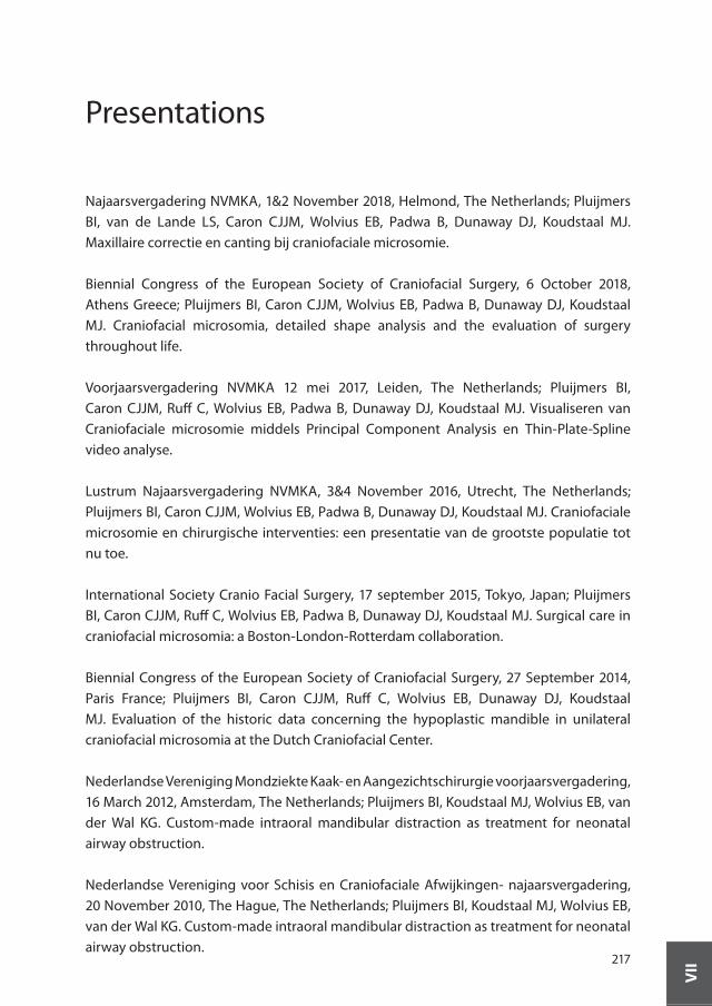
217
VII
Presentations
Najaarsvergadering NVMKA, 1&2 November 2018, Helmond, The Netherlands; Pluijmers BI, van de Lande LS, Caron CJJM, Wolvius EB, Padwa B, Dunaway DJ, Koudstaal MJ. Maxillaire correctie en canting bij craniofaciale microsomie.
Biennial Congress of the European Society of Craniofacial Surgery, 6 October 2018, Athens Greece; Pluijmers BI, Caron CJJM, Wolvius EB, Padwa B, Dunaway DJ, Koudstaal MJ. Craniofacial microsomia, detailed shape analysis and the evaluation of surgery throughout life.
Voorjaarsvergadering NVMKA 12 mei 2017, Leiden, The Netherlands; Pluijmers BI, Caron CJJM, Ruff C, Wolvius EB, Padwa B, Dunaway DJ, Koudstaal MJ. Visualiseren van Craniofaciale microsomie middels Principal Component Analysis en Thin-Plate-Spline video analyse.
Lustrum Najaarsvergadering NVMKA, 3&4 November 2016, Utrecht, The Netherlands; Pluijmers BI, Caron CJJM, Wolvius EB, Padwa B, Dunaway DJ, Koudstaal MJ. Craniofaciale microsomie en chirurgische interventies: een presentatie van de grootste populatie tot nu toe.
International Society Cranio Facial Surgery, 17 september 2015, Tokyo, Japan; Pluijmers BI, Caron CJJM, Ruff C, Wolvius EB, Padwa B, Dunaway DJ, Koudstaal MJ. Surgical care in craniofacial microsomia: a Boston-London-Rotterdam collaboration.
Biennial Congress of the European Society of Craniofacial Surgery, 27 September 2014, Paris France; Pluijmers BI, Caron CJJM, Ruff C, Wolvius EB, Dunaway DJ, Koudstaal MJ. Evaluation of the historic data concerning the hypoplastic mandible in unilateral craniofacial microsomia at the Dutch Craniofacial Center.
Nederlandse Vereniging Mondziekte Kaak- en Aangezichtschirurgie voorjaarsvergadering, 16 March 2012, Amsterdam, The Netherlands; Pluijmers BI, Koudstaal MJ, Wolvius EB, van der Wal KG. Custom-made intraoral mandibular distraction as treatment for neonatal airway obstruction.
Nederlandse Vereniging voor Schisis en Craniofaciale Afwijkingen- najaarsvergadering, 20 November 2010, The Hague, The Netherlands; Pluijmers BI, Koudstaal MJ, Wolvius EB, van der Wal KG. Custom-made intraoral mandibular distraction as treatment for neonatal airway obstruction.
PSM 20190527 Proefschrift Britt Pluijmers.indd 217 24-07-19 15:04
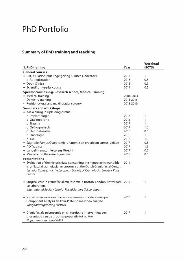
218
PhD Portfolio
Summary of PhD training and teaching
1. PhD training Year Workload (ECTS)
General courses • BROK (‘Basiscursus Regelgeving Klinisch Onderzoek’
o Re-registration• Open Clinica• Scientific Integrity course
2012 2016 20132014
1 0.50.50.5
Specific courses (e.g. Research school, Medical Training) • Medical training• Dentistry training• Residency oral and maxillofacial surgery
2006-20132013-20162015-2019
Seminars and workshops • Kaakchirurg In Opleiding cursus
o Implantologieo Oral medicineo Traumao Orthognatischo Dentoalveolairo Oncologieo TMJ
• Sagittale Ramus Osteotomie: anatomie en practicum cursus, Leiden• AO Trauma• Landelijk anatomie cursus Utrecht• Mini around the nose Nijmegen
20162016201720172018201820182017201720172018
1 1 1 1.5 0.511.00.51.50.50.5
Presentations • Evaluation of the historic data concerning the hypoplastic mandible
in unilateral craniofacial microsomia at the Dutch Craniofacial Center.Biennial Congress of the European Society of Craniofacial Surgery, Paris France
• Surgical care in craniofacial microsomia: a Boston-London-Rotterdam collaboration.International Society Cranio- Facial Surgery Tokyo, Japan
• Visualiseren van Craniofaciale microsomie middels Principal Component Analysis en Thin-Plate-Spline video analyse. Voorjaarsvergadering NVMKA
• Craniofaciale microsomie en chirurgische interventies: een presentatie van de grootste populatie tot nu toe.Najaarsvergadering NVMKA
2014
2015
2016
2017
1
1
1
1
PSM 20190527 Proefschrift Britt Pluijmers.indd 218 24-07-19 15:04
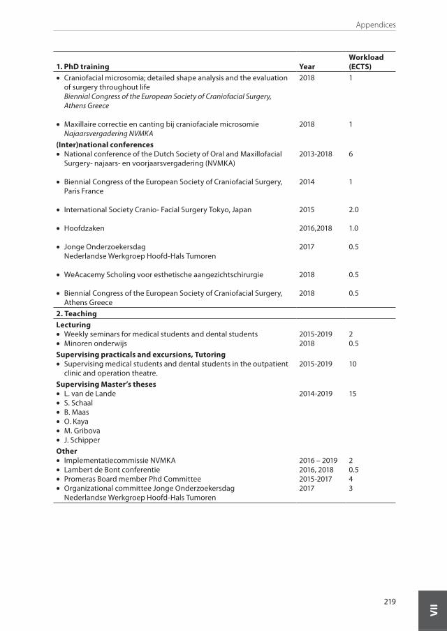
Appendices
219
VII
1. PhD training Year Workload (ECTS)
• Craniofacial microsomia; detailed shape analysis and the evaluation of surgery throughout lifeBiennial Congress of the European Society of Craniofacial Surgery, Athens Greece
• Maxillaire correctie en canting bij craniofaciale microsomieNajaarsvergadering NVMKA
2018
2018
1
1
(Inter)national conferences • National conference of the Dutch Society of Oral and Maxillofacial
Surgery- najaars- en voorjaarsvergadering (NVMKA)
• Biennial Congress of the European Society of Craniofacial Surgery, Paris France
• International Society Cranio- Facial Surgery Tokyo, Japan
• Hoofdzaken
• Jonge OnderzoekersdagNederlandse Werkgroep Hoofd-Hals Tumoren
• WeAcacemy Scholing voor esthetische aangezichtschirurgie
• Biennial Congress of the European Society of Craniofacial Surgery, Athens Greece
2013-2018
2014
2015
2016,2018
2017
2018
2018
6 1
2.0
1.0
0.5
0.5
0.5
2. Teaching Lecturing • Weekly seminars for medical students and dental students• Minoren onderwijs
2015-20192018
20.5
Supervising practicals and excursions, Tutoring • Supervising medical students and dental students in the outpatient
clinic and operation theatre.
2015-2019
10
Supervising Master’s theses • L. van de Lande• S. Schaal• B. Maas• O. Kaya • M. Gribova • J. Schipper
2014-2019
15
Other • Implementatiecommissie NVMKA • Lambert de Bont conferentie• Promeras Board member Phd Committee • Organizational committee Jonge Onderzoekersdag
Nederlandse Werkgroep Hoofd-Hals Tumoren
2016 – 20192016, 20182015-20172017
20.543
PSM 20190527 Proefschrift Britt Pluijmers.indd 219 24-07-19 15:04

220
Dankwoord
Het is af, tijd voor reflectie en om woorden van dank uit te spreken. Dit werk is tot stand gekomen
met de hulp van velen. Eenieder die aan dit boek heeft bijgedragen wil ik dan ook hartelijk danken.
Zonder iemand te kort te doen wil ik een aantal mensen in het bijzonder noemen:
De patiënten, dank. Hopelijk heeft dit onderzoek bijgedragen aan de verbetering van de zorg.
Beste Maarten, 10 jaar geleden kwam ik bleu de afdeling opgewandeld met de vraag of het
mogelijk was om onderzoek te doen. Op de vraag of ik al eens wat had geschreven antwoordde
ik zelfverzekerd ‘ja, een systematic review voor het keuze onderwijs’ (lees; een 5 weken durend
studieproject, niks peer reviewed). Little did I know.. Dank voor je enthousiasme keer op keer,
kritische vragen en vrijheid voor eigen plan van aanpak. Ik kijk uit naar verdere toekomstige
samenwerking, en alle daarbij horende pizzamomenten. ;)
Beste professor Wolvius, dank voor het redigeren van mijn stukken met daarbij scherp commentaar.
Ik wil u danken voor de mogelijkheid die ik heb gekregen en krijg binnen uw afdeling. Ik volg met
veel plezier de opleiding bij u!
Dear professor Dunaway, dear David, many thanks for giving me the opportunity to start doing
research at your department as a young student and further along as a PhD candidate. I have
learned a great deal of not only the care of these patients during my time at the GOSH, but also the
academic work which comes along, thank you.
Dear professor Padwa, dear Bonnie, many thanks for the chance to study the CFM population at
your department. I am very grateful for your comments and edits to improve each manuscript.
I would love to come to the Children’s Hospital in Boston one day again (and of course order a
sushiboat).
Beste professor Kleinrensink en professor Mathijssen. Hartelijk dank dat u het werk reeds kritisch
heeft beoordeeld. Ik kijk uit, met gezonde spanning, naar uw vragen 11 september.
Dear Cliff, many thanks for your time and enthusiasm to explain how PCA works and what the
possibilities are with vectors and PDMs. We still ought to see a QPR game!
Beste dr. Looman, beste Caspar, dank voor uw geduld tijdens het uitleggen hoe “R” werkt en alle
analyses en mogelijkheden die wij met u hebben onderzocht!
PSM 20190527 Proefschrift Britt Pluijmers.indd 220 24-07-19 15:04

Appendices
221
VII
Dear co-authors and contributors to this thesis: Mr. Ayliffe, Alessandro Borghi, Wicher Braams, Mr.
Bulstrode, Kimberly Daniels, Mr. Evans, Paul Knoops, prof. Mathijssen, dr. Mulliken, dr. Pauws, dr.
Resnick, Femke Staal, many thanks for your time, help and guidance and contribution to this work.
Lieve Britta, Lara, Maria, Onur en Sontje een speciaal dankwoord aan jullie. Ik heb er veel van
geleerd om jullie te begeleiden, dank voor jullie kritische vragen en jullie inzet. Dit boekje zou er
niet zijn zonder jullie!
Beste Jan-Aart, Ruben, Yoram en Wietse. Dank dat jullie duizend en 1 dingen hier, maar ook over-
zees hebben opgezocht als er weer wat was met de database!
Lieve Lin, we hebben het geflikt! Dank voor alle mentale support, het sparren en de peptalk. Wat
ben ik blij dat we dit samen hebben gedaan en vandaag ook allebei de verdediging mogen doen.
Joe houdoe en de groeten!
Alle stafleden, maar ook poli- en OK- assistenten en secretariaat, van het EMC en ETZ Elisabeth,
dank voor de interesse naar mijn onderzoek. Dank voor het opleiden tot specialist in het mooiste
vakgebied: MKA-chirurgie.
Sannie! Spil van de afdeling! Dank voor het meedenken, omroosteren en lachen!
Collega AIOS EMC en ETZ Elisabeth, dank/mercikes voor het lachen en de koffie, keeps me sane!
#AIOSliefde #Mojo #bakkiedanmaar
Lieve vriendinnen en vrienden, ja het is ein-de-lijk af! Dank voor jullie blijvende interesse en
vriendschap door de jaren heen. Dank voor alle: hutjes op de hei, memorable avonden en
babyborrels op woensdag, BFFF en magnums, carnaval en biertjes in de trein, Mariah Carey en
BoysIIMen, My tribes, to the grounds met vergaderen en natuurlijk ook watermeloenen!
Lieve Suus, in 2010 (wist ik veel dat het zo lang zou duren) op de Philips Willem al gevraagd, blij dat
je vandaag naast mij staat.
Lieve Moes, Ivvy, Inge, Linnea, Gijs en Telma, Heleen en Paul, familie kan je niet kiezen, schoonfamilie
nog minder, maar ik had zelf niets anders gekozen.
Mam en pap, op geen enkele wijze kan ik jullie genoeg bedanken voor de onvoorwaardelijke steun
die ik van jullie krijg en de stimulatie om alles eruit te halen wat erin zit. Dank voor het liefdevol
zorgen voor Michiel als Coen of ik er niet zijn, it truly takes a village! Mam bedankt (weer) voor alle
creatieve inspanningen op deze dag.
PSM 20190527 Proefschrift Britt Pluijmers.indd 221 24-07-19 15:04
Appendices
222
Vincent, Jimmy en Melvyn. De een getuige op mijn huwelijk, de ander babs en Melle jij nu getuige
bij het huwelijk met de wetenschap! Dank voor dit proefschrift, want laten wij eerlijk zijn zonder
een diepgewortelde bewijsdrang van kleins af aan naar jullie toe, stond ik hier niet. ;) Dank dat ik
jullie vierde broertje mag zijn!
Coen, liefde van mijn leven. Dank dat je in veel mee gaat, maar ook op tijd tegengas geeft. En, dat
jij mij altijd laat lachen. Zin in wat de toekomst ons brengt.
Lieve Michiel, jij en je zusje blijven mijn grootste geschenk, dank!
PSM 20190527 Proefschrift Britt Pluijmers.indd 222 24-07-19 15:04
curriculum vitae
Britt Pluijmers was born on the 16th of November 1987 in Cardi� (UK), and spent part ofher childhood in Belgium before moving to the Netherlands. She graduated from LibanonLyceum high school in Rotterdam in 2005.In 2006 she started medical training at the Erasmus University Hospital in Rotterdam(EMC). During her studies she was a board member of the Education committee EMC, anddid voluntary work in the Palestinian Territories.In her third year, under direct supervision of dr. Koudstaal, she started research at thedepartment of Oral and Maxillofacial surgery at the EMC led by em. prof. dr. van der Wal.Her Master thesis was carried out under the guidance of prof. Dunaway, at the GreatOrmond Street Hospital in London (UK). During her internship at the department of Oraland Maxillofacial surgery EMC, she started this thesis, with dr. Koudstaal as co-promotorand prof. dr. Wolvius as promotor.In 2013, she graduated Cum Laude from medical school, and commenced her dentistrystudies at Radboud University, Nijmegen, from which she graduated in 2016.Simultaneously, she was a board member of Promeras, the PhD committee of theErasmus MC. In 2015 she started her Oral- and Maxillofacial surgery training (undersupervision of prof. dr. Wolvius) at the Erasmus Medical Center in Rotterdam and the St.Elisabeth Hospital in Tilburg (under supervision of J.P.O. Scheerlinck). She was a memberof the organizational committee of the JOD NWHHT in 2017.At present, she is in her senior year of her training, and a member of the implementationcommittee for the new curriculum for Oral- and Maxillofacial surgery residents.
Britt is married to Coen Iordens and proud mother to Michiel. She has 3 awesomebrothers.
ON CRANIOFACIAL MICROSOMIASHAPE AND SURGERY
BRITT PLUIJMERS
ON
CRAN
IOFA
CIAL M
ICROSO
MIA
SHA
PE AN
D SU
RGERY
B.I. PLUIJM
ERS
Britt Pluijmers Cover v6.indd 1-3 24-7-2019 16:56:17
Uitnodiging
voor het bijwonen van de openbare verdediging van het
proefschrift
On Craniofacial MicrosomiaShape and Surgery
door Britt Pluijmers
Op woensdag 11 september 2019 om 11:30
in de Prof. Andries Queridozaal van het Onderwijscentrum van het Erasmus Medisch Centrum Wytemaweg 80 te Rotterdam
Na a� oop van de promotie bent u van harte welkom op de receptie
Britt PluijmersMathenesserlaan 371a
3023 GD Rotterdam0647480499
ParanimfenSuzanne Deetman
Melvyn [email protected]
Britt Pluijmers Cover v6.indd 4 24-7-2019 16:56:36
Uitnodiging
voor het bijwonen van de openbare verdediging van het
proefschrift
On Craniofacial MicrosomiaShape and Surgery
door Britt Pluijmers
Op woensdag 11 september 2019 om 11:30
in de Prof. Andries Queridozaal van het Onderwijscentrum van het Erasmus Medisch Centrum Wytemaweg 80 te Rotterdam
Na a� oop van de promotie bent u van harte welkom op de receptie
Britt PluijmersMathenesserlaan 371a
3023 GD Rotterdam0647480499
ParanimfenSuzanne Deetman
Melvyn [email protected]
Britt Pluijmers Cover v6.indd 4 24-7-2019 16:56:36Naamloos-2 1-4 24-7-2019 17:05:31
Related Documents