On Caries Risk Profile and Prevention in an Adult Saudi Population Helal Sonbul Department of Cariology Institute of Odontology at Sahlgrenska Academy University of Gothenburg, Sweden MINISTRY OF HIGHER EDUCATION SAUDI ARABIA Gothenburg 2010

Welcome message from author
This document is posted to help you gain knowledge. Please leave a comment to let me know what you think about it! Share it to your friends and learn new things together.
Transcript
On Caries Risk Profile and Prevention
in an Adult Saudi Population
Helal Sonbul
Department of Cariology Institute of Odontology at Sahlgrenska Academy
University of Gothenburg, Sweden
MINISTRY OF HIGHER EDUCATION SAUDI ARABIA
Gothenburg 2010

A doctoral thesis at a university in Sweden is produced either as a monograph or as a collection of papers. In the latter case, the introductory part constitutes the formal thesis, which summarises the accompanying papers. They have already been published, accepted or submitted for publication. No part of this publication may be reproduced or transmitted, in any form or by any means, without written permission.
The cover page illustration was made by Yvonne Heijl.
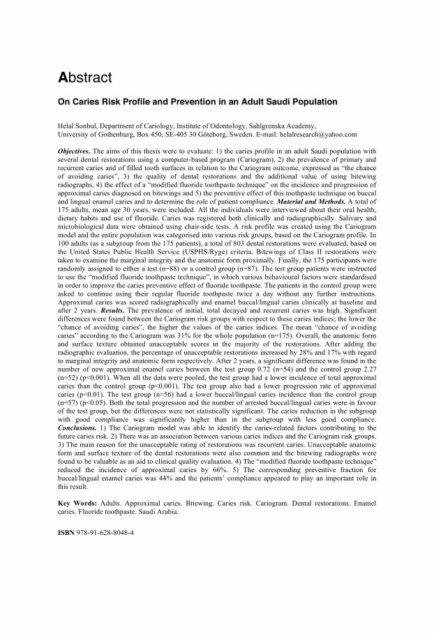
Abstract On Caries Risk Profile and Prevention in an Adult Saudi Population Helal Sonbul, Department of Cariology, Institute of Odontology, Sahlgrenska Academy, University of Gothenburg, Box 450, SE-405 30 Göteborg, Sweden. E-mail: [email protected] Objectives. The aims of this thesis were to evaluate: 1) the caries profile in an adult Saudi population with several dental restorations using a computer-based program (Cariogram), 2) the prevalence of primary and recurrent caries and of filled tooth surfaces in relation to the Cariogram outcome, expressed as “the chance of avoiding caries”, 3) the quality of dental restorations and the additional value of using bitewing radiographs, 4) the effect of a “modified fluoride toothpaste technique” on the incidence and progression of approximal caries diagnosed on bitewings and 5) the preventive effect of this toothpaste technique on buccal and lingual enamel caries and to determine the role of patient compliance. Material and Methods. A total of 175 adults, mean age 30 years, were included. All the individuals were interviewed about their oral health, dietary habits and use of fluoride. Caries was registered both clinically and radiographically. Salivary and microbiological data were obtained using chair-side tests. A risk profile was created using the Cariogram model and the entire population was categorised into various risk groups, based on the Cariogram profile. In 100 adults (as a subgroup from the 175 patients), a total of 803 dental restorations were evaluated, based on the United States Public Health Service (USPHS/Ryge) criteria. Bitewings of Class II restorations were taken to examine the marginal integrity and the anatomic form proximally. Finally, the 175 participants were randomly assigned to either a test (n=88) or a control group (n=87). The test group patients were instructed to use the “modified fluoride toothpaste technique”, in which various behavioural factors were standardised in order to improve the caries preventive effect of fluoride toothpaste. The patients in the control group were asked to continue using their regular fluoride toothpaste twice a day without any further instructions. Approximal caries was scored radiographically and enamel buccal/lingual caries clinically at baseline and after 2 years. Results. The prevalence of initial, total decayed and recurrent caries was high. Significant differences were found between the Cariogram risk groups with respect to these caries indices; the lower the “chance of avoiding caries”, the higher the values of the caries indices. The mean “chance of avoiding caries” according to the Cariogram was 31% for the whole population (n=175). Overall, the anatomic form and surface texture obtained unacceptable scores in the majority of the restorations. After adding the radiographic evaluation, the percentage of unacceptable restorations increased by 28% and 17% with regard to marginal integrity and anatomic form respectively. After 2 years, a significant difference was found in the number of new approximal enamel caries between the test group 0.72 (n=54) and the control group 2.27 (n=52) (p<0.001). When all the data were pooled, the test group had a lower incidence of total approximal caries than the control group (p<0.001). The test group also had a lower progression rate of approximal caries (p<0.01). The test group (n=56) had a lower buccal/lingual caries incidence than the control group (n=57) (p<0.05). Both the total progression and the number of arrested buccal/lingual caries were in favour of the test group, but the differences were not statistically significant. The caries reduction in the subgroup with good compliance was significantly higher than in the subgroup with less good compliance. Conclusions. 1) The Cariogram model was able to identify the caries-related factors contributing to the future caries risk. 2) There was an association between various caries indices and the Cariogram risk groups. 3) The main reason for the unacceptable rating of restorations was recurrent caries. Unacceptable anatomic form and surface texture of the dental restorations were also common and the bitewing radiographs were found to be valuable as an aid to clinical quality evaluation. 4) The “modified fluoride toothpaste technique” reduced the incidence of approximal caries by 66%. 5) The corresponding preventive fraction for buccal/lingual enamel caries was 44% and the patients’ compliance appeared to play an important role in this result. Key Words: Adults. Approximal caries. Bitewing. Caries risk. Cariogram. Dental restorations. Enamel caries. Fluoride toothpaste. Saudi Arabia.
ISBN 978-91-628-8048-4

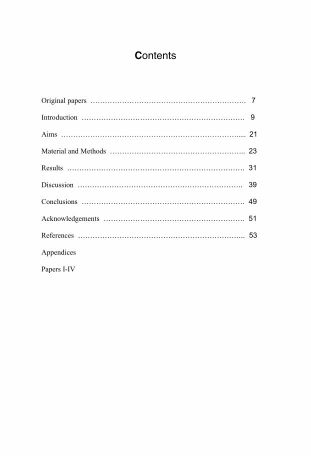
Contents
Original papers ………………………………………………………. 7
Introduction …………………………………………………………. 9
Aims ………………………………………………………………..... 21
Material and Methods ……………………………………………….. 23
Results ………………………………………………………………. 31
Discussion ………………………………………………………….. 39
Conclusions …………………………………………………………. 49
Acknowledgements …………………………………………………. 51
References …………………………………………………………... 53
Appendices Papers I-IV

7
Original papers
This thesis is based on the following four papers, which will be referred to in the text by their Roman numerals:
I. Sonbul H, Al-Otaibi M, Birkhed D. Risk pro le of adults with several dental
restorations using the Cariogram model. Acta Odontol Scand 2008;66:351-357. II. Sonbul H, Birkhed D. Risk pro le and quality of dental restorations: a cross-
sectional study. Acta Odontol Scand 2010;68:122-128. III. Sonbul H, Birkhed D. The preventive effect of a modi ed fluoride toothpaste
technique on approximal caries in adults with high caries prevalence: a 2 -year clinical trial. Swed Dent J 2010;34:9-16.
IV. Sonbul H, Birkhed D. The preventive effect of a modi ed fluoride toothpaste
technique on buccal/lingual enamel caries in adults with high caries prevalence: a 2-year clinical trial. Submitted.

8

9
Introduction
ental caries has been extensively investigated during the last decades, with the
focus on pre-school and school children and adolescents [Whelton, 2004; Sheiham, 2005].
However, as the retention of teeth in adult populations increases, dental caries has become
a burden for ageing adults. According to the United States National Institutes of Health
Consensus Development Panel [2001], more than two-thirds of American adults aged 35
to 44 years have lost at least one tooth due to dental caries. The caries incidence in adult
and geriatric populations may vary because of host and environmental factors [Selwitz et
al., 2007]. In Sweden, several studies reveal that many elderly people keep their own
natural teeth, which may result in an increased risk of developing caries [Lundgren et al.,
1997; Fure, 2003, 2004; Österberg et al., 2006; Johanson et al., 2009]. Whelton [2004] has
reviewed several studies regarding the risk of caries in adults. It might be concluded that
the caries incidence increases with old age, even in adults who have received the benefits
of caries prevention in childhood. In this context, dental caries remains a major problem in
adults, even in developed countries, such as the United States and Sweden [2001;
Hugoson and Koch, 2008].
Global changes in the pattern of dental caries have taken place in recent decades. Data
collection on the incidence and progression of coronal and root caries in an age-related
study design is important in determining the lifetime pattern of caries [for a review, see
Selwitz et al., 2007; Whelton, 2004]. As an example, enamel lesions, particularly on
approximal surfaces, should be considered in epidemiological studies to evaluate actual
caries prevalence, at both a population and an individual level. Several reports concluded
that the true prevalence of caries has often been underestimated due to the exclusion of
initial caries from the evaluation [Amarante et al., 1998; Machiulskiene et al., 1998;
Poorterman et al., 2002; Moberg Sköld et al., 2005; Hopcraft and Morgan, 2006].
Dental caries results from an ecological imbalance in the equilibrium between tooth
minerals and oral biofilms (plaque) [Fejerskov, 2004; Takahashi and Nyvad, 2008]. The
biofilm is characterised by microbial activity, resulting in fluctuations in plaque pH. This
is a result of both bacterial acid production and buffering action from saliva and the
surrounding tooth structure. The tooth surface is therefore in a dynamic equilibrium with
its surrounding environment. As the pH falls below a critical value, the demineralisation
10
of enamel, dentine or cementum occurs, while a gain of mineral (remineralisation) occurs
as the pH increases [Manji et al., 1991; Kidd and Fejerskov, 2004]. The process of
demineralisation and remineralisation takes place frequently during the day. Over time,
this process leads to either caries lesions or the repair and reversal of a lesion
[Featherstone, 2004].
Primary caries can occur on different tooth surfaces. On an approximal surface, the
lesion starts and forms beneath the contact area between teeth. Caries on an occlusal
surface is also a localised phenomenon in pit and fissure. On both occlusal and approximal
surfaces, enamel caries is a three-dimensional subsurface demineralisation that spreads
along the enamel prisms. Recurrent (secondary) caries is a lesion located at the margin of
a dental restoration. From an etiological point of view, recurrent caries does not differ
from primary caries. Histologically, it represents a caries lesion adjacent to the margin and
there may be signs of demineralisation (wall lesions) along the cavity wall. They could be
a consequence of microleakage. However, clinical and microbiological studies indicate
that this leakage does not lead to active demineralisation beneath the restoration [for a
review, see Kidd and Beighton, 1996; Mjör and Toffenetti, 2000].
Caries risk and related factors
Several factors can influence the microbial metabolic activity in the dental biofilm. These
factors include plaque composition and thickness, cariogenic bacteria and diet content and
frequency as risk factors. The flow rate, buffer capacity of saliva and presence of fluoride
are risk inhibitors providing protection from caries. In addition, previous caries
experience, as well as social and behavioural factors, are risk indicators that could indicate
the probability of developing caries, but they are not be directly involved in the causal
chain [for a review, see Zero et al., 2001; Burt, 2005]. In the present thesis, all these
factors are collectively referred to as “caries-related factors”.
There are certain locations on the tooth that are prone to caries: the occlusal pit and
fissure, the approximal surface cervical to the contact point, buccal or lingual surfaces
along the gingival margin and tooth-restoration interfaces. These areas do not differ from
the other tooth surfaces with regard to tooth structure, but they are susceptible to caries
only because the biofilm tends to stagnate and remain for a prolonged period of time. The
biofilm that forms, grows and matures over time does not necessarily lead to clinically
11
visible caries lesions. Indeed, its presence is a prerequisite for demineralisation and/or
remineralisation. The composition of the biofilm itself and the length of time it remains
attached and undisturbed are the prime concerns from a cariological point of view [for a
review, see Marsh and Nyvad, 2008].
The mutans streptococci (MS) are the major pathogens of dental caries. This is because
MS are highly acidogenic and aciduric and they are able to produce extracellular matrix of
water-insoluble glucans, which enhances bacterial adhesion to the tooth surface and to
other bacteria [Hamada and Slade, 1980; Loesche, 1986]. A systematic review by Tanzer
et al. [2001] confirms the major role played by MS in the initiation of dental caries on
enamel and root surfaces. However, some studies indicate that the relationship between
MS and caries is not absolute and that caries can develop in the absence of these species
[Bowden, 1997; Aas et al., 2008]. It has been suggested that other acidogenic and aciduric
bacteria, including non-MS and Actinomyces, may also be responsible for caries
development [Sansone et al., 1993; van Houte et al., 1994]. When members of the resident
flora obtain a selective advantage over other species, the homeostatic balance of the
biofilm is disturbed [Marsh, 1999]. This has been explained as an ecological hypothesis
[Marsh, 1994]. It is therefore important to describe not only the type or number of bacteria
involved in caries but also their activity [Takahashi and Nyvad, 2008]. Even though the
presence of lactobacilli (LB) is associated with, but not primarily responsible for, caries
development, their increased numbers are found with a high consumption of
carbohydrates. The high proportions of MS and LB may be regarded as “biomarkers” of
caries development [Nyvad and Kilian, 1990; Chhour et al., 2005] and their count in
plaque is positively correlated with their numbers in saliva [Emilson, 1983]and with caries
susceptibility [Krasse, 1988; Demers et al., 1990; Powell, 1998].
Fermentable carbohydrates, in particular sucrose, have been shown to be associated
with caries initiation and development [Paes Leme et al., 2006]. The bacterial metabolism
may be dramatically enhanced by changing nutritional conditions, as the presence of
fermentable carbohydrates will lower the plaque pH. Any shift in pH will influence the
chemical composition and the bacterial flora inside the biofilm over time [Marsh and
Nyvad, 2008]. If a prolonged acidic environment persists, more aciduric bacteria, such as
MS and LB, will selectively grow and accumulate and caries lesions will therefore occur
or progress. Although diet and oral hygiene maintenance are factors that are indirectly

12
related to the severity of dental caries, individuals exposed to similar circumstances may
vary in their susceptibility to develop caries. Systematic reviews have shown that, with
frequent fluoride exposure, the relationship between sugar intake and caries experience is
not consistent and controlling the consumption is not always the most important aspect
[Burt and Pai, 2001; Zero, 2004]. According to another review, there are no studies in
which reduced sugar intake alone affects caries prevalence [Lingström et al., 2003].
However, reducing the frequency of sugar intake could be a means of reducing caries
[Sheiham, 2001].
Saliva plays a critical role in the prevention or reversal of the caries process [for a
review, see Lenander-Lumikari and Loimaranta, 2000]. It maintains the super-saturation
of calcium in plaque. It also neutralises acids, raises the pH and reverses the diffusion rate
of calcium and phosphate towards the tooth surface. In the event of extensive challenge,
e.g. poor oral hygiene and a low salivary secretion rate, these protective benefits will be
disturbed or impaired, placing an individual at high risk of developing caries. It should be
noted that various salivary parameters other than bacterial count, e.g. salivary buffer and
antimicrobial agents, are of little predictive value as far as caries susceptibility is
concerned [Tenovuo, 1997].
The fluoride content in saliva and plaque as a result of using fluoride toothpaste or
other fluoride-containing products appears to be more important than the other parameters.
It is well known that fluoride has an anti-cariogenic effect that prevents caries and
decreases or even reverses the progression of caries lesions. Its mechanisms of action are
based on three principles: 1) inhibiting demineralisation, 2) enhancing remineralisation
and 3) inhibiting bacterial metabolism [Featherstone, 2000]. Its effect on caries prevalence
and incidence is well documented in the literature [Stephen, 1999; Featherstone, 2000;
Marinho, 2008]. However, a skewed distribution of caries is evident in epidemiological
studies, as a considerable percentage of the population exhibits significantly more caries
and runs a high risk of developing new carious lesions, even in fluoridated areas [Whelton,
2004; Selwitz et al., 2007]. This could be attributed to other risk factors that reduce the
capability of fluoride to overcome these challenges.
13
Cariogram
The multi-factorial caries entity makes risk assessment and the prediction of caries
development a complex process. The interplay of the caries-related factors can vary over
time, between populations, individuals and even within one and the same individual. As a
result, dental caries can develop and progress rather rapidly in some individuals compared
with others. There is no single test that is able accurately to predict an individual’s
susceptibility to caries [Reich et al., 1999]. Numerous risk models have been introduced in
the literature. However, their predictive outcomes are different and depend on the study
population [for a review, see Zero et al., 2001].
A computer-based program (Cariogram) has been developed by Swedish researchers
for caries risk assessment [Bratthall, 1996; Bratthall and Hänsel-Petersson, 2003, 2005]. It
can be described as a simple tool for a difficult multi-factorial process. Previous studies
found that the risk assessment using the Cariogram is in agreement with the opinions of
dentists and dental hygienists [Hänsel-Petersson and Bratthall, 2000]. The Cariogram can
be used as a prediction or risk model. As a prediction model, the data show a correlation
between the Cariogram results and the caries increment over time for children and the
elderly [Hänsel-Petersson et al., 2002; Hänsel-Petersson et al., 2003]. Several studies have
used the Cariogram as a risk model, where the caries risk was evaluated by identifying the
caries-related factors [Tayanin et al., 2005; Ruiz Miravet et al., 2007; Zukanovi et al.,
2007; Campus et al., 2009]. However, the populations involved in these studies were
mostly children and adolescents. It may therefore be of interest to study adult population
using the Cariogram model to evaluate the estimated future risk of developing caries and
to identify the caries-related factors. It should be noted that the program does not identify
the number of cavities that will or will not develop [Bratthall and Hänsel-Petersson, 2005].
One advantage of the Cariogram is its capability to give an individual’s total possible risk
as a single value expressed as the percentage “chance of avoiding caries”, after weighing
up all the other caries-related factors in the equation. It is an interactive program that can
be used to enhance the patient’s motivation by demonstrating how the caries risk can
change by modifying the various caries-related factors.
Ten factors are included in the Cariogram model: 1) caries experience, 2) related
diseases, 3) salivary flow rate, 4) salivary buffer capacity, 5) plaque amount, 6) diet
frequency, 7) diet content, 8) mutans streptococcus count, 9) fluoride programme and 10)
14
clinical judgement. These ten factors are given a score according to a predetermined scale
and are entered in the program. The results are displayed as a pie chart with five different
coloured sectors. Four of these sectors represent the extent (in per cent) to which various
caries-related factors could affect the fifth one, i.e. the green sector, expressed as “chance
of avoiding caries” (for examples, see Appendices 1 & 2). The dark blue sector (Diet) is a
combination of diet content and diet frequency; the red sector (Bacteria) is a combination
of the amount of plaque and mutans streptococci; the light blue sector (Susceptibility) is a
combination of fluoride intake, saliva secretion and saliva buffer capacity; the yellow
sector (Circumstances) is a combination of past caries experience and related diseases and,
finally, the green sector represents the “chance of avoiding caries”. When this latter sector
shows a large percentage, the caries risk is probably low and vice versa. Fluoridated toothpaste
The efficacy of topically applied fluoride in caries prevention has been studied extensively
[Groeneveld et al., 1990; Featherstone, 2000; Marinho, 2008]. The decline in caries
prevalence in the developed countries over the last four decades is believed to be due to
the widespread use of fluoride toothpaste, which is the most common vehicle for
delivering fluoride topically [WHO, 1994; Bratthall et al., 1996; Marinho et al., 2003].
Daily toothbrushing with fluoride toothpaste has shown a strong evidence in preventing
caries in children and adolescents [Twetman et al., 2003]. It has been documented that
fluoride toothpaste reduces the number of new caries lesions over 3 years by 24%
[Marinho et al., 2003]. However, no evidence of the effect of fluoride on the prevention of
primary caries or recurrent caries in adults is available [WHO 2001; Twetman et al.,
2003]. One aspect of the present thesis is therefore to evaluate this effect on a group from
an adult population.
A recent Cochrane review [Walsh et al., 2010] confirms the efficacy of fluoride
toothpaste in caries prevention, but significantly only for a concentration of >1,000 ppm,
and the relative caries preventive effect increases as the fluoride concentration increases.
Increased fluoride concentration is accompanied by an increase in plaque fluoride levels
[Duckworth and Morgan, 1991]. Furthermore, toothpastes containing 5,000 ppm fluoride
have been shown to be more effective at remineralising primary root caries than those
containing 1,100 ppm fluoride [Baysan et al., 2001]. In overall terms, a linear correlation
15
was found between toothpaste fluoride concentrations between 0 and 5,000 ppm and
clinical caries efficacy [Tavss et al., 2003].
Many behavioural factors could influence the efficacy of fluoride toothpaste in caries
prevention [Davies et al., 2003]. They include the frequency of brushing, the amount of
toothpaste applied, the duration of brushing and the time of day. It has been reported that
caries increments in individuals who brush only once a day were 20-30% higher than in
those who brushed twice a day [O'Mullane et al., 1997]. Julihn et al. [2006] found that
irregular toothbrushing at night was strongly associated with a high caries experience.
Furthermore, the mean fluoride level in saliva after brushing with a small amount of
toothpaste ( 0.25 g) was approximately one third of that obtained with a normal amount
( 1.0 g) [DenBesten and Ko, 1996]. Another study conducted in adults recommended the
application of 1 g or more of the fluoride toothpaste containing 1,000 ppm to increase both
the fluoride uptake in the surface enamel and the fluoride concentration in the oral fluid
[Koga et al., 2007]. In a recent study, Zero et al. [2010] suggested that both brushing time
and toothpaste quantity may be important determinants of both fluoride retention and
consequent enamel remineralisation. The question of whether toothbrushing should take
place before or after eating is still controversial. Attin et al. [2005] stated: “Study of the
literature gives no clear evidence as to the optimal time-point of tooth brushing (before or
after meals). However, in order to eliminate food impaction and to shorten the duration of
sucrose impact by tooth cleaning after meals seems to be recommendable”. The use of
fluoride toothpaste before “going to bed” has also been supported by Davies et al. [2003].
Another interesting factor is that the post-brushing water rinsing behaviour might affect
the availability of fluoride in saliva and dental plaque. Individuals who rinse with large
volumes of water have a higher caries incidence than those who rinse with smaller
amounts [O'Mullane et al., 1997; Chestnutt et al., 1998].
In a clinical trial, Sjögren et al. [1995] found that the approximal caries in Swedish
preschool children was reduced by an average of 26% after using a certain technique
called “modified toothpaste technique”. Children were instructed to place toothpaste on
the teeth prior to brushing and swish the toothpaste foam, together with a sip of water
(approximately 10 ml), around the dentition by active cheek movements for 1 minute,
before expectorating. In the present thesis, several factors involved in the behaviour of

16
using the fluoride toothpaste were standardised and the effect on dental caries was then
evaluated in a group of adults.
Restorative treatment of caries
The approach of “extension for prevention”, described by G.V Black, as a means of caries
management has been a cornerstone of 20th century dentistry [Osborne and Summitt,
1998]. This approach, which depends on the “drill and fill” theory, is still utilised and is
the favoured method in many countries, even developed ones. Unfortunately, this
approach has neither prevented caries nor addressed the challenge of the restoration/re-
restoration cycle. Studies have demonstrated that restorations have a limited life span and
that the restoration is likely to be replaced many times [Elderton and Nuttall, 1983;
Elderton, 1990]. This may lead to repetitive restorative cycles with larger restorations,
weaker teeth and an increased risk of more complex treatment [Elderton, 1990; Brantley et
al., 1995].
A number of studies have reported that recurrent caries is the most common reason for
the replacement of dental restorations [Deligeorgi et al., 2001; Mjör, 2005]. The number of
restorations replaced as a result of recurrent caries is higher in general dental practice than
in controlled clinical trials [Letzel et al., 1989]. Recurrent caries lesions are most often
located at the gingival margins that obscure their detection by direct vision. It is difficult
to distinguish marginal discrepancies (e.g. ditching) and discoloration from recurrent
lesions. As a result, some dentists replace fillings with staining and minor defects in the
belief that these clinical signs are indicative of microleakage that leads to caries
[Goldberg, 1981,1990]. However, recurrent caries does not develop as a result of
microleakage along the tooth-restoration interface [Mjör, 2005; Sarrett, 2007]. Bacteria
may invade larger gaps (>0.4 mm) [Dérand et al., 1991; Kidd et al., 1995; Kidd and
Beighton, 1996], but their presence should not be confused with recurrent caries. In fact,
only frankly cavitated caries lesions at restoration margins constitute a reliable diagnosis
of recurrent caries [Kidd and Beighton, 1996]. Furthermore, taking bitewing radiographs
to evaluate teeth with clinically defective restorations could be of value in recurrent caries
diagnosis [Hewlett et al., 1993]. However, the type and density of the restorative materials
might influence the detection of these lesions [Goshima and Goshima, 1990; Nair et al.,
1998].
17
The success or failure of dental restorations in clinical practice relies on several factors
related to the dentist, the patient and the type of dental restoration used [Jokstad et al.,
2001]. Numerous studies have assessed the failure rate of different types of dental
restoration, particularly in posterior teeth [Manhart et al., 2004; Opdam et al., 2007]. In a
Cochrane review, Yengopal et al. [2009] reported that there was no significant difference
in the survival rate among different types of restoration used to treat caries. Although
dental materials have improved dramatically in the last decade, their physical and
mechanical properties differ from those of a tooth and no ideal material currently exists.
Some inherent properties, such as marginal ditching in amalgam, the polymerisation
shrinkage of composite and the durability of bonding systems, cannot be avoided and they
will eventually lead to various discrepancies, prompting caries development.
Although restorative treatment is essential for removing the pathological tissue and
restoring form, esthetics and function to the dentition, it does not appear to have a
prolonged effect on salivary bacterial populations, including MS [Wright et al., 1992;
Gregory et al., 1998]. It was reported that the surfaces most colonised by MS were also
those most treated with restorations [Lindquist and Emilson, 1990]. Furthermore, patients
with multiple restorations may run a high risk of developing additional caries. The quality
of the restorations might deteriorate over time, which in turn influences and increases the
risk of developing new caries. It is possible that imperfect restoration margins, rough
restoration surfaces, overhang and faulty contours are retentive areas for plaque
accumulation. Inadequate cleaning, especially when the margins or fillings are located in
difficult areas, is another contributory factor. Iatrogenic damage to the neighbouring tooth
is a common side-effect of operative interventions with approximal caries that increases
caries progression and the need for restorative treatment in the adjacent tooth [Qvist et al.,
1992]. In addition, fillings might obscure caries or makes diagnosis more difficult,
resulting in a greater chance of progression. To summarise, placing a restoration neither
stops caries nor reduces the likelihood of caries development in the future [WHO 2001;
Sheiham, 1997].
Anusavice [2005] has stated that: “Because there is a wide variability in treatment
decisions on when and how to prevent new lesions, on how to arrest the progression of
existing lesions, and on when and how to place initial and replacement restorations, the
findings from some studies differ significantly from the results of other studies”. The
18
decision to place the first restoration in a previously unrestored surface is a crucial event
in the life of a tooth, because a permanent restoration, in the true meaning of the word,
does not exist. It treats the effect of the disease, not its cause. Consequently, the modern
management of caries should entail treating patients according to caries risk assessment
and detecting and monitoring early lesions [Deligeorgi et al., 2001; WHO 2001; Fontana
and Zero, 2006]. In this context, modifying the risk factors and implementing preventive
measures, in conjunction with restorative treatment, should be the ultimate goal in order to
prevent new caries, i.e. “primary prevention”, while arresting progression at an early stage
and increasing the longevity of the already restored tooth is “secondary prevention”.
Caries status in Saudi Arabia
The prevalence of dental caries in Saudi Arabia is regarded as high, as it is in many
developing countries. However, precise information on the epidemiological patterns of
dental caries is limited. The mean DMFT of 12- to 14-year-olds has been reported to be
5.9 [Al-Sadhan, 2006]; the corresponding value in 35- to 44-year-olds is around 9,
according to the WHO Oral Health Country/Area Profile Programme (1992). In a cross-
sectional study involving 198 adolescents (14-19 years of age), the prevalence of tooth
loss due to caries was 41% [Atieh, 2008]. Another study conducted in adults (25 to 55
years of age) reported a range of 6 to 20 in the DMFT score, which increased with age
[Almas and Al Jasser, 1996]. Another study involving a random sample of 312 subjects
(age groups 6-11, 12-17 and 18-40 years of age) revealed that females, as well as the
oldest age group, had higher DMFT scores than males and younger patients [Farsi, 2008].
In a recent large-scale survey, the caries prevalence in the permanent dentition in all age
groups was 71% and the mean DMFT score was 4.92; in the 35-44 age group, the DMFT
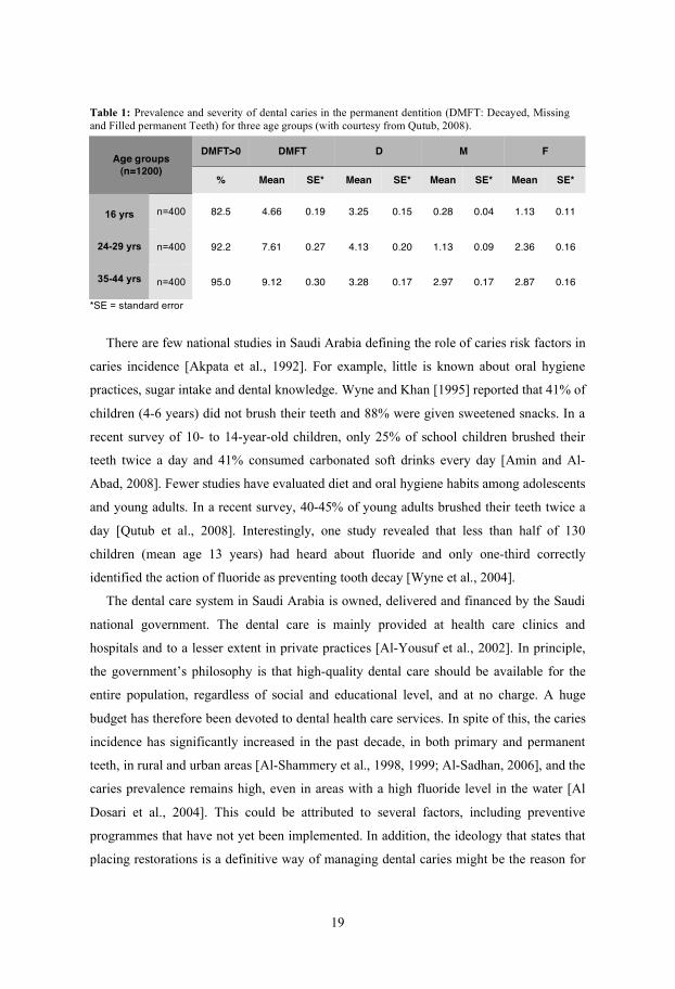
was approximately 9 [Qutub et al., 2008] (Table 1).
19
Table 1: Prevalence and severity of dental caries in the permanent dentition (DMFT: Decayed, Missing and Filled permanent Teeth) for three age groups (with courtesy from Qutub, 2008).
*SE = standard error
There are few national studies in Saudi Arabia defining the role of caries risk factors in
caries incidence [Akpata et al., 1992]. For example, little is known about oral hygiene
practices, sugar intake and dental knowledge. Wyne and Khan [1995] reported that 41% of
children (4-6 years) did not brush their teeth and 88% were given sweetened snacks. In a
recent survey of 10- to 14-year-old children, only 25% of school children brushed their
teeth twice a day and 41% consumed carbonated soft drinks every day [Amin and Al-
Abad, 2008]. Fewer studies have evaluated diet and oral hygiene habits among adolescents
and young adults. In a recent survey, 40-45% of young adults brushed their teeth twice a
day [Qutub et al., 2008]. Interestingly, one study revealed that less than half of 130
children (mean age 13 years) had heard about fluoride and only one-third correctly
identified the action of fluoride as preventing tooth decay [Wyne et al., 2004].
The dental care system in Saudi Arabia is owned, delivered and financed by the Saudi
national government. The dental care is mainly provided at health care clinics and
hospitals and to a lesser extent in private practices [Al-Yousuf et al., 2002]. In principle,
the government’s philosophy is that high-quality dental care should be available for the
entire population, regardless of social and educational level, and at no charge. A huge
budget has therefore been devoted to dental health care services. In spite of this, the caries
incidence has significantly increased in the past decade, in both primary and permanent
teeth, in rural and urban areas [Al-Shammery et al., 1998, 1999; Al-Sadhan, 2006], and the
caries prevalence remains high, even in areas with a high fluoride level in the water [Al
Dosari et al., 2004]. This could be attributed to several factors, including preventive
programmes that have not yet been implemented. In addition, the ideology that states that
placing restorations is a definitive way of managing dental caries might be the reason for
DMFT>0 DMFT D M F Age groups
(n=1200) % Mean SE* Mean SE* Mean SE* Mean SE*
n=400
82.5 4.66 0.19 3.25 0.15 0.28 0.04 1.13 0.11
n=400
92.2 7.61 0.27 4.13 0.20 1.13 0.09 2.36 0.16
16 yrs
24-29 yrs
35-44 yrs
n=400
95.0 9.12 0.30 3.28 0.17 2.97 0.17 2.87 0.16

20
the high incidence [Guile and Al-Shammary, 1987]. However, several national surveys
recommended and raised the importance of the caries risk assessment, oral health
education, implementation of community-based preventive measures and the need to
develop a national food policy, including controlling sugar intake [Almas and Al Jasser,
1996; Gandeh and Milaat, 2000; Amin and Al-Abad, 2008; Farsi, 2008]. The means of
implementing a preventive concept of this kind and the way this priority can be
transformed into reality have not yet been determined. In this context, a huge effort is still
needed in Saudi Arabia in order to fulfil the goals of the WHO Oral Health Country/Area
Profile Programme in lowering the global DMFT [FDI, 1982].
21
Aims
The present thesis consists of two parts. The first examines the caries prevalence and
caries risk profile of an adult Saudi population using the Cariogram model and evaluates
the quality of dental restorations (Papers I & II). The second part evaluates the effect of
applying the “modified fluoride toothpaste technique” on both approximal and
buccal/lingual enamel caries (Papers III & IV). To achieve these general objectives, the
aims of this thesis were:
to evaluate the caries risk profile in a group of Saudi adults with several dental
restorations, by assessing various caries-related factors using the Cariogram model,
to evaluate initial caries, total decayed surfaces, filled surfaces and recurrent caries
in relation to the Cariogram data, expressed as the “chance of avoiding caries”,
to evaluate the quality of dental restorations in a group of Saudi adults,
emphasising the additional value of bitewing radiographs as an aid to quality
evaluations of the restorations,
to investigate the effect of the “modified fluoride toothpaste technique” on the
incidence and progression of approximal caries using bitewing radiographs after 2
years and
to evaluate the preventive effect of the “modified fluoride toothpaste technique” on
buccal/lingual enamel caries clinically after 2 years and to determine the role of
patient compliance.
•
•
•
•
•

22
23
Material and Methods
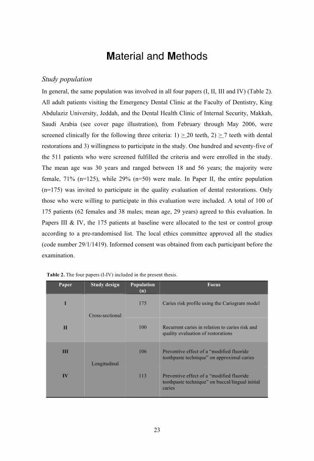
Study population In general, the same population was involved in all four papers (I, II, III and IV) (Table 2).
All adult patients visiting the Emergency Dental Clinic at the Faculty of Dentistry, King
Abdulaziz University, Jeddah, and the Dental Health Clinic of Internal Security, Makkah,
Saudi Arabia (see cover page illustration), from February through May 2006, were
screened clinically for the following three criteria: 1) > 20 teeth, 2) > 7 teeth with dental
restorations and 3) willingness to participate in the study. One hundred and seventy-five of
the 511 patients who were screened fulfilled the criteria and were enrolled in the study.
The mean age was 30 years and ranged between 18 and 56 years; the majority were
female, 71% (n=125), while 29% (n=50) were male. In Paper II, the entire population
(n=175) was invited to participate in the quality evaluation of dental restorations. Only
those who were willing to participate in this evaluation were included. A total of 100 of
175 patients (62 females and 38 males; mean age, 29 years) agreed to this evaluation. In
Papers III & IV, the 175 patients at baseline were allocated to the test or control group
according to a pre-randomised list. The local ethics committee approved all the studies
(code number 29/1/1419). Informed consent was obtained from each participant before the
examination.
Table 2. The four papers (I-IV) included in the present thesis. Paper Study design Population
(n) Focus
175
Caries risk profile using the Cariogram model
I
II
Cross-sectional
100 Recurrent caries in relation to caries risk and quality evaluation of restorations
106
Preventive effect of a “modified fluoride toothpaste technique” on approximal caries
III
IV
Longitudinal
113 Preventive effect of a “modified fluoride toothpaste technique” on buccal/lingual initial caries

24
Papers I & II
Baseline data Figures 1 & 2 outline the baseline data collected for Papers I & II, including interviews,
bitewing radiographs, photographs, plaque scores, salivary and microbiological factors
and caries registration. Only in those who agreed to participate in Paper II was the quality
of the restorations additionally examined.
Figure 1. The set-up for Paper I. Figure 2. The set-up for Paper II.
25
Using a standardised structured questionnaire according to the Cariogram manual
[Bratthall et al., 2004], all the patients were interviewed, about their medical and dental
histories, dietary habits and the use of fluoride. The baseline plaque index (PI) was scored
according to Silness and Löe [1964]. Paraffin-stimulated whole saliva was collected and
the secretion rate was expressed as ml/min. A chair-side test (CRT Bacteria®, Ivoclar-
Vivadent, Schaan, Liechtenstein) was used to evaluate MS and LB counts. The buffer
capacity was determined using CRT Buffer® (Buffer Strip, Ivoclar-Vivadent).
Caries registration Dental caries for the entire dentition, excluding third molars, was recorded clinically by
one examiner (H.S.) according to the WHO criteria [1997]. The number of manifest caries
(Dm), missing (M) and filled (F) tooth surfaces (S) were scored for each patient and
calculated as DmMFS. Tooth surfaces with initial caries (DiS) were registered separately,
then combined with Dm as Di+mS and added to DmMFS, resulting in a total number of
Di+mMFS.
A dental assistant took four bitewing radiographs of each patient. Approximal caries
was scored using the 6-grade scale (0 to 5) according to Gröndahl et al. [1977], illustrated
by Mejàre et al. [1998]. Approximal restorations, missing and unreadable surfaces
(overlapped or questionable) were scored as 6, 7 and 8 respectively. Recurrent caries as a
distinct radiolucency at the approximal gingival margin of the restoration was scored as 9.
One examiner (H.S.) evaluated 680 bitewing radiographs using a light desk and a
magnifying viewer. All approximal surfaces from the distal surface of the first premolar to
the mesial surface of the second molars were included, making a total of 24 surfaces.
Scores of 1 to 5 were added to the Di+mS index, apart from scores of 4 and 5 in teeth
previously registered as clinically decayed. To validate intra-examiner reliability, 120
radiographs from 35 individuals were re-evaluated after one month by the same examiner
(Cohen’s kappa value was 0.77).
Caries risk profile (Cariogram) Since all the patients had several restorations ( 7), the factor “caries experience” was
given a score of 3, i.e. worse status than normal for age group. The “clinical judgement”
factor was scored as 1, i.e. the risk was evaluated according to the other factors. All the

26
data were entered according to predetermined scales. Consequently, a pie chart with five
coloured sectors that represent (in percentages) the impact of various risk factors related to
caries was created for each patient (for examples, see Appendices 1 & 2). The patient’s
estimated percentage “chance of avoiding caries” was used for the analysis.
In Paper I, the 175 patients were divided into four risk groups according to the
percentage “chance of avoiding caries”: 1) 0-20% (high risk; n=66), 2) 21-40% (medium
risk; n=43), 3) 41-60% (low risk; n=50) and 4) 61-100% (very low risk; n=16). In Paper
II, the 100 patients were categorised into three risk groups instead of four: 1) 0-20% (high;
n=38), 2) 21-40% (medium; n=28) and 3) 41-100% (low and very low; n=34). This was
due to the small number of patients who were assigned to the “very low risk” group
according to the Cariogram. The last two risk groups were therefore combined.
Quality evaluation of restorations (Paper II) A total of 803 restorations in the 100 patients were clinically evaluated according to the
United States Public Health Service (USPHS/Ryge) criteria [1980]. Each restoration was
evaluated on the following criteria: 1) presence of recurrent caries, 2) marginal integrity,
3) anatomic form, 4) surface texture and 5) colour match. Only frank caries lesions and/or
decalcification at the margin of the restoration were registered and marginal staining was
excluded. Each criterion was graded as A or B, if clinically “acceptable”, and C or D, if
“unacceptable” [Ryge and DeVincenzi, 1983; Allander et al., 1989]. Only A, B or C
ratings were used for recurrent caries. Fifty-six of the restorations were re-evaluated after
2 weeks; the kappa value was 0.89.
The bitewing radiographs were examined to evaluate the proximal part of Class II
restorations with respect to: 1) marginal integrity at the gingival wall, in which the
presence or absence of “radiolucency” was recorded, and 2) anatomic form, in which
under-contour or over-hang restorations were identified.
Papers III & IV
After 2 years, the follow-up data were collected including plaque scores, enamel caries
registration, bitewing radiographs and compliance (Figure 3).
27
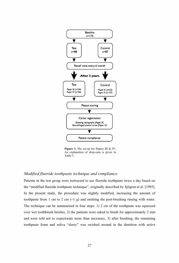
Figure 3. The set-up for Papers III & IV. An explanation of drop-outs is given in Table 3.
Modified fluoride toothpaste technique and compliance Patients in the test group were instructed to use fluoride toothpaste twice a day based on
the “modified fluoride toothpaste technique”, originally described by Sjögren et al. [1995].
In the present study, the procedure was slightly modified, increasing the amount of
toothpaste from 1 cm to 2 cm ( 1 g) and omitting the post-brushing rinsing with water.
The technique can be summarised in four steps: 1) 2 cm of the toothpaste was squeezed
over wet toothbrush bristles, 2) the patients were asked to brush for approximately 2 min
and were told not to expectorate more than necessary, 3) after brushing, the remaining
toothpaste foam and saliva “slurry” was swished around in the dentition with active
28
movements of the cheeks, lips and tongue, forcing the slurry in between the teeth for about
30 sec before expectorating and 4) no post-brushing water rinsing or eating/drinking for a
minimum of 2 hr post-brushing (see Appendix 3). In order to encourage compliance, all
the patients were given a pamphlet illustrating all the steps in coloured photographs.
The toothpaste used by patients in the test group was Colgate Maximum Cavity
Protection (Colgate, Piscataway, NJ, USA), which contains 1,450 ppm F. Control patients
were directed to continue using their regular fluoride toothpaste (also containing 1,450
ppm F) twice a day and were not given any further instructions. The type of fluoride
toothpaste was identified prior to the trial.
During the 2-year period, the patients in the test group were recalled every 6 months
and the toothpaste technique instructions were reinforced while the patient brushed. At the
end of the session, each patient was given another illustrated pamphlet and four tubes of
the toothpaste (120 ml). After 2 years, test group patients were monitored by a well-
trained dental assistant, while performing the technique, and compliance was assessed.
The patients were also interviewed about the regular use of the fluoride toothpaste,
frequency of brushing and refraining from eating/drinking for 2 hrs. Compliance in
patients who followed the four steps in principle was scored as A, while those who
brushed their teeth only once a day and/or rinsed with a sip of water post-brushing were
scored as B.
Patients in the control group were also recalled every 6 months, the use of fluoride
toothpaste twice a day was emphasised and they were given a toothbrush. At the end of
the study, their compliance (i.e. regular use of the fluoride toothpaste and the frequency of
brushing) was assessed by the interviewer.
Caries registration In Paper III, four bitewing radiographs were taken at baseline and after 2 years for
approximal caries evaluation. Only approximal surfaces that could be evaluated at both
examinations were included in the result. Approximal restored surfaces and recurrent
caries were scored. Surfaces that were unreadable at baseline but were scored as sound at
follow-up were considered to be caries free at baseline. Any sound surface at baseline that
had an enamel or a dentine lesion or had been filled after 2 years was defined in the
present study as “caries incidence”; the total “approximal caries incidence” was calculated
29
after pooling all these data. A change in score from enamel to dentine, from enamel to
filled or from filled to recurrent caries was regarded as “approximal caries progression”.
In Paper IV, all buccal and lingual surfaces were examined for non-cavitated lesions
(enamel caries). The caries diagnostic criteria were modified after those of Nyvad et al.
[1999] and only scores for the non-cavitated lesions were used in the present study.
Cavitated caries lesions (manifest caries) were registered, regardless of the state of
activity. Buccal/lingual surfaces with cervical restorations were recorded as filled.
After 2 years (Paper IV), all the sound buccal/lingual surfaces at baseline that had
enamel, manifest caries or had been filled were defined as total “buccal/lingual enamel
caries incidence”. A change in an enamel surface lesion to surface discontinuity in enamel
or a cavity in dentine, or from surface discontinuity in enamel to a cavity in dentine, was
defined as “progressed enamel caries”. All enamel caries that had been filled was
registered. Only decalcification and frank caries lesions at the margin of the restoration
were registered as recurrent caries. The summation of all these occurrences was defined as
total “buccal/lingual enamel caries progression”. Furthermore, all changes from active to
inactive caries were collectively referred to as “arrested buccal/lingual caries”.
The examiner (H.S.) was masked to patient assignment to test or control group and to
compliance results, which were collected by a dental assistant.
Statistical analysis All the data were analysed using the SPSS statistical package (SPSS Inc., Chicago, IL,
USA).
In Paper I, descriptive statistics, including means and standard deviations (SD) of all
the caries indices, were calculated for the 175 individuals and for the four Cariogram risk
groups. Analysis of variance (ANOVA), followed by Scheffé’s test of multiple
comparisons, was used to compare the mean numbers of DiS, Di+mS, FS and Di+mMFS
between the Cariogram risk groups.
In Paper II, frequency distribution and the percentage of the quality ratings for the 803
restorations were calculated. The percentage of recurrent caries was obtained by dividing
the number of restorations diagnosed with recurrent caries by the total number of
restorations per patient. ANOVA was used to compare the mean percentage of recurrent
caries between the risk groups. When evaluating the difference between clinical

30
judgement alone and in addition to radiographs, the restoration was regarded as a unit and
a paired Z-test was used. A power analysis with an assumption significance level of 1%,
SD of 0.5 and a power of 80% to detect a difference of at least 0.15 was performed; a
sample size of 260 paired observations was obtained.
In Papers III and IV, Student’s t-test was used to compare the test and control groups in
terms of caries incidence and progression. Statistical significance tests were performed at
individual but not at site level. In Paper IV, the Mann-Whitney U test was used to compare
the compliance of patients in the test group in terms of total caries incidence and
progression. A power analysis with an assumption significance level of 5%, SD of 3.0 and
a power of 90% to detect at least 2.0 differences was performed; a sample size of 48
individuals per group was obtained.
In all the analyses, the level of significance was considered at p < 0.05.
31
Results
Papers I & II
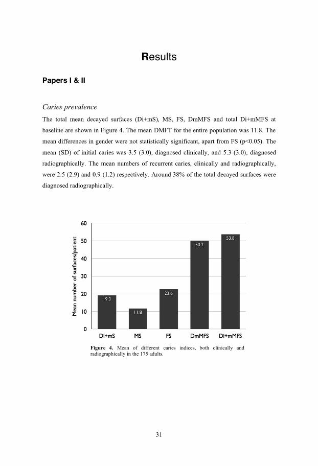
Caries prevalence The total mean decayed surfaces (Di+mS), MS, FS, DmMFS and total Di+mMFS at
baseline are shown in Figure 4. The mean DMFT for the entire population was 11.8. The
mean differences in gender were not statistically significant, apart from FS (p<0.05). The
mean (SD) of initial caries was 3.5 (3.0), diagnosed clinically, and 5.3 (3.0), diagnosed
radiographically. The mean numbers of recurrent caries, clinically and radiographically,
were 2.5 (2.9) and 0.9 (1.2) respectively. Around 38% of the total decayed surfaces were
diagnosed radiographically.
Figure 4. Mean of different caries indices, both clinically and radiographically in the 175 adults.
32
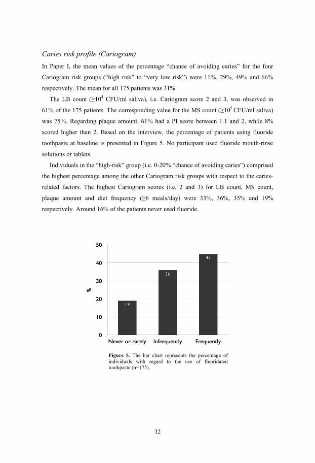
Caries risk profile (Cariogram) In Paper I, the mean values of the percentage “chance of avoiding caries” for the four
Cariogram risk groups (“high risk” to “very low risk”) were 11%, 29%, 49% and 66%
respectively. The mean for all 175 patients was 31%. The LB count ( 104 CFU/ml saliva), i.e. Cariogram score 2 and 3, was observed in
61% of the 175 patients. The corresponding value for the MS count ( 105 CFU/ml saliva)
was 75%. Regarding plaque amount, 61% had a PI score between 1.1 and 2, while 8%
scored higher than 2. Based on the interview, the percentage of patients using fluoride
toothpaste at baseline is presented in Figure 5. No participant used fluoride mouth-rinse
solutions or tablets.
Individuals in the “high-risk” group (i.e. 0-20% “chance of avoiding caries”) comprised
the highest percentage among the other Cariogram risk groups with respect to the caries-
related factors. The highest Cariogram scores (i.e. 2 and 3) for LB count, MS count,
plaque amount and diet frequency ( 6 meals/day) were 33%, 36%, 35% and 19%
respectively. Around 16% of the patients never used fluoride.
Figure 5. The bar chart represents the percentage of individuals with regard to the use of fluoridated toothpaste (n=175).
33
When comparing the risk groups with regard to DiS, Di+mS, FS and total Di+mMFS,
ANOVA revealed statistically significant differences between the high-risk group and the
other three groups regarding Di+mS (p<0.01) and FS (p<0.05). The mean DiS of the high-
risk group differed significantly from that of the low-risk group (p<0.05). The mean value
for Di+mMFS was similar in the four risk groups (NS). In Paper II, a significant difference was found when comparing the mean percentage of
recurrent caries for the three Cariogram risk groups; the lower the likelihood of new caries
being avoided in the near future, the higher the percentage of recurrent caries (p<0.05).
Quality evaluation of restorations (Paper II) The 803 restorations were distributed as follows: Class I (n=334), Class II (n=281), Class
III (n=100), Class IV (n=9), Class V (n=50) and composite veneer (n=29). The majority of
restorations were located on the posterior teeth, 625 (78%), and only 178 (22%) on the
anterior dentition. The distribution according to the restoration material was amalgam
(n=249), composite (n=422) and glass ionomer (n=132).
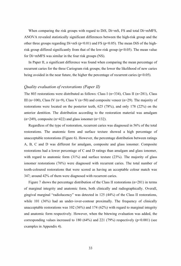
Regardless of the type of restoration, recurrent caries was diagnosed in 56% of the total
restorations. The anatomic form and surface texture showed a high percentage of
unacceptable restorations (Figure 6). However, the percentage distribution between ratings
A, B, C and D was different for amalgam, composite and glass ionomer. Composite
restorations had a lower percentage of C and D ratings than amalgam and glass ionomer,
with regard to anatomic form (31%) and surface texture (23%). The majority of glass
ionomer restorations (76%) were diagnosed with recurrent caries. The total number of
tooth-coloured restorations that were scored as having an acceptable colour match was
347; around 42% of them were diagnosed with recurrent caries.
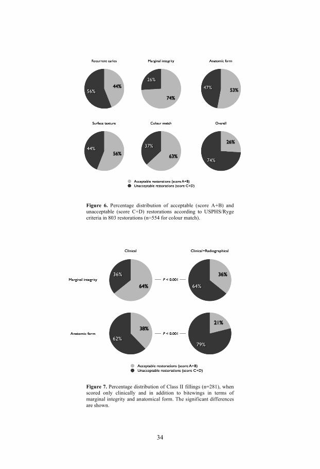
Figure 7 shows the percentage distribution of the Class II restorations (n=281) in terms
of marginal integrity and anatomic form, both clinically and radiographically. Overall,
gingival marginal “radiolucency” was detected in 125 (44%) of the Class II restorations,
while 101 (36%) had an under-/over-contour proximally. The frequency of clinically
unacceptable restorations was 102 (36%) and 174 (62%) with regard to marginal integrity
and anatomic form respectively. However, when the bitewing evaluation was added, the
corresponding values increased to 180 (64%) and 221 (79%) respectively (p<0.001) (see
examples in Appendix 4).
34
Figure 6. Percentage distribution of acceptable (score A+B) and unacceptable (score C+D) restorations according to USPHS/Ryge criteria in 803 restorations (n=554 for colour match).
Figure 7. Percentage distribution of Class II fillings (n=281), when scored only clinically and in addition to bitewings in terms of marginal integrity and anatomical form. The significant differences are shown.
35
Papers III & IV
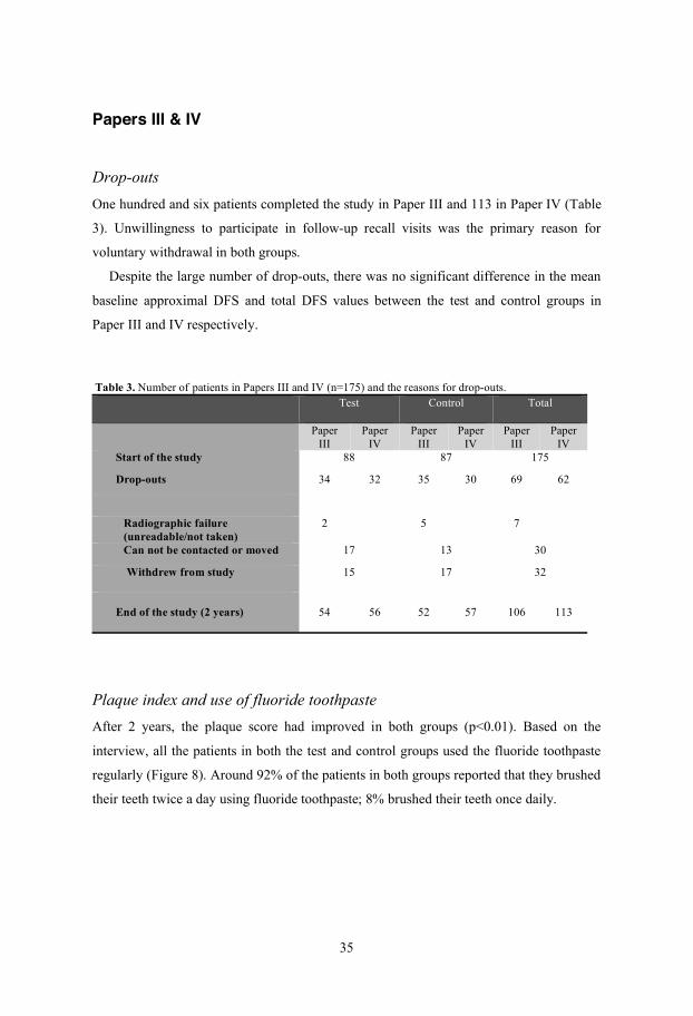
Drop-outs One hundred and six patients completed the study in Paper III and 113 in Paper IV (Table
3). Unwillingness to participate in follow-up recall visits was the primary reason for
voluntary withdrawal in both groups.
Despite the large number of drop-outs, there was no significant difference in the mean
baseline approximal DFS and total DFS values between the test and control groups in
Paper III and IV respectively.
Table 3. Number of patients in Papers III and IV (n=175) and the reasons for drop-outs.
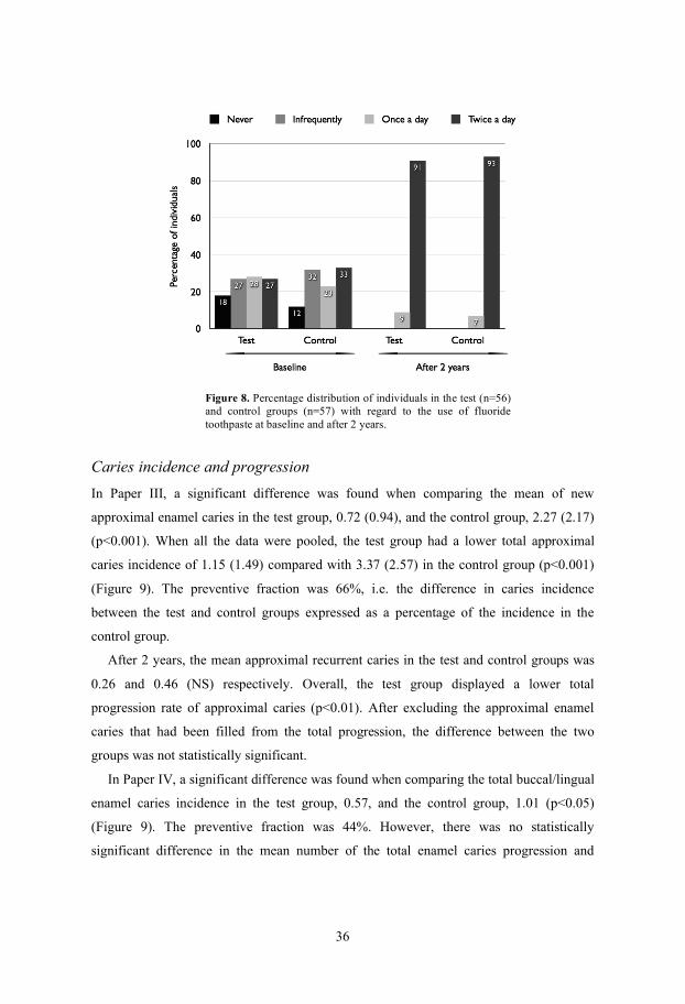
Plaque index and use of fluoride toothpaste After 2 years, the plaque score had improved in both groups (p<0.01). Based on the
interview, all the patients in both the test and control groups used the fluoride toothpaste
regularly (Figure 8). Around 92% of the patients in both groups reported that they brushed
their teeth twice a day using fluoride toothpaste; 8% brushed their teeth once daily.
Test
Control
Total
Paper III
Paper IV
Paper III
Paper IV
Paper III
Paper IV
Start of the study 88 87 175
Drop-outs 34 32 35 30 69 62
Radiographic failure (unreadable/not taken)
2 5 7
Can not be contacted or moved 17 13 30
Withdrew from study
15 17 32
End of the study (2 years)
54
56
52
57
106
113
36
Figure 8. Percentage distribution of individuals in the test (n=56) and control groups (n=57) with regard to the use of fluoride toothpaste at baseline and after 2 years.
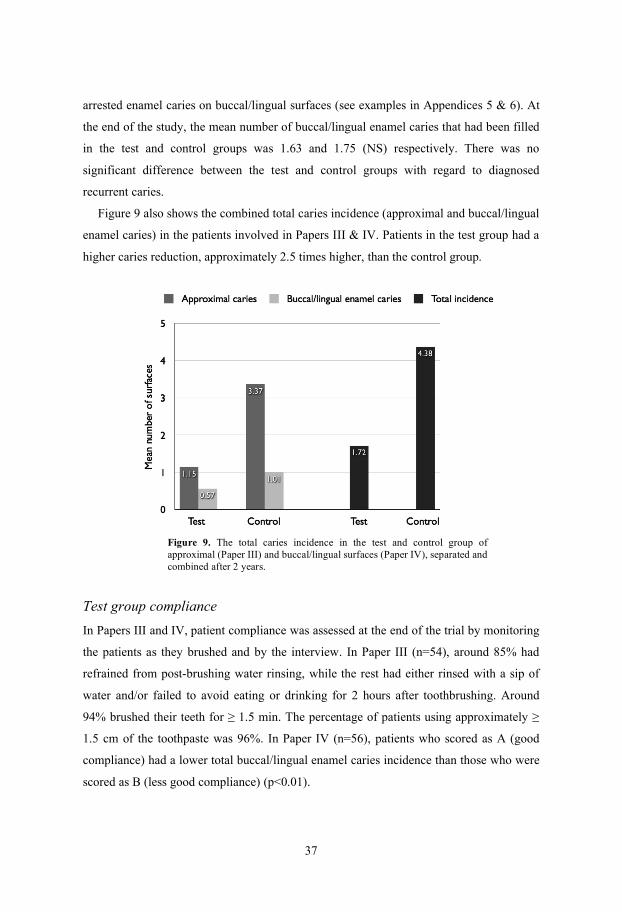
Caries incidence and progression In Paper III, a significant difference was found when comparing the mean of new
approximal enamel caries in the test group, 0.72 (0.94), and the control group, 2.27 (2.17)
(p<0.001). When all the data were pooled, the test group had a lower total approximal
caries incidence of 1.15 (1.49) compared with 3.37 (2.57) in the control group (p<0.001)
(Figure 9). The preventive fraction was 66%, i.e. the difference in caries incidence
between the test and control groups expressed as a percentage of the incidence in the
control group.
After 2 years, the mean approximal recurrent caries in the test and control groups was
0.26 and 0.46 (NS) respectively. Overall, the test group displayed a lower total
progression rate of approximal caries (p<0.01). After excluding the approximal enamel
caries that had been filled from the total progression, the difference between the two
groups was not statistically significant.
In Paper IV, a significant difference was found when comparing the total buccal/lingual
enamel caries incidence in the test group, 0.57, and the control group, 1.01 (p<0.05)
(Figure 9). The preventive fraction was 44%. However, there was no statistically
significant difference in the mean number of the total enamel caries progression and
37
arrested enamel caries on buccal/lingual surfaces (see examples in Appendices 5 & 6). At
the end of the study, the mean number of buccal/lingual enamel caries that had been filled
in the test and control groups was 1.63 and 1.75 (NS) respectively. There was no
significant difference between the test and control groups with regard to diagnosed
recurrent caries.
Figure 9 also shows the combined total caries incidence (approximal and buccal/lingual
enamel caries) in the patients involved in Papers III & IV. Patients in the test group had a
higher caries reduction, approximately 2.5 times higher, than the control group.
Figure 9. The total caries incidence in the test and control group of approximal (Paper III) and buccal/lingual surfaces (Paper IV), separated and combined after 2 years.
Test group compliance In Papers III and IV, patient compliance was assessed at the end of the trial by monitoring
the patients as they brushed and by the interview. In Paper III (n=54), around 85% had
refrained from post-brushing water rinsing, while the rest had either rinsed with a sip of
water and/or failed to avoid eating or drinking for 2 hours after toothbrushing. Around
94% brushed their teeth for 1.5 min. The percentage of patients using approximately
1.5 cm of the toothpaste was 96%. In Paper IV (n=56), patients who scored as A (good
compliance) had a lower total buccal/lingual enamel caries incidence than those who were
scored as B (less good compliance) (p<0.01).

38
39
Discussion
Caries risk profile (Cariogram)
The Cariogram is regarded as a useful tool for caries risk assessment and prediction and
has been used and validated for both children and elderly individuals [Hänsel-Petersson et
al., 2002, 2003]. Young or middle-aged adults (18-33 years old) have rarely been studied.
In the present thesis, the mean age of the individuals was 30 years. The Cariogram was
used as a model to estimate the risk of developing caries for the study population and to
identify the various caries-related factors contributing to that risk.
The main finding in Paper I was that the majority of the patients had a high caries risk;
the high- and medium-risk groups among the 175 patients (62%) had a less than 40%
“chance of avoiding caries” according to the Cariogram. If the entire population is
considered, the mean Cariogram value was only 31%, which is considered to be a low
value, i.e. a high caries risk. Of the various caries-related factors included in the
Cariogram model, four (LB, MS, plaque index and use of fluoride) obtained high
Cariogram scores (i.e. 2 and 3) in the majority of patients. Regarding the high-risk group,
the diet frequency score was also considered high and was therefore identified as an
additional warning signal. In addition, it should be noted that many of these patients did
not use fluoride. This reflects less motivation and awareness of dental care than the other
Cariogram risk groups, making them more likely to run a high risk of developing caries.
In overall terms, the caries-related factors identified by the Cariogram could explain
both the high caries prevalence in the study population and the probability of a high risk of
developing caries. Consequently, action should be taken to modify these factors, on both a
population and an individual level, to increase the percentage “chance of avoiding caries”
in the future. All the patients in the present study were informed of their estimated caries
risk profile (Cariogram outcome) and were encouraged to improve their oral health
accordingly.
Cariogram and caries experience
When the patients were divided into four Cariogram risk groups, there were statistically
significant differences between various caries components, such as Di+mS and DiS, and
the risk groups. Neither of these two indices is specifically included in the Cariogram

40
model, but they are generally involved in the “caries experience” factor, i.e. DMFT or
DMFS. The various caries indices were therefore analysed independently in the risk
groups and the results revealed that the high-risk group differed significantly from the
others. The Cariogram identified that this group had the highest total decayed component
(Di+mS) compared with the other risk groups. One finding that might be somewhat
confusing is that the high-risk group had the smallest number of FS, which differed from
the other three risk groups. The reason might be that these patients had a high numbers of
recurrent caries, which were counted as decayed and not as filled surfaces. This
speculation was confirmed in Paper II, where the high-risk group had the highest
percentage of recurrent caries compared with the other risk groups.
The total caries experience, i.e. DMFS, has been documented as a strong predictor for
future caries [Reich et al., 1999; Zero et al., 2001; Fontana and Zero, 2006]. For this
reason, the identification of individuals with a high caries risk is relatively accurate where
children and adolescents are concerned and when sufficient baseline data are available.
However, in daily practice, an examination only depicts the historical background of
caries not the current caries risk of the patient. In adults, the total DMFS value could be
overestimated because of the high FS components. The existing DMFS is less sensitive for
predicting future caries in adults compared with children [Reich et al., 1999]. It is
therefore probably unwise to use the total DMFS as the sole indication of caries risk
without weighing the other caries-related factors. It could be of interest to use the
Cariogram model in which the current total patient DMFS is readily involved and weighed
with other caries-related factors. This will assist the dentist in implementing the optimum
caries treatment and evaluating its outcome.
This interpretation was supported by the results of Paper I, where the mean DMFS
values were almost equal in the four risk groups, although they might differ in terms of
caries risk. In this context, it is important to emphasise that the high caries experience
score of the study population does not influence the percentage “chance of avoiding
caries”. In the Cariogram, the caries experience factor is regarded as a risk marker that
might indicate the increased probability of new caries, but it is not a part of the causal
chain that lead to caries development. It therefore has less weight than the other risk
factors in the built-in algorithm [Bratthall and Hänsel-Petersson, 2005]. This is probably
due to the fact that the Cariogram model was originally developed to predict future caries
41
lesions. As a result, the other risk factors involved in the causal chain were given more
weight by the program developer.
Initial caries is likely to have a profound effect as far as the caries risk estimation is
concerned. When the total initial caries was added to the total decayed index, the high-risk
group differed significantly from all the other Cariogram risk groups and not only from the
low-risk group. Adding approximal caries lesions, diagnosed by bitewing radiographs, to
the decayed surfaces index might be beneficial for actual caries prevalence estimation
[Anderson et al., 2005]. In patients with several dental restorations, as in the present study,
the detection of approximal caries by bitewing radiograph is important. According to
Powell [1998], approximal tooth surfaces become better predictors of future disease,
thereby underlining the importance of the bitewing radiographs. In Paper I, 38% of the
total caries diagnosed via bitewing radiographs. This is in agreement with Hopcraft and
Morgan [2005] survey, in which more than twice as many additional approximal lesions
were detected by bitewing radiographs than by clinical examination in adults aged 17-30
years.
Recurrent caries
One important outcome in Paper II is that, the lower the likelihood of new caries being
avoided in the near future, the higher the percentage of recurrent caries. Regardless of the
restoration material, recurrent caries was diagnosed in more than half the total restorations.
Such a high percentage indicates that these restorations were initially placed without any
attempt to evaluate the patient’s caries risk. Presumably, these restorations will be
replaced for the same reason in the future. Mjör [2005] reported that 50% of restorations
in adults were replaced because of recurrent caries. Powell et al. [2000] showed that
patients who had restorations placed due to caries had significantly higher MS, resulting in
a higher potential for continued caries activity than those who had received no
restorations. Furthermore, Sunnegårdh-Grönberg et al. [2009] found that restorations in
high caries risk patients had a shorter longevity than those in low- or moderate-risk
groups. One recent study demonstrated that the rate of restoration failure due to caries
could be reduced in the long term by changing the level of overall caries risk factors
[Miyamoto et al., 2007]. In Saudi Arabia, where the caries prevalence has been reported to
be high, the placement of dental restorations as the only means of treating caries should be
42
discouraged. It is imperative to implement national preventive programmes based on risk
assessments in various age groups in Saudi Arabia.
The management of caries needs to be based on the patient’s risk of developing caries
in order to be most health and cost effective [Reich et al., 1999]. In this context, the
Cariogram could be of great benefit in daily clinical practice when it comes to evaluating
the patient’s caries profile. Indeed, this pedagogic model would help allocate patients to
the right caries risk category and to identify the caries-related factors that could be
modified accordingly. The treatment plan for patients would therefore be more preventive
and conservative in design and would not just treat the patients with more fillings, thereby
exposing them to further risk [Fontana and Zero, 2006]. This would preserve the tooth
structure, increase the longevity of restorations and interrupt the restoration/replacement
cycle due to caries.
Quality of dental restorations
Quality evaluations of dental fillings in a cross-sectional design like the present study have
to be interpreted with caution. Several similar studies reported that the age of failed
restorations has been found to be lower than that reported in controlled clinical trials
[Mjör, 1997; Burke et al., 1999, 2001]. Although they are not rated highly in the hierarchy
of acceptable evidence, cross-sectional studies involving a large number of fillings and
practitioners might shed light on factors influencing the performance of restorations in
routine practice situations. In the present study, no attempt was made to compare filling
materials in terms of longevity or age at failure. However, an overview of the quality of
different filling materials was given and related to the estimated caries risk evaluated by
the Cariogram.
In the present survey, the anatomic form and surface texture showed a high percentage
of unacceptable restorations. Composite restorations received more acceptable ratings for
these two criteria than amalgam and glass ionomer. This is probably because composite
restorations in general have improved dramatically during the last decade. In addition, the
light-cured composite can be adjusted and polished the day it is placed, in contrast to glass
ionomer and amalgam. Restorations with a deteriorated contour, i.e. under- or over-
contoured, and rough surfaces, especially adjacent to gingival margins, could favour
bacterial growth and plaque maturation. The roughness of intra-oral hard surfaces, e.g.

43
fillings, will promote plaque formation and maturation, making the tooth surface more
vulnerable to caries [Quirynen and Bollen, 1995; Bollen et al., 1997]. This kind of
unfavourable quality could have a profound effect, particularly in high-risk patients, as in
the present study.
In the present survey, the majority of glass ionomer restorations were diagnosed with
recurrent caries, in spite of the release of fluoride, a finding reported in other studies
[Randall and Wilson, 1999; Manhart et al., 2004; Wiegand et al., 2007]. For this reason,
the fluoride-releasing property of glass ionomer should not be relied upon as a means of
preventing caries, while ignoring other caries-related factors.
Matching tooth-coloured restorations is regarded as a critical factor by clinicians and
patients from an aesthetic point of view. However, 42% of the tooth-coloured fillings with
an acceptable colour match had recurrent caries in the present study (Paper II). Miyamoto
et al. [2007] demonstrated that previously restored teeth experienced an increased rate of
recurrent caries compared with unrestored teeth. Accordingly, placing or replacing fillings
solely for aesthetic reasons should not be undertaken without seriously considering the
patient’s risk.
The value of bitewing radiographs in addition to clinical quality evaluations in Class II
restorations was confirmed in the present study. The unacceptable ratings for proximal
marginal integrity and anatomic form increased by 28% and 17% respectively, when
restorations were evaluated with bitewing radiographs. It is unlikely that the presence of
radiolucency and/or failed anatomic form at the gingival wall of Class II restorations will
be detected by clinical examination alone. However, the clinical interpretation of this
radiolucency could be crucial. For example, it could be due to the failure of proper
condensation with an amalgam, while, in a composite, a thick layer of adhesive could
appear to be radiolucent in a radiograph, or it could be a recurrent caries lesion that was
not observed on clinical evaluation. Regardless of the cause, this “radiolucency” is
regarded as a potential factor for developing caries, particularly in high-risk patients. Mjör
[2005] reported that the gingival wall in Class II restorations is the most common site of
recurrent caries. Furthermore, proximal overhangs, even minute ones, are predisposed to
plaque accumulation and the development of recurrent caries [Mjör and Gordan, 2002;
Mjör, 2005]. A number of studies have used bitewing radiographs in the quality evaluation
of restorations, emphasising their extra diagnostic value [Poorterman et al., 1999,2000;
44
Levin et al., 2007]. The information from bitewings could therefore refine the clinical
quality evaluation of the restorations. However, the decision on whether or not to expose
patients to radiation should be based on the dentist’s clinical judgement and should not be
performed as a routine procedure.
Caries prevention using the modified fluoride toothpaste
technique
The main finding in the second part of the present thesis was that “the modified fluoride
toothpaste technique” had a preventive effect on approximal caries and enamel caries
located on buccal/lingual surfaces. The difference in caries incidence between the test and
control groups could be due to many factors involved in this technique, such as the amount
of toothpaste applied and the duration of brushing. However, the influence of avoiding
post-brushing water rinsing and refraining from eating or drinking for two hours after
brushing could be of interest. As a result, the prolonged availability of a high level of
fluoride could be attained. This improves the chance of fluoride being incorporated into
the enamel and dentine, thereby rendering the surface more resistant to acidic challenge
[ten Cate, 1999]. Studies have shown that the cariostatic effect of topical fluoride is partly
related to the sustained presence or release of low fluoride concentrations in the oral
environment [Featherstone, 1999; Ellwood et al., 2008]. By avoiding water rinsing,
particularly in adults, the salivary fluoride level can remain high up to 2 hrs post-brushing
[Issa and Toumba, 2004]. A prolonged low concentration of fluoride in saliva and plaque
might enhance the rate of remineralisation [Featherstone et al., 1990]. Furthermore,
[Sjögren and Birkhed, 1993] reported that adult patients with high caries activity rinsed
with water after brushing more frequently than those with low caries activity. Zero [2006]
stated that “In addition to the inherent properties of a fluoride dentifrice product,
biological and behavioral factors can modify its anticaries effectiveness. All of these
factors interplay in what can be described as the ’application’ phase (the initial
interaction of relatively high concentrations of fluoride with the tooth surface and plaque),
and the ’retention’ phase (the fluoride remaining in the mouth after brushing that is
retained in saliva, plaque and plaque fluid, the tooth surface, and oral soft tissue
reservoirs)”. By optimising the behavioural factors, as was the intention in our study, the
45
“modified fluoride toothpaste technique” fulfilled these two phases described by Zero
[2006].
In Paper III, the most profound difference was found in the incidence of new
approximal enamel caries. The preventive fraction in the present study was around 66%.
This percentage is higher than that reported by Sjögren et al. [1995] and is probably due to
differences in the study population and patient compliance. The patients involved in the
present trial were all adults with a high caries prevalence, which might accentuate the
preventive effect of fluoride toothpaste. Marinho et al. [2003] found that the effect of
fluoride toothpaste increased with higher baseline levels of DMFS. In a review of fluoride
toothpastes, strong evidence of a preventive effect was found in the young permanent
dentition, while there was incomplete evidence in the primary dentition [Twetman et al.,
2003]. This might indicate that the “modified fluoride toothpaste technique” could have a
greater preventive effect in adults than in children, particularly in those with a high caries
prevalence. Moreover, a recent study showed that the fluoride concentration in interdental
saliva and plaque was higher without post-brushing water rinsing [Nordström and
Birkhed, 2009]. Since the toothbrush does not reach the approximal area, slurry rinsing
through active cheek movements could result in the rapid transport of fluoride into the
interdental plaque [Sjögren et al., 1996]. This could affect the metabolic activity of the
bacterial flora, enhance remineralisation and inhibit the demineralisation of approximal
tooth surfaces [Tenuta et al., 2009].
There is, however, some controversy regarding the effect of post-brushing water
rinsing per se on caries development. The difference between the mean caries increments
with and without rinsing was only 0.8 DS in a longitudinal clinical trial and had no
statistical significance [Machiulskiene et al., 2002]. In that study, however, the population
was not comprised of caries-prone individuals, as in the present trial. In addition, there
was no information as to whether the children were instructed not to eat or drink post-
brushing. This might be an important factor when it comes to maintaining a high fluoride
level in the oral cavity after refraining from water rinsing, as Sjögren and Birkhed [1994]
found that eating/drinking directly after brushing reduced the fluoride concentration
considerably.
46
Prevention of recurrent caries
In Papers III and IV, many restorations on the approximal and buccal/lingual surfaces
respectively had recurrent caries at the end of the trial and the frequency was higher in the
control group than in the test group. Although the differences were not significant,
recurrent caries does not differ from primary caries with regard to aetiology. In this
context, it might be true to say that fluoride apparently prevents recurrent caries
development. A recent in situ study demonstrated that fluoride toothpaste might maintain
a high level of fluoride in the biofilm, thereby reducing demineralisation and caries
progression adjacent to restorations [Cenci et al., 2008]. Moreover, recurrent caries is one
of the most common reasons for the failure of dental fillings [Mjör, 2005]. Consequently,
the investigated “modified fluoride toothpaste technique” might be beneficial in
preventing recurrent caries or at least interrupting its progression. This could eventually
improve filling longevity.
Compliance
It should be pointed out that the 6-month recall visits played a major role in improving the
oral hygiene of both test and control patients by the end of the study. In Papers III and IV,
the plaque reduction was more or less the same in both groups and all the patients used
fluoride toothpaste regularly. Nevertheless, the test group had a lower incidence of both
approximal and enamel buccal/lingual caries than the control group. The “modified
fluoride toothpaste technique” might therefore be the factor that contributed to the caries
reduction in the test group. Another interesting observation was that the majority of test
group patients complied with the instructions, indicating that the technique is easy for
adults to learn.
Frequency of toothbrushing, as well as post-rinsing behaviour, are probably the most
important as far as caries prevention is concerned. Patients in the test group who brushed
their teeth only once a day and/or rinsed with a sip of water post-brushing were therefore
scored as having “less good compliance”. The buccal/lingual enamel caries incidence was
significantly higher in these patients than in those with “good compliance”. This is in
agreement with other reports that showed an association between caries incidence and the
frequency of brushing and post-brushing behaviour [O'Mullane et al., 1997; Chestnutt et
al., 1998].
47
It is important to remember the efforts made at the recall visits to emphasise the
“modified fluoride toothpaste technique” instructions. These recall visits clearly
encouraged patient compliance, as shown by the final results of the study (Paper III).
However, the most common complaint from patients who wanted to rinse after brushing
was that they did not like the strong taste of the toothpaste slurry that was left after
brushing.
The amount of fluoride that might be swallowed as a result of the “modified fluoride
toothpaste technique” is less critical in an adult compared with a child. Up to 20% of
toothpaste was swallowed when no water rinsing took place post-brushing [Sjögren et al.,
1994]. This corresponds to 0.3 mg F/brushing, when using 1 g of a toothpaste with 1,500
ppm F; this amount corresponds to one cup of tea ( 1 dL) containing 3 mg F/L [Wu and
Wei, 2009]. From a toxicological point of view, this is regarded as negligible for an adult.
Caries progression
According to the present thesis, the “modified fluoride toothpaste technique” in general
did not have a pronounced effect on caries progression, compared with the development of
new lesions. Although the presence of fluoride enhances the remineralisation process, it
has been reported that the effect is more apparent in the demineralisation of enamel rather
than the remineralisation [ten Cate et al., 1995].
In Paper III, the number of approximal enamel lesions that progressed into the dentine
was somewhat higher in the control group, although the difference was not statistically
significant. The reason could be that the rate of approximal caries progression per se is
slow in young adults [Mejàre et al., 1999; Mejàre et al., 2004]. A 2-year trial is therefore
not long enough to evaluate caries progression, particularly in adults. Moreover, in a group
of patients with a large number of FS, such as those in the present trial, including enamel
lesions that were filled during the trial when calculating progression might be disputed. It
was difficult to trace whether they had first progressed into the dentine. The total
progression was therefore re-calculated, excluding this event, but the differences between
the control and test groups were not statistically significant.
In Paper IV, the clinical examination of the buccal and lingual enamel caries was
dependent on visual and tactile criteria. Although these criteria provide much more
information about caries lesion transition patterns in the clinical trials [Baelum et al.,
48
2003; Hausen et al., 2007; Lima et al., 2008], however, they do not provide much data on
the depth or degree of mineral loss at microscopic level. The clinical depth of the lesions
could influence the efficacy of fluoride toothpaste [ten Cate et al., 2006]. Consequently,
methods that measure the degree of mineral loss and the depth of the lesion should be used
in order better to evaluate the possible effect of the “modified fluoride toothpaste
technique” on progression.
The number of arrested buccal/lingual enamel caries was similar in both groups. This
could be attributed to the fact that the buccal enamel caries is accessible to the mechanical
action of the toothbrush bristles that might lead to the abrasion of the superficial layer of
the lesion. An effect of this kind has been reported in clinical studies [Holmen et al., 1987;
Årtun and Thylstrup, 1989].
Many enamel caries were filled at the end of the trial (Papers III and IV). This
somewhat “aggressive” approach should be discouraged and replaced by promoting the
conservative management of enamel caries, including the use of fluoride toothpaste, and
pursuing caries risk assessment. Moreover, it is apparent that the dentists did not fully
appreciate the importance of arresting early caries. In dental practice, the diagnosis of
early caries lesions should therefore include detecting the lesion, estimating its depth and
degree of demineralisation and, most importantly, making a decision regarding its activity
[Nyvad and Fejerskov, 1997]. Lesion surface texture alterations are associated with
changes in the progression rate of caries. Active non-cavitated enamel lesions are dull and
rough, while inactive non-cavitated enamel lesions are shiny and smooth [for a review, see
Thylstrup et al., 1994]. These clinical changes have been reported to provide a reliable,
valid indication of caries activity [Nyvad et al., 1999, 2003]. Consequently, using the
combined information obtained from the tactile sensation, visual appearance and lesion
location could indicate the caries activity of early lesions. Accordingly, treatment
decisions can be more conservative and appropriately determined [Fontana and Zero,
2006; Mjör et al., 2008].

49
Conclusions
The Cariogram evaluated the caries risk profile in the present adult population and
the caries-related factors contributing to the risk were identified.
There was an association between many caries indices (total decayed surfaces,
filled surfaces and recurrent caries) and the Cariogram risk groups but not with the
total DMFS.
The main reason for the unacceptable rating of restorations was recurrent caries.
Unacceptable anatomic form and surface texture were also common. Bitewing
radiographs were found to be an important aid when evaluating proximal marginal
integrity and anatomic form.
The “modified fluoride toothpaste technique” had a preventive effect on
approximal caries in this adult Saudi population with a high caries prevalence. It
reduced the incidence of approximal caries by around 66%.
Using this fluoride toothpaste technique resulted in a preventive fraction for
buccal/lingual enamel caries of approximately 44%. Patient compliance appeared
to play an important role in this result.
•
•
•
•
•

50

51
Acknowledgements
All praise to God who gave me the support and help to accomplish this project. I would like to express my sincere gratitude to a number of individuals who have contributed to the successful completion of this thesis. Firstly, I would like to extend my deep gratitude to my supervisor, Professor Dowen Birkhed, for his unbounded advice, encouragement and mentorship. He provided me with hugs and company during my study. His open-door policy and humour made the process truly enjoyable. Professor Claes-Göran Emilson and Professor Peter Lingström, for providing valuable feedback and support. Many thanks to my colleague Dr Mishari Al-otaibi, for his wholehearted co-operation and magnificent effort in referring patients A special vote of thanks must be extended to Mrs. Ann-Britt Lundberg and Mrs. Ann-Charlott Börjesson, for helping me enter the data and make figures; this reduced many nightmares during my data analysis. In addition, I would like to express my deep gratitude to the nurses, particularly Mrs. Jaliha, and administrators at both the King Abdulaziz University and Dental Health Clinic of Internal Security, for the time that was taken in order to follow up patients; without their help I would not have been able to collect the required data. Dr Tommy Johnsson, for statistical assistance. Rita Shafer and Jeanette Kliger for excellent revision of the English text. Also, all my friends and the staff at the Department of Cariology, for making these five years memorable – thank you for your listening ears and for making me laugh when I needed it most. The Ministry of Higher Education in Saudi Arabia, along with the King Abdulaziz University, Saudi Arabia, for fully funding this project This project would not have been possible without the love and support of my family. Most importantly, I wish to thank my beloved parents, Mohammad and Maryam for sharing the journey of developing this thesis with me and for their encouragement and positive spirit. Also my respected father- and mother- in law, Ahmad and Zakiyah for their continued love and support. Special thanks to my dear wife, Samah, and our lovely children, Mohammad and Rama for allowing me to uproot the family in order to pursue my dreams and goals. I shall always be grateful for her unconditional support, her ability to keep me grounded and for

52
understanding the bigger picture. Last but not least, many thanks to my brother and sisters for their love and admiration, which continue to support me through my difficult times.

53
References
Aas JA, Griffen AL, Dardis SR, Lee AM, Olsen I, Dewhirst FE, Leys EJ, Paster BJ:
Bacteria of dental caries in primary and permanent teeth in children and young adults. J Clin Microbiol 2008;46:1407-1417.
Akpata ES, Al-Shammery AR, Saeed HI: Dental caries, sugar consumption and restorative dental care in 12-13-year-old children in Riyadh, Saudi Arabia. Community Dent Oral Epidemiol 1992;20:343-346.
Al-Dosari AM, Wyne AH, Akpata ES, Khan NB: Caries prevalence and its relation to water fluoride levels among schoolchildren in Central Province of Saudi Arabia. Int Dent J 2004;54:424-428.
Al-Shammery A, El Backly M, Guile EE: Permanent tooth loss among adults and children in Saudi Arabia. Community Dent Health 1998;15:277-280.
Al-Sadhan S: Dental caries prevalence among 12-14 year-old schoolchildren in Riyadh: A 14 year follow-up study of the Oral Health Survey of Saudi Arabia Phase I. Saudi Dent J 2006;18:2-7.
Al-Shammery AR: Caries experience of urban and rural children in Saudi Arabia. J Public Health Dent 1999;59:60-64.
Al-Yousuf M, Akerele TM, Al-Mazrou YY: Organization of the Saudi health system. East Mediterr Health J 2002;8:645-653.
Allander L, Birkhed D, Bratthall D: Quality evaluation of anterior restorations in private practice. Swed Dent J 1989;13:141-150.
Almas K, Al Jasser N: Prevalence of dental caries and periodontal disease in a Saudi Population. Saudi Med J 1996;17:640-644.
Amarante E, Raadal M, Espelid I: Impact of diagnostic criteria on the prevalence of dental caries in Norwegian children aged 5, 12 and 18 years. Community Dent Oral Epidemiol 1998;26:87-94.
Amin TT, Al-Abad BM: Oral hygiene practices, dental knowledge, dietary habits and their relation to caries among male primary school children in Al Hassa, Saudi Arabia. Int J Dent Hyg 2008;6:361-370.
Anderson M, Stecksén-Blicks C, Stenlund H, Ranggård L, Tsilingaridis G, Mejàre I: Detection of approximal caries in 5-year-old Swedish children. Caries Res 2005;39:92-99.
Anusavice KJ: Present and future approaches for the control of caries. J Dent Educ 2005;69:538-554.
Årtun J, Thylstrup A: A 3-year clinical and SEM study of surface changes of carious enamel lesions after inactivation. Am J Orthod Dentofacial Orthop 1989;95:327-333.
Atieh MA: Tooth loss among Saudi adolescents: social and behavioural risk factors. Int Dent J 2008;58:103-108.
Attin T, Hornecker E: Tooth brushing and oral health: how frequently and when should tooth brushing be performed? Oral Health Prev Dent 2005;3:135-140.
Baelum V, Machiulskiene V, Nyvad B, Richards A, Vaeth M: Application of survival analysis to carious lesion transitions in intervention trials. Community Dent Oral Epidemiol 2003;31:252-260.
54
Baysan A, Lynch E, Ellwood R, Davies R, Petersson L, Borsboom P: Reversal of primary root caries using dentifrices containing 5,000 and 1,100 ppm fluoride. Caries Res 2001;35:41-46.
Bollen CM, Lambrechts P, Quirynen M: Comparison of surface roughness of oral hard materials to the threshold surface roughness for bacterial plaque retention: a review of the literature. Dent Mater 1997;13:258-269.
Bowden GH: Does assessment of microbial composition of plaque/saliva allow for diagnosis of disease activity of individuals? Community Dent Oral Epidemiol 1997;25:76-81.
Brantley CF, Bader JD, Shugars DA, Nesbit SP: Does the cycle of re-restoration lead to larger restorations? J Am Dent Assoc 1995;126:1407-1413.
Bratthall D: Dental caries: intervened--interrupted--interpreted. Concluding remarks and cariography. Eur J Oral Sci 1996;104:486-491.
Bratthall D, Hänsel-Petersson G: Cariogram--a multifactorial risk assessment model for a multifactorial disease. Community Dent Oral Epidemiol 2005;33:256-264.
Bratthall D, Hänsel-Petersson G, Stjernswärd J: Cariogram Internet Version 2.01. April 2;2004.
Bratthall D, Hänsel-Petersson G, Sundberg H: Reasons for the caries decline: what do the experts believe? Eur J Oral Sci 1996;104:416-422.
Burke FJ, Cheung SW, Mjör IA, Wilson NH: Restoration longevity and analysis of reasons for the placement and replacement of restorations provided by vocational dental practitioners and their trainers in the United Kingdom. Quintessence Int 1999;30:234-242.
Burke FJ, Wilson NH, Cheung SW, Mjör IA: Influence of patient factors on age of restorations at failure and reasons for their placement and replacement. J Dent 2001;29:317-324.
Burt BA: Concepts of risk in dental public health. Community Dent Oral Epidemiol 2005;33:240-247.
Burt BA, Pai S: Sugar consumption and caries risk: a systematic review. J Dent Educ 2001;65:1017-1023.
Campus G, Cagetti MG, Sacco G, Benedetti G, Strohmenger L, Lingström P: Caries risk profiles in Sardinian schoolchildren using Cariogram. Acta Odontol Scand 2009;67:146-152.
Cenci MS, Tenuta LM, Pereira-Cenci T, Del Bel Cury AA, ten Cate JM, Cury JA: Effect of microleakage and fluoride on enamel-dentine demineralization around restorations. Caries Res 2008;42:369-379.
Chestnutt IG, Schafer F, Jacobson AP, Stephen KW: The influence of toothbrushing frequency and post-brushing rinsing on caries experience in a caries clinical trial. Community Dent Oral Epidemiol 1998;26:406-411.
Chhour KL, Nadkarni MA, Byun R, Martin FE, Jacques NA, Hunter N: Molecular analysis of microbial diversity in advanced caries. J Clin Microbiol 2005;43:843-849.
Davies RM, Ellwood RP, Davies GM: The rational use of fluoride toothpaste. Int J Dent Hyg 2003;1:3-8.
Deligeorgi V, Mjör IA, Wilson NH: An overview of reasons for the placement and replacement of restorations. Prim Dent Care 2001;8:5-11.

55
Demers M, Brodeur JM, Simard PL, Mouton C, Veilleux G, Frechette S: Caries predictors suitable for mass-screenings in children: a literature review. Community Dent Health 1990;7:11-21.
DenBesten P, Ko HS: Fluoride levels in whole saliva of preschool children after brushing with 0.25 g (pea-sized) as compared to 1.0 g (full-brush) of a fluoride dentifrice. Pediatr Dent 1996;18:277-280.
Dérand T, Birkhed D, Edwardsson S: Secondary caries related to various marginal gaps around amalgam restorations in vitro. Swed Dent J 1991;15:133-138.
Duckworth RM, Morgan SN: Oral fluoride retention after use of fluoride dentifrices. Caries Res 1991;25:123-129.
Elderton RJ: Clinical studies concerning re-restoration of teeth. Adv Dent Res 1990;4:4-9. Elderton RJ, Nuttall NM: Variation among dentists in planning treatment. Br Dent J
1983;154:201-206. Ellwood R, Fejerskov O, Cury JA, Clarkson B. Fluoride in caries control. In: Fejerskov O,
Kidd EA, editors. Dental caries. The disease and its clinical management. 2d ed. Oxford, UK; Blackwell Munksgaard 2008:287-327.
Emilson CG: Prevalence of Streptococcus mutans with different colonial morphologies in human plaque and saliva. Scand J Dent Res 1983;91:26-32.
Farsi N: Dental caries in relation to salivary factors in Saudi population groups. J Contemp Dent Pract 2008;9:16-23.
Fédération Dentaire Internationale FDI. Global goals for oral health in the year 2000. Int Dent J 1982;32:74-77.
Featherstone JD: Prevention and reversal of dental caries: role of low level fluoride. Community Dent Oral Epidemiol 1999;27:31-40.
Featherstone JD: The science and practice of caries prevention. J Am Dent Assoc 2000;131:887-899.
Featherstone JD: The caries balance: the basis for caries management by risk assessment. Oral Health Prev Dent 2004;2 Suppl 1:259-264.
Featherstone JD, Glena R, Shariati M, Shields CP: Dependence of in vitro demineralization of apatite and remineralization of dental enamel on fluoride concentration. J Dent Res 1990;69:620-625.
Fejerskov O: Changing paradigms in concepts on dental caries: consequences for oral health care. Caries Res 2004;38:182-191.
Fontana M, Zero DT: Assessing patients' caries risk. J Am Dent Assoc 2006;137:1231-1239.
Fure S: A ten-year cross-sectional and follow-up study of salivary flow rates and mutans streptococci and lactobacillus counts in elderly Swedish individuals. Oral Health Prev Dent 2003;1:185-194.
Fure S: Ten-year cross-sectional and incidence study of coronal and root caries and some related factors in elderly Swedish individuals. Gerodontology 2004;21:130-140.
Gandeh MB, Milaat WA: Dental caries among schoolchildren: report of a health education campaign in Jeddah, Saudi Arabia. East Mediterr Health J 2000;6:396-401.
Goldberg AJ: Deterioration of restorative materials and the risk for secondary caries. Adv Dent Res 1990;4:14-18.
Goldberg J, Tanzer J, Munster E, Amara J, Thal F, Birkhed D: Cross-sectional clinical evaluation of recurrent enamel caries, restoration of marginal integrity, and oral hygiene status. J Am Dent Assoc 1981;102:635-641.
56
Goshima T, Goshima Y: Radiographic detection of recurrent carious lesions associated with composite restorations. Oral Surg Oral Med Oral Pathol 1990;70:236-239.
Gregory RL, el-Rahman AM, Avery DR: Effect of restorative treatment on mutans streptococci and IgA antibodies. Pediatr Dent 1998;20:273-277.
Groeneveld A, Van Eck AA, Backer Dirks O: Fluoride in caries prevention: is the effect pre- or post-eruptive? J Dent Res 1990;69:751-755.
Gröndahl HG, Hollender L, Malmcrona E, Sundquist B: Dental caries and restorations in teenagers. I. Index and score system for radiographic studies of proximal surfaces. Swed Dent J 1977;1:45-50.
Guile EE, Shammary AA: Saudi Arabian dentistry: a factual and conceptual view. J Public Health Dent 1987;47:16-18.
Hamada S, Slade HD: Biology, immunology, and cariogenicity of Streptococcus mutans. Microbiol Rev 1980;44:331-384.
Hänsel-Petersson G: Assessing caries risk--using the Cariogram model. Swed Dent J Suppl 2003:1-65.
Hänsel-Petersson G, Bratthall D: Caries risk assessment: a comparison between the computer program 'Cariogram', dental hygienists and dentists. Swed Dent J 2000;24:129-137.
Hänsel-Petersson G, Fure S, Bratthall D: Evaluation of a computer-based caries risk assessment program in an elderly group of individuals. Acta Odontol Scand 2003;61:164-171.
Hänsel-Petersson G, Twetman S, Bratthall D: Evaluation of a computer program for caries risk assessment in schoolchildren. Caries Res 2002;36:327-340.
Hausen H, Seppa L, Poutanen R, Niinimaa A, Lahti S, Karkkainen S, Pietila I: Noninvasive control of dental caries in children with active initial lesions. A randomized clinical trial. Caries Res 2007;41:384-391.
Hewlett ER, Atchison KA, White SC, Flack V: Radiographic secondary caries prevalence in teeth with clinically defective restorations. J Dent Res 1993;72:1604-1608.
Holmen L, Thylstrup A, Årtun J: Surface changes during the arrest of active enamel carious lesions in vivo. A scanning electron microscope study. Acta Odontol Scand 1987;45:383-390.
Hopcraft MS, Morgan MV: Comparison of radiographic and clinical diagnosis of approximal and occlusal dental caries in a young adult population. Community Dent Oral Epidemiol 2005;33:212-218.
Hopcraft MS, Morgan MV: Pattern of dental caries experience on tooth surfaces in an adult population. Community Dent Oral Epidemiol 2006;34:174-183.
Hugoson A, Koch G: Thirty year trends in the prevalence and distribution of dental caries in Swedish adults (1973-2003). Swed Dent J 2008;32:57-67.
Issa AI, Toumba KJ: Oral fluoride retention in saliva following toothbrushing with child and adult dentifrices with and without water rinsing. Caries Res 2004;38:15-19.
Johanson CN, Österberg T, Steen B, Birkhed D: Prevalence and incidence of dental caries and related risk factors in 70- to 76-year-olds. Acta Odontol Scand 2009:1-9.
Jokstad A, Bayne S, Blunck U, Tyas M, Wilson N: Quality of dental restorations. FDI Commission Project 2-95. Int Dent J 2001;51:117-158.
Julihn A, Barr Agholme M, Grindefjord M, Modeer T: Risk factors and risk indicators associated with high caries experience in Swedish 19-year-olds. Acta Odontol Scand 2006;64:267-273.

57
Kidd EA, Beighton D: Prediction of secondary caries around tooth-colored restorations: a clinical and microbiological study. J Dent Res 1996;75:1942-1946.
Kidd EA, Fejerskov O: What constitutes dental caries? Histopathology of carious enamel and dentin related to the action of cariogenic biofilms. J Dent Res 2004;83:35-38.
Kidd EA, Joyston-Bechal S, Beighton D: Marginal ditching and staining as a predictor of secondary caries around amalgam restorations: a clinical and microbiological study. J Dent Res 1995;74:1206-1211.
Koga H, Yamagishi A, Takayanagi A, Maeda K, Matsukubo T: Estimation of optimal amount of fluoride dentifrice for adults to prevent caries by comparison between fluoride uptake into enamel in vitro and fluoride concentration in oral fluid in vivo. Bull Tokyo Dent Coll 2007;48:119-128.
Krasse B: Biological factors as indicators of future caries. Int Dent J 1988;38:219-225. Lenander-Lumikari M, Loimaranta V: Saliva and dental caries. Adv Dent Res 2000;14:40-
47. Letzel H, van 't Hof MA, Vrijhoef MM, Marshall GW, Jr., Marshall SJ: A controlled
clinical study of amalgam restorations: survival, failures, and causes of failure. Dent Mater 1989;5:115-121.
Levin L, Coval M, Geiger SB: Cross-sectional radiographic survey of amalgam and resin-based composite posterior restorations. Quintessence Int 2007;38:511-514.
Lima TJ, Ribeiro CC, Tenuta LM, Cury JA: Low-fluoride dentifrice and caries lesion control in children with different caries experience: a randomized clinical trial. Caries Res 2008;42:46-50.
Lindquist B, Emilson CG: Distribution and prevalence of mutans streptococci in the human dentition. J Dent Res 1990;69:1160-1166.
Lingström P, Holm AK, Mejàre I, Twetman S, Söder B, Norlund A, Axelsson S, Lagerlöf F, Nordenram G, Petersson LG, Dahlgren H, Källestål C: Dietary factors in the prevention of dental caries: a systematic review. Acta Odontol Scand 2003;61:331-340.
Loesche WJ: Role of Streptococcus mutans in human dental decay. Microbiol Rev 1986;50:353-380.
Lundgren M, Emilson CG, Osterberg T, Steen G, Birkhed D, Steen B: Dental caries and related factors in 88- and 92-year-olds. Cross-sectional and longitudinal comparisons. Acta Odontol Scand 1997;55:282-291.
Machiulskiene V, Nyvad B, Baelum V: Prevalence and severity of dental caries in 12-year-old children in Kaunas, Lithuania 1995. Caries Res 1998;32:175-180.
Machiulskiene V, Richards A, Nyvad B, Baelum V: Prospective study of the effect of post-brushing rinsing behaviour on dental caries. Caries Res 2002;36:301-307.
Manhart J, Chen H, Hamm G, Hickel R: Buonocore Memorial Lecture. Review of the clinical survival of direct and indirect restorations in posterior teeth of the permanent dentition. Oper Dent 2004;29:481-508.
Manji F, Fejerskov O, Nagelkerke NJ, Baelum V: A random effects model for some epidemiological features of dental caries. Community Dent Oral Epidemiol 1991;19:324-328.
Marinho VC: Evidence-based effectiveness of topical fluorides. Adv Dent Res 2008;20:3-7.
Marinho VC, Higgins JP, Sheiham A, Logan S: Fluoride toothpastes for preventing dental caries in children and adolescents. Cochrane Database Syst Rev 2003:CD002278.

58
Marsh PD: Microbial ecology of dental plaque and its significance in health and disease. Adv Dent Res 1994;8:263-271.
Marsh PD: Microbiologic aspects of dental plaque and dental caries. Dent Clin North Am 1999;43:599-614, v-vi.
Marsh PD, Nyvad B. The oral microflora and biofilms on teeth. In: Fejerskov O, Kidd EA, editors. Dental caries. The disease and its clinical management. 2nd ed. Oxford, UK; Blackwell Munksgaard 2008:163-187.
Mejàre I, Kallest l C, Stenlund H: Incidence and progression of approximal caries from 11 to 22 years of age in Sweden: A prospective radiographic study. Caries Res 1999;33:93-100.
Mejàre I, Källestål C, Stenlund H, Johansson H: Caries development from 11 to 22 years of age: a prospective radiographic study - Prevalence and distribution. Caries Res 1998;32:10-16.
Mejàre I, Stenlund H, Zelezny-Holmlund C: Caries incidence and lesion progression from adolescence to young adulthood: a prospective 15-year cohort study in Sweden. Caries Res 2004;38:130-141.
Miyamoto T, Morgano SM, Kumagai T, Jones JA, Nunn ME: Treatment history of teeth in relation to the longevity of the teeth and their restorations: outcomes of teeth treated and maintained for 15 years. J Prosthet Dent 2007;97:150-156.
Mjör IA: The reasons for replacement and the age of failed restorations in general dental practice. Acta Odontol Scand 1997;55:58-63.
Mjör IA: Clinical diagnosis of recurrent caries. J Am Dent Assoc 2005;136:1426-1433. Mjör IA, Gordan VV: Failure, repair, refurbishing and longevity of restorations. Oper
Dent 2002;27:528-534. Mjör IA, Holst D, Eriksen HM: Caries and restoration prevention. J Am Dent Assoc
2008;139:565-570; quiz 626. Mjör IA, Toffenetti F: Secondary caries: a literature review with case reports.
Quintessence Int 2000;31:165-179. Moberg Sköld U, Birkhed D, Borg E, Petersson LG: Approximal caries development in
adolescents with low to moderate caries risk after different 3-year school-based supervised fluoride mouth rinsing programmes. Caries Res 2005;39:529-535.
Nair MK, Tyndall DA, Ludlow JB, May K, Ye F: The effects of restorative material and location on the detection of simulated recurrent caries. A comparison of dental film, direct digital radiography and tuned aperture computed tomography. Dentomaxillofac Radiol 1998;27:80-84.
National Institutes of Health Consensus Development Conference statement. Diagnosis and management of dental caries throughout life, March 26-28, 2001. J Am Dent Assoc 2001;132:1153-1161.
Nordström A, Birkhed D: Fluoride retention in proximal plaque and saliva using two NaF dentifrices containing 5,000 and 1,450 ppm F with and without water rinsing. Caries Res 2009;43:64-69.
Nyvad B, Fejerskov O: Assessing the stage of caries lesion activity on the basis of clinical and microbiological examination. Community Dent Oral Epidemiol 1997;25:69-75.
Nyvad B, Kilian M: Microflora associated with experimental root surface caries in humans. Infect Immun 1990;58:1628-1633.
59
Nyvad B, Machiulskiene V, Baelum V: Reliability of a new caries diagnostic system differentiating between active and inactive caries lesions. Caries Res 1999;33:252-260.
Nyvad B, Machiulskiene V, Baelum V: Construct and predictive validity of clinical caries diagnostic criteria assessing lesion activity. J Dent Res 2003;82:117-122.
O'Mullane DM, Kavanagh D, Ellwood RP, Chesters RK, Schafer F, Huntington E, Jones PR: A three-year clinical trial of a combination of trimetaphosphate and sodium fluoride in silica toothpastes. J Dent Res 1997;76:1776-1781.
Opdam NJ, Bronkhorst EM, Roeters JM, Loomans BA: A retrospective clinical study on longevity of posterior composite and amalgam restorations. Dent Mater 2007;23:2-8.
Osborne JW, Summitt JB: Extension for prevention: is it relevant today? Am J Dent 1998;11:189-196.
Österberg T, Johanson C, Sundh V, Steen B, Birkhed D: Secular trends of dental status in five 70-year-old cohorts between 1971 and 2001. Community Dent Oral Epidemiol 2006;34:446-454.
Paes Leme AF, Koo H, Bellato CM, Bedi G, Cury JA: The role of sucrose in cariogenic dental biofilm formation--new insight. J Dent Res 2006;85:878-887.
Poorterman JH, Aartman IH, Kalsbeek H: Underestimation of the prevalence of approximal caries and inadequate restorations in a clinical epidemiological study. Community Dent Oral Epidemiol 1999;27:331-337.
Poorterman JH, Aartman IH, Kieft JA: Radiographic prevalence of approximal enamel lesions and relationship with dentine lesions and restorations in Dutch adolescents. Int Dent J 2002;52:15-19.
Poorterman JH, Aartman IH, Kieft JA, Kalsbeek H: Value of bite-wing radiographs in a clinical epidemiological study and their effect on the DMFS index. Caries Res 2000;34:159-163.
Powell LV: Caries prediction: a review of the literature. Community Dent Oral Epidemiol 1998;26:361-371.
Powell V, Leroux BG, Martin JA, White BA: Identification of adult populations at high risk for dental caries using a computerized database and patient records: a pilot project. J Public Health Dent 2000;60:82-84.
Quirynen M, Bollen CM: The influence of surface roughness and surface-free energy on supra- and subgingival plaque formation in man. A review of the literature. J Clin Periodontol 1995;22:1-14.
Qutub AF, Leake JL, Birch S, Zakus D, Ghaznawi H. A Need-Based Approach for Health Human Resources Planning for Dentistry in Jeddah, Saudi Arabia. Faculty of Dentistry, University of Toronto, Canada; 2008.
Qvist V, Johannessen L, Bruun M: Progression of approximal caries in relation to iatrogenic preparation damage. J Dent Res 1992;71:1370-1373.
Randall RC, Wilson NH: Glass-ionomer restoratives: a systematic review of a secondary caries treatment effect. J Dent Res 1999;78:628-637.
Reich E, Lussi A, Newbrun E: Caries-risk assessment. Int Dent J 1999;49:15-26. Ruiz Miravet A, Montiel Company JM, Almerich Silla JM: Evaluation of caries risk in a
young adult population. Med Oral Patol Oral Cir Bucal 2007;12:E412-418. Ryge G: Clinical criteria. Int Dent J 1980;30:347-358. Ryge G, DeVincenzi RG: Assessment of the clinical quality of health care. Search for a
reliable method. Eval Health Prof 1983;6:311-326.
60
Sansone C, Van Houte J, Joshipura K, Kent R, Margolis HC: The association of mutans streptococci and non-mutans streptococci capable of acidogenesis at a low pH with dental caries on enamel and root surfaces. J Dent Res 1993;72:508-516.
Sarrett DC: Prediction of clinical outcomes of a restoration based on in vivo marginal quality evaluation. J Adhes Dent 2007;9 Suppl 1:117-120.
Selwitz RH, Ismail AI, Pitts NB: Dental caries. Lancet 2007;369:51-59. Sheiham A: Impact of dental treatment on the incidence of dental caries in children and
adults. Community Dent Oral Epidemiol 1997;25:104-112. Sheiham A: Dietary effects on dental diseases. Public Health Nutr 2001;4:569-591. Sheiham A: Oral health, general health and quality of life. Bull World Health Organ
2005;83:644. Silness J, Löe H: Periodontal disease in pregnancy. II. Correlation between oral hygiene
and periodontal condition. Acta Odontol Scand 1964;22:121-135. Sjögren K, Birkhed D: Factors related to fluoride retention after toothbrushing and
possible connection to caries activity. Caries Res 1993;27:474-477. Sjögren K, Birkhed D: Effect of various post-brushing activities on salivary fluoride
concentration after toothbrushing with a sodium fluoride dentifrice. Caries Res 1994;28:127-131.
Sjögren K, Birkhed D, Rangmar B: Effect of a modified toothpaste technique on approximal caries in preschool children. Caries Res 1995;29:435-441.
Sjögren K, Birkhed D, Rangmar S, Reinhold AC: Fluoride in the interdental area after two different post-brushing water rinsing procedures. Caries Res 1996;30:194-199.
Sjögren K, Ekstrand J, Birkhed D: Effect of water rinsing after toothbrushing on fluoride ingestion and absorption. Caries Res 1994;28:455-459.
Stephen KW: Fluoride prospects for the new millennium--community and individual patient aspects. Acta Odontol Scand 1999;57:352-355.
Sunnegårdh-Grönberg K, van Dijken JW, Funegard U, Lindberg A, Nilsson M: Selection of dental materials and longevity of replaced restorations in Public Dental Health clinics in northern Sweden. J Dent 2009;37:673-678.
Takahashi N, Nyvad B: Caries ecology revisited: microbial dynamics and the caries process. Caries Res 2008;42:409-418.
Tanzer JM, Livingston J, Thompson AM: The microbiology of primary dental caries in humans. J Dent Educ 2001;65:1028-1037.
Tavss EA, Mellberg JR, Joziak M, Gambogi RJ, Fisher SW: Relationship between dentifrice fluoride concentration and clinical caries reduction. Am J Dent 2003;16:369-374.
Tayanin GL, Petersson GH, Bratthall D: Caries risk profiles of 12-13-year-old children in Laos and Sweden. Oral Health Prev Dent 2005;3:15-23.
ten Cate JM: Current concepts on the theories of the mechanism of action of fluoride. Acta Odontol Scand 1999;57:325-329.
ten Cate JM, Buijs MJ, Damen JJ: pH-cycling of enamel and dentin lesions in the presence of low concentrations of fluoride. Eur J Oral Sci 1995;103:362-367.
ten Cate JM, Exterkate RA, Buijs MJ: The relative efficacy of fluoride toothpastes assessed with pH cycling. Caries Res 2006;40:136-141.
Tenovuo J: Salivary parameters of relevance for assessing caries activity in individuals and populations. Community Dent Oral Epidemiol 1997;25:82-86.
Tenuta LM, Zamataro CB, Del Bel Cury AA, Tabchoury CP, Cury JA: Mechanism of fluoride dentifrice effect on enamel demineralization. Caries Res 2009;43:278-285.
61
Thylstrup A, Bruun C, Holmen L: In vivo caries models--mechanisms for caries initiation and arrestment. Adv Dent Res 1994;8:144-157.
Twetman S, Axelsson S, Dahlgren H, Holm AK, Källestål C, Lagerlöf F, Lingström P, Mejàre I, Nordenram G, Norlund A, Petersson LG, Söder B: Caries-preventive effect of fluoride toothpaste: a systematic review. Acta Odontol Scand 2003;61:347-355.
van Houte J, Lopman J, Kent R: The predominant cultivable flora of sound and carious human root surfaces. J Dent Res 1994;73:1727-1734.
Walsh T, Worthington HV, Glenny AM, Appelbe P, Marinho VC, Shi X: Fluoride toothpastes of different concentrations for preventing dental caries in children and adolescents. Cochrane Database Syst Rev 2010:CD007868.
Whelton H: Overview of the impact of changing global patterns of dental caries experience on caries clinical trials. J Dent Res 2004;83:29-34.
World Health Organization. Fluorides and oral health. Report of a WHO Expert Committee on Oral Health Status and Fluoride Use. World Health Organ Tech Rep Ser 1994;846:1-37.
World Health Organization. Oral health surveys - basic methods, 4th ed. Geneva: World Health Organization;1997.
Wiegand A, Buchalla W, Attin T: Review on fluoride-releasing restorative materials--fluoride release and uptake characteristics, antibacterial activity and influence on caries formation. Dent Mater 2007;23:343-362.
Wright JT, Cutter GR, Dasanayake AP, Stiles HM, Caufield PW: Effect of conventional dental restorative treatment on bacteria in saliva. Community Dent Oral Epidemiol 1992;20:138-143.
Wu CD, Wei G. Tea as a functional food for oral health. In: Wilson M, editor. Food constituents and oral health. Current status and future prospects.1st ed. Cambridge, UK; Woodhead Publishing Limited 2009:402-404.
Wyne AH, Chohan AN, Al-Dosari K, Al-Dokheil M: Oral health knowledge and sources of information among male Saudi school children. Odontostomatol Trop 2004;27:22-26.
Wyne AH, Khan N: Use of sweet snacks, soft drinks and fruit juices, tooth brushing and first dental visit in high DMFT 4-6 year olds of Riyadh region. Indian J Dent Res 1995;6:21-24.
Yengopal V, Harneker SY, Patel N, Siegfried N: Dental fillings for the treatment of caries in the primary dentition. Cochrane Database Syst Rev 2009:CD004483.
Zero D, Fontana M, Lennon AM: Clinical applications and outcomes of using indicators of risk in caries management. J Dent Educ 2001;65:1126-1132.
Zero DT: Sugars - the arch criminal? Caries Res 2004;38:277-285. Zero DT: Dentifrices, mouthwashes, and remineralization/caries arrestment strategies.
BMC Oral Health 2006;6 Suppl 1:S9. Zero DT, Creeth JE, Bosma ML, Butler A, Guibert RG, Karwal R, Lynch RJ, Martinez-
Mier EA, Gonzalez-Cabezas C, Kelly SA: The Effect of Brushing Time and Dentifrice Quantity on Fluoride Delivery in vivo and Enamel Surface Microhardness in situ. Caries Res 2010;44:90-100.
Zukanovi A, Kobaslija S, Ganibegovic M: Caries risk assessment in Bosnian children using Cariogram computer model. Int Dent J 2007;57:177-183.

62

Appendices

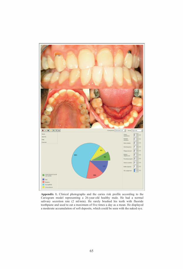
65
Appendix 1. Clinical photographs and the caries risk profile according to the Cariogram model representing a 26-year-old healthy male. He had a normal salivary secretion rate (2 ml/min). He rarely brushed his teeth with fluoride toothpaste and used to eat a maximum of five times a day as a mean. He displayed a moderate accumulation of soft deposits, which could be seen with the naked eye.
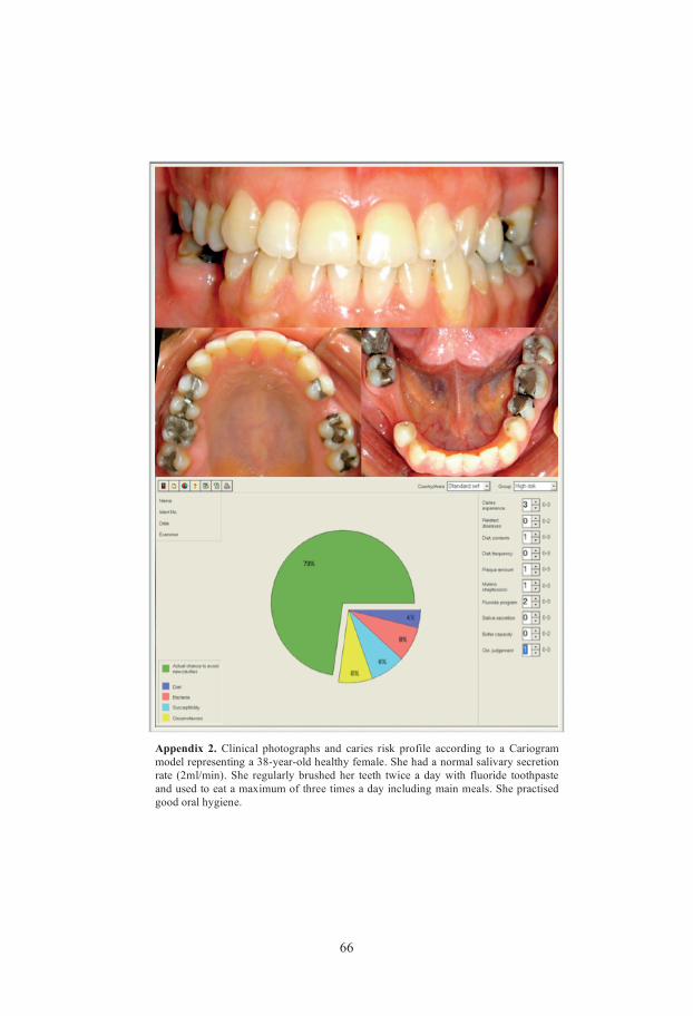
66
Appendix 2. Clinical photographs and caries risk profile according to a Cariogram model representing a 38-year-old healthy female. She had a normal salivary secretion rate (2ml/min). She regularly brushed her teeth twice a day with fluoride toothpaste and used to eat a maximum of three times a day including main meals. She practised good oral hygiene.
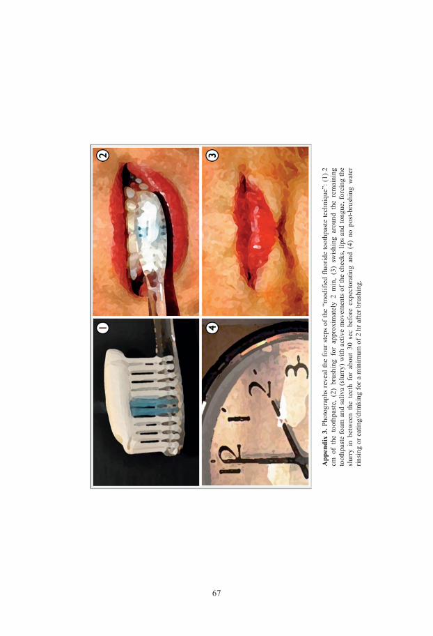
67
App
endi
x 3.
Pho
togr
aphs
rev
eal t
he fo
ur s
teps
of t
he “
mod
ified
fluo
ride
toot
hpas
te te
chni
que”
: (1)
2
cm o
f th
e to
othp
aste
, (2
) br
ushi
ng f
or a
ppro
xim
atel
y 2
min
, (3
) sw
ishi
ng a
roun
d th
e re
mai
ning
to
othp
aste
foam
and
sal
iva
(slu
rry)
with
act
ive
mov
emen
ts o
f the
che
eks,
lips
and
tong
ue, f
orci
ng th
e sl
urry
in
betw
een
the
teet
h fo
r ab
out
30 s
ec b
efor
e ex
pect
orat
ing
and
(4)
no p
ost-b
rush
ing
wat
er
rinsi
ng o
r eat
ing/
drin
king
for a
min
imum
of 2
hr a
fter b
rush
ing.

68
Appendix 4. Clinical photographs of restorations and the corresponding bitewing radiographs in three examples. The bitewing radiographs reveal approximal overhangs and gingival “radiolucency” that could not be detected clinically.
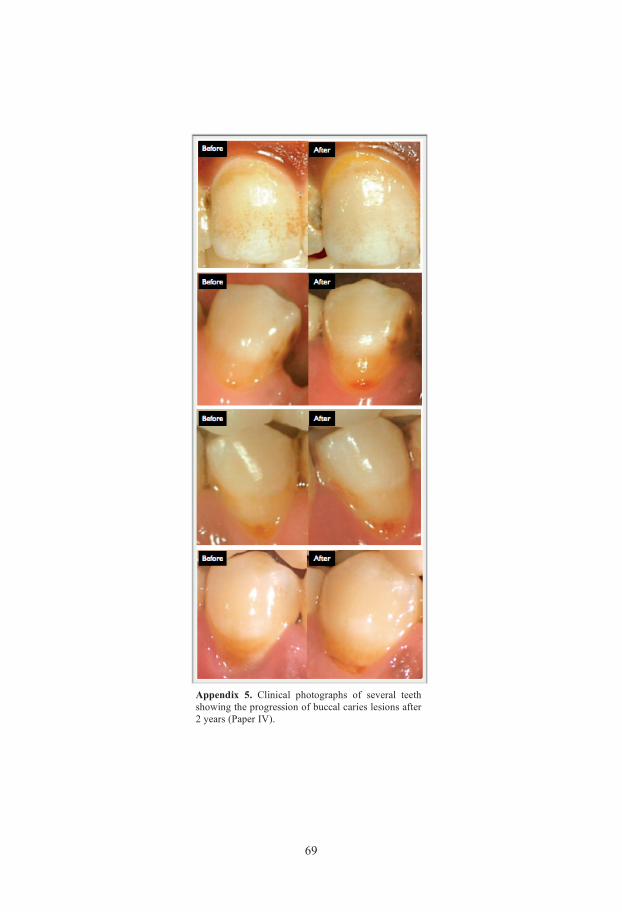
69
Appendix 5. Clinical photographs of several teeth showing the progression of buccal caries lesions after 2 years (Paper IV).
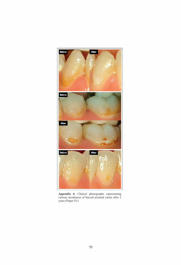
70
Appendix 6. Clinical photographs representing various incidences of buccal arrested caries after 2 years (Paper IV).
Related Documents