Are YOU the gap in the workforce? The Royal College of Pathologists Pathology: the science behind the cure Number 165 January 2014 The Bulletin of The Royal College of Pathologists In this issue The importance of the College workforce census Celebrating success, sharing good practice Research Medal winning essay

Welcome message from author
This document is posted to help you gain knowledge. Please leave a comment to let me know what you think about it! Share it to your friends and learn new things together.
Transcript
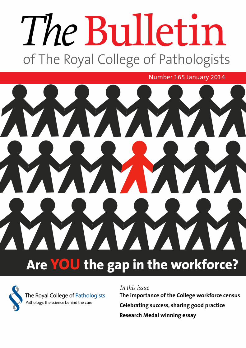
Are YOU the gap in the workforce?
The Royal College of PathologistsPathology: the science behind the cure
Number 165 January 2014
The Bulletinof The Royal College of Pathologists
In this issueThe importance of the College workforce census
Celebrating success, sharing good practice
Research Medal winning essay
www.rcpath.org/bulletin
The Royal College of Pathologists2 Carlton House Terrace, London SW1Y 5AF
telephone 020 7451 6700email [email protected] www.rcpath.org
President Dr Archie PrenticeVice Presidents Dr Bernie Croal Dr Suzy Lishman Professor Mike WellsRegistrar Dr Rachael LiebmannAssistant Registrar Dr Terry JonesTreasurer Dr David CassidyCEO Daniel RossBulletin Editor Dr Laszlo IgaliManaging Editor Edward HulmeAssociate Editor Annabel RiesPrinted by Connekt Colour
Subscribe to the Bulletin of The Royal College of Pathologists
The College’s quarterly membership journal, The Bulletin, is the main means of communications between the College and its members, and between the members themselves. It features topical articles on the latest development in pathology, news from the College, as well as key events and information related to pathology.
The Bulletin is delivered free of charge to all active College Members, retired Members who choose to receive mailings and Registered Trainees, and is published four times a year, in January, April, July and October.
It is also available for our members to download on the College website at www.rcpath.org/bulletin
The subscription rate for libraries and non-members is £100 per annum. To subscribe, contact the Publications Department on 020 7451 6730 or [email protected] Sign up today and keep up to date on what goes on in the world of pathology!
CONTENTSJanuary 2014
Number 165
www.rcpath.org Number165 January2014 1
The Royal College of PathologistsPathology: the science behind the cure
29 Training 29 Trainees’notes 32 NewclinicalscientistFRCPath(HSST)curricula
launched 33 Celebratingsuccess,sharinggoodpractice:The
LondonDeanerySchoolofPathology’sNotablePracticeProject
37 Pathologywebsitesforundergraduateeducation 40 APTqualificationsandModernisingScientific
Careers
42 Research 42 KateHughes’winningResearchMedalessay
43 Clinicaleffectiveness 43 Theclinicaleffectivenessagendaforpathology 45 Venacavafilteraudit 47 DelayinCSFsampledelivery:isthisaproblem?
52 People 52 ProfessorMeenaUpadhyayareceives
internationalgeneticresearchaward 52 ProfessorSirColinBerry 52 Appreciations 55 Deaths 56 Medicalconsultants:newappointmentoffers 58 Examinationsresults:Autumn2013
61 Letters
62 Meetingreview 62 ‘Surgicalsiteinfections’:meetingreview
64 Bookreviews 66 Discountsonbooksandjournals
67 Collegesymposia 67 Forthcomingsymposia 68 Conferenceapplicationformandproformainvoice
69 Noticeboard
71 Legacies
2 Editorial
3 FromthePresident
6 CollegeNews 6 ElectionofCollegePresident 7 HonoraryFellows 8 NewFellows’AdmissionsCeremony–apersonal
view 9 RCPathWalesRoadshow 10 TheEventsandFacilitiesDepartment
11 Workingsmarter 11 TheimportanceoftheCollegeworkforcecensus
toUKconsultants 12 Slide-baseduropathologyEQA–recentprogress
andfuturedirections 13 Demandmanagementinlaboratorymedicine
whilemaintainingquality 15 Zerotoleranceforlabellingofallpathology
specimens:arecommendationfromSHOT2013 17 Adverseincidentsinpathology
19 Publicengagement 19 ‘AHistoryofPathologyin50Objects’lecture
series 20 Apottedhistoryofthepot 21 WestMidlandsDeaneryCareersFair 22 DiseasedetectivesattheHunterian 22 Designermicrobes:designtheultimatedisease-
causingmicroorganism 23 TheGenomeFactory 24 Thejourneyofabloodsample 24 Yourbody,yourconsent 25 ‘DiseaseDetectives’atCelebrateScience2013,
Durham 25 ScienceenrichmentatWilmslow 26 ‘SwipeHeretoDonate’ 27 RecipientsofPublicEngagementInnovation
Grants 28 NationalPathologyWeek2014
Photo credits:The Royal College of Pathologists, JohnGoodman,Warren Potter, Photos.com.
Disclaimers: Authors’ views are personal and are not indicative of College policy, except when College Officers write in their official capacity. Advertisements are paid for by external agencies and do not indicate endorsement or otherwise by the College.
Editorial
2 January2014 Number165 TheBulletinofTheRoyalCollegeofPathologists
looking forward, looking back…
By the time you read this, it will already be 2014. I cannot help feeling that in the back of the minds of many of us there is a little thought that, however good 2013 was, it is better to have left it behind.
Dr Laszlo IgaliBulletin Editor
Every year, it is customary to take stock and try to figure out what a new year may bring and make plans, to make the best of opportunities. We were all hoping that the austerity measures and lack of resources in our professional and private lives are getting closer to the end, but as far I can see, the big storm is far from over.
ResearchI was invited to the launch event of the All-Party Parliamentary Group Report on Pancreatic Cancer. While we were listening to the eminent speakers – all of whom acknowledged the sorry state of pancre-atic cancer diagnosis and treatment, which has not improved significantly in the last 40 years – I was contemplating why research is treating this area rather badly. It may be that the lack of progress in the research of this disease is because it is difficult to diagnose and treat, and thus even less attractive for researchers, resulting in a negative spiral – but I could not avoid thinking about the wider aspects of research. The entire academic area of pathology and research seems to have been on a back-burner for some time now. It is not mentioned too much outside the profession, as basic science is usually not at the forefront of discussion. The occasional stories making big news on the media are usually related to discoveries that may result in better understanding and treatment of cancer, but the long-term and hard work done in basic science labs are not the makings of a good news story. If the dire state of academic re-search is coupled with a disease that is difficult to di-agnose and almost impossible to treat, the inevitable result is that the new generation of researchers will be tempted to look for a career elsewhere. The lack of funding is probably half-attributable to the lack of high-quality research proposals, but the other half (or third) of the truth may be that the mainstream stories of more frequent cancers steal the limelight.
Not all is bleak news, however. We have some-thing exciting in the making. Not just one, but two pieces of news to start the year on a good note.
Digital pathology Following long discussions, many meetings and innumerable hours of work, the College has finally entered the digital pathology age. This news may be less exciting for those whose work does not in-volve glass slides, but for those who do this every
day, it is very important. The digital transition of almost all other branches of pathology has been already done – just think about the automated analysers which are able to measure almost every parameter of blood, and which were integrated to computerised systems a long time ago.
Thanks to the generosity of General Electric, the College has been given an opportunity to develop our digital histology image database. Teaching is one of the main pillars of the College, and with this new technology we are able to make it more acces-sible for all members, including those abroad.
It does, however, comes at a price: we need to fill the technology with meaning – we need to supply good cases to be included in this database. Sending slides for scanning is one aspect, but to make it useful, clinical and pathology information must be included. There are many image databases on the internet, but their usefulness is limited by the little information included. Please send your interesting cases for scanning, but do not forget to include the relevant information!
New scientific journalThere is another piece of very good news: a new scientific journal of the College is just about to launch, crafted by Professor Finbarr Cotter. He set up the new publication, entitled Pathogenesis, with the aim of being able to include many different facets of pathology. Before you ask the question, the costs of the journal are met by the publisher, but this way we will have an additional tool for the College at no cost. I am sure this new avenue of publishing is welcomed by many of us.
One more thing…The 50-year review of the College suggested the re-organisation and refreshment of our regional struc-ture. I hope many of you are considering taking up the new regional roles of Advocacy, Learning and Professionalism Leads. This division of work areas has proved to be successful and the new roles will have central administrative support from the new Regional Coordination Manager in the College. There is no lack of challenges with this new struc-ture, but I feel that being involved is the only way to influence decisions for a better future.
Dr Laszlo IgaliBulletin Editor
www.rcpath.org Number165 January2014 3
Dr Archie PrenticeCollege President
It was difficult to find a speaker for this win-ter’s College dinner so I did it myself. It was very therapeutic, for me if not for the guests. It wasn’t directly about pathology but gave me the chance to reflect on almost 50 years since starting medical school and 40 years since starting specialty train-ing. Entering the third and final year as President in a somewhat detached position, detached from personal practice that is, and with no obvious, professional stake in the future of medicine, I have been reflecting on why we have doctors. This is not only because of the many distracting agendas with which the College has had to engage recently. It’s not that such engagement is all new but there seem to be so many that are so conflicting, including the confusion surrounding training, service, workforce and public and patient engagement.
This reflection has in turn increasingly re-minded me of medical school and the forgotten uncertainty about why I was there. First year was a boring repetition of school science which was a complete waste of time apart from the social life. Second year was intensive anatomy, physiology and biochemistry with no real direct connection to clinical practice. Dismantling a body was a fas-cinating rite of passage but it didn’t tell me more about why medicine had attracted me. The social life was even more interesting and culminated in the mass arrest of almost 200 of us at an end of year party; this is no boast or secret as it made front page news in the Daily Record. The Dean was censorious although amused and I wonder what would happen now in a GMC fitness to practise context. Third year was two disconnected, paral-lel streams of clinical methods (a real and new case-based experience every morning at the bed-side, not in a teaching lab or at a PC) and heavy, didactic pathology (with some extremely boring pharmacology lectures read directly from notes). The disconnection between these two streams didn’t help my understanding either.
This came in the integrated fourth year when all of the pathological, diagnostic and therapeutic aspects of medicine and surgery were taught sys-tem by system while the live case-based exposure continued on the wards and in clinics. This was not called case-based (CBL) or problem-based leaning (PBL), but that’s what it was, and we didn’t need purist educationalists to tell us that this was a great way to learn. Perhaps the integra-tion worked because of the immediately preced-ing didactic ground work. From then on though,
through more hands-on, real case-based medicine, surgery, paediatrics and obstetrics and gynaecol-ogy, the “learning experience” made it much easier to grasp what being a doctor was all about, including the encouragement to do junior house officer locums in 5th and 6th year. Will the GMC bring these back?
The core of all this learning was quite simply seeking, explaining and applying both diagnosis and treatment for patients, students and colleagues. This continued into pre-registration house jobs, core medical training (CMT) and then specialty training. The shape of this was curiously similar to that proposed by Sir David Greenaway to the Health Secretaries of all four administrations in the UK (www.shapeoftraining.co.uk) last month, in-cluding CMT as if it was new. The sturm und drang around this report is at times as theatrical as the or-igin of the phrase (Klinger 1776), particularly at the postgraduate school level. There, in some schools, the proposed creation of the College’s learning leads mapped on to Local Education and Training Boards is bizarrely seen as competitive rather than supportive, but I’ll return to that. The point about “Greenaway” is that it should focus our efforts on what is needed for progressive understanding and application of the core of medical practice, diagno-sis and treatment, and not distract us into spurious arguments about the balance between generalism and specialism.
So what are these distracting agendas? It is difficult to understand and explain the origins, evidence base and intentions of many of them and this is a personal view, so please be patient with this and please let me know your own views.
The first distraction is the undergraduate cur-riculum which one might assume is intended to produce doctors who are uniformly competent at diagnosis and treatment. If not then the “Shape of Training” makes no sense when “specialised generalism” is to be promoted. There are no two curricula which are the same across the UK’s 32 medical schools. The GMC accepts the validity of all of them. Yet there is abundant evidence from the NHS Atlas of Variation in Diagnostic Services (www.rightcare.nhs.uk) that the use of diagnos-tic tests is inexplicably variable to a degree that beggars belief and there is widespread concern about the dangers of poor prescribing by junior doctors (BMJ Qual Saf 2013;22:97-102 doi:10.1136/bmjqs-2012-001175). This is not a question of whether CBL/PBL or didactic teaching is better
reflections and distractions
FroM tHE PrESidENt
FROM THE PRESIDENT
4 January2014 Number165 TheBulletinofTheRoyalCollegeofPathologists
when neither is effective without the other. The question is how to strike the balance to produce reliably doctors who know what they are doing. My personal challenge for the GMC, the Medical Schools Council, the Department of Health and Health Education England and its equivalents in other administrations is simple. If 32 different curricula are acceptable to the regulator and the government, then what could possibly be the objections to prospective, randomised, controlled trials of teaching and testing methods in an era of evidence-based medicine to illuminate a rela-tively evidence-light issue?
There seems to be little appetite for this chal-lenge as the next distraction implies. “Greena-way” recommends licensing on graduation after a five year course. I was not ready to be set loose on an unsuspecting public after six years (were you?) and probably a good deal “safer” once I had completed CMT. This proposal looks like a cyni-cal dodging of the problem of producing too many graduates for the training opportunities available in the UK, especially if those we keep are simply for the delivery of government’s targets by gen-eralists. The excess is presumably for export. Ex-ported doctors can’t work unless licensed before they leave. Medical schools cannot or don’t want to re-absorb a sixth year into undergraduate stud-ies. The positive side to this proposal might be that it increases the pressure to have a uniformly reliable output from medical schools and the need for a second licensing step for those who remain in the UK. Let’s hope the Health Secretaries will rec-ognise this and propose how to deal with it even if medical schools don’t or can’t. If this licensed output is to be pushed towards generalist training in the UK, perhaps it might be worth examining the evidence that more generalists provide better care. That’s not apparent in the Atlas of Variation referred to above. These new specialised general-ists will certainly need to be of a very high quality which implies tougher selection and longer and more intensive training and this cannot be done at the expense of personalised, precision, molecular medicine. We need a training model to persuade Local Education and Training Boards (LETBs) how we can preserve and develop with Health Educa-tion England (HEE) a production line for all the professions and disciplines needed for accurate diagnosis. HEE agree but it will take time to per-suade LETBs and I’m not sure that Postgraduate Medical Deans and Heads of Schools agree that, or fully understand how, the College could contrib-ute more to this process.
The third initiative distracting me from core values is the meaning of the phrase “patient-cen-tred care”. I think, perhaps naively, that’s always what I’ve done and I have to suppress a flush of an-ger when I hear professional colleagues condemn us for our “patronising” and “patrician” approach
to patients. You might be surprised at the sort of stuff one has to address in this post and my self-control in responding. The pressure from patients’ advocacy groups is sometimes very interesting. Individual control of care, direct access to a cho-sen place or form of treatment and direct access to tests and results all sound superficially very laud-able. They may benefit the well-informed, middle classes or those with chronic conditions, but what about the uninformed, confused, afraid or lacking in capacity. Are they to be disadvantaged further by the smarter and more wealthy? Isn’t that what happens in other countries whose care systems we do not admire? We should consider carefully that many lay people were misled by dubious evi-dence and remain unfortunately convinced that measles vaccine causes autism, etc. and this may have helped to delay eradication of that endemic and occasionally fatally epidemic viral infection. I recall the words of my final year teacher in Medi-cine, a strict Calvinist. “Prentice, remember that patients are struck with fear at your words and can remember only three things. Once you have said ‘Good morning, my name is Dr Prentice’ you have only one left”. His advice seemed to me then to be deeply unreasonable but, having struggled and failed in my view over 40 years to understand fully and to satisfactorily apply “informed consent”, I understand better that his harsh message to all his students contained a truth. In reaching a diagnosis and deciding on best treatment nothing is better than an open partnership in which the doctor and the patient look together carefully at evidence. If that partnership is lost, who is responsible for the success or failure of decisions? This partnership needs to be very carefully managed however and it doesn’t work with written, impersonal transac-tions or in one encounter. That is equally true of the relationship between the pathologist and the requesting clinician. This problem is no clearer than when complete, early and non-negotiated transparency about a new, major health problem leads to complete collapse of morale, not just in an individual but in a whole family.
The fourth distraction of the initiative called “Integrated Care” may not interest many pa-thologists but it should. Billions are coming out of health spending to go to social care. That means inevitably less clinical care and therefore a reduced chance of effective integration. This is a clear abrogation of the politicians’ responsibility to fund social care adequately. Doctors and nurses should not be used as surrogate social workers even if Pathology could and should reach out fur-ther and more effectively into community medi-cine and general practice as the Atlas of Variation suggests. Simply moving money from one budget to another will not solve the problem of keeping people out of hospital. The Health and Social Care Act was a deception in the sense that the title was
FROM THE PRESIDENT
www.rcpath.org Number165 January2014 5
a beard to dodge this issue. It has failed signally so far to mend the disruption in diagnosis and treat-ment across the artificial finance-management boundaries of the NHS. This requires collabora-tion between commissioners (at all levels) and the College to produce a service model which covers whole populations. In other words stopping the clock and going back to immediately post-Carter, if and where one can. Reconfiguration should have been and could still be based on the quality and value of a service for a given population and not its cost. NHS England is interested as are some Clinical Commissioning Groups.
There are many other distractions such as revalidation, quality assessment, accredita-tion, leadership and management, integrity and whistleblowing, commercial competition, 24/7 or seven day working and causes of death. These will have to wait for another edition and many relate to what I’ve covered above. This column is already long enough but the point of it is that the College cannot afford to lose sight of the core activities of medicine, diagnosis and treatment, whilst it deals with all of these associated distrac-tions. It must try to retain influence centrally and locally wherever decisions are being made that affect the ability of all doctors to perform these key tasks effectively. That is why the College has had to reorganise local representation and ap-pointed last week its new Regional Coordination Manager. Applications for the posts of the local representatives of Learning, Professionalism and Advocacy have been slow. This is not surprising for such a radical change and it’s clear that it’s get-ting harder to find the time for such College work. I hope that will not deter fellows from applying
for these critically important posts.I cannot emphasise enough that we are in-
creasing central support for these activities to a level that the College has never before provided. All the other changes to the governance and man-agement of the College are now approved by the Privy Council and in place. If we cannot keep in touch with what is happening locally to the ca-pacity to train for and deliver accurate diagnosis on which most therapy is based, then pathology and this College have a bleak future, the latter as a pointless livery company whose officers enjoy endless “jollies” at members’ expense. Anyone who thinks, as some clearly do, that is what hap-pens now at 2 Carlton House Terrace and else-where is very welcome to a personal tutorial. That is not what the College is about and never should be. Once again the message is that the fellows are the College and your engagement determines its future. This College has always enjoyed success through the great benefits of that engagement by members, for example in its 800 examiners to mention only one of many aspects of the College’s work. I am constantly surprised by the respect in which it is held in the UK and abroad. That reputation and the successful future work of the College depend on its members’ engagement. The new structure makes the potential benefits of our work even greater and the likely influence of the College in sustaining diagnosis and treatment even more effective.
Dr Archie PrenticePresident
Speak up: give your view on College consultations
All members should get involved in College’s consultations on the guidance and documents that are relevant to their specialty.
Your opinion is vital in helping us ensure that all the documents we produce are reli-able and workable in practice – and what you say, counts. You can also claim up to 2 CPD credits for this work. All College documents are put for consultation on the website, you just need to login and visit www.rcpath.org/fellows
Here you will find all the documents open for consultation and information on the status of documents in the process of being revised before final publication. When a new document is posted, we send out an email to the relevant members advising them of the open consultation. If you’ve forgotten your login details for the website, please contact [email protected]
6 January2014 Number165 TheBulletinofTheRoyalCollegeofPathologists
CollEgE NEwS
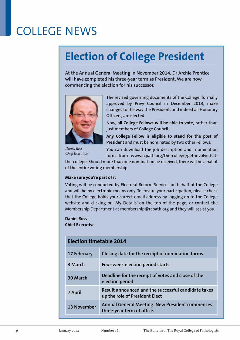
Election of College PresidentAt the Annual General Meeting in November 2014, Dr Archie Prentice will have completed his three-year term as President. We are now commencing the election for his successor.
The revised governing documents of the College, formally approved by Privy Council in December 2013, make changes to the way the President, and indeed all Honorary Officers, are elected.
Now, all College Fellows will be able to vote, rather than just members of College Council.
Any College Fellow is eligible to stand for the post of President and must be nominated by two other Fellows.
You can download the job description and nomination form from www.rcpath.org/the-college/get-involved-at-
the-college. Should more than one nomination be received, there will be a ballot of the entire voting membership.
Make sure you’re part of it
Voting will be conducted by Electoral Reform Services on behalf of the College and will be by electronic means only. To ensure your participation, please check that the College holds your correct email address by logging on to the College website and clicking on ‘My Details’ on the top of the page, or contact the Membership Department at [email protected] and they will assist you.
Daniel RossChief Executive
Daniel RossChief Executive
Election timetable 2014
17February Closingdateforthereceiptofnominationforms
3March Four-weekelectionperiodstarts
30March Deadlineforthereceiptofvotesandcloseoftheelectionperiod
7April ResultannouncedandthesuccessfulcandidatetakesuptheroleofPresidentElect
13November AnnualGeneralMeeting.NewPresidentcommencesthree-yeartermofoffice.
COLLEGE NEWS
www.rcpath.org Number165 January2014 7
Honorary Fellows
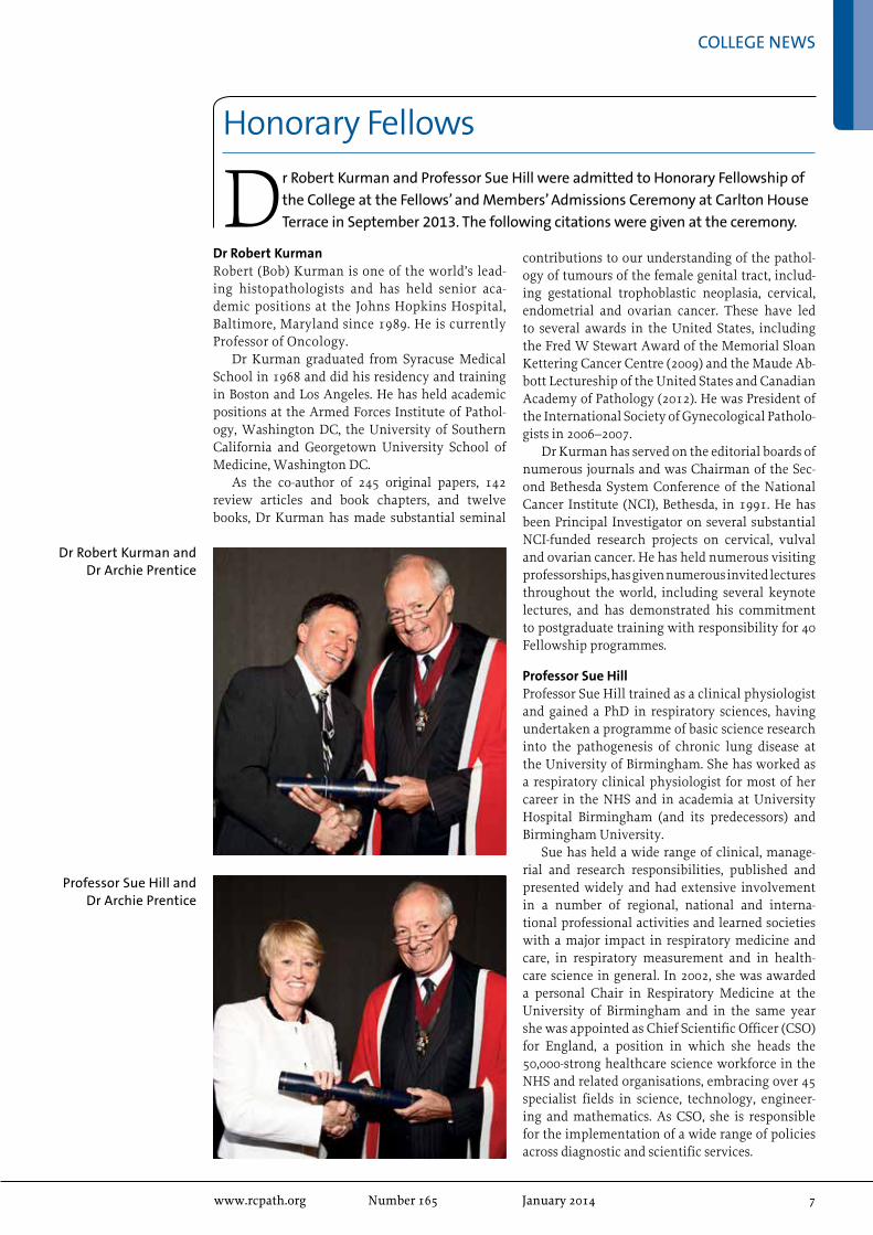
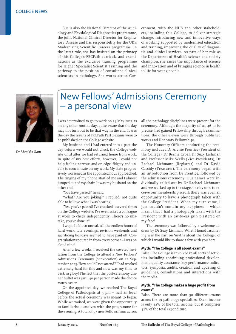
Dr Robert Kurman and Professor Sue Hill were admitted to Honorary Fellowship of the College at the Fellows’ and Members’ Admissions Ceremony at Carlton House Terrace in September 2013. The following citations were given at the ceremony.
Dr Robert KurmanRobert (Bob) Kurman is one of the world’s lead-ing histopathologists and has held senior aca-demic positions at the Johns Hopkins Hospital, Baltimore, Maryland since 1989. He is currently Professor of Oncology.
Dr Kurman graduated from Syracuse Medical School in 1968 and did his residency and training in Boston and Los Angeles. He has held academic positions at the Armed Forces Institute of Pathol-ogy, Washington DC, the University of Southern California and Georgetown University School of Medicine, Washington DC.
As the co-author of 245 original papers, 142 review articles and book chapters, and twelve books, Dr Kurman has made substantial seminal
contributions to our understanding of the pathol-ogy of tumours of the female genital tract, includ-ing gestational trophoblastic neoplasia, cervical, endometrial and ovarian cancer. These have led to several awards in the United States, including the Fred W Stewart Award of the Memorial Sloan Kettering Cancer Centre (2009) and the Maude Ab-bott Lectureship of the United States and Canadian Academy of Pathology (2012). He was President of the International Society of Gynecological Patholo-gists in 2006–2007.
Dr Kurman has served on the editorial boards of numerous journals and was Chairman of the Sec-ond Bethesda System Conference of the National Cancer Institute (NCI), Bethesda, in 1991. He has been Principal Investigator on several substantial NCI-funded research projects on cervical, vulval and ovarian cancer. He has held numerous visiting professorships, has given numerous invited lectures throughout the world, including several keynote lectures, and has demonstrated his commitment to postgraduate training with responsibility for 40 Fellowship programmes.
Professor Sue HillProfessor Sue Hill trained as a clinical physiologist and gained a PhD in respiratory sciences, having undertaken a programme of basic science research into the pathogenesis of chronic lung disease at the University of Birmingham. She has worked as a respiratory clinical physiologist for most of her career in the NHS and in academia at University Hospital Birmingham (and its predecessors) and Birmingham University.
Sue has held a wide range of clinical, manage-rial and research responsibilities, published and presented widely and had extensive involvement in a number of regional, national and interna-tional professional activities and learned societies with a major impact in respiratory medicine and care, in respiratory measurement and in health-care science in general. In 2002, she was awarded a personal Chair in Respiratory Medicine at the University of Birmingham and in the same year she was appointed as Chief Scientific Officer (CSO) for England, a position in which she heads the 50,000-strong healthcare science workforce in the NHS and related organisations, embracing over 45 specialist fields in science, technology, engineer-ing and mathematics. As CSO, she is responsible for the implementation of a wide range of policies across diagnostic and scientific services.
Dr Robert Kurman and Dr Archie Prentice
Professor Sue Hill and Dr Archie Prentice
COLLEGE NEWS
8 January2014 Number165 TheBulletinofTheRoyalCollegeofPathologists
New Fellows’ admissions Ceremony – a personal view
I was determined to go to work on 24 May 2013 as on any other routine day, quite aware that the day may not turn out to be that way in the end. It was the day the results of FRCPath Part 2 exams were to be published on the College website.
My husband and I had entered into a pact the day before: we would not check the College web-site until after we had returned home from work. In spite of my best efforts, however, I could not help feeling nervous and on edge, fidgety and un-able to concentrate on my work. My state progres-sively worsened as the appointed hour approached. The ringing of my phone startled me and I almost jumped out of my chair! It was my husband on the other end.
“You have passed!” he said.“What? Are you joking?” I replied, not quite
able to believe what I was hearing! “Yes, you’ve passed! I’ve checked it several times
on the College website. I’ve even asked a colleague at work to check independently. There’s no mis-take, you’ve done it!”
I wept. It felt so unreal. All the endless hours of hard work, late evenings, revision weekends and sacrificing holidays seemed to have paid off! Con-gratulations poured in from every corner – I was on cloud nine!
After a few weeks, I received the coveted invi-tation from the College to attend a New Fellows’ Admissions Ceremony (convocation) on 12 Sep-tember 2013. How could I not attend? I had worked extremely hard for this and now was my time to bask in glory! The fact that the post-ceremony din-ner buffet was just £40 per person made the choice much easier!
On the appointed day, we reached The Royal College of Pathologists at 5 pm – half an hour before the actual ceremony was meant to begin. While we waited, we were given the opportunity to familiarise ourselves with the programme for the evening. A total of 57 new Fellows from across
all the pathology disciplines were present for the ceremony. Although the majority of us, 46 to be precise, had gained Fellowship through examina-tions, the other eleven were through published works and Honorary Fellowships.
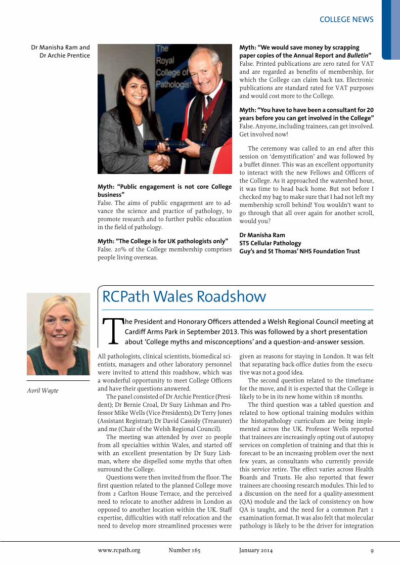
The Honorary Officers conducting the cere-mony included Dr Archie Prentice (President of the College), Dr Bernie Croal, Dr Suzy Lishman and Professor Mike Wells (Vice-Presidents), Dr Rachael Liebmann (Registrar) and Dr David Cassidy (Treasurer). The ceremony began with an introduction from Dr Prentice, followed by the admissions ceremony. Our names were in-dividually called out by Dr Rachael Liebmann and we walked up to the stage, one by one, to re-ceive our membership scroll; there was even an opportunity to have a photograph taken with the College President. When my turn came, I just couldn’t contain my happiness – which meant that I had a photograph taken with the President with an ear-to-ear grin plastered on my face!
The ceremony was followed by a welcome ad-dress by Dr Suzy Lishman. What I found fascinat-ing was the part on ‘myths about the College’, of which I would like to share a few with you here.
Myth: “The College is all about exams”False. The College is involved in all sorts of activi-ties including continuing professional develop-ment, quality assurance, key performance indica-tors, symposia, audits, creation and updating of guidelines, consultations and interactions with the media.
Myth: “The College makes a huge profit from exams”False. There are more than 50 different exams across the 19 pathology specialties. Exam income is only 22% of the total income, but it comprises 32% of the total expenditure.
Dr Manisha Ram
Sue is also the National Director of the Audi-ology and Physiological Diagnostics programme, the joint National Clinical Director for Respira-tory Disease and has responsibility for the UK’s Modernising Scientific Careers programme. In the latter role, she has insisted on the primacy of this College’s FRCPath curricula and exami-nations as the exclusive training programme for Higher Specialist Scientist Training and the pathway to the position of consultant clinical scientists in pathology. She works across Gov-
ernment, with the NHS and other stakehold-ers, including this College, to deliver strategic change, introducing new and innovative ways of working supported by modernised education and training, improving the quality of diagnos-tic and clinical services. As part of her role as the Department of Health’s science and society champion, she raises the importance of science and innovation and of bringing science in health to life for young people.
COLLEGE NEWS
www.rcpath.org Number165 January2014 9
Myth: “Public engagement is not core College business”False. The aims of public engagement are to ad-vance the science and practice of pathology, to promote research and to further public education in the field of pathology.
Myth: “The College is for UK pathologists only”False. 20% of the College membership comprises people living overseas.
Myth: “We would save money by scrapping paper copies of the Annual Report and Bulletin”False. Printed publications are zero rated for VAT and are regarded as benefits of membership, for which the College can claim back tax. Electronic publications are standard rated for VAT purposes and would cost more to the College.
Myth: “You have to have been a consultant for 20 years before you can get involved in the College” False. Anyone, including trainees, can get involved. Get involved now!
The ceremony was called to an end after this session on ‘demystification’ and was followed by a buffet dinner. This was an excellent opportunity to interact with the new Fellows and Officers of the College. As it approached the watershed hour, it was time to head back home. But not before I checked my bag to make sure that I had not left my membership scroll behind! You wouldn’t want to go through that all over again for another scroll, would you?
Dr Manisha RamST5 Cellular PathologyGuy’s and St Thomas’ NHS Foundation Trust
rCPath wales roadshow
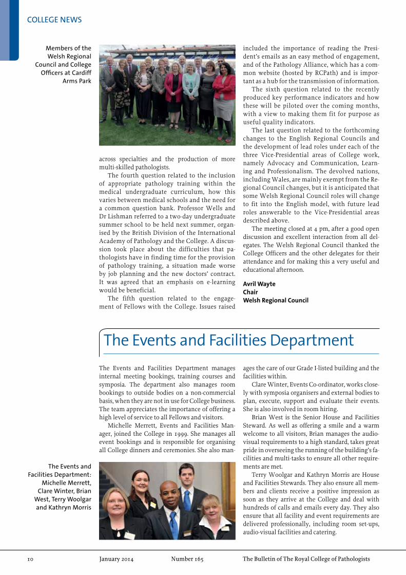
The President and Honorary Officers attended a Welsh Regional Council meeting at Cardiff Arms Park in September 2013. This was followed by a short presentation about ‘College myths and misconceptions’ and a question-and-answer session.
Avril Wayte
All pathologists, clinical scientists, biomedical sci-entists, managers and other laboratory personnel were invited to attend this roadshow, which was a wonderful opportunity to meet College Officers and have their questions answered.
The panel consisted of Dr Archie Prentice (Presi-dent); Dr Bernie Croal, Dr Suzy Lishman and Pro-fessor Mike Wells (Vice-Presidents); Dr Terry Jones (Assistant Registrar); Dr David Cassidy (Treasurer) and me (Chair of the Welsh Regional Council).
The meeting was attended by over 20 people from all specialties within Wales, and started off with an excellent presentation by Dr Suzy Lish-man, where she dispelled some myths that often surround the College.
Questions were then invited from the floor. The first question related to the planned College move from 2 Carlton House Terrace, and the perceived need to relocate to another address in London as opposed to another location within the UK. Staff expertise, difficulties with staff relocation and the need to develop more streamlined processes were
given as reasons for staying in London. It was felt that separating back-office duties from the execu-tive was not a good idea.
The second question related to the timeframe for the move, and it is expected that the College is likely to be in its new home within 18 months.
The third question was a tabled question and related to how optional training modules within the histopathology curriculum are being imple-mented across the UK. Professor Wells reported that trainees are increasingly opting out of autopsy services on completion of training and that this is forecast to be an increasing problem over the next few years, as consultants who currently provide this service retire. The effect varies across Health Boards and Trusts. He also reported that fewer trainees are choosing research modules. This led to a discussion on the need for a quality-assessment (QA) module and the lack of consistency on how QA is taught, and the need for a common Part 1 examination format. It was also felt that molecular pathology is likely to be the driver for integration
Dr Manisha Ram and Dr Archie Prentice
COLLEGE NEWS
10 January2014 Number165 TheBulletinofTheRoyalCollegeofPathologists
across specialties and the production of more multi-skilled pathologists.
The fourth question related to the inclusion of appropriate pathology training within the medical undergraduate curriculum, how this varies between medical schools and the need for a common question bank. Professor Wells and Dr Lishman referred to a two-day undergraduate summer school to be held next summer, organ-ised by the British Division of the International Academy of Pathology and the College. A discus-sion took place about the difficulties that pa-thologists have in finding time for the provision of pathology training, a situation made worse by job planning and the new doctors’ contract. It was agreed that an emphasis on e-learning would be beneficial.
The fifth question related to the engage-ment of Fellows with the College. Issues raised
included the importance of reading the Presi-dent’s emails as an easy method of engagement, and of the Pathology Alliance, which has a com-mon website (hosted by RCPath) and is impor-tant as a hub for the transmission of information.
The sixth question related to the recently produced key performance indicators and how these will be piloted over the coming months, with a view to making them fit for purpose as useful quality indicators.
The last question related to the forthcoming changes to the English Regional Councils and the development of lead roles under each of the three Vice-Presidential areas of College work, namely Advocacy and Communication, Learn-ing and Professionalism. The devolved nations, including Wales, are mainly exempt from the Re-gional Council changes, but it is anticipated that some Welsh Regional Council roles will change to fit into the English model, with future lead roles answerable to the Vice-Presidential areas described above.
The meeting closed at 4 pm, after a good open discussion and excellent interaction from all del-egates. The Welsh Regional Council thanked the College Officers and the other delegates for their attendance and for making this a very useful and educational afternoon.
Avril WayteChairWelsh Regional Council
the Events and Facilities department
Members of the Welsh Regional
Council and College Officers at Cardiff
Arms Park
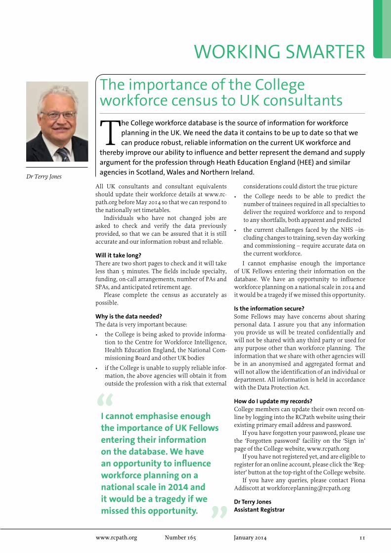
The Events and Facilities Department:
Michelle Merrett, Clare Winter, Brian
West, Terry Woolgar and Kathryn Morris
The Events and Facilities Department manages internal meeting bookings, training courses and symposia. The department also manages room bookings to outside bodies on a non-commercial basis, when they are not in use for College business. The team appreciates the importance of offering a high level of service to all Fellows and visitors.
Michelle Merrett, Events and Facilities Man-ager, joined the College in 1999. She manages all event bookings and is responsible for organising all College dinners and ceremonies. She also man-
ages the care of our Grade I-listed building and the facilities within.
Clare Winter, Events Co-ordinator, works close-ly with symposia organisers and external bodies to plan, execute, support and evaluate their events. She is also involved in room hiring.
Brian West is the Senior House and Facilities Steward. As well as offering a smile and a warm welcome to all visitors, Brian manages the audio-visual requirements to a high standard, takes great pride in overseeing the running of the building’s fa-cilities and multi-tasks to ensure all other require-ments are met.
Terry Woolgar and Kathryn Morris are House and Facilities Stewards. They also ensure all mem-bers and clients receive a positive impression as soon as they arrive at the College and deal with hundreds of calls and emails every day. They also ensure that all facility and event requirements are delivered professionally, including room set-ups, audio-visual facilities and catering.
www.rcpath.org Number165 January2014 11
workiNg SMartEr
Dr Terry Jones
“I cannot emphasise enough the importance of UK Fellows entering their information on the database. We have an opportunity to influence workforce planning on a national scale in 2014 and it would be a tragedy if we missed this opportunity. ”
the importance of the College workforce census to Uk consultants
The College workforce database is the source of information for workforce planning in the UK. We need the data it contains to be up to date so that we can produce robust, reliable information on the current UK workforce and
thereby improve our ability to influence and better represent the demand and supply argument for the profession through Heath Education England (HEE) and similar agencies in Scotland, Wales and Northern Ireland.
All UK consultants and consultant equivalents should update their workforce details at www.rc-path.org before May 2014 so that we can respond to the nationally set timetables.
Individuals who have not changed jobs are asked to check and verify the data previously provided, so that we can be assured that it is still accurate and our information robust and reliable.
Will it take long?There are two short pages to check and it will take less than 5 minutes. The fields include specialty, funding, on-call arrangements, number of PAs and SPAs, and anticipated retirement age.
Please complete the census as accurately as possible.
Why is the data needed?The data is very important because:
• the College is being asked to provide informa-tion to the Centre for Workforce Intelligence, Health Education England, the National Com-missioning Board and other UK bodies
• if the College is unable to supply reliable infor-mation, the above agencies will obtain it from outside the profession with a risk that external
considerations could distort the true picture
• the College needs to be able to predict the number of trainees required in all specialties to deliver the required workforce and to respond to any shortfalls, both apparent and predicted
• the current challenges faced by the NHS –in-cluding changes to training, seven-day working and commissioning – require accurate data on the current workforce.
I cannot emphasise enough the importance of UK Fellows entering their information on the database. We have an opportunity to influence workforce planning on a national scale in 2014 and it would be a tragedy if we missed this opportunity.
Is the information secure?Some Fellows may have concerns about sharing personal data. I assure you that any information you provide us will be treated confidentially and will not be shared with any third party or used for any purpose other than workforce planning. The information that we share with other agencies will be in an anonymised and aggregated format and will not allow the identification of an individual or department. All information is held in accordance with the Data Protection Act.
How do I update my records?College members can update their own record on-line by logging into the RCPath website using their existing primary email address and password.
If you have forgotten your password, please use the ‘Forgotten password’ facility on the ‘Sign in’ page of the College website, www.rcpath.org
If you have not registered yet, and are eligible to register for an online account, please click the ‘Reg-ister’ button at the top-right of the College website.
If you have any queries, please contact Fiona Addiscott at [email protected]
Dr Terry JonesAssistant Registrar
WORKING SMARTER
12 January2014 Number165 TheBulletinofTheRoyalCollegeofPathologists
Slide-based uropathology EQa – recent progress and future directions
Many interpretative EQA schemes in histopathology have recently experienced significant increases in participant numbers, coinciding with EQA becoming more formally integrated into the process of appraisal and
revalidation. The National Slide-Based Uropathology EQA Scheme is no exception, which has grown from only 100 members in 2003 to over 350 today.
Such expansion brings huge challenges to the organisation of these schemes, which have tra-ditionally been run by consultants in their spare time. The OMNIS computer software developed in the early 1990s by Professor Peter Furness has been, and still is, used by many schemes, but was designed in an era before widespread use of email and the web. It requires manual input of responses into the database by the scheme organiser, making it very laborious. In 2011, after approval at the par-ticipants’ meeting, we embarked on the develop-ment of a new web-based system in collaboration with a software company (KPMD IT Solutions Ltd – http://www.kpmd.co.uk), which designed the software for the laboratory NEQAS scheme.
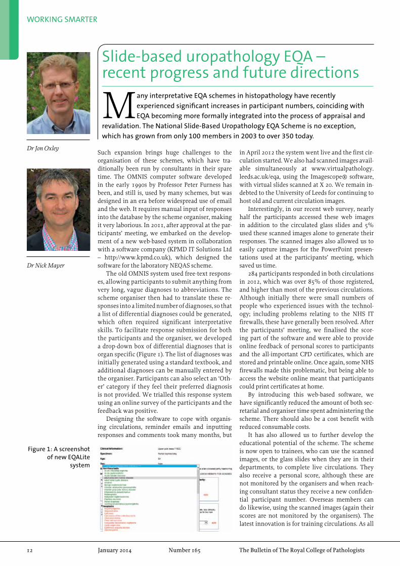
The old OMNIS system used free-text respons-es, allowing participants to submit anything from very long, vague diagnoses to abbreviations. The scheme organiser then had to translate these re-sponses into a limited number of diagnoses, so that a list of differential diagnoses could be generated, which often required significant interpretative skills. To facilitate response submission for both the participants and the organiser, we developed a drop-down box of differential diagnoses that is organ specific (Figure 1). The list of diagnoses was initially generated using a standard textbook, and additional diagnoses can be manually entered by the organiser. Participants can also select an ‘Oth-er’ category if they feel their preferred diagnosis is not provided. We trialled this response system using an online survey of the participants and the feedback was positive.
Designing the software to cope with organis-ing circulations, reminder emails and inputting responses and comments took many months, but
in April 2012 the system went live and the first cir-culation started. We also had scanned images avail-able simultaneously at www.virtualpathology.leeds.ac.uk/eqa, using the Imagescope® software, with virtual slides scanned at X 20. We remain in-debted to the University of Leeds for continuing to host old and current circulation images.
Interestingly, in our recent web survey, nearly half the participants accessed these web images in addition to the circulated glass slides and 5% used these scanned images alone to generate their responses. The scanned images also allowed us to easily capture images for the PowerPoint presen-tations used at the participants’ meeting, which saved us time.
284 participants responded in both circulations in 2012, which was over 85% of those registered, and higher than most of the previous circulations. Although initially there were small numbers of people who experienced issues with the technol-ogy; including problems relating to the NHS IT firewalls, these have generally been resolved. After the participants’ meeting, we finalised the scor-ing part of the software and were able to provide online feedback of personal scores to participants and the all-important CPD certificates, which are stored and printable online. Once again, some NHS firewalls made this problematic, but being able to access the website online meant that participants could print certificates at home.
By introducing this web-based software, we have significantly reduced the amount of both sec-retarial and organiser time spent administering the scheme. There should also be a cost benefit with reduced consumable costs.
It has also allowed us to further develop the educational potential of the scheme. The scheme is now open to trainees, who can use the scanned images, or the glass slides when they are in their departments, to complete live circulations. They also receive a personal score, although these are not monitored by the organisers and when reach-ing consultant status they receive a new confiden-tial participant number. Overseas members can do likewise, using the scanned images (again their scores are not monitored by the organisers). The latest innovation is for training circulations. As all
Dr Jon Oxley
Dr Nick Mayer
Figure 1: A screenshot of new EQALite
system
WORKING SMARTER
www.rcpath.org Number165 January2014 13
the previous meeting reports and scanned images are available online, participants can now com-plete old circulations, which are instantly scored, and a report can be printed.
If more schemes adopt this software in the future, an additional benefit is that a participant only needs a single login to access all their registered schemes.
It is important to emphasise that we as indi-viduals, and our scheme, have no financial interest in the company that developed the software and maintain the website.
We hope that this new web-based approach will facilitate greater participation in EQA
schemes for participants and enhance their edu-cational experience. In addition, it should reduce the administrative burden for scheme organisers and secretaries. We encourage other schemes to consider this system and are happy to share our experience with them.
Dr Jon OxleyDr Nick MayerScheme [email protected]
demand management in laboratory medicine while maintaining quality
The importance of pathology tests in patient management goes without saying, but a significant proportion of pathology tests requested in practice are not necessary, or are duplicated. Sometimes, a guideline or an algorithm steering a
testing scheme has been replaced with a new one, but the old scheme still remains in operation, creating unnecessary requesting.
It has widely been held that results from unnec-essary testing have no benefit to patient care, can create confusion or even be detrimental to pa-tient care. Significant sums of taxpayers’ money could be saved by scrutinising these practices. This is important as the year-on-year rises in specimen requests plus new, usually expensive, tests added to existing batteries of tests strain laboratory medicine budgets.
During the recent significant financial chal-lenge, hospital departments in The Newcastle Upon Tyne Hospitals NHS Foundation Trust, as elsewhere, were required to deliver financial sav-ings, while maintaining the quality of service.
With modern systems of electronic ordering and advanced laboratory database facilities, most laboratories can now implement demand man-agement strategies, which in real time can reduce waste and duplication. More importantly, increas-ing financial demands, coupled with new IT tech-nology has offered an opportunity to rethink the overall direction of pathology services and to take stock of the testing strategies.
In our Microbiology Department we took up the challenge of increasing demands on a labora-tory service in the current financial climate, hop-ing that by curtailing unnecessary and duplicate tests, the tools of demand management, we could deliver our targets. Considering the success of our campaign, we wish to share our experience with others.
Methods and results1. Extractionofdata We extracted data of requests for laboratory
tests from electronic ordering and the laborato-ry data management system (e-record and iLab, the systems used in this Trust) for different categories of requesting during the previous six months. In our search, we focused on the major service users (we cannot name any clini-cal directorate for the reason of sensitivity) and mainly on costly tests such as PCRs and other sent-away tests.
The data was tabulated for activity over time to identify peaks. For a selection of patients, data was sorted to examine for duplicity of requests and adherence of clinical teams with relevant protocols.
The data showed duplicate testing as high as 20% of the total workload in some areas. These included some of the most expensive tests, at costs of over £60 each. We needed the help of clinical teams in trying to understand the oc-casional activity peaks in requesting over this period.
With the data sets we approached the clinical teams.
2. Meetingswiththeusers We wrote to the relevant departments’ clini-
cal directors, directorate managers and senior clinicians, setting out the urgency and impor-
Dr Muhammad Raza
Dr Michael Ford
WORKING SMARTER
14 January2014 Number165 TheBulletinofTheRoyalCollegeofPathologists
tance of our agenda by highlighting that if ‘we’ were not prudent in the usage of the service, we ran the risk of service disruptions at the end of the financial year. We emphatically maintained that the service the Microbiology Department was delivering in fact belonged to them, and it was for them to keep it healthy and running.
To avoid any surprises we wanted the teams to know beforehand that the activity data we had for the meetings could highlight irregularities in their practices. We emphasised that the main aims of the campaign were for the labo-ratory to help the clinical teams identifying problems in the use of the laboratory service and to assist in their resolution, while ensur-ing quality of service.
3. Actions Contrary to our fears that these meetings
would be unwelcomed, we consistently found that the clinical teams were emphatically interested in our findings and were keen to cooperate. We achieved the objectives of the meeting, plus more.
• The teams agreed to have a fresh look on the testing algorithms to see if they could be rationalised.
• Relevant policies and protocols were re-circulated among the team members.
• To stop duplicate requests, we agreed that:
– the teams put in place some mecha-nisms suitable to individual depart-ments, e.g. restricted authorisation of requests
– the lab will be expected to discard a repeat request for a test made within a minimum time (agreed in the meetings) of a similar request already made
– this would be made possible by plac-ing a default input into the laboratory computer system
– rejected samples were to be stored for a finite period, to allow clinical staff to review the decision
– the lab will generate data on duplicate requests to share periodically with the users.
• Order sets in e-record were reviewed with clinical teams to remove unnecessary tests within them and new order sets were built that reflected the actual clini-cal needs for multiple test orders, rather than a wish list.
We achieved even more from these meet-ings: goodwill, rapport and better commu-nication. We also planned to develop joint audit projects.
We also identified the need for the clinical staff to know how best they could use the microbiol-ogy services. This led us on a campaign to pro-mote awareness and education.
4. Widerawarenessandeducation We conducted an awareness campaign to high-
light the rational use of microbiology services.
• We organised brief laboratory visits for the clinical staff to highlight Team Lead-ers and to understand the general layout and working of the lab. Half-day and full-day sessions were arranged for those who wanted to have a deeper understanding of the lab. (The scheme could be promoted further by developing it as a CPD activity, with CPD points attached.)
• We developed a presentation on the ra-tional use of microbiology services to be delivered by a team of microbiology con-sultants and biomedical scientists, high-lighting various aspects of the service and the impact of its irregular use. It was pre-sented to various clinical and educational meetings and groups in the Trust, with excellent feedback.
• The presentation consisted of layout of the laboratory, its working, turnaround times, test costs and a few real examples of avoid-able wastes of resources. (For a copy of the PowerPoint presentation, please email [email protected].)
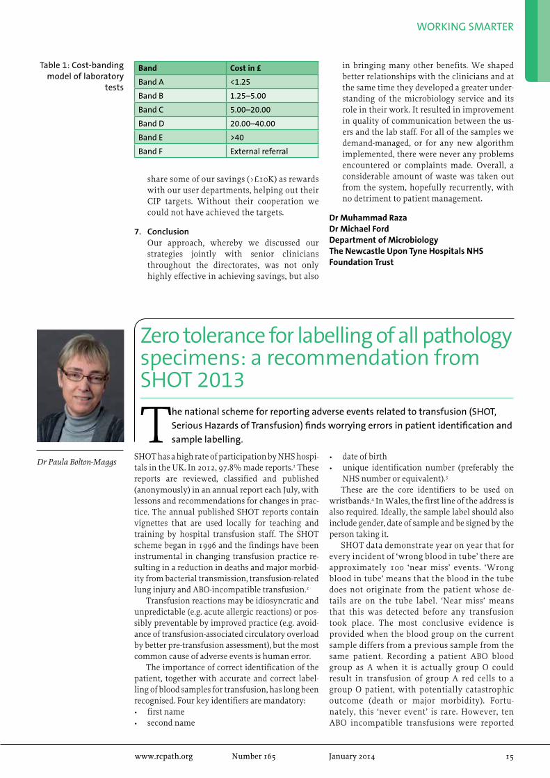
• We found most of the users of the service were not aware of the cost of tests. This information, when provided, was a very effective tool in raising awareness. Labo-ratory tests range from under £1 to over £700. To aid a better understanding of test costs, a cost-banding model was developed and circulated to Trust users (see Table 1). It was recommended that the use of tests in higher bands could be restricted for the senior members of the teams.
5. Follow-up Data on workload at three and six months
after this exercise were collected to measure the short- and medium-term impact of the campaign. This showed that we achieved more than our targeted 20–25% reduction in the activity in the categories of tests discussed in the meetings; in some case the reduction was up to 50%. A similar impact was noticed in areas of work not discussed in the meetings, reflecting effectiveness of the overall campaign.
6. Sharingthereward Since we could save more than our CIP tar-
gets from this campaign, we were able to
WORKING SMARTER
www.rcpath.org Number165 January2014 15
share some of our savings (>£10K) as rewards with our user departments, helping out their CIP targets. Without their cooperation we could not have achieved the targets.
7. Conclusion Our approach, whereby we discussed our
strategies jointly with senior clinicians throughout the directorates, was not only highly effective in achieving savings, but also
in bringing many other benefits. We shaped better relationships with the clinicians and at the same time they developed a greater under-standing of the microbiology service and its role in their work. It resulted in improvement in quality of communication between the us-ers and the lab staff. For all of the samples we demand-managed, or for any new algorithm implemented, there were never any problems encountered or complaints made. Overall, a considerable amount of waste was taken out from the system, hopefully recurrently, with no detriment to patient management.
Dr Muhammad RazaDr Michael FordDepartment of MicrobiologyThe Newcastle Upon Tyne Hospitals NHS Foundation Trust
Band Cost in £
Band A <1.25
Band B 1.25–5.00
Band C 5.00–20.00
Band D 20.00–40.00
Band E >40
Band F External referral
Table 1: Cost-banding model of laboratory
tests
Zero tolerance for labelling of all pathology specimens: a recommendation from SHot 2013
The national scheme for reporting adverse events related to transfusion (SHOT, Serious Hazards of Transfusion) finds worrying errors in patient identification and sample labelling.
SHOT has a high rate of participation by NHS hospi-tals in the UK. In 2012, 97.8% made reports.1 These reports are reviewed, classified and published (anonymously) in an annual report each July, with lessons and recommendations for changes in prac-tice. The annual published SHOT reports contain vignettes that are used locally for teaching and training by hospital transfusion staff. The SHOT scheme began in 1996 and the findings have been instrumental in changing transfusion practice re-sulting in a reduction in deaths and major morbid-ity from bacterial transmission, transfusion-related lung injury and ABO-incompatible transfusion.2
Transfusion reactions may be idiosyncratic and unpredictable (e.g. acute allergic reactions) or pos-sibly preventable by improved practice (e.g. avoid-ance of transfusion-associated circulatory overload by better pre-transfusion assessment), but the most common cause of adverse events is human error.
The importance of correct identification of the patient, together with accurate and correct label-ling of blood samples for transfusion, has long been recognised. Four key identifiers are mandatory:• first name• second name
• date of birth• unique identification number (preferably the
NHS number or equivalent).3
These are the core identifiers to be used on wristbands.4 In Wales, the first line of the address is also required. Ideally, the sample label should also include gender, date of sample and be signed by the person taking it.
SHOT data demonstrate year on year that for every incident of ‘wrong blood in tube’ there are approximately 100 ‘near miss’ events. ‘Wrong blood in tube’ means that the blood in the tube does not originate from the patient whose de-tails are on the tube label. ‘Near miss’ means that this was detected before any transfusion took place. The most conclusive evidence is provided when the blood group on the current sample differs from a previous sample from the same patient. Recording a patient ABO blood group as A when it is actually group O could result in transfusion of group A red cells to a group O patient, with potentially catastrophic outcome (death or major morbidity). Fortu-nately, this ‘never event’ is rare. However, ten ABO incompatible transfusions were reported
Dr Paula Bolton-Maggs
WORKING SMARTER
16 January2014 Number165 TheBulletinofTheRoyalCollegeofPathologists
to SHOT in 2012, three of which resulted in major morbidity.1 It is this risk that led to clear recommendations for full sample labelling for transfusion samples,3,5 which are well accepted.
‘Near miss’ reports constitute about a third over-all of all reports to SHOT each year (980 of 3545 in 2012). About half of these (534 in 2012) are sam-ple errors, of which 95% (505 in 2012) are ‘wrong blood in tube’. The majority, about 70%, are caused by failure to correctly identify the patient, or label-ling the sample away from the patient’s side. About 40% of these are samples taken by medical staff, about another 30% by nursing and midwifery staff, but less than 5% by phlebotomists who probably take most hospital blood samples. If the ‘near miss’ events had not been recognised, 70% would have resulted in a wrong component transfusion.
Correct identification of the patient is crucial in all aspects of medicine and should never be as-sumed. Patients should be asked to identify them-selves and not just to confirm their name (positive identification). A national comparative audit of sample collection and labelling also noted that doc-tors were the staff group most likely to be responsi-ble for mislabelling.6
Complete and correct labelling is important for all pathology specimens. The SHOT report for 2012 noted the transfusion of patients who did not require it, because the transfusion was given on the basis of wrong haemoglobin results.1 Such un-necessary transfusion puts patients at risk of trans-fusion-associated circulatory overload (TACO), which is a serious complication. Half of the 30 deaths that were either directly or possibly related to transfusion in the last three years (2010–2012) were related to TACO. Patients may also be put at risk of wrong medication as a result of wrong co-
agulation or biochemistry results. Mislabelling of histology or microbiology samples could result in inappropriate diagnosis and management. SHOT therefore recommends the same standard of sam-ple labelling for all pathology specimens and that transfusion samples should not be singled out for special treatment.1
Even for transfusion samples, laboratory staff do not always practise what they preach – as was demonstrated in the recent national clinical audit of transfusion sample labelling.6 While 154 hospi-tals said they had a zero tolerance policy for sample labelling, in fact 50 permitted amendments.
Patient safety has been much in focus this year. The Francis7 and Berwick8 reports remind us that the safety of the patient must be at the centre of everything we do. It is clear from 16 years of SHOT reporting that most transfusion incidents are caused by human error. Failure to identify the patient correctly at the time of blood sampling and at the time of transfusion remain the most common causes, and many reports have evidence of multiple errors.
Transfusion is particularly well regulated and it is likely that similar errors affect all branches of pathology. SHOT therefore recommends improved (zero tolerance) sample labelling for all pathology specimens to ensure the core identifiers are used. Pathology laboratory managers need to implement this recommendation, with support from their chief executives.
Dr Paula Bolton-MaggsMedical DirectorSerious Hazards of Transfusion Scheme (SHOT)Manchester Blood Centre
References1. Bolton-Maggs P, Poles D, Watt A, Cohen H, Thomas D. The Annual SHOT Report 2012. www.
shotuk.org/shot-reports (accessed 4 December 2013).2. Bolton-Maggs PH, Cohen H. Serious Hazards of Transfusion (SHOT) haemovigilance and
progress is improving transfusion safety. British Journal of Haematology 2013;163:303–314. Epub 2013/09/17.
3. BCSH. Guideline on the Administration of Blood Components, 2010. www.bcshguidelines.com/documents/Admin_blood_components_bcsh_05012010.pdf (accessed
4 December 2013).4. NPSA. Standardising wristbands improves patient safety, 2007. www.nrls.npsa.nhs.uk/resources/
type/alerts/?entryid45=59824&q=0%c2%acwristbands%c2%ac 2007 (accessed 4 December 2013).5. BCSH. Guidelines on hospital blood bank documentation and procedures. Clinical and Laboratory
Haematology 1990;12:209–220.6. NCA. Audit of Blood Sample Collection and Labelling, 2012. hospital.blood.co.uk/library/pdf/Au-
dit_of_Blood_Sample_Collection_and_Labelling.pdf (accessed 4 December 2013).7. Francis R. Report of the Mid Staffordshire NHS Foundation Trust Public Enquiry, 2013. www.midstaff-
spublicenquiry.com (accessed 4 December 2013).8. Berwick D, England National Advisory Group on the Safety of Patients in England. A Promise to
Learn – A commitment to act: Improving the safety of patients in England, 2013. https://www.gov.uk/government/uploads/system/uploads/attachment_data/file/226703/Berwick_Report.pdf (ac-cessed 4 December 2013).
WORKING SMARTER
www.rcpath.org Number165 January2014 17
adverse incidents in pathology
Every year, over 16 million diagnostic tests are carried out across the NHS (with more tests performed in private practice and in people’s homes). Medical devices are therefore a key part of NHS pathology services. Problems with medical devices
can lead to a decrease in the overall performance of the laboratory.
Medicines and Healthcare products Regulatory Agency (MHRA) is a government agency that en-sures that medical devices in the UK work and are acceptably safe.
We are the executive agency of the Depart-ment of Health charged with protecting and promoting public health and patient safety by ensuring that medicines, healthcare products and medical equipment meet appropriate standards of safety, quality, performance and effectiveness, and that they are used safely.
Where MHRA is aware of a problem with a diag-nostic test, we will work to address it. This includes changes to the device design or quality system, pro-duction of a Field Safety Notice by the manufacturer or the publication of alerts and guidance by MHRA.
MHRA is also responsible for inspection of blood establishments and running Serious Adverse Blood Reactions and Events (SABRE).
What is an IVD?In-vitro diagnostics (IVDs) can be found every-where:
• in the laboratory (analysers, reagents, test kits, control materials and software)
• in the ward, clinic, GP surgery and pharmacy (point-of-care tests: blood gas analysers, glucose tests, urine dipsticks)
• in people’s homes and bathrooms (home tests: pregnancy tests, glucose tests).
In effect, an IVD is something that tests a human sample to give information for a medical purpose.
Why should I report to you?MHRA collates reports from a range of sources to determine if there is a need for further action. We will also investigate individual incidents to help determine the root cause of the problem and en-sure corrective actions are effective. Information from device users helps us to manage manufactur-ers’ corrective actions.
By using the best available information, MHRA can ensure that problems with IVDs are identified quickly and that manufacturers’ actions are effective in dealing with the problem. We will also publish information of interest in pathology on our website: www.mhra.gov.uk/Safetyinformation/Healthcare-providers/Pathology/index.htm
What do you want me to report?You should report all adverse events with an IVD to MHRA. An adverse incident is an event that
causes, or has the potential to cause, unexpected or unwanted effects involving the safety of device users (including patients) or other persons.
Adverse incidents with IVDs could include un-expected false results in the lab that could have (or did) lead to:
• incorrect or delayed results• incorrect or delayed diagnosis or treatment• transfusion of inappropriate material• review of results, retesting of retained samples
or recall of patients for resampling.
Problems may be detected during commission-ing or quality control/quality assurance or from feedback from patients or clinicians.
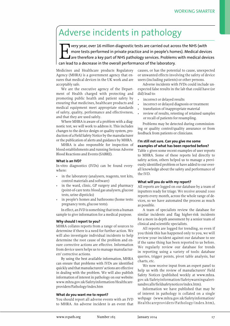
I’m still not sure. Can you give me some examples of what has been reported before?Table 1 gives some recent examples of user reports to MHRA. Some of these reports led directly to safety action, others helped us to manage a previ-ously identified problem or have added to our over-all knowledge about the safety and performance of the IVD.
What will you do with my report?All reports are logged on our database by a team of inputters ready for triage. We receive around 1000 reports every month, across the whole range of de-vices, so we have automated the process as much as possible.
A team of specialists review the database for similar incidents and flag higher-risk incidents for a more in-depth assessment by a senior team of clinical and scientific specialists.
All reports are logged for trending, so even if you think this has happened only to you, we will review your incident against our database to see if the same thing has been reported to us before. We regularly review our database for trends in reporting using a variety of tools: database queries, trigger points, pivot table analysis, bar charts, etc.
We now receive input from an expert panel to help us with the review of manufacturers’ Field Safety Notices (published weekly at www.mhra.gov.uk/Safetyinformation/Safetywarningsalert-sandrecalls/fieldsafetynotices/index.htm).
Information we have published that may be of interest in pathology is collated on a single webpage (www.mhra.gov.uk/Safetyinformation/Healthcareproviders/Pathology/index.htm),
WORKING SMARTER
18 January2014 Number165 TheBulletinofTheRoyalCollegeofPathologists
Device User report Outcome
Microbiology analyser
Error transferring a reagent from one cassette to another, leading to a review of previous results.
Manufacturer revised their risk assessment and altered the design, labeling and instructions of the device. MHRA also advised on the need to review results.
Transport swab Hair in unused swab. Isolated event.
HIV test Sample from patient known to be HIV positive was inadvertently tested and found to be HIV negative.
Patient-specific factors led to an unusual test result profile. Isolated event. No further action.
Histology stainer Poor quality tissue samples from the histology stainer could not be read by the pathologist and test needed to be repeated. No error messages.
No root cause identified. No further action. Isolated event.
Clinical chemistry analyser
Two occurrences of falsely low results in a neonatal screening test picked up during quality assurance, leading to local review of previous results.
The manufacturer issued a field safety notice and upgraded systems worldwide.
Home glucose test (reported by the HCP)
One meter gave two different readings within two minutes (3.0 mmol/l and 10.3 mmol/L). No adverse effect on patient.
Isolated event.
Point-of-care testing urine dipstick
Dipstick negative for glucose but patient admitted two days later for high HBA1c. This report was matched with other reports of possible false readings for ketones, proteins, blood and leukocytes.
Manufacturer reduced the open box stability. MHRA supported the manufacturer’s letter with an alert.
Haematology analyser
Very high titre antibody samples may carry over to other test wells.
Confirmed event. Following investigation, this event was considered to be extremely rare and unlikely to lead to patient harm. No further action. Isolated event.
Table 1: Recent examples of user reports to MHRA
where you can locate safety guidance and alerts that will be of interest in pathology.
But it wasn’t my fault!MHRA is concerned with preventing the occur-rence of adverse incidents, not with assigning blame or liability.
MHRA regulates the device and not the testing service or the professions. We try to work coopera-tively with the health service and device manufac-turers to find the best solution. We are not looking to apportion blame and we understand the need for good relationships between industry and the health service.
We can also issue warnings to the health service to help prevent recurrence of common use errors.
Great! How can I report incidents?MHRA runs an online reporting system (www.mhra.gov.uk/Safetyinformation/Reportingsafe-typroblems/Devices/index.htm) and printable adverse incident report forms are available from the MHRA website, along with further, regularly updated, supporting information.
Approximately 80% of reports from medical device users are now submitted through our online system. Successful use of this route provides the reporter with immediate confirmation of receipt, a unique incident reference number and, where requested, an emailed copy of the submitted report in pdf format. Copies of the report may also be sent to any other specified email address.
Good practice should generally be to report to the manufacturer and MHRA at the same time.
ConclusionMedical devices are key to quality in pathology. Reporting adverse incidents to MHRA can help improve quality in pathology by helping to ensure that pathology results are reliable and consistent.
Stephen LeeBiosciences Team ManagerMHRAmhra.gov.uk/stayconnected
www.rcpath.org Number165 January2014 19
PUBliC ENgagEMENt
‘a History of Pathology in 50 objects’ lecture series
The 50 objects lecture series was developed with the aim of promoting the work of pathologists to a wider audience and it has proven to be a highly successful step. Dr Suzy Lishman and Dr Paola Domizio delivered the initial two lectures and
in this issue they describe their experiences and objects highlighted in their talk. The next edition of the Bulletin will feature more lectures, including ‘The Story of Your Blood’ lecture by Dr Archie Prentice at Gresham College. For further details of the next lectures visit www.ilovepathology.org/events/ourevents
50 Objects at the Old Operating TheatreThe Old Operating Theatre was full, with several members of the audience having attended previ-ous National Pathology Week and Year events at the same venue. A selection of objects from the book were chosen, including several which feature in the new museum trail for children (see below). Objects were chosen to reflect as many different pa-thology specialties as possible. The evening began with a short account of the operating theatre itself; why it was built, how it was used and how it came to be preserved as a museum, before moving on to ten of the 50 objects.
Objects 1–5The first object was arguably the most important in the history of pathology: the light microscope. A 1950s microscope was displayed and photomicro-graphs were circulated to demonstrate microscop-ic appearances of histology sections and bacteria. The second object explored an advance that has transformed the lives of people with diabetes: the glucose meter. A primitive early-20th century glu-cose testing kit was displayed, along with a modern glucose meter. Object 3 was two in one: the Petri dish and agar plate. A selection of different agar plates was demonstrated and empty Petri dishes were passed around. Object 4 was a preserved frog, representing the pregnancy test. A modern over-the-counter test was also displayed for comparison. Next to the frog was object 5: a very life-like stuffed pigeon, representing Darwin’s work on evolution. The pigeon was accompanied by a copy of On the Origin of Species and now takes pride of place in the museum’s ‘Animals in medicine’ display cabinet.
Objects 6–10Object 6 was a bowl of corks, representing the cork that Robert Hooke viewed using a primitive micro-scope in 1665. Hooke’s comparison of the units mak-ing up a section of cork to monks’ quarters gave us the word ‘cell’. A copy of Hooke’s original drawing of cells was circulated. The seventh object was a bowl of dried peas, representing the pea plants that Aus-trian monk, Gregor Mendel, used in his experiments on heredity. Object 8 was the plastic blood bag, one of which was displayed alongside the 1970s glass version that it replaced. Object 9 was Lister’s donkey engine, a hand pumped carbolic acid spray, used to disinfect operating theatres. A bar of carbolic acid soap was passed around to give the audience an idea of how strong the smell in Lister’s operating theatre must have been. A bottle of Listerine completed the display for this object, the mouthwash having been named after Lister in 1879. The final object was a
Dr Suzy Lishman
‘A History of Pathology’ trailA new pathology trail for school students was launched in September at The Old Operating Theatre Museum and Herb Garret, in London.
The trail, which is based on our recent book, ‘A History of Pathology in 50 Objects’, was launched by Dr Suzy Lishman during her lecture at the museum.
With the help of discreetly placed clues and cards in parts of the museum, students will learn about some of the weird and wonderful objects that have played an important role in pathology, including why Japanese seaweed is essential for diagnosing disease.
The trail is a fun and informative way of engaging the public with pathology and a great opportunity to visit and explore the Museum‘s amazing collection.
Public engagement can take many forms and, by embedding the trail in a known public space, we hope to improve awareness and understanding of pathology and also continue the legacy of ‘A History of Pathology in 50 Objects’.
The trail runs until August 2014, so make sure you look out for it next time you are in London.
PUBLIC ENGAGEMENT
20 January2014 Number165 TheBulletinofTheRoyalCollegeofPathologists
a potted history of the pot
Professor Paola Domizio
Despite being the College’s Director of Public En-gagement and giving many talks to lay audiences, I still felt thrilled and privileged to give the second in the series of public lectures based on A History of Pathology in 50 Objects, in October 2013. My object was number 6, the pathology pot, so the venue – the Pathology Museum at St Bartholomew’s Hospi-tal – couldn’t have been more appropriate. Another of my roles is Curator of the Pathology Museum, so lecturing there felt a bit like lecturing from my own front room, perhaps mirroring how my 18th-century predecessors used to teach their students.
Joining me was Carla Valentine, the Museum’s Assistant Technical Curator, and we approached the topic from both an historical and human per-spective. I gave a historical talk entitled ‘A Potted History of the Pot’, while Carla’s aim was to ‘Reflesh the Bones’ and talk about the personal stories be-hind the museum’s specimens.
I started by recounting the origins of the Bart’s Museum in 1726 as a “repository for anatomical and chirurgical preparations” and described the very first specimens, which consisted predomi-nantly of bladder stones, reflecting the fact that lithotomy was one of the most frequent operations in the days before anaesthesia. I then gave a whis-tle-stop tour of the famous names associated with the museum and the pots they donated: Percivall Pott and chimney sweeps’ cancer, John Abernethy and Bellingham’s skull, James Paget and the bones from the first patient with osteitis deformans – all of them proudly displayed on the museum shelves.
Having taken the audience through the mu-seum’s heyday in the early 20th century and the re-organisation following bomb damage in the Second
World War, I arrived at the modern era. I described my early years as a lecturer at Bart’s, when the museum and its pots were the centre of pathology teaching. But then I recalled how, almost overnight, the museum was shut down and for the entire first decade of the 2000s it was empty and unused. The pots were badly neglected and it was a miracle that they weren’t damaged beyond repair.
But why the turn of fortune? I speculated on two possible reasons. First, the major curriculum chang-es engendered by Tomorrow’s Doctors, which led to a drastic reduction in pathology teaching and the per-ception that pathology pots were irrelevant. Second, the events at Bristol and Alder Hey, which led to a huge public backlash against organ retention.
I then described how Carla’s appointment in 2011 brought newfound enthusiasm and the museum’s resurrection. Through a programme of restoration, the pots are being returned to their former glory and with charitable investment in the fabric of the building it has once again become a vibrant, welcoming place. The changing educa-tional environment is being recognised through the development of the virtual pathology museum (vpathmuseum.smd.qmul.ac.uk).
I concluded by saying that, far from being irrel-evant, learning from pathology pots is even more important now than ever before. Now students can no longer attend post-mortem demonstrations regularly, it is important that they see examples of disease in pathology pots, rather than on a page or screen. If pathology museums are shut down and pots are disposed of, not only would it be a betrayal of the individuals who provided these gifts, it would also deny future medical students the opportunity
display of early-20th century microscope slides and a bottle of haematoxylin. The slides, collected by a medical student in the 1930s, included specimens we see regularly today, such as rodent ulcer (BCC) and gout, but also included an anthrax pustule and syphilitic chancre, which are certainly not part of my routine reporting.
FeedbackMembers of the audience were asked to complete evaluation forms but also had the opportunity to chat over a glass of wine in the museum after the talk. Feedback in person and on the forms was very positive, with several people asking for longer events to go into more detail, or similar events based on other objects in the book. Details of the rest of the lecture series were given.
Suzy LishmanAuthor, A History of Pathology in 50 Objects
Object 4: The frog
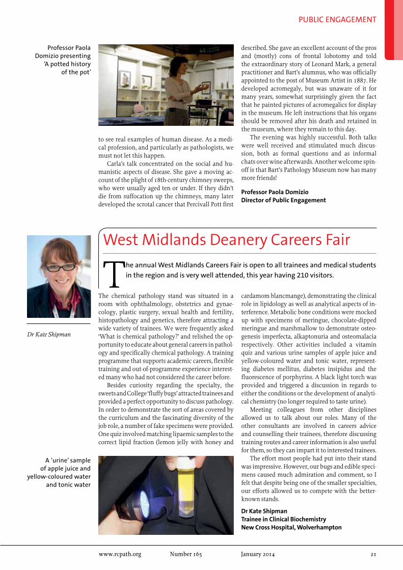
PUBLIC ENGAGEMENT
www.rcpath.org Number165 January2014 21
Professor Paola Domizio presenting
‘A potted history of the pot’
The chemical pathology stand was situated in a room with ophthalmology, obstetrics and gynae-cology, plastic surgery, sexual health and fertility, histopathology and genetics, therefore attracting a wide variety of trainees. We were frequently asked ‘What is chemical pathology?’ and relished the op-portunity to educate about general careers in pathol-ogy and specifically chemical pathology. A training programme that supports academic careers, flexible training and out-of-programme experience interest-ed many who had not considered the career before.
Besides curiosity regarding the specialty, the sweets and College ‘fluffy bugs’ attracted trainees and provided a perfect opportunity to discuss pathology. In order to demonstrate the sort of areas covered by the curriculum and the fascinating diversity of the job role, a number of fake specimens were provided. One quiz involved matching lipaemic samples to the correct lipid fraction (lemon jelly with honey and
cardamom blancmange), demonstrating the clinical role in lipidology as well as analytical aspects of in-terference. Metabolic bone conditions were mocked up with specimens of meringue, chocolate-dipped meringue and marshmallow to demonstrate osteo-genesis imperfecta, alkaptonuria and osteomalacia respectively. Other activities included a vitamin quiz and various urine samples of apple juice and yellow-coloured water and tonic water, represent-ing diabetes mellitus, diabetes insipidus and the fluorescence of porphyrins. A black light torch was provided and triggered a discussion in regards to either the conditions or the development of analyti-cal chemistry (no longer required to taste urine).
Meeting colleagues from other disciplines allowed us to talk about our roles. Many of the other consultants are involved in careers advice and counselling their trainees, therefore discussing training routes and career information is also useful for them, so they can impart it to interested trainees.
The effort most people had put into their stand was impressive. However, our bugs and edible speci-mens caused much admiration and comment, so I felt that despite being one of the smaller specialties, our efforts allowed us to compete with the better-known stands.
Dr Kate ShipmanTrainee in Clinical BiochemistryNew Cross Hospital, Wolverhampton
west Midlands deanery Careers Fair
The annual West Midlands Careers Fair is open to all trainees and medical students in the region and is very well attended, this year having 210 visitors.
A ‘urine’ sample of apple juice and
yellow-coloured water and tonic water
Dr Kate Shipman
to see real examples of human disease. As a medi-cal profession, and particularly as pathologists, we must not let this happen.
Carla’s talk concentrated on the social and hu-manistic aspects of disease. She gave a moving ac-count of the plight of 18th-century chimney sweeps, who were usually aged ten or under. If they didn’t die from suffocation up the chimneys, many later developed the scrotal cancer that Percivall Pott first
described. She gave an excellent account of the pros and (mostly) cons of frontal lobotomy and told the extraordinary story of Leonard Mark, a general practitioner and Bart’s alumnus, who was officially appointed to the post of Museum Artist in 1887. He developed acromegaly, but was unaware of it for many years, somewhat surprisingly given the fact that he painted pictures of acromegalics for display in the museum. He left instructions that his organs should be removed after his death and retained in the museum, where they remain to this day.
The evening was highly successful. Both talks were well received and stimulated much discus-sion, both as formal questions and as informal chats over wine afterwards. Another welcome spin-off is that Bart’s Pathology Museum now has many more friends!
Professor Paola DomizioDirector of Public Engagement
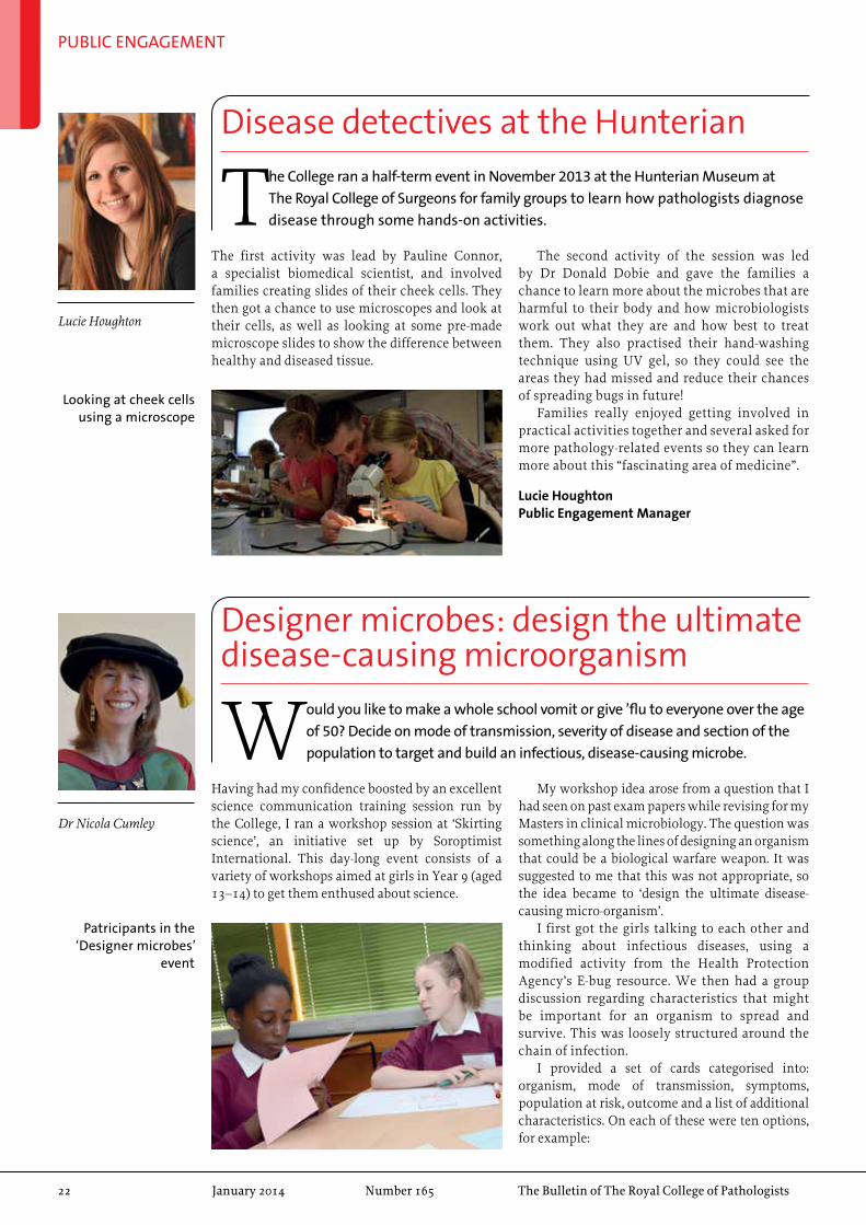
PUBLIC ENGAGEMENT
22 January2014 Number165 TheBulletinofTheRoyalCollegeofPathologists
Patricipants in the ‘Designer microbes’
event
designer microbes: design the ultimate disease-causing microorganism
Would you like to make a whole school vomit or give ’flu to everyone over the age of 50? Decide on mode of transmission, severity of disease and section of the population to target and build an infectious, disease-causing microbe.
Having had my confidence boosted by an excellent science communication training session run by the College, I ran a workshop session at ‘Skirting science’, an initiative set up by Soroptimist International. This day-long event consists of a variety of workshops aimed at girls in Year 9 (aged 13–14) to get them enthused about science.
My workshop idea arose from a question that I had seen on past exam papers while revising for my Masters in clinical microbiology. The question was something along the lines of designing an organism that could be a biological warfare weapon. It was suggested to me that this was not appropriate, so the idea became to ‘design the ultimate disease-causing micro-organism’.
I first got the girls talking to each other and thinking about infectious diseases, using a modified activity from the Health Protection Agency’s E-bug resource. We then had a group discussion regarding characteristics that might be important for an organism to spread and survive. This was loosely structured around the chain of infection.
I provided a set of cards categorised into: organism, mode of transmission, symptoms, population at risk, outcome and a list of additional characteristics. On each of these were ten options, for example:
disease detectives at the Hunterian
The College ran a half-term event in November 2013 at the Hunterian Museum at The Royal College of Surgeons for family groups to learn how pathologists diagnose disease through some hands-on activities.
The first activity was lead by Pauline Connor, a specialist biomedical scientist, and involved families creating slides of their cheek cells. They then got a chance to use microscopes and look at their cells, as well as looking at some pre-made microscope slides to show the difference between healthy and diseased tissue.
The second activity of the session was led by Dr Donald Dobie and gave the families a chance to learn more about the microbes that are harmful to their body and how microbiologists work out what they are and how best to treat them. They also practised their hand-washing technique using UV gel, so they could see the areas they had missed and reduce their chances of spreading bugs in future!
Families really enjoyed getting involved in practical activities together and several asked for more pathology-related events so they can learn more about this “fascinating area of medicine”.
Lucie HoughtonPublic Engagement Manager
Lucie Houghton
Looking at cheek cells using a microscope
Dr Nicola Cumley
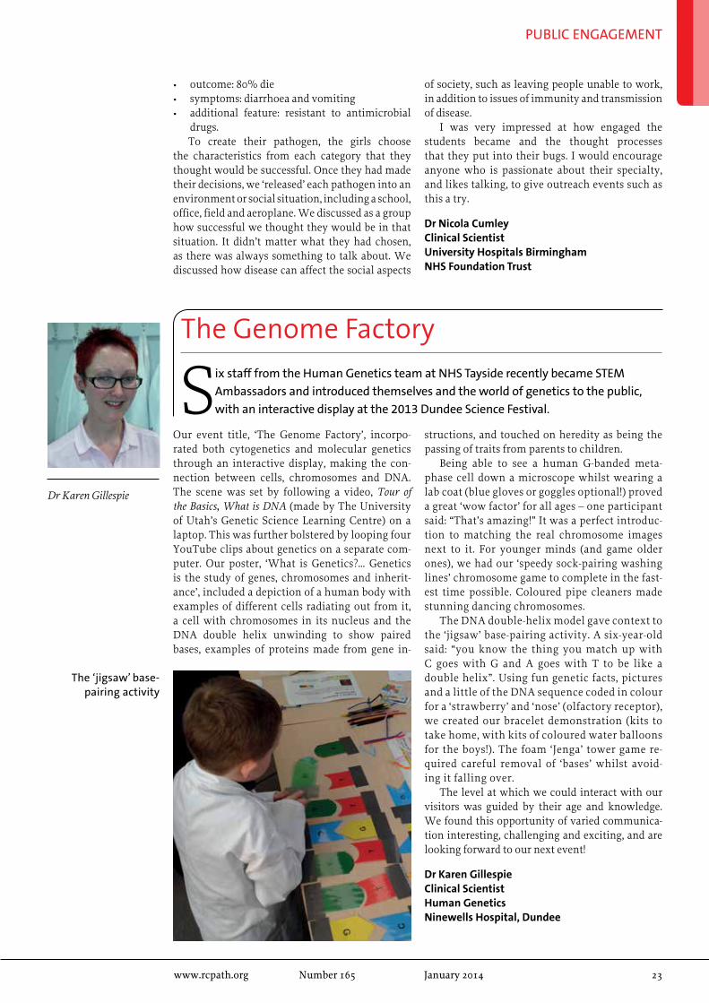
PUBLIC ENGAGEMENT
www.rcpath.org Number165 January2014 23
• outcome: 80% die• symptoms: diarrhoea and vomiting• additional feature: resistant to antimicrobial
drugs.To create their pathogen, the girls choose
the characteristics from each category that they thought would be successful. Once they had made their decisions, we ‘released’ each pathogen into an environment or social situation, including a school, office, field and aeroplane. We discussed as a group how successful we thought they would be in that situation. It didn’t matter what they had chosen, as there was always something to talk about. We discussed how disease can affect the social aspects
of society, such as leaving people unable to work, in addition to issues of immunity and transmission of disease.
I was very impressed at how engaged the students became and the thought processes that they put into their bugs. I would encourage anyone who is passionate about their specialty, and likes talking, to give outreach events such as this a try.
Dr Nicola CumleyClinical ScientistUniversity Hospitals Birmingham NHS Foundation Trust
Our event title, ‘The Genome Factory’, incorpo-rated both cytogenetics and molecular genetics through an interactive display, making the con-nection between cells, chromosomes and DNA. The scene was set by following a video, Tour of the Basics, What is DNA (made by The University of Utah’s Genetic Science Learning Centre) on a laptop. This was further bolstered by looping four YouTube clips about genetics on a separate com-puter. Our poster, ‘What is Genetics?… Genetics is the study of genes, chromosomes and inherit-ance’, included a depiction of a human body with examples of different cells radiating out from it, a cell with chromosomes in its nucleus and the DNA double helix unwinding to show paired bases, examples of proteins made from gene in-
structions, and touched on heredity as being the passing of traits from parents to children.
Being able to see a human G-banded meta-phase cell down a microscope whilst wearing a lab coat (blue gloves or goggles optional!) proved a great ‘wow factor’ for all ages – one participant said: “That’s amazing!” It was a perfect introduc-tion to matching the real chromosome images next to it. For younger minds (and game older ones), we had our ‘speedy sock-pairing washing lines’ chromosome game to complete in the fast-est time possible. Coloured pipe cleaners made stunning dancing chromosomes.
The DNA double-helix model gave context to the ‘jigsaw’ base-pairing activity. A six-year-old said: “you know the thing you match up with C goes with G and A goes with T to be like a double helix”. Using fun genetic facts, pictures and a little of the DNA sequence coded in colour for a ‘strawberry’ and ‘nose’ (olfactory receptor), we created our bracelet demonstration (kits to take home, with kits of coloured water balloons for the boys!). The foam ‘Jenga’ tower game re-quired careful removal of ‘bases’ whilst avoid-ing it falling over.
The level at which we could interact with our visitors was guided by their age and knowledge. We found this opportunity of varied communica-tion interesting, challenging and exciting, and are looking forward to our next event!
Dr Karen GillespieClinical ScientistHuman GeneticsNinewells Hospital, Dundee
the genome Factory
Six staff from the Human Genetics team at NHS Tayside recently became STEM Ambassadors and introduced themselves and the world of genetics to the public, with an interactive display at the 2013 Dundee Science Festival.
Dr Karen Gillespie
The ‘jigsaw’ base-pairing activity
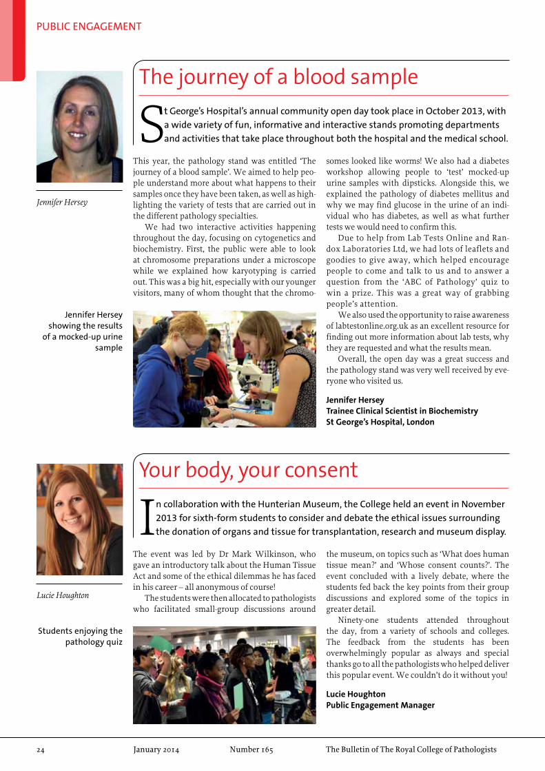
PUBLIC ENGAGEMENT
24 January2014 Number165 TheBulletinofTheRoyalCollegeofPathologists
Your body, your consent
In collaboration with the Hunterian Museum, the College held an event in November 2013 for sixth-form students to consider and debate the ethical issues surrounding the donation of organs and tissue for transplantation, research and museum display.
The event was led by Dr Mark Wilkinson, who gave an introductory talk about the Human Tissue Act and some of the ethical dilemmas he has faced in his career – all anonymous of course!
The students were then allocated to pathologists who facilitated small-group discussions around
the museum, on topics such as ‘What does human tissue mean?’ and ‘Whose consent counts?’. The event concluded with a lively debate, where the students fed back the key points from their group discussions and explored some of the topics in greater detail.
Ninety-one students attended throughout the day, from a variety of schools and colleges. The feedback from the students has been overwhelmingly popular as always and special thanks go to all the pathologists who helped deliver this popular event. We couldn’t do it without you!
Lucie HoughtonPublic Engagement Manager
Lucie Houghton
Students enjoying the pathology quiz
the journey of a blood sample
St George’s Hospital’s annual community open day took place in October 2013, with a wide variety of fun, informative and interactive stands promoting departments and activities that take place throughout both the hospital and the medical school.
Jennifer Hersey
This year, the pathology stand was entitled ‘The journey of a blood sample’. We aimed to help peo-ple understand more about what happens to their samples once they have been taken, as well as high-lighting the variety of tests that are carried out in the different pathology specialties.
We had two interactive activities happening throughout the day, focusing on cytogenetics and biochemistry. First, the public were able to look at chromosome preparations under a microscope while we explained how karyotyping is carried out. This was a big hit, especially with our younger visitors, many of whom thought that the chromo-
somes looked like worms! We also had a diabetes workshop allowing people to ‘test’ mocked-up urine samples with dipsticks. Alongside this, we explained the pathology of diabetes mellitus and why we may find glucose in the urine of an indi-vidual who has diabetes, as well as what further tests we would need to confirm this.
Due to help from Lab Tests Online and Ran-dox Laboratories Ltd, we had lots of leaflets and goodies to give away, which helped encourage people to come and talk to us and to answer a question from the ‘ABC of Pathology’ quiz to win a prize. This was a great way of grabbing people’s attention.
We also used the opportunity to raise awareness of labtestonline.org.uk as an excellent resource for finding out more information about lab tests, why they are requested and what the results mean.
Overall, the open day was a great success and the pathology stand was very well received by eve-ryone who visited us.
Jennifer HerseyTrainee Clinical Scientist in BiochemistrySt George’s Hospital, London
Jennifer Hersey showing the results
of a mocked-up urine sample
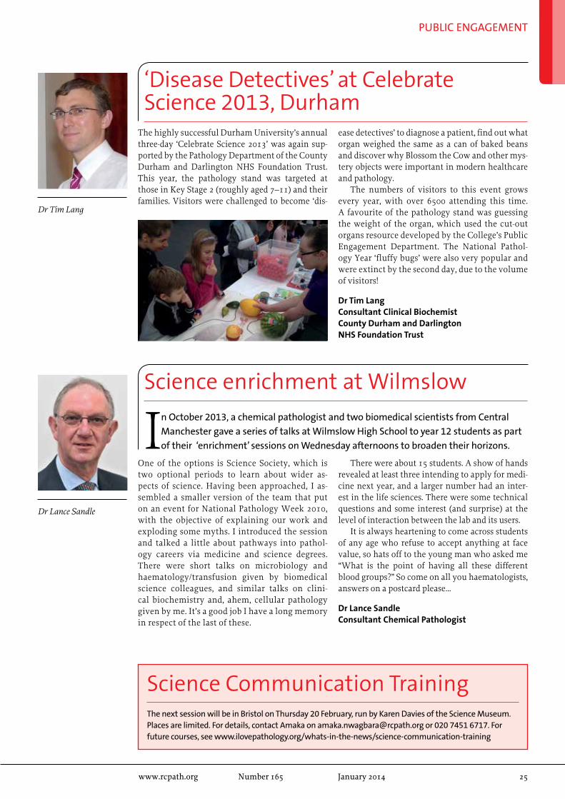
PUBLIC ENGAGEMENT
www.rcpath.org Number165 January2014 25
‘disease detectives’ at Celebrate Science 2013, durham
The highly successful Durham University’s annual three-day ‘Celebrate Science 2013’ was again sup-ported by the Pathology Department of the County Durham and Darlington NHS Foundation Trust. This year, the pathology stand was targeted at those in Key Stage 2 (roughly aged 7–11) and their families. Visitors were challenged to become ‘dis-
ease detectives’ to diagnose a patient, find out what organ weighed the same as a can of baked beans and discover why Blossom the Cow and other mys-tery objects were important in modern healthcare and pathology.
The numbers of visitors to this event grows every year, with over 6500 attending this time. A favourite of the pathology stand was guessing the weight of the organ, which used the cut-out organs resource developed by the College’s Public Engagement Department. The National Pathol-ogy Year ‘fluffy bugs’ were also very popular and were extinct by the second day, due to the volume of visitors!
Dr Tim LangConsultant Clinical BiochemistCounty Durham and Darlington NHS Foundation Trust
Dr Tim Lang
Science enrichment at wilmslow
In October 2013, a chemical pathologist and two biomedical scientists from Central Manchester gave a series of talks at Wilmslow High School to year 12 students as part of their ‘enrichment’ sessions on Wednesday afternoons to broaden their horizons.
Dr Lance Sandle
One of the options is Science Society, which is two optional periods to learn about wider as-pects of science. Having been approached, I as-sembled a smaller version of the team that put on an event for National Pathology Week 2010, with the objective of explaining our work and exploding some myths. I introduced the session and talked a little about pathways into pathol-ogy careers via medicine and science degrees. There were short talks on microbiology and haematology/transfusion given by biomedical science colleagues, and similar talks on clini-cal biochemistry and, ahem, cellular pathology given by me. It’s a good job I have a long memory in respect of the last of these.
There were about 15 students. A show of hands revealed at least three intending to apply for medi-cine next year, and a larger number had an inter-est in the life sciences. There were some technical questions and some interest (and surprise) at the level of interaction between the lab and its users.
It is always heartening to come across students of any age who refuse to accept anything at face value, so hats off to the young man who asked me “What is the point of having all these different blood groups?” So come on all you haematologists, answers on a postcard please…
Dr Lance SandleConsultant Chemical Pathologist
Science Communication trainingThe next session will be in Bristol on Thursday 20 February, run by Karen Davies of the Science Museum. Places are limited. For details, contact Amaka on [email protected] or 020 7451 6717. For future courses, see www.ilovepathology.org/whats-in-the-news/science-communication-training
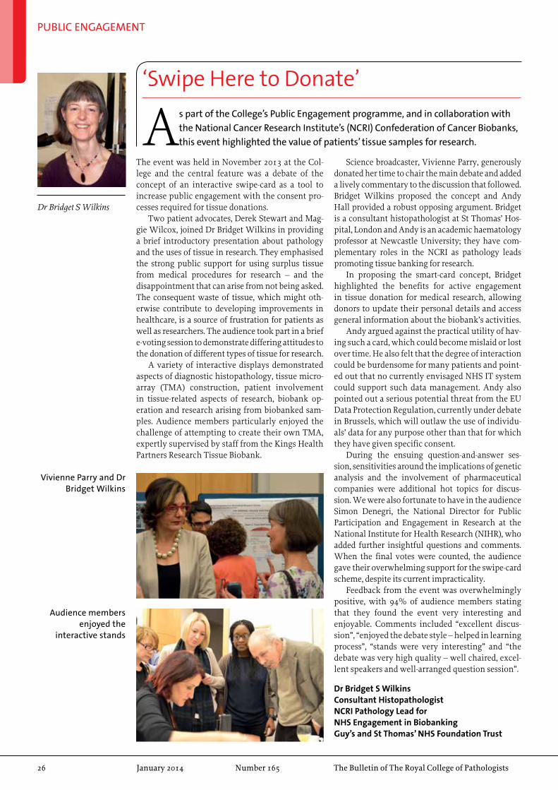
PUBLIC ENGAGEMENT
26 January2014 Number165 TheBulletinofTheRoyalCollegeofPathologists
‘Swipe Here to donate’
As part of the College’s Public Engagement programme, and in collaboration with the National Cancer Research Institute’s (NCRI) Confederation of Cancer Biobanks, this event highlighted the value of patients’ tissue samples for research.
The event was held in November 2013 at the Col-lege and the central feature was a debate of the concept of an interactive swipe-card as a tool to increase public engagement with the consent pro-cesses required for tissue donations.
Two patient advocates, Derek Stewart and Mag-gie Wilcox, joined Dr Bridget Wilkins in providing a brief introductory presentation about pathology and the uses of tissue in research. They emphasised the strong public support for using surplus tissue from medical procedures for research – and the disappointment that can arise from not being asked. The consequent waste of tissue, which might oth-erwise contribute to developing improvements in healthcare, is a source of frustration for patients as well as researchers. The audience took part in a brief e-voting session to demonstrate differing attitudes to the donation of different types of tissue for research.
A variety of interactive displays demonstrated aspects of diagnostic histopathology, tissue micro-array (TMA) construction, patient involvement in tissue-related aspects of research, biobank op-eration and research arising from biobanked sam-ples. Audience members particularly enjoyed the challenge of attempting to create their own TMA, expertly supervised by staff from the Kings Health Partners Research Tissue Biobank.
Science broadcaster, Vivienne Parry, generously donated her time to chair the main debate and added a lively commentary to the discussion that followed. Bridget Wilkins proposed the concept and Andy Hall provided a robust opposing argument. Bridget is a consultant histopathologist at St Thomas’ Hos-pital, London and Andy is an academic haematology professor at Newcastle University; they have com-plementary roles in the NCRI as pathology leads promoting tissue banking for research.
In proposing the smart-card concept, Bridget highlighted the benefits for active engagement in tissue donation for medical research, allowing donors to update their personal details and access general information about the biobank’s activities.
Andy argued against the practical utility of hav-ing such a card, which could become mislaid or lost over time. He also felt that the degree of interaction could be burdensome for many patients and point-ed out that no currently envisaged NHS IT system could support such data management. Andy also pointed out a serious potential threat from the EU Data Protection Regulation, currently under debate in Brussels, which will outlaw the use of individu-als’ data for any purpose other than that for which they have given specific consent.
During the ensuing question-and-answer ses-sion, sensitivities around the implications of genetic analysis and the involvement of pharmaceutical companies were additional hot topics for discus-sion. We were also fortunate to have in the audience Simon Denegri, the National Director for Public Participation and Engagement in Research at the National Institute for Health Research (NIHR), who added further insightful questions and comments. When the final votes were counted, the audience gave their overwhelming support for the swipe-card scheme, despite its current impracticality.
Feedback from the event was overwhelmingly positive, with 94% of audience members stating that they found the event very interesting and enjoyable. Comments included “excellent discus-sion”, “enjoyed the debate style – helped in learning process”, “stands were very interesting” and “the debate was very high quality – well chaired, excel-lent speakers and well-arranged question session”.
Dr Bridget S WilkinsConsultant HistopathologistNCRI Pathology Lead for NHS Engagement in BiobankingGuy’s and St Thomas’ NHS Foundation Trust
Dr Bridget S Wilkins
Vivienne Parry and Dr Bridget Wilkins
Audience members enjoyed the
interactive stands
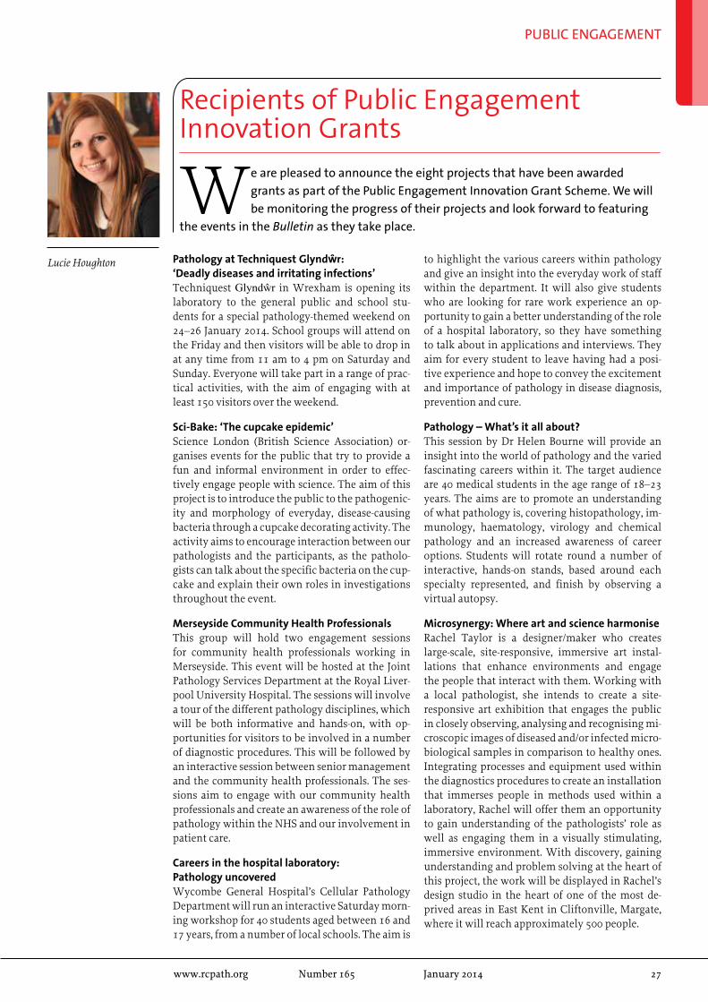
PUBLIC ENGAGEMENT
www.rcpath.org Number165 January2014 27
recipients of Public Engagement innovation grants
We are pleased to announce the eight projects that have been awarded grants as part of the Public Engagement Innovation Grant Scheme. We will be monitoring the progress of their projects and look forward to featuring
the events in the Bulletin as they take place.
Lucie Houghton Pathology at Techniquest Glyndŵr: ‘Deadly diseases and irritating infections’Techniquest Glyndŵr in Wrexham is opening its laboratory to the general public and school stu-dents for a special pathology-themed weekend on 24–26 January 2014. School groups will attend on the Friday and then visitors will be able to drop in at any time from 11 am to 4 pm on Saturday and Sunday. Everyone will take part in a range of prac-tical activities, with the aim of engaging with at least 150 visitors over the weekend.
Sci-Bake: ‘The cupcake epidemic’Science London (British Science Association) or-ganises events for the public that try to provide a fun and informal environment in order to effec-tively engage people with science. The aim of this project is to introduce the public to the pathogenic-ity and morphology of everyday, disease-causing bacteria through a cupcake decorating activity. The activity aims to encourage interaction between our pathologists and the participants, as the patholo-gists can talk about the specific bacteria on the cup-cake and explain their own roles in investigations throughout the event.
Merseyside Community Health ProfessionalsThis group will hold two engagement sessions for community health professionals working in Merseyside. This event will be hosted at the Joint Pathology Services Department at the Royal Liver-pool University Hospital. The sessions will involve a tour of the different pathology disciplines, which will be both informative and hands-on, with op-portunities for visitors to be involved in a number of diagnostic procedures. This will be followed by an interactive session between senior management and the community health professionals. The ses-sions aim to engage with our community health professionals and create an awareness of the role of pathology within the NHS and our involvement in patient care.
Careers in the hospital laboratory: Pathology uncoveredWycombe General Hospital’s Cellular Pathology Department will run an interactive Saturday morn-ing workshop for 40 students aged between 16 and 17 years, from a number of local schools. The aim is
to highlight the various careers within pathology and give an insight into the everyday work of staff within the department. It will also give students who are looking for rare work experience an op-portunity to gain a better understanding of the role of a hospital laboratory, so they have something to talk about in applications and interviews. They aim for every student to leave having had a posi-tive experience and hope to convey the excitement and importance of pathology in disease diagnosis, prevention and cure.
Pathology – What’s it all about?This session by Dr Helen Bourne will provide an insight into the world of pathology and the varied fascinating careers within it. The target audience are 40 medical students in the age range of 18–23 years. The aims are to promote an understanding of what pathology is, covering histopathology, im-munology, haematology, virology and chemical pathology and an increased awareness of career options. Students will rotate round a number of interactive, hands-on stands, based around each specialty represented, and finish by observing a virtual autopsy.
Microsynergy: Where art and science harmoniseRachel Taylor is a designer/maker who creates large-scale, site-responsive, immersive art instal-lations that enhance environments and engage the people that interact with them. Working with a local pathologist, she intends to create a site-responsive art exhibition that engages the public in closely observing, analysing and recognising mi-croscopic images of diseased and/or infected micro-biological samples in comparison to healthy ones. Integrating processes and equipment used within the diagnostics procedures to create an installation that immerses people in methods used within a laboratory, Rachel will offer them an opportunity to gain understanding of the pathologists’ role as well as engaging them in a visually stimulating, immersive environment. With discovery, gaining understanding and problem solving at the heart of this project, the work will be displayed in Rachel’s design studio in the heart of one of the most de-prived areas in East Kent in Cliftonville, Margate, where it will reach approximately 500 people.
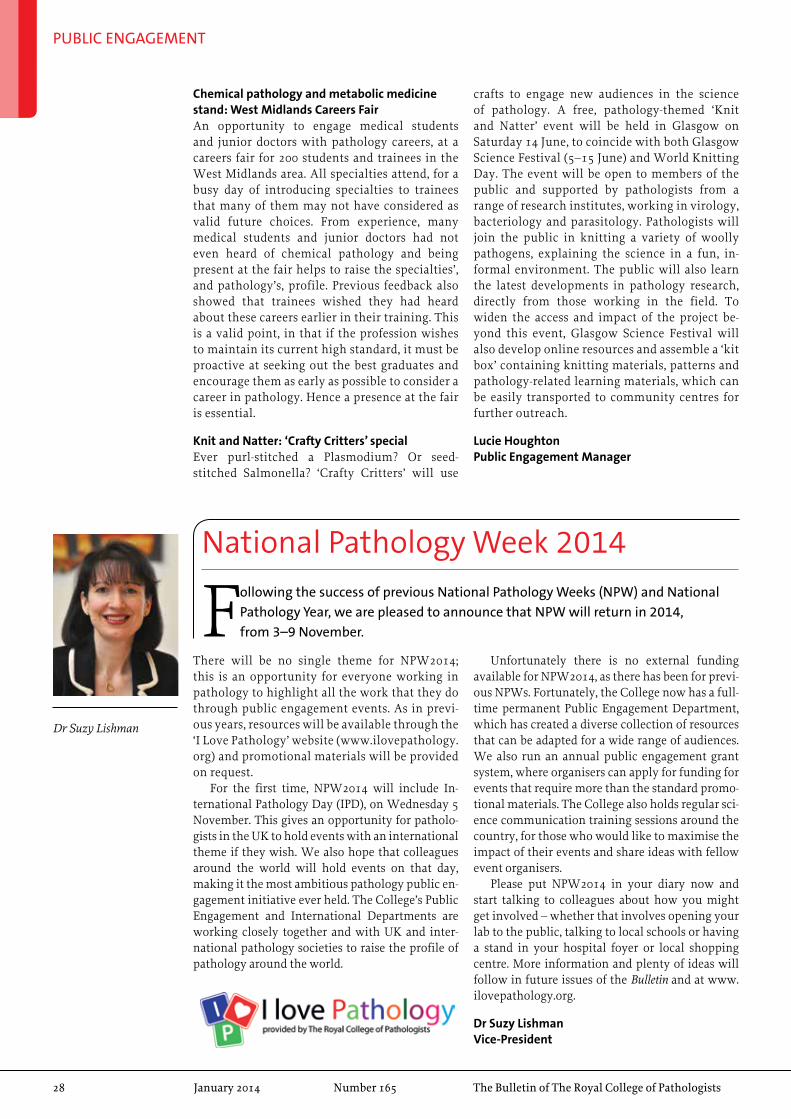
PUBLIC ENGAGEMENT
28 January2014 Number165 TheBulletinofTheRoyalCollegeofPathologists
Chemical pathology and metabolic medicine stand: West Midlands Careers FairAn opportunity to engage medical students and junior doctors with pathology careers, at a careers fair for 200 students and trainees in the West Midlands area. All specialties attend, for a busy day of introducing specialties to trainees that many of them may not have considered as valid future choices. From experience, many medical students and junior doctors had not even heard of chemical pathology and being present at the fair helps to raise the specialties’, and pathology’s, profile. Previous feedback also showed that trainees wished they had heard about these careers earlier in their training. This is a valid point, in that if the profession wishes to maintain its current high standard, it must be proactive at seeking out the best graduates and encourage them as early as possible to consider a career in pathology. Hence a presence at the fair is essential.
Knit and Natter: ‘Crafty Critters’ specialEver purl-stitched a Plasmodium? Or seed-stitched Salmonella? ‘Crafty Critters’ will use
crafts to engage new audiences in the science of pathology. A free, pathology-themed ‘Knit and Natter’ event will be held in Glasgow on Saturday 14 June, to coincide with both Glasgow Science Festival (5–15 June) and World Knitting Day. The event will be open to members of the public and supported by pathologists from a range of research institutes, working in virology, bacteriology and parasitology. Pathologists will join the public in knitting a variety of woolly pathogens, explaining the science in a fun, in-formal environment. The public will also learn the latest developments in pathology research, directly from those working in the field. To widen the access and impact of the project be-yond this event, Glasgow Science Festival will also develop online resources and assemble a ‘kit box’ containing knitting materials, patterns and pathology-related learning materials, which can be easily transported to community centres for further outreach.
Lucie HoughtonPublic Engagement Manager
National Pathology week 2014
Following the success of previous National Pathology Weeks (NPW) and National Pathology Year, we are pleased to announce that NPW will return in 2014, from 3–9 November.
Dr Suzy Lishman
There will be no single theme for NPW2014; this is an opportunity for everyone working in pathology to highlight all the work that they do through public engagement events. As in previ-ous years, resources will be available through the ‘I Love Pathology’ website (www.ilovepathology.org) and promotional materials will be provided on request.
For the first time, NPW2014 will include In-ternational Pathology Day (IPD), on Wednesday 5 November. This gives an opportunity for patholo-gists in the UK to hold events with an international theme if they wish. We also hope that colleagues around the world will hold events on that day, making it the most ambitious pathology public en-gagement initiative ever held. The College’s Public Engagement and International Departments are working closely together and with UK and inter-national pathology societies to raise the profile of pathology around the world.
Unfortunately there is no external funding available for NPW2014, as there has been for previ-ous NPWs. Fortunately, the College now has a full-time permanent Public Engagement Department, which has created a diverse collection of resources that can be adapted for a wide range of audiences. We also run an annual public engagement grant system, where organisers can apply for funding for events that require more than the standard promo-tional materials. The College also holds regular sci-ence communication training sessions around the country, for those who would like to maximise the impact of their events and share ideas with fellow event organisers.
Please put NPW2014 in your diary now and start talking to colleagues about how you might get involved – whether that involves opening your lab to the public, talking to local schools or having a stand in your hospital foyer or local shopping centre. More information and plenty of ideas will follow in future issues of the Bulletin and at www.ilovepathology.org.
Dr Suzy LishmanVice-President

www.rcpath.org Number165 January2014 29
traiNiNg
First, Dr Catharine Dhaliwal, an ST2 histopathol-ogy trainee in Edinburgh, interviews Professor James Ironside CBE, who is Professor of Clinical Neuropathology at the University of Edinburgh and Honorary Consultant Neuropathologist at the Western General Hospital in Edinburgh.
Professor Ironside is best known for his ground-breaking work and expertise on Creutzfeldt Jakob disease (CJD) and other prion diseases. In addition to clinical and research work, he is also Director of the MRC UK Brain Banks Network and was a founder
member of the Human Tissue Authority. Professor Ironside has been awarded a CBE for his substantial contributions to medicine.
Professor Ironside, how did you develop an interest in neuropathology?I don’t come from a medical background, but it was clear to me from my early days as a medical student that I enjoyed learning about the mechanisms of disease and so, as part of my undergraduate train-ing, I decided to do an intercalated BSc in pathol-ogy. During this time, I realised that I felt very comfortable working in a laboratory environment and I decided to pursue a career in pathology. Hav-ing completed my house jobs, I started a pathology job in Dundee in 1980. It was whilst working here that I found I was particularly interested in and fascinated by neuropathology.
Who has been the biggest influence on your career and what lessons did that person teach you?A number of people have influenced my career. First was the Professor of Pathol-ogy in Dundee where I trained: John Beck,
trainees’ notes
Traineescomprise8%oftheCollegemembershipand,whilsttraversingourwayfromregistrationtoFellowship,all840ofusareshapingtheveryfutureofourprofession.Thisfeatureiswrittenbyandfortraineestoreflectonthejourney,
discusstrainingissuesandoptimiseengagementwiththeCollegeacrossallthespecialties.
Dr Emma Johnson
Dr Catharine Dhaliwal
Professor James Ironside
now deceased, who was a terrific teacher. I was fascinated by his lectures and in particular his very logical progression through disease mechanisms. Whilst working in Dundee, I developed an inter-est in neuropathology under the influence of John Anderson. He was doing research into neuropa-thology at both extremes of age, including work on Alzheimer’s disease and brain atrophy, together with research into mechanisms of brain injury in neonates. I subsequently decided to specialise in neuropathology and moved as a trainee to Shef-field, where I worked with Walter Timperley, a wonderful neuropathologist and a terrific char-acter. He was very grounded and taught me more than I can ever thank him for. Finally, in Sheffield I also had the chance to do some research with Pro-fessor James Underwood and he made me realise that it was possible to be both an academic patholo-gist and a good diagnostic pathologist.
What have been the most memorable moments of your career?There are many memorable moments. Firstly pass-ing the pathology exams! My involvement in the identification of variant CJD has led to worldwide collaborations and has been both challenging and rewarding. I have enjoyed teaching MD/PhD stu-dents and trainees, and seeing them progress in their careers. More recently, I was delighted to receive my CBE at Buckingham Palace with my family.
Which one research question would you like to answer?One recent challenge that is being taken up in neuropathology is to understand the basis of the neurodegenerative diseases that are so common and cause such an affliction in our ageing popula-tion. These are characterised by protein misfolding and accumulation and also by a very predictable anatomical spread of pathology in the brain. So, starting from my background of CJD and prion diseases where there is also accumulation of ab-normal protein, but one that seems to be transmis-sible, I would like to understand more about how proteins misfold and how these abnormal proteins then spread through the brain. If we could under-stand that better, then we could do something to
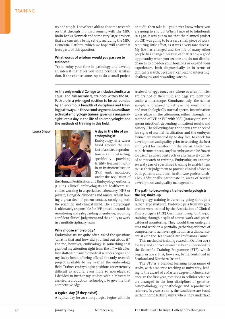
TRAINING
30 January2014 Number165 TheBulletinofTheRoyalCollegeofPathologists
try and stop it. I have been able to do some research on that through my involvement with the MRC Brain Banks Network and some very large projects that are currently being set up, including the MRC Dementia Platform, which we hope will answer at least parts of this question.
What words of wisdom would you pass on to trainees?Try to enjoy your time in pathology and develop an interest that gives you some personal satisfac-tion. If the chance comes up to do a small project
Laura Shaw
or audit, then take it – you never know where you are going to end up! When I moved to Edinburgh in 1990, it was put to me that the planned project on CJD was going to be a very small piece of work, requiring little effort, as it was a very rare disease. My life has changed and the life of many other people has changed because of that! Know a good opportunity when you see one and do not dismiss chances to broaden your horizons or expand your experiences, both diagnostically or in terms of clinical research, because it can lead to interesting, challenging and rewarding careers.
As the only medical College to include scientists as equal and full members, trainees within the RC-Path are in a privileged position to be surrounded by an enormous breadth of disciplines and train-ing pathways. In this second segment, Laura Shaw, a clinical embryology trainee, gives us a unique in-sight into a day in the life of an embryologist and the methods of training in this field.
A day in the life of an embryologistEmbryology is a career based around the sub-ject of assisted reproduc-tion in a clinical setting, specifically providing fertility treatment with-in an in vitro fertilisation (IVF) unit, monitored under the regulation of
the Human Fertilisation and Embryology Authority (HFEA). Clinical embryologists are healthcare sci-entists working in a specialised laboratory, NHS or private, alongside clinicians and nurses, whilst hav-ing a great deal of patient contact, satisfying both the scientific and clinical mind. The embryologist is ultimately responsible for IVF procedures and the monitoring and safeguarding of embryos, requiring confident clinical judgement and the ability to work in a multidisciplinary team.
Why choose embryology?Embryologists are quite often asked the questions ‘what is that and how did you find out about it?’ For me, however, embryology is something that grabbed my attention right from the off, with a lec-ture slotted into my biomedical sciences degree and my lucky break of being offered the only research project available to my year in the embryology field. Trainee embryologist positions are extremely difficult to acquire, even more so nowadays, so I decided to further my studies with a Masters in assisted reproduction technology, to give me that competitive edge.
A typical day (if they exist!)A typical day for an embryologist begins with the
retrieval of eggs (oocytes), where ovarian follicles are drained of their fluid and eggs are identified under a microscope. Simultaneously, the semen sample is prepared to retrieve the most motile and morphologically normal sperm. Insemination takes place in the afternoon, either through the method of IVF or IVF with ICSI (intracytoplasmic sperm injection), depending on patient results and history. The following day, the oocytes are checked for signs of normal fertilisation and the embryos formed are monitored up to day five, to check for development and quality prior to selecting the best embryo(s) for transfer into the uterus. Under cer-tain circumstances, surplus embryos can be frozen for use in a subsequent cycle or alternatively donat-ed to research or training. Embryologists undergo several years of specialised training to enable them to use their judgement to provide clinical advice to both patients and other health care professionals. They additionally participate in areas of service development and quality management.
The path to becoming a trained embryologist: the big shake-upEmbryology training is currently going through a rather large shake-up. Embryologists from my gen-eration were trained by the Association of Clinical Embryologists (ACE) Certificate, using ‘on-the-job’ training through a split of course work and practi-cal-based monitoring. They would then undergo a viva and work on a portfolio, gathering evidence of competence to achieve registration as a clinical sci-entist with the Health and Care Professions Council.
This method of training ceased in October 2013 for England and Wales and has been superseded by the Scientific Training Programme (STP), which began in 2011. It is, however, being continued in Scotland and Northern Ireland.
The STP is a blended learning programme of study, with academic teaching at university, lead-ing to the award of a Masters degree in clinical sci-ence. In the first year, rotations in cellular sciences are arranged in the four disciplines of genetics, histopathology, cytopathology and reproductive sciences. In years 2 and 3, the candidates are based in their home fertility units, where they undertake
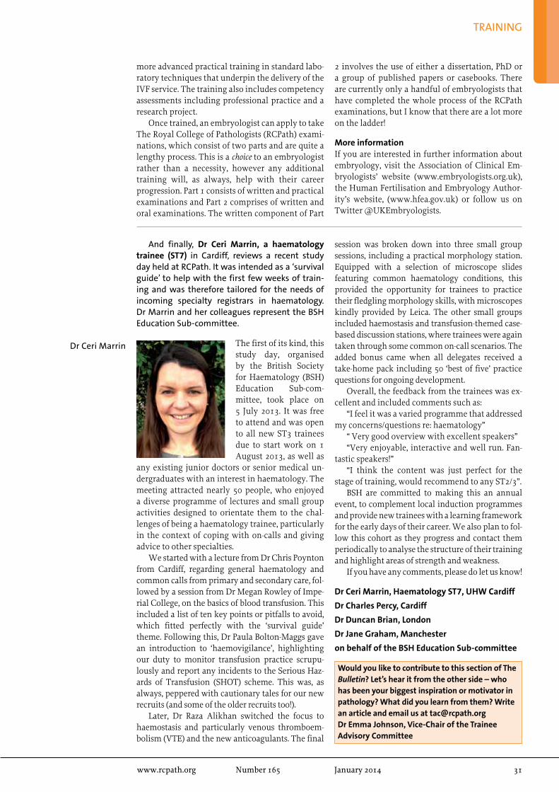
TRAINING
www.rcpath.org Number165 January2014 31
Would you like to contribute to this section of The Bulletin? Let’s hear it from the other side – who has been your biggest inspiration or motivator in pathology? What did you learn from them? Write an article and email us at [email protected] Emma Johnson, Vice-Chair of the Trainee Advisory Committee
Dr Ceri Marrin
more advanced practical training in standard labo-ratory techniques that underpin the delivery of the IVF service. The training also includes competency assessments including professional practice and a research project.
Once trained, an embryologist can apply to take The Royal College of Pathologists (RCPath) exami-nations, which consist of two parts and are quite a lengthy process. This is a choice to an embryologist rather than a necessity, however any additional training will, as always, help with their career progression. Part 1 consists of written and practical examinations and Part 2 comprises of written and oral examinations. The written component of Part
2 involves the use of either a dissertation, PhD or a group of published papers or casebooks. There are currently only a handful of embryologists that have completed the whole process of the RCPath examinations, but I know that there are a lot more on the ladder!
More informationIf you are interested in further information about embryology, visit the Association of Clinical Em-bryologists’ website (www.embryologists.org.uk), the Human Fertilisation and Embryology Author-ity’s website, (www.hfea.gov.uk) or follow us on Twitter @UKEmbryologists.
And finally, Dr Ceri Marrin, a haematology trainee (ST7) in Cardiff, reviews a recent study day held at RCPath. It was intended as a ‘survival guide’ to help with the first few weeks of train-ing and was therefore tailored for the needs of incoming specialty registrars in haematology. Dr Marrin and her colleagues represent the BSH Education Sub-committee.
The first of its kind, this study day, organised by the British Society for Haematology (BSH) Education Sub-com-mittee, took place on 5 July 2013. It was free to attend and was open to all new ST3 trainees due to start work on 1 August 2013, as well as
any existing junior doctors or senior medical un-dergraduates with an interest in haematology. The meeting attracted nearly 50 people, who enjoyed a diverse programme of lectures and small group activities designed to orientate them to the chal-lenges of being a haematology trainee, particularly in the context of coping with on-calls and giving advice to other specialties.
We started with a lecture from Dr Chris Poynton from Cardiff, regarding general haematology and common calls from primary and secondary care, fol-lowed by a session from Dr Megan Rowley of Impe-rial College, on the basics of blood transfusion. This included a list of ten key points or pitfalls to avoid, which fitted perfectly with the ‘survival guide’ theme. Following this, Dr Paula Bolton-Maggs gave an introduction to ‘haemovigilance’, highlighting our duty to monitor transfusion practice scrupu-lously and report any incidents to the Serious Haz-ards of Transfusion (SHOT) scheme. This was, as always, peppered with cautionary tales for our new recruits (and some of the older recruits too!).
Later, Dr Raza Alikhan switched the focus to haemostasis and particularly venous thromboem-bolism (VTE) and the new anticoagulants. The final
session was broken down into three small group sessions, including a practical morphology station. Equipped with a selection of microscope slides featuring common haematology conditions, this provided the opportunity for trainees to practice their fledgling morphology skills, with microscopes kindly provided by Leica. The other small groups included haemostasis and transfusion-themed case-based discussion stations, where trainees were again taken through some common on-call scenarios. The added bonus came when all delegates received a take-home pack including 50 ‘best of five’ practice questions for ongoing development.
Overall, the feedback from the trainees was ex-cellent and included comments such as:
“I feel it was a varied programme that addressed my concerns/questions re: haematology”
“ Very good overview with excellent speakers”“Very enjoyable, interactive and well run. Fan-
tastic speakers!”“I think the content was just perfect for the
stage of training, would recommend to any ST2/3”.BSH are committed to making this an annual
event, to complement local induction programmes and provide new trainees with a learning framework for the early days of their career. We also plan to fol-low this cohort as they progress and contact them periodically to analyse the structure of their training and highlight areas of strength and weakness.
If you have any comments, please do let us know!
Dr Ceri Marrin, Haematology ST7, UHW Cardiff
Dr Charles Percy, Cardiff
Dr Duncan Brian, London
Dr Jane Graham, Manchester
on behalf of the BSH Education Sub-committee
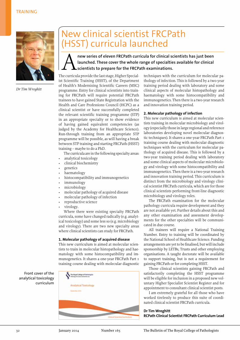
TRAINING
32 January2014 Number165 TheBulletinofTheRoyalCollegeofPathologists
New clinical scientist FrCPath (HSSt) curricula launched
AnewseriesofelevenFRCPathcurriculaforclinicalscientistshasjustbeenlaunched.ThesecoverthewholerangeofspecialtiesavailableforclinicalscientiststopreparefortheFRCPathexaminations.
Dr Tim Wreghitt
The curricula provide the last stage, Higher Special-ist Scientific Training (HSST), of the Department of Health’s Modernising Scientific Careers (MSC) programme. Entry for clinical scientists into train-ing for FRCPath will require potential FRCPath trainees to have gained State Registration with the Health and Care Professions Council (HCPC) as a clinical scientist or have successfully completed the relevant scientific training programme (STP) in an appropriate specialty or to show evidence of having gained equivalent competencies (as judged by the Academy for Healthcare Science). Run-through training from an appropriate STP programme will be possible, as will having a break between STP training and starting FRCPath (HSST) training – maybe to do a PhD.
The curricula are in the following specialty areas:• analytical toxicology• clinical biochemistry• genetics• haematology• histocompatibility and immunogenetics• immunology• microbiology• molecular pathology of acquired disease• molecular pathology of infection• reproductive science• virology.
Where there were existing specialty FRCPath curricula, some have changed radically (e.g. analyt-ical toxicology) and some less so (e.g. microbiology and virology). There are two new specialty areas where clinical scientists can study for FRCPath.
1. Molecular pathology of acquired diseaseThis new curriculum is aimed at molecular scien-tists to train in molecular histopathology and hae-matology with some histocompatibility and im-munogenetics. It shares a one-year FRCPath Part 1 training course dealing with molecular diagnostic
techniques with the curriculum for molecular pa-thology of infection. This is followed by a two-year training period dealing with laboratory and some clinical aspects of molecular histopathology and haematology with some histocompatibility and immunogenetics. Then there is a two-year research and innovation training period.
2. Molecular pathology of infectionThis new curriculum is aimed at molecular scien-tists training in molecular microbiology and virol-ogy (especially those in large regional and reference laboratories developing novel molecular diagnos-tic techniques). It shares a one-year FRCPath Part 1 training course dealing with molecular diagnostic techniques with the curriculum for molecular pa-thology of acquired disease. This is followed by a two-year training period dealing with laboratory and some clinical aspects of molecular microbiolo-gy and virology with some histocompatibility and immunogenetics. Then there is a two-year research and innovation training period. This curriculum is distinct from the microbiology and virology clini-cal scientist FRCPath curricula, which are for those clinical scientists performing front-line diagnostic microbiology and virology roles.
The FRCPath examination for the molecular pathology curricula require development and they are not available yet. Further details about this and any other examination and assessment develop-ments for the other specialties will be communi-cated in due course.
All trainees will require a National Training Number. Entry to training will be coordinated by the National School of Healthcare Science. Funding arrangements are yet to be finalised, but will include sponsorship by LETBs, Trusts and other employing organisations. A taught doctorate will be available to support training, but is not a requirement for gaining FRCPath or for completing HSST.
Those clinical scientists gaining FRCPath and satisfactorily completing the HSST programme will be eligible for inclusion in a proposed new vol-untary Higher Specialist Scientist Register and for appointment to consultant clinical scientist posts.
I am extremely grateful for all those who have worked tirelessly to produce this suite of coordi-nated clinical scientist FRCPath curricula.
Dr Tim WreghittRCPath Clinical Scientist FRCPath Curriculum Lead
Front cover of the analytical toxicology
curriculum
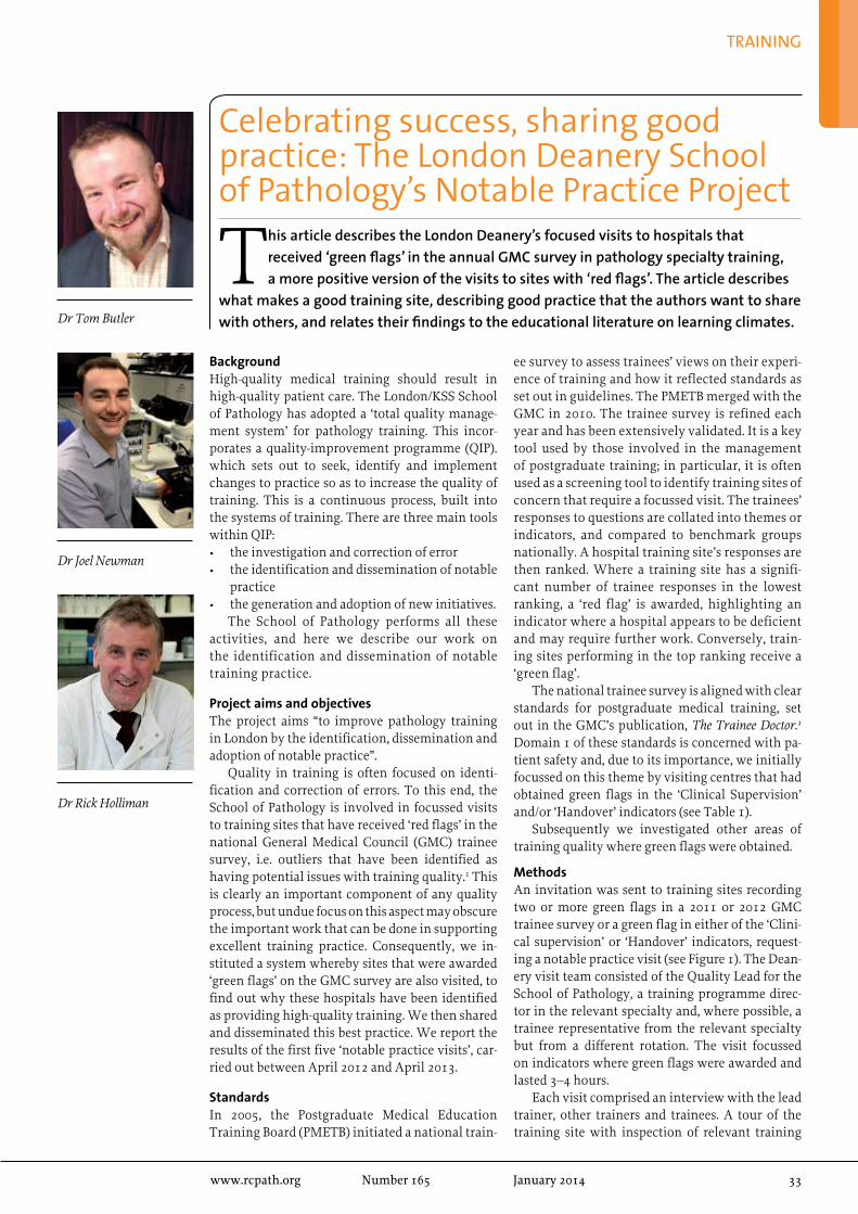
TRAINING
www.rcpath.org Number165 January2014 33
Celebrating success, sharing good practice: the london deanery School of Pathology’s Notable Practice Project
ThisarticledescribestheLondonDeanery’sfocusedvisitstohospitalsthatreceived‘greenflags’intheannualGMCsurveyinpathologyspecialtytraining,amorepositiveversionofthevisitstositeswith‘redflags’.Thearticledescribes
whatmakesagoodtrainingsite,describinggoodpracticethattheauthorswanttosharewithothers,andrelatestheirfindingstotheeducationalliteratureonlearningclimates.Dr Tom Butler
BackgroundHigh-quality medical training should result in high-quality patient care. The London/KSS School of Pathology has adopted a ‘total quality manage-ment system’ for pathology training. This incor-porates a quality-improvement programme (QIP). which sets out to seek, identify and implement changes to practice so as to increase the quality of training. This is a continuous process, built into the systems of training. There are three main tools within QIP:• the investigation and correction of error• the identification and dissemination of notable
practice• the generation and adoption of new initiatives.
The School of Pathology performs all these activities, and here we describe our work on the identification and dissemination of notable training practice.
Project aims and objectivesThe project aims “to improve pathology training in London by the identification, dissemination and adoption of notable practice”.
Quality in training is often focused on identi-fication and correction of errors. To this end, the School of Pathology is involved in focussed visits to training sites that have received ‘red flags’ in the national General Medical Council (GMC) trainee survey, i.e. outliers that have been identified as having potential issues with training quality.1 This is clearly an important component of any quality process, but undue focus on this aspect may obscure the important work that can be done in supporting excellent training practice. Consequently, we in-stituted a system whereby sites that were awarded ‘green flags’ on the GMC survey are also visited, to find out why these hospitals have been identified as providing high-quality training. We then shared and disseminated this best practice. We report the results of the first five ‘notable practice visits’, car-ried out between April 2012 and April 2013.
StandardsIn 2005, the Postgraduate Medical Education Training Board (PMETB) initiated a national train-
ee survey to assess trainees’ views on their experi-ence of training and how it reflected standards as set out in guidelines. The PMETB merged with the GMC in 2010. The trainee survey is refined each year and has been extensively validated. It is a key tool used by those involved in the management of postgraduate training; in particular, it is often used as a screening tool to identify training sites of concern that require a focussed visit. The trainees’ responses to questions are collated into themes or indicators, and compared to benchmark groups nationally. A hospital training site’s responses are then ranked. Where a training site has a signifi-cant number of trainee responses in the lowest ranking, a ‘red flag’ is awarded, highlighting an indicator where a hospital appears to be deficient and may require further work. Conversely, train-ing sites performing in the top ranking receive a ‘green flag’.
The national trainee survey is aligned with clear standards for postgraduate medical training, set out in the GMC’s publication, The Trainee Doctor.2 Domain 1 of these standards is concerned with pa-tient safety and, due to its importance, we initially focussed on this theme by visiting centres that had obtained green flags in the ‘Clinical Supervision’ and/or ‘Handover’ indicators (see Table 1).
Subsequently we investigated other areas of training quality where green flags were obtained.
MethodsAn invitation was sent to training sites recording two or more green flags in a 2011 or 2012 GMC trainee survey or a green flag in either of the ‘Clini-cal supervision’ or ‘Handover’ indicators, request-ing a notable practice visit (see Figure 1). The Dean-ery visit team consisted of the Quality Lead for the School of Pathology, a training programme direc-tor in the relevant specialty and, where possible, a trainee representative from the relevant specialty but from a different rotation. The visit focussed on indicators where green flags were awarded and lasted 3–4 hours.
Each visit comprised an interview with the lead trainer, other trainers and trainees. A tour of the training site with inspection of relevant training
Dr Joel Newman
Dr Rick Holliman

TRAINING
34 January2014 Number165 TheBulletinofTheRoyalCollegeofPathologists
Clinical supervision questions
Did you always know who was providing your clinical supervision when you were working?
How often did you feel forced to cope with clinical problems beyond your competence or experience
Did you always know who your available senior support was, during on call?
How often, if ever, were you clinically supervised by someone who you felt wasn’t competent to do so?
Table 1: Example questions asked
by GMC survey in indicators focused on
patient safety
accommodation, equipment and/or documenta-tion was also made. The findings were documented in a standardised report, which was discussed with the relevant Specialty Training Committee and the School of Pathology Board. Anonymised versions of each notable practice visit report were also posted on the Deanery’s Synapse website3 and advertised as examples of notable practice for emulation.
ResultsIn general, the visits found that excellence in train-ing was fostered by the comprehensive application of best practice, rather than any one novel initiative.
a) Patient safetyThe first two visits focussed on patient safety as the training sites had achieved green flags for the ‘Clinical supervision’ and/or ‘Handover’ domains. A consistent finding was that of excellent work-ing relationships amongst members of the team: amongst consultants, amongst trainees, between consultants and trainees, between medical staff and other professionals such as nursing and scientific healthcare workers. Organisation was key, with a deeply embedded structure to the organisation of clinical work: detailed daily timetables and clear relationships with other teams and clinical nurse specialists. Support from senior house officers and nurse practitioners enabled effective delivery of an off-site, out-of-hours service.
Handover was highly structured, with often imaginative use of IT. One site used an online clinical database for documentation of advice. Consultants reviewed new admissions within 24 hours, with twice-weekly review during consult-ant-led ward rounds. There was generally a strong emphasis on good documentation presenting clear management plans.
b) TrainingOther sites had been identified as positive outli-ers in the following indicators of the GMC survey: ‘Overall satisfaction’, ‘Access to educational re-sources’, ‘Local teaching’, ‘Study leave’, ‘Handover’ and ‘Feedback’. The associated interviews drew out several recurring themes.
c) Training cultureTraining was regarded as a key activity in all the visited sites and clinical service delivery was rou-tinely used as a training opportunity. An obvious enthusiasm for training amongst the consultants was reciprocated amongst the trainees by their seeking out of training experiences. A supportive culture for learning was fostered, with no ‘learning by humiliation’. Where the evidence base did not define best practice, the variation in clinical prac-tice between different consultants was highlighted as a learning point for the trainees. Study leave for courses and meetings was encouraged, and equi-table distribution of leave was actively managed. There was a clear focus on training by apprentice-ship. Regular appraisal, both formal and informal, was key and feedback was delivered in a safe and constructive manner. There was a strong emphasis on quality and continuous improvement in train-ing, with the use of anonymised questionnaires being common as a means of evaluating local training. Educational leadership was important: training was often led by a highly enthusiastic and dedicated consultant, with one educational super-visor covering all trainees in some training sites.
d) StructureEach site operated within a rotational and regional context: at all sites there was a good mix of teach-ing hospital and DGH experience within rotations.
GMC Indicator Haematology1
Haematology2
Haematology3
Microbiology Histopathology
Clinical supervision
Handover
Overall satisfaction
Study leave
Feedback
Access to educational resources
Local teaching
Figure 1: Details of sites visited and GMC
survey indicators where green flags
were received
TRAINING
www.rcpath.org Number165 January2014 35
There were generally large amounts of formal teaching, journal clubs, grand rounds and slide ‘black boxes’. Trainees routinely engaged with ongoing departmental audit projects. A clear con-sultant presence in the lab, clinics and wards was generally found, with explicit lines of clinical su-pervision. Working hours actually matched those specified on the rota. Even when the numbers of trainees or consultants was low and the workload high, trainees felt they were in a safe environment that still valued training.
e) Avoiding ‘trainee-only tasks’Service workload was seen as the responsibility of all medical staff and there were few ‘trainee-only tasks’. Trainee absences were covered by consult-ants as far as possible. Within this structure, trainees were allowed to progress, take on further responsibility and develop professional autonomy appropriate to their level of competence.
Specialist nurses and healthcare scientists often performed routine tasks, allowing trainees to en-gage in other activities, and this relationship was reciprocal, highlighting the value of inter-profes-sional collaboration in pathology. There was often good support from Foundation doctors and core medical trainees, and their introduction to an ex-cellent training culture was said to have enhanced recruitment, particularly within haematology.
f) Training resources and critical massAll of the visited sites had several trainees and there was the recognition of the benefits of a ‘critical mass’ to encourage formal training ses-sions, as an adequate audience could be guaran-teed. Accessible locations and excellent library facilities were benefits of some sites. Adequate office space with high-quality microscopes and computing facilities were valued, as were the availability of multi-headed microscopes, video projection facilities and seminar rooms. Admin-istrative support was often noted as important. The proximity of laboratory and haematology wards was an asset on one site, allowing the in-tegration of laboratory work within the overall work of the department.
g) Training quality improvementYear-to-year comparison of the London Dean-ery’s pan-pathology performance in the annual GMC trainee survey showed little change from 2011 to 2012. However, following full implemen-tation of the notable practice visit project, total green flag awards increased by 20% and red flags were reduced by 20%, when comparing 2012 and 2013 results.
DiscussionCommon themes emerged from the multiple vis-its and can be assessed in relation to educational theory.
a) ClimatesA meteorological discussion of climate is a daily occurrence in workplaces, and the weather can have a major impact on the wellbeing of an individual. The discussion of environment or climate is also common in the educational litera-ture. The terms ‘environment’ and ‘climate’ are often used interchangeably:
“School climate refers to the impressions, beliefs, and expectations held by members of the school community about their school as a learning environment, their as-sociated behaviour and the symbols and institutions that represent the patterned expression of behaviour.” 4
Cross et al highlighted the importance of the perceived environment in establishing a successful career choice.5 The training environment is crucial for attracting young doctors into a particular spe-cialty and having a healthy climate is the first step in this process.
Learners should feel themselves part of a posi-tive learning environment, respecting their teach-ers and the system within which they train. If the local training environment and culture is appro-priate, then trainees will actively seek out training opportunities, engage in continuous quality-im-provement projects and, by becoming enthusias-tic trainers, perpetuate this healthy climate. The converse is true: poor training environments will be passed down through the generations.
There is an evidence base for measuring the learning environment in medical education, with some well-validated tools such as the Dundee Ready Educational Environment Measure (DREEM) and the Postgraduate Hospital Educational Environ-ment Measure (PHEEM).6 The GMC national train-ee survey is a validated tool that provides some quantitative measures of a training environment. In general, it is best to complement these indica-tors with qualitative methods such as interviews or focus groups, which will provide much richer impressions of the educational climate. We use the GMC survey as a screening tool to identify sites for visits, in order to explore the issues in greater depths using structured interview techniques. An analogy might be drawn with the use of statistics such as hospital standardised mortality ratios: whilst they may be useful screening tools, on their own they cannot provide the only measure of a hospital’s quality, and focussed visits are necessary for triangulation.7
Site visits have been used for many years in evaluating training posts,8 and some visitors have experience of visiting hundreds of different sites and therefore become expert in this role. It is important that these visits are structured in such a way as to be reproducible and robust, and visitors must be trained in their role. It may be argued that the visit itself is a valuable way of dis-seminating good practice, and so involvement of all those engaged in training might therefore be
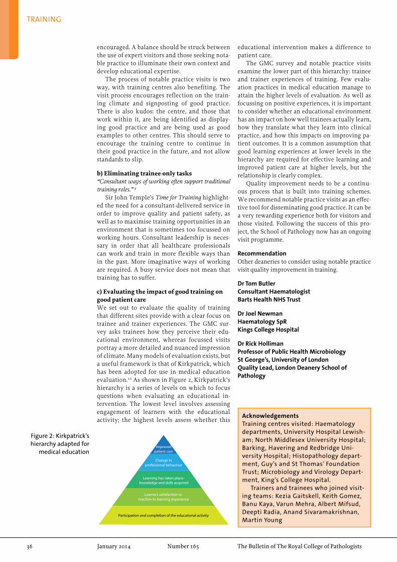
TRAINING
36 January2014 Number165 TheBulletinofTheRoyalCollegeofPathologists
encouraged. A balance should be struck between the use of expert visitors and those seeking nota-ble practice to illuminate their own context and develop educational expertise.
The process of notable practice visits is two way, with training centres also benefiting. The visit process encourages reflection on the train-ing climate and signposting of good practice. There is also kudos: the centre, and those that work within it, are being identified as display-ing good practice and are being used as good examples to other centres. This should serve to encourage the training centre to continue in their good practice in the future, and not allow standards to slip.
b) Eliminating trainee-only tasks“Consultant ways of working often support traditional training roles.” 9
Sir John Temple’s Time for Training highlight-ed the need for a consultant-delivered service in order to improve quality and patient safety, as well as to maximise training opportunities in an environment that is sometimes too focussed on working hours. Consultant leadership is neces-sary in order that all healthcare professionals can work and train in more flexible ways than in the past. More imaginative ways of working are required. A busy service does not mean that training has to suffer.
c) Evaluating the impact of good training on good patient careWe set out to evaluate the quality of training that different sites provide with a clear focus on trainee and trainer experiences. The GMC sur-vey asks trainees how they perceive their edu-cational environment, whereas focussed visits portray a more detailed and nuanced impression of climate. Many models of evaluation exists, but a useful framework is that of Kirkpatrick, which has been adopted for use in medical education evaluation.10 As shown in Figure 2, Kirkpatrick’s hierarchy is a series of levels on which to focus questions when evaluating an educational in-tervention. The lowest level involves assessing engagement of learners with the educational activity; the highest levels assess whether this
educational intervention makes a difference to patient care.
The GMC survey and notable practice visits examine the lower part of this hierarchy: trainee and trainer experiences of training. Few evalu-ation practices in medical education manage to attain the higher levels of evaluation. As well as focussing on positive experiences, it is important to consider whether an educational environment has an impact on how well trainees actually learn, how they translate what they learn into clinical practice, and how this impacts on improving pa-tient outcomes. It is a common assumption that good learning experiences at lower levels in the hierarchy are required for effective learning and improved patient care at higher levels, but the relationship is clearly complex.
Quality improvement needs to be a continu-ous process that is built into training schemes. We recommend notable practice visits as an effec-tive tool for disseminating good practice. It can be a very rewarding experience both for visitors and those visited. Following the success of this pro-ject, the School of Pathology now has an ongoing visit programme.
RecommendationOther deaneries to consider using notable practice visit quality improvement in training.
Dr Tom ButlerConsultant HaematologistBarts Health NHS Trust
Dr Joel NewmanHaematology SpRKings College Hospital
Dr Rick HollimanProfessor of Public Health MicrobiologySt George’s, University of LondonQuality Lead, London Deanery School of Pathology
Figure 2: Kirkpatrick’s hierarchy adapted for
medical educationImproved
patient care
Change inprofessional behaviour
Learning has taken place-knowledge and skills acquired
Learners satisfaction or reaction to learning experience
Participation and completion of the educational activity
AcknowledgementsTraining centres visited: Haematology departments, University Hospital Lewish-am; North Middlesex University Hospital; Barking, Havering and Redbridge Uni-versity Hospital; Histopathology depart-ment, Guy’s and St Thomas’ Foundation Trust; Microbiology and Virology Depart-ment, King’s College Hospital.
Trainers and trainees who joined visit-ing teams: Kezia Gaitskell, Keith Gomez, Banu Kaya, Varun Mehra, Albert Mifsud, Deepti Radia, Anand Sivaramakrishnan, Martin Young
TRAINING
www.rcpath.org Number165 January2014 37
References
1. GMC national trainees surveys 2011 and 2012. www.gmc-uk.org/education/surveys.asp (accessed 18 November 2013).
2. GMC. The Trainee Doctor. http://www.gmc-uk.org/education/postgraduate/standards_and_guid-ance.asp (accessed 18 November 2013).
3. London Deanery. Synapse. https://secure.synapse.nhs.uk/groups/quality-work-stream---school-of-pathology
4. Homana G, Henry Barber C, Torney-Porta J. School Citizenship Education Climate Assessment. Denver: National Centre for Learning and Citizenship, Education Committee of the States, 2005.
5. Cross V, Hicks C, Parle J, Field S. Perceptions of the learning environment in higher specialist training of doctors: implications for recruitment and retention. Medical Education 2006;40:121–128.
6. Roff S, McAleer S, Harden RM , Al-Qahtani M, Ahmed AU, Deza H et al. Development and validation of the Dundee Ready Education Environment Measure (DREEM). Medical Teacher 1997;19:295–299.
7. NHS. The Keogh Mortality Review outcome reports. www.nhs.uk/NHSEngland/bruce-keogh-review/Pages/published-reports.aspx (accessed 18 November 2013).
8. Pereira Gray RJ, Webb NM. Training for General Practice. Plymouth: MacDonald and Evans, 1982.
9. Temple J. Time for Training: A Review of the impact of the European Working Time Directive on the qual-ity of training. Medical Education England, 2010. www.mee.nhs.uk/our_work/work_priorities/better_training_better_care.aspx (accessed 18 November 2013).
10. Belfield CR, Thomas HR, Bullock AD, Eynon R, Wall D. Measuring effectiveness for best evidence medical education – a discussion. Medical Teacher 2001;23:164–170.
Pathology websites for undergraduate education
Forseveralyears,third-yearundergraduatemedicalstudentsintheUniversityofLiverpoolwhoundertake‘specialstudymodules’(student-selectedmodules)inpathologyhavebeenencouragedtoconsiderthetypesofonlineresourcesthat
areavailabletosupportmedicaleducationandthentodevelopelectronicresourcesthatmightassisttheirfellowstudentsinthefuture.
Dr Tim Helliwell These resources are available on the University’s intranet. In 2013, students were invited to review open-access, general pathology websites. This has identified a need for additional resources to help students to integrate clinical and pathological in-formation, and has also informed the development of e-Learning resources in The Royal College of Pa-thologists. This article briefly describes the process and outcomes, and is followed by students’ reflec-tions on participation in the module.
MethodsInternet search engines were used to identify gen-eral pathology sites using a range of search criteria:
• pathology revision sites• pathology education• pathology education undergraduate • pathology teaching• pathology resources.
Additional sites were identified from those used by residents in pathology. Sites dealing with the pathology of single diseases, or diseases of a single tissue or organ were excluded, as they were considered likely to be too specialised for general undergraduate education.
Sites were assessed, using published evaluation criteria1,2 for:• accountability: websites should provide clear
information regarding site authorship, author credentials and associated affiliations. Sources and references used must be stated, along with disclosures on sponsorship/advertising. Good websites also state creation dates and dates of last modification
• content quality: websites should provide a good depth of information, whilst ensuring that the information is credible and accurate. Websites with sources such as published,
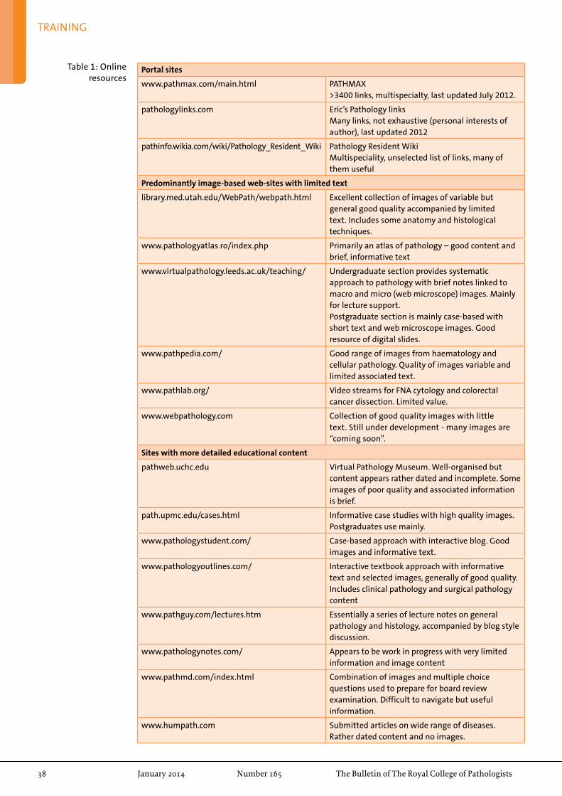
TRAINING
38 January2014 Number165 TheBulletinofTheRoyalCollegeofPathologists
Portal sites
www.pathmax.com/main.html PATHMAX>3400 links, multispecialty, last updated July 2012.
pathologylinks.com Eric’s Pathology linksMany links, not exhaustive (personal interests of author), last updated 2012
pathinfo.wikia.com/wiki/Pathology_Resident_Wiki Pathology Resident WikiMultispeciality, unselected list of links, many of them useful
Predominantly image-based web-sites with limited text
library.med.utah.edu/WebPath/webpath.html Excellent collection of images of variable but general good quality accompanied by limited text. Includes some anatomy and histological techniques.
www.pathologyatlas.ro/index.php Primarily an atlas of pathology – good content and brief, informative text
www.virtualpathology.leeds.ac.uk/teaching/ Undergraduate section provides systematic approach to pathology with brief notes linked to macro and micro (web microscope) images. Mainly for lecture support.Postgraduate section is mainly case-based with short text and web microscope images. Good resource of digital slides.
www.pathpedia.com/ Good range of images from haematology and cellular pathology. Quality of images variable and limited associated text.
www.pathlab.org/ Video streams for FNA cytology and colorectal cancer dissection. Limited value.
www.webpathology.com Collection of good quality images with little text. Still under development - many images are “coming soon”.
Sites with more detailed educational content
pathweb.uchc.edu Virtual Pathology Museum. Well-organised but content appears rather dated and incomplete. Some images of poor quality and associated information is brief.
path.upmc.edu/cases.html Informative case studies with high quality images. Postgraduates use mainly.
www.pathologystudent.com/ Case-based approach with interactive blog. Good images and informative text.
www.pathologyoutlines.com/ Interactive textbook approach with informative text and selected images, generally of good quality. Includes clinical pathology and surgical pathology content
www.pathguy.com/lectures.htm Essentially a series of lecture notes on general pathology and histology, accompanied by blog style discussion.
www.pathologynotes.com/ Appears to be work in progress with very limited information and image content
www.pathmd.com/index.html Combination of images and multiple choice questions used to prepare for board review examination. Difficult to navigate but useful information.
www.humpath.com Submitted articles on wide range of diseases. Rather dated content and no images.
Table 1: Online resources
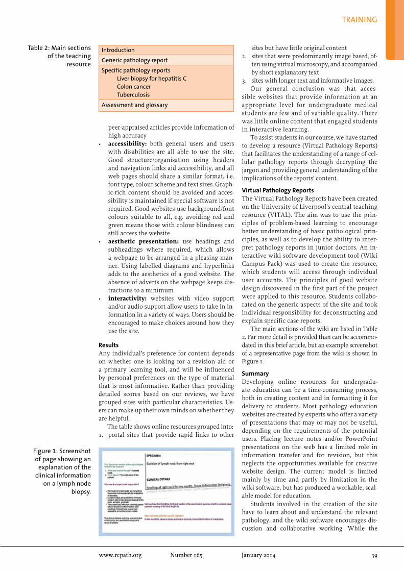
TRAINING
www.rcpath.org Number165 January2014 39
Introduction
Generic pathology report
Specific pathology reportsLiver biopsy for hepatitis CColon cancerTuberculosis
Assessment and glossary
Table 2: Main sections of the teaching
resource
peer-appraised articles provide information of high accuracy
• accessibility: both general users and users with disabilities are all able to use the site. Good structure/organisation using headers and navigation links aid accessibility, and all web pages should share a similar format, i.e. font type, colour scheme and text sizes. Graph-ic-rich content should be avoided and acces-sibility is maintained if special software is not required. Good websites use background/font colours suitable to all, e.g. avoiding red and green means those with colour blindness can still access the website
• aesthetic presentation: use headings and subheadings where required, which allows a webpage to be arranged in a pleasing man-ner. Using labelled diagrams and hyperlinks adds to the aesthetics of a good website. The absence of adverts on the webpage keeps dis-tractions to a minimum
• interactivity: websites with video support and/or audio support allow users to take in in-formation in a variety of ways. Users should be encouraged to make choices around how they use the site.
ResultsAny individual’s preference for content depends on whether one is looking for a revision aid or a primary learning tool, and will be influenced by personal preferences on the type of material that is most informative. Rather than providing detailed scores based on our reviews, we have grouped sites with particular characteristics. Us-ers can make up their own minds on whether they are helpful.
The table shows online resources grouped into:1. portal sites that provide rapid links to other
sites but have little original content2. sites that were predominantly image based, of-
ten using virtual microscopy, and accompanied by short explanatory text
3. sites with longer text and informative images.Our general conclusion was that acces-
sible websites that provide information at an appropriate level for undergraduate medical students are few and of variable quality. There was little online content that engaged students in interactive learning.
To assist students in our course, we have started to develop a resource (Virtual Pathology Reports) that facilitates the understanding of a range of cel-lular pathology reports through decrypting the jargon and providing general understanding of the implications of the reports’ content.
Virtual Pathology ReportsThe Virtual Pathology Reports have been created on the University of Liverpool’s central teaching resource (VITAL). The aim was to use the prin-ciples of problem-based learning to encourage better understanding of basic pathological prin-ciples, as well as to develop the ability to inter-pret pathology reports in junior doctors. An in-teractive wiki software development tool (Wiki Campus Pack) was used to create the resource, which students will access through individual user accounts. The principles of good website design discovered in the first part of the project were applied to this resource. Students collabo-rated on the generic aspects of the site and took individual responsibility for deconstructing and explain specific case reports.
The main sections of the wiki are listed in Table 2. Far more detail is provided than can be accommo-dated in this brief article, but an example screenshot of a representative page from the wiki is shown in Figure 1.
SummaryDeveloping online resources for undergradu-ate education can be a time-consuming process, both in creating content and in formatting it for delivery to students. Most pathology education websites are created by experts who offer a variety of presentations that may or may not be useful, depending on the requirements of the potential users. Placing lecture notes and/or PowerPoint presentations on the web has a limited role in information transfer and for revision, but this neglects the opportunities available for creative website design. The current model is limited mainly by time and partly by limitation in the wiki software, but has produced a workable, scal-able model for education.
Students involved in the creation of the site have to learn about and understand the relevant pathology, and the wiki software encourages dis-cussion and collaborative working. While the
Figure 1: Screenshot of page showing an
explanation of the clinical information
on a lymph node biopsy.
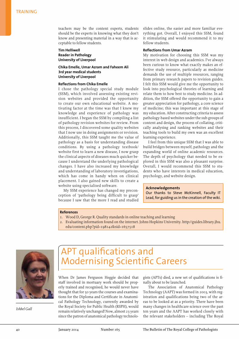
TRAINING
40 January2014 Number165 TheBulletinofTheRoyalCollegeofPathologists
References1. Wood D, George R. Quality standards in online teaching and learning2. Evaluating information found on the internet. Johns Hopkins University. http://guides.library.jhu.
edu/content.php?pid=198142&sid=1657518
teachers may be the content experts, students should be the experts in knowing what they don’t know and presenting material in a way that is ac-ceptable to fellow students.
Tim HelliwellReader in PathologyUniversity of Liverpool
Chika Emelle, Umar Azram and Faheem Ali3rd year medical studentsUniversity of Liverpool
Reflections from Chika EmelleI chose the pathology special study module (SSM), which involved assessing existing revi-sion websites and provided the opportunity to create our own educational website. A mo-tivating factor at the time was that I knew my knowledge and experience of pathology was insufficient. I began the SSM by compiling a list of pathology revision websites for review. From this process, I discovered some quality websites that I now use in doing assignments or revision. Additionally, this SSM taught me the value of pathology as a basis for understanding disease conditions. By using a pathology textbook/website first to learn a new disease, I now grasp the clinical aspects of diseases much quicker be-cause I understand the underlying pathological changes. I have also increased my knowledge and understanding of laboratory investigations, which has come in handy when on clinical placement. I also gained new skills to create a website using specialised software.
My SSM experience has changed my precon-ception of ‘pathology being difficult to grasp’ because I saw that the more I read and studied
slides online, the easier and more familiar eve-rything got. Overall, I enjoyed this SSM, found it stimulating and would recommend it to my fellow students.
Reflections from Umar AzramMy motivation for choosing this SSM was my interest in web design and academics. I’ve always been curious to know what exactly makes an ef-fective study resource, particularly as medicine demands the use of multiple resources, ranging from primary research papers to revision guides. I felt this SSM would give me the opportunity to look into psychological theories of learning and relate them to how best to study medicine. In ad-dition, the SSM offered the opportunity to gain a greater appreciation for pathology, a core science of medicine; this was important at this stage of my education. After constructing criteria to assess pathology-based websites under the sub-groups of content and design, the process of collating, criti-cally analysing and ranking websites and their teaching tools to build my own was an excellent learning experience.
I feel from this unique SSM that I was able to build bridges between myself, pathology and the expanding world of online academic resources. The depth of psychology that needed to be ex-plored in this SSM was also a pleasant surprise. Overall, I would recommend this SSM to stu-dents who have interests in medical education, psychology, and website design.
aPt qualifications and Modernising Scientific Careers
Ishbel Gall
When Dr James Ferguson Heggie decided that staff involved in mortuary work should be prop-erly trained and recognised, he would never have thought that for 50 years the courses and examina-tions for the Diploma and Certificate in Anatomi-cal Pathology Technology, currently awarded by the Royal Society for Public Health (RSPH), would remain relatively unchanged! Now, almost 23 years since the patron of anatomical pathology technolo-
gists (APTs) died, a new set of qualifications is fi-nally about to be launched.
The Association of Anatomical Pathology Technology (AAPT) was formed in 2003, with reg-istration and qualifications being two of the ar-eas to be looked at as a priority. There have been many changes in healthcare science over the past ten years and the AAPT has worked closely with the relevant stakeholders – including The Royal
AcknowledgementsOur thanks to Steve McKinnell, Faculty IT Lead, for guiding us in the creation of the wiki.
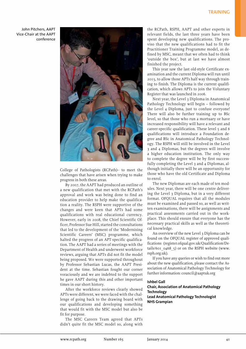
TRAINING
www.rcpath.org Number165 January2014 41
College of Pathologists (RCPath)– to meet the challenges that have arisen when trying to make progress in both these areas.
By 2007, the AAPT had produced an outline of a new qualification that met with the RCPath’s approval and work was being done to find an education provider to help make the qualifica-tion a reality. The RSPH were supportive of the changes and were keen that APTs had some qualifications with real educational currency. However, early in 2008, the Chief Scientific Of-ficer, Professor Sue Hill, started the consultations that led to the development of the ‘Modernising Scientific Careers’ (MSC) programme, which halted the progress of an APT-specific qualifica-tion. The AAPT had a series of meetings with the Department of Health and underwent workforce reviews, arguing that APTs did not fit the model being proposed. We were supported throughout by Professor Sebastian Lucas, the AAPT Presi-dent at the time. Sebastian fought our corner voraciously and we are indebted to the support he gave AAPT during this and other important times in our short history.
After the workforce reviews clearly showed APTs were different, we were faced with the chal-lenge of going back to the drawing board with our qualifications and developing something that would fit with the MSC model but also be fit for purpose.
The MSC Careers Team agreed that APTs didn’t quite fit the MSC model so, along with
the RCPath, RSPH, AAPT and other experts in relevant fields, the last three years have been spent developing new qualifications. The pro-viso that the new qualifications had to fit the Practitioner Training Programme model, as de-fined by MSC, meant that we often had to think ‘outside the box’, but at last we have almost finished the project.
This year saw the last old-style Certificate ex-amination and the current Diploma will run until 2015, to allow those APTs half way through train-ing to finish. The Diploma is the current qualifi-cation, which allows APTs to join the Voluntary Register that was launched in 2006.
Next year, the Level 3 Diploma in Anatomical Pathology Technology will begin – followed by the Level 4 Diploma, just to confuse everyone! There will also be further training up to BSc level, so that those who run a mortuary or have increased responsibility will have a relevant and career-specific qualification. These level 5 and 6 qualifications will introduce a Foundation de-gree and BSc in Anatomical Pathology Technol-ogy. The RSPH will still be involved in the Level 3 and 4 Diplomas, but the degrees will involve a higher education institution. The only way to complete the degree will be by first success-fully completing the Level 3 and 4 Diplomas, al-though initially there will be an opportunity for those who have the old Certificate and Diploma to enrol.
The new Diplomas are each made of ten mod-ules. Next year, there will be one centre deliver-ing the Level 3 Diploma, but in a very different format. OFQUAL requires that all the modules must be examined and passed so, as well as writ-ten examinations, there will be assignments and practical assessments carried out in the work-place. This should ensure that everyone has the necessary practical skills as well as the theoreti-cal knowledge.
An overview of the new Level 3 Diploma can be found on the OFQUAL register of approved quali-fications (register.ofqual.gov.uk/Qualification/De-tails/601_0468_5) or on the RSPH website (www.rsph.org.uk).
If you have any queries or wish to find out more about the new qualification, please contact the As-sociation of Anatomical Pathology Technology for further information: [email protected]
Ishbel GallChair, Association of Anatomical Pathology TechnologyLead Anatomical Pathology TechnologistNHS Grampian
John Pitchers, AAPT Vice-Chair at the AAPT
conference
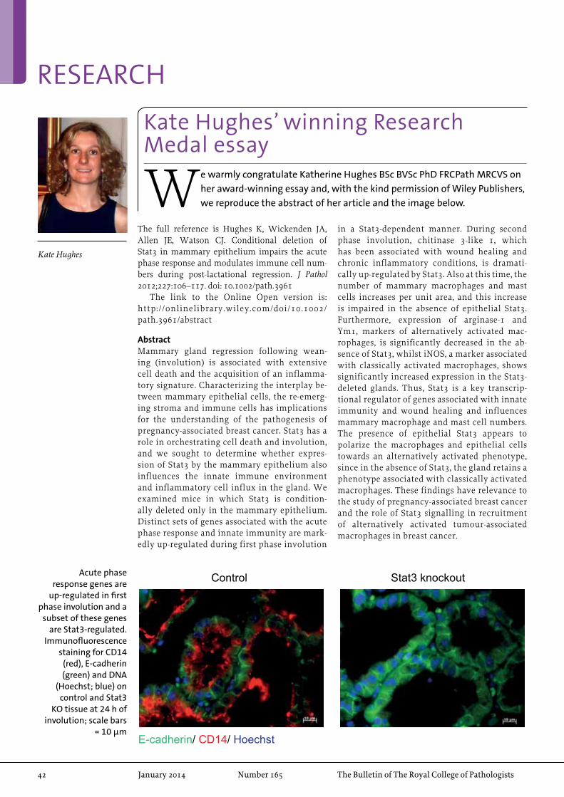
42 January2014 Number165 TheBulletinofTheRoyalCollegeofPathologists
rESEarCH
The full reference is Hughes K, Wickenden JA, Allen JE, Watson CJ. Conditional deletion of Stat3 in mammary epithelium impairs the acute phase response and modulates immune cell num-bers during post-lactational regression. J Pathol 2012;227:106–117. doi: 10.1002/path.3961
The link to the Online Open version is: http://onlinelibrary.wiley.com/doi/10.1002/path.3961/abstract
AbstractMammary gland regression following wean-ing (involution) is associated with extensive cell death and the acquisition of an inflamma-tory signature. Characterizing the interplay be-tween mammary epithelial cells, the re-emerg-ing stroma and immune cells has implications for the understanding of the pathogenesis of pregnancy-associated breast cancer. Stat3 has a role in orchestrating cell death and involution, and we sought to determine whether expres-sion of Stat3 by the mammary epithelium also influences the innate immune environment and inflammatory cell influx in the gland. We examined mice in which Stat3 is condition-ally deleted only in the mammary epithelium. Distinct sets of genes associated with the acute phase response and innate immunity are mark-edly up-regulated during first phase involution
in a Stat3-dependent manner. During second phase involution, chitinase 3-like 1, which has been associated with wound healing and chronic inflammatory conditions, is dramati-cally up-regulated by Stat3. Also at this time, the number of mammary macrophages and mast cells increases per unit area, and this increase is impaired in the absence of epithelial Stat3. Furthermore, expression of arginase-1 and Ym1, markers of alternatively activated mac-rophages, is significantly decreased in the ab-sence of Stat3, whilst iNOS, a marker associated with classically activated macrophages, shows significantly increased expression in the Stat3-deleted glands. Thus, Stat3 is a key transcrip-tional regulator of genes associated with innate immunity and wound healing and influences mammary macrophage and mast cell numbers. The presence of epithelial Stat3 appears to polarize the macrophages and epithelial cells towards an alternatively activated phenotype, since in the absence of Stat3, the gland retains a phenotype associated with classically activated macrophages. These findings have relevance to the study of pregnancy-associated breast cancer and the role of Stat3 signalling in recruitment of alternatively activated tumour-associated macrophages in breast cancer.
kate Hughes’ winning research Medal essay
We warmly congratulate Katherine Hughes BSc BVSc PhD FRCPath MRCVS on her award-winning essay and, with the kind permission of Wiley Publishers, we reproduce the abstract of her article and the image below.
Kate Hughes
Acute phase response genes are
up-regulated in first phase involution and a
subset of these genes are Stat3-regulated.
Immunofluorescence staining for CD14
(red), E-cadherin (green) and DNA
(Hoechst; blue) on control and Stat3
KO tissue at 24 h of involution; scale bars
= 10 µm
Control Stat3 knockout
E-cadherin/ CD14/ Hoechst
www.rcpath.org Number165 January2014 43
CliNiCal EFFECtiVENESS
As UK healthcare and public expectations change, pathology needs to lead the way by example. Quality is not a new concept to us. Neither is standardisation. However, there is now a new focus on these concepts arising from a variety of political imperatives, including the reconfigura-tion of pathology services and the recommenda-tions of reports such as Francis and the Pathology Quality Assurance Review of Dr Ian Barnes. Pathol-ogy needs to spread its influence by assuming responsibility for the entire specimen pathway, from ‘brain to brain’ with meaningful user in-volvement. We need to inform commissioners at national and local level about what they should expect and how to go about asking for it. The Atlas of Variation should shame us into action and in-crease the impetus towards standardisation. The College will work with other organisations that have a role in quality, such as NICE and CPA. We are also moving towards a spirit of openness and candour. Sharing information, including our de-fects, will become the order of the day. Pathology will become less authoritarian in its dealings with users, whilst avoiding the common misperception of its position as one of a subservient ‘support’ ser-vice. Pathology must also be well placed to meet the demands of a technological revolution, which will see more molecular and genomic testing in all disciplines and perhaps the devolution of test-ing to a point closer to the patient than it has ever been. The College’s new Department of Clinical Effectiveness was established to work through these issues and help to secure the College’s right-ful place in the vanguard of change.
Regularising the standard-setting processThere needs to be a consistent and meaningful way of determining standards in pathology. This means that the actual process for identifying standard working practices should itself comply with acceptable standards.
This is best illustrated by the RCPath Cancer Datasets and Tissue Pathways. These are two se-
ries of guidelines for cellular pathologists on the standards of their working practice and are pro-duced through a process that has been accredited by NICE. Such accreditation is not easily gained. Neither is it easily retained. The inclusion of the NICE accreditation logo on a document assures the reader that it has met 25 criteria based on AGREE II instrument and represents the latest, evidence-based guidance or, in the absence of a robust evidence base, consensus expert opinion.
The cellular pathology documents are the only documents solely produced by the College to have achieved accreditation. In microbiology, the College has been involved in an equal-part-nership collaboration with professional societies and Public Health England in the production of the NICE accredited Standardised Microbiol-ogy Investigations. Similar partnerships in other disciplines of pathology will enable a healthy throughput of up-to-date and authoritative doc-uments, whilst sharing the substantial workload associated with developing and maintaining guidelines and achieving accreditation. Guide-line production will need to be carefully planned and managed, with appropriate prioritisation.
Another aspect of standards setting is the pathology catalogues. The National Labora-tory Medicine Catalogue (NLMC) and the Pa-thology Bounded Code Language (PBCL) are projects that seek to standardise the vertical two-way communication between patholo-gists and service users, including patients, and the horizontal communication between organ-isations in different geographical areas. This will include standard requesting, standard re-porting, standard units and, as far as possible, standard interpretation of results. The Clinical Effectiveness Department has a governance role in the work that is carried out on behalf of the NHS National Pathology Programme.
A third area of standards setting is pathol-ogy demand optimisation. The College will, in partnership with the Pathology Alliance, gov-
the clinical effectiveness agenda for pathology
The new Clinical Effectiveness Department (CED) is part of the College’s structural reorganisation. Its overall aim is to promote and develop standardisation of clinical and laboratory practice against a culture of continuous quality improvement (CQI)
and to achieve this in partnership with service users, including clinical, patient and lay involvement. The first three-year strategy was presented to Council in November 2013 and this article sets out the key elements of that strategy.
Dr Peter Cowling
44 January2014 Number165 TheBulletinofTheRoyalCollegeofPathologists
CLINICAL EFFECTIVENESS
ernment and industry, issue guidance on how to eliminate the waste associated with unnecessary testing, including minimum retesting intervals. It is expected that such guidance will be used by commissioners in setting the specifications of pathology service provision.
Regularising the measurement of practices against standardsThere are a number of ways in which pathology services can be scrutinised to assure compliance with accepted standards of working. Tradition-ally, laboratory and clinical audits using the audit cycle methodology, laboratory quality assurance schemes, both internal and external, clinical governance activities such as incident reporting and, of course, laboratory accreditation have been the predominant methods. As CQI becomes more widely adopted, other methods of examining not only the laboratory processes but also the wider aspects of the entire specimen pathway are also being introduced.
The College will embrace CQI and promote its use in our pathology services. At the present time, we have a well-established audit certifica-tion scheme, which allows Fellows to submit their clinical and laboratory audits online for peer review and accreditation of quality with the award of a certificate for each successful completion of the audit cycle. Some of the best examples of these are published in the College Bulletin and the database of certificated audits is available to Fellows. A similar approach could be developed for other CQI activities, particu-larly those with a similar cyclical methodology such as ‘Plan, Do, Study, Act’ (PDSA) and A3 problem solving.
Further development of the key performance indicators in pathology (KPIs) will be supported by the Clinical Effectiveness Department. A pi-lot project is underway to assess the feasibility of inspecting KPI compliance within laboratory accreditation schemes and this pilot will in-form us of any modifications required or gaps that exist in the present KPI repertoire.
Of supreme importance in measuring per-formance against standards is quality assurance (QA). This is a complex subject. There is an urgent need for the College to clarify its position on the whole issue of QA and to assert its rightful role as the guardian of standards in pathology by influ-encing the underlying principles and the actual delivery of QA schemes. If the College accepts that all aspects of the service delivered by the pathol-ogy workforce should be subject to scrutiny, then it follows that QA needs to be expanded to include interpretative schemes. Those responsible for QA schemes need to be cognisant of the changing societal expectations of openness and candour – both generally in public life and specifically in healthcare. No person or organisation involved in
QA should shirk their responsibilities because of the difficulty of the task.
Securing the authority of the College as the guardian of standards in pathologyThe College has an important leadership role. It must earn and deserve its authority and it must win friends and influence people – or more ac-curately, organisations. There are already a great number of lines of communication and joint working projects with other organisations, but these need to be strengthened and expanded. The College should be systematic about its own publications, such as guidance, advice, and me-dia releases, and it should have in place a robust method of engaging in the work of external or-ganisations through, for example, consultations or more direct involvement as a partner. A more rigorous and timely approach towards every-thing that is publically visible will ensure that the College will be the fountainhead of sensible opinion and will establish its position as an in-dispensible resource to government, profession-al societies, other bodies and the wider public. Furthering the cause of pathology education by influencing the development of curricula would also be part of this leadership role.
Embedding clinically effective pathology into practiceChange is constant. That is the inevitable conse-quence of CQI. All of us need to work differently through continuous improvement. The stressful nature of change is well understood and those un-dergoing change require effective leadership.
The College is committed to developing the present and future leaders of our profession. To this end, we have been successful, in partner-ship with The Royal College of Radiologists and the National Pathology Programme, in securing matched funding from the Academy of Medi-cal Royal Colleges to deliver a leadership pro-gramme that will be phase 3 of a successful series of programmes (the first two being ‘Influencing the Future: Leading transformational culture change’ and ‘Leadership in Action’).
Dr Peter CowlingDirector of Clinical Effectiveness
www.rcpath.org Number165 January2014 45
CLINICAL EFFECTIVENESS
Vena cava filter audit
The College’s Professional Standards Unit wishes to encourage high-quality clinical audit. We therefore periodically publish interesting examples of audits that have been successfully evaluated through our clinical audit certification scheme.
Dr Gill Turner
Dr Jennie Wimperis
Dr Ali Reza Rismani
IntroductionThe BCSH guidelines on use of vena cava (VC) filters (Baglin T et al, 2006) give clear indications for the insertion of VC filters. The indications are to prevent pulmonary embolus (PE) in the following groups:
• patients with venous thromboembolism (VTE) in whom anticoagulation is contraindicated
• selected patients who develop PE despite thera-peutic anticoagulation. Prior to VC filter inser-tion, long-term high-intensity oral anticoagu-lant therapy or treatment with low molecular weight heparin (LMWH) should be considered
• pregnant patients who develop extensive VTE shortly before delivery
• patients who must undergo surgery for which anticoagulation has to be interrupted within one month of a VTE
• patients undergoing a pulmonary endarterec-tomy for chronic thromboembolic pulmonary hypertension.In 2009, an audit was undertaken at Norfolk
and Norwich University Hospital (NNUH), which found that 16/48 patients (33%) undergoing VC filter insertion did not have one of the above indi-cations. This audit also found that local guidelines to discuss cases with a consultant haematologist were not being followed in the vast majority of cases. In addition to this, a substantial number of filters which should have been temporary were not removed (75% or 24/32). A summary is provided at the end of this article.
Following this audit, a revised guideline, in-tended to give clearer instruction to clinicians, was issued and in January 2011 a service was set up to manage the insertion of VC filters and prevent in-appropriate insertions from taking place.
A re-audit of VC filter insertions was carried out in 2012, collecting data retrospectively from January 2011 to September 2012 and prospectively from September to December 2012 to assess what impact, if any, the service and revised guideline had had on reducing inappropriate VC filter inser-tions, removal of temporary filters and compliance with local guidelines to discuss each case with a consultant haematologist prior to insertion.
ResultsFrom January 2011 to December 2012 (24 months), 70 VC filter insertions were carried out on 69 patients (one reinsertion). Forty patients were
female (58%) and 29 male (42%). Patients were aged between 26 and 92 years old; the median age at time of insertion was 73 years old. Thirty-nine patients (57%) had active malignancy at the time of insertion.
The indications for insertion were as follows (detailed in Table 1):
• prevention of PE in patient with VTE where anticoagulation is contraindicated (59%)
• PE despite anticoagulation (9%) – all of these patients had an active malignancy
• VTE in late stages of pregnancy (1%)
• pre-operatively within one month of a VTE (23%)
• pre-pulmonary endarterectomy (9%)
• other indication (7%).NB. Five VC insertions were for more than one in-dication.
In total, five (7%) insertions took place for an indication other than those stipulated in the BCSH guidelines (Figure 1).
Of these, two patients had a VC filter inserted following extension of DVT despite therapeutic anticoagulation. Both patients had active malig-nancy; one had previously had a saddle embolus. A further two patients had a VC filter inserted prior to surgery which took place more than one month after VTE. Both patients had suffered PEs, one had surgery six weeks afterwards and the other patient had the additional risk factor of polycythaemia. Both patients had surgery for active malignancy.
The remaining case not fitting the criteria for in-sertion specified in the BCSH guidelines bypassed the normal route for insertion of VC filters and was inserted at the time of directed thrombolysis in a patient with extensive thrombosis of both internal iliac veins, inferior vena cava and renal veins. On review of all the cases, this one case (1% of all in-sertions) was the only one that was not felt to have been indicated.
Seventeen out of 70 cases (24%) had no docu-mentation of a discussion with a haematology consultant, as specified in the local guideline for VC filter insertion. Five of these were cases where a filter had been requested by the cardiothoracic surgeons pre-pulmonary endarterectomy and so bypassed discussion with haematology (Figure 2).
In 19 cases (27%), no mention of the VC filter insertion was made in the hospital discharge let-ter (Figure 3). One patient had no discharge letter
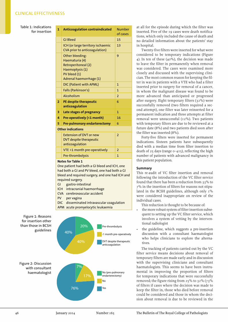
46 January2014 Number165 TheBulletinofTheRoyalCollegeofPathologists
CLINICAL EFFECTIVENESS
1 Anticoagulation contraindicated Number of cases
GI Bleed 15
ICH (or large territory ischaemic CVA prior to anticoagulation)
13
Other bleeding:Haematuria (4)Retroperitoneal (2)Haemoptysis (1)PV bleed (1)Adrenal haemorrhage (1)
9
DIC (Patient with APML) 1
Falls (Parkinson's) 1
Alcoholism 2
2 PE despite therapeutic anticoagulation
6
3 Late stages of pregnancy 1
4 Pre-operatively (<1 month) 16
5 Pre-pulmonary endarterectomy 6
Other indications
Extension of DVT or new DVT despite therapeutic anticoagulation
2
VTE >1 month pre-operatively 2
Pre-thrombolysis 1
Notes for Table 1One patient had both a GI bleed and ICH, one had both a GI and PV bleed, one had both a GI bleed and required surgery, and one had ICH and required surgery.GI gastro-intestinalICH intracranial haemorrhageCVA cerebrovascular accidentPV per vaginaDIC disseminated intravascular coagulationAPM acute promyelocytic leukaemia
Table 1: Indications for insertion
DVT despite therapeuticanticoagulation
›1 month pre-operatively
Pre-thrombolysis20%
40%
40%
Figure 1: Reasons for insertion other
than those in BCSH guidelines
Yes
No
No (pre-pulmonaryendarterectomy)
7%
17%
76%
Figure 2: Discussion with consultant
haematologist
at all for the episode during which the filter was inserted. Five of the 19 cases were death notifica-tions, which only included the cause of death and no detailed information about the patients’ stay in hospital.
Twenty-five filters were inserted for what were considered to be temporary indications (Figure 4). In ten of these (40%), the decision was made to leave the filter in permanently when removal was considered. The cases were examined more closely and discussed with the supervising clini-cian. The most common reason for keeping the fil-ter in was in patients with a VTE who had a filter inserted prior to surgery for removal of a cancer, in whom the malignant disease was found to be more advanced than anticipated or progressed after surgery. Eight temporary filters (32%) were successfully removed (two filters required a sec-ond attempt), one filter was later reinserted for a permanent indication and three attempts at filter removal were unsuccessful (12%). Two patients with temporary filters are due to be reviewed at a future date (8%) and two patients died soon after the filter was inserted (8%).
Forty-five filters were inserted for permanent indications. Sixteen patients have subsequently died with a median time from filter insertion to death of 25 days (range 0–415), reflecting the high number of patients with advanced malignancy in this patient population.
SummaryThis re-audit of VC filter insertion and removal following the introduction of the VC filter service found that there has been a reduction from 33% to 7% in the insertion of filters for reasons not stipu-lated in the BCSH guidelines, although only 1% were considered inappropriate on review of the individual cases.
This reduction is thought to be because of:• the more robust system of filter insertion subse-
quent to setting up the VC filter service, which involves a system of vetting by the interven-tional radiologist
• the guideline, which suggests a pre-insertion discussion with a consultant haematologist who helps clinicians to explore the alterna-tives.
The tracking of patients carried out by the VC filter service means decisions about removal of temporary filters are made early and in discussion with the supervising clinicians and consultant haematologists. This seems to have been instru-mental in improving the proportion of filters for temporary indications that were successfully removed; the figure rising from 25% to 32% (73% of filters if cases where the decision was made to keep the filter in, those who died before removal could be considered and those in whom the deci-sion about removal is due to be reviewed in the
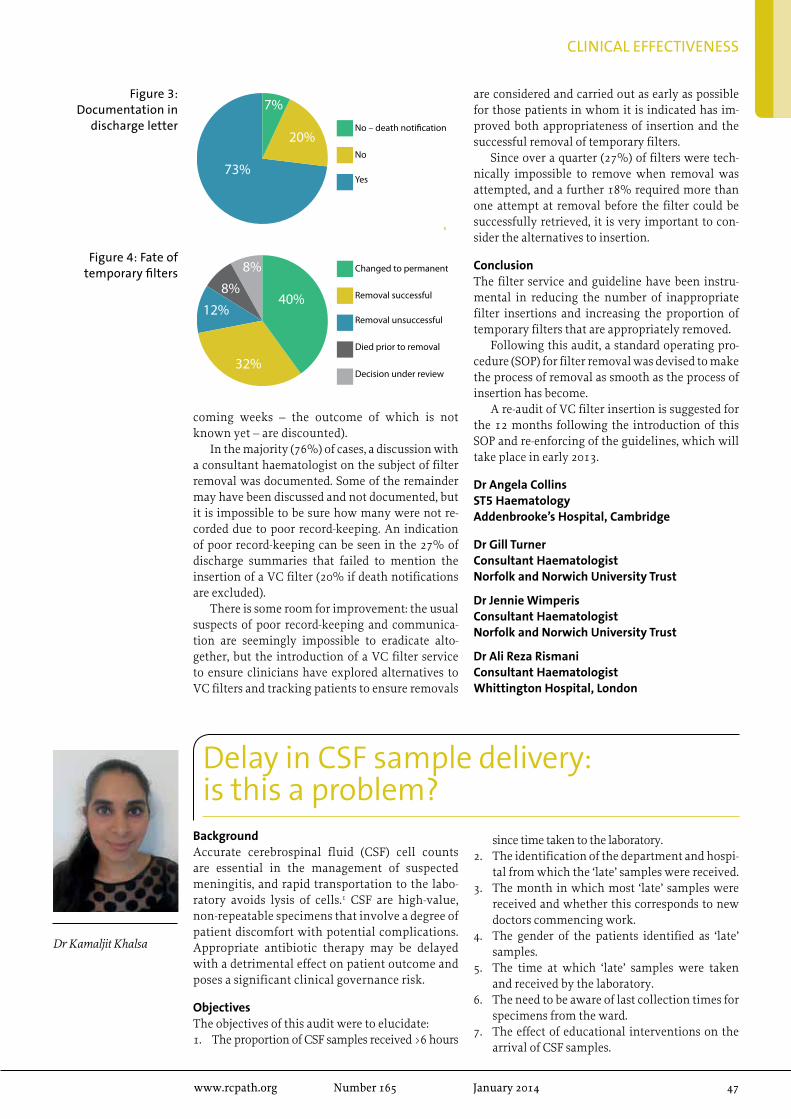
www.rcpath.org Number165 January2014 47
CLINICAL EFFECTIVENESS
coming weeks – the outcome of which is not known yet – are discounted).
In the majority (76%) of cases, a discussion with a consultant haematologist on the subject of filter removal was documented. Some of the remainder may have been discussed and not documented, but it is impossible to be sure how many were not re-corded due to poor record-keeping. An indication of poor record-keeping can be seen in the 27% of discharge summaries that failed to mention the insertion of a VC filter (20% if death notifications are excluded).
There is some room for improvement: the usual suspects of poor record-keeping and communica-tion are seemingly impossible to eradicate alto-gether, but the introduction of a VC filter service to ensure clinicians have explored alternatives to VC filters and tracking patients to ensure removals
are considered and carried out as early as possible for those patients in whom it is indicated has im-proved both appropriateness of insertion and the successful removal of temporary filters.
Since over a quarter (27%) of filters were tech-nically impossible to remove when removal was attempted, and a further 18% required more than one attempt at removal before the filter could be successfully retrieved, it is very important to con-sider the alternatives to insertion.
ConclusionThe filter service and guideline have been instru-mental in reducing the number of inappropriate filter insertions and increasing the proportion of temporary filters that are appropriately removed.
Following this audit, a standard operating pro-cedure (SOP) for filter removal was devised to make the process of removal as smooth as the process of insertion has become.
A re-audit of VC filter insertion is suggested for the 12 months following the introduction of this SOP and re-enforcing of the guidelines, which will take place in early 2013.
Dr Angela CollinsST5 HaematologyAddenbrooke’s Hospital, Cambridge
Dr Gill TurnerConsultant HaematologistNorfolk and Norwich University Trust
Dr Jennie WimperisConsultant HaematologistNorfolk and Norwich University Trust
Dr Ali Reza RismaniConsultant HaematologistWhittington Hospital, London
Yes
No
No – death noti�cation
7%
20%
73%
Figure 3: Documentation in
discharge letter
Removal unsuccessful
Removal successful
Changed to permanent
40%
32%
12%8%
8%
Died prior to removal
Decision under review
Figure 4: Fate of temporary filters
delay in CSF sample delivery: is this a problem?
Dr Kamaljit Khalsa
BackgroundAccurate cerebrospinal fluid (CSF) cell counts are essential in the management of suspected meningitis, and rapid transportation to the labo-ratory avoids lysis of cells.1 CSF are high-value, non-repeatable specimens that involve a degree of patient discomfort with potential complications. Appropriate antibiotic therapy may be delayed with a detrimental effect on patient outcome and poses a significant clinical governance risk.
ObjectivesThe objectives of this audit were to elucidate:1. The proportion of CSF samples received >6 hours
since time taken to the laboratory.2. The identification of the department and hospi-
tal from which the ‘late’ samples were received.3. The month in which most ‘late’ samples were
received and whether this corresponds to new doctors commencing work.
4. The gender of the patients identified as ‘late’ samples.
5. The time at which ‘late’ samples were taken and received by the laboratory.
6. The need to be aware of last collection times for specimens from the ward.
7. The effect of educational interventions on the arrival of CSF samples.
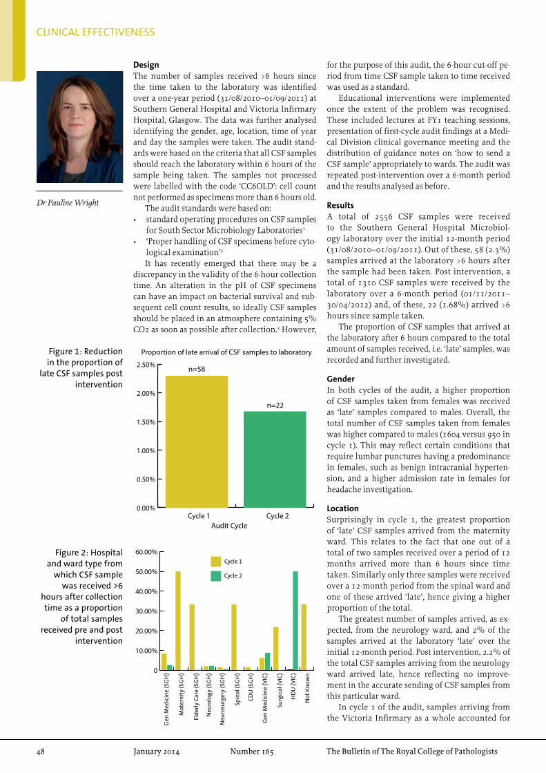
48 January2014 Number165 TheBulletinofTheRoyalCollegeofPathologists
CLINICAL EFFECTIVENESS
Dr Pauline Wright
DesignThe number of samples received >6 hours since the time taken to the laboratory was identified over a one-year period (31/08/2010–01/09/2011) at Southern General Hospital and Victoria Infirmary Hospital, Glasgow. The data was further analysed identifying the gender, age, location, time of year and day the samples were taken. The audit stand-ards were based on the criteria that all CSF samples should reach the laboratory within 6 hours of the sample being taken. The samples not processed were labelled with the code ‘CC6OLD’: cell count not performed as specimens more than 6 hours old.
The audit standards were based on:• standard operating procedures on CSF samples
for South Sector Microbiology Laboratories1
• ‘Proper handling of CSF specimens before cyto-logical examination’2
It has recently emerged that there may be a discrepancy in the validity of the 6-hour collection time. An alteration in the pH of CSF specimens can have an impact on bacterial survival and sub-sequent cell count results, so ideally CSF samples should be placed in an atmosphere containing 5% CO2 as soon as possible after collection.3 However,
for the purpose of this audit, the 6-hour cut-off pe-riod from time CSF sample taken to time received was used as a standard.
Educational interventions were implemented once the extent of the problem was recognised. These included lectures at FY1 teaching sessions, presentation of first-cycle audit findings at a Medi-cal Division clinical governance meeting and the distribution of guidance notes on ‘how to send a CSF sample’ appropriately to wards. The audit was repeated post-intervention over a 6-month period and the results analysed as before.
ResultsA total of 2556 CSF samples were received to the Southern General Hospital Microbiol-ogy laboratory over the initial 12-month period (31/08/2010–01/09/2011). Out of these, 58 (2.3%) samples arrived at the laboratory >6 hours after the sample had been taken. Post intervention, a total of 1310 CSF samples were received by the laboratory over a 6-month period (01/11/2011–30/04/2012) and, of these, 22 (1.68%) arrived >6 hours since sample taken.
The proportion of CSF samples that arrived at the laboratory after 6 hours compared to the total amount of samples received, i.e. ‘late’ samples, was recorded and further investigated.
GenderIn both cycles of the audit, a higher proportion of CSF samples taken from females was received as ‘late’ samples compared to males. Overall, the total number of CSF samples taken from females was higher compared to males (1604 versus 950 in cycle 1). This may reflect certain conditions that require lumbar punctures having a predominance in females, such as benign intracranial hyperten-sion, and a higher admission rate in females for headache investigation.
LocationSurprisingly in cycle 1, the greatest proportion of ‘late’ CSF samples arrived from the maternity ward. This relates to the fact that one out of a total of two samples received over a period of 12 months arrived more than 6 hours since time taken. Similarly only three samples were received over a 12-month period from the spinal ward and one of these arrived ‘late’, hence giving a higher proportion of the total.
The greatest number of samples arrived, as ex-pected, from the neurology ward, and 2% of the samples arrived at the laboratory ‘late’ over the initial 12-month period. Post intervention, 2.2% of the total CSF samples arriving from the neurology ward arrived late, hence reflecting no improve-ment in the accurate sending of CSF samples from this particular ward.
In cycle 1 of the audit, samples arriving from the Victoria Infirmary as a whole accounted for
Figure 1: Reduction in the proportion of
late CSF samples post intervention
0.00%
0.50%
1.00%
1.50%
2.00%
2.50%
Cycle 1 Cycle 2
n=58
n=22
Audit Cycle
Proportion of late arrival of CSF samples to laboratory
0
10.00%
20.00%
30.00%
40.00%
50.00%
60.00%
Gen
Med
icin
e (S
GH
)
Mat
erni
ty (S
GH
)
Elde
rly
Care
(SG
H)
Neu
rolo
gy (S
GH
)
Neu
rosu
rger
y (S
GH
)
Spin
al (S
GH
)
CD
U (S
GH
)
Gen
Med
icin
e (V
IC)
Surg
ical
(VIC
)
HD
U (V
IC)
Not
Kno
wn
Cycle 1
Cycle 2
Figure 2: Hospital and ward type from
which CSF sample was received >6
hours after collection time as a proportion
of total samples received pre and post
intervention
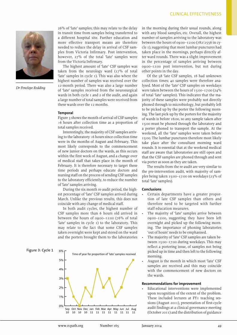
www.rcpath.org Number165 January2014 49
CLINICAL EFFECTIVENESS
Dr Penelope Redding
26% of ‘late’ samples; this may relate to the delay in transit time from samples being transferred to a different hospital site. Further education and more effective transport means are therefore needed to reduce the delay in arrival of CSF sam-ples from Victoria Infirmary. Post intervention, however, 27% of the total ‘late’ samples were from the Victoria Infirmary.
The highest amount of ‘late’ CSF samples was taken from the neurology ward (17% of total ‘late’ samples in cycle 1). This was also where the highest number of samples was received over the 12-month period. There was also a large number of ‘late’ samples received from the neurosurgical wards in both cycle 1 and 2 of the audit, and again a large number of total samples were received from these wards over the 12 months.
TemporalFigure 3 shows the month of arrival of CSF samples >6 hours after collection time as a proportion of total samples received.
Interestingly, the majority of CSF samples arriv-ing to the laboratory >6 hours since collection time were in the months of August and February. This most likely corresponds to the commencement of new junior doctors on the wards, as this occurs within the first week of August, and a change over of medical staff that takes place in the month of February. It is therefore necessary to target these time periods and perhaps educate doctors and nursing staff on the process of sending CSF samples to the laboratory efficiently, to reduce the number of ‘late’ samples arriving.
During the six month re-audit period, the high-est percentage of ‘late’ CSF samples arrived during March. Unlike the previous results, this does not coincide with any change of medical staff.
In both audit cycles, the highest number of CSF samples more than 6 hours old arrived in between the hours of 09:00–12:00 (76% of total ‘late’ samples in cycle 1) to the laboratory. This may relate to the fact that some CSF samples taken overnight were kept and stored on the ward and the porters brought them to the laboratories
in the morning during their usual rounds, along with any blood samples, etc. Overall, the highest number of samples arriving to the laboratory was between the hours of 09:00–12:00 (782/2556 in cy-cle 1), suggesting that most lumbar punctures had taken place in the mornings, perhaps directly af-ter ward rounds. There was a slight improvement in the percentage of samples arriving between 09:00–12:00 post intervention, but not during other points in the day.
Of the 58 ‘late CSF samples, 16 had unknown collection times; 40 samples were therefore ana-lysed. Most of the ‘late’ CSF samples on weekdays were taken between the hours of 15:00–17:00 (24% of total ‘late’ samples). This indicates that the ma-jority of these samples were probably not directly phoned through to microbiology, but probably left to be picked up by the porter the following morn-ing. The last pick-up by the porters for the majority of wards is before 16:00, so any sample taken after 15:00 must be phoned through the laboratory and a porter phoned to transport the sample. At the weekend, all the ‘late’ samples were taken before 15:00. The lumbar punctures therefore most likely take place after the consultant morning ward rounds. It is essential that at the weekend medical staff are aware that laboratories are still open and that the CSF samples are phoned through and sent via porter as soon as they are taken.
The results from the re-audit are very similar to the pre-intervention audit, with majority of sam-ples being taken 15:00–17:00 on weekdays (23% of total ‘late’ samples).
Conclusions• Certain departments have a greater propor-
tion of late CSF samples than others and therefore need to be targeted with further staff education measures.
• The majority of ‘late’ samples arrive between 09:00–12:00, suggesting they have been left overnight and picked up the following morn-ing. The importance of phoning laboratories ‘out of hours’ needs to be emphasised.
• The majority of ‘late’ CSF samples are taken be-tween 15:00–17:00 during weekdays. This may reflect a portering issue, of samples not being picked up in time and then left to the following morning.
• August is the month in which most ‘late’ CSF samples are received and this may coincide with the commencement of new doctors on the wards.
Recommendations for improvement• Educational interventions were implemented
upon recognition of the extent of the problem. These included lectures at FY1 teaching ses-sions (August 2011), presentation of first-cycle audit findings at a clinical governance meeting (October 2011) and the distribution of guidance
Figure 3: Cycle 1
0%
1%
2%
3%
4%
5%
6%
Sep10
Oct10
Nov10
Dec10
Jan11
Feb11
Mar11
Apr11
May11
Jun11
Jul11
Aug11
Time of year for proportion of ‘late’ samples received
50 January2014 Number165 TheBulletinofTheRoyalCollegeofPathologists
CLINICAL EFFECTIVENESS
notes on ‘How to send a CSF sample’ appropri-ately to wards. These should be reiterated to ensure newer junior doctors are aware of the issues regarding late CSF samples.
• Summary sheets (see below) were designed and provided to all wards that are involved in sending CSF samples (August 2011). These can be pinned on the walls and used as an aide memoire.
• An NHS DOTS module will be created for junior medical staff, highlighting the process of phoning and sending microbiological speci-mens during and out of hours. This is currently in progress.
Re-auditPlan to re-audit in January 2014, to ensure the above interventions have had an impact of reduc-ing late CSF samples.
Dr K KhalsaDr P ReddingDr P WrightNHS Greater Glasgow and Clyde, Acute Services Division
Acknowledgements Special thanks to Jim Lamb and Bill Cowan.
References1. Standard operating procedures on CSF samples for South Sector Microbiology Laboratories. 11 July
2011. Authorised by C Stewart.2. Kluge H et al. Proper handling of CSF specimens before cytological examination. In: Atlas of
CSF Serology, 2007, pp 7–8.3. Cunniffe J et al. Effect of pH changes in cerebrospinal fluid specimens on bacterial survival and
antigen test results. Journal of Clinical Pathology 1996;49:249–253.
Appendix 1: Audit action planAn audit on the late arrival of CSF samples to the laboratoryAudit recommendation
Objective Action Timescale Barriers and constraints
Outcome Monitoring
Reduce the late arrival of CSF samples from the neurology, neurosurgical and general medical wards in particular
Try to reduce the proportion of late CSF samples to <1%
Educational interventions by presentation, DOPS modules and summary sheets
9 months Transport barriers, i.e. time taken to taxi, targeting new doctors as constantly changing over
Prevent CSF samples being left overnight for next day collection
Completely stop CSF samples from being left overnight
Educational interventions by presentation, DOPS modules and summary sheets
9 months Educational interventions may not target all individuals
Ensure junior doctors in particular are aware about the protocol of sending a CSF sample
Teach all junior medical doctors early in their training the procedure to follow when sending a CSF sample
Teaching should be targeted to August
6 months Educational interventions may not target all individuals also individuals may forget and need reminders
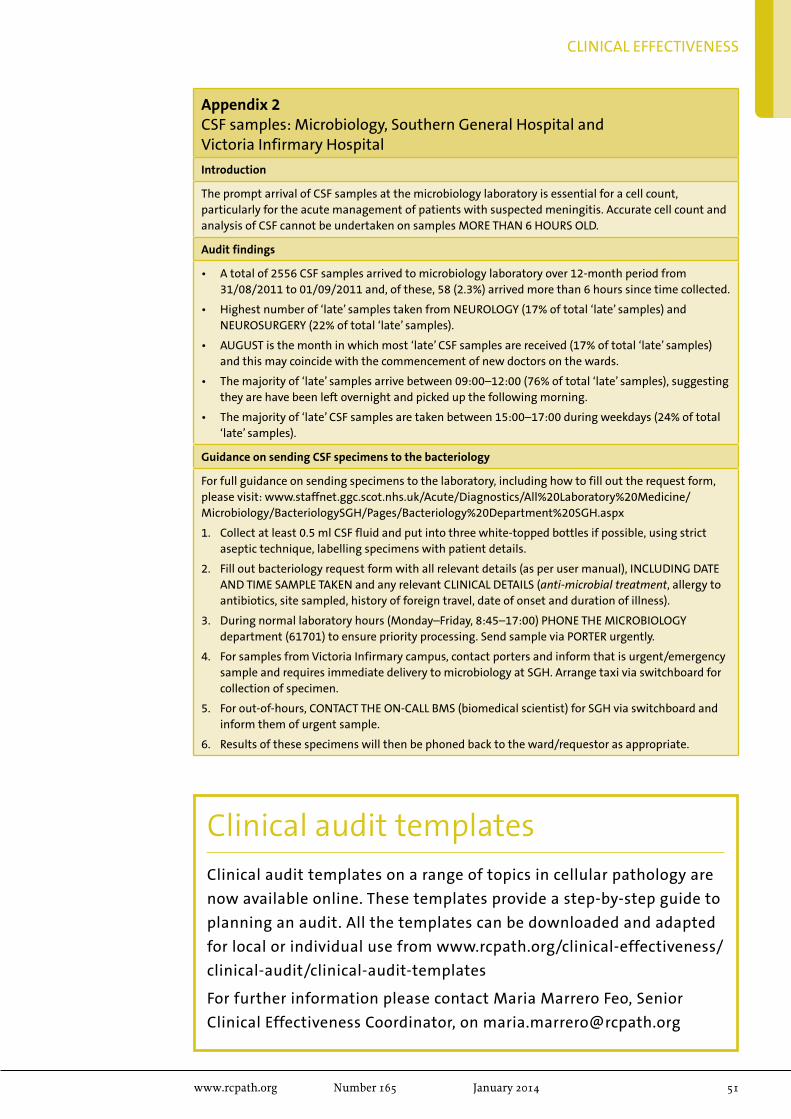
www.rcpath.org Number165 January2014 51
CLINICAL EFFECTIVENESS
Clinical audit templatesClinical audit templates on a range of topics in cellular pathology are
now available online. These templates provide a step-by-step guide to
planning an audit. All the templates can be downloaded and adapted
for local or individual use from www.rcpath.org/clinical-effectiveness/
clinical-audit/clinical-audit-templates
For further information please contact Maria Marrero Feo, Senior
Clinical Effectiveness Coordinator, on [email protected]
Appendix 2CSF samples: Microbiology, Southern General Hospital andVictoria Infirmary Hospital
Introduction
The prompt arrival of CSF samples at the microbiology laboratory is essential for a cell count, particularly for the acute management of patients with suspected meningitis. Accurate cell count and analysis of CSF cannot be undertaken on samples MORE THAN 6 HOURS OLD.
Audit findings
• A total of 2556 CSF samples arrived to microbiology laboratory over 12-month period from 31/08/2011 to 01/09/2011 and, of these, 58 (2.3%) arrived more than 6 hours since time collected.
• Highest number of ‘late’ samples taken from NEUROLOGY (17% of total ‘late’ samples) and NEUROSURGERY (22% of total ‘late’ samples).
• AUGUST is the month in which most ‘late’ CSF samples are received (17% of total ‘late’ samples) and this may coincide with the commencement of new doctors on the wards.
• The majority of ‘late’ samples arrive between 09:00–12:00 (76% of total ‘late’ samples), suggesting they are have been left overnight and picked up the following morning.
• The majority of ‘late’ CSF samples are taken between 15:00–17:00 during weekdays (24% of total ‘late’ samples).
Guidance on sending CSF specimens to the bacteriology
For full guidance on sending specimens to the laboratory, including how to fill out the request form, please visit: www.staffnet.ggc.scot.nhs.uk/Acute/Diagnostics/All%20Laboratory%20Medicine/Microbiology/BacteriologySGH/Pages/Bacteriology%20Department%20SGH.aspx
1. Collect at least 0.5 ml CSF fluid and put into three white-topped bottles if possible, using strict aseptic technique, labelling specimens with patient details.
2. Fill out bacteriology request form with all relevant details (as per user manual), INCLUDING DATE AND TIME SAMPLE TAKEN and any relevant CLINICAL DETAILS (anti-microbial treatment, allergy to antibiotics, site sampled, history of foreign travel, date of onset and duration of illness).
3. During normal laboratory hours (Monday–Friday, 8:45–17:00) PHONE THE MICROBIOLOGY department (61701) to ensure priority processing. Send sample via PORTER urgently.
4. For samples from Victoria Infirmary campus, contact porters and inform that is urgent/emergency sample and requires immediate delivery to microbiology at SGH. Arrange taxi via switchboard for collection of specimen.
5. For out-of-hours, CONTACT THE ON-CALL BMS (biomedical scientist) for SGH via switchboard and inform them of urgent sample.
6. Results of these specimens will then be phoned back to the ward/requestor as appropriate.
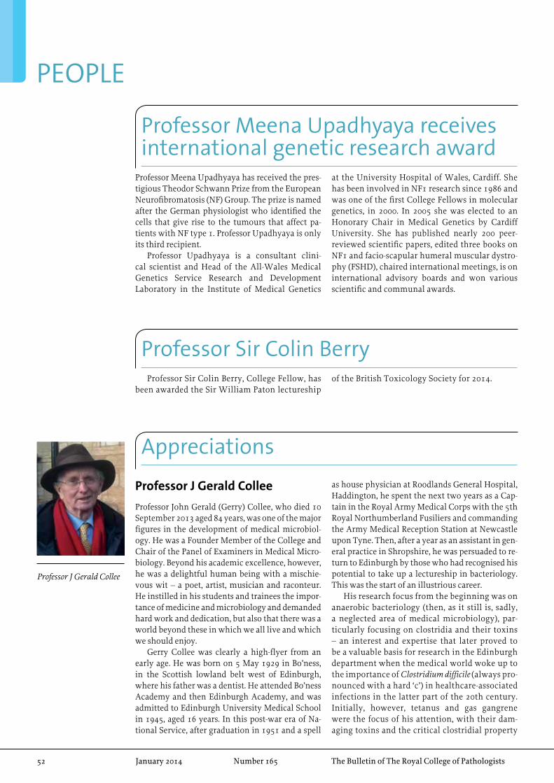
PEoPlE
52 January2014 Number165 TheBulletinofTheRoyalCollegeofPathologists
Professor J Gerald Collee
Professor John Gerald (Gerry) Collee, who died 10 September 2013 aged 84 years, was one of the major figures in the development of medical microbiol-ogy. He was a Founder Member of the College and Chair of the Panel of Examiners in Medical Micro-biology. Beyond his academic excellence, however, he was a delightful human being with a mischie-vous wit – a poet, artist, musician and raconteur. He instilled in his students and trainees the impor-tance of medicine and microbiology and demanded hard work and dedication, but also that there was a world beyond these in which we all live and which we should enjoy.
Gerry Collee was clearly a high-flyer from an early age. He was born on 5 May 1929 in Bo’ness, in the Scottish lowland belt west of Edinburgh, where his father was a dentist. He attended Bo’ness Academy and then Edinburgh Academy, and was admitted to Edinburgh University Medical School in 1945, aged 16 years. In this post-war era of Na-tional Service, after graduation in 1951 and a spell
as house physician at Roodlands General Hospital, Haddington, he spent the next two years as a Cap-tain in the Royal Army Medical Corps with the 5th Royal Northumberland Fusiliers and commanding the Army Medical Reception Station at Newcastle upon Tyne. Then, after a year as an assistant in gen-eral practice in Shropshire, he was persuaded to re-turn to Edinburgh by those who had recognised his potential to take up a lectureship in bacteriology. This was the start of an illustrious career.
His research focus from the beginning was on anaerobic bacteriology (then, as it still is, sadly, a neglected area of medical microbiology), par-ticularly focusing on clostridia and their toxins – an interest and expertise that later proved to be a valuable basis for research in the Edinburgh department when the medical world woke up to the importance of Clostridium difficile (always pro-nounced with a hard ‘c’) in healthcare-associated infections in the latter part of the 20th century. Initially, however, tetanus and gas gangrene were the focus of his attention, with their dam-aging toxins and the critical clostridial property
appreciations
Professor J Gerald Collee
Professor Meena Upadhyaya receives international genetic research award
Professor Meena Upadhyaya has received the pres-tigious Theodor Schwann Prize from the European Neurofibromatosis (NF) Group. The prize is named after the German physiologist who identified the cells that give rise to the tumours that affect pa-tients with NF type 1. Professor Upadhyaya is only its third recipient.
Professor Upadhyaya is a consultant clini-cal scientist and Head of the All-Wales Medical Genetics Service Research and Development Laboratory in the Institute of Medical Genetics
at the University Hospital of Wales, Cardiff. She has been involved in NF1 research since 1986 and was one of the first College Fellows in molecular genetics, in 2000. In 2005 she was elected to an Honorary Chair in Medical Genetics by Cardiff University. She has published nearly 200 peer-reviewed scientific papers, edited three books on NF1 and facio-scapular humeral muscular dystro-phy (FSHD), chaired international meetings, is on international advisory boards and won various scientific and communal awards.
Professor Sir Colin BerryProfessor Sir Colin Berry, College Fellow, has
been awarded the Sir William Paton lectureship of the British Toxicology Society for 2014.
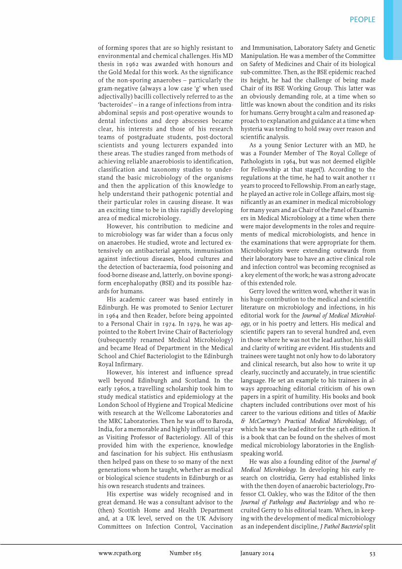
PEOPLE
www.rcpath.org Number165 January2014 53
of forming spores that are so highly resistant to environmental and chemical challenges. His MD thesis in 1962 was awarded with honours and the Gold Medal for this work. As the significance of the non-sporing anaerobes – particularly the gram-negative (always a low case ‘g’ when used adjectivally) bacilli collectively referred to as the ‘bacteroides’ – in a range of infections from intra-abdominal sepsis and post-operative wounds to dental infections and deep abscesses became clear, his interests and those of his research teams of postgraduate students, post-doctoral scientists and young lecturers expanded into these areas. The studies ranged from methods of achieving reliable anaerobiosis to identification, classification and taxonomy studies to under-stand the basic microbiology of the organisms and then the application of this knowledge to help understand their pathogenic potential and their particular roles in causing disease. It was an exciting time to be in this rapidly developing area of medical microbiology.
However, his contribution to medicine and to microbiology was far wider than a focus only on anaerobes. He studied, wrote and lectured ex-tensively on antibacterial agents, immunisation against infectious diseases, blood cultures and the detection of bacteraemia, food poisoning and food-borne disease and, latterly, on bovine spongi-form encephalopathy (BSE) and its possible haz-ards for humans.
His academic career was based entirely in Edinburgh. He was promoted to Senior Lecturer in 1964 and then Reader, before being appointed to a Personal Chair in 1974. In 1979, he was ap-pointed to the Robert Irvine Chair of Bacteriology (subsequently renamed Medical Microbiology) and became Head of Department in the Medical School and Chief Bacteriologist to the Edinburgh Royal Infirmary.
However, his interest and influence spread well beyond Edinburgh and Scotland. In the early 1960s, a travelling scholarship took him to study medical statistics and epidemiology at the London School of Hygiene and Tropical Medicine with research at the Wellcome Laboratories and the MRC Laboratories. Then he was off to Baroda, India, for a memorable and highly influential year as Visiting Professor of Bacteriology. All of this provided him with the experience, knowledge and fascination for his subject. His enthusiasm then helped pass on these to so many of the next generations whom he taught, whether as medical or biological science students in Edinburgh or as his own research students and trainees.
His expertise was widely recognised and in great demand. He was a consultant advisor to the (then) Scottish Home and Health Department and, at a UK level, served on the UK Advisory Committees on Infection Control, Vaccination
and Immunisation, Laboratory Safety and Genetic Manipulation. He was a member of the Committee on Safety of Medicines and Chair of its biological sub-committee. Then, as the BSE epidemic reached its height, he had the challenge of being made Chair of its BSE Working Group. This latter was an obviously demanding role, at a time when so little was known about the condition and its risks for humans. Gerry brought a calm and reasoned ap-proach to explanation and guidance at a time when hysteria was tending to hold sway over reason and scientific analysis.
As a young Senior Lecturer with an MD, he was a Founder Member of The Royal College of Pathologists in 1964, but was not deemed eligible for Fellowship at that stage(!). According to the regulations at the time, he had to wait another 11 years to proceed to Fellowship. From an early stage, he played an active role in College affairs, most sig-nificantly as an examiner in medical microbiology for many years and as Chair of the Panel of Examin-ers in Medical Microbiology at a time when there were major developments in the roles and require-ments of medical microbiologists, and hence in the examinations that were appropriate for them. Microbiologists were extending outwards from their laboratory base to have an active clinical role and infection control was becoming recognised as a key element of the work; he was a strong advocate of this extended role.
Gerry loved the written word, whether it was in his huge contribution to the medical and scientific literature on microbiology and infections, in his editorial work for the Journal of Medical Microbiol-ogy, or in his poetry and letters. His medical and scientific papers ran to several hundred and, even in those where he was not the lead author, his skill and clarity of writing are evident. His students and trainees were taught not only how to do laboratory and clinical research, but also how to write it up clearly, succinctly and accurately, in true scientific language. He set an example to his trainees in al-ways approaching editorial criticism of his own papers in a spirit of humility. His books and book chapters included contributions over most of his career to the various editions and titles of Mackie & McCartney’s Practical Medical Microbiology, of which he was the lead editor for the 14th edition. It is a book that can be found on the shelves of most medical microbiology laboratories in the English-speaking world.
He was also a founding editor of the Journal of Medical Microbiology. In developing his early re-search on clostridia, Gerry had established links with the then doyen of anaerobic bacteriology, Pro-fessor CL Oakley, who was the Editor of the then Journal of Pathology and Bacteriology and who re-cruited Gerry to his editorial team. When, in keep-ing with the development of medical microbiology as an independent discipline, J Pathol Bacteriol split
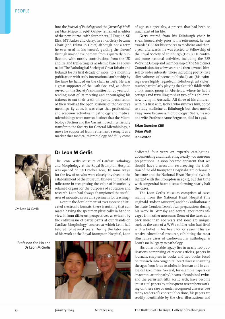
PEOPLE
54 January2014 Number165 TheBulletinofTheRoyalCollegeofPathologists
Dr Leon M Gerlis
Dr Leon M Gerlis
The Leon Gerlis Museum of Cardiac Pathology and Morphology at the Royal Brompton Hospital was opened on 28 October 2003. In some ways, for the few of us who were closely involved in the establishment of the museum, this event marked a milestone in recognising the value of historically retained organs for the purposes of education and research. Leon had always championed the useful-ness of mounted museum specimens for teaching.1
Despite the development of ever more sophisti-cated electronic formats, there is nothing that can match having the specimen physically in hand to view it from different perspectives, as evident by the enthusiasm of participants at our ‘Hands-on Cardiac Morphology’ courses at which Leon had tutored for several years. During the later years of his work at the Royal Brompton Hospital, Leon
dedicated four years on expertly cataloguing, documenting and illustrating nearly 300 museum preparations. It soon became apparent that we should have a museum, resurrecting the tradi-tion of the old Brompton Hospital/Cardiothoracic Institute and the National Heart Hospital (which merged with the Brompton in 1971), but this time with congenital heart disease forming nearly half the cases.
The Leon Gerlis Museum comprises of cases mainly from the National Heart Hospital (the Reginald Hudson Museum) and the Cardiothoracic Institute, London, Leon’s own preparations during his work in Grimsby and several specimens sal-vaged from other museums. Some of the cases date back more than 100 years and some are unique, such as the case of a WW1 soldier who had lived with a bullet in his heart for 55 years.2 This ex-tensive educational resource, exhibiting the most illustrative cases of cardiovascular pathology, is Leon’s main legacy to pathology.
His other notable legacy lies in nearly 100 pub-lications comprising of review articles, papers in journals, chapters in books and two books based on research into congenital heart disease spanning the ages from fetus to adults, in human and in zoo-logical specimens. Several, for example papers on ‘macaroni arteriopathy’, hearts of conjoined twins, and the persistent fifth aortic arch, have become ‘must cite’ papers by subsequent researchers work-ing on these rare or under recognised diseases. For many readers of Leon’s publications, his papers are readily identifiable by the clear illustrations and
into the Journal of Pathology and the Journal of Medi-cal Microbiology in 1968, Oakley remained as editor of the new journal with four others: JP Duguid, SD Elek, MT Parker and Gerry. In 1974, Gerry became Chair (and Editor in Chief, although not a term he ever used in his tenure), guiding the Journal through major development from a quarterly pub-lication, with mostly contributions from the UK and Ireland (reflecting its academic base as a jour-nal of The Pathological Society of Great Britain and Ireland) for its first decade or more, to a monthly publication with truly international authorship by the time he handed on the chair in 1988. He was a great supporter of the ‘Path Soc’ and, as Editor, served on the Society’s committee for 20 years, at-tending most of its meeting and encouraging his trainees to cut their teeth on public presentation of their work at the open sessions of the Society’s meetings. By 2000, it was clear that professional and academic activities in pathology and medical microbiology were now so distinct that the Micro-biology Section and the Journal moved in a friendly transfer to the Society for General Microbiology, a move he supported from retirement, seeing it as a marker that medical microbiology had fully come
of age as a specialty, a process that had been so much part of his life.
Gerry retired from his Edinburgh chair in 1992. Immediately prior to his retirement, he was awarded CBE for his services to medicine and then, a year afterwards, he was elected to Fellowship of the Royal Society of Edinburgh (FRSE). He contin-ued some national activities, including the BSE Working Group and membership of the Medicines Commission, for a few years and then devoted him-self to wider interests. These including poetry (five slim volumes of poems published), art (his paint-ings were highly regarded in Edinburgh art cicles), music (particularly playing the Scottish fiddle with a folk music group in Aberfeldy, where he had a cottage) and travelling to visit two of his children, now living in Australia. All three of his children, with his first wife, Isobel, who survives him, opted to study medicine at Edinburgh but then moved away; none became a microbiologist! Sadly, his sec-ond wife, Professor Anne Ferguson, died in 1998.
Brian Duerden CBE
Brian Watt
Ian Poxton
Professor Yen Ho and Dr Leon M Gerlis
PEOPLE
www.rcpath.org Number165 January2014 55
exquisite drawings, made in his distinctive style.Compared to many other subspecialties of
pathology, cardiovascular pathology (and mor-phology) is far from glamorous. However, it has many followers, since it is deemed fundamental knowledge for workers in cardiology, cardiac sur-gery, congenital heart, paediatrics, neonatology and so on. The number of specialists in this field is relatively small so losing Leon is a huge loss for our community. Leon was an unsung hero, who taught a great many people from all over the world.
Leon was born on 24 November 1919. His in-terest in congenital heart malformations began in 1947 when, as a young trainee pathologist in Grimsby, he met Dr James W Brown who was an eminent authority in congenital heart disease. Over the next few years, Leon learnt about the subject and worked tirelessly, making preparations of con-genital heart specimens in his spare time. As these early years were before the era of sequential seg-mental analysis and modern paediatric cardiology and surgery, Leon’s observations and meticulous descriptions of congenital heart malformations were remarkable. In Leon’s words, “Cardiac pathol-ogy developed into a second, unpaid occupation but I still kept my day job as a general pathologist and haematologist”.
Leon retired from Grimsby in 1979. He subse-quently joined the special cardiac unit at Killing-beck Hospital in Leeds as Honorary Consultant Pathologist and as Consultant Cardiac Pathologist to the National Heart Research Fund, until retiring (again) and moving to London in 1986.
Leon’s career spanned over six decades and he became the Visiting Professor of Cardiac Pathology, Imperial College London, and Honorary Consult-ant in Cardiac Histopathology, Royal Brompton Hospital, London.
I am privileged to have worked with him for the last two decades until May 2007 when, at the age
of 87 years, he hung up his lab coat in our Cardiac Morphology Unit at the Royal Brompton for the final time. I very much appreciate his steadfast sup-port of the unit over the years. I’ll always remember him as a gentleman of the ‘old school’ with time for everyone (and every cat), and his sense of humour. Greeting and stroking cats that he encountered on his walk to the underground station was a particu-lar delight in his journey to work. He was a keen teacher and kindly, despite his occasional growl of “If I’m a train, you’d have missed me!”, to students who turn up late for his tutorials.
Although Leon never sold himself short, he was not one to boast either. He quietly beavered away, researching in great detail all the historical papers relating to whichever topic he was working on, without the help of electronic search engines. He had often remarked that, as far as he knew, hard work had never killed anyone, setting an example for us all.
In his eighties, Leon adapted to using the com-puter proficiently for emails and presentations, and used voice-recognition software for dictations. Soon after his last retirement, I was astonished to receive an invitation to join his social media page! Leon certainly kept up with the time.
He passed away on 23 August 2013.
Professor Yen HoCardiac Morphology UnitRoyal Brompton Hospital, London
References1. Gerlis LM. Cardiac museum specimens in
London. Heart 1996;75:71.2. Gerlis LM. Bizzare presentation and long
survival after a gunshot wound involving the heart. Int J Cardiol 2006;108:408–409.
deathsThedeathsofthefollowingFellowswereannouncedattheNovember2013Councilmeeting.Weextendourcondolencestothosewhogrieveforthem.
William Stewart Alexander Fellow Overseas
Daniel Stephen Brudney Fellow UK
John Gerald Collee Fellow UK
Bojan Flaks Fellow UK
Ernest Gradwell Fellow UK
Eduard Daniel Krahe Fellow UK
Wataru Mori Fellow Overseas
Adebayo Babajide Olujohungbe Fellow Overseas
Keith Lockyer Former College Secretary
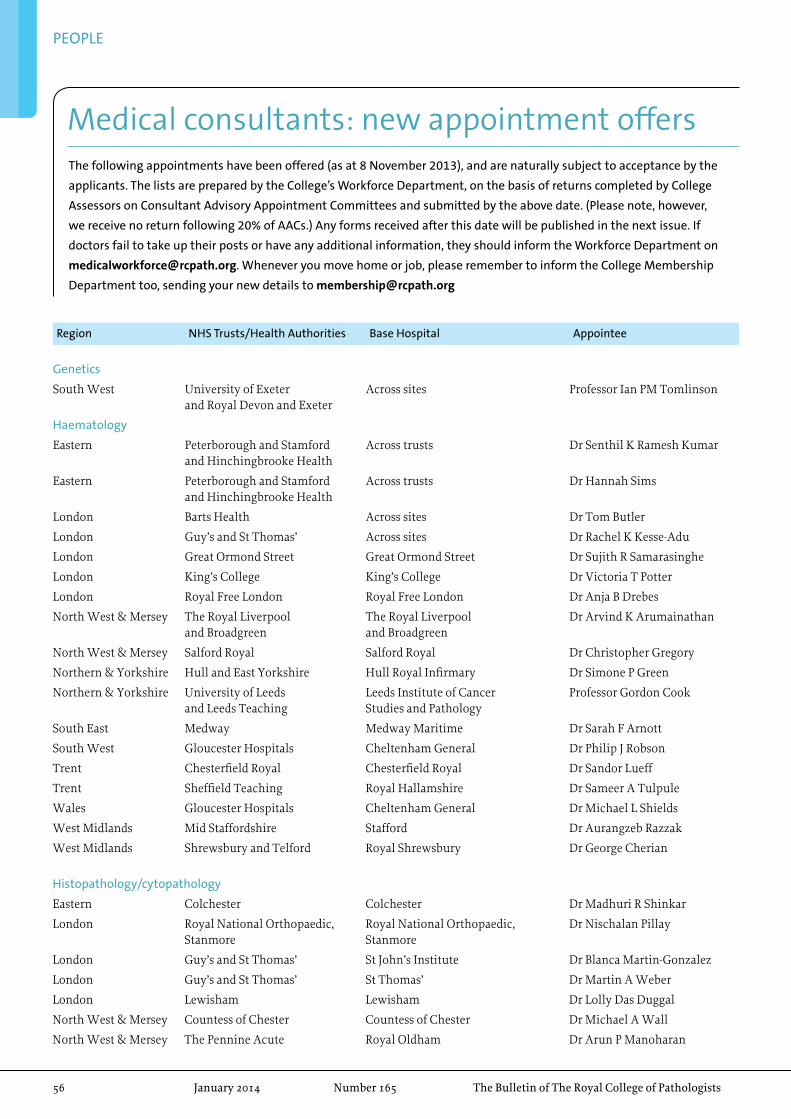
PEOPLE
56 January2014 Number165 TheBulletinofTheRoyalCollegeofPathologists
Genetics
South West University of Exeter Across sites Professor Ian PM Tomlinson and Royal Devon and Exeter
Haematology
Eastern Peterborough and Stamford Across trusts Dr Senthil K Ramesh Kumar and Hinchingbrooke Health
Eastern Peterborough and Stamford Across trusts Dr Hannah Sims and Hinchingbrooke Health
London Barts Health Across sites Dr Tom Butler
London Guy’s and St Thomas’ Across sites Dr Rachel K Kesse-Adu
London Great Ormond Street Great Ormond Street Dr Sujith R Samarasinghe
London King’s College King’s College Dr Victoria T Potter
London Royal Free London Royal Free London Dr Anja B Drebes
North West & Mersey The Royal Liverpool The Royal Liverpool Dr Arvind K Arumainathan and Broadgreen and Broadgreen
North West & Mersey Salford Royal Salford Royal Dr Christopher Gregory
Northern & Yorkshire Hull and East Yorkshire Hull Royal Infirmary Dr Simone P Green
Northern & Yorkshire University of Leeds Leeds Institute of Cancer Professor Gordon Cook and Leeds Teaching Studies and Pathology
South East Medway Medway Maritime Dr Sarah F Arnott
South West Gloucester Hospitals Cheltenham General Dr Philip J Robson
Trent Chesterfield Royal Chesterfield Royal Dr Sandor Lueff
Trent Sheffield Teaching Royal Hallamshire Dr Sameer A Tulpule
Wales Gloucester Hospitals Cheltenham General Dr Michael L Shields
West Midlands Mid Staffordshire Stafford Dr Aurangzeb Razzak
West Midlands Shrewsbury and Telford Royal Shrewsbury Dr George Cherian
Histopathology/cytopathology
Eastern Colchester Colchester Dr Madhuri R Shinkar
London Royal National Orthopaedic, Royal National Orthopaedic, Dr Nischalan Pillay Stanmore Stanmore
London Guy’s and St Thomas’ St John’s Institute Dr Blanca Martin-Gonzalez
London Guy’s and St Thomas’ St Thomas’ Dr Martin A Weber
London Lewisham Lewisham Dr Lolly Das Duggal
North West & Mersey Countess of Chester Countess of Chester Dr Michael A Wall
North West & Mersey The Pennine Acute Royal Oldham Dr Arun P Manoharan
Region NHS Trusts/Health Authorities Base Hospital Appointee
Medical consultants: new appointment offersThe following appointments have been offered (as at 8 November 2013), and are naturally subject to acceptance by the
applicants. The lists are prepared by the College’s Workforce Department, on the basis of returns completed by College
Assessors on Consultant Advisory Appointment Committees and submitted by the above date. (Please note, however,
we receive no return following 20% of AACs.) Any forms received after this date will be published in the next issue. If
doctors fail to take up their posts or have any additional information, they should inform the Workforce Department on
[email protected]. Whenever you move home or job, please remember to inform the College Membership
Department too, sending your new details to [email protected]
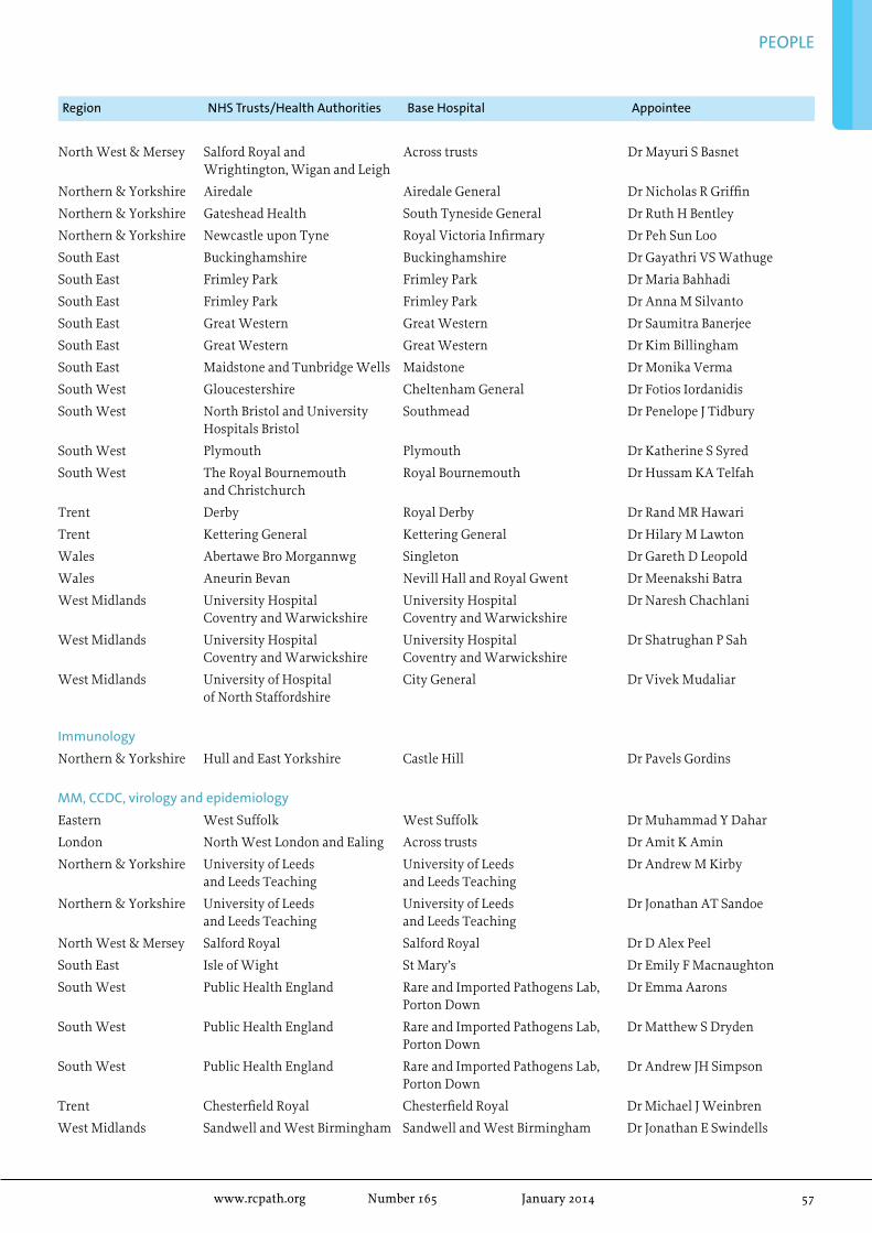
PEOPLE
www.rcpath.org Number165 January2014 57
North West & Mersey Salford Royal and Across trusts Dr Mayuri S Basnet Wrightington, Wigan and Leigh
Northern & Yorkshire Airedale Airedale General Dr Nicholas R Griffin
Northern & Yorkshire Gateshead Health South Tyneside General Dr Ruth H Bentley
Northern & Yorkshire Newcastle upon Tyne Royal Victoria Infirmary Dr Peh Sun Loo
South East Buckinghamshire Buckinghamshire Dr Gayathri VS Wathuge
South East Frimley Park Frimley Park Dr Maria Bahhadi
South East Frimley Park Frimley Park Dr Anna M Silvanto
South East Great Western Great Western Dr Saumitra Banerjee
South East Great Western Great Western Dr Kim Billingham
South East Maidstone and Tunbridge Wells Maidstone Dr Monika Verma
South West Gloucestershire Cheltenham General Dr Fotios Iordanidis
South West North Bristol and University Southmead Dr Penelope J Tidbury Hospitals Bristol
South West Plymouth Plymouth Dr Katherine S Syred
South West The Royal Bournemouth Royal Bournemouth Dr Hussam KA Telfah and Christchurch
Trent Derby Royal Derby Dr Rand MR Hawari
Trent Kettering General Kettering General Dr Hilary M Lawton
Wales Abertawe Bro Morgannwg Singleton Dr Gareth D Leopold
Wales Aneurin Bevan Nevill Hall and Royal Gwent Dr Meenakshi Batra
West Midlands University Hospital University Hospital Dr Naresh Chachlani Coventry and Warwickshire Coventry and Warwickshire
West Midlands University Hospital University Hospital Dr Shatrughan P Sah Coventry and Warwickshire Coventry and Warwickshire
West Midlands University of Hospital City General Dr Vivek Mudaliar of North Staffordshire
Immunology
Northern & Yorkshire Hull and East Yorkshire Castle Hill Dr Pavels Gordins
MM, CCDC, virology and epidemiology
Eastern West Suffolk West Suffolk Dr Muhammad Y Dahar
London North West London and Ealing Across trusts Dr Amit K Amin
Northern & Yorkshire University of Leeds University of Leeds Dr Andrew M Kirby and Leeds Teaching and Leeds Teaching
Northern & Yorkshire University of Leeds University of Leeds Dr Jonathan AT Sandoe and Leeds Teaching and Leeds Teaching
North West & Mersey Salford Royal Salford Royal Dr D Alex Peel
South East Isle of Wight St Mary’s Dr Emily F Macnaughton
South West Public Health England Rare and Imported Pathogens Lab, Dr Emma Aarons Porton Down
South West Public Health England Rare and Imported Pathogens Lab, Dr Matthew S Dryden Porton Down
South West Public Health England Rare and Imported Pathogens Lab, Dr Andrew JH Simpson Porton Down
Trent Chesterfield Royal Chesterfield Royal Dr Michael J Weinbren
West Midlands Sandwell and West Birmingham Sandwell and West Birmingham Dr Jonathan E Swindells
Region NHS Trusts/Health Authorities Base Hospital Appointee
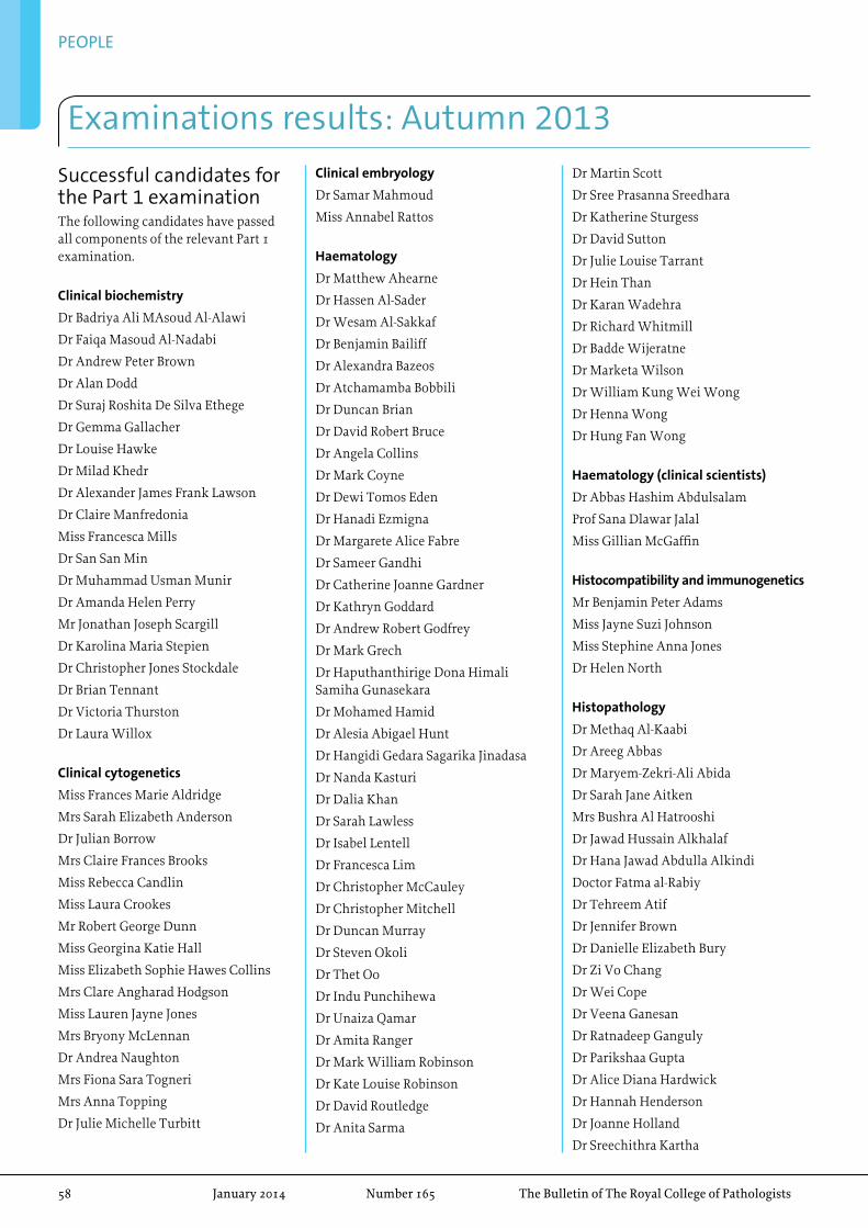
PEOPLE
58 January2014 Number165 TheBulletinofTheRoyalCollegeofPathologists
Successful candidates for the Part 1 examinationThe following candidates have passed all components of the relevant Part 1 examination.
Clinical biochemistry
Dr Badriya Ali MAsoud Al-Alawi
Dr Faiqa Masoud Al-Nadabi
Dr Andrew Peter Brown
Dr Alan Dodd
Dr Suraj Roshita De Silva Ethege
Dr Gemma Gallacher
Dr Louise Hawke
Dr Milad Khedr
Dr Alexander James Frank Lawson
Dr Claire Manfredonia
Miss Francesca Mills
Dr San San Min
Dr Muhammad Usman Munir
Dr Amanda Helen Perry
Mr Jonathan Joseph Scargill
Dr Karolina Maria Stepien
Dr Christopher Jones Stockdale
Dr Brian Tennant
Dr Victoria Thurston
Dr Laura Willox
Clinical cytogenetics
Miss Frances Marie Aldridge
Mrs Sarah Elizabeth Anderson
Dr Julian Borrow
Mrs Claire Frances Brooks
Miss Rebecca Candlin
Miss Laura Crookes
Mr Robert George Dunn
Miss Georgina Katie Hall
Miss Elizabeth Sophie Hawes Collins
Mrs Clare Angharad Hodgson
Miss Lauren Jayne Jones
Mrs Bryony McLennan
Dr Andrea Naughton
Mrs Fiona Sara Togneri
Mrs Anna Topping
Dr Julie Michelle Turbitt
Clinical embryology
Dr Samar Mahmoud
Miss Annabel Rattos
Haematology
Dr Matthew Ahearne
Dr Hassen Al-Sader
Dr Wesam Al-Sakkaf
Dr Benjamin Bailiff
Dr Alexandra Bazeos
Dr Atchamamba Bobbili
Dr Duncan Brian
Dr David Robert Bruce
Dr Angela Collins
Dr Mark Coyne
Dr Dewi Tomos Eden
Dr Hanadi Ezmigna
Dr Margarete Alice Fabre
Dr Sameer Gandhi
Dr Catherine Joanne Gardner
Dr Kathryn Goddard
Dr Andrew Robert Godfrey
Dr Mark Grech
Dr Haputhanthirige Dona Himali Samiha Gunasekara
Dr Mohamed Hamid
Dr Alesia Abigael Hunt
Dr Hangidi Gedara Sagarika Jinadasa
Dr Nanda Kasturi
Dr Dalia Khan
Dr Sarah Lawless
Dr Isabel Lentell
Dr Francesca Lim
Dr Christopher McCauley
Dr Christopher Mitchell
Dr Duncan Murray
Dr Steven Okoli
Dr Thet Oo
Dr Indu Punchihewa
Dr Unaiza Qamar
Dr Amita Ranger
Dr Mark William Robinson
Dr Kate Louise Robinson
Dr David Routledge
Dr Anita Sarma
Dr Martin Scott
Dr Sree Prasanna Sreedhara
Dr Katherine Sturgess
Dr David Sutton
Dr Julie Louise Tarrant
Dr Hein Than
Dr Karan Wadehra
Dr Richard Whitmill
Dr Badde Wijeratne
Dr Marketa Wilson
Dr William Kung Wei Wong
Dr Henna Wong
Dr Hung Fan Wong
Haematology (clinical scientists)
Dr Abbas Hashim Abdulsalam
Prof Sana Dlawar Jalal
Miss Gillian McGaffin
Histocompatibility and immunogenetics
Mr Benjamin Peter Adams
Miss Jayne Suzi Johnson
Miss Stephine Anna Jones
Dr Helen North
Histopathology
Dr Methaq Al-Kaabi
Dr Areeg Abbas
Dr Maryem-Zekri-Ali Abida
Dr Sarah Jane Aitken
Mrs Bushra Al Hatrooshi
Dr Jawad Hussain Alkhalaf
Dr Hana Jawad Abdulla Alkindi
Doctor Fatma al-Rabiy
Dr Tehreem Atif
Dr Jennifer Brown
Dr Danielle Elizabeth Bury
Dr Zi Vo Chang
Dr Wei Cope
Dr Veena Ganesan
Dr Ratnadeep Ganguly
Dr Parikshaa Gupta
Dr Alice Diana Hardwick
Dr Hannah Henderson
Dr Joanne Holland
Dr Sreechithra Kartha
Examinations results: autumn 2013
PEOPLE
www.rcpath.org Number165 January2014 59
Dr Olexandra Kozyar
Dr Priyanka Maheshwari
Doctor Smita Matthai
Dr Muhammad Zain Mehdi
Dr Anita Merritt
Dr John Robert Millar
Dr Sarah Mukhtar
Dr Susruthan Muralitharan
Doctor Priyathersini Nagarajan
Dr Sophia Neda
Dr Wahalatantrige Shriya Thushari Perera
Dr Michele Jane Prance
Dr James Henry Pratt
Dr Omar Layth Qassid
Dr Anirudha Rajguru
Dr Hiran Ravindran
Dr Asha Rupani
Dr Mohammed Sami Saeed
Dr. Karokh Hassan Salih
Mr Marcos Rodrigo Saravia
Dr Vijay Sharma
Dr Katharine Sheppard
Dr Mohammad Sami Ullah
Dr Athanasia Vargiamidou
Dr Sridevi Vijayasankar
Dr Afsheen Wasif
Dr Rafil Toma Hurmiz Yaqo
Dr Maidie Ming Yan Yeung
Dr Carmel Bridget Ryan
Immunology
Dr Cathal Steele
Dr Alison Whitelegg
Medical microbiology
Dr Arvind Kumar Achra
Dr Alexander Aiken
Dr Aishah M KH S M Al Haqan
Dr Mahmoud AL Subhi
Dr Alwarith Nasser Salem Alkharusi
Dr Hessah S Alotaibi
Dr Abdullah Al-owaisheer
Dr Nassery Al-Rubaiaan
Dr Anna Maria Amanuah Aryee
Mr Kevin Richard Barker
Dr Edward Robert Bevan
Dr Kar Ki Chik
Dr Cordelia Coltart
Dr Roisin Maire Connolly
Dr Susanna Davis
Dr Simon Dewar
Dr Katherine Emary
Dr Kathryn French
Dr Amira Ghareeb
Dr Daniel Edwin Greaves
Dr Umar Khurshid
Dr Kwan Ki Karrie Ko
Dr Dinoop Korol Ponnambath
Dr Laura Veronica Lloyd
Dr Nasrullah Malik
Dr Arfath Yousuf Mohd
Dr Sylvia Murray
Dr Laura Nabarro
Ms Chika Julie Ozongwu
Dr Irene Pachiadakis
Dr Leon William Peto
Dr Christina Petridou
Ms Elaine Phelan
Dr Joanna Louise Stephens
Dr Nasir Rafi
Dr Laura Ryan
Dr Patrick Stapleton
Dr Milada Tavodova
Dr Michelle Toleman
Dr Serena Venturelli
Dr Christopher Charles Ward
Dr Katherine Watson
Dr James Stephen Wilson
Dr Bernadette Young
Molecular genetics
Mr Christopher James Campbell
Dr Natalie Jane Chandler
Dr Jayne Ann Louise Houghton
Dr Emma Howard
Ms Eleni Mavraki
Mrs Joanne Marie McLean
Ms Martina Owens
Mrs Frances Caroline Ryan
Mrs Melissa Sloman
Mrs Laura June Yarram-Smith
Veterinary pathology
Ms Jill Bryan
Dr Melanie Dobromylskyj
Mr Sean R McKeag
Ms Maria Jose Pinillapaz-Penuelas
Virology
Dr Kate El Bouzidi
Successful candidates for the Part 2 examinationThe following candidates have passed all components of the relevant Part 2 examination:
Clinical biochemistry
Dr Moza Mohammed Al-Nabhani
Dr Christopher Boot
Dr Nicholas Guy Martin
Mr Pervaz Mohammed
Dr Stewart John Pattman
Mrs Claudia Tomkins
Dr Barry John Toole
Miss Jenna Louise WaldronDr Michelle Young
Forensic pathology
Dr Gemma Louise Kemp
Dr Michael Lawrence Parsons
Haematology
Dr Jagdish Raghvan Adiyodi
Dr Ian Amott
Dr Naima Ansari
Dr Sarah Elizabeth Antcliff
Dr Saiyed Saeed Anwer
Dr Buddhika Badugama
Dr Caroline Bateman
Dr Charlotte Bomken
Dr Shahzya Shahrin Chaudhury
Dr Neil Chauhan
Dr Freya Louise Collings
Dr Sarah Lorraine Davis
Dr Widanalage Sanjay Prasad De Mel
Mrs Korale Kankanamage Ransiri Dinuka De Silva
Dr Perla Eleftheriou
Dr Heather Eve
Dr Emma Fosbury
Dr Sara Ghorashian
Dr Satyen Harish Gohil
Dr Matthew Jay Goodyer
Dr Suzanne Melanie Hall
Dr Suzan Homeida
Dr David Irvine
PEOPLE
60 January2014 Number165 TheBulletinofTheRoyalCollegeofPathologists
Dr Mohammad Aminul Islam
Dr Manish Jain
Dr Pallavi Kalkur
Dr Oluseye Olusoji Kolade
Dr Michelle Mary Lavin
Dr Gavin Chien Pang Ling
Dr Padmaja Lokireddy
Dr Pardeep Maheshwari
Dr Mohsen Norouzi
Dr Ciara O’Rafferty
Dr Joseph Padayatty
Dr Mahesh Kumar Panatt Prahladan
Dr Muhammad Sajid Pervaiz
Dr Fernando De Oliveira Pinto
Dr Andrew Retter
Dr Suzanne Elizabeth Roberts
Dr Nandini Sadasivam
Dr David James Simcox
Dr Binyam Swamimdathil Mohammed Usman
Dr David Alistair Veale
Dr Usira Sarasa Vithanarachchi
Dr Peter Wasswa
Histocompatibility and immunogenetics
Dr Medhat Askar
Mrs Sarah Peacock
Histopathology
Dr Charles Acuda
Dr Yasir Riyadh Alwahab
Dr Alan Beausang
Dr Paul Bennett
Dr Shauna Casey
Dr Maesha Deheragoda
Dr Mona Elshafie
Dr Somaia Elsheikh
Dr Pelvender Gill
Dr Thomas Grigor
Dr Nandita Gupta
Dr Katherine Harrison
Dr Savita Honakeri
Dr Miangela Marie Laclé
Dr Kay Lawson
Dr Noori Noorahmed Maka
Dr Marianne Mason
Dr Linda Mulligan
Dr Rajeev Prasad
Dr Shilpa Santosh
Dr Seemanti Srivastava
Dr Kate Struthers
Dr Manish Mani Subramaniam
Dr Chee Thum
Dr My-Anh Tran-Dang
Dr Yuening Zhang
Immunology
Dr Sara Drinkwater
Dr Khairin Ezlin Mohamed Khalib
Dr Sai Murng
Dr James Thaventhiran
Dr Patrick Foh Khing Yong
Medical microbiology
Dr Amal Al-Tai
Dr Suryabrata Banerjee
Dr Srivalli Bhagavatula
Dr Huma Changez
Dr Celia Chu
Dr Oyewole Christopher Durojaiye
Dr Noha El Sakka
Dr Andrew Flatt
Dr David James Griffith
Dr Joanna Hargreaves
Dr Leo Lui
Dr Tabitha Wambura Mahungu
Dr Emilia Mamwa
Dr Jenifer Mason
Dr Angela Minassian
Dr Cassie Francesca Pope
Dr Anna-Rose Prior
Dr Giuditta Sanna
Dr Robert Lodu Serafino-Wani
Dr Vrinda Gopalkrishna Shet
Dr Rocky Shum
Dr Eamonn Trainor
Dr Reela Varghese
Dr Daniel Weiand
Dr Gemma Wheldon
Dr Eftihia Pavlos Yiannakis
Paediatric pathology
Dr Edmund Cheesman
Veterinary pathology
Dr Kerstin Erles
Dr Cristina Garcia-Rueda
Miss Begonya Garcia-Montoliu
Dr Ross Harley
Ms Pamela Kelly
Dr Maire Caitriona McElroy
Virology
Dr Javeed Ahmed
Dr Wai Sum Li
Dr Stephen Winchester
Successful candidates for the Certificate examinationsThe following candidates have passed the Certificate in Higher Autopsy Training:
Dr Paul Bennett
Dr Lindsey Patricia Mary Clarke
Dr Michelle Downes
Dr Aaron Joseph Ervine
Dr Lucy Foster
Dr Noel Gatt
Dr Thomas Grigor
Dr Sean Oliver Hynes
Dr Mohammed F M Jawad Alsharbaty
Dr Sangeetha N Kalimuthu
Dr Sarah Mahon
Dr Linda Mulligan
Dr Gordon Reid
Dr Jane Thorne
Dr Nicholas West
The following candidates have passed the Certificate in Higher Cervical Cytopathology Training:
Dr Derrick Lian
Dr Khurshid Merchant
Dr Wen Wei Ng
Successful candidates for the diploma examinationsThe following candidates have passed all components of the Diploma in Dermatopathology.
Dr Jose Cardoso
Dr Blanca Martin Gonzalez
Dr Nicholas John Tiffin
lEttErSWe welcome your letters. Please mark correspondence for the attention of the Editor of The Bulletin, and email it care of the Publications Department at [email protected]. The copy deadline for the April 2014 issue is 7 February 2014.
Thanks to the PresidentDear Editor
As always, the President’s column in the October 2013 Bulletin was stimulating, thought provoking, challenging and… encouraging.
He is to be thanked for reminding us of the importance of acting upon the unexpected result. In cellular pathology, this regularly arises for myriad reasons and it will be interesting to see the Specialty Advisory Committee (SAC) deliberations and guidelines on the matter.
My own apprenticeship-style postgraduate training con-stantly emphasised the central importance of the patient and the necessity to communicate results in an expeditious and clinically relevant way. This has remained a cornerstone of my consultant practice and the teams with which I have had the privilege to work.
Direct communication with consultant surgeons is facili-tated by telephone, conversations in the coffee or lunch queues, and firewall-protected emails straight to their mobile phones. Contemporary multidisciplinary meetings have integrated cel-lular pathologists at the helm giving diagnostic, management and prognostic data, continuity with the past and present, and context from a wider perspective than super-specialised clinical colleagues. Trainee and consultant pathologists are reminded that they are clinically experienced medical practitioners, with-out whose opinion the wider clinical team cannot function. As the President emphasises, with this comes a responsibility to perform to appropriate professional and clinical standards.
Thankfully I have no personal experience of a pathology team working in isolation from the clinician and patient.
So rally to the call, all pathologists: reclaim your identity as a doctor… and remember there is always an anxious patient await-ing your opinion!
Dr Derek AllenConsultant HistopathologistBelfast City Hospital
Digital pathologyDear Editor
I always enjoy going through the College Bulletin and I thorough-ly agreed with Dr Chris Simpson’s view on digital pathology that appeared in the October issue.
While new technology has its own privilege over the existing ones, we really need a critical evaluation for any drastic revo-lution. The diagnostic acumen that we acquire over the years following moving the glass slides on the stage of a microscope can never be replaced by digitisation. Moreover, in developing nations, its cost-effectiveness will be an important issue.
Change is the most constant thing in nature. However, our history tells us that all the permanent changes have not always been good in the long run. While we open the door to
a new technology, the older one can peacefully coexist with its own grace.
Dr Ruma RayDepartment of PathologyAll India Institute of Medical Sciences, New Delhi
Human tissues from autopsies – leadership neededDear Editor
I enjoyed reading Professor Lucas’ article about human tissues from autopsies and I think it was the most interesting article of the October edition. What I found bizarre was the disclaimer underneath the article, stating that the views are those of the author alone and not necessarily of the College. If that is so, then what are the views of the college? Does the RCPath not recom-mend sampling of all major organs in all autopsies? In view of this, I thought this “disclaimer” seemed hypocritical.
The young child with sickle cell disease mentioned in the arti-cle might not have died of sickle cell disease. How can we exclude dual pathology? The child could have had an additional inherited metabolic disease and this would have implications for siblings.
My solution to the problem is simple: if the coroner does not let me take histology or do the necessary tests for me to give a cause of death, I call it “Unascertained”.
The last time I was not allowed to do toxicology on hair to investigate chronic cocaine abuse in a young man, I stated in my autopsy report that I cannot exclude cardiac damage due to chronic cocaine abuse, because I was not allowed to do the test. Shortly before the inquest, I got a phone call from the coroner’s secretary asking me not to attend.
We should not let coroners bully us into providing a cause of death without doing the necessary tests. Is it possible that the fees that come with a coroner’s post-mortem could have anything to do with it? This financial interest could be cured if coroners’ post-mortems would be incorporated into the job plan, rather than being carried out in a hurry out of NHS hours.
Dr Cordelia HowittConsultant HistopathologistUniversity Hospital of North Staffordshire NHS Trust
Response
Dear Dr Howitt,
This is our standard disclaimer when publishing articles. I agree with you and Professor Lucas that post-mortem examinations and histology are very important, and should be done with diligence, otherwise they are not worth doing. Incorporating this into the NHS work may be a solution; there are some examples already.
Dr Laszlo IgaliBulletin Editor
www.rcpath.org Number165 January2014 61
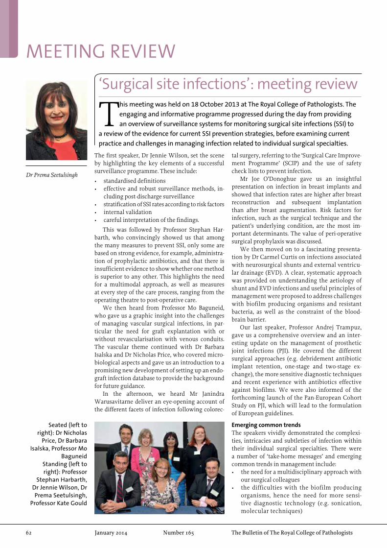
62 January2014 Number165 TheBulletinofTheRoyalCollegeofPathologists
MEEtiNg rEViEw
The first speaker, Dr Jennie Wilson, set the scene by highlighting the key elements of a successful surveillance programme. These include:
• standardised definitions• effective and robust surveillance methods, in-
cluding post-discharge surveillance• stratification of SSI rates according to risk factors• internal validation • careful interpretation of the findings.
This was followed by Professor Stephan Har-barth, who convincingly showed us that among the many measures to prevent SSI, only some are based on strong evidence, for example, administra-tion of prophylactic antibiotics, and that there is insufficient evidence to show whether one method is superior to any other. This highlights the need for a multimodal approach, as well as measures at every step of the care process, ranging from the operating theatre to post-operative care.
We then heard from Professor Mo Baguneid, who gave us a graphic insight into the challenges of managing vascular surgical infections, in par-ticular the need for graft explantation with or without revascularisation with venous conduits. The vascular theme continued with Dr Barbara Isalska and Dr Nicholas Price, who covered micro-biological aspects and gave us an introduction to a promising new development of setting up an endo-graft infection database to provide the background for future guidance.
In the afternoon, we heard Mr Janindra Warusavitarne deliver an eye-opening account of the different facets of infection following colorec-
tal surgery, referring to the ‘Surgical Care Improve-ment Programme’ (SCIP) and the use of safety check lists to prevent infection.
Mr Joe O’Donoghue gave us an insightful presentation on infection in breast implants and showed that infection rates are higher after breast reconstruction and subsequent implantation than after breast augmentation. Risk factors for infection, such as the surgical technique and the patient’s underlying condition, are the most im-portant determinants. The value of peri-operative surgical prophylaxis was discussed.
We then moved on to a fascinating presenta-tion by Dr Carmel Curtis on infections associated with neurosurgical shunts and external ventricu-lar drainage (EVD). A clear, systematic approach was provided on understanding the aetiology of shunt and EVD infections and useful principles of management were proposed to address challenges with biofilm producing organisms and resistant bacteria, as well as the constraint of the blood-brain barrier.
Our last speaker, Professor Andrej Trampuz, gave us a comprehensive overview and an inter-esting update on the management of prosthetic joint infections (PJI). He covered the different surgical approaches (e.g. debridement antibiotic implant retention, one-stage and two-stage ex-change), the more sensitive diagnostic techniques and recent experience with antibiotics effective against biofilms. We were also informed of the forthcoming launch of the Pan-European Cohort Study on PJI, which will lead to the formulation of European guidelines.
Emerging common trendsThe speakers vividly demonstrated the complexi-ties, intricacies and subtleties of infection within their individual surgical specialties. There were a number of ‘take-home messages’ and emerging common trends in management include:• the need for a multidisciplinary approach with
our surgical colleagues• the difficulties with the biofilm producing
organisms, hence the need for more sensi-tive diagnostic technology (e.g. sonication, molecular techniques)
‘Surgical site infections’: meeting review
This meeting was held on 18 October 2013 at The Royal College of Pathologists. The engaging and informative programme progressed during the day from providing an overview of surveillance systems for monitoring surgical site infections (SSI) to
a review of the evidence for current SSI prevention strategies, before examining current practice and challenges in managing infection related to individual surgical specialties.
Dr Prema Seetulsingh
Seated (left to right): Dr Nicholas
Price, Dr Barbara Isalska, Professor Mo
BaguneidStanding (left to right): Professor
Stephan Harbarth, Dr Jennie Wilson, Dr Prema Seetulsingh,
Professor Kate Gould
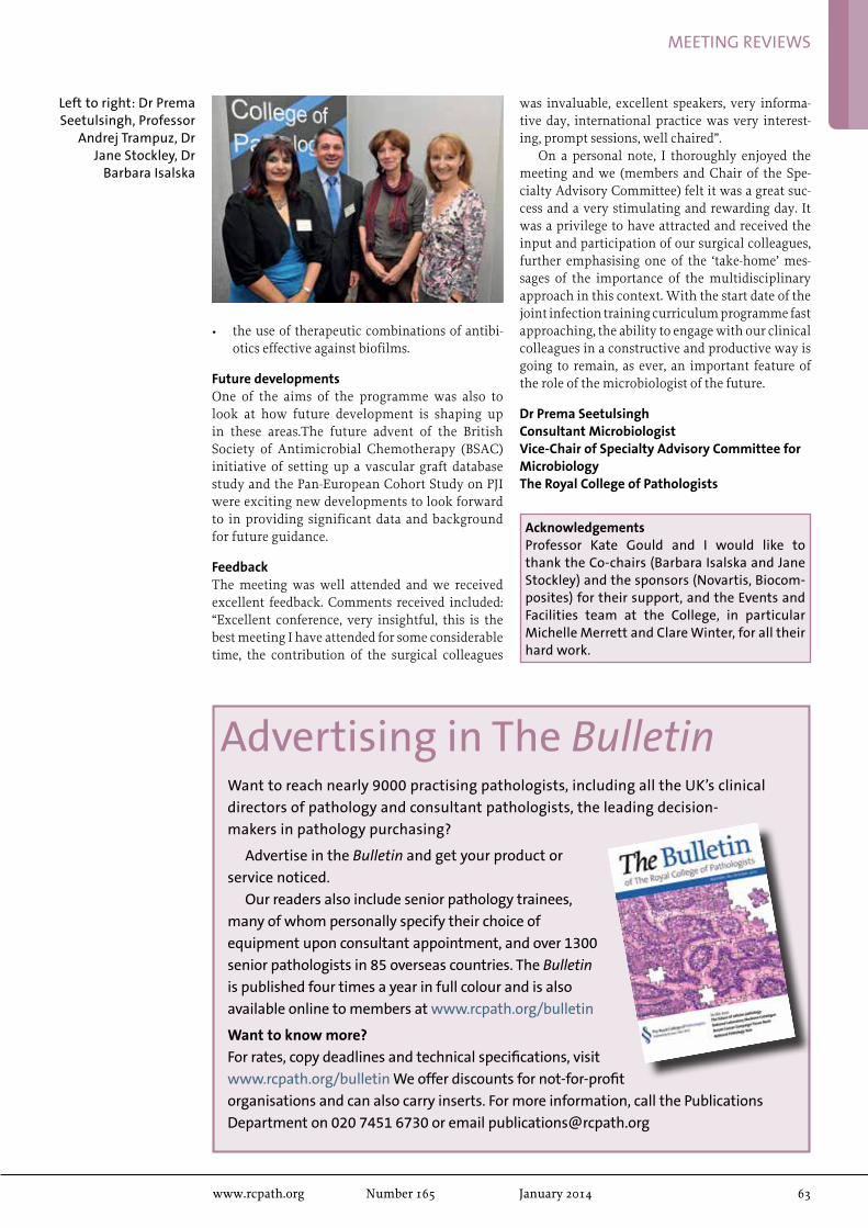
MEETING REVIEWS
www.rcpath.org Number165 January2014 63
• the use of therapeutic combinations of antibi-otics effective against biofilms.
Future developmentsOne of the aims of the programme was also to look at how future development is shaping up in these areas.The future advent of the British Society of Antimicrobial Chemotherapy (BSAC) initiative of setting up a vascular graft database study and the Pan-European Cohort Study on PJI were exciting new developments to look forward to in providing significant data and background for future guidance.
FeedbackThe meeting was well attended and we received excellent feedback. Comments received included: “Excellent conference, very insightful, this is the best meeting I have attended for some considerable time, the contribution of the surgical colleagues
was invaluable, excellent speakers, very informa-tive day, international practice was very interest-ing, prompt sessions, well chaired”.
On a personal note, I thoroughly enjoyed the meeting and we (members and Chair of the Spe-cialty Advisory Committee) felt it was a great suc-cess and a very stimulating and rewarding day. It was a privilege to have attracted and received the input and participation of our surgical colleagues, further emphasising one of the ‘take-home’ mes-sages of the importance of the multidisciplinary approach in this context. With the start date of the joint infection training curriculum programme fast approaching, the ability to engage with our clinical colleagues in a constructive and productive way is going to remain, as ever, an important feature of the role of the microbiologist of the future.
Dr Prema SeetulsinghConsultant MicrobiologistVice-Chair of Specialty Advisory Committee for MicrobiologyThe Royal College of Pathologists
Want to reach nearly 9000 practising pathologists, including all the UK’s clinical directors of pathology and consultant pathologists, the leading decision-makers in pathology purchasing?
Advertise in the Bulletin and get your product or service noticed.
Our readers also include senior pathology trainees, many of whom personally specify their choice of equipment upon consultant appointment, and over 1300 senior pathologists in 85 overseas countries. The Bulletin is published four times a year in full colour and is also available online to members at www.rcpath.org/bulletin
Want to know more?For rates, copy deadlines and technical specifications, visit www.rcpath.org/bulletin We offer discounts for not-for-profit organisations and can also carry inserts. For more information, call the Publications Department on 020 7451 6730 or email [email protected]
Advertising in The Bulletin
Left to right: Dr Prema Seetulsingh, Professor
Andrej Trampuz, Dr Jane Stockley, Dr
Barbara Isalska
AcknowledgementsProfessor Kate Gould and I would like to thank the Co-chairs (Barbara Isalska and Jane Stockley) and the sponsors (Novartis, Biocom-posites) for their support, and the Events and Facilities team at the College, in particular Michelle Merrett and Clare Winter, for all their hard work.
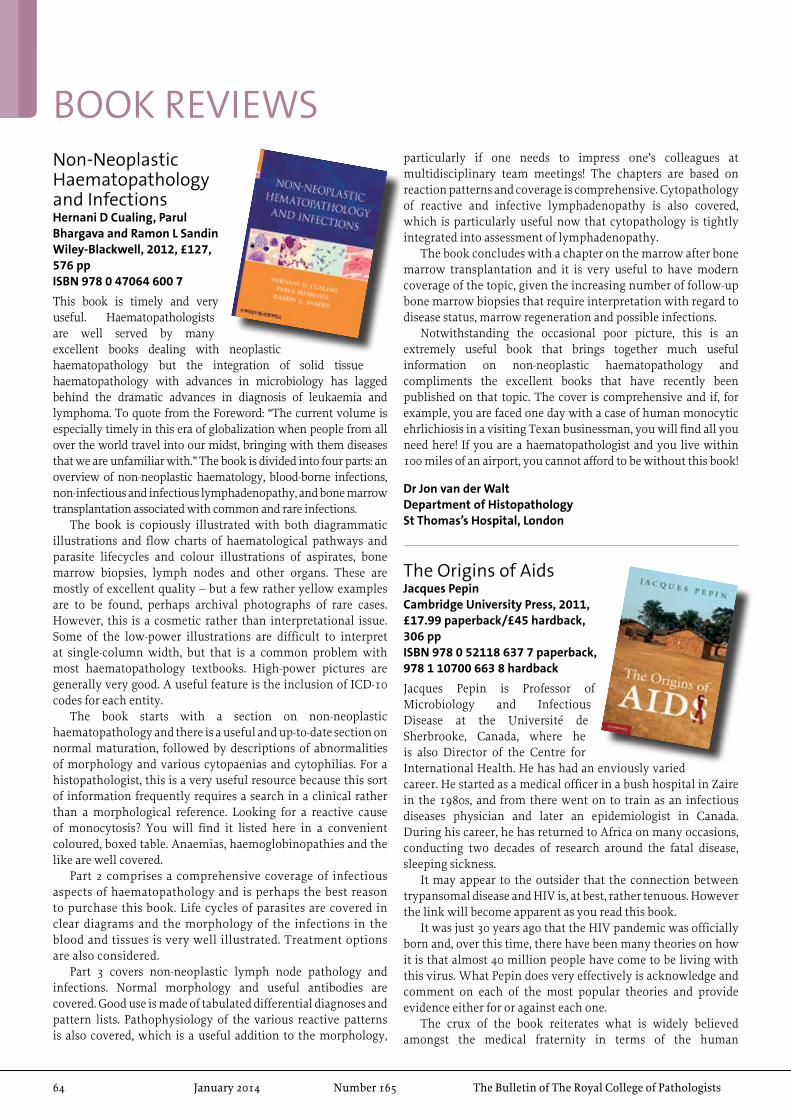
64 January2014 Number165 TheBulletinofTheRoyalCollegeofPathologists
Book rEViEwSNon-Neoplastic Haematopathology and infectionsHernani D Cualing, Parul Bhargava and Ramon L SandinWiley-Blackwell, 2012, £127, 576 ppISBN 978 0 47064 600 7
This book is timely and very useful. Haematopathologists are well served by many excellent books dealing with neoplastic haematopathology but the integration of solid tissue haematopathology with advances in microbiology has lagged behind the dramatic advances in diagnosis of leukaemia and lymphoma. To quote from the Foreword: “The current volume is especially timely in this era of globalization when people from all over the world travel into our midst, bringing with them diseases that we are unfamiliar with.” The book is divided into four parts: an overview of non-neoplastic haematology, blood-borne infections, non-infectious and infectious lymphadenopathy, and bone marrow transplantation associated with common and rare infections.
The book is copiously illustrated with both diagrammatic illustrations and flow charts of haematological pathways and parasite lifecycles and colour illustrations of aspirates, bone marrow biopsies, lymph nodes and other organs. These are mostly of excellent quality – but a few rather yellow examples are to be found, perhaps archival photographs of rare cases. However, this is a cosmetic rather than interpretational issue. Some of the low-power illustrations are difficult to interpret at single-column width, but that is a common problem with most haematopathology textbooks. High-power pictures are generally very good. A useful feature is the inclusion of ICD-10 codes for each entity.
The book starts with a section on non-neoplastic haematopathology and there is a useful and up-to-date section on normal maturation, followed by descriptions of abnormalities of morphology and various cytopaenias and cytophilias. For a histopathologist, this is a very useful resource because this sort of information frequently requires a search in a clinical rather than a morphological reference. Looking for a reactive cause of monocytosis? You will find it listed here in a convenient coloured, boxed table. Anaemias, haemoglobinopathies and the like are well covered.
Part 2 comprises a comprehensive coverage of infectious aspects of haematopathology and is perhaps the best reason to purchase this book. Life cycles of parasites are covered in clear diagrams and the morphology of the infections in the blood and tissues is very well illustrated. Treatment options are also considered.
Part 3 covers non-neoplastic lymph node pathology and infections. Normal morphology and useful antibodies are covered. Good use is made of tabulated differential diagnoses and pattern lists. Pathophysiology of the various reactive patterns is also covered, which is a useful addition to the morphology,
particularly if one needs to impress one’s colleagues at multidisciplinary team meetings! The chapters are based on reaction patterns and coverage is comprehensive. Cytopathology of reactive and infective lymphadenopathy is also covered, which is particularly useful now that cytopathology is tightly integrated into assessment of lymphadenopathy.
The book concludes with a chapter on the marrow after bone marrow transplantation and it is very useful to have modern coverage of the topic, given the increasing number of follow-up bone marrow biopsies that require interpretation with regard to disease status, marrow regeneration and possible infections.
Notwithstanding the occasional poor picture, this is an extremely useful book that brings together much useful information on non-neoplastic haematopathology and compliments the excellent books that have recently been published on that topic. The cover is comprehensive and if, for example, you are faced one day with a case of human monocytic ehrlichiosis in a visiting Texan businessman, you will find all you need here! If you are a haematopathologist and you live within 100 miles of an airport, you cannot afford to be without this book!
Dr Jon van der WaltDepartment of HistopathologySt Thomas’s Hospital, London
the origins of aidsJacques PepinCambridge University Press, 2011, £17.99 paperback/£45 hardback, 306 pp ISBN 978 0 52118 637 7 paperback, 978 1 10700 663 8 hardback
Jacques Pepin is Professor of Microbiology and Infectious Disease at the Université de Sherbrooke, Canada, where he is also Director of the Centre for International Health. He has had an enviously varied career. He started as a medical officer in a bush hospital in Zaire in the 1980s, and from there went on to train as an infectious diseases physician and later an epidemiologist in Canada. During his career, he has returned to Africa on many occasions, conducting two decades of research around the fatal disease, sleeping sickness.
It may appear to the outsider that the connection between trypansomal disease and HIV is, at best, rather tenuous. However the link will become apparent as you read this book.
It was just 30 years ago that the HIV pandemic was officially born and, over this time, there have been many theories on how it is that almost 40 million people have come to be living with this virus. What Pepin does very effectively is acknowledge and comment on each of the most popular theories and provide evidence either for or against each one.
The crux of the book reiterates what is widely believed amongst the medical fraternity in terms of the human
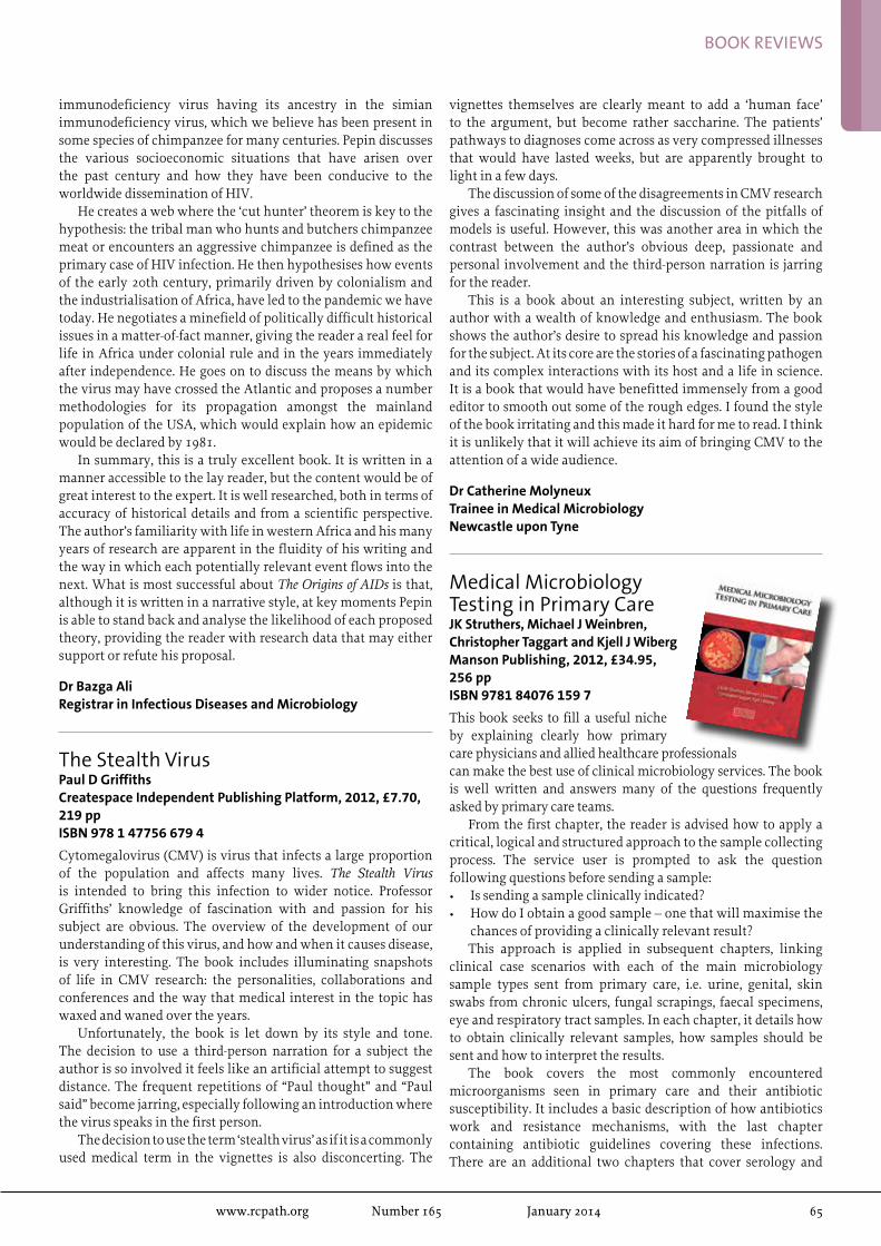
BOOK REVIEWS
www.rcpath.org Number165 January2014 65
immunodeficiency virus having its ancestry in the simian immunodeficiency virus, which we believe has been present in some species of chimpanzee for many centuries. Pepin discusses the various socioeconomic situations that have arisen over the past century and how they have been conducive to the worldwide dissemination of HIV.
He creates a web where the ‘cut hunter’ theorem is key to the hypothesis: the tribal man who hunts and butchers chimpanzee meat or encounters an aggressive chimpanzee is defined as the primary case of HIV infection. He then hypothesises how events of the early 20th century, primarily driven by colonialism and the industrialisation of Africa, have led to the pandemic we have today. He negotiates a minefield of politically difficult historical issues in a matter-of-fact manner, giving the reader a real feel for life in Africa under colonial rule and in the years immediately after independence. He goes on to discuss the means by which the virus may have crossed the Atlantic and proposes a number methodologies for its propagation amongst the mainland population of the USA, which would explain how an epidemic would be declared by 1981.
In summary, this is a truly excellent book. It is written in a manner accessible to the lay reader, but the content would be of great interest to the expert. It is well researched, both in terms of accuracy of historical details and from a scientific perspective. The author’s familiarity with life in western Africa and his many years of research are apparent in the fluidity of his writing and the way in which each potentially relevant event flows into the next. What is most successful about The Origins of AIDs is that, although it is written in a narrative style, at key moments Pepin is able to stand back and analyse the likelihood of each proposed theory, providing the reader with research data that may either support or refute his proposal.
Dr Bazga AliRegistrar in Infectious Diseases and Microbiology
the Stealth VirusPaul D GriffithsCreatespace Independent Publishing Platform, 2012, £7.70, 219 ppISBN 978 1 47756 679 4
Cytomegalovirus (CMV) is virus that infects a large proportion of the population and affects many lives. The Stealth Virus is intended to bring this infection to wider notice. Professor Griffiths’ knowledge of fascination with and passion for his subject are obvious. The overview of the development of our understanding of this virus, and how and when it causes disease, is very interesting. The book includes illuminating snapshots of life in CMV research: the personalities, collaborations and conferences and the way that medical interest in the topic has waxed and waned over the years.
Unfortunately, the book is let down by its style and tone. The decision to use a third-person narration for a subject the author is so involved it feels like an artificial attempt to suggest distance. The frequent repetitions of “Paul thought” and “Paul said” become jarring, especially following an introduction where the virus speaks in the first person.
The decision to use the term ‘stealth virus’ as if it is a commonly used medical term in the vignettes is also disconcerting. The
vignettes themselves are clearly meant to add a ‘human face’ to the argument, but become rather saccharine. The patients’ pathways to diagnoses come across as very compressed illnesses that would have lasted weeks, but are apparently brought to light in a few days.
The discussion of some of the disagreements in CMV research gives a fascinating insight and the discussion of the pitfalls of models is useful. However, this was another area in which the contrast between the author’s obvious deep, passionate and personal involvement and the third-person narration is jarring for the reader.
This is a book about an interesting subject, written by an author with a wealth of knowledge and enthusiasm. The book shows the author’s desire to spread his knowledge and passion for the subject. At its core are the stories of a fascinating pathogen and its complex interactions with its host and a life in science. It is a book that would have benefitted immensely from a good editor to smooth out some of the rough edges. I found the style of the book irritating and this made it hard for me to read. I think it is unlikely that it will achieve its aim of bringing CMV to the attention of a wide audience.
Dr Catherine MolyneuxTrainee in Medical MicrobiologyNewcastle upon Tyne
Medical Microbiology testing in Primary CareJK Struthers, Michael J Weinbren, Christopher Taggart and Kjell J WibergManson Publishing, 2012, £34.95, 256 ppISBN 9781 84076 159 7
This book seeks to fill a useful niche by explaining clearly how primary care physicians and allied healthcare professionals can make the best use of clinical microbiology services. The book is well written and answers many of the questions frequently asked by primary care teams.
From the first chapter, the reader is advised how to apply a critical, logical and structured approach to the sample collecting process. The service user is prompted to ask the question following questions before sending a sample:• Is sending a sample clinically indicated?• How do I obtain a good sample – one that will maximise the
chances of providing a clinically relevant result?This approach is applied in subsequent chapters, linking
clinical case scenarios with each of the main microbiology sample types sent from primary care, i.e. urine, genital, skin swabs from chronic ulcers, fungal scrapings, faecal specimens, eye and respiratory tract samples. In each chapter, it details how to obtain clinically relevant samples, how samples should be sent and how to interpret the results.
The book covers the most commonly encountered microorganisms seen in primary care and their antibiotic susceptibility. It includes a basic description of how antibiotics work and resistance mechanisms, with the last chapter containing antibiotic guidelines covering these infections. There are an additional two chapters that cover serology and
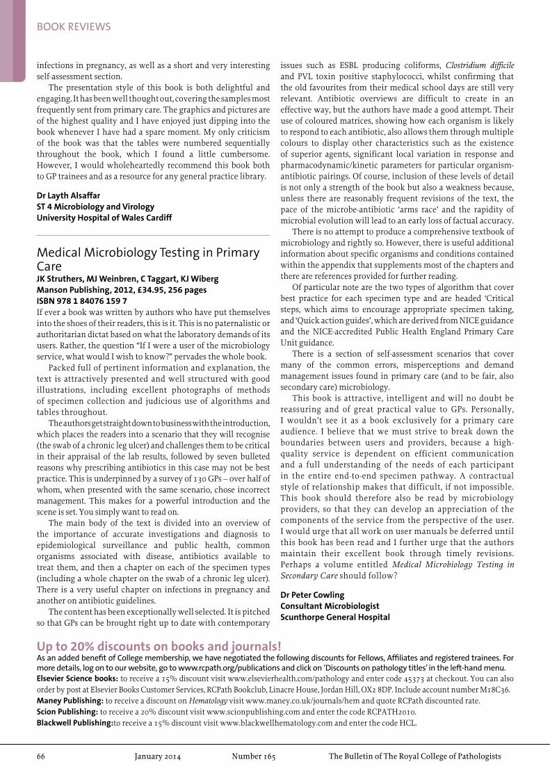
66 January2014 Number165 TheBulletinofTheRoyalCollegeofPathologists
BOOK REVIEWS
infections in pregnancy, as well as a short and very interesting self-assessment section.
The presentation style of this book is both delightful and engaging. It has been well thought out, covering the samples most frequently sent from primary care. The graphics and pictures are of the highest quality and I have enjoyed just dipping into the book whenever I have had a spare moment. My only criticism of the book was that the tables were numbered sequentially throughout the book, which I found a little cumbersome. However, I would wholeheartedly recommend this book both to GP trainees and as a resource for any general practice library.
Dr Layth AlsaffarST 4 Microbiology and VirologyUniversity Hospital of Wales Cardiff
Medical Microbiology testing in Primary CareJK Struthers, MJ Weinbren, C Taggart, KJ Wiberg Manson Publishing, 2012, £34.95, 256 pages ISBN 978 1 84076 159 7If ever a book was written by authors who have put themselves into the shoes of their readers, this is it. This is no paternalistic or authoritarian dictat based on what the laboratory demands of its users. Rather, the question “If I were a user of the microbiology service, what would I wish to know?” pervades the whole book.
Packed full of pertinent information and explanation, the text is attractively presented and well structured with good illustrations, including excellent photographs of methods of specimen collection and judicious use of algorithms and tables throughout.
The authors get straight down to business with the introduction, which places the readers into a scenario that they will recognise (the swab of a chronic leg ulcer) and challenges them to be critical in their appraisal of the lab results, followed by seven bulleted reasons why prescribing antibiotics in this case may not be best practice. This is underpinned by a survey of 130 GPs – over half of whom, when presented with the same scenario, chose incorrect management. This makes for a powerful introduction and the scene is set. You simply want to read on.
The main body of the text is divided into an overview of the importance of accurate investigations and diagnosis to epidemiological surveillance and public health, common organisms associated with disease, antibiotics available to treat them, and then a chapter on each of the specimen types (including a whole chapter on the swab of a chronic leg ulcer). There is a very useful chapter on infections in pregnancy and another on antibiotic guidelines.
The content has been exceptionally well selected. It is pitched so that GPs can be brought right up to date with contemporary
issues such as ESBL producing coliforms, Clostridium difficile and PVL toxin positive staphylococci, whilst confirming that the old favourites from their medical school days are still very relevant. Antibiotic overviews are difficult to create in an effective way, but the authors have made a good attempt. Their use of coloured matrices, showing how each organism is likely to respond to each antibiotic, also allows them through multiple colours to display other characteristics such as the existence of superior agents, significant local variation in response and pharmacodynamic/kinetic parameters for particular organism-antibiotic pairings. Of course, inclusion of these levels of detail is not only a strength of the book but also a weakness because, unless there are reasonably frequent revisions of the text, the pace of the microbe-antibiotic ‘arms race’ and the rapidity of microbial evolution will lead to an early loss of factual accuracy.
There is no attempt to produce a comprehensive textbook of microbiology and rightly so. However, there is useful additional information about specific organisms and conditions contained within the appendix that supplements most of the chapters and there are references provided for further reading.
Of particular note are the two types of algorithm that cover best practice for each specimen type and are headed ‘Critical steps, which aims to encourage appropriate specimen taking, and ‘Quick action guides’, which are derived from NICE guidance and the NICE-accredited Public Health England Primary Care Unit guidance.
There is a section of self-assessment scenarios that cover many of the common errors, misperceptions and demand management issues found in primary care (and to be fair, also secondary care) microbiology.
This book is attractive, intelligent and will no doubt be reassuring and of great practical value to GPs. Personally, I wouldn’t see it as a book exclusively for a primary care audience. I believe that we must strive to break down the boundaries between users and providers, because a high-quality service is dependent on efficient communication and a full understanding of the needs of each participant in the entire end-to-end specimen pathway. A contractual style of relationship makes that difficult, if not impossible. This book should therefore also be read by microbiology providers, so that they can develop an appreciation of the components of the service from the perspective of the user. I would urge that all work on user manuals be deferred until this book has been read and I further urge that the authors maintain their excellent book through timely revisions. Perhaps a volume entitled Medical Microbiology Testing in Secondary Care should follow?
Dr Peter CowlingConsultant MicrobiologistScunthorpe General Hospital
Up to 20% discounts on books and journals!As an added benefit of College membership, we have negotiated the following discounts for Fellows, Affiliates and registered trainees. For more details, log on to our website, go to www.rcpath.org/publications and click on ‘Discounts on pathology titles’ in the left-hand menu.Elsevier Science books: to receive a 15% discount visit www.elsevierhealth.com/pathology and enter code 45373 at checkout. You can also order by post at Elsevier Books Customer Services, RCPath Bookclub, Linacre House, Jordan Hill, OX2 8DP. Include account number M18C36.Maney Publishing: to receive a discount on Hematology visit www.maney.co.uk/journals/hem and quote RCPath discounted rate.Scion Publishing: to receive a 20% discount visit www.scionpublishing.com and enter the code RCPATH2010.Blackwell Publishing:to receive a 15% discount visit www.blackwellhematology.com and enter the code HCL.
www.rcpath.org Number165 January2014 67
CollEgE SYMPoSia
March 2014
The autopsy in the 21st century and Pathology Section President’s prize 2014A joint meeting with Royal Society of Medicine
Friday 14 March 2014
Royal Society Of Medicine, 1 Wimpole Street, London
Early bird rates for this meeting expire on Thursday 13 February 2014. For further information please contact the coordinator on +44 (0)20 2790 3937 or email [email protected]. Online booking available at www.rsm.ac.uk/academ/pte02.php
April 2014
A new era in diagnostic microbiologysymposium (2) MALDI-TOF mass spectrometryTuesday 15 April 2014
2 Carlton House Terrace, London
This second symposium examines current high abundance ribosomal-based protein analysis using MALDI-TOF mass spectrometry, which is emerging as a frontline tool in a new
era of clinical microbiology. Mass spectrometry has been used is microbiology for several decades, but only when efforts were concentrated on MALDI-TOF-MS, extensive databases and new data analysis software began improving and the technique began to gain momentum. Hailed as a quantum leap in microbar diagnostics, MALDI-TOF-MS is now finding broad applications across a diverse range of pathogens. Here too novel technologies are developing and will be presented.
Dates for your diary
Further information to follow at www.rcpath/org/meetings
March/April 2014Study day for mentors and trainees in the ASD in cervical cytology
May/June 2014Neuropathology conference
May 2014Research day for trainees
September 2014Liver biopsy in the assessment of medical liver disease
To see programmes in full and get online booking discounts, please visit
www.rcpath.org/meetings/college-conferences
Alternatively, fill in the application form on the following page or contact Michelle Merrett, Events Manager, on 020 7451 6740 or [email protected]
Further meetings are constantly being arranged, so please visit our website regularly for details.
Tweet us!If you are on Twitter you can follow us @RCPath, where you can find out all the latest news from the College, as well as being able to send us your feedback and suggestions on any area of College life.
Link up on LinkedInOur overseas members can link up and share conversations, ideas and experiences on the College Linkedin profile at www.linkedin.com/in/globalpathology.
Like us on FacebookAimed mainly at the public, our Facebook page www.facebook.com/NPWUK posts updates about the College’s Public Engagement work and daily news articles about pathology.
Connect with the College on Twitter, Facebook and Linkedin!
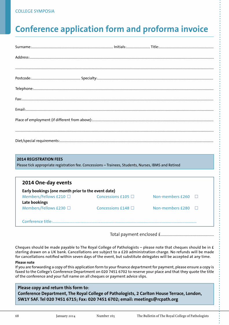
COLLEGE SYMPOSIA
68 January2014 Number165 TheBulletinofTheRoyalCollegeofPathologists
Conference application form and proforma invoice
Surname: ............................................................................................. Initials:........................... Title:..............................................................
Address: ..................................................................................................................................................................................................................
..................................................................................................................................................................................................................................
Postcode:........................................................ Specialty:....................................................................................................................................
Telephone: .............................................................................................................................................................................................................
Fax:..........................................................................................................................................................................................................................
Email:......................................................................................................................................................................................................................
Place of employment (if different from above):..........................................................................................................................................
..................................................................................................................................................................................................................................
Diet/special requirements: ...............................................................................................................................................................................
2014 REGISTRATION FEESPlease tick appropriate registration fee. Concessions – Trainees, Students, Nurses, IBMS and Retired
Cheques should be made payable to The Royal College of Pathologists – please note that cheques should be in £ sterling drawn on a UK bank. Cancellations are subject to a £20 administration charge. No refunds will be made for cancellations notified within seven days of the event, but substitute delegates will be accepted at any time.
Please noteIf you are forwarding a copy of this application form to your finance department for payment, please ensure a copy is faxed to the College’s Conference Department on 020 7451 6702 to reserve your place and that they quote the title of the conference and your full name on all cheques or payment advice slips.
Please copy and return this form to: Conference Department, The Royal College of Pathologists, 2 Carlton House Terrace, London, SW1Y 5AF. Tel 020 7451 6715; Fax: 020 7451 6702; email: [email protected]
total payment enclosed £.....................................................
2014 One-day events
Early bookings (one month prior to the event date)Members/Fellows £210 Concessions £105 Non-members £260 Late bookingsMembers/Fellows £230 Concessions £148 Non-members £280
Conference title:……………………………………………………………..................................................................................
NotiCEBoard
www.rcpath.org Number165 January2014 69
Formorelistings,seeourwebsiteatwww.rcpath.org/conferencesandclickon‘Externalconferences’.Ifyouwouldliketoadvertiseacourseorserviceadvertisedinthissection(freeofcharge),emailthedetailstopublications@rcpath.orgby7FebruaryforpublicationintheApril2014issue.
Exploiting bacteriophages for bioscience, biotechnology and medicine (the 5th in a biennial series)23 January 2014, Londonwww.regonline.co.uk/bacteriophage2014
Biobanking 201417–19 February 2014, Londonhttp://biobanking2014.com/
The 2014 Ageing Summit24–26 February 2014, Londonhttp://ageingsummit2014.com/
Forensic Forums 20143–6 March 2014, Londonhttp://forensicforums2014.com/
The 2014 Mycobacterium tuberculosis Summit24–26 March 2014, Londonhttp://tbsummit2014.com/
The 2014 Obesity Summit1–3 April 2014, Londonhttp://www.obesitysummit2014.com/
The 2014 Controlling Cancer Summit12–14 May 2014, Londonhttp://controllingcancersummit2014.com/
The 2014 Tissue Engineering Congress2–4 June, 2014, Londonhttp://tissueengineeringcongress2014.com/
The following events are organised by Euroscicon. There are discounts of 20% off the registration fee for College
members, using the code RCPAT20, for all meetings. All Euroscicon meetings have CPD accreditation.
7th Annual Cell Culture Technology Event: Recent advances, future prospect16 June 2014, Londonwww.regonline.co.uk/cellculture2014
Therapeutic Proteins and Antibodies: from design to function18 June 2014, Londonwww.regonline.co.uk/thera2014
The 2014 Alzheimer’s Disease Congress23–25 June 2014, Londonhttp://alzheimersdiseasecongress2014.com/
Beating Malaria – London 20141–3 July 2014, Londonhttp://beatingmalarialondon2014.com/
Morphology Update for Biomedical Scientists And 1st Year Senior Trainees17 February 2014, LondonPlease visit www1.imperial.ac.uk/depart-mentofmedicine/divisions/experimental-medicine/haematology/courses_haemo/ or email [email protected]
Update in thrombosis and haemostasis for consultants and advanced senior trainees17 February 2014, LondonPlease visit www1.imperial.ac.uk/depart-mentofmedicine/divisions/experimental-medicine/haematology/courses_haemo/ or email [email protected]
Laboratory aspects of haemoglobinopathy diagnosis18 February 2014, LondonPlease visit www1.imperial.ac.uk/depart-mentofmedicine/divisions/experimental-medicine/haematology/courses_haemo/ or email [email protected]
Advanced haematology morphologyPlease note: this course is not suitable for BMSs
19–20 February 2014, LondonPlease visit www1.imperial.ac.uk/depart-mentofmedicine/divisions/experimental-medicine/haematology/courses_haemo/ or email [email protected]
Histopathology of the bone marrow21 February 2014, LondonPlease visit www1.imperial.ac.uk/depart-mentofmedicine/divisions/experimental-medicine/haematology/courses_haemo/ or email [email protected]
Hands-on cardiac morphology26–28 February 2014, LondonSee www.imperial.ac.uk/nhli/hocm for details.
A practical integrated approach to the diagnosis of haematological malignancies12–14 March 2014, LondonFor details and booking, email [email protected] or [email protected]
2014 Dermpedia Comprehensive Review of Cutaneous Hematopathology and Dermatopathology Update10–12 April, 2014, Scottsdale, Arizona, USAFor details pleasevisit http://www.dermpedia.org/event/scottsdale2014
Criminal Justice Residential Training Course for Pathologists2–6 June 2014, DurhamTo register your interest in attending this course, or for further information, please email: [email protected]
London Uropathology Conference19–20 June 2014, LondonSee www.uropathologyuk.com for details.
2nd British Society for Dermatopathology Self Assessment30 June 2014, GlasgowFor details email [email protected] or visit www.britsocdermpath.co.uk/index.htm
Practical pulmonary pathology30 June–2 July 2014, LondonFor further information, contact Professor Nicholson on: [email protected]
The Sheffield oral and maxillofacial pathology course11–12 July 2014, SheffieldFor details email [email protected] or visit https://sites.google.com/a/sheffield.ac.uk/the-sheffield-oral-and-maxillofacial-pathology-course/
Techniques and Applications of Molecular Biology: A Course for Medical Practitioners14–17 July 2014, WarwickFor details email [email protected] or visit www2.warwick.ac.uk/fac/sci/lifesci/study/shortcourses/molecularbiology )
7th European Multidisciplinary Colorectal Cancer Congress (EMCCC)23–25 November 2014, AmsterdamPlease register online at www.dccg.nl
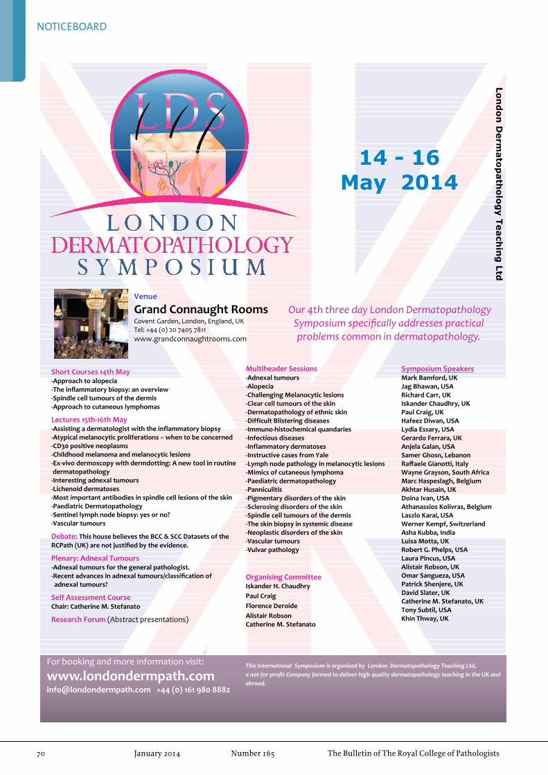
NOTICEBOARD
70 January2014 Number165 TheBulletinofTheRoyalCollegeofPathologists
Multiheader Sessions ‐Adnexal tumours ‐Alopecia ‐Challenging Melanocytic lesions ‐Clear cell tumours of the skin ‐Dermatopathology of ethnic skin ‐Difficult Blistering diseases ‐Immuno‐histochemical quandaries ‐Infectious diseases ‐Inflammatory dermatoses ‐Instructive cases from Yale ‐Lymph node pathology in melanocytic lesions ‐Mimics of cutaneous lymphoma ‐Paediatric dermatopathology ‐Panniculitis ‐Pigmentary disorders of the skin ‐Sclerosing disorders of the skin ‐Spindle cell tumours of the dermis ‐The skin biopsy in systemic disease ‐Neoplastic disorders of the skin ‐Vascular tumours ‐Vulvar pathology
Organising Committee Iskander H. Chaudhry Paul Craig Florence Deroide Alistair Robson Catherine M. Stefanato
For booking and more information visit:
www.londondermpath.com [email protected] +44 (0) 161 980 8882
Symposium Speakers Mark Bamford, UK Jag Bhawan, USA Richard Carr, UK Iskander Chaudhry, UK Paul Craig, UK Hafeez Diwan, USA Lydia Essary, USA Gerardo Ferrara, UK Anjela Galan, USA Samer Ghosn, Lebanon Raffaele Gianotti, Italy Wayne Grayson, South Africa Marc Haspeslagh, Belgium Akhtar Husain, UK Doina Ivan, USA Athanassios Kolivras, Belgium Laszlo Karai, USA Werner Kempf, Switzerland Asha Kubba, India Luisa Motta, UK Robert G. Phelps, USA Laura Pincus, USA Alistair Robson, UK Omar Sangueza, USAPatrick Shenjere, UK David Slater, UK Catherine M. Stefanato, UK Tony Subtil, USA Khin Thway, UK
Our 4th three day London Dermatopathology Symposium specically addresses practical problems common in dermatopathology.
14 - 16 May 2014
Lond
on D
ermatop
atholog
y Teach
ing
Ltd
Venue
Grand Connaught Rooms Covent Garden, London, England, UK Tel: +44 (0) 20 7405 7811 www.grandconnaughtrooms.com
Short Courses 14th May ‐Approach to alopecia ‐The inammatory biopsy: an overview ‐Spindle cell tumours of the dermis ‐Approach to cutaneous lymphomas
Lectures 15th‐16th May ‐Assisting a dermatologist with the inflammatory biopsy ‐Atypical melanocytic proliferations – when to be concerned ‐CD30 positive neoplasms ‐Childhood melanoma and melanocytic lesions ‐Ex‐vivo dermoscopy with dermdotting: A new tool in routine dermatopathology ‐Interesting adnexal tumours ‐Lichenoid dermatoses ‐Most important antibodies in spindle cell lesions of the skin ‐Paediatric Dermatopathology ‐Sentinel lymph node biopsy: yes or no? ‐Vascular tumours
Debate: This house believes the BCC & SCC Datasets of the RCPath (UK) are not justied by the evidence.
Plenary: Adnexal Tumours ‐Adnexal tumours for the general pathologist. ‐Recent advances in adnexal tumours/classication of adnexal tumours?
Self Assessment Course Chair: Catherine M. Stefanato
Research Forum (Abstract presentations)
This International Symposium is organised by London Dermatopathology Teaching Ltd, a not‐for‐prot Company formed to deliver high quality dermatopathology teaching in the UK and abroad.
NOTICEBOARD
www.rcpath.org Number165 January2014 71
legaciesThe objectives of the College are to advance the science and practice of pathology, to educate the public in matters relating to pathology and to promote study and research work in pathology and related subjects and publish the result of such study and research. Financially, the College aims to match activities to projected income. The College is funded from subscriptions, examinations and related fees, investment income, grants from outside bodies and charitable donations.
Bequests or legacies are always gratefully received. Leaving a gift to charity in your will is a very special way of helping to secure the future for organisations such as The Royal College of Pathologists. Legacies to the College have the added benefit of being exempt from inheritance tax. An open legacy may be made toward the general purposes of the College. This is pre-ferred because it allows the College to apply the funds donated where the need is greatest at the time the legacy eventually be-comes available. This can be quite different from the perceived need when a will is made. However, you may legally oblige the College to spend the money in a particular area of College work or for a specific purpose by making a restricted legacy.
The College undertakes many educational initiatives. We are actively developing an outreach programme that will spread the awareness of pathology throughout the UK and abroad.
No other UK college has committed so much to the future of our profession in terms of time and resources. This will be-gin to promote the importance of pathology to the grass roots of this country through schools, colleges, hospitals and many other sites where the general public can have access to impor-tant healthcare information. If we are to safeguard the future of our profession in the face of increasing competition from other medical and science career opportunities, it is vital that we com-mit ourselves to the promotion and awareness of pathology, and
continue to train our young professionals to the very highest standards.
This public engagement programme will require finan-cial support from the College for many years to come and we hope very much that we can build upon the tremendous support you have already given and ask if you would con-sider leaving a legacy.
Additions to your existing will can be made using a ‘Form of codicil’, printed on the following page. Please note that witnesses should be present when you sign the form, but it should not be witnessed by a College member or the spouse of a College mem-ber. As a general point, we always recommend consulting a so-licitor or qualified will-writer before making a will; they should give you all the legal and tax advice that you require.
If you are considering including a legacy to the College in your will, we would very much appreciate being informed of your generous act. To inform us of your bequest or for specific advice on legacies to the College, please contact me.
Daniel RossChief Executive020 7451 [email protected]
Association of Clinical Pathologists 28th ACP Management Course
run by Pathologists for Pathologists
3–5 September 2014
Hardwick Hall Hotel, Sedgefield, County Durham
This is a wide ranging, residential, 3 day course introducing management issues relevant to the running of a modern pathology service.
It is intended for specialist registrars and trainees in pathology in their final year of training, clinical scientists and those who have held their first consultant post for less than 2 years.
Course organisers: Dr Gavin Spickett, Dr Helen Bourne and Dr Catherine Stroud.
For further information and a registration form please visit our website:www.pathologists.org.ukTelephone: 01273 775700E-mail: [email protected]
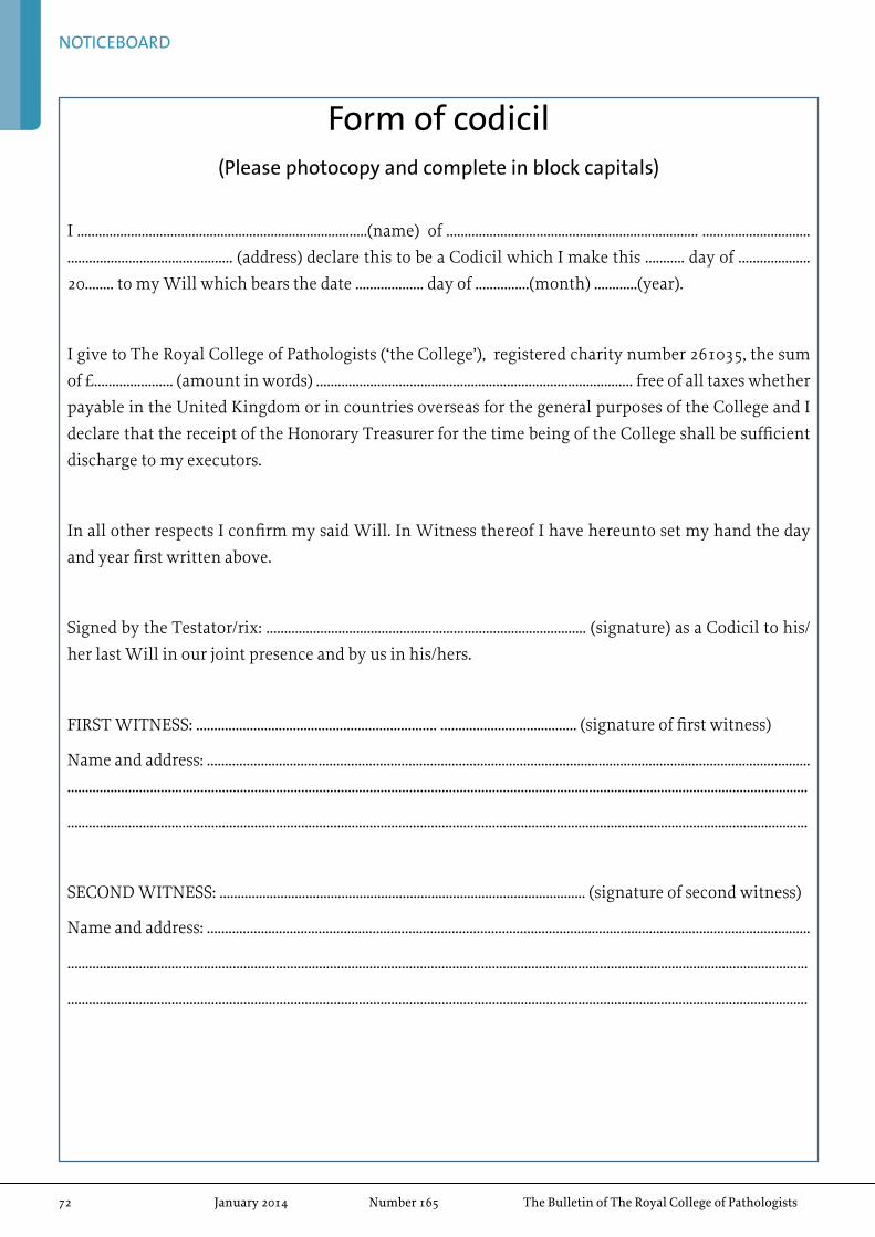
NOTICEBOARD
72 January2014 Number165 TheBulletinofTheRoyalCollegeofPathologists
Form of codicil(Please photocopy and complete in block capitals)
I .................................................................................(name) of ……………………………………………………..…..... …………………………
…………………………..…........... (address) declare this to be a Codicil which I make this ……….. day of ……………..…
20…….. to my Will which bears the date ………………. day of ……………(month) …………(year).
I give to The Royal College of Pathologists (‘the College’), registered charity number 261035, the sum
of £…………………. (amount in words) ………..........…………………………………………………………… free of all taxes whether
payable in the United Kingdom or in countries overseas for the general purposes of the College and I
declare that the receipt of the Honorary Treasurer for the time being of the College shall be sufficient
discharge to my executors.
In all other respects I confirm my said Will. In Witness thereof I have hereunto set my hand the day
and year first written above.
Signed by the Testator/rix: …………………………………………………..……........................ (signature) as a Codicil to his/
her last Will in our joint presence and by us in his/hers.
FIRST WITNESS: ..................................…………………………… ……….......................…... (signature of first witness)
Name and address: …………………………………………………...............................................................................………….......………....
................……………………………………………………………………………………………….......………..................................................................
................……………………………………………………………………………………………….......………..................................................................
SECOND WITNESS: .................................…………………................................................ (signature of second witness)
Name and address: ………………………………………………….........….......………………..........................................................................
.................……………….......……………………………………………………………………….......…………………………..............................................
................……………………………………………………………………………………………….......………..................................................................
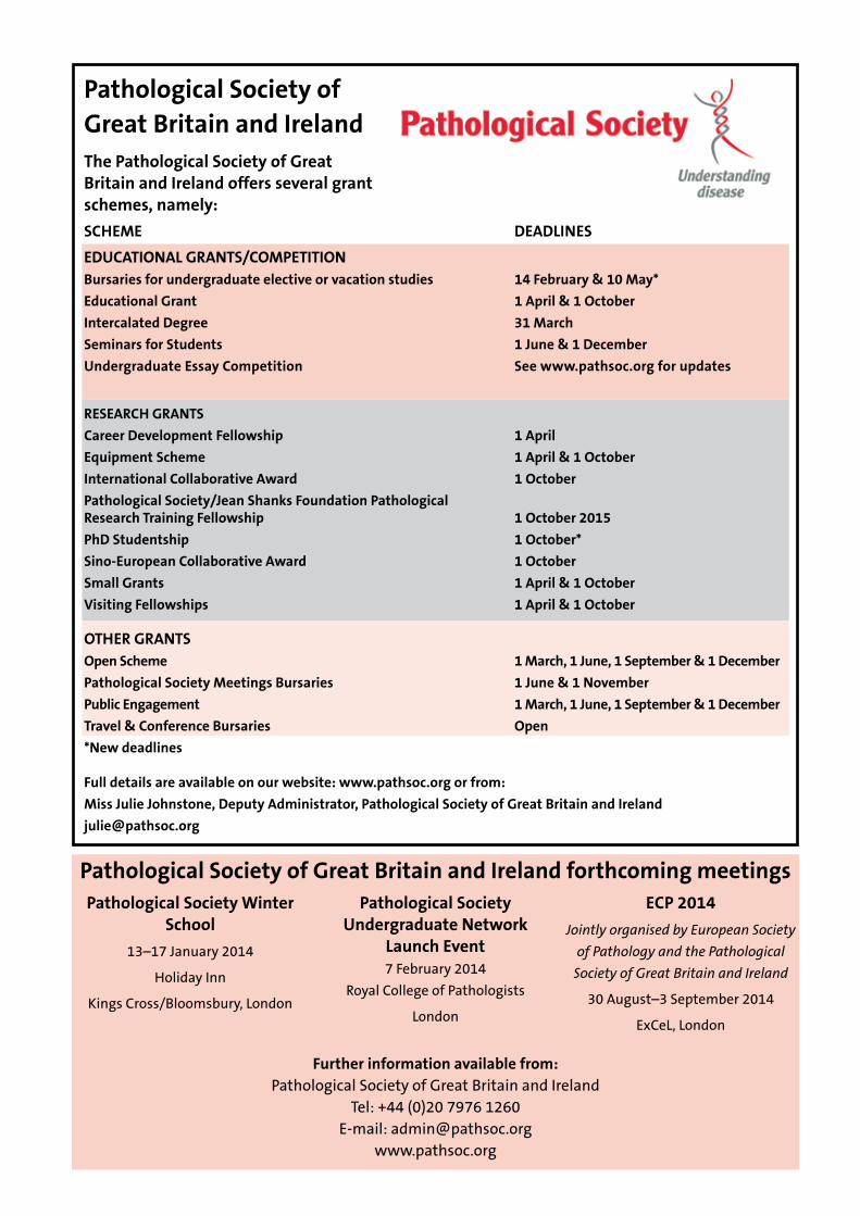
Pathological Society ofGreat Britain and IrelandThe Pathological Society of Great Britain and Ireland offers several grant schemes, namely:
SCHEME DEADLINES
EDUCATIONAL GRANTS/COMPETITIONBursaries for undergraduate elective or vacation studies 14 February & 10 May*
Educational Grant 1 April & 1 October
Intercalated Degree 31 March
Seminars for Students 1 June & 1 December
Undergraduate Essay Competition See www.pathsoc.org for updates
RESEARCH GRANTS
Career Development Fellowship 1 April
Equipment Scheme 1 April & 1 October
International Collaborative Award 1 October
Pathological Society/Jean Shanks Foundation Pathological Research Training Fellowship 1 October 2015
PhD Studentship 1 October*
Sino-European Collaborative Award 1 October
Small Grants 1 April & 1 October
Visiting Fellowships 1 April & 1 October
OTHER GRANTSOpen Scheme 1 March, 1 June, 1 September & 1 December
Pathological Society Meetings Bursaries 1 June & 1 November
Public Engagement 1 March, 1 June, 1 September & 1 December
Travel & Conference Bursaries Open
*New deadlines
Full details are available on our website: www.pathsoc.org or from:
Miss Julie Johnstone, Deputy Administrator, Pathological Society of Great Britain and Ireland
Pathological Society Winter School
13–17 January 2014
Holiday Inn
Kings Cross/Bloomsbury, London
Pathological Society Undergraduate Network
Launch Event7 February 2014
Royal College of Pathologists
London
ECP 2014
Jointly organised by European Society
of Pathology and the Pathological
Society of Great Britain and Ireland
30 August–3 September 2014
ExCeL, London
Pathological Society of Great Britain and Ireland forthcoming meetings
Further information available from:Pathological Society of Great Britain and Ireland
Tel: +44 (0)20 7976 1260 E-mail: [email protected]
www.pathsoc.org
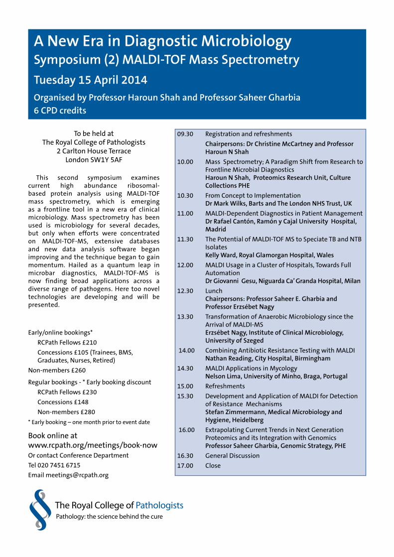
A New Era in Diagnostic MicrobiologySymposium (2) MALDI-TOF Mass Spectrometry
Tuesday 15 April 2014Organised by Professor Haroun Shah and Professor Saheer Gharbia6 CPD credits
09.30 Registration and refreshments
Chairpersons: Dr Christine McCartney and Professor Haroun N Shah
10.00 Mass Spectrometry; A Paradigm Shift from Research to Frontline Microbial Diagnostics
Haroun N Shah, Proteomics Research Unit, Culture Collections PHE
10.30 From Concept to Implementation Dr Mark Wilks, Barts and The London NHS Trust, UK
11.00 MALDI-Dependent Diagnostics in Patient Management Dr Rafael Cantón, Ramón y Cajal University Hospital,
Madrid
11.30 The Potential of MALDI-TOF MS to Speciate TB and NTB Isolates
Kelly Ward, Royal Glamorgan Hospital, Wales
12.00 MALDI Usage in a Cluster of Hospitals, Towards Full Automation
Dr Giovanni Gesu, Niguarda Ca’ Granda Hospital, Milan
12.30 Lunch Chairpersons: Professor Saheer E. Gharbia and
Professor Erzsébet Nagy
13.30 Transformation of Anaerobic Microbiology since the Arrival of MALDI-MS
Erzsébet Nagy, Institute of Clinical Microbiology, University of Szeged
14.00 Combining Antibiotic Resistance Testing with MALDI Nathan Reading, City Hospital, Birmingham
14.30 MALDI Applications in Mycology Nelson Lima, University of Minho, Braga, Portugal
15.00 Refreshments
15.30 Development and Application of MALDI for Detection of Resistance Mechanisms
Stefan Zimmermann, Medical Microbiology and Hygiene, Heidelberg
16.00 Extrapolating Current Trends in Next Generation Proteomics and its Integration with Genomics
Professor Saheer Gharbia, Genomic Strategy, PHE
16.30 General Discussion
17.00 Close
To be held atThe Royal College of Pathologists
2 Carlton House TerraceLondon SW1Y 5AF
This second symposium examines current high abundance ribosomal-based protein analysis using MALDI-TOF mass spectrometry, which is emerging as a frontline tool in a new era of clinical microbiology. Mass spectrometry has been used is microbiology for several decades, but only when efforts were concentrated on MALDI-TOF-MS, extensive databases and new data analysis software began improving and the technique began to gain momentum. Hailed as a quantum leap in microbar diagnostics, MALDI-TOF-MS is now finding broad applications across a diverse range of pathogens. Here too novel technologies are developing and will be presented.
Early/online bookings*
RCPath Fellows £210
Concessions £105 (Trainees, BMS, Graduates, Nurses, Retired)
Non-members £260
Regular bookings - * Early booking discount
RCPath Fellows £230
Concessions £148
Non-members £280
* Early booking – one month prior to event date
Book online atwww.rcpath.org/meetings/book-nowOr contact Conference Department
Tel 020 7451 6715
Email [email protected]
The Royal College of PathologistsPathology: the science behind the cure
Related Documents