日本財団補助金による 2000 年度日中医学学術交流促進事業 ⑤.在留中国人研究者研究助成 (22) B A 1 1 遺伝子の抗腫蕩機能に対する基礎研究

Welcome message from author
This document is posted to help you gain knowledge. Please leave a comment to let me know what you think about it! Share it to your friends and learn new things together.
Transcript

日本財団補助金による2000年度日中医学学術交流促進事業
⑤.在留中国人研究者研究助成
(22) B A 1 1遺伝子の抗腫蕩機能に対する基礎研究
BAI1遺伝子の抗腫蕩機能に対する基礎研究 i
康照雄 l
白求恩医科大学第三附属病院検査部主任
東京大学医科学研究所附属先端医療研究センタ一
一分子療法研究分野.客員研究員一一
要旨(日本語)
当センターで最近新しくクロ二ンクされた脳特異的発現する新生血管抑制因子-
brain-specific angiogenesis inhibitor 1 (BAll)はmVIVOの実験で非常に強い血管新
生抑制機能を現し、さらに動物の腫蕩モデルで血管新生抑制による抗腫蕩効果
が観察された。現在抗腫蕩遺伝子治療の候補として研究を進めている。
KEY WORDS
BAI1遺伝子 抗腫蕩機能
研究報告
目的
↓
方法
↓
結果
↓
考察
↓
参考文献
Antiangiogenic
Implication of
glioblastoma
potency of BAI1
gene therapy for
In VIVO:
human
X.KANGJ K.TANl,1s.NlSHlMORIY MSUNAMURA,3.L.LUClAN,3
T.TOKINO,2 Y.NAKAMURA, 2 S. ASANO. 1
I.SAITOH,4
Department of Hematology/Oncology¥ Human genome center2, genome analysis 4 The
Institute of Medical science,The University of Tokyo; Department of Surgery 1st, Tohoku
university, School of Medicine3

¥..:.
ABSTRACT
BAI1 (brain-specific angiogenesis inhibitor 1) is an 1584・amino-acidproduct containing
five thrombospondin type 1 repeats and is specifically expressed in the brain. This BAI1
gene is now considered as one of novel p53・induciblegenes. As angiogenesis is
required for the growth and progression of malignancies. we investigated the in vivo
antiangiogenic and antitumor effects of the wild-type BAI1 gene transfer on five humans
glioblastoma cells. We constructed BAl1-expressing glioblastoma cells after the
transduction with the BAI1 cDNA using a recombinant adenoviral vector (BAI1 cDNA
inserted into pAdex1 Cawt at Swa1 site). The Northern blot analysis and Western blot
analysis. demonstrated respectely the mRNA and protein of BAI1 in the transduced
tumor cells. In vivo neovascularization assay of adenoBAI1 infected cells was then
peげOrmedusing a transparent skin fold chamber model implanted in SCID mice. In
this assays, BAI1 gene transduced glioma cells significantly inhibited angiogenesis in
vivo. In tumor inhibition assay, transduced or untransduced human glioma cells were
implanted intradermally to the right flank of SCID mice. The presence of detectable
tumor was confirmed by palpation and the tumor size was measured periodically. In vivo
antitumor assay demonstrated the inhibited tumor growth after the direct i吋ectionof
BAI1 adenovirus into tumors. Morphologically, tumor showed signs of impaired
angiogenesis, such as extensive necrosis and reduced tumor vascular density. Our data
demonstrated that a recombinant adenovirus expressing the BAI1 gene had
antiangiogenic role for glioma cells and this virus also inhibited the growth of glioma
cells in vivo. BAI1 was considered to be one of the potent candidate genes for cancer
gene therapy.
Overview summary
Recombinant adenovirus carrying the human BAI1 cDNA (AdBAI1) was constructed using
COS-TPC method. In vivo growth inhibition and antiangiogenic e仔'ectswere observed with
BAI1 gene transducced U373MG tumor cells. using Skin fold transparent chamber
angiogenesisnmodel, our results also suggested that a AdBAI1 had antitumor effects due to
antiangiogenic function of BAI1 protein in human glioblastoma cells carrying codon 237
cgt(Arg)-cat(His)mutation of p53 gene. The coexp陪 ssionof BAI1 and p53 gene did not
enhance the antitumor e仔'ectsof BAI1 gene. BAI1 was considered to be promising
candidate gene for human cancer gene therapy targeting glioblastoma cells with p53 gene
mutations.
-・・・・・・・ー・・--- ーー---・・・・・・・・・・・・・・ー・・----------------------------・・・・・・・・・ーー・・-------

. 8
'
e . 8
8 . ' ' ' '
-・・・・・・・・.ー--------------------ーーーーーーーー ーーーーーーーー司ーーー・・・・・・ ・・・・・・・・・・------------------ー----------・・・・ー------
Introduction
Angiogenesis is considered to be regulated by a net become of positive and negative
regulators of blood vessel growth. Recent discoveries of endogenous negative regulators of
angiogenesis, thrombospondin (TSP), endostatin, angiostatin and glioma-derived
angiogenesis inhibitory factor, all associated with neovascularized tumors, suggest a new
paradigm of tumorigenesis(Folkman, 1989, 1995). Negative regulators would predict how a
primary tumor grows and whether metastases appease. Gliomas, particularly glioblastomas
are a prominent target of cancer gene therapy because of their poor prognosis despite all
current conventional therapies. Intratumoral transplantation of HSV・TKretrovirus producer
cells has been under clinical investigation. Gene therapy strategies developed to interfere
with the normal function of vascular endothelial growth factor receptor is another new
theraputic approach and have been successfully used in di仔'erentexperimental models to
block tumor angiogenesis and the growth (Marcia,1999). Tumor cell transduced with the
wild type p53 gene inhibited the in vivo tumor growth of adjacent non transduced cells.
This data suggest that a recombinant adenovirus expressing the wild type-p53 gene is
antiangiogenic, which may explain, in part, the mechanism of the bystander effect
induced by the wlid type-p53 gene transfer on adjacent tumor cells (NISH区AKI,
1999 ) Recently, novel P53 inducible gene that encode a 1584-amino acid product
containing five thrombospondin type1 repeats has been cleared by one group and has been
demonstrated to be specifically expressed in the brain. A recombinant protein
corresponding to the TSP-type 1 repeats of this gene product inhibitied in vivo
neovascularization induced by bFGF on the rat cornea. The expression of this gene,
designated BAI1 (brain-specific angiogenesis inhibitor 1) was absent or significantly
reduced in eight of nine glioblastoma cellline, suggesting BAI1 played a significant role
of angiogenesis inhibition, as a mediator of p53. (HIROYUKINISHIMORI.,1997)
cancer BAI1 expression was significantly reduced in colorectal cancers as compared to
the extra neoplastic tissues. BAI1 expresson was inversely correlated with vascular
invasion and metastasis. Moreover, vaeculaty in the colorectal cancer was inversery
correlated with BAI1 gene expression. BAI1 expression was seggested to inhibit
angiogenesis and metastasis of colorectal cancer (YOSHITAKA FUKUSHIMA., 1998 )
But whether it has direct inhibit tumor in vivo effect and mechanism remains unclear .
In this study we studied the in vivo growth inhibitory potency of BAI1 gene in using
human glioblastoma implanted to SCID mice.
f____________________________ーーーーーーーーー
ーーーーーー----・・・・・・・・・・.・---------_...・・・・・ーーーーーーーーーーーー ーーーーーーー-------・・・・・・・・・・・・・・・・・・__-'
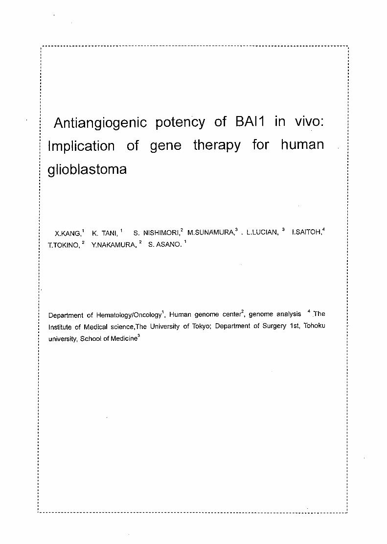
MATERIALS AND METHODS
Construction of recombinant adenovirus We used the COS-TPC method to construct recombinant adenovirus car叩ingthe human
BAI1 cDNA. Briefly, the human BAI1 cDNA was inserted into the cassette cosmid, pAdexlw,
which is an 11 kb charomid vector bearing an adenovirus 5 genome with deletions of E 1 and
E3. The recombinant adenovirus was generated by in vivo homologous recombination in
293 cells between the cosmid cassette and Nsil・digestedadenovirus DNA tagged with viral
terminal troteins. The recombinant adenovirus carrying a LacZ expression unit also
controlled by the CAG promoter (AxCALacZ). For in vivo transfer, The adenovirus was
purified and concentrated through CsCI step gradients followed by dialysis against
phosphate-buffered saline (PBS)-10% glycerol buffer. Titration of recombinant adenoviruses
was performed by 50% tissue culture infectious dose (TCID50). Lack of contamination by
replication-competent adenovirus (RCA) was confirmed by an inability to amplify a fragment
of the E1B region by polymerase chain reaction (PCR). AII viruses were puri行edand
expanded on 293 cells by the standard technique. Each viral inoculum was purified by a
CsCI step gradient followed by a CsCI equilibrium gradient. The final viral band was diluted
1: 1 with sterile glycerol and stored at -80・C.Concentration of the virus was determined by
measuring their optical density at 260 nm. The multiplicity of infection (MOりwascalculated
on the basis of the ratio of functional virus as assayed on 293 cell is reported
befo陪 (reference).
Animals. The'experiments were perfomed using SCID mice (SPFNAF mause, C.S-17I1cr
Crj-scid), purchased from Nippon charles River co. Yokohama, bred and maintained in a
specific pathogenic germ-free environment in our laboratory. For the surgical procedure,
mice (females, 25-30 9 body weight). Formulas of the antimicrobial reagents added to
the drinking water of the animals were Oxytetracycline 0.3g, Ampicillin 0.3g, Cloxacillin
0.2g and Potassium sorbate 1.35g in one litre , by HCI to pH 2.5.( Knut ,1984)0 A 2ぃlaliquot of a suspended U373MG cells (4X10⁵) was i吋ectedinto the brain of male and
female SCID mice under sodium pentobarbital (Nembutal) anesthesia 50mg/kg, i.p.
Celllines Five glioblastoma cell lines were used in this study. YKG1 was purchased from HSRRB
(Tokyo); U87MG, U373MG, SW1783 and DBTRG05MG were from ATCC. YKG1,
U87MG,U373MG and DVTRG05MG cell lines were maintained in Eagle.s . MEM medium
with non-essential amino acids, sodium pyruvate, 1 mM and Earle‘s BSS, (10% fetal bovine
serum, 2mM L-glutamine, streptomycin [100IJg/ml], and penicillin [100U/ml]). SW1783 was
' ・・ー・・・・・・・・・・・・・・・・・・・・・・・・・・・・・・・・・・・・・・・・・・・・・・・・・・・・・・・・・・・・・・・・・・・・・・・・・・・・・・・・・・・・・・・・・・・・・・・・・ 4
maintained in Leibovitz、sし15medium, 10% fetal bovine serum. In a free gas exchange with
at mospheric air. The kidney embryonic cell line 293 was obtained from Microbix 8iosistems
(TORONTO, Ontario, Canada) were maintaned in complete Oulbecco's medium (OMEM).
Western blot analysis Western blot analysis was essentially peげormedfollowing standard method. 8riefly, cells
were homogenized and the centrifuged supernatant was electrophoreses in 7% gel and
blotled onto a nitrocellulose membrane. The membrane was locked with 3% bovine serum
albumin (8SA) in P8S, and incubated for 2 hr with rabbit anti-human 8AI1 serum diluted to
1 :2000 in 1 % 8SA・0.05%Tween 20 in P8S. After several washings, the membrane was
incubated for 1 hr with horseradish peroxidase-Iabeled goat anti-rabbit IgG (Jackson
Immuno Research Laboratories. INC ) diluted to 1 :5000 in 1 % 8SA・0.05%Tween 20 in P8S.
For detection, enhanced chemiluminescence (ECL; Amersham , Arlington Heights , IL) was
used and the membrane was exposed to Fuji (Tokyo, Japan) RX-U film for 10・30sec.
Northern blot analysis Northern blot analysis of 8AI1 transduced glioblastoma cells showed 8AI1 mRNA
expressed properly. RNA was extracted from cultured cells with TRlzol (Life Technologies).
RNA from tumor cell lines and tumor tissue was separated on formaldehyde-agarose gels
and blotled onto nitrocellulose filter (Micron Separations, Westboro, MA). 8AI1 mRNA was
detected by hybridization of membranes with 32p labelled 8AI1 cONA. As previously
reported (Nishimori,1997). As control for quality and loding of mRNA, blots were also
hybridized with a 32p labeled s actin cONA. This cONA probes were labeled with [32p] dCTP
(Amersham life Sciences) by the random-primer method to a specific activity of >2x10⁹
dpmlμg. 810ts were hybridized in QuickHyb solution (Stratagene) at 68・Cfor 1 hr followed
by washing in 0.1xSSC・0.1%SOS at 65・Cand autoradiography (1xSSC is 0.15M NaCI plus
0.015M sodium cit悶te).
The radioactivity of each band was quantitated with a F吋i8as 1000 bioimaging analyzer
and Mac8as software. For experiments designed to determine mRNA , Glioblastoma tumor
cells were grown to confluence in a six-well plate and infected with 5x10⁸ PFU/ml Ad8AI1 or
any infection virus. Twenty-four hours after infection, cells were harvested at indicated times,
and the RNA was extracted and subjected to Northern analysis, as described above.
Dorsal skin fold transparent chamber Dorsal skin fold transparent chamber (DSFTC) was composed of two titanium frames and
the total weight was 1.8g. The frame was implanted to mice so as to sandwich the extended
double layers of skin perpendicularly to the animal's back. One layer of skin was completely
,-ー・・・・・・・・・・・・・・・・・・・・・・-----------------------------------------------・・・・・・ー---・・・・・・・・・・・・・・・・・ 4
removed circulaly with the diameter of 15mm, and the remaining layer composed of
epidermis, subcutaneous tissue and muscle was covered with a cover slip incorporated in
one of the frames. Using stainless steel nuts as spacers, frame-to-frame distance of
400-450um was maintained to prevent the compression of nutritional blood vessels. Fine
polyethylene catheters were inserted into the jugular vein and the caroid aはery,respectively,
passed subscutaneously to the dorsal side of the neck, closed, and sutured to the titanium
frames. The animals tolerated to the chambers and showed no signs of discomfort. In
particular, no e仔'ecton sleeping and feeding habit was observed. The chamber implantation
time was about 20 min per mouse. For intravital microscopy, unanesthetized animals were
immobilized in polyethylen tubes attached to the stage of a microscope (OPTIPHOTO 66,
Nikon, Tokyo, Japan). Observations were made with the following objectives: Nikon X2
(numerical ape吋ure,0.05), Nikon X4 (numerical aperture, 0.10), Nikon X10 (numerical
apeはure,0.25) and Nikon X20 (numerical aperture,0.40) using transillumination and a
filter for conveはingartificial light into daylight. Observations were captured with an
intensified CCD camera TEC-470 Optronics Co., Chelmsford, MA, USA), attached to the
microscope and serially connected to a TV monitor (BM-1400S Vitor Co., Tokyo, Japan) and
recorded on a S-VHS video tape recorder (HR-X1 Video Co., Tokyo, Japan). During off-line
analysis of the video tape recordings, stil video frames (680x480 pixels) were captured
at a resolution of 72 pixelelinch with Avid Video Shop 3.0 (Avid Technology, Inc.,
Tewksbury, MA, USA) on a Power Macintosh 7100/80AV computer (Apple Computer,
Inc., Cupertino, CA, USA). The video frames taken with X10 objective were
subsequently processed in order to obtain vascular parameters. The vessel out line of
each of the frame was carefully drawn in a separate transparent layer using Adobe
Photoshop 4.0 (Adobe systems, Inc., Mountain View, CA, USA) with the pencil tool of
one pixel line width after the defective channels of the RGB color images of the video
captured frame were eliminated and the image transformed to the grey scale mode. In
this study, observations were perfomed at days 0, 3, 6, 9 and 12 after tumor inoculation.
Histology Specimens were fixed in phospho・Iysine-paraformadehydesolution at 4・Cover night,
embedded in Tissue-Tek O.C.T. Compound (Sakura Finetechnical Co., Ltd., Tokyo,
Japan) and stored at・80・C.Five IJm sections were cut for immuno histochemical
staining. Expression of von Willebrand factor was detected using rabbit polyclonal
antibodies, 8mg/ml (Dako, Tokyo, Japan). After the standard protocol from Dako
Sheet. Tissue sections were incubated with 3% hydrogen peroxide in distilled water
for 5minutes and rinsed with distilled water and placed in Tris -buffered saline (TBS) for
5 minutes. After the rinse , the specimen were covered with 2・3dropsof DAKO EPOS
.-・・・ー・・・・・・・・・・・・・・・・------ ----ー・・・・ー・・・・・・・・・・・・・・・・・・---・・・-----------------
-・・・・・・・・・・ー・・・・・・・・・・・・・・・・・・・・・・・・・--・・・・・・--・・・ー・・・・ー-・・・・・・・・・・・・・-----------------------------
Anti-Von Willebrand Factor/HRP (one drop equals 50μ りandIncubated about 60
minutes at room temperature. As negative control, DAKO EPOS Negative Control (code
No.U 0951) was used following manufactures protocal,The specimen were than rinse
in TBS for 5 minutes, incubated with DAB for 5-15 minutes, rinssed with distilled water,
counterstained and mounted with coverslip as reported before.
BAI1 protein immuno histochemical staining as described above.
X-gal staing Cells were spinned down and fixed with a methanol-formaldehyde mixture for 1 hr and
0.2%副lutaraldehydein PBS for 30min, stained for s -galactosidase activity by
incubating in PBS including 5・bromo・4・chloro・3・indolyl-beta-D-galactopyranoside (X-
Gal, 400 mg/ml), 5 mM potassium ferricyanide, 5 mM potassium ferocyanide, and 2mM
MgCI2 for 4 hr at 370C, and lightly counter stained with eosin. Section were immersed
briefly in 4% paraformaldehyde, washed extensively with PBS and immersed in a
solution containing 1 mg/ml X-gal, 2 mM MgCI2, 5mM K3Fe(CN)6 and 5 mM K4Fe(CN)6 in
PBS overnight at 37C. Sections were rinsed in PBS and counterstained with
hematoxylin/eosin and coverslipped in PBS/glycerol for microscopic observation.
Human Glioma Therapy Model in SCID Mice
1 x1 07 each of human glioma cells were implanted intradermally to the right flank of SCID
mice. Tumor with diameters of 5-10mm developed in 14 days and 25mm in 56 days after
transplantation. 2X10⁹o,o pfu in 50-100(J1 volume of either AdBAI1, AdLacZ, AdV-RR5 were
injeded into tumor using a 26・gaugeneedle. The injection was repeated every other day
(Figure5). The presence of detectable tumor was confirmed by palpation and the tumor size
was followed periodicallμ
Data Analysis The significance of differences between two groups was determined by unpaired t test.
Statistical analyses were performed on a mocrocomputer with the aid of the SigmaStat
program(Jandel Scientific, San rafael,CA). Data are prosented as mean :!:SEM.
Resulis
BAI1 expression
Northern blot analysis of BAl1-transduced SW1783, YKG1, DBTRG05MG, U87MG and
U373MG glioblastoma cells showed BAI1 mRNA expressed properly. But non transduced
wild type cells did not expressed BAI1 mRNA (Figure 2).
--------------------- ・・・・・・・・・・・・・・・・・・・・・・・・・・・・・・・ー・・・・・---・・-------ー--------'

Western blot analysis showed BAI1 transduced YKG1, DBTRG05MG, U87MG and
U373MG glioblastoma cells expressed BAI1 protein. But non transduced wild type cells did
not expressed BAI1 protein (Figure 3).
In vivo transduced AdBAl1 after 48hour U373MG cell were immunochamistry stained with
the antibody for anti BAI1 antigen as an expressed the cell marker. The staining revealed
that MOCK and AdLacZ transduced cell was negative result(Figuer8 A and B). and BAI1
transduced cell is positive(Figuer8 C, 0, E and F)., demonstrated the inserted BAI1gene
expressed protein.
Angiogenesis inhibition effect
Transplanted BAI1 transduced U373MG cells were observed to have significantly inhibited
tumor cell-induced angiogenesis in vivo with DSFTC. Angiogenesis network was observed
in mice transplanted with in wildtype and AdLacZ transduced cells mice over all tumor area
at day 12. But angiogenesis was completely inhibited in dorsal skinfold chamber of SCID
mice transplanted with AdBAけ transducedcells at Day12 (Figure 4) .
In injected DMEM and AdLacZ tumors showed blood vessel infiltration (Fig8 H and I ),
immunohistochemical staining showed normal capillaries in AdLacZ injected mice
tumor(16.0+ー3.2blood vessels per 200X field) but stained vessels in the AdBAl1 injected
tumor (1.6+ー2.5blood vessels per 200x frame) displayed very few and extremely small
bloodvessels. (Fig8 G),
Tumor inhibition effect
The U373MG tumor bearing SCID mice mean tumor sizes of WT and Adeno were
statistically larger than those of the other 3 groups of P53, BAI1 or P53+BA11 35days after
i吋ection(P<O.05 , Figure 6) tumor.
The suppressed tumor growth was observed in mice group transplanted with AdBAl1
transduced cells in contrast to controls(*p<O.05,Figure 6).
The in vivo antitumoral effects of intratumorolly injected AdLacZ , AdBAl1 or DMEM were
analyed in SCID mice bearing U-373 MG tumors. Tumolals injected with AdBAl1 regressed
significantly compared with the tumors injected with AdLacZ (Fig.5;N=20;P=O.003)and
DMEM(Fig.5 ;N=18;P=O.003). The experiments were repeated twice, and the results .were
reproducible. Representative data are included the Fig 8.
Combination Therapy
Combined use of AdBAl1 and AdP53 did not potentiate U373MG tumor growth nhibition by
AdBAl1 or AdP53. the mean tumor sizes of DMEM and Adeno were statistically larger than

those of the other 3 groups of P53, BAI1 or P53+BA11 35 days after i吋ection(p<0.05,
Figure6)
Survival
The survival of mice after in vivo transduction of AdBAI1 vector or control vector at U373MG
tumor was shown in Fig7. Mean survival time of mice transplanted with U373MG ranged
between 42 and 70 day and the survival rate was 20%. Mice transplanted with U373MG
cells and treated with AdBAI1 survived much longer in 80 day in 80%(p<0.05). Mice treated
with AdLacZ and DMEM survived similarly.The U373MG tumor bearing SCID mice survival
curves of animals injected with AdLacZ or DMEM(p<0.05 ,Figure 6).
Discussion
angiogenesis is crucial for the growth of tumors and metastasis diffusion. The beginning
of tumor angiogenesis is caused by the selection of a cancer cell clone which induces an
unbalance between inducers and inhibitors of angiogenesis.
In this study, we examined the efficacy of the novel p53-inducible genes BAI1 alon or BAI1 :
and p53 combination for antitumor response. Both genes were intrgrated into adenoviral
vectors and transferred alone or in combination directly into human glioblastoma, Analysis
the antiangiogenesis and antitumor effect.
The BAI1 protein are a family of three related protein, each gene designated BAI1,
BAI2 and BAI3, were specifically expressed in brain, and are likely to be expressed in the
same type of cells, members of this novel gene family considered to be a member of the
secretin receptor family (Shiratuchi,1998). This novel gene family may play important roles
in suppression of glioblastoma Progression of a glioma to its more malignant form,
glioblastoma, is associated with dramatic neovascularization. The most frequently altered
gene in human glioblastomas is p53. Neovascularization might be a direct consequence of
inactivation of p53, possibly due to loss of transactivation of one or more target genes
regulated by p53 (Van,1994). p53 produced a secretory inhibitor of capillary endothelial cell
migration designated glioma-derived angiogenesis inhibitory factor Isolated a novel brain-
specific gene BAI1 (Nishimori,1997). BAI1 contained at least one“fuctional" p53・bindingsite
within an intron, and its expression was shown to be induced by wild type p53. The BAI1
protein includes a 7・spantransmembrane region similar to that of the secretin receptor. The
axtracellular region of BAI1 possesses a single arg-gly-asp motif recognized by integrins,
and 5 sequences corresponding to the thrombospondin type 1 repeats that can inhibit
experimental angiogenesis induced by basic fibroblast growth factor. in spite of similar
-・・・・・ー・・・・・・・・・・・・・・・・・・・・・・・・・・・・・・・・・・・・・・・・・・・・・・・・・・・・・・・・・・・・・・・・・・・・・・・・・・・・・・・・・・・・・・・・・・・・・ d
tissue specificity among the 3 BAI gene, only BAI1 was transcriptionally regulated by p53.
this novel gene may play impo吋antroles in supperession of glioblastoma by fluorescence in
situ hybridization, (shiratruchi et aI.1997).
we used BAI1 have evaluated the potential of gene therapy of human glioblastoma cells,
using viral vectors encoding human BAI1 cDNA.
First was BAI1 transduction rate in glioblastoma cell line. Fortunately, 5 human
剖ioblastomacell lines all was high level expressed BAI1 gene. Futher. analysis transduced
BAI1 gene cell lines BAI1 protein expresse function also peげbrmed4glioblastoma cell lines
was detected. In curture transduced BAI1 cell lines, BAI1 protein were pa吋iallyreleased
solubility protein, moreover multi of infection (MOりdependentincreased this release in
perfomed between MOI 1to 20.
BAI1 in vivo have a anti angiogenesis e仔'ect.In this study, transparent chamber model and
implanted intradermally to the right flank of SCID mice model were used to study the e仔"ect
of BAI1 on antiangiogenesis and antitumor function in U373MG glioblastoma. Transparent
chamber is a model particularly useful to study the temporal sequence of events occurring
during early stages of angiogenesis. In the present studies, BAI1 suppressed tumor growth
apparently by preventing efficient vascularization of U373MG inoculation within the dorsal
skinfold chamber of SCID mice. Two week after tumor inoculation, functionl vascular
networks, that consistently covered all tumor areas, developed in animals treated with
AdLacZ and DMEM as vehicle. Showed significantly lager vessles, but not showed in Ad
BAI1.
BAI1 have a direct antiangiogenesis e仔'ectand through this ability inhibit tumor growth. In
this study A substantial reduction of tumor volume was present in mice injected with the
AdBAI1. exogenous human BAI1 can be e宵icientlytransduced and expressed in human
glioblastoma cells by an Ad-vector. High level of wild-type BAI1 protein detected at least 3
days after infection in vitro. and significantly inhibited growth of AdBAI1 injected tumor
compared to MOCK injected mice in vivo tumoにsuggestingthat the growth-inhibiting e仔ect
is not mediated by the virus itself and this tumor inhibition e仔'ectfrom anti angiogenesis
result.
BAI1 have a extension of life effect in carried tumor mice. Mean survival time of mice
transplanted with U373MG ranged between 42 and 70 day, the survival rate was 10% in day
80 . Mice transplanted with U373MG cells and treated with AdBAI1 survived much longer in
80 day in 80%. Mice treated with AdLacZ and DMEM survived similarly. Suggest BAI
transduced tumor carried mice extensied of life.
BAI1 antitumor effect was direct producted from the lower reaches(下流), and not induced
by p53. We study observed in vivo a same to invitro of lesult. Introduction of BAI1 or p53
alone each was induce growth retardation in the majority of mice. the combination of Adp53
with AdBAI1 expressing Ad-vector induced a greater antitumor response. But, where as it
was not showed combination group than either BAI1 or p53 vector alone more stronger a
syngenic e偽 ct.We considered that was because in vivo between p53 and BAI1 in the
express process be extent feedback mechanism, when the BAI1 starting express or
concentration arrive at high level, conversely inhibition p53 to keep up appearance tumor
inhibition function. It was different with physiological phenomenon, BAI1 induced by upper
reacher p53. BAI1 was direct display antiangiogenesis' and antitumor e仔ectin the lower
reaches. Therefore in p53 and BAI1 combination treated tumor effect keep with any BAI1 or
p53 alone treated tumor antitumor e仔'ectlevel.
A previous study has shown that p53 inhibition of human tumors closely correlates with a
reduction of blood vessel density in the tumor, suggesting that p53 effect are mediated, at
least in pa吋, byan antiangiogenesis mechanism (Xu,1997). Maybe BAI1 was one ofamong
this madia of p53 antiangiogenesis more over antitumor.
these findings and specifically discuss their implications for brain tumor genesis, molecular
diagnosis and prognosis. Of clinical importance are the findings that brain tumors with wild
type (wt) or mutant p53 status may respond di仔'erentlyto radiation therapy and that novel
therapeutic strategies using TP53 gene transfer or specifically targeting tumor cells with
mutated p53 are being evaluated in clinical trials. (Fulci G, 1998). We result suggested BAI1
directly have a antiangiogenesis and antitumor e汗'ect.And in p53 antibodies were detected
case, BAI1 may substitute P53 treatment. p53 antibodies were detected in the serum in 2 of
14 patients but never in the cerebrospinal fluid (CSF). Soluble p53 protein was detected
neither in serum nor in CSF of the glioma patients. CSF levels of the immunosuppressive
cytokine, transforming growth factor (TGF)-beta, were elevated in the glioma patients,
including those with a humoral response to p53. These preliminary findings raise the
possibility of systemic humoral immune responses to antigens, including mutant p53,
expressed by glioma cells in the central nervous system (Weller M, 1998). Hypoxia-
mediated selection for p53 mutant cells with diminished apoptotic potential in solid tumors
may account for the high prevalence of p53 mutations in human cancers. Our increasing
understanding of the role of p53 mutations and apoptosis in human cancers has also
provided some insights into strategies for anticancer therapy. Studies reconstituting the
wild-type p53 through gene therapy have been encouraging. More importantly, further
elucidation of the mechanisms of therapy-induced p53・independentapoptosis in cancer
cells will facilitate the development of more e仔icient,less toxic anticancer therapy. (Wang
TH,. 1996)0 The BAI1 gene may be impoはantnew media for antiangiogenic gene therapy.
As stated above, simply add BAI1 protein or AdBAI1 treatment seems to have a profound
e仔'ecton the ability of tumors to growth and/or metastasize. This molecule inhibitors might
therefore be useful as anti-angiogenic drugs or targets gene in the treatment of human
cancers or angiogenesis disease as with many new anti angiogenesis methods
(Lyden,1999.,Napoleone,1999). In this case need diagnosis by association experiment,
especially check p53 gene mutation in glioblastoma patient.
In the future tumor clinical is quanti打開tionof microvessel density in tumor specimen
correlates either metastasis or recurrence in many malignancies such as breast cancer and
lung cance仁 Therefore,assessment of tumor angiogenesis may serve as prognostic
factors. Therapeutic applications include the development of new agents with
antiangiogenic propeはies,vascular targeting drugs, antibody-based therapy, and BAI1gene
therapy. Combination of antiangiogenic therapy with cytotoxic drugs may enhance antitumor
activity. Moreover, the role of antiangiogenic therapy in adjuvant setting may provide and
alternative approach to betler cancer t陪 atmentin the near future.( Voravud N, 1999)
therapy-induced independent factor “BAI1“in cancer cells will facilitate the development of
mo陪 efficient,less toxic anticancer therapy. (Wang TH,. 1996 )
Fig.1.Constructs of BAI1 Adenovirus vector. BAI1 cDNA was inserted to Swa1 site of
pAdex1 Cawt.
Fig.2. Northern blot analysis of BAI1 transduced glioblastoma cell. Five glioblastoma cell
lines(YKG1, U87MG, U373MG, SW1783 and DBTRG05MG) were transduced with AdBAI1
vector and Northern blot analysis was peげormedAII of the BAI1 transduced glioblastoma
cells expressed BAI1 mRNA. But non transduced wild type cells did not.
(+) means transduced BAI1 adeno vector, (ー)means untransduced BAI1 adeno vector
Fig.3. Western blot analysis of BAI1 transduced glioblastoma cells. Four glioblastoma cell
Iines(YKG1 , U87MG, U373MG and SW1783 were transduced with AdBAI1 vector and
Western blot analysis was peげoemed.AII of the BAI1 transduced glioblastoma cells
expressed BAI1 protein band a placed in 17Kd. But non transduced wild type cells did not
expressed BAI1 protein. (+) means transduced BAI1 adeno vector, (ー)means untransduced
BAI1 adeno vector
Fig.4. The observation of in vivo angiogenetic effects AdLacZ or BAI1 transduced U373MG
tumor cell using DSFTC in SCID mice. 2x10⁵ of transduced or non tranduced U373MG
tumor cells were inoculated. Functional angiogenesis network was seen in all of the mice
transplanted with wr U373MG cell(n =5) and AdLacZ transduced cells(n =5) at day 12. Angiogenesis inhibition was observed in all (n=6)mice transplanted with AdBAI1 transduced
cells at Day12.
Fig.5. The in vivo inhibit or e汗'ectof AdBAI1 to growth of U373MG tumor. 1x10⁷ of U373MG
cells were intradermally implanted. To the right flank of SCID mouse. AdBAI1 vector, AdLacZ
or DMEM were injected every three days for 5 times then every 7 days for 3 times. Each
group consisted of 10 mice. The diameters of tumors were determined palpation by every
3-4 days. The AdBAI1 group showed remarkable reduction of tumor growth inhibition in
contrast to controls groups{ P<O.05)
Fig.6. Comparison of the tumor forming effects of BAI1,P53 and BA11+P53 transduced
U373MG cell. 1x10⁷ of BAI1, P53 and BA11+P53 transduced U373MG cells were implanted
to the right flank of SCID mouse. The product of bisecting diameters of U373MG cells was
estimated by palpation every 3-4 days. The mean tumor sizes of wr and Adeno groups were statistically lager than those of the other p53, BAI1 and P53+BA11 3 groups in 35days
after the observation period (p<O.05).
Fig.7. The BAI1 gene therapeutic e仔'ectson SCID mice established with U373MG tumor.
Survival was compared among the SCID mice treated with AdBAI1 virus, AdLacZ virus or
DMEM. The SCID mice with BAI1 significantly (p<O.05) showed longe while survival than
those treated with LacZ or medium. While i吋ectionwith LacZ did not length then the survival
time compared to that of DMEM control.
Fig.8. in vitro transduced AdBAI1, AdLacZ and MOCK with U373MG cell after 48Hr stained
by anti BAI1 rabbit antibody and anti rabbit HRPgoat antibody. A and B was MOCK and
AdLacZ transduced U373MG cell, C,D,Eand F was BAI1 transduced U373MG cell.A,B
and C was 80X folder,D,E and F was 400X folder , G,H and I was 40X folder.
Decreased Vascularization of xenografts in AdBAI1 compared with AdLacZ and DMEM
treated U373MG with SCID mouse. G was injected AdBAI1 U373MG formed tumor. H and
I was i吋ectedDMEM and AdLacZ i吋ectedU373MG formed tumor section.
REFFERENCES
FOLKMAN, J.,(1995). AngiClgenesis in cancer,vascular, rheumatoid and other disease.
Nat Med Jan. 1,27・31
FOLKMAN, J.,(1989). What is the evidence that tumors are angiogenesis dependent?
J.トJat.CancerInst. 82.4-6
NISH区AKI, M., HUJIWARA, T., NAKAMURA, Y., TANIO,T., HIZUTA,A.,
NISHIMORI,H., TOKINO,T., NAKAMURA,Y., MICHAEL,B., ROTH,J.A.AND TANAKA,N.
(1999). Recombinant Adenovirus Expressing Wild -type p53 Is Antiangiogenic: A
Proposed Machanism for Bystander Effect. Clinical Cancer Reserch. 5 , 1015-1023.
DAMICO, T.A., MASSEY, M., HERNDON ,J.E., MOORE, M.B., and HARPOLE, D.H.,
(1999). A biologic risk model for stage 1 lung cancer: immunohistochemical analysis of
408 patients with the use of ten molecular markers. J. thorac Cardiovasc Surg 117,
736-743
MARCIA, R., MACHEIN, W. R., and KARL H.P., (1999). Antiangiogenitic Gene Therapy
in a Dominant-Negative Vascular Endothelial Growth Factor Receptor2. Hum.Gene
The仁10,1117-1128.
. NISHIMORI,H., SHIRATSUCHI,T., URANO,T.,KIMURA,Y., KIYONO,K., TATSUMI,K.,
YOSHITA,S.,ONO,M.,KUWANO,M., NAKAMURA,Y., and TOKINO,T. (1997).Anovel
brain-specific p53・target gene, BAI1,containing thrombospondin Type 1 repeats
inhibitas experimental angiogenesis. Oncogene. 15,2145-2150.
FUKUSHIMA, Y., OSHIKA, Y.,TSUCHITA,T., TOKUNAGA,T., HATANAKA,H., KIJIMA,H.,
YAMAZAKI,H., UEYAMA,Y., TAMAOKI,N., AND NAKAMURA,M. (1998). Brain-specific
angiogenesis inhibitor 1 expression is inve厄elycorrelated with vascularity and distand
metastasis of colorectal cance仁 Intern.J. oncology 13,967-970.
DARLAND,D.C.( 1999) Blood vessel maturation : vascular developmen comes of
age. J. Clin. Invest.,103:157-158,
SHIRATSUCHI,T., NISHIMORI,H., ICHISE,H., NAKAMURA,Y.,AND TOKINO,主, (1998)
Cloning and characterization of BAP3, a C2 domain-containing protein that interacts with
BAI1. Biochem Biophys Res commun 251. 158-165
VAN MEIL, E, G., POLVERINI,P.J., CHAZIN,V.R., HUANG, H,J., DETRIBOLET, N AND
CAVENEE川 K.,(1994) :Release of an inhibitor of angiogenesis upon induction of wild type
p53 expression in glioblastoma cells. Nature Genes. 8. 171-176
SHIRATSUCHI,T., NISHIMORI,H., ICHISE,H., NAKAMURA,Y., TOKINO, T.,(1997) Cloning
and characterization of BAI1 and BAI3, novel genes homologous to brain-specific
angiogenesis inhibitor 1 ( BAI1). Cytogenet. Cell Genet. 79.103・108
LYOEN,0.,(1999) Idl ANO 103 are required for neurogenesis, angiogenesis and
vascularization of tumor xenografts. Nature. Med.12. 1359-1364
NAPOLEONE.F(1999). Clinical applications of angiogenic growth factors and their inhibitors.
Nature Med. 12. 1359-1364
KNUT, H. F., EINAR, K., ROFSTAD, BRUSTAD, T., MARTON,P (1984) .A
Rransparent Chamber for the Dorsal Skin Fold of Athymic Mice. Expl Cell Biol. 52: 260-
268
F
FLUCI.G, ISHII N, VAN MEIR EG (1998) p53 and brain tumors: from gene mutations to
gene therapy. Brain Pathol Oct;8.599-613
WELLER M, BORNEMANN A, STANOER M, SHABET M, OICARB J, MEYERMANN
R(1998) Humoral immune response to p53 in malignant glioma J Neurol .245.169・72
WANG TH, WANG HS (1996 ) p53, apoptosis and human cancers. J Formos Med Assoc
95509-22
Related Documents