Odorant-Induced and Sniff-Induced Activation in the Cerebellum of the Human Noam Sobel, 1 Vivek Prabhakaran, 1 Catherine A. Hartley, 2 John E. Desmond, 3 Zuo Zhao, 3 Gary H. Glover, 4 John D.E. Gabrieli, 1,3 and Edith V. Sullivan 1,5 Programs in 1 Neuroscience and 2 Symbolic Systems, and Departments of 3 Psychology, 4 Radiology, and 5 Psychiatry and Behavioral Sciences, Stanford University, Stanford, California, 94305 Functional magnetic resonance imaging was used to test whether odorants induce activation in the cerebellum of the human. The odorants vanillin and propionic acid both induced significant activation, primarily in the posterior lateral hemi- spheres. Activation was concentration-dependent, greater after stimulation with higher concentration odorants. By contrast, the action of sniffing nonodorized air induced significant activation in the anterior cerebellum, primarily in the central lobule. These findings demonstrate that the cerebellum plays a role in human olfaction. A hypothesis is proposed whereby the cerebellum maintains a feedback mechanism that regulates sniff volume in relation to odor concentration. Key words: olfaction; cerebellum; sniffing; odor; smell; human The cerebellum is a brain structure located at the back of the brain that in the human contains more neurons than the rest of the brain combined (Williams and Herrup, 1988). The cerebellum has classically been considered as primarily a motor control organ (Ito, 1984; Thach et al., 1992; Horne and Butler, 1995) with a specific role in motor learning (Lisberger, 1988; Lisberger et al., 1994). Recent f unctional imaging experiments in humans pointed to cerebellar involvement in a host of additional f unctions such as tactile sensory discrimination (Gao et al., 1996), attention (Allen et al., 1997), and cognitive function (Petersen et al., 1988; Kim et al., 1994; Raichle et al., 1994; Fiez, 1996; Parsons and Fox, 1997; Desmond et al., 1997, 1998; Poldrack et al., 1998; Schmahmann and Sherman, 1998). Whereas cerebellar functions in visually and auditory related tasks have been extensively described (Snider and Eldred, 1948; Bloedel, 1973; Stein and Glickstein, 1992; Huang and Liu, 1991), no role has yet been suggested for the cerebellum in olfaction. This is surprising because olfaction is a sensory process largely dependent on the fine motor process of sniffing (Adrian, 1942; Le Magnen, 1945; Rehn, 1978; Mozell et al., 1983). Sniffing plays a major role not only in transport of the olfactory stimulus (Hahn et al., 1994) but also in patterns of neural activity in primary olfactory cortex in the human (Sobel et al., 1998). Furthermore, a fine reciprocal interaction persists whereby sniffing strategy and timing modulate odorant intake, and in turn, odorant intake content modulates further sniffing. For example, in response to increasing odorant concentration there is a decrease in sniff volume (Laing, 1983; Youngentob et al., 1987). C erebellar involvement in respiration (Mansfeld and T yukody, 1936; Colebatch et al., 1991) suggests that sniff motor/sensory circuits may be in part controlled by cerebellar circuits. Thus, considering that odor content affects sniffing, odor content infor- mation may also be relayed to the cerebellum. Although prelim- inary reports using f unctional magnetic resonance imaging (fMRI) suggested that odorants may indeed activate the cerebel- lum (Yousem et al., 1997; Sobel et al., 1997a), this question has not been addressed in detail. Here we test whether odorants induce activation in the cerebellum and whether this activation would be dissociated from activation induced by sniffing. Findings of cerebellar involvement in various tasks and modal- ities may still be explained within the framework of the cerebel- lum as a motor control system (Thach, 1996; Bloedel and Bracha, 1997), but they have also given rise to new theories of cerebellar function (Schmahmann, 1997). These include timing of motor performance (Ivry, 1997), coordinating acquisition of sensory data (Bower, 1997), neural representation of moving systems (Paulin, 1997), and facilitating attentional shifts (Courchesne et al., 1994; Akshoomoff et al., 1997). Elucidating a cerebellar role in olfaction may enable f urther characterization of the role of the cerebellum in relation to these theories. MATERIALS AND METHODS Subjects Participants included nine men and eight women; all were right-handed and ranged in age from 20 to 39 (mean age, 25). Six subjects performed all three main experiments: two odorant tasks and a sniffing task. The remaining subjects performed one or two of the main experiments and the relevant control tasks. Each scanning session lasted ;2 hr. The study was approved by the Stanford University Institutional Review Board, and all subjects signed informed consent. Stimuli and stimuli generation Methods of air dilution olfactometry were modified to accommodate the MRI environment (for methods in detail, see Sobel et al., 1997b). The system enabled switching from odorant to no odorant conditions in ,500 msec. The alternation from odorant to no odorant conditions produced no auditory, visual, tactile, or thermal cues regarding the alteration between conditions. The odorants used were high (3% v/v in the liquid) and low (0.3% v/v) concentrations of vanillin (VAN), and high (5% v/v), intermediate (2% v/v), and low (0.5% v/v) concentrations of propionic acid (PROP), both diluted in double-distilled deionized water. Whereas Received June 16, 1998; revised Aug. 13, 1998; accepted Aug. 19, 1998. N.S. was supported by an SGF Smith fellowship. E.V.S was supported by National Institutes of Health Grants AA10723 and AA05965. This work was made possible by Professor L ubert Stryer whom we thank for his advice and generosity. We also thank Rehan Khan for his advice (see World Book of Khans), Anne Sawyer, Jeff Wine, and Elite HaArak. Correspondence should be addressed to Dr. Noam Sobel, Jordan Hall Building 420, Stanford University, Stanford, CA 94305. Copyright © 1998 Society for Neuroscience 0270-6474/98/188990-12$05.00/0 The Journal of Neuroscience, November 1, 1998, 18(21):8990–9001

Welcome message from author
This document is posted to help you gain knowledge. Please leave a comment to let me know what you think about it! Share it to your friends and learn new things together.
Transcript

Odorant-Induced and Sniff-Induced Activation in the Cerebellum ofthe Human
Noam Sobel,1 Vivek Prabhakaran,1 Catherine A. Hartley,2 John E. Desmond,3 Zuo Zhao,3 Gary H. Glover,4John D.E. Gabrieli,1,3 and Edith V. Sullivan1,5
Programs in 1Neuroscience and 2Symbolic Systems, and Departments of 3Psychology, 4Radiology, and 5Psychiatry andBehavioral Sciences, Stanford University, Stanford, California, 94305
Functional magnetic resonance imaging was used to testwhether odorants induce activation in the cerebellum of thehuman. The odorants vanillin and propionic acid both inducedsignificant activation, primarily in the posterior lateral hemi-spheres. Activation was concentration-dependent, greater afterstimulation with higher concentration odorants. By contrast, theaction of sniffing nonodorized air induced significant activation
in the anterior cerebellum, primarily in the central lobule. Thesefindings demonstrate that the cerebellum plays a role in humanolfaction. A hypothesis is proposed whereby the cerebellummaintains a feedback mechanism that regulates sniff volume inrelation to odor concentration.
Key words: olfaction; cerebellum; sniffing; odor; smell; human
The cerebellum is a brain structure located at the back of thebrain that in the human contains more neurons than the rest ofthe brain combined (Williams and Herrup, 1988). The cerebellumhas classically been considered as primarily a motor control organ(Ito, 1984; Thach et al., 1992; Horne and Butler, 1995) with aspecific role in motor learning (Lisberger, 1988; Lisberger et al.,1994). Recent functional imaging experiments in humans pointedto cerebellar involvement in a host of additional functions such astactile sensory discrimination (Gao et al., 1996), attention (Allenet al., 1997), and cognitive function (Petersen et al., 1988; Kim etal., 1994; Raichle et al., 1994; Fiez, 1996; Parsons and Fox, 1997;Desmond et al., 1997, 1998; Poldrack et al., 1998; Schmahmannand Sherman, 1998).
Whereas cerebellar functions in visually and auditory relatedtasks have been extensively described (Snider and Eldred, 1948;Bloedel, 1973; Stein and Glickstein, 1992; Huang and Liu, 1991),no role has yet been suggested for the cerebellum in olfaction.This is surprising because olfaction is a sensory process largelydependent on the fine motor process of sniffing (Adrian, 1942; LeMagnen, 1945; Rehn, 1978; Mozell et al., 1983). Sniffing plays amajor role not only in transport of the olfactory stimulus (Hahnet al., 1994) but also in patterns of neural activity in primaryolfactory cortex in the human (Sobel et al., 1998). Furthermore, afine reciprocal interaction persists whereby sniffing strategy andtiming modulate odorant intake, and in turn, odorant intakecontent modulates further sniffing. For example, in response toincreasing odorant concentration there is a decrease in sniffvolume (Laing, 1983; Youngentob et al., 1987).
Cerebellar involvement in respiration (Mansfeld and Tyukody,1936; Colebatch et al., 1991) suggests that sniff motor/sensory
circuits may be in part controlled by cerebellar circuits. Thus,considering that odor content affects sniffing, odor content infor-mation may also be relayed to the cerebellum. Although prelim-inary reports using functional magnetic resonance imaging(fMRI) suggested that odorants may indeed activate the cerebel-lum (Yousem et al., 1997; Sobel et al., 1997a), this question hasnot been addressed in detail. Here we test whether odorantsinduce activation in the cerebellum and whether this activationwould be dissociated from activation induced by sniffing.
Findings of cerebellar involvement in various tasks and modal-ities may still be explained within the framework of the cerebel-lum as a motor control system (Thach, 1996; Bloedel and Bracha,1997), but they have also given rise to new theories of cerebellarfunction (Schmahmann, 1997). These include timing of motorperformance (Ivry, 1997), coordinating acquisition of sensorydata (Bower, 1997), neural representation of moving systems(Paulin, 1997), and facilitating attentional shifts (Courchesne etal., 1994; Akshoomoff et al., 1997). Elucidating a cerebellar rolein olfaction may enable further characterization of the role of thecerebellum in relation to these theories.
MATERIALS AND METHODSSubjectsParticipants included nine men and eight women; all were right-handedand ranged in age from 20 to 39 (mean age, 25). Six subjects performedall three main experiments: two odorant tasks and a sniffing task. Theremaining subjects performed one or two of the main experiments andthe relevant control tasks. Each scanning session lasted ;2 hr. The studywas approved by the Stanford University Institutional Review Board, andall subjects signed informed consent.
Stimuli and stimuli generationMethods of air dilution olfactometry were modified to accommodate theMRI environment (for methods in detail, see Sobel et al., 1997b). Thesystem enabled switching from odorant to no odorant conditions in ,500msec. The alternation from odorant to no odorant conditions producedno auditory, visual, tactile, or thermal cues regarding the alterationbetween conditions. The odorants used were high (3% v/v in the liquid)and low (0.3% v/v) concentrations of vanillin (VAN), and high (5% v/v),intermediate (2% v/v), and low (0.5% v/v) concentrations of propionicacid (PROP), both diluted in double-distilled deionized water. Whereas
Received June 16, 1998; revised Aug. 13, 1998; accepted Aug. 19, 1998.N.S. was supported by an SGF Smith fellowship. E.V.S was supported by National
Institutes of Health Grants AA10723 and AA05965. This work was made possible byProfessor Lubert Stryer whom we thank for his advice and generosity. We also thankRehan Khan for his advice (see World Book of Khans), Anne Sawyer, Jeff Wine,and Elite HaArak.
Correspondence should be addressed to Dr. Noam Sobel, Jordan Hall Building420, Stanford University, Stanford, CA 94305.Copyright © 1998 Society for Neuroscience 0270-6474/98/188990-12$05.00/0
The Journal of Neuroscience, November 1, 1998, 18(21):8990–9001

VAN is a pure olfactant (Doty et al., 1978), PROP is an odorant with astrong trigeminal component (Kendal-Reed et al., 1998).
Task designSmelling tasks. Alternating half blocks of diluent with odorant versusdiluent only were generated (Fig. 1). Eight such 40 sec half blocks, for atotal duration of 320 sec constituted a single scan. During a scan, a lineof script reading: “Sniff and respond, is there an odor? Press the rightbutton for yes or the left button for no” was projected to the subject onceevery 5 sec. Subjects sniffed and then responded by using the right indexfinger only to press one of two buttons. The number of sniffs and buttonpresses was thus balanced over the odorant and the no odorant condi-tions, and constituted a constant baseline. The only difference betweenthe half blocks was in the presence or absence of the odorant. Sniffduration was held constant by instructing the subjects to maintain theinhalation of the sniff for the duration of the projected message that wasset to 800 msec. Response accuracy was recorded on a computer thatcontrolled the olfactometer determining stimulus presence and triggeredthe scanner, thus maintaining synchronization between the task, stimuluspresentation, and data acquisition.
Sniffing task. Alternating half blocks of sniffing versus no sniffing weregenerated (Fig. 1). Eight such 40 sec half blocks, for a total duration of320 sec constituted a single scan. During a sniffing half block, a line ofscript reading: “Sniff” was projected every 5 sec for 40 sec. During a nosniffing half block, a line of script reading “No sniff” was projected every5 sec for 40 sec. Sniff duration was held constant by instructing thesubjects to maintain the inhalation of the sniff for the duration of theprojected message that was set to 800 msec. The air sniffed in these taskswas clean air passed through active charcoal filters.
Data acquisitionIn previous fMRI studies of the primary olfactory cortex, we used a sliceorientation that serendipitously contained the cerebellum in the poste-rior tail of the acquisition (Sobel et al., 1997b, 1998). In these studies, inwhich we consistently noticed odorant-induced activation in the cerebel-lum, we used anteriorly placed surface coils for maximizing signalreception from primary olfactory regions. This led to a significant fMRIsignal drop at the posterior end of the image that contained the cerebel-lum. We were, therefore, cautious in interpreting our initial findings ofcerebellar activation. Here we use a slice orientation centered at thecerebellum combined with coil-placement maximizing cerebellar signal.
Imaging was performed using a 1.5 T whole-body MRI scanner (GESigna, Revision 5.6 Echospeed). For functional imaging, a single 5-inch-diameter local receive coil was positioned centered at the inion under theback of the head. Head movement was minimized using a custom-builtbite bar that was made to the dental impression of each subject. AT2*-sensitive gradient echo spiral sequence (Glover and Lai, 1998),which is relatively insensitive to cardiac pulsatility motion artifacts was
used with parameters of repetition time (TR) 5 540 msec, echo time(TE) 5 40 msec, flip angle 5 60°. Spatial resolution was set by a 153 3153 voxel matrix covering a 36 3 36 cm field of view resulting in anin-plane resolution of 2.35 3 2.35 mm. Four interleaves were collectedfor each frame, with total acquisition time of 2.16 sec per frame; 153frames were acquired for a total scan duration of 330.5 sec.
Six 5-mm-thick slices with a 1.5 mm interslice gap were acquired at anoblique coronal plane parallel to the brainstem (Fig. 2). The experimen-tal sequence automatically initiated 10.5 sec after scanning onset, allow-ing the first five frames to be discarded from the analysis. This eliminatedtransients arising before the achievement of dynamic equilibrium. T1-weighted flow compensated spin-warp anatomy images (TR 5 500 msec,minimum TE) were acquired at the same plane as a substrate on whichto overlay functional data. For each subject, an additional acquisition of20 T1-weighted flow-compensated spin-warp anatomy images was col-lected in the sagittal plane to later assist in the validation of localizationof cerebellar regions.
Analysis of functional dataAnalysis was performed using standard methods (Friston et al., 1994,1996; Desmond et al., 1995, 1997). Image reconstruction was performedoff-line on a Sun SparcStation. A gridding algorithm was used to resa-mple the raw data into a Cartesian matrix before processing with two-dimensional fast Fourier transform. Motion artifacts were assessed (Fris-ton et al., 1996) and corrected (Woods et al., 1992). Once individualimages were reconstructed, the time series of each pixel was correlatedwith a reference waveform and transformed into a Z score map, SPM{Z}(Friston et al., 1994). The waveform was calculated by convolving asquare wave representing the time course of the alternating conditions(odorant /no odorant or sniffing/no sniffing) with a data-derived estimateof the hemodynamic response function. The frequency of the squarewave in these experiments was four cycles/320 sec 5 0.0125 Hz. SPM{Z}map averaging and subject-by-subject-based region of interest (ROI)analysis were then used to analyze patterns of functional activationacross subjects. Averaging was performed by first creating an outline ofeach oblique coronal section using a T1-weighted anatomy image of arepresentative subject to form a template for that slice. Then eachsubject’s functional map at each section was transformed into the regionspecified by the template, as described by Desmond et al. (1997), using
Figure 1. Task design of the smelling and sniffing tasks. Whereas in thesmelling tasks sniffs constituted a constant baseline, and odorant presencealternated with odorant absence, in the sniffing task periods of sniffingalternated with periods of no sniffing.
Figure 2. Six slices collected parallel to the brainstem
Sobel et al. • Cerebellar Role in Olfaction J. Neurosci., November 1, 1998, 18(21):8990–9001 8991
the following steps: (1) translating, scaling, and rotating the functionalmap to match the centroid and dimensions of the template; (2) defininga matching set of points around the perimeter of the functional map andthat of the template; (3) creating a grid of points from the perimeterpoints of the functional map and a corresponding grid on the templatesuch that a one-to-one mapping existed for the grid points in each set;and (4) mapping the values from the grid points of the functional imageto the grid points of the template. The resulting averaged functionalactivation maps were then intensity thresholded at a p , 0.01 level(two-tailed), and each slice was subjected to a cluster analysis procedure(Xiong et al., 1995) to correct for multiple statistical comparisons, usinga spatial extent threshold that yielded a p , 0.01 significance level overthe entire composite image. The composite image that is obtainedthrough this process inherently contains a loss in spatial resolution incomparison to the single subject SPM{Z} and ROI-based analysis. Thus,to faithfully represent the spatial resolution of the composite, rather thanpresent it overlaid on the template subject or line drawing, the compositeis presented overlaid on similarly composited T1 anatomy images of allsubjects (Fig. 3).
The ROI-based analysis was accomplished by first manually outliningROIs for the entire volume of the acquisition in each subject (Fig. 3). Theoutlining was performed in the absence of any functional activation.Published atlases (Courchesne et al., 1989; Press et al., 1989; Press andCourchesne, 1992) were referenced to identify on each slice all relevantfissures that separate the cerebellar lobules. Lobular regions were thenoutlined and titled with the abbreviations used by Press and Courchesne(1992) (Table 1). These abbreviations will be used from here on in thetext. All localizations were cross-validated on the sagittal acquisition.This was performed using a cross-referencing program that matched anypoint on the x, y, and z coordinates in the coronal acquisition to theidentical point in the sagittal acquisition (Desmond et al., 1995; Desmondand Lim, 1997). In contrast to the cerebellar lobules that can be accu-rately delineated, the exact borders of the cerebellar deep nuclei are notreadily discernible on the MR images. The dentate nucleus (D) that ispartially evident as a difference in signal contrast on the anatomicalimage, was outlined separately; the remaining deep nuclei are embeddedwithin the ROI of the corpus medullare (Cm).
After outlining of the ROIs, activations were quantified using twomethods. The first was computing the mean Z score in the SPM corre-lation Z map for that ROI, and the second was computing the percentagefMRI-signal change that occurred in that ROI relative to baseline. MeanZ was calculated rather than counting the number of pixels that satisfiedthe significance criteria used in the composite image, because using athreshold can lead to a loss of potentially important subthreshold differ-ences in activation. The values obtained with this method are typicallysmall, because the mean Z is diluted over the large anatomical region.
RESULTSMain experimentsSubjects responded to the detection command 40 times within ascan. Detection accuracy was computed by: ([(hits 1 correctrejections)/40] * 100). Detection accuracy during the scans forPROP ranged from 84 to 100% (mean 93%) and for VAN rangedfrom 80 to 95% (mean 88%). One subject was at 52% accuracy inone scan with VAN. Because performance in this scan was atchance, it was omitted from further analysis.
In the smelling tasks, both PROP and VAN induced significantactivation in all subjects (all statistical tests are presented in thefigure captions). Odorant-induced activation occurred primarilyin the lateral hemispheres and was greater in the posterior thananterior cerebellum. The composite image revealed significantgroup activations for both PROP and VAN primarily in thesuperior portion of the semilunar lobule (SeS), the posteriorportion of the quadrangular lobule (Qup), and the inferior por-tion of the semilunar lobule (SeI) (Fig. 3).
By contrast, sniffing induced activation primarily in the anteriorcentral portion of the cerebellum in all subjects. The compositeimage revealed significant group activations for sniffing primarilyin the central lobule (C), the lobules of the anterior vermis (Ave),and the SeI (Fig. 3). Significant out-of-phase activation occurredin the posterior cerebellum during the sniffing task (out-of-phaseactivation reflects an increase in activation during the baselinecondition in comparison to the experimental condition).
The results of the ROI-based analysis for each region for thesix subjects that participated in all three basic tasks is seen inFigure 4. The mean SPM{Z} scores and percentage signal changevalues obtained for each region in all tasks were significantlycorrelated (Table 1).
The magnitude of activation in all regions was rank ordered(Table 2). Rank ordering of the activations was consistent withthe composite image in showing that whereas the odorants in-duced activation primarily in Qup, SeI, and SeS, sniffing inducedactivation primarily in C, Ave, and SeI.
The SeI was highly activated by both the smelling tasks and thesniffing task (Table 2). The SeI spans from the anterior to the
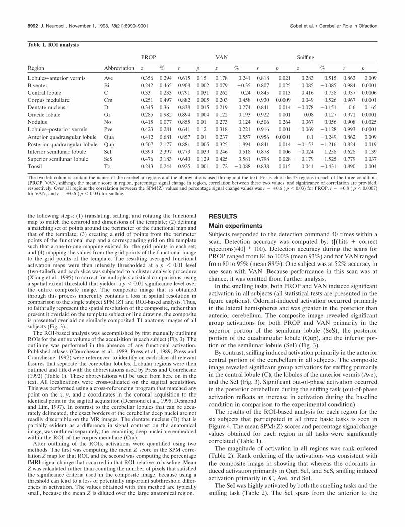
Table 1. ROI analysis
Region Abbreviation
PROP VAN Sniffing
z % r p z % r p z % r p
Lobules–anterior vermis Ave 0.356 0.294 0.615 0.15 0.178 0.241 0.818 0.021 0.283 0.515 0.863 0.009Biventer Bi 0.242 0.465 0.908 0.002 0.079 20.35 0.807 0.025 0.085 20.085 0.984 0.0001Central lobule C 0.33 0.233 0.791 0.031 0.262 0.24 0.845 0.013 0.416 0.758 0.937 0.0006Corpus medullare Cm 0.251 0.497 0.882 0.005 0.203 0.458 0.930 0.0009 0.049 20.526 0.967 0.0001Dentate nucleus D 0.345 0.36 0.838 0.015 0.219 0.274 0.841 0.014 20.078 20.151 0.6 0.165Gracile lobule Gr 0.285 0.982 0.894 0.004 0.122 0.193 0.922 0.001 0.08 0.127 0.971 0.0001Nodulus No 0.415 0.077 0.855 0.01 0.273 0.124 0.506 0.264 0.367 0.056 0.908 0.0025Lobules–posterior vermis Pve 0.423 0.281 0.641 0.12 0.318 0.221 0.916 0.001 0.069 20.128 0.993 0.0001Anterior quadrangular lobule Qua 0.412 0.681 0.857 0.01 0.237 0.557 0.956 0.0001 0.1 20.249 0.862 0.009Posterior quadrangular lobule Qup 0.507 2.177 0.881 0.005 0.325 1.894 0.841 0.014 20.153 21.216 0.824 0.019Inferior semilunar lobule SeI 0.399 2.397 0.773 0.039 0.246 0.518 0.878 0.006 20.024 1.258 0.628 0.139Superior semilunar lobule SeS 0.476 3.183 0.640 0.129 0.425 3.581 0.798 0.028 20.179 21.525 0.779 0.037Tonsil To 0.243 0.244 0.925 0.001 0.172 20.088 0.838 0.015 0.041 20.431 0.890 0.004
The two left columns contain the names of the cerebellar regions and the abbreviations used throughout the text. For each of the 13 regions in each of the three conditions(PROP, VAN, sniffing), the mean z score in region, percentage signal change in region, correlation between these two values, and significance of correlation are provided,respectively. Over all regions the correlation between the SPM{Z} values and percentage signal change values was r 5 10.6 ( p , 0.03) for PROP, r 5 10.8 ( p , 0.0007)for VAN, and r 5 10.6 ( p , 0.03) for sniffing.
8992 J. Neurosci., November 1, 1998, 18(21):8990–9001 Sobel et al. • Cerebellar Role in Olfaction

Figure 3. Composite activations overlaid on the composite anatomy of six subjects who performed all tasks. Left column is an example of the ROIsdrawn for each subject shown here for the left cerebellar hemisphere, spanning from the anterior (slice #1) to the posterior (slice #6 ) cerebellum. Secondcolumn is the averaged fMRI activation induced by PROP. Third column is the averaged fMRI activation induced by VAN. Right column is the averagedfMRI activation induced by sniffing clean air. Significance of in-phase activation is color coded from red to yellow, and out-of-phase activation is codedfrom dark blue to light blue. The right side of the image corresponds to the right side of the brain.
Sobel et al. • Cerebellar Role in Olfaction J. Neurosci., November 1, 1998, 18(21):8990–9001 8993

posterior cerebellum, and the composite image suggested a dis-sociation within SeI, whereby sniffing induced activation primar-ily in the anterior portion of the SeI, and odorants inducedactivation primarily in the posterior portion of the SeI. To quan-tify this effect, the SeI was separated into an anterior portioncomposed of its representation in slices 1 and 2, versus a posteriorportion composed of its representation in slices 5 and 6. Thisseparation clearly revealed that sniffing induced greater activationin the anterior portion of the SeI and the odorants inducedgreater activation in the posterior portion of the SeI (Fig. 5).
Whereas SeI was significantly activated during both sniffing andsmelling, two other regions exhibited highly task-dependent acti-vation: C was activated almost exclusively during sniffing, and SeS
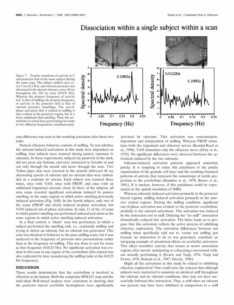
was activated almost exclusively during smelling (Table 2). Thesetwo regions significantly dissociated on these tasks (Fig. 6). Thisdissociation between activation induced by sniffing and activationinduced by the odorants was evident in the Fourier transform ofthe signal-time-series in each subject (Fig. 7).
Control experimentsTo assess the effects of variations in odor content on the differentregions, four subjects were scanned with both high and lowconcentrations of PROP, and five subjects were scanned with bothhigh and low concentrations of VAN. One of the subjects wasscanned with three concentrations of PROP (Fig. 8). Activationin all regions but the biventer (Bi) showed a trend toward con-
Figure 4. Percentage fMRI signal change inall 13 cerebellar regions in the six subjectswho performed all three tasks. Note doublingof Y scale for Qup, SeI, and SeS from 3 to6%. Error bars are SEM. An omnibus withinsubject repeated-measures ANOVA withfactors of region (13 ROIs), task (Prop., Van.,and Sniff.), and hemisphere (lef t and right)revealed a significant effect for region (F(12)5 2.27; p , 0.02), and a significant interac-tion for region ✽ task (F(24) 5 3.1; p 50.0001), reflecting significant differences inregions C (Sniff. . Van., t(5) 5 3.27, p 5 0.02;Sniff. . Prop., t(5) 5 2.17, p 5 0.08), D (Prop.. Sniff., t(5) 5 2.94, p 5 0.03; Van. . Sniff.,t(5) 5 2.58, p 5 0.05), Qua (Prop. . Sniff., t(5)5 4.1, p 5 0.009), Qup (Prop. . Sniff., t(5) 54.7, p 5 0.005; Van. . Sniff., t(5) 5 2.8, p 50.04), and SeS (Prop. . Sniff., t(5) 5 3.36, p 50.02; Van. . Sniff., t(5) 5 2.73, p 5 0.04).
8994 J. Neurosci., November 1, 1998, 18(21):8990–9001 Sobel et al. • Cerebellar Role in Olfaction

centration dependence that was significant in D, Qua, Qup, andSeS (Fig. 9). Rank ordering of the concentration dependency ofthe region showed significant positive correlation with the rankorder of the odor regions but a nonsignificant negative correlationwith the rank order of the sniff regions (Table 2). This significantdifference between the correlations shows that the regions thatwere more responsive to odorant presence were also more re-sponsive to concentration changes. By contrast, regions that weremore responsive to sniffing were less responsive to odorant con-centration changes.
To assess the effects of sniff rate on the different regions, asingle subject was scanned while sniffing at different sniff rateswithin the sniffing half block. Increasing sniff rate induced anincrease in significant activation primarily in the anterior cere-bellum in C and less in the posterior cerebellum (Fig. 8).
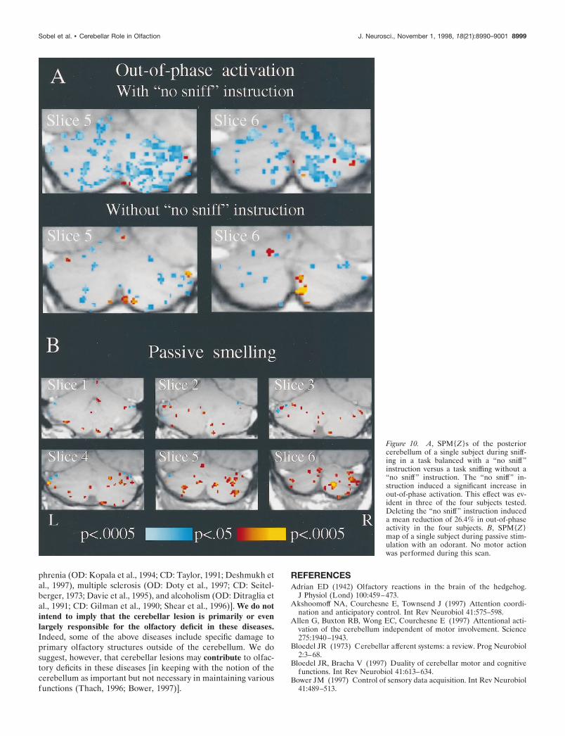
Significant out-of-phase activation occurred in the posteriorcerebellum during the sniffing task, i.e., activation associated withthe specific instruction not to sniff (Fig. 3, slices 5 and 6). Thissuggests that this activation may be related to an inhibitoryprocess of suppressing olfactory input. To address this issue, foursubjects were scanned twice, once in a sniffing task that containedthe “no sniff” instruction and once in an identical sniffing task, butwithout the “no sniff” instruction. The only difference betweenthese scans was that whereas in the first scan not sniffing wasachieved by specifically instructing the subject not to sniff, in thesecond scan not sniffing was achieved merely by not instructing tosniff. Sniff rate and number were identical in these two scans.Deleting the “no sniff” instruction induced a dramatic decrease inout-of-phase activation in three of the four subjects that partici-pated in this control (Fig. 10A).
The behavior of sniffing to the instruction “sniff” versus sniffingto the instruction “sniff and respond, is there an odor?” may notbe the same. Whereas the former is purely a motor function, thelatter is a motor function directed at sensory acquisition. Toaddress any possible differences in activation related to this dif-
ference, four subjects were scanned twice, once at a regularsniffing task, and once at a sniffing task in which the instruction“sniff” was replaced with the instruction “sniff and respond, isthere an odor?”. Although there was no odorant generated inthese tasks, subjects were led to believe that an odorant would begenerated and were instructed to try and detect an odor that theywould be questioned about after the scan. No consistent signifi-
Table 2. Rank ordering of regional activation levels
PROP VAN SniffingConcentrationdependence
SeS SeS SeI SeSSeI Qup C QupQup Qua Ave SeIGr SeI Gr GrQua Cm No CmCm D Bi QuaBi Ave Pve AveD C D CAve Pve Qua DPve Gr To PveTo No Cm ToC To Qup NoNo Bi SeS Bi
Regional activation was rank ordered. Spearman rank correlations revealed a non-significant positive correlation between the regions activated in the odor tasks(PROP and VAN Rho 5 0.25; p 5 0.38) and a nonsignificant negative correlationbetween the odor and sniff tasks (PROP and sniff Rho 5 20.19, p 5 0.5; VAN andsniff Rho 5 20.03, p 5 0.89). Concentration dependency was positively correlatedwith both odor tasks (PROP Rho 5 10.69, p , 0.02; VAN Rho 5 10.64, p , 0.03)but nonsignificantly negatively correlated with sniffing (sniffing Rho 5 20.352; p 50.22). Transformation to Z values revealed that the odorant regions–concentrationregions correlation was significantly greater than the sniffing regions–concentrationregions correlation ( p , 0.01).
Figure 5. Activation in the anterior and posterior SeI. Activation inslices 1 and 2 was combined to form the anterior portion, and activationin slices 5 and 6 was combined to form the posterior portion. The odorantsinduced greater activation in the posterior than the anterior portions, andsniffing induced greater activation in the anterior than the posteriorportions.
Figure 6. C and SeS dissociated in activation patterns after sniffing andsmelling (odorant tasks). The two odorants were collapsed to a singlesmelling condition. A repeated-measures ANOVA with factors of region(C and SeS) and task (odorants and sniffing) revealed a significant effectfor task (F(1) 5 9.64; p 5 0.03) and a significant interaction for region ptask (F(1) 5 10.84; p 5 0.02), reflecting greater activation in the posteriorregion during smelling than during sniffing (t(5) 5 3.22; p 5 0.02) butgreater activation in the anterior region during sniffing than duringsmelling (t(5) 5 2.75; p 5 0.04).
Sobel et al. • Cerebellar Role in Olfaction J. Neurosci., November 1, 1998, 18(21):8990–9001 8995
cant difference was seen in the resulting activation after these twotasks.
Natural olfactory behavior consists of sniffing. To test whetherthe odorant-induced activations in this study were dependent onsniffing, four subjects were scanned during passive exposure toodorants. In these experiments, subjects lay passively in the dark,did not press any buttons, and were instructed to breathe in andout only through the mouth and never through the nose. TwoTeflon pipes that were inserted in the nostrils delivered 40 secalternating epochs of odorant and no odorant that were embed-ded in a constant air stream. Each subject was scanned threetimes, once with VAN, once with PROP, and once with anadditional trigeminal odorant; citral. In three of the subjects, allnine scans revealed significant activations induced by passivesmelling, in the same regions in which active smelling previouslyinduced activation (Fig. 10B). In the fourth subject, only two ofthe scans (PROP and citral) induced in-phase activation, butVAN induced out-of-phase activation. In sum, 11 of the 12 scansin which passive smelling was performed induced activation in thesame regions in which active smelling induced activation.
In a final control, a “sham” scan was performed, in which asubject performed the smelling task, i.e., constantly sniffing andtrying to detect an odorant, but no odorant was generated. Thisscan was identical in behavior to the pure sniffing scans, but it wasanalyzed at the frequency of previous odor presentations ratherthan at the frequency of sniffing. This was done to test for noiseat that frequency (0.0125 Hz). No significant activation was evi-dent in this scan in any region of the cerebellum (this control wasalso replicated by later reanalyzing the sniffing tasks at the 0.0125Hz frequency).
DISCUSSIONThese results demonstrate that the cerebellum is involved inolfaction in the human. Both the composite SPM{Z} map and theindividual ROI-based analysis were consistent in showing thatthe posterior lateral cerebellar hemispheres were significantly
activated by odorants. This activation was concentration-dependent and independent of sniffing. Whereas PROP stimu-lates both the trigeminal and olfactory nerves (Kendal-Reed etal., 1998), VAN stimulates only the olfactory nerve (Doty et al.,1978). No significant differences were observed between the ac-tivations induced by the two odorants.
Odorant-induced activation patterns appeared somewhatpatchy. It is tempting to relate this patchiness to the patchyorganization of the granule cell layer and the resulting fracturedpatterns of activity that represent the somatotopy of tactile pro-jections to the cerebellum (Shambes et al., 1978; Bower et al.,1981). It is unclear, however, if this patchiness could be repre-sented at the spatial resolution of fMRI.
Whereas odorants induced activation primarily in the posteriorlateral regions, sniffing induced activation primarily in the ante-rior central regions. During the sniffing condition, significantout-of-phase activation was evident in the posterior cerebellum,medially to the odorant activations. This activation was inducedby the instruction not to sniff. Deleting the “no sniff” instructiondramatically reduced this activation. The latter leads us to pro-pose that this activation reflects the active process of inhibitingolfactory exploration. The activation differences between notsniffing when specifically told not to, versus not sniffing justbecause no instruction to do so was generated, constitute anintriguing example of attentional effects on cerebellar activation.This effect resembles activity that occurs in motor associationregions after merely anticipating or rehearsing a movement with-out actually performing it (Evarts and Tanji, 1974; Tanji andEvarts, 1976; Roland et al., 1987; Decety, 1996).
Might all the activations in this study be related to inhibitingolfactory exploration? One could raise the concern that althoughsubjects were instructed to maintain an identical sniff throughoutthe odorant and no odorant conditions, they may not have suc-cessfully followed this instruction. Thus, a sniff when an odorantwas present may have been inhibited in comparison to a sniff
Figure 7. Fourier transform of activity in Cand posterior SeS of the same subject duringthe same scan. The subject sniffed once ev-ery 5 sec (0.2 Hz), and odorant presence wasalternated with odorant absence every 40 secthroughout the 320 sec scan (0.0125 Hz).Whereas the primary frequency of activityin C is that of sniffing, the primary frequencyof activity in the posterior SeS is that ofodorant presence (smelling). The out-of-phase activation that is related to sniffing isalso evident in the posterior region, but at alower amplitude than smelling. Thus, the cer-ebellum is viewed here performing two tasksat two different frequencies, simultaneously.
8996 J. Neurosci., November 1, 1998, 18(21):8990–9001 Sobel et al. • Cerebellar Role in Olfaction

without an odorant, because an increase in odorant concentrationinduces a sniff of lesser volume (Laing, 1983). Such limiting of asniff would result in an increase in activation, as seen in the formof out-of-phase activation in the sniffing tasks. Thus, we mayerroneously attribute activation to the presence of an odorantwhen it is actually related to inhibiting sniffing (because of thepresence of that odorant). The latter concern was largely negatedby the passive task: The presence of an odorant induced activa-tion in the cerebellum in the absence of any motor function,sniffing included. The activation induced by passive smelling wasless robust than that induced by odorants perceived via a sniff. Infact, we have found that passive smelling does not induce aconsistent fMRI signal in primary olfactory cortex [the latter,however, may be related to the possibility of temporal encoding ofodor information in the ventral temporal areas that would notinduce increased activation as assessed with these methods (Sobelet al., 1998)]. That said, the remote possibility remains that theactivation was related to the intention to inhibit the sniff once an
odorant was perceived, regardless of whether the sniff was ulti-mately executed or not.
What may be the role of the cerebellum in olfaction? Thefollowing is a working hypothesis: sniff volume is inversely pro-portional to odor concentration (Laing, 1983). Maintaining thisinverse proportionality calls for an accurate rapid feedback mech-anism that monitors the sensory input (odor concentration) andmodulates the motor output (sniff volume). Cerebellar mainte-nance of such feedback mechanisms has been extensively de-scribed for tactile information, as well as other senses, like thecerebellum receiving sensory information regarding retinal slip tothen effect the vestibulo-ocular reflex to reduce that slip (Robin-son, 1976; Lisberger and Sejnowski, 1992), and like the cerebel-lum receiving auditory input that may then effect the pinna, thusmodulating further auditory input (suggested by Bower 1997; seealso Huang et al., 1991; Cicirata et al., 1992; Young et al., 1992).Here we suggest that the cerebellum is receiving olfactory infor-mation for modulating the sniff, which in turn modulates further
Figure 8. SPM{Z}s of anterior and poste-rior slices of a single subject during separatescans in which either odor concentration orsniff rate were varied parametrically. Acti-vation was both odor concentration- andsniff rate-dependent. A greater odor con-centration dependency is evident in the pos-terior versus the anterior slice, and greatersniff rate dependency is evident in the an-terior versus the posterior slice. Note thatonly in-phase activation is shown in thisfigure.
Sobel et al. • Cerebellar Role in Olfaction J. Neurosci., November 1, 1998, 18(21):8990–9001 8997

olfactory input. In this capacity, the cerebellum could be subserv-ing maintenance of the Teghtsoonian model of olfactory sizeconstancy (Teghtsoonian et al., 1978).
By which pathway may the olfactory information reach thecerebellum? Whereas well described trigeminal projections to thecerebellum (Yatim et al., 1996) may explain activation induced byPROP, a candidate pathway for both PROP and VAN is lessevident. Olfactory information is initially projected from theolfactory bulb directly to primary olfactory cortex (piriform)(Price, 1990). Olfactory projections are then widely spread withinthe ventral temporal region and throughout the brain. Although awell described candidate pathway would be the hypothalamocer-ebellar fibers (Haines et al., 1997), a pathway that traversesprimary olfactory cortex is also available. The ventral tegmentalarea (VTA) in the rat is strongly interconnected to primaryolfactory cortex (Oades and Halliday, 1987). Using double label-ing, Ikai et al. (1992) first found projections from the VTA of therat to the cerebellar cortex and lateral cerebellar nucleus, andlater single neurons in the VTA that project collaterals to bothpiriform cortex and the cerebellum (Ikai et al., 1994). As theseauthors note, the dopaminergic axons of VTA neurons project tothe pontocerebellum, which also subserves programming andcoordination of voluntary motor behaviors. This circuit that con-tains a direct connection between primary olfactory cortex andthe cerebellum is a well suited candidate to control the sniff-volume odorant-concentration feedback mechanism that we haveproposed. Thus, odor information may be relayed from primaryolfactory cortex to the posterior lateral cerebellum; based on odorcontent, cerebellar circuits would then determine optimal sniffvolume for further odorant sampling. Cerebellar efferents wouldthen modulate sniff parameters.
In what way do our findings relate to the ongoing debateregarding the role of the cerebellum? The role we have suggestedfor the cerebellum in olfaction supports the model proposed byBower et al. (1981), suggesting that the cerebellum coordinates
acquisition of sensory information. For tactile information, Bower(1997) proposed that “the cerebellum is responsible for monitor-ing incoming sensory data from these surfaces and adjusting theirpositions relative to each other and relative to the object beingexplored, in real time”. Here too, we suggest that the cerebellumis monitoring incoming data (odorant concentration) and adjust-ing the position of the stimulus (odorant air stream) relative tothe sensory surface (olfactory epithelium) by controlling themotor behavior (sniff), in real time. That said, our findings maystill be incorporated within other models of cerebellar functionnoted in the introduction, as here to, there is an element oftiming, an element of attentional modulation, and most impor-tantly, an element of feedback for motor control.
The cerebellar model of Bower et al. (1981) was supported inan fMRI study in which an increase in dentate activation was seenduring a tactile stimulation task when it included an element oftactile discrimination (Gao et al., 1996). We, therefore, expectedan increase in dentate activation when sniffing in response to the“sniff and respond, is there an odor” instruction in comparison tosniffing to the “sniff” instruction. In the four subjects that partic-ipated in this task, two showed an increase in dentate activation,and two showed a decrease. Whereas the latter finding does notsupport the Bower (1981) model, it may be attributed to that inthe context of an olfaction experiment, subjects may be searchingfor odorants even in a scan in which they are instructed just tosniff and informed that no odorants will be present.
If the cerebellum plays a role in olfactory processing, onewould expect an olfactory deficit in patients with cerebellar le-sions. To the best of our knowledge, in every disease in whichthere is cerebellar damage and olfaction has been tested, anolfactory deficit has been found [e.g., Alzheimer’s disease (olfac-tory deficit [OD]: Moberg et al., 1987; Doty et al., 1991; cerebellardamage (CD): Joachim et al., 1989); Parkinson’s disease (OD:Doty et al., 1988; CD: Heimburger, 1969), Korsakoff (OD: Joneset al., 1975; CD: Butterworth, 1993; Shear et al., 1996), schizo-
Figure 9. Concentration dependence in all13 regions. The difference in fMRI response tothe high versus the low concentration of theodorants was significant in regions D (t(8) 5 2.5;p 5 0.04), Qua (t(8) 5 2.7; p 5 0.03), Qup(t(8) 5 3.2; p 5 0.01), and SeS (t(8) 5 3.3;p 5 0.01).
8998 J. Neurosci., November 1, 1998, 18(21):8990–9001 Sobel et al. • Cerebellar Role in Olfaction
phrenia (OD: Kopala et al., 1994; CD: Taylor, 1991; Deshmukh etal., 1997), multiple sclerosis (OD: Doty et al., 1997; CD: Seitel-berger, 1973; Davie et al., 1995), and alcoholism (OD: Ditraglia etal., 1991; CD: Gilman et al., 1990; Shear et al., 1996)]. We do notintend to imply that the cerebellar lesion is primarily or evenlargely responsible for the olfactory deficit in these diseases.Indeed, some of the above diseases include specific damage toprimary olfactory structures outside of the cerebellum. We dosuggest, however, that cerebellar lesions may contribute to olfac-tory deficits in these diseases [in keeping with the notion of thecerebellum as important but not necessary in maintaining variousfunctions (Thach, 1996; Bower, 1997)].
REFERENCESAdrian ED (1942) Olfactory reactions in the brain of the hedgehog.
J Physiol (Lond) 100:459–473.Akshoomoff NA, Courchesne E, Townsend J (1997) Attention coordi-
nation and anticipatory control. Int Rev Neurobiol 41:575–598.Allen G, Buxton RB, Wong EC, Courchesne E (1997) Attentional acti-
vation of the cerebellum independent of motor involvement. Science275:1940–1943.
Bloedel JR (1973) Cerebellar afferent systems: a review. Prog Neurobiol2:3–68.
Bloedel JR, Bracha V (1997) Duality of cerebellar motor and cognitivefunctions. Int Rev Neurobiol 41:613–634.
Bower JM (1997) Control of sensory data acquisition. Int Rev Neurobiol41:489–513.
Figure 10. A, SPM{Z}s of the posteriorcerebellum of a single subject during sniff-ing in a task balanced with a “no sniff”instruction versus a task sniffing without a“no sniff” instruction. The “no sniff” in-struction induced a significant increase inout-of-phase activation. This effect was ev-ident in three of the four subjects tested.Deleting the “no sniff” instruction induceda mean reduction of 26.4% in out-of-phaseactivity in the four subjects. B, SPM{Z}map of a single subject during passive stim-ulation with an odorant. No motor actionwas performed during this scan.
Sobel et al. • Cerebellar Role in Olfaction J. Neurosci., November 1, 1998, 18(21):8990–9001 8999
Bower JM, Beermann DH, Gibson JM, Shambes GM, Welker W (1981)Principles of organization of a cerebro-cerebellar circuit: Micromap-ping the projections from cerebral (SI) to cerebellar (granule cell layer)tactile areas of rats. Brain Behav Evol 18:1–18.
Butterworth RF (1993) Pathophysiology of cerebellar dysfunction in theWernicke-Korsakoff syndrome. Can J Neurol Sci 20:S123–S126.
Cicirata F, Angaut P, Serapide MF, Panto MR, Nicotra G (1992) Mul-tiple representation in the nucleus lateralis of the cerebellum: anelectrophysiologic study in the rat. Exp Brain Res 89:352–362.
Colebatch JG, Adams L, Murphy K, Martin AJ, Lammertsma AA,Tochon-Danguy HJ, Clark JC, Friston KJ, Guz A (1991) Regionalcerebral blood flow during volitional breathing in man. J Physiol (Lond)443:91–103.
Courchesne E, Press GA, Murakami J, Berthoty D, Grafe M, Wiley CA,Hesselink JR (1989) The cerebellum in sagittal plane–anatomic-MRcorrelation: 1. The vermis. AJR Am J Roentgenol 153:829–835.
Courchesne E, Townsend J, Akshoomoff NA, Saitoh O, Yeung-Courchesne R, Lincoln AJ, James HE, Haas RH, Schreibman L, LauL (1994) Impairment in shifting attention in autistic and cerebellarpatients. Behav Neurosci 108:848–865.
Davie CA, Barker GJ, Webb S, Tofts PS, Thompson AJ, Harding AE,McDonald WI, Miller DH (1995) Persistent functional deficit in mul-tiple sclerosis and autosomal dominant cerebellar ataxia is associatedwith axon loss. Brain 118:1583–1592.
Decety J (1996) Do imagined and executed actions share the sameneural substrate? Brain Res Cogn Brain Res 3:87–93.
Deshmukh A, Sullivan EV, Mathalon DH, Desmond JE, Lim KO, Pfef-ferbaum A (1997) Regional cerebellar volume deficits in schizophre-nia, alcoholism, and schizophrenia with alcohol comorbidity. SchizophrRes 24:142–143.
Desmond JE, Lim KO (1997) On- and offline Talairach registration forstructural and functional MRI studies. Hum Brain Mapp 5:58–73.
Desmond JE, Sum JM, Wagner AD, Demb JB, Shear PK, Glover GH,Gabrieli JD, Morrell MJ (1995) Functional MRI measurement of lan-guage lateralization in Wada-tested patients. Brain 118:1411–1419.
Desmond JE, Gabrieli JD, Wagner AD, Ginier BL, Glover GH (1997)Lobular patterns of cerebellar activation in verbal working-memoryand finger-tapping tasks as revealed by functional MRI. J Neurosci17:9675–9685.
Desmond JE, Gabrieli JDE, Glover GH (1998) Dissociation of frontaland cerebellar activity in a cognitive task: evidence for a distinctionbetween selection and search. NeuroImage 7:368–376.
Ditraglia GM, Press DS, Butters N, Jernigan TL, Cermak LS, Velin RA,Shear PK, Irwin M, Schuckit M (1991) Assessment of olfactory defi-cits in detoxified alcoholics. Alcohol 8:109–115.
Doty RL, Brugger WE, Jurs PC, Orndorff MA, Snyder PJ, Lowry LD(1978) Intranasal trigeminal stimulation from odorous volatiles: Psy-chometric responses from anosmic and normal humans. Physiol Behav20:175–185.
Doty RL, Deems DA, Stellar S (1988) Olfactory dysfunction in parkin-sonism: a general deficit unrelated to neurologic signs, disease stage, ordisease duration. Neurology 38:1237–1244.
Doty RL, Perl DP, Steele JC, Chen KM, Pierce JD Jr, Reyes P, KurlandLT (1991) Olfactory dysfunction in three neurodegenerative diseases.Geriatrics 46:47–51.
Doty RL, Li C, Mannon LJ, Yousem DM (1997) Olfactory dysfunctionin multiple sclerosis. N Engl J Med 336:1918–1919.
Evarts EV, Tanji J (1974) Gating of motor cortex reflexes by priorinstruction. Brain Res 71:479–494.
Fiez JA (1996) Cerebellar contributions to cognition. Neuron 16:13–15.Friston KJ, Jezzard P, Turner R (1994) Analysis of functional MRI
time-series. Hum Brain Mapp 1:153–171.Friston KJ, Williams S, Howard R, Frackowiak RS, Turner R (1996)
Movement-related effects in fMRI time-series. Magn Reson Med35:346–355.
Gao JH, Parsons LM, Bower JM, Xiong J, Li J, Fox PT (1996) Cere-bellum implicated in sensory acquisition and discrimination rather thanmotor control. Science 272:545–547.
Gilman S, Adams K, Koeppe RA, Berent S, Kluin KJ, Modell JG, KrollP, Brunberg JA (1990) Cerebellar and frontal hypometabolism in al-coholic cerebellar degeneration studied with positron emission tomog-raphy. Ann Neurol 28:775–785.
Glover GH, Lai S (1998) Self-navigated spiral fMRI: interleaved versussingle-shot. Magn Reson Med 39:361–368.
Hahn I, Scherer PW, Mozell MM (1994) A mass transport model ofolfaction. J Theor Biol 167:115–128.
Haines DE, Dietrichs E, Mihailoff GA, McDonald EF (1997) Thecerebellar-hypothalamic axis: basic circuits and clinical observations.Int Rev Neurobiol 41:83–107.
Heimburger RF (1969) The role of the cerebellar nuclei in dyskineticdisorders. Confin Neurol 31:57–69.
Horne MK, Butler EG (1995) The role of the cerebello-thalamo-corticalpathway in skilled movement. Prog Neurobiol 46:199–213.
Huang CM, Liu GL (1991) Auditory responses in the posterior vermis ofthe cat: the buried cerebellar cortex. Brain Res 553:201–205.
Huang CM, Liu GL, Yang BY, Mu H, Hsiao CF (1991) Auditory recep-tive area in the cerebellar hemisphere is surrounded by somatosensoryareas. Brain Res 541:252–256.
Ikai Y, Takada M, Shinonaga Y, Mizuno N (1992) Dopaminergic andnon-dopaminergic neurons in the ventral tegmental area of the ratproject, respectively, to the cerebellar cortex and deep cerebellar nuclei.Neuroscience 51:719–728.
Ikai Y, Takada M, Mizuno N (1994) Single neurons in the ventraltegmental area that project to both the cerebral and cerebellar corticalareas by way of axon collaterals. Neuroscience 61:925–934.
Ito M (1984) The cerebellum and neural control. New York: Raven.Ivry R (1997) Cerebellar timing systems. Int Rev Neurobiol 41:555–573.Joachim CL, Morris JH, Selkoe DJ (1989) Diffuse senile plaques occur
commonly in the cerebellum in Alzheimer’s disease. Am J Pathol135:309–319.
Jones BP, Moskowitz RH, Butters N (1975) Olfactory discrimination inalcoholic Korsakoff patients. Neuropsychologia 13:173–179.
Kendal-Reed M, Walker JC, Morgan WT, LaMacchio M, Lutz RW(1998) Human responses to prop. acid. I. Quantification of within- andbetween-participant variation in perception by normosmics and anos-mics. Chem Senses 23:71–82.
Kim SG, Ugurbil K, Strick PL (1994) Activation of a cerebellar outputnucleus during cognitive processing. Science 265:949–951.
Kopala LC, Good KP, Honer WG (1994) Olfactory hallucinations andolfactory identification ability in patients with schizophrenia and otherpsychiatric disorders. Schizophr Res 12:205–211.
Laing DG (1983) Natural sniffing gives optimum odor perception forhumans. Perception 12:99–117.
Lisberger SG (1988) The neural basis for learning of simple motor skills.Science 242:728–735.
Lisberger SG, Sejnowski TJ (1992) Motor learning in a recurrent net-work model based on the vestibulo-ocular reflex. Nature 360:159–161.
Lisberger SG, Pavelko TA, Bronte-Stewart HM, Stone LS (1994) Neu-ral basis for motor learning in the vestibuloocular reflex of primates. II.Changes in the responses of horizontal gaze velocity Purkinje cells inthe cerebellar flocculus and ventral paraflocculus. J Neurophysiol72:954–973.
Le Magnen J (1945) Etude des facteurs dynamiques de l’excitation ol-factive. L’Annee Psychologique. 77–89.
Mansfeld G, Tyukody V (1936) Atemzentrum und narkose. Arch IntPharmacodyn Ther 54:219.
Moberg PJ, Pearlson GD, Speedie LJ, Lipsey JR, Strauss ME, FolsteinSE (1987) Olfactory recognition: differential impairments in early andlate Huntington’s and Alzheimer’s diseases. J Clin Exp Neuropsychol9:650–664.
Mozell MM, Hornung DE, Leopold DA, Youngentob SL (1983) Initialmechanisms basic to olfactory perception. Am J Otolaryngol4:238–245.
Oades RD, Halliday GM (1987) Ventral tegmental (A10) system: neu-robiology. 1. Anatomy and connectivity. Brain Res 434:117–165.
Parsons LM, and Fox PT (1997) Sensory and cognitive functions. IntRev Neurobiol 41:255–271.
Paulin MG (1997) Neural representations of moving systems. Int RevNeurobiol 41:515–533.
Petersen SE, Fox PT, Posner MI, Mintun M, Raichle ME (1988)Positron emission tomographic studies of the cortical anatomy ofsingle-word processing. Nature 331:585–589.
Poldrack RA, Desmond JE, Glover GH, Gabrieli JD (1998) The neuralbasis of visual skill learning: an fMRI study of mirror reading. CerebCortex 8:1–10.
Press GA, Courchesne E (1992) Atlas of cerebellar hemispheres andvermis. In: Clinical brain imaging (Hayman LA, Hinck VC, eds), pp251–286. St. Louis: Mosby Year Book.
Press GA, Murakami J, Courchesne E, Berthoty DP, Grafe M, Wiley CA,
9000 J. Neurosci., November 1, 1998, 18(21):8990–9001 Sobel et al. • Cerebellar Role in Olfaction

Hesselink JR (1989) The cerebellum in sagittal plane–anatomic-MRcorrelation: 2. The cerebellar hemispheres. AJR Am J Roentgenol153:837–846.
Price JL (1990) Olfactory system. In: The human nervous system (Paxi-nos G, ed), pp 979–1001. San Diego: Academic.
Raichle ME, Fiez JA, Videen TO, MacLeod AM, Pardo JV, Fox PT,Petersen SE (1994) Practice-related changes in human brain func-tional anatomy during nonmotor learning. Cereb Cortex 4:8–26.
Rehn T (1978) Perceived odor intensity as a function of air flow throughthe nose. Sens Processes 2:198–205.
Robinson DA (1976) Adaptive gain control of vestibulo-ocular reflex bythe cerebellum. J Neurophysiol 39:954–969.
Roland PE, Eriksson L, Stone-Elander S, Widen L (1987) Does mentalactivity change the oxidative metabolism of the brain? J Neurosci7:2373–2389.
Schmahmann JD (1997) Rediscovery of an early concept. Int Rev Neu-robiol 41:3–27.
Schmahmann JD, Sherman JC (1998) The cerebellar cognitive affectivesyndrome. Brain 121:561–579.
Seitelberger F (1973) Pathology of multiple sclerosis. Ann Clin Res5:337–344.
Shambes GM, Beermann DH, Welker W (1978) Multiple tactile areas incerebellar cortex: another patchy cutaneous projection to granule cellcolumns in rats. Brain Res 157:123–128.
Shear PK, Sullivan EV, Lane B, Pfefferbaum A (1996) Mammillary bodyand cerebellar shrinkage in chronic alcoholics with and without amne-sia. Alcohol Clin Exp Res 20:1489–1495.
Snider RS, Eldred E (1948) Cerebral projections to the tactile, auditory,and visual areas of the cerebellum. Anat Rec 100:82.
Sobel N, Prabhakaran V, Desmond J, Glover G, Goode RL, Sullivan E,Gabrieli JDE (1998) Sniffing and smelling: separate subsystems in thehuman olfactory cortex. Nature 392:282–286.
Sobel N, Prabhakaran V, Desmond J, Glover G, Sullivan E, Gabrieli JDE(1997a) Separate cerebellar components subserve sniffing and smell-ing. Paper presented at Society for Neuroscience 27th Annual Meeting,Washington, DC, November.
Sobel N, Prabhakaran V, Desmond J, Glover G, Sullivan E, Gabrieli JDE(1997b) A method for functional magnetic resonance imaging of ol-faction. J Neurosci Methods 78:115–121.
Stein JF, Glickstein M (1992) Role of the cerebellum in visual guidanceof movement. Physiol Rev 72:967–1017.
Tanji J, Evarts EV (1976) Anticipatory activity of motor cortex neuronsin relation to direction of an intended movement. J Neurophysiol39:1062–1068.
Taylor, MA (1991) The role of the cerebellum in the pathogenesis ofschizophrenia. Neuropsychiatry Neuropsychol Behav Neurol4:251–280.
Teghtsoonian R, Teghtsoonian M, Berglund B, Berglund U (1978) In-variance of odor strength with sniff vigor: an olfactory analogue to sizeconstancy. J Exp Psychol Hum Percept Perform 4:144–152.
Thach WT (1996) On the specific role of the cerebellum in motorlearning and cognition: clues from PET activation and lesion studies inman. Behav Brain Sci 19:411–431.
Thach WT, Goodkin HP, Keating JG (1992) The cerebellum and theadaptive coordination of movement. Annu Rev Neurosci 15:403–442.
Williams RW, Herrup K (1988) The control of neuron number. AnnuRev Neurosci 11:423–453.
Woods RP, Cherry SR, Mazziotta JC (1992) A rapid automated algo-rithm for accurately aligning and re-slicing positron emission tomogra-phy images. J Comput Assist Tomogr 16:620–633.
Xiong J, Gao JH, Lancaster JL, Fox PT (1995) Clustered pixels analysisfor functional MRI activation studies of the human brain. Hum BrainMapp 3:287–301.
Yatim N, Billig I, Compoint C, Buisseret P, Buisseret-Delmas C (1996)Trigeminocerebellar and trigemino-olivary projections in rats. NeurosciRes 25:267–283.
Young ED, Spirou GA, Rice JJ, Voigt HF (1992) Neural organizationand responses to complex stimuli in the dorsal cochlear nucleus. PhilosTrans R Soc Lond B Biol Sci 336:407–413.
Youngentob SL, Mozell MM, Sheehe PR, Hornung DE (1987) A quan-titative analysis of sniffing strategies in rats performing odor detectiontasks. Physiol Behav 41:59–69.
Yousem DM, Williams SC, Howard RO, Andrew C, Simmons A, Allin M,Geckle RJ, Suskind D, Bullmore ET, Brammer MJ, Doty RL (1997)Functional MR imaging during odor stimulation: preliminary data.Radiology 204:833–838.
Sobel et al. • Cerebellar Role in Olfaction J. Neurosci., November 1, 1998, 18(21):8990–9001 9001
Related Documents