. . OSteOpotos Int (2015) 26:699- 712 D0110.J007/s00198-014-2944-6 ORIGINAL ARTJCLE Odan~catib for the treatment of postmenopausal osteoporosis: development history and design and participant characteristics of LOFT, the Long-Term Odanacatib Fracture Trial H. G. Bone • D. W. Dempster· J. A. Eisman • S. L. Greenspan • M. R. McClung • T. Nakamura· S. Papapoulos • W. J. Shih· A. Rybak-Feig)in • A. C. Santora· N. Verbruggen • A. T. Leung· A. Lombardi Received: 4 June 2014 / Accepted: 2 October 2014 / Publisbed online! 29 November 2014 © Tiie Author(s) 2014. This article is published with open access at Spriogcrlink.eom Abstract Summary Odanacatib is a cathepsin K inhibitor investigated for the treatment of postmenopausal osteoporosis. Phase 2 data indicate that 50 mg once weekly inhibits bone resorption and increases bone mineral density, with only a transien1 decreas.e in bone fonnation. We describe the background, design and participant characteristics for the phase 3 registra- tion trial. Introduction Odanacatib (ODN) is a selective cathepsin K inhibitor being evaluated for the treatment of osteoporosis. ln a phase 2 trial, ODN 50 mg once weekly reduced bone resorption while preserving bone fonnation and progressively increased BMD over 5 years. We describe the phase lil Long- Term ODN F racture Trial (LOFT) , an event -driven, This study was registered with elinicaltrials.gov with registration number NCT00529373. Electronic supplementary material The onlinc version of this article (doi: I 0.1007/sOOl 98-014-2944-6) contains supplementary material, which is avai lable to authorized users. H. G. Bone (181) Michigan Bone & Mineral Clinic, Detroit, MI, USA e- mail: [email protected] D. W. Dempster Columbia University, New York, NY, USA J. A. Eisman Garvan Institute of Medical Research, University ofNotre Dame Australia, St Vincent's Hospital and UNSW Australia, Sydne y, Australia S. L. Greenspan University of Pittsburgh, Pittsburgh, PA, USA M. R. McC!ung Oregon Osteoporos1s Center, Portland, OR, USA randomized, blinded placebo-controlled trial , with preplanned interim analyses to permit early termination if significant fracture risk reduction was demonstrated. An extension was planned, with participants remaining on their randomized treatment for up to 5 years, then transitioning to open-label ODN. Methods The three primary outcomes were radiologically detemtined vertebral, hip, and clinical non-vertebral fractures. Secondary end points included clinical vertebral fractures, BMD, bone turnover markers, and safety and tolerability, including bone histology . Participants were women, 65 years or older, with a BMD T-score ::S - 2.5 at the total hip (TH) or femoral neck (FN) or with a prior radiographic vertebral fracture and a T-score ::; -1 .5 at the TH or FN. They were T. Nakamura University ofOceupational and Envirowncntal Health, Kitakyushu, Japan S. Papapoulos Leiden Universi ty Medical Center, Leiden, Netherlands W. J. Shih Robert Wood Johnson Medical School, Piscabiway , NJ, USA A. Rybak-Feiglin · A. C. Santora· N. Verbruggcn · A. T. Leung· A. Lombnrdi Mcrclc Sharp and Dolune Corp., Whitehouse Station, NJ, USA H. G. Bone The Osteoporosis Center at St. Luke's Hospital, Chesterfield, MO, USA Springer

Welcome message from author
This document is posted to help you gain knowledge. Please leave a comment to let me know what you think about it! Share it to your friends and learn new things together.
Transcript
. . OSteOpotos Int (2015) 26:699- 712 D0110.J007/s00198-014-2944-6
ORIGINAL ARTJCLE
Odan~catib for the treatment of postmenopausal osteoporosis: development history and design and participant characteristics of LOFT, the Long-Term Odanacatib Fracture Trial
H. G. Bone • D. W. Dempster· J. A. Eisman • S. L. Greenspan • M. R. McClung • T. Nakamura· S. Papapoulos • W. J. Shih· A. Rybak-Feig)in • A. C. Santora· N. Verbruggen • A. T. Leung· A. Lombardi
Received: 4 June 2014 /Accepted: 2 October 2014 / Publisbed online! 29 November 2014 © Tiie Author(s) 2014. This article is published with open access at Spriogcrlink.eom
Abstract Summary Odanacatib is a cathepsin K inhibitor investigated for the treatment of postmenopausal osteoporosis. Phase 2 data indicate that 50 mg once weekly inhibits bone resorption and increases bone mineral density, with only a transien1 decreas.e in bone fonnation. We describe the background, design and participant characteristics for the phase 3 registration trial. Introduction Odanacatib (ODN) is a selective cathepsin K inhibitor being evaluated for the treatment of osteoporosis. ln a phase 2 trial, ODN 50 mg once weekly reduced bone resorption while preserving bone fonnation and progressively increased BMD over 5 years. We describe the phase lil LongTerm ODN Fracture Trial (LOFT), an event-driven,
This study was registered with elinicaltrials.gov with registration number NCT00529373.
Electronic supplementary material The onlinc version of this article (doi: I 0.1007/sOOl 98-014-2944-6) contains supplementary material, which is available to authorized users.
H. G. Bone (181) Michigan Bone & Mineral Clinic, Detroit, MI, USA e-mail: [email protected]
D. W. Dempster Columbia University, New York, NY, USA
J. A. Eisman Garvan Institute of Medical Research, University ofNotre Dame Australia, St Vincent's Hospital and UNSW Australia, Sydney, Australia
S. L. Greenspan University of Pittsburgh, Pittsburgh, PA, USA
M. R. McC!ung Oregon Osteoporos1s Center, Portland, OR, USA
randomized, blinded placebo-controlled trial, with preplanned interim analyses to permit early termination if significant fracture risk reduction was demonstrated. An extension was planned, with participants remaining on their randomized treatment for up to 5 years, then transitioning to open-label ODN. Methods The three primary outcomes were radiologically detemtined vertebral, hip, and clinical non-vertebral fractures. Secondary end points included clinical vertebral fractures, BMD, bone turnover markers, and safety and tolerability, including bone histology. Participants were women, 65 years or older, with a BMD T-score ::S- 2.5 at the total hip (TH) or femoral neck (FN) or with a prior radiographic vertebral fracture and a T-score ::;-1 .5 at the TH or FN. They were
T. Nakamura University ofOceupational and Envirowncntal Health, Kitakyushu, Japan
S. Papapoulos Leiden University Medical Center, Leiden, Netherlands
W. J. Shih Robert Wood Johnson Medical School, Piscabiway, NJ, USA
A. Rybak-Feiglin · A. C. Santora· N. Verbruggcn · A. T. Leung· A. Lombnrdi Mcrclc Sharp and Dolune Corp., Whitehouse Station, NJ, USA
H. G. Bone The Osteoporosis Center at St. Luke's Hospital, Chesterfield, MO, USA
~ Springer
700
randomized to ODN or placebo tablets. All received weekly vitamin D3 (5600 international units (JU)) and daily calcium supplements as needed to ensure a daily intake of approximately 1200 mg. Resulls Altogether, 16,713 participants were randomized at 387 centers. After a planned interim analysis, an independent · data monitoring committee recommended that the study be stopped early due to robust efficacy and a favorab le benefit/ risk profile. Following the base study closeout, 8256 participants entered the study extension. Co11clusio11s This report details the background and study design of this fracture end point trial and describes the baseline characteristics of its participants.
Keywords Cathepsin K · Fracture · Odanacatib · Osteoporosis · Postmenopausal
Introduction
Osteoporosis is a metabolic bone disease characterized by a chronic excess of bone r~orption relative to bone fonnation, leading to progressive bone loss, deterioration of bone microarchitecture, and increased risk of fracture [1].
Bone formation and resorption are tightly coordinated during the remodeling cycle. Osteocytes exhibit a mechanosensory capacity and are major regulators of bone remodeling [2]. Osteoclast-medi"ated bone resorption releases growth factors from the bone matrix [3, 4] that increase osteoblast progenitor number, r~ruitment, differentiation, and activity. ln addition, osteoclasts regulate osteoblasts directly via cell-cell contact (3, 5) and by secreting ·additional factors [6) that increase osteoblast number and/or activity. Osteoclast number and activity are regulated by receptor activator of nuclear factor kappa-B ligand (RANKL), a membrane-bound protein produced by neighboring osteocytes _and osteoblasts (7]. Thus, the products of osteoclast activity play a key role in the complex interactive regulation of bone remodeling.
With the excepti_on of parathyroid hormone (PTI:l) and its analogs, all agents currently used in the treatment of osteoporosis, such as bisphosphonates, selective estrogen receptor modulators, and the anti-RANKL antibody, act primarily by decreasing osteoclast-mediated bone resorption [8), allowing osteoblasts to increase bone mass by filling in the remodeling space [9, 10]. Furthermore, slower bone remodeling allows increased bone mineralization, at least for some agents, and leads to a reduction in cortical porosity (9-12). However, inhibition of osteoclast activity also leads to a secondary reduction in bone fonnation [8, 13), limiting the ultimate increase in bone mass.
The mechanism of osteoclast-mediated bone resorption is well characterized [7]. Osteoclasts attach to the bone surface
~ Springer
Osteoporos Int (2015) 26:699-712
and secrete hydrogen ions and proteolytic enzymes into the remodeling space. The acidic environment dissolves bone mineral and exposes bone matrix proteins. Cathepsin K (CatK) is the primary osteoclast-produced protease involved in the degradation of type f collagen and other bone matrix proteins [14); its pH optimum is in the range of the acidic remodeling space.
Congenital absence of CatK in humans results in p ycnodysostosis, a rare autosomal recessive osteochondrodysplasia. 1bis disease is characterized not only by high bone mass but also increased risk of fractures and bone defonnities [15). hnportantly, the increased fragility and bone defonnities are not present in heterozygotes [ 15]. Targeted disruption of the CatK gene in mice produced a high bone mass phenotype (16] while overexpression ofCatK increased bone turnover and decreased trabecular bone volume [I 7).
The effect of CatK inhibition has been studied in estrogendeficient non-human primates and rabbits. In these studies, CatK inhibitors significantly reduced bone resorption while relatively preserving bone formation, resulting in both increased BMD and bone strength, in contrast to congenital absence ofCatK [16). CatI< inhibition decreased the removal of bone matrix protein but did not reduce osteoclast number or affect other osteoclast activities, such as the production of osteoblast-stimulating factors (16).
Odanacatib (ODN, MK.-0822) is a selective, orally administered CatK inhibitor. Unlike basic CatK. inhibitoril, ODN is neutral, so it does not accumulate in the acidic environment of lysosomes, wl1ich could lead to off-target inhibition of other cathepsins [l.8, 19). ODN is metabolized by CYP3A4, has an apparent terminal half-life of approximately 40 to 80 h [20, 21], and its absorption is not impaired by food intake [20, 21].
A phase iib dose-ranging trial and its extensions have demonstrated progressive dose-dependent increases in BMD in postmenopausal women with low BMD [22- 24]. ODN substantially reduced biochemical markers of bone resorption. However, markers of bone formation decreased to a lesser degree than was previously observed with other antiresorptive treatments and returned towards baseline within 2 years, on continued treatment [22- 24). H.istomorphometry of bone biopsies from participants in the phase Ub study indicated that osteoclast number was maintained [22), con finning preclinical results ( 16). Lack of reduction of serum TRAPSb levels with ODN treatment in the phase IIb trial con finned osteoclast viability. Discontinuation of ODN treatment resulted in increased bone turnover and reduction in BMD, which returned to baseline within l to 2 years (23]. Based on BMD data after 12 months from the phase Ilb trial, ODN 50 mg once weekly was selected as the dosing regimen for the phase m trial [22].
A prospective study was designed to assess the antifracture efficacy and safety of ODN in postrnenopausal women with osteoporosis. This report describes the design of this

O'stcoporos int (2015) 26:699-712
study and presents the key baseline characteristics of its participants.
Study design
OveTView of study design
Toe study, odanacatib Protocol 18, was designed by the sponsor, Merck & Co. Jnc., Whitehouse Station, NJ, USA, in collaboration with a Scjentific Advisory Committee (SAC) that provided input on the clinical development program.
The study was planned as a multinational, randomized, double-blind, event-driven placebo-controlled tri11l of approximately 5 years duration and was approved by goveming ethical review committees (ERCs) and institutional review boards (IRBs). Provision was made for early tennination of the study if criteria for reduction of fracture risk versus placebo were met in prespecified interim analyses (see "Statistical methods").
Study participants were women at least 65 years of age and postmenopausal for at least 5 years, who were osteoporosis treatment-naive or had been exposed to minimal prior osteoporosis treatment. Two categories of participants were eligible (Table 1):
Women with a prior radiographic vertebral fracture were required to have a BMD T-score :S-l.5 at either the femoral neck (FN) or total hip (TH). Those without a prior vertebral fracture were reqlrired to have a BMD T-score ~-2.5 at the FN or TH, using the NHANES II1 1998 database (reference values are those for Caucasian young adult women).
In either category, women with prior hip fractures at any time or clinical fragility fractures within the prior 2 years or whose BMD T-score was <-4.0 at either the FN or TH were excluded to minimize individual participant risk, unless they were unable or unwilhng t~ use available osteoporosis treatments.
Thi~ trial was designed to test the hypotheses that treatment with ODN 50 mg once weekly reduces the risk of radiographically detected vertebral, clinical hip, and clinical nonvertebral fractures (all primary end points) in postmenopausal women with osteoporosis. Secondary end points included the incidence of clinical vertebral fractures; change in height; BMD at the TH, FN, trochanter, lumbar spine (LS), 1/3 radius; biochemical markers of bone fonnation and resorption; and safety and tolerability, including bone histology.
Participants were randomized in a 1: I ratio to talce either ODN 50 mg or matching placebo once weekly, without regard to food or physical position. They were given 5600 intemationa l units (IU) of vitamin D3 weekly and calcium
701
supplements as needed to ensure a total daily calcium intake of approximately 1200 mg. A dietary calcium questionnaire was administered at intake and at yearly intervals during the trial. Following screening and randomization visits, study visits in the clinic were scheduJed every 3 months in the first year of the study and every 6 months thereafter. Telephone contact alternated with clinic visits every 3 months after the first year.
Rationale for study design
This trial was designed with primary fracture end points, consistent with regulatory requirements for new drugs for the treatment of postmenopausal osteoporosis. It employed a placebo control group, consistent with previous and current registration trials of drugs for that indication. This approach has been the subject of extensive discnssion (25), including a US FDA Advisory Committee Meeting [26) which provided a frame of reference for recent trial design. Placebo-controlled pivotal trials in osteoporosis were considered acceptable, provided that appropriate limits are placed on the severity of disease in those individuals who could otherwise be treated with available medication. Background treatment with adequate calcium and vitamin D3 intake for all participants was also regarded as an important consideration. Limits regarding lhe range of acceptable BMD and fractures prior to study entry as well as monitoring for excessive on-study bone density decreases have been generally consistent in more recently completed (27) and ongoing (28, 29) phase ill trials.
Accordingly, the present study excluded participants at relatively high risk for fracture, using specific criteria based on BMD and prior fracture history, and employed safeguards to protect participant safety. The trial was overseen by an external data monitoring committee (DMC) that periodicaJJy reviewed unblinded safety data. BMD was measured yearly and identified participants with excessive bone loss (EBL) (predefined as >7 % from baselio~ at any time at the LS or TH). If the decrease was confirmed, the participant was then discontinued from study medication and treated conventionally. Adverse events of interest were referred for adjudication by panels of experts with relevant scientific expertise (see section on "Adverse event reporting and adjudication").
The study was powered to detect a significant effect of ODN to rednce the risk of hip fractures. Because of the safety criteria at enrollment, the baseline individual hip fracture risk in the study population was limited. Hence, the sample size estimate was about twice that of the largest osteoporosis trial. The event-driven design of the study allowed for some uncertainty in the estimate of the rate at which participants would experience fracture events.
~ Springer

p
t
· 702'
Table l . Inclusion criteria and exclusion criteriii
Inclusion criteria
Participant:
1) Is a woman at least 65 years of age
2) Meets one of the following criteria:
Osteoporos Int (20 15) 26:699--7!2
Critt:rion Femoral neck or total hip Femoral neck and totul hip Prior vertebral Suitable candidate for a,rdilable osteoporosis fracture1
A
B
C
BMD
:$ -1.5
:$-2.5
:$-1.S
BMD
~ 4.0
2:-4.0
No restriction
therdpy
Yes
0 Yes
2:1 No
D :$ ·2.5 No res~ricrion 0 No
'.l) May not be a suitable candidate for commercially available osteoporosis therapy, e.g., due to contraindication, established intolcrnnce, physician's judgment, or participant's unwillingness
4) Has at least one hip that is evaluable by DXA
5) Posuncnopausal for at least 5 years
6) Understands the study procedures, alternative treatments available, an<l voluntarily agrees to participate by giving written informed consent
7) ls ambulatory
8) Is able to read, understand, and complete questionnaires and diaries
Exd1.111ion criteria
Participant:
I) Has chosen treatment with agents demonstrated to reduce the risk of hip fracture
2) Has prior fragility hip fracture and is a suitable candidate for osteoporosis therapy (i.e., bisphosphonates, strontium ranelate, or PTH)
3) Ha.'! experienced a clinical fragility fracture {including a clinical vertebral fracture) within the prior 24 months
4) Had more than one prior vertebral fracture, as defined in incluRion criterion 2 above and is a suitable candidate for u::.'teoporosis therapy
5) Has evidence of a metabolic bone disorder other than osteoporosis
6) Has a history of renal stones and serum calcium, scrum 25-hydroxyvitamin D, and scrum PTH arc not all within normal limits. Serum 25-hydroxyvitamin D levels below 20 nglmL (50 nmoVL) were considered abnormal
7) Has active parathyroid dise11se
8) Has a history of thyroid disease not adequately controlled by medication
9) Has scrum creatininc > 1.6 mg'dL and is considered to have severe renal insufficiency defined 11s calculated creatinine clearance ::,.29 mUmin2
I 0) Has received treatment with an agent that has an effect on bone including
Bisphosphonates
• Use of any oral bisphosphonate in the 6 months prior to screening
• Use of any oral bisphosphonate for more than 3 months within the prior 2 years
• Lifetime use of more than 6 months total; any lifetime use of IV zoledronatc3
Within the prior 12 months, u~e of
• PTH (1-34 or 1-84)
Within the prior 6 months, use of
• Estrogen with or without progestin
• Raloxifene or other SERM, tibolone or an aromatase inhibitor
• Subcutaneous calcitonin4
Within the prior 6 months, use for more than 2 weeks of
• Systemic glucoconicoids ~ 5 mg/day of prcdnisune or equivalent)
• Cyclo~porin
Within the prior 3 months, use of
• Activated vitamin D (e.g., alphacalcidol)
At any rime, use of
• Any anabolic steroid
• Fluoride treatment at a dose greater than I mg/day for more than 2 weeks
• Growth hormone
• Any cathepsin K inhibitor
• RANK ligand inhibitor
~ Spdnger
Ostcoporo8 int (2015) 26:699-712 703
Table 1 (continued)
• Strontium-containing products • Protease inhibitors for HIV trcallllent
Current use • Chemotherapy or heparin • Vitamin A > I O,OOOlU daily • Vitamin D supplement> l2001U daily and is unwilling to limit vitanlin D supplement • Anti-seizure medication and indices of calcium metabolism are not within nonnal limits • Systemically administc1·cd azolc antifungals
J I) Has a daily calcium intake of <I 200mg and is unwilling to take study-prescribed calcium supplements 12) Has a history or current evidence of any condition, th~py, lab abnormality, or other circumstance that might confound the results of the study or
interfere with the participant's participation for the full duration of the study 13) Has n history of malignancy SSyears prior to signing infonned consent 14) ls >80years old and has a history of recurrent falls (;!2 falls in I year) 15) Is cum:ntly participating in a study with an investigational compound or device J 6) Is a user of recreational or illicit dnigs or has n recent histo1y (within the last year) of drug or alcohol abuse or dependence 17) Demonstrates hepatic dysfunction defined as
Elevation in aspartate aminotransferase (ASn or alanine aminotransferase (ALT) >3 xupper limit of nonnal (ULN) and Elevation in total bilirubin >2xuppcr limit of nonnal (ULN)
J 8) ls considered to be at excessive risk of incident fracture according to local Ethics Committee and/or local regulatory agency
1 Defined as anterior, mid, or posterior height loss of >20 % 2 Using the Cock croft-Gault fmmula for crcatininc clearance
> One dose of N pamidrooate or N ibandronate more than l year prior to screening is allowed 4 Use of intranasal calcitonin either prior to or during the study is pennined
Recruitment, follow-up, and assessments
Recruitment
Informed consent was obtained from all study participants before any study procedure was perf01med. It was required that a qualified physician discuss alternative treatment options with each participant. At the request of a regulatory agency, participant enrollment was conducted in two phases to allow expansion of the safety experience for ODN before the entire study was enrolled.
The first phase (the lead cohort) randomized 1499 participants between November 2007 and March 2008. After 9 months of ~tment, the lead cohort safety data were evaluated by the DMC ( see below for details), which recommend-
' ed that ~e enrollment of the remainder of the participants (the main cohort) could commence. The enrollment of 15,214 participants in the main cohort occurred from January to
, November 2009 (Fig. 1 ). Thus, a total of 16,713 women were recruited at 387 investigational sites in 40 countries.
At one investigative site, sponsor inspections identified issues that materially affected data integrity. The sponsor detennined that these findings warranted exclusion of that site's data from the primary efficacy and safety analyses and infonned the US FDA. (All data, including iliose excluded from the primary analyses, are to be provided in regulatory $Ubmissions). Data from 483 participants were affected, including 111 in the lead cohort. An additional 159 participants were excluded from the analysis because they never took study medication (N,:: 156) or because they had been assigned
duplicate allocation numbers (N=3). Thus, the number of evaluable participants was 16,071, including 1373 in the lead cohort.
Randomization and stratification
Randomization was perfonned by a computer-generated allocation schedule using an interactive voice recognition system (IVRS), which randomly assigned each participant to ODN or placebo. Participants were assigned to one of two strata, according to vertebral fracture status. Participants without prior vertebral fracture were assigned to the no-prior-vertebral-fi-acture stratum. At least two thirds of the participants in the no-prior-fracture stratum were required to be at least 70 years of age. Participants with a prior vertebral fracture were assigned to the prior-vertebral fracture stratum. IVRS was used to keep track of enrollment and the number of participants randomized into each stratum and to close a stratum as needed. IVRS was also available for emergency unblinding iftlvs were required for an individual participant's safety.
Blinding
Study participants, investigators and their staff, and sponsor's personnel were bJjnded to treatment allocation. Only the DMC and support statistical personnel (see below) had access to unblinded data. Selected adverse events (AEs) and changes in selected laboratory results were provided to the FDA in an
~ Springer

704 Osteoporos Int(2015) 26:699--112
Fig. 1 CONSORT Diagram. The asterisk indicate.~ 642 randomized participanti; were excluded from all analyses due to the following reasons: allocation of duplicate allocation numbers (n=3), took no study medication (11 = 156), or because their study site was closed and excluded from all efficacy and safety data analyses (n=483). The primary allpatients-as-tre11ted analysis will be based on 16,07 l participants, of whom 1373 were in the lead cohort. The dagger indicates 245 of the 8256 patients were found to be ineligible because they had experienced excessive bone loss detected at the transitional visit
Odanacatib PN018 Fracture Trial
Base Study
Extension
unblinded fashion, while maintaining the blind for study participants, investigators, and other sponsor personnel.
Assessments
Prior to randomization, a medical history, including assessment of concomitant medications, physical examination, BMD measurements by dual-energy X-ray absorptiometry (DXA), latera_l spine radiographs for vertebral fracture assessment, laboratory assessments, and review of inclusion and exclusion criteria were perfo1med. follow-up assessments were conducted as follows: limited physical examination including vital signs and laboratory assessment every 3 months during the first year, and then every 6 months. Drug accounting and AE assessments were perfonned at every visit. Lateral spine radiographs were perfonned at baseline, month 6, month 12, and every 12 months thereafter. BMD was measured at baseline and yearly thereafter. Height was assessed using a calibrated stadiometer at baseline and yearly.
Radiographic vertebral fracture assessmenl
Spine radiographs were evalualed at a central site (Synarc, Newark, CA, USA). Screening spine radiographs were evaluated for the presence or absence of a vertebral fracture using the Genant semi.,quantitative scale [301. Only vertebral
~ Springer
Assessed for eligibility
(N=46, 130) Excluded
I (n=29,414)
Randomized l
(N=16,713)*
Lead cohort i (n=1,499)
Evaluable (n=16,071}
Lead cohort evaluable (n=1,373)
- - - -~ - - - -- - ------Eligible for study extension
(n=9.613)
1 Entered the extension
(n=8,256)t
+
+
Main cohort (n==15,214)
Main cohort evaluable
(n=14,698)
fractures in the region T4 to IA were counted. Participants with Genant grade 1-3 deformities (mild, moderate, or severe) were included in the stratum with prior vertebral fracture.
Follow-up radiographs were evaluated to detect incident (new or worsening) vertebral fractures, first using the Gcnant semi-quantitative method and upon detection of a fracture, morphometric analysis for confinnation.
BMD measurements
BfvID was measured using Lunar, Hologic, or Norland densitometers, at femoral sites (total hip, femoral neck, and trochanter) and the lumbar spine in all participants and at the Ii3 radius and total body ( excluding head) in a randomly selected l O % subset of pat1icipants ti:om sites with appropriate equip· ment. The central OXA assessment site (Synarc, Newark, CA, USA) perfmmcd QC assessment of all DXA scanners and analysis of all DXA scans.
Laboratory assessments
Serum chemistry, including serum calcium (total and co1Tected for serum albumin), phosphate, magnesium, liver function tests, hematology, and u1inalysis were performed at baseline and follow-up. Serum 25-hydroxyvitamin D, PTH, and biochemical markers of bone turnover were measured in

ostooporos I.nt (2015) 26:699- 712
tlie same subset of participants who underwent 1/3 radius and total body DXA measurements as described above. The following bone turnover markers were measured by Synarc Labs (Lyon, France) on specimens obtained in the fastfog state: urinary N-telopeptides of type I collagen (NTx, VITRO$ ECL/EClQ, Ortho Clinical Diagnostics, Rochester NY, USA), serum C-telopeptides of type I collagen (CTx, Elecsys-CrossLaps/serum ECL, Roche Diagnostics, Mannheim Germany), scrum procollagen type I N-terminal propeptide (PJNP, Elecsys/serum ECL kit, Roche Diagnostics, Mannheim Genuany), and se.rum bone-specific alkaline phosphatase (BSAP, Access Ostase, Beckman Coulter, Brea, CA, USA). Fasting urine and serum specimens were a.rchived from each participant at all v1sits.
Bone biopsy
Transilial bone biopsy was an optional procedure in partici~nts who provided a separate consent. A total of341 biopsies were obtained from 272 participants. Forty biopsies were obtained at baseline and 301 after 12, 24, or 36 months. Double-fluorochrome labeling with either tetracycline or demeclocycline was used to assess bone fonnation. The biopsy specimens were analyzed at the Osteoporosis Research Center, Creighton University, Omaha, NE, USA using histomorphometric and micro-CT techniques [31). Biopsy examiners were blinded to participant identity, treatment allocation and time-point. Histomorphometric variables were measured, calculated, and expressed according to the guidelines of the American Society of Bone and Mineral research (ASBMR) Nomenclature Committee [32].
Clinical fracture end point assessment
Clinical vertebral and non-vertebral fractures were key end points of the study. Therefore, all fractures reported as AEs were adjudicated by a clinical adjudication committee (CAC) of radiologists at the central evaluation site (Synarc, Newark, CA, USA). Based on clinical history and radiographs, the CAC detem,ined whether a fracture was confirmed; its anatomic location and laterality; and whether it was osteoporotic or due to trawna, stress, or other pathology (neoplasm or infection). The adjudication involved a two-round process. A unanimous vote from three CAC members, chosen at random from a panel of four, who reviewed the participant data independently via internet-based software (Virtual Panel System, Parexel International, Waltham, MA, USA), was . ~
required to complete the adjudication during the first review. If the vote were not unanimous, then all four CAC members Would review the case in a conference (second review) and a three-fourths majority vote was required to confinn the rracture. The CAC could also detennine that insufficient data were available to complete the adjudication of a fracture event.
705
Fractures that were possible atypical femoral shaft fi-acture events were adjudicated according to the ASBMR criteria and were always discussed by four CAC members in a one-round process (33, 34].
Adverse event reporting and adjudication
At each clinic or telephone visit, participants were queried about interval AEs and the responses recorded. Participants were also instn1cted to report any serious AEs such as hospitaljzation to the investigational sites immediately. Changes in laboratory parameters were evaluated by the investigators to determine whether they were clinically significant and reportable as AEs.
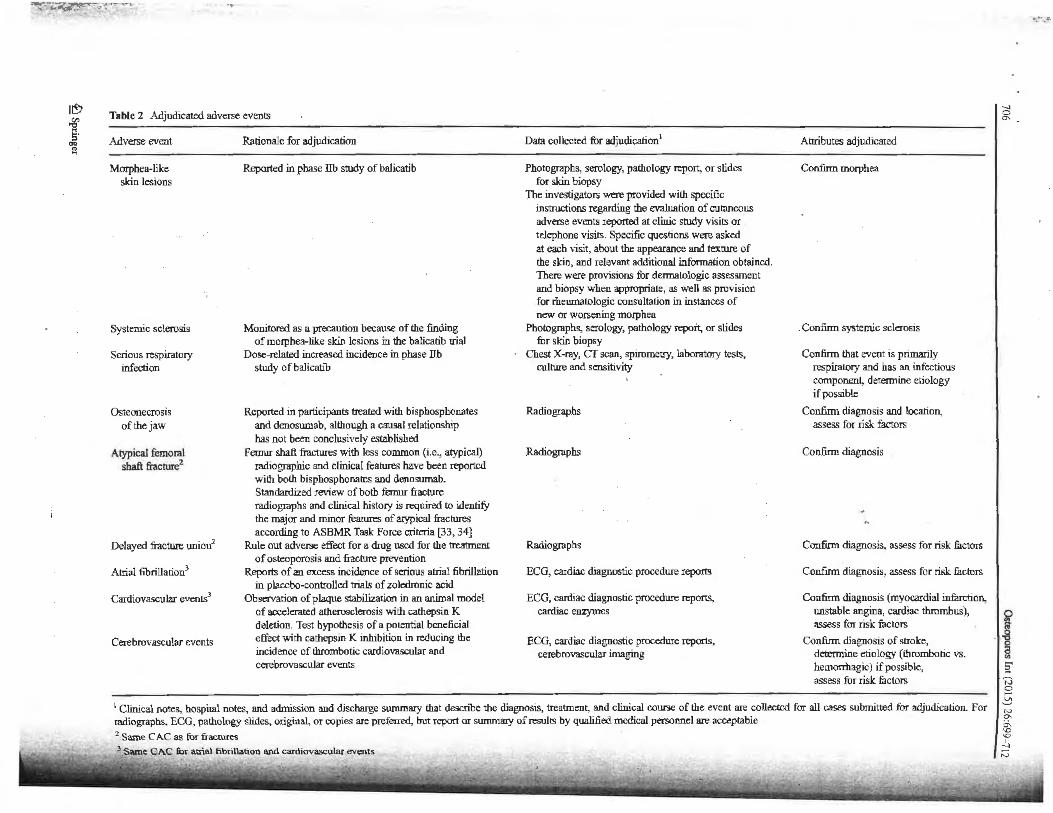
Several categories of specific AEs were designated for adjudication by external CAC committees (cardiovascular, cerebrovascular, dental, skin thickening and hardening, delayed fracture union, and serious respiratory events) to confinn their occurrence and specific attributes. These AEs, rationale for their adjudfoation, data collected for the adjudication. and specific attributes to be adjudicated are summarized in Table 2. These AEs were adjudicated in a two-round process similar to that previously described for fractures. Adjudication of a1J AEs was managed by a central site (Parexel lntemational, Waltham, MA, USA), which was responsible for collection of all clinical data, electronic distribution to the CAC members, coordination of CAC member conferences, collation of adjudication results, and final data transfer to the sponsor.
Discontinuation criteria
Participants were discontinued from blinded study drug if the investigator considered that the risk outweighed the benefit of continued participation on blinded therapy for an individual participant. Additionally, participants were discontinued from blinded study drug if they experienced excessive bone loss, as described above; initiated treatment with a strong CYP3A4 inducer (e.g., carbamazepine, phenyto.in, rifampin, St. John's wort) for longer than 6 months; commenced treatment with other drugs for osteoporosis; or bad persistent elevations in serum transaminases or bilirubin. The sponsor provided alendronate 70 mg OW to patients who were discontinued from study drug, although other treatment options were not prohibited. Participants were followed for outcome and safety monitoring tlu·ougb the end of the base trial even if blinded study therapy was discontinued.
Data monitoring committee
The ODN clinical development program, including this fra<r lure outcome study, was monitored by a DMC, which consisted of seven experts not involved in the conduct of the
~ Springer
I~ ~ s· 1
Table 2 Adjudicated adverse events
Adverse event
Morphea-like skin lesions
Systemic sclerosis
Serious respiratory infection
Osteonecrosis of the jaw
Atypical femoral shaft fracrure2
Delayed fracture union2
Atrial fibrillation3
Cardiovascular events3
Cerebrovascular events
Rationale for adjudication
Reported in phase llb study of balicatib
Monitored as a precaution because of the finding of morphea-like skin lesions in the balicatib trial
Dose-related increased incidence in phase Ub study of balicafib
Reported in participants treated with bispbosphonates and denosumab, although a causal relationship has not been conclusively established
Feurur shaft fractures with less common (i.e., atypical) radiographic and clinical features have been reported with both bisphospbonatcs and denosumab. Standardized review of both femur fracture radiographs and clinical history is required to identify the major and minor features of atypical fractures according to ASBMR Task Force criteria [33, 34]
Rule out adver..c effect for a drug used for the treatment of osteoporosis and fracture prevention
Reports of an excess incidence of serious atrial fibrillation in placebo-controlled trials of zoledronic acid
Observation of plaque stabilization in an animal model of aocelerated atherosclerosis with cathepsin K deletion. Test hypothesis of a potential beneficial effect with cathepsin K inhibition in reducing the incidence of thrombotic cardiovascular and cerebrovascular events
Data collected for adjudication'
Photographs, serology, pathology report, or slides for skin biopsy
The investigators were provided with specific instructions regarding the evaluation of cutaneous adverse events reported at clinic study visits or telephone visits. Specific questions were asked at each visit, about the appearance and texrure of the skin, and relevant additional information obtained. There were provisions fur dennatologic assessment and biopsy when appropriate, as well a.~ provision for rhcumatologic consultation in instances of new or worsening mo,:phea
Photographs, serology, pathology repoit, or slides for skin biopsy
Chest X-ray, er scan, spirometry, laboratory tests., culture and sensitivity
Radiogtaphs
Radiograpbs
Radiographs
ECG, cardiac diagnostic procedure reports
ECG, cardiac diagnostic procedure reports, cardiac enzymes
ECG, cardiac diagno:.iic procedure reports, cerebrovascular imaging
Attributes adjudicated
Confum morphea
. Confum systemic sclerosis
Confinn that event is primarily respiratory and has an infectious component, determine etiology if possible
Confirm diagnosis and location, assess for risk factors
Confirm diagnosis
Confirm diagnosis, assess for risk factors
Confirm diagnosis, assess for risk factors
Confum diagnosis (myocardial infarction, unstable angina, cardiac thrombus), assess for risk factors
Confinn diagnosis of stroke, determine etiology (thrombotic vs. hemorrhagic) if possible, assess for risk factors
1 Clinical notes, hospital notes, and admission !llld discharge summary that describe the diagnosis, treatment, and clinical course of the event are collected for all cases submitted for adjudication. For radiographs, ECG, pathology slides, original, 01 copies are preferred, but report or summary of resultS by qualified medical personnel are acceptable 2 SaroeCAC~furfracrures
._, ~
~ 8
·• "8 ~ !a 'iJ ~ ~ ..., °' 0\ '° 'O
.!.i ,.._,

()steoporos Int (2015) 26:699-712
study, including six physicians, five of whom were experts in osteoporosis with subspecialty expertise in endocrinology, rheumatology, geriatrics, and epidemiology; a dennatologist; and a non-physician statistician. The DMC was supported by a statistician and statistical programmer from the sponsor. Only the DMC and their support statistical personnel had access to unblinded data from the trial, with the exception that safety data were provided to the FDA in an unblinded fashion as described above. Since September 2007, the DMC has met at inteJVals of approximately 4 months to review safety data from the ODN program, including data from this trial, and to make recommendations on the conduct of the trials. The DMC was also responsible for the review of the two planned interim efficacy analyses and had the option of recommending to the sponsor to tenninate the study early due to futility or due to robust efficacy and a favorable benefit-risk profile of the drug.
Ancillary studies
Population pharmacokinetics study
Three specimens were collected from each participant in the lead cohort for the measurement of plasma ODN levels to confinn findings from phase l. Information was collected with regard to timing of the collection and last dose of ODN and characteristics of the meal prior to last dose.
Special imaging sub-study
A sub-study of l 64 participants investigated the effect of ODN on volumetric BMD of the hip and spine and, specifically, on the trabecular and cortical components at each site, using quantitative computed tomography (QCT, instruments from GE Medical Systems, Phillips, Siemens, and Toshiba). High-resolution peripheral QCT (Scanco Xtreme CT) was employed to investigate the effect of ODN on bone microarchitecture at the distal radius and distal tibia in some of these participants. Imaging procedures were perfonned yearly, and all participants in this study had biochemical markers of bone turnover measured at these time-poin1s. Only participants in this sub-study were asked to consent to optional bone biopsies at baseline and 12 months.
Sarcopenia end point sub-study
This was an exploratory sub-study of 556. participants designed to identify molecular signatures of sarcopenia using DNA and RNA biomarkers in the blood and to examine changes in appendicular lean body mass, measured by total body DXA, and physical function, measured by the Short Physical Performance Battery (SPPB).
707
Participation in the special imaging and sarcopenia substudies was optional and involved separate consent fonns from those used for the main study.
Statistical methods
Data analysis
The primary end points of the study were the first morphometrically confinned vertebral fracture per participant, first osteoporotic clinical hip fracture confirmed by adjudication, and fust osteoporotic clinical non-vertebral fracture confumed by adjudication. There was a hierarchical order from morpl10-metric vertebral fractures (the first primary end point) to the other two end points; there was no order set between hip and non-vertebral fractures.
The all-patients-as-treated population (n = 16,071) was used for safety analyses and included all participants who took at least one dose of study medication and had at least one follow-up visit with the exclusions noted above. Treatment groups were compared using the Miettinen and Nurminen method [35], adjusted for differential follow-up period, for prespecified sets of AEs of clinical relevance.
The full-analysis population was used for efficacy analyses. With the exclusions noted above in "Recruitment," it included all randomized participants who took at least one dose of study medication regardless of protocol violation, adherence to study medication, or early discontinuation from the study and had at Least one follow-up efficacy assessment. TI1e follow-up period was from randomization to study termination. The number of patients included in the efficacy analyses varied according to the number and timing of observations for each end point.
Morphometrically confitmed vertebral fracture data were evaluated with a generalized linear model for binary data with the complementary log-log transfonnation of the probability of an event up to the time-point. Clinical hip and non-vertebral fracture data were analyzed by time-to-event methods using Kaplan-Meier estimates and a Cox proportional hazards
model. A step-down procedure (36] controlled for increased false
positive error risk due to multiple primary end points, in the order of(l) morphometric vertebral fractures and (2) clinical hip and clinical non-vertebral fractures. Significance of the morphometric vertebral fracture primary end point was required before testing the other two primary end points. The Hochberg procedure was used to control for the multiplicity of
fracture end points (37). For BMD end points, treatments were compared using a
longitudinal model with tenns for treatment, stratum, geogra:phic region, and treatment-time interaction on the percent
~ Springer
·-----"

708
change from baseline. Log-transformed fractional change from baseline in biochemical markers was analyz.ed using the same rnodel as BMD. Yearly rate of height loss was analyzed using a longitudinal model. Testing for secondary end points was performed only if significance for the first primary end point (morphometric vertebral fractures) was obtained. For the purpose of addressing the issue of multiplicity adjustment, three groups were considered: (l) BJ'vID measures, (2) biochemical markers of bone resorption, and (3) clinical vertebral fractures and height. Hochberg multiplicity adjustment procedure was used within e~h of three groups. No adjustment for multiplicity between the three groups was applied.
Sample size considerations
To provide at least 90 % statistical power to demonstrate a risk reduction of vertebral fracture by 50 %, hip fracture by 3 5 %, or non-vertebral fracture by 20 %, the estimated required numbers of participants with these fractures were 114, 237, and 824, respectively. Sample size estimates were based on the fracture incidence in the idendronate Fracture Intervention Trial (FIT) and assumed that approximately 25 % of participants would have a prior radiographic vertebral fracture [38]. The estimated enroIJment requirement was based on hlp fracture projections, since these events are least frequent. On this basis, randomization of approximately 16,000 participants was planned.
Interim analyses
Two interim analyses were planned to be conducted, when approximately 70 and 85 % of the targeted 237 participants had experienced an osteoporotic hip fracture event To adjust for multiplicity, an alpha spending function was used for the interim and final analyses, such that p values of approximately 0.007, 0.018, and 0.046 were required for the first and second interim and final analyses, respectively.
Planned subgroup analyses
Prespecified subgroup analyses for the primary efficacy end points included age (<70 and "?:.70 years), race, prior radiographic vertebral fracture, baseline BMD T-score tertiles, baseline biochemical bone turnover marker tertiles, geographic region, baseline renal function, bisphosphonate intolerance, vitamin D status, BMI tertiles, and participation in the lead or main cohort. Subgroup analyses including age, race, bisphosphonate intolerance, and ability or willingness to be treated with available osteoporosis therapies were conducted for the following safety end points: combined clinical as well as combined laboratory AEs and skin disorders and dental disorders reported by investigators as AEs.
~ Springer
Osteoporos Int (2015) 26:699--1 12
Participant baseline characteristics
The numbers of participants screened and enrolled are shown in Fig. l and their geographical distribution in Table 3. Baseline characteristics are shown in Table 4. The average age of the srudy population was 72.8 years, 56.5 % of the participants were Caucasian, and the average number of years since menopause was 25.3, Baseline vertebral fractures were identified in 46.5 % of subjects:
Extension study
In recent years, questions have arisen about the benefits and risks of long-tenn treatment with osteoporosis medications [39]. At the initiation of the current trial, it was anticipated that up to 5 years of blinded therapy might be required to evaluate its end points, and this duration was specified in the protocol and participant consent fonns that were approved by ERCs and IRBs. Provision was made for early tennination of the study if significant reductions in fracture risk were demonstrated in either preplanned interim analysis. In order to ensure collection of long-term data, an extension study was planned in which study participants would continue in a blinded fashion on their randomized treatment until they
Table 3 Geographic distribution of investigational sites and participants
Geographic Number (%) of Nwnbcr (%) of region investigational sites participants randomized
Fracture trial
Asia 110 (28.4) 3151 (18.9)
Latin America 62 (16.0) 5442 (32.6)
Europe/US/others 215 (55.6) 8120 (48.6)
Total 387 16,7131
Special imaging sub-study
South Africa 3 (30.0) 61 (37 .2)
Europe/US/others 7 (70.0) 103 (62.8)
Total 10 164
Sarcopenia end point sub~tudy
Asia 3 (8.8) 73 (13.1)
Latin America 8 (23.5) 263 (47.3)
Europe/US/others 23 (67.6) 220 (39.6)
Total 34 556
Bone biopsy
Asia 2 (7.7) 6 (2.2)
Latin America 9 (34.6) 51 (18.6)
Europe/US/others ·15 (57.7) 215 (792)
Total 26 272
1 Three randomized participants were subsequently excluded due to assignment of duplicate allocation numbers

· osreoporos Int (2015) 26:699-712
fable 4 Participant baseline characteristics
Age (years), mean (SD)
>70 years old, n (%)
Race, 11 (%)
Caucasian
Asian
Multiracial
Black/Afiican American
Native American
Pacific Islander
BMD T-score, mean (SD)
Lumbar spine
Total hip
Femoral neck
Trochanter
Bone turnover markers, mean (SD)1
uNTX/Cr (nmol/mmol Cr)
sCTX (ng/mL)
sBSAP (ng/mL)
sPJNP (ng/mL)
Clinical fracture histoiy since menopause, 11 (%)
Any
Evaluable participants (n~l6,071)
72.8 (5.3)
11,004 (68.5)
9085 (56.5)
2832 (17.6)
3706 (23.1)
261 (1.6)
185 (1.2)
2 (0.OOJ)
-2.7 (1.2)
-2.4 (0.7)
-2.7 (0.5)
-2.3 (0.8)
47.4 (37.4)
0.44 (0.22)
15.78 (6.50)
58.62 (28.81)
5552 (34.5)
Hip 224 (1.4)
Spine 1439 (9.0)
Other 4407 (27.4)
Years since menopause, mean (SD) 25.3 (7.7)
Prior radiographically assessed vertebral .fracture, 11 (%)2 7446 (46.4)
Mild fracture (Gcnant grade I) 4414 (59.3)
Moderate fracture (Genant grade 2) 1858 (25.0)
Severe fracture (Genant grade 3) 1174 (1 5.8)
Unreliable or missing data 36
1 Measured in IO % subset of partic:ipants 2 Grade of fracture was detennined based on the worst grade per participant over all available scans up to 7 days after first study dose
reached the originally anticipated total of 5 years, after which participants would transition to open-label ODN.
Current status
On July 11, 2012, the sponsor announced the decision to terminate the base study early based on the DMC recommendation following its review of the first interim analysjs. That analysis was perfonned after approximately 70 % of the target hip fracture events (158 participants with an incident osreoporotic hip fracture) had occurred. The DMC based its recommendation on the observation of robust efficacy and a
709
favorable benefit/risk profile of ODN relative to placebo. The DMC also recommended that additional safety data be obtained in the previously planned blinded extension study. Following this recommendation, study participants attended a final base study visit and, if eligible, were asked to consent to enter the extension study. The base study was closed out between August and November, 2012, and will fonn the basis for the primary comparative efficacy and safety analyses.
There were important differences between the base and extension studies in study design and conduct. In the base study, all participants who discontinued study medication were allowed to remain in the trial and be followed unless they withdrew consent. However, participants who were no longer taking study medication at the time of their final base study visit were not eligible to enter the extension. Of 16,713 women initially randomized in the base study, 8256 entered the extension study. Of the approxn:nately 8000 women who did not enter the extension, most were ineligible because they had not completed the base study on therapy or because the investigational site did not participate in this phase of the trial. A total of 1281 women experienced excessive bone loss during the base study. Most of them discontinued prior to entering the extension. However, 245 women were excluded shortly after entering the extension because of excessive bone loss detected at the transitional visit.
The Joss of nearly half the original participants created a substantial likelihood that the extension population would differ materially from the original trial population. Participants who experienced excessive bone loss during the base study were discontinued fiom study drug and were therefore ineligible to enter the study extension. It would be expected that a greater number of participants from.the placebo arm were discontinued due to excessive bone Joss. Thus, although it was blinded, the extension cannot be presumed to have maintained the randomization.
As osteoporosis requires long-term treatment, extension studies of active treatment, with or without a withdrawal arm, have become customary. Prior trials have not generally extended the randomized placebo phase beyond the primary end point. In the repo1t of the IO-year extended phase ill alendronate studies, the authors used the expected effect of aging on fracture rates in the original placebo group in order to provide context for interpretation of the rate~ observed.on active treatment in the latter years [ 40]. A more sophisticated approach, taking more variables into account, was subsequently developed and validated using data from the alendron.ate FIT trial [41]. This "virt:!lal twin" model has been used in the analyses of data from the long-term extension of the denoswnab phase ill trial for which a placebo comparison was no longer available [42]. A similar model will be developed from the placebo group in the ODN base study. This may provide useful information about expected fracture rates and other efficacy and safety results for a virtual long-term placebo group with characteristics matching the active treatment population.
~ Springer

.710
Summary
Inhibition of cathepsin K is an attractive phannacological approach with therapeutic potential in osteoporosis. Preclinical and initial clinical data provided a strong rationale for the use of ODN in the treatment of osteoporosis, and the phase lib trial identified 50 mg once weekly as a dose regimen that is effective on BMD end points. The ODN phase m fracture trial was designed to rigorously investigate the efficacy and safety of this selective CatK inhibitor in the treatment of osteoporosis in postmenopausal women.
This placebo-controlled study excluded· participants who were at excessively high individual risk of fracture and included safeguards to ensure participant safety. In addition to fractures, the study evaluated multiple end points that not only provide infonnation about ODN but may also help expand our knowledge of osteoporosis. An interim analysis demonstrated sufficiently robust anti-fracture efficacy and favorable benefitrisk profile to warrant early tennination of the base study. The blinded extension study enrolled more than 8000 continuing participants and will provide additional data conceming the longer-term safety and efficacy ofODN.
Acknowledgments We would like to thank Boyd B Scott Ph.D., Elizabeth Rosenberg Ph.D., and Keith Kaufman M.D. (l\,1erck & Co. Inc.) fur their assistance in writing and critical review of this manuscript and Jennifer Pawlowski, MS, (Merclc & Co. Inc.) for her logistical support in the submission of this manuscript.
Funding statement This study was funded by Merck & Co., Inc., Whitehouse Station, NJ.
Contlicts of interest Henry Bone received consultancy fees or honoraria from Merck & Co. Inc., Amgen, Aclairo, Takeda, OSK, Radius, Azelon, Purdue Phannaceuticals, Mission Pharmacal, Akros, Novcn, Tarsa, and Novartis; grants from Merck & Co. Inc., Amgen, NPS, Nordic Bioscience, Tarsa, and Novartis; speaker fees from Amgen, Pfizer, ISCb, and Merck & Co. Inc.; and travel support from Merck & Co. Inc.
David Dempster received consulting fees or honoraria from Merck & Co. Inc., Amgen Inc., Eli Lilly & Co and speaker fees from Amgen Inc. and Eli Lilly & Co.
John A. Eisman received investigator fees, consulting fees or hono· rarium, and tr-<1vel support from Merck & Co. rnc. He receives similar support a11d at times untied grant support from Amgen, Eli Lilly, SanofiAventis, Servier, and Novartis.
Susan L. Greenspan rect:ived advisory board honoraria from Merck & Co. Inc.; travel support from Merck & Co. lnc.; and grants from Eli Lilly, Amgen, Warner-Chilcott, and Tarsa Therapeutics
Michael McClung received investigator fees from Merck & Co. Inc.; consttlting fees or honoraria from Merck & Co., Amgen, Eli Lilly, and Novartis; grants from Amgen; and ~peaker foes from Amgen l.nc. and Warner Chilcott.
Toshitaka Nakamura Received consulting ft:e.s or honoraria from Amgen, Eli Lilly, Chugai, Daiichi Sankyo, and Asahi Kasei; grants _from
Eisai, Takeda, Astellas, and Ono; and speaker fees from Eli Lilly, Chugai, Takeda, and Da:iichi Sankyo.
Socrates Papapoulos received consulting fees or honoraria from Merck & Co., Amgen, Novartis, Pfizer, Roche, GSK, and Axsomc Therapeutics and speaker fet:s from Amgen [nc., GSK. Merck & Co., Eli Lilly, and Roche.
~ Springer
Osteoporos Int (2015) 26:699--712
Weichung J. ShiJ1 declares no conflict of interest, apart from honorana for participation on the odanacatib scientific advisory committee.
Albert T. Leung, Arthur Santora, Nadia Verbruggen, Andrea RybakFeiglin, and Amonio Lombardi are employees of Merck & Co. Inc. Whitehouse Station, NJ, and may own stock or stoc,k options in Merck &Co. lnc.
Scientific advisory committee
Henry B9-ne. Michigan Bone & Mineral Clinic, Detroit, Ml, USA David Dempster, Colwnbia University Medical Center, New York · NY, USA '
John A Eisman, Gm-van Institute of Medical Research, University of Notre Dame Australia, St Vincent's Hospital aJJd UNSW Australia Sydney, Atistrnlia '
Susan Greenspan, University of Pittsburgh, Pittsburgh, PA, USA
Michael Mcclung, Oregon Osteoporosis Center, Ponland, Otego11,
USA
Toshitaka Nakamura, University of Occupational and P.nvironmental Health, Kitakyushu, Japan
Socrates Papapoulos, Leiden University Medical Center, Leiden, Netherlands ·
Weichung J. Shih, Robert Wood Johnson Medical School, Piscataway, NJ, USA
Data monitoring committee
Robert Wallace (chairperson), University oflowa College of Public Health, lowa City, IA, USA
Cyrus Cooper, MRC Epidemiology Research Centre, Southampton General Hospital, SouthamplOn, UK
Kristine Ensrud, Minneapolis Veteran Affai!'l! Medical Center and University of Minnesota, Minneapolis, MN, USA
David Kcndler, ProHealth Clinical Research, Vancouver, BC, Canada
Leonaro Milstone, Yale University School of Medicine, New Haven, CT, USA
Stuart Ralston, Molecular Medicine Centre, Western General Hos· pital F..dinburgh, UK
Bruce Turnbull, Cornell University School of Operation.~ Research and [nfonnation Engineering, Ithaca, NY, USA
Open Access This article is distributed under the temts of the Creative Commons Attribution Nonconuncrcial License which pcnnits any noncommercial use, dii.tribution, and reproduction in any medium, provided the original author(s) and the source arc credited.
Reterences
I. Osteoporosis prevention, diagnosis, and therapy. JAMA 2001; 285; I: 785-95
2. Bonewald LF (2011) The amazing osteocyte. J Bone Miner Res 26: 229--238
3. Martin TJ, Sims NA (2005) Osteoclast-dcrived activity in the coupling of bone formation lO resorption. Trends Mol Med 11 :76-81
4. Tang Y, Wu X, Lei W, Pang L, Wan C, Shi Z, Zhao L, Nagy TR, Peng X, Hu J, Feng X, V811 Hui W, Wan M, Cao X (2009) TGF-bctal •

Osteuporos Int (2015) 26:699-712
induced migration of bone mesenchymal stem cells couples bone resorption with fom,ation. Nat Med 15:757- 765
5. Zhao C, Irie N, Takada Y, Shimoda K, Miyamoto T, Nishiwaki T, Suda T, Matsuo K (2006) Bidirectional epbrinB2-Eph84' signaling controls bone homeostasis. Cell Metab 4:111-121
6. Lotinun S, Kiviranta R, Matsubara T, Alzate JA, Neff L, Lutl1 A, Koskivirta I, Kleuser B, Vacher J, Vuorio E, Home WC, Baron R (2013) Osteoclast-spccific cathepsin K deletion stimulates SIPdcpendcnt bone formation. J Clin lnvest 123(1 ):666-681
7. Boyle WJ, Simonet WS, Lacey DL (2003) Osteoclast differentiation and activation. Nature 423(1):337-342
8. Baron R, Ferrari S, Russell RG (2011) Denosumab and bisphosphonatcs: different mechanisms of action and effucts. Bone 48(5):677-692
9. Heaney RP, YatesAJ, Santora AC (1997) Bisphosphonate effects and the bone remodeling tmnsicn1. J Bone Miner Res 12(1):1143-1151
J 0. Hcruandez CJ, Beaupre GS, Marcus R, Carter DR (2001) A theoretical analysis of the contributions ofremodeling space, roinerali7.ation, and bone balance to changes in bone mineral density during alendronate treatment. Bone 29(1):511- 516
11. Roschger P, Rinnerthaler S, Yates J, Rodan GA, Fratzl P, Klaushofer K (2001) Alendronate increases degree and uniformity of minerali-7,ation in cancellous bone and decreases the porosity in cortical bone of osteoporotic women. Bone 29(2): 185-191
12. Zebaze RM, Libanati C, Austin M, Ghasem-Zadeh A, Hanley DA, Zanchetta JR, Thomas T, Boutroy S, Bogado CE, Bilezikian JP, Seeman E (2014) Differing effects of denosmnab and alendronate on cortical and trabecular bone. Bone 59(2): 173-179
13. Lcwiecki EM (2011) New targets for intervention in the treatment of postmenopausal osteoporosis. Nnt Rev Rhcumatol 7(1 ):631-638
14. Costa AG, Cusano NE, Silva BC, Cremers S, Bilezikian JP (2011) Calhepsin K: its skeletal actions and role as a therapeutic target in osteoporosis. Nat Rev Rheumato.1 7(1 ):447-456
15 . Gelb BD, Shi GP, Chapman HA, Desnick RJ ( 1996) Pycnodysostosis, a lysosomaJ disease caused by catl1epsin K deficiency. Science 273: 1236-1238
16. Duong L (2012) 1l1empeutic inhibition of catllepsin K- reducing bone resorption while maintaining bone fonnalion. BoneKEy Rep I: 1-8
17. Kiviranta R, Morko J, Uusitalo H, Aro HT, Vuorio E, Rantakokko J (200 l) Accelerated turnover of metapbyseal trabecular bone in mice overexprcssing catl1epsin K. J Bone Miner Res 16: 1444-1452
18. Falgueyret JP, Desmarais S, Oballa R, Black WC, Cromlish W, Khougaz K, Lamontagne S, Masse F, Riendeau D, Toulmond S, Percival MD (2005) Lysosomotropism of basic cathepsin K inhibitors contributes to increased cellular potencies against off-target cathepsins and red.uced functional selectivity. J Med Chem 48(2): 7535-7543
19. Gauthier JY, Chau.rel N, Cromlish W, Desmarais S, Duong LT, Falgueyret JP, Kimmel DB, Lamontagne S, Leger S, LeRichc T, Li CS, MasseF, McKay DJ, Nicoll-Griffith DA, Oballa RM, Palmer JT, Percival MD, Riendeau D, Robichaud J, Rodan GA, Rodan SB, Seto C, TI1erien M, Trnong VL, Venuti MC, We-~olowski G, Young RN, Zamboni R, Black WC (2008) The discovery of odanacatib (MK-0822), a selective inhibitor of cothepsin K. Bioorg Med Chem Lert 18:923-928
20. Stoch SA, Zajic S, StoneJ, Miller DL, Van Dyck K, Gutierrez MJ, De Decker M, Liu L, Liu Q, Scott BB, Panebianco D, Jin B, Duong LT, Gottesdiener K, Wagner JA (2009) Effect oftbe cathepsin K inhibitor odanacatib on bone resorption biomarkers in healtJ1y postmenopausa I women: two double-blind, randomized, placebo<onrrolled phase I studies. Clin Pharmacol Ther 86:175-182
21 . Stoch SA, Zajic S, Stone JA, Miller DL, vru, Bortel L, Lasseter KC, Pramanik B, Cil issen C, Liu Q, Liu L, Scott BB, Panebianco D, Ding Y, Gottesdiener K, Wagner JA (2013) Odanacatlb, a selective cathepsin K i11hibitor to treat osteoporosis: safety, tolerability,
711
pharrnacolcinetics and pharmacodynamic&-results from single oral dose studies in healthy volunteers. Br J Clin Pbannacol 75:1240-1254
22. Bone HG, McClung MR, Roux C, Recker RR, Eisman JA, Verbruggen N, Hustad CM, DaSilva C, Sru1tora AC, Ince BA (20 I 0) Odanaeatib, a cathepsin-K inhibitor for osteoporosis: a twoyear study in postrnenopausal women with low bone density. J Bone Miner Res 25:93 7- 947
23. Eisman JA, Bone HG, Hosking DJ, McClung MR, Reid IR, Rizzoli R., Resch H, Vcrbruggcn N, Hustad CM, DaSilva C, Petrovic R, Santora AC, Ince BA, Lombardi A (2011) Odanneatib in the treatment of posnnenopausal women with low bone mineral density: three-year continued the.rapy and·resolution of effect. J Bone Miner Res 26:242-251
24. Langdahl B, Binkley N, Bone H, Gilchrist N, Resch H, Rodriguez P J, Denker A, Lombard.i A, Le Bailly DT, DaSilva C, Rosenberg E, Leung A (2012) Odanacatib in the treatment of posnnenopausal women wit.h low bone mineral density: live years of continued tllerapy in a phase 2 study. J Bone Miner Res 27(1):2251-2258
25. Kanis JA, Alexandre JM, Bone HG, Abadie E, Brasseur D, Chassany 0, Durrleman S, Lekkerkerker JF, CauLin F (2003) Study design in osteoporosis: a European perspective. J Bone Miner Res 18(24): 1133- 1138
26. Clinical trials for new osteoporosis rreatments 2002. http://www.fda. gov/ohrrns/dockets/ac/cder02 hbn#EodoerinologicMetabolicDrugs
27. Cummings SR, Sru1 Manin J, McClung MR, Sirls ES, Eastell R, Reid IR, Delmas P, Zoog HB, Austin M, Wang A, Kutilek S, Adami S, Zanchetta J, Libanati C, Siddhanti S, Christiansen C (2009) Denosumnb for prevention of fractures in postrnenopnusal women with osteoporosis. N Engl J Med 361:756-765
28. Study to evaluate tl1e safety and efficacy ofBA058 for prevention of fracn1rc in postmenopausal women 2013. http://www.clinicaltrials. gov/ct2/show/NCTO I 343004?term=-BA-058&.rank=3
29. Multicenter, international, randomized, double-blind, placebo-controlled, parallel-group study to assess the efficacy and safety of AMG 785 treatment in postmenopausal women with osteoporosis 2012. http://www.cliniealtrials.gov/ct2/show/NCTO 1575834 ?tern,"' AMG+785&.ranlr-l 3A
30. Genant HK, Wu CY, van Kuijk C, Nevitt MC (1993) Vertebral fracrure assessment using a semiquantitative technique. J Bone Miner Res 8(2): 1137- 1148
3 J. Recker RR, Kimmel DB, Dempster D, Weinstein RS, Wronski Ti, Burr DB (2011) Issues in modem bone histomorphometry. Bone 49(1):955-964
32. Dempster OW, Compston JE, Drewer MK, Glorieux FH, Kanis JA, Malluche H, Mewiier PJ, Ott SM, Recker RR, Parl'itt AM (20 13) Standardized nomenclarure, symbols, and units for bone histomorphometry: a 2012 update of the repon of tl1e ASBMR Histomorphometry Nomenclnture Committee. J Bone Miner Res 28(2):2-17
33. Shane E, Burr D, Ebeling PR, Abrahamsen B, Adler RA, Brown TD, Cheung AM, Cosman F, Curtis JR, Dell R, Dempster D, Einhorn TA, Genant HK, Geusens P, Klaushofcr K, Koval K, Lane JM, McKiernan F, McKinney R, Ng A, Nieves J, O'Keefc R, Papapoulos S, Sen HT, van der Meulen MC, Weinstein RS, Whyte M (20 I 0) Atypical subtrochanterie and diaphyseal femoral fractures: report of a task force of the American Soci.ety for Bone and Mineral Research. J Bone Miner Res 25()):2267- 2294
34. Shane E, Burr D, Abmhomsen B, Adler RA, Brown TD, Cheung AM, Cosman F, Curtis JR, Dell R, Dempster OW, Ebeling PR, Einhorn TA, Genant HK, Geusens P, KJaushofer K, Lane JM, McKiernon F, McKinney R, Ng A, Nieves J, O'Keefe R, Papapoulos S, Howe TS, van der Mculen MC, Weinstein RS, Whyte MP (2014) Atypical subtrocbanteric 11nd diaphyseal femoral fractures: second report of a task force of the American Society for Bone and Mineral Research. J Bone Miner Res 29(1):1- 23
~ Springer
I

112
35. Miettinen 0, Nurminen M (1985) Comparative analysis of two rates. Stat Med 4(2):213-226, 13
36. Benjamini Y, Hochberg Y (1995) Controlling the false discovery rate: a practical and powerful approach to multiple testing. J R Stat Soc Ser B Met.hodol 57:289-300
37. Hochberg YA (1988) A sharper Bonferroni procedure for multiple tests of significance. Biometrica 75:800-802
38. Black DM, Cummings SR, Karpf DB, Cauley JA, Thompson DE, Nevitt MC, Bauer DC, Genant HK, Haskell WL, Marcus R, Ott SM, Torner JC, Quandt SA, Reiss TF, EnSTUd KE (1996) Randomised trial of effect of alendronate on risk of fracture in women with existing vertebral fractures. fracture lnterve1\tion Trial Research Group. Lancet 348: 1535- 1541
39. Black DM. Bauer DC, Schwartz AV, Cummings S~ Rosen CJ (2012) Continuing bisphosphonate treatment for osteoporosis-for whom and for how long? N Engl J Med 366:2051-2053
~ Springer
Osteoporos Int (2015) 26:699-712
40. Bone HG, Hosking D, Devogelaer JP, Tucci JR, Emkcy RD, Toruno RP, Rodriguez-Portales JA, Downs RW, Gupta J, Santora AC Libennan UA (2004) Ten years' experience with alcndronate ro; osteoporosis in postrnenopausnl women. N Engl J Med 350(1 ): 1189-1199
41 . Vittinghoff E, McCulloch CE, Woo C, Cummings SR (20 IO) Estimating long-term effects of treatment from placebo-controlled trials ·with an extension period, using virtual twins. Stat Med 29(1 ): 1127- 1136
42. Bone HG, Chapurlat R, Brandi ML, Brown JP, Czerwinski E, Krieg MA, MeUstrom D, Radominski SC, Reginster JY, Resch H, lvona JA, Roux C, Vittinghoff H, Daizadeh NS, Wang A, Bradley MN, Franchimoot N, Geller ML, Wagman RB, Cummings SR, Papapoulos S (2013} The effect of three or six years of denosumab exposure in women with postmcnopausal osteoporosis: results from the FRBBDOM extension. J Clio Endocrinol Merab 98(1):4483-4492
Related Documents