Brit. J. Ophthal. (1962) 46, 1. COMMUNICATIONS OCULAR LESIONS OF ONCHOCERCIASIS* BY F. H. BUDDENt Formerly Specialist Ophthalmologist, Overseas Civil Service, Northern Nigeria THIS paper aims at describing, illustrating, and interpreting the ocular lesions associated with onchocerciasis. The literature of onchocerciasis already includes many pictures of the ocular lesions. The paintingst and diagrams in this paper supplement those previously published and are intended to fill gaps in the existing literature. The accompanying text expresses the author's views on the interpretation of the lesions and refers to alternative opinions. Particular attention is paid to the choroido-retinal lesions because they have been the subject of much controversy. It is hoped that the paper as a whole will assist workers in the field to distinguish and understand the lesions of onchocerciasis. Eyelids and Conjunctiva (1) Oedema of Eyelids.-This is usually a temporary lesion, no doubt similar to that occurring elsewhere in infected skin. (2) Conjunctivitis.-Conjunctival infection occurs in response to the local presence, or more probably local death, of microfilariae. Mild chemosis is present in the more severe cases and a limbal inflammatory swelling, some- what similar to, although more diffuse than, that found in "spring catarrh", may develop. Conjunctival phlyctens are occasionally present in heavy infections. These lesions are associated with photophobia and watering of the eyes. In long-standing infections, as with other chronic inflammations, the African conjunctiva becomes heavily pigmented, particularly near the limbus. The clinical appearance suggests an allergic reaction and the finding of eosinophils in conjunctival biopsies supports this interpretation. The active inflammatory lesions of lids and conjunctiva are more marked in young persons (i.e. at an early stage of ocular invasion by microfilariae) and may be precipitated by therapy with diethylcarbamazine which kills microfilariae. * Received for publication June 5, 1961. t My thanks are due to the Medical Research Council for a grant covering the cost of the coloured plates, to Mr. G. T. W. Cashell for reading the draft, and to the Principal Medical Officer, Curative Division, Ministry of Health, Northern Nigeria, for permission to publish this paper. T I apologize for the amateur quality of the illustrations. They were painted direct from patients mostly in temporary mat shelters in remote villages. No artist being available, they were made by the author who has had no previous experience of ocular painting. 1 1 on December 3, 2021 by guest. Protected by copyright. http://bjo.bmj.com/ Br J Ophthalmol: first published as 10.1136/bjo.46.1.1 on 1 January 1962. Downloaded from

Welcome message from author
This document is posted to help you gain knowledge. Please leave a comment to let me know what you think about it! Share it to your friends and learn new things together.
Transcript
Brit. J. Ophthal. (1962) 46, 1.
COMMUNICATIONS
OCULAR LESIONS OF ONCHOCERCIASIS*BY
F. H. BUDDENtFormerly Specialist Ophthalmologist, Overseas Civil Service, Northern Nigeria
THIS paper aims at describing, illustrating, and interpreting the ocular lesionsassociated with onchocerciasis. The literature of onchocerciasis alreadyincludes many pictures of the ocular lesions. The paintingst and diagramsin this paper supplement those previously published and are intended to fillgaps in the existing literature. The accompanying text expresses theauthor's views on the interpretation of the lesions and refers to alternativeopinions. Particular attention is paid to the choroido-retinal lesionsbecause they have been the subject of much controversy. It is hoped thatthe paper as a whole will assist workers in the field to distinguish andunderstand the lesions of onchocerciasis.
Eyelids and Conjunctiva(1) Oedema of Eyelids.-This is usually a temporary lesion, no doubt
similar to that occurring elsewhere in infected skin.
(2) Conjunctivitis.-Conjunctival infection occurs in response to the localpresence, or more probably local death, of microfilariae. Mild chemosis ispresent in the more severe cases and a limbal inflammatory swelling, some-what similar to, although more diffuse than, that found in "spring catarrh",may develop. Conjunctival phlyctens are occasionally present in heavyinfections. These lesions are associated with photophobia and watering ofthe eyes. In long-standing infections, as with other chronic inflammations,the African conjunctiva becomes heavily pigmented, particularly near thelimbus. The clinical appearance suggests an allergic reaction and thefinding of eosinophils in conjunctival biopsies supports this interpretation.
The active inflammatory lesions of lids and conjunctiva are more markedin young persons (i.e. at an early stage of ocular invasion by microfilariae)and may be precipitated by therapy with diethylcarbamazine which killsmicrofilariae.
* Received for publication June 5, 1961.t My thanks are due to the Medical Research Council for a grant covering the cost of the coloured plates, to Mr.
G. T. W. Cashell for reading the draft, and to the Principal Medical Officer, Curative Division, Ministry of Health,Northern Nigeria, for permission to publish this paper.
T I apologize for the amateur quality ofthe illustrations. They were painted direct from patients mostly in temporarymat shelters in remote villages. No artist being available, they were made by the author who has had no previousexperience of ocular painting.
1 1
on Decem
ber 3, 2021 by guest. Protected by copyright.
http://bjo.bmj.com
/B
r J Ophthalm
ol: first published as 10.1136/bjo.46.1.1 on 1 January 1962. Dow
nloaded from

F. H. BUDDEN
Microfilariae may be found emerging from a snip of bulbar conjunctivamounted in normal saline or water. However, although this procedure hasbeen advocated for routine diagnosis, I do not recommend it. The chanceof finding microfilariae is considerably less in a conjunctival biopsy than inskin from the outer canthus. If confirmation is required of the presence ofmicrofilariae in the vicinity of the eye, a skin snip from the latter site causesless alarm to the patient and is more likely to prove positive.
Cornea(1) Intra-corneal Microfilariae.-These are seen, after their death, as
refringent, wavy, linear opacities, about 0-3 mm. in length, lying in thesubstantia propria. They may occur in any position but are most frequentin the interpalpebral area, near Bowman's membrane and close to the limbus.Possible explanations for this distribution are:
FIG. 1. FIG. 2.FIG. 1.-Diagram showing, from above down, stagesin development of "cracked ice" corneal opacityaround the body of a dead intra-corneal microfilaria.FIG. 2.-Diagram showing, from above down, two"snow-flake" corneal opacities, two nummularopacities, and an epithelial scar.
(a) Microfilariae entering thecornea at the limbus arephototropic and do not pene-trate freely.
(b) Microfilariae reaching the in-terpalpebral area are killed byexposure to light.
(2) "Cracked Ice" Opacities(Fig. 1) and "Snow-flake"Opacities (Fig. 2, and the excellentpainting published by Choyce,1958, p. 1 16).-The "cracked ice"lesions were more common in myAfrican patients, whereas " snow-flake" opacities were more com-mon in my European patients.It is sometimes convenient toclassify the two types together as"fluffy" opacities. Like micro-filariae these lesions are mostcommon in the interpalpebralarea, close to the limbus and inthe superficial layers of the sub-stantia propria. In early cornealinvolvement the opacities arelarge (O 5 mm. or more in dia-meter) and show up clearlyagainst the normal cornealstroma. However, in long-
2
on Decem
ber 3, 2021 by guest. Protected by copyright.
http://bjo.bmj.com
/B
r J Ophthalm
ol: first published as 10.1136/bjo.46.1.1 on 1 January 1962. Dow
nloaded from

OCULAR LESIONS OF ONCHOCERCIASIS
standing ocular infections, particularly after the development of oncho-cercal sclerosing keratitis, the fluffy reaction is smaller and less clearlydefined and tends to be obscured by widespread corneal oedema and sclerosis."Fluffy" opacities are associated with mild photophobia, excessive lacrima-tion, and sometimes with a temporary slight impairment of visual acuity.On several occasions I have watched a " cracked ice " opacity slowly develop
during a period of weeks around a dead microfilaria. At an intermediatestage of development an elongated "cracked ice" pattern is seen around adisintegrating microfilaria (Fig. 1). It would appear that the "crackedice" appearance is due to separation of fibres of the corneal stroma byoedema developing around a dead microfilaria.
"Fluffy" opacities persist for a considerable time. This is well illustratedby patients who receive combined treatment with suramin and diethyl-carbamazine, which eliminates microfilariae from the body. " Fluffy"opacities remain for several months but eventually resolve leaving no trace.In untreated patients, individual "fluffy" opacities probably resolve but arereplaced by fresh opacities so long as intra-corneal death of microfilariaecontinues. However, where massive death of microfilariae occurs sclerosingkeratitis may follow.
"Fluffy" opacities are easily distinguished from sub-epithelial nummularopacities (Fig. 2, and Hissette, 1938, p. 76) which are discoid and have a clear-cut margin. Unlike "fluffy" opacities they are permanent lesions. Ridley(1945) used the term "nummular" to describe the punctate corneal opacitiesproduced by onchocerciasis, although it is clear from his description that hereferred to lesions which are here called "fluffy". True discoid opacities arecommon in Nigeria, particularly in the Northern Region, and the termnummular should perhaps be reserved for these lesions. Their incidence indifferent villages is= not related to that of onchocercal infection. Lesionsclosely resembling the "fluffy" opacities produced by onchocerciasis can becaused by other agents, notably viruses. However, although "fluffy"opacities occur in villages where onchocerciasis is not endemic, their incidenceis very low. In villages where onchocerciasis is endemic, the incidence ofthese lesions rises with increases in the incidence and intensity of skininfection in the community.
(3) Sclerosing Keratitis.-I have watched this lesion develop in an area ofcornea where the death of large numbers of microfilariae had producedconfluent "fluffy" opacities. A few superficial blood vessels would invadethe area and fibrosis would lead to permanent irregular opacification.At a later stage oedema, pigment deposits, and eventually degenerativechanges, occur in the overlying epithelium. The lesion appears typicallyas tongues in the 4 and 8 o'clock positions, and extends around the lowercorneal margin. An area of cornea adjacent to the limbus at 12 o'clock is
3
on Decem
ber 3, 2021 by guest. Protected by copyright.
http://bjo.bmj.com
/B
r J Ophthalm
ol: first published as 10.1136/bjo.46.1.1 on 1 January 1962. Dow
nloaded from

almost invariably spared. This distribution follows that of the intra-corneal microfilariae from which the lesion is derived.The appearance was described and illustrated by Hissette (1938, p. 78)
and described by Ridley (1945) under the alternative name of pannus. Inview of the characteristic paucity of vessels, which Ridley noted, the termsclerosing keratitis suggested by French ophthalmologists is perhapspreferable.The occurrence of inflammatory intra-corneal lesions around fragments
of dead microfilariae has been demonstrated histologically by Rodger(1960).
(4) Pigmentation in the corneal epithelium is common in African eyes(presumably produced by migration of pigmented conjunctival epithelium).It occurs in onchocerciasis but is of no diagnostic value.
Anterior ChamberMicrofilariae are well adapted to the human host and, whilst they are
alive in the anterior chamber, they appear to cause no damage. They maybe observed by the following methods:
(a) By direct illumination with the beam of a slit-lamp. They are then seen asbright motile threads against the dark background of the pupil or iris.
(b) By retro-illumination using the +25D lens of a direct ophthalmoscope.This is best performed with the pupil widely dilated. Microfilariae are thenseen as dark motile threads against the bright red fundus reflex in the pupilarea (Sarkies, 1951).
The former method gives greater magnification but, since the eye of anAfrican villager, the microfilariae, and the slit-lamp beam may all be movingat the same time, it is often difficult to find the microfilariae and impossibleto count them accurately. The latter method gives smaller magnificationbut, since it provides a greater depth of focus and a view of the entire pupilarea, a count may be made of the number of microfilariae in this field at anyone time. However, counts of microfilariae in the anterior chamber, evenby this method, often vary markedly from minute to minute.
Uvea
(1) Inflammation of the Anterior Uvea.-This is usually mild. Totalpopulation surveys in. five infected rural communities of Northern Nigeriaand the Southern Cameroons showed that, although no therapy had beenadministered, the commonest sign of anterior uveitis was inflammatorypigment disturbance without posterior synechiae. However, plastic iritis
4 F. H. BUDDEN
on Decem
ber 3, 2021 by guest. Protected by copyright.
http://bjo.bmj.com
/B
r J Ophthalm
ol: first published as 10.1136/bjo.46.1.1 on 1 January 1962. Dow
nloaded from

OCULAR LESIONS OF ONCHOCERCIASIS
(characterized by posterior synechiae and sometimes evidence of severeexudate) was found with increasing frequency as the incidence and intensityof infection rose in the community.The active phase presents ciliary injection (often somewhat masked by
circum-corneal pigment), keratic precipitates, flare, and cells in the anteriorchamber. There may be exudate and I have seen hypopyon on fouroccasions in heavily infected patients. The resultant lesions are:
(a) Inflammatory Iris Pigment Disturbance.-In onchocerciasis this is diffuserather than patchy. Both the posterior pigmnent epithelium and the stromaare affected. A dramatic clinical feature is loss of the frill of pigmentat the pupillary border. The frill is normally well marked in the negroid eye(Fig. 3, overleaf). The crypts and irregularities of the anterior surface aresmoothed over so that the iris loses its normal lustre. The dark browncolour also changes. The iris comes to look like golden yellow blottingpaper. Eventually the grey trabecular fibres may be exposed, although thisis more characteristic of senile atrophy. Fine uveal pigment is deposited onthe corneal endothelium and on the anterior surface of the lens (illustratedby Hissette, 1938, p. 64).
(b) Posterior Synechiae and Gross Pigment Deposits on the anterior lens capsule.The pupil is sometimes completely secluded by posterior synechiae.
(c) Organized Exudate in the pupil area or the base of the anterior chamber.The pupil may be drawn towards the latter site so that its border becomespear-shaped (illustrated by Hissette, 1938, pp. 64, 70; and by Ridley, 1945,p. 26).
(d) Secondary Glaucoma is a late complication.
(e) Cataract associated with onchocercal uveitis is discussed later.
(2) Inflammation of the Posterior Uvea.-As in the anterior uvea, theinflammation is usually of low grade and long standing. Choroidal sclerosisis produced. Focal choroiditis of conventional type, although far lesscommon than plastic iritis, is sometimes seen when infection is heavy.
(a) Inflammatory Choroidal Sclerosis.-The initial sign is oedema,,usually atthe posterior pole and often to the outer side of the macula. This causes anirregular retinal reflex (Figs 4 and 5, overleaf). Later pigment disturbance atthe posterior pole produces a mottled appearance. Established choroidalsclerosis causes a marked tigroid appearance of the fundus which is usuallygeneralized by the time the African seeks medical advice. The overlyingretinal pigment epithelium may remain intact, although apparently deficientin pigment (Fig. 6, overleaf), but is often denuded over wide clearly demar-cated areas. Here the large choroidal vessels are clearly seen displayingvarying degrees of sclerosis (Bryant, 1935, p. 526; Hissette, 1938, p. 84;Toulant and Boithias, 1954, pp. 569, 570; d'Haussy, Rit, and Lagraulet,1958, p. 342; Figs 7, 8, 9, overleaf).
5
on Decem
ber 3, 2021 by guest. Protected by copyright.
http://bjo.bmj.com
/B
r J Ophthalm
ol: first published as 10.1136/bjo.46.1.1 on 1 January 1962. Dow
nloaded from

Choroidal exposure often commences at the outer side of the macula,spreads over the posterior pole of the eye and in long standing cases maybecome generalized. Gross retinal pigment masses are commonly foundoverlying, or adjacent to, these areas ofexposure; whereas fine bone corpusclepatterns are more common where the retinal pigment epithelium is notcompletely denuded. It should be noted that, although clearly definedareas of choroidal exposure and underlying sclerosis are frequently seen inonchocerciasis, the surrounding choroid is usually also sclerosed. Ridley'spainting (1945, Plate III), showing a patch of advanced choroidal sclerosis atthe posterior pole surrounded by normal retina, is not typical of lesionsseen in Nigeria. Small haemorrhages are occasionally seen.The patients usually suffer from night blindness and visual fields become
progressively restricted. Fairly good central telescopic vision may sometimesbe retained for many years.
(b) Focal Choroiditis (Fig. 10, overleaf).-In my experience this lesion is seenin heavily infected patients, is uncommon, and is usually, if not always,superimposed on inflammatory choroidal sclerosis.
Nature and Aetiology of Uveal Lesions
Hissette (1932) and Ridley (1945) believed the lesions to be inflammatoryand excited by local death of microfilariae. This interpretation of theanterior uveal lesions has not been seriously challenged. Alternativesuggestions have been made as to the cause of inflammatory choroidalsclerosis *:
Bryant (1935) reported the histological findings in an eye with advanced choroidalsclerosis. There were no microfilariae or signs of inflammatory reaction in the choroidor elsewhere in the eye, but there was marked interstitial fibrosis in the choroid andatrophy of the cellular layers of the retina. Clark (1947) found a similar histologicalpicture. Bryant contrasted these findings with the presence of microfilariae throughoutthe eye and the perivascular inflammation around choroidal vessels, both of which werefound in patients with advanced onchocercal keratitis.
Rodger (1957, 1958, 1960) also distinguished two lesions in the choroid. He postu-lated that "exudative" and "degenerative" lesions were aetiologically, clinically, andhistologically distinct. The "exudative" lesion was an acute focal choroiditis believedto be due to local death of microfilariae in the choroid. The "degenerative" lesion wascharacterized by fibrosis of the stroma which insidiously overgrew the choriocapillaris,arterioles, and arteries. However, Rodger found plasma cell infiltration and intimalhyperplasia in some "degenerative" cases. He believed that this lesion was excited bythe co-existence of low vitamin A intake and circulating onchocercal toxin.
Choyce (1958) noted that the clinical appearance of choroido-retinal lesions in fiveonchocercal patients resembled generalized choroidal sclerosis of familial type. Hesuggested that choroido-retinal degeneration, previously attributed to onchocerciasis,was due to genetic disease.
* This lesion has been referred to as choroido-retinal degeneration, even by those who believed the underlyingchoroidal lesion to be inflammatory. As will be apparent from the discussion, the term inflammatory choroidal sclerosisreflects the usual histological findings more accurately.
6 F. H. BUDDEN
on Decem
ber 3, 2021 by guest. Protected by copyright.
http://bjo.bmj.com
/B
r J Ophthalm
ol: first published as 10.1136/bjo.46.1.1 on 1 January 1962. Dow
nloaded from

FIG. 3.-Normal iris (on left). Onchocercal iris atrophy (on right).
To face page 6]
on Decem
ber 3, 2021 by guest. Protected by copyright.
http://bjo.bmj.com
/B
r J Ophthalm
ol: first published as 10.1136/bjo.46.1.1 on 1 January 1962. Dow
nloaded from

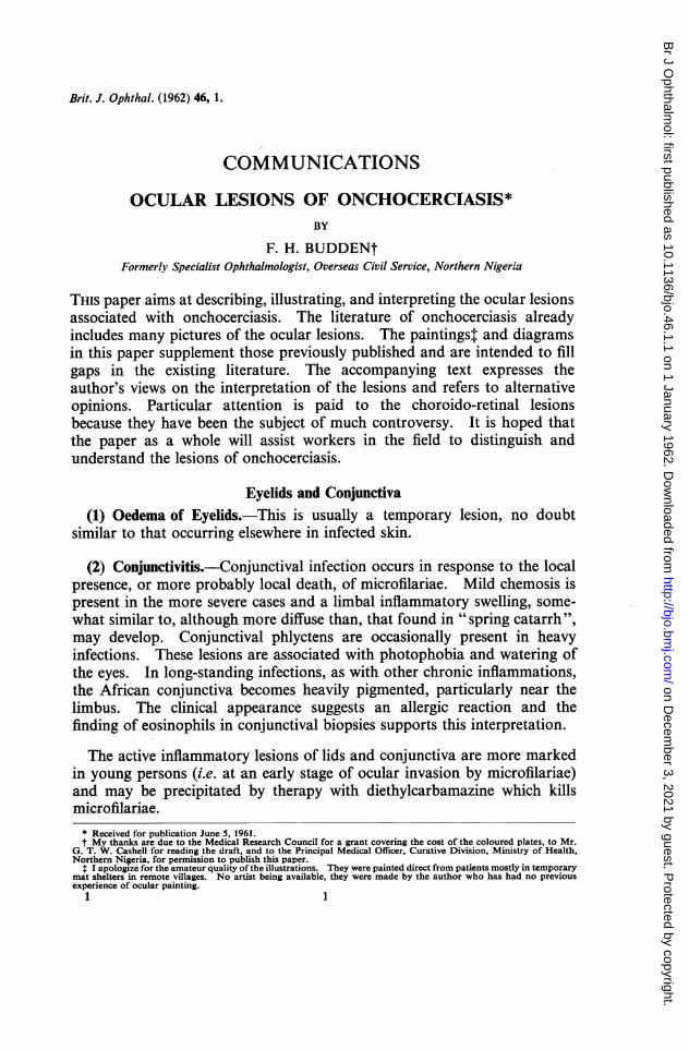
FIG. 4.-Right fundus of male African, 29 to 29-year age group (No. 474 Gangaransurvey). Two microfilariae per microscope field at left iliac crest. Visual acuity inthe right eye 6/9. Three dead microfilariae and eight fluffy opacities in cornea, onemicrofilaria in anterior chamber, early inflammatory iris pigment disturbance, but nokeratic precipitates, flare, or synechiae.
FIG. 5.-Right fundus of male African, 15 to 19-year age group (No.494 Gangaran survey). Ten microfilariae per microscope field at leftiliac crest, marked onchocercal depigmentation of shins. Visual acuityin the right eye 6/9. No signs of inflammation in anterior segment.
on Decem
ber 3, 2021 by guest. Protected by copyright.
http://bjo.bmj.com
/B
r J Ophthalm
ol: first published as 10.1136/bjo.46.1.1 on 1 January 1962. Dow
nloaded from

FIG. 6.-Left fundus of male African, 50 to 59-year age group (No.527 Zagun survey). Three microfilariae per microscope field at leftiliac crest. Visual acuity in the left eye perception of light. No signsof inflammation in anterior segment.
FIG. 7.-Right fundus of male African, 30 to 39-year age group (No. 206 Pyelanbirnisurvey). Three microfilariae per microscope field at left iliac crest. Visual acuityin the right eye 6/24. Sclerosing keratitis and gross inflammatory iris pigmentdisturbance.
on Decem
ber 3, 2021 by guest. Protected by copyright.
http://bjo.bmj.com
/B
r J Ophthalm
ol: first published as 10.1136/bjo.46.1.1 on 1 January 1962. Dow
nloaded from

FIG. 8.-Left fundus of male African, 40 to 49-year age group(No. 3360 Imbop survey). One microfilaria per field at iliaccrest. No perception of light in left eye. No lesion of anteriorsegment.
FIG. 9.-Right fundus of male African, 40 to 49-year age group (No. 3799 Kaduna clinic). Eightmicrofflariae per microscope field at left iliac crest. Visual acuity in the right eye hand movementsonly. Many fluffy opacities and sclerosing keratitis, many microfilariae in anterior chamber,inflammatory iris pigment disturbance.
on Decem
ber 3, 2021 by guest. Protected by copyright.
http://bjo.bmj.com
/B
r J Ophthalm
ol: first published as 10.1136/bjo.46.1.1 on 1 January 1962. Dow
nloaded from
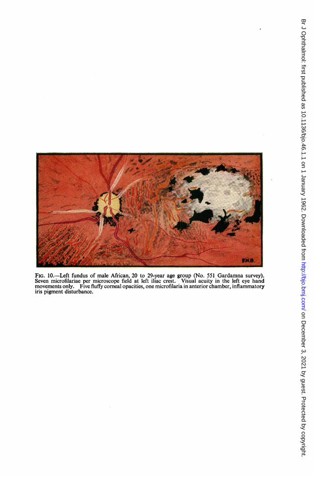
FIG. 10.-Left fundus of male African, 20 to 29-year age group (No. 551 Gardamna survey).Seven microfilariae per microscope field at left iliac crest. Visual acuity in the left eye handmovements only. Five fluffy corneal opacities, one microfilaria in anterior chamber, inflammatoryiris pigment disturbance.
on Decem
ber 3, 2021 by guest. Protected by copyright.
http://bjo.bmj.com
/B
r J Ophthalm
ol: first published as 10.1136/bjo.46.1.1 on 1 January 1962. Dow
nloaded from

OCULAR LESIONS OF ONCHOCERCIASIS
Further Epidemiological and Clinical Evidence
The following facts indicate a close association between inflammatorychoroidal sclerosis and onchocerciasis:
(a) The lesion is universally associated with onchocerciasis (d'Haussy andothers, 1958).
(b) The incidence of the lesion is directly related to the incidence of onchocercalinfection in village communities of Northern Nigeria (Budden, 1955, 1958).
(c) In villages where onchocerciasis is endemic I have found microfilariae morecommon in the cornea and anterior chamber of eyes presenting choroidalsclerosis than in eyes with normal fundi in the same age group.
(d) The lesion tends to be more severe in the more heavily infected.
In contradistinction to Rodger's evidence, my investigations show that:(a) The "degenerative" and "exudative" lesions are not as distinct as Rodger
suggests. At one extreme, when infection is low and the inflammatoryreaction mild, a widespread choroidal sclerosis may be produced in theabsence of gross retinal pigment disturbance. This has been likened inappearance to Sorsby's familial choroidal sclerosis (Choyce, 1958). At theother extreme a patch of focal choroiditis may be seen in heavily infectedpatients. Most lesions lie between these extremes, and focal choroiditisis usually superimposed on choroidal sclerosis.
(b) The incidence of "degenerative" lesions is not related to low vitamin Aintake. The lesions are in fact common in some places where there is ahigh consumption of vitamin A in the diet (e.g. in the Southern Cameroons).
(c) Vitamin A therapy does not produce a significant improvement in visualacuity in these patients.
No familial pattern has been demonstrated in support of Choyce'sevidence. Moreover, although the clinical fundal appearance, especially inlight infections, may occasionally simulate appearances seen in the abio-trophies described by Sorsby (1939), Sorsby and Crick (1953), Sorsby andDavey (1955), Sorsby, Mason, and Gardener (1949), the full clinical pictureis different (Budden, 1958; d'Haussy, 1960).
Further Histological Evidence
There have been many histological reports on the choroidal lesion inaddition to those already quoted (e.g., Hissette, 1932; Semadeni, 1943;Lavier, Lagraulet, and d'Haussy, 1956; Offret, Lagraulet, and d'Haussy,1958; d'Haussy, Rit, and Lagraulet, 1958; Lagraulet, 1958, 1960; personalcommunications on eyes submitted to Schofield in 1956 and to Lagraulet in1958). The finding common to these reports was round cell infiltration ofthe choroid, often most marked around the vessels, with associated peri-arteritis and sometimes endarteritis. Vascular sclerosis had developed in
7
on Decem
ber 3, 2021 by guest. Protected by copyright.
http://bjo.bmj.com
/B
r J Ophthalm
ol: first published as 10.1136/bjo.46.1.1 on 1 January 1962. Dow
nloaded from

F. H. BUDDEN
many eyes. The overlying retinal pigment epithelium and outer layers ofthe retina showed degenerative changes. Microfilariae were not usuallyfound in the choroid. However, Hissette (1932) states that microfilariae arefound in the choroid, Bryant (1935) states that they are found in the choroidof patients with coincident onchocercal keratitis, and Rodger (1960) foundthem in some of his sections of the choroid. One of my male patients withearly inflammatory choroidal sclerosis, whose age was estimated between30 and 40 years, died after a surgical operation whilst the ocular lesions werestill active. Ashton (personal communication, 1958) examined the eyesfrom this patient and found "two microfilariae in the choroid in relation tothe round cell infiltration".
Discussion of Uveal LesionsKoch's postulates have not been satisfied and it is just conceivable that
some factor other than onchocerciasis is responsible for the lesions. How-ever the epidemiological, clinical, and histological evidence quoted aboveindicates a cause and effect relationship between onchocerciasis and inflam-matory choroidal sclerosis. There is parallel evidence for the iris lesions.The possibility that some uveal lesions attributed to onchocerciasis(particularly uncommon lesions, e.g. choroidal sclerosis with minimalmassing of retinal pigment, and focal choroiditis) may be due to other causesmust be remembered during clinical investigation and when interpretinghistological material.The factor immediately responsible for uveal lesions is not known. A
circulating toxin or local liberation of toxin from dead microfilariae havebeen suggested. It is difficult to decide which is responsible. The presenceof microfilariae in the eye usually reflects a high concentration of micro-filariae in the body (Kershaw, Duke, and Budden, 1954) and probably alsoa high concentration of circulating toxin. Thus the association of localmicrofilariae with uveal lesions does not prove that they are causal. On theother hand, the absence of microfilariae at the time of clinical examinationor excision does not prove that the uveal lesions were not initiated by thelocal presence of microfilariae. It must be remembered that microfilariaetend to disappear from the eye in burnt-out cases (Kershaw and others,1954).The reported histological findings in onchocercal patients presenting
choroidal changes may be classified as:(a) Fibrosis of the stroma with obliteration of vessels but no inflammatory cells;(b) Round-cell infiltration of the stroma associated with varying degrees of
perivascular inflammation and vascular sclerosis;(c) Focal exudative choroiditis as described by Rodger (1960).The first finding is associated with light infection and the third with heavy
infection; the second is the most usual.
8
on Decem
ber 3, 2021 by guest. Protected by copyright.
http://bjo.bmj.com
/B
r J Ophthalm
ol: first published as 10.1136/bjo.46.1.1 on 1 January 1962. Dow
nloaded from
OCULAR LESIONS OF ONCHOCERCIASIS
When interpreting these histological differences it must be rememberedthat, not only were they associated with differences in clinical appearancewhich can be broadly related to differences in intensity of infection, but alsothat some material was collected during an active inflammatory phasewhilst other material was collected during a late phase when inflammationmay well have subsided. In particular, material falling into group (a) abovemay have been collected in a burnt-out phase.
Although it is convenient to describe the mild and severe uveal lesionsseparately, it should not be assumed that they are necessarily produced bydifferent aetiological factors. Clinically and histologically one sees manygradations between the extremes and this suggests that they may be dueto the same factor.The commonest lesions, i.e. inflammatory iris pigment disturbance and
inflammatory choroidal sclerosis, are characteristically widespread insidiousinflammations not associated with clinical exudate. The lesions might wellresult from the peculiarly long-standing, low-grade inflammatory reactionthat would be expected in response to the persistent presence of microfilarialtoxin over many years (the toxin might be circulating or produced locally bymicrofilariae). The less common exudative lesions found in heavy infectionsmay well result from more intense reaction to a higher concentration oftoxin. On the other hand, Rodger believes that the "exudative" and"degenerative" lesions in the choroid are produced by different factors,although he draws no parallel distinction in the anterior uvea.To sum up, lesions of both the anterior and posterior uvea, whether mild
or severe, may be produced in the same way, i.e. by reaction to onchocercaltoxin liberated locally or at a distance. This simple explanation may wellbe correct but reference is made to alternative suggestions.
Lens
Cataract.-The incidence is higher in villages where onchocerciasis isendemic (99 cataracts in 2,106 eyes) than in villages where it is not endemic(19 cataracts in 763 eyes). However, the cataracts seen in infected personsare usually of the senile type and complicated posterior cortical cataract isuncommon. It seems that onchocercal infection favours the developmentof the senile type of lens sclerosis.
Optic Nerve
(1) Consecutive Optic Atrophy.-This is associated with choroido-retinallesions. It is illustrated in Ridley's fundus painting (1945, Plate III) andin Figs 6 and 7.
9
on Decem
ber 3, 2021 by guest. Protected by copyright.
http://bjo.bmj.com
/B
r J Ophthalm
ol: first published as 10.1136/bjo.46.1.1 on 1 January 1962. Dow
nloaded from
(2) Papillitis.-I have seen a number of young adults presenting withpapillitis (Fig. 4) and heavy infection of the head region with microfilariae.The disc in these cases was hyperaemic and its margins blurred, the veinswere dilated, and the surrounding retina was also oedematous. In myseries swelling of the nervehead was never gross and no exudate or haemor-rhage was seen around the disc. However, the gross post-neuritic opticatrophy commonly seen suggests that severe papillitis may occur.
(3) Post-neuritic Atrophy.-This is common. Although most cases areassociated with choroido-retinal lesions, the presence of a neuritic element isobvious from the appearance of the disc margin (Figs 8, 9, and 10).
(4) Primary Optic Atrophy.-An atrophy which appears to be primary,for it is not associated either with choroido-retinal lesions or with signs ofprevious inflammation of the nervehead, is less common than the other typesof atrophy. However, the incidence is significantly higher whereonchocerciasis is endemic than in nearby villages where the disease is notendemic. I suspect this lesion is produced by perivascular onchocercalinflammation interfering with the blood supply to the optic nerve.
Retinal Vessels
(1) Sheathing of Retinal Blood Vessels (Sarkies, 1952, p. 87; Figs 5, 8,9, 10).-Arteries are more commonly involved than veins. The sheath mayextend up to three disc diameters beyond the disc. The lesion was found in28 per cent. of fundi presenting choroido-retinal lesions.
(2) Narrowing of Retinal Vessels.-This is associated with sheathing andmay also be marked whenever retinal atrophy is advanced (Figs 6 and 7).
Differential Diagnosis
Probably none of the lesions described is specific to onchocerciasis, andall may be simulated by lesions produced by other causes. However, theincidence of these lesions is high amongst sufferers from onchocerciasis,and the incidence of similar lesions in communities not exposed to oncho-cerciasis is low (Budden, 1958). Therefore the finding of any of these lesionsin a person exposed to onchocercal infection should arouse the suspicion ofocular onchocerciasis. If onchocercal infection is confirmed by a positiveskin snip or suggested by other ocular signs, dermal signs, subcutaneousnodules, a positive Mazotti test, a positive skin antigen test, or high eosino-philia (where other causes can be excluded), the diagnosis is strengthened.A knowledge of the epidemiology (Budden, 1956) and natural history(Budden, 1957) is of assistance in arriving at a probable diagnosis. Forexample, if a patient resident in an intensely infected area presented quiescentsclerosing keratitis and uveitis with negative skin snips, a diagnosis of ocular
F. H. BUDDEN10
on Decem
ber 3, 2021 by guest. Protected by copyright.
http://bjo.bmj.com
/B
r J Ophthalm
ol: first published as 10.1136/bjo.46.1.1 on 1 January 1962. Dow
nloaded from

OCULAR LESIONS OF ONCHOCERCIASIS 11
onchocerciasis would be reasonable in an elderly person but unreasonablein a child or young adult.
SummaryThe ocular lesions associated with onchocerciasis are described and
illustrated; their aetiology and differential diagnosis are discussed.
REFERENCES
BRYANT, J. (1935). Trans. roy. Soc. trop. Med. Hyg., 28, 523.BUDDEN, F. H. (1955). Brit. J. Ophthal., 39, 321.
(1956). Trans. roy. Soc. trop. Med. Hyg., 50, 366.(1957). Brit. J. Ophthal., 41, 214.(1958). Trans. roy. Soc. trop. Med. Hyg., 52, 500.
CHOYCE, D. P. (1958). Ibid., 52, 112.CLARK, W. B. (1947). Trans. Amer. ophthal. Soc., 45, 461.D'HAUSSY, R. (1960). Lectures to Training Course on "Ophthalmological Aspects of Oncho-
cerciasis", at Bamako (August, 1958), p. 157. World Health Organization, Geneva.and seven others (1958). Letter to Lancet, 2, 960.RIT, J. M., and LAGRAULET, J. (1958). Med. trop., 18, 340.
HISSETTE, J. (1932). Ann. Soc. belge Mid. trop., 12, 433.(1938). Amer. J. trop. Med., 18, Suppl., p. 58.
KERSHAW, W. E., DUKE, B. 0. L., and BUDDEN, F. H. (1954). Brit. med. J., 2, 724.LAGRAULET, J. (1958). Bull. Soc. franc. Ophtal., 71, 266.
(1960). Lecture to Training Course on "Ophthalmological Aspects of Onchocerciasis"at Bamako (August, 1958), p. 143. World Health Organization, Geneva.
LAVIER, G., LAGRAULET, J., and D'HAUSsY, R. (1956). Bull. Soc. Path. exot., 49, 434.OFFRET, G., LAGRAULET, J., and D'HAUSSY, R. (1958). Bull. Soc. Ophtal. Fr., p. 110.RIDLEY, H. (1945). "OcularOnchocerciasis", Brit. J. Ophthal., MonographSuppl. 10. Pulman,
London.RODGER, F. C. (1957). Brit. J. Ophthal., 41, 544.
(1958). Ibid., 42, 21.(1960). Amer. J. Ophthal., 49, 560.
SARKES, J. w. R. (1951). Lancet, 1, 1205.(1952). Brit. J. Ophthal., 36, 81.
SEMADINI, B. (1943). Schweiz. med. Wschr., 73, 75.SORSBY, A. (1939). Brit. J. Ophthal., 23, 433.
and CRICK, R. P. (1953). Ibid., 37, 129.and DAVEY, J. B. (1955). Ibid., 39, 257.MASON, M. E. J., and GARDENER, N. (1949). Ibid., 33, 67.
TOULANT, P., and BOITHIAS, R. (1954). Arch. OphtaL., n.s., 14, 567.
on Decem
ber 3, 2021 by guest. Protected by copyright.
http://bjo.bmj.com
/B
r J Ophthalm
ol: first published as 10.1136/bjo.46.1.1 on 1 January 1962. Dow
nloaded from
Related Documents