British Journal of Ophthalmology, 1981, 65, 494-502 Ocular changes with oral and transepidermal diethylcarbamazine therapy of onchocerciasis HUGH R. TAYLOR' AND BRUCE M. GREENE2 From the 'International Center for Epidemiologic and Preventive Ophthalmology, Wilmer Ophthalmological Institute, Johns Hopkins Medical Institutions, Baltimore, Maryland 21205, USA, and the 2Division of Geographic Medicine, Department of Medicine, Case Western Reserve Ujniversity and University Hospitals, Cleveland, Ohio 44106, USA SUMMARY Twenty men with moderate infection of Onchocerca volvulus were studied in a double- masked, controlled clinical trial to compare the safety and efficacy of oral diethylcarbamazine (DEC) with topical DEC lotion. Visual acuity and colour vision did not alter during the 6 months of observation, although 2 patients receiving DEC lotion and 3 patients receiving oral DEC developed either visual field constriction or optic atrophy. Fluffy corneal opacities were common in both groups. Intraocular microfilariae also appeared in both groups but to a greater extent in those receiving DEC lotion. New chorioretinal changes developed in 4 men receiving lotion and in only 1 receiving tablets. It is concluded that DEC lotion offers no advantage over tablets in the treatment of ocular onchocerciasis and in fact may be associated with more ocular complications than the conventional oral treatment. Onchocerciasis, or river blindness, is the disease caused by the filarial parasite Onchocerca volvulus. It is characterised by the presence of relatively few adult worms encapsulated in subcutaneous nodules and invasion of the body, especially the skin and the eyes, by millions of microfilariae. The main disability caused by onchocerciasis is blindness, either from corneal or chorioretinal scarring, or from the sequelae of anterior uveitis. The pathological changes seen with onchocerciasis appear to be related directly or indirectly to the death of microfilariae. While recent efforts have resulted in an improved understanding of the clinical presentation and natural history of onchocerciasis, methods of treating the disease remain unsatisfactory. Suramin sodium is the only clinically acceptable drug that kills the adult worms of Onchocerca volvulus. Its usefulness, how- ever, is severely limited because it must be given intra- venously, and it has a number of toxic side effects.' Diethylcarbamazine (DEC), on the other hand, has relatively few intrinsic toxic side effects.2 But, although it promotes killing of microfilariae in vivo, it has no demonstrable effect on adult worms.3 In addition, as initially described by Mazotti,4 DEC Correspondence to Hugh R. Taylor, MD, Johns Hopkins Hospital, Wilmer Institute, Woods Research Bldg., Room 261, Baltimore, Maryland 21205, USA treatment of people with onchocerciasis frequently causes serious systemic, dermal, and ocular side effects. Although DEC is readily absorbed when given by mouth, its transepidermal application also has been advocated.5 DEC citrate is absorbed by the skin, and in theory the transepidermal application should pro- vide a therapeutic concentration of the drug in the skin, where most of the microfilariae reside, with minimal systemic absorption. It was hoped that a reduction in systemic absorption would lead to a decrease in systemic and ocular side effects. A recently reported double-masked, controlled clinical trial evaluated the safety and efficacy of oral DEC and topical DEC in Liberian males with moderate infections of 0. volvulus.3 This study showed that topical DEC was not only less effective than oral DEC in reducing the number of micro- filariae in skin snips, but that topical DEC therapy was accompanied by at least as many systemic side effects as oral DEC, if not more. Despite this, it has been suggested that the use of topical DEC may have some role in the reduction of intraocular microfilariae.6 The purpose of this report is to present in detail the ocular changes observed in a double-masked con- trolled trial which compared oral and transepidermal administration of DEC. 494 on November 20, 2021 by guest. Protected by copyright. http://bjo.bmj.com/ Br J Ophthalmol: first published as 10.1136/bjo.65.7.494 on 1 July 1981. Downloaded from

Welcome message from author
This document is posted to help you gain knowledge. Please leave a comment to let me know what you think about it! Share it to your friends and learn new things together.
Transcript
British Journal ofOphthalmology, 1981, 65, 494-502
Ocular changes with oral and transepidermaldiethylcarbamazine therapy of onchocerciasisHUGH R. TAYLOR' AND BRUCE M. GREENE2
From the 'International Center for Epidemiologic and Preventive Ophthalmology, Wilmer OphthalmologicalInstitute, Johns Hopkins Medical Institutions, Baltimore, Maryland 21205, USA, and the2Division of Geographic Medicine, Department of Medicine, Case Western Reserve Ujniversity andUniversity Hospitals, Cleveland, Ohio 44106, USA
SUMMARY Twenty men with moderate infection of Onchocerca volvulus were studied in a double-masked, controlled clinical trial to compare the safety and efficacy of oral diethylcarbamazine(DEC) with topical DEC lotion. Visual acuity and colour vision did not alter during the 6 months ofobservation, although 2 patients receivingDEC lotion and 3 patients receiving oral DEC developedeither visual field constriction or optic atrophy. Fluffy corneal opacities were common in bothgroups. Intraocular microfilariae also appeared in both groups but to a greater extent in thosereceiving DEC lotion. New chorioretinal changes developed in 4 men receiving lotion and in only 1receiving tablets. It is concluded that DEC lotion offers no advantage over tablets in the treatmentof ocular onchocerciasis and in fact may be associated with more ocular complications than theconventional oral treatment.
Onchocerciasis, or river blindness, is the diseasecaused by the filarial parasite Onchocerca volvulus. Itis characterised by the presence of relatively few adultworms encapsulated in subcutaneous nodules andinvasion of the body, especially the skin and the eyes,by millions of microfilariae. The main disabilitycaused by onchocerciasis is blindness, either fromcorneal or chorioretinal scarring, or from the sequelaeof anterior uveitis. The pathological changes seenwith onchocerciasis appear to be related directly orindirectly to the death of microfilariae.While recent efforts have resulted in an improved
understanding of the clinical presentation and naturalhistory of onchocerciasis, methods of treating thedisease remain unsatisfactory. Suramin sodium is theonly clinically acceptable drug that kills the adultworms of Onchocerca volvulus. Its usefulness, how-ever, is severely limited because it must be given intra-venously, and it has a number of toxic side effects.'Diethylcarbamazine (DEC), on the other hand, hasrelatively few intrinsic toxic side effects.2 But,although it promotes killing of microfilariae in vivo, ithas no demonstrable effect on adult worms.3 Inaddition, as initially described by Mazotti,4 DECCorrespondence to Hugh R. Taylor, MD, Johns Hopkins Hospital,Wilmer Institute, Woods Research Bldg., Room 261, Baltimore,Maryland 21205, USA
treatment of people with onchocerciasis frequentlycauses serious systemic, dermal, and ocular sideeffects.
Although DEC is readily absorbed when given bymouth, its transepidermal application also has beenadvocated.5 DEC citrate is absorbed by the skin, andin theory the transepidermal application should pro-vide a therapeutic concentration of the drug in theskin, where most of the microfilariae reside, withminimal systemic absorption. It was hoped that areduction in systemic absorption would lead to adecrease in systemic and ocular side effects.A recently reported double-masked, controlled
clinical trial evaluated the safety and efficacy of oralDEC and topical DEC in Liberian males withmoderate infections of 0. volvulus.3 This studyshowed that topical DEC was not only less effectivethan oral DEC in reducing the number of micro-filariae in skin snips, but that topical DEC therapywas accompanied by at least as many systemic sideeffects as oral DEC, if not more. Despite this, it hasbeen suggested that the use of topical DEC may havesome role in the reduction ofintraocular microfilariae.6The purpose of this report is to present in detail the
ocular changes observed in a double-masked con-trolled trial which compared oral and transepidermaladministration of DEC.
494
on Novem
ber 20, 2021 by guest. Protected by copyright.
http://bjo.bmj.com
/B
r J Ophthalm
ol: first published as 10.1136/bjo.65.7.494 on 1 July 1981. Dow
nloaded from
Ocular changes with oral and transepidermal diethylcarbamazine therapy of onchocerciasis
Materials and methods
SUBJECTSTwenty male employees of the Firestone PlantationCompany, Harbel, Liberia, were selected on the basisof their having a moderate infection with 0. volvulus,as determined by skin-snip microfilariae count,without having significant visual impairment or othersignificant illness. The methodology used in this studyhas been described in detail elsewhere.3
CLINICAL EXAMINATION
A detailed ophthalmological examination was per-formed, including visual acuity, confrontation visualfields (with a 0-8 cm diameter red target), colourvision (AO pseudoisochromatic plates), and pupillaryresponse. After head-down posturing, the cornea andanterior chamber were examined with a Haag-StreitSeries 900 slit-lamp for the presence of microfilariae,corneal changes, and uveitis. Intraocular pressurewas measured with a pneumatonometer. Mydriaticdirect and indirect ophthalmoscopy were performed.Colour photographs of the optic disc and other fundalstructures were taken with a Kowa fundus camera,and external ocular photographs were taken with a
35 mm camera with a macro lens.Patients were admitted to hospital for the first 2
weeks of treatment. The ophthalmic and generalphysical examinations were performed before treat-ment and again at 1, 3, 7, and 14 days, and at 2 and 6months after the start of treatment. Skin snips alsowere taken at these times to determine the micro-filarial density in the skin.
TREATMENT PROTOCOL
The patients were randomised into 2 groups in a
double-masked fashion. One group, called the 'tabletgroup', received DEC citrate tablets (50 mg tablet,Lederle Laboratories, Valley Stream, NY) andplacebo lotion (Nivea lotion, Biersdorf, SouthNorwalk, Conn.). The other group, called the 'lotiongroup', received a placebo tablet (Lederle), and 2%DEC lotion made by adding powdered DEC citrateto the Nivea lotion to make a 2% (w/v) preparation.On the first day of therapy all patients received 1tablet, either DEC or placebo, and 15 ml of lotion,either placebo or DEC. The lotion was applied to thewhole body except to the scalp and the soles of thefeet. For the next 7 days tablets were given 3 timesdaily and lotion once daily after a bath. From thesecond week patients were given tablets and lotion ononly 1 day a week. During the first 2 months 3 tabletswere taken in 3 divided doses on 1 day a week (totaldose, 150 mg), and for the last 4 months 4 tablets weregiven in 2 divided doses (total dose, 200 mg). Medica-tion was taken or applied under direct supervision.
Results
GENERAL FEATURESRandomisation produced 2 groups that closelyresembled each other. The men in the 2 groups wereof similar age (mean age for the lotion group was39-8±11-2 years and for the tablet group 37-4+14-3years) and had comparable involvement withonchocerciasis (Table 1). All patients had palpablenodules. The geometric means of microfilarial counts
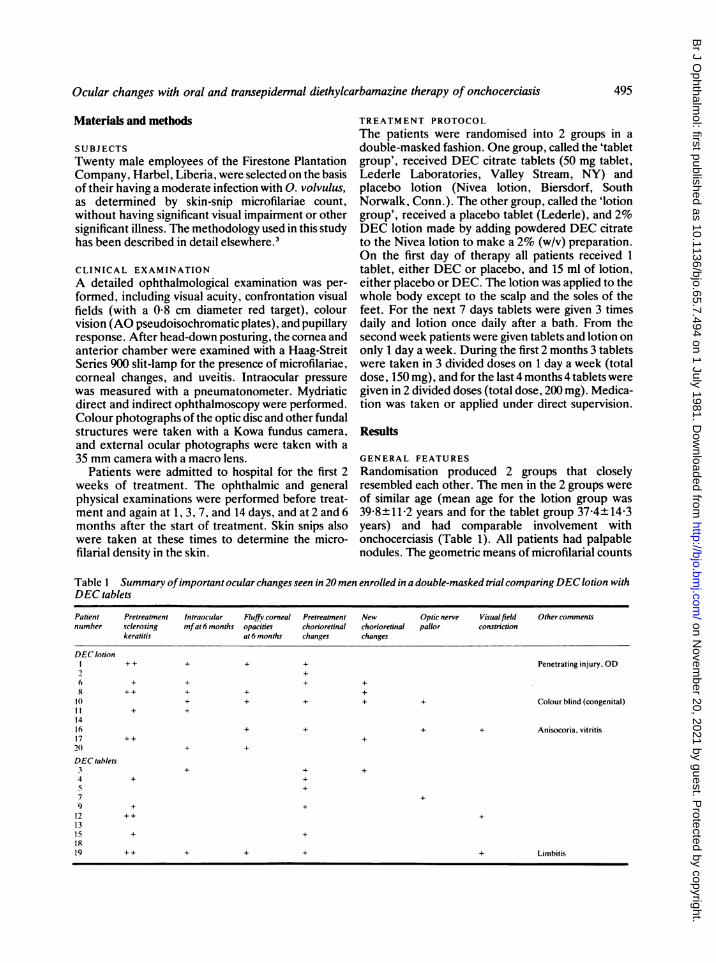
Table I Summary ofimportant ocular changes seen in 20 men enrolled in a double-masked trial comparing DEC lotion withDEC tablets
Patient Pretreatment Intraocular Fluffy corneal Pretreatment New Optic nerve Visualfield Other commentsnumber sclerosing mfat 6 months opacities chorioretinal chorioretinal pallor constriction
keratitis at 6 months changes changes
DEC lotionI + + + + + Penetrating injury, OD2 +
6 + + + +X +4+ + + +10 + + + + + Colour blind (congenital)11 + +
1416 + + + + Anisocoria. vitritis17 ++ +20 + +
DEC tablets3 + + +4 + +5 +7 +9 + +12 ++ +
1315 + +1819 + + + + + + Limbitis
495
on Novem
ber 20, 2021 by guest. Protected by copyright.
http://bjo.bmj.com
/B
r J Ophthalm
ol: first published as 10.1136/bjo.65.7.494 on 1 July 1981. Dow
nloaded from
Hugh R. Taylor and Bruce M. Greene
30O
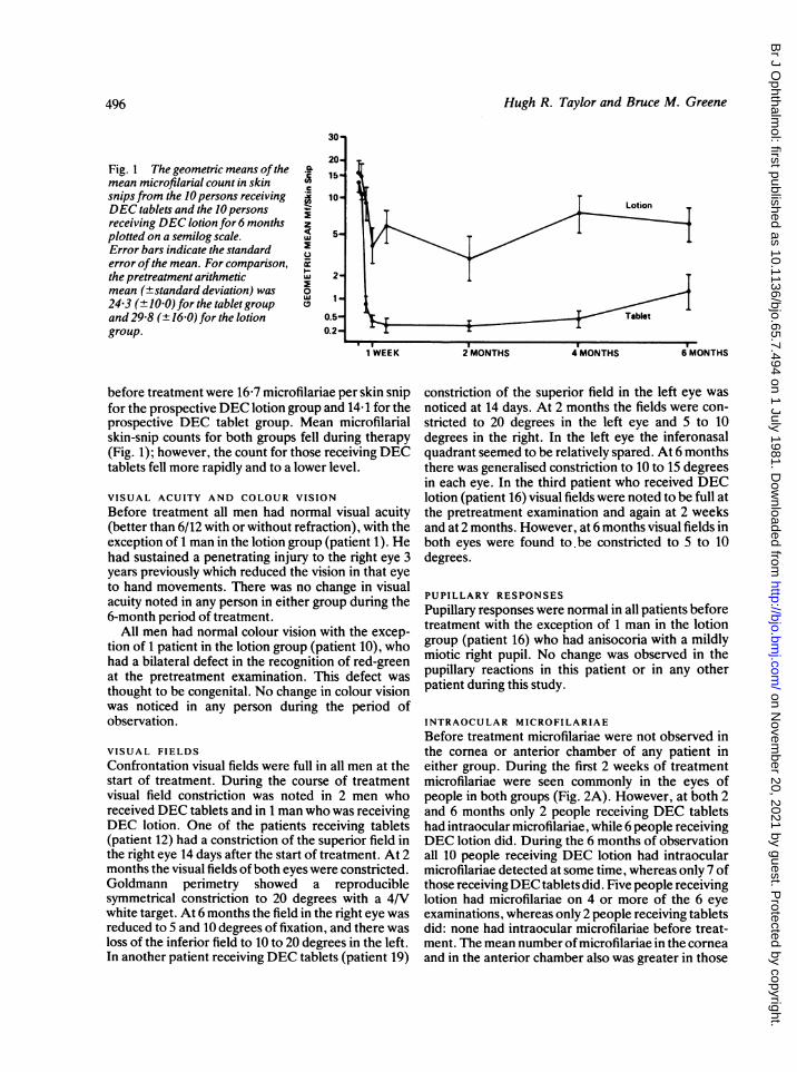
Fig. I The geometric means ofthemean microfilarial count in skinsnipsfrom the 10 persons receivingDEC tablets and the 10 personsreceiving DEC lotion for 6 monthsplotted on a semilog scale.Error bars indicate the standarderror of the mean. For comparison,the pretreatment arithmeticmean (±standard deviation) was24-3 (+10 0) for the tablet groupand 29-8 (± 16-0) for the lotiongroup.
CL
z4Luw
0
wuC
20.15-
10-
2-
1-.
0.5-0.2
1 WEEK 2 MONTHS 4 MONTHS
before treatment were 16-7 microfilariae per skin snipfor the prospective DEC lotion group and 14- 1 for theprospective DEC tablet group. Mean microfilarialskin-snip counts for both groups fell during therapy(Fig. 1); however, the count for those receiving DECtablets fell more rapidly and to a lower level.
VISUAL ACUITY AND COLOUR VISIONBefore treatment all men had normal visual acuity(better than 6/12 with or without refraction), with theexception of 1 man in the lotion group (patient 1). Hehad sustained a penetrating injury to the right eye 3years previously which reduced the vision in that eyeto hand movements. There was no change in visualacuity noted in any person in either group during the6-month period of treatment.
All men had normal colour vision with the excep-tion of 1 patient in the lotion group (patient 10), whohad a bilateral defect in the recognition of red-greenat the pretreatment examination. This defect wasthought to be congenital. No change in colour visionwas noticed in any person during the period ofobservation.
VISUAL FIELDS
Confrontation visual fields were full in all men at thestart of treatment. During the course of treatmentvisual field constriction was noted in 2 men whoreceived DEC tablets and in 1 man who was receivingDEC lotion. One of the patients receiving tablets(patient 12) had a constriction of the superior field inthe right eye 14 days after the start of treatment. At 2months the visual fields of both eyes were constricted.Goldmann perimetry showed a reproduciblesymmetrical constriction to 20 degrees with a 4/Vwhite target. At 6 months the field in the right eye wasreduced to 5 and 10 degrees of fixation, and there wasloss of the inferior field to 10 to 20 degrees in the left.In another patient receiving DEC tablets (patient 19)
constriction of the superior field in the left eye wasnoticed at 14 days. At 2 months the fields were con-stricted to 20 degrees in the left eye and 5 to 10degrees in the right. In the left eye the inferonasalquadrant seemed to be relatively spared. At 6 monthsthere was generalised constriction to 10 to 15 degreesin each eye. In the third patient who received DEClotion (patient 16) visual fields were noted to be full atthe pretreatment examination and again at 2 weeksand at 2 months. However, at 6 months visual fields inboth eyes were found to. be constricted to 5 to 10degrees.
PUPILLARY RESPONSES
Pupillary responses were normal in all patients beforetreatment with the exception of 1 man in the lotiongroup (patient 16) who had anisocoria with a mildlymiotic right pupil. No change was observed in thepupillary reactions in this patient or in any otherpatient during this study.
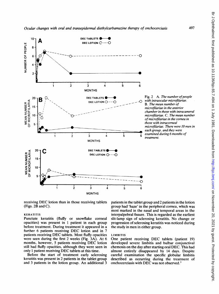
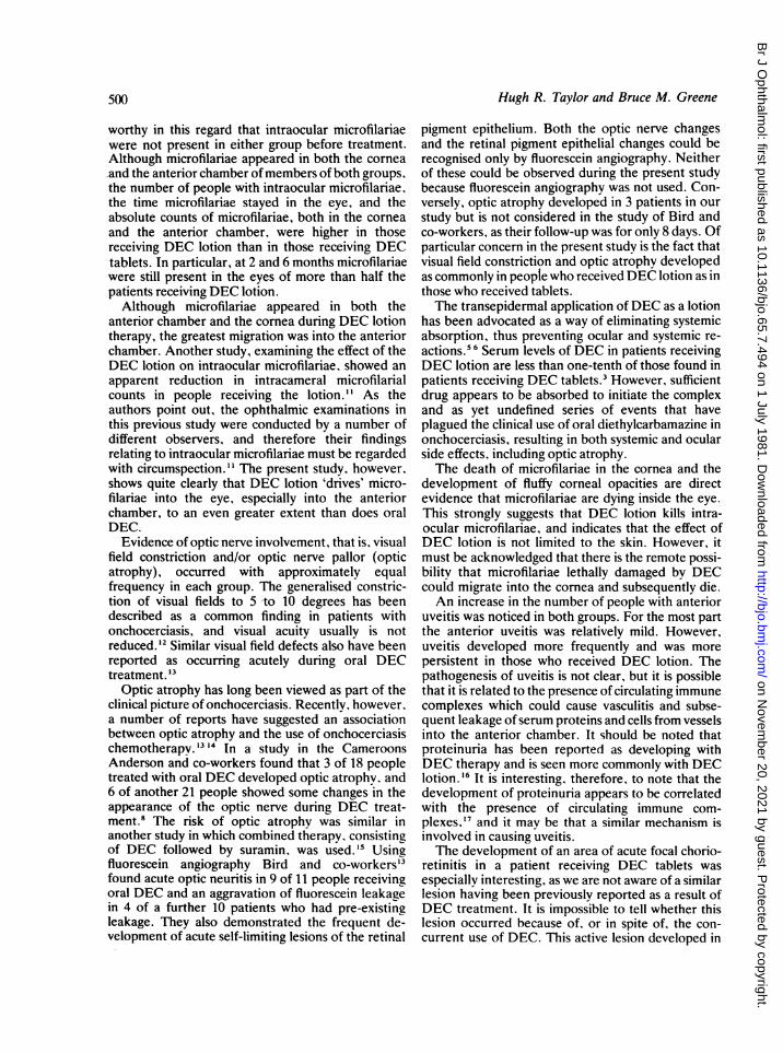
INTRAOCULAR MICROFILARIAEBefore treatment microfilariae were not observed inthe cornea or anterior chamber of any patient ineither group. During the first 2 weeks of treatmentmicrofilariae were seen commonly in the eyes ofpeople in both groups (Fig. 2A). However, at both 2and 6 months only 2 people receiving DEC tabletshad intraocular microfilariae, while 6 people receivingDEC lotion did. During the 6 months of observationall 10 people receiving DEC lotion had intraocularmicrofilariae detected at some time, whereas only 7 ofthose receivingDEC tablets did. Five people receivinglotion had microfilariae on 4 or more of the 6 eyeexaminations, whereas only 2 people receiving tabletsdid: none had intraocular microfilariae before treat-ment. The mean number of microfilariae in the corneaand in the anterior chamber also was greater in those
6 MONTHS
496
on Novem
ber 20, 2021 by guest. Protected by copyright.
http://bjo.bmj.com
/B
r J Ophthalm
ol: first published as 10.1136/bjo.65.7.494 on 1 July 1981. Dow
nloaded from
Ocular changes with oral and transepidermal diethylcarbamazine therapy of onchocerciasis
A DEC TABLETS -
DEC LOTION 0 - - C
i----_or\ ---------
Ii/
I~~
2 3
MONTHS
DEC TABLETS * * Fig. 2 A. The number ofpeopleDEC LOTION 0- - -C _0 with intraocular microfilariae.
" N _- ~~ B. The mean number ofNXNN - - ~microfilariae in the anterior
chamber in those with intracameral0-- - microfilariae. C. The mean number
ofmicrofilariae in the cornea inthose with intracornealmicrofilariae. There were 10 men in
_ each group, and they were2 3 4 5 6 examined during 6 months of
treatment.MONTHS
CDEC TABLETS
*
DEC LOTION ---C
A_~~~~AS ~ ~~~~~~~~~~~~----- -
I
1 2 3 4 5 6
MONTHS
receiving DEC lotion than in those receiving tablets(Figs. 2B and C).
KERATITIS
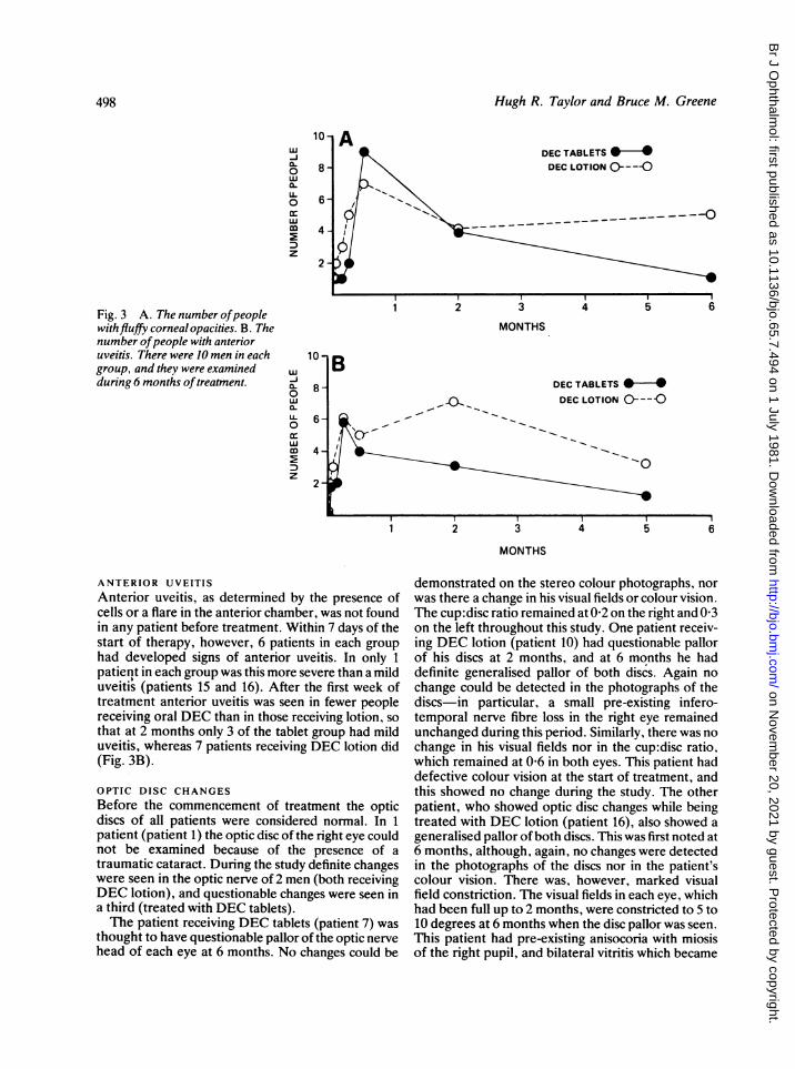
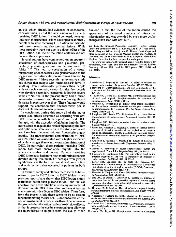
Punctate keratitis (fluffy or snowflake comealopacities) was present in 1 patient in each groupbefore treatment. During treatment it appeared in afurther 6 patients receiving DEC lotion and in 7patients receiving DEC tablets. Most fluffy opacitieswere seen during the first 2 weeks (Fig. 3A). At 6months, however, 5 patients receiving DEC lotionstill had fluffy opacities, although they were seen inonly 1 patient receiving DEC tablets at this time.
Before the start of treatment early sclerosingkeratitis was present in 2 patients in the tablet groupand 3 patients in the lotion group. An additional 3
patients in the tablet group and 2 patients in the lotiongroup had 'haze' in the peripheral cornea, which wasmost marked in the nasal and temporal areas in theinterpalpebral fissure. This is regarded as the earliestslit-lamp sign of sclerosing keratitis. No change orprogression of sclerosing keratitis was noticed duringthe study in men in either group.
LIMBITIS
One patient receiving DEC tablets (patient 19)developed severe limbitis and bulbar conjunctivalchemosis on the day after starting oral DEC. This hadalmost entirely disappeared by 14 days. Despitecareful examination the specific globular limbitisdescribed as occurring during the treatment ofonchocerciasis with DEC was not observed.7
10w-J0- 8-0wa.o 6
wm 4-
2-
4 5
B "
o9l60
6
20
mu< 1 5-1
Z °z 10-zo
0
< 20J -m> 15
z oZ C) 1 00_
O 5
D1
I
I
497
1
5 -
on Novem
ber 20, 2021 by guest. Protected by copyright.
http://bjo.bmj.com
/B
r J Ophthalm
ol: first published as 10.1136/bjo.65.7.494 on 1 July 1981. Dow
nloaded from
Hugh R. Taylor and Bruce M. Greene
-Ja-0wcLLL0cr
z
Fig. 3 A. The number ofpeoplewithfluffy corneal opacities. B. Thenumber ofpeople with anterioruveitis. There were 10 men in eachgroup, and they were examinedduring 6 months oftreatment.
MONTHS
w-Ja.0w0*LL0
w
Dz
8
6-
4-
2-
DEC TABLETS 0-*DEC LOTION O----O
--o
2 3 4 5 6
MONTHS
ANTERIOR UVEITISAnterior uveitis, as determined by the presence ofcells or a flare in the anterior chamber, was not foundin any patient before treatment. Within 7 days of thestart of therapy, however, 6 patients in each grouphad developed signs of anterior uveitis. In only 1patient in each group was this more severe than a milduveitis (patients 15 and 16). After the first week oftreatment anterior uveitis was seen in fewer peoplereceiving oral DEC than in those receiving lotion, sothat at 2 months only 3 of the tablet group had milduveitis, whereas 7 patients receiving DEC lotion did(Fig. 3B).
OPTIC DISC CHANGESBefore the commencement of treatment the opticdiscs of all patients were considered normal. In 1patient (patient 1) the optic disc of the right eye couldnot be examined because of the presence of a
traumatic cataract. During the study definite changeswere seen in the optic nerve of 2 men (both receivingDEC lotion), and questionable changes were seen ina third (treated with DEC tablets).The patient receiving DEC tablets (patient 7) was
thought to have questionable pallor of the optic nervehead of each eye at 6 months. No changes could be
demonstrated on the stereo colour photographs, norwas there a change in his visual fields or colour vision.The cup:disc ratio remained at 0-2 on the right and 0 3on the left throughout this study. One patient receiv-ing DEC lotion (patient 10) had questionable pallorof his discs at 2 months, and at 6 months he haddefinite generalised pallor of both discs. Again nochange could be detected in the photographs of thediscs-in particular, a small pre-existing infero-temporal nerve fibre loss in the right eye remainedunchanged during this period. Similarly, there was nochange in his visual fields nor in the cup:disc ratio,which remained at 0-6 in both eyes. This patient haddefective colour vision at the start of treatment, andthis showed no change during the study. The otherpatient, who showed optic disc changes while beingtreated with DEC lotion (patient 16), also showed ageneralised pallor of both discs. This was first noted at6 months, although, again, no changes were detectedin the photographs of the discs nor in the patient'scolour vision. There was, however, marked visualfield constriction. The visual fields in each eye, whichhad been full up to 2 months, were constricted to 5 to10 degrees at 6 months when the disc pallor was seen.This patient had pre-existing anisocoria with miosisof the right pupil, and bilateral vitritis which became
498
on Novem
ber 20, 2021 by guest. Protected by copyright.
http://bjo.bmj.com
/B
r J Ophthalm
ol: first published as 10.1136/bjo.65.7.494 on 1 July 1981. Dow
nloaded from
Ocular changes with oral and transepidermal diethylcarbamazine therapy of onchocerciasis
more severe during the study. The optic nerve of eacheye showed no cupping during the period of observa-tion.
In the other 2 men who showed visual field constric-tion (patients 12 and 19) optic nerve examinationremained normal throughout the study, and therewere no changes in the cup:disc ratios. Further, therewas no change in the photographic appearance of thenerve heads.
CHORIORETINITISPre-existing chorioretinal changes attributable toonchocerciasis were present in 5 men who were toreceive DEC lotion and in 6 who were to receive DECtablets. In 3 men receiving DEC tablets (patients 3, 5,and 9) and in 4 receiving DEC lotion (patients 1, 2, 6,and 10) this took the form of peripheral chorioretinalscarring, although in I patient (patient 1) this wasassociated with marked retinal pigment epithelialatrophy in the macular area. One man in the lotiongroup (patient 16) had bilateral vitritis, bilateralperipheral chorioretinal scars, and epiretinal mem-branes in the left macular region. A posterior polarchorioretinal scar was present in 1 patient receivingDEC tablets (patient 4), peripapillary atrophyoccurred bilaterally in another receiving DEC tablets(patient 19), and a glial proliferation was present in athird (patient 15).
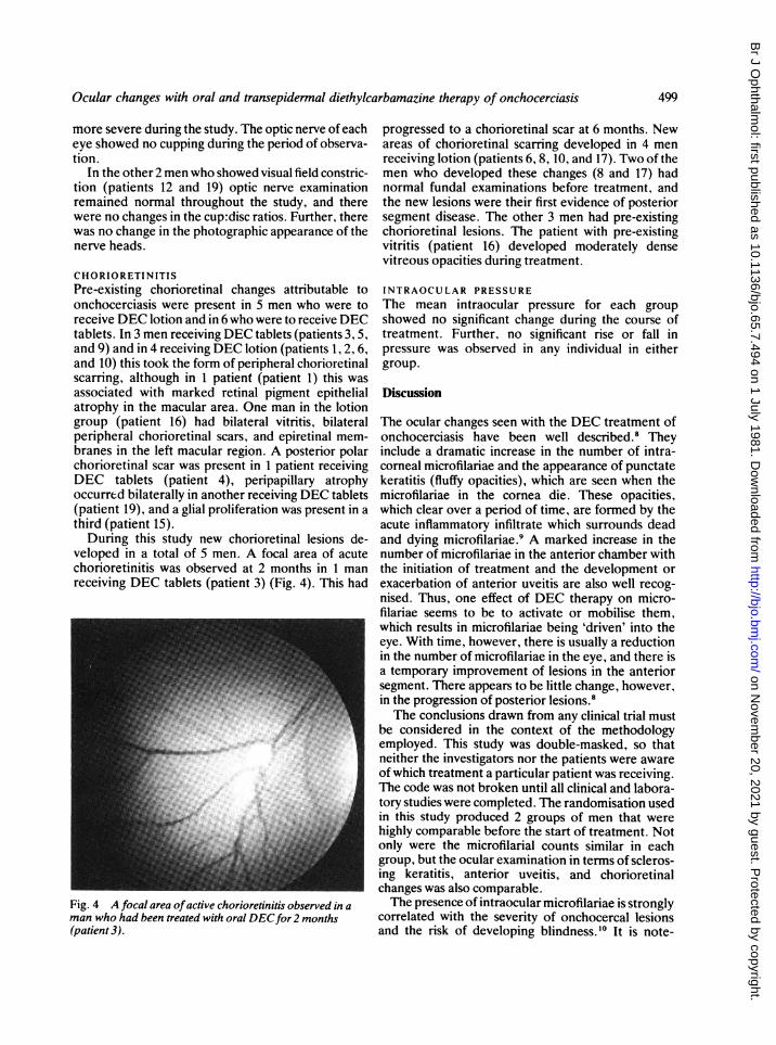
During this study new chorioretinal lesions de-veloped in a total of 5 men. A focal area of acutechorioretinitis was observed at 2 months in 1 manreceiving DEC tablets (patient 3) (Fig. 4). This had
Fig. 4 A focal area ofactive chorioretinitis observed in aman who had been treated with oral DECfor 2 months(patient 3).
progressed to a chorioretinal scar at 6 months. Newareas of chorioretinal scarring developed in 4 menreceiving lotion (patients 6, 8, 10, and 17). Two of themen who developed these changes (8 and 17) hadnormal fundal examinations before treatment, andthe new lesions were their first evidence of posteriorsegment disease. The other 3 men had pre-existingchorioretinal lesions. The patient with pre-existingvitritis (patient 16) developed moderately densevitreous opacities during treatment.
INTRAOCULAR PRESSUREThe mean intraocular pressure for each groupshowed no significant change during the course oftreatment. Further, no significant rise or fall inpressure was observed in any individual in eithergroup.
Discussion
The ocular changes seen with the DEC treatment ofonchocerciasis have been well described.8 Theyinclude a dramatic increase in the number of intra-corneal microfilariae and the appearance of punctatekeratitis (fluffy opacities), which are seen when themicrofilariae in the cornea die. These opacities,which clear over a period of time, are formed by theacute inflammatory infiltrate which surrounds deadand dying microfilariae.9 A marked increase in thenumber of microfilariae in the anterior chamber withthe initiation of treatment and the development orexacerbation of anterior uveitis are also well recog-nised. Thus, one effect of DEC therapy on micro-filariae seems to be to activate or mobilise them,which results in microfilariae being 'driven' into theeye. With time, however, there is usually a reductionin the number of microfilariae in the eye, and there isa temporary improvement of lesions in the anteriorsegment. There appears to be little change, however,in the progression of posterior lesions.8The conclusions drawn from any clinical trial must
be considered in the context of the methodologyemployed. This study was double-masked, so thatneither the investigators nor the patients were awareof which treatment a particular patient was receiving.The code was not broken until all clinical and labora-tory studies were completed. The randomisation usedin this study produced 2 groups of men that werehighly comparable before the start of treatment. Notonly were the microfilarial counts similar in eachgroup, but the ocular examination in terms of scleros-ing keratitis, anterior uveitis, and chorioretinalchanges was also comparable.The presence of intraocular microfilariae is strongly
correlated with the severity of onchocercal lesionsand the risk of developing blindness.'0 It is note-
499
on Novem
ber 20, 2021 by guest. Protected by copyright.
http://bjo.bmj.com
/B
r J Ophthalm
ol: first published as 10.1136/bjo.65.7.494 on 1 July 1981. Dow
nloaded from
Hugh R. Taylor and Bruce M. Greene
worthy in this regard that intraocular microfilariaewere not present in either group before treatment.Although microfilariae appeared in both the corneaand the anterior chamber of members of both groups.the number of people with intraocular microfilariae,the time microfilariae stayed in the eye, and theabsolute counts of microfilariae, both in the corneaand the anterior chamber, were higher in thosereceiving DEC lotion than in those receiving DECtablets. In particular, at 2 and 6 months microfilariaewere still present in the eyes of more than half thepatients receiving DEC lotion.Although microfilariae appeared in both the
anterior chamber and the cornea during DEC lotiontherapy, the greatest migration was into the anteriorchamber. Another study, examining the effect of theDEC lotion on intraocular microfilariae, showed anapparent reduction in intracameral microfilarialcounts in people receiving the lotion." As theauthors point out, the ophthalmic examinations inthis previous study were conducted by a number ofdifferent observers, and therefore their findingsrelating to intraocular microfilariae must be regardedwith circumspection." The present study, however.shows quite clearly that DEC lotion 'drives' micro-filariae into the eye, especially into the anteriorchamber, to an even greater extent than does oralDEC.
Evidence of optic nerve involvement, that is, visualfield constriction and/or optic nerve pallor (opticatrophy), occurred with approximately equalfrequency in each group. The generalised constric-tion of visual fields to 5 -to 10 degrees has beendescribed as a common finding in patients withonchocerciasis, and visual acuity usually is notreduced.'2 Similar visual field defects also have beenreported as occurring acutely during oral DECtreatment. 13
Optic atrophy has long been viewed as part of theclinical picture of onchocerciasis. Recently, however.a number of reports have suggested an associationbetween optic atrophy and the use of onchocerciasischemotherapy. 13 1' In a study in the CameroonsAnderson and co-workers found that 3 of 18 peopletreated with oral DEC developed optic atrophv. and6 of another 21 people showed some changes in theappearance of the optic nerve during DEC treat-ment.8 The risk of optic atrophy was similar inanother study in which combined therapy, consistingof DEC followed by suramin, was used. ' Usingfluorescein angiography Bird and co-workers'3found acute optic neuritis in 9 of II people receivingoral DEC and an aggravation of fluorescein leakagein 4 of a further 10 patients who had pre-existingleakage. They also demonstrated the frequent de-velopment of acute self-limiting lesions of the retinal
pigment epithelium. Both the optic nerve changesand the retinal pigment epithelial changes could berecognised only by fluorescein angiography. Neitherof these could be observed during the present studybecause fluorescein angiography was not used. Con-versely, optic atrophy developed in 3 patients in ourstudy but is not considered in the study of Bird andco-workers, as their follow-up was for only 8 days. Ofparticular concern in the present study is the fact thatvisual field constriction and optic atrophy developedas commonly in people who received DEC lotion as inthose who received tablets.The transepidermal application of DEC as a lotion
has been advocated as a way of eliminating systemicabsorption, thus preventing ocular and systemic re-actions.56 Serum levels of DEC in patients receivingDEC lotion are less than one-tenth of those found inpatients receiving DEC tablets.3 However, sufficientdrug appears to be absorbed to initiate the complexand as yet undefined series of events that haveplagued the clinical use of oral diethylcarbamazine inonchocerciasis, resulting in both systemic and ocularside effects, including optic atrophy.The death of microfilariae in the cornea and the
development of fluffy corneal opacities are directevidence that microfilariae are dying inside the eye.This strongly suggests that DEC lotion kills intra-ocular microfilariae, and indicates that the effect ofDEC lotion is not limited to the skin. However, itmust be acknowledged that there is the remote possi-bilitv that microfilariae lethally damaged by DECcould migrate into the cornea and subsequently die.An increase in the number of people with anterior
uveitis was noticed in both groups. For the most partthe anterior uveitis was relatively mild. However,uveitis developed more frequently and was morepersistent in those who received DEC lotion. Thepathogenesis of uveitis is not clear, but it is possiblethat it is related to the presence of circulating immunecomplexes which could cause vasculitis and subse-quent leakage ofserum proteins and cells from vesselsinto the anterior chamber. It should be noted thatproteinuria has been reported as developing withDEC therapy and is seen more commonly with DEClotion. 6 It is interesting, therefore, to note that thedevelopment of proteinuria appears to be correlatedwith the presence of circulating immune com-plexes.'" and it may be that a similar mechanism isinvolved in causing uveitis.The development of an area of acute focal chorio-
retinitis in a patient receiving DEC tablets wasespecially interesting, as we are not aware of a similarlesion having been previously reported as a result ofDEC treatment. It is impossible to tell whether thislesion occurred because of, or in spite of, the con-current use of DEC. This active lesion developed in
500
on Novem
ber 20, 2021 by guest. Protected by copyright.
http://bjo.bmj.com
/B
r J Ophthalm
ol: first published as 10.1136/bjo.65.7.494 on 1 July 1981. Dow
nloaded from
Ocular changes with oral and transepidermal diethylcarbamazine therapy of onchocerciasis
an eye which already had evidence of onchocercalchorioretinitis, as did the new lesions in 2 patientsreceiving DEC lotion. It should be noted, however,that new chorioretinal lesions developed in another 2people who were receiving DEC lotion and who didnot have pre-existing chorioretinal lesions. Whilethese probably were not due to a direct effect of theDEC lotion, the use of the lotion certainly did notprevent their appearance.
Several authors have commented on an apparentassociation of onchocerciasis and glaucoma, par-ticularly juvenile glaucoma, in certain areas ofAfrica.'8 19 This led to speculation of a causalrelationship of onchocerciasis to glaucoma and to thesuggestion that intraocular pressure was lowered byDEC treatment.5 More recently, an extensive studyhas shown that people with onchocerciasis have, ifanything, lower intraocular pressures than peoplewithout onchocerciasis, except for the few peoplewho develop secondary glaucoma following severeuveitis.20 No one in the present study had a raisedintraocular pressure, and there was no significantdecrease in pressure over time. These findings wouldsupport the contention that onchocerciasis per sedoes not elevate intraocular pressure.
In conclusion, during this study all of the majorocular side effects described as occurring with oralDEC were seen with both topical and oral DECtherapy, with the exception of globular limbitis. Themore subtle changes in the retinal pigment epitheliumand optic nerve were not seen in this study and couldnot have been detected without fluorescein angio-graphy. The transepidermal administration of DECas a 2% lotion was associated with a higher incidenceof ocular changes than was the oral administration ofDEC. In particular, those patients receiving DEClotion had more microfilariae migrate into theanterior chamber and cornea. Patients receivingDEC lotion also had more new chorioretinal changesdevelop during treatment. Of perhaps even greatersignificance was the fact that visual field constrictionand optic nerve pallor occurred in patients in bothgroups.
In terms of safety and efficacy there seems to be noreason to prefer DEC lotion to DEC tablets, sinceprevious reports have shown that DEC lotion is onlymarginally better than placebo lotion" and far lesseffective than DEC tablets3 in reducing microfilarialskin-snip counts. DEC lotion also produces at least asmany systemic side effects asDEC tablets. Therefore,from the present study there seems to be no reasonfor contemplating the use ofDEC lotion to reduce theocular involvement in patients with onchocerciasis onthe grounds that the lotion has less 'toxic' side effects,or that it protects the eye by encouraging or allowingthe microfilariae to migrate from the eye to other
tissues.6 In fact the use of the lotion caused theappearance of increased numbers of intraocularmicrofilariae and was attended by even more ocularchanges than seen with oral DEC.
We thank the Firestone Plantations Company, Harbel, Liberia(under the direction of Mr R. C. Larson), Drs Z. D. Traub and E.Jallah, Mary and Willem Kraaij, Jennifer Dennis, Carol Vispo, andother personnel of the Firestone Medical Center and PlantationsCompany, and Drs M. Del Monte and M. E. Langham, of the JohnsHopkins University, for their co-operation and support.
This work was supported by research grants from the RockefellerFoundation, New York, and from the Firestone Tire and RubberCompany, Akron, Ohio, and by NIH grants BRS 16-80 andEY-03318.
References
I Anderson J, Fuglsang H, Marshall TF. Effects of suramin onocular onchocerciasis. Tropenmed Parasitol 1976; 27: 279-96.
2 Hawking F. Diethylcarbamazine and new compounds for thetreatment of filariasis. Adv Pharmacol Chemother 1979; 16:129-94.
3 Taylor HR, Greene BM, Langham ME. Controlled clinical trialof oral and topical diethylcarbamazine in treatment ofonchocerciasis. Lancet 1980; i: 943-6.
4 Mazzotti L. Possibilidad de utilizar como medio diagnosticoauxiliar en la oncocercosis, las reacciones alergicas consecutivas ala administracion de 'Hetrazan'. Rev Inst Salubr Enferm Trop(Mex) 1948; 9:235-7.
5 Langham ME, Traub ZD, Richardson R. A transepidermalchemotherapy of onchocerciasis. Tropenmed Parasitol 1978; 29:156-62.
6 Langham ME. Diethylcarbamazine lotion in onchocerciasis.Lancet 1980; i: 977-8.
7 Jones BR, Anderson J, Fugisang H. Effects of various concen-trations of diethylcarbamazine citrate applied as eye drops inocular onchocerciasis, and the possibilities of improved therapyfrom continuous non-pulsed delivery. Br J Ophthalmol 1978; 62:428-39.
8 Anderson J, Fuglsang H, Marshall TF. Effects of diethylcarb-amazine on ocular onchocerciasis. Tropenmed Parasitol 1976; 27:263-78.
9 Garner A. Pathology of ocular onchocerciasis: human andexperimental. Trans R Soc Trop Med Hyg 1976; 70: 374-7.
10 Thylefors B, Brinkmann UK. The microfilarial load in theanterior segment of the eye. A parameter of intensity ofonchocerciasis. Bull WHO 1977; 55: 731-7.
11 Taylor HR, Langham ME, de Stahl EM, Figueroa LN,Beltranena F. Chemotherapy of onchocerciasis: a controlledclinical trial of topical diethylcarbamazine (DEC) in Guatemala.Tropenmed Parasitol 1980; 31: 357-64.
12 Thylefors B, Tonjum AM. Visual field defects in onchocerciasis.BrJ Ophthalmol 1978; 62: 462-7.
13 Bird AC, El-Sheikh H, Anderson J, Fuglsang H. Changes invisual function and in the posterior segment of the eye duringtreatment of onchocerciasis with diethylcarbamazine citrate. BrJOphthalmol 1980; 64:191-200.
14 Thylefors B, Rolland A. The risk of optic atrophy followingsuramin treatment of ocular onchocerciasis. Bull WHO 1979; 57:479-80.
15 Anderson J, Fuglsang H. Further studies on the treatment ofocular onchocerciasis with diethylcarbamazine and suramin. BrJOphthalmol 1978: 62: 450-7.
16 Greene BM, Taylor HR, Humphrey RL. Proteinuria associatedwith diethylcarbamazine treatment of onchocerciasis. Lancet1980; i: 254-5.
17 Greene BM, Taylor HR, Humphrey RL, Lawley TJ. Circulating
501
on Novem
ber 20, 2021 by guest. Protected by copyright.
http://bjo.bmj.com
/B
r J Ophthalm
ol: first published as 10.1136/bjo.65.7.494 on 1 July 1981. Dow
nloaded from

Hugh R. Taylor and Bruce M. Greene
immune complexes in onchocerciasis: significance and influenceof diethylcarbamazine therapy. Clin Res 1980; 28: 370A.
18 Berghout E. Onchocerciasis and glaucoma in the forest area ofGhana. Trop Geogr Med 1973; 25: 233-7.
19 Langham ME, Frentzel-Beyme RR, Traub Z-D. Intraocular
pressure and onchocerciasis infection in Liberia. Ophthalmic Res1975; 7: 368-80.
20 Thvlefors B, Duppenthaler JL. Epidemiological aspects of intra-ocular pressure in an onchocerciasis endemic area. Bull WHO1979; 57: 963-9.
502
on Novem
ber 20, 2021 by guest. Protected by copyright.
http://bjo.bmj.com
/B
r J Ophthalm
ol: first published as 10.1136/bjo.65.7.494 on 1 July 1981. Dow
nloaded from
Related Documents