Ocular Motility in Health and Disease Contents: Extraocular Muscles Eye Movements Single Binocular Vision Strabismus Amblyopia Objectives: By the end of this course the undergraduate student should be able to: Detect strabismus by general inspection, the corneal light reflex test, and the cover test. Know the basic types of strabismus and be able to differentiate between them, and recognize which cases need prompt management Understand the rationale of different lines of management, in order to point out their indications and limitations Know when amblyopia should be suspected and when it must be prevented and treated.

Welcome message from author
This document is posted to help you gain knowledge. Please leave a comment to let me know what you think about it! Share it to your friends and learn new things together.
Transcript
Ocular Motility in Health and Disease
Contents:
Extraocular Muscles
Eye Movements
Single Binocular Vision
Strabismus
Amblyopia
Objectives:
By the end of this course the undergraduate student should be able
to:
Detect strabismus by general inspection, the corneal light reflex test, and
the cover test.
Know the basic types of strabismus and be able to differentiate between
them, and recognize which cases need prompt management
Understand the rationale of different lines of management, in order to
point out their indications and limitations
Know when amblyopia should be suspected and when it must be
prevented and treated.
The Ocular Motility and Squint (Strabismus)
THE OCULAR MOTILITY
EXTRAOCULAR MUSCLES
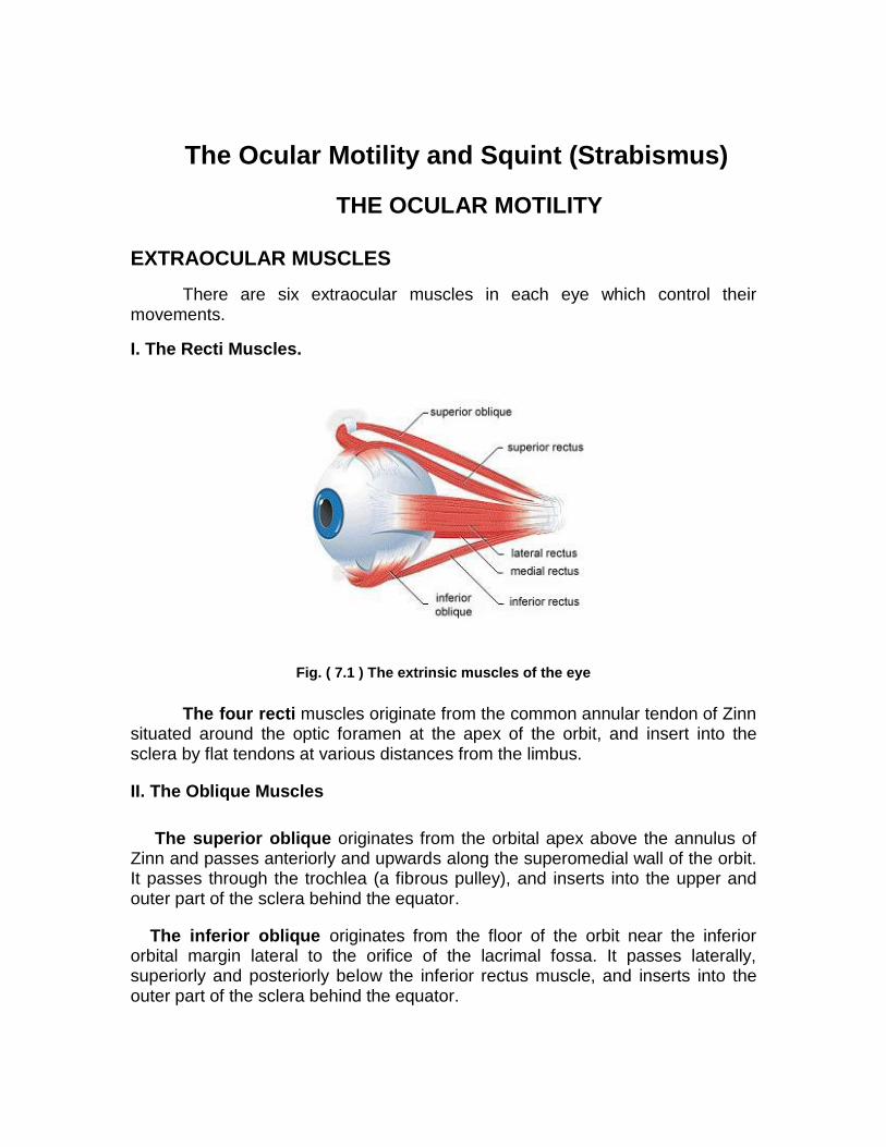
There are six extraocular muscles in each eye which control their movements.
I. The Recti Muscles.
Fig. ( 7.1 ) The extrinsic muscles of the eye
The four recti muscles originate from the common annular tendon of Zinn situated around the optic foramen at the apex of the orbit, and insert into the sclera by flat tendons at various distances from the limbus.
II. The Oblique Muscles
The superior oblique originates from the orbital apex above the annulus of Zinn and passes anteriorly and upwards along the superomedial wall of the orbit. It passes through the trochlea (a fibrous pulley), and inserts into the upper and outer part of the sclera behind the equator.
The inferior oblique originates from the floor of the orbit near the inferior orbital margin lateral to the orifice of the lacrimal fossa. It passes laterally, superiorly and posteriorly below the inferior rectus muscle, and inserts into the outer part of the sclera behind the equator.
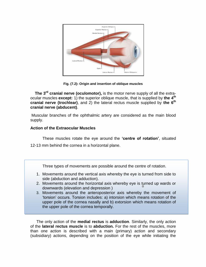
Fig. (7.2): Origin and insertion of oblique muscles
The 3rd cranial nerve (oculomotor), is the motor nerve supply of all the extra-ocular muscles except: 1) the superior oblique muscle, that is supplied by the 4th cranial nerve (trochlear), and 2) the lateral rectus muscle supplied by the 6th cranial nerve (abducent).
Muscular branches of the ophthalmic artery are considered as the main blood supply.
Action of the Extraocular Muscles
These muscles rotate the eye around the ‘centre of rotation’, situated
12-13 mm behind the cornea in a horizontal plane.
Three types of movements are possible around the centre of rotation.
1. Movements around the vertical axis whereby the eye is turned from side to side (abduction and adduction).
2. Movements around the horizontal axis whereby eye is turned up wards or downwards (elevation and depression )
3. Movements around the anteroposterior axis whereby the movement of ‘torsion’ occurs. Torsion includes: a) intorsion which means rotation of the upper pole of the cornea nasally and b) extorsion which means rotation of the upper pole of the cornea temporally.
The only action of the medial rectus is adduction. Similarly, the only action of the lateral rectus muscle is to abduction. For the rest of the muscles, more than one action is described with a main (primary) action and secondary (subsidiary) actions, depending on the position of the eye while initiating the

movement. This physiologic fact is based on the anatomical relationship of the muscle axis to the axis of the vision (visual axis). Figure(7.3)
Fig (7.3): the relation between muscle axis and visual axis
Orbital axis, muscle axis and visual axis :
The lateral and medial walls of the orbit make an angle of 45o with each other. The
orbital axis therefore forms an angle of 22.5o (23 for simplicity) with either the lateral or
medial walls. Because most extraocular muscles ( 5 out of 6) originate from the orbital
apex, the orbital axis is considered to represent the muscle axis too. When the eye is
looking straight ahead (primary position of gaze), its visual axis forms an angle of 23o
with the orbital axis (muscle axis). The actions of the extraocular muscles depend on the
position of the globe at the time of muscle contraction. The primary action of a muscle is
its major effect when the eye is in the primary position and its subsidiary actions are the
additional effects exerted on the eye in the same ( primary) position.
Therefore, the superior rectus elevates the eye (primary action), as well as adducts and intorts it (subsidiary actions), and the Inferior rectus acts as a depressor (primary action), together with adduction and extorsion (subsidiary actions).
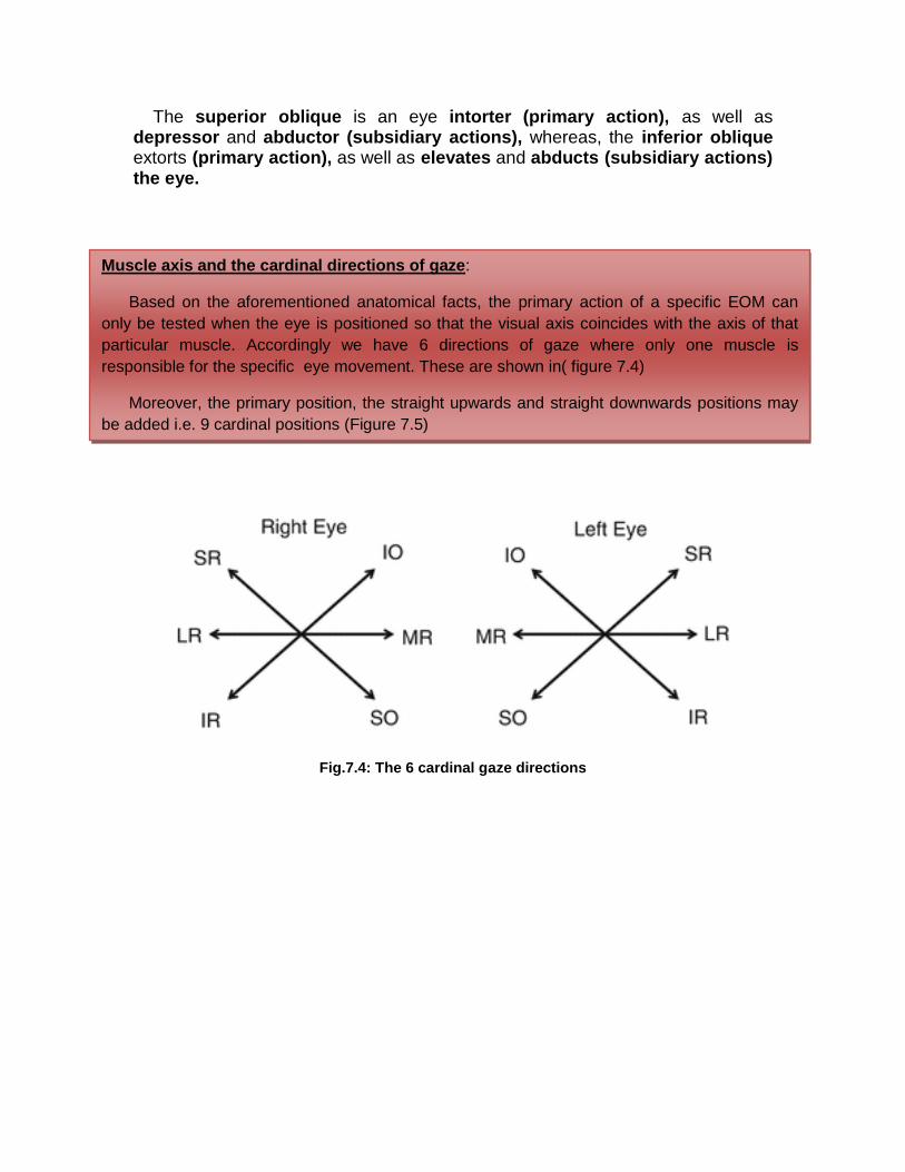
Muscle axis and the cardinal directions of gaze:
Based on the aforementioned anatomical facts, the primary action of a specific EOM can
only be tested when the eye is positioned so that the visual axis coincides with the axis of that
particular muscle. Accordingly we have 6 directions of gaze where only one muscle is
responsible for the specific eye movement. These are shown in( figure 7.4)
Moreover, the primary position, the straight upwards and straight downwards positions may
be added i.e. 9 cardinal positions (Figure 7.5)
The superior oblique is an eye intorter (primary action), as well as depressor and abductor (subsidiary actions), whereas, the inferior oblique extorts (primary action), as well as elevates and abducts (subsidiary actions) the eye.
Fig.7.4: The 6 cardinal gaze directions
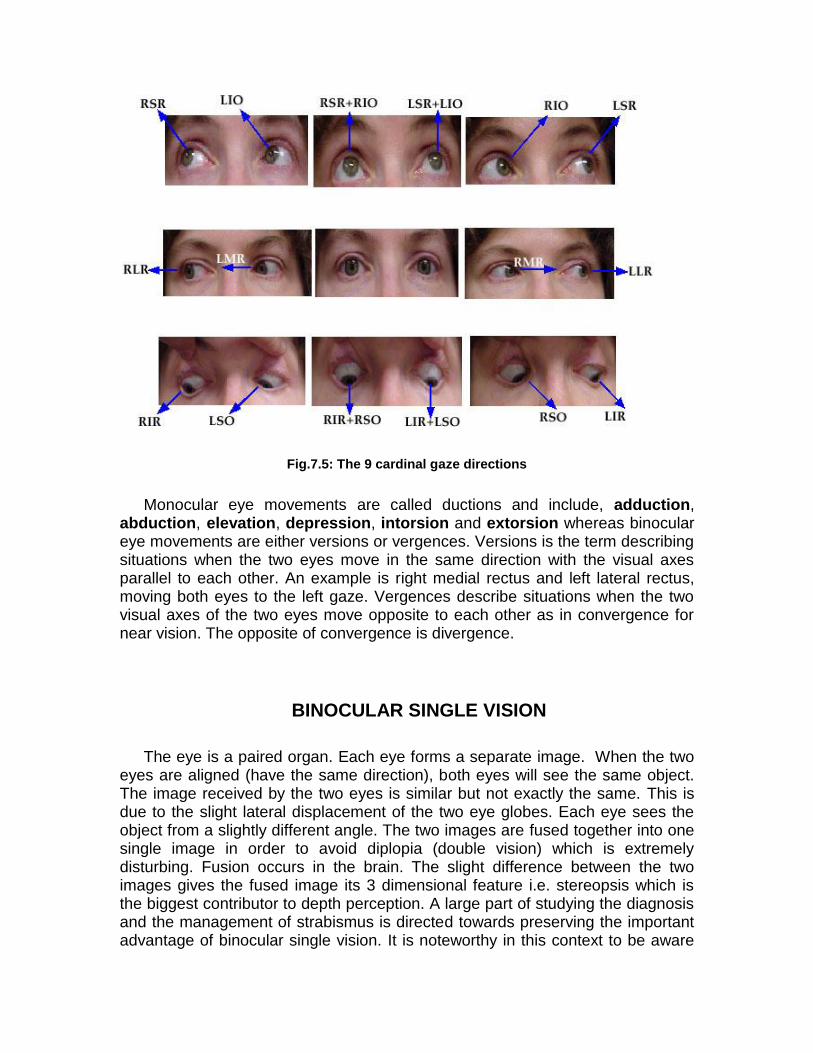
Fig.7.5: The 9 cardinal gaze directions
Monocular eye movements are called ductions and include, adduction, abduction, elevation, depression, intorsion and extorsion whereas binocular eye movements are either versions or vergences. Versions is the term describing situations when the two eyes move in the same direction with the visual axes parallel to each other. An example is right medial rectus and left lateral rectus, moving both eyes to the left gaze. Vergences describe situations when the two visual axes of the two eyes move opposite to each other as in convergence for near vision. The opposite of convergence is divergence.
BINOCULAR SINGLE VISION
The eye is a paired organ. Each eye forms a separate image. When the two eyes are aligned (have the same direction), both eyes will see the same object. The image received by the two eyes is similar but not exactly the same. This is due to the slight lateral displacement of the two eye globes. Each eye sees the object from a slightly different angle. The two images are fused together into one single image in order to avoid diplopia (double vision) which is extremely disturbing. Fusion occurs in the brain. The slight difference between the two images gives the fused image its 3 dimensional feature i.e. stereopsis which is the biggest contributor to depth perception. A large part of studying the diagnosis and the management of strabismus is directed towards preserving the important advantage of binocular single vision. It is noteworthy in this context to be aware
Note :
Having the eyes set anatomically close together is a feature of humans and higher animals
giving them the advantage of stereoscopic vision. Apparently, this is more important than
having a wide field without stereopsis as observed in other members of the animal kingdom
where the eyes are anatomically set to the sides of the head.
that, in the normal state, binocular single vision is acquired and reinforced during the first months of life.
SQUINT (Strabismus)
Under normal conditions, both eyes move synchronously and meet at the
object of regard (fixation point). Strabismus (squint) is the condition where one
eye deviates away from the fixation point (ocular misalignment).
Examination of a case of squint :
Proper history taking is very important in every case of strabismus, that help in
the diagnosis as well as for the prognosis.
History taking in a case of strabismus
1. Age of onset
If convergent squint appears before 6 months of age it is termed congenital
esotropia which is a special entity to be dealt with differently by the pediatric
ophthalmologist. This particular situation requires surgery in the first year of life.
Generally the later the onset the better the prognosis
3. Is the squint intermittent or constant?
4. Is there deviation of only one eye or alternate eye?
5. Family history of squint or refractive error is useful.
6. History of glasses or previous strabismus surgery.
2) Testing the ocular motility in the 6 cardinal directions of gaze to exclude
paralysis or paresis.
Assessment of ocular motility
Monocular movements (Duction)
Here ocular motility of each eye is assessed alone , by asking the patient to
follow a moving target with one eye closed
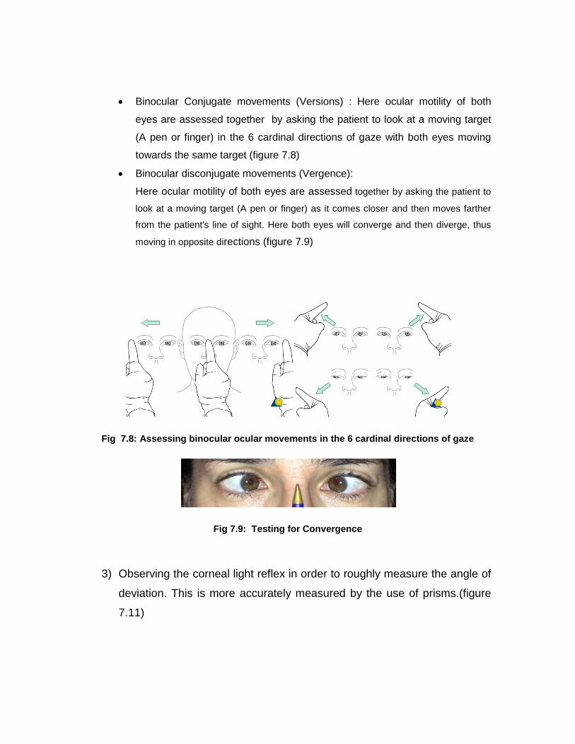
Binocular Conjugate movements (Versions) : Here ocular motility of both
eyes are assessed together by asking the patient to look at a moving target
(A pen or finger) in the 6 cardinal directions of gaze with both eyes moving
towards the same target (figure 7.8)
Binocular disconjugate movements (Vergence):
Here ocular motility of both eyes are assessed together by asking the patient to
look at a moving target (A pen or finger) as it comes closer and then moves farther
from the patient's line of sight. Here both eyes will converge and then diverge, thus
moving in opposite directions (figure 7.9)
Fig 7.8: Assessing binocular ocular movements in the 6 cardinal directions of gaze
Fig 7.9: Testing for Convergence
3) Observing the corneal light reflex in order to roughly measure the angle of
deviation. This is more accurately measured by the use of prisms.(figure
7.11)
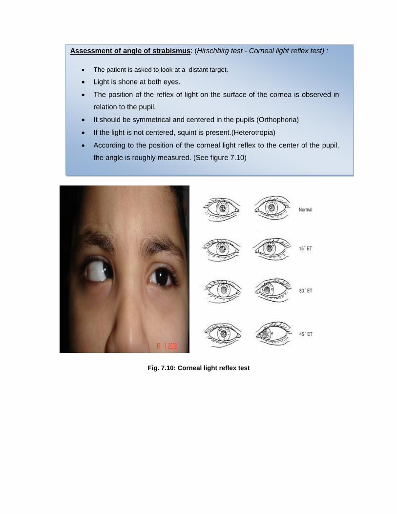
Assessment of angle of strabismus: (Hirschbirg test - Corneal light reflex test) :
The patient is asked to look at a distant target.
Light is shone at both eyes.
The position of the reflex of light on the surface of the cornea is observed in
relation to the pupil.
It should be symmetrical and centered in the pupils (Orthophoria)
If the light is not centered, squint is present.(Heterotropia)
According to the position of the corneal light reflex to the center of the pupil,
the angle is roughly measured. (See figure 7.10)
Fig. 7.10: Corneal light reflex test
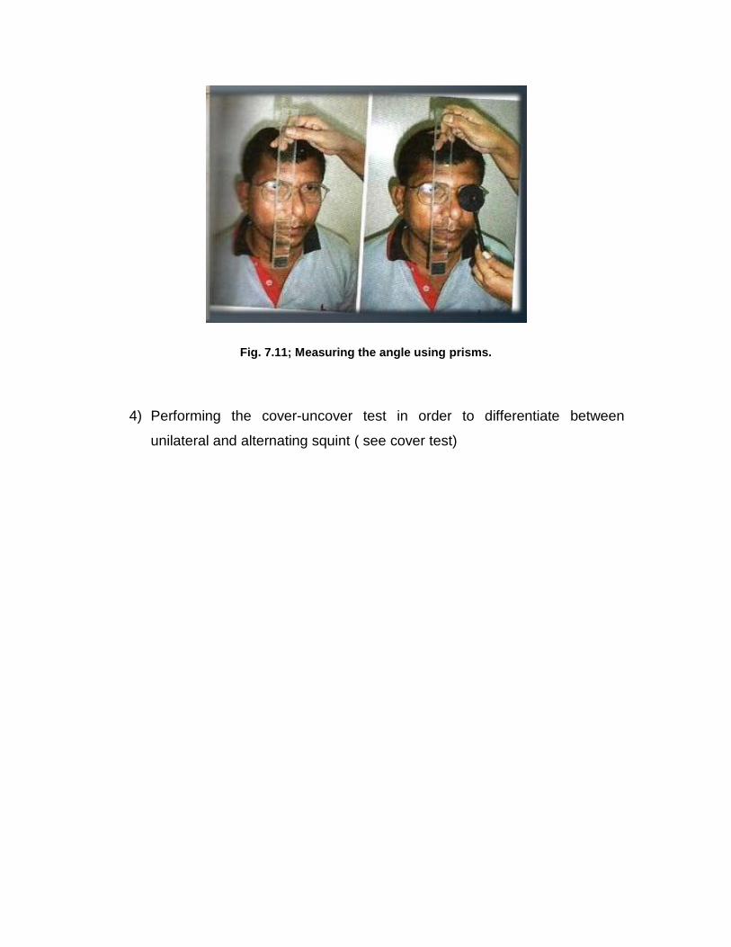
Fig. 7.11; Measuring the angle using prisms.
4) Performing the cover-uncover test in order to differentiate between
unilateral and alternating squint ( see cover test)
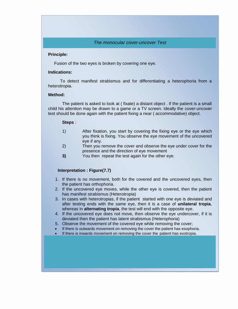
The monocular cover-uncover Test
Principle:
Fusion of the two eyes is broken by covering one eye.
Indications:
To detect manifest strabismus and for differentiating a heterophoria from a heterotropia.
Method:
The patient is asked to look at ( fixate) a distant object . If the patient is a small child his attention may be drawn to a game or a TV screen. Ideally the cover-uncover test should be done again with the patient fixing a near ( accommodative) object.
Steps :
1) After fixation, you start by covering the fixing eye or the eye which you think is fixing. You observe the eye movement of the uncovered eye if any.
2) Then you remove the cover and observe the eye under cover for the presence and the direction of eye movement
3) You then repeat the test again for the other eye.
Interpretation : Figure(7.7)
1. If there is no movement, both for the covered and the uncovered eyes, then the patient has orthophoria.
2. If the uncovered eye moves, while the other eye is covered, then the patient has manifest strabismus (Heterotropia)
3. In cases with heterotropias, if the patient started with one eye is deviated and after testing ends with the same eye, then it is a case of unilateral tropia, whereas in alternating tropia, the test will end with the opposite eye.
4. If the uncovered eye does not move, then observe the eye undercover, if it is deviated then the patient has latent strabismus (Heterophoria)
5. Observe the movement of the covered eye while removing the cover; If there is outwards movement on removing the cover the patient has esophoria.
If there is inwards movement on removing the cover the patient has exotropia.
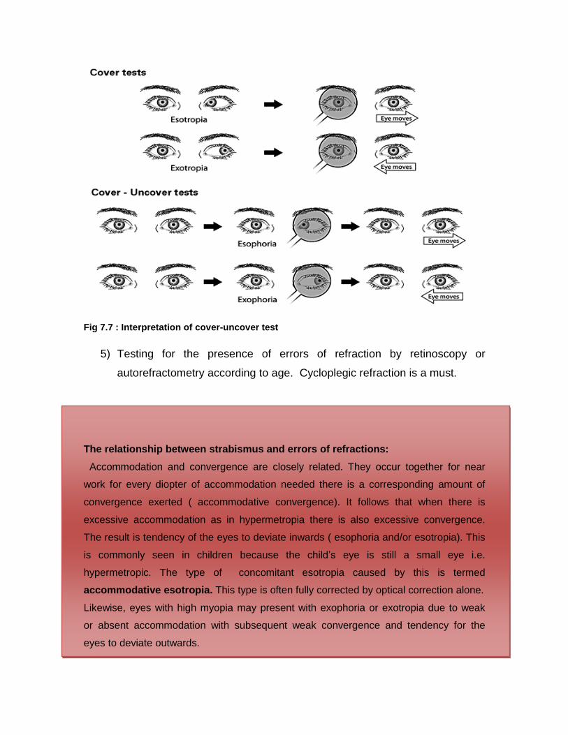
Fig 7.7 : Interpretation of cover-uncover test
5) Testing for the presence of errors of refraction by retinoscopy or
autorefractometry according to age. Cycloplegic refraction is a must.
The relationship between strabismus and errors of refractions:
Accommodation and convergence are closely related. They occur together for near
work for every diopter of accommodation needed there is a corresponding amount of
convergence exerted ( accommodative convergence). It follows that when there is
excessive accommodation as in hypermetropia there is also excessive convergence.
The result is tendency of the eyes to deviate inwards ( esophoria and/or esotropia). This
is commonly seen in children because the child’s eye is still a small eye i.e.
hypermetropic. The type of concomitant esotropia caused by this is termed
accommodative esotropia. This type is often fully corrected by optical correction alone.
Likewise, eyes with high myopia may present with exophoria or exotropia due to weak
or absent accommodation with subsequent weak convergence and tendency for the
eyes to deviate outwards.
6) Ocular examination especially fundus examination is an essential part of
examination ( see box).
Fundus examination is an essential step in every strabismus case.
Fundus lesions may be the primary cause of strabismus e.g. macular scars
Strabismus may be the first presentation of other pathologies e.g.
retinoblastoma, papilloedema.
.
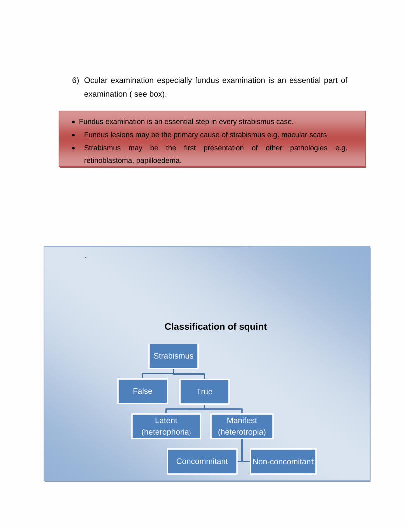
Classification of squint
Strabismus
False True
Latent
(heterophoria)
Manifest
(heterotropia)
Concommitant Non-concomitant
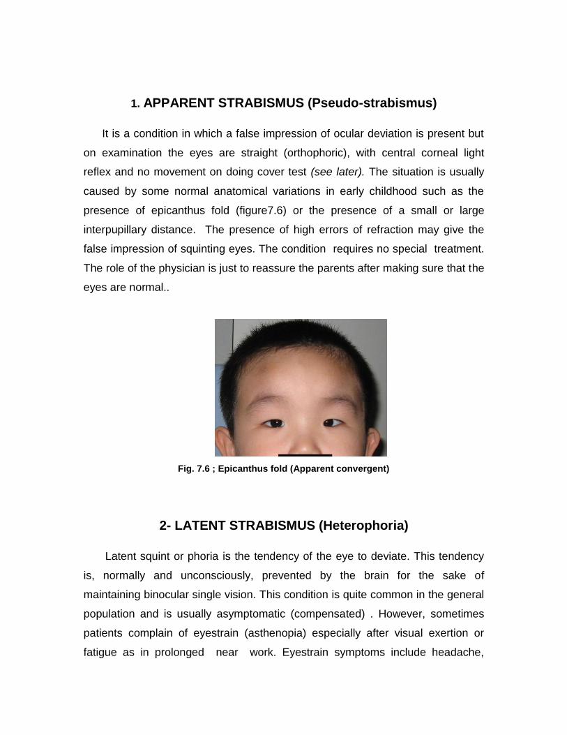
1. APPARENT STRABISMUS (Pseudo-strabismus)
It is a condition in which a false impression of ocular deviation is present but
on examination the eyes are straight (orthophoric), with central corneal light
reflex and no movement on doing cover test (see later). The situation is usually
caused by some normal anatomical variations in early childhood such as the
presence of epicanthus fold (figure7.6) or the presence of a small or large
interpupillary distance. The presence of high errors of refraction may give the
false impression of squinting eyes. The condition requires no special treatment.
The role of the physician is just to reassure the parents after making sure that the
eyes are normal..
Fig. 7.6 ; Epicanthus fold (Apparent convergent)
2- LATENT STRABISMUS (Heterophoria)
Latent squint or phoria is the tendency of the eye to deviate. This tendency
is, normally and unconsciously, prevented by the brain for the sake of
maintaining binocular single vision. This condition is quite common in the general
population and is usually asymptomatic (compensated) . However, sometimes
patients complain of eyestrain (asthenopia) especially after visual exertion or
fatigue as in prolonged near work. Eyestrain symptoms include headache,

ocular or periorbital pain, chronic eye redness, feeling of tiredness or blurring of
prints and overlapping of words or lines while reading (running of the letters). If
these symptoms persist or when the latent squint turns into manifest we call it
decompensated.
Still the role or errors of refraction to be considered in cases with decompansated
phorias;
Hypermetropia Esophoria (Excess accommodation)
Myopia Exophoria (Weak accommodation)
There are many tests to diagnose and even measure phorias. The simplest and
most commonly used test is the monocular cover/uncover test. This test is
extremely important in diagnosing other types of squint as well. (see box for the
cover-uncover test)
As a rule, asymptomatic cases of phoria require no treatment. Errors of
refraction should be corrected. Exercises to increase the fusional ability (fusional
reserve) may be advised. If exophoria deteriorates so that a manifest deviation
occurs intermittently at frequent intervals (intermittent exotropia) surgical
intervention is needed.
3- MANIFEST STRABISMUS (Heterotropia)
In manifest strabismus the deviation of the eye is evident. It is of two main
types namely concomitant and incomitant (paralytic) strabismus.
1. Concomitant strabismus
In concomitant deviations, the deviation remains the same in all the
directions of gaze. The condition may be congenital (esotropia is much more
common than exotropia), or acquired. Acquired deviation may be attributed to
refractive errors (Hypermtropia is associated with esotropia & Myopia is
associated with exotropia) or may be sensory (caused by impaired vision in one
eye).
The hallmark of concomitant squint (as compared to paralytic squint), is the
absence of limitation of ocular motility. Concomitant squint may be unilateral
(when one eye is always deviating) or alternating if the 2 eyes alternately
deviate. Unilateral deviations indicate presence of poor vision in the deviating
eye (amblyopia).
Treatment of concomitant strabismus:
1) Correction of the error of refraction (reference to the box of
relationship with errors). In many situations this is the only
treatment needed e.g. accommodative squint.
2) Treatment of amblyopia if present by * occlusion of the sound
eye (either full time or part time) or by * blurring its image by
atropine (penalization). See later
3) Surgery
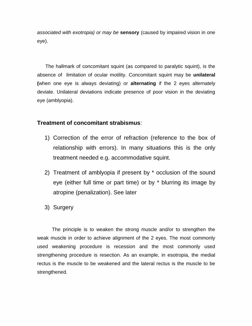
The principle is to weaken the strong muscle and/or to strengthen the
weak muscle in order to achieve alignment of the 2 eyes. The most commonly
used weakening procedure is recession and the most commonly used
strengthening procedure is resection. As an example, in esotropia, the medial
rectus is the muscle to be weakened and the lateral rectus is the muscle to be
strengthened.
It must be noted that surgery and wearing eye glasses are not mere
alternatives, Parents cannot decide to choose surgery because they find it
difficult to make their child wear his correction. (Figure 7.12)
Fig.7.12 : Surgical technique for extraocular muscle surgery
2. PARALYTIC STRABISMUS
In paralytic strabismus, deviation of the eye is caused by paralysis or paresis
of one or more Of the extraocular muscles. The deviation of the eye varies in
different directions of gaze. The patients commonly present with a manifest
ocular deviation, binocular diplopia (seeing double objects), that is most marked
in the direction of action of the paralyzed muscle, and limitation of ocular motility.

Causes of Paralytic Strabismus:
1. Lesions of the motor nerve nucleus.
a. Congenital absence of the nucleus.
b. Inflammations such as encephalitis, disseminated sclerosis.
c. Degenerative and vascular lesions.
ii. Lesions of nerve trunk
d. Trauma by direct injury or by pressure.
e. Inflammations such as diabetes mellitus neuropathy.
Iii. Lesions of the muscle.
f. Congenital absence or mal-development of the muscle.
g. Direct injury.
h. Myopathy.
On examination, there is an ocular deviation (The eye deviates to the
opposite direction of action of paralysed muscle). Limitations of movements, and
false projection (the patient is not able to grasp or point the object correctly) are
more evident in the direction of action of the paralyzed muscle.
Frequently, the patient may present with an abnormal head posture (the
patient’s head and face is turned towards the direction of the action of the
paralysed muscle) to avoid symptomatic diplopia. Diplopia often occurs in
paralytic squint and not in concomitant squint.
Compensatory Mechanisms for Diplopia
1. Suppression- It is an adaptation which occurs mainly in children. The visual
cortex in the brain neglects the blurred image it receives from the deviating eye.
(the image is blurred because it does not fall on the fovea).Suppression occurs
more easily in children because of plasticity of their brains. This is often the
precursor of amblyopia.
2. Abnormal head posture
Identifying the affected muscle(s), is an essential step in examining a case with
paralytic strabismus (examination of ocular duction & version) in order to identify
the affected cranial nerve.
OCULAR CRANIAL NERVES PALSIES
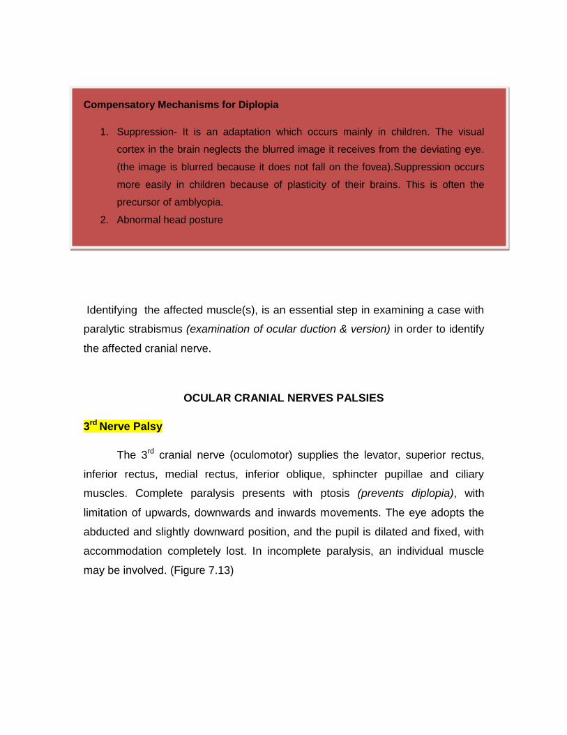
3rd Nerve Palsy
The 3rd cranial nerve (oculomotor) supplies the levator, superior rectus,
inferior rectus, medial rectus, inferior oblique, sphincter pupillae and ciliary
muscles. Complete paralysis presents with ptosis (prevents diplopia), with
limitation of upwards, downwards and inwards movements. The eye adopts the
abducted and slightly downward position, and the pupil is dilated and fixed, with
accommodation completely lost. In incomplete paralysis, an individual muscle
may be involved. (Figure 7.13)
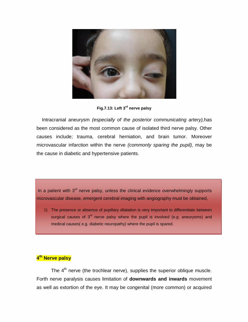
Fig.7.13: Left 3rd
nerve palsy
Intracranial aneurysm (especially of the posterior communicating artery),has
been considered as the most common cause of isolated third nerve palsy. Other
causes include; trauma, cerebral herniation, and brain tumor. Moreover
microvascular infarction within the nerve (commonly sparing the pupil), may be
the cause in diabetic and hypertensive patients.
In a patient with 3rd nerve palsy, unless the clinical evidence overwhelmingly supports
microvascular disease, emergent cerebral imaging with angiography must be obtained,
1) The presence or absence of pupillary dilatation is very important to differentiate between
surgical causes of 3rd
nerve palsy where the pupil is involved (e.g. aneurysms) and
medical causes( e.g. diabetic neuropathy) where the pupil is spared.
4th Nerve palsy
The 4th nerve (the trochlear nerve), supplies the superior oblique muscle.
Forth nerve paralysis causes limitation of downwards and inwards movement
as well as extortion of the eye. It may be congenital (more common) or acquired
Congenital cases may present in adulthood with difficulty in reading binocularly.
For acquired cases, microvascular disease (in diabetic and hypertensive
patients) is considered the most frequent cause in unilateral fourth nerve palsy,
whereas, closed head trauma, is the most frequent cause of bilateral cases.
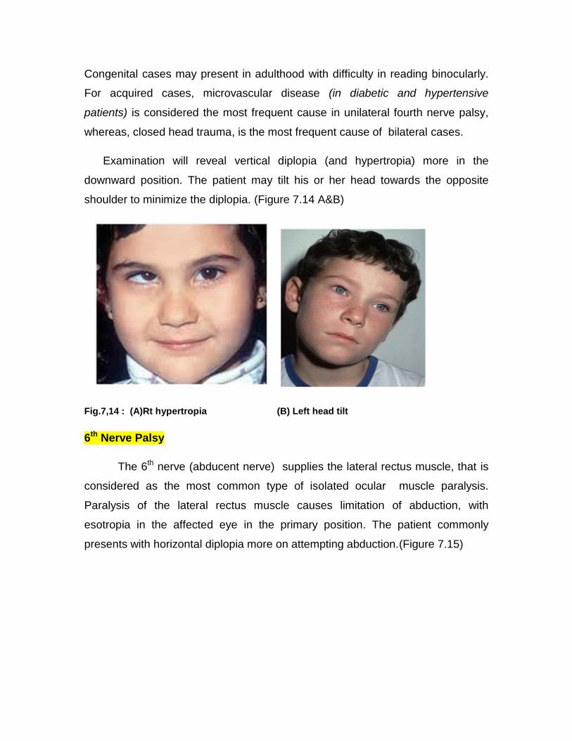
Examination will reveal vertical diplopia (and hypertropia) more in the
downward position. The patient may tilt his or her head towards the opposite
shoulder to minimize the diplopia. (Figure 7.14 A&B)
Fig.7,14 : (A)Rt hypertropia (B) Left head tilt
6th Nerve Palsy
The 6th nerve (abducent nerve) supplies the lateral rectus muscle, that is
considered as the most common type of isolated ocular muscle paralysis.
Paralysis of the lateral rectus muscle causes limitation of abduction, with
esotropia in the affected eye in the primary position. The patient commonly
presents with horizontal diplopia more on attempting abduction.(Figure 7.15)
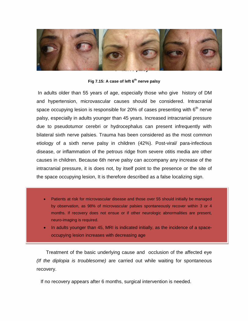
6th nerve palsy
Fig 7.15: A case of left 6th
nerve palsy
In adults older than 55 years of age, especially those who give history of DM
and hypertension, microvascular causes should be considered. Intracranial
space occupying lesion is responsible for 20% of cases presenting with 6th nerve
palsy, especially in adults younger than 45 years. Increased intracranial pressure
due to pseudotumor cerebri or hydrocephalus can present infrequently with
bilateral sixth nerve palsies. Trauma has been considered as the most common
etiology of a sixth nerve palsy in children (42%). Post-viral/ para-infectious
disease, or inflammation of the petrous ridge from severe otitis media are other
causes in children. Because 6th nerve palsy can accompany any increase of the
intracranial pressure, it is does not, by itself point to the presence or the site of
the space occupying lesion, It is therefore described as a false localizing sign.
Patients at risk for microvascular disease and those over 55 should initially be managed
by observation, as 98% of microvascular palsies spontaneously recover within 3 or 4
months. If recovery does not ensue or if other neurologic abnormalities are present,
neuro-imaging is required.
In adults younger than 45, MRI is indicated initially, as the incidence of a space-
occupying lesion increases with decreasing age
Treatment of the basic underlying cause and occlusion of the affected eye
(If the diplopia is troublesome) are carried out while waiting for spontaneous
recovery.
If no recovery appears after 6 months, surgical intervention is needed.
In patients with incomitent strabismus due to restrictive causes; the condition is mainly
due to muscle affection rather than nerve palsy e.g restrictive myopathy in thyroid eye
disease, OR due to mechanical entrapment of the muscle, e.g. orbital floor fracture with
inferior rectus muscle entrapment.
Amblyopia
Amblyopia is defined as a condition of impaired vision in the absence of
organic disease. It is usually unilateral
This may be due to constant unilateral strabismus occurring early in life
(Strabismus amblyopia), that may lead to suppression which in turn causes
amblyopia. Uncorrected anisometropia is another cause (Anisometropic
amblyopia). Amblyopia may also occur as a result of media opacities e.g.
congenital cataract or fundus lesions present at an early age (Sensory
deprivation amblyopia)
Amblyopia should be considered only when the difference in the best
corrected visual acuity between the two eyes exceeds 2 chart lines .
In cases where amblyopia is suspected, patients should be referred for proper
evaluation and management, with the main principles of management are 1)
Treatment of the cause, 2) Occlusion of the normal eye to force the patient to use
the amblyopic eye and/or 3) Penalization (blurring the normal eye by instilling
cycloplegic drops thus giving advantage for the weaker eye in near vision). The
protocol for occlusion and / or penalization to be set by a specialist according to
the age of the patient and the extent of the amblyopia.
Related Documents