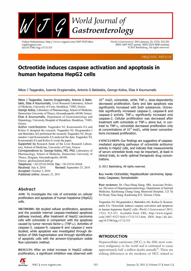
Octreotide induces caspase activation and apoptosis in human hepatoma HepG2 cells Nikos J Tsagarakis, Ioannis Drygiannakis, Antonis G Batis- takis, Elias A Kouroumalis, Liver Research Laboratory, School of Medicine, University of Crete, Heraklion, 71003, Greece George Kolios, Laboratory of Pharmacology, School of Medicine, Democritus University of Thrace, Alexandroupolis, 68100, Greece Elias A Kouroumalis, Department of Gastroenterology and Hepatology, University Hospital of Heraklion, Heraklion, 71003, Greece Author contributions: Tsagarakis NJ, Kouroumalis EA and Kolios G designed the research; Tsagarakis NJ, Drygiannakis I and Batistakis AG performed the research; Tsagarakis NJ, Drygi- annakis I and Kouroumalis EA analyzed the data; Tsagarakis NJ, Kouroumalis EA and Kolios G wrote the paper. Supported by Research funds of the Liver Research Labora- tory, School of Medicine, University of Crete, Greece Correspondence to: George Kolios, MD, PhD, Laboratory of Pharmacology, School of Medicine, Democritus University of Thrace, Dragana, Alexandroupolis, 68100, Greece. [email protected] Telephone: +30-25510-30368 Fax: +30-25510-30368 Received: July 4, 2010 Revised: September 25, 2010 Accepted: October 2, 2010 Published online: January 21, 2011 Abstract AIM: To investigate the role of octreotide on cellular proliferation and apoptosis of human hepatoma (HepG2) cells. METHODS: We studied cellular proliferation, apoptosis and the possible internal caspase-mediated apoptosis pathway involved, after treatment of HepG2 carcinoma cells with octreotide in comparison with the apoptosis caused by tumor necrosis factor-α (TNF-α). Activities of caspase-3, caspase-9, caspase-8 and caspase-2 were studied, while apoptosis was investigated through de- tection of DNA fragmentation and through identification of apoptotic cells with the annexin-V/propidium iodide flow cytometric method. RESULTS: After an initial increase in HepG2 cellular proliferation, a significant inhibition was observed with 10 -8 mol/L octreotide, while TNF-α dose-dependently decreased proliferation. Early and late apoptosis was significantly increased with both substances. Octreo- tide significantly increased caspase-3, caspase-8 and caspase-2 activity. TNF-α significantly increased only caspase-2. Cellular proliferation was decreased after treatment with octreotide or TNF-α alone but, in con- trast to TNF-α, octreotide decreased proliferation only at concentrations of 10 -8 mol/L, while lower concentra- tions increased proliferation. CONCLUSION: Our findings are suggestive of caspase- mediated signaling pathways of octreotide antitumor activity in HepG2 cells, and indicate that measurements of serum octreotide levels may be important, at least in clinical trials, to verify optimal therapeutic drug concen- trations. © 2011 Baishideng. All rights reserved. Key words: Octreotide; Hepatocellular carcinoma; Apop- tosis; Caspases; Somatostatin Peer reviewer: Dr. Chao-Hung Hung, MD, Associate Profes- sor, Division of Hepatogastroenterology, Department of Internal Medicine, Kaohsiung Chang Gung Memorial Hospital, 123 Ta Pei Road, Niao Sung, Kaohsiung 833, Taiwan, China Tsagarakis NJ, Drygiannakis I, Batistakis AG, Kolios G, Kourou- malis EA. Octreotide induces caspase activation and apoptosis in human hepatoma HepG2 cells. World J Gastroenterol 2011; 17(3): 313-321 Available from: URL: http://www.wjgnet. com/1007-9327/full/v17/i3/313.htm DOI: http://dx.doi. org/10.3748/wjg.v17.i3.313 INTRODUCTION Hepatocellular carcinoma (HCC) is the fifth most com- mon malignancy in the world and is estimated to cause approximately half a million deaths annually [1] . There are striking differences in the incidence of HCC related to Nikos J Tsagarakis, Ioannis Drygiannakis, Antonis G Batistakis, George Kolios, Elias A Kouroumalis ORIGINAL ARTICLE World J Gastroenterol 2011 January 21; 17(3): 313-321 ISSN 1007-9327 (print) ISSN 2219-2840 (online) © 2011 Baishideng. All rights reserved. Online Submissions: http://www.wjgnet.com/1007-9327office [email protected] doi:10.3748/wjg.v17.i3.313 313 January 21, 2011|Volume 17|Issue 3| WJG|www.wjgnet.com

Welcome message from author
This document is posted to help you gain knowledge. Please leave a comment to let me know what you think about it! Share it to your friends and learn new things together.
Transcript
Octreotide induces caspase activation and apoptosis in human hepatoma HepG2 cells
Nikos J Tsagarakis, Ioannis Drygiannakis, Antonis G Batis-takis, Elias A Kouroumalis, Liver Research Laboratory, School of Medicine, University of Crete, Heraklion, 71003, GreeceGeorge Kolios, Laboratory of Pharmacology, School of Medicine, Democritus University of Thrace, Alexandroupolis, 68100, GreeceElias A Kouroumalis, Department of Gastroenterology and Hepatology, University Hospital of Heraklion, Heraklion, 71003, GreeceAuthor contributions: Tsagarakis NJ, Kouroumalis EA and Kolios G designed the research; Tsagarakis NJ, Drygiannakis I and Batistakis AG performed the research; Tsagarakis NJ, Drygi-annakis I and Kouroumalis EA analyzed the data; Tsagarakis NJ, Kouroumalis EA and Kolios G wrote the paper.Supported by Research funds of the Liver Research Labora-tory, School of Medicine, University of Crete, GreeceCorrespondence to: George Kolios, MD, PhD, Laboratory of Pharmacology, School of Medicine, Democritus University of Thrace, Dragana, Alexandroupolis, 68100, Greece. [email protected]: +30-25510-30368 Fax: +30-25510-30368Received: July 4, 2010 Revised: September 25, 2010Accepted: October 2, 2010Published online: January 21, 2011
AbstractAIM: To investigate the role of octreotide on cellular proliferation and apoptosis of human hepatoma (HepG2) cells.
METHODS: We studied cellular proliferation, apoptosis and the possible internal caspase-mediated apoptosis pathway involved, after treatment of HepG2 carcinoma cells with octreotide in comparison with the apoptosis caused by tumor necrosis factor-α (TNF-α). Activities of caspase-3, caspase-9, caspase-8 and caspase-2 were studied, while apoptosis was investigated through de-tection of DNA fragmentation and through identification of apoptotic cells with the annexin-V/propidium iodide flow cytometric method.
RESULTS: After an initial increase in HepG2 cellular proliferation, a significant inhibition was observed with
10-8 mol/L octreotide, while TNF-α dose-dependently decreased proliferation. Early and late apoptosis was significantly increased with both substances. Octreo-tide significantly increased caspase-3, caspase-8 and caspase-2 activity. TNF-α significantly increased only caspase-2. Cellular proliferation was decreased after treatment with octreotide or TNF-α alone but, in con-trast to TNF-α, octreotide decreased proliferation only at concentrations of 10-8 mol/L, while lower concentra-tions increased proliferation.
CONCLUSION: Our findings are suggestive of caspase-mediated signaling pathways of octreotide antitumor activity in HepG2 cells, and indicate that measurements of serum octreotide levels may be important, at least in clinical trials, to verify optimal therapeutic drug concen-trations.
© 2011 Baishideng. All rights reserved.
Key words: Octreotide; Hepatocellular carcinoma; Apop-tosis; Caspases; Somatostatin
Peer reviewer: Dr. Chao-Hung Hung, MD, Associate Profes-sor, Division of Hepatogastroenterology, Department of Internal Medicine, Kaohsiung Chang Gung Memorial Hospital, 123 Ta Pei Road, Niao Sung, Kaohsiung 833, Taiwan, China
Tsagarakis NJ, Drygiannakis I, Batistakis AG, Kolios G, Kourou-malis EA. Octreotide induces caspase activation and apoptosis in human hepatoma HepG2 cells. World J Gastroenterol 2011; 17(3): 313-321 Available from: URL: http://www.wjgnet.com/1007-9327/full/v17/i3/313.htm DOI: http://dx.doi.org/10.3748/wjg.v17.i3.313
INTRODUCTIONHepatocellular carcinoma (HCC) is the fifth most com-mon malignancy in the world and is estimated to cause approximately half a million deaths annually[1]. There are striking differences in the incidence of HCC related to
Nikos J Tsagarakis, Ioannis Drygiannakis, Antonis G Batistakis, George Kolios, Elias A Kouroumalis
ORIGINAL ARTICLE
World J Gastroenterol 2011 January 21; 17(3): 313-321 ISSN 1007-9327 (print) ISSN 2219-2840 (online)
© 2011 Baishideng. All rights reserved.
Online Submissions: http://www.wjgnet.com/[email protected]:10.3748/wjg.v17.i3.313
313 January 21, 2011|Volume 17|Issue 3|WJG|www.wjgnet.com

age, gender, race, and geographic region, with hepatitis C virus (HCV) infection acquired 2-4 decades previ-ously explaining at least half of the observed increase in HCC[2]. The survival rates remain generally dismal (medi-an 8 mo)[2]. Undoubtedly, the best available treatment for all liver tumors is complete surgical resection. However, the synthetic somatostatin analogue octreotide has been found effective in inhibiting tumor growth in a variety of experimental models[3,4]. Octreotide did nοt influence hepatic or portal blood flow, although significantly in-creased reticuloendothelial system activity[5-7]. Apart from stimulation of the reticuloendothelial system, octreotide may have other mechanisms of action, to inhibit the growth of hepatic tumors. One of the mechanisms sug-gested may be a direct antiproliferative effect, through receptor-mediated growth inhibition[8]. In vitro studies have shown that octreotide demonstrates high affinity binding tοwards somatostatin receptors sstr2, sstr3 and sstr5, while nο binding affinity is found towards recep-tors sstrl and sstr4[9-11]. It has been reported that octreo-tide inhibits the proliferation and induces apoptosis of different HCC cell lines in vitro[12-18]. The mechanisms of apoptosis induction however are not well understood.
Tumor necrosis factor-α (TNF-α) is a well established as a means of apoptosis induction in a variety of cell types through specific responsive receptors, but other cells re-quire transcriptional arrest[19]. Hepatoma cells treated with TNF-α and cycloheximide (CHX) undergo apoptosis, which is preceded by a strong activation of c-jun N-termi-nal kinase[20]. The human HCC cell line, SMMC-7721, was insensitive to TNF-α cytotoxicity, but quickly underwent apoptosis in the presence of TNF-α and CHX[21].
Apoptosis is a complex process characterized by cas-pase activation, cell shrinkage, chromatin condensation and internucleosomal DNA fragmentation[22-25]. Caspases are cysteine-containing aspartic acid-specific proteases and all have similar site-specific proteolytic activity. Caspases are divided into 3 distinct groups based on their substrate specificities. Group Ⅰ (YVADase) includes caspase-1, -4 and -5 which are involved in cytokine production. Group Ⅱ (DEVDase) caspases, e.g. caspase-3 and -7 are the main effector caspases during apoptosis. These are cleaved by group Ⅲ (IETDase) caspases (e.g. caspase-6, -8, -9 or -10) early in the onset of apoptosis[26]. Upon activation, group Ⅱ caspases act on various cell proteins[26]. Most, but not all, events in apoptosis appear to require a caspase-mediat-ed proteolytic step[25].
Octreotide has been clinically used for treatment of HCC, with conflicting results. Both increased surviv-al[27,28] and no effect[29,30] have been reported. Although negative studies have been criticized[31], the mechanisms by which octreotide may act have not been adequately clarified. Apoptosis may be a fundamental mechanism. In this study, we examined the effect of octreotide on cellular proliferation, apoptosis and caspase activation in HepG2 HCC cells. The model of TNF-α-induced apoptosis was chosen for comparison with octreotide in a study of the biological behavior of caspases, after treatment of HepG2 cells with octreotide.
MATERIALS AND METHODSOctreotide was from Novartis (Basel, Switzerland) and was used at concentrations of 10-10 mol/L to 10-7 mol/L, to identify the optimal concentration for inhibition of cell proliferation. Incubations with TNF-α (R&D Systems, Minneapolis, USA) were made at concentrations from 0.1 to 100 ng/mL (0.1, 1, 10, 20, 100 ng/mL). According to the proliferation curve, the suitable concentrations for further experiments were 10-8 mol/L for octreotide and 20 ng/mL for TNF-α. These concentrations were used for all further combinations and measurements of apop-totic features.
Cell culture and incubation conditions The HepG2 cell line is a human hepatocyte carcinoma cell line derived from a well-differentiated human hepa-toblastoma and was purchased from the European Col-lection of Cell Cultures (ECACC, Porton Down, UK). HepG2 cells are maintained in continuous culture in our laboratory in RPMI supplemented with 10% fetal bo-vine serum (FBS, Gibco, Paisey, UK), at 37℃ and in an atmosphere of 5% CO2. For experiments, HepG2 cells were seeded in 24-well plates at a density of 2 × 10⁴/cm2. Twenty four hours before the experiment, they were cultured in fresh medium without FBS, and then treated with different concentrations and combinations of all substances. Incubations were made at 37℃ in 5% CO2. Supernatants were collected and stored in -80℃, while cell extracts where used for measurements of caspase activity. Control medium was complete media with 10% FBS. HepG2 cells cultured in control medium are re-ferred to as control cells.
Proliferation assaysFor measurement of growth inhibition, the sulforhoda-mine B colorimetric assay (SRB Assay; Biotium Inc., Hay-ward, CA, USA) was used, as previously described[32,33]. HepG2 cells were plated in 96-well plates, at an initial density of 5 × 10³ cells, with 200 μL medium per well. All substances were added to cultures 1 d after seeding, in or-der to obtain best attachment of the cells at the beginning of the experiments. Cells were grown for a total of 6 d, with a change of medium and substances on the third day after treatment. Measurements were made as described in the original protocol. Briefly, 50 μL of 50% trichloroace-tic acid were placed into the 200 μL medium and plates were stored at 4℃ for 30 min. After washing 5 times with deionized water, plates were left to dry for 24 h at room temperature. Then, 70 μL of 0.4% sulforhodamine B in 1% acetic acid were placed in every well and left at room temperature for 20 min. Before air drying for a second time, plates were washed 5 times with 1% acetic acid. At the end of the procedure, 200 μL of unbuffered Tris-base solution (pH 10.5) were added to each well and measure-ments were made at 490 nm, subtracting the background at 620 nm. The mean of the optical densities of 8 dif-ferent controls was considered to be 100% and all other values were expressed as a percentage of the controls.
314 January 21, 2011|Volume 17|Issue 3|WJG|www.wjgnet.com
Tsagarakis NJ et al . Apoptotic effect of octreotide on HepG2

Detection of apoptosis DNA fragmentation: For detection of apoptosis, a sand-wich, one step, colorimetric enzyme-linked immunosorbent assay, the Cell Death Detection ELISA Plus kit (Roche Diagnostics, Mannheim, Germany) was used. The assay al-lows for the specific determination of histone-complexed DNA fragments (mono and oligonucleosomes) from the cytoplasm of cells, after the induction of apoptosis. Briefly, after induction of apoptosis and 24-h incubation, the cells were pelleted by centrifugation (200 g, 10 min) and the supernatants (containing DNA from necrotic cells that leaked through the membrane during incubation) were discarded. Cells were resuspended and incubated for 30 min in lysis buffer. After lysis, intact nuclei were pel-leted by centrifugation. Aliquots of the supernatants were transferred to a streptavidin-coated well of a microtiter plate with 2 monoclonal antibodies, antihistone (biotin-labeled) and anti-DNA (peroxidase-conjugated), so that nucleosomes in the supernatant created antibody-nucleo-some complexes, which were continuously bound to the microtiter plate by the streptavidin. All samples were then incubated with peroxidase substrate and absorbance was measured at 405 nm. The mean of the optical densities of 8 different controls was considered to be 100% and all other values were expressed as a percentage of the con-trols.
Annexin-V/propidium iodide staining: For better evaluation of apoptotic features, the modified Annexin-V Apoptosis Detection Kit (BioVision, Mountain View, CA, US) was used, which is based on the observation that soon after initiation of apoptosis, cells translocate the mem-brane phosphatidylserine (PS) from the inner face of the plasma membrane to the cell surface, but they also shrink, increasing their side scatter (SS) and reducing their forward scatter (FS) characteristics. Once on the cell surface, PS can be easily detected by staining with a fluorescent con-jugate of Annexin-V, that has a high affinity for PS. Cells that have bound Annexin-V-fluorescein isothiocyanate (FITC) (early apoptotic) show green staining in the plasma membrane, while cells that have lost membrane integrity will show red staining [propidium iodide (PI)] throughout the nucleus and a halo of green staining (FITC) on the cell surface (late apoptotic or necrotic cells).
After treatment and 24-h incubation in 24-well plates, adherent HepG2 cells were gently trypsinized and washed once with serum-containing medium. Then, 1-5 × 10⁵ cells were collected by centrifugation (98 g, 5 min) and re-suspended in 300 μL of 1 × Binding Buffer. After gentle pipetting to resuspend the cell pellets, 3 μL of Annexin-V-FITC and 3 μL of 50 μg/mL PI, were added, followed by a 5-min incubation at room temperature in the dark. Annexin-V-FITC binding was analyzed by flow cytometry (Epics Elite) (Ex = 488 nm; Em = 530 nm) using a FITC signal detector and PI staining by the phycoerythrin emis-sion signal detector. Debris was excluded by scatter gating (forward vs side). At least 10 000 events were counted for each sample.
Caspase activityThe activities of caspase-3, caspase-9, caspase-8 and cas-pase-2 were measured. For the evaluation of caspase activ-ity, colorimetric activity assay kits (Chemicon, Temecula, CA, USA) were used. The assays are based on specto-photometric detection of the chromophore p-nitroaniline (pNA) after cleavage from the labeled substrate DEVD-pNA (caspase-3), LEHD-pNA (caspase-9), IETD-pNA (caspase-8) and VDVAD-pNA (caspase-2), respectively. Briefly, after treatment and 24-h incubation, supernatants were collected and cells were resuspended in 250 μL of chilled lysis buffer and incubated on ice for at least 10 min. After centrifugation (5 min, 10 000 g), supernatants (cyto-solic extracts) were transferred to a fresh tube and placed on ice. The protein concentration for each sample set was assayed with BIORAD Protein assay (BIORAD, Munchen, Germany)[34]. Samples were incubated for 2-3 h at 37℃ and measured at 405 nm, as indicated. The absorbance of pNA from every sample was compared with the uninduced controls and values were expressed as μmol/L of pNA per microgram of cytosolic protein (μmol/L per microgram).
StatisticsStatistical analysis was performed using Microsoft Excel 2007 and Instat software (GraphPad software inc., San Diego, California, USA). Results are expressed as mean ± standard error of the mean (SE). The Kolmogorov and Smirnov test was used to check the Gaussian distribution of data. Statistical comparisons were performed using one-way analysis of variance with Tukey’s post hoc com-parisons. The non parametric Kruskal-Wallis test was used instead if Bartlett’s test indicated a significant difference between standard deviations. P < 0.05 was considered sta-tistically significant.
RESULTSEffect of octreotide on HepG2 proliferationOctreotide caused an initial increase in HepG2 prolifera-tion (165.2% ± 6.2% and 127% ± 3% with octreotide 10-10 mol/L and 10-9 mol/L, respectively) followed by a significant inhibition at a concentration of 10-8 mol/L (concentration expected in the blood of patients receiv-ing treatment), reducing cellular proliferation to 77.5% ± 1.9% of control (Figure 1). No difference was observed at a concentration of 10-7 mol/L (108.3% ± 3.8%).
The effect of TNF-α was also examined, at concentra-tions from 0.1 ng/mL to 100 ng/mL. At the low concen-trations of 0.1 and 1 ng/mL, TNF-α had no significant effect on the proliferation of HepG2 cells (96.9% ± 5% and 90.6% ± 4.5%, respectively), after 6 d of incubation. A marked inhibitory effect was detected with 10, 20 and 100 ng/mL TNF-α, which reduced cellular proliferation to 69.4% ± 4%, 69.6% ± 2.3% and 61.4% ± 1.7% of control, respectively (Figure 2). However, because TNF-α at a concentration of 20 ng/mL had the optimum inhibi-tory effect in another HCC cell line (SMMC-7721 cells)[19], this concentration was selected for further experiments.
315 January 21, 2011|Volume 17|Issue 3|WJG|www.wjgnet.com
Tsagarakis NJ et al . Apoptotic effect of octreotide on HepG2

Effect of octreotide on HepG2 apoptosisApoptosis was detected based on determination of histone- complexed DNA fragments (mono and oligonucleosomes) from the cytoplasm of apoptotic cells. Similar non sig-nificant detection of DNA fragmentation was noted after 24-h treatment of HepG2 cells with either octreotide or TNF-α (115.2% ± 6.95% and 115.2% ± 8.17%, respec-tively) (Figure 3).
Necrotic cells should be visualized as double positive cells of large dimensions, as they rapidly lose membrane integrity and swelling occurs before destruction, or as fractured membranes of low FS and SS, PI only positive or double positive, but in the place where debris is usually detected and excluded. We also observed that all double positive cells had very small dimensions (data not shown), although they were still intact. We considered double posi-tive cells as cells that followed the apoptotic rather than the necrotic procedure, having increased their SS and de-creased their FS characteristics, but still retaining relatively small dimensions. That was the reason why we followed
a specific gating strategy for analysis. Octreotide caused a significant increase in early apoptosis (7.2% ± 1.4%, P < 0.01, Annexin-V positive cells) and a highly significant increase in late apoptosis (15.3% ± 2.7%, P < 0.001, Annexin-V/PI double positive cells). TNF-α significantly increased early (12.5% ± 1.4%, P < 0.001) and more so late apoptosis (26.4% ± 4%, P < 0.001, Figure 4). All comparisons were made with untreated HepG2 cells used as control cells (0.5% ± 0.3% and 2 ± 0.3% for early and late apoptosis, respectively) (Figure 4).
Effect of octreotide on caspase activity in HepG2 cellsCaspase-3 activity was significantly increased after treatment of HepG2 cells with octreotide (4.71 ± 0.81 μmol/L per microgram protein, P < 0.01) alone, while after TNF-α only, a non significant increase was found (3.28 ± 0.55 μmol/L per microgram protein), compared with uninduced cells (1.87 ± 0.24 μmol/L per microgram protein) (Figure 5). A small but not significant increase in caspase-9 activity was detected after treatment of HepG2 cells with TNF-α (2.44 ± 0.33 μmol/L per microgram protein) or octreotide alone (2.42 ± 0.77 μmol/L per microgram protein) (Figure 5), compared to uninduced cells (1.56 ± 0.21 μmol/L per microgram pro-tein). TNF-α caused a non significant increase in caspase-8 activity (0.9 ± 0.18 μmol/L per microgram protein) com-pared with control cells (0.51 ± 0.06 μmol/L per microgram protein) (Figure 5). However, octreotide caused a significant increase (1.3 ± 0.1 μmol/L per microgram protein, P < 0.01). TNF-α and octreotide caused a significant increase in cas-pase-2 activity (1.73 ± 0.17 μmol/L per microgram protein and 1.7 ± 0.18 μmol/L per microgram protein, respectively, P < 0.001), compared with control cells (0.8 ± 0.09 μmol/L per microgram protein) (Figure 5).
DISCUSSIONClinical studies of non-neuroendocrine tumors demon-strate that octreotide can inhibit the growth of a variety of tumors, either directly, through binding on the sstrs of tu-mor cells, or indirectly, through an immunomodulatory or an antiangiogenic effect[35-37]. Several reports indicate that
316 January 21, 2011|Volume 17|Issue 3|WJG|www.wjgnet.com
175
150
125
100
75
50
25
0Control 10-10 10-9 10-8 10-7
% o
f in
hibi
tion
of c
ell p
rolif
erat
ion
Octreotide (mol/L)
d
d
b
NS
Figure 1 Octreotide at a concentration of 10-8 mol/L had a statistically sig-nificant inhibitory effect on cellular proliferation of HepG2 hepatocellular carcinoma cells, compared to untreated cells. Lower concentrations caused an initial increase in proliferation. The results represent the mean of 8 different experiments ± SE (bP < 0.01, dP < 0.001). NS: Not significant.
100
75
50
25
0Control 0.1 1 10 20 100
% o
f in
hibi
tion
of c
ell p
rolif
erat
ion
TNF-α (ng/mL)
d d
d
Figure 2 Tumor necrosis factor-α at concentrations of 10, 20 and 100 ng/mL had a statistically significant inhibitory effect on cellular proliferation of HepG2 cells, compared to untreated cells. The results represent the mean of 8 different experiments ± SE (dP < 0.001). TNF-α: Tumor necrosis factor-α.
125
100
75
50
25
0Control Octreotide TNF-α
% o
f ap
opto
tic D
NA
frag
men
tatio
n
NS NS
Figure 3 Detection of DNA fragmentation revealed a non significant in-crease in DNA fragments, after 24-h treatment with octreotide or tumor necrosis factor-α (n = 8). TNF-α: Tumor necrosis factor-α; NS: Not significant.
Tsagarakis NJ et al . Apoptotic effect of octreotide on HepG2

octreotide inhibits the proliferation and induces apoptosis of HCC cells in vitro[12-18]. In this study we confirmed that octreotide inhibits HepG2 proliferation[12,15,18], but only at a concentration of 10-8 mol/L, although an initial increase at lower concentrations was observed.
In contrast to these findings, there are also reports that proliferation of HCC cells or hepatic stellate cells is not affected by octreotide[38,39]. In the study of Reynaert et al[39], shorter periods of culture compared to ours were used, while activation of sstrs was achieved with individual synthetic agonists, therefore a possible combined effect of concomitant receptor activation may have been missed. Similarly, clinical trials have demonstrated a survival ben-efit of patients with inoperable HCC treated with octreo-tide[27,28], but also negative studies have been published[29,30] and recently criticized[31]. Interestingly, in our study, lower concentrations of octreotide increase proliferation and
this is possibly an additional reason for divergent results in both clinical trials and in vitro studies of octreotide in HCC. Our findings also indicate that measurements of se-rum octreotide levels may be important, at least in clinical trials, to verify optimal therapeutic drug concentrations.
Octreotide binds mainly to sstr2, sstr3 and sstr5[40], the presence of which has been recently documented in HepG2 cells[12,41]. The antiproliferative effect of octreotide is thought to be mediated by sstr2[42] and sstr5[43]. Even when a significant amount of sstr2 binding in cellular membranes is not evident, it is possible that octreotide is internalized either along with sstr2 or alone as reported by Dournaud et al[44] and Hornick et al[45]. Recently a de-sensitization of sstr2 has been reported after short term incubation of an HCC cell line with octreotide, which is probably reversed after long term incubation[46]. We have previously reported an IC50 of 1.25 nmol/L for the
317 January 21, 2011|Volume 17|Issue 3|WJG|www.wjgnet.com
PI
Annexin-V control100 101 102 103 104
100
10
1
102
1
03 1
04
A
PI
Annexin-V octreotide100 101 102 103 104
100
10
1
102
10
3 1
04
A
PI
Annexin-V TNF100 101 102 103 104
100
10
1
102
10
3 1
04
A
F5
ss control
0
1
023
0 1023
6-5
A
F5
ss octreotide
0
10
23
0 1023
A
F5
ss TNF
0
10
23
0 1023
A
A
Figure 4 The apoptotic effect of octreotide and tumor necrosis factor-α alone is shown. A: Octreotide and tumor necrosis factor-α (TNF-α) significantly in-creased early (Annexin-V only positive, right lower quadrant) and more so late apoptotic cells [Annexin-V and propidium iodide (PI) positive, right upper quadrant]. Every sample was analyzed with the same gating strategy (Gate A) to exclude debris and non-specific binding of Annexin-V, while control refers to uninduced HepG2 cells; B: Annexin-V positive cells; C: Annexin-V/PI positive cells. B and C: Mean of 8 different experiments ± SE (bP < 0.01, dP < 0.001).
30
20
10
0Control Octreotide TNF-α
% o
f An
nexi
n-V
(+)
cells
b
d
30
20
10
0Control Octreotide TNF-α
% o
f An
nexi
n-V
+ P
I (+
) ce
lls
d
dCB
Tsagarakis NJ et al . Apoptotic effect of octreotide on HepG2

antiproliferative effect of octreotide for HepG2 cells[41] which is within the range of IC50 for sstr2 but it is lower from the IC50 reported for sstr5. In the case of sstr5, it is possible that a biological effect can be achieved without activation of the total number of receptors.
The antiproliferative effect of octreotide may be due to either cell necrosis or cell apoptosis[47]. Therefore, we inves-tigated the apoptotic effect of octreotide, particularly in as-sociation with caspase activation, comparing this effect with the well-described pathways of TNF-α-mediated apopto-sis. The machinery of apoptosis includes death receptors, adaptor proteins and proteolytic enzymes (caspases). Death receptors belong to the tumor necrosis factor receptor gene superfamily. Among these receptors, TNF receptor-1 (TNFR1) and Fas (CD95) are the most extensively char-acterized, and both are abundantly expressed in liver[20,48]. TNF-α at 20 ng/mL induced apoptosis in human hepato-ma cell line SMMC-7721 in vitro, which was exacerbated by the hypoxanthine-xanthine oxidase system and CHX, but alleviated by superoxide dismutase, suggesting that TNF-α-induced apoptosis may be due to oxidative stress[49]. The SMMC-7721 cell line was insensitive to TNF-α cytotoxicity and underwent apoptosis quickly in the presence of TNF-α and CHX[21]. In accordance with this study, the optimal concentration of TNF-α was also found to be 20 ng/mL for HepG2 cells in our study.
Our findings with flow cytometry showed that both octreotide and TNF-α induced a significant early and late apoptosis of HepG2 cells. DNA fragmentation mea-surements also demonstrated a non significant induction
of apoptosis. This possibly means that flow cytometry is a more sensitive method for quantification of apoptosis.
The mechanism by which octreotide induces apoptosis is not well understood. Changes in sstr expression because of downregulation or possible heterodimerization[27,50] of a receptor, together with changes in the expression of regula-tory proteins required for correct trafficking of specific sstr subtypes, could affect the direct antitumor effect of octreo-tide, which has been previously demonstrated in models ex-pressing sstr2[51,52]. Mediated by sstr2, octreotide upregulates tumor necrosis factor-related apoptosis-inducing ligand (TRAIL), death receptor 4 (DR4) and TNFR1, and down-regulates Bcl-2, which results in apoptosis[53]. Mediated by sstr3, octreotide upregulates p53[47] or induces Bcl-2-asso-ciated protein Bax[47,54] and acidic endonuclease, resulting in apoptosis. In addition to these mechanisms, in this study we demonstrated a direct effect of octreotide on caspase activation. The effect of octreotide on caspase-mediated apoptosis, is limited to caspase-3 activation, as the main ef-fector caspase supportive of apoptosis and it was detected in primary pheochromocytoma cells[55], in radiation-induced intestinal damage[56] and in activated lymphocytes[57]. Inter-estingly, decreased caspase-3 mRNA expression in Kupffer cells also indicates a possible additional beneficial effect of octreotide in HCC, through an antiapoptotic effect on Kupffer cells[58].
We suggest a caspase-mediated apoptotic pathway after treatment of HCC cells with octreotide, where, un-like TNF-α induced apoptosis, 3 out of 4 caspases tested were significantly increased. The activation of all caspases
318 January 21, 2011|Volume 17|Issue 3|WJG|www.wjgnet.com
Tsagarakis NJ et al . Apoptotic effect of octreotide on HepG2
A B
C D
Figure 5 Caspase-2 (A), -3 (B), -8 (C) and -9 (D) activities, after treatment of HepG2 cells with 10-8 mol/L octreotide and 20 ng/mL tumor necrosis factor-α, com-pared to untreated cells. The results represent the mean of 10 different experiments ± SE (bP < 0.01, dP < 0.001). TNF-α: Tumor necrosis factor-α; NS: Not significant.
3
2
1
0Control Octreotide TNF-α
Casp
ase-
9 ( μ
mol
/L p
er
mic
rogr
am p
rot)
NS
NS
2.0
1.5
1.0
0.5
0.0Control Octreotide TNF-α
Casp
ase-
2 ( μ
mol
/L p
er
mic
rogr
am p
rot)
d d 6
5
4
3
2
1
0Control Octreotide TNF-α
Casp
ase-
3 ( μ
mol
/L p
er
mic
rogr
am p
rot)
b
NS
1.5
1.0
0.5
0.0Control Octreotide TNF-α
Casp
ase-
8 ( μ
mol
/L p
er
mic
rogr
am p
rot)
b
NS

319 January 21, 2011|Volume 17|Issue 3|WJG|www.wjgnet.com
indicates a possible mitochondria-dependent apoptotic pathway. In contrast, findings from TNF-α-induced apop-tosis possibly indicate a different pathway.
A TNF-α-mediated pathway is reported to activate caspase-8, which promotes cleavage of various down-stream caspases, including caspases-3, -6 and -7. Caspase-8 can also cleave the Bcl-2 homologue Bid to reveal an ac-tive truncated Bid fragment inducing cell death through a mitochondrial pathway[59-62]. However in our study, caspase-8, caspase-3 and caspase-9 were all increased by TNF-α, but this was not statistically significant. Further-more, caspase-2 was significantly increased by TNF-α, a finding not reported before. In a previous study, we pre-sented a TNF-α-induced increase of caspase-2, but this increase did not reach the statistical significance[63]. In the present study, with an increased number of experiments, we found a significant increase in caspase-2 by TNF-α in HepG2 cells. This may be related to inefficient cleavage of Bid, so that all caspases can be significantly activated[59].
Caspase-2 seems to play critical and specific roles in programmed cell death[64]. It has been difficult to assign caspase-2 to the effector or initiator caspase groups. Cy-tokine-induced and stress-induced apoptosis act through conceptually similar pathways in which mitochondria are amplifiers of caspase activity rather than initiators of cas-pase activation[65]. In our study, caspase-2 appeared to be activated independent of significant (octreotide) or non-significant (TNF-α) activation of the mitochondria-medi-ated pathway. This may have been the result of intracellu-lar events (such as pH or stress) or feedback activation by effector caspases (such as caspase-3).
Thus, our findings suggest that in HepG2 cells octreo-tide probably causes apoptosis by a mitochondrial apopto-sis pathway, sequentially implicating caspase-8, -2, -9 and -3, although further experiments are required to define the exact initiator pathway. TNF-α on the other hand seems to induce caspase-2 activation, possibly mediated through oxidative stress, as suggested before[65]. The non-significant activation of the extrinsic pathway (caspase-8) or of the intrinsic pathway (caspase-9), perhaps due to in-efficient Bid cleavage, is maybe the cause of the resistance observed in previous studies and of the eliminated TNF-α-mediated apoptotic effects observed in our study.
In summary, our results support the induction of a caspase-mediated apoptotic pathway by octreotide in HCC cells, implicating both the receptor-mediated and the mitochondrial-apoptotic pathway. The correlation of specific apoptotic, caspase-mediated pathways, with the expression of sstrs in HCC cells needs more investigation to better define and clarify the intracellular mechanisms of the antiproliferative effects of octreotide.
COMMENTSBackgroundHepatocellular carcinoma (HCC) is the fifth most common malignancy in the world and is estimated to cause approximately half a million deaths annually. Undoubt-edly, the best available treatment for all liver tumors is complete surgical resec-tion. However, the synthetic somatostatin analogue octreotide has been found effective in inhibiting tumor growth in a variety of experimental models.
Research frontiersApart from stimulation of reticuloendothelial system, octreotide may have other mechanisms of action, to inhibit the growth of hepatic tumors. It has been re-ported that octreotide inhibits the proliferation and induces apoptosis of different HCC cell lines in vitro. The mechanisms of apoptosis induction however are not well understood.Innovations and breakthroughsSeveral reports indicate that octreotide inhibits the proliferation and induces apoptosis of HCC cells in vitro. In this study, the authors confirmed that octreo-tide inhibited HepG2 proliferation at a concentration of 10-8 mol/L. Interestingly, lower concentrations of octreotide increased proliferation and this is possibly an additional reason for divergent results in both clinical trials and in vitro stud-ies of octreotide in HCC. Also, their results support the induction of a caspase-mediated apoptotic pathway by octreotide in HepG2 cells, implicating both a receptor-mediated and mitochondrial-apoptotic pathway.ApplicationsThe findings of the present study indicate that measurements of serum octreo-tide levels may be important, at least in clinical trials, to verify optimal therapeu-tic drug concentrations. Also, based on the recently documented presence of sstr2, sstr3 and sstr5 in HepG2 cells, the need for further correlation of specific apoptotic, caspase-mediated pathways, with the expression of somatostatin re-ceptors in HCC cells, is highlighted, to better define and clarify the intracellular mechanisms of the antiproliferative effects of octreotide.Peer reviewThe authors evaluated the role of octreotide on cellular proliferation and apoptosis of HepG2 cells. Their results support the induction of a caspase-mediated apop-totic pathway by octreotide in HepG2 cells, implicating both a receptor-mediated and a mitochondrial-apoptotic pathway. They, also, indicated that measurements of serum octreotide levels may be important, at least in clinical trials, to verify opti-mal therapeutic drug concentrations.
REFERENCES1 El-Serag HB. Hepatocellular carcinoma: an epidemiologic
view. J Clin Gastroenterol 2002; 35: S72-S782 El-Serag HB. Epidemiology of hepatocellular carcinoma in
USA. Hepatol Res 2007; 37 Suppl 2: S88-S943 Davies N, Kynaston H, Yates J, Nott DM, Nash J, Taylor BA,
Jenkins SA. Octreotide inhibits the growth and development of three types of experimental liver metastases. Br J Surg 1995; 82: 840-843
4 Frizelle FA. Octreotide inhibits the growth and development of three types of experimental liver metastasis. Br J Surg 1995; 82: 1577
5 Valatas V, Kolios G, Manousou P, Xidakis C, Notas G, Lju-movic D, Kouroumalis EA. Secretion of inflammatory me-diators by isolated rat Kupffer cells: the effect of octreotide. Regul Pept 2004; 120: 215-225
6 Davies N, Yates J, Kynaston H, Taylor BA, Jenkins SA. Ef-fects of octreotide on liver regeneration and tumour growth in the regenerating liver. J Gastroenterol Hepatol 1997; 12: 47-53
7 Xidakis C, Ljumovic D, Manousou P, Notas G, Valatas V, Kolios G, Kouroumalis E. Production of pro- and anti-fibrot-ic agents by rat Kupffer cells; the effect of octreotide. Dig Dis Sci 2005; 50: 935-941
8 Reisine T, Bell GI. Molecular biology of somatostatin recep-tors. Endocr Rev 1995; 16: 427-442
9 Bruns C, Raulf F, Hoyer D, Schloos J, Lübbert H, Weckbeck-er G. Binding properties of somatostatin receptor subtypes. Metabolism 1996; 45: 17-20
10 Patel YC, Srikant CB. Subtype selectivity of peptide analogs for all five cloned human somatostatin receptors (hsstr 1-5). Endocrinology 1994; 135: 2814-2817
11 Raynor K, Murphy WA, Coy DH, Taylor JE, Moreau JP, Yasuda K, Bell GI, Reisine T. Cloned somatostatin receptors: identification of subtype-selective peptides and demonstra-tion of high affinity binding of linear peptides. Mol Pharmacol 1993; 43: 838-844
12 Liu HL, Huo L, Wang L. Octreotide inhibits proliferation
COMMENTS
Tsagarakis NJ et al . Apoptotic effect of octreotide on HepG2
320 January 21, 2011|Volume 17|Issue 3|WJG|www.wjgnet.com
and induces apoptosis of hepatocellular carcinoma cells. Acta Pharmacol Sin 2004; 25: 1380-1386
13 Diaconu CC, Szathmári M, Kéri G, Venetianer A. Apoptosis is induced in both drug-sensitive and multidrug-resistant hepatoma cells by somatostatin analogue TT-232. Br J Cancer 1999; 80: 1197-1203
14 Chen X, Liu Z, Ai Z. Antineoplastic mechanism of Octreo-tide action in human hepatoma. Chin Med J (Engl) 2001; 114: 1167-1170
15 Wang C, Tang C, Tang L. [Inhibition effects of octreotide on the growth of hepatocellular carcinoma in vitro and in vivo]. Zhonghua Yixue Zazhi 2001; 81: 1194-1197
16 Raderer M, Hejna MH, Muller C, Kornek GV, Kurtaran A, Virgolini I, Fiebieger W, Hamilton G, Scheithauer W. Treat-ment of hepatocellular cancer with the long acting soma-tostatin analog lanreotide in vitro and in vivo. Int J Oncol 2000; 16: 1197-1201
17 Xie Y, Tang CW, Wang CH. [Effect of HBV X gene transfec-tion on octreotide-inhibited growth of hepatocellular carci-noma cell line HepG2]. Ai Zheng 2005; 24: 965-969
18 Ma Q, Meng LQ, Liu JC, Hu JP, Ge J, Wan YL, Jiang S. [Oc-treotide induces apoptosis of human hepatoma cells by the mechanism of facilitating the Fas/FasL gene expression therein]. Zhonghua Yixue Zazhi 2008; 88: 716-718
19 Leist M, Gantner F, Bohlinger I, Germann PG, Tiegs G, Wen-del A. Murine hepatocyte apoptosis induced in vitro and in vivo by TNF-alpha requires transcriptional arrest. J Immunol 1994; 153: 1778-1788
20 Liedtke C, Plümpe J, Kubicka S, Bradham CA, Manns MP, Brenner DA, Trautwein C. Jun kinase modulates tumor ne-crosis factor-dependent apoptosis in liver cells. Hepatology 2002; 36: 315-325
21 Fang Y, Wang L, Jin J, Zha X. Focal adhesion kinase affects the sensitivity of human hepatocellular carcinoma cell line SMMC-7721 to tumor necrosis factor-alpha/cycloheximide-induced apoptosis by regulating protein kinase B levels. Eur J Biochem 2001; 268: 4513-4519
22 Kerr JF. Shrinkage necrosis: a distinct mode of cellular death. J Pathol 1971; 105: 13-20
23 Wyllie AH. Glucocorticoid-induced thymocyte apoptosis is associated with endogenous endonuclease activation. Nature 1980; 284: 555-556
24 Orrenius S, McConkey DJ, Bellomo G, Nicotera P. Role of Ca2+ in toxic cell killing. Trends Pharmacol Sci 1989; 10: 281-285
25 Samali A, Nordgren H, Zhivotovsky B, Peterson E, Orrenius S. A comparative study of apoptosis and necrosis in HepG2 cells: oxidant-induced caspase inactivation leads to necrosis. Biochem Biophys Res Commun 1999; 255: 6-11
26 Nicholson DW, Thornberry NA. Caspases: killer proteases. Trends Biochem Sci 1997; 22: 299-306
27 Kouroumalis E, Skordilis P, Thermos K, Vasilaki A, Mos-chandrea J, Manousos ON. Treatment of hepatocellular car-cinoma with octreotide: a randomised controlled study. Gut 1998; 42: 442-447
28 Samonakis DN, Moschandreas J, Arnaoutis T, Skordilis P, Leontidis C, Vafiades I, Kouroumalis E. Treatment of hepato-cellular carcinoma with long acting somatostatin analogues. Oncol Rep 2002; 9: 903-907
29 Yuen MF, Poon RT, Lai CL, Fan ST, Lo CM, Wong KW, Wong WM, Wong BC. A randomized placebo-controlled study of long-acting octreotide for the treatment of advanced hepatocellular carcinoma. Hepatology 2002; 36: 687-691
30 Becker G, Allgaier HP, Olschewski M, Zähringer A, Blum HE. Long-acting octreotide versus placebo for treatment of advanced HCC: a randomized controlled double-blind study. Hepatology 2007; 45: 9-15
31 Samonakis DN, Notas G, Christodoulakis N, Kouroumalis EA. Mechanisms of action and resistance of somatostatin analogues for the treatment of hepatocellular carcinoma: a message not well taken. Dig Dis Sci 2008; 53: 2359-2365
32 Rubinstein LV, Shoemaker RH, Paull KD, Simon RM, Tosini S, Skehan P, Scudiero DA, Monks A, Boyd MR. Comparison of in vitro anticancer-drug-screening data generated with a tetrazolium assay versus a protein assay against a diverse panel of human tumor cell lines. J Natl Cancer Inst 1990; 82: 1113-1118
33 Skehan P, Storeng R, Scudiero D, Monks A, McMahon J, Vistica D, Warren JT, Bokesch H, Kenney S, Boyd MR. New colorimetric cytotoxicity assay for anticancer-drug screening. J Natl Cancer Inst 1990; 82: 1107-1112
34 Compton SJ, Jones CG. Mechanism of dye response and in-terference in the Bradford protein assay. Anal Biochem 1985; 151: 369-374
35 Susini C, Buscail L. Rationale for the use of somatostatin analogs as antitumor agents. Ann Oncol 2006; 17: 1733-1742
36 Weckbecker G, Raulf F, Tolcsvai L, Bruns C. Potentiation of the anti-proliferative effects of anti-cancer drugs by octreo-tide in vitro and in vivo. Digestion 1996; 57 Suppl 1: 22-28
37 Kvols LK, Woltering EA. Role of somatostatin analogs in the clinical management of non-neuroendocrine solid tumors. Anticancer Drugs 2006; 17: 601-608
38 Reynaert H, Rombouts K, Jia Y, Urbain D, Chatterjee N, Uyama N, Geerts A. Somatostatin at nanomolar concentra-tion reduces collagen I and III synthesis by, but not prolifera-tion of activated rat hepatic stellate cells. Br J Pharmacol 2005; 146: 77-88
39 Reynaert H, Rombouts K, Vandermonde A, Urbain D, Ku-mar U, Bioulac-Sage P, Pinzani M, Rosenbaum J, Geerts A. Expression of somatostatin receptors in normal and cirrhotic human liver and in hepatocellular carcinoma. Gut 2004; 53: 1180-1189
40 Patel YC. Somatostatin and its receptor family. Front Neuro-endocrinol 1999; 20: 157-198
41 Notas G, Kolios G, Mastrodimou N, Kampa M, Vasilaki A, Xidakis C, Castanas E, Thermos K, Kouroumalis E. Cor-tistatin production by HepG2 human hepatocellular carci-noma cell line and distribution of somatostatin receptors. J Hepatol 2004; 40: 792-798
42 Ferjoux G, Bousquet C, Cordelier P, Benali N, Lopez F, Rochaix P, Buscail L, Susini C. Signal transduction of soma-tostatin receptors negatively controlling cell proliferation. J Physiol Paris 2000; 94: 205-210
43 Ballarè E, Persani L, Lania AG, Filopanti M, Giammona E, Corbetta S, Mantovani S, Arosio M, Beck-Peccoz P, Faglia G, Spada A. Mutation of somatostatin receptor type 5 in an ac-romegalic patient resistant to somatostatin analog treatment. J Clin Endocrinol Metab 2001; 86: 3809-3814
44 Dournaud P, Boudin H, Schonbrunn A, Tannenbaum GS, Beaudet A. Interrelationships between somatostatin sst2A receptors and somatostatin-containing axons in rat brain: ev-idence for regulation of cell surface receptors by endogenous somatostatin. J Neurosci 1998; 18: 1056-1071
45 Hornick CA, Anthony CT, Hughey S, Gebhardt BM, Espe-nan GD, Woltering EA. Progressive nuclear translocation of somatostatin analogs. J Nucl Med 2000; 41: 1256-1263
46 Hua YP, Yin XY, Peng BG, Li SQ, Lai JM, Liang HZ, Liang LJ. Mechanisms and influence of octreotide-induced regula-tion of somatostatin receptor 2 on hepatocellular carcinoma. Chemotherapy 2009; 55: 312-320
47 Sharma K, Patel YC, Srikant CB. Subtype-selective induc-tion of wild-type p53 and apoptosis, but not cell cycle arrest, by human somatostatin receptor 3. Mol Endocrinol 1996; 10: 1688-1696
48 Kaplowitz N. Cell death at the millennium. Implications for liver diseases. Clin Liver Dis 2000; 4: 1-23, v
49 Li J, Zheng R, Li J, Wang Z. Mechanisms of the induction of apoptosis in human hepatoma cells by tumour necrosis factor-alpha. Cell Biol Int 2001; 25: 1213-1219
50 Ayuk J, Stewart SE, Stewart PM, Sheppard MC. Long-term safety and efficacy of depot long-acting somatostatin analogs for the treatment of acromegaly. J Clin Endocrinol Metab 2002;
Tsagarakis NJ et al . Apoptotic effect of octreotide on HepG2
321 January 21, 2011|Volume 17|Issue 3|WJG|www.wjgnet.com
87: 4142-414651 Weckbecker G, Raulf F, Stolz B, Bruns C. Somatostatin ana-
logs for diagnosis and treatment of cancer. Pharmacol Ther 1993; 60: 245-264
52 Froidevaux S, Eberle AN. Somatostatin analogs and radio-peptides in cancer therapy. Biopolymers 2002; 66: 161-183
53 Guillermet J, Saint-Laurent N, Rochaix P, Cuvillier O, Levade T, Schally AV, Pradayrol L, Buscail L, Susini C, Bous-quet C. Somatostatin receptor subtype 2 sensitizes human pancreatic cancer cells to death ligand-induced apoptosis. Proc Natl Acad Sci USA 2003; 100: 155-160
54 Sharma K, Srikant CB. G protein coupled receptor signaled apoptosis is associated with activation of a cation insensitive acidic endonuclease and intracellular acidification. Biochem Biophys Res Commun 1998; 242: 134-140
55 Pasquali D, Rossi V, Conzo G, Pannone G, Bufo P, De Bellis A, Renzullo A, Bellastella G, Colao A, Vallone G, Bellastella A, Sinisi AA. Effects of somatostatin analog SOM230 on cell proliferation, apoptosis, and catecholamine levels in cultured pheochromocytoma cells. J Mol Endocrinol 2008; 40: 263-271
56 Abbasoğlu SD, Erbil Y, Eren T, Giriş M, Barbaros U, Yücel R, Olgaç V, Uysal M, Toker G. The effect of heme oxygenase-1 induction by octreotide on radiation enteritis. Peptides 2006; 27: 1570-1576
57 Lattuada D, Casnici C, Venuto A, Marelli O. The apoptotic effect of somatostatin analogue SMS 201-995 on human lym-
phocytes. J Neuroimmunol 2002; 133: 211-21658 Xidakis C, Kolios G, Valatas V, Notas G, Mouzas I, Kouroum-
alis E. Effect of octreotide on apoptosis-related proteins in rat Kupffer cells: a possible anti-tumour mechanism. Anticancer Res 2004; 24: 833-841
59 Wagner KW, Engels IH, Deveraux QL. Caspase-2 can func-tion upstream of bid cleavage in the TRAIL apoptosis path-way. J Biol Chem 2004; 279: 35047-35052
60 Budd RC. Death receptors couple to both cell proliferation and apoptosis. J Clin Invest 2002; 109: 437-441
61 Kim TH, Zhao Y, Barber MJ, Kuharsky DK, Yin XM. Bid-induced cytochrome c release is mediated by a pathway independent of mitochondrial permeability transition pore and Bax. J Biol Chem 2000; 275: 39474-39481
62 Li H, Zhu H, Xu CJ, Yuan J. Cleavage of BID by caspase 8 mediates the mitochondrial damage in the Fas pathway of apoptosis. Cell 1998; 94: 491-501
63 Tsagarakis NJ, Drygiannakis I, Batistakis AG, Kolios G, Kouroumalis EA. A concentration-dependent effect of urso-deoxycholate on apoptosis and caspases activities of HepG2 hepatocellular carcinoma cells. Eur J Pharmacol 2010; 640: 1-7
64 Troy CM, Shelanski ML. Caspase-2 redux. Cell Death Differ 2003; 10: 101-107
65 Lassus P, Opitz-Araya X, Lazebnik Y. Requirement for cas-pase-2 in stress-induced apoptosis before mitochondrial per-meabilization. Science 2002; 297: 1352-1354
S- Editor Sun H L- Editor Cant MR E- Editor Lin YP
Tsagarakis NJ et al . Apoptotic effect of octreotide on HepG2
Related Documents