OCTOBER 30, 2012 STANDARD: SAP2b Explain how the skeletal structures provide support and protection for tissues and function together with the muscular system to make movements possible. EQ: What are the components and function of the skeletal system? WARM-UP: Name the bone 1. 2. 3.

OCTOBER 30, 2012 STANDARD: SAP2b Explain how the skeletal structures provide support and protection for tissues and function together with the muscular.
Dec 27, 2015
Welcome message from author
This document is posted to help you gain knowledge. Please leave a comment to let me know what you think about it! Share it to your friends and learn new things together.
Transcript

OCTOBER 30, 2012STANDARD: SAP2b Explain how the skeletal structures provide support and protection for tissues and function together with the muscular system to make movements possible.EQ: What are the components and function of the skeletal system?WARM-UP: Name the bone1. 2. 3.

The Skeletal System
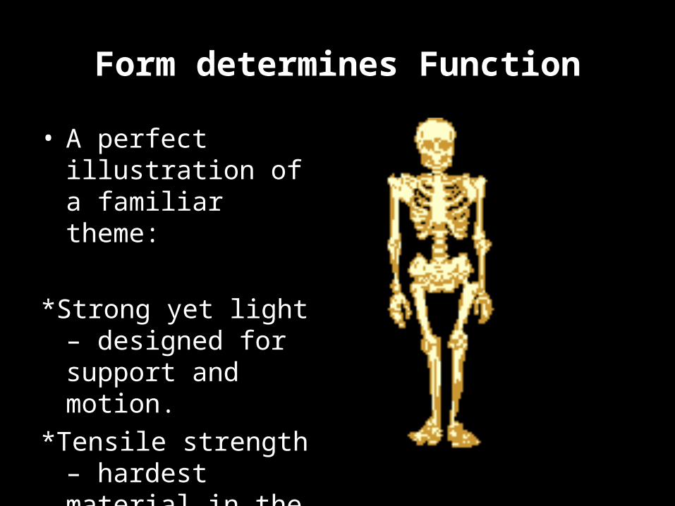
Form determines Function
• A perfect illustration of a familiar theme:
*Strong yet light – designed for support and motion.
*Tensile strength – hardest material in the body, yet can resist tension and other forces.

Components of Skeletal System
1. Bone - 2 types: compact and spongy
2. Joints – (articulation) – classified as functionally or structurally
3. Cartilage
4. Ligaments – fibrous cords that bind bones together at joints

Functions of BoneFive Functions:
1) Supports and anchors soft organs
2) Protects soft body organs
3) Moves the body and it parts
Bones are attached to muscle by tendons
4) Stores minerals and fats
Ca & P – transmits messages, contracts muscles and clots blood
5) Produces blood cells (hematopoiesis) within the marrow of certain bones

Two types of bone:
1) Compact bone – dense
2) Spongy bone – open spaces
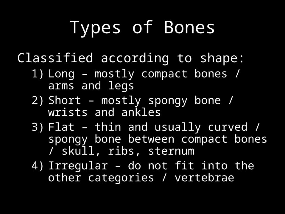
Types of Bones
Classified according to shape:1) Long – mostly compact bones / arms and
legs2) Short – mostly spongy bone / wrists and
ankles3) Flat – thin and usually curved / spongy bone
between compact bones / skull, ribs, sternum
4) Irregular – do not fit into the other categories / vertebrae

Structure of long bone:
• Diaphysis – shaft of bone, compact bone.
• Periosteum – tough, fibrous membrane covering bone.
• Epiphysis – ends of long bones, spongy bone.
• Articular cartilage – covers ends of epiphysis; thin and glassy to decrease friction in joint cavity.
Epiphyseal line – remnant of epiphyseal plate or growth area.
Medullary cavity – hollow middle of bone.Used for storage of fat in adults
(yellow marrow).Used for blood cell formation in children
(red marrow).
Projections and depressions – anchor muscles, tendons and ligaments.


Review• How many bones does the human skeleton contain?
206
• Name 2 functions of bones?
Support, Protection, Movement, Storage, Blood Cell Formation
• Name 4 components of the skeletal system?
Joints, Cartilage, Bone, Ligaments
• Name 2 classifications of bone?
Compact and Spongy
• Which division of the skeletal system includes the skull?
Axial

NOVEMBER 5, 2012
WARM-UP:
EQ: What are the microscopic components of compact bone?
What is the difference between osteocytes, osteoblasts and osteoclasts?
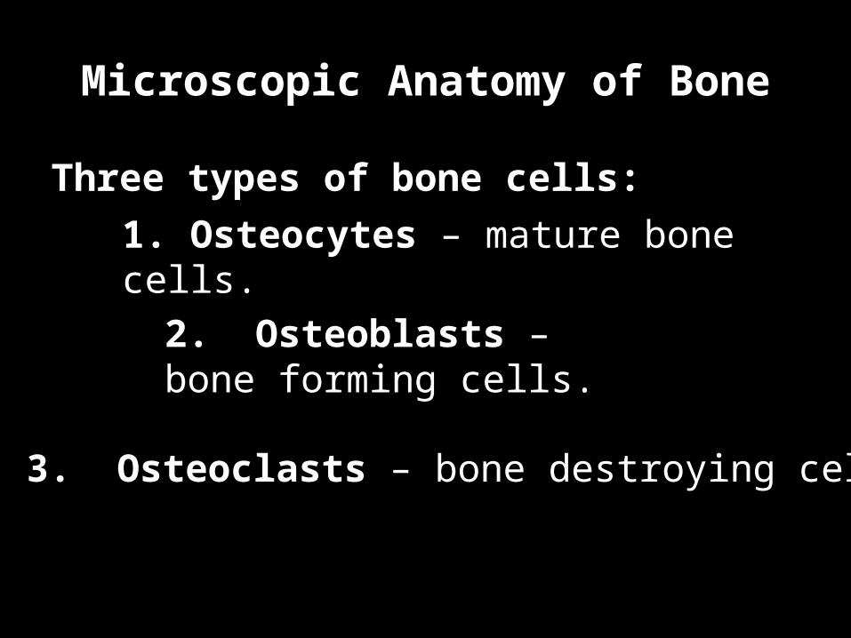
Microscopic Anatomy of Bone
Three types of bone cells:
1. Osteocytes – mature bone cells.
2. Osteoblasts – bone forming cells.
3. Osteoclasts – bone destroying cells.

Microscopic Anatomy of Bone Three types of bone cells
*Osteocytes
*Mature bone cells
*Live in spaces in bone called lacunae.
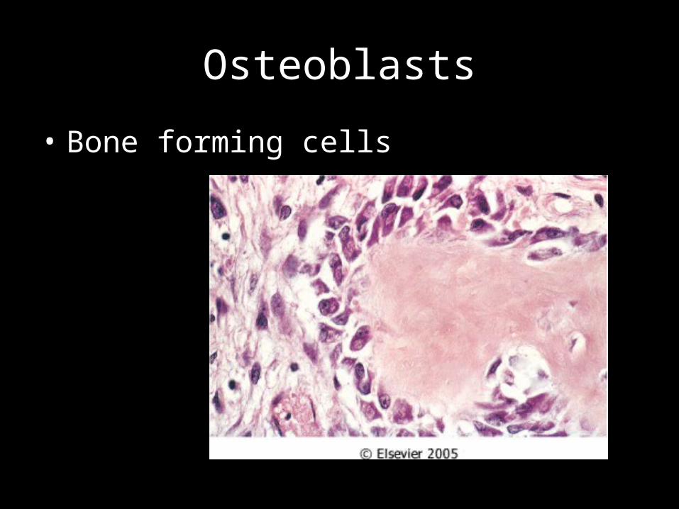
Osteoblasts
• Bone forming cells

Osteoclasts
• Bone-destroying cells

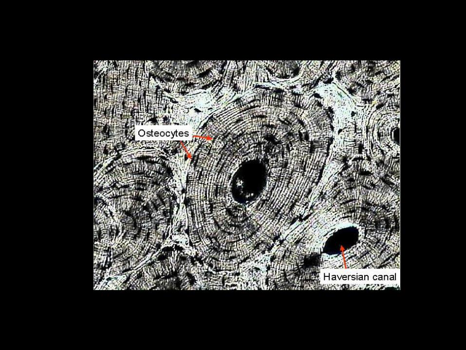
Compact bone is composed of organized passageways called Haversian Systems
(Osteon).*Osteocytes are formed when osteoblasts
are trapped in bony matrix.
*Lacunae – tiny cavities in the matrix that contain osteocytes.
*Lamellae – concentric circles of lacunae arranged around a central (Haversian) canal.
*Haversian canals – contain blood vessels and nerves.
(you)
(your house)
(your streets)
(main street)

•Haversian system – complex of central canal and rings of matrix.
Canaliculi – radiate outward from Haversian canals forming transport system for nutrients.
This is why bone injuries heal quickly.(side streets)
(neighborhood)


MUSICAL NOTES:
1.Each group will be responsible for taking notes on the following topics:
a. Bone formation (pgs-)b. Bone fractures (pgs-)c. Steps in repairing a fracture (pgs-)
2.The notes will be written on the construction paper at each station.
3.Students will rotate while music is playing and take down the notes located at each station.

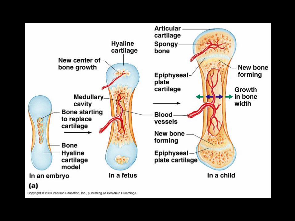
Bone Formation
• In embryos, the skeleton is mostly composed of hyaline cartilage.
• In young children, the cartilage is replaced by bone.

Ossification (bone formation)
1. Hyaline cartilage model is covered by bony matrix by osteoblasts.
2. Hyaline cartilage model is digested by osteoclasts, forming a medullary cavity within the newly formed bone.
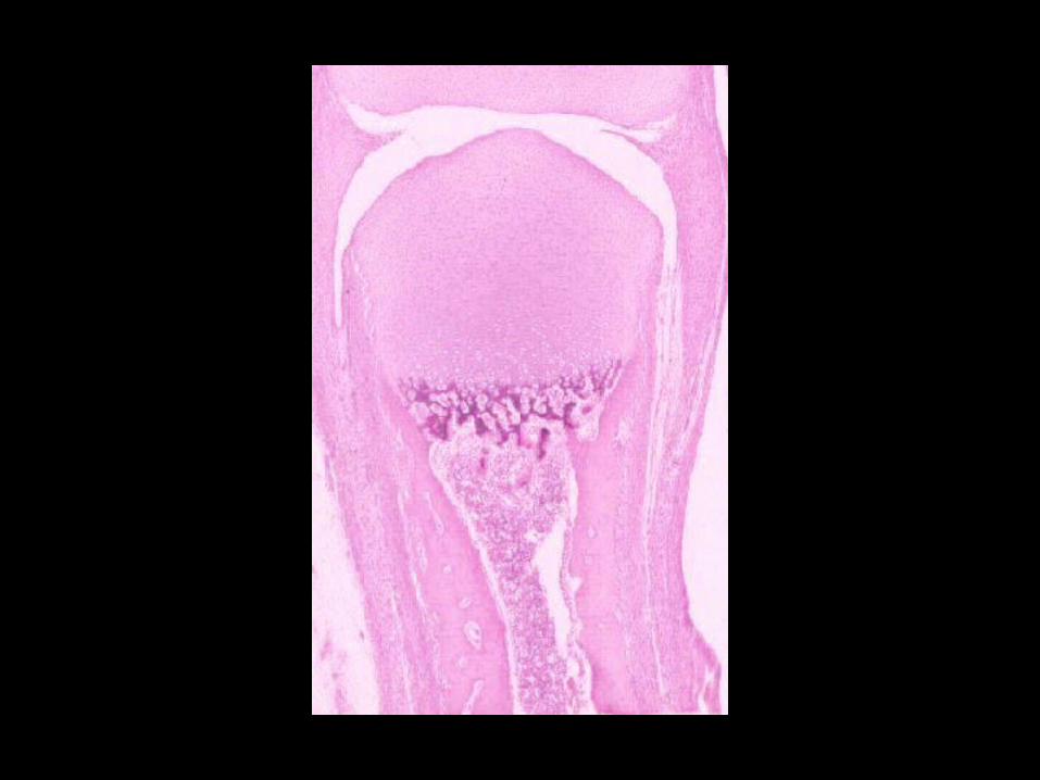
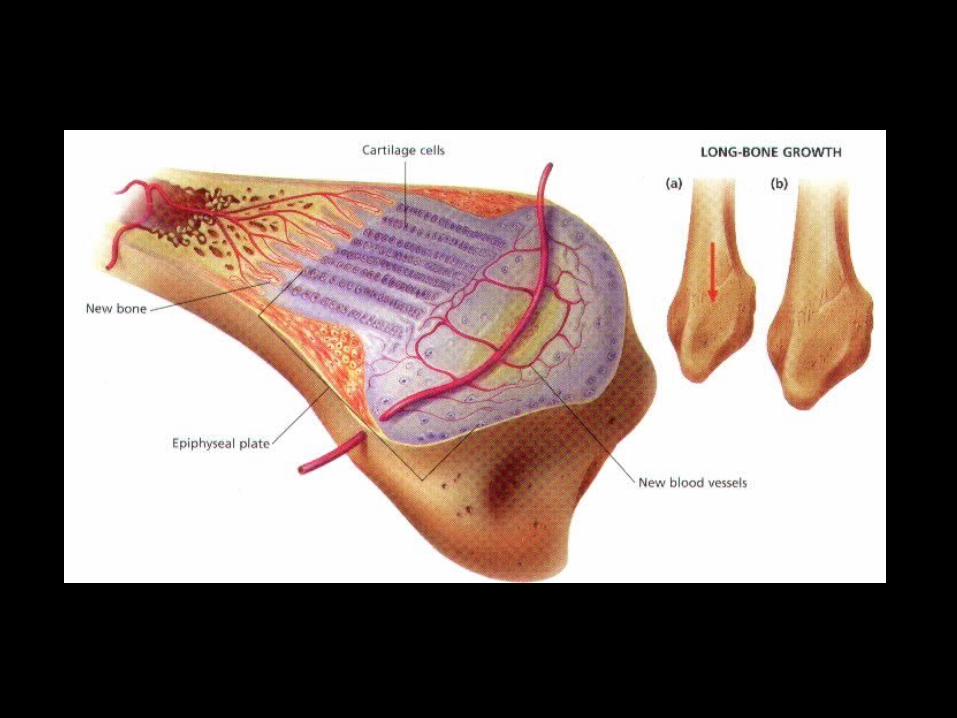
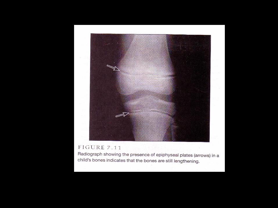
3. Epiphyseal plates – areas of cartilage at distal ends of long bones where growth takes place.

4) New cartilage is formed at distal ends of epiphyseal plates.
5) Bone replaces cartilage at proximal ends of plates.
This results in lengthening of the bone.This process is controlled by growth hormone in children and sex hormones during puberty.
6) This process ends at adolescence when the epiphyseal plates are covered in bone.

Bone is a dynamic and active tissue!
Bones are constantly remodeled in response to
two factors:1. Calcium levels in the blood – when blood calcium
drops below homeostatic levels, parathyroid hormone is released into the blood, which activates osteoclasts to break down bone and release calcium into the blood.
2. The pull of gravity and muscles on the skeleton – when the skeleton is pulled, bones respond by becoming stronger.


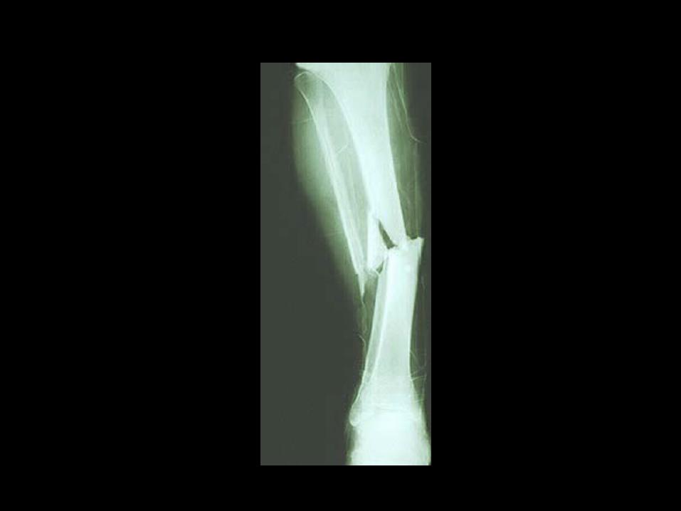
Bone Fractures
• Youth – most fractures result from trauma (accidents).
• Old age – fractures occur as bones become thin and weak.


Fractures
• Closed (Simple) Fracture– Bone breaks clean but does not penetrate the skin
• Open (Compound) Fracture– Broken bone ends penetrate through the skin
• Comminuted-bone breaks into many fragments
• Compression- bone is crushed
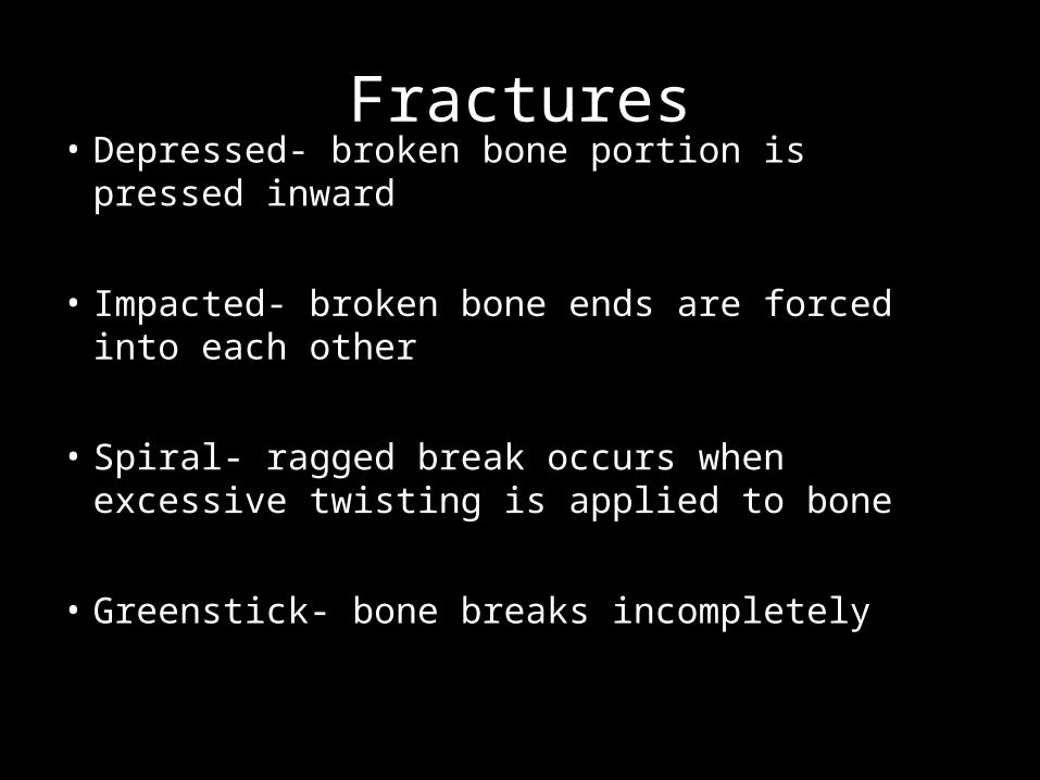
Fractures• Depressed- broken bone portion is pressed inward
• Impacted- broken bone ends are forced into each other
• Spiral- ragged break occurs when excessive twisting is applied to bone
• Greenstick- bone breaks incompletely

Fractures


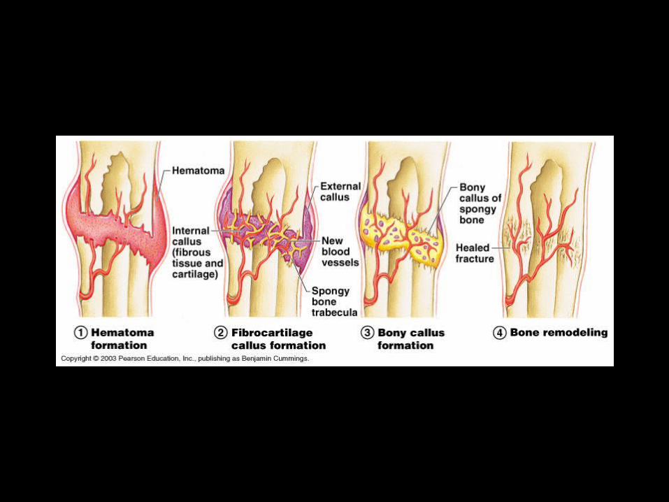
Steps in repair of fracture:
1. Hematoma – blood fills area of break; blood vessels are torn.
2. Fibrocartilage Callous – new capillaries, phagocytes and collagen fibers fill the space forming granulation tissue.
3. Bony Callous – osteoblasts invade area and lay down bone.
4. Remodeling – by osteoclasts to form strong, permanent patch.

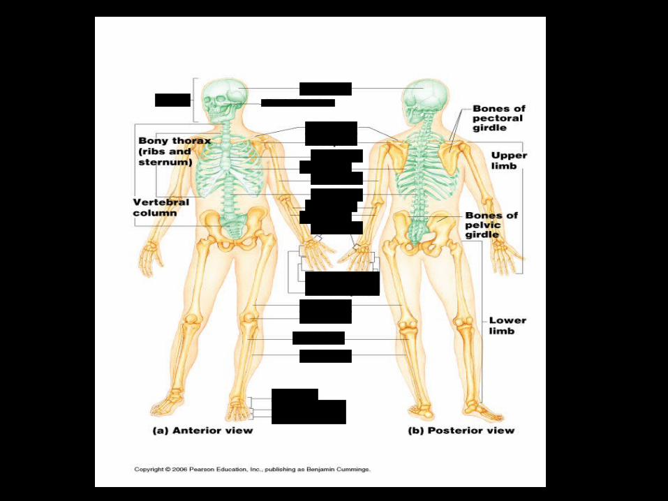
Bones of the Human Body
Two divisions of the human skeleton:
1. Axial Skeleton – bones that form the longitudinal axis of the body (head and trunk).
2. Appendicular Skeleton – bones of limbs (arms and legs) and girdles (pectoral and pelvic).

NOVEMBER 8, 2012
WARM-UP:
EQ: What are the bones of the human body?
Answer the following questions:
1. What are the organized passage ways for blood vessels in compact bone called?
Haversian System (Osteon)
2. Osteocytes are __________ bone cells found in _____________.
3. If a bone breaks, _______________ are responsible for laying down new bone.
4. Name 2 types of bone?
Compact and Spongy
mature lacunae
osteoblasts


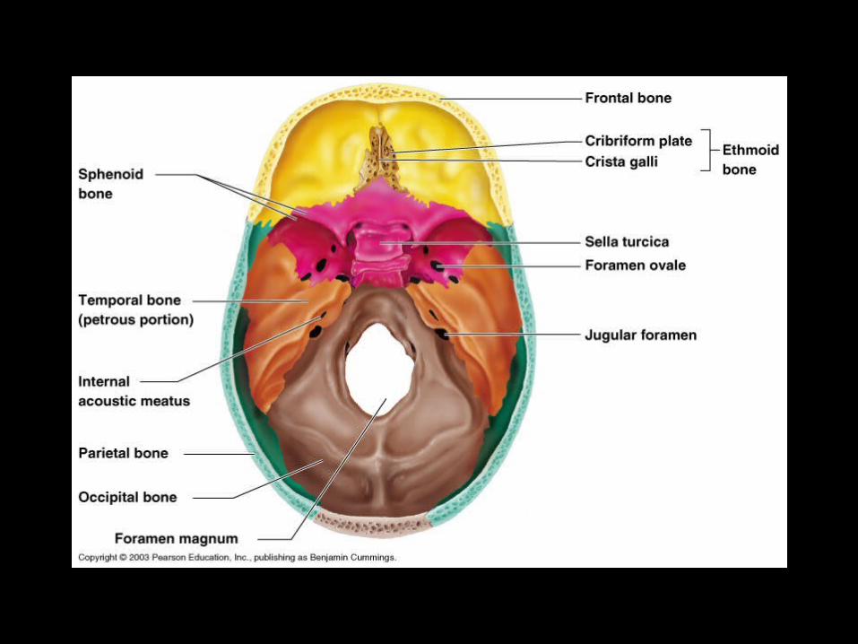
Axial Skeleton – skull, vertebral column and thorax
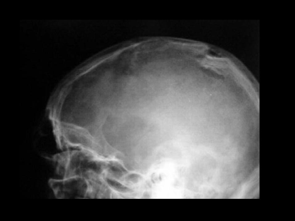
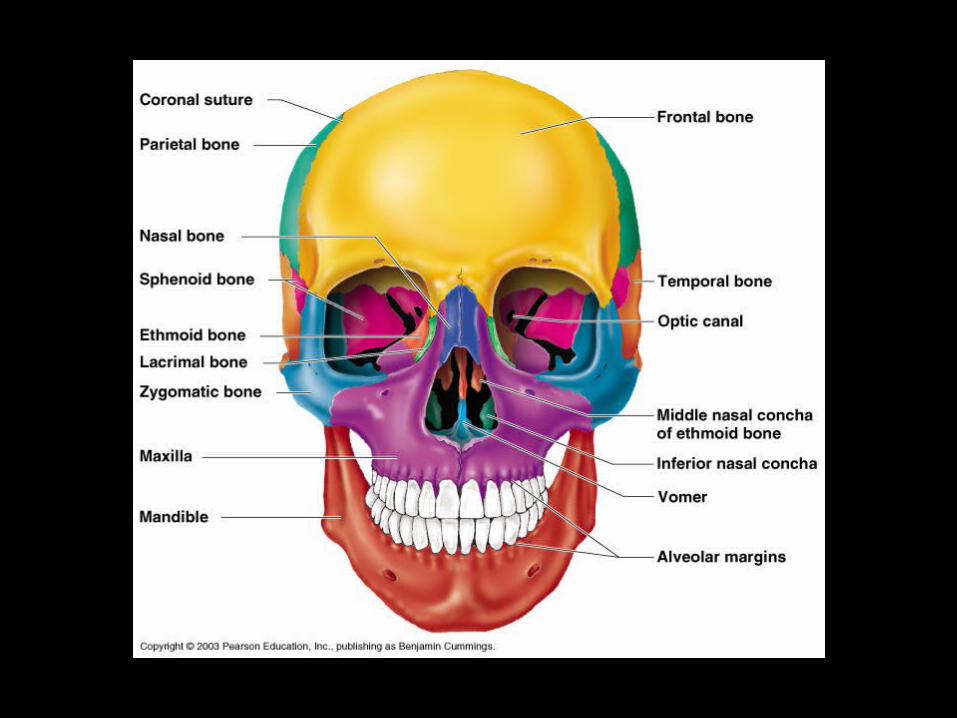
A. Skull – cranial and facial bones
1. Cranial bones – 8 large, flat bones
a. Frontal bone – forehead
b. Parietal bones (2) – superior and lateral walls of cranium.
c. Temporal bones (2) – inferior to parietal bones.

d. Occipital bone – floor and back wall of
skull.»Foramen magnum – where spinal
cord connects to brain.
e. Sphenoid bone – floor of cranial cavity.
f. Ethmoid bone – roof of nasal cavity.

2. Facial bones
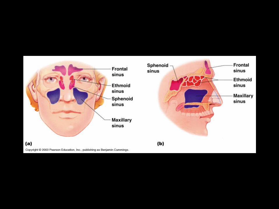
a. Maxillary bones (2) – fused to form upper jaw.
Paranasal sinuses – 4 pair.– Lighten the skull bones.– Amplify sounds.– Sinusitis – infection resulting from the fact
that lining of throat and nose is continuous.

b. Palatine bones – posterior part of hard
palate.• cleft palate - birth defect that results
when palatine bones fail to fuse during embryonic development.
c. Zygomatic bones – cheekbones.
d. Nasal bones – bridge of nose (tip is cartilage).


e. Mandible – largest and strongest bone in the face.
• TMJ (temperomandibular joints) – join temporal bones to sides of face.
f. Hyoid bone – only bone that does not articulate with another bone – provides a moveable base for the tongue.

Temporomandibular Joint
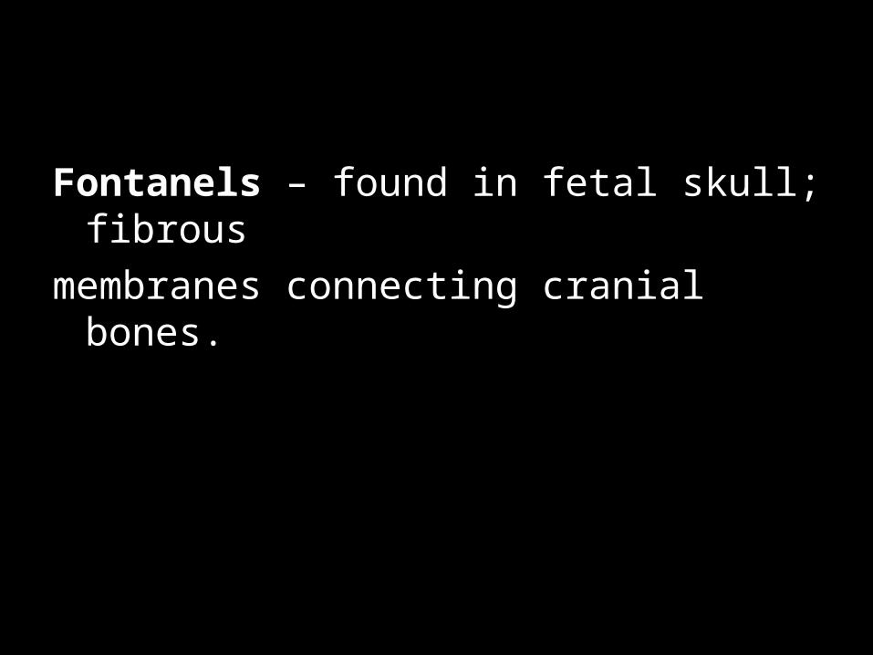
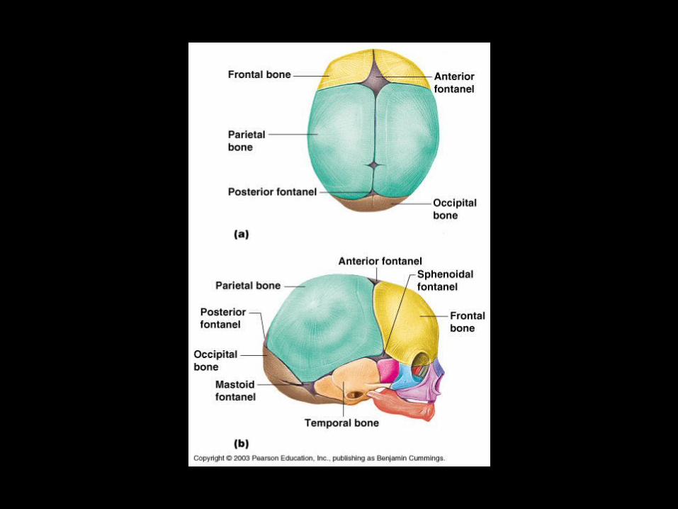
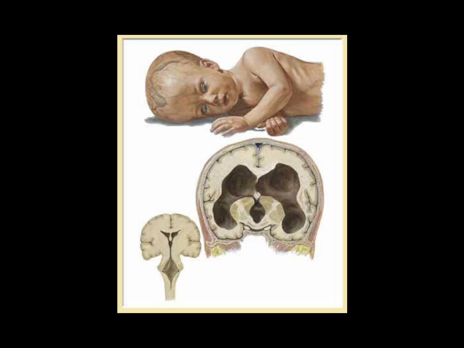
Fontanels – found in fetal skull; fibrous
membranes connecting cranial bones.


Axial Skeleton – skull, vertebral column, thorax
B. Vertebral Column
1. cervical vertebrae (7) – in neck; small and light.
2. thoracic vertebrae (12) – found in
chest.
3. lumbar vertebrae (5) – largest bones
in lower back.
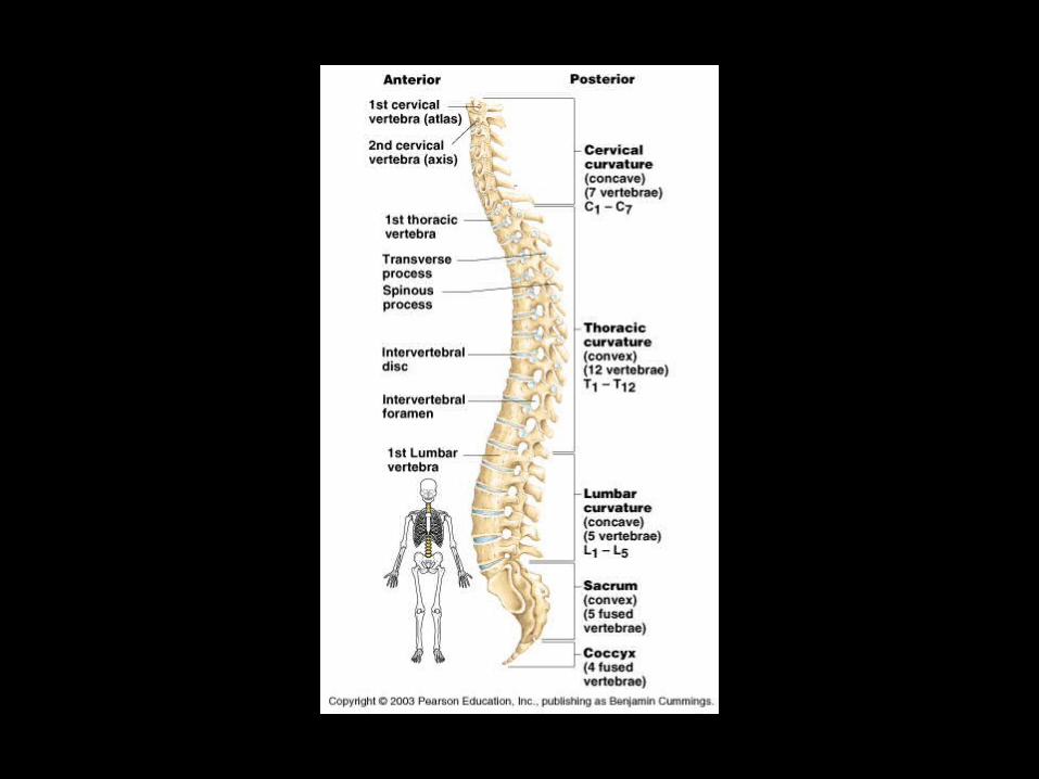
Intervertebral discs – flexible pads to separate vertebrae.• Cushion vertebrae and absorb shocks.• These become less flexible with age.• Herniated disc – protrudes and presses on spinal
cord and nerves.


Figure 26-12 Osteoporotic vertebral body (right) shortened by compression fractures, compared with a normal vertebral body. Note that the osteoporotic vertebra has a characteristic loss of horizontal trabeculae and thickened vertical trabeculae.
Downloaded from: Robbins & Cotran Pathologic Basis of Disease (on 8 February 2005 01:14 AM)
© 2005 Elsevier

4. Sacrum – triangle shaped bone.
5. Coccyx – remnant of tail bone.
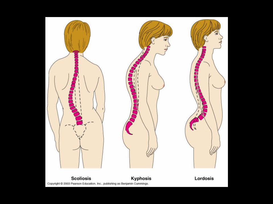

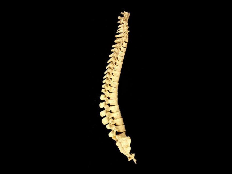
Scoliosis

C. Thorax
1. Sternum – contains red bone marrow
in adults.
2. Ribs – 12 pair
a. True ribs – attach to sternum
via costal cartilages.
b. False ribs – 5 pair (2 pair – floating ribs).


Appendicular Skeleton – arms, legs, pectoral and pelvic girdles
A. Pectoral Girdle1. Clavicle – collarbone
2. Scapula – shoulder blades (triangular)
This girdle is easily dislocated because it is light and allows for exceptionally free movement.


B. Upper limb1. Arm – Humerus – typical long bone.
2. Forearma. Radius – lateral bone in anatomic position.
b. Ulna – medial bone in anatomic position.


Hand
a. Carpals – 2 rows of 4 bones each form wrist.
b. Metacarpals – form the palm of the hand.
c. Phalanges – form the fingers; 3 bones each.


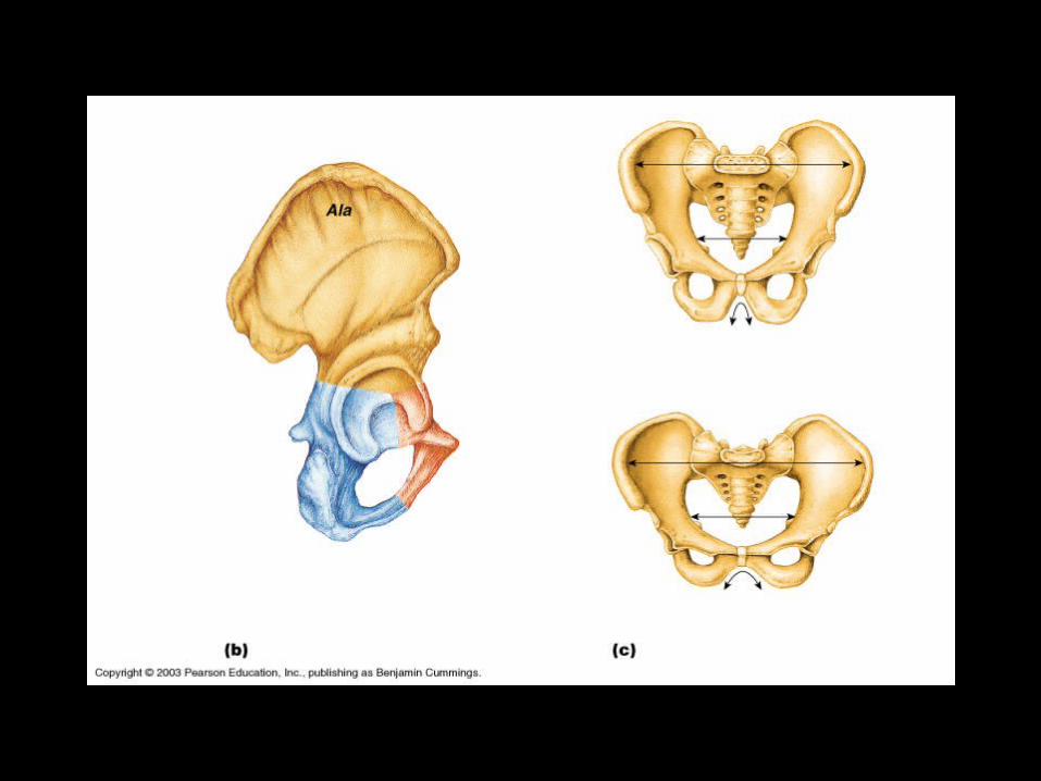
C. Pelvic Girdle – supports weight of body,
attachment for legs.
Coxal bones (2); large and heavy; 3
fused bones:
a. Ilium – large flaring bone.
b. Ischium – most inferior part.
c. Pubis – anterior part of bone.


Riddles
• I am a window, I am a lamp, I am clouded, I am shining, and I am colored. I fill with water and overflow. I say much but I have no words. What am I?
Your Eye• I am taken from a mine and shut up in a wooden case from which I
am never released and yet I am used by almost everyone. What am I?
Pencil Lead• I bind it and it walks. I loose it and it stops. What is it?
Sandal• What comes once in a minute, twice in a moment, but never in a
thousand years?
Letter M
How are male and female pelvises different?
1. Bones of female pelvis – lighter and
thinner.
2. Female ilia are more flared.
3. Female inlet is larger for childbirth.
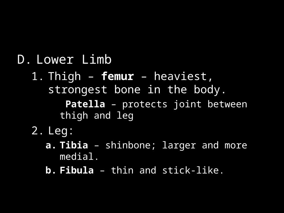
D. Lower Limb1. Thigh – femur – heaviest, strongest bone in
the body. Patella – protects joint between thigh and leg
2. Leg:a. Tibia – shinbone; larger and more medial.
b. Fibula – thin and stick-like.


3. Foot – supports the weight of the body
a. Tarsals – heel and ankle
b. Metatarsals – sole of foot and arch
c. Phalanges – toes; each toe has 3
phalanges except for big toe
which has 2


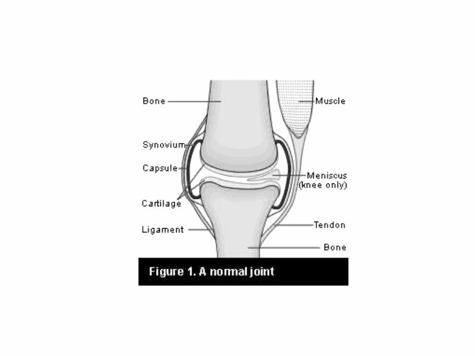
Joints
1. Immovable – flat bones of cranium2. Slightly movable – vertebrae of spinal
column3. Freely movable – most bones of
skeletonTwo bones held together by joint capsule:
• Outer layer of ligaments• Inner layer – synovial membrane which
secretes synovial fluid to reduce friction• Bursa – fluid filled sac

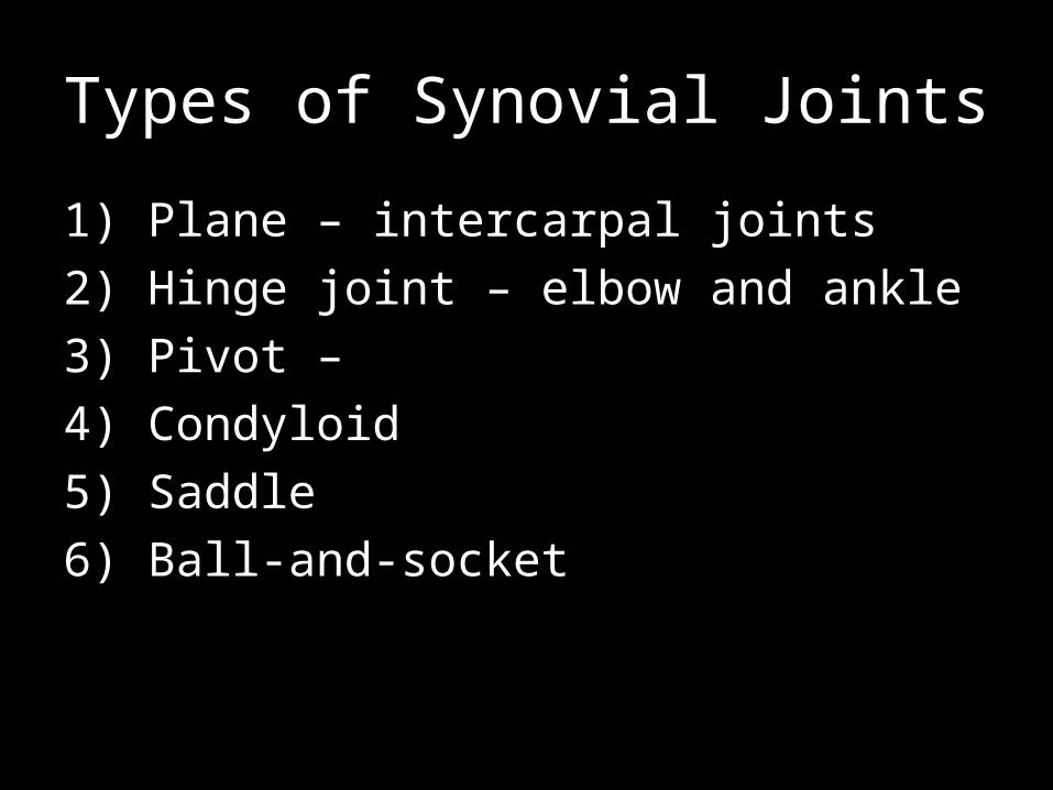
Types of Synovial Joints
1) Plane – intercarpal joints
2) Hinge joint – elbow and ankle
3) Pivot –
4) Condyloid
5) Saddle
6) Ball-and-socket


Disorders of Skeletal System
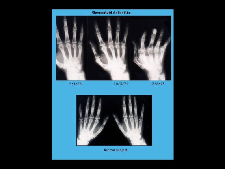
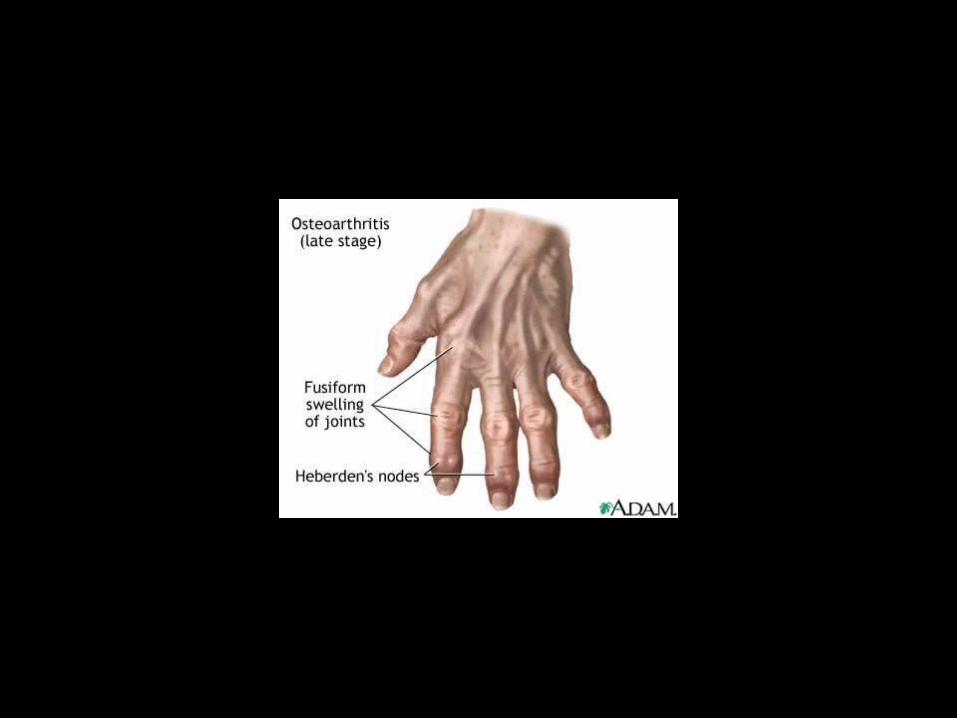
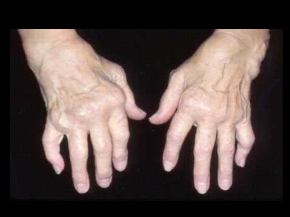
Arthritis – causes inflamed, swollen and painful joints; two main types:
A. Rheumatoid arthritis– The most painful and crippling form of arthritis– Fibrous tissue invades joint capsule and may
become ossified, so that bones are fused together.


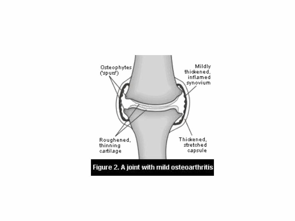
B. Osteoarthritis• Commonly occurs as a result of aging• Articular cartilage softens, disintegrates
and becomes rough• Joints are sore and motion is limited• Commonly occurs in knees and lower
back (joints which have been used the most)


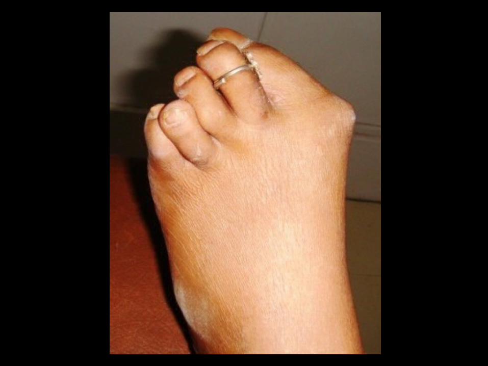
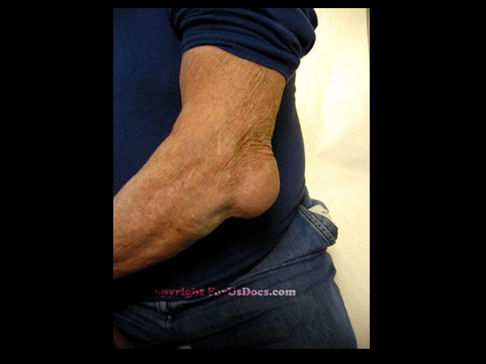
Arthritis
• Gout– A disease in which uric acid accumulates in
the blood and may be deposited as needle-shaped crystals in the soft tissue of joints
– Agonizing– Often affects big toe joint– Most common in males after age 30– Tends to run in families
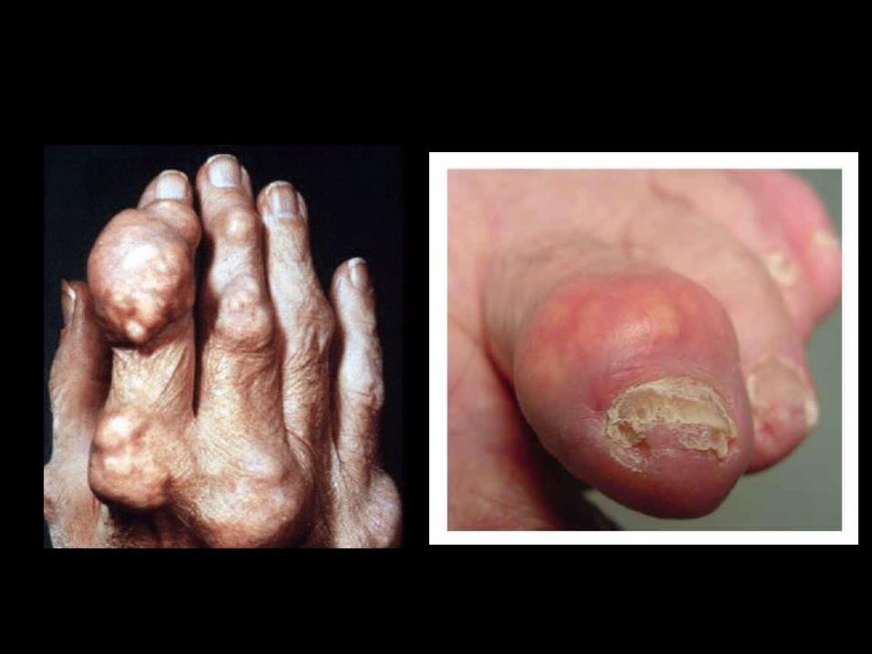
Chronic Gout
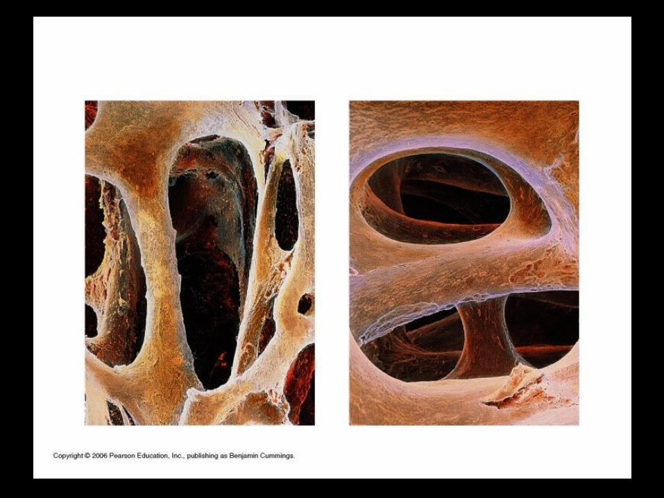
Osteoporosis
• Bones lose their density, becoming weak and brittle
• Loss of calcium and phosphorus from the bone• Factors that accelerate:
– Diet low in calcium– Lack of exercise– Cigarette smoking– Anorexia– Steroid drugs


Symptoms of osteoporosis:– Loss in height– Forward curvature of spine– Fractures of hip and wrist
Treatments:– Calcium and Vitamin D supplements– Hormone replacement therapy– Calcitonin – a hormone that causes calcium to
be moved into the bone from the blood


Problems in Bone Growth
• Dwarfism- a disorder caused by insufficient production of growth hormone
• Acromegaly- a disorder caused by excessive production of growth hormone


Bone Formation and Growth
• Rickets Disease– Disease of Children– Bones fail to calcify and bow under weight– Caused by lack of Vitamin D (needed to
absorb Calcium)


Problems in Bone Growth
• Dwarfism- a disorder caused by insufficient production of growth hormone
• Acromegaly- a disorder caused by excessive production of growth hormone
Related Documents


