Oct4 expression in adult human stem cells: evidence in support of the stem cell theory of carcinogenesis Mei-Hui Tai 1,2 , Chia-Cheng Chang 1 , L.Karl Olson 2 and James E.Trosko 1, 1 National Food Safety Toxicology Center, Department of Pediatrics and Human Development and 2 Department of Physiology, Michigan State University, East Lansing, MI 48824, USA To whom correspondence should be addressed. Tel: þ517 432 3100; Fax: þ1 517 432 6340; Email: [email protected] The Oct3/4 gene, a POU family transcription factor, has been noted as being specifically expressed in embryonic stem cells and in tumor cells but not in cells of differen- tiated tissues. With the ability to isolate adult human stem cells it became possible to test for the expression of Oct3/4 gene in adult stem cells and to test the stem cell theory of carcinogenesis. Using antibodies and PCR primers we tested human breast, liver, pancreas, kidney, mesenchyme and gastric stem cells, the cancer cell lines HeLa and MCF-7 and human, dog and rat tumors for Oct4 expres- sion. The results indicate that adult human stem cells, immortalized non-tumorigenic cells and tumor cells and cell lines, but not differentiated cells, express Oct4. Oct4 is expressed in a few cells found in the basal layer of human skin epidermis. The data demonstrate that adult stem cells maintain expression of Oct4, consistent with the stem cell hypothesis of carcinogenesis. Introduction Stem cells exist in most adult organs, being best characterized in the bone marrow and gut. They are defined as cells that undergo symmetric and asymmetric division to give rise to daughter cells needed for self-renewal and amplification or to a daughter cell that acts as a progenitor cell for the purpose of producing specific differentiated lineages, respectively. Given the recent interest in the multiple uses of embryonic and adult stem cells for basic and applied research (i.e. reproductive cloning or regenerative tissue therapy), attempts have been made to characterize markers that would identify these stem cells. Oct3/4 or Oct4 (also referred to as Pou5f1), a transcrip- tion factor, was discovered in 1990 (1--3). It was found in ovulated oocytes, mouse pre-implantation embryos, ectoderm of the gastrula (but not in other germ layers) and primordial germ cells, as well as in embryonic stem cells but not in their differentiated daughters (4). More recently, OCT4/Pou5f1 has been shown in cells isolated from human amniotic fluid (5). Subsequent studies seemed to suggest that Oct4 might be a specific gene marker for totipotency or a gene required for totipotency (6--8). In fact, it is thought that the success or failure of cloning depends on expression of this gene during reprogramming of the genome of a nucleus transferred to an enucleated oocyte (9). Seemingly in contrast, the Oct4 gene has also been shown to be expressed in some human tumor cells but not in normal somatic tissues (10--12). Since cancer cells have been charac- terized as having many phenotypic traits similar to undiffer- entiated embryonic cells (cancers as a ‘disease of differentiation’, a stem cell disease or as ‘oncogeny as blocked or partially blocked ontogeny’) (13--15), the question that still needs to be answered is ‘is the tumor derived from an adult stem cell in the tissue or from some somatic progenitor or differentiated cell that has de-differentiated?’ (16). Among the ‘hallmarks’ of cancer cells (17) is the potential for indefinite proliferation. In addition, cancer cells do not have functional homologous or heterologous gap junctional intercellular communication (GJIC) (18), due either to non- expression of connexins (e.g. HeLa and MCF-7 cells) or to non-functioning of expressed connexins (tumor cells expres- sing the ras, src or neu oncogenes) (19). Gap junctions have been associated with both normal development (20), growth control, differentiation, wound repair, synchronization of metabolic secretion and electrotonic function in tissues (21,22). Interestingly, several isolated presumptive adult human stem cells have been characterized as being deficient in expression of connexins and GJIC [kidney epithelial cells (23), breast epithelial cells (24), pancreas cells (25), keratino- cyte cells (26), corneal epithelium cells (27) and mesenchymal cells (28)]. In this study we have found that the Oct4 gene and Oct4 protein are expressed in several adult pluripotent stem cells, as well as in several human and rat tumor cells, but not in normal differentiated daughters of these stem cells. Two major con- clusions were drawn from these observations, namely that adult cells expressing the Oct4 gene are potential pluripotent stem cells and that these cells could be the target cells for initiation of the carcinogenic process. Materials and methods Materials Keratinocyte serum-free medium, Neurobasal medium with N2 supplement, RPMI-1640 medium, recombinant human epidermal growth factor (EGF), bovine pituitary extract (BPE), fetal bovine serum (FBS), penicillin, strepto- mycin, trypsin--EDTA and TRIZOL reagent were purchased from Invitrogen (Carlsbad, CA). N-acetyl-L-cysteine, DMSO and nicotinamide were obtained from Sigma Chemical Co. (St Louis, MO). L-Ascorbic acid 2-phosphate was from Wako Pure Chemical Industries Ltd (Osaka, Japan). Exendin-4 was obtained from Bachem Bioscience (Torrance, CA). LY294002 was obtained from Calbiochem (La Jolla, CA). Anti-Oct4 monoclonal antibodies were purchased from Santa Cruz (Santa Cruz, CA). Generation of adult human tissue-derived stem cell cultures The cell culture methods used to develop two types of normal human breast epithelial cell (HBEC) cultures from reduction mammoplasty material have been described (24). The stem cell characteristics of Type I HBEC are: (i) a Abbreviations: BPE, bovine pituitary extract; cpdl, cumulative population doublings; DAPI, 4 0 ,6 0 -diamidino-2-phenylindole dihydrochloride; EGF, epidermal growth factor; ES, embryonic stem; GJIC, gap junctional intercellular communication; HBEC, human breast epithelial cell; HPCs, human pancreatic stem cells; PBS, phosphate buffered-saline. Carcinogenesis vol.26 no.2 # Oxford University Press 2005; all rights reserved. 495 Carcinogenesis vol.26 no.2 pp.495--502, 2005 doi:10.1093/carcin/bgh321 Downloaded from https://academic.oup.com/carcin/article-abstract/26/2/495/2476063 by guest on 04 April 2019

Welcome message from author
This document is posted to help you gain knowledge. Please leave a comment to let me know what you think about it! Share it to your friends and learn new things together.
Transcript
Oct4 expression in adult human stem cells: evidence in support of the stem celltheory of carcinogenesis
Mei-Hui Tai1,2, Chia-Cheng Chang1, L.Karl Olson2
and James E.Trosko1,�
1National Food Safety Toxicology Center, Department of Pediatrics andHuman Development and 2Department of Physiology, Michigan StateUniversity, East Lansing, MI 48824, USA
�To whom correspondence should be addressed.Tel: þ517 432 3100; Fax: þ1 517 432 6340;Email: [email protected]
The Oct3/4 gene, a POU family transcription factor, hasbeen noted as being specifically expressed in embryonicstem cells and in tumor cells but not in cells of differen-tiated tissues. With the ability to isolate adult human stemcells it became possible to test for the expression of Oct3/4gene in adult stem cells and to test the stem cell theoryof carcinogenesis. Using antibodies and PCR primers wetested human breast, liver, pancreas, kidney, mesenchymeand gastric stem cells, the cancer cell lines HeLa andMCF-7 and human, dog and rat tumors for Oct4 expres-sion. The results indicate that adult human stem cells,immortalized non-tumorigenic cells and tumor cells andcell lines, but not differentiated cells, express Oct4. Oct4 isexpressed in a few cells found in the basal layer of humanskin epidermis. The data demonstrate that adult stem cellsmaintain expression of Oct4, consistent with the stem cellhypothesis of carcinogenesis.
Introduction
Stem cells exist in most adult organs, being best characterizedin the bone marrow and gut. They are defined as cells thatundergo symmetric and asymmetric division to give rise todaughter cells needed for self-renewal and amplification or toa daughter cell that acts as a progenitor cell for the purpose ofproducing specific differentiated lineages, respectively. Giventhe recent interest in the multiple uses of embryonic and adultstem cells for basic and applied research (i.e. reproductivecloning or regenerative tissue therapy), attempts have beenmade to characterize markers that would identify these stemcells. Oct3/4 or Oct4 (also referred to as Pou5f1), a transcrip-tion factor, was discovered in 1990 (1--3). It was found inovulated oocytes, mouse pre-implantation embryos, ectodermof the gastrula (but not in other germ layers) and primordialgerm cells, as well as in embryonic stem cells but not in theirdifferentiated daughters (4). More recently, OCT4/Pou5f1 hasbeen shown in cells isolated from human amniotic fluid (5).Subsequent studies seemed to suggest that Oct4 might be aspecific gene marker for totipotency or a gene required for
totipotency (6--8). In fact, it is thought that the success orfailure of cloning depends on expression of this gene duringreprogramming of the genome of a nucleus transferred to anenucleated oocyte (9).Seemingly in contrast, the Oct4 gene has also been shown to
be expressed in some human tumor cells but not in normalsomatic tissues (10--12). Since cancer cells have been charac-terized as having many phenotypic traits similar to undiffer-entiated embryonic cells (cancers as a ‘disease ofdifferentiation’, a stem cell disease or as ‘oncogeny as blockedor partially blocked ontogeny’) (13--15), the question that stillneeds to be answered is ‘is the tumor derived from an adultstem cell in the tissue or from some somatic progenitor ordifferentiated cell that has de-differentiated?’ (16).Among the ‘hallmarks’ of cancer cells (17) is the potential
for indefinite proliferation. In addition, cancer cells do nothave functional homologous or heterologous gap junctionalintercellular communication (GJIC) (18), due either to non-expression of connexins (e.g. HeLa and MCF-7 cells) or tonon-functioning of expressed connexins (tumor cells expres-sing the ras, src or neu oncogenes) (19). Gap junctions havebeen associated with both normal development (20), growthcontrol, differentiation, wound repair, synchronization ofmetabolic secretion and electrotonic function in tissues(21,22). Interestingly, several isolated presumptive adulthuman stem cells have been characterized as being deficientin expression of connexins and GJIC [kidney epithelial cells(23), breast epithelial cells (24), pancreas cells (25), keratino-cyte cells (26), corneal epithelium cells (27) and mesenchymalcells (28)].In this study we have found that the Oct4 gene and Oct4
protein are expressed in several adult pluripotent stem cells, aswell as in several human and rat tumor cells, but not in normaldifferentiated daughters of these stem cells. Two major con-clusions were drawn from these observations, namely thatadult cells expressing the Oct4 gene are potential pluripotentstem cells and that these cells could be the target cells forinitiation of the carcinogenic process.
Materials and methods
Materials
Keratinocyte serum-free medium, Neurobasal medium with N2 supplement,RPMI-1640 medium, recombinant human epidermal growth factor (EGF),bovine pituitary extract (BPE), fetal bovine serum (FBS), penicillin, strepto-mycin, trypsin--EDTA and TRIZOL reagent were purchased from Invitrogen(Carlsbad, CA). N-acetyl-L-cysteine, DMSO and nicotinamide were obtainedfrom Sigma Chemical Co. (St Louis, MO). L-Ascorbic acid 2-phosphate wasfrom Wako Pure Chemical Industries Ltd (Osaka, Japan). Exendin-4 wasobtained from Bachem Bioscience (Torrance, CA). LY294002 was obtainedfrom Calbiochem (La Jolla, CA). Anti-Oct4 monoclonal antibodies werepurchased from Santa Cruz (Santa Cruz, CA).
Generation of adult human tissue-derived stem cell cultures
The cell culture methods used to develop two types of normal human breastepithelial cell (HBEC) cultures from reduction mammoplasty material havebeen described (24). The stem cell characteristics of Type I HBEC are: (i) a
Abbreviations: BPE, bovine pituitary extract; cpdl, cumulative populationdoublings; DAPI, 40,60-diamidino-2-phenylindole dihydrochloride; EGF,epidermal growth factor; ES, embryonic stem; GJIC, gap junctionalintercellular communication; HBEC, human breast epithelial cell; HPCs,human pancreatic stem cells; PBS, phosphate buffered-saline.
Carcinogenesis vol.26 no.2 # Oxford University Press 2005; all rights reserved. 495
Carcinogenesis vol.26 no.2 pp.495--502, 2005doi:10.1093/carcin/bgh321
Dow
nloaded from https://academ
ic.oup.com/carcin/article-abstract/26/2/495/2476063 by guest on 04 April 2019
deficiency in GJIC; (ii) the ability to differentiate into Type II HBEC; (iii) theability to form budding ductal structures on Matrigel similar to mammarygland lobule or terminal end buds, where mammary stem cells are primarilylocated; (iv) a high susceptibility to telomerase activation, immortalization andneoplastic transformation (24,29,30). Immortal SV40 large T-antigen-trans-fected Type I cells (clone M13SV1), X-ray-induced weakly tumorigenic cells(clone M13SV1R2) and neu oncogene-transfected highly tumorigenic cells(clone M13SV1R2N1) (31) were cultured in MSU-1 medium with 5% FBSat 37�C in a humidified incubator with 5% CO2 (24). Human pancreatic isletsand duct and acini remnants were obtained from the JDRF HumanIslet Distribution Program. Upon arrival cells were plated and cultured inRPMI-1640 medium supplemented with 8.0 mM glucose and 10% FBS.After 24 h the culture medium was then switched to keratinocyte serum-freemedium supplemented with 5 ng/ml recombinant human EGF, 50 mg/ml BPE,2.0 mMN-acetyl-L-cysteine and 0.2 mM L-ascorbic acid 2-phosphate. To inducepancreatic endocrine cell differentiation from the progenitor cells, the cells wereplated and cultured in neurobasal medium containing 1% N2 supplement with10 mM nicotinamide, 0.1 mM exendin-4 and 5 mM LY 294002 for 14 daysbefore RNA was extracted. The method to develop putative human fetal kidneyepithelial stem cells has been previously reported (23). These putative kidneyepithelial stem cells have been shown to be contact insensitive and to bedeficient in GJIC (23). The procedure to obtain adult human liver stem/precursorcells of clonal origin has been described (32). The stem cell features of thesecells include: (i) a high proliferation potential, with more than 50 cumulativepopulation doublings (cpdl); (ii) a deficiency in GJIC; (iii) the ability foranchorage-independent growth; (iv) expression of liver ‘oval cell’ markers, i.e.vimentin and a-fetoprotein. The isolation of gastric stem/precursor cells andmesenchymal stem cells from adipose tissues was as described for liver stemcells (32). Although the derivation of mesenchymal stem cells from adiposetissues has been reported before (33), our method gave a greater number of cellsin a shorter time (32 cpdl in 51 days compared with 22 cpdl in 165 days) (28).We have shown that these cells had a high differentiation ability to becomeadipocytes, osteoblasts and chondrocytes (28).
Cell preparation and immunostaining
Different cell cultures were plated in glass chamber slides for a few days untilthey reached the desired confluency. Cells were fixed with 4% paraformalde-hyde in phosphate buffer.
After washing with phosphate buffered-saline (PBS), paraformaldehyde-fixed cells were permeablized with 0.2% Triton X-100. Cells were thenblocked for 60 min with PBS containing 10% normal goat serum. Cells werethen incubated with primary antibody for 2 h at room temperature or overnightat 4�C. The primary antibodies were then removed and the cells were washedwith PBS and incubated for 2 h with Cyanine 3- or FITC-conjugated goat anti-mouse secondary antibodies (Jackson Immunoresearch Laboratories, WestGrove, PA). Slides were counterstained with 40,60-diamidino-2-phenylindoledihydrochloride (DAPI) for 5 min before final embedding. Fluorescent imageswere obtained using a Nikon epifluorescent microscope equipped with aSPOT-RT digital camera (Diagnostic Instruments, Detroit, MI), interfacedwith a Dell Pentium 4 PC installed with Spot Advanced analysis software(Diagnostic Instruments).
Tissue sections
Human normal tissue sections were purchased from Zymed (SanFrancisco, CA). Zymed’s MaxArray Human Normal Tissue Arrays contain30 human samples arrayed on microscope slides. Slides were deparaffinized,rehydrated and then processed following the protocol for immunohisto-chemistry. The slides were analyzed under a bright field microscope for Oct4staining. Dog mast cell tumor sections were provided by Dr Matti Kiupel atthe Department of Veterinary Medicine, Michigan State University. Dogmast tumor sections were processed by the same procedure as for Oct4immunostaining.
Reverse transcription--PCR (RT-PCR)
Total RNA was extracted from the cells with TRIZOL following the protocolsuggested by the manufacturer and treated with DNase I to remove contam-inating DNA. Oligo(dT) primers (Integrated DNA Technologies, IA) wereused with Superscript II reverse transcriptase (Invitrogen) for cDNA synthesisfrom 1 mg total RNA following the guidelines provided by the manufacturer.PCR was conducted with PlatinumTaq Polymerase (Invitrogen). We usedglyceraldehyde 3-phosphate dehydrogenase as an internal standard for Oct4RT-PCR. The primer sequences for human Oct4 were: 50-GAC AAC AATGAG AAC CTT CAG GAG A-30 and 50-CTG GCG CCG GTT ACA GAACCA-30. The mixture was first heated at 94�C for 3 min in a PTC-200 DNAEngine Thermal Cycler (MJ Research, Waltham, MA). Amplification wasperformed for 35 cycles at 94�C for 45 s, 55�C for 30 s and 72�C for 90 s,followed by 72�C for 10 min. The PCR products were separated on 1.5%
agarose gels by electrophoresis. Digital images were captured on a Kodak geldocumentation system.
Results
Oct4 expression in human adult breast stem cells duringdifferentiation, immortalization and neoplastic transformation
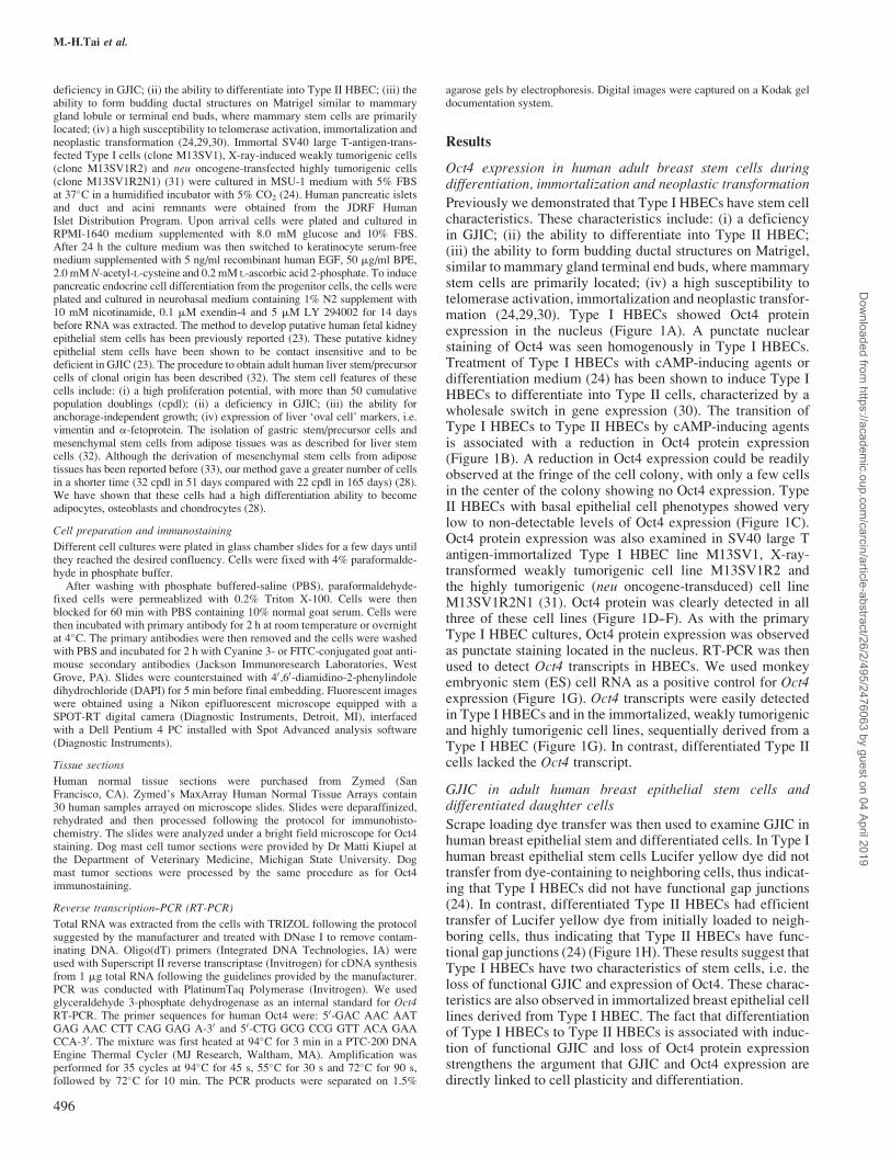
Previously we demonstrated that Type I HBECs have stem cellcharacteristics. These characteristics include: (i) a deficiencyin GJIC; (ii) the ability to differentiate into Type II HBEC;(iii) the ability to form budding ductal structures on Matrigel,similar to mammary gland terminal end buds, where mammarystem cells are primarily located; (iv) a high susceptibility totelomerase activation, immortalization and neoplastic transfor-mation (24,29,30). Type I HBECs showed Oct4 proteinexpression in the nucleus (Figure 1A). A punctate nuclearstaining of Oct4 was seen homogenously in Type I HBECs.Treatment of Type I HBECs with cAMP-inducing agents ordifferentiation medium (24) has been shown to induce Type IHBECs to differentiate into Type II cells, characterized by awholesale switch in gene expression (30). The transition ofType I HBECs to Type II HBECs by cAMP-inducing agentsis associated with a reduction in Oct4 protein expression(Figure 1B). A reduction in Oct4 expression could be readilyobserved at the fringe of the cell colony, with only a few cellsin the center of the colony showing no Oct4 expression. TypeII HBECs with basal epithelial cell phenotypes showed verylow to non-detectable levels of Oct4 expression (Figure 1C).Oct4 protein expression was also examined in SV40 large Tantigen-immortalized Type I HBEC line M13SV1, X-ray-transformed weakly tumorigenic cell line M13SV1R2 andthe highly tumorigenic (neu oncogene-transduced) cell lineM13SV1R2N1 (31). Oct4 protein was clearly detected in allthree of these cell lines (Figure 1D--F). As with the primaryType I HBEC cultures, Oct4 protein expression was observedas punctate staining located in the nucleus. RT-PCR was thenused to detect Oct4 transcripts in HBECs. We used monkeyembryonic stem (ES) cell RNA as a positive control for Oct4expression (Figure 1G). Oct4 transcripts were easily detectedin Type I HBECs and in the immortalized, weakly tumorigenicand highly tumorigenic cell lines, sequentially derived from aType I HBEC (Figure 1G). In contrast, differentiated Type IIcells lacked the Oct4 transcript.
GJIC in adult human breast epithelial stem cells anddifferentiated daughter cells
Scrape loading dye transfer was then used to examine GJIC inhuman breast epithelial stem and differentiated cells. In Type Ihuman breast epithelial stem cells Lucifer yellow dye did nottransfer from dye-containing to neighboring cells, thus indicat-ing that Type I HBECs did not have functional gap junctions(24). In contrast, differentiated Type II HBECs had efficienttransfer of Lucifer yellow dye from initially loaded to neigh-boring cells, thus indicating that Type II HBECs have func-tional gap junctions (24) (Figure 1H). These results suggest thatType I HBECs have two characteristics of stem cells, i.e. theloss of functional GJIC and expression of Oct4. These charac-teristics are also observed in immortalized breast epithelial celllines derived from Type I HBEC. The fact that differentiationof Type I HBECs to Type II HBECs is associated with induc-tion of functional GJIC and loss of Oct4 protein expressionstrengthens the argument that GJIC and Oct4 expression aredirectly linked to cell plasticity and differentiation.
M.-H.Tai et al.
496
Dow
nloaded from https://academ
ic.oup.com/carcin/article-abstract/26/2/495/2476063 by guest on 04 April 2019
Oct4 expression in adult human pancreatic stem cells duringdifferentiation and in pancreatic cancer cells
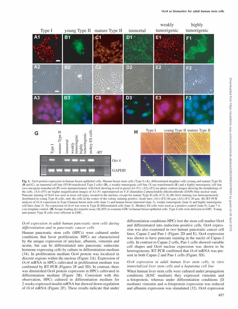
Human pancreatic stem cells (HPCs) were cultured underconditions that favor proliferation. HPCs are characterizedby the unique expression of amylase, albumin, vimentin andnestin, but can be differentiated into pancreatic endocrinehormone expressing cells by culture in differentiation medium(34). In proliferation medium Oct4 protein was localized indiscrete regions within the nucleus (Figure 2A). Expression ofOct4 mRNA in HPCs cultivated in proliferation medium wasconfirmed by RT-PCR (Figures 2F and 3D). In contrast, therewas diminished Oct4 protein expression in HPCs cultivated indifferentiation medium (Figure 2B). Consistent with thisobservation, HPCs cultured in differentiation medium for2 weeks expressed insulin mRNA but showed down-regulationof Oct4 mRNA (Figure 2F). These results indicate that under
differentiation conditions HPCs lost the stem cell marker Oct4and differentiated into endocrine-positive cells. Oct4 expres-sion was also examined in two human pancreatic cancer celllines, Capan-2 and Pan-1 (Figure 2D and E). Oct4 expressionwas shown to have punctate staining in the nuclei of Capan-2cells. In contrast to Capan-2 cells, Pan-1 cells showed variablecell shapes and Oct4 nuclear expression was shown to beheterogeneous. RT-PCR confirmed that Oct4 mRNA was pre-sent in both Capan-2 and Pan-1 cells (Figure 3D).
Oct4 expression in adult human liver stem cells, in vitroimmortalized liver stem cells and a hepatoma cell line
When human liver stem cells were cultured under propagationconditions (KNC medium) they expressed vimentin anda-fetoprotein, whereas under differentiation conditions (Dmedium) vimentin and a-fetoprotein expression was reducedand albumin expression was stimulated (32). Oct4 expression
Type I young Type II mature Type II immortalweakly
tumorigenichighly
tumorigenic
Type
I
mat
ure
Type
II
imm
orta
l
wea
kly
tum
orin
gnic
high
lytu
mor
inge
nic
mok
ey E
S ce
lls
-RT
Type I young Type II mature Type II
GAPDH
Oct 4
Fig. 1. Oct4 protein expression in human breast epithelial cells. Human breast stem cells (Type I) (A), differentiated daughter cells (young and mature Type II)(B and C), an immortal cell line (SV40-transfected Type I cells) (D), a weakly tumorigenic cell line (X-ray-transformed) (E) and a highly tumorigenic cell line(neu oncogene-transduced) (F) were immunostained, with Oct4 showing in red or green (A1--F1). (A2)--(F2) are phase contrast images showing the morphology ofthe cells. (A3)--(F3) are higher magnification images of A1--F1 superimposed on 40,60-diamidino-2-phenylindole dihydrochloride (DAPI) blue nuclear stain.Punctate staining of Oct4 was seen in most cell types, located in the nucleus, except for mature Type II cells (C3). In (B) Oct4 staining was heterogeneouslydistributed in young Type II cells, only the cells in the center of the colony staining positive. Scale bars: (A1)--(F2) 60 mm; (A3)--(F3) 20 mm. (G) RT-PCRanalysis of Oct4 expression in Type I human breast stem cells (lane 1) and human breast immortal (lane 3), weakly tumorigenic (lane 4) and highly tumorigeniccell lines (lane 5). No expression of Oct4 was seen in Type II differentiated cells (lane 2). Monkey ES cells were used as a positive control (lane 6). Lane 7 isa no template control. (H) Scrape loading dye transfer assay (SL/DT) to examine GJIC in human breast epithelial cells. Type I cells were deficient in GJIC. Youngand mature Type II cells were efficient in GJIC.
Oct4 as biomarker for adult human stem cells
497
Dow
nloaded from https://academ
ic.oup.com/carcin/article-abstract/26/2/495/2476063 by guest on 04 April 2019
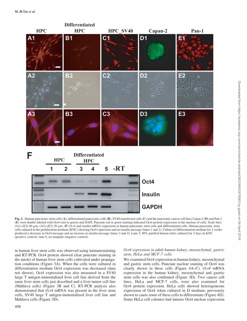
in human liver stem cells was observed using immunostainingand RT-PCR. Oct4 protein showed clear punctate staining inthe nuclei of human liver stem cells cultivated under propaga-tion conditions (Figure 3A). When the cells were cultured indifferentiation medium Oct4 expression was decreased (datanot shown). Oct4 expression was also measured in a SV40large T antigen-immortalized liver cell line derived from thesame liver stem cells just described and a liver tumor cell line(Mahlava cells) (Figure 3B and C). RT-PCR analysis alsodemonstrated that Oct4 mRNA was present in the liver stemcells, SV40 large T antigen-immortalized liver cell line andMahlava cells (Figure 3D).
Oct4 expression in adult human kidney, mesenchymal, gastricstem, HeLa and MCF-7 cells
We examined Oct4 expression in human kidney, mesenchymaland gastric stem cells. Punctate nuclear staining of Oct4 wasclearly shown in these cells (Figure 4A--C). Oct4 mRNAexpression in the human kidney, mesenchymal and gastricstem cells was also confirmed (Figure 3D). Two cancer celllines, HeLa and MCF-7 cells, were also examined forOct4 protein expression. HeLa cells showed heterogeneousexpression of Oct4 when cultured in D medium, previouslyshown to cause some of these cells to differentiate (Figure 4D).Some HeLa cell colonies had intense Oct4 nuclear expression
Fig. 2. Human pancreatic stem cells (A), differentiated pancreatic cells (B), SV40-transfected cells (C) and the pancreatic cancer cell lines Capan-2 (D) and Pan-1(E) were double labeled with Oct4 (red or green) and DAPI. Punctate red or green staining indicated Oct4 protein expression in the nucleus of cells. Scale bars:(A1)--(E2) 60 mm; (A3)--(E3) 20 mm. (F) Oct4 and insulin mRNA expression in human pancreatic stem cells and differentiated cells. Human pancreatic stemcells cultured in the proliferation medium (KNC) showing Oct4 expression and no insulin message (lanes 1 and 2). Culture in differentiation medium for 2 weeksproduced a decrease in Oct4 message and an increase in insulin message (lanes 3 and 4). Lane 5, 80% purified human islets cultured for 2 days in KNC(positive control); lane 6, no template (negative control).
M.-H.Tai et al.
498
Dow
nloaded from https://academ
ic.oup.com/carcin/article-abstract/26/2/495/2476063 by guest on 04 April 2019
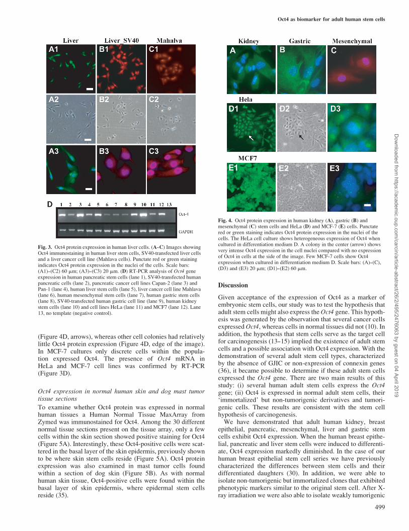
(Figure 4D, arrows), whereas other cell colonies had relativelylittle Oct4 protein expression (Figure 4D, edge of the image).In MCF-7 cultures only discrete cells within the popula-tion expressed Oct4. The presence of Oct4 mRNA inHeLa and MCF-7 cell lines was confirmed by RT-PCR(Figure 3D).
Oct4 expression in normal human skin and dog mast tumortissue sections
To examine whether Oct4 protein was expressed in normalhuman tissues a Human Normal Tissue MaxArray fromZymed was immunostained for Oct4. Among the 30 differentnormal tissue sections present on the tissue array, only a fewcells within the skin section showed positive staining for Oct4(Figure 5A). Interestingly, these Oct4-positive cells were scat-tered in the basal layer of the skin epidermis, previously shownto be where skin stem cells reside (Figure 5A). Oct4 proteinexpression was also examined in mast tumor cells foundwithin a section of dog skin (Figure 5B). As with normalhuman skin tissue, Oct4-positive cells were found within thebasal layer of skin epidermis, where epidermal stem cellsreside (35).
Discussion
Given acceptance of the expression of Oct4 as a marker ofembryonic stem cells, our study was to test the hypothesis thatadult stem cells might also express the Oct4 gene. This hypoth-esis was generated by the observation that several cancer cellsexpressed Oct4, whereas cells in normal tissues did not (10). Inaddition, the hypothesis that stem cells serve as the target cellfor carcinogenesis (13--15) implied the existence of adult stemcells and a possible association with Oct4 expression. With thedemonstration of several adult stem cell types, characterizedby the absence of GJIC or non-expression of connexin genes(36), it became possible to determine if these adult stem cellsexpressed the Oct4 gene. There are two main results of thisstudy: (i) several human adult stem cells express the Oct4gene; (ii) Oct4 is expressed in normal adult stem cells, their‘immortalized’ but non-tumorigenic derivatives and tumori-genic cells. These results are consistent with the stem cellhypothesis of carcinogenesis.
We have demonstrated that adult human kidney, breastepithelial, pancreatic, mesenchymal, liver and gastric stemcells exhibit Oct4 expression. When the human breast epithe-lial, pancreatic and liver stem cells were induced to differenti-ate, Oct4 expression markedly diminished. In the case of ourhuman breast epithelial stem cell series we have previouslycharacterized the differences between stem cells and theirdifferentiated daughters (30). In addition, we were able toisolate non-tumorigenic but immortalized clones that exhibitedphenotypic markers similar to the original stem cell. After X-ray irradiation we were also able to isolate weakly tumorigenic
Fig. 3. Oct4 protein expression in human liver cells. (A--C) Images showingOct4 immunostaining in human liver stem cells, SV40-transfected liver cellsand a liver cancer cell line (Mahlava cells). Punctate red or green stainingindicates Oct4 protein expression in the nuclei of the cells. Scale bars:(A1)--(C2) 60 mm; (A3)--(C3) 20 mm. (D) RT-PCR analysis of Oct4 geneexpression in human pancreatic stem cells (lane 1), SV40-transfected humanpancreatic cells (lane 2), pancreatic cancer cell lines Capan-2 (lane 3) andPan-1 (lane 4), human liver stem cells (lane 5), liver cancer cell line Mahlava(lane 6), human mesenchymal stem cells (lane 7), human gastric stem cells(lane 8), SV40-transfected human gastric cell line (lane 9), human kidneystem cells (lane 10) and cell lines HeLa (lane 11) and MCF7 (lane 12). Lane13, no template (negative control).
Fig. 4. Oct4 protein expression in human kidney (A), gastric (B) andmesenchymal (C) stem cells and HeLa (D) and MCF-7 (E) cells. Punctatered or green staining indicates Oct4 protein expression in the nuclei of thecells. The HeLa cell culture shows heterogeneous expression of Oct4 whencultured in differentiation medium D. A colony in the center (arrow) showsvery intense Oct4 expression in the cell nuclei compared with no expressionof Oct4 in cells at the side of the image. Few MCF-7 cells show Oct4expression when cultured in differentiation medium D. Scale bars: (A)--(C),(D3) and (E3) 20 mm; (D1)--(E2) 60 mm.
Oct4 as biomarker for adult human stem cells
499
Dow
nloaded from https://academ
ic.oup.com/carcin/article-abstract/26/2/495/2476063 by guest on 04 April 2019

clones which could be rendered highly tumorigenic after trans-fection with the c-erb B-2/neu oncogene. These cells main-tained many of the original phenotypic characteristics of theadult stem cells (e.g. non-expression of the connexin genes andestrogen receptor-positivity) (30). These results, the constantexpression of Oct4 from the adult stem cell through the immor-talized, non-tumorigenic cell, to both the weakly and highlyinduced tumorigenic clones, provides strong evidence consis-tent with the stem cell hypothesis of carcinogenesis. In theliterature the Oct4 gene was claimed not to be expressed inadult human tissue (37). As we have demonstrated here, nor-mal human adult breast epithelial cells (Type II) lack Oct4gene expression. Adult human stem cells, which in the adulttissue are few in number, do express the Oct4 gene. On isolat-ing and enriching Type I stem cells from the tissue and enrich-ing them in vitro, Oct4 gene expression was clearly seen byimmunofluorescence and by RT-PCR.The presence of Oct4 in the other adult human stem cells
from kidney, pancreas, liver, adipose (mesenchymal) tissueand gastric tissue, which are all characterized by not havingfunctional GJIC or by a lack of expression of connexins,supports the general conclusion that adult stem cells are char-acterized by the expression of Oct4 and by non-functionalGJIC. Since our preliminary report that adult stem cellsexpressed Oct4, Young and Black (38), Young et al. (39) andDyce et al. (40) have reported expression of Oct4 in clonal cellsisolated from post-natal rats and pigs. In the case of post-natalrodent mesenchymal stem cells (41), although Oct4 wasdetected by quantitative RT-PCR, Oct4 expression was 1000-fold lower than in ES cells. Other reports suggesting that Oct4is not expressed in normal tissues (37) are probably mislead-ing. Normal tissue consists of mostly terminally differentiated
cells, as well as progenitor cells that have a finite lifespan.These would not be expected to exhibit Oct4. Normal tissues,however, will contain a few adult stem cells. Unless one islooking for these few stem cells, one could conclude thatnormal tissue has no cells expressing Oct4.Tumor cell lines from the pancreas and liver, as well as the
classic cancer cell lines HeLa and MCF-7, all exhibited Oct4expression. Most interestingly, both HeLa and MCF-7 cellsexhibited heterogeneous expression of Oct4. This is notsurprising in that cultures of these cells have often beencharacterized by their morphological/genetic/epigenetic het-erogeneity. It was noticed that when these two classic cancercell lines were grown in low calcium medium the heterogene-ity decreased, suggesting that the high calcium level in themedium induced some differentiation of the cells, therebyreducing expression of Oct4. It must be pointed out that oneof the major factors allowing us to isolate and propagate theseadult human stem cells was the culture conditions under whichthey were grown. The culture medium was deliberatelyprepared as a serum-free, low calcium medium supplementedwith antioxidants (conditions favoring cell proliferation andpreventing differentiation). Under these conditions Oct4 wasexpressed and no functional GJIC was observed. With therecent demonstration of the existence of ‘cancer stem cells’(42), whether Oct4 expression in tumor tissues is hetero-geneous and whether Oct4 is expressed in the ‘cancer stemcells’ identified by those reported studies (43--47) needs to beinvestigated.Out of 30 normal human tissues screened for Oct4 protein
expression, only a few Oct4-positive cells were observed, inand near the basal layer of the skin epidermis. As the cellsdifferentiate away from the basal layer, no Oct4-expressing
Dog
Fig. 5. Oct4 protein expression in stem cells of human and dog skin basal layers. Human (A) and dog (B) skin tissue sections were deparaffinized andsubsequently stained with Oct4 primary antibody and avidin--HRP and finally visualized with DAB (dark brown color). Oct4 expression, shown in brown, islocated in the nuclei of the cells, suggesting that the few Oct4-positive cells are stem cells (arrowheads). Scale bars: (A1) and (B1) 100 mm; (A2) and (B2) 50 mm.
M.-H.Tai et al.
500
Dow
nloaded from https://academ
ic.oup.com/carcin/article-abstract/26/2/495/2476063 by guest on 04 April 2019
cells, except in one case, were seen above an Oct4 expressingcell in the basal layer. One might speculate that this representsan adult skin stem cell having divided symmetrically to formthe two neighboring Oct4 cells, with one of the stem cellsdividing asymmetrically, giving rise to one Oct4 daughter nolonger tied to the basal layer. This cell might then lose itsability to express Oct4, differentiate and become a terminallydifferentiated keratinocyte. This observation could also resultfrom previously reported classical micro-environmental orstromal-epithelial events (48--50).Placing these observations in the context of the multi-stage,
multi-mechanism (‘initiation/promotion/progression’) processof carcinogenesis (51), a new paradigm could be emerging.The prevailing paradigm of carcinogenesis has a normal,‘mortal’ cell first being ‘immortalized’, in order that theinitiated cell can survive long enough to acquire the ‘hallmarksof cancer’ (17). If our hypothesis is correct and if it is a stemcell or its early derivative daughter cell that has not yet lostOct4 expression and telomerase activity (52), then this adultstem cell would be naturally ‘immortal’ until it was inducedto become ‘mortalized’, i.e. differentiate, and would be a targetcell for initiation of the carcinogenic process. If an Oct4-positive stem cell is the target for carcinogenesis initiation,this has implications for the identification of premalignant andmalignant cells, as well as for the prevention and treatment ofcancers.Initiation of carcinogenesis has been defined as the irrever-
sible alteration of a single normal cell. If that single normal cellis an adult stem cell expressing Oct4 and without functionalexpression of connexins, then initiation can be defined as anirreversible change in some gene that prevents terminal differ-entiation. It could be prevention of the suppression of the Oct4transcription factor. These cells would be capable of symme-trical cell division when stimulated to proliferate and resistantto apoptosis (i.e. the promotion process) (19). In addition, sincetumor cells exhibit a wide range of phenotypes from veryembryonic to nearly differentiated, a second cell type couldbe the target, namely an early derivative of the adult stem cellthat still maintains Oct4 expression but which has started toexpress connexins. This cell could start to partially differenti-ate because of its having functional GJIC. Tumor promotingchemicals and activated oncogenes, such as src, ras and neu,would transiently or stably down-regulate gap junction func-tion, respectively (19). The progressive steps of carcinogenesiswould include acquisition of all the other hallmarks of cancerduring clonal expansion of the initiated stem or early progeni-tor cell, both of which still express the Oct4 gene.In conclusion, our study has revealed two important find-
ings: (i) expression of Oct4 in several human adult stem cells;(ii) evidence that is consistent with the stem cell theory ofcarcinogenesis. Identification of Oct4-expressing cells inhuman tissues could be used to identify the stem cells, theirimmortalized, pre-malignant clones and malignant cells.Equally important is the challenge to the prevailing paradigmthat the first step of the carcinogenic process is ‘immortaliza-tion’ of a normal, mortal cell. Rather, it seems that the firststep of carcinogenesis, ‘initiation’, is the blockage of‘mortalization’ of a naturally immortal stem cell. In otherwords, instead of stating that embryonic-like genes are‘re-expressed’ during the carcinogenic process, the carcino-genic process actually prevents the down-regulation of genessuch as Oct4, which would normally start the terminal differ-entiation process in the adult stem cell. If this hypothesis is
correct, much of the current gene expression profiling usingDNA microarray technology, in comparing normal tissueagainst cancer tissues, will miss the critical gene profile differ-ences between the few target adult stem cells in normal tissuesand the few tumor stem cells in the heterogeneous mass ofpartially differentiated and genetically unstable tumor cells.Using RNA interference technology, as has been done in
human embryonic stem and human embryonal carcinoma cells(53), as well in mouse embryonic stem cells (54), it has beenshown that Oct4 plays a critical role in regulating stem cellidentity. In addition, on down-regulating Oct4 gene activity,differentiation of both stem cells and embryonal carcinomacells occurred. To test if our hypothesis that adult stem cellsexpress Oct4 as a fundamental property and that these stemcells are the target cells for carcinogenesis is correct, addi-tional experiments using interference RNA will be necessary.
Acknowledgements
We wish to thank Drs Matti Kiupel and Josh Webster for the histologicalwork and sharing data prior to publication on animal tumors. We also thankDr Jose Cibelli for providing the monkey ES cell RNA. We are also grateful toDr B.V. Madhukar for his assistance in the early characterization of the humanpancreatic stem cells. This work was supported by gifts from Dr BarnettRosenberg and Mr and Mrs Gary W.Eaker to J.E.T., a grant from MichiganLife Science Corridor (GR-178) and National Institutes of Health (R21DK57173-01) to L.K.O.
References
1.Okamoto,K., Okazawa,H., Okuda,A., Sakai,M., Muramatsu,M. andHamada,H. (1990) A novel octamer binding transcription factor isdifferentially expressed in mouse embryonic cells. Cell, 60, 461--472.
2.Scholer,H.R., Ruppert,S., Suzuki,N., Chowdhury,K. and Gruss,P. (1990)New type of POU domain in germ line-specific protein Oct-4. Nature, 344,435--439.
3.Rosner,M.H., Vigano,M.A., Ozato,K., Timmons,P.M., Poirier,F.,Rigby,P.W. and Staudt,L.M. (1990) A POU-domain transcription factorin early stem cells and germ cells of the mammalian embryo. Nature, 345,686--692.
4.Solter,D. (2000) Mammalian cloning: advances and limitations. NatureRev. Genet., 1, 199--207.
5.Prusa,A.R., Marton,E., Rosner,M., Bernaschek,G. and Hengstschlager,M.(2003) Oct-4-expressing cells in human amniotic fluid: a new source forstem cell research? Hum. Reprod., 18, 1489--1493.
6.Pesce,M., Gross,M.K. and Scholer,H.R. (1998) In line with our ancestors:Oct-4 and the mammalian germ. Bioessays, 20, 722--732.
7.Pesce,M. and Scholer,H.R. (2001) Oct-4: gatekeeper in the beginnings ofmammalian development. Stem Cells, 19, 271--278.
8.Deyev,I.E. and Polanovsky,O.L. (2004) The oct genes and Oct proteins.Mol. Biol., 38, 48--55.
9.Boiani,M., Eckardt,S., Scholer,H.R. and McLaughlin,K.J. (2002) Oct4distribution and level in mouse clones: consequences for pluripotency.Genes Dev., 16, 1209--1219.
10.Monk,M. and Holding,C. (2001) Human embryonic genes re-expressed incancer cells. Oncogene, 20, 8085--8091.
11.Gidekel,S., Pizov,G., Bergman,Y. and Pikarsky,E. (2003) Oct-3/4 is adose-dependent oncogenic fate determinant. Cancer Cell, 4, 361--370.
12.Abate-Shen,C. (2003) Homeobox genes and cancer. New OCTaves for anold tune. Cancer Cell, 4, 329--330.
13.Markert,C.L. (1968) Neoplasia: a disease of cell differentiation. CancerRes., 28, 1908--1914.
14.Till,J.E. (1982) Stem cells in differentiation and neoplasia. J. Cell.,Physiol., Suppl., 1, 3--11.
15.Potter,V.R. (1978) Phenotypic diversity in experimental hepatomas: theconcept of partially blocked ontogeny. Br. J. Cancer, 38, 1--23.
16.Sell,S. (1993) Cellular origin of cancer: dedifferentiation or stem cellmaturation arrest? Environ. Health Perspect., 101 (suppl. 5), 15--26.
17.Hanahan,D. and Weinberg,R.A. (2000) The hallmarks of cancer. Cell, 100,57--70.
18.Yamasaki,H., Hollstein,M., Mesnil,M., Martel,N. and Aguelon,A.M.(1987) Selective lack of intercellular communication between
Oct4 as biomarker for adult human stem cells
501
Dow
nloaded from https://academ
ic.oup.com/carcin/article-abstract/26/2/495/2476063 by guest on 04 April 2019
transformed and nontransformed cells as a common property of chemicaland oncogene transformation of BALB/c3T3 cells. Cancer Res., 47,5658--5664.
19.Trosko,J.E. and Ruch,R.J. (1998) Cell--cell communication in carcino-genesis. Front. Biosci., 3, 208--236.
20.Lo,C.W. (1996) The role of gap junction membrane channels indevelopment. J. Bioenerg. Biomembr., 28, 379--385.
21.Evans,W.H. and Martin,P.E. (2002) Gap junctions: structure and function(Review). Mol. Membr. Biol., 19, 121--136.
22.Tenbroek,E.M., Lampe,P.D., Solan,J.L., Reynhout,J.K. and Johnson,R.G.(2001) Ser364 of connexin43 and the upregulation of gap junctionassembly by cAMP. J. Cell Biol., 155, 1307--1318.
23.Chang,C.C., Trosko,J.E., El-Fouly,M.H., Gibson,D.R. andD’Ambrosio,S.M. (1987) Contact insensitivity of a subpopulation ofnormal human fetal kidney epithelial cells and of human carcinoma celllines. Cancer Res., 47, 1634--1645.
24.Kao,C.Y., Nomata,K., Oakley,C.S., Welsch,C.W. and Chang,C.C. (1995)Two types of normal human breast epithelial cells derived from reductionmammoplasty: phenotypic characterization and response to SV40transfection. Carcinogenesis, 16, 531--538.
25.Tai,M.H., Madhukar,B.V., Olson,L.K., Linning,K.D., VanCamp,L.,Tsao,M.S. and Trosko,J.E. (2003) Characterization of gap junctionalintercellular communication in immortalized human ductal epithelial cellswith stem cell characteristics. Pancreas, 26, e18--e26.
26.Matic,M., Evans,W.H., Brink,P.R. and Simon,M. (2002) Epidermal stemcells do not communicate through gap junctions. J. Invest. Dermatol., 118,110--116.
27.Matic,M., Petrov,I.N., Chen,S., Wang,C., Dimitrijevich,S.D. andWolosin,J.M. (1997) Stem cells of the corneal epithelium lack connexinsand metabolite transfer capacity. Differentiation, 61, 251--260.
28.Lin,T.M., Tsai,J.L., Lin,S.D., Lai,C.S. and Chang,C.C. (2004) Acceleratedgrowth and prolonged lifespan of adipose tissue-derived human mesen-chymal stem cells in a medium using reduced calcium and antioxidants.Stem Cell Dev., in press.
29.Sun,W., Kang,K.S., Morita,I., Trosko,J.E. and Chang,C.C. (1999) Highsusceptibility of a human breast epithelial cell type with stem cellcharacteristics to telomerase activation and immortalization. Cancer Res.,59, 6118--6123.
30.Chang,C.C., Sun,W., Cruz,A., Saitoh,M., Tai,M.H. and Trosko,J.E. (2001)A human breast epithelial cell type with stem cell characteristics as targetcells for carcinogenesis. Radiat. Res., 155, 201--207.
31.Kang,K.S., Sun,W., Nomata,K., Morita,I., Cruz,A., Liu,C.J., Trosko,J.E.and Chang,C.C. (1998) Involvement of tyrosine phosphorylation ofp185 (c-erbB2/neu) in tumorigenicity induced by X-rays and the neuoncogene in human breast epithelial cells. Mol. Carcinog., 21,225--233.
32.Tsai,J.L., Chang,C.C., Kuo,K.K., Wang,K.H., Chiang,C.H., Kao,A.P.,Tai,M.H. and Trosko,J.E. (2004) Expression of Oct-4, alpha fetoproteinand vimentin and lack of gap-junctional intercellular communication(GJIC) as common phenotypes for human adult liver stem cells andhepatoma cells. Proc. Am. Assoc. Cancer Res., 45, 642.
33.Zuk,P.A., Zhu,M., Mizuno,H., Huang,J., Futrell,J.W., Katz,A.J.,Benhaim,P., Lorenz,H.P. and Hedrick,M.H. (2001) Multilineage cellsfrom human adipose tissue: implications for cell-based therapies. TissueEng., 7, 211--228.
34.Linning,K.D., Tai,M.-H., Madhukar,B.V., Chang,C.C., Reed,D.N.,Ferber,S., Trosko,J.E. and Olson,L.K. (2004) Redox-mediated enrichmentof self-renewing adult human pancreatic cells which possess endocrinedifferentiation potential. Pancreas, 29, e64--e76.
35.Harle-Bachor,C. and Boukamp,P. (1996) Telomerase activity in theregenerative basal layer of the epidermis in human skin and in immortal
and carcinoma-derived skin keratinocytes. Proc. Natl Acad. Sci. USA, 93,6476--6481.
36.Trosko,J.E.,Chang,C.C.,Wilson,M.R.,Upham,B.,Hayashi,T. andWade,M.(2000) Gap junctions and the regulation of cellular functions of stem cellsduring development and differentiation. Methods, 20, 245--264.
37.Mitsui,K., Tokuzawa,Y., Itoh,H., Segawa,K., Murakami,M., Takahashi,K.,Maruyama,M., Maeda,M. and Yamanaka,S. (2003) The homeoproteinNanog is required for maintenance of pluripotency in mouse epiblast andES cells. Cell, 113, 631--642.
38.Young,H.E. and Black,A.C. (2004) Adult stem cells. Anat. Rec., 276A,75--102.
39.Young,H.E., Duplaa,C., Young,T.M. et al. (2004) Clonogenic analysisreveals reserve stem cells in postnatal mammals: II. Pluripotent epiblast-like stem cells. Anat. Rec., 277A, 178--203.
40.Dyce,P.W., Zhu,H., Craig,J. and Li,J. (2004) Stem cells with multilineagepotential derived from porcine skin. Biochem. Biophys. Res. Commun.,316, 651--657.
41. Jiang,Y., Jahagirdar,B.N., Reinhardt,R.L. et al. (2002) Pluripotency ofmesenchymal stem cells derived from adult marrow. Nature, 418, 41--49.
42.Al Hajj,M., Wicha,M.S., Benito-Hernandez,A., Morrison,S.J. andClarke,M.F. (2003) Prospective identification of tumorigenic breast cancercells. Proc. Natl Acad. Sci. USA, 100, 3983--3988.
43.Dick,J.E. (2003) Breast cancer stem cells revealed. Proc. Natl Acad. Sci.USA, 100, 3547--3549.
44.Singh,S.K., Clarke,I.D., Terasaki,M., Bonn,V.E., Hawkins,C., Squire,J.and Dirks,P.B. (2003) Identification of a cancer stem cell in human braintumors. Cancer Res., 63, 5821--5828.
45.Cozzio,A., Passegue,E., Ayton,P.M., Karsunky,H., Cleary,M.L. andWeissman,I.L. (2003) Similar MLL-associated leukemias arising fromself-renewing stem cells and short-lived myeloid progenitors. Genes Dev.,17, 3029--3035.
46.Li,Y., Welm,B., Podsypanina,K. et al. (2003) Evidence that transgenesencoding components of the Wnt signaling pathway preferentially inducemammary cancers from progenitor cells. Proc. Natl Acad. Sci. USA, 100,15853--15858.
47.Kondo,T., Setoguchi,T. and Taga,T. (2004) Persistence of a smallsubpopulation of cancer stem-like cells in the C6 glioma cell line. Proc.Natl Acad. Sci. USA, 101, 781--786.
48.Rubin,H. (1985) Cancer as a dynamic developmental disorder. CancerRes., 45, 2935--2942.
49.Barcellos-Hoff,M.H. (2001) It takes a tissue to make a tumor: epigenetics,cancer and the microenvironment. J. Mammary Gland Biol. Neoplasia, 6,213--221.
50.Bissell,M.J. and Radisky,D. (2001) Putting tumours in context. NatureRev. Cancer, 1, 46--54.
51.Pitot,H.C. and Dragan,Y.P. (1991) Facts and theories concerning themechanisms of carcinogenesis. FASEB J., 5, 2280--2286.
52.Trosko,J.E. (2003) The role of stem cells and gap junctional inter-cellular communication in carcinogenesis. J. Biochem. Mol. Biol., 36,43--48.
53.Velkey,J.M. and O’Shea,K.S. (2004) Oct4 RNA interference inducestrophectoderm differentiation in mouse embryonic stem cells. Genesis, 37,18--24.
54.Matin,M.M., Walsh,J.R., Gokhale,P.J., Draper,J.S., Bahrami,A.R.,Morton,I., Moore,H.D. and Andrews,P.W. (2004) Specific knockdownof Oct4 and beta2-microglobulin expression by RNA interference inhuman embryonic stem cells and embryonic carcinoma cells. Stem Cells,22, 659--668.
Received September 1, 2004; revised October 14, 2004;accepted October 16, 2004
M.-H.Tai et al.
502
Dow
nloaded from https://academ
ic.oup.com/carcin/article-abstract/26/2/495/2476063 by guest on 04 April 2019
Related Documents