© 2019 Dental Press Journal of Orthodontics Dental Press J Orthod. 2019 Jan-Feb;24(1):88-105 88 special article Occlusal plane canting: a treatment alternative using skeletal anchorage Marcel Marchiori Farret 1,2,3,4,5 1 Fundação para Reabilitação das Deformidades Crânio-Faciais - FUNDEF, Curso de Especialização em Ortodontia (Lajeado/RS, Brazil). 2 Centro de Estudos Odontológicos Meridional - CEOM, Curso de Especialização em Ortodontia (Passo Fundo/RS, Brazil). 3 Private practice (Santa Maria/RS, Brazil). » The authors report no commercial, proprietary or financial interest in the products or companies described in this article. A canted occlusal plane is the cause of unaesthetic smile, and also represents a challenge, due to the complex orthodontic procedures involved in its treatment. The skeletal anchorage allows successful treatment of this asymmetry in the majority of cases, with less dependency on the patient cooperation and reducing the necessity of orthognatic surgery. Given this condition, this article aims at discussing the main aspects related to the diagnosis of occlusal plane canting, treatment plan, and orthodontic mechanics using skeletal anchorage either by mini-implants or miniplates. In this context, five cases will be reported, showing the main details related to the orthodontic mechanics used to correct the occlusal plane, avoiding side effects and successfully reaching treatment objectives and long-term stability. Keywords: Occlusal plane canting. Skeletal anchorage. Adult treatment. DOI: https://doi.org/10.1590/2177-6709.24.1.088-105.sar How to cite: Farret MM. Occlusal plane canting: a treatment alternative using skeletal anchorage. Dental Press J Orthod. 2019 Jan-Feb;24(1):88-105. DOI: https://doi.org/10.1590/2177-6709.24.1.088-105.sar Submitted: November 26, 2018 - Revised and accepted: December 19, 2018 » Patients displayed in this article previously approved the use of their facial and intraoral photographs. Contact address: Marcel Marchiori Farret E-mail: [email protected] O plano oclusal inclinado é causa de um sorriso esteticamente desagradável e representa um desafio, devido à comple- xidade dos procedimentos ortodônticos envolvidos no seu tratamento. A ancoragem esquelética permite a correção da inclinação do plano oclusal, na maior parte dos casos, sem a necessidade de cirurgia ortognática e com menor dependên- cia da colaboração dos pacientes. Com base nisso, o objetivo desse artigo é discutir os principais aspectos relacionados ao diagnóstico, plano de tratamento e mecânica ortodôntica envolvida no tratamento do plano oclusal inclinado com mini-implantes ou miniplacas. Nesse contexto, cinco casos serão apresentados, demonstrando os principais detalhes rela- cionados à mecânica utilizada na correção do plano oclusal inclinado com a utilização de ancoragem esquelética. Palavras-chave: Plano oclusal inclinado. Ancoragem esquelética. Tratamento em adultos.

Welcome message from author
This document is posted to help you gain knowledge. Please leave a comment to let me know what you think about it! Share it to your friends and learn new things together.
Transcript

© 2019 Dental Press Journal of Orthodontics Dental Press J Orthod. 2019 Jan-Feb;24(1):88-10588
special article
Occlusal plane canting: a treatment alternative
using skeletal anchorage
Marcel Marchiori Farret1,2,3,4,5
1 Fundação para Reabilitação das Deformidades Crânio-Faciais - FUNDEF, Curso de Especialização em Ortodontia (Lajeado/RS, Brazil).
2 Centro de Estudos Odontológicos Meridional - CEOM, Curso de Especialização em Ortodontia (Passo Fundo/RS, Brazil).
3 Private practice (Santa Maria/RS, Brazil).
» The authors report no commercial, proprietary or financial interest in the products or companies described in this article.
A canted occlusal plane is the cause of unaesthetic smile, and also represents a challenge, due to the complex orthodontic procedures involved in its treatment. The skeletal anchorage allows successful treatment of this asymmetry in the majority of cases, with less dependency on the patient cooperation and reducing the necessity of orthognatic surgery. Given this condition, this article aims at discussing the main aspects related to the diagnosis of occlusal plane canting, treatment plan, and orthodontic mechanics using skeletal anchorage either by mini-implants or miniplates. In this context, five cases will be reported, showing the main details related to the orthodontic mechanics used to correct the occlusal plane, avoiding side effects and successfully reaching treatment objectives and long-term stability.
Keywords: Occlusal plane canting. Skeletal anchorage. Adult treatment.
DOI: https://doi.org/10.1590/2177-6709.24.1.088-105.sar
How to cite: Farret MM. Occlusal plane canting: a treatment alternative using skeletal anchorage. Dental Press J Orthod. 2019 Jan-Feb;24(1):88-105. DOI: https://doi.org/10.1590/2177-6709.24.1.088-105.sar
Submitted: November 26, 2018 - Revised and accepted: December 19, 2018
» Patients displayed in this article previously approved the use of their facial and intraoral photographs.
Contact address: Marcel Marchiori FarretE-mail: [email protected]
O plano oclusal inclinado é causa de um sorriso esteticamente desagradável e representa um desafio, devido à comple-xidade dos procedimentos ortodônticos envolvidos no seu tratamento. A ancoragem esquelética permite a correção da inclinação do plano oclusal, na maior parte dos casos, sem a necessidade de cirurgia ortognática e com menor dependên-cia da colaboração dos pacientes. Com base nisso, o objetivo desse artigo é discutir os principais aspectos relacionados ao diagnóstico, plano de tratamento e mecânica ortodôntica envolvida no tratamento do plano oclusal inclinado com mini-implantes ou miniplacas. Nesse contexto, cinco casos serão apresentados, demonstrando os principais detalhes rela-cionados à mecânica utilizada na correção do plano oclusal inclinado com a utilização de ancoragem esquelética.
Palavras-chave: Plano oclusal inclinado. Ancoragem esquelética. Tratamento em adultos.

© 2019 Dental Press Journal of Orthodontics Dental Press J Orthod. 2019 Jan-Feb;24(1):88-10589
Farret MM special article
INTRODUCTIONAsymmetric cases may be considered a huge chal-
lenge for orthodontists due to the complex mechanics and the uncertain stability of the treatment. Occlusal plane canting is one of the asymmetries that usually cause additional complexity in the treatment.1-6
During the time preceding the advent of skel-etal anchorage, occlusal plane canting was normally treated with complex mechanics using elastics, asym-metric bends in the archwires, bite blocks, high-pull headgears, or orthognathic surgery, in cases with severe deviations.2,3,6,7 Recently, mini-implants and miniplates have been used as good options to correct occlusal plane deviations either in the frontal or the lateral view.2,4,7-14 Mini-implants are a good option in mild to moderate deviations, whereas miniplates are considered a good option when larger deviations are diagnosed or when a group of teeth must be moved in different directions at the same time.2,8,9,15
When the skeletal anchorage is indicated, a di-agnosis is essential to determine where to insert the temporary anchorage device and which part should be intruded or extruded to eliminate canting.2,12 Furthermore, a number of details must be addressed in the mechanics to control the side effects, avoiding unexpected results at the end of the treatment.3,9,14
Based on the aesthetic relevance and frequency of the canting of the occlusal plane in orthodontic pa-tients, this manuscript aims to discuss the consider-able resources that may be used to establish the cor-rect diagnosis and prepare the ideal treatment plan for cases of occlusal plane canting.
DIAGNOSIS
In cases of canted occlusal plane, is essential to de-fine which side should be intruded or extruded to level the plane. Usually the upper arch serves as the reference to the diagnosis through the exposure of the crowns and the gingiva, and the orthodontist must know all the aes-thetic commandments to interpret the smile. Numer-ous resources may be used to evaluate how canting is established, as described below.5,16-24
Photographs of smilePhotographs are the most traditional resources to
study smile aesthetics, which are important keys to di-agnose asymmetries in the occlusal plane. First, a pho-tograph of a spontaneous smile must be captured to show the maximum elevation of the upper lip.16,18,21,25,26 The photograph of the smile during occlusion is a part of the regular orthodontic documentation and may be used to identify any deviation in the upper arch.17,27,28 Fur-thermore, another photograph may be captured, with a spontaneous smile and the mouth slightly opened, to evaluate the lower arch and the parallelism of the cur-vature of the upper arch with the lower lip. Capturing a series of smile photographs is recommended to better represent the dynamics of the smile, facilitating the iden-tification of deviations from normality. Another impor-tant resource is the use of oral retractors for taking facial photographs in occlusion and with the mouth slightly open, also helping to identify deviations of the upper and lower occlusal planes in comparison to the face.
Software Currently, with the aid of computers and soft-
ware, such as PowerPoint or Keynote, the analysis of smiles can be facilitated by the use of reference lines.5,18,21,27,29 One of these lines is the bipupilar line, which may be transferred from the original position to the commissures region, to the gingival contour or tip of the cusp of one canine, or even the inci-sal edge of one incisor, depending on the necessity, being this method appropriated to evaluate either the anterior or posterior region of the upper arch. Other lines may be drawn following the contour of the upper arch, lower arch, lower lip or the labial ar-chitecture, to compare the symmetry among them. It is important to emphasize that some patients who show a cant of the occlusal plane also have an asym-metry in the labial architecture when smiling and this asymmetry should not be taken into account in the definition of the diagnosis and treatment plan, being therefore, the bipupilar line a more reliable reference in these situations (Figs 1 and 2).
© 2019 Dental Press Journal of Orthodontics Dental Press J Orthod. 2019 Jan-Feb;24(1):88-10590
Occlusal plane canting: a treatment alternative using skeletal anchoragespecial article
Devices Several devices may be used to facilitate the analysis
and evaluation of asymmetries on the posterior region. Either a wooden tongue depressor or a metallic ruler can be used in the posterior region, with the patient in occlusion, allowing the analysis of the asymmetries in this region with greater precision.2,17,27
Radiographies or tomographies Other resources are the PA teleradiography and the
tomography of the face, which must be used especially when severe skeletal asymmetries are present, and the treatment plan requires an orthognathic surgery as the main step to correct the canted occlusal plane.3,12,16,30,31
Midline The diagnosis of the upper and lower midlines posi-
tion in cases of occlusal plane asymmetries must follow different rules, in comparison with symmetric cases.1,28,32 Usually there is incorrect axial inclination of the ante-rior teeth associated with the cant of the occlusal plane, therefore, in these cases the professional must measure the dental midline in the papillar region, either in the upper or lower arch. Such requirement must be met be-cause with the incorrect angulation of the anterior teeth, caused by occlusal plane canting, the midline measured in the incisal border does not represent the correct center of that group of anterior teeth. After correcting the oc-clusal plane, the teeth are uprighted according to the pa-
Figure 1 - Bipupilar line transferred to the com-missures region (A) and to the cuspid tip of tooth #23 (B), to verify the occlusal plane canting in both the posterior and anterior regions.
Figure 2 - Possibilities of drawing reference lines, using software such as Keynote or PowerPoint, to verify the symmetry of the upper occlusal plane in relation to the lower lip (A) and in re-lation to the lip architecture (B) in occlusion. Reference lines to verify the symmetry of the up-per and lower occlusal planes in relation to the lower lip (C) and in relation to the lip architec-ture (D) with open mouth.
A
C
B
D
A B
© 2019 Dental Press Journal of Orthodontics Dental Press J Orthod. 2019 Jan-Feb;24(1):88-10591
Farret MM special article
pilla, and not according to the incisal area. The scheme of Figure 3 shows how the upper and lower midlines should be diagnosed in patients with asymmetries.
TREATMENT PLAN Based on the diagnosis, the treatment plan for occlu-
sal plane canting may follow different patterns, which are discussed below.
Unilateral intrusion of the upper arch
In the upper arch, when the diagnosis reveals good teeth and gingival display on one side and an excessive gingival display on the other side, this side must be in-truded, followed by the extrusion of the antagonist teeth of the lower arch. After intrusion on the upper arch, the arch must be held in position, whereas the lower arch is extruded with intermaxillary vertical elastics, which are connected directly to the upper arch or occasionally connected to the skeletal anchorage device.
Intrusion of the lower archIn cases where the upper arch displays 100% of the
crown without gingiva exposure in one side, whereas the other side the exposure of the crown is less than 100%, we should avoid intrusion on the upper arch, be-cause the intrusion would reduce exposure on the good side, considerably impairing the aesthetics of the smile. In this case, the correction of the occlusal plane must begin with an intrusion on the lower arch, on the same side with reduced exposure of the crowns on the upper arch. After intrusion on the lower arch, this arch is sta-bilized, and the patient is advised to use vertical elastics on this side, to provoke the extrusion of the upper arch.
Combination of intrusion on both arches When one side on the upper arch displays no gingiva
and less than 100% of the crowns, whereas the other side shows the entire crowns and excess of gingiva, the option should be the combination of intrusion on the
Figure 3 - Definition of the midline in a patient with incorrect axial inclination of the anterior teeth. A) Facial photography of the smile in oc-clusion, and the facial midline traced; B) close-up view of the midline in the region of the pa-pilla and incisal region — observe how there is a large discrepancy between the papilla and the incisal region, in comparison to the facial midline. C) Smile photograph with open mouth, and facial midline drawn; D) close-up view with midline in the papilla region and incisal region of the lower arch. It is important to highlight that, in situations of surgical correction of mandibu-lar asymmetry, the definition of the lower arch midline should follow a different protocol, ac-companying the center of the chin.
A C
B D

© 2019 Dental Press Journal of Orthodontics Dental Press J Orthod. 2019 Jan-Feb;24(1):88-10592
Occlusal plane canting: a treatment alternative using skeletal anchoragespecial article
side with excess, and extrusion of the side where the crowns are incompletely exposed. However, pre-viously, an intrusion on the lower arch on this side is necessary. Therefore, the correction of canting in such cases must start with the intrusion on both arches. When the extent of the intrusion is the same on the upper and lower arches, an extrusion on the opposite arch is unnecessary, because intrusion on both sides will correct the canting itself. When the extent of intrusion differs between the sides, vertical elastics might be occasionally necessary on one side to establish a good intercuspation. This option is preferably indicated in hyperdivergent patients, be-cause only intrusive forces and little or no extrusive mechanics will be used.
MECHANICS Mini-implants or miniplates?
Intrusion may be carried out with mini-implants when occlusal plane canting is slight or moderate, and no anteroposterior mechanics are necessary, for ex-ample, Class II correction. Miniplates are indicated when the canting is severe, because this device may receive a higher load and shows no risk for root con-tacting with screws during the intrusion — which can occur when using mini-implants. Miniplates are also a good option when, in addition to occlusal plane canting, a sagital correction must be performed in the same side. In these cases, movements in more than one direction can be performed, reducing the total time of treatment.
Controlling the side effects
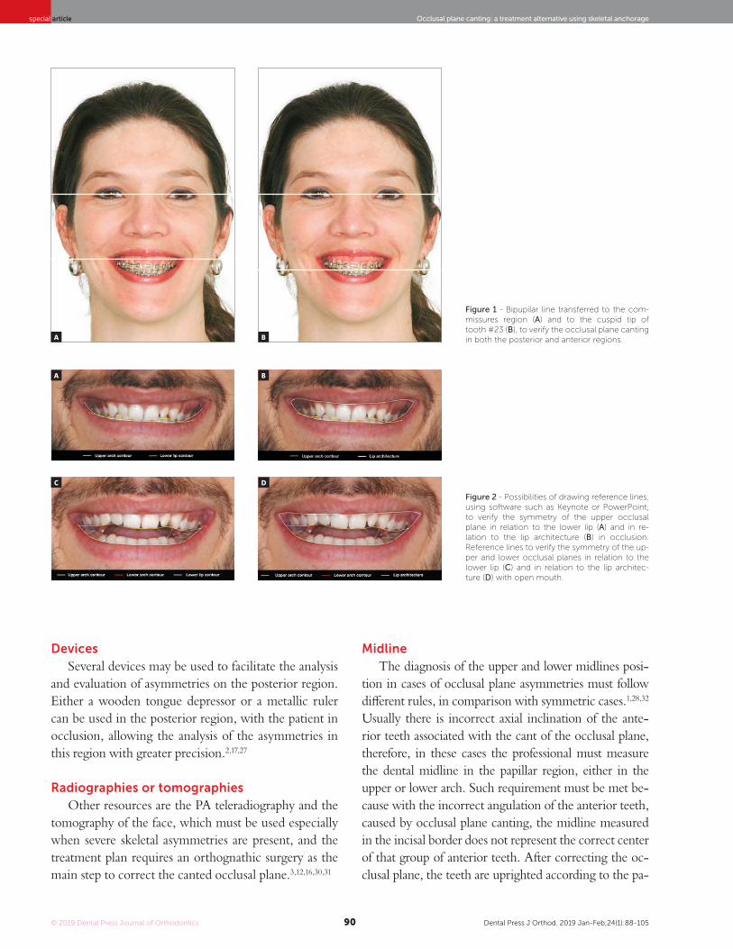
Although the mechanics is performed with the aid of skeletal anchorage, the side effects are not totally avoided and must be controlled by the orthodontist. The most common side effect, when the mechanics is performed only on the buccal side, is the buccal flaring of the intruded teeth, and it occurs due to the distance from the point where the force is applied and the center of resistance of the group of teeth, creating a moment of force and moving the crowns buccally. A tendency for crossbite is commonly observed on the opposite side, due to the rotation of occlusal plane, moving the crowns lingually on this side. One alternative to avoid these undesirable effects in the posterior region of the upper arch is to use a removable transpalatal bar (TPB),
which avoids the overexpansion of the arch on the side of intrusion and aids in controlling the torque on both sides. Another option is to use buccal and palatal tem-porary anchorage device, thereby eliminating the need for TPB to control side effects on that side. Further-more, during the intrusion, the use of 0.019 × 0.025-in stainless steel archwire is essential, allowing the control of the torque, with an accentuated buccal root torque on the side of the intrusion, whether a lingual root torque must be inserted on the opposite side, avoiding the ten-dency for crossbite. If the intrusion is performed on the lower arch, a lingual arch is the option to avoid the same side effects, associated with the same rectangular arch-wire and torque control as in the upper arch.
Figure 4 - Scheme of control of the side effects during the mechanics to cor-rect a canted occlusal plane with skeletal anchorage: A) Torque control in the archwire during the intrusion on the left side of the upper arch, B) auxiliary torque control through the transpalatal bar (TPB).
A
B

© 2019 Dental Press Journal of Orthodontics Dental Press J Orthod. 2019 Jan-Feb;24(1):88-10593
Farret MM special article
Intermaxillary vertical elastics Simultaneously performing intrusion and extrusion
is not recommended, due to the impossibility of estab-lishing the same force for both movements. After intru-sion on the upper or lower arch, the extrusion on the opposite arch must be performed. First, the intruded region must be stabilized with metal ligature connected with a mini-implant or a miniplate and connected to the teeth or directly to the archwire. Afterward, the most common way for extrusion is the use of vertical elastics connecting the intruded teeth to the opposite arch or connecting the skeletal anchorage device to the opposite side. Similar in intrusion, the side effects are constantly present in the extrusion with elastics, showing a tendency for lingual crown movement, which should be avoided with the same 0.019 × 0.025-in rectangular stain-less steel archwire. A lingual root torque must lie on the side of the extrusion, avoiding the lingual inclination of the crowns and buccal root torque on the opposite side, and avoiding the crossbite tendency on the other.
MidlinesThe upper and lower midlines usually are deviate
from the facial midline in cases with occlusal plane canting. As discussed in the Diagnosis section, the cor-rect midline must be defined through clinical examina-
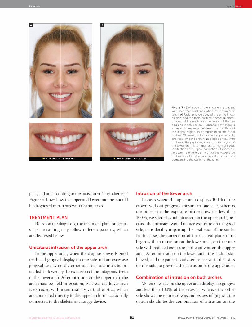
tion and mainly by smile photographs. Mechanically, the midline measured in the incisal area of the incisors presents more significant changes during the treat-ment, whereas the midline measured in the papillar area shows a lower tendency to change. If an intrusion is performed on one side of the upper arch, the upper midline measured in the incisal region will shift con-siderably to the same side, whereas the midline in the papillar region will present less shifting. During extru-sion, the midline in the incisal and papillar regions shift to the opposite side. Therefore, these changes must be carefully considered before starting the treatment, to avoid unexpected effects during treatment. If the upper and lower midlines are canted before treatment, coin-cidentally, the correction of occlusal plane canting will generate uprighted upper and lower midlines; however, those midlines will be deviated between them, and ad-ditional mechanics will be necessary to correct this de-viation, increasing the total treatment time and prob-ably upsetting the patient. Considering this condition before the beginning of the treatment, the orthodon-tist may plan additional anchorage devices or different mechanics to correct all deviations, reducing the total treatment time. The scheme of Figure 6 shows how the upper midline responds to the intrusion and extrusion movements used to correct the occlusal plane canting.
Figure 5 - Torque control in the lower arch during the extrusion mechanics with intermaxillary elastics.

© 2019 Dental Press Journal of Orthodontics Dental Press J Orthod. 2019 Jan-Feb;24(1):88-10594
Occlusal plane canting: a treatment alternative using skeletal anchoragespecial article
CASE 1Diagnosis and treatment plan
This case presents the sequence of an unsuccessful orthodontic treatment conducted for 3 years. A 29-year-old woman has been treated to correct a Class II, subdi-vision left malocclusion, with accentuated midline de-viation, using unilateral intermaxillary elastics. The side effects of the long period using the elastics were occlusal plane canting, mainly in the region of canine and adja-cent teeth, which can be identified in the frontal smile photograph. The upper midline was deviated 4 mm to
the right, with slight inclination of the anterior teeth to the right, while the lower midline was correct. Similar-ly, the patient presented an accentuated gummy smile, with 6-mm of gingival display on the upper central inci-sors region. Intraoral analysis showed a Class II maloc-clusion on the left side, accentuated deviation between the midlines, and a 4-mm overjet, whereas the lower anterior teeth were rotated. The chief complaint of the patient was the gummy smile and occlusal plane cant-ing, which according to her was non-existent before the first treatment (Fig 7).
Figure 6 - Effect on the upper midline provoked by the correction of occlusal plane: A) Correct midline in the region of the papilla at the beginning, and correct at the end of occlusal plane canting correction with intrusion; B) correct midline at the incisal edge at the beginning and deviated after occlusal plane canting correction with intrusion; C) midline correct in the papilla region at the beginning and correct at the end of the occlusal plane canting correction with extrusion; D) correct midline at the incisal edge at the beginning and deviated after the occlusal plane canting correction with extrusion.
A
C
B
D
Midline correct in the papilla before the intrusion

© 2019 Dental Press Journal of Orthodontics Dental Press J Orthod. 2019 Jan-Feb;24(1):88-10595
Farret MM special article
Figure 7 - Initial photographs.
Based on the diagnosis, the planned treatment was the use of two miniplates in the upper arch to cor-rect occlusal plane canting, associated to Class II re-lationship correction. As the occlusal plane canting was caused by the first orthodontic treatment during the use of unilateral Class II elastics, it was decided to use a similar sagittal force; however, with upward di-rection, provoking an intrusion on the left side of the upper arch. As the Class II was more pronounced on the left side, the force should be higher in this side, al-lowing the correction of the midline as well. With the miniplates, it was possible to considerably reduce the total treatment time, because sagittal and vertical prob-lems would be corrected at the same time and a group of teeth could be moved together. After orthodontic treatment, the patient would be refereed to periodontal surgery and rehabilitation of the anterior teeth.
Treatment progressAll the brackets from the previous treatment were
removed, and the orthodontic appliance was rebond-ed. Standard Edgewise brackets with 0.022 × 0.028-in slots were used, and alignment and leveling started with
rounded nickel-titanium archwires (0.012 and 0.014-in), followed by 0.016-in to 0.019 × 0.025-in stainless steel archwires on both arches. One miniplate was installed on each side of the upper arch in the zygomatic pillar area, and the mechanics began with a retraction force (400 g/f) delivered by the elastic chains with upward direction on the left side and without vertical force on the right side (150 g/f). The force on the left side was higher, in order to correct the Class II in this side and correct the upper mid-line deviation. Two months later, a TPB was positioned, and a vertical force (200 g/f) was applied directly to tooth #26 to level the posterior region of the arch. In the fourth month of mechanics, the force was applied only on the left side and, after the correction of Class II relationship on the left side, vertical intermaxillary elastics were used only on the left side, to extrude the lower teeth (Fig 8). Occlusal plane canting and the gummy smile progres-sively reduced, as shown in the smile photographs. After achieving the treatment objectives, a period of 4 months has been considered before the debonding. After the orth-odontic treatment, a periodontal surgery was performed. Then, new restorations were made on the upper incisors and the final results can be observed in the Figure 9.

© 2019 Dental Press Journal of Orthodontics Dental Press J Orthod. 2019 Jan-Feb;24(1):88-10596
Occlusal plane canting: a treatment alternative using skeletal anchoragespecial article
Figure 9 - Final photographs after periodontal surgery and restorations.
Figure 8 - Photographs of treatment progress: A, B) beginning of the mechanics with mini-plates with 400g of force in the left side, with an upward direction; and 150g in the right side, without vertical component; C, D) two months of mechanics and application of 200g of in-trusive force in tooth #26 region; E, F) end of Class II and occlusal plane canting correction on the left side, stabilization with two elastics with intrusive and retraction force, and begin-ning of the use of intermaxillary elastics for ex-trusion of the lower arch.
CASE 2Diagnosis and treatment plan
The patient was a 24-year-old woman who previ-ously underwent orthodontic treatment for 3 years. The patient was unsatisfied with the incorrect angulation and excessive proclination of the anterior teeth, with crowd-ing on the lower arch. Facial analysis revealed a convex profile and mandibular asymmetry, with deviation to the right side, which probably provoked an asymmetry on the upper arch. The analysis of the midline showed a 3-mm deviation in comparison with the facial midline, with angulation of the anterior teeth to the right side.
Analysis of the intraoral photographs showed a Class I relationship on both sides (Fig 10). On this basis, one of the treatment options for this case was four premo-lar extractions. This option would encompass almost all the necessities of the case. However, the occlusal plane canting would remain uncorrected. Furthermore, the patient refused the extractions due to the spaces that would be created before closing with orthodontic me-chanics. Thus, the best option considered was the use of skeletal anchorage to level the occlusal plane and obtain spaces to reduce the projection of the incisors and elimi-nate crowding in the lower arch.
A
C
E
B
D
F

© 2019 Dental Press Journal of Orthodontics Dental Press J Orthod. 2019 Jan-Feb;24(1):88-10597
Farret MM special article
Treatment progressStandard Edgewise brackets with 0.022 × 0.028-in
slots were bonded on the upper and lower arches, except for the lower incisors. Alignment and leveling were per-formed from the 0.012 and 0.014-in nickel-titanium arch-wires through the 0.016, 0.018, and 0.020-in stainless steel archwires up to rectangular 0.019 × 0.025-in archwires. At this moment, four miniplates were positioned, one in each posterior region of the quadrants, and the intrusion on the left side of the upper arch started with elastomeric chains connected directly from the miniplate to the arch, with 200 g/f. After one month, the retraction of both arch-es with elastics connected from the miniplates to hooks
Figure 10 - Initial photographs.
welded over the arches was started. Three months after initiating the retraction, the lower incisors were bonded, and an overlaid 0.012-in nickel-titanium archwire was inserted to align and level the teeth. The treatment pro-gressed until correction of the upper occlusal plane, and at this moment, this arch was stabilized with metal ligatures connected to the miniplate. The patient was instructed to use intermaxillary elastics directly connected to the mini-plate in the upper arch and to the lower teeth, to promote the lower dentition extrusion on the left side (Fig 11). Af-ter the extrusion of the lower arch, the treatment was sta-bilized for 4 months before debonding. The final results of the treatment are shown in Figure 12.

© 2019 Dental Press Journal of Orthodontics Dental Press J Orthod. 2019 Jan-Feb;24(1):88-10598
Occlusal plane canting: a treatment alternative using skeletal anchoragespecial article
Figure 11 - Treatment progress: A, B) Beginning of the mechanics with upper left side intrusion, C, D) end of upper intrusion and inclusion of lower incisors, after lower teeth retraction; E) beginning of mechanics with elastics connected directly to the miniplate and to the lower arch and F) end of the lower extrusion.
Figure 12 - Final photographs.
A C E
B D F
CASE 3 Diagnosis and treatment plan
A 48-year-old woman sought for orthodontic retreat-ment, complaining about unaesthetic smile. The facial analysis revealed an increased lower third height, and the patient presented no passive lip seal. The profile was convex, and the smile aesthetics was impaired due to oc-clusal plane canting associated to incorrect angulation of the anterior teeth to the right side. Intraoral photographs showed: Class III relationship and crossbite on the right side; Class II relationship on the left side; upper midline deviated 1.5 mm to the right, and edge-to-edge relation-ship among incisors. The patient received implant-pros-thetic rehabilitation on the right maxillary first molar, right maxillary lateral incisor, and left mandibular first molar (Figs 12 and 13). The first alternative considered
was the surgical correction of the occlusal plane cant-ing through the maxillary impaction on the left side and mandibular surgical rotation, which could lead to a counterclockwise rotation of the mandible, reducing the facial height and profile convexity. However, the patient refused the orthognathic surgery. Thus, this option was discarded. The second option considered was the combi-nation of intrusion on the maxillary left side and mandib-ular right side with the aid of miniplates. This alternative could provoke the mandibular counterclockwise rotation and reduce the facial vertical pattern. Moreover, the Class III relationship would be corrected by means of distal-ization on the right side, eliminating the anterior edge-to-edge relationship, whereas the lower left second molar would be uprighted and supported by the implant on the first molar. The patient chose this treatment option.

© 2019 Dental Press Journal of Orthodontics Dental Press J Orthod. 2019 Jan-Feb;24(1):88-10599
Farret MM special article
Figure 13 - Initial photographs.
Treatment progressThe treatment started with alignment and leveling
with 0.012 and 0.014-in nickel-titanium archwires, fol-lowed by 0.016 to 0.019 × 0.025-in stainless steel arch-wires with 0.022 × 0.028-in ceramic standard Edgewise brackets. After six months, a TPB was installed on the upper arch, and the patient was referred to a maxillofacial surgeon to insert a miniplate on the zygomatic area on the left side of the maxilla and on the external oblique ridge on the right side of the mandible. One month after the surgery, power arms were adapted on the tubes fit-ted to the miniplates, and elastomeric chains were con-nected from the power arms to the archwire. Only an intrusion force was present on the upper arch, whereas on the lower arch, intrusion and retraction forces were delivered by the elastics, to correct the Class III relation-ship and anterior crossbite. On the lower arch, the lingual arch was discarded due to the implant on the left side; the implant was used with a contracted archwire during the intrusion, avoiding the overexpansion of the arch on the right side. After 4 months of intrusion, the posterior oc-clusal plane canting was almost totally corrected. How-
ever, the upper arch presented occlusal plane canting that was localized mainly on the left side of the anterior re-gion. Therefore, a cantilever made with 0.017 × 0.025-in titanium-molybdenum wire was inserted in the bracket of the upper right lateral incisor, which was an implant, and connected to the region between the upper left lat-eral incisor and canine, with an intrusion force of 100 g/f. Then, the lower arch would be extruded on the left side. The option was bonding another bracket over the buc-cal surface of the mandibular left first molar, which was also an implant, and connecting a similar cantilever to the maxillary arch, delivering an extrusion force of 100 g/f. Another step was performed on the posterior region of the arch, close to the implant, provoking an extrusion of the premolar region (Fig 14). Four months after the orth-odontic treatment, the patient was referred to perform a gingival surgery from the maxillary right second premo-lar to the maxillary left premolar, increasing the crown lengthening and establishing symmetry among the gingi-val contour of the maxillary teeth, and after the surgery, the patient started her aesthetic rehabilitation. The final results can be observed in Figure 15.

© 2019 Dental Press Journal of Orthodontics Dental Press J Orthod. 2019 Jan-Feb;24(1):88-105100
Occlusal plane canting: a treatment alternative using skeletal anchoragespecial article
CASE 4Diagnosis and treatment plan
A 32-year-old woman sought for orthodontic treatment, complaining about the crowding on the anterior region of the mandibular arch. The smile analysis revealed an occlusal plane canting, with more gingiva displayed on the left side. Intraoral analysis showed: Class I molar relationship, slight Class III ca-nine relationship, correct upper midline (measured in the papilla), and lower midline deviated 2 mm to the right. The lower arch discrepancy was -7 mm, and
the upper arch discrepancy totaled -2 mm. Analysis of Bolton discrepancy revealed a 2-mm excess on the anterior region of the lower arch (Fig 16).
The treatment plan involved intrusion of the up-per left side with mini-implant positioned between the premolars, as the initial periapical radiography showed a good space on that region. Given the increased lower discrepancy and the Bolton discrepancy with lower ex-cess, the decision was to extract tooth #41 and close the space with tooth #31 in the midline.
Figure 15 - Final photographs.
Figure 14 - Photographs of treatment progress: A-C) Mechanics with miniplates for upper left intrusion and intrusion in the lower right side, associated with retraction, D) end of the intrusion with miniplates, E) intrusion of the left side of anterior region with cantilever connected to the implant of tooth #12 and F) ex-trusion of the left side of the lower arch, with aid of cantilever connected to the implant of the tooth #36.
A C
E
B
D F

© 2019 Dental Press Journal of Orthodontics Dental Press J Orthod. 2019 Jan-Feb;24(1):88-105101
Farret MM special article
Figure 16 - Initial photographs.
Treatment progress Treatment started with bonding of standard Edge-
wise brackets on both arches and extraction of tooth #41. Alignment and leveling were performed with rounded nickel-titanium archwires until the 0.019 × 0.025-in stainless steel archwires. At this moment, a mini-implant was inserted between teeth #24 and #25, and a small force (50 g/f) was immediately applied from the mini-implant to the upper arch with an elastic chain. One month after, the force was increased to 200 g/f, and the intrusion was
monitored monthly. After three months, canting of the upper arch was corrected; this arch was stabilized with metallic ligatures, as shown in Figure 17. To promote the lower teeth extrusion, intermaxillary elastics (3/8-in) were connected from the upper to the lower teeth, in the shape of an "M'. After correcting the lower arch cant, the treatment was stabilized for four more months before the removal. After the orthodontic treatment, the patient un-derwent oral rehabilitation with the new prosthesis and restorations (Fig 18).

© 2019 Dental Press Journal of Orthodontics Dental Press J Orthod. 2019 Jan-Feb;24(1):88-105102
Occlusal plane canting: a treatment alternative using skeletal anchoragespecial article
Figure 18 - Final photographs.
Figure 17 - Photographs of treatment progress: A, B) beginning of upper arch intrusion and C, D) stabilization of the upper arch in the mini-implant and extrusion of the lower arch with in-termaxillary elastics.
A
D
B
C
CASE 5Diagnosis and treatment plan
Female patient, 36 years of age at the beginning of treatment, whose facial analysis revealed asymmetry, with mandibular deviation to the left side. Due to man-dibular asymmetry, there was also an asymmetry of lips in both resting and smiling. In addition, in the smile it was possible to identify an asymmetry of the occlusal plane, with good exposure of the upper teeth on the right side and reduced exposure of the upper teeth on the left side. The upper midline was coincident in the papilla and devi-ated 1.5 mm to the left in the incisal region, and the lower midline was coincident in the incisal region and 2 mm deviated to the left in the papilla region. In the intraoral analysis, it was observed a Class II relationship on the right side and Class I on the left side, crowding in the an-
terior region of both arches and a subgingival fracture of the tooth #36. On the panoramic radiograph, it was pos-sible to verify that tooth #36 presented extensive resto-ration and poorly conducted endodontics, which would require further intervention. In addition, the patient had the tooth #38 in the mouth and with good crown and root shape, and the tooth #48 was impacted and in a posi-tion of difficult traction (Fig 19).
The planned treatment was the extraction of teeth #36 and #45, to create spaces in the lower arch to align the incisors, and the rest of the space would be used for loss of anchorage with the help of Class II elastics. After the closure of the lower spaces, a mini-implant would be used in the left side of the lower arch, to provoke the intrusion of this region, followed by the extrusion of the upper teeth with the aid of intermaxillary elastics.

© 2019 Dental Press Journal of Orthodontics Dental Press J Orthod. 2019 Jan-Feb;24(1):88-105103
Farret MM special article
Figure 19 - Initial photographs.
Treatment progressStandard Edgewise brackets with 0.022 × 0.028-in
slots were initially bonded in both arches, except for the lower incisors. Alignment and leveling was conducted with round nickel-titanium (0.012 and 0.014-in) arches, followed by 0.016 to 0.019 × 0.025-in steel wires in both arches. At that moment, the teeth #36 and #45 were ex-tracted and the closure of the spaces started with elastic chains connecting an anterior segment on each side in tie-together and the teeth to be mesialized (#37 and #46). Thus, it would be obtained distalization of the anterior segment to obtain spaces for the incisors, and mesializa-tion of the posterior teeth would also be obtained to close the spaces. After three months of mechanics with elastics, the lower incisors were bonded and aligned and leveled with superimposed nickel-titanium archwires. The clo-sure of the residual spaces of the lower arch was then per-
formed with rectangular arches and T-loops, and with the aid of Class II elastics. After the spaces were closed, a mini-implant was inserted between the teeth #34 and #35, and the process of intrusion was started with the at-tached elastic chain from the mini-implant to the lower arch, which had buccal root torque in the left side and lingual torque on the right side. After the intrusion, the lower arch was stabilized with metal ligatures and elastic chain, and the patient proceeded to use 3/8-in intermax-illary elastic in a shape of "M", to provoke upper left extru-sion (Fig 20). After the upper extrusion, some rebondings and bends were made, defining the spaces to fill the up-per lateral incisors, then the appliance was removed. After removal, the patient was referred to perform bleaching procedures and restorations on anterior teeth. Figure 21 shows the final orthodontic result, before the bleaching and restorations on the anterior teeth.

© 2019 Dental Press Journal of Orthodontics Dental Press J Orthod. 2019 Jan-Feb;24(1):88-105104
Occlusal plane canting: a treatment alternative using skeletal anchoragespecial article
Figure 21 - Final photographs prior to rehabilita-tion.
Figure 20 - Photographs of treatment progress: A, B) analysis of the smile before the beginning of the occlusal plane canting correction; C, D) end of the lower occlusal plane canting correction, with the aid of mini-implant; and E) end of the extrusion of the upper arch with intermaxillary elastics.
C
A
D E
B

© 2019 Dental Press Journal of Orthodontics Dental Press J Orthod. 2019 Jan-Feb;24(1):88-105105
Farret MM special article
FINAL CONSIDERATIONS Vertical asymmetries have always represented a great-
er complexity in the orthodontic treatment of adult pa-tients. Usually in cases of slight deviations, with the use of conventional mechanics, these asymmetries were treated in a limited way or with important side effects in the final result. In cases of moderate to severe deviations, the asso-ciation with orthognathic surgery was essential to obtain satisfactory results. The possibility of using skeletal an-chorage in these cases allowed the correction of impor-tant asymmetries without side effects and reducing the necessity for orthognathic surgery. However, it is fun-damental to perform a correct diagnosis in these cases, in order to plan the positioning of the skeletal anchor-age device and consequently of the region to be moved, correcting the asymmetry. In addition, the orthodontist must have control of unwanted effects from the mechan-ics, avoiding the prolongation of the orthodontic treat-ment or even unsatisfactory results.
1. Burstone CJ. Diagnosis and treatment planning of patients with asymmetries. Semin Orthod. 1998;4(3):153-64.
2. Jeon YJ, Kim YH, Son WS, Hans MG. Correction of a canted occlusal plane with miniscrews in a patient with facial asymmetry. Am J Orthod Dentofacial Orthop. 2006;130(2):244-52.
3. Takano-Yamamoto T, Kuroda S. Titanium screw anchorage for correction of canted occlusal plane in patients with facial asymmetry. Am J Orthod Dentofacial Orthop. 2007;132(2):237-42.
4. Kang YG, Nam JH, Park YG. Use of rhythmic wire system with miniscrews to correct occlusal-plane canting. Am J Orthod Dentofacial Orthop. 2010;137(4):540-7.
5. Machado AW. 10 commandments of smile esthetics. Dental Press J Orthod. 2014;19(4):136-57.
6. Farret " MM. Class II malocclusion with accentuated occlusal plane inclination corrected with miniplate: a case report. Dental Press J Orthod. 2016;21(3):94-103.
7. Sherwood KH, Burch J, Thompson W. Intrusion of supererupted molars with titanium miniplate anchorage. Angle Orthod. 2003;73(5):597-601.
8. Faber J, Morum TFA, Leal S, Berto PM, Carvalho CKS. Miniplates allow efficient and effective treatment of anterior open bites. Dental Press J Orthod. 2008;13(5):144-57.
9. Umemori M, Sugawara J, Mitani H, Nagasaka H, Kawamura H. Skeletal anchorage system for open-bite correction. Am J Orthod Dentofacial Orthop. 1999;115(2):166-74.
10. Sugawara J, Baik UB, Umemori M, Takahashi I, Nagasaka H, Kawamura H, et al. Treatment and posttreatment dentoalveolar changes following intrusion of mandibular molars with application of a skeletal anchorage system (SAS) for open bite correction. Int J Adult Orthodon Orthognath Surg. 2002;17(4):243-53.
11. Park YC, Lee SY, Kim DH, Jee SH. Intrusion of posterior teeth using mini-screw implants. Am J Orthod Dentofacial Orthop. 2003 June;123(6):690-4.
12. Hashimoto T, Fukunaga T, Kuroda S, Sakai Y, Yamashiro T, Takano-Yamamoto T. Mandibular deviation and canted maxillary occlusal plane treated with miniscrews and intraoral vertical ramus osteotomy: functional and morphologic changes. Am J Orthod Dentofacial Orthop. 2009;136(6):868-77.
REFERENCES
13. Tekale PD, Vakil KK, Sastri MR, Vakil JK, Agrawal CO, Gore KA, et al. Correction of severe deep bite and gummy smile using mini-screw anchorage: a case report. J World Fed Orthod. 2015;4(4):162-7.
14. Iwasa A, Horiuchi S, Kinouchi N, Izawa T, Hiasa M, Kawai N, et al. Skeletal anchorage for intrusion of bimaxillary molars in a patient with skeletal open bite and temporomandibular disorders. J Orthod Sci. 2017;6(4):152-8.
15. Cornelis MA, Scheffler NR, Nyssen-Behets C, De Clerck HJ, Tulloch JF. Patients' and orthodontists' perceptions of miniplates used for temporary skeletal anchorage: a prospective study. Am J Orthod Dentofacial Orthop. 2008;133(1):18-24.
16. Padwa BL, Kaiser MO, Kaban LB. Occlusal cant in the frontal plane as a reflection of facial asymmetry. J Oral Maxillofac Surg. 1997;55(8):811-6; discussion 7.
17. Benson KJ, Laskin DM. Upper lip asymmetry in adults during smiling. J Oral Maxillofac Surg. 2001;59(4):396-8.
18. Edler R, Wertheim D, Greenhill D. Clinical and computerized assessment of mandibular asymmetry. Eur J Orthod. 2001;23(5):485-94.
19. Kokich VO, Kokich VG, Kiyak HA. Perceptions of dental professionals and laypersons to altered dental esthetics: asymmetric and symmetric situations. Am J Orthod Dentofacial Orthop. 2006;130(2):141-51.
20. Lee MS, Chung DH, Lee JW, Cha KS. Assessing soft-tissue characteristics of facial asymmetry with photographs. Am J Orthod Dentofacial Orthop. 2010;138(1):23-31.
21. Jackson TH, Mitroff SR, Clark K, Proffit WR, Lee JY, Nguyen TT. Face symmetry assessment abilities: Clinical implications for diagnosing asymmetry. Am J Orthod Dentofacial Orthop. 2013;144(5):663-71.
22. Olivares A, Vicente A, Jacobo C, Molina SM, Rodriguez A, Bravo LA. Canting of the occlusal plane: Perceptions of dental professionals and laypersons. Med Oral Patol Oral Cir Bucal. 2013 May 1;18(3):e516-20.
23. Rosa M, Olimpo A, Fastuca R, Caprioglio A. Perceptions of dental professionals and laypeople to altered dental esthetics in cases with congenitally missing maxillary lateral incisors. Prog Orthod. 2013 Oct 1;14:34.
24. Corte CC, Silveira BL, Marquezan M. Influence of occlusal plane inclination and mandibular deviation on esthetics. Dental Press J Orthod. 2015 Sept-Oct;20(5):50-7.
25. Colombo VL, Moro A, Rech R, Verona J, Costa GCA. Análise facial frontal em repouso e durante o sorriso em fotografias padronizadas. Parte II: Avaliação durante o sorriso. Rev Dental Press Ortodon Ortop Facial. 2004 Jul-Ago;9(4):86-97.
26. Camara CA, Martins RP. Functional Aesthetic Occlusal Plane (FAOP). Dental Press J Orthod. 2016 July-Aug;21(4):114-25.
27. Pereira CB, Justus R, Pinzan A, Vieira Bastos SH, Lopes SL. The importance of evaluating the transverse cant of the occlusal plane in intraoral photographs. J World Fed Orthod. 2014;3(1):e19-25.
28. Kai R, Umeki D, Sekiya T, Nakamura Y. Defining the location of the dental midline is critical for oral esthetics in camouflage orthodontic treatment of facial asymmetry. Am J Orthod Dentofacial Orthop. 2016;150(6):1028-38.
29. Ishizaki K, Suzuki K, Mito T, Tanaka EM, Sato S. Morphologic, functional, and occlusal characterization of mandibular lateral displacement malocclusion. Am J Orthod Dentofacial Orthop. 2010;137(4):454 e1-9; discussion -5.
30. Ko EW, Huang CS, Chen YR. Characteristics and corrective outcome of face asymmetry by orthognathic surgery. J Oral Maxillofac Surg. 2009;67(10):2201-9.
31. Silva NCF, Aquino ERB, Mello KCFR, Mattos JNR, Normando D. Orthodontists’ and laypersons’ perception of mandibular asymmetries. Dental Press J Orthod. 2011;16(4):38.e1-8.
32. Williams RP, Rinchuse DJ, Zullo TG. Perceptions of midline deviations among different facial types. Am J Orthod Dentofacial Orthop. 2014;145(2):249-55.
Author’s contribution (ORCID )Marcel M. Farret (MMF): 0000-0003-0713-8099
Conception or design of the study, data acquisition, analy-sis or interpretation, writing the article, critical revision of the article, final approval of the article, obtained funding and overall responsibility: MMF.
Related Documents
![A device for occlusal plane determination TO MARCH 2019/14.pdfpatients, but the device lacked any posterior determinants of the plane.[10] The correct orientation of the occlusal plane](https://static.cupdf.com/doc/110x72/5e6bb8ceec327e298e63e919/a-device-for-occlusal-plane-to-march-201914pdf-patients-but-the-device-lacked.jpg)