LECTURE NOTES Obstetrics and Gynaecology DIANA HAMILTON-FAIRLEY 2nd edition

Welcome message from author
This document is posted to help you gain knowledge. Please leave a comment to let me know what you think about it! Share it to your friends and learn new things together.
Transcript

LECTURE NOTES
Obstetrics andGynaecologyDIANA HAMILTON-FAIRLEY
2nd edition

Lecture Notes: Obstetrics and Gynaecology
AMIPR 6/9/04 5:30 PM Page i

AMIPR 6/9/04 5:30 PM Page ii

Lecture Notes
Obstetrics andGynaecology
Diana Hamilton-FairleyMD, FRCOGConsultant Obstetrician and GynaecologistGuy’s and St Thomas’s Hospital NHS Trust, London
Second Edition
AMIPR 6/9/04 5:30 PM Page iii

© 2004 D. Hamilton-Fairley© 1999 Blackwell Science LtdPublished by Blackwell Publishing Ltd
Blackwell Publishing, Inc., 350 Main Street, Malden, Massachusetts 02148-5020, USABlackwell Publishing Ltd, 9600 Garsington Road, Oxford OX4 2DQ, UKBlackwell Publishing Asia Pty Ltd, 550 Swanston Street, Carlton, Victoria 3053, Australia
The right of the Author to be identified as the Author of this Work has been asserted in accordance with the Copyright, Designs and Patents Act 1988.
All rights reserved. No part of this publication may be reproduced, stored in a retrieval system, or transmitted, in anyform or by any means, electronic, mechanical, photocopying, recording or otherwise, except as permitted by the UKCopyright, Designs and Patents Act 1988, without the prior permission of the publisher.
First published 1999Reprinted 2000, 2001Second edition 2004
Library of Congress Cataloging-in-Publication DataHamilton-Fairley, Diana.
Lecture notes on obstetrics and gynaecology / Diana Hamilton-Fairley. —2nd ed.p. ; cm.
Rev. ed. of: Lecture notes on obstetrics and gynaecology / Geoffrey Chamberlain,Diana Hamilton-Fairley.
Includes index.ISBN 1-4051-2066-51. Obstetrics. 2. Gynecology.[DNLM: 1. Obstetrics. 2. Gynecology. WQ 100 H217L 2004] I. Title: Obstetrics and
gynaecology. II. Chamberlain, Geoffrey, 1930 —Lecture notes on obstetrics and gynaecology. III. Title.RG526.C43 2004618 —dc22
2004007260ISBN 1-4051-2066-5
A catalogue record for this title is available from the British Library
Set in 8/12 Stone Serif by SNP Best-set Typesetter Ltd., Hong KongPrinted and bound in India by Replika Press Pvt. Ltd.
Commissioning Editor: Vicki NoyesEditorial Assistant: Nic UlyattProduction Editor: Helen Harvey and Karen MooreProduction Controller: Kate Charman
For further information on Blackwell Publishing, visit our website:http://www.blackwellpublishing.com
The publisher’s policy is to use permanent paper from mills that operate a sustainable forestry policy, and which hasbeen manufactured from pulp processed using acid-free and elementary chlorine-free practices. Furthermore, thepublisher ensures that the text paper and cover board used have met acceptable environmental accreditation standards.
AMIPR 6/9/04 5:30 PM Page iv

v
12 Normal labour, 157
13 Abnormal labour, 174
14 Puerperium, 203
15 The newborn, 211
Part 4 The mature woman
16 Abnormal vaginal blood loss, 219
17 Pelvic pain, 228
18 Breast disease, 247
19 Screening for gynaecological cancer, 258
Part 5 The older woman
20 Malignant gynaecological conditions, 267
21 The menopause, 278
22 Pelvic floor disorders, 284
Part 6 Audit of obstetrics and gynaecology
23 Statistics of reproductive medicine, 297
Answers to self-assessment questions, 306
Index, 323
Contents
Preface, vii
Acknowledgements, viii
1 Basic science, 1
Part 1 The woman
2 The woman as a patient, 19
Part 2 The young woman
3 Puberty and menstrual problems of young
women, 29
4 Subfertility, 38
5 Pregnancy prevention, 46
6 Benign diseases, genital tract infections and
sexual problems, 62
Part 3 The reproductive years
7 The mother and fetus in pregnancy, 83
8 Bleeding in pregnancy, 95
9 The antenatal period, 105
10 Diseases of pregnancy, 122
11 Diseases in pregnancy, 138
AMIPR 6/9/04 5:30 PM Page v

vii
Preface
Welcome to the second edition of Lecture Notes:
Obstetrics and Gynaecology. Professor Geoffrey
Chamberlain asked me to assist him with the com-
bining of the original well-established separate
Lecture Notes on Obstetrics and Lecture Notes on
Gynaecology by joining him as editor of this text-
book aimed at undergraduate medical, midwifery
and nursing students, junior doctors, nurses and
midwives. He told me then that he intended to re-
tire from the editorship for the second edition. I
owe him an enormous debt as a teacher, mentor
and guide through my career and into the complex
area of editing a book with an illustrious list of em-
inent obstetricians and gynaecologists as its previ-
ous editors. He graciously agreed to proof read this
edition and I thank him for his helpful contribu-
tion to the final version. He continues to work as
the Emeritus Professor of History of Medicine at
the University of Wales.
In this edition I have asked two of my colleagues
at Guy’s, King’s and St Thomas’s Medical
School/Guy’s and St Thomas’s Hospital NHS Trust
to expand the sections on Sexually Transmitted
Diseases and Breast Disease to reflect the changes
in the undergraduate medical curriculum which
combines Obstetrics and Gynaecology, Breast
Disease and Sexual Health in several UK universi-
ties. I would like to thank them both: Dr David
Lewis FRCP, MD from Sexual Health and Mr
Nicholas Beechey Newman FRCS, MS who wrote
the chapter on Breast Disease. I think their two
chapters (6 and 18) are a valuable addition to the
book and I hope you, the reader, will agree.
Feedback from students, Senior Lecturers and
Professors has led to many smaller changes in the
book including an expansion on the history taking
and examination sections. At the end of each chap-
ter there are five self-assessment questions with the
answers/marking schemes given in Answers to self-
assessment questions (p. 306). The questions cover
the full range that may be found within the exam-
ination system in the United Kingdom, both at
undergraduate and postgraduate levels, including
extended matched questions, scenarios for practic-
ing history taking as in Objective Structured Clini-
cal Examination (OSCE) as well as the more
traditional Multiple Choice Questions. I trust
they will be of help in the learning and revision
process.
Over the decades this series has been translated
into many languages, thus reaching an interna-
tional audience. I hope those using this book all
over the world and those who are not doctors in
training will find the changes made to this book
an addition to their learning even though many
of them are based on the changes that have
occurred in the British Medical Undergraduate
curriculum.
As the editor, I would like this book to aid its
readers’ understanding of this very important area
of health care and that some of you will turn to
caring for women and their families as your long-
term career. If this book contributes to either or
both of these then I am pleased.
Diana Hamilton-Fairley, 2004
AMIPR 6/9/04 5:30 PM Page vii

viii
I would like to thank the following for their in-
valuable contribution to this book: Dr David Lewis
FRCP, MD Consultant, Department of Genitouri-
nary Medicine, Guy’s and St Thomas’s Hospital
NHS Trust; Mr Nicholas Beechey Newman FRCS,
MS Senior Lecturer, Department of Endocrine
Surgery, Guy’s, King’s and St Thomas’s Hospital
Medical School, King’s College London; Professor
Geoffrey Chamberlain Emeritus Professor of
History of Medicine, University of Wales; and
the editorial and publishing staff of Blackwell
Publishing.
Acknowledgements
AMIPR 6/9/04 5:30 PM Page viii
Female anatomy
The woman’s body is built in a different way from
that of the male; it is less muscular and therefore
has a slighter skeleton to support the muscles. In
the abdomen, the non-pelvic organs are similar
and subject to the same diseases. Readers are there-
fore referred to books on general anatomy and this
chapter is concerned with female pelvic anatomy.
Since much changes in pregnancy we will intro-
duce the pregnancy aspects in this section and
Chapter 7.
Uterus (Box 1.1)
A hollow, muscle-walled organ in the pelvis com-
municating with each fallopian tube and, through
its cervix, the vagina.
Pre-pregnancy: 7 ¥ 5 ¥ 3cm; weight, 40g.
Full term: 30 ¥ 25 ¥ 20cm; weight, 1000g.
Structure
Muscle in three layers with vascular anastomosis
between them.
1 Outer: thin, longitudinal, merging with ligaments.
2 Middle: very thick, spiral muscle fibres with
blood vessels between.
3 Inner: thin, oblique with condensation at each
cornu and at the upper and lower end of the cervi-
cal canal —the internal and external os.
Increase in size during pregnancy is mostly
due to hypertrophy of existing cells rather than
increase in number. Changes are stimulated by
oestrogen and gradual stretch (maximum
effective stretch about term).
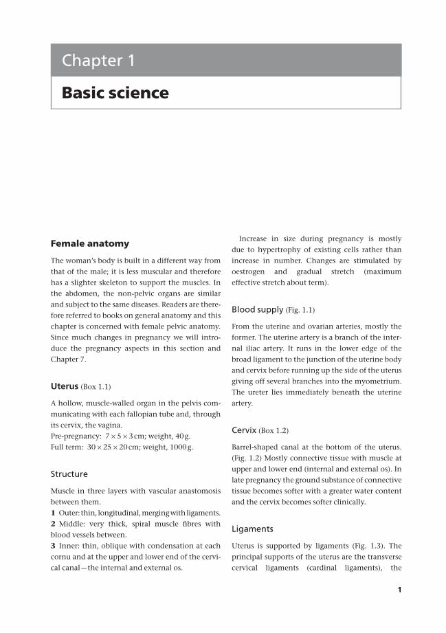
Blood supply (Fig. 1.1)
From the uterine and ovarian arteries, mostly the
former. The uterine artery is a branch of the inter-
nal iliac artery. It runs in the lower edge of the
broad ligament to the junction of the uterine body
and cervix before running up the side of the uterus
giving off several branches into the myometrium.
The ureter lies immediately beneath the uterine
artery.
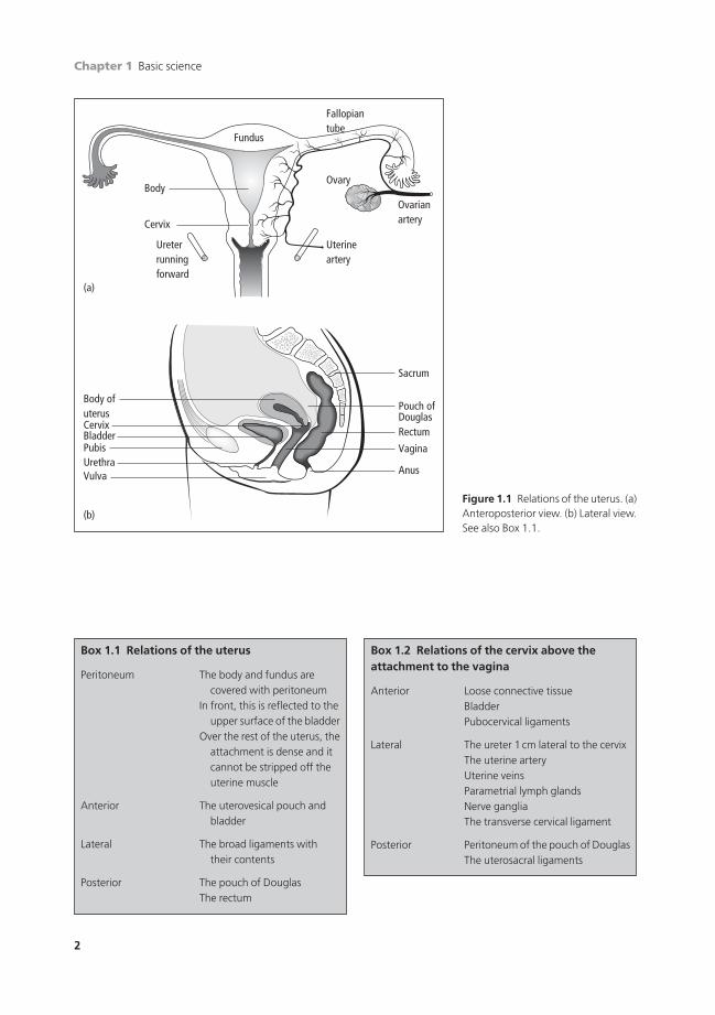
Cervix (Box 1.2)
Barrel-shaped canal at the bottom of the uterus.
(Fig. 1.2) Mostly connective tissue with muscle at
upper and lower end (internal and external os). In
late pregnancy the ground substance of connective
tissue becomes softer with a greater water content
and the cervix becomes softer clinically.
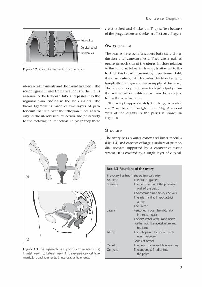
Ligaments
Uterus is supported by ligaments (Fig. 1.3). The
principal supports of the uterus are the transverse
cervical ligaments (cardinal ligaments), the
11
Chapter 1
Basic science
AMI1 6/9/04 4:57 PM Page 1
Chapter 1 Basic science
2
Ovarianartery
Ovary
Fallopian tube
Fundus
Uterineartery
Ureterrunningforward
Sacrum
Pouch ofDouglasRectum
Vagina
Anus
Body ofuterusCervixBladderPubisUrethraVulva
Body
(a)
(b)
Cervix
Figure 1.1 Relations of the uterus. (a)Anteroposterior view. (b) Lateral view.See also Box 1.1.
Box 1.2 Relations of the cervix above theattachment to the vagina
Anterior Loose connective tissueBladderPubocervical ligaments
Lateral The ureter 1cm lateral to the cervixThe uterine arteryUterine veinsParametrial lymph glandsNerve gangliaThe transverse cervical ligament
Posterior Peritoneum of the pouch of DouglasThe uterosacral ligaments
Box 1.1 Relations of the uterus
Peritoneum The body and fundus arecovered with peritoneum
In front, this is reflected to theupper surface of the bladder
Over the rest of the uterus, theattachment is dense and itcannot be stripped off theuterine muscle
Anterior The uterovesical pouch andbladder
Lateral The broad ligaments withtheir contents
Posterior The pouch of DouglasThe rectum
AMI1 6/9/04 4:57 PM Page 2
Basic science Chapter 1
3
are stretched and thickened. They soften because
of the progesterone and relaxin effect on collagen.
Ovary (Box 1.3)
The ovaries have twin functions; both steroid pro-
duction and gametogenesis. They are a pair of
organs on each side of the uterus, in close relation
to the fallopian tubes. Each ovary is attached to the
back of the broad ligament by a peritoneal fold,
the mesovarium, which carries the blood supply,
lymphatic drainage and nerve supply of the ovary.
The blood supply to the ovaries is principally from
the ovarian arteries which arise from the aorta just
below the renal arteries.
The ovary is approximately 4cm long, 3cm wide
and 2cm thick and weighs about 10g. A general
view of the organs in the pelvis is shown in
Fig. 1.1b.
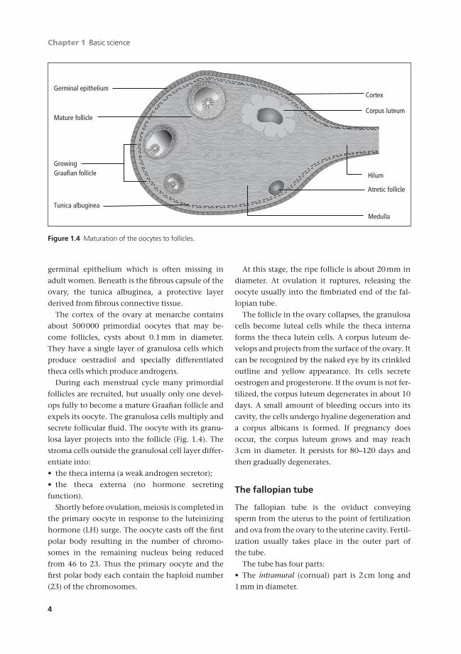
Structure
The ovary has an outer cortex and inner medulla
(Fig. 1.4) and consists of large numbers of primor-
dial oocytes supported by a connective tissue
stroma. It is covered by a single layer of cubical,
Internal os
Cervical canal
External os
Figure 1.2 A longitudinal section of the cervix.
uterosacral ligaments and the round ligament. The
round ligament rises from the fundus of the uterus
anterior to the fallopian tube and passes into the
inguinal canal ending in the labia majora. The
broad ligament is made of two layers of peri-
toneum that run over the fallopian tubes anteri-
orly to the uterovesical reflection and posteriorly
to the rectovaginal reflection. In pregnancy these
Box 1.3 Relations of the ovary
The ovary lies free in the peritoneal cavityAnterior The broad ligamentPosterior The peritoneum of the posterior
wall of the pelvisThe common iliac artery and veinThe internal iliac (hypogastric)
arteryThe ureter
Lateral Peritoneum over the obturatorinternus muscle
The obturator vessels and nerveFurther out, the acetabulum and
hip jointAbove The fallopian tube, which curls
over the ovaryLoops of bowel
On left The pelvic colon and its mesenteryOn right The appendix if it dips into
the pelvis
(a)
(b)
1
2
3
31
Figure 1.3 The ligamentous supports of the uterus. (a)Frontal view. (b) Lateral view. 1, transverse cervical liga-ment; 2, round ligaments; 3, uterosacral ligaments.
AMI1 6/9/04 4:57 PM Page 3
Chapter 1 Basic science
4
germinal epithelium which is often missing in
adult women. Beneath is the fibrous capsule of the
ovary, the tunica albuginea, a protective layer
derived from fibrous connective tissue.
The cortex of the ovary at menarche contains
about 500000 primordial oocytes that may be-
come follicles, cysts about 0.1mm in diameter.
They have a single layer of granulosa cells which
produce oestradiol and specially differentiated
theca cells which produce androgens.
During each menstrual cycle many primordial
follicles are recruited, but usually only one devel-
ops fully to become a mature Graafian follicle and
expels its oocyte. The granulosa cells multiply and
secrete follicular fluid. The oocyte with its granu-
losa layer projects into the follicle (Fig. 1.4). The
stroma cells outside the granulosal cell layer differ-
entiate into:
• the theca interna (a weak androgen secretor);
• the theca externa (no hormone secreting
function).
Shortly before ovulation, meiosis is completed in
the primary oocyte in response to the luteinizing
hormone (LH) surge. The oocyte casts off the first
polar body resulting in the number of chromo-
somes in the remaining nucleus being reduced
from 46 to 23. Thus the primary oocyte and the
first polar body each contain the haploid number
(23) of the chromosomes.
At this stage, the ripe follicle is about 20mm in
diameter. At ovulation it ruptures, releasing the
oocyte usually into the fimbriated end of the fal-
lopian tube.
The follicle in the ovary collapses, the granulosa
cells become luteal cells while the theca interna
forms the theca lutein cells. A corpus luteum de-
velops and projects from the surface of the ovary. It
can be recognized by the naked eye by its crinkled
outline and yellow appearance. Its cells secrete
oestrogen and progesterone. If the ovum is not fer-
tilized, the corpus luteum degenerates in about 10
days. A small amount of bleeding occurs into its
cavity, the cells undergo hyaline degeneration and
a corpus albicans is formed. If pregnancy does
occur, the corpus luteum grows and may reach
3cm in diameter. It persists for 80–120 days and
then gradually degenerates.
The fallopian tube
The fallopian tube is the oviduct conveying
sperm from the uterus to the point of fertilization
and ova from the ovary to the uterine cavity. Fertil-
ization usually takes place in the outer part of
the tube.
The tube has four parts:
• The intramural (cornual) part is 2cm long and
1mm in diameter.
Germinal epithelium
Mature follicle
Growing Graafian follicle
Tunica albuginea
Cortex
Corpus luteum
Medulla
Hilum
Atretic follicle
Figure 1.4 Maturation of the oocytes to follicles.
AMI1 6/9/04 4:57 PM Page 4

Basic science Chapter 1
5
• The isthmus is thick-walled and is 3cm long and
0.7mm in diameter.
• The ampulla is wide, thin-walled, being about
5cm long and 20mm in diameter (Fig. 1.5).
• The infundibulum is the lateral end of the tube. It
is trumpet shaped, crowned with the fimbriae that
surround the outer opening of the tube. The
fimbriae stabilize the abdominal ostium over the
ripening follicle in the ovary.
Structure
The tube has three coats.
• An outer serous layer of peritoneum which
covers the tube except in its intramural part and
over a small area of its attachment to the broad
ligament.
• A muscle layer with outer longitudinal and inner
circular smooth muscle.
• The mucosa or endosalpinx which lines the tube
that is thrown into numerous longitudinal folds or
rugae. The rugae have a core of connective tissue
covered with a tall columnar epithelium.
Three types of cell are found in the mucosa.
• Ciliated cells, which beat a current usually in a
medial direction.
• Secretory cells, which provide the secretion for the
rapidly developing blastocyst allowing exchange
of oxygen, nutrients and metabolites.
• Intercillary cells with long narrow nuclei,
squeezed between the other cells. There are rhyth-
mic changes in the epithelium during the men-
strual cycle; in the proliferative phase the cells
increase in height and activity with increased
secretions just after ovulation.
Vagina (Box 1.4)
The vagina is a fibromuscular canal extending
from the vestibule of the vulva to the cervix,
around which it is attached to form the fornices.
Structure
The anterior vaginal wall is about 10cm long and
the posterior wall 15cm. It is capable of great
distension, as in childbirth, after the prolonged
Round ligament
Fallopian tube
Infundibulum
Ovary
Ampulla
Infundibular ligament Peritoneal folds
Intramural (cornual)
Isthmus
Figure 1.5 Peritoneal folds to two layers of peritoneum.
Box 1.4 Relations of the vagina
Anterior The bladder and urethra
Posterior Upper —the pouch of DouglasLower —the rectum, separated by the
rectovaginal septum and perineal body
Lateral The cardinal ligaments and the levatorani muscles
AMI1 6/9/04 4:57 PM Page 5
Chapter 1 Basic science
6
hormonal stimulation of pregnancy. Normally, the
anterior and posterior walls are in contact so the
cavity is represented by an H-shaped slit.
The walls have:
• an outer connective tissue layer to which the
ligaments are attached —it contains blood vessels,
lymphatics and nerves;
• a muscular layer consisting of an outer longitu-
dinal layer and an inner circular layer of variable
thickness and function;
• the epithelium of stratified squamous epitheli-
um which in adult women contains glycogen and
is composed of three layers:
(a) a basal layer;
(b) a functional layer;
(c) a cornified layer.
The epithelium undergoes cyclical changes dur-
ing the menstrual cycle and characteristic changes
during pregnancy. After the menopause it atro-
phies so that smears taken from postmenopausal
women contain a high proportion of basal cells.
There are no glandular cells in the vaginal epithe-
lium and so the term vaginal mucosa should not be
used.
Vaginal fluid is composed of cervical secretion
and transudation through the vaginal epithelium.
The vagina allows colonization of lactobacilli
which produce lactic acid from the glycogen in the
epithelial cells.
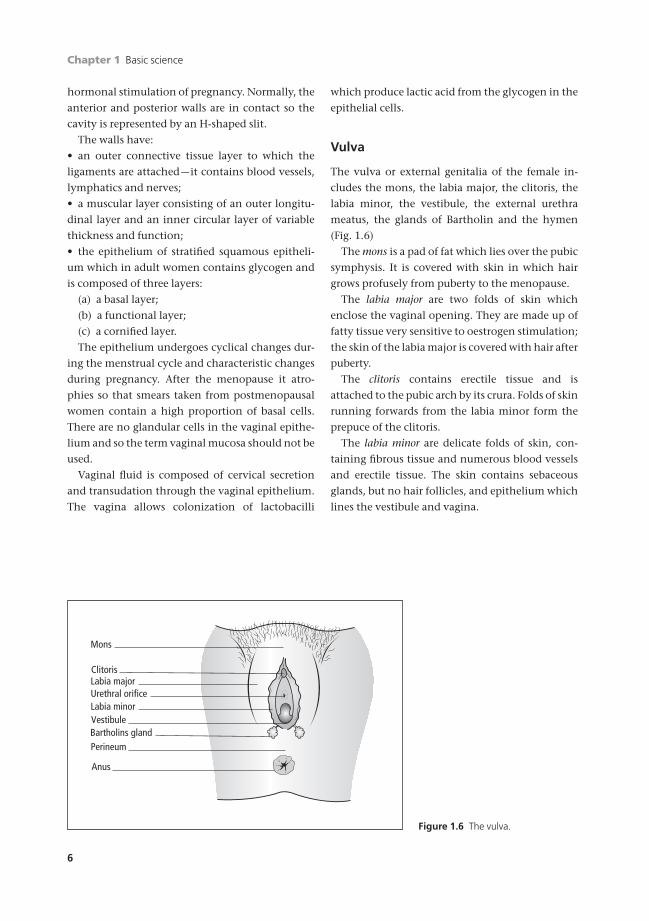
Vulva
The vulva or external genitalia of the female in-
cludes the mons, the labia major, the clitoris, the
labia minor, the vestibule, the external urethra
meatus, the glands of Bartholin and the hymen
(Fig. 1.6)
The mons is a pad of fat which lies over the pubic
symphysis. It is covered with skin in which hair
grows profusely from puberty to the menopause.
The labia major are two folds of skin which
enclose the vaginal opening. They are made up of
fatty tissue very sensitive to oestrogen stimulation;
the skin of the labia major is covered with hair after
puberty.
The clitoris contains erectile tissue and is
attached to the pubic arch by its crura. Folds of skin
running forwards from the labia minor form the
prepuce of the clitoris.
The labia minor are delicate folds of skin, con-
taining fibrous tissue and numerous blood vessels
and erectile tissue. The skin contains sebaceous
glands, but no hair follicles, and epithelium which
lines the vestibule and vagina.
Mons
Clitoris
Vestibule
Urethral orificeLabia minor
Labia major
Bartholins glandPerineum
Anus
Figure 1.6 The vulva.
AMI1 6/9/04 4:57 PM Page 6

Basic science Chapter 1
7
The vestibule is the area between the labia minor
into which opens the vagina, with the external
meatus of the urethra in front and the ducts of the
Bartholin glands behind.
The external urethral meatus is the opening of
the urethra covered with squamous epithe-
lium. Skene’s ducts from the posterior urethral
glands open on to the posterior margin of the
meatus.
The Bartholin glands are a pair of glands, the
ducts of which are lined by columnar epithelium.
Each gland is the size of a pea and in structure re-
sembles salivary glands. The secretion is colourless
and mucoid and is produced mainly on sexual
excitement.
The hymen is a circular or crescentic fold of
squamous epithelium and connective tissue
which partly closes the vaginal entrance in young
women. Its shape and size varies. It is often
ruptured or stretched by tampon insertion or by
intercourse —childbirth destroys it.
Perineum
The perineum is the area between the vaginal
opening and the anus. The perineal body is a
pyramidal mass of fibromuscular tissue into
which the fibres of the levator ani and the deep
transverse perineal muscles are inserted. These
are the muscles which are often torn or cut (epi-
siotomy) during childbirth.
Bony pelvis
The false pelvis is to true pelvis like a saucer on
top of a cup. The true pelvis is important in obstet-
rics, the false pelvis is not. Diameters are shown in
Fig. 1.7.
(a)
(b)
(c)
11
12
13
12
12
12
13
Antero-posterior
Oblique
Diameter (cm)
Transverse
12
11
Figure 1.7 The bony pelvis. (a) Inlet.Longest diameter transverse. Beanshaped. (b) Mid-cavity. All diametersequal. Circle. (c) Outlet. Longest diameter anteroposterior. Diamondshaped.
AMI1 6/9/04 4:57 PM Page 7
Chapter 1 Basic science
8
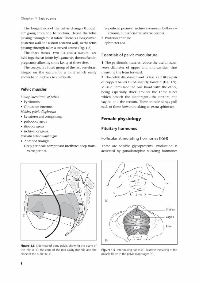
The longest axis of the pelvis changes through
90° going from top to bottom. Hence the fetus
passing through must rotate. There is a long curved
posterior wall and a short anterior wall, so the fetus
passing through takes a curved course (Fig. 1.8).
The three bones —two ilia and a sacrum —are
held together at joints by ligaments, these soften in
pregnancy allowing some laxity at these sites.
The coccyx is a fused group of the last vertebrae,
hinged on the sacrum by a joint which easily
allows bending back in childbirth.
Pelvic muscles
Lining lateral wall of pelvis
• Pyriformis.
• Obturator internus.
Making pelvic diaphragm
• Levatores ani comprising:
• pubococcygeus
• iliococcygeus
• ischiococcygeus.
Beneath pelvic diaphragm
1 Anterior triangle.
Deep perineal: compressor urethrae; deep trans-
verse perinei.
X
Z
Z
X
Figure 1.8 Side view of bony pelvis, showing the plane ofthe inlet (x–x), the zone of the mid-cavity (toned), and theplane of the outlet (z–z).
Urethra
Vagina
Anus
(a)
(b)
Figure 1.9 Interlocking hands (a) illustrate the lacing of themuscle fibres in the pelvic diaphragm (b).
Superficial perineal: ischiocavernosus; bulbocav-
ernosus; superficial transverse perinei.
2 Posterior triangle.
Sphincter ani.
Essentials of pelvic musculature
1 The pyriformis muscles reduce the useful trans-
verse diameter of upper and mid-cavities, thus
thrusting the fetus forward.
2 The pelvic diaphragm and its fascia are like a pair
of cupped hands tilted slightly forward (Fig. 1.9).
Muscle fibres lace the one hand with the other,
being especially thick around the three tubes
which broach the diaphragm —the urethra, the
vagina and the rectum. These muscle slings pull
each of these forward making an extra sphincter.
Female physiology
Pituitary hormones
Follicular stimulating hormones (FSH)
These are soluble glycoproteins. Production is
activated by gonadotrophic releasing hormones
AMI1 6/9/04 4:57 PM Page 8
Basic science Chapter 1
9
4
3
2
1
14 12 10 8 6 4 2
OvulationDays after ovulationDays preceding ovulation
2 4 6 8 10 12 14Pr
egna
ndio
l (µg
/24h
ours
)
15
10
5Oes
trad
iol (
µg/2
4hou
rs)
100
25
50
75
0
FSH
& L
H (IU
/24h
ours
)
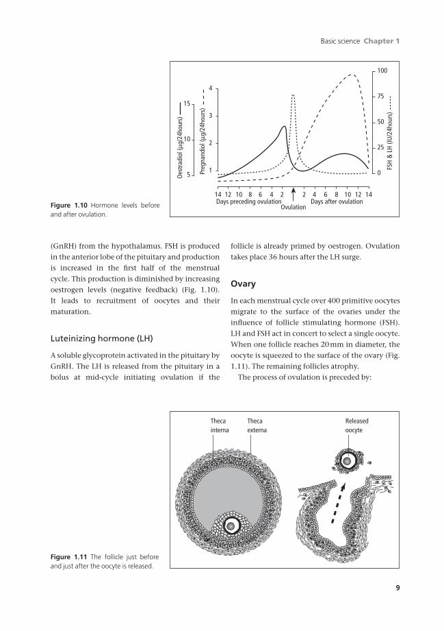
Figure 1.10 Hormone levels beforeand after ovulation.
(GnRH) from the hypothalamus. FSH is produced
in the anterior lobe of the pituitary and production
is increased in the first half of the menstrual
cycle. This production is diminished by increasing
oestrogen levels (negative feedback) (Fig. 1.10).
It leads to recruitment of oocytes and their
maturation.
Luteinizing hormone (LH)
A soluble glycoprotein activated in the pituitary by
GnRH. The LH is released from the pituitary in a
bolus at mid-cycle initiating ovulation if the
follicle is already primed by oestrogen. Ovulation
takes place 36 hours after the LH surge.
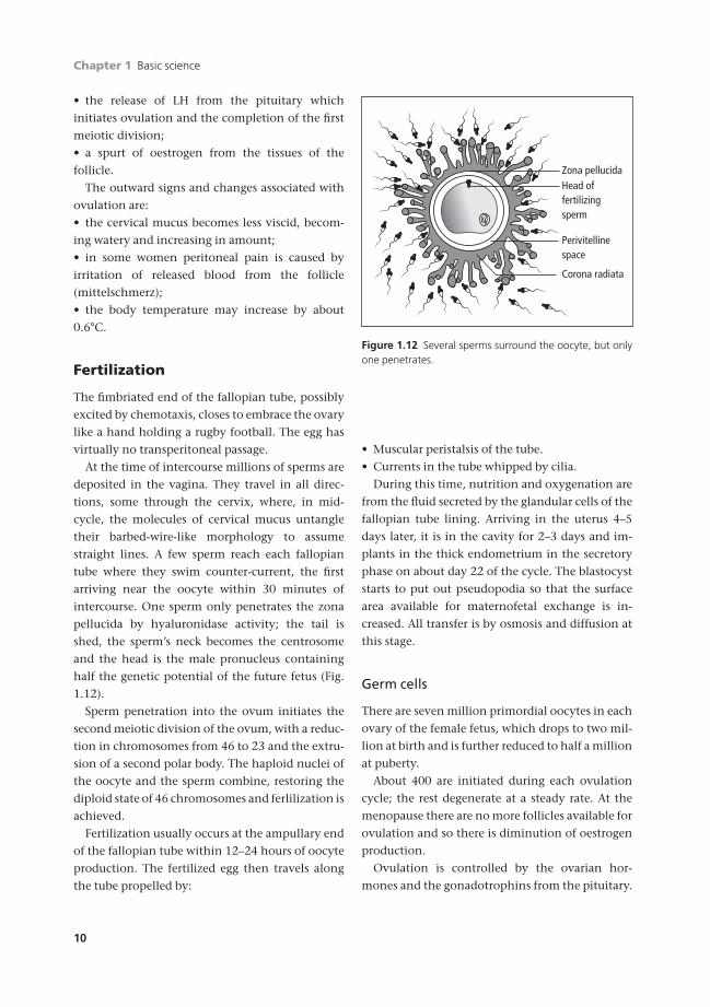
Ovary
In each menstrual cycle over 400 primitive oocytes
migrate to the surface of the ovaries under the
influence of follicle stimulating hormone (FSH).
LH and FSH act in concert to select a single oocyte.
When one follicle reaches 20mm in diameter, the
oocyte is squeezed to the surface of the ovary (Fig.
1.11). The remaining follicles atrophy.
The process of ovulation is preceded by:
Thecainterna
Thecaexterna
Releasedoocyte
Figure 1.11 The follicle just beforeand just after the oocyte is released.
AMI1 6/9/04 4:57 PM Page 9
Chapter 1 Basic science
10
• the release of LH from the pituitary which
initiates ovulation and the completion of the first
meiotic division;
• a spurt of oestrogen from the tissues of the
follicle.
The outward signs and changes associated with
ovulation are:
• the cervical mucus becomes less viscid, becom-
ing watery and increasing in amount;
• in some women peritoneal pain is caused by
irritation of released blood from the follicle
(mittelschmerz);
• the body temperature may increase by about
0.6°C.
Fertilization
The fimbriated end of the fallopian tube, possibly
excited by chemotaxis, closes to embrace the ovary
like a hand holding a rugby football. The egg has
virtually no transperitoneal passage.
At the time of intercourse millions of sperms are
deposited in the vagina. They travel in all direc-
tions, some through the cervix, where, in mid-
cycle, the molecules of cervical mucus untangle
their barbed-wire-like morphology to assume
straight lines. A few sperm reach each fallopian
tube where they swim counter-current, the first
arriving near the oocyte within 30 minutes of
intercourse. One sperm only penetrates the zona
pellucida by hyaluronidase activity; the tail is
shed, the sperm’s neck becomes the centrosome
and the head is the male pronucleus containing
half the genetic potential of the future fetus (Fig.
1.12).
Sperm penetration into the ovum initiates the
second meiotic division of the ovum, with a reduc-
tion in chromosomes from 46 to 23 and the extru-
sion of a second polar body. The haploid nuclei of
the oocyte and the sperm combine, restoring the
diploid state of 46 chromosomes and ferlilization is
achieved.
Fertilization usually occurs at the ampullary end
of the fallopian tube within 12–24 hours of oocyte
production. The fertilized egg then travels along
the tube propelled by:
• Muscular peristalsis of the tube.
• Currents in the tube whipped by cilia.
During this time, nutrition and oxygenation are
from the fluid secreted by the glandular cells of the
fallopian tube lining. Arriving in the uterus 4–5
days later, it is in the cavity for 2–3 days and im-
plants in the thick endometrium in the secretory
phase on about day 22 of the cycle. The blastocyst
starts to put out pseudopodia so that the surface
area available for maternofetal exchange is in-
creased. All transfer is by osmosis and diffusion at
this stage.
Germ cells
There are seven million primordial oocytes in each
ovary of the female fetus, which drops to two mil-
lion at birth and is further reduced to half a million
at puberty.
About 400 are initiated during each ovulation
cycle; the rest degenerate at a steady rate. At the
menopause there are no more follicles available for
ovulation and so there is diminution of oestrogen
production.
Ovulation is controlled by the ovarian hor-
mones and the gonadotrophins from the pituitary.
Corona radiata
Zona pellucidaHead of fertilizingsperm
Perivitellinespace
Figure 1.12 Several sperms surround the oocyte, but onlyone penetrates.
AMI1 6/9/04 4:57 PM Page 10

Basic science Chapter 1
11
Major ovarian hormones
Oestrogens
These are mostly produced by the maturing
follicle. Levels gradually increase to a peak at the
time of ovulation (Fig. 1.13).
The recognized functions of oestrogens are to:
• stimulate growth of the vagina, uterus and
oviducts in childhood;
• increase the thickness of the vaginal wall and
distal one-third of the urethra by increased stratifi-
cation of the epithelium;
• reduce vaginal pH by the action of the
Doderlein’s bacillus on the glycogen to form lactic
acid;
• decrease viscosity of cervical mucus to facilitate
sperm penetration;
• facilitate the development of primordial
follicles;
• inhibit follicle stimulating hormone (FSH)
secretion;
• stimulate proliferation of the endometrium;
• increase myometrial contractility;
• stimulate growth of breasts with duct
proliferation;
• promote calcification of bone;
• promote female fat distribution;
• promote female hair distribution.
Oestrogen is metabolized by the liver and conju-
gated with glucuronic acid so that 65% is excreted
in urine.
Progesterone
This hormone is produced by the corpus luteum in
large amounts following ovulation and by the
placenta in pregnancy. Its functions are to:
• induce endometrial secretory changes;
• increase the growth of the myometrium in
pregnancy;
• decrease myometrial activity in pregnancy;
• increase secretory activity in the uterine
tubes;
• decrease motility of the uterine tubes;
• increase the glandular activity in the breasts.
Progesterones are metabolized in the liver, 80%
becomes pregnanediol.
PregnenoloneCholesterol
17-hydroxy-pregnenolone
17-hydroxy-progesterone
Dehydroepi-androsterone
5-androstenediol
PROGESTERONE
5 Adrenal pathway 4 Ovarian pathway
Androstenedione
Thecalcells
(LH)
(FSH)
Granulosacells
TESTOSTERONE
PeripheralOESTRONE
OESTRADIOL
Figure 1.13 Pathways of oestrogen metabolism. Oestradiol is a pregnancy oestrogen metabolized by the fetoplacentalunit and does not appear here.
AMI1 6/9/04 4:57 PM Page 11

Chapter 1 Basic science
12
Physiology
The menstrual cycle
The cyclical interaction of the hormones from the
hypothalamus, anterior pituitary and the ovaries is
shown in Fig. 1.14.
• The production of oestrogen and later oestrogen
and progesterone by the ovaries results in changes
in the endometrium.
2
4
6
8
10
12
1416
18
20
22
24
26
28
Hypothalamusreleasing factors
Anterior pituitary
Folliclestimulating
hormone
Graafian follicle maturing
Activity inthe ovary
Progesterone concentration in blood
LUTEAL PHASE
FOLLICULAR PHASE
Oestrogen concentration in blood
Day ofcycle
Corpus luteum maturing and degenerating
Prolactin
Ovulation
Days of the menstrual cycle
Luteinizing hormone
Uterine endometrium
Secr
etio
n
Menstruation
Proliferation
Figure 1.14 Composite diagram of the menstrual cycle and histology of the endometrium.
AMI1 6/9/04 4:57 PM Page 12

Basic science Chapter 1
13
• The endometrium is the mucous membrane of the
uterus, consisting of tubular glands with support-
ing stroma. There are numerous blood vessels
which arise from the spiral arterioles, the terminal
branches of the uterine arteries.
• The endometrium rests on the uterine muscula-
ture; its basal areas are so closely applied they can-
not be removed with a curette but can be reached
at endometrial ablation.
• The basal layer of tubular glands which regener-
ate following menstruation.
• The superficial compact layer is covered with cili-
ated columnar epithelial cells which extend down
into the endometrial glands.
Changes in the menstrual cycle
At the end of menstruation, the endometrium en-
ters a short resting phase, when it is thin, its glands
are straight and the stroma compact and non-
vascular. As oestrogen levels rise, the endometrium
enters a follicular or proliferative phase with the
endometrial glands becoming tortuous; the stroma
becomes cellular.
After ovulation, the corpus luteum is formed
under the influence of LH; it secretes oestrogen and
progesterone. In the luteal phase, the endometri-
um becomes secretory; it is thick, pale and glyco-
gen appears in the glands which in turn become
full of secretions.
If the ovum is fertilizedThe endometrium grows to become the decidua of
pregnancy. Stroma cells swell. Implantation occurs
on the decidua, which provides nutrition for the
rapidly developing blastocyst.
In the absence of fertilizationAbout 12–14 days after ovulation, there is an in-
tense spasm of the endometrial arterioles leading
to tissue hypoxia and death in the superficial
layers. Fissuring of the endometrium follows with
cleavage of the endometrium from its spongy
layer. It is shed in small areas with accompanying
bleeding —the menstrual loss. Following this, re-
generation occurs from the remaining basal layer
and the cycle recommences.
The fallopian tubes (Box 1.5)
Their functions are:
• to convey a spermatozoon from the endometrial
cavity to the ovum in the outer third of the
fallopian tube;
• to transmit the fertilized oocyte into the en-
dometrial cavity;
• to provide nutrients to the developing embryo
on its five day passage.
Oestrogen reduces the peristalsis of the tubes; at
the time of ovulation there is a reversal of peristal-
sis to help the sperm to travel more easily up the
crypts between the folds of the mucus.
The oocyte is squeezed out of the follicle and
sticks to the surface of the ovarian fimbria of the
tube. The fimbria embraces the ovary and the
oocyte moves directly into the fallopian tube with
no transperitoneal journey. Fertilization is by a
single sperm penetrating the zona pellucida.
Peristalsis of the muscle of the tube and the
action of fine cilia move oviduct fluid and the
passive ovum from the peritoneal end of the fal-
lopian tube into the endometrial cavity taking
about five days.
During this passage, the fertilized ovum receives
nutrition from secretions of the mucosa of the
tube. Here gas exchange between the rapidly grow-
ing blastocyst and fallopian tube fluid also takes
place. These tubal secretions are under the influ-
ence of oestrogen priming and increase greatly
Box 1.5 Relations of the fallopian tubes
Anterior Top of the bladderUterovesical peritoneal pouch
Superior Coils of intestineOn the right, caecumOn the left, pelvic colon
Posterior OvaryPouch of Douglas and its contents
Lateral Peritoneum over the obturator muscleObturator vessels and nerve
Inferior Structures in the broad ligament
AMI1 6/9/04 4:57 PM Page 13
Chapter 1 Basic science
14
with progesterone. Mucopolysaccharide concen-
tration and the calcium ions within the tubes also
increase.
The vulva and vagina
The vagina is a tube lined by stratified squamous
epithelium which contains no mucous glands and
so there are no vaginal secretions. Any lubrication
is a combination of secretions from the cervical
canal mixed with secretions from vulval glands
and a transudate from the vagina.
The labia minor are normally in apposition as
are the fatter labia major in normal standing, sit-
ting and lying down positions, only parted when
the legs abduct.
Sexual activity
On sexual stimulation, there is a vascular engorge-
ment of the labia major, minor and the clitoris. The
sweat glands of the labia minor increase their
secretions and at the same time mucus is secreted
from the Bartholin’s glands and endocervical glan-
dular epithelium.
Abduction of the thighs opens the labia major
and the voluntary musculature of the vagina and
vulva helps to dilate the upper vagina whilst grip-
ping the penis in the lower vagina.
The sexual response in women is usually slower
than in men, but a plateau of response is more
prolonged and it does not disappear so rapidly
after orgasm as is often the case in men.
Self-assessment
1.1 From the list of words/phrases below fill in the blanks.The uterine artery is a branch of the (1) _______ artery. The uterus is a hollow, muscle-walled organ in direct com-munication with the (2)_______ and the vagina. Inferior to the uterine artery lies the (3) _______ The ligaments thatsupport the uterus include the (4) _______ and (5) _______.(a) external iliac(b) transverse cervical(c) pudendal(d) ureter(e) pectineal(f) bladder(g) fallopian tubes(h) internal iliac(i) ovaries(j) uterosacral
1.2 Which of the following statements are true?(a) The granulosa cells secrete androstenedione.(b) The granulosa cells become luteal cells following the release of the oocyte.(c) Luteal cells secrete progesterone alone.(d) The ovary contains around 50000 oocytes at menarche.(e) The primordial oocytes are found in the cortex of the ovary.
1.3 Which of the following statements are true of the menstrual cycle?(a) The LH surge causes the oocyte to undergo meiosis reducing the chromosome number in the oocyte to 23.(b) Oestradiol causes the endometrial glands to secrete glycogen.(c) The endometrium is shed because the spiral arterioles lose their elasticity and start to bleed.(d) The luteal phase lasts for a fixed duration of 12–14 days.(e) In the proliferative phase of the cycle the endometrial glands become tortuous.
AMI1 6/9/04 4:57 PM Page 14

Basic science Chapter 1
15
Self-assessment Continued
1.4 Which of the following statements are true?(a) Oestradiol exerts a negative feedback on FSH.(b) The secretion of FSH and LH is under the control of the thalamus.(c) FSH catalyses the conversion of testosterone to oestradiol.(d) Testosterone is essential for the production of oestradiol.(e) Progesterone concentrations reach their peak at the time of ovulation.
1.5 Using the words and phrases below label the diagram provided.(a) Uterine fundus(b) Uterine corpus(c) Endometrium(d) Isthmus of fallopian tube(e) Ampulla of fallopian tube(f) Infundibulopelvic ligament(g) Internal os of cervix(h) External os of cervix(i) Fimbriae of fallopian tube(j) Round ligament
1
2
3
4
5
AMI1 6/9/04 4:57 PM Page 15

AMI1 6/9/04 4:57 PM Page 16

17
Part 1
The woman
AMI2 6/9/04 5:10 PM Page 17

AMI2 6/9/04 5:10 PM Page 18
Attitudes of women
The attitudes of women towards their medical
attendants has changed in the last 50 years. The
subservient doctor-knows-best approach has been
modified amongst young and intelligent women
who ask more questions. They are more informed
about medical matters because of press articles,
television, radio and internet. They query the au-
thority of the doctor more, not because they mis-
trust him or her but to ensure they understand
their condition.
In the 1960s there was a more aggressive ap-
proach by women asking for more recognition.
This had its major opportunity with the onset of
oral contraception which for the first time put
fertility squarely in the hands of women. Here
too was the release from the doctor’s benevolent
parentalism.
About 70% of women prefer to be looked after by
women doctors. This is understandable and if staff
ratios allow, this should be attended to.
Clinical approach
Doctors should remember the sensitive nature of
gynaecological and obstetrical problems which are
very personal to women. No one wants to visit the
gynaecologist but they do if they think it would
help. The attitude towards the obstetrician is mol-
lified by the fact that women realize that there are
two patients and problems may arise in pregnancy
both for the mother and the fetus. Generally, diffi-
culties can be assuaged by allowing more time for
such a consultation. Many find it difficult to dis-
cuss the intimate sexual details of their lives with
doctors and so tact and discretion are needed.
Often further points of history come out whilst the
examination is being performed or at the next
visit.
When examining a female patient, all doctors
should have a chaperone, who need not be present
during the history but could be introduced at the
time of examination. The attitude of the doctor to-
wards the woman is terribly important and can set
the whole tone of the relationship. Friendly, but
not affectionate, should be the tone of the doctor’s
behaviour.
Women’s choice
The Patient’s Charters issued by the Department of
Health have raised expectations about women’s
choice of doctors. The general practitioners of a
given area look after their population of men and
women usually with complete confidence on both
sides, but provision has been made for the rotation
between practices of those who do not wish to
accept the management and treatment protocols
of a given practice.
When a woman has to be referred to hospital,
she may request that she goes to a certain unit. This
1919
Chapter 2
The woman as a patient
AMI2 6/9/04 5:10 PM Page 19

Chapter 2 The woman as a patient
20
applies mostly in the big towns, for in rural areas
there is usually only one District General Hospital.
There again the woman may request to see (or not
see) any given consultant for her own reasons. In
the out-patients this can usually be arranged but
not at an emergency level where consultants work
to a rota.
The presence of junior doctors or medical stu-
dents at teaching hospitals is being highlighted at
the moment. Naturally women want privacy, but
when it is explained to them that these are the
doctors of the future, they usually understand and
allow them to be present.
Ethics
Ethics is the science of morals but probably is
better interpreted as the rules of conduct recog-
nized in certain departments of human life. Those
in the medical profession owe an ethical duty to
do their best for those who seek their care. In
latter years the subject has moved more towards
the science and people have tried to lay down
guidelines.
Generally speaking, the ethics of medicine are
covered by the General Medical Council, the
British Medical Association and the Ethical Com-
mittees of the various Colleges including the Royal
College of Obstetricians and Gynaecologists. De-
tails proliferate but a central principal remains that
you should do unto others as you would they
should do unto you. Always imagine your mother
or your daughter as the patient and how you would
like them to be treated. This will generally lead to
good ethical behaviour.
The pregnant woman
When a woman becomes pregnant she usually
consults her family doctor first. There may be
records going back many years and the doctor may
know the woman from previous medical encoun-
ters. There is already a rapport between the doctor
and the woman. While many of the items needed
in the antenatal record for the history are already
in the practice records, it is wise to keep a pro forma
especially for each pregnancy with summaries of
detailed notes held elsewhere. A National Mater-
nity Record has now been developed. With team
obstetrics becoming common, midwives need to
know of certain events in a woman’s life. This
raises the complication of the inclusion of events
of a sensitive nature such as previous terminations
of pregnancy or sexually transmitted diseases.
Practitioners must seek the permission of the
woman as to how much of this goes into the
woman’s hand-held notes, but for obvious reasons
this should be as complete as possible. If the
woman wishes to keep confidential essential pieces
of information which may affect the clinical
management then marks such as an asterisk or
euphemisms should be recorded in her notes that
will alert your colleagues. For example, if a woman
does not wish her HIV status to be recorded then it
is acceptable to write that the woman should not
have an instrumental delivery or breastfeed. This
will clearly indicate to both midwives and doctors
that she is HIV positive but will not mean anything
to a non-medically trained person who may see her
notes.
If the woman attends an antenatal clinic where
she is not known, one has to start from the begin-
ning. The history, examination and investigation
of the woman are taken at the booking clinic when
she attends for the first time in pregnancy (see
Chapter 9). Ideally, this should be at 8–10 weeks of
gestation but more often in Britain it has slipped to
12–14 weeks, hence invalidating all the help that
can be offered to the woman in the first trimester
and passing the time when teratogenesis might
have been avoided.
The gynaecological patient
Most women in their lives will consult a doctor
about gynaecological symptoms. Initially this will
be with a general practitioner. If the condition war-
rants, the woman may be referred to a hospital
gynaecologist. Be it specialist or general practition-
er, the same logical processes must be used to make
a diagnosis and direct management. (The obstetric
assessment is in Chapter 9.)
The gynaecological assessment will be con-
sidered under three headings.
AMI2 6/9/04 5:10 PM Page 20

The woman as a patient Chapter 2
21
• History.
• Examination.
• Investigations.
History
This is best considered under systematic headings
so that no important symptoms are omitted. It is
often necessary to ask leading questions.
• The woman herself may not realize the signifi-
cance of her symptoms.
• She may be reluctant to mention symptoms con-
nected with sexual troubles.
The following is a useful pro forma.
Personal information• Name, age, date of birth.
• Married, single, widowed, divorced, separated.
• Occupation past and present.
• Hours and conditions of work.
• Partner’s occupation.
• Type of housing.
Chief symptom• Duration.
• Periodicity.
• Severity and description.
Any treatment of present complaint so farAll drugs taken recently must be noted, especially
tranquillizers, oral contraceptives, hormones and
antibiotics.
History of past major illness or operations• All admissions to hospital with approximate
dates.
• A written report obtained from another hospital
may be helpful especially with conditions such as
infertility, to check what has been done.
Social history• Home conditions (including nature and state
of relationships with other people in the
residence).
• Conditions of work.
• Occupation.
• Smoking habits.
• Alcohol habits.
• Drugs (cannabis, etc.).
Family history• Health of parents and siblings.
• History of hereditary or familial disease.
Sexual history• Dyspareunia.
• Difficulty with coitus.
• Use of contraception.
• Sexually transmitted diseases.
Obstetric history• Number of pregnancies.
• Dates.
• Mode of termination of each, i.e. full-term birth,
premature birth, stillbirth, miscarriage, ectopic
pregnancy.
• Abnormalities of:
(a) pregnancy;
(b) labour;
(c) puerperium.
• Birth weights of children and their names.
• Their present state of health.
Menstruation• Age at onset (menarche).
• Approximate duration of each menstrual bleed;
• Interval from the first day of one to the first day
of the next period.
• Estimate of amount and character of loss.
• Any recent change:
(a) increase;
(b) decrease;
(c) clots or flooding.
• Any pain associated with menstruation.
• Date of last period.
• Date of last cervical smear.
Vaginal discharge• Character of discharge:
(a) mucoid;
(b) purulent;
Kdays of bleeding
length of cycle dl-dl=
( )
AMI2 6/9/04 5:10 PM Page 21

Chapter 2 The woman as a patient
22
(c) colour;
(d) quantity;
(e) bloodstained.
• Discharge may be offensive or may cause:
(a) soreness;
(b) irritation.
Micturition• Frequency, day and night.
• Pain on micturition.
• Urge incontinence (micturition must occur on
the urge).
• Stress incontinence (loss occurs on virtually any
physical effort).
Bowels• Regularity.
• Use of purgatives.
• Any history of piles, pain or difficulty on
defaecation.
• Rectal bleeding.
Examination
The general appearance of the patient should be
observed.
• Height.
• Weight: calculate body mass index (BMI)
BMI =
• Does she look anxious or ill?
A systematic examination is made with special
attention to the reproductive system.
• The lower eyelid mucous membrane should be
inspected for anaemia.
• The breasts should be examined in the over 35-
year-old (see Chapter 18).
• Other relevant symptoms, such as breathlessness
or cough, call for examination of the heart and
lungs.
Abdominal examination
The patient should be asked to empty her bladder
before examining her abdomen and pelvis. The
abdomen should be exposed from the costal mar-
wt kght m
( )( )2 2
gin to the pubes and the patient should lie com-
fortably relaxed. A sheet or light blanket over the
pubis is used to prevent unnecessary exposure.
Inspection• Skin quality and fat or wasting.
• Distension or any visible tumour.
• Operation scars, especially a laparoscopy cres-
cent at the umbilicus or lower abdomen curved
scar for pelvic surgery.
PalpationThis is done lightly at first to test for any localized
tenderness or rigidity. Deep palpation is used to
confirm the presence of a tumour or enlargement,
especially of uterus or ovaries.
PercussionIf there is a central tumour it will be dull to percus-
sion with hollow sounds from the flanks. Ascites
may produce shifting dullness in the flanks and
central resonance.
AuscultationAlthough this will rarely help, it may give reassur-
ance about intestinal activity, and bowel sounds
may be heard. Fetal heart sounds may help make a
diagnosis of pregnancy using a handheld Doppler-
tone after 12 weeks.
Pelvic examination
A chaperone should always be present when per-
forming a vaginal examination to act as an advo-
cate for the woman and to potentially protect the
doctor (male and female) from accusations of as-
sault or inappropriate behaviour. You should offer
a full explanation of the examination you are
about to perform. Verbal consent should be ob-
tained from the woman in the presence of a chap-
erone. Prior to commencing the examination,
ensure that you have all the necessary equipment,
speculum, swabs, spatulas/cytobrushes, slides that
you may require, ready. If you are going to perform
a cervical smear ensure that the slide is labelled in
pencil (the fixative dissolves ink from biros
and pens) with the patient’s name, date of birth,
AMI2 6/9/04 5:10 PM Page 22
The woman as a patient Chapter 2
23
hospital number if she has one and the date of the
examination before taking the smear.
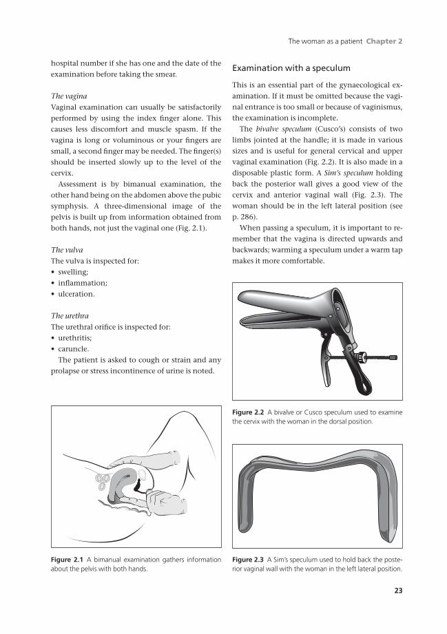
The vaginaVaginal examination can usually be satisfactorily
performed by using the index finger alone. This
causes less discomfort and muscle spasm. If the
vagina is long or voluminous or your fingers are
small, a second finger may be needed. The finger(s)
should be inserted slowly up to the level of the
cervix.
Assessment is by bimanual examination, the
other hand being on the abdomen above the pubic
symphysis. A three-dimensional image of the
pelvis is built up from information obtained from
both hands, not just the vaginal one (Fig. 2.1).
The vulvaThe vulva is inspected for:
• swelling;
• inflammation;
• ulceration.
The urethraThe urethral orifice is inspected for:
• urethritis;
• caruncle.
The patient is asked to cough or strain and any
prolapse or stress incontinence of urine is noted.
Examination with a speculum
This is an essential part of the gynaecological ex-
amination. If it must be omitted because the vagi-
nal entrance is too small or because of vaginismus,
the examination is incomplete.
The bivalve speculum (Cusco’s) consists of two
limbs jointed at the handle; it is made in various
sizes and is useful for general cervical and upper
vaginal examination (Fig. 2.2). It is also made in a
disposable plastic form. A Sim’s speculum holding
back the posterior wall gives a good view of the
cervix and anterior vaginal wall (Fig. 2.3). The
woman should be in the left lateral position (see
p. 286).
When passing a speculum, it is important to re-
member that the vagina is directed upwards and
backwards; warming a speculum under a warm tap
makes it more comfortable.
Figure 2.1 A bimanual examination gathers informationabout the pelvis with both hands.
Figure 2.2 A bivalve or Cusco speculum used to examinethe cervix with the woman in the dorsal position.
Figure 2.3 A Sim’s speculum used to hold back the poste-rior vaginal wall with the woman in the left lateral position.
AMI2 6/9/04 5:10 PM Page 23

Chapter 2 The woman as a patient
24
Bimanual examination
When a cervical smear or a high vaginal swab is to
be taken, it is best to pass the speculum before mak-
ing a bimanual examination. This examination
may be performed in the dorsal or left lateral
position, a matter of personal preference among
gynaecologists. In either case the patient should be
spared unnecessary exposure by covering her with
a sheet or light blanket. The gloved index finger
may be lightly lubricated and is introduced gently
into the vagina.
• The condition of the vaginal walls is noted.
• The cervix is palpated for softening, tears or
polypi.
• The uterus is palpated between the two hands
noting:
(a) size;
(b) consistency;
(c) shape;
(d) mobility;
(e) tumours;
(f ) tenderness on pressure.
The finger in the vagina is now moved into the
right lateral fornix, the hand on the abdomen fol-
lows to explore for any enlargement or tenderness
of the tubes or ovaries. A similar examination is
made of the left adnexa. The finger is passed to the
posterior fornix to detect swelling in the pouch of
Douglas.
Rectal examination
This can be valuable in certain aspects of gynaecol-
ogy, but the patient may find it the most uncom-
fortable part of the whole examination. It permits
bimanual examination of the uterus, tubes and
ovaries if vaginal examination is impossible or un-
desirable. It may further be easier to feel a retro-
verted uterus or a swelling in the pouch of Douglas
and allows an easier approach to the parametrium
and uterosacral ligaments. The possibility of rectal
disease must always be borne in mind.
Investigations
BloodThe haemoglobin level should always be measured
before an operation, however minor. It should
certainly be done in cases of excessive uterine
bleeding (menorrhagia) and as a routine in early
pregnancy. Blood disorders may be associated with
a bleeding tendency so a platelet count, bleeding
time and clotting time may also be done.
In black women, a sickle test should be done,
and in women of Mediterranean or Middle East
origin, there may be a thalassaemia trait which is
diagnosed by electrophoresis.
Serological tests of syphilis and human immuno-
deficiency virus 1 (HIV 1) antibodies are done after
counselling if there is any suspicion of either
disease.
Blood urea and other tests for renal function
should be done where indicated.
Human chorionic gonadotrophin (hCG) levels
may be checked if a pregnancy is suspected. Other
hormone levels in the blood may be measured in
specific conditions. Their ranges are wide.
UrineThe urine should be tested as appropriate for:
• albumin;
• sugar;
• bacilluria by nitrite dipstick;
• microscopy and culture.
CytologyExfoliative cytology in gynaecology examines cells
desquamated from the epithelium of the genital
tract. Material may be obtained by scraping the
cervix with Ayre’s wooden spatula, or a cytobrush.
Cytology is principally used in the early detection
of premalignant lesions of the cervix and is consid-
ered in Chapter 19.
ColposcopyColposcopy examines the cervix under magnifica-
tion in the out-patient department. It is used in
conjunction with cervical cytology so that biopsies
can be accurately taken from suspicious areas and
treatment performed (Chapter 19).
AMI2 6/9/04 5:10 PM Page 24
The woman as a patient Chapter 2
25
LaparoscopyVisual examination of the pelvis and peritoneal
cavity is invaluable when investigating pain or
fertility potential.
HysteroscopyEndoscopy of the uterine cavity demands a fluid or
gas under pressure to open up the cavity. Then the
endometrium can be inspected and biopsied.
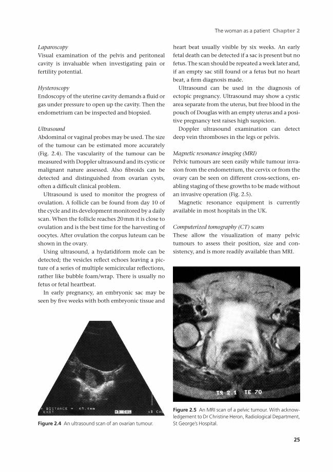
UltrasoundAbdominal or vaginal probes may be used. The size
of the tumour can be estimated more accurately
(Fig. 2.4). The vascularity of the tumour can be
measured with Doppler ultrasound and its cystic or
malignant nature assessed. Also fibroids can be
detected and distinguished from ovarian cysts,
often a difficult clinical problem.
Ultrasound is used to monitor the progress of
ovulation. A follicle can be found from day 10 of
the cycle and its development monitored by a daily
scan. When the follicle reaches 20mm it is close to
ovulation and is the best time for the harvesting of
oocytes. After ovulation the corpus luteum can be
shown in the ovary.
Using ultrasound, a hydatidiform mole can be
detected; the vesicles reflect echoes leaving a pic-
ture of a series of multiple semicircular reflections,
rather like bubble foam/wrap. There is usually no
fetus or fetal heartbeat.
In early pregnancy, an embryonic sac may be
seen by five weeks with both embryonic tissue and
heart beat usually visible by six weeks. An early
fetal death can be detected if a sac is present but no
fetus. The scan should be repeated a week later and,
if an empty sac still found or a fetus but no heart
beat, a firm diagnosis made.
Ultrasound can be used in the diagnosis of
ectopic pregnancy. Ultrasound may show a cystic
area separate from the uterus, but free blood in the
pouch of Douglas with an empty uterus and a posi-
tive pregnancy test raises high suspicion.
Doppler ultrasound examination can detect
deep vein thromboses in the legs or pelvis.
Magnetic resonance imaging (MRI)Pelvic tumours are seen easily while tumour inva-
sion from the endometrium, the cervix or from the
ovary can be seen on different cross-sections, en-
abling staging of these growths to be made without
an invasive operation (Fig. 2.5).
Magnetic resonance equipment is currently
available in most hospitals in the UK.
Computerized tomography (CT) scansThese allow the visualization of many pelvic
tumours to assess their position, size and con-
sistency, and is more readily available than MRI.
Figure 2.4 An ultrasound scan of an ovarian tumour.
Figure 2.5 An MRI scan of a pelvic tumour. With acknow-ledgement to Dr Christine Heron, Radiological Department,St George’s Hospital.
AMI2 6/9/04 5:10 PM Page 25
Chapter 2 The woman as a patient
26
X-raysStraight films of the abdomen can show:
• gas and fluid levels in the obstructed intestine;
• calcium in the urinary tract or dermoid ovarian
tumour;
• radiopaque dye instillation shows the outline of
the uterine cavity and spill from the fimbriated
ends to be seen indicating patency of the tubes at
fertility investigations.
Intravenous urographyThe diagnosis of pelvic tumours and renal tract
disease may be helped by intravenous urography.
Before radical operations in the pelvis the course
of the ureters can be checked.
Barium studiesA barium enema may be helpful in the diagnosis of
rectal conditions. A barium meal with follow-
through to the ileocaecal region may be useful in
cases where symptoms are right-sided.
Pelvic lymphangiographyBy injecting radiopaque contrast material into
the lymphatics in the foot, the lymphatic drainage
of the lower limb and pelvis is outlined. It is useful
to detect secondaries in the lymph glands from
malignant disease in the pelvis.
Ventilation (v)/perfusion (Q) scanThis is used to detect a pulmonary embolism
along with serum estimation of D-dimers. Some-
times a spiral CT scan of the chest may be used in
pregnancy.
Self-assessment
Ask a friend to role-play a patient and practise taking a history using the following role-plays. The instructions forthe candidate are that you should take a history from the role-player in 10 minutes. At the end, the candidate maybe expected to give a two-sentence summary of the case. The role-player needs to make up a name for herself andfill in some personal details. The scoring scheme may be purely for communication skills or may include marks forinformation given. (See Answers to self-assessment questions, p. 306)
2.1 You are a 22-year-old woman who has come to see the doctor because of painful periods. You have had them sinceyou started your periods at the age of 12. They are becoming worse. The pain is crampy and radiates down the frontof your legs. You take Nurofen regularly but it only helps a little bit. The pain starts the day before your period andcontinues for 3 days. Your periods are regular, bleeding for 5 days in every 26–29 days. You have never been preg-nant and are not in a relationship at present. You had a smear last year which was normal. (All other details are upto the imagination of your role-player!)
2.2 You are a 27-year-old woman who has been trying for a baby for 18 months. You had a child at the age of 22 by adifferent partner and an ectopic pregnancy 2 years ago. You have never had an infection but, now you come to thinkabout it, you were supposed to take a course of tablets 3 years ago after one of your boyfriends had been to a clinic, but you never bothered. You have been with your current partner, aged 32 for the last 2 years and are planning to get married next year. Your periods are regular and you have no other health problems. Your baby (aboy) was born at term vaginally with no problems. (All other details are up to the imagination of your role-player!)
2.3 You are a 32-year-old woman who is seeing your GP about future contraception. You have had 3 children and don’treally want any more. During your first pregnancy you developed high blood pressure and the baby was delivered at36 weeks by Caesarean section. Your other two children were born naturally. Your mother has diabetes which wasdiagnosed in her 50s. Your father is alive and well but has high blood pressure. You do not like taking the pill be-cause you keep forgetting it and it gives you headaches. You are concerned that the other hormonal preparationswill make you put on weight which is a problem for your job as an air hostess. You have never had any other illnesses.You smoke 30 cigarettes a day and drink socially. (All other details are up to the imagination of your role-player!)
AMI2 6/9/04 5:10 PM Page 26

27
Part 2
The young woman
AMI3 6/9/04 5:15 PM Page 27

AMI3 6/9/04 5:15 PM Page 28

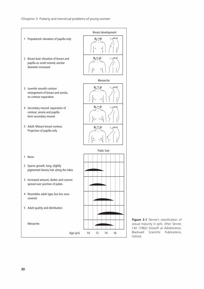
Puberty defines the period in a girl’s life when she
undergoes a series of physiological changes which
lead to the achievement of sexual maturity and the
ability to reproduce.
There are three phases of change:
1 Adrenarche. Increased production of androgens
by the adrenal gland which are converted centrally
by the liver and ovaries and peripherally in the adi-
pose tissue to oestrogens. This usually starts at the
age of 8–10 years and leads to:
• increased sebaceous gland activity;
• sweating;
• hair growth;
• pubic hair which follows axillary hair.
2 Sexual characteristics.
• Usually start at the age of 9–11 years.
• Breast development usually precedes pubic
hair growth and takes 5–6 years to reach Tanner’s
stage 5.
• Pubic hair growth takes only 3 or 4 years and
so is often complete before breast development
(Fig. 3.1).
• Menarche usually coincides with breast devel-
opment to Tanner’s stage 3.
• Average age of menarche in the UK is 12.9
years. It is earlier in countries nearer the Equator.
3 Growth. The onset of puberty coincides with a
rapid increase in growth velocity.
• In girls growth gain is 25–28cm and in boys
26–30cm. Boys go through puberty later than
girls and therefore start their growth spurt from a
higher starting point which accounts for their
greater adult height.
• The pituitary gland increases its frequency of
pulsed growth hormone (GH) and luteinizing
hormone (LH). The mechanism of this is un-
known.
• The greatest release of GH and LH is at night
during sleep; this may account for the increased
need for sleep in adolescents.
• The increase in LH acts on the thecal cells of
the ovary to increase androgen production. This
starts the maturation of the oocytes in the ovary
from primordial phase to the antral phase when
they are ready to be recruited for final matura-
tion and release. Once this begins the young girl
starts her periods.
• The onset of puberty is weight related; the av-
erage weight of a girl starting her periods is 45kg.
Anorexia in teenagers can arrest puberty if they
become underweight for their age.
Germ cells
There are about seven million primordial oocytes
or germ cells in each ovary of the female fetus at 15
weeks of intrauterine life. This drops to two million
germ cells at birth and is further reduced to half a
million at puberty.
About 400 will be recruited during each ovula-
tion cycle during the reproductive life; the rest de-
generate at a steady rate. The stromal tissue of the
2929
Chapter 3
Puberty and menstrual problems ofyoung women
AMI3 6/9/04 5:15 PM Page 29
Chapter 3 Puberty and menstrual problems of young women
30
Prepubertal: elevation of papilla only
Breast development
Breast bud: elevation of breast and papilla as small mound; areolar diameter increased
Juvenile smooth contour:enlargement of breast and areola;no contour separation
Secondary mound: separation ofcontour; areola and papillaform secondary mound
Adult: Mature breast contour.Projection of papilla only
1
2
3
4
5
None
Pubic hair
Menarche
Sparse growth, long, slightly pigmented downy hair along the labia
Increased amount, darker and coarser;spread over junction of pubes
Resembles adult type, but less areacovered
Adult quality and distribution
Menarche
Age (yrs) 10 12 14 16
1
2
3
4
5
Figure 3.1 Tanner’s classification ofsexual maturity in girls. After Tanner,J.M. (1962) Growth at Adolescence.Blackwell Scientific Publications, Oxford.
AMI3 6/9/04 5:15 PM Page 30

Puberty and menstrual problems of young women Chapter 3
31
ovary produces androgenic hormones which may
be metabolized in peripheral fat to produce
oestrogens.
Ovulation is controlled by the ovarian hor-
mones and the gonadotrophins from the pituitary.
Menstrual cycle
Three structures are involved with the regulation
of ovulation and menstruation.
1 The anterior pituitary gland.
2 The ovary.
3 The uterus.
These are all dealt with in Chapter 1 considering
the anatomy and physiology of these organs.
Amenorrhoea
Primary amenorrhoea
Definition: no periods experienced by the age
of 16.
Investigations of this condition may be divided
according to whether secondary sexual characteris-
tics are present or not. If absent, girls should be in-
vestigated at the age of 16. If present, investigation
can wait until the age of 18.
Causes of primary amenorrhoea
• Hypothalamic (absence of gonadotrophic releasing
hormone, GnRH) or hypogonadatrophic (no LH or fol-
licle stimulating hormone, FSH).
This may be:
• Idiopathic.
• Following radiotherapy.
• Following surgery.
• Craniopharyngomas in childhood.
• Anorexia.
• Excessive exercise (ballet dancers).
• Chromosomal.
• Congenital.
Chromosomal causesThe normal human has 46 chromosomes, 44 auto-
somes and two sex chromosomes. The number is
halved in both gametes, the oocyte and spermato-
zoon; when fertilization occurs the original num-
ber is restored in the resulting fertilized ovum (see
Chapter 1).
In the normal female the sex chromosomes are
XX, in the normal male XY. All oocytes carry the X
chromosome, while about half the spermatozoa
carry X, the others Y. Thus, the resulting offspring
are either XX (female) or XY (male).
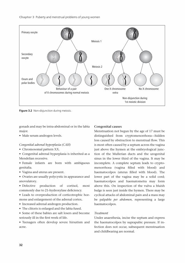
Sex chromosome abnormalities mainly arise
from non-disjunction (Fig. 3.2). At the division of
the primary oocyte while still sited in the ovary,
the two chromosomes fail to separate so that a pri-
mary oocyte is produced which may have two X
chromosomes or none; conversely, the first polar
body will contain the converse—none or two. Fer-
tilization by a spermatozoon which may carry X or
Y can therefore result in abnormal patterns, XXX,
XXY or XO. YO has not been described as this
genetic combination is lethal.
The description is a simplification as more com-
plex anomalies may occur, for example mosaics or
individuals of mixed chromosomal patterns.
Turner’s syndrome• Chromosome pattern XO.
• Incidence about three in 10000 full-term births.
• Present with primary amenorrhoea for there are
either no ovaries or non-functioning streaks of
tissue with no oogenesis.
• The vagina and uterus are present.
• Poor breast development.
• Little or no axillary and pubic hair.
• Short stature.
• Webbing of the neck.
• A wide carrying angle in the arms.
• Coarctation of the aorta.
• Congenital malformation of the kidneys may be
found.
Androgen insensitivity syndrome (AIS)• Chromosomal pattern XY.
• Due to lack of androgen receptors (deletion on X
chromosome).
• Active breast development (hepatic oestrogens).
• Absent or scanty axillary and pubic hair.
• Usually absent uterus with a very short vagina.
• The gonads are testes or undifferentiated
AMI3 6/9/04 5:15 PM Page 31
Chapter 3 Puberty and menstrual problems of young women
32
gonads and may be intra-abdominal or in the labia
major.
• Male serum androgen levels.
Congenital adrenal hyperplasia (CAH)• Chromosomal pattern XX.
• Congenital adrenal hyperplasia is inherited as a
Mendelian recessive.
• Female infants are born with ambiguous
genitalia.
• Vagina and uterus are present.
• Ovaries are usually polycystic in appearance and
anovulatory.
• Defective production of cortisol, most
commonly due to 21-hydroxylase deficiency.
• Leads to overproduction of corticotrophic hor-
mone and enlargement of the adrenal cortex.
• Increased adrenal androgen production.
• The clitoris is enlarged and the labia fused.
• Some of these babies are salt losers and become
seriously ill in the first week of life.
• Teenagers often develop severe hirsutism and
acne.
Congenital causesMenstruation not begun by the age of 17 must be
distinguished from cryptomenorrhoea—hidden
loss caused by obstruction to menstrual flow. This
is most often caused by a septum across the vagina
just above the hymen at the embryological junc-
tion of the Mullerian ducts and the urogenital
sinus in the lower third of the vagina. It may be
incomplete. A complete septum leads to crypto-
menorrhoea (vagina filled with blood) and
haematocolpos (uterus filled with blood). The
lower part of the vagina may be a solid cord;
haematocolpos and haematometra may form
above this. On inspection of the vulva a bluish
bulge is seen just inside the hymen. There may be
cyclical attacks of abdominal pain and a mass may
be palpable per abdomen, representing a large
haematocolpos.
TreatmentUnder anaesthesia, incise the septum and express
the haematocolpos by suprapubic pressure. If in-
fection does not occur, subsequent menstruation
and childbearing are normal.
Primary oocyte
Secondary oocyte
Ovum and polar bodies
Meiosis 1
Meiosis 2
Behaviour of a pair of X chromosomes during normal meiosis
One X chromosome extra
No X chromosome
Non-disjunction during 1st meiotic division
Figure 3.2 Non-disjunction during meiosis.
AMI3 6/9/04 5:15 PM Page 32

Puberty and menstrual problems of young women Chapter 3
33
Investigation of primary amenorrhoea
• A history is taken including a family history as
AIS may affect other females in the family.
• Physical examination. The general examination
should begin by recording the girl’s height—if by
16 she is less than 147cm there is a possibility of
ovarian agenesis (Turner’s syndrome) or (pan)hy-
popituitarism. If there is a decrease in body weight,
calculate Body Mass Index (wt(kg)/ht(m2)). A gen-
eral examination checks the development of sec-
ondary sexual characteristics, hair patterns and
density.
• A pelvic examination should be performed if the
examiner really thinks a positive finding will be
there. For most young women with primary amen-
orrhoea it will not be useful particularly at first
consultation. The vulva is inspected to see that the
introitus is patent; there may be cryptomenor-
rhoea, congenital absence of the vagina or a blind
vagina as in AIS.
• Investigations —a buccal smear and an examina-
tion of the polymorphonuclear leucocytes to de-
termine if chromatin positive (probably XX) or
chromatin negative (probably XO or XY); in other
cases a full chromosome analysis may be needed to
exclude mosaicism and AIS.
• Hormonal investigations should include LH, FSH,
oestradiol and testosterone levels.
• Ultrasound will help determine the presence, state
and size of the ovaries and any follicular activity.
Uterine size can also be seen. It is rarely necessary to
perform a laparoscopy to assess the pelvic organs.
Management
If the girl is normally developed, with normal
breast development, the uterus and vagina are nor-
mal and she is chromatin positive, the most likely
diagnosis is delayed menarche. It is reasonable to
await events; menstruation is not established in
some individuals before 18 years.
For those with a diagnosable pathological cause,
the aim must be to restore normal function as far as
possible and, although fertility may not be pos-
sible, enable the individual to lead as normal a
sexual life as possible.
Cases of Turner’s syndrome should receive long-
term treatment with cyclical hormones, oestrogen
and progestogen (hormone replacement therapy).
There is a small risk of uterine carcinoma.
In AIS, the gonads are testes that are often found
inside the abdomen or inguinal canal. Since these
testes have a 25% risk of becoming malignant (ter-
atoma or dysgerminoma) they should be removed
soon after puberty and an artificial vagina may be
constructed or dilators used to permit sexual inter-
course. Treatment with oestrogen should also be
given to augment breast development and prevent
osteoporosis.
In cases of congenital absence of the vagina
and uterus the ovaries are usually normal. An arti-
ficial vagina may be constructed to permit sexual
intercourse.
Abnormalities of pituitary secretion should be
treated with oestrogen or progesterone until fertil-
ity is desired.
Secondary amenorrhoea
Definition: no periods for over 6 months, having
once started.
Causes of secondary amenorrhoea
PhysiologicalAmenorrhoea occurs naturally in:
• pregnancy;
• lactation;
• after menopause.
PituitaryTotal pituitary ablation or destruction by radio-
therapy after puberty. Tumours may also destroy it.
In Sheehan’s syndrome, severe postpartum haem-
orrhage causes pituitary anoxia with failure of lac-
tation, amenorrhoea and other manifestations of
pituitary failure.
Hyperprolactinaemia may be due to a definitive
pituitary adenoma or scattered microadenoma
causing galactorrhoea, visual disturbances and
headaches. It may be a side-effect of some drugs,
particularly cimetidine and phenothiazine.
Anorexia nervosa may lead to pituitary damage
AMI3 6/9/04 5:15 PM Page 33
Chapter 3 Puberty and menstrual problems of young women
34
and thus to permanent amenorrhoea and steril-
ity. Periods cease before weight loss becomes
apparent.
Thyroid diseaseAll thyroid disorders may cause amenorrhoea or
excessive bleeding. Correction of the thyroid func-
tion may restore normal menstruation.
OvaryPolycystic ovary syndrome (PCOS) is the common-
est cause of anovulation in young women, with one
in four women having the morphological picture on
ultrasound scan. They have stromal hyperplasia
of the ovaries, leading to an excess secretion
of testosterone and the formation of multiple
follicular cysts; it is associated with amenorrhoea,
oligomenorrhoea, hirsutism and infertility, sec-
ondary to anovulation. Women with PCOS are
often hyperinsulinaemic. This predisposes them to
obesity and diabetes. Obesity increases the preva-
lence of anovulation and hirsutism. Anovulation
and obesity both predispose women to endome-
trial hyperplasia/carcinoma because of the effects
of unopposed estrogen on the endometrium.
Weight control and regular withdrawal bleeds are
therefore essential.
Spontaneous premature ovarian failure (POF)
in the absence of disease causes premature
menopause. The ovary ceases to respond to pitu-
itary gonadotrophins which are usually excreted in
excessive amounts or may be normal. This follows
a lack of oocytes or their autoimmune destruction
in the ovary.
Castration by surgical removal of the ovaries or
by exposure to irradiation leads to amenorrhoea.
Extensive destruction of the ovaries by infection
or tumours is a rare cause.
UterineThe endometrium can be destroyed by the
following.
• Disease:
(a) tuberculosis;
(b) severe postpartum infection.
• Formal endometrial ablation (by laser or
diathermy).
• Severe curettage usually following pregnancy,
miscarriage or therapeutic abortion (Asherman’s
syndrome).
General diseaseAmenorrhoea may occur in any debilitating dis-
ease, e.g. pulmonary tuberculosis, not necessarily
with involvement of the pelvic organs.
Terminal stages of diseases such as Addison’s dis-
ease and uraemia due to renal disease.
Starvation may lead to amenorrhoea, similar to
that seen in anorexia nervosa.
Obesity can also cause amenorrhoea (most com-
monly associated with PCOS) and in grossly obese
young women weight reduction may restore nor-
mal menstrual function.
History
• Family history.
• Hot flushes.
• Drugs such as reserpine, digoxin, phenothiazines
and hormones, including oral contraceptives.
• Change in body weight, i.e. obesity or sudden
loss of weight.
• Galactorrhoea.
• Headache, visual disturbance (hemianopia).
Physical examination
• Height measurement.
• Weight.
• Blood pressure.
• Breasts for evidence of pregnancy or milk
secretion.
• Pelvic examination to:
(a) exclude pregnancy (a woman may still con-
ceive in the course of a period of amenorrhoea);
(b) assess the size and position of the uterus to
exclude a pelvic tumour.
Investigations
• Pregnancy test.
• Computerized tomography (CT) scan or mag-
netic resonance imaging (MRI) of pituitary area.
• Thyroid stimulating hormone.
AMI3 6/9/04 5:15 PM Page 34

Puberty and menstrual problems of young women Chapter 3
35
• Plasma hormone levels of FSH.
• LH.
• Oestradiol.
• Prolactin.
• Testosterone.
• Progesterone withdrawal test: give a progestogen
for 5 days. If the woman bleeds afterwards, she has
oestrogen in her circulation and a uterus.
Ultrasound assessment of:
• uterine size;
• pregnancy;
• ovarian size and morphology;
• follicular function.
Examination under anaesthesia if congenital
abnormality:
• assess the pelvic organs;
• perform a laparoscopy to inspect the pelvic
organs and to take a biopsy of the ovaries.
The differential diagnosis of amenorrhoea is
given in Box 3.1.
Treatment
Treat the cause if one is discovered. If not, manage-
ment will depend upon whether fertility is desired
or not.
Women with low oestrogen levels should be
treated with oestrogen and progesterone replace-
ment (the oral contraceptive pill or hormone
replacement therapy).
In anorexia nervosa the periods may cease
before weight loss is evident; treatment should
include efforts to restore weight to normal. In
gross obesity, weight loss may result in normal
menstruation.
Teenagers who over-exercise (ballet dancers and
gymnasts), should be encouraged to reduce their
training schedule.
In polycystic ovary syndrome, if the woman is
obese, referral to a dietician is recommended to
reduce weight. In some women metformin may
help to restore ovulation in conjunction with a
low-calorie diet. Regular progesterone therapy for
12 days every three months will give a regular
withdrawal bleed or the oral contraceptive pill
with cyproterone acetate (an antiandrogen) will
give a regular period and improve hirsutism and
acne.
A premature menopause is characterized by low
oestrogen levels and high FSH. They should receive
cyclical oestrogen and progesterone such as
Cyclo-Progynova to prevent osteoporosis.
Where excess of prolactin (over 1000mu/l) is
secreted, perform a CT/MRI scan of the pituitary
fossa to exclude a tumour. In the absence of such a
space occupying lesion, microadenomata of the
Box 3.1 Tests: differential diagnosis of primary and secondary amenorrhoea
ProgesteroneChromosomes FSH LH Oestradiol Testosterone Prolactin withdrawal
Primary amenorrhoeaTurner’s XO ≠ ≠ Ø = = -veAndrogen XY ≠ ≠ = ≠ = -ve
insensitivitysyndrome
Hypogonadotrophic XX Ø Ø Ø = = or Ø -vehypogondism
Secondary amenorrhoeaProlactinoma XX Ø Ø Ø = ≠ -vePolycystic ovary XX = ≠ or = = ≠ = +ve
syndromePremature ovarian XX ≠ ≠ Ø = = -ve
failure
AMI3 6/9/04 5:15 PM Page 35

Chapter 3 Puberty and menstrual problems of young women
36
pituitary body are postulated. Treatment of amen-
orrhoea with prolactin excess is with bromocrip-
tine or the longer acting cabergoline (both are
dopamine agonists) in perpetuity. Bromocriptine
and cabergoline should be stopped if pregnancy
occurs. Periods will return on treatment once pro-
lactin levels are rendered normal.
Bleeding in infancy
Female infants may have vaginal bleeding during
the first week of life. This is rarely significant and
probably due to withdrawal of maternally derived
oestrogens which had crossed the placenta in
pregnancy.
Precocious puberty is caused by premature matura-
tion of the ovaries in most cases; very rarely there
may be a granulosa cell tumour of the ovary. The
breasts grow while pubic and axillary hair develops
and endometrial bleeding occurs. Provided a tu-
mour of the ovary can be excluded, the child may
be allowed to develop normally but the parents
must be warned of the danger of pregnancy.
The differential diagnosis includes:
• a mixed mesodermal tumour;
• adenocarcinoma of the vagina; particularly at
risk were adolescent girls whose mothers received
stilboestrol during pregnancy but this is now
history.
Menorrhagia under 18 years
Adolescent bleeding of a sufficient amount to be con-
sidered as menorrhagia is usually dysfunctional,
secondary to anovulation. There may be pro-
longed episodes of painless bleeding and the girl
can become anaemic. Examination of the en-
dometrium may show an anovulatory pattern or
hyperplasia.
Treatment consists of eliminating any organic
cause and correcting anaemia if present. Hormone
treatment is usually successful using a progestogen
such as norethisterone (15mg daily) (see Chapter
16). When the treatment is stopped, withdrawal
bleeding with shedding of the endometrium anal-
ogous with normal menstruation occurs. Treat-
ment should be given cyclically for a further three
to six months. An alternative is use of the oral con-
traceptive pill.
Self-assessment
Read the following clinical scenarios carefully and answer the questions below.1 A 16-year-old presents with a history of primary amenorrhoea. She is 1.45m tall and weighs 53kg (BMI 25.2). On
physical examination her breasts are Tanner stage 2, she has a normal vulva and vagina. Her mother reports that shestruggles at school and is being teased because she is so short.
2 A 16-year-old girl presents with a history of primary amenorrhoea. She is 1.64m tall and weighs 47kg (BMI 17.5). Herbreast development is Tanner stage 3 and she has scanty pubic hair, she has a normal vulva and vagina. She representsGreater London in marathon running and is otherwise well.
3 A 17-year-old girl presents with a history of secondary amenorrhoea. She is 1.64m tall and weighs 56kg (BMI 21). Shestarted her periods at the age of 14 and they were irregular and light until one year ago when they stopped. On phys-ical examination she has acne on her face and back which she finds distressing. Her legs and arms are hairy and shehas hair on her abdomen in an inverted triangle. Her vulva and vagina are normal. Her mother has type 2 diabetes.She is otherwise well.
4 A woman of 24 years old presents to the clinic with secondary amenorrhoea. Her periods started at the age of 12 andwere regular until 2 years ago when they became irregular and light. She has noticed a discharge from her breasts andsuffers from severe headaches. On physical examination she has a white discharge from her breasts and is otherwisenormal.
5 A 15-year-old girl presents to the clinic with primary amenorrhoea. She complains of intermittent abdominal pain andhas noticed that her school skirt is becoming tighter. Her weight and height are normal for her age. On examinationher breasts are Tanner stage 4 and pubic hair stage 5. Her vulva looks swollen at the introitus and she has a palpableabdominal mass which is tender.
AMI3 6/9/04 5:15 PM Page 36
Puberty and menstrual problems of young women Chapter 3
37
Self-assessment Continued
Questions
3.1 From the list below choose the single most likely diagnosis for each of the clinical scenarios above.(a) Androgen insensitivity syndrome(b) Cryptomenorrhoea(c) Pregnancy(d) Polycystic ovary syndrome(e) Ashermans syndrome(f) Turner’s syndrome(g) Congenital adrenal hyperplasia(h) Hyperprolactinaemia(i) Constitutional delay of puberty(j) Anorexia/exercise related amenorrhoea
3.2 From the lists below select the most useful first line investigations to establish the diagnosis for each clinical scenario.(a) Ultrasound scan(b) Ultrasound scan, LH, FSH and testosterone(c) Ultrasound scan, LH, FSH, prolactin(d) Chromosome analysis(e) Pregnancy test(f) LH, FSH, oestradiol(g) Ultrasound scan, LH, FSH, testosterone, adrenal androgen profile(h) Thyroid function tests
3.3 From the list below select the most appropriate first line treatment for each condition.(a) Weight gain(b) Removal of gonads/ovaries(c) Incision of vaginal septum(d) Referral to the antenatal clinic(e) Bromocriptine(f) Progestogerone therapy every three months(g) Creation of a vagina(h) Oestrogen replacement therapy(i) Prednisolone(j) Weight loss
AMI3 6/9/04 5:15 PM Page 37

38
Subfertility is defined as failure to conceive after
one year of unprotected coitus at frequent inter-
vals. A woman who has never conceived has
primary subfertility, a woman who has conceived
before has secondary subfertility, even though this
episode did not result in a live birth, e.g. miscar-
riage or ectopic pregnancy.
Causes of infertility (Box 4.1)
Both partners
• Mechanical difficulty in coitus with inadequate
penetration, often associated with lack of ability in
the male to maintain an erection.
• Periods of separation so that there is no inter-
course at the most fertile time.
Male
• Impotence.
• Premature ejaculation.
• Azoospermia/oligospermia (<20 ¥ 106/ml).
• Poor sperm motility.
• >10% abnormal forms.
Female
• Fallopian tubes —infection may close or partly
obstruct.
• Ovarian dysfunction (Box 4.2):
(a) ovulation may not occur;
(b) ovulation is irregular with anovulatory
cycles;
(c) polycystic ovary syndrome;
(d) hyperprolactinaemia;
Chapter 4
Subfertility
Box 4.1 Factors in infertility
Ovulation disorder 30%Tubal factor 30%Male factor 30%Unexplained infertility 10%
Box 4.2 Causes of anovulation
Primary ovarian dysfunctionGenetic, e.g. Turner’s syndrome 1%Autoimmune
Secondary ovarian dysfunctionDisorders of gonadotrophin regulationHyperprolactinaemia 15%
FunctionalWeight loss 10%Exercise
Gonadotrophin deficiencyPituitary tumourPituitary infarction 4%Pituitary ablation
Polycystic ovary syndrome 70%
AMI4 6/9/04 5:17 PM Page 38

Subfertility Chapter 4
39
(e) perimenopausal;
(f ) premature ovarian failure.
• Intact hymen —a woman may complain of subfer-
tility when her marriage has not been consummated.
• Vagina —congenital malformation.
• Uterus —congenital malformation, or tubercu-
lous endometritis.
Investigation of the infertile couple
History from the female
• Age, occupation, length of time with partner, use
of contraception or avoidance of pregnancy,
previous sexual activity.
• Previous pregnancies, including abortions, mis-
carriages, ectopic.
• Menstrual history: age at onset, cycle and dura-
tion of flow, dysmenorrhoea, ovulation pain, re-
cent change in the cycle.
• Vaginal discharge: character, amount, whether
associated with irritation or soreness.
• Previous illnesses, especially pelvic inflamma-
tory disease (PID), diabetes, renal disease.
• Operations, especially in the abdomen or pelvis.
• Coitus frequency, difficulties, relation to fertile
days.
• Previous investigation or treatment of infertility.
Examination of the female
• General examination —physical development,
evidence of endocrine disorder.
• Abdominal examination —scars, tenderness,
guarding, masses.
• Vaginal examination —state of introitus, size and
mobility of the uterus, uterine enlargement, en-
largement of the ovaries.
History from the male
• Age, occupation, including absence from home,
length of time with partner and duration of
infertility.
• Sexual performance —frequency, ability to
ejaculate in upper vagina.
• Previous relationships, fathering of any
pregnancies.
• History of mumps with orchitis, injury to geni-
talia or operations for hernia or varicocoele, any
recent debilitating illness.
Examination of the male
• General build and appearance.
• Examination of genitalia, hypospadias.
• Palpation of testicles, size, consistency. Relate
size to standard models (Fig. 4.1).
Investigations
Seminal analysis (Box 4.3)
Best performed on a masturbation specimen which
should be examined within two hours of collec-
25
2015 12
10
8
6
5
4321
5cm
Figure 4.1 Models of testicular size to allow a stan-dardization of the examined testes size with an objectivecomparison.
Box 4.3 Normal seminal analysis
Volume 2–5mlLiquefaction time Within 30 minutesCount 20–150 million/mlMotility >50% motility at 1.5 hoursSperm morphology >10% normal forms
AMI4 6/9/04 5:17 PM Page 39

Chapter 4 Subfertility
40
tion. It should not be from semen ejaculated at in-
tercourse using a condom as most modern sheaths
are lubricated with spermicidal cream.
The motility of sperms is important —direct,
straight moving sperms being the more likely to
effect fertilization.
There should be at least 20 million sperms per
millilitre with a total count of not less than 100
million. Fertility reduces progressively with num-
bers below this. Abnormal forms should not ex-
ceed 10%. Reliance should not be placed on a
single sperm count.
In cases of severe oligospermia/azoospermia
the cause should be sought. It may be due to a
chromosomal abnormality such as Klinefelter’s
syndrome (XXY). It may be due to primary
hypogonadism, when the level of gonadotrophic
hormone will be high, or secondary hypogon-
adism, where gonadotrophic hormone will be
low; in the latter case there may be excess of pro-
lactin secretion, usually due to a pituitary tumour
and possibly responsive to bromocriptine. Other
causes of azoospermia may be related to congenital
absence of the vasa deferentia or to obstruction in
the epididymis.
Basal temperature charts
The woman may keep charts of her basal tempera-
ture for a period of three months. This is best taken
first thing in the morning before leaving bed. In
theory, the raised progesterone levels elevate the
basal body temperature by 0.3–0.5°C within 12
hours of ovulation. The graphic chart produced
bears some relation to ovulation when it is regular.
However, the correlation of temperature to ovula-
tion is less easily seen when ovulation is irregular.
Other extraneous causes of temperature fluctua-
tion (e.g. ’flu) intrude on this test as do unusual life
rhythms (e.g. nurses on night duty). These charts
are very difficult to use prospectively and have
largely been abandoned.
Ovulation predictor tests
A daily test using a few drops of urine which de-
tects the LH surge by giving a colour change on a
stick in the presence of a high LH. When it is posi-
tive the woman knows that she is going to ovulate
within 36 hours. It is helpful in women with a reg-
ular cycle but in women who have PCOS and an
irregular cycle it is rarely helpful since they can
have a raised LH in the follicular phase without a
mature follicle.
Tests for tubal patency
Hysterosalpingography using a radiopaque dye
shows blockage of the tubes and indicates the site
of the obstruction; it can also demonstrate a con-
genital malformation of the cavity of the uterus
which will not be apparent at laparoscopy. A new
contrast medium containing microbubbles may be
injected transcervically and its passage along the
fallopian tube detected with ultrasound (HyCoSy®).
Tubal patency may be tested under direct vision
at laparoscopy. A solution of methylene blue is
injected through a tightly fitting cannula in the
cervical canal. The passage of the dye may be
observed. When the tubes are normally patent the
dye pours out of the fimbriated end of the tube into
the pouch of Douglas. Tubal obstruction may be
recognized as can the presence of adhesions; a
hydrosalpinx may be seen to fill with dye which
does not spill.
Hormone tests
Serum progesterone levels in the mid second half
of the cycle (days 21–23 of a 28 day cycle) are ten
times as high as those of the rest of the cycle
(30ng/ml compared with 3–6ng/ml) if ovulation
has occurred. Luteinizing hormone (LH), follicle
stimulating hormone (FSH), testosterone (if poly-
cystic ovary syndrome is suspected) should be
taken between days 3–8 of the cycle.
Prolactin levels should be measured to exclude
microadenomata of the pituitary gland; levels
above 1000mu/l are significant and should lead
to computerized tomography (CT) scan of the
pituitary fossa.
AMI4 6/9/04 5:17 PM Page 40

Subfertility Chapter 4
41
Ultrasound
An ultrasound scan of the pelvis, especially with a
vaginal probe, gives excellent views of the ovaries
and uterus if pathology in either is suspected (e.g.
polycystic ovary syndrome).
Treatment of subfertility
A couple seeking medical advice for subfertility are
obviously anxious and concerned. They should
always be offered help even if the period of sub-
fertility is relatively short. In most couples,
investigations should be done after one year of
trying.
About 25–30% of women seeking advice for sub-
fertility become pregnant during investigation and
treatment. There are various lines of treatment
which may prove successful depending on the
condition found.
Correction of coital difficulties
An intact hymen should be removed or dilated. A
vaginal septum may have to be removed but usua-
lly they cause little difficulty. The woman should
be taught to pass vaginal dilators and this helps to
give her confidence. The use of a lubricant at coitus
also helps.
In the male, the commonest problems with fer-
tility are premature ejaculation and inadequate
erection. Both of these are discussed in Chapter 6.
Lesions of the uterine body
Removal of small uterine polypi (see p. 223) at
curettage is often successful. Myomectomy should
be performed if the fibroids are blocking the
fallopian tubes or are submucous (see p. 231).
Laparoscopy or laparotomy may reveal other con-
ditions such as endometriosis or peritubal adhe-
sions which require relevant therapy.
Lesions of the tubes
Various operations may be undertaken to restore
patency and function in cases where the tubes
have been damaged by infection (most commonly
with chlamydia).
• Salpingostomy where the fimbrial end is opened
and held open by turning out a cuff (commonly
done laparoscopically).
• Tubal reimplantation where the isthmus is
blocked. The medial tubal end is freed and is reim-
planted into the uterine cavity.
• Salpingolysis where peritubal adhesions are
divided (usually at laparoscopy).
• Reanastomosis if the tube is blocked in mid-
segment; the obstructed area is resected and the
open ends reanastomosed often using microsurgery.
Results of many of these operations are disap-
pointing because:
• patency can easily be restored but the tube may
be too rigid to allow peristalsis;
• infection may have caused the tube to be fixed to
other organs by adhesions and so the fimbrial end
cannot freely manoeuvre and thus harvest the
oocytes;
• after surgery there may be too short a length of tube
so that passage of the fertilized oocyte is so rapid
that the endometrium is not yet ready to receive it.
In consequence, the success rates of tuboplasty
after infection range from 2 to 10%. The best re-
sults come after reanastomosis of sterilization pro-
cedures when most surgeons will report a 40–60%
success rate, which can be improved to about 75%
by the use of microsurgery. Despite these poor re-
sults there are some women who will be prepared
to submit to such an operation even though the
hope of a successful pregnancy is slight. For the
majority of women with tubal disease, in vitro fer-
tilization (IVF) gives the best chance of a successful
pregnancy (see p. 43).
In cases of non-ovulation, further investigation
is indicated.
If there is a high level of prolactin on more than
one occasion a CT scan should be done to exclude
a pituitary tumour. Excess of prolactin is treated
with bromocriptine or cabergoline. As prolactin
levels return to normal, menstruation restarts and
normal fertility is restored.
Cases of primary or secondary ovarian failure
show high levels of FSH and low levels of oestro-
gen. Induction of ovulation is impossible for there
AMI4 6/9/04 5:17 PM Page 41

Chapter 4 Subfertility
42
are no more oocytes, but the patient should be
offered oestrogen replacement therapy.
Cases of ovulation failure with low FSH and LH
levels may be treated by the use of gonadotrophins.
Human menopausal gonadotrophin (hMG) con-
tains both gonadotrophins or FSH can be given
alone. Treatment with these must be monitored
carefully with ultrasound to prevent hyperstimula-
tion, secondary to multiple ovulation and thence to
multiple pregnancy.
In practice, 75 iu of FSH are given daily from
about days 2–3 of cycle. The dose is increased week-
ly, depending on the ultrasound examination of
the ovary which shows the number and size of
follicles coming towards ovulation. When the
response is satisfactory, an injection of human
chorionic gonadotrophin (hCG) is given which
acts in a similar manner to the LH surge. Once a
satisfactory dosage pattern has been established in
one cycle, this can be repeated in other cycles in up
to six treatment courses or until a pregnancy is
achieved.
Cases of anovulation with polycystic ovary syn-
drome may be treated with clomiphene citrate.
The dose is 50mg for five days commencing on the
third day of menstruation and mid-luteal proges-
terone levels on day 21 are measured to confirm
ovulation. Provided ovulation occurs treatment
should be repeated for six months or until the
woman is pregnant. If the first dose is unsuccessful
it is increased maximally to 150mg daily. There is a
rise of multiple follicular development from this
hyperstimulation and therefore multiple pregnan-
cies are more common (5–10%).
Treatment of male infertility
If it has been established that male factors are asso-
ciated with infertility, treatment can proceed. This
is best done in two phases:
• less invasive treatments;
• more specific therapies.
Phase one
The first phase should last about three months.
Two specimens of semen should be examined to
establish levels in the sperm count. Certain aspects
of the man’s way of life may need to be altered:
• over-exertion;
• excessive smoking;
• excess alcohol consumption;
• poorly controlled diabetes;
• hypertension;
• being overweight.
If the scrotal temperature is raised, it is wise to
wear boxer shorts to allow the testes to hang in a
cooler atmosphere.
The timing of intercourse may need discussion
so that, around the time of ovulation, the couple
have intercourse. A few days of abstinence before
this may boost the sperm count if there is a defi-
ciency; otherwise timing is irrelevant.
Any varicocoele causing a raised temperature of
the scrotum and the efferent ducts from the testes
is managed by ligation. Three-quarters of men im-
prove their sperm count after this if a varicocoele
has been a feature.
Phase two
Specific treatments will depend upon the results of
investigations. A low sperm count with low FSH
and testosterone level may indicate treatment with
stimulating hormones (very rare).
In the absence of hormone deficiency, endocrine
therapy is less easily justified. Hyperprolactin-
aemia is rare in males, but if it is present then
bromocriptine should be used.
Impaired fertility is associated sometimes with
an increased incidence of chronic prostatitis. If
present, long-term, low-dose antibiotic treatment
may remove this potential cause (erythromycin
250mg twice a day for a month).
Sperm washing has been described with
variable results. Ejaculated sperm is washed in
phosphate buffered saline and resuspended for
insemination into the uterus (intrauterine insem-
ination, IUI). His partner is usually treated with
clomiphene or FSH to increase the number of ma-
ture oocytes to 2 or 3 to increase the chance of con-
ception. The risk of a multiple pregnancy is around
10% and ovarian hyperstimulation syndrome
(OHSS) 1%.
AMI4 6/9/04 5:17 PM Page 42
Subfertility Chapter 4
43
Generally speaking, the management of male
factors of infertility is disappointing and the suc-
cess rate usually ranges between 20 and 40%. The
introduction of assisted conception techniques has
altered this considerably.
Assisted conception
Artificial fertilization methods have increased in
use greatly in the last decade with probably over
10000 children in the world born as a result. Artifi-
cial fertilization has received considerable media
cover but should be considered as only one part of
infertility management, particularly for those who
cannot transmit sperms or eggs along the fallopian
tubes due to their damage or absence (Box 4.4) or
abnormal sperm.
Patients selected for programmes are usually
under 40 years of age and in a stable relationship.
They should be free of medical or psychological
disease and the woman should have a normal
uterus.
Technique
For the process to take place, it is essential that
oocytes be recovered. Most women now have ovar-
ian cycles stimulated by gonadotrophins following
treatment with a gonadotrophic releasing hor-
mone (GnRH) analogue to stop the woman’s own
hormone production. hCG is given to stimulate
ovulation. This can be monitored by daily ultra-
sound scanning when follicle size can be meas-
ured; the follicle should measure about 20mm in
diameter for oocyte recovery.
Oocytes are aspirated through the posterior
fornix of the vagina or bladder guided by ultra-
sonic scanning. Usually several (3–15) oocytes are
recovered at the same procedure.
The oocytes are mixed on a warmed flat dish in
special media with semen obtained from the hus-
band by masturbation. Fertilization takes place in
vitro and under specially controlled conditions of
temperature and atmospheric gases. The eggs are
allowed to develop to the four–eight cell stage, and
introduced to the woman’s uterus through the
cervix, using a fine cannula in as atraumatic a fash-
ion as possible. A maximum of two or three fertil-
ized ova are inserted 2 days after egg collection.
Luteal support with progesterone or hCG is
given in the days following embryo replacement
until 8–10 weeks of pregnancy or when menstrua-
tion begins if conception has not occurred.
Results
Conception rates at established IVF units are about
20–30% per cycle. Success should be judged by a
live birth and not just by the implantation of a fer-
tilized egg —a biochemical pregnancy shown by a
rise in hCG levels. The average take home baby rate
in the UK is 20%.
Gamete intra-fallopian tube transfer (GIFT)
If the tubes are present and patent but convention-
al methods have failed, one may use gamete intra-
fallopian tube transfer (GIFT). The oocytes are
recovered at laparoscopy under general anaesthe-
sia. Prepared motile sperm from the male semen
is then passed through the laparoscope into the
fallopian tube and one or two of the oocytes
are put into the same tube. The whole procedure
takes about half an hour and there is no extracor-
poreal phase. Success rates are slightly lower than
IVF. It is less commonly offered as a treatment
these days.
Zygote intra-fallopian transfer (ZIFT)
Zygote intra-fallopian transfer (ZIFT) puts fertilized
Box 4.4 Indications for in vitrofertilization (IVF)
Cause of infertility %
Disease or absence of the tubes 50–70Endometriosis 7–15Sperm abnormalities or low count 5–20Sperm antibodies 1–5Idiopathic infertility 3–15Failed donor insemination (DI) 1–5
AMI4 6/9/04 5:17 PM Page 43

Chapter 4 Subfertility
44
ova back into the fallopian tube two days after
fertilization instead of replacement into the uter-
ine cavity as in GIFT. This technique is very rarely
used.
In utero insemination (IUI)
Washed semen are injected into the uterine cavity
to meet an oocyte by travelling up the tube as
happens naturally.
Direct injection of sperm into oocyte(intracytoplasmic sperm injection —ICSI)
Micromanipulation is performed at special centres
placing an individual sperm under the zona and
into the oocyte. This has a similar success rate to
IVF. Sperm may also be extracted from the epi-
didymis or testis in cases of azoospermia.
Artificial insemination
In cases where the male is infertile, insemina-
tion with donor semen (donor insemination, DI)
may be considered; this is best done by a doctor.
Careful counselling of the couple about the impli-
cations is essential. Success rates are 15–40% per
ovulation.
Donors
Facilities should be available for accumulating a
sperm bank with samples from young donors
preferably of proven fertility. A sufficient variety of
donors must allow the matching of height, hair
colour and race by the doctor in charge of DI. Each
donor’s sample can only be used to help six
couples.
Samples
Samples are produced by masturbation and are
usually divided into tubes or straws of about 0.5ml
each, so that the average donor will produce
enough to fill six to ten tubes at any ejaculation.
These are stored in liquid nitrogen under careful
conditions and checked every two years.
DI and HIV
Until recently, fresh semen was used for DI; this
was marginally better at achieving pregnancy than
using frozen semen. One of the major fears of arti-
ficial insemination has become the theoretical risk
of contamination from HIV. In consequence, all
donor units now check their donors for HIV anti-
bodies at the time they produce their sample. The
samples are then frozen and the donor is rechecked
three months later in case he was incubating AIDS
at the time he produced the sample. If the second
test is negative then the sample can be used for
insemination. The percentage of pregnancies
achieved is 10–20% less than with fresh semen.
Technique
The woman is positioned comfortably and a specu-
lum inserted to expose the cervix. Insemination is
usually performed into the cervical canal with re-
cently defrosted semen; not more than 0.5ml of
semen is used because it would be painful to dis-
tend the canal with a greater volume than this.
Disclosure of donor
Changes in society are occurring that demand
greater freedom of information. One of these may
be that, in future, a child from a pregnancy which
started with DI may wish to know the identity of
the donor. If this becomes law, the donor must give
his consent before giving the first semen sample.
Short of this, confidentiality must be kept and the
clinic must keep information about donors and
recipients separately, but be capable of linking.
Adoption
In cases of intractable infertility, adoption may be
considered although there is a great shortage of
babies for adoption in the Western world.
Surrogate mothers
Increasingly, society is accepting the use of
surrogate parenthood. If a woman has no uterus or
AMI4 6/9/04 5:17 PM Page 44

Subfertility Chapter 4
45
has had it surgically removed, she still can make
oocytes. Gametes, fertilized by her partner, can
be cultured in the uterus of another woman,
perhaps a relative such as a mother or a sister.
This baby is genetically the same as the
parents and only lodges for 38 weeks in the body of
another.
Other variations include donated oocytes or
sperm if such gonadal material is unavailable natu-
rally. In the UK, the baby is legally the child of the
woman who bears him or her, i.e. the surrogate
mother and legal issues have arisen in the past
when the surrogate mother changes her mind and
wishes to keep the baby after birth.
Self-assessment
4.1 Which of the following statements are true?(a) On semen analysis more than 10% normal forms is considered normal.(b) The commonest cause of anovulatory subfertility is hyperprolactinaemia.(c) Women with premature ovarian failure (POF) can be successfully treated with follicle stimulating hormone (FSH).(d) Male subfertility due to oligospermia can be successfully treated with intracytoplasmic sperm injection (ICSI).(e) The risk of multiple pregnancy in ovulation induction is around 30%.(f) The commonest cause of anovulatory subfertility is polycystic ovary syndrome.(g) Progesterone levels should be tested in the mid-follicular phase.(h) A sperm count of 10 million per ml would be considered normal.(i) Tubal patency can be checked using ultrasound.(j) Progesterone levels should be tested in the mid-luteal phase.
4.2 Which of the following are associated with causing tubal damage leading to tubal subfertility?(a) Trichomonas vaginalis.(b) Gonorrhoea.(c) Previous ectopic pregnancy.(d) Use of the oral contraceptive pill.(e) Chlamydia.
4.3 Match the diagnosis (a–e) to the first line treatment (1–10) that could be offered to give the best chance of a successful pregnancy.(a) Bilateral tubal blockage.(b) Polycystic ovary syndrome.(c) Hyperprolactinaemia.(d) Oligospermia due to Klinefelter’s syndrome.(e) Turner’s syndrome.
(1) Egg donation.(2) Bromocriptine.(3) Follicle stimulating hormone (FSH).(4) Weight gain.(5) In vitro fertilization.(6) Weight loss.(7) Intracytoplasmic sperm injection.(8) Clomiphene.(9) Tubal surgery.(10) Donor insemination.
AMI4 6/9/04 5:17 PM Page 45
46
Contraception is the use of temporary techniques
to prevent pregnancy while allowing intercourse
to continue. The ideal contraceptive should be
safe, harmless and not interfere with the sexual
enjoyment of either party.
Distinguish between family planning and popu-
lation control.
• Family planning, a personal matter demanding a
low failure rate.
• Population control, where the need for cheapness
and ease of use may make a less exacting standard
of efficacy acceptable.
The failure rate of any method of contraception
is judged by the Pearl Index (PI): the number of
women having regular intercourse who become
pregnant within a year out of 100 couples using the
method.
Contraception
Trends
Contraception has been available in the UK for sev-
eral centuries, mostly in the form of barrier meth-
ods in the earlier days. In the 1960s, the hormonal
method and intrauterine devices (IUDs) became
popular. In the 1970s, free family planning was
available from the National Health Service. This
PI =number of pregnancies
number of couples using the method¥ 100
led to an increase in availability and uptake of all
methods in all groups of age, sex or marital status.
In the 1970s doubts were raised about the risks of
hormonal contraception which, by the 1980s, had
been resolved somewhat only to be followed by the
fears of HIV which led to the wider use of the con-
dom for safer sex. At the same time, the long-
lasting IUDs came on the market and offered less
intrusion upon sexual life. In the mid-1990s long-
lasting injectables also became more widely used.
Table 5.1 shows the World Health Organization
(WHO) data in 2001 of various usages in different
regions.
Sterilization, a permanent method, also has in-
creased in popularity since the late 1970s so that
about a quarter of couples choose this as their
method.
Counselling
Family planning and birth control need discussion
of more than just the mechanics of the methods.
They are part of reproductive life linked with emo-
tional and sexual life. There is sometimes embar-
rassment surrounding family planning and so this
matter is not discussed openly. For example, con-
traceptive advertising is not accepted on the
London Underground, but is at Heathrow Airport.
It is for professionals to try and help break these
barriers by discussing the matter in a clear and
simple fashion.
Chapter 5
Pregnancy prevention
AMI5 6/9/04 5:24 PM Page 46

Pregnancy prevention Chapter 5
47
The use of contraception is influenced by many
factors other than just the regulation of reproduc-
tion. These include:
• cultural background;
• religion;
• partnership status;
• personal health;
• personal habits.
The influence of peer comments is probably
even greater than those of professionals. If a
woman has met someone who had a bad time on
the pill, this will be remembered more than advice
given by the family planner. When counselling,
the professionals should listen quite as much as
they should advise. There are not enough data
available on the use of methods to be absolute. The
failure rate is not the only thing that influences
people. When counselling, the items in Box 5.1
should all be included. The provision of contracep-
tion is one of the most intimate areas of an
individual’s life and requires high skills in
communication and information giving.
Methods used by the female
Hormonal• The pill —combined oral contraceptive (oestro-
gen and progestogen).
• The emergency pill —high dose progestogen.
• The mini-pill —progestogen-only pills.
• Injectable hormones —progestogens.
• Implantable hormones —progestogens.
Intrauterine contraceptive devices (IUCDs)• Copper bearing devices.
• Progestogen bearing devices.
Barriers• Diaphragm or Dutch cap.
• Cervical cap.
• Vault pessary.
• Vaginal sheath.
Chemical spermicides• Soluble pessaries.
• Creams, foams and jellies.
• Medicated sponges.
• Douching.
Hormones: oral contraceptives
Combined pillThe pill is used by about a third of women in the
UK who use contraception. There is a wide range of
oral contraceptives (Table 5.2). Most are a mixture
of oestrogen and a progestogen taken for all of the
21 days of the packet.
Table 5.1 World wide usage of contraceptives by percentage. Data from the World Health Organization, 2001.
Oral Intrauterine OtherSterilization
contraceptives device Condom methods Male Female
More developed regions 17.3 7.6 15.0 2.7 7.0 10.4
Less developed regions 5.9 16.3 3 3.9 3.6 22
World 7.8 14.9 5 3.6 4.1 20.1
Box 5.1 Check list of contraceptive counselling
When discussing any methods of contraception consider:• suitability• side-effects• risks• benefits• how it works• after sales service• professional care needed
AMI5 6/9/04 5:24 PM Page 47

Chapter 5 Pregnancy prevention
48
The pill:
• inhibits ovulation by interfering with gonado-
trophin releasing hormones;
• modifies the endometrium preventing
implantation;
• makes the cervical secretion more viscid and less
permeable to spermatozoa.
Advantages• The pill is the most effective method of re-
versible birth control provided the instructions are
followed and it is taken regularly.
• The method is not related to the act of
intercourse.
• Women who suffer from dysmenorrhoea or
heavy periods often find their periods less painful
and the flow diminished.
• Menstruation occurs at regular intervals of four
weeks.
• Haemoglobin levels are maintained so that
anaemia is less common.
• Acne and hirsutism may improve.
Metabolic effectsIn different women there may be as much
as a 10-fold variation in tissue levels of the
hormones and therefore of their effects because
of:
Table 5.2 Some examples of oral contraceptives in current use in the UK.
Pill type Preparation Oestrogen (mg) Progestogen (mg)
Combined
Ethinyloestradiol/norethisterone Loestrin 20 20 1
Loestrin 30 30 1.5
Brevinor 35 0.5
Norimin 35 1
Ethinyloestradiol/levonorgestrel Microgynon 30 30 0.15
Eugynon 30 30 0.25
Ovran 50 2.25
Ethinyloestradiol/desogestrel Marvelon 30 0.15
Ethinyloestradiol/gestodene Femodene 30 0.075
Ethinyloestradiol/norgestimate Cilest 35 0.25
Ethinyloestradiol/norethisterone Norinyl-1 50 1
Ortho-Novin 50 1
1/50
Biphasic BiNovum 35 0.5 (7 tabs)
35 1 (14 tabs)
Triphasic TriNovum 35 0.5 (7 tabs)
35 0.75 (7 tabs)
35 1 (7 tabs)
Ethinyloestradiol/levonorgestrel Trinordiol 30 0.05 (6 tabs)
40 0.075 (5 tabs)
30 0.125 (10 tabs)
Ethinyloestradiol/gestodene Tri-Minulet 30 0.05 (6 tabs)
40 0.07 (5 tabs)
30 0.1 (10 tabs)
Progestogen ONLY
Norethisterone Noriday — 0.35 norethisterone
Femulen — 0.5 ethynodiol diacetate
Levonorgestrel Microval — 0.03
Neogest — 0.075 norgestrel
AMI5 6/9/04 5:24 PM Page 48
Pregnancy prevention Chapter 5
49
• difference in absorption;
• difference in liver metabolism of steroids;
• difference in fat layers of body as fat absorbs
steroids avidly.
Glucose tolerance may be impaired.
There may be an increase in:
• low density lipoproteins;
• cholesterol;
• serum iron;
• serum copper;
• circulating blood coagulation factors VII, IX;
• fibrinogen.
Side-effects• There may be fluid retention and weight gain.
• Break-through bleeding may occur in the first
cycle and later if the amount of oestrogen is too
low.
• Thromboembolism may occur, mainly with
high oestrogen dosage and a very small increased
risk in users of desogestrel/gestodene preparations.
• Skin pigmentation like the chloasma of preg-
nancy may develop.
• Migraine may be aggravated.
• Depression occurs in a few.
• There is a little evidence that the pill is carcino-
genic to the cervix or the breast. Much of this
relates to the higher-dose oestrogens and
progestogens used in oral contraceptives (OCs) 40
years ago.
• There is a lower incidence of cancer of the ovary
(by 40%) and endometrium (by 50%).
Contraindications• The most serious hazards are:
(a) thromboembolism;
(b) coronary thrombosis;
(c) cerebrovascular accident.
• The pill should be avoided in women:
(a) with a history or family history of thrombo-
phlebitis, severe heart disease or cerebrovascular
accidents;
(b) over 40 if they are obese and smoke heavily;
(c) with liver damage including recent infective
hepatitis or glandular fever;
(d) with a history of breast cancer;
(e) with excess weight, more than 50% of ideal;
(f) with moderate hypertension;
(g) with true sickle cell disease —genotype SS or
SC (but not sickle trait, genotype AS).
Women taking the pill who undergo surgery face
an increased risk of thrombosis and embolism
during the post-operative period. The pill should
ideally be stopped four weeks before elective sur-
gery and immediately in the case of illness or acci-
dents leading to long immobilization.
Drug interactionsCertain drugs may interfere with the absorption,
metabolism or efficacy of oral contraceptives.
• Phenytoin.
• Barbiturates.
• Anti-tuberculous drugs (e.g. rifampicin).
• Antibiotics (e.g. griseofulvin and tetracycline).
The dose of hypoglycaemic agents may need in-
creasing while the effect of corticosteroids may be
enhanced. Epileptic women may need double the
normal oral contraceptive dose to inhibit break-
through bleeding and achieve maximum safety if
they are on sodium valproate or clonazepam.
Prescribing the pillA careful history should be taken with reference to
conditions such as heavy smoking which may in-
crease the risk of the pill.
Examination should include a record of blood
pressure and body weight. The breasts, heart
and abdomen should be examined. A pelvic
examination should be made to exclude pelvic
pathology.
The choice of oral contraception often depends
on the doctor’s preference; the list of available pills
is extensive (Table 5.2). In general:
• 20mg pills are best kept for the very slim;
• 50mg pills are only used as emergency contracep-
tion or for women with epilepsy.
Watch for interacting drugs that are also being
taken. The choice lies between a 30 or 35mg pill to
be taken either continuously or as one of the
biphasic or triphasic pills.
The first pill is taken on the first day of menstru-
ation. Successors are either taken for 21 days with
seven pill-free days during which a withdrawal
bleed occurs or continuously depending on the
AMI5 6/9/04 5:24 PM Page 49

Chapter 5 Pregnancy prevention
50
brand. Good instructions come with each packet
and should be read.
There may be some side-effects such as early
morning nausea, breast tenderness and slight
bleeding during the first cycle. The tricyclic regime
suits some women; here a 35mg pill with varying
doses of progestogen is given for 21 days, then 7
pill free days when bleeding should occur. This
may help sufferers from migraine or epilepsy.
The combined pill should not be given during
lactation, the progestogen-only pill being pre-
ferred then. The pill may be started on the
second day after a miscarriage or termination of
pregnancy.
The method should be carefully explained and
discussed. The woman is advised to report adverse
effects immediately. Regular examination with
tests for blood pressure, glycosuria and excessive
weight gain is essential.
Forgotten pillsIf a pill is missed, advise the woman to take the
missed one as soon as it is realized. If it is within 12
hours of the usual time, contraception is probably
secure. If longer than this, the pill may not work.
Additional contraception (i.e. condom) or absti-
nence is advised for seven days. If the missed OC is
within the last seven days of the pack, go straight
into the next cycle’s pack —there will probably be
no period but contraceptive effect will be main-
tained. If the missed pill is within the first seven
days of a pack the couple should be advised to use
additional barrier contraception for at least two
weeks.
Emergency pill (post-coital contraception)Oestrogen taken in large doses after unprotected
coitus and before implantation may prevent preg-
nancy. Combined preparations containing 50mg of
ethinyloestradiol and 250mg of levonorgestrel
(Ovran or Norinyl-1) may be used in double dose.
Two tablets are given immediately and repeated
after 12 hours. High dose levonorgestrel alone is
now the recommended emergency contraceptive.
The main side-effects are nausea and vomiting.
Treatment should be given within 72 hours of a
single incident of unprotected intercourse. The
next period can be early or be late; the woman
should have a pregnancy test if she does not
menstruate.
An alternative, which is being evaluated and
may be more effective, is the antiprogesterone
mifepristone.
Progestogen-only pillProgestogens are used for oral contraception; they
probably act not by inhibiting ovulation but by
their effect on the cervical mucus and the en-
dometrium. They also reduce tubal motility so in-
creasing the risk of ectopic pregnancy.
The progestogen-only pill is taken continuously
at the same time of day from the first day of men-
struation. If the pill is delayed more than three
hours, additional precautions or abstinence are
needed until the pill has been taken continuously
for 14 days.
There is a failure rate of 1–4 per 100
women/years. There may be irregular bleeding or
amenorrhoea and the risk of thrombosis is less
than that for the combined pill. They are useful in
older women and in women with serious medical
disorders, e.g. sickle cell disease.
Injectable contraceptivesThose most commonly used consist of a progesto-
gen given either by intramuscular injection or as
subcutaneous implants. They are not the first
choice for contraception but are widely used
in less developed countries and in the UK for
women when other methods are unacceptable or
contraindicated.
Depo-medroxy-progesterone acetate (Depo-
Provera) is given in a dose of 150mg repeated every
12 weeks or 90 days. Norethisterone enanthate
(Noristerat) is given in doses of 200mg every eight
weeks.
Side-effects include weight gain, irregular bleed-
ing and amenorrhoea.
Silastic capsules injected subcutaneously under
local anaesthesia are now available in the UK. Lev-
onorgestrel implants (Norplant) offer up to five
years of protection and can be removed when the
return of fertility is required. Alternatively, at five
years they can be replaced.
AMI5 6/9/04 5:24 PM Page 50
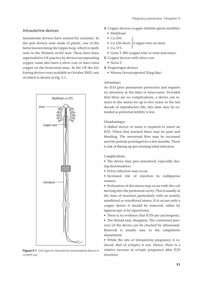
Pregnancy prevention Chapter 5
51
Intrauterine devices
Intrauterine devices have existed for centuries. In
the past devices were made of plastic, one of the
better known being the Lippes loop, which is rarely
seen in the Western world now. These have been
superseded in UK practice by devices incorporating
copper; some also have a silver core or have extra
copper on the horizontal arms. In the UK the fol-
lowing devices were available in October 2002, one
of which is shown in Fig. 5.1.
1 Copper devices (copper inhibits sperm motility)
• Multiload.
• Cu 250.
• Cu 250 short. Copper wire on stem.
• Cu 375.
• Gyne T 380 (copper wire or stem and arms).
2 Copper devices with silver core
• Nova-T.
3 Progestogen devices
• Mirena (levonorgestrel 20mg/day)
AdvantagesAn IUD gives permanent protection and requires
no attention at the time of intercourse. Provided
that there are no complications, a device can re-
main in the uterus for up to five years; in the last
decade of reproductive life, this time may be ex-
tended as potential fertility is less.
DisadvantagesA skilled doctor or nurse is required to insert an
IUD. When first inserted there may be pain and
bleeding. The menstrual flow may be increased
and the periods prolonged for a few months. There
is risk of flaring up pre-existing tubal infection.
Complications• The device may pass unnoticed, especially dur-
ing menstruation.
• Pelvic infection may occur.
• Increased risk of rejection in nulliparous
women.
• Perforation of the uterus may occur with the coil
moving into the peritoneal cavity. This is usually at
the time of insertion particularly with an acutely
anteflexed or retroflexed uterus. If it occurs with a
copper device it should be removed, either by
laparoscopy or by laparotomy.
• There is no evidence that IUDs are carcinogenic.
• The thread may disappear. The continued pres-
ence of the device can be checked by ultrasound.
Removal is usually easy in the outpatients
department.
• While the rate of intrauterine pregnancy is re-
duced, that of ectopics is not. Hence, there is a
relative increase in ectopic pregnancy after IUD
insertion.
Multiload–cu 375
Arms
Copper wire
Introducer
Figure 5.1 One type of intrauterine contraceptive device incurrent use.
}
AMI5 6/9/04 5:24 PM Page 51

Chapter 5 Pregnancy prevention
52
• The progestogen intrauterine system (IUS) re-
duces menstrual flow and often dysmenorrhoea.
It has a lower incidence of pelvic inflammatory
disease.
ContraindicationsAn IUD should not be inserted in the presence of:
• pelvic infection;
• large or submucosal uterine fibroids;
• genital malignancy;
• abnormal bleeding;except Mirena
• menorrhagia.
Method of insertionThe device is supplied in a sterile pack with full in-
structions for insertion. This should be done with
aseptic and antiseptic precautions (Fig. 5.2).
• The best time is at the end of menstruation.
• The cervix is exposed and may be steadied with a
single-toothed forceps.
• A uterine sound measures cavity length.
• The device is loaded into the introducer and
inserted.
• The introducer is withdrawn and the nylon
threads cut leaving 1.5–2.0cm in the upper vagina.
• The woman should be taught to identify the
threads.
A vasovagal attack may occur at the time of
insertion, following cervical stimulation. This
usually responds on stopping the insertion and
lowering the woman’s head. Formal resuscitation is
very rarely needed, but facilities should be available
at the family planning clinic for the rare occasion.
Post-coital contraceptionAn IUD may be used for post-coital contraception
to prevent implantation if inserted within five days
of unprotected intercourse. The woman must be
seen again to ensure menstruation has occurred.
This is an emergency measure, but if normal men-
struation occurs the device may be left for per-
manent contraception.
PregnancyThe pregnancy rate with copper devices is reported
as 1.4 per 100 women/years. Should pregnancy
occur the possibility of ectopic pregnancy must be
excluded.
• If the tail of the device is visible the device
should be removed by pulling gently on the
thread.
• If the tail of the device is not found the position
of the device must be checked by ultrasound.
• The device may be left in the uterus throughout
pregnancy.
Barrier contraceptionThe most effective is the vaginal diaphragm or
Dutch cap which consists of a watch spring or
coiled spring edged with a dome of latex. They are
made in various sizes and for maximum safety
must be used with a spermicide jelly or cream.
• The correct size must be selected before exam-
ination (Fig. 5.3).
• The woman should be taught how to insert and
remove it.
• Always use it with a spermicide.
• Leave it in for eight hours after intercourse.
• A check of the fit and, if needs refitting, after six
months, and after childbirth.
If there is prolapse or a retroverted uterus, an al-
ternative is the cervical cap made of rubber or plas-
tic. This is harder to get in place and easier to
displace at intercourse.
The vaginal sheath is a plastic bag which lines the
vagina. It retains its place by a spring ring in the
fornices. The woman can insert it at leisure. It has
(a) (b)
Figure 5.2 Method of insertion. (a) The T-shaped IUD isstraightened inside a plastic tube and inserted through thecervix. (b) When pushed from the hollow tube it resumes itsold shape.
}
AMI5 6/9/04 5:24 PM Page 52
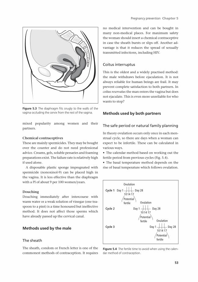
Pregnancy prevention Chapter 5
53
mixed popularity among women and their
partners.
Chemical contraceptivesThese are mainly spermicides. They may be bought
over the counter and do not need professional
advice. Creams, gels, soluble pessaries and foaming
preparations exist. The failure rate is relatively high
if used alone.
A disposable plastic sponge impregnated with
spermicide (nonoxinol-9) can be placed high in
the vagina. It is less effective than the diaphragm
with a PI of about 9 per 100 women/years.
DouchingDouching immediately after intercourse with
warm water or a weak solution of vinegar (one tea-
spoon to a pint) is a time honoured but ineffective
method. It does not affect those sperms which
have already passed up the cervical canal.
Methods used by the male
The sheath
The sheath, condom or French letter is one of the
commonest methods of contraception. It requires
no medical intervention and can be bought in
many non-medical places. For maximum safety
the woman should insert a chemical contraceptive
in case the sheath bursts or slips off. Another ad-
vantage is that it reduces the spread of sexually
transmitted infections, including HIV.
Coitus interruptus
This is the oldest and a widely practised method:
the male withdraws before ejaculation. It is not
always reliable for human beings are frail. It may
prevent complete satisfaction to both partners. In
coitus reservatus the man enters the vagina but does
not ejaculate. This is even more unreliable for who
wants to stop?
Methods used by both partners
The safe period or natural family planning
In theory ovulation occurs only once in each men-
strual cycle, so there are days when a woman can
expect to be infertile. These can be calculated in
various ways.
• The calendar method based on working out the
fertile period from previous cycles (Fig. 5.4).
• The basal temperature method depends on the
rise of basal temperature which follows ovulation.
Figure 5.3 The diaphragm fits snugly to the walls of thevagina occluding the cervix from the rest of the vagina.
Cycle 1
Potentialfertile
Day 1 Day 28
Ovulation
10 14 17
Cycle 2
Potentialfertile
Day 1 Day 28
Ovulation
10 14 17
Cycle 3
Potentialfertile
Day 1 Day 28
Ovulation
10 14 17
Figure 5.4 The fertile time to avoid when using the calen-dar method of contraception.
AMI5 6/9/04 5:24 PM Page 53

Chapter 5 Pregnancy prevention
54
Intercourse is stopped for three days before and
after ovulation.
• Teaching the woman to note the changes in cer-
vical mucus which occur at ovulation and mark
the peak of fertility. Instead of sticky glue-like
mucus, it becomes thin and runny. Motivation is
important as is adequate instruction.
• Ovulation predictor tests, but these are
expensive.
The disadvantage of the safe period is that it is
not really safe if menstruation is irregular, after
childbirth or abortion or in women approaching
the menopause.
Sterilization
Sterilization is an operation aimed at the perma-
nent occlusion of tubes carrying the gametes.
Counselling for sterilization
Sterilization is an important step in the life of any
man or woman. It should be considered as irrevo-
cable for, while reversal is possible for both males
and females, success cannot be guaranteed even in
the most expert hands. Counselling is important
and consent must be given in writing. The consent
of the spouse is no longer necessary legally, but
it is desirable that the couple should be seen to-
gether and the full implications of the procedure
explained.
A girl under 16 cannot consent to sterilization,
nor can her parents insist on it. The same applies to
individuals who are mentally retarded to a degree
that they cannot understand the meaning and
consequences of the operation. At present such
operations can only be performed with the con-
sent of a High Court judge.
Female sterilization
The most practical place to block the female geni-
tal tract is at the fallopian tubes. These are deep in-
side the peritoneal cavity, so their approach is a
bigger procedure than operating on the male. The
use of the laparoscope has reduced much of the
need for large incisions in the majority of cases, but
it is still an intraperitoneal operation requiring a
general anaesthetic and the availability of full sur-
gical skills.
Laparotomy sterilization
The woman should plan to stay in hospital for
2–3 days depending on how fit she is. The simplest
operation was described by Pomeroy; it is not often
performed now. A more common technique is the
Irving method, where each tube is divided and sep-
arated. The medial end is implanted in a tunnel in
the wall of the uterus.
The operation of formal surgical division is
commonly performed these days whilst doing
an elective Caesarean section when the abdomen
is open. The failure rate is about 2–4 per 1000
operations.
A lesser abdominal operation (Mini-Lap) can be
performed by an experienced surgeon. A 5cm
transverse suprapubic incision is extended to the
rectus sheath and the surgeon then separates the
rectus muscles. This allows a bivalve speculum to
be introduced through the incision into the peri-
toneal cavity. Through this, each fallopian tube in
turn can be sought, drawn up and operated upon.
It is divided and the two ends overlapped so that
the ends are separated. This operation is often
done in developing countries where laparoscopy is
not readily available. The failure rate for this oper-
ation is about 1–2 per 1000 operations (Fig. 5.5).
Two clampsmid section
excised
Ends areoverlapped
and tied
Figure 5.5 Division and overlapping of tubes at mini-laparotomy.
AMI5 6/9/04 5:24 PM Page 54

Pregnancy prevention Chapter 5
55
Laparoscopic sterilization
This operation is performed through a small inci-
sion but is potentially just as hazardous as a larger
operation. It should only be performed by those
who are skilled in general gynaecological surgery
because the abdomen may have to be opened at
any time to deal with complications. These are,
however, uncommon and usually the laparoscopic
sterilization can be done as a day case.
The tube is then blocked by one of three
methods.
1 A mechanical clip (Hulka–Clemens, Filshie) may
be applied to each tube. This may be a spring-
loaded clip or a plastic one with a grip (Fig. 5.6).
2 A silastic ring (Falope ring) may be applied to the
medial narrow part of each tube through the la-
paroscope (Fig. 5.7). A knuckle of tube is drawn up
and over it is slipped a silastic ring which constricts
the neck of the knuckle. This causes necrosis of the
tube at the bound point, which then fibroses,
blocking the lumen and then pulls apart leaving
the tube with a gap.
3 Unipolar or bipolar diathermy used to cauterize the
fallopian tube in two places about 1cm apart in the
isthmal area. There is a risk of heat damage. For this
reason, it is less commonly used in the Western
world.
Failure rates of laparoscopic sterilizations are
about 2–3 per 1000 operations.
Other methods
Transuterine methods of sterilization without
anaesthesia are being attempted using a hystero-
scope and passing catheters through the uterine
cavity into the fallopian tubes. A rapidly setting
plastic resin is injected; alternatively, electrical
cautery or cryosurgery by narrow cooling probes
has been used by the same route. None of these
methods has yet been shown to produce reliable
results.
Causes of failure
Despite these operations, a woman may still get
pregnant in 2–3 per 1000 cases in the UK.
• The woman may already have a fertilized oocyte
in the proximal tube or even the uterus at the time
of the operation. Hence try to operate in the first
two weeks of the cycle.
• All women undergoing sterilization should have
a pregnancy test before the procedure.
• The occluding clip or ring may be correctly
placed but spring off or break after the operator has
left the abdomen. Unless a good knuckle of tube is
brought through the elastic ring, it may not be
pinching it securely. The clip may re-open under
pressure of the tissues although this is less com-
mon with the beaked Filshie clip. Very rarely steril-
(a) (b)
Figure 5.6 Clip sterilization. (a) A Filshie clip and (b) itsmethod of application.
Ring
(a) (b) (c) (d)
Ring
Figure 5.7 A silastic ring and its method of application. (a)The springy forceps grasp the tube. (b) Retracting the for-ceps draws the tube through the plastic ring. (c) Passing theforceps back releases the tube and the tube is constricted atits neck by the ring. (d) The loop becomes hypoxic, dies andthe ends separate when the ring drops off.
AMI5 6/9/04 5:24 PM Page 55

Chapter 5 Pregnancy prevention
56
ization clips have broken. Thus they have no oc-
clusive effect on the fallopian tube and have
been removed subsequently from the abdomen in
pieces.
• Although the tube is blocked by the clip and the
short length of fibrosis it causes, the two ends of
the tube may not separate. A small fistula can form
between the contiguous ends bypassing the
occluding device or burrowing through the short
length of fibrous tissue. This would allow sperm to
pass up readily although fertilized eggs might pass
down less commonly; in consequence an ectopic
pregnancy may result.
• The occluding device may be put on the wrong
structure. Laparoscopic sterilization should be
done only when the surgeon has good vision.
Occasionally, however, with less than perfect sight,
a clip is put on the round ligament just in front of
the fallopian tube.
A hysterosalpingogram is occasionally per-
formed 16 weeks after the operation to ensure
tubal blockage. It is unwise to do it before as the
pressure of the injected dye may disrupt healing
tissues and produce a fistula between the blocked
parts of the tube.
Male sterilization
Blockage is performed by division of each vas def-
erens in the groin, a lesser surgical operation than
for the female, and done under local anaesthesia
(Fig. 5.8). Unguarded intercourse should await two
semen specimens showing no sperm, often as long
as 16 weeks after the surgery.
Complications are few:
• Haematoma of scrotum.
• Infection.
• Failure of blockage.
• Long-term generation of sperm antibodies and
non-specific antibodies in later life.
Termination of pregnancy
Pregnancy may be terminated by intervention
with instruments or by drugs. If done incorrectly,
there is a high risk of trauma and death of the
mother. It is illegal in some societies, but many
sovereign states have now passed laws which
allow a termination to be performed by doctors
under certain constraints.
Religious and cultural factors dictate whether
termination is allowed in a country. Generally,
termination of pregnancy (TOP) is unacceptable
to the Roman Catholic Church, to Moslems and
some other major world religions. In actuality,
women seek termination of unwanted pregnancies
world-wide, irrespective of the official religion or
laws of a country.
In some countries, TOP is used as a part of the
contraceptive programme; in Communist Eastern
Europe, up to a third of women used termination as
their primary means of contraception. In most of
the Western world, this is not so, for it is appreciat-
ed that TOP has many more complications than the
more conventional methods of contraception.
Four factors have recently rendered TOP safer in
Western society.
• Better training of doctors in the subject.
• Safer general anaesthesia or wider use of local
anaesthetic agents.
• Asepsis and antisepsis have reduced infection.
• Liberalization of abortion laws encourage early
medical advice.
Position in the UK
Major changes in TOP were associated with the
1967 Abortion Act (which does not apply in
Northern Ireland) and the subsequent 1991
amendment. Before then, a few TOPs had been car-
ried out under an obiter dictum or case law which
Divide
Figure 5.8 Male sterilization. Divided ends of the vas deferens are turned back and ligated to ensure that theopen ends are not just separated but point in opposite directions.
AMI5 6/9/04 5:24 PM Page 56
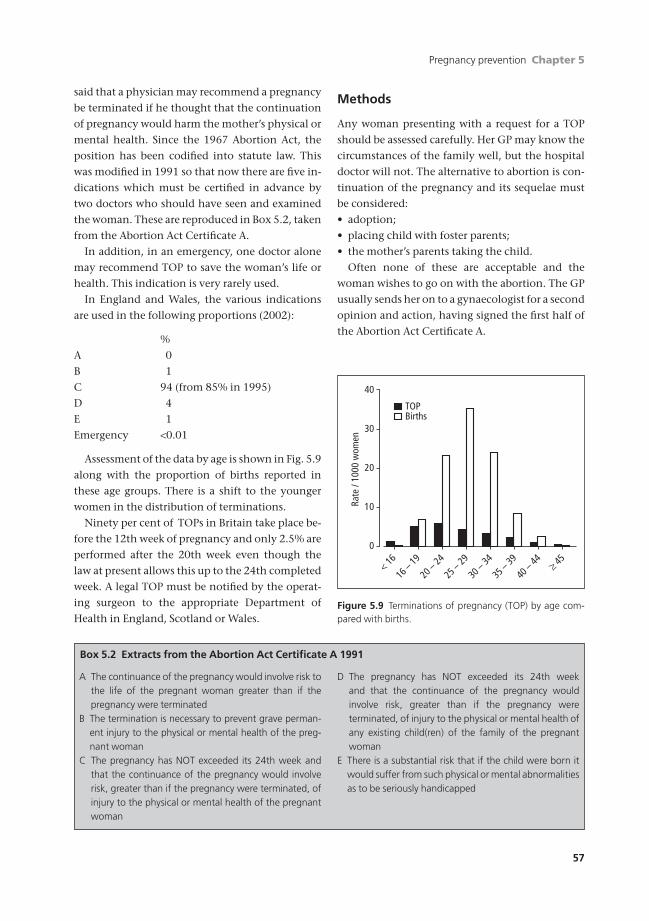
Pregnancy prevention Chapter 5
57
said that a physician may recommend a pregnancy
be terminated if he thought that the continuation
of pregnancy would harm the mother’s physical or
mental health. Since the 1967 Abortion Act, the
position has been codified into statute law. This
was modified in 1991 so that now there are five in-
dications which must be certified in advance by
two doctors who should have seen and examined
the woman. These are reproduced in Box 5.2, taken
from the Abortion Act Certificate A.
In addition, in an emergency, one doctor alone
may recommend TOP to save the woman’s life or
health. This indication is very rarely used.
In England and Wales, the various indications
are used in the following proportions (2002):
%
A 0
B 1
C 94 (from 85% in 1995)
D 4
E 1
Emergency <0.01
Assessment of the data by age is shown in Fig. 5.9
along with the proportion of births reported in
these age groups. There is a shift to the younger
women in the distribution of terminations.
Ninety per cent of TOPs in Britain take place be-
fore the 12th week of pregnancy and only 2.5% are
performed after the 20th week even though the
law at present allows this up to the 24th completed
week. A legal TOP must be notified by the operat-
ing surgeon to the appropriate Department of
Health in England, Scotland or Wales.
Methods
Any woman presenting with a request for a TOP
should be assessed carefully. Her GP may know the
circumstances of the family well, but the hospital
doctor will not. The alternative to abortion is con-
tinuation of the pregnancy and its sequelae must
be considered:
• adoption;
• placing child with foster parents;
• the mother’s parents taking the child.
Often none of these are acceptable and the
woman wishes to go on with the abortion. The GP
usually sends her on to a gynaecologist for a second
opinion and action, having signed the first half of
the Abortion Act Certificate A.
Box 5.2 Extracts from the Abortion Act Certificate A 1991
A The continuance of the pregnancy would involve risk tothe life of the pregnant woman greater than if thepregnancy were terminated
B The termination is necessary to prevent grave perman-ent injury to the physical or mental health of the preg-nant woman
C The pregnancy has NOT exceeded its 24th week andthat the continuance of the pregnancy would involverisk, greater than if the pregnancy were terminated, ofinjury to the physical or mental health of the pregnantwoman
D The pregnancy has NOT exceeded its 24th week and that the continuance of the pregnancy would involve risk, greater than if the pregnancy were terminated, of injury to the physical or mental health ofany existing child(ren) of the family of the pregnantwoman
E There is a substantial risk that if the child were born itwould suffer from such physical or mental abnormalitiesas to be seriously handicapped
40
20
30
10
0
< 16
16 –
19
20 –
24
25 –
29
30 –
34
35 –
39
40 –
44 ≥ 45
Rate
/ 10
00 w
omen
TOPBirths
Figure 5.9 Terminations of pregnancy (TOP) by age com-pared with births.
AMI5 6/9/04 5:24 PM Page 57

Chapter 5 Pregnancy prevention
58
In the hospital, if in agreement with the GP, the
specialist signs the second part. Time should be al-
lowed for the woman to reconsider; if she wishes to
go ahead she is checked for fitness for operation
under anaesthesia and tested for chlamydia. The
future method of contraception is discussed to
avoid a repeat unwanted pregnancy. Many
women are day case admissions, especially those
seen in the earlier weeks of gestation (before
14 weeks). TOP must be performed in a National
Health Hospital or clinic specifically licensed for
the purpose.
Early abortion
Surgical terminationFor pregnancies up to 12 weeks’ gestation, TOP is
often by a vacuum aspiration of the uterine cavity.
A hollow plastic catheter is passed through the
cervix which has been gently dilated, usually after
prostaglandin ripening. A vacuum suction then re-
moves the uterine contents and a gentle curettage
ensures the uterus is empty. This technique has few
complications and a high success rate.
Medical terminationThe use of antiprogesterone steroids like mifepris-
tone has spread from Europe to the UK, but not
widely in the USA because of the views of their
politicians. An increasing number of women at less
than 9 weeks’ gestation elect for this day case TOP
which is very effective in producing total abortions
in 96% —a figure comparable with the success rate
of aspiration TOP. Women opt for this since it
avoids anaesthesia and surgery; they consider it
more natural and it gives them control.
Used in the outpatients, a single oral dose of
mifepristone is given followed 36–48 hours later by
200–600mg oral misoprostol or 0.5mg gemeprost
vaginal pessary. While the measured blood loss in
both medical and surgical methods is the same,
women report a longer period of blood loss after
medical TOP.
Mifepristone is also most useful at 16–18 weeks’
gestation when 200mg is given 24 hours before
prostaglandins (PGE2a) and works swiftly.
Mifepristone can also be injected into ectopic
gestational sacs in ectopic pregnancy (Chapter 8)
and can be used for post-coital emergency
contraception.
Complications of early termination of pregnancy• Haemorrhage —separation of the sac and forming
placenta causes blood loss. Syntometrine is
often needed intravenously to help the myo-
metrium clamp down.
• Perforation —if the uterus is perforated, la-
paroscopy with examination and repair of the
uterus may be required.
• Infection —TOP is an invasive procedure, passing
instruments through the potentially septic area of
the vagina into the sterile area of the uterus. The
operator cleans the upper vagina with antiseptic
before starting and antibiotics may be given before
the operation if vaginal infection is suspected.
If infection occurs, particularly with chlamydia, it
must be treated promptly with antibiotics or a tubal
infection may follow leading to future infertility.
• Incomplete termination —this leads to retention of
products of conception. Bleeding occurs and
re-evacuation of the uterus is necessary.
• Psychological complications —many women have
a natural grief reaction after early TOP; if the
abortion was voluntary, that reaction passes in a
few weeks or months. Should the woman have
been coerced into termination, the reaction can
continue for much longer; up to 25% of women in
this latter group may require psychotherapy.
Future pregnanciesEarly TOP usually has no effect on future pregnan-
cies. If the cervix is properly dilated (usually not
more than 8mm), there should be no cervical
incompetence following early TOP; this is more
commonly found after 10–12mm dilatation,
particularly if no prostaglandin was given preoper-
atively to ripen the cervix.
Mid-trimester abortion
After 14 weeks of gestation, pregnancy termina-
tion becomes more difficult.
AMI5 6/9/04 5:24 PM Page 58

Pregnancy prevention Chapter 5
59
Morcellation and extractionIn the hands of an expert and experienced gynae-
cologist, under anaesthesia the cervix can be di-
lated to well beyond 10mm. Crushing instruments
are introduced into the uterus to break up the fetus
which is then extracted piecemeal. This is an un-
pleasant and potentially hazardous way to abort,
but in expert hands it does mean the whole proce-
dure is over in a few minutes with the mother
asleep and unaware of the abortion. She often goes
home later the same day.
ProstaglandinsThe uterus can be made to contract by giving
prostaglandins (PGE2a) per vaginam, intra-amnioti-
cally, extra-amniotically or intravenously. Under
sterile conditions, an amniocentesis can be per-
formed drawing 20ml of amniotic fluid. In its
place, 40mg of PGE2a is injected in the amniotic
cavity. Alternatively, PGE2a gel/tablet is inserted
into the vagina causing the uterus to contract.
A mini-labour follows and delivery usually takes
place in 10–20 hours. This is painful and so should
be well covered with analgesia, even an epidural.
Evacuation of the placenta is needed in many cases.
HysterotomyIf prostaglandins are not available, pregnancies
after about 14 weeks require a surgical termination
and a mini-Caesarean section is performed. There
is no lower segment and so the uterine incision has
to be vertical. This is rarely done in the UK now but
often in the USA.
Complications• Bleeding —this is uncommon after prostaglandin
TOP, because the uterus has been contracting
through the mini-labour. However, the placenta is
often retained in terminations between 14 and 22
weeks of pregnancy and often requires removal
under general anaesthesia.
Bleeding is not a major problem after hys-
terotomy for it is a surgical procedure at which
haemostasis is achieved at the time of the
operation.
• Infection —infection is not common after mid-
trimester abortion procedures and should be pre-
vented as it is in any labour or surgical operation by
good asepsis and antisepsis.
• Psychological problems —these are commoner
after mid-trimester abortion. The woman has been
pregnant for a longer period and the fetus is more
developed. This is an area of great sensitivity which
the attendants must be very careful in handling.
Some people wish to bury the fetus with a religious
service; these natural reactions should be assisted.
• Future pregnancies —following mid-trimester
TOP, the cervix has been dilated and cervical in-
competence can follow.
A vertical hysterotomy scar on the uterus might
cause a problem, for it is more liable to rupture in a
subsequent pregnancy than a lower segment trans-
verse incision.
Later terminations
Until 1991, TOP was only permitted in England,
Wales and Scotland before 28 weeks, the time of
presumed viability in law. With modern neo-
natal developments, the law has reduced this to
24 weeks. Clauses C and D limit terminations to
below 24 weeks, but the other clauses (risk of
death, grave permanent injury to the mother or
fetal abnormalities) are not time limited. Some-
times ultrasound may only show a fetal abnormal-
ity at 26–28 weeks or cordocentesis done for
karyotyping studies may give results as late as
28–29 weeks.
Late termination is difficult to accept for the
woman and to all who have to care for the woman,
but it is a logical extension of the law’s previous po-
sition with more up to date diagnostic tests on the
fetus. Such terminations are usually done with
mifepristone and prostaglandins.
AMI5 6/9/04 5:24 PM Page 59

Chapter 5 Pregnancy prevention
60
Self-assessment
5.1 OSCE role-play question (10 minutes).
Candidate’s instructions: A 14 year-old girl comes to your practice requesting contraception. You are the GP whois going to counsel her. She has no medical illnesses and her weight is normal.
Role-player’s instructions: You are a 14-year-old girl who is fully developed. Your periods started when you were11. You have been going out with a 16-year-old boy in your school for the last three months. Your relationship hasdeveloped to the point that you are thinking about having sex with him. Your parents do not know about the rela-tionship and you do not want to tell them. You get on reasonably well with your father but he is never at home ashe is a long distance lorry driver. You constantly fight with your mother who does not understand you at all andwould be horrified that you had a boyfriend. She disapproves of your friends and will not let you go out late at week-ends. You tend to defy her and take the consequences in the morning. You know that she would be furious if yougot pregnant so you want to be sensible and take precautions so this can’t happen. Your friend has told you to goto the GP and get the pill. She has reassured you that s/he cannot tell your parents without your permission. Youhaven’t told your boyfriend because you do not want him to know that you are ready for sex, you just want to beprotected in case. You have no medical problems, you smoke three cigarettes a day and are of normal weight. Youhave never taken any illegal substances. The rest is up to the imagination of the role-player.
5.2 OSCE role-play question (10 minutes).
Candidate’s instructions: A woman of 35 comes to your clinic requesting sterilization. She is fit and well and ofnormal weight. You are the doctor who will counsel her.
Role-player’s instructions: You are a woman of 35. You have 3 children and do not want any more. You havebeen married for 12 years and your children are 10, 7 and 3 years old, all fit and well. You and your husband havediscussed this and have decided that you should have the operation. You have been on the pill for the last three yearsbut you know you should stop taking it as you smoke 40 cigarettes a day. Your periods were regular but quite heavybefore you had your youngest daughter. You have no other medical problems and all the children were normal de-liveries at term. You do not want to use barrier methods because they are messy and unreliable. You are not keenon a coil because you have heard they cause heavy periods and infection. You are worried about putting on weightafter the operation and have heard that the mini-pill and the injections are also bad for your weight. All other de-tails are up to the role-player.
5.3 Which of the following statements are true about the combined oral contraceptive pill (COCP)?(a) COCP should be started on the seventh day of a woman’s period.(b) If the woman misses the first two pills in her packet she should use additional protection for at least two weeks.(c) COCP should be taken at the same time of day to ensure protection from unwanted pregnancy.(d) A past medical history of thrombosis is an absolute contraindication for prescribing the COCP.(e) Women on antiepileptic medication should be prescribed a pill with a low estrogen content.
AMI5 6/9/04 5:24 PM Page 60

Pregnancy prevention Chapter 5
61
Self-assessment Continued
5.4 Which of the following are absolute contraindications to the use of an intrauterine contraceptive device.(a) Past history of pelvic inflammatory disease.(b) Past history of sickle cell disease.(c) Past history of termination of pregnancy.(d) Past history of an abnormal cervical smear.(e) Past history of heart valve replacement.
5.5 Which of the following are recognized methods of emergency contraception.(a) High dose estrogen given in two doses 12 hours apart.(b) High dose estrogen and progestogen given in two doses 12 hours apart.(c) High dose progestogen given in two doses 12 hours apart.(d) Endometrial curettage.(e) Insertion of an intrauterine device (IUD) within five days of unprotected intercourse.
AMI5 6/9/04 5:24 PM Page 61
62
Introduction
This chapter will address benign conditions and in-
fections affecting the lower genital tract. In reading
the chapter, it is useful to remember that all infec-
tions may be sexually transmitted (STIs) and there-
fore it is essential to consider this possibility in all
women who are, or have recently been, sexually
active. It is important to have knowledge of these
diseases which are the most widespread infectious
diseases in the UK and many other parts of the
world. Some of the STIs have significant long-term
sequelae including infertility, ectopic pregnancy,
chronic pelvic pain as well as the possibility of
mother-to-child transmission during pregnancy or
at delivery and postpartum (if breast-feeding).
The increase in the reported incidence of STIs in
the UK and other countries reflects a number of
factors, including worsening of access to over-
burdened sexual health services, a more liberal
attitude to sexual intercourse, the younger age of
starting sexual activity, a lack of political will to
deal with STI-related issues, and less than ideal sex
education in some schools and homes. In addition,
the introduction of more sensitive DNA
amplification-based techniques has resulted in
more diagnoses of some conditions, e.g. Chlamydia
trachomatis. The UK has a well-established network
of genitourinary medicine (GUM) clinics which
provide primary care access to patients at risk of
STIs. These clinics provide quarterly statistical
returns on their workload; a statutory requirement
in the UK (KC60). In addition, these clinics have
health advisers who oversee partner notification
(contact tracing) of all patients diagnosed with im-
portant STIs. They also possess expertise in risk
assessment for HIV infection and provide pre- and
post-test counselling for those wishing to know
their HIV status.
In an attempt to deal with the alarming increase
in STI diagnoses and the increasing waiting times
in GUM clinics, the UK Government recently re-
leased a National Strategy for Sexual Health and
HIV which aims to increase the amount of STI test-
ing and treatment undertaken in general practice
and community reproductive and sexual health
(family planning) services. However, it should be
noted that, despite the importance of sexual ill-
health among younger members of our society, the
UK Government has not chosen to make sexual
health a National Service Framework. This political
move would ensure a marked improvement in the
quality of sexual health services in the UK.
Any health care worker who takes on the respon-
sibility of treating an STI (presumed or confirmed)
must ensure that sexual partners are treated as well.
There is little or no point in treating the index case
when they will simply be reinfected by an un-
treated partner. Even if they are no longer at risk of
reacquiring the STI from their expartner, it is im-
portant that the source is treated from the public
health view point.
Chapter 6
Benign diseases, genital tractinfections and sexual problems
AMI6 6/9/04 5:25 PM Page 62
Benign diseases, genital tract infections and sexual problems Chapter 6
63
Taking a sexual history
1 History of presenting condition.
• Describe the current symptoms.
2 Sexual contact history.
• Cover sexual contacts for the past three
months (six months if secondary syphilis or
acute hepatitis B are likely).
• Gender of partner(s) and whether regular or
non-regular partner.
• Sexual orientation of patient.
• Type of sexual intercourse: vaginal, oral, anal,
fingers, sex toys.
• Use of condoms and other contraceptive
methods.
3 History of past STIs.
• Ask about past chlamydial, gonococcal and
syphilis infection.
• Warts and herpes may recur spontaneously.
• Fully document previous treatment for posi-
tive treponemal serology.
• Document previous HIV test result (with year).
4 Recent antibiotic and drug history.
• Relevant for gonorrhoea in terms of possible
resistance.
• Document allergies.
5 Hepatitis B immunization.
• Hepatitis B immunization is recommended for
those using IV drugs, sex workers and those who
are partners of Hepatitis B carriers.
6 HIV risk assessment.
• Necessary only for period since last negative
HIV test.
• Number of sexual partners with whom patient
practised unprotected vaginal or anal
intercourse.
• Has the patient had unprotected sexual inter-
course with men in or from high HIV-risk
countries.
• IV drug abuse with sharing of needles.
• Blood transfusion at time when blood supplies
were not checked for HIV antibodies.
Benign conditions
The vulva and vagina
Pruritus vulvae
Pruritus vulvae is a common symptom —an irrita-
tion of the vulva sufficient to lead to scratching.
CausesIrritating vaginal discharges of Trichomonas vaginalis
or monilia. Since many infections are common to
the vulva and vagina, these are considered collec-
tively in later sections. Other causes are:
• Parasites such as scabies and crab lice.
• Fungi such as athlete’s foot affects the feet, the
groin and the vulva.
• Sensitivity to drugs or chemicals including:
(a) soap and disinfectants;
(b) detergents used for washing underwear;
(c) contraceptives made of rubber;
(d) commercial spermicides;
(e) ointments containing benzocaine and
amethocaine.
• Iron deficiency anaemia associated with glossitis.
• Gross vitamin deficiencies, A and B group, espe-
cially in elderly women.
• Glycosuria due to any cause, but principally dia-
betes. Glycosuria probably causes irritation be-
cause the vulva becomes infected with a fungus. A
random fasting blood glucose should be measured.
Degenerative conditions of the vulva occur mainly
in postmenopausal women and are associated with
irritation and soreness. There are two main
varieties.
• Vulval dystrophy —an important, but uncommon
condition specific to the vulva. It presents as a
thickening and hypertrophy of the vulval skin
often spreading into the groin and around the
anus. The hypertrophic keratin layer causes white
patches (which used to be called leukoplakia). It
may be precancerous and tends to recur in time
after surgical removal of the vulva. Diagnosis is by
biopsy; this shows thickening and increase in
depth of the keratin layer, while the basal papillae
are hypertrophied and dip deeply into the dermis.
AMI6 6/9/04 5:25 PM Page 63
Chapter 6 Benign diseases, genital tract infections and sexual problems
64
• Lichen sclerosus et atrophicus is an atrophic condi-
tion usually seen in postmenopausal women and
pre-pubertal girls. It differs from vulval dystrophy
in that it is atrophic and does not spread beyond
the skin of the vulva itself. Biopsy shows a general
thinning of all the layers of the skin; the keratin
layer is deficient and the basal papillae are flat-
tened. There is hyaline change in the dermis which
is infiltrated with lymphocytes.
Careful investigation reveals a cause for pruritus
vulvae in over 90% of cases. True pruritus must be
distinguished from soreness of the vulva, also a
common symptom, and often caused by vaginitis,
oestrogen deficiency states and postmenopausal
atrophy.
InvestigationsThese should include:
• a careful history, including the use of any sub-
stance which might lead to allergy;
• examination to determine characteristics and
limits of physical signs;
• bacteriological examination of vaginal secretions
and scrapings from the skin;
• a full blood count;
• a fasting blood glucose if relevant;
• biopsy of the skin of the vulva.
TreatmentWomen tend to be very sensitive and shy about
pruritus and often delay in seeking advice. Self-
medication often makes the condition worse.
No treatment should be attempted without full
examination and investigation. Blind treatment
may mean that a serious condition, such as an
early carcinoma or diabetes, is overlooked. With
full investigation, the cause can be found in almost
every case. The help of a dermatologist may be
sought in difficult cases. The treatment is that of
the cause, e.g. diabetes or anaemia.
Fungal infections should be treated with
imidazole drugs and vaginal applications should
be supplemented with antifungal creams for
vulvitis.
Dystrophy and lichen sclerosis may respond to
ointments containing hydrocortisone. In cases
which do not improve, simple vulvectomy may be
undertaken but the condition often returns.
Care should be taken in using local analgesics,
especially benzocaine and amethocaine, as acute
sensitivity may develop.
Non-STI conditions of the vulva
• Infections, boils and carbuncles may affect the
vulva, especially in women with glycosuria and
diabetes.
• Swellings of Bartholin’s gland. Cysts of Bartholin’s
duct and a Bartholin’s abscess are common. Both
present as swellings in the fourchette. An acute
abscess is painful and tender like a boil; an abscess
may rupture spontaneously, but tends to recur.
Treatment of both cysts and abscesses is by marsu-
pialization which permits adequate drainage and
in many cases the function of the gland is retained.
The pus in an abscess should always be cultured
and a search made for gonococci in the urethra and
cervix, since some Bartholin’s abscesses are due to
gonorrhoea.
• Vaginal and uterine causes. A patient complaining
of a lump in the vulva may be suffering from:
• prolapse;
• a large polyp;
• inversion of the uterus;
• a vaginal cyst.
Conditions of the urethra
Urethral caruncle occurs mostly in postmenopausal
women. It presents as a bright red, exquisitely ten-
der swelling at the posterior margin of the urethral
meatus. Symptoms include dysuria, bleeding and
dyspareunia. Treatment is to excise the caruncle
with diathermy.
Prolapse of the urethra, which may be acute or
chronic, involves the whole circumference of the
urethra and not just the posterior margin of the
meatus. It may give similar symptoms to a
caruncle. If symptoms are severe, the prolapsed
urethra must be excised and repaired.
AMI6 6/9/04 5:25 PM Page 64

Benign diseases, genital tract infections and sexual problems Chapter 6
65
Conditions of the vagina and cervix
Infections of the vagina may or may not be
sexually-transmitted infections (STIs), whereas
infections of the cervix are usually caused by one
of two STI pathogens, Neisseria gonorrhoeae and
Chlamydia trachomatis.
Natural protection of the genital tract is
provided by:
• squamous epithelium thickened by oestrogens;
• low pH(4) from lactic acid derived from intra-
epithelial glycogen breakdown;
• mucus from the cervix, Bartholin’s gland and
Skene’s glands rich in bactericidal lysozymes.
This protection is diminished by a number of
causes.
• Pre-pubertal and postmenopausal low oestrogen
levels resulting in a thinner epithelium, a higher
pH and less mucus production.
• Antibiotics which destroy the commensal
flora, in particular lactobacilli (Doderlein’s
bacilli).
• Chemical douches which wash away the natural
protective secretions.
• Foreign bodies in the vagina such as pessaries or
tampons.
• Abortion and menstruation which render the pH
more alkaline and remove the protective discharge
in the cervical cavity.
• Debilitation.
Symptoms
VAGINAL DISCHARGE
• Details of the onset, e.g. postcoital, postantibiotics.
• The volume —does it soil the clothing or require
sanitary towels to be used?
• The colour and consistency:
(a) white and thick —candidiasis;
(b) yellow, green and frothy —trichomoniasis;
(c) mucus —cervical origin.
Itchiness or pruritus is commonly associated with
candidiasis and trichomoniasis.
SORENESS
• Comes with secondary bacterial invasion or
herpes simplex.
CERVICITIS
• Postcoital or intermenstrual bleeding.
• May be associated with pelvic pain or deep
dyspareunia.
ULCERATION
• Associated with viral infections (herpes) or expo-
sure to chemicals, e.g. detergent which causes
inflammation.
OFFENSIVE DISCHARGE
• Often associated with anaerobic bacterial
activity caused by foreign bodies and carcinoma.
ExaminationInspect the vulva for:
• reddening;
• oedema;
• excoriation from scratching;
• ulceration.
Inspect the external urethral meatus for prolapse
or caruncle.
Vaginal examination to check for:
• patchy, reddening associated with tricho-
moniasis;
• white plaques associated with candidiasis;
• punctate vesicle type ulceration associated with
herpes simplex.
Examine the cervix for:
• reddening;
• mucopurulent cervical discharge;
• contact bleeding.
Palpate the urethra for thickening and tender-
ness. Check the Bartholin’s glands are not enlarged
or tender.
InvestigationsSwabs should be taken from the cervical canal,
lower urethra, posterior fornix and from any overt
lesions on the walls of the lower genital tract. In-
stant microscopy in saline can sometimes demon-
strate the presence of Trichomonas vaginalis and
candida. All the swabs should be sent to the
laboratory in Stuart’s medium as speedily as pos-
sible for culture.
AMI6 6/9/04 5:25 PM Page 65

Chapter 6 Benign diseases, genital tract infections and sexual problems
66
TreatmentThe principles of therapy for infection are:
• to avoid indiscriminate treatment;
• to ensure that there is no sexually transmitted
disease or carcinoma present;
• to avoid excessive treatment, which may lead to
a chemical vaginitis.
Foreign bodies in the vaginaThis usually presents with a foul discharge caused
by the anaerobic organism. This is commonest in:
• children —beads or toys, etc;
• mentally subnormal —beads or coins, etc;
• those in custody —vagina used to hide objects
(e.g. drugs), which are then forgotten.
Treatment requires the removal of foreign
bodies. Lactic acid pessaries restore pH and encour-
age regrowth of lactobacilli.
Toxic shock syndromeCaused by a Staphylococcus aureus septicaemia
toxaemia. Associated with:
• obstructed drainage of menses by tampons;
• abrasion of the cervical surface by tampons;
• retrograde flow of menses into the peritoneal
cavity.
SymptomsShivering, diarrhoea, erythematous rash and faint-
ing. Occurs mostly on second and third days at the
peak of menstrual flow.
Physical signs• Hypotension.
• Fever.
• Tenderness of vagina and cervix.
Treatment• Removal of tampon.
• Intravenous fluid resuscitation.
• Systemic antibiotics, e.g. Magnapen.
Microbiology and therapy of commongenital tract infections
Bacterial vaginosisThis condition is due to an imbalance in the vagi-
nal microflora, although the exact mechanisms
which result in this change remain uncertain.
There is a decrease in the number of acid-
producing lactobacilli and an increase in other
vaginal organisms, including Gardnerella vaginalis,
Mycoplasma species, Mobiluncus species and other
anaerobes. It appears to be more common in cer-
tain ethnic groups, e.g. African Caribbeans, and
also in those who are sexually active.
Symptoms and signsTypically there is a creamy vaginal discharge, often
with an offensive fish-like smell. Sometimes, it is
the smell alone which brings the patient to the
doctor. The symptoms tend to be worse around the
period and the smell may be exacerbated after un-
protected sexual intercourse due to the alkalinity
of semen. In more severe cases, there may be vulval
soreness and itching.
InvestigationsThe diagnosis of bacterial vaginosis (BV) should be
made by Gram staining a specimen taken from the
lateral wall of the vagina. It is no longer recom-
mended to culture for Gardnerella vaginalis as this
organism is a normal commensal in the vagina of
some asymptomatic women. Features consistent
with BV include a reduction in numbers (or ab-
sence) of lactobacilli, the presence of mixed flora
consistent with an increase in anaerobes and
Mobiliuncus species and the presence of more than
20% ‘clue cells’. Clue cells are vaginal epithelial
cells with an adherent mixture of Gram negative
and positive organisms (Fig. 6.1).
Treatment• Metronidazole 400mg two times a day for five
days.
• 2% Clindamycin cream at night for seven
nights.
• No indication to treat the male partner as this is
not an STI.
AMI6 6/9/04 5:25 PM Page 66

Benign diseases, genital tract infections and sexual problems Chapter 6
67
Candidiasis (thrush)This is caused by a dimorphic fungus which exists
in both yeast and mycelial forms. It thrives in
sugar-rich environment and is commonly found
in:
• pregnancy;
• diabetes mellitus;
• oral contraceptive or oestrogen therapy;
• following exposure to broad-spectrum anti-
biotics which kill off the competing commensal
organisms.
Symptoms• Vulval itching.
• Superficial dyspareunia.
• Vaginal discharge of various appearances, in-
cluding thick ‘cottage cheese’ and watery ‘milky’
types.
• May be asymptomatic and found incidentally on
speculum examination or cervical cytology.
• As this is not an STI, there is no need to treat the
male partner.
• Men in contact with candidal proteins may
develop a blotchy red rash on their glans penis.
• Diabetic men may develop extensive candidal
balanitis, which may be the presenting feature of
their diabetes.
Treatment• It is important to treat intravaginally with a pes-
sary or cream as well as topically on the vulva.
• A number of suitable imidazole agents exist, e.g.
clotrimazole 500mg pessary as a single dose with
two weeks of topical 1% clotrimazole cream to the
vulva twice daily for two weeks.
• Oral fluconazole 150mg as a single dose or itra-
conazole 200mg twice daily for one day may be
used when topical therapy is not suitable.
• Male candidal-associated balanitis responds well
to a combination of 1% hydrocortisone and an
antifungal, e.g. Canestan HC or Daktacort, given
twice a day for one to two weeks.
Sexually transmitted diseases
Trichomoniasis
This is an STI in which the male is transiently colo-
nized from the reservoir in the female vagina. This
occurs because the adhesion proteins of this proto-
zoon organism are expressed preferentially in acid
pH, typically found in the vagina whereas the male
urethral pH is approximately 7.4. In addition,
trichomonads are repeatedly washed out of the
urethra when men micturate. The organism can
colonize the male prostate, and the female equiva-
lent (Skene’s glands).
Symptoms and signsThe woman has an intensely irritating vaginal dis-
charge with inflammation of the vulva, vagina and
cervix. The cervical appearance may manifest as
a ‘strawberry cervix’. The vaginal discharge is
greenish-yellow, frothy and offensive with a fish-
like smell. Men typically have no symptoms but,
when present, the symptoms include urethral dis-
comfort, dysuria and occasional urethral discharge.
InvestigationsSame day diagnosis may be made with the use of a
saline wet-mount in which a drop of vaginal fluid
from the posterior fornix is placed in a drop of
saline on which a coverslip is floated. The tri-
chomonads have a characteristic motility due to
Figure 6.1 Gram-stained slide showing ‘clue cell’ in bac-terial vaginosis. With acknowledgement to ProfessorCatherine Ison, Sexually Transmitted Bacteria ReferenceLaboratory, London.
AMI6 6/9/04 5:25 PM Page 67

Chapter 6 Benign diseases, genital tract infections and sexual problems
68
their five flagella. Culture is more sensitive al-
though the gold standard is DNA amplification,
currently unavailable in many clinical settings. It is
important to screen for other common STIs.
Treatment and follow-up• This is an STI, so sexual partner(s) must be
treated with antimicrobials.
• Metronidazole 2g single dose or 400mg twice a
day for five days.
• Tinidazole 2g single dose.
• Various complicated regimens exist for metro-
nidazole resistant trichomoniasis and referral to an
STI specialist is recommended.
• Partner notification (contact tracing) should be
initiated at diagnosis.
• Patients should return for a test of cure when
they have been off antibiotics for at least seven
days.
Chlamydial infection
Chlamydia trachomatis is the most common
bacterial STI in the UK and the predominant
organism responsible for pelvic inflammatory
disease (PID) and ectopic pregnancy. It is a potent
mediator of chronic inflammation and causes fine
adhesions in the pelvis surrounding the tubes and
ovaries. There may be adhesions between the liver
capsule and diaphragm (Fitz-Hugh Curtis syn-
drome). There has been a recent increase in
Chlamydia cases in men and women in the UK (see
Fig. 6.2).
Symptoms and signsMany women are asymptomatic although male
partners may complain of either dysuria or a ure-
thral discharge, which is mucopurulent in nature,
and may occasionally present with epididymo-
orchitis. If symptoms/signs are present in women,
they may include any of the following: vaginal
discharge, mucopurulent cervical discharge,
cervicitis, deep dyspareunia, pelvic pain, cervical
excitation and other symptoms of PID.
Investigations• Chlamydia trachomatis is difficult to grow and
requires a specialist laboratory.
• DNA amplification techniques, which have in-
creased sensitivity over other diagnostic methods,
are the diagnostic methods of choice (e.g. SDA,
PCR, TMA).
• DNA amplification tests can be performed on
non-invasive specimens such as urines or vulval
swabs. These tests will detect both dead and live or-
ganisms but will not provide data on antimicrobial
susceptibility.
• Enzyme linked immunoassay (ELISA) tech-
niques are still used in many laboratories.
• Fluorescent-labelled monoclonal antibodies can
be used to detect chlamydial organisms on an
endocervical smear.
90000
80000
70000
60000
50000
40000
30000
20000
10000
0
No.
of c
ases
1996 1997 1998 1999 2000 2001 2002Year
Women
men
Total
Figure 6.2 Uncomplicated chlamy-dial infection in England and Wales(1996–2002). (Source, PHLS.)
AMI6 6/9/04 5:25 PM Page 68
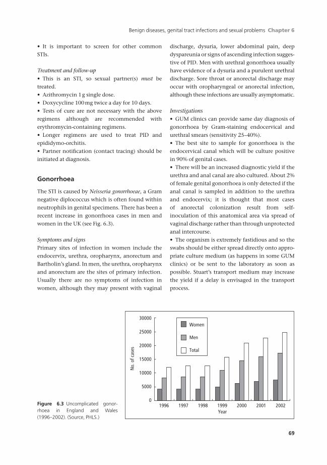
Benign diseases, genital tract infections and sexual problems Chapter 6
69
• It is important to screen for other common
STIs.
Treatment and follow-up• This is an STI, so sexual partner(s) must be
treated.
• Azithromycin 1g single dose.
• Doxycycline 100mg twice a day for 10 days.
• Tests of cure are not necessary with the above
regimens although are recommended with
erythromycin-containing regimens.
• Longer regimens are used to treat PID and
epididymo-orchitis.
• Partner notification (contact tracing) should be
initiated at diagnosis.
Gonorrhoea
The STI is caused by Neisseria gonorrhoeae, a Gram
negative diplococcus which is often found within
neutrophils in genital specimens. There has been a
recent increase in gonorrhoea cases in men and
women in the UK (see Fig. 6.3).
Symptoms and signsPrimary sites of infection in women include the
endocervix, urethra, oropharynx, anorectum and
Bartholin’s gland. In men, the urethra, oropharynx
and anorectum are the sites of primary infection.
Usually there are no symptoms of infection in
women, although they may present with vaginal
discharge, dysuria, lower abdominal pain, deep
dyspareunia or signs of ascending infection sugges-
tive of PID. Men with urethral gonorrhoea usually
have evidence of a dysuria and a purulent urethral
discharge. Sore throat or anorectal discharge may
occur with oropharyngeal or anorectal infection,
although these infections are usually asymptomatic.
Investigations• GUM clinics can provide same day diagnosis of
gonorrhoea by Gram-staining endocervical and
urethral smears (sensitivity 25–40%).
• The best site to sample for gonorrhoea is the
endocervical canal which will be culture positive
in 90% of genital cases.
• There will be an increased diagnostic yield if the
urethra and anal canal are also cultured. About 2%
of female genital gonorrhoea is only detected if the
anal canal is sampled in addition to the urethra
and endocervix; it is thought that most cases
of anorectal colonization result from self-
inoculation of this anatomical area via spread of
vaginal discharge rather than through unprotected
anal intercourse.
• The organism is extremely fastidious and so the
swabs should be either spread directly onto appro-
priate culture medium (as happens in some GUM
clinics) or be sent to the laboratory as soon as
possible. Stuart’s transport medium may increase
the yield if a delay is envisaged in the transport
process.
30000
25000
20000
15000
10000
5000
01996 1997 1998 1999 2000 2001 2002
Women
Men
Total
No.
of c
ases
Year
Figure 6.3 Uncomplicated gonor-rhoea in England and Wales(1996–2002). (Source, PHLS.)
AMI6 6/9/04 5:25 PM Page 69
Chapter 6 Benign diseases, genital tract infections and sexual problems
70
• Once isolated, the gonococci can be screened for
antimicrobial susceptibility.
• DNA amplification tests are now available for the
diagnosis of gonorrhoea using non-invasive speci-
mens such as urine or vulval swabs. These tests
will detect both dead and live organisms but will
not provide data on antimicrobial susceptibility.
• It is important to screen for other common STIs.
Treatment and follow-up• Amoxycillin 2g oral and Probenecid 1g oral,
both as a single dose.
• Ciprofloxacin 500mg oral single dose.
• Ceftriaxone 250mg i.m. single dose.
• Cefotaxime 500mg i.m. single dose.
Longer courses are recommended for the treat-
ment of upper genital tract complications.
• Penicillin resistance may be chromosomally-
mediated or plasmid mediated through the pro-
duction of beta-lactamase (penicillinase).
• High-level ciprofloxacin resistance is increasing
worldwide.
• No documented resistance to third generation
cephalosporins has yet been reported.
• Partner notification (contact tracing) should be
initiated at diagnosis.
• Patients should return for a test of cure when
they have been off antibiotics for at least three
days.
Herpes simplex infection
Genital herpes is caused by herpes simplex virus
(HSV) types 1 and 2 which enter the host through
mucocutaneous surfaces. The viruses are able to
establish latency and subsequent reactivation may
give rise to repeated episodes of the disease. Factors
involved in reactivation include local trauma,
menstruation, HSV 2 viral type and stress.
Symptoms and signsThe first attack is often characterized by extensive
genital ulceration together with local regional lym-
phadenopathy and systemic symptoms. The ulcer-
ation may last up to three weeks if untreated and
first attacks are more severe in vulval and perianal
locations in comparison with penile infection. The
lesions present as vesicles which burst to leave a
superficial tender ulcer with an erythematous halo
and a greyish-white exudate. Complications in-
clude dissemination to distant sites (e.g. finger,
thighs), meningitis, sacral radiculomyelopathy
and urinary difficulties or retention. Recurrent in-
fections are shorter and less severe than primary
infections. Prodromal neuralgia-type pain radiat-
ing down the thigh or buttocks is common.
Investigations• Culture of HSV using a swab of vesicular fluid
(best) or ulcer base which is sent to the laboratory
in viral transport medium.
• Amplification of HSV target DNA by the poly-
merase chain reaction (PCR).
• Serology is not helpful in individual diagnosis
but may have a role in the management of partners
of patients with known HSV type 2 recurrent geni-
tal lesions.
Treatment and partner notification• Primary episodes should be treated with aci-
clovir 200mg five times a day for five days (double
dose in HIV positive patients).
• Recurrent episodes generally do not need
treating.
• If more than six recurrent episodes occur per
year, consider maintenance therapy with aciclovir
400mg twice a day for 6 month minimum.
• The value of partner notification (contact trac-
ing) for first attack HSV episodes is debated as these
may occur after many years since infection.
Genital warts
Genital warts, also known as condylomata acu-
minata, are caused by various genotypes of the
human papillomaviruses (HPV). Some HPV types
(e.g. 16 and 18) are associated with oncogenicity or
intraepithelial neoplasia.
Symptoms and signsIn females, genital warts typically occur on the
vulva at the vaginal introitus, posterior fourchette,
on the labia, around the clitoris and in the perineal
and perianal regions. In men, warts occur at the
AMI6 6/9/04 5:25 PM Page 70

Benign diseases, genital tract infections and sexual problems Chapter 6
71
frenulum, around the coronal sulcus, on the inner
aspect of the prepuce, on the penile shaft, within
the meatus and at the perianal margin. Procto-
scopy may reveal internal warts as far as the
squamocolumnar junction between the anus and
the rectum. The lesions feel hard to the touch and
are often raised with an irregular surface. The
application of 5% acetic acid may help in the diag-
nosis of uncertain lesions but the technique is
non-specific and not suitable for widespread use.
Investigations• The diagnosis is a clinical one.
• It is important to screen for other common STIs.
Treatment• Self-application of podophyllotoxin may be
undertaken in which the reagent is applied to the
warts twice a day for three days followed by a four
day rest. This cycle may be repeated three more
times before the patient is reviewed by a clinician.
• Cryotherapy.
• Imiquimod topical therapy three nights per
week for four weeks, followed by clinical review.
• Trichloroacetic acid (caustic agent to be handled
with care).
• Electrocautery.
• Surgery (curettage, scissor excision).
General advice• Warts may recur within the first year after
therapy in up to one-third of cases.
• Condoms should be used with new sexual partners.
• Long-term partners are likely to be already in-
fected with the same HPV genotypes as the patient,
so instructions on condom use may not be helpful
in this situation.
• Partner notification (contact tracing) should be
initiated at diagnosis.
• It is important to ensure that cervical smears are
being undertaken in line with the recommenda-
tions for national screening programmes (see
Chapter 19).
Syphilis
This disease, caused by Treponema pallidum, was a
major cause of morbidity and mortality in the pre-
antibiotic era. It remains an important disease in
resource-poor settings and has recently re-emerged
as an important STI in gay men, many of whom are
HIV positive. There continue to be outbreaks of
syphilis among women in the USA and, more
recently, this has occurred in the UK. There exist
epidemiological links to crack cocaine use, social
deprivation and ethnic minorities in these cases.
Symptoms and signsSyphilis can present in the primary stage (9–90
days), secondary stage (six weeks to six months) or
tertiary stage which include gummatous lesions,
neurosyphilis and cardiovascular syphilis (10–40
years). Alternatively, latent syphilis may be
detected in patients during opportunistic serologi-
cal screening at GUM or antenatal clinics. Patients
are only infectious during the first two years of the
infection, i.e. primary, secondary or early latent
stages, which will be the focus of this chapter. Con-
genital syphilis may occur in infants born to those
women who are infected and book late for their
antenatal care, or in whom infection is acquired
after their initial booking blood tests are performed.
The outcome for the fetus varies depending on the
gestational stage at which infection is acquired.
The primary stage is manifest by a typically pain-
less ulcer at the site of inoculation, often associated
with regional lymphadenitis. Characteristically
the ulcer is indurated with a serous exudate, al-
though ulcers in women tend to have more subtle
clinical appearances. In women, the ulcer is
usually on the vulva but may be intravaginal, on
the cervix or in the perineal or perianal regions. In
men, the ulcer may be in the coronal sulcus, on the
penile shaft or on the glans penis. Gay and
bisexual men may present with primary lesions at
the anal margin, or on the tonsils, lips or nipples.
Secondary syphilis manifests with a widespread
maculopapular rash, which may affect the palms
and soles, as well as with generalized lym-
phadenopathy, mouth ulcers, condylomata lata in
moist areas and alopecia.
InvestigationsIn centres equipped with a dark ground micro-
AMI6 6/9/04 5:25 PM Page 71
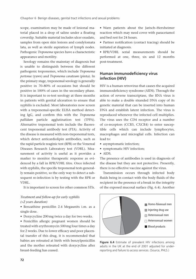
Chapter 6 Benign diseases, genital tract infections and sexual problems
72
scope, examination may be made of lesional ma-
terial placed in a drop of saline under a floating
coverslip. Suitable material includes ulcer exudate,
samples from open skin lesions and condylomata
lata, as well as sterile aspiration of lymph nodes.
Pathogenic Treponema species have a characteristic
appearance and motility.
Serology remains the mainstay of diagnosis but
is unable to distinguish between the different
pathogenic treponemes, which include Treponema
pertenue (yaws) and Treponema carateum (pinta). In
the primary stage, treponemal serology is generally
positive in 70–80% of occasions but should be
positive in 100% of cases in the secondary phase.
It is important to re-test serology at three months
in patients with genital ulceration to ensure that
syphilis is excluded. Most laboratories now screen
with a treponemal-specific ELISA method detect-
ing IgG, and confirm this with the Treponema
pallidum particle agglutination test (TPPA).
Alternative treponemal tests include the fluores-
cent treponemal antibody test (FTA). Activity of
the disease is measured with non-treponemal tests,
which detect anticardiolipin antibodies, such as
the rapid particle reaginic test (RPR) or the Venereal
Diseases Research Laboratory test (VDRL). Mea-
surement of activity is useful as it provides a
marker to monitor therapeutic response as evi-
denced by a fall in RPR/VDRL titre. Once infected
with syphilis, the specific treponemal tests general-
ly remain positive, so the only way to detect a sub-
sequent re-infection is by testing with the RPR or
VDRL.
It is important to screen for other common STIs.
Treatment and follow-up for early syphilis (>2 years duration)• Benzathine penicillin 2.4 Megaunits i.m. as a
single dose.
• Doxycycline 200mg twice a day for two weeks.
• Penicillin allergic pregnant women should be
treated with erythromycin 500mg four times a day
for 2 weeks. Due to lower efficacy and poor placen-
tal transfer of this drug, it is recommended that
babies are retreated at birth with benzylpenicillin
and the mother retreated with doxycycline after
breast-feeding has ceased.
• Warn patients about the Jarisch–Herxheimer
reaction which may need cover with paracetamol
and bed rest for 24 hours.
• Partner notification (contact tracing) should be
initiated at diagnosis.
• RPR/VDRL serial measurements should be
performed at one, three, six and 12 months
post-treatment.
Human immunodeficiency virusinfection (HIV)
HIV is a human retrovirus that causes the acquired
immunodeficiency syndrome (AIDS). Through the
action of reverse transcriptase, the RNA virus is
able to make a double stranded DNA copy of its
genetic material that can be inserted into human
DNA and establish latent infection. The virus is
reproduced whenever the infected cell multiplies.
The virus uses the CD4 receptor and a number
of co-receptors (CCR5, CXCR4) to enter suscep-
tible cells which can include lymphocytes,
macrophages and microglial cells. Infection can
lead to:
• asymptomatic infection;
• symptomatic HIV infection;
• AIDS.
The presence of antibodies is used in diagnosis of
the disease but they are not protective. Presently,
there is no effective vaccine against the virus.
Transmission occurs through infected body
fluids being in contact with the body fluids of the
recipient in the presence of a break in the integrity
of the exposed mucosal surface (Fig. 6.4). Another
Homo-/bisexual men
Injecting drug use
Heterosexual men
Heterosexual women
Blood products
Figure 6.4 Estimate of prevalent HIV infections amongadults in the UK at the end of 2001 adjusted for under-reporting and failure to access services. (Source, PHLS.)
AMI6 6/9/04 5:25 PM Page 72

Benign diseases, genital tract infections and sexual problems Chapter 6
73
major route of infection is blood-borne, either
through sharing needles for IV drug use or through
transfusion of whole blood and concentrated
blood products. Blood products are currently test-
ed for HIV antibodies and so new infections rarely
occur through this route now. At the start of the
HIV epidemic in the developed countries, most
cases of HIV were detected in gay/bisexual men,
haemophiliacs and drug addicts. At the current
time in the UK, the greatest increases in newly
diagnosed cases are occurring in heterosexual men
and women, the majority of whom are thought to
have acquired their infection overseas.
The fetus can be infected in utero from the
mother, although it is thought that the majority
of transmission occurs at birth and through
breast-feeding. The current policy in the UK of
prescribing antiretrovirals in the second and third
trimester, performing Caesarian section and avoid-
ing breast-feeding has dramatically reduced the
risk of mother-to-child HIV transmission (Fig. 6.5).
Essential to the success of this policy is encourage-
ment for all pregnant women to undergo HIV test-
ing at antenatal booking. It should be pointed out
that, in resource poor countries, a balance has to be
made between the risk of preventing HIV transmis-
sion through stopping breast-feeding and the risk
of gastroenteritis through imperfect bottle feeding.
Symptoms and signs• Seroconversion illness is rarely seen in practice
but presents with a glandular fever-type illness
which may be associated with a rash. Neurological
manifestations such as Guillain-Barre syndrome
and encephalitis have been described.
• Asymptomatic infections by definition are only
detected by screening.
• Symptomatic HIV infection may present with
non-specific weight loss, fatigue, lymphadenopa-
thy, diarrhoea and night sweats. Clues may come
from recurrent vulvo-vaginal or oral candidiasis,
recurrent and extensive genital warts, shingles,
worsening eczema and psoriasis, or oral hairy
leukoplakia on the tongue.
• AIDS is determined by the onset of certain
opportunistic infections or AIDS-related
malignancies. Examples include tuberculosis,
Pneumocystis carinii pneumonia, cryptococcal
infection, cryptosporidiosis, Kaposi’s sarcoma and
lymphoma.
Investigations• Detection of anti-HIV antibodies in the serum by
ELISA.
• Confirmation is obtained using different ELISAs
(usually 2 more).
• Western blots can be performed to investigate
discrepant results in reference centres.
• Plasma viral load gives an estimation of the
amount of HIV virus replication.
• The immune status of the patient is measured by
sequential CD4 counts.
• It is important to screen for other common STIs.
When to treat HIV infection?• The best time to start treatment may vary
450400350300250200150100
500
1984
–85
1986
–87
1988
–89
1990
–91
1992
–93
1994
–95
1996
–97
1998
–99
2000
–200
1
No.
of c
ases
Year
Infected (AIDS)Infected (not AIDS)Not infectedIndeterminate
Figure 6.5 Children born to HIV in-fected mothers (1984–2001). Indeter-minate category includes infantsunder 18 months when last tested forHIV antibody and without evidence ofHIV infection (includes 95 children lostto follow up). (Source, PHLS.)
AMI6 6/9/04 5:25 PM Page 73

Chapter 6 Benign diseases, genital tract infections and sexual problems
74
between countries and has varied over time within
countries.
• In the UK, treatment is recommended for all
patients with a CD4 count of less than 200 ¥ 106/l,
regardless of the presence or absence of symptoms.
• In the UK, treatment should be considered in pa-
tients with a CD4 count between 200–350 ¥ 106/l
if they are significantly symptomatic due to HIV
infection or their CD4 count is falling rapidly.
• In the UK, although not clearly of benefit
on clinical grounds, some patients undergoing HIV
seroconversion may elect to go on short course
(one year) of HIV therapy in an attempt to preserve
immunological memory.
HIV treatment options• There now exist many drugs with which to treat
HIV infection.
• It is now accepted that patients should take three
active HIV drugs as highly active antiretroviral
therapy (HAART).
• In patients on salvage therapy, more than
three drugs with diminished activity may be used
(mega-HAART).
• Adherence is crucial to the success of therapy
and resistance rapidly ensues if patients do not
adhere meticulously to their regimens.
• Monitoring of HIV RNA viral load shows
whether the HAART is working and patients
should remain undetectable (<50 copies/ml) on
their regimens if adherent until resistance
ensues.
• Antiretroviral resistance can be readily measured
using genotypic assays but must be done whilst
patients are on the drugs in question.
• Most patients have wild-type virus archived
in sanctuary sites and this virus will replicate and
replace resistance virus once drug pressure is
removed as resistant virus strains tend to be less fit.
• Classes of drugs include nucleoside analogues
(NRTIs), nucleotide analogues (NtRTIs), non-
nucleoside analogues (NNRTIs), protease inhibitors
(PIs) and fusion inhibitors.
• NRTIs include AZT, 3TC, ddI, d4T, ddC, abacavir.
• NtRTIs include tenofovir.
• PIs include nelfinavir, saquinavir, lopinavir,
ritonavir, indinavir, amprenavir, atazanavir.
• T20 is the first of a new class of fusion inhibitors
which needs to be given by subcutaneous injection
(salvage therapy at present).
• Regimens are chosen on the basis of side-effect
profile, patient’s daily schedule, pill and dosage
burden, previous HIV drug therapy, CD4 count
and presence of AIDS-defining illnesses.
• Some HIV drugs are available in combination
tablets to help with adherence, e.g. Combivir (AZT
+ 3TC).
• Some PIs require boosting with low dose riton-
avir to achieve sufficient drug levels in body fluids,
e.g. lopinavir and saquinavir.
• There have been trials assessing the efficacy of
interleukin-2 (IL-2) therapy as an immunothera-
peutic agent but the results have not been
promising.
HIV prevention and partner notification• Partner notification (contact tracing) should be
initiated at diagnosis.
• The importance of using condoms for sexual in-
tercourse must be explained to all HIV seropositive
patients in order to prevent new infections.
• The patient should avoid acquiring drug
resistant HIV virus from other sexual partners as
this will limit the success of future therapeutic drug
interventions.
• IV drug users should be provided with sterile
needles through needle exchange programmes.
• Blood and blood products as well as organ dona-
tions need to be screened for HIV prior to use.
• Health care workers should take precautions to
ensure they do not become infected through
sharps injuries or contact with potentially infec-
tious body fluids of patients.
• Surgical instruments should be autoclaved satis-
factorily and inspected for possible contamination
before use in theatres.
• There is no effective HIV vaccine available at the
present time although several trials are ongoing
with potential vaccine candidates.
Sexual problems
Many students do not like considering patients’
sexual problems, for they feel they have hardly
AMI6 6/9/04 5:25 PM Page 74

Benign diseases, genital tract infections and sexual problems Chapter 6
75
sorted out their own sexual lives and are therefore
ill-equipped to help others. A more open discus-
sion of this subject helps the student to be at ease
when discussing sexuality later with patients. It
helps the students to become more aware of their
attitudes and how these may influence the future
doctor/patient relationship.
Patients often consult their doctors about sexual
problems and expect them to have the knowledge
and skills to help them. It is necessary for a doctor
to know the interrelation of sexually related
matters and their treatments to general diseases
such as heart disease and hypertension.
The facts of human sexuality have not always
been known but, by proper analysis, assessment
and randomized controlled trials of various thera-
pies, more is becoming comprehensible.
History and examination
As with the rest of medicine, a systematic approach
to sexual problems can help make a diagnosis.
A full general and gynaecological history should
be taken including details of past gynaecological
events, the type of contraception used and obstet-
ric history.
The sexual problem should be discussed allow-
ing the woman or man to use their own phrases,
preferably in their own time. If they wander, pointed
questions drawn from the following material may
be needed to bring them back to the point.
Questions
• Duration and frequency of intercourse.
• Factors making the problem better.
• Factors making the problem worse.
• Problem happens with other partners.
• Other associated factors such as alcohol, work or
drugs.
• Other life events at the time of onset.
• The relationship of the partners.
• History of previous sex knowledge.
• Family background to sex education in
childhood.
• Past sexual relationships.
• Possible child abuse.
• Present sexual history.
• Details of usual sex activities, e.g. position and
foreplay.
• Sexual fantasies.
Examination is usually unrewarding.
• In the male —obvious abnormalities of the penis
and testes should be excluded.
• In the female —the ease of allowing a pelvic ex-
amination may be helpful in assessing the degree
of the problem. Any structural abnormalities of the
vulva, vagina, cervix or uterus should be excluded.
The examination can be used positively as an
opportunity to educate about genital anatomy.
The use of a mirror to help a woman identify her
clitoris can be helpful.
It is important to detect general physical abnor-
malities which might make intercourse difficult or
painful before exploring the possibility of psycho-
sexual problems. The patient’s comfort or discom-
fort with their own body and specifically genitalia
can give useful information.
The male
Although this is not strictly a part of gynaecology,
anyone dealing with sexual problems must have a
knowledge of the male partner and his problems.
Failure to ejaculate
The inability to produce semen is not always asso-
ciated with the sex drive itself or the ability to have
an erection.
Causes• Sympathectomy.
• Psychosexual features:
(a) past humiliating sexual rejection;
(b) fear of a pregnancy in partner;
(c) past maternal domination;
(d) repressive sexual teaching as a child;
(e) doubts about sexual orientation.
TreatmentSympathectomy aspects can be treated with drugs.
• Thioridazine or
• Indoramin.
AMI6 6/9/04 5:25 PM Page 75

Chapter 6 Benign diseases, genital tract infections and sexual problems
76
The psychological aspects may require long-
term psychotherapy, because many cases are due to
the man avoiding depositing semen in his part-
ner’s vagina; this produces further anxieties.
Lesser degrees can be dealt with by sympathetic
handling by the woman and extravaginal sexual
techniques. The encouragement of the use of erot-
ic material can help if there is loss of control at the
point of ejaculation.
Premature ejaculation
Ejaculation occurs with minimal stimulation or
before or shortly after penetration and before
either party wishes it.
Ejaculation is under sympathetic control medi-
ated by adrenoreceptors. It probably means that
the mediated system enhances this whilst the sero-
tonin balance is inhibitory. These equilibria may
be over-ridden by behavioural patterns.
CausesBehavioural patterns are associated with:
• inexperience;
• adolescent conditioning to rapid response;
• rejection by other women.
TreatmentMen can be treated individually or with their
partners. Sympathetic handling by the woman
is important. Male confidence must be generated
as he learns to recognize the point of ejaculatory
inevitability and to control stimulation to delay
ejaculation until the time of his choosing. A
squeeze technique applied by the woman to the
penis at the moment before ejaculatory inevitabil-
ity can produce delay in ejaculation.
If a programme is embarked on over the course
of weeks or months, this produces good results.
Drugs are occasionally helpful if there is poor
response to the psychological approach:
• Clomipramine or
• 5-hydroxytryptamine (5HT) reuptake inhibitors
may be helpful.
Erectile dysfunction
Impotence in the male may be primary or secondary.
The former is nearly always psychological due to prob-
lems in the family background/upbringing while sec-
ondary impotence may be physical or psychological.
Causes
STRUCTURAL
• After major pelvic operations.
• Pudendal vein thrombosis.
• Hypospadias.
• Peyronie’s disease (fibrosis of the dorsum of the
penis).
ENDOCRINE DISEASE
• Diabetes.
• Hypogonadism.
• Hypothyroidism.
• Pituitary tumour.
• Cushing’s syndrome.
MEDICAL PROBLEMS
• Peripheral vascular disease.
• Hypertension.
• Cerebral vascular accident.
• Multiple sclerosis.
• Spinal cord injury.
• Increasing age.
DRUGS
• Alcohol.
• Antihypertensives.
• Antidepressants.
• Antipsychotics.
• Hormones.
PSYCHOLOGICAL FEATURES
• Stress.
• Performance pressure.
• Parental influence.
• Ignorance of sexual matters.
• Poor self image.
• Guilt.
• Anger.
• Relationship problems.
AMI6 6/9/04 5:25 PM Page 76

Benign diseases, genital tract infections and sexual problems Chapter 6
77
It is important to differentiate the psychogenic
from the organic causes. The former are associated
with:
• rapid onset;
• a recent depression;
• life or family stress;
• normal erections:
(a) on waking;
(b) masturbation;
(c) in response to erotic material;
(d) with a different partner.
TreatmentPsychological treatment starts with reassurance
and education leading on to non-demand, touch-
ing exercises based on Masters and Johnson’s sen-
sate focus programme.
Erection can be produced by pharmacological
means.
• Papaverine injected into the corpus cavernosum
works by smooth muscle relaxation.
• Other injectable drugs are phentolamine and
prostaglandin E1. These work particularly for men
with spinal cord injuries.
• To avoid the problems of injection of the penis,
intraurethral prostaglandins or oral Viagra are both
being tried in several countries. Preliminary results
would seem favourable to producing an erection
without serious side-effects.
• Some anti-hypertensives may be changed for
others which have less effect on the erectile
function.
The female
Superficial dyspareunia
Vaginal pain during intercourse may be due to a
variety of causes. Physical causes of superficial
dyspareunia include:
• infection of the vulva or vagina;
• dermatological disease of the vulva or vagina;
• postmenopausal atrophy;
• painful perineal scar from episiotomy;
• an undilated hymen —this is very rare and
mostly related to women who maintain virginity
into the late 30s.
TreatmentDealt with according to the cause. Surgical
reconstruction may be required for badly healed
episiotomy or a rigid undilated hymen.
Deep dyspareunia
This is felt higher up in the vagina and in
the pelvis. It often lasts for some hours after
intercourse and can be reproduced at vaginal
examination by pressing over relevant parts of
the female pelvis.
Causes• Chronic pelvic infection.
• Endometriosis.
• Pelvic tumours.
• Fixed uterine retroversion trapping ovaries
behind, e.g. in endometriosis.
• Pelvic congestion.
• Bladder or bowel pathology.
• Failure of arousal response. Superficially this
is due to failure of lubrication and deeply due to
failure of vaginal ballooning during coitus.
TreatmentThat of the basic cause; results depend on the
responsiveness to the treatment of physical
problems.
Results where no pathology is demonstrated are
variable. In these women, dyspareunia may be due
to intra- or interpersonal conflicts. The use of
lubricants and delay of penetration until the
woman is fully aroused can be helpful.
Vaginismus
Spasm of the superficial and deep pelvic muscles
prevents the introduction of the penis and is
apparent at a pelvic examination.
CausesThere may be an organic cause but usually it
is a psychological result of apprehension and fear.
Previous attempts at forced entry may lead to
this.
AMI6 6/9/04 5:25 PM Page 77
Chapter 6 Benign diseases, genital tract infections and sexual problems
78
TreatmentA sympathetic approach to examination can help,
but no force should be used. Attempts should stop
before pain is caused and the woman should feel in
control at all times.
Occasionally examination under anaesthesia
may be required to exclude structural causes and
thus be able to reassure the woman that she is
anatomically normal. Usually this is not necessary
as she is likely to respond to gradual desensitiza-
tion techniques. To this end, the use of graduated
vaginal trainers is often helpful. Fingers or tam-
pons are sometimes preferred. Referral to a trained
sex therapist may be needed.
Anorgasmia
This is not uncommon in the female. It is nearly
always psychological, but in a small percentage it
is physical.
CausesA failure to:
• receive stimulation —psychological in origin;
• respond to stimulation —due to family upbring-
ing/background;
• performance pressure from partner or self;
• doubts about sexual orientation.
Other causes:
• fear of pregnancy;
• dyspareunia;
• debilitating disease;
• chronic constipation.
TreatmentMechanical and organic causes are treated appro-
priately. Psychological causes require the attention
of a sex therapist who would discuss the problem
fully with the individual or the couple.
Too easily the woman is labelled as frigid and
then accepts this as a part of life. Fifty per cent
of women are not orgasmic during penetrative
sex, but are orgasmic with clitoral stimulation.
Understanding this can help remove pressure from
both partners to achieve coital orgasm on every
occasion.
Treatment usually involves helping the woman
to achieve orgasm through masturbation and
learning to lose control. The use of erotic material
or a vibrator can help. Progress to coital orgasm is
then made. Use of vibrators may also help with
coital orgasmic problems.
Rape
Rape is unlawful sexual intercourse with a woman
against her will; only the slightest penetration of
the vulva by the penis is required. Issues of
whether the hymen is intact or if semen has been
deposited in the vagina are irrelevant.
Rape is unfortunately all too common and the
woman may report it to the police, sometimes a
day or so later. A doctor may then be called to ex-
amine the victim of alleged rape. The practitioner
should ensure that he or she has the authority
and consent for the examination and has the
equipment for taking the appropriate specimens
properly.
A history of what happened is taken and careful
notes made. Examination is made of the general
demeanour of the woman and of her clothing.
Bruises and scratches around the lower abdomen,
thighs and vulva should be noted, preferably
in a diagrammatic form. The vulva should be
examined in detail for bruising or tears.
If any suspicion of semen in the vagina is found,
a careful specimen should be taken, placed in
appropriate containers and labelled fully in the
presence of a woman police constable (WPC) and
the complainant. This should then be handed to
the WPC for transport to forensic laboratories and
a receipt should be received or the chain of
evidence may be questioned in any subsequent
legal enquiry.
Other swabs and blood may be taken to
exclude sexually transmitted diseases. While these
often have no legal standing, they may be impor-
tant in the medical management of the woman’s
future.
Many police forces have a rape investigation
team who are able to satisfy both the law and the
psychological needs of the woman who is in this
situation. A knowledge of the procedures involved
is helpful to all practitioners.
AMI6 6/9/04 5:25 PM Page 78

Benign diseases, genital tract infections and sexual problems Chapter 6
79
Criminal abortion
One of the results of the laws about therapeutic
termination of pregnancy in England, Wales and
Scotland has been the massive reduction in crim-
inal abortions. Not a single death has been repor-
ted in the 20 years covered by the Confidential
Enquiries into Maternal Deaths (1982–2001). This
is a very satisfactory situation in the UK but crimi-
nal abortion still goes on throughout the world. It
is estimated that of the half a million women who
die every year of maternity causes, about a quarter
of these do so from incompetently performed ille-
gal abortions.
If a doctor is asked to examine a woman who
may have undergone a criminal abortion, he or she
first must obtain her consent if she is conscious.
HistoryThis should be taken but may be only partly
truthful.
ExaminationThe woman may be pyrexial with dull pain in the
lower abdomen. Pelvic examination may show the
cervix to be soft and the os dilated. Blood or preg-
nancy tissue may be passing through it. There may
be signs of the bite of a volsellum (toothed forceps)
on the anterior lip of the cervix. She may have an
offensive discharge coming through the cervix.
Damage to the genital tract may occur, e.g. the pos-
terior fornix is commonly perforated by incompe-
tent abortionists who force their instruments into
the cul-de-sac of the vagina and penetrate it,
breaching the peritoneal cavity.
TreatmentThe woman should be admitted to hospital. An-
tibiotics should be given urgently, a broad spec-
trum used at first until the results of high vaginal
swabs are known. Consider the diagnosis of gas
gangrene and if relevant give antigas gangrene
serum. If the woman is still bleeding from the
uterus, a curettage may remove septic products
from the cavity and hasten healing. If the bleeding
still persists or is very heavy, hysterectomy may
be required for infection may have entered the
substance of the uterus. Check the haemoglobin
level; a blood transfusion may be required for
toxic anaemia. Watch for anuria which commonly
follows gross toxic infection.
Whilst confidentiality to the patient is the first
concern of the doctor, if the woman becomes seri-
ously ill or approaches death, legal authorities may
be involved. Take advice about this from the legal
department of the hospital or the doctor’s defence
society. Keep careful notes. Be prepared to take, or
act as witness to a dying declaration.
AMI6 6/9/04 5:25 PM Page 79
Chapter 6 Benign diseases, genital tract infections and sexual problems
80
Self-assessment
6.1 Which of the following statements are true of sexually transmitted infections (STIs)?(a) Gonorrhoea in women is best diagnosed by cultures of material from the posterior vaginal fornix.(b) Syphilis is adequately treated in penicillin-allergic pregnant women by erythromycin.(c) Contact tracing partners of women with first attack genital herpes is helpful.(d) Bacterial vaginosis should be diagnosed by culture of a high vaginal swab (HVS) for Gardnerella vaginalis.(e) Chlamydia trachomatis is routinely diagnosed by culture of endocervical swabs.
6.2 Which of the following statements are true about HIV infection?(a) It is caused by a DNA virus that binds to the CD4 receptor.(b) Seroconversion commonly presents with a glandular fever illness.(c) In the UK, all patients with a CD4 count below 200 ¥ 106/l should be encouraged to start anti-HIV drugs.(d) Antiretroviral resistance testing will show resistance patterns to both current and previous anti-HIV drugs.(e) Tenofovir is a new nucleotide reverse transcription inhibitor.
6.3 Ask a friend to role-play a patient and practise taking a sexual history using the following role-play. The instructionsfor the candidate are that you should take the history from the role-player in eight minutes. At the end, the candi-date may be expected to give a four-minute summary of the case and outline appropriate investigations for this lady.The role-player needs to make up a name for herself and fill in some personal details. The scoring system will awardmarks for communication skills, history-taking and appropriate investigations (maximum 12 marks).
Role-player’s instructions: You are a 35-year-old married woman with two healthy children, aged five and seven.You have come to the doctor because of vulval itching. You last had sexual intercourse (unprotected, only divulge ifasked) with your husband three nights ago and have been together for over 10 years. Recently, your husband wentto Thailand on a business trip. Two weeks previously, whilst he was away, you had sex with a male friend and thecondom broke (only divulge this if asked). Your last smear test was normal one year ago and you have never had anSTI before. You are very worried.
6.4 Which of the following statements are true?(a) Female vulvovaginal candidiasis has an offensive smell and presents with vulval itching.(b) Condylomata lata do not respond well to podophyllotoxin.(c) Women who have been raped once have a higher chance than average women of being raped again.(d) The forensic examination and STI screening are best done at the same time by the same doctor.(e) Trichomoniasis is best detected by culture of Trichomonas vaginalis.
6.5 Which of the following are true?(a) Examination of patients with psychosexual problems is rarely rewarding.(b) Ejaculation is under parasympathetic control.(c) Psychosexual problems are best managed by seeing both partners together.(d) Vaginismus only occurs with penile entry into the vagina.(e) Anorgasmia is more common in men than women.
AMI6 6/9/04 5:25 PM Page 80

81
Part 3
The reproductive years
AMI7 6/9/04 5:26 PM Page 81

AMI7 6/9/04 5:26 PM Page 82

For most women, childbearing is a major event in
their lives. The basic changes of pregnancy are
covered in this chapter; the rest of pregnancy and
childbirth are discussed in Chapters 8–14 and a
short chapter, Chapter 15, covers those aspects the
obstetrician should know about the newborn
child.
Maternal changes in pregnancy
During pregnancy, oestrogen increases vascularity
and progesterone permits muscular relaxation and
softening of the connective tissue sheath of the
vagina by an increase in fluid. Over 38 weeks the
tube becomes much more stretchable so that, by
full term, the vagina and vulva permit the passage
of an infant with a head diameter of approximately
10cm. The perineum with the squamous epithe-
lium in the region of the fourchette does not always
stretch so readily and so may tear on occasions.
Pregnancy causes alterations not just in the
mother’s pelvis and abdomen but the whole body.
Adaptations in the function of various systems
occur to minimize the stresses imposed and are
interlinked smoothly so the function of the whole
organism does not deteriorate.
Uterus
By term, a litre of blood can be in the uterine vas-
culature. Branches increase in size, number and
diameter from each side of the uterus. The placen-
tal site gets preferential blood supply. Penetrating
branches pass through the myometrium, under
the surface of the decidua. They become spiral
arteries and penetrate the decidua. In early preg-
nancy their exits into the placental bed pool are
narrow, but trophoblast invasion by 16 weeks
normally widens them into deltas so reducing
resistance and improving flow. If invasion is
incomplete, flow is restricted so that:
• in late pregnancy, the fetus gets fewer nutrients
for growth;
• in labour, the fetus gets less O2 and so fetal dis-
tress follows more readily.
The uterus grows through hypertrophy of the
myometrial cells rather than by an increase in
numbers of myometrial cells. From 28 weeks
the lower third of the uterus thins and becomes
less vascular forming the lower segment of the
uterus (hence lower segment Caesarean section).
Metabolism
Increased to provide for:
• Growth of fetus and placenta.
• Increased growth of the uterus.
• Increased growth of support systems.
• Preparation for lactation.
8383
Chapter 7
The mother and fetus in pregnancy
AMI7 6/9/04 5:26 PM Page 83

Chapter 7 The mother and fetus in pregnancy
84
Weight increase (Table 7.1)
Usually 10–14kg (22–30lb) in whole pregnancy.
For example:
0–14 weeks: 2kg (4.5 lb) —may be a loss because of
vomiting;
13–28 weeks: 5kg (11lb);
28–40 weeks: 5kg (11lb) —may be a loss in last 2–3
weeks because of diminution of amniotic fluid.
The rest is extracellular fluid, fat and protein
storage —6kg.
A sharp increase in the mother’s weight gain in
late pregnancy may indicate increased water reten-
tion, a facet of pre-eclampsia. Weight loss, if per-
sistent, may reflect poor fetal progress although
there is little precision in this and many obstetri-
cians do not use weight as a measure of well-being.
ProteinThe fetus needs little protein in early pregnancy so
the woman is in negative balance.
Two-thirds of the fetal protein is acquired in the
last 12 weeks (a half in last 4 weeks). Also maternal
uterus and breasts use much protein in growing
tissues and storage occurs for lactation. Approxi-
mately 12g of nitrogen a day are needed for the
development of these and the fetus.
CarbohydratePregnancy is diabetogenic and calorie need is
slightly increased.
FatThe fetus accumulates fat late, from 2% of the fetal
body weight at 32 weeks to 12% at term. Fetal
neolipogenesis accounts for most of the baby’s fat
with low transfer rate of precursors across the pla-
centa. The mother has a higher circulating lipid
and lipoprotein level.
CalciumThe fetus utilizes calcium late, taking from the long
bones of the mother. If the stores are insufficient
the fetus still utilizes calcium leading to maternal
osteomalacia. Maternal serum calcium levels stay
steady.
IronIron is mostly passed to the fetus in the last weeks
of pregnancy. It is stored in the liver. The mother
may have poor iron stores because of:
• too little in diet, therefore give supplements;
• too poor absorption.
Cardiovascular system
LoadPregnancy is an increased load so more work is re-
quired by the heart.
• Growing fetal tissues which have high O2 con-
sumption rates.
• Hypertrophied uterus and breasts require more
O2.
• Increased muscular effort by mother to cope
with weight gain of 10–14kg (22–30lb).
• In last weeks of pregnancy, the placental bed
may act like an arteriovenous fistula. More work is
required to overcome this shunt.
Cardiac outputIncreased needs are met by increasing cardiac
output.
In pregnancy, pulse rate is raised but most increase
in output comes from larger stroke volume with
enlarged heart chambers and muscle hypertrophy.
Output increases rapidly in the first trimester by
up to 40% and steadies for the rest of pregnancy
(Fig. 7.1).
During labour, cardiac output can increase by
a further 2 l/min in association with uterine
contractions.
Cardiac output = stroke volume pulse rate.¥Table 7.1 Breakdown of approximate weight increase
during pregnancy.
Fetus 3.5kg 7 lb
Placenta 0.5 1
Amniotic fluid 1.5 2
Uterus 1.0 2
Blood increase 1.5 3
Breasts 1.0 2
Total 9.0kg 17 lb
AMI7 6/9/04 5:26 PM Page 84

The mother and fetus in pregnancy Chapter 7
85
Systolic and diastolic pressure is much lower
in early and mid-pregnancy, rising in the last
trimester. Peripheral resistance is decreased and,
since cardiac output is raised, pulse pressure is
increased.
Blood volumeReturn of blood to the heart is maintained by an in-
creased blood volume. Plasma volume increases
more than red cells so that relative haemodilution
occurs. This used to be called physiological
anaemia, but this is a bad term, for no pathological
process can be physiological.
Heart changesPregnancy is a hyperkinetic state. The heart is:
• Enlarged.
• Pushed up.
• Unfolded upon aorta.
These changes produce electrocardiogram (ECG)
and X-ray changes which are normal for preg-
nancy, but may appear pathological if interpreted
without knowledge of pregnancy.
There are also sometimes extra murmurs, normal
hypervolaemic sounds such as the systolic ejection
murmur and that over the internal mammary
arteries supplying the breasts.
Respiratory system
Pressure of the growing uterus forces the dia-
phragm up and lower ribs out but vital capacity is
not reduced in late pregnancy.
Raised progesterone levels increase respiratory
rate.
Urinary tract
Renal function• Renal plasma flow increases by 30–50%.
• Glomerular filtration rate increases by 30–50%.
• Tubular re-absorption increases by 30–50%.
• Patchy glomerular leak happens occasionally
(e.g. glucose).
Lower urinary tract• Bladder more irritated as growing uterus pushes
on it.
• Ureters:
(a) Longer, wider, lower tone because of proges-
terone effects.
(b) Stasis in ureter and pelvis of kidney may lead
to infection.
Alimentary tract
• Teeth more susceptible to spreading caries and
gingivitis because of increased cortisone levels.
• Nausea and vomiting.
• Hypomotility of gut may lead to constipation.
• Hypochlorhydria —regurgitation of alkaline
chyle into stomach.
• Slow emptying of gall bladder.
• Increased gastro-oesophageal reflux.
Early fetal development
Fetal development is well documented in most
mammalian species including the human.
Since many women cannot time the precise act
of coitus at which fertilization occurred, it is con-
ventional to date pregnancy in weeks from the 1st
day of the last normal menstrual period (LNMP).
The difference in the clinical timing of pregnancy
and biological age (from conception) is readily
2
Car
diac
out
put
(l/m
in)
3
4
5
6
0 10 20Weeks of pregnancy
30 40
7
8
Figure 7.1 Cardiac output in pregnancy in normal women.The lines on the graph represent the mean ± 2 SD of themean.
AMI7 6/9/04 5:26 PM Page 85
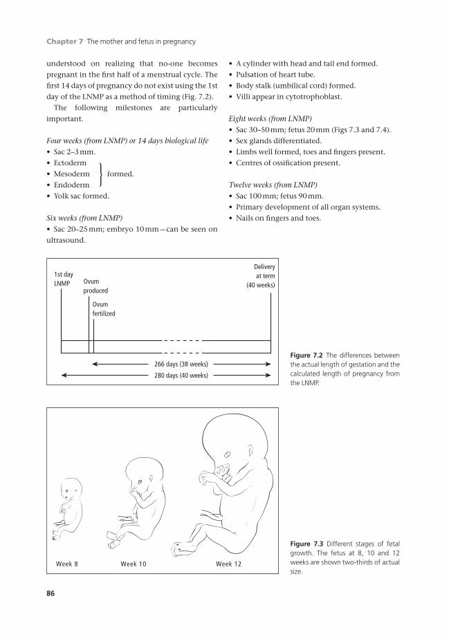
Chapter 7 The mother and fetus in pregnancy
86
understood on realizing that no-one becomes
pregnant in the first half of a menstrual cycle. The
first 14 days of pregnancy do not exist using the 1st
day of the LNMP as a method of timing (Fig. 7.2).
The following milestones are particularly
important.
Four weeks (from LNMP) or 14 days biological life• Sac 2–3mm.
• Ectoderm
• Mesoderm formed.
• Endoderm
• Yolk sac formed.
Six weeks (from LNMP)• Sac 20–25mm; embryo 10mm —can be seen on
ultrasound.
• A cylinder with head and tail end formed.
• Pulsation of heart tube.
• Body stalk (umbilical cord) formed.
• Villi appear in cytotrophoblast.
Eight weeks (from LNMP)• Sac 30–50mm; fetus 20mm (Figs 7.3 and 7.4).
• Sex glands differentiated.
• Limbs well formed, toes and fingers present.
• Centres of ossification present.
Twelve weeks (from LNMP)• Sac 100mm; fetus 90mm.
• Primary development of all organ systems.
• Nails on fingers and toes.
}
266 days (38 weeks)
280 days (40 weeks)
1st dayLNMP
Deliveryat term
(40 weeks)Ovumproduced
Ovumfertilized
Figure 7.2 The differences betweenthe actual length of gestation and thecalculated length of pregnancy fromthe LNMP.
Week 8 Week 10 Week 12
Figure 7.3 Different stages of fetalgrowth. The fetus at 8, 10 and 12weeks are shown two-thirds of actualsize.
AMI7 6/9/04 5:26 PM Page 86

The mother and fetus in pregnancy Chapter 7
87
Early placental development
The placenta (Fig. 7.5) is formed from:
• chorioncovered by amnion.
• decidua basalis
Villi are buds from chorionic plate. At first they
are made of cytotrophoblast tissue only. Mesoderm
appears in situ in the centre of the core of each
villus (Fig. 7.6).
In this mesodermal core, angioblastic strands are
formed. The cells on the edge of these become the
endothelium of blood vessels and the central cells,
the red blood cells. The vessels of the villus join the
vessels formed in the mesoderm. By 22 days, the
fetal heart pumps blood and a functioning circula-
tion starts.
By eight weeks, the villi are 200mm in diameter
with a well-organized circulatory system and a
Figure 7.4 Different stages of fetal growth. (a) Ultrasound showing a sac at 7 weeks. (b) Ultrasound showing a sac andfetus at 12 weeks.
Amnion
ChorionDecidua
Placenta
Placentalbed
Figure 7.5 Formation of placenta in relation to fetus andfetal membranes.
(b)(a) (c)
Figure 7.6 Development of blood vessels in the villi. (a)Mesoderm appears in situ in the core of a villus of prolifer-ating trophoblast cells. (b) Blood vessels form and join upwith those in the mesoderm layer. (c) Capillaries from ar-terial side circulate blood back to veins.
}
(a) (b)
AMI7 6/9/04 5:26 PM Page 87

Chapter 7 The mother and fetus in pregnancy
88
double layer of epithelium (cytotrophoblast cov-
ered by a cellular syncytiotrophoblast).
Further demands of fetal metabolism require
swifter exchanges at the placenta. These come as a
result of:
1 Greater surface area —longer and branching villi.
2 Thinning of epithelium so that syncytiotro-
phoblast is in direct contact with blood capillary.
3 Nuclei in syncytiotrophoblast migrate from
areas over capillaries where exchange actually
occurs.
4 Localized dome-like swellings occur on the villi
protruding into the intervillous space. These areas
are especially thin-walled and are probably the site
of much of the gas exchange.
Villi are like fronds of seaweed under water as the
maternal blood circulates around them (Fig. 7.7).
As the placenta grows, fetal size is proportional to
the surface area available for exchange at first. The
number of stem villi does not increase after the
12th week. Hence the number of lobules is now
fixed. The rest of growth is by proliferation and by
growth of peripheral villi.
Fetal physiology
The major functions will be reviewed particu-
larly where they differ from adult physiological
patterns.
Cardiovascular system
The heart is beating by 22 days and can be detected
with vaginal ultrasound at 5 weeks (from LNMP).
There are bypasses in the system since the lungs
are not used; less than 10% of blood goes through
them (Fig. 7.8). These bypasses include:
• foramen ovale between the right and left atria
so that the majority of oxygenated blood passes
straight to the left side of the heart.
• ductus arteriosus from the pulmonary artery
to the aorta so that only a small amount of
blood from the right side of the heart goes into
the lungs and the rest can use the bypass into the
aorta.
Umbilical blood flow increases with fetal weight.
This increase is disproportionate, but with en-
hanced O2 carrying capacity of the fetal blood, the
total O2 transport is increased. Flow is about
100ml/kg/min, as measured experimentally, but
may be greater in vivo.
Fetal haemoglobinHbA (adult haemoglobin) differs from HbF (fetal
haemoglobin) by a 25% alteration of amino acid
radicals in chains. At any given PO2, the O2 dissoci-
ation curve of HbF is to the left of HbA so it has
greater O2 affinity (Fig. 7.9). The fetus has higher
Hb concentration than the adult (18g/dl in the
blood compared with 13g/dl) allowing further O2
uptake at the placenta and greater release to tis-
sues. Production of HbF diminishes before birth
and has usually ceased by the age of one year
(Fig. 7.10).
Respiratory system
Within 1–2 minutes this has to adjust from an in-
trauterine to an independent state. Vascular loops
occur in the lungs by 18 weeks. Alveoli develop by
22 weeks.
DeciduaVein Spiralartery
Vein
Figure 7.7 Circulation of maternal blood around fine exchange villi.
AMI7 6/9/04 5:26 PM Page 88
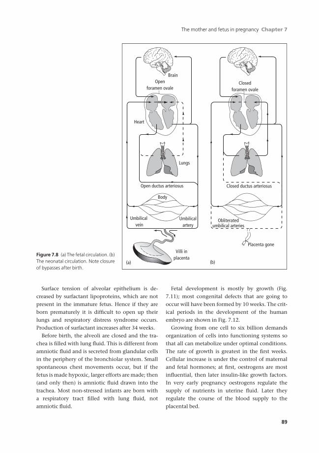
The mother and fetus in pregnancy Chapter 7
89
Surface tension of alveolar epithelium is de-
creased by surfactant lipoproteins, which are not
present in the immature fetus. Hence if they are
born prematurely it is difficult to open up their
lungs and respiratory distress syndrome occurs.
Production of surfactant increases after 34 weeks.
Before birth, the alveoli are closed and the tra-
chea is filled with lung fluid. This is different from
amniotic fluid and is secreted from glandular cells
in the periphery of the bronchiolar system. Small
spontaneous chest movements occur, but if the
fetus is made hypoxic, larger efforts are made; then
(and only then) is amniotic fluid drawn into the
trachea. Most non-stressed infants are born with
a respiratory tract filled with lung fluid, not
amniotic fluid.
Fetal development is mostly by growth (Fig.
7.11); most congenital defects that are going to
occur will have been formed by 10 weeks. The crit-
ical periods in the development of the human
embryo are shown in Fig. 7.12.
Growing from one cell to six billion demands
organization of cells into functioning systems so
that all can metabolize under optimal conditions.
The rate of growth is greatest in the first weeks.
Cellular increase is under the control of maternal
and fetal hormones; at first, oestrogens are most
influential, then later insulin-like growth factors.
In very early pregnancy oestrogens regulate the
supply of nutrients in uterine fluid. Later they
regulate the course of the blood supply to the
placental bed.
BrainOpen
foramen ovale
Open ductus arteriosus
Heart
Lungs
Obliteratedumbilical arteries
Umbilical vein
Umbilicalartery
Body
Villi inplacenta
Placenta gone
(a) (b)
Closedforamen ovale
Closed ductus arteriosus
Figure 7.8 (a) The fetal circulation. (b)The neonatal circulation. Note closureof bypasses after birth.
AMI7 6/9/04 5:26 PM Page 89

Chapter 7 The mother and fetus in pregnancy
90
After mid-pregnancy, growth is also determined
by placental transfer. This could be impaired by:
1 A low environment supply from the mother of:
• oxygen —only has effect in last weeks, e.g.
living at high altitudes;
• nutrients —shows with extremes of specific
deprivation or general starvation.
2 Reduced blood flow to the placental bed. This
follows lack of normal invasion of the arcuate
00 20 40
O2 (mmHg)
O2
satu
rati
on (
%)
60
MotherFetus
80
20
40
60
80
100
P
Figure 7.9 Oxyhaemoglobin dissociation curves for humanmaternal and fetal blood at pH 7.4 and 37°C.
013 20
Weeks of pregnancy Months of lifeANTENATAL POSTNATAL
Hb
pres
ent
(%)
40/0
HbA
HbF
6
20
40
60
80
100
3Figure 7.10 Proportion of HbF andHbA present at different stages offetal and postnatal life.
0
Wei
ght
(kg)
1
2
3
4
0 10 20Intrauterine
weeksExtrauterine
weeks
30 1040/0
Delivery
Figure 7.11 The weight gain of the fetus and newbornchild. The growth potential falls off in the last few weeks ofpregnancy. Note that, after the immediate weight drop,neonatal growth continues at the same incremental rate asit did in the uterus.
arteries by trophoblasts at 16–18 weeks. This can be
estimated by Doppler ultrasound.
3 Poor exchange across the syncytiotrophoblast
membrane; if this should be reduced a smaller baby
results.
Overall growth is determined by:
1 Genetic factors inherited from both parents.
2 Placental transfer of nutrients dependent on:
• placental bed flow rates;
• placental membrane transfer.
AMI7 6/9/04 5:26 PM Page 90
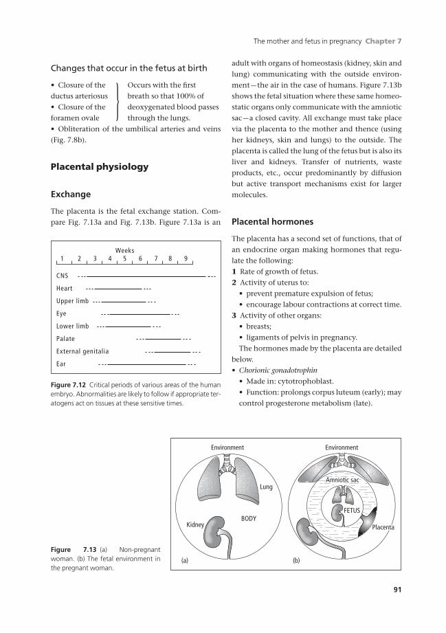
The mother and fetus in pregnancy Chapter 7
91
Changes that occur in the fetus at birth
• Closure of the Occurs with the first
ductus arteriosus breath so that 100% of
• Closure of the deoxygenated blood passes
foramen ovale through the lungs.
• Obliteration of the umbilical arteries and veins
(Fig. 7.8b).
Placental physiology
Exchange
The placenta is the fetal exchange station. Com-
pare Fig. 7.13a and Fig. 7.13b. Figure 7.13a is an
adult with organs of homeostasis (kidney, skin and
lung) communicating with the outside environ-
ment —the air in the case of humans. Figure 7.13b
shows the fetal situation where these same homeo-
static organs only communicate with the amniotic
sac —a closed cavity. All exchange must take place
via the placenta to the mother and thence (using
her kidneys, skin and lungs) to the outside. The
placenta is called the lung of the fetus but is also its
liver and kidneys. Transfer of nutrients, waste
products, etc., occur predominantly by diffusion
but active transport mechanisms exist for larger
molecules.
Placental hormones
The placenta has a second set of functions, that of
an endocrine organ making hormones that regu-
late the following:
1 Rate of growth of fetus.
2 Activity of uterus to:
• prevent premature expulsion of fetus;
• encourage labour contractions at correct time.
3 Activity of other organs:
• breasts;
• ligaments of pelvis in pregnancy.
The hormones made by the placenta are detailed
below.
• Chorionic gonadotrophin
• Made in: cytotrophoblast.
• Function: prolongs corpus luteum (early); may
control progesterone metabolism (late).
Weeks
CNS
Heart
Upper limb
Eye
Lower limb
Palate
External genitalia
Ear
1 2 3 4 5 6 7 8 9
Figure 7.12 Critical periods of various areas of the humanembryo. Abnormalities are likely to follow if appropriate ter-atogens act on tissues at these sensitive times.
}
Environment Environment
Lung
BODYKidney
Amniotic sac
FETUS
Placenta
(b)(a)
Figure 7.13 (a) Non-pregnantwoman. (b) The fetal environment inthe pregnant woman.
AMI7 6/9/04 5:26 PM Page 91

Chapter 7 The mother and fetus in pregnancy
92
• Oestrogens
• Made in: all tissues of placenta.
• Function: stimulate uterine growth and
development.
• Progesterone
• Made in: cytotrophoblast.
• Function: damps down intrinsic uterine ac-
tion in pregnancy.
• Human placental lactogen
• Made in: syncytiotrophoblast.
• Function: alters glucose and insulin metabo-
lism; may initiate lactation.
Placental tissues age. Maximum efficiency is at
37–38 weeks; many functions deteriorate after this.
Beware extrapolations between transfer and en-
docrine functions of the placenta. Correlations
may not be valid.
The fetus and placenta at term
The fetus
The anatomical features of the fetus which most
concern the obstetrician are those found in the
mature fetus after 36 weeks’ gestation. The most
important area is that which is largest, hardest and
most difficult to deliver —the head.
The headCertain measurements should be remembered
(Fig. 7.14). These diameters engage in the maternal
pelvic brim at different degrees of flexion of the
fetal head on the neck.
The intracranial arrangement of meninges is im-
portant (Fig. 7.15) because, under stress, it can be
damaged to produce intracranial haemorrhage.
The bodyThe rest of the fetus will usually pass where the
head leads. The bisacromial diameter of the shoul-
ders is about 10cm.
Placenta
A discoid with 15–20 lobules packed together.
• Fetal surface. Covered with amnion (not chorion
which fuses with the placental edge). Fetal vessels
Occiput
Bregma(centre of anterior fontanelle)
Frontaleminence
Mentum(chin)
Parietal eminence
63
51
24
Figure 7.14 The important diameters of the fetal head of a3-kg baby:1 Suboccipitobregmatic, 10cm: vertex presentation2 Suboccipitofrontal, 11cm: various flexions of cephalicpresentations3 Occipitofrontal, 12cm4 Submentobregmatic, 10cm: face presentation5 Mentovertical, 13cm: brow presentation6 Biparietal, 10cm
Superior sagittal sinus
Pia materBrain
Skin
Skull
Arachnoidmater
Dura mater
Cerebral veindraining intosinus at rightangle
Figure 7.15 Arrangement of themeninges showing how cerebral veinstraverse them. If much intracranialmovement occurs, the arachnoidmoves with the brain but the durastays with the skull. Hence the veincan be torn at the arrowed sites.
AMI7 6/9/04 5:26 PM Page 92

The mother and fetus in pregnancy Chapter 7
93
(arteries paler than veins) course over it diving into
each lobule as an end vessel.
• Maternal surface. Lobules of compressed villi (like
seaweed out of water) separated from each other by
sulci.
Maternal side of the placental circulationMaternal blood is in vessels except in the placental
bed where it is in contact with foreign tissues
(syncytiotrophoblast of villi). Spiral arteries (about
200) lead blood from the uterine arteries to the pla-
cental bed pool. Maternal blood spurts under arter-
ial blood pressure, loses way against a mass of villi
and passes laterally, pushed by vis a tergo to the pla-
cental bed veins scattered over the floor of the
placental bed.
Measurement of blood flow to the placental bed
has been very difficult because it involved direct
measurement in animals (unphysiological) or indi-
rect methods with electromagnetic flow meters in
humans (imprecise). Now indirect measurements
with Doppler ultrasound allow more precise non-
invasive flow studies in humans.
Maternal blood flow to the uterus is 100–
150ml/kg/min in late pregnancy, of which 80–
85% goes to the placenta.
Abnormal implantation• Placenta accreta. Villi penetrate decidua just into
the myometrial layer; difficulty in separation.
• Placenta increta. Villi penetrate deeply into myo-
metrium. Even more difficult to separate.
• Placenta percreta. Villi penetrate to subperitoneal
myometrium. Impossible to separate.
The three diagnoses above cannot usually be dif-
ferentiated clinically. They are pathological ones
made at sectioning a uterus after removal.
• Placenta praevia. Implantation in the lower seg-
ment of the uterus.
Umbilical cord
At term the umbilical cord is about 50cm long,
2cm in diameter. It contains two arteries and a vein
which is derived from the left umbilical vein of the
embryo. (The right one usually disappears.) For
types of cord insertion, see Fig. 7.16.
Arteries spiral and give a cord-like appear-
ance. Possibly their pattern wrapped around the
vein allows their pulsations to help massage
blood back along the umbilical vein. The vessels
are packed and protected by a viscous fluid —
Wharton’s jelly.
There are no nerves in the cord or placenta.
Hence ligation and cutting the umbilical cord does
not hurt the fetus.
Amniotic fluid
This surrounds the fetus.
Battledore placenta,like a squash racket
Succenturiate lobe,placental vessels runin membranes
Velamentous insertion,cord vessels run in membranes over internal os of cervix
Bipartite placenta,roughly equal areas of placenta separated by membranes
Figure 7.16 Types of umbilical cord insertion.
Box 7.1 Diagnostic uses of checking amniotic fluid
• Chromosome content of amniocytes and fetal skincells in genetic diseases• Rhesus effect measuring bilirubin breakdown products• Metabolic upset of the fetus• Infection of the amniotic cavity in premature ruptureof membranes• Respiratory maturity by measuring the lecithin–sphingomyelin ratio
AMI7 6/9/04 5:26 PM Page 93

Chapter 7 The mother and fetus in pregnancy
94
• Produced:
• in early pregnancy: from amnion over pla-
centa and sac;
• in late pregnancy: from fetal urine as well.
• Volume:
• increase to 38 weeks; 500–1500ml.
• Osmolality:
• decreases in late pregnancy.
• Creatinine:
• increases in late pregnancy.
• Acid–base:
• normally accumulation of CO2 and fixed
acid causes a slight reduction in pH (about
7.15–7.20).
Amniotic fluid can be removed at amniocentesis
and used to diagnose a number of factors (Box 7.1).
Self-assessment
7.1 Fill in the blanks in the following sentences from the list below.In pregnancy the maternal cardiac output increases principally because of a greater (1) _______. Haemoglobin con-centrations decrease because of an increased (2) _______ despite an increased (3) _______. The uterus grows by (4) _______ with a blood flow at term of (5) _______ ml/kg/min.(a) 100–150(b) red cell volume(c) stroke volume(d) 200–250(e) hypertrophy(f) plasma volume(g) pulse rate(h) red cell mass(i) mitosis(j) mean haemoglobin concentration
7.2 Which of the following statements are true?(a) Fetal haemoglobin shifts the oxygen dissociation curve to the right of that for haemoglobin A.(b) At birth, changes in the neonatal circulation enable the entire circulating volume to enter the pulmonary tree.(c) In fetal life oxygenated blood from the umbilical arteries flows directly to the left side of the heart.(d) During fetal life the lungs are filled with amniotic fluid.(e) Exposure to teratogens is more likely to cause congenital abnormalities in the first trimester of pregnancy.
7.3 The functions of the placenta include which of the following?(a) Transfer of oxygen from the mother to the fetus.(b) Transfer of urea from the mother to the fetus.(c) Prevention of premature labour.(d) Transfer of nutrients from the fetus to the mother.(e) Regulation of fetal metabolism of insulin and glucose.
7.4 The smallest diameters of the fetal skull include which of the following?(a) Mentovertical.(b) Submentobregmatic.(c) Suboccipitobregmatic.(d) Biparietal.(e) Occiptofrontal.
AMI7 6/9/04 5:26 PM Page 94

Miscarriage or abortion
An abortion is the expulsion of the products of
conception before the 24th week of pregnancy.
The word abortion is often considered by women
to be a procured termination of pregnancy, legal or
criminal. Hence, the softer term miscarriage is bet-
ter used for the spontaneous event. A simple classi-
fication is helpful in understanding the various
terms used (Fig. 8.1).
Causes of spontaneous miscarriage
These are maternal, fetal and possibly paternal or
genetic.
Maternal causes
General• Age.
• Obesity.
• Acute febrile illness.
• Septicaemia with infection of the fetus.
• Severe hypertension or renal disease.
• Diabetes.
• Hypothyroidism.
• Trauma.
• A surgical operation.
• Emotional shock, perhaps more in folklore than
actuality.
Drugs like ergot, quinine and lead may be taken
to induce abortion. They are not very effective and
the risk of poisoning is great.
Local• Uterine fibroids.
• Congenital uterine malformations.
• Incompetence of the internal os:
• congenital;
• acquired after difficult dilatation of the
cervix.
• Hormone deficiency:
• progesterone —the corpus luteum usually pro-
duces progesterone which helps embedding of
the embryo;
• systemic lupus erythematosus;
• anti-phospholipid syndrome.
Fetal causes
• Genetic abnormalities.
• Congenital malformations.
• Faulty implantation.
Congenital and genetic malformationsExamination of the chromosomes in material from
spontaneous abortion shows gross abnormalities
in over half —often the embryo has failed to devel-
op or has been absorbed. In these cases, miscarriage
usually takes place at about eight weeks. Ultra-
sound shows that the amniotic sac contains no
9595
Chapter 8
Bleeding in pregnancy
AMI8 6/9/04 5:27 PM Page 95
Chapter 8 Bleeding in pregnancy
96
embryo. In other cases, gross malformation of the
fetus is shown.
Faulty implantationThe embryo may become implanted in an un-
favourable site in the uterus, for example in the
isthmus, cervical canal or in the uterine cornu.
Most of these cases end in spontaneous miscar-
riage; rarely the pregnancy continues.
Incidence of spontaneous miscarriage
The frequency depends on the definition:
• in clinically diagnosed pregnancies 15–20% will
miscarry in early pregnancy;
• non-development of the blastocyst within 14
days occurs in up to 50% of conceptions.
Clinical features and management ofspontaneous miscarriage
Threatened miscarriage
Symptoms• Scanty uterine bleeding preceded by symptoms
of pregnancy.
• Pain is usually absent; there may be backache or
slight uterine contractions.
Examination• The breasts may be active.
• The uterus is enlarged corresponding with dates
of amenorrhoea.
• The cervix is closed.
• There is no pelvic tenderness.
Differential diagnosis• Delayed miscarriage when the uterus is smaller
than expected. Check with ultrasound.
• Ectopic pregnancy when pain generally precedes
bleeding.
• Dysfunctional uterine bleeding —where no signs
of pregnancy.
Ultrasound can show a sac (five weeks), an em-
bryo and the fetal heart beat (six weeks). Human
chorionic gonadotrophin (hCG) can be measured
in blood or urine.
TreatmentTreatment is usually rest until fresh bleeding has
ceased.
After bleeding has ceased, the woman should
avoid exertion and intercourse till after the 12th
week of pregnancy. Progesterone therapy is inef-
fective, but still used.
Inevitable miscarriage
SymptomsBleeding and pain are characteristic, bleeding is
heavier than in threatened miscarriage. There may
be crampy, low abdominal pains and an escape of
amniotic fluid.
Examination• Uterus enlarged.
• The internal os of the cervix is dilated. Prod-
Threatened
Delayed SepticComplete Incomplete
Inevitable
Spontaneous Induced
AbortionMiscarriage
Recurrent Illegal LegalTherapeutic
Figure 8.1 A simple classification ofthe terms used.
AMI8 6/9/04 5:27 PM Page 96
Bleeding in pregnancy Chapter 8
97
ucts of conception may be felt in the cervical
canal. Once this has occurred, miscarriage is
inevitable.
TreatmentBefore 12 weeks’ gestation, evacuate the uterus
under general anaesthesia in an operating
theatre.
After 12 weeks, allow miscarriage to take place
spontaneously, but be prepared to evacuate the
uterus if it is incomplete. If bleeding is severe,
Syntometrine should be given, 5 units/0.5mg
intramuscularly.
Incomplete miscarriage
An incomplete miscarriage occurs when some
of the products of conception are retained in
the uterus. These are usually parts of the
placenta or chorionic tissue attached to the uterine
wall.
Symptoms• Continued bleeding after a period of
amenorrhoea.
Differential diagnosis• Threatened miscarriage.
• Ectopic pregnancy.
• Dysfunctional uterine bleeding.
Ultrasound may help to clarify the diagnosis.
Treatment• Conservative management.
• Evacuation of the uterus in the operating
theatre.
Complete miscarriage
If all the products of conception have been passed
and the uterus is empty, the miscarriage is com-
plete. There is little bleeding, the uterus is small
with the cervix closed or merely patulous in a mul-
tiparous woman.
No treatment is required provided the differen-
tial diagnosis of ectopic pregnancy has been ex-
cluded (see p. 100).
Delayed or missed miscarriage
The embryo dies in early development and is
retained there and/or the sac continues to develop.
The early embryo is commonly reabsorbed leav-
ing an empty sac —a blighted ovum (a term with
unpleasant connotations for the parents and is
best avoided when talking to the patient and her
partner).
Symptoms• At first those of pregnancy, but these disappear.
• The breasts become soft.
• Dark brown vaginal discharge.
The cervix is closed and the uterus smaller than
would be expected; hCG levels drop in 7–10 days.
Differential diagnosis• Tubal mole.
• An incomplete miscarriage.
• A complete miscarriage.
Ultrasound will confirm the diagnosis.
Treatment• Surgical evacuation. This should be offered if an
embryo with an equivalent size of >8 weeks is
present.
• Medical treatment. Evacuation of the uterus can
be successful in 50% of cases by giving vaginal or
oral prostaglandins every three hours until miscar-
riage takes place.
• Expectant management. This is usually safe and
effective if the sac is empty or the embryo is <8
weeks in size. The sac usually reabsorbs with mini-
mal bleeding but it may take several weeks and the
patients should be offered regular follow-up until
miscarriage is complete.
Recurrent miscarriage
Three consecutive spontaneous miscarriages con-
stitutes recurrent miscarriage. This may be primary
where the woman has borne no viable child, or sec-
ondary. The most important associations are:
• Maternal:
(a) antiphospholipid syndrome (APS) (40%);
(b) polycystic ovaries (Chapter 3) (50%);
AMI8 6/9/04 5:27 PM Page 97
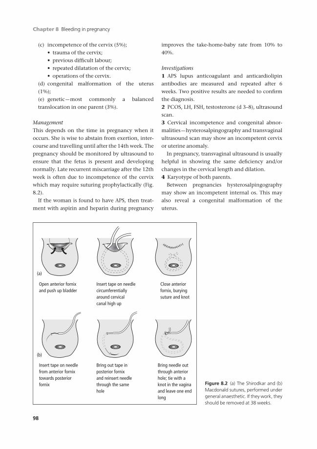
Chapter 8 Bleeding in pregnancy
98
(c) incompetence of the cervix (5%);
• trauma of the cervix;
• previous difficult labour;
• repeated dilatation of the cervix;
• operations of the cervix.
(d) congenital malformation of the uterus
(1%);
(e) genetic —most commonly a balanced
translocation in one parent (3%).
ManagementThis depends on the time in pregnancy when it
occurs. She is wise to abstain from exertion, inter-
course and travelling until after the 14th week. The
pregnancy should be monitored by ultrasound to
ensure that the fetus is present and developing
normally. Late recurrent miscarriage after the 12th
week is often due to incompetence of the cervix
which may require suturing prophylactically (Fig.
8.2).
If the woman is found to have APS, then treat-
ment with aspirin and heparin during pregnancy
improves the take-home-baby rate from 10% to
40%.
Investigations1 APS lupus anticoagulant and anticardiolipin
antibodies are measured and repeated after 6
weeks. Two positive results are needed to confirm
the diagnosis.
2 PCOS, LH, FSH, testosterone (d 3–8), ultrasound
scan.
3 Cervical incompetence and congenital abnor-
malities —hysterosalpingography and transvaginal
ultrasound scan may show an incompetent cervix
or uterine anomaly.
In pregnancy, transvaginal ultrasound is usually
helpful in showing the same deficiency and/or
changes in the cervical length and dilation.
4 Karyotype of both parents.
Between pregnancies hysterosalpingography
may show an incompetent internal os. This may
also reveal a congenital malformation of the
uterus.
(a)
Open anterior fornix and push up bladder
Insert tape on needlecircumferentiallyaround cervicalcanal high up
Close anterior fornix, burying suture and knot
Insert tape on needlefrom anterior fornixtowards posteriorfornix
Bring out tape inposterior fornix and reinsert needlethrough the samehole
Bring needle outthrough anteriorhole; tie with a knot in the vagina and leave one end long
(b)
Figure 8.2 (a) The Shirodkar and (b)Macdonald sutures, performed undergeneral anaesthetic. If they work, theyshould be removed at 38 weeks.
AMI8 6/9/04 5:27 PM Page 98

Bleeding in pregnancy Chapter 8
99
Counselling after miscarriage
Counselling is often needed because a spontan-
eous miscarriage may lead to bereavement even
though the event was too early in pregnancy for a
viable baby to be born. The woman and her partner
should receive an explanation of what happened
and of the possible cause of the miscarriage if it is
known. If there is a treatable cause, such as uterine
fibroids, treatment of this should be discussed even
though the operation itself may be best postponed
for about three months.
The couple are often anxious to have another
pregnancy. In most normal cases, where no serious
cause is identified, there is no reason why they
should not immediately, that is, as soon as the
woman has had a normal period or two. Com-
monly given advice to wait 3–6 months has no
logical basis.
Parents find it helpful to know the chances of a
sucessful pregnancy next time (see Box 8.1).
Criminal abortion
Under the Offences Against the Person Act 1861,
any attempt to procure abortion by the woman
herself or by another person is a felony, irrespective
of whether she is in fact pregnant. The position of
termination of pregnancy under the Abortion Act
is considered in Chapter 5.
Abortion may be procured by:
• drugs;
• instruments passed into the uterus;
• foreign bodies —slippery elm bark introduced
into the cervix;
• injections of soap or douching with soap or anti-
septic solutions.
Criminal abortion is dangerous:
• drugs may cause poisoning;
• infection can easily be introduced;
• risk of severe haemorrhage can occur;
• embolism with air or soap solution.
The doctor called to deal with a woman who has
had a criminal abortion is bound to respect her
confidence. He or she should not inform the police
or any other person unless the woman dies or
appears likely to do so, when a dying declaration
should be obtained. Deaths must be reported to the
coroner (see also p. 79).
Septic abortion
Infection of the uterine cavity following an abor-
tion leads to septic abortion. It can occur after
spontaneous miscarriage, but most cases result
from criminal interference with non-sterile
instruments.
Spreading infection leads rapidly to salpingitis,
pelvic peritonitis, pelvic cellulitis, septicaemia and
pyaemia. Infection with tetanus will give the typi-
cal features of the disease and can be fatal. Infec-
tion with Clostridium welchii is not uncommon in
criminal abortions; the picture is that of severe in-
fection with shock and tachycardia. Such cases
need prompt and efficient treatment preferably in
an intensive care unit.
Symptoms• A history of abortion, often criminal.
• Pelvic infection and septicaemia.
• Fever.
• Pelvic tenderness with foul discharge from the
uterus and bleeding.
• Neutrophil leucocytosis is found on the blood
count.
• The haemoglobin level may be reduced.
Treatment• Admission to hospital.
• Swabs should be taken for cultures from the
cervix and blood culture taken in seriously ill pa-
tients.
• Adequate doses of antibiotics should start at
once; a combination of amoxycillin, clindamycin
Box 8.1 Chances of a live birth in a subsequentpregnancy
No. of miscarriages %• One 85• Two 75• Three or more 60
Unless APS diagnosed• No treatment 10• Aspirin and heparin 40
AMI8 6/9/04 5:27 PM Page 99

Chapter 8 Bleeding in pregnancy
100
and metronidazole may be used initially. A change
of antibiotic may be needed when the cultures and
sensitivities become available.
• Full intravenous supportive measures and
steroids may be needed.
• If the products of conception are retained, the
uterus should be evacuated. This should be done at
once if there is severe bleeding, otherwise it is
preferable to wait 24 hours to allow the antibiotics
to take effect.
Ectopic pregnancy
Ectopic pregnancy is one outside the uterine
cavity, the commonest site being the fallopian
tube. It may also occur, although rarely, in:
• uterine cornu;
• ovary;
• cervix;
• abdominal cavity;
• broad ligament.
For practical purposes, ectopic pregnancy will be
considered as tubal pregnancy.
IncidenceEstimates of the incidence of tubal pregnancy vary.
In the UK it is 0.5%. In other countries, especially
in Africa, it may be as high as 1% because of the
higher prevalence of chronic tubal disease.
CausesThe ovum is fertilized in the fallopian tube and
reaches the uterus in about five days. Anything
that delays the passage of the fertilized ovum to the
uterus can result in tubal pregnancy, such as
intrauterine device (IUD) and progesterone-only
pill (POP) (see Box 8.2).
Salpingitis is the commonest predisposing cause.
This may not be so severe as to cause complete clos-
ing of the tube, but it may destroy tubal cilia, kink-
ing or narrowing the tube. Chlamydia is now the
commonest organism associated with tubal dam-
age. The woman is often unaware that she has had
a tubal infection. Tuberculous salpingitis is an im-
portant but, in the UK, rare cause, for it damages
the tube but often leaves it patent.
Congenital malformation of the tube may lead to
crypts and diverticula providing sites for ectopic
implantation (very rare).
PathologyThe muscular walls of the tube do not allow the
embryo to grow beyond a certain size. The tro-
phoblast gradually invades and erodes the tubal
wall which, unlike the endometrium, is not pre-
pared for implantation. Blood vessels are damaged
and eventually bleeding takes place.
A tubal pregnancy may terminate in a number of
ways.
• Absorption —in a few cases it is possible that ab-
sorption of a very early tubal pregnancy occurs.
The embryo dies in the tube, with a small
amount of bleeding, and is partly absorbed.
• Tubal abortion —part or all of the products of con-
ception are expelled from the tube into the peri-
toneal cavity.
• Tubal rupture —this is the most dramatic and
best known termination of a tubal pregnancy,
though in fact less common than absorption or
tubal abortion. There is acute intraperitoneal
haemorrhage from erosion of an artery. The preg-
nancy is often implanted in the narrower isthmus
of the tube.
• Secondary abdominal pregnancy —this is the rarest
outcome of all. The embryo is expelled complete
from the tube and acquires a secondary attach-
ment in the peritoneal cavity. It can occasionally
go on to a full term abdominal pregnancy. Many
cases of children delivered from the peritoneal cav-
ity by laparotomy have been reported, particularly
in the West Indies.
Clinical featuresThe picture of ectopic pregnancy is:
Box 8.2 Risk factors for ectopic pregnancy
Pelvic inflammatory disease (PID)Previous pelvic surgeryPrevious ectopic pregnancyIntrauterine device (IUD)Progesterone only pill (POP)DepoproveraEmergency contraceptionSterilization
AMI8 6/9/04 5:27 PM Page 100

Bleeding in pregnancy Chapter 8
101
• Amenorrhoea —this can be 4–8 weeks’ duration,
but may present before amenorrhoea is noticed.
• Pain —typically constant and often unilateral
due to spasm of the tubal muscle. There may be re-
ferred shoulder pain via the phrenic nerve from
blood in the abdominal cavity.
• Vaginal bleeding —when a pregnancy implants in
the tube, the uterine endometrium is still con-
verted into decidua. When the embryo dies, the
decidua in the uterus separates. The bleeding is
usually scanty, less than a normal period and dark
brown in colour.
• Faintness or even shock with an acute rupture.
UNRUPTURED ECTOPIC PREGNANCY
This may present with:
• slight activity of the breasts;
• slight tenderness over one side of the uterus.
On bimanual examination, the uterus is slightly
enlarged and the cervix is soft. There may be dark
blood oozing from the external os. The pregnant
tube is usually not palpable. There will be cervical
excitation with tenderness on the side of the
ectopic.
On bimanual examination, the tubal mass is
rarely felt as a diffuse boggy swelling. If it is of any
size, it displaces the uterus to the opposite side of
the pelvis.
ACUTE RUPTURE OF A TUBAL ECTOPIC
This presents as an acute abdominal emergency
with:
• collapse;
• severe abdominal pain;
• pallor, rapid pulse and hypotension;
• blood may track up under the diaphragm giving
shoulder pain;
• the abdomen is slightly distended, tender and
rigid;
• on vaginal examination, the uterus is soft and
may be enlarged but is very tender;
• a tender tubal mass may not be palpated because
of the extreme tenderness and guarding.
Investigations
• Ultrasound is helpful. While it may not always
show the embryo or its sac in the tube, findings
may include:
• an empty uterus with thickened decidua;
• fluid (blood) in the pouch of Douglas;
• a multi-echo mass in the region of the tube.
• Progesterone levels are commonly low because the
pregnancy is failing.
• Serum bhCG is usually lower than expected for
gestation and on serial measurements increases by
less than 60% over 48 hours.
• Laparoscopy is the ultimate investigation to make
the diagnosis with direct vision.
Differential diagnosisThe diagnosis is from any other acute abdominal
catastrophe such as rupture of a viscus or acute
peritonitis. The clinical picture is so typical that in
most cases diagnosis presents no difficulty. Other
diagnoses which may confuse are:
• inevitable miscarriage;
• bleeding with an ovarian cyst;
• pelvic appendicitis;
• acute salpingitis.
TreatmentThe treatment of tubal pregnancy is removal of the
pregnancy and sometimes the affected tube by
laparoscopy or laparotomy. If the tube is patent
and not seriously damaged, it may be possible to
conserve it and thus leave the woman with a
chance of conception later in life.
Laparoscopy techniques exist to:
• kill the embryo with a direct injection of
methotrexate or mifepristone allowing absorption
so requiring no surgery on the tube;
• incise the swollen tube over the ectopic preg-
nancy, aspirate the embryo, and achieve
haemostasis (salpingostomy).
In a case of severe haemorrhage, the patient
must be taken immediately to the operating thea-
tre. Little time should be wasted in attempting re-
suscitation which can prove useless and may only
increase bleeding. An intravenous drip should be
set up and a blood transfusion given as soon as pos-
sible. It is sometimes possible later to collect the
extravasated blood from the peritoneal cavity with
a Cellsaver and return it to the circulation.
AMI8 6/9/04 5:27 PM Page 101

Chapter 8 Bleeding in pregnancy
102
In most cases the affected tube should be re-
moved; an exception may be made if the woman
desires children and the other tube is already miss-
ing or seriously diseased. The disadvantage of con-
servation is the increased risk of recurrence of
ectopic pregnancy.
Tubal pregnancy and normal intrauterine preg-
nancy may occur simultaneously in rare circum-
stances, most commonly after assisted conception.
• Medical treatment with methotrexate can be
used if the hCG level is less than 5000iu/l and the
ectopic mass is less than 4cm in diameter on ultra-
sound scan. There should be no symptoms or signs
of rupture.
Hydatidiform mole
Hydatidiform mole is a benign tumour of both
parts of the chorion; the cytotrophoblast and the
syncytiotrophoblast may be found in varying pro-
portions. The villi undergo cystic or hydropic
degeneration and a certain amount of bleeding
almost always occurs.
Hydatidiform moles vary greatly in their rate
of growth, in the amount of chorionic gonadotro-
phin produced and in the amount of invasion of
the uterine wall.
Only rarely can a fetus be found, but hydatidi-
form degeneration may occur in the placenta. The
birth of a living fetus with a hydatidiform mole has
been described.
IncidenceIn the UK one in 2500 pregnancies, but much com-
moner in the Far East.
Clinical featuresThe typical clinical features are amenorrhoea fol-
lowed by continuous or intermittent vaginal
bleeding. The other symptoms of pregnancy occur,
often exaggerated; vomiting may be severe and
early pre-eclampsia can develop. The uterus is
often larger than the dates would suggest and feels
very soft and boggy. Theca lutein cysts may deve-
lop in the ovaries.
Vesicles of the mole may be passed spontan-
eously and this is diagnostic of the condition.
InvestigationsChorionic gonadotrophin excretion in urine is
often greater than other pregnancies; while levels
of 40000 to 60000iu/l are common, concentra-
tions of over 100000iu/l are generally diagnostic
of a mole. Ultrasonic examination is reliable in
showing the absence of a fetus with the character-
istic picture of snowflakes or soap bubbles (bright
sunlight shining through the washing-up water).
TreatmentThe uterus must be emptied completely in all cases.
If there seems to be spontaneous evacuation, the
uterus must still be carefully aspirated and
curetted, the specimen being sent for histology
examination.
An intact mole may be dealt with by suction
evacuation under cover of continuous oxytocin i.v.
drip, curetting gently to remove all the mole. In-
travenous Syntometrine must be given to mini-
mize bleeding. Suction evacuation is safest and
carries less risk of perforation than curettage.
Dangers include:
• haemorrhage which can be profuse;
• sepsis;
• perforation of the uterus;
• air embolism;
• incomplete evacuation of the uterus.
A second aspiration or curettage may be needed
two weeks later to be sure that the mole has been
completely removed.
Follow-upThe woman should be followed-up by regular esti-
mations of urinary hCG for at least a year and
should avoid pregnancy for six months after hCG
levels have returned to normal. Tests should be car-
ried out monthly for the first six months and after
that every two months. Persistence of hCG after
one month may suggest incomplete evacuation of
the uterus or malignant change. Persistence is an
indicator for chemotherapeutic treatment with
actinomycin D or methotrexate to prevent
choriocarcinoma.
During the period of follow-up, the woman
should not take the contraceptive pill but use bar-
rier methods of contraception.
AMI8 6/9/04 5:27 PM Page 102

Bleeding in pregnancy Chapter 8
103
In the UK, follow-up is undertaken in specialist
centres.
Invasive mole
In some cases of hydatidiform mole, there may be
great trophoblastic activity with penetration of the
uterine wall. This can still be a simple invasive
mole causing uterine enlargement and bleeding
with a positive pregnancy test; these may require
hysterectomy.
In severer cases trophoblast penetrates into the
parametrium and leads to internal haemorrhage.
The level of hCG is very high. These cases require
urgent hysterectomy.
Choriocarcinoma
This malignant tumour invades the uterine wall
and metastasizes widely through the bloodstream.
Rarely primary tumours are found in the ovary or
testis as a form of teratoma.
It is fortunately an unusual tumour; about 40%
follow hydatidiform mole, 40% follow abortion
and 20% pregnancy at term. Conversely, a hyda-
tidiform mole may go on to choriocarcinoma in
4–5% of women with a mole, compared with
0.0002% after normal pregnancy.
Clinical featuresUterine bleeding is the commonest symptom. Sec-
ondaries may appear rapidly and are most often
found in the lungs, the uterus and the vagina, but
they can involve the liver and the central nervous
system. The levels of hCG are very high, often
above 1000000iu/l.
Choriocarcinoma is sensitive to cytotoxic drugs
and is now curable in the majority of patients.
Those with low-grade disease treated with
methotrexate can retain their fertility and have
further successful pregnancies. Assay of hCG in
serum or urine is used as a tumour marker and re-
duction in its levels is a test of cure.
Chemotherapy is best given in units which spe-
cialize in its use and various combinations of drugs
are given; cisplatin is the first line of attack and
may be combined with methotrexate, vincristine,
cyclophosphamide and actinomycin D.
With modern treatment, hysterectomy is now
rarely indicated except with massive tumours caus-
ing severe bleeding or when the response to
chemotherapy is poor.
AMI8 6/9/04 5:27 PM Page 103

Chapter 8 Bleeding in pregnancy
104
Self-assessment
8.1 A woman presents with her second miscarriage. Which of the following pieces of information should she be givenin respect of miscarriage?(a) It is unlikely that she has done/taken anything to cause the miscarriage.(b) She has a 75% chance of miscarrying again.(c) She may try again for a pregnancy after one normal period.(d) She has a 25% chance of miscarrying again.(e) The majority of miscarriages are due to an abnormality in the baby.
8.2 Which of the following are risk factors for ectopic pregnancy?(a) Combined oral contraceptive pill (COCP).(b) Progestogen only pill (POP).(c) Subserosal fibroids.(d) Chlamydia.(e) Corpus luteal cyst.
8.3 Which of the following statements are true of a hydatidiform mole?(a) May be associated with early onset pre-eclampsia.(b) Is more common in the UK than in the Far East.(c) Should be treated with methotrexate.(d) Should be followed up for a minimum of one year in a specialist centre.(e) Is associated with a higher than average hCG concentration.
8.4 Which of the following is true of choriocarcinoma?(a) There is early spread to bones.(b) Cisplatin is the chemotherapeutic agent of choice.(c) Hysterectomy is only indicated if there is severe bleeding.(d) After hydatidiform mole the risk of choriocarcinoma is 40%.(e) After hydatidiform mole the risk of choriocarcinoma is 4%.
AMI8 6/9/04 5:27 PM Page 104

The aims of antenatal care are to bring the mother
and child to labour in the best possible condition.
They are:
1 A screening process applied to the entire preg-
nant population to detect subgroups at higher risk
for complications of pregnancy.
2 Suitable diagnostic procedures to determine
who are really at risk.
3 The management of high-risk pregnancies.
4 The educational preparation of the couple for
childbirth and the rearing of the infant.
Diagnosis of pregnancy
Symptoms
Amenorrhoea
The monthly shedding of the endometrium is pre-
vented by higher progesterone levels from the per-
sistence of the corpus luteum. Pregnancy is dated
from the first day of the last normal menstrual
period (LNMP) even though conception does not
occur until about 14 days later. Any bleeding after
the LNMP should be considered as abnormal.
Nausea and vomiting
Nausea occurs in 80% of nulliparous and 60% of
multiparous women. For many pregnant women
this is the first sign of pregnancy with the symp-
toms occurring even before the first period is
missed.
The nausea and vomiting usually disappears by
16 weeks’ gestation and lessens in severity after
about 12 weeks. Although some women are sick
first thing in the morning, it is not unusual to find
that vomiting may occur at any time of the day.
Commonly some biscuits or sweets help prevent
nausea.
There is usually no accompanying metabolic
upset, women do not feel ill all the time, and it
does not affect their daily activities. They do not
usually require hospitalization. Specific causes may
include urinary tract infection or ingestion of iron
tablets.
Breast symptoms
Breast enlargement accompanied by tingling of the
skin and nipples. Montgomery’s tubercles develop
from between six and eight weeks’ gestation and
colostrum may be secreted from the nipples after
about 12 weeks’ gestation.
Urinary symptoms
From six weeks’ gestation onwards, many women
experience increased frequency of micturition.
This is due to:
• Increased renal blood flow in the early stages.
105105
Chapter 9
The antenatal period
AMI9 6/9/04 5:28 PM Page 105

Chapter 9 The antenatal period
106
• Pressure on the bladder from the growing uterus
in later pregnancy.
Signs
Uterus
• An increased softness and enlargement of the
uterus can be felt on bimanual vaginal examina-
tion from 6–8 weeks of gestation.
Breasts
• Increased in size and feel warm.
• The areolae darken.
• Montgomery’s tubercles develop.
• Tortuous skin veins dilate.
Investigations
Pregnancy test
Animal pregnancy tests and early crude immuno-
logical tests have now been replaced by accurate,
sensitive tests involving monoclonal antibodies.
Human chorionic gonadotrophin (hCG) is a glyco-
protein hormone that contains two carbohydrate
side chains: alpha (a) and beta (b). The a subunit is
identical to that of follicle stimulating hormone
(FSH), luteinizing hormone (LH) and thyrotrophin
(TSH). The b subunit is immunologically specific.
hCG is secreted by the trophoblast cells of the fer-
tilized ovum and later by the definitive placenta.
Modern tests can detect hCG levels as low as
25 iu/l, before the time of the missed menses. Such
tests can be performed in two minutes and are un-
affected by urine contaminated by proteinuria, or
bacterial contamination. Only a few drops of urine
are required. The tests come in a variety of kits
which can be bought in any chemist and are based
on a colour change occurring if hCG binds to the
monoclonal antibody embedded in the absorbent
paper. Two main sorts are available: a double band
of blue or a central spot of pink indicates a positive
test while a single band of blue or absence of a pink
spot indicates a negative pregnancy test.
Ultrasound
Real-time ultrasound machines will detect an in-
trauterine gestation sac from five weeks of amenor-
rhoea, with fetal heart activity becoming visible at
six weeks and a fetal pole visible at seven weeks.
Transvaginal probes enable a better resolution
image than transabdominal ultrasound allowing
the diagnosis of an intrauterine pregnancy to be
made one week earlier (5–6 weeks).
Antenatal visits
The current method of antenatal care was estab-
lished 80 years ago but is now subject to change.
In particular, the visits in mid-pregnancy (12–34
weeks) may be reduced.
Traditionally, the woman is seen monthly from
the booking visit until 28 weeks, fortnightly until
36 weeks and then weekly until delivery. A reduc-
tion in the number of visits does not affect the
outcome of pregnancy (Table 9.1) and is very
popular with women.
The aim of the visits is to screen the low-risk
population by means of history, examination and
investigation; then antenatal care for high-risk
Table 9.1 The spacing of antenatal visits by traditional
and by modern care
Traditional(gestation in weeks) Modern
6–12 8–12
16
20 20
24
26
28
30
32 32
34
36 36
37
38 38
39
40 40
41 41
AMI9 6/9/04 5:28 PM Page 106
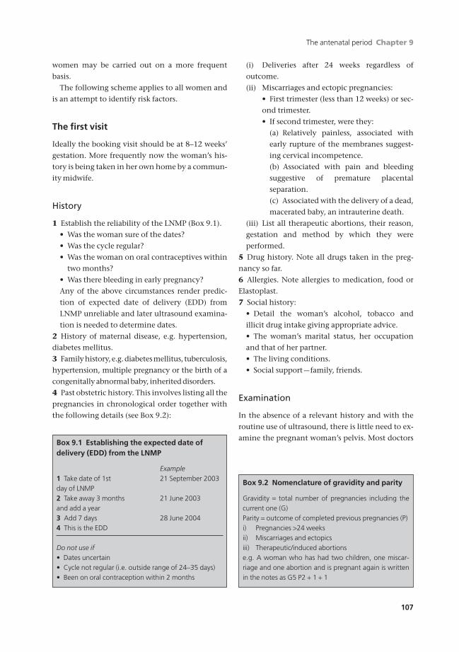
The antenatal period Chapter 9
107
women may be carried out on a more frequent
basis.
The following scheme applies to all women and
is an attempt to identify risk factors.
The first visit
Ideally the booking visit should be at 8–12 weeks’
gestation. More frequently now the woman’s his-
tory is being taken in her own home by a commun-
ity midwife.
History
1 Establish the reliability of the LNMP (Box 9.1).
• Was the woman sure of the dates?
• Was the cycle regular?
• Was the woman on oral contraceptives within
two months?
• Was there bleeding in early pregnancy?
Any of the above circumstances render predic-
tion of expected date of delivery (EDD) from
LNMP unreliable and later ultrasound examina-
tion is needed to determine dates.
2 History of maternal disease, e.g. hypertension,
diabetes mellitus.
3 Family history, e.g. diabetes mellitus, tuberculosis,
hypertension, multiple pregnancy or the birth of a
congenitally abnormal baby, inherited disorders.
4 Past obstetric history. This involves listing all the
pregnancies in chronological order together with
the following details (see Box 9.2):
(i) Deliveries after 24 weeks regardless of
outcome.
(ii) Miscarriages and ectopic pregnancies:
• First trimester (less than 12 weeks) or sec-
ond trimester.
• If second trimester, were they:
(a) Relatively painless, associated with
early rupture of the membranes suggest-
ing cervical incompetence.
(b) Associated with pain and bleeding
suggestive of premature placental
separation.
(c) Associated with the delivery of a dead,
macerated baby, an intrauterine death.
(iii) List all therapeutic abortions, their reason,
gestation and method by which they were
performed.
5 Drug history. Note all drugs taken in the preg-
nancy so far.
6 Allergies. Note allergies to medication, food or
Elastoplast.
7 Social history:
• Detail the woman’s alcohol, tobacco and
illicit drug intake giving appropriate advice.
• The woman’s marital status, her occupation
and that of her partner.
• The living conditions.
• Social support —family, friends.
Examination
In the absence of a relevant history and with the
routine use of ultrasound, there is little need to ex-
amine the pregnant woman’s pelvis. Most doctorsBox 9.1 Establishing the expected date ofdelivery (EDD) from the LNMP
Example1 Take date of 1st 21 September 2003day of LNMP2 Take away 3 months 21 June 2003and add a year3 Add 7 days 28 June 20044 This is the EDD
Do not use if• Dates uncertain• Cycle not regular (i.e. outside range of 24–35 days)• Been on oral contraception within 2 months
Box 9.2 Nomenclature of gravidity and parity
Gravidity = total number of pregnancies including thecurrent one (G)Parity = outcome of completed previous pregnancies (P)i) Pregnancies >24 weeksii) Miscarriages and ectopicsiii) Therapeutic/induced abortionse.g. A woman who has had two children, one miscar-riage and one abortion and is pregnant again is writtenin the notes as G5 P2 + 1 + 1
AMI9 6/9/04 5:28 PM Page 107

Chapter 9 The antenatal period
108
and midwives, however, would still perform the
following examinations:
1 Maternal blood pressure.
2 The respiratory system.
3 The breasts to check for:
• Lumps.
• Inverted nipples, which may require advice for
breast feeding.
4 The spine for kyphosis or scoliosis.
5 The abdomen looking for scars, masses and, if
the pregnancy is sufficiently advanced, the size of
the uterus.
6 The legs looking for varicose veins.
7 Vaginal examination is not usually required at a
booking visit but sometimes is done:
• To confirm the
pregnancy. If ultrasound
• To check uterine size. not available.
• To exclude uterine
or ovarian masses.
• To take a smear if the patient has not had one
within the last three years, although this is often
left until the postpartum visit.
Investigations
Urine1 Proteinuria —renal disease.
2 Glucose —diabetes.
3 White blood cells —response to infection.
4 Nitrite —bacteria.
Blood1 Haemoglobin.
2 Red cell indices, particularlyAnaemia.
the mean corpuscular volume
(MCV).
3 ABO and Rhesus (Rh) group (if negative need
for Anti-D).
4 The presence of atypical antibodies.
5 Sickle cell screen particularly if the patient is
Afro-Caribbean.
6 Haemoglobin electrophoresis, looking for thal-
assaemia if the patient is Asian or Mediterranean in
origin.
7 Test for hepatitis antigens.
8 Test for rubella antibodies.
9 Human immunodeficiency virus (HIV) test. All
women after appropriate counselling should be of-
fered an HIV antibody test but the following pa-
tients are at high risk:
• Women from or with partners from sub-
Saharan Africa.
• Drug abusers or partners of drug abusers.
• Women who have bisexual partners.
• Women with haemophiliac partners.
• Women who have had a blood transfusion
overseas.
In places where HIV is more prevalent, universal
testing is performed with an opt-out policy. In the
UK this includes London and Edinburgh.
10 Screening test for syphilis (usually VDRL, Ven-
ereal Disease Research Laboratory test). If positive,
more specific tests are required.
General advice for healthy pregnancy
1 Establish a rapport between the woman and the
antenatal clinic staff.
2 Show the woman where she can discover more
about her pregnancy and delivery from:
• books available;
• parentcraft classes;
• relaxation classes;
• video and TV programmes.
3 Discuss the social welfare benefits available.
4 Make arrangements for the medical social
worker to see the woman if there are any difficul-
ties such as care of the other children or housing.
5 Advise a visit to the dentist reasonably soon, as
dental care in pregnancy is free, and there is an in-
creased prevalence of tooth decay and gingivitis in
pregnancy.
6 Give dietary advice. This should be simple
advice as most people in the UK have a more than
adequate diet. The idea of eating for two should be
discouraged and, in general, pregnant women
need only an additional 500kcal (2100J) a day to
ensure normal fetal growth.
Vegans may require specialized advice from the
dietitian in order to ensure adequate nutrition
throughout the pregnancy especially for certain
amino acids. Similarly, some Asian women may
}
}
AMI9 6/9/04 5:28 PM Page 108

The antenatal period Chapter 9
109
need dietary advice or supplements of vitamin D as
a consequence of living in the cloudy Northern
Hemisphere.
7 Advise the woman to stop smoking since it in-
creases the risk of intrauterine growth retardation
and delayed fetal maturation.
8 Advise the woman to stop drinking alcohol or
cut down on her intake.
9 Advise the woman to avoid unpasteurized
products, soft cheese and paté as these have been
associated with intrauterine death secondary to
listeriosis.
10 Advise the woman to be careful when dealing
with cats’ litter by avoiding emptying the tray and
using rubber gloves because of the risk of acquiring
toxoplasmosis which may lead to mental retarda-
tion in the fetus.
11 Consider providing iron supplementation. The
routine of prophylactic iron supplements in preg-
nancy is controversial. Many obstetricians only
provide iron if the woman has a haemoglobin of
less than 10.5g/dl or a MCV of less than 84fl at the
booking visit. Additional indications may be for
multiple pregnancies or the previous pregnancy
within 2 years.
Most women’s haemoglobin level will fall by
about 1g/dl due to haemodilution that occurs in
pregnancy.
If iron is given, it should be taken with
meals as it is only absorbed in the ferrous state
and this is best achieved in the presence of vitamin
C.
In the non-pregnant state, about 10% of iron is
absorbed and this is thought to double in preg-
nancy. When supplementation is given, you should
aim to give at least 100mg of elemental iron a day.
12 Vitamin supplements. These are not usually re-
quired by women receiving an adequate diet. An
exception is folic acid as it is often only barely suf-
ficient in many diets. The requirements in preg-
nancy rise from 50mg a day to 300mg a day. Many
women are therefore given prophylactic iron
tablets that also contain folic acid (500mg a day.)
Folic acid supplements have been shown to reduce
the incidence of neural tube defects (NTDs) when
taken preconceptually and up to 14 weeks’
gestation.
Special visits
To be performed at all visits
1 Check the history of recent events and ensure
that the baby is moving.
2 Examine:
• Blood pressure.
• The growth of the uterus and its contents can
be assessed by measuring the symphysiofundal
height (SFH; Fig. 9.1). Normal growth is 1cm per
week ±2cm, e.g. at 32 weeks the SFH should be
30–34cm. Less than 30cm may indicate growth
retardation or oligohydramnios; greater than
34cm may indicate a multiple pregnancy, poly-
hydramnios or macrosomia. Clinical assessment
by palpation is rather a crude method with only
40% of small babies accurately detected and of
these only 60% will still be small for dates at birth.
The SFH is measured by palpating the fundus, a
tape measure is placed under the left index finger
and laid over the abdomen to the top of the sym-
physis which should be felt for gently as it can be
tender. The tape measure should be face down to
prevent cheating (making the measurement fit the
gestation). Beyond 26 weeks the following should
be noted in the notes:
• lie (longitudinal, oblique, transverse) (Fig. 9.2);
• presentation (cephalic, breech, none) (Fig. 9.3);
Figure 9.1 The symphysiofundal height (SFH).
AMI9 6/9/04 5:28 PM Page 109
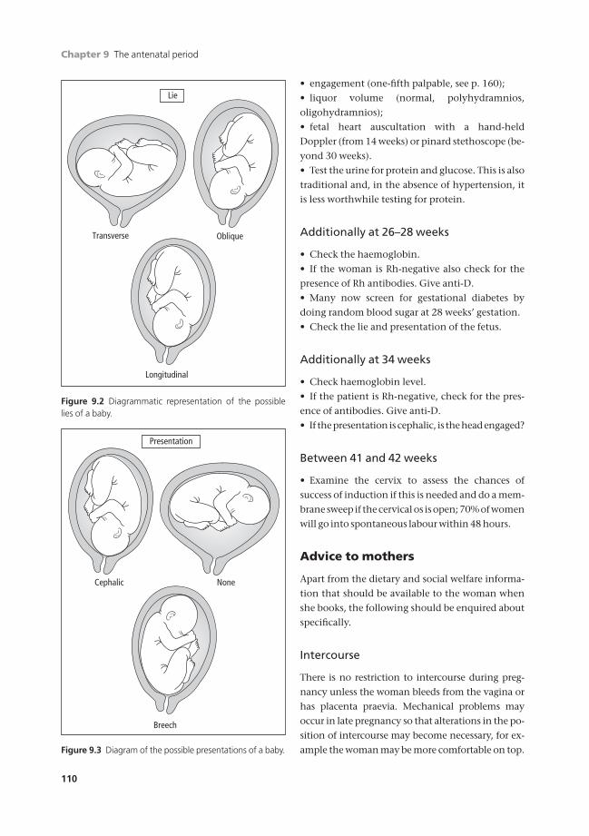
Chapter 9 The antenatal period
110
• engagement (one-fifth palpable, see p. 160);
• liquor volume (normal, polyhydramnios,
oligohydramnios);
• fetal heart auscultation with a hand-held
Doppler (from 14 weeks) or pinard stethoscope (be-
yond 30 weeks).
• Test the urine for protein and glucose. This is also
traditional and, in the absence of hypertension, it
is less worthwhile testing for protein.
Additionally at 26–28 weeks
• Check the haemoglobin.
• If the woman is Rh-negative also check for the
presence of Rh antibodies. Give anti-D.
• Many now screen for gestational diabetes by
doing random blood sugar at 28 weeks’ gestation.
• Check the lie and presentation of the fetus.
Additionally at 34 weeks
• Check haemoglobin level.
• If the patient is Rh-negative, check for the pres-
ence of antibodies. Give anti-D.
• If the presentation is cephalic, is the head engaged?
Between 41 and 42 weeks
• Examine the cervix to assess the chances of
success of induction if this is needed and do a mem-
brane sweep if the cervical os is open; 70% of women
will go into spontaneous labour within 48 hours.
Advice to mothers
Apart from the dietary and social welfare informa-
tion that should be available to the woman when
she books, the following should be enquired about
specifically.
Intercourse
There is no restriction to intercourse during preg-
nancy unless the woman bleeds from the vagina or
has placenta praevia. Mechanical problems may
occur in late pregnancy so that alterations in the po-
sition of intercourse may become necessary, for ex-
ample the woman may be more comfortable on top.
Longitudinal
ObliqueTransverse
Lie
Figure 9.2 Diagrammatic representation of the possiblelies of a baby.
Presentation
Breech
NoneCephalic
Figure 9.3 Diagram of the possible presentations of a baby.
AMI9 6/9/04 5:28 PM Page 110

The antenatal period Chapter 9
111
Alcohol
Alcohol crosses the placenta readily and so can af-
fect a fetus as much as an adult. Excessive drinking
in pregnancy is associated with the fetal alcohol
syndrome, characterized by a short nose with a low
bridge, small eyes with a narrow palpebral fissure
and mental retardation.
No ill-effects have been described with an alco-
hol intake below 4 units a week.
Rest and exercise
Even in normal pregnancy, the extra weight carried
by the woman may increase her sense of tiredness
and lethargy. Sensible exercise such as walking and
swimming or organized exercise to which the
woman is accustomed (e.g. aerobics) should be
allowed in pregnancy.
Travel
The woman should only travel over distances
which are comfortable to her.
Air travel is probably better than train for long
distances, but airlines can refuse to carry women
over 34 weeks’ gestation for international flights
and over 36 weeks’ gestation for domestic travel.
They are the final arbiters, not the travel agents.
Clothes
Women should be advised to wear what looks good
and feels comfortable.
Maternity brassieres are often not required until
late pregnancy, but women should be advised to
move into them as soon as they feel that their pres-
ent brassiere is inadequate for support.
Bathing
The woman should bathe as she wishes. Avoid
vaginal douching in pregnancy.
Bowels
Pregnancy tends to make women constipated
because of the progestogenic effect of relaxing
smooth muscles. This is best overcome by increas-
ing fluid intake, fresh fruit and by the use of foods
rich in fibre. Laxatives should not be used unless
the constipation becomes symptomatic.
Onset of labour
Many nulliparous women have no idea what to ex-
pect; up to 10% of women who present with pains
are subsequently proved not to be in labour.
Advise that the onset of labour is usually accom-
panied by one of the following:
1 Regular painful contractions coming from the
small of the back and radiating to the lower ab-
domen. Nulliparous women are usually advised to
come into hospital when such contractions are oc-
curring once in every 5–10min.
2 A bloody or mucous show. This is not neces-
sarily a sign of labour. If accompanied by uterine
contractions women should be advised to contact
their midwife or delivery unit.
3 Rupture of the membranes accompanied by a
gush of amniotic fluid. In this case, women should
be advised to come into hospital because of the risk
of cord prolapse.
It should be emphasized that it is much better for
a woman to come into hospital if she thinks she is
in labour. Even if she is not, many of the causes of
uterine pain may be serious and should be evalu-
ated in the maternity unit.
Psychological preparation
Pregnancy and delivery are a worry to most
women. Commonly there is a fear of:
• The unknown.
• Giving birth to an abnormal baby.
• Giving birth to a dead baby.
• The pain that accompanies labour.
Many women’s fears can be alleviated by proper
antenatal preparation and by encouraging them to
ask questions at the antenatal clinic. Women and
their partners should be encouraged to attend talks
about childbirth and subsequent rearing of their
children. The antenatal clinic is a busy place, but
doctors and midwives should never appear to be
AMI9 6/9/04 5:28 PM Page 111

Chapter 9 The antenatal period
112
rushed and should encourage women to express
their fears or anxieties and to ask questions.
Parentcraft classes
The aim of these classes is to help women and their
partners to prepare for labour, delivery and the care
of their newborn baby. Couples should be encour-
aged to attend together.
The following areas are covered:
1 Stages of labour.
2 Possible abnormalities of labour.
3 Methods of delivery.
4 Pain relief.
(a) Natural methods —teaching the woman
exercises.
• In the first stage, slow breathing between
contractions and quick shallow breathing
during contractions.
• In the second stage, women are taught ex-
pulsive breathing that involves fixing the
diaphragm and the upper abdomen.
(b) Inhalational gases (N2O).
(c) Transepithelial nerve stimulation (TENS).
(d) Regional local anaesthesia: epidural.
(e) Acupuncture/hypnotherapy.
(f) Opiates —these are no longer recommended.
5 Place of birth.
• Hospital 90%
• GP/midwifery unit 5%
• Home 5%
Five percent choose in early pregnancy to de-
liver at home but only 2% actually deliver there.
40% of prigravida will be transferred mostly in
labour because of failure to progress in the first or
second stage of labour. This can be very distressing
for all concerned. They must be counselled that re-
gional anaesthesia, resuscitation of the newborn
and treatment of a postpartum haemorrhage
will be more difficult compared with delivery in
hospital.
Women in their first pregnancy often have very
high expectations of having a normal labour and
delivery which can be unrealistic. They may write
detailed birth plans (virtually never written by
women in their second or third pregnancy) outlin-
ing their desired management of labour. It is im-
portant to try to make their expectations realistic
otherwise the mother (and her partner) may expe-
rience profound disappointment, anger and a
sense of failure which may affect both her ability to
care for her newborn baby and increase her risk of
postnatal depression.
Assessment of fetal well-being
The obstetrician is responsible for the care of two
patients in labour, one of them being the fetus. The
hidden patient is guarded by the following
barriers:
• Anatomical. These can be overcome to some ex-
tent by ultrasound imaging.
• Physiological. These need an understanding
of the interaction between fetal and maternal
physiology.
• Psychological. These need an explanation to the
mother, her relatives, and often medical and mid-
wifery staff to overcome the in-built resistance to
investigating the unborn.
Screening for neural tube defects (NTDs)and Down’s syndrome
Serum a fetoprotein
NTDs account for 50% of congenital abnormali-
ties. Some hospitals offer a blood test at 15–17
weeks to measure maternal serum a fetoprotein,
although the majority of these defects are now
detected by high resolution real-time ultrasound
routinely performed at 18–20 weeks’ gestation.
The triple test
Whilst a high a fetoprotein result may indicate an
increased risk of the baby having a NTD, a low re-
sult is associated with an increased risk of Down’s
syndrome. This is not a highly specific or sensitive
test and, combined with the measurement of hCG
and oestriol, (expected to be raised and lowered re-
spectively in the presence of Down’s syndrome)
makes a screening test aimed at detecting those ba-
bies at high risk of Down’s syndrome. The results
AMI9 6/9/04 5:28 PM Page 112

The antenatal period Chapter 9
113
are expressed as medians of the mean and related
to the age and weight of the mother. The test result
is given as a risk and a test is considered positive if
the risk is greater than 1 :250. The test is gestation
dependent so the woman must have a dating scan
to confirm the gestation of the baby and she must
be aware that the result only gives the risk of the
baby having Down’s syndrome and this is not a di-
agnosis. The test will detect seven out of 10 babies
with Down’s syndrome but may give a false
positive result (i.e. a high risk with a normal baby)
or a false negative result (i.e. a low risk with an
abnormal baby). Women with a positive result are
offered amniocentesis to get fetal cells and so make
a definitive karyotypic diagnosis.
Nuchal translucency
More recently, a test becoming increasingly
used is the measurement of the nuchal (behind
the neck) fat pad of the baby. The measurement
is done by ultrasound scan at between 11 and 13
weeks. It gives a risk, similar to that of the triple
test, but can pick up fetuses with other trisomies
or congenital heart disease. Since it is done
earlier the women whose fetuses are at high risk
of abnormality may be offered chorionic villus
sampling.
Routine anomaly ultrasound scanning(18–20 weeks)
Most hospitals in the UK now offer a routine ultra-
sound examination at 18–20 weeks’ gestation. The
aim of this ultrasound examination is:
• To establish gestational age.
• To exclude major structural abnormalities of the
fetus.
• To diagnose multiple pregnancy.
A small pulse of ultrasound is sent into the tis-
sues and a recorder in the same transducer, then
detects the echoes. The distance between tissue
boundaries can be assessed by determining the dif-
ferences in time taken for the echoes to return from
each boundary (Fig. 9.4).
At the 18–20-week routine ultrasound visit, the
following are assessed:
• The biparietal diameter (BPD; Fig. 9.5).
• The head circumference (HC).
• The abdominal circumference (AC).
• Femur length (FL).
These measurements are used to confirm the gesta-
tional age of the fetus. The EDD may be changed if
the measurements are more than two weeks greater
or smaller than the 50th centile at 18–20 weeks.
Distancefrom sourceto proximalside offetal head
Distancefrom sourceto distalside offetal head
Biparietaldiameterof fetalhead
Figure 9.4 From the combined source and receiver, thetransducer, the ultrasound impulses go out in straight lines.Only those which strike reflective surfaces at right angleswill return along the same path. Hence the highest and low-est points of the fetal skull may be determined. The distancebetween them can be measured to a sensitivity of 1mm.
10
Bipa
riet
al d
iam
eter
(m
m)
20
30
40
50
10 14 30Weeks of gestation
Centiles:95th50th5th
42
60
80
90
100
70
3418 22 26 38
Figure 9.5 Growth measurement of the biparietal diam-eter to determine the gestational age.
AMI9 6/9/04 5:28 PM Page 113

Chapter 9 The antenatal period
114
The later the scan is done the less accurate dating
by ultrasound becomes and caution should be ex-
ercised before changing the EDD if the first scan is
done after 22 weeks.
The ultrasound scan will detect the following:
• Multiple pregnancy.
• Placental site —particularly low-lying placenta
(5% at 20 weeks but 0.5% at 34 weeks when the
scan should be repeated).
• Fetal congenital abnormalities:
• Anencephaly (absent top of the head and
brain).
• Spina bifida.
• Double bubble of dilated stomach and duode-
num in duodenal atresia (common in Down’s
syndrome).
• Some cardiac abnormalities (Fallot’s tetralogy,
ventricular septal defect (VSD), atrial septal de-
fect (ASD)).
• Hydrocephaly.
• Renal pelvic dilatation (outflow obstruction —
urethral valves in boys).
• Sacral agenesis (insulin-dependent diabetics).
• Major limb defects (dwarfism).
• The ultrasound scan is the first time that parents
see their baby and it is known to increase the bond-
ing that they feel towards their baby.
Third trimester assessment
Maternal assessment of fetal movement
• The mother counts ten fetal movements enter-
ing them on a chart (Fig. 9.6). She is asked to start
counting fetal movements from 9.00 a.m. and
then to record the time by which she has felt ten
movements. If this is later than 9.00 p.m. she is
asked to report for further examination with a car-
diotocograph (CTG).
Current evidence suggests that maternal appre-
ciation of fetal movements is of value in high-risk
pregnancies but does not seem to prevent unex-
plained stillbirths in pregnancies thought to be at
low risk.
9 am30
30
30
30
30
30
30
30
30
30
30
30
9
8
7
6
5
4
3
2
1
0
* If
less
tha
n 10
by
9 pm
reco
rd t
otal
mov
ts. h
ere
10
11
12
M T W T F S S M T W T F S S M T W T F S S M T W T F S S M T W T F S S M T W T F S S
1 pm
2
3
4
5
6
7
8
9
36th week 37th week 38th week 39th week 40th week 41st week
Figure 9.6 The Cardiff count-to-tenfetal activity chart (see text).
AMI9 6/9/04 5:28 PM Page 114
The antenatal period Chapter 9
115
Biochemical tests
These tests have largely been replaced by biophysi-
cal methods of monitoring fetal health because of:
• A wide range of normal values obtained in
pregnancy.
• The errors in laboratory measurement.
• The need, in most cases, for serial testing.
These tests used to include measurement of
oestriol and human placental lactogen (HPL) but
are rarely used in modern obstetric units.
Biophysical methods
These may be considered as:
1 Short-term methods.
• The biophysical profile.
• The CTG.
• Doppler studies of the fetal circulation.
2 Medium-term methods.
• Measurements of fetal growth.
• Doppler measurements of the uteroplacental
circulation.
Short-term methods
The biophysical profileUltrasound examination of the following features
composes the biophysical profile:
• Fetal movements.
• Fetal breathing movements.
• Fetal tone.
• The amniotic fluid volume.
• The CTG.
Each element is scored 0, 1 or 2 over 40 minutes
giving a maximum possible score of 10. A score of
<6 is evidence of fetal compromise and delivery
should be considered.
The biophysical profile is not used routinely in
the UK, but is useful in high-risk pregnancies par-
ticularly after intrauterine growth retardation has
been detected. Measurement of the amniotic fluid
volume is one of the most sensitive measures of
fetal well-being and a significant reduction is asso-
ciated with a poor outcome for the baby.
The CTGAn antenatal record of the fetal heart rate is re-
corded with Doppler ultrasound. In addition, fetal
movements and uterine activity are measured by
an external pressure transducer. A mnemonic, DR
C BRAVADO, has been developed to ensure a
more standard evaluation of the CTG by doctors
and midwives. The date (D) and reason/risk (R) for
doing the CTG should be recorded on the CTG
printout as well as the name, date of birth (DOB)
and hospital number of the patient. The frequency
and strength of contractions (C) should be re-
corded in the woman’s notes or on the partogram
(see p. 160).
The essential features of the CTG are:
1 The baseline rate (BR). Between 110 and 160
beats/minute (bpm), with a variability of 5–
15bpm. Rates outside these limits are extremely
rare antenatally.
• Baseline bradycardia usually suggests congeni-
tal heart disease.
• Fetal tachycardias are seen in the presence of
anything that causes a rise in the maternal pulse
rate such as a maternal pyrexia. In the absence of
a maternal cause, the fetal tachycardia should be
taken as a sign of fetal distress.
2 Acceleration (A). In a 20-minute recording, the
fetus normally produces an acceleration at least
twice (Fig. 9.7). An acceleration is a rise in fetal
heart rate of 15bpm above the baseline that is sus-
tained for more than 15 seconds.
A CTG that shows two or more accelerations in a
20-minute period is considered reactive. Non-
reactive traces should not last more than 20 minutes
in the third trimester.
3 Variability (VA) (Fig. 9.8). The fetal heart rate
from 26 weeks is controlled by a balance between
the sympathetic and parasympathetic nervous sys-
tem resulting in a natural variability of 5–15bpm.
Baseline heart rate variation of less than 5bpm is
rarely seen antenatally although it may follow
drugs such as diazepam or night sedation.
In the absence of drugs it may indicate fetal
distress.
4 Decelerations (D). Antenatal decelerations in the
fetal heart rate that are not associated with con-
tractions are of serious significance and should
AMI9 6/9/04 5:28 PM Page 115

Chapter 9 The antenatal period
116
indicate delivery taken in association with other
factors.
5 Overall impression (O). Reassuring or non-
reassuring. This final conclusion leads the doctor
or midwife to make decisions about the manage-
ment of the pregnancy. Senior medical staff must
be involved in the decision making if the CTG is
non-reassuring as it may reflect fetal distress. It
should be remembered that the CTG is only one of
many parameters for assessing fetal well-being and
even non-reassuring CTGs are sometimes asso-
ciated with a perfectly healthy, well oxygenated,
baby. Examples of non-reassuring CTG traces are
shown in Chapter 12.
There is no universal agreement as to how fre-
quently the CTG should be performed. As with
many other things in obstetrics, it should be
planned and interpreted in the light of the
woman’s circumstances, e.g. if there has been a
small unexplained antepartum haemorrhage
(APH) only a daily CTG is required, but a
growth-retarded baby in a woman with severe
hypertension may warrant two or even three CTGs
per day.
At present, the predictive value of a normal CTG
is in doubt.
Doppler waveforms from the fetal circulationIf sound is aimed at a moving target, the echoes
that return from the target will have shifted in fre-
quency —the Doppler shift. The blood cells mov-
ing in the umbilical artery can be readily detected
200
180
160
140
120
100
200
180
160
140
120
100
80
60
40
0
80
60
40
0
20Figure 9.7 Acceleration of fetal heartrate (above) with uterine contraction(below).
200180160140
100120
80
200180160140
100120
80
6040
020
6040
020
Figure 9.8 Antenatal CTG showingloss of baseline variability.
AMI9 6/9/04 5:28 PM Page 116
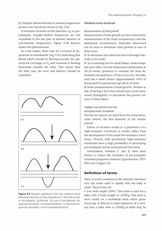
The antenatal period Chapter 9
117
by Doppler ultrasound and in normal pregnancies
produce the waveform shown in Fig. 9.9a.
If resistance increases in the placenta, e.g. in pre-
eclampsia, Doppler-shifted frequencies are not
recordable in the last part of diastole (absence of
end-diastolic frequencies). Figure 9.9b demon-
strates this phenomenon.
In a few babies, there may be a reversal of fre-
quencies in end-diastole (Fig. 9.9c) indicating that
blood which should be flowing towards the pla-
centa for exchange of O2 and nutrients is flowing
backwards towards the baby. This means that
the baby may die soon and delivery should be
expedited.
Medium-term methods
Measurements of fetal growthMeasurements of fetal growth are best achieved by
measurements of the head circumference and the
abdominal circumference. Real-time ultrasound
can be used to determine fetal growth in one of
three ways:
1 To determine size when the fetus is thought clin-
ically to be small.
2 As a screening test for small babies. Some hospi-
tals now offer a second ultrasound examination at
30–34 weeks’ gestation to measure the fetal ab-
dominal circumference. If this is not low, the baby
only has a small chance (approximately 10%) of
being small for gestational age (SGA) at birth.
3 Serial measurements of fetal growth. Women at
risk of having a SGA fetus should have serial ultra-
sound (fortnightly) to document the growth vel-
ocity of their babies.
Doppler waveforms from the uteroplacental circulationWaveforms may be recorded from the maternal ar-
cuate arteries, the first branches of the uterine
arteries.
Failure of invasion results in a persistence of a
high-resistance waveform (a notch) rather than
the development of the usual low-resistance wave-
forms. Women with persistently high-resistance
waveforms have a high probability of developing
pre-eclampsia and an asymmetrical SGA fetus.
Antioxidants (vitamin C and E) have been
shown to reduce the incidence of pre-eclamptic
toxaemia/pregnancy-induced hypertension (PET/
PIH) (see Chapter 10).
Definition of terms
There is much confusion in the obstetric literature
over the terms used to signify that the baby is
small. These terms are:
• Low birth weight (LBW). This term is used for a
baby with a birth weight of <2500g. This term is
most useful on a worldwide basis where gesta-
tional age at delivery is often unknown. It is obvi-
ous that a baby who is <2500g at birth may be
(b)
(c)
(a)
Figure 9.9 Doppler waveforms from the umbilical arteryreflecting resistance to flow (impedance) in the fetal vesselsof the placenta. (a) Normal. (b) Loss of end-diastolic fre-quencies (arrowed) —increased resistance. (c) Reversed fre-quencies (arrowed) —much increased resistance.
AMI9 6/9/04 5:28 PM Page 117
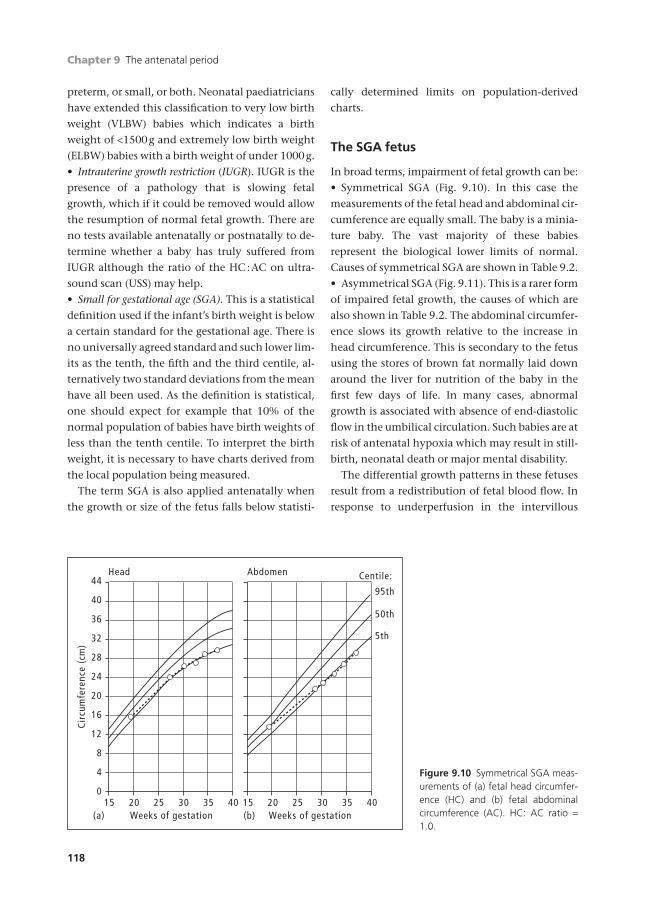
Chapter 9 The antenatal period
118
preterm, or small, or both. Neonatal paediatricians
have extended this classification to very low birth
weight (VLBW) babies which indicates a birth
weight of <1500g and extremely low birth weight
(ELBW) babies with a birth weight of under 1000g.
• Intrauterine growth restriction (IUGR). IUGR is the
presence of a pathology that is slowing fetal
growth, which if it could be removed would allow
the resumption of normal fetal growth. There are
no tests available antenatally or postnatally to de-
termine whether a baby has truly suffered from
IUGR although the ratio of the HC:AC on ultra-
sound scan (USS) may help.
• Small for gestational age (SGA). This is a statistical
definition used if the infant’s birth weight is below
a certain standard for the gestational age. There is
no universally agreed standard and such lower lim-
its as the tenth, the fifth and the third centile, al-
ternatively two standard deviations from the mean
have all been used. As the definition is statistical,
one should expect for example that 10% of the
normal population of babies have birth weights of
less than the tenth centile. To interpret the birth
weight, it is necessary to have charts derived from
the local population being measured.
The term SGA is also applied antenatally when
the growth or size of the fetus falls below statisti-
cally determined limits on population-derived
charts.
The SGA fetus
In broad terms, impairment of fetal growth can be:
• Symmetrical SGA (Fig. 9.10). In this case the
measurements of the fetal head and abdominal cir-
cumference are equally small. The baby is a minia-
ture baby. The vast majority of these babies
represent the biological lower limits of normal.
Causes of symmetrical SGA are shown in Table 9.2.
• Asymmetrical SGA (Fig. 9.11). This is a rarer form
of impaired fetal growth, the causes of which are
also shown in Table 9.2. The abdominal circumfer-
ence slows its growth relative to the increase in
head circumference. This is secondary to the fetus
using the stores of brown fat normally laid down
around the liver for nutrition of the baby in the
first few days of life. In many cases, abnormal
growth is associated with absence of end-diastolic
flow in the umbilical circulation. Such babies are at
risk of antenatal hypoxia which may result in still-
birth, neonatal death or major mental disability.
The differential growth patterns in these fetuses
result from a redistribution of fetal blood flow. In
response to underperfusion in the intervillous
0
Cir
cum
fere
nce
(cm
)
4
12
28
20
15 20 30Weeks of gestation
Head
(a)4025 35
36
8
24
16
32
40
44
15 20 30Weeks of gestation
Abdomen
(b)
Centile:
95th
50th
5th
4025 35
Figure 9.10 Symmetrical SGA meas-urements of (a) fetal head circumfer-ence (HC) and (b) fetal abdominalcircumference (AC). HC: AC ratio =1.0.
AMI9 6/9/04 5:28 PM Page 118

The antenatal period Chapter 9
119
space, there is an increase in resistance to blood
flow within the fetal circulation. This means that
blood returning from the placenta to the fetus
takes the path of least resistance and is diverted to
the fetal brain, coronary arteries and adrenals. Ini-
tially, this is to the benefit of the fetus but, if it con-
tinues for too long, then the fetal bowel, kidneys
and liver become ischaemic resulting in the com-
plications of asymmetrical SGA babies: necrotizing
enterocolitis, renal failure and failure of coagula-
tion due to insufficient production of coagulation
factors by the liver.
Table 9.2 Causes of babies who are small for gestational age (SGA)
Symmetrical SGA (60%) Asymmetrical SGA (40%)
Race (white > black > Asian) Poor maternal response to
Sex (boy > girl) • pregnancy
Maternal size • pre-eclampsia
Toxins • poor trophoblast invasion
• alcohol Cigarettes
• cigarettes Drug abuse
• heroin Chromosomal and congenital abnormalities
• methadone
Congenital infections
• cytomegalovirus
• parvovirus
• rubella
• syphilis
• toxoplasmosis
Malnutrition
0
Cir
cum
fere
nce
(cm
)
4
12
28
20
15 20 30Weeks of gestation
Head
(a)4025 35
36
8
24
16
32
40
44
15 20 30Weeks of gestation
Abdomen
(b)
Centile:
95th
50th
5th
4025 35
Figure 9.11 Asymmetrical SGAmeasurements of (a) fetal head cir-cumference and (b) fetal abdominalcircumference.
AMI9 6/9/04 5:28 PM Page 119

Chapter 9 The antenatal period
120
Management of SGA
Symmetrical SGAMost SGA fetuses represent the biological lower
limits of the normal range and only require serial
measurements of ultrasound growth performed on
a fortnightly basis. If the baby demonstrates nor-
mal growth (see Fig. 9.10), no further action is
necessary.
The only problem posed for the obstetrician is to
recognize those few babies that are congenitally
abnormal or infected. The following actions are
recommended:
• Check maternal blood for infections that are
known to cross the placenta (syphilis, toxoplasmo-
sis, rubella, parvovirus and cytomegalovirus).
• Search the fetus carefully with ultrasound look-
ing for structural markers that may suggest a chro-
mosome abnormality. If these are present the baby
should be karyotyped, usually by means of a fetal
blood sample obtained from the umbilical cord
(cordocentesis). This is of value even in the third
trimester as it is well known that fetuses with tri-
somies are prone to fetal distress in labour. If the
trisomy is lethal then, after discussion with the
parents, a Caesarean section may not be advised
should such distress develop.
Asymmetrical SGA (IUGR)Management should be:
• Fortnightly ultrasound measurements of head
circumference and abdominal circumference to de-
termine growth rate.
The amniotic fluid volume should be measured
by determining the height of the largest column of
fluid or the addition of a column from the four
quadrants of the uterus giving an amniotic fluid
index. The former is normally between 2 and
8cm. Less than 2cm suggests increasing fetal
compromise.
• Doppler waveforms from the umbilical circula-
tion. If these are normal they should be repeated
on a weekly basis. Delivery is not indicated in the
presence of normal umbilical artery waveforms.
Absent end-diastolic flow should indicate delivery
in a fetus that is considered to be viable, >24 weeks,
>500g.
• In the absence of Doppler waveforms, immedi-
ate fetal well-being should be monitored by daily
CTGs and maternal counting of fetal movement.
Delivery is indicated for cessation of fetal growth
over a 4-week period, for abnormalities of the CTG,
significant reduction in fetal movements.
Self-assessment
9.1 Mrs Walker is 36-weeks pregnant in her first pregnancy. Please examine her and give your conclusions to the exam-iner. (Mannequins are available to practise on, although ideally you should perform the examination on a woman inthe antenatal clinic, having asked her permission to do so first. Remember that you should always have a femalechaperone with you whenever you examine a woman.) As you examine the woman run through the examinationand present to a clinician or fellow student who will score against the checklist in Answers to self-assessment questions, p. 312.
9.2 Which of the following investigations are performed at the first antenatal visit in the UK?(a) Toxoplasmosis.(b) Rubella.(c) Cytomegalovirus (CMV).(d) Syphilis.(e) Hepatitis A.
9.3 Which of the following are used as indicators of fetal well-being beyond 24 weeks of pregnancy?(a) Liquor volume.(b) Fetal lie.(c) Fetal movements.(d) Uterine artery dopplers.(e) Umbilical artery dopplers.
AMI9 6/9/04 5:28 PM Page 120

The antenatal period Chapter 9
121
Self-assessment Continued
9.4 A woman presents at 12 weeks of pregnancy. She has had two livebirths at term, delivered a live baby at 21 weekswho died within a few minutes of birth, one pregnancy loss at 8 weeks and an intrauterine death at 25 weeks.Which one of the following correctly expresses her gravidity and parity?(a) Gravida 6 Para 4 + 1.(b) Gravida 6 Para 3 + 2.(c) Gravida 5 Para 3 + 2.(d) Gravida 5 Para 4 + 1.(e) Gravida 6 Para 5 + 0.
AMI9 6/9/04 5:28 PM Page 121

122
Hyperemesis gravidarum
IncidenceLess than 1 :1000 pregnancies in UK; a rare condi-
tion in the endogenous population.
Aetiology• Hormonal —rapid increase in hCG and proges-
terone; hypothyroidism.
• Reflex —a chemosensitive trigger zone which
stimulates the vomiting centre.
• Ketosis—after excess vomiting, build up of ketones
exacerbates the vomiting and a vicious circle develops.
• Hydatidiform mole —very high hCG levels.
ProgressCan lead to:
• Dehydration.
• Hypovolaemia.
• Electrolyte depletion.
• Vitamin deficiency, particularly thiamine.
• Death from liver failure or the end processes of
the above.
Presentation• Cannot retain food or fluid.
• Weight loss because of loss of body fluid and
burning up of fat.
• Haemoconcentration and unstable acid–base
balance.
• Ketosis.
Management1 Exclude other diseases:
• Urinary infection.
• Hiatus hernia and gall bladder disease.
• Obstructive gut lesions.
• Central nervous system (CNS)-expanding
lesions.
2 Exclude obstetric cause:
• Multiple pregnancy.
• Hydatidiform mole.
• Acute yellow atrophy of the liver.
3 Restore fluid and electrolyte balance intra-
venously (i.v.).
4 Specific anti-vomiting drugs, e.g. cyclizine or
Maxolon.
5 Thiamine to prevent Wernicke’s encephal-
opathy.
6 Steroid therapy —being assessed.
7 Psychological treatment —most respond to sug-
gestion. If not, formal psychotherapy is needed.
8 Therapeutic abortion —very rarely required.
Hypertensive disorders of pregnancy
Hypertension has these risks:
• In the mother.
• Cerebrovascular accident.
• Renal failure.
• Heart failure.
• Coagulation failure.
Chapter 10
Diseases of pregnancy
AMI10 6/9/04 4:58 PM Page 122
Diseases of pregnancy Chapter 10
123
• Liver failure.
• Adrenal failure.
• Eclampsia: a generalized convulsive disorder
like epilepsy but which is peculiar to pregnancy.
• In the fetus:
• Asymmetrical intrauterine growth restriction.
• Placental abruption.
• Iatrogenic preterm delivery.
DefinitionsThe currently internationally agreed definition of
hypertensive disease in pregnancy is:
• Pregnancy-induced hypertension (PIH): hyper-
tension occurring for the first time after 20 weeks’
gestation.
Hypertension in pregnancy is defined as one of
the following:
• Blood pressure of 140/90mmHg on two occa-
sions more than 4 hours apart.
• A rise of more than 30mmHg in systolic blood
pressure over the booking blood pressure.
• A rise of more than 15mmHg in diastolic blood
pressure over the booking figure.
PIH may be classified as:
• Mild: a blood pressure up to 140/100mmHg
without proteinuria.
• Moderate: a blood pressure up to 160/110mmHg
without proteinuria. In the absence of pro-
teinuria PIH is rarely dangerous to mother or
fetus.
• Severe: a blood pressure of more than 160/
110mmHg; and the presence of proteinuria
(pre-eclampsia/pre-eclamptic toxaemia (PET)).
Proteinuria in pregnancy is defined as the
following:
• More than 300mg on a 24-hour collection of
urine.
• Oedema associated with hypertension and
proteinuria is a sign of worsening pre-eclampsia.
Oedema alone is of little significance.
PrevalenceThis varies with the population but in the UK
10–15% of primigravid women will develop some
form of hypertension. Of these, about 6% may be
considered as suffering from PIH and 2% will
develop pre-eclampsia.
PIH is almost entirely a disease of primigravidae.
Pre-eclampsia only occurs in multigravid women
under the following conditions:
• Those who have had it severely in the first
pregnancy.
• Those who have changed their partner between
pregnancies.
• Pregnancies complicated by hydatidiform mole.
• Multiple pregnancies.
• Gestational diabetes.
• Those with antiphospholipid syndrome.
AetiologyThe precise mechanism is unknown; the following
are recognized:
• Women who develop pre-eclampsia have a fail-
ure of the second wave of trophoblastic invasion.
• This failure probably leads to a local alteration of
the prostacyclin : thromboxane ratio. Both these
prostaglandins are produced by trophoblast and
exert opposite effects. In PIH, the balance of the
ratio appears to favour thromboxane. This leads to
local vasoconstriction and platelet agglutination
on already undilated vessels.
• The combination of the above two factors is as-
sociated with failure of the initial fall in peripheral
resistance and hence blood pressure in mid-
pregnancy is maintained —it normally shows a
marked fall. Subsequent narrowing or clotting of the
abnormal blood vessels leads to a further increase in
peripheral resistance and hence hypertension.
• The narrowing of the blood vessels also leads to
decreased perfusion of the intervillous space and
hence the development of an asymmetrical small
for gestational age (SGA) fetus.
Antioxidants (vitamin C and E) in pregnancy
have been shown to reduce the prevalence of
pre-eclampsia in women who are at high risk —
previous early onset of PET, women with
antiphospholipid syndrome.
Clinical coursePIH usually presents in primigravidae in the late
third trimester. It usually requires either no treat-
ment or anti-hypertensive therapy alone while
awaiting the onset of labour. Occasionally this pro-
gresses to the development of pre-eclampsia. A few
AMI10 6/9/04 4:58 PM Page 123

Chapter 10 Diseases of pregnancy
124
women present with the symptoms and signs of
pre-eclampsia and occasionally this can occur in
the late second or early third trimester. The pres-
ence of symptoms, rising blood pressure or increas-
ing proteinuria heralds the onset of fulminating
pre-eclampsia and requires prompt treatment and
delivery to prevent the development of eclampsia
and renal/cerebral damage (Table 10.1).
Mild disease
Women with mild PIH may be discharged from
hospital and assessed as out-patients if:
• The blood pressure remains below 140/
100mmHg.
• They do not develop proteinuria.
• The fetus does not demonstrate asymmetrical
SGA.
The woman’s blood pressure should be moni-
tored at least twice a week and fetal growth should
be monitored fortnightly by ultrasound. If the con-
dition does not deteriorate, it is difficult to justify
induction of labour although few obstetricians
would be prepared to let these women go past 40
weeks’ gestation.
Moderate disease
All primigravidae who have a sustained blood pres-
sure of 140/90mmHg or more should be moni-
tored in a day assessment unit or in hospital as
the subsequent course of their disease cannot be
predicted. In the absence of rapidly progressive
disease, the following management features are
relevant.
Maternal1 Measurement of blood pressure. There is no evi-
dence that treating maternal blood pressure with
anti-hypertensive drugs alters the course of the
pre-eclamptic disease process or improves the
prognosis of the fetus, but treatment is indicated to
protect the maternal circulation. Sustained blood
pressures of more than 160/100mmHg would
therefore indicate treatment, unless delivery was
imminent. The current choice of therapy is oral
Table 10.1 Pregnancy-induced hypertension.
Mild Moderate Severe (pre-eclampsia)
Symptoms None Mild headache Frontal headache
Oedema Oedema ++Visual disturbance
Signs
BP <140/100 <160/110 >160/110
Proteinuria None None ++ or +++Reflexes Normal Normal Hyper-reflexia/clonus
Fundi Normal Normal Occasional papilloedema
Renal Normal Normal Decreasing urinary output
Bloods
FBC Normal Normal Rising or falling Hb
Decreasing platelets
Urate Normal Slightly raised Increasing
LFTs Normal Normal Increasing
Clotting Normal Normal Prolonged
Fetus Normal Normal/SGA Asymmetric SGA
Treatment None Anti-hypertensives Anti-hypertensives
Anti-epileptics/MgSO4
? Delivery Delivery
FBC, full blood count; LFTs, liver function tests; SGA, small for gestational age.
AMI10 6/9/04 4:58 PM Page 124

Diseases of pregnancy Chapter 10
125
methyldopa, nifedipine or labetalol but magne-
sium sulphate is now being used more.
2 Assessment of maternal renal function. All patients
should have:
• Urinary protein (daily).
• Plasma urea and electrolyte estimation
(weekly).
• Plasma urate levels (weekly).
• Total urinary protein excretion (once).
• Liver function tests (twice weekly).
• Full blood count and clotting screen.
A rising urate (>0.35), urinary 24-hour protein
excretion >300mg/24h or haemoglobin level in-
dicates haemoconcentration. These are signs of
fulminating pre-eclampsia and delivery should be
considered. Rising liver enzymes (aspartate amino-
transferase, AST; alanine aminotransferase, ALT), a
falling haemoglobin and/or platelets are signs of
the development of HELLP syndrome (Haemolysis,
Elevated Liver Enzymes, Low Platelets) and are an
indication for immediate delivery as these women
are at high risk of developing eclampsia and liver
failure.
FETAL WELL-BEING
• Real-time ultrasound assessment of fetal size. If
this indicates asymmetrical SGA then carry out:
(a) daily or twice daily cardiotocographs (CTGs)
(minimum 1 hour each time);
(b) a weekly examination of the umbilical circu-
lation by Doppler ultrasound.
(c) Doppler waveforms from utero-placental
circulation.
In moderate disease, delivery is indicated for:
• Progression to pre-eclampsia.
• Declining maternal renal function.
• Fetal distress, which usually means an abnormal
CTG or absence of end-diastolic flow in the
Doppler measurement of the umbilical
circulation.
• Placental abruption.
In the absence of the above features, most obste-
tricians would consider inducing the pregnancy
after 38 weeks’ gestation, if the cervix is favourable
and neonatal facilities are adequate.
Severe or fulminating pre-eclampsia
Symptoms• Frontal and often occipital headache due to cerebral
oedema. The headache is dragging or throbbing in
nature and is worse when the woman is supine. It
occurs classically first thing in the morning and re-
solves to some extent during the day if the patient
is mobile.
• Visual disturbances due to oedema of the optic
nerve or the retina consisting of black holes in the
visual field or double vision.
• Epigastric pain. This is due to stretching of the
liver capsule.
Signs• Hyper-reflexia and clonus. This is due to the cere-
bral oedema and gives the clinical picture of an
upper motor neurone lesion.
Hyper-reflexia, in obstetric terms, is defined as
the ability to obtain the reflex away from the ten-
don that usually causes it, e.g. the knee jerk reflex
occurs by tapping the anterior surface of the tibia
rather than the infrapatellar tendon.
Clonus in obstetric terms is only considered seri-
ous if it is sustained for more than four beats.
• A rapid rise in blood pressure.
• Rapid increase in proteinuria.
• Decreasing urine output.
TreatmentThis disease process starts to reverse as soon as the
placenta is delivered and hence the solution to ful-
minating pre-eclampsia is to end the pregnancy.
Before this happens, the maternal condition must
be prevented from worsening:
• Control the maternal blood pressure. At present
this is done by the use of i.v. hydralazine or labetalol.
• Prevent maternal fits with magnesium sulphate
or i.v. diazepam.
Having controlled the blood pressure and re-
duced the risk of fitting, the baby should be
delivered preferably vaginally but indications for
Caesarean section are:
• An unfavourable cervix.
• An abnormal fetal position such as a breech
presentation.
AMI10 6/9/04 4:58 PM Page 125

Chapter 10 Diseases of pregnancy
126
• Fetal distress.
• Abruption of the placenta.
• A failed induction.
• Difficulty in controlling the maternal blood pressure.
Prognosis
MATERNAL
In the absence of eclampsia, maternal mortality
should be low, but it must be remembered that pre-
eclampsia is one of the leading factors of maternal
death, even in developed countries (7.5/106 mater-
nities, 1997–99). Maternal morbidity may occur
from subsequently badly managed fluid balance. If
the woman can be helped over the first 48 hours
after delivery without serious injury, then the dis-
ease rapidly gets better. This is usually indicated by
a diuresis. Usually no permanent long-term renal
or vascular damage follows pre-eclampsia.
FETAL
The perinatal mortality rate (PNMR) increases with
the severity of the disease, but may be summarized
as follows:
• Mild: no change in PNMR.
• Moderate: slightly increased depending upon
gestation at birth.
• Severe: double PNMR.
• Severe pre-eclampsia superimposed on PIH:
treble PNMR.
The morbidity to the baby is difficult to quantify
as it depends upon the gestation at delivery and
the fetal size (Chapter 23).
Eclampsia
Eclampsia is characterized by epileptiform fits asso-
ciated with hypertension of a moderate to severe
degree. In the UK it is rare with a prevalence of
about 1 :3000 deliveries. Worldwide it is usually
preceded by pre-eclampsia, but the quality of ante-
natal care in the UK now is such that three-quarters
of cases of eclampsia occur without pre-existing
recorded evidence of hypertension.
PrevalenceIn the UK the disease is rare because of:
1 Better antenatal care which has led to earlier
recognition of pre-eclampsia.
2 More aggressive treatment of pre-eclampsia
which has lessened the incidence of subsequent
eclampsia.
• The rate of eclampsia may be taken as a guide
to antenatal care —to its availability, usage and
quality.
• Less than 1% of women in the UK with moder-
ate or severe PIH will go on to develop eclampsia.
Aetiology• Cerebral oedema.
• Cerebral vasoconstriction.
• Cerebral hypoxia.
These lead to cerebral ischaemia and hence fits.
Clinical courseAt present in the UK about 25% of women with
eclampsia will have a fit before labour; most of the
rest are likely to have a fit in the postpartum peri-
od. The character of the fit is very similar to an
epileptic fit with a typical fit consisting of:
• Twitching: 30 seconds.
• Tonic phase: 30 seconds.
• Clonic phase: 2 minutes.
• Coma: 10–30 minutes.
Such fits may repeat frequently.
Treatment
AIMS
• Keep the woman alive during the fit.
• Prevent more fits.
• Deliver the baby.
PREVENTION
• Magnesium sulphate reduces the incidence and
severity of fits.
DURING THE FIT
• Turn the woman on her side.
• Maintain the airway.
• Stop the fit by giving i.v. diazepam and magne-
sium sulphate.
AMI10 6/9/04 4:58 PM Page 126

Diseases of pregnancy Chapter 10
127
AFTER THE FIT
• Prevent further fits. This is usually done by giv-
ing a continuous infusion of magnesium sulphate
or diazepam.
• If the woman is not in hospital, arrange an emer-
gency transfer giving adequate anticonvulsants to
cover the journey.
• Lower the blood pressure by use of i.v.
hydralazine, labetalol or magnesium sulphate.
• Deliver the baby. As with fulminating PIH, such
women are best if the baby is delivered vaginally, as
this speeds the recovery process. The indications
for Caesarean section are those listed in the section
on pre-eclampsia.
Prognosis
MATERNAL MORTALITY
In the UK death from eclampsia is rare with the
woman more likely to die from the hypertensive
effects on the cerebral circulation from a cere-
brovascular accident.
FETAL MORTALITY
During an eclamptic fit: 300/1000. Overall:
150/1000 intrauterine deaths from hypoxia or
neonatal death from prematurity.
Rhesus (Rh) incompatibility (Box 10.1)
Rh genes
The Rh genes are carried on a pair of chromosomes.
There are six Rh antigens (C, D, E, c, d, e) of which
D and d are the most important, for upon these
depend whether a person is designated Rh-positive
or Rh-negative.
The individual making these gametes may be
heterozygous if some of the gametes contain c, d, e
and others C, D, E, or the person may be homozy-
gous if all gametes carry c, d, e.
There is no problem if the woman is Rh-positive,
even if her partner is a Rh-negative man; if
homozygous, all her children will be Rh-positive;
Box 10.1 Risks of Rhesus incompatibility effect.
Rh +ve father Rhesus -ve mother
Rh antibodies
Rh +ve cellsMiscarriage/termination of
pregnancyEctopic pregnancyAmniocentesisAntepartum haemorrhageDelivery of the placenta
1st Rh +ve baby Pass toUsually unaffected 2nd Rh +ve baby
Haemolysis ofRh +ve cells
Fetal anaemia
Hydrops fetalis
Treatment Intrauterine transfusionPostnatal exchange transfusion
AMI10 6/9/04 4:58 PM Page 127

Chapter 10 Diseases of pregnancy
128
if heterozygous, she may have a Rh-negative child
but that is no problem.
Should she be Rh-negative and her partner ho-
mozygous Rh-positive (35% of the male popula-
tion), she will always have a Rh-positive child and
there may be problems.
He may be heterozygous Rh-positive (65% of the
male population) producing equal numbers of
Rh-positive and Rh-negative gametes having equal
chances of giving his Rh-negative partner a
Rh-positive or a Rh-negative child.
Immunization
A Rh-positive mother cannot be immunized
against the Rh factor and so there are no problems
for her and her baby.
The Rh-negative woman can be affected if she is
inoculated with Rh-positive blood. The Rh gp anti-
gens evoke an antibody response against the Rh gp
(most marked against the D antigen —anti-D).
In Rh-negative women the inoculation of
Rh-positive cells can occur from:
• The passage of red cells from a Rh-positive baby.
• An incomplete cross-matched transfusion.
The former is more likely in a major fetomaternal
bleed which may occur in the following situations:
antepartum haemorrhage (APH), spontaneous mis-
carriage, ectopic pregnancy, therapeutic abortion,
amniocentesis, external cephalic version (ECV),
but most commonly during the third stage of labour
when the placenta separates from the uterine wall.
In most Rh-incompatible pregnancies no anti-
body is formed until after the first fetomaternal
bleed most commonly in the third stage of labour
and, consequently, the baby of the first pregnancy
is unaffected.
In subsequent pregnancies, if the fetus is Rh-
positive, small fetomaternal bleeds may evoke a
major secondary antibody response. Large
amounts of antibody (immunoglobulin G (IgG))
cross the placenta and can cause increasingly se-
vere Rh disease in successive pregnancies if the
fetus is Rh-positive. The antibody weakens the en-
velopes of the fetal red cells, which are then broken
down in the spleen. Depending on the speed and
degree of cell breakdown, this can produce:
1 Fetal anaemia.
2 Hyperbilirubinaemia. In utero the excess biliru-
bin is removed across the placenta to the maternal
circulation but following delivery the bilirubin ac-
cumulates and so the infant becomes jaundiced.
3 Oedema.
Clinical pictureThis can vary.
• The fetus may die in utero if the anaemia is severe
enough.
• The infant may be born grossly anaemic and
oedematous with hepato-splenomegaly —hydrops
fetalis. There is a rapid rise in bilirubin following
birth. Jaundice develops rapidly within the first 24
hours of life.
• The infant can be anaemic and continues to
break down red blood cells after delivery as the
maternal Rh antibodies are still circulating in his
blood, and so can become more anaemic and jaun-
diced during the postnatal period.
Management
PREVENTION
• Either give 500iu anti-D immunoglobulin to all
Rh-negative women at 26 and 34 weeks;
• Or be selective and give 500iu anti-D im-
munoglobulin if she has a:
• Therapeutic abortion.
• Spontaneous abortion/ectopic pregnancy.
• Amniocentesis.
• Any bleeding in pregnancy/threatened
miscarriage.
• ECV.
• After delivery at any gestation.
The former is now the recommended pro-
gramme for prevention of Rh disease.
VARIABLE DOSES
• After delivery of a baby to a Rh-negative mother,
the baby’s blood group should be checked and a
Kleihauer test performed on the maternal blood.
Acid is added to the maternal blood; fetal cells are
resistant to destruction in acid so the amount of
fetal blood that has entered the maternal circula-
tion can be calculated. If the baby’s blood group is
AMI10 6/9/04 4:58 PM Page 128

Diseases of pregnancy Chapter 10
129
positive the dose of anti-D is adjusted to ensure
that all the Rh-positive fetal cells are destroyed
without sensitizing the mother. This prevents the
development of Rh disease in the next baby.
DETECT AT-RISK FETUS
• Maternal Rh screening, anti-D antibody titres.
• Ultrasound scan to detect hydrops fetalis —
oedema of the skin, pleural effusion, ascites,
hepato-splenomegaly, cardiac enlargement.
• Amniocentesis or cordocentesis is performed
under ultrasound guidance. 10ml of amniotic fluid
(AF) or 5ml fetal blood is removed. The content of
bilirubin is measured by spectrometry (AF) or di-
rectly in the serum and the haemoglobin can be
measured in the blood. If the bilirubin is raised in
the amniotic fluid the need for a transfusion is
calculated from a Lilley’s at-risk graph.
History1 Check history of:
• Previous transfusion.
• Jaundiced babies.
• Exchange transfusions.
• Hydrops.
• Stillbirth or neonatal death.
2 Check all Rh-negative pregnant women for anti-
D and, if above 20iu/l, perform an indirect
Coombs’ test.
Check:
• On booking.
• If negative at booking, at 26 and 34 weeks.
• If positive at booking, at 20, 24, 28, 32 and 36
weeks or more frequently if rapidly rising.
If antibody titre rises above 1 :8 by 20 weeks, do an
amniocentesis.
To reduce risks carry out amnio/cordocentesis
under ultrasound guidance. Remove 10ml AF or 5ml
of fetal blood. Check for haemoglobin and bilirubin.
3 Check cord blood immediately after birth for:
• ABO group and Rh group.
• Haemoglobin.
• Direct Coombs’ test.
• Bilirubin.
Treatment• Intrauterine transfusion.
• Elect time of delivery.
• Exchange transfusion after delivery.
• Phototherapy after delivery.
• Top-up transfusion.
INTRAUTERINE TRANSFUSION
In very severe Rh disease the fetus can die in utero
from anaemia and hydrops before he can be deliv-
ered. An intrauterine transfusion can prolong the
life in utero of an infant to a gestation where the
risks of prematurity are estimated as being less than
those of the Rh disease. This can be done by an:
1 Intraperitoneal transfusion guided by
ultrasound.
2 Umbilical vein transfusion guided by
ultrasound.
Rh-negative blood is either transfused under
ultrasound control into the fetal peritoneal cavity,
or into an umbilical vein. Repeat as necessary, ac-
cording to amniotic optical density, or fetal
haematocrit. The intravenous route is becoming
increasingly the preferred method.
CHOOSE TIME OF INDUCTION AND BEST METHOD
OF DELIVERY
Balance the risks of prematurity (too soon) with
that of worsening Rh disease (too late). Consider
the risks of vaginal delivery and be prepared for a
lower segment Caesarean section (LSCS). The pae-
diatric team should be in close liaison and a senior
paediatrician present at the delivery with fresh
Rh-negative blood available.
RESUSCITATION AND EXCHANGE TRANSFUSION
Good resuscitation is essential. In an anaemic and
premature infant, lung disease is common. It can
be due to:
• Surfactant deficiency at very early delivery.
• Pulmonary oedema from anaemia and
hypoproteinaemia.
• Hypoplastic lungs secondary to pleural effusions.
In severe Rh haemolytic disease of the newborn,
an umbilical artery catheter should be inserted as
soon as possible to assess and control PaO2 and pH.
• Central venous pressure should be measured.
• Drain pleural effusions and ascites at
resuscitation.
AMI10 6/9/04 4:58 PM Page 129
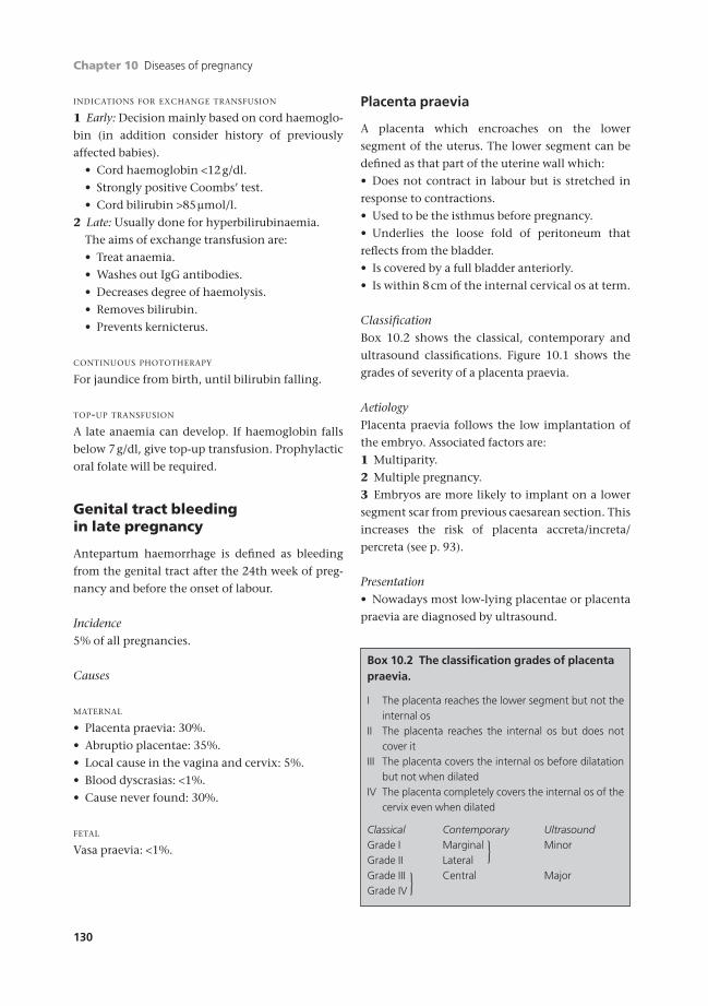
Chapter 10 Diseases of pregnancy
130
INDICATIONS FOR EXCHANGE TRANSFUSION
1 Early: Decision mainly based on cord haemoglo-
bin (in addition consider history of previously
affected babies).
• Cord haemoglobin <12g/dl.
• Strongly positive Coombs’ test.
• Cord bilirubin >85mmol/l.
2 Late: Usually done for hyperbilirubinaemia.
The aims of exchange transfusion are:
• Treat anaemia.
• Washes out IgG antibodies.
• Decreases degree of haemolysis.
• Removes bilirubin.
• Prevents kernicterus.
CONTINUOUS PHOTOTHERAPY
For jaundice from birth, until bilirubin falling.
TOP-UP TRANSFUSION
A late anaemia can develop. If haemoglobin falls
below 7g/dl, give top-up transfusion. Prophylactic
oral folate will be required.
Genital tract bleeding in late pregnancy
Antepartum haemorrhage is defined as bleeding
from the genital tract after the 24th week of preg-
nancy and before the onset of labour.
Incidence5% of all pregnancies.
Causes
MATERNAL
• Placenta praevia: 30%.
• Abruptio placentae: 35%.
• Local cause in the vagina and cervix: 5%.
• Blood dyscrasias: <1%.
• Cause never found: 30%.
FETAL
Vasa praevia: <1%.
Placenta praevia
A placenta which encroaches on the lower
segment of the uterus. The lower segment can be
defined as that part of the uterine wall which:
• Does not contract in labour but is stretched in
response to contractions.
• Used to be the isthmus before pregnancy.
• Underlies the loose fold of peritoneum that
reflects from the bladder.
• Is covered by a full bladder anteriorly.
• Is within 8cm of the internal cervical os at term.
ClassificationBox 10.2 shows the classical, contemporary and
ultrasound classifications. Figure 10.1 shows the
grades of severity of a placenta praevia.
AetiologyPlacenta praevia follows the low implantation of
the embryo. Associated factors are:
1 Multiparity.
2 Multiple pregnancy.
3 Embryos are more likely to implant on a lower
segment scar from previous caesarean section. This
increases the risk of placenta accreta/increta/
percreta (see p. 93).
Presentation• Nowadays most low-lying placentae or placenta
praevia are diagnosed by ultrasound.
Box 10.2 The classification grades of placentapraevia.
I The placenta reaches the lower segment but not theinternal os
II The placenta reaches the internal os but does notcover it
III The placenta covers the internal os before dilatationbut not when dilated
IV The placenta completely covers the internal os of thecervix even when dilated
Classical Contemporary UltrasoundGrade I Marginal MinorGrade II LateralGrade III Central MajorGrade IV }
}
AMI10 6/9/04 4:58 PM Page 130
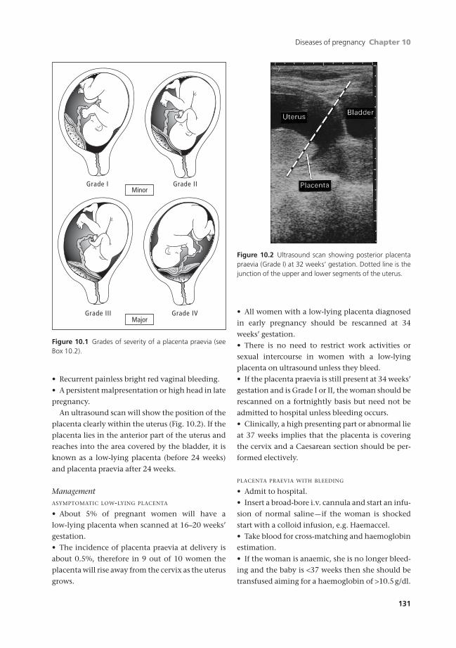
Diseases of pregnancy Chapter 10
131
• Recurrent painless bright red vaginal bleeding.
• A persistent malpresentation or high head in late
pregnancy.
An ultrasound scan will show the position of the
placenta clearly within the uterus (Fig. 10.2). If the
placenta lies in the anterior part of the uterus and
reaches into the area covered by the bladder, it is
known as a low-lying placenta (before 24 weeks)
and placenta praevia after 24 weeks.
ManagementASYMPTOMATIC LOW-LYING PLACENTA
• About 5% of pregnant women will have a
low-lying placenta when scanned at 16–20 weeks’
gestation.
• The incidence of placenta praevia at delivery is
about 0.5%, therefore in 9 out of 10 women the
placenta will rise away from the cervix as the uterus
grows.
• All women with a low-lying placenta diagnosed
in early pregnancy should be rescanned at 34
weeks’ gestation.
• There is no need to restrict work activities or
sexual intercourse in women with a low-lying
placenta on ultrasound unless they bleed.
• If the placenta praevia is still present at 34 weeks’
gestation and is Grade I or II, the woman should be
rescanned on a fortnightly basis but need not be
admitted to hospital unless bleeding occurs.
• Clinically, a high presenting part or abnormal lie
at 37 weeks implies that the placenta is covering
the cervix and a Caesarean section should be per-
formed electively.
PLACENTA PRAEVIA WITH BLEEDING
• Admit to hospital.
• Insert a broad-bore i.v. cannula and start an infu-
sion of normal saline —if the woman is shocked
start with a colloid infusion, e.g. Haemaccel.
• Take blood for cross-matching and haemoglobin
estimation.
• If the woman is anaemic, she is no longer bleed-
ing and the baby is <37 weeks then she should be
transfused aiming for a haemoglobin of >10.5g/dl.
Grade I Grade II
Grade IVGrade III
Minor
Major
Figure 10.1 Grades of severity of a placenta praevia (seeBox 10.2).
Figure 10.2 Ultrasound scan showing posterior placentapraevia (Grade I) at 32 weeks’ gestation. Dotted line is thejunction of the upper and lower segments of the uterus.
AMI10 6/9/04 4:58 PM Page 131
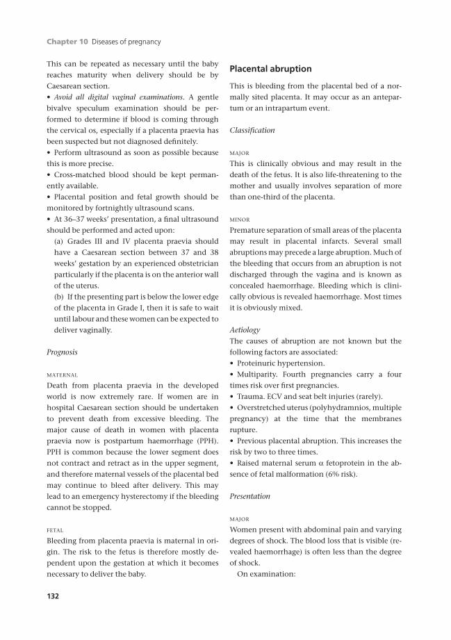
Chapter 10 Diseases of pregnancy
132
This can be repeated as necessary until the baby
reaches maturity when delivery should be by
Caesarean section.
• Avoid all digital vaginal examinations. A gentle
bivalve speculum examination should be per-
formed to determine if blood is coming through
the cervical os, especially if a placenta praevia has
been suspected but not diagnosed definitely.
• Perform ultrasound as soon as possible because
this is more precise.
• Cross-matched blood should be kept perman-
ently available.
• Placental position and fetal growth should be
monitored by fortnightly ultrasound scans.
• At 36–37 weeks’ presentation, a final ultrasound
should be performed and acted upon:
(a) Grades III and IV placenta praevia should
have a Caesarean section between 37 and 38
weeks’ gestation by an experienced obstetrician
particularly if the placenta is on the anterior wall
of the uterus.
(b) If the presenting part is below the lower edge
of the placenta in Grade I, then it is safe to wait
until labour and these women can be expected to
deliver vaginally.
Prognosis
MATERNAL
Death from placenta praevia in the developed
world is now extremely rare. If women are in
hospital Caesarean section should be undertaken
to prevent death from excessive bleeding. The
major cause of death in women with placenta
praevia now is postpartum haemorrhage (PPH).
PPH is common because the lower segment does
not contract and retract as in the upper segment,
and therefore maternal vessels of the placental bed
may continue to bleed after delivery. This may
lead to an emergency hysterectomy if the bleeding
cannot be stopped.
FETAL
Bleeding from placenta praevia is maternal in ori-
gin. The risk to the fetus is therefore mostly de-
pendent upon the gestation at which it becomes
necessary to deliver the baby.
Placental abruption
This is bleeding from the placental bed of a nor-
mally sited placenta. It may occur as an antepar-
tum or an intrapartum event.
Classification
MAJOR
This is clinically obvious and may result in the
death of the fetus. It is also life-threatening to the
mother and usually involves separation of more
than one-third of the placenta.
MINOR
Premature separation of small areas of the placenta
may result in placental infarcts. Several small
abruptions may precede a large abruption. Much of
the bleeding that occurs from an abruption is not
discharged through the vagina and is known as
concealed haemorrhage. Bleeding which is clini-
cally obvious is revealed haemorrhage. Most times
it is obviously mixed.
AetiologyThe causes of abruption are not known but the
following factors are associated:
• Proteinuric hypertension.
• Multiparity. Fourth pregnancies carry a four
times risk over first pregnancies.
• Trauma. ECV and seat belt injuries (rarely).
• Overstretched uterus (polyhydramnios, multiple
pregnancy) at the time that the membranes
rupture.
• Previous placental abruption. This increases the
risk by two to three times.
• Raised maternal serum a fetoprotein in the ab-
sence of fetal malformation (6% risk).
Presentation
MAJOR
Women present with abdominal pain and varying
degrees of shock. The blood loss that is visible (re-
vealed haemorrhage) is often less than the degree
of shock.
On examination:
AMI10 6/9/04 4:58 PM Page 132

Diseases of pregnancy Chapter 10
133
1 The uterus is woody hard; due to a tonic
contraction.
2 The fetal parts cannot be felt.
3 The fetus may be dead.
MINOR
Minor abruptions are often not diagnosed until
after delivery. They may present with:
• Mild abdominal pain associated with threatened
preterm labour.
• Unexplained APH.
• Tenderness over one area of the uterus only.
ComplicationsSevere abruption may result in:
• Shock from blood loss due to a large retroplacen-
tal clot, often concealed.
• A disseminated intravascular coagulopathy
(DIC).
• Oliguria or anuria due to hypovolaemia.
Minor degrees of placental abruption may
result in impaired fetal growth and/or hypoxic
ischaemic encephalopathy (HIE) —cerebral palsy.
ManagementMajor placental abruption is a life-threatening
condition for both the mother and the baby.
If the fetus is still alive:
• Insert two large-bore i.v. cannulae and infusion
of normal saline/colloid.
• Send 20ml blood for cross-match of 4 units,
haemoglobin and coagulation studies.
• Perform an immediate Caesarean section if
necessary to save the baby’s life (high risk of post-
partum haemorrhage).
• Ensure adequate fluid replacement following the
Caesarean section.
• Leave an indwelling urinary catheter in to mon-
itor urinary output.
• Consider insertion of a central line to monitor
maternal intravascular volume more closely.
If the fetus is dead, then the woman should
be allowed to deliver vaginally. This usually hap-
pens rapidly (within 4–6 hours) as the abruption
stimulates labour. If not in labour, rupture of mem-
branes usually leads to a swift delivery. The rele-
vant points of the management of labour are:
• Epidural analgesia is contraindicated because of
the risk of coagulopathy, but a patient-controlled
opiate infusion can be used.
• If a coagulopathy has developed (prolonged
APTT, PTT, increased fibrin degradation products,
low platelets) or the woman starts to bleed, she
should be managed in the following manner in
conjunction with a consultant haematologist.
(a) Give 4 units of fresh frozen plasma.
(b) Ask the blood bank to get 6 units of platelets
ready.
The consumptive coagulopathy begins to im-
prove immediately after the uterus has been evacu-
ated of its contents. Marked abnormalities of the
coagulation tests usually resolve within 4–6 hours
of delivery of the placenta.
Local causes of bleeding in late pregnancy
Urinary or anal bleeding may be reported as
vaginal bleeding in error. They need exclusion and
their own treatments.
Cervicitis and vaginitis
• Occasional excessive infection (especially with
Candida).
• Treat cause.
Cervical polyp
• Scanty bleeding. Can be seen with speculum.
• Leave alone in pregnancy and treat later if
necessary.
Cervical ectropion
• Spotting of blood only. Can be seen with
speculum.
• Leave alone in pregnancy and treat later if
necessary.
Varicosities of vagina
• Moderate bleeding in mid-trimester.
• Treat with pressure if close to vulva.
AMI10 6/9/04 4:58 PM Page 133
Chapter 10 Diseases of pregnancy
134
• Only ligate surgically in pregnancy if absolutely
necessary; it is difficult.
Cancer of cervix
• Rare but important.
• Irregular bleeding and discharge. Confirm diag-
nosis by biopsy.
• If before 24 weeks, hysterotomy and im-
mediate Wertheim hysterectomy followed by
radiation.
• If after 24 weeks, may await 32 weeks then
Caesarean Wertheim hysterectomy followed by
radiation.
Blood dyscrasias
These are extremely rare. Bleeding may be seen in
the following conditions.
• Idiopathic thrombocytopenia.
• Von Willebrand’s disease.
• Leukaemia.
• Hodgkin’s disease.
• Antiphospholipid syndrome.
ManagementThese conditions are usually known before preg-
nancy and are best managed in conjunction with a
relevant specialist.
Fetal bleeding
This occurs from rupture of vasa praevia when
there is a velamentous insertion of cord vessels and
these cross the cervical os.
DiagnosisThis condition usually presents with scanty bleed-
ing at the time of membrane rupture. It may be
associated with alterations in the fetal heart rate
producing a sinusoidal pattern (see p. 169).
ConfirmationIf there is time, the blood lost can be checked
for fetal haem by its resistance to alkalinization
(Kleihauer test). Alternatively the condition may
be suspected when an ultrasound examination
reveals the presence of a succenturate lobe on the
opposite side of the internal os to the placenta.
TreatmentDeliver the fetus as soon as possible and prepare to
transfuse.
Polyhydramnios
Definition: an excess of amniotic fluid detected
clinically. The range of normal volumes of fluid
present is wide and varies with the duration of
pregnancy. Average values for amniotic fluid are:
12 weeks: 50ml;
24 weeks: 500ml;
36 weeks: 1000ml;
The normal range at term in a singleton pregnancy
is large —500–1500ml.
DiagnosisThis is either clinical or by simple ultrasound. Other
methods of measuring amniotic fluid in situ are too
complex for routine use and often unreliable.
History• Tenseness of abdomen.
• Unable to lie comfortably in any position.
• Dyspnoea, indigestion, piles and varicose veins.
• Decreased sensation of fetal movements.
Examination• Increased symphysio-fundal height.
• Very tense, cystic uterus bigger than maturity
(like a balloon filled with water).
• Difficult to feel any fetal parts.
InvestigationsUltrasound. The deepest column >8cm or the amni-
otic fluid index is greater than the 95th centile.
Differential diagnosis• Twins: laxer feel to uterus and too many fetal
parts felt.
• Ovarian cyst: uterus displaced to one side in later
pregnancy.
• Full bladder.
All are resolved by ultrasound examination.
AMI10 6/9/04 4:58 PM Page 134

Diseases of pregnancy Chapter 10
135
Associations
MATERNAL
• Diabetes.
FETAL
• Congenital abnormality; anencephaly;
meningomyelocoele; upper alimentary atresia e.g.
tracheoesophageal fistula.
• Twins (particularly monozygotic).
Clinical course
ACUTE
• Painful with tense uterus and oedematous ab-
dominal wall.
• Primiparous.
• Pre-eclampsia.
• Often early (22–32 weeks’ gestation).
CHRONIC
• Slower onset.
• Uncomfortable rather than painful.
• Last weeks of pregnancy.
Management
ACUTE
1 Bed rest.
2 Ultrasound to rule out twins or abnormality.
3 Release fluid from uterus.
• If fetus normal: through abdominal wall with
narrow-bore needle. Drain fluid off slowly until
the woman is comfortable (500–1000ml over
4–8 hours).
• If fetus abnormal and viable —consider induc-
tion. If not viable —paracentesis.
CHRONIC
1 Bed rest.
2 Ultrasound to rule out twins and fetal abnormality.
3 Glucose tolerance test.
4 Sedation if very painful.
5 Treat underlying maternal condition.
6 If fetus normal, induce labour when indicated
by fetal state not because of the polyhydramnios.
7 Watch for uterine dysfunction and postpartum
haemorrhage (PPH) after labour.
Oligohydramnios
A lack of amniotic fluid, a much rarer
condition.
Diagnosis• Uterus is small for dates (early).
• Uterus feels full of fetus (later).
• Ultrasound shows reduced amniotic fluid index
(<2cm columns).
Fetal associations• Adhesions from fetal skin to amnion.
• Renal agenesis.
• Asymmetrical SGA.
Clinical course• Labour often preterm.
• High fetal death rate.
• High rate of fetal abnormalities (e.g. dislocated
hips and talipes).
Obstetric cholestasis
Obstetric cholestasis only occurs in pregnancy and
usually presents in the third trimester. It is more
common in multiple pregnancies.
Presentation• Itching, often generalized but commonly worst
on palms and soles of feet.
• Absence of rash.
• Insomnia.
• Right upper quadrant pain.
• Malaise.
• Intolerance of fatty foods.
InvestigationLiver function test (LFT).
• Raised transaminase concentrations (ALT and
AST).
• Raised bile salts.
Risks
MATERNAL
• Increased risk of PPH.
AMI10 6/9/04 4:58 PM Page 135

Chapter 10 Diseases of pregnancy
136
FETAL
• Increased risk of fetal distress.
• Increased risk of stillbirth over 38 weeks
(18/1000 births).
• Double the incidence of premature labour.
• Increased risk of intracerebral haemorrhage.
Treatment• Prophylactic Vitamin K until delivery because of
reduced absorption leading to increased risk of
PPH.
• Antihistamines, calamine, aqueous cream (for
itching).
• Ursodeoxycholic acid —effective in reducing
itching and usually returns liver function tests to
normal or near normal. However it does not alter
the outcome of pregnancy, but the symptoms
become tolerable allowing the baby to mature to
term in the majority of cases (37 weeks).
Monitoring
MATERNAL
• Weekly LFTs and clotting screen.
FETAL
• Alternate day CTG.
• Weekly liquor volume and umbilical artery
Doppler.
• Fortnightly growth ultrasound scan.
DeliveryAll women should be delivered at 37–38 weeks to
try and prevent intrauterine death. Vaginal deliv-
ery is indicated in the majority of cases and it is rare
for induction to fail even in primigravidas. As there
is an increased risk of fetal distress the fetal heart
rate should be monitored continuously electroni-
cally (CTG). Syntometrine / Syntocinon must be
given with the birth of the anterior shoulder to re-
duce the risk of postpartum haemorrhage (PPH).
RecurrenceThere is a high likelihood (>60%) of recurrence in
subsequent pregnancies.
Self-assessment
10.1 Which of the following statements are true?(a) Pre-eclamptic toxaemia is defined as a rise in the diastolic blood pressure after 20 weeks of pregnancy by more
than 15mm/Hg from the booking blood pressure without proteinuria.(b) Women with pre-eclampsia are at increased risk of developing gestational diabetes.(c) Pre-eclamptic toxaemia is defined as a rise in the diastolic blood pressure after 20 weeks of pregnancy by more
than 15mm/Hg from the booking blood pressure with proteinuria.(d) The most dangerous risk of pre-eclampsia for the mother is cerebral oedema leading to fitting.(e) The most dangerous risk of pre-eclampsia for the mother is developing HELLP syndrome.
10.2 A woman of 38 presents at 36 weeks in her first pregnancy with a headache. The symphysiofundal height is 35cm,cephalic presentation with the head 4/5 palpable per abdomen. Her blood pressure is 174/112, pulse 82 beats perminute, temperature 36.7°C. The CTG is reassuring. Her reflexes are brisk bilaterally with 4 beats of clonus in eachankle. Which of the following is the most appropriate treatment?(a) Magnesium sulphate infusion followed by delivery of the baby.(b) Magnesium sulphate infusion with hydrallazine followed by delivery of the baby.(c) Immediate delivery of the baby.(d) Phenytoin infusion with hydrallazine followed by delivery of the baby.(e) Diazepam with hydrallazine followed by delivery of the baby.
10.3 Anti-D should be given to all women who are Rh negative in which of the following scenarios?(a) Threatened miscarriage.(b) Following the birth of a baby whose blood group is A negative.
AMI10 6/9/04 4:58 PM Page 136

Diseases of pregnancy Chapter 10
137
Self-assessment Continued
(c) Following the birth of a baby whose blood group is A positive.(d) Ectopic pregnancy.(e) Medical termination of pregnancy.
10.4 A woman of 28 presents at 34 weeks of pregnancy with constant abdominal pain and a small amount of bleeding.Her symphysiofundal height is 37cm, her uterus is tender and firm. The CTG shows unprovoked decelerations. Herpulse is 108 beats per minute and her blood pressure 100/60mmHg. What is the most likely diagnosis?(a) Placenta praevia.(b) Cervical cancer.(c) Von-Willebrand’s disease.(d) Placental abruption.(e) Cervical polyp.
10.5 Polyhydramnios is associated with which of the following?(a) Gestational diabetes.(b) Fetal renal agenesis.(c) Pre-eclamptic toxaemia.(d) Obstetric cholestasis.(e) Tracheoesophageal fistula.
AMI10 6/9/04 4:58 PM Page 137
138
The majority of women who become pregnant are
healthy and remain so throughout their pregn-
ancy. A few women present with pre-existing medi-
cal disorders that may affect the pregnancy. Some
disorders may arise during the pregnancy. Doctors
should be aware of the effect that pregnancy may
have on these disorders and their treatment.
Urinary tract infection in pregnancy
During pregnancy the ureters are dilated and
kinked because of:
• Increased progesterone levels which relax the
smooth muscle.
• Mild obstruction of the lower ureters in late
pregnancy.
This encourages:
• Stasis of urine.
• Reflux of infected urine to the kidney evoking
pyelonephritis.
Asymptomatic bacteriuria
The presence of more than 105 bacteria/ml of urine
in the absence of symptoms.
IncidenceAbout 3% of pregnant women —increases with par-
ity and age.
SignificanceAsymptomatic bacteriuria is associated with a risk of:
• Acute pyelonephritis in pregnancy (30%).
• Structural abnormalities in the urinary tract
(3–5%).
ScreeningIn early pregnancy, all women should have urine
tested for either:
• The presence of white cells and nitrites, or;
• cultured for bacteria.
TreatmentThe most common organisms grown are:
• Escherichia coli.
• Proteus mirabilis.
These are usually sensitive to amoxycillin,
trimethoprim or nitrofurantoin. A 5-day course of
an antibiotic to which the organism is sensitive
should be prescribed. This will result in a cure in
more than 85% of women, but the urine should be
recultured one week after treatment.
A renal ultrasound and an intravenous (i.v.)
urogram should be performed 3 months after
delivery to exclude a structural urinary tract
abnormality.
Symptomatic infections
Incidence1–2%; commoner in primigravidae.
Chapter 11
Diseases in pregnancy
AMI11 6/9/04 4:59 PM Page 138

Diseases in pregnancy Chapter 11
139
Symptoms• Dysuria (due to urethritis).
• Increased frequency (due to trigonitis).
• Backache, loin pains, night sweats and rigors
(due to pyelonephritis).
• Headache, vomiting and muscle aches (due to
pyrexia).
Examination• The woman is usually pyrexial if the infection
has involved the kidneys. In many cases this may
be at levels of up to 40.5°C.
• If the woman has pyelonephritis she will be ten-
der in the renal angles.
InvestigationA mid-stream specimen of urine (MSU) should be
sent for:
• Dipstick for nitrites and leucocytes.
• Microscopy for white cells.
• Culture to determine the organism responsible.
• Sensitivity of organisms to antibiotics.
ManagementAll women who have renal angle tenderness or a
pyrexia must be admitted to hospital because of
the threat of preterm labour.
Management consists of:
1 Bed rest.
2 Ample fluid intake, at least 3 litres a day; if nau-
seated, give i.v.
3 Start a broad-spectrum i.v. antibiotic such as
amoxycillin; may need changing when an organ-
ism’s antibiotic sensitivities are known.
4 If the woman has pyelonephritis, do a renal
ultrasound when she has recovered from the
infection.
5 Keep her in hospital until the renal angle ten-
derness has disappeared.
6 The antibiotics can be given orally once temper-
ature is normal. A complete 5-day course at least
should be given. The urine should be recultured 5
days after the last dose of antibiotic has been given.
Chronic renal disease
Renal changes in normal pregnancy• Renal blood flow increases.
• Glomerular filtration rate (GFR) increases.
• Plasma concentrations of urea and creatinine fall
in normal pregnancy.
• There is an increase in total body water that ex-
ceeds the increase in total body sodium, resulting
in a decrease in plasma osmolality.
• There is a 25% fall in serum uric acid concentra-
tions during the first two trimesters but this returns
to pre-pregnant levels by the third trimester. Watch
if using urate to monitor pre-eclampsia.
PrognosisThe outcome of the pregnancy is worse if:
1 The woman was hypertensive before pregnancy.
2 The woman had proteinuria before pregnancy
started.
3 There is active progression of renal disease or it is
associated with other medical conditions.
Pregnancy probably has no long-term adverse
effects on renal disease.
Fetal prognosis• Normotensive women with chronic renal dis-
ease have 2–3 times greater risk of developing
pre-eclampsia. In the absence of pre-eclamptic
toxaemia (PET) perinatal mortality is not in-
creased. If PET develops, the risk of fetal death is
directly related to the gestation at delivery.
• Women with more severe renal disease have a
high incidence of both PET and impaired fetal
growth. Among women with pre-existing hyper-
tension and proteinuria, the perinatal mortality
rates approach 30%. Cause of death is from
preterm delivery and complications associated
with small for gestational age (SGA).
Acute renal failure in pregnancy
This may be:
1 Tubular necrosis: this is largely recoverable.
2 Cortical necrosis: this is usually irrecoverable
and these patients go on to need long-term dialysis
or transplantation.
AMI11 6/9/04 4:59 PM Page 139

Chapter 11 Diseases in pregnancy
140
Presentation1 Oliguria: <500ml/day (20ml/hour), the min-
imum volume to remove catabolites.
2 Anuria: the absence of urine.
Aetiology in obstetrics1 Hypovolaemia.
• Severe pre-eclampsia.
• Placental abruption.
• Postpartum haemorrhage (PPH).
• Hyperemesis gravidarum.
• Miscarriage.
2 Gram-negative shock. This may result from:
• Pyelonephritis.
• Chorioamnionitis.
• Puerperal infections.
• Septic miscarriage.
The usual organism is E. coli, but it may be
Clostridium.
3 Nephrotoxins. In modern obstetric practice these
are rare. Illegal abortions may result in infection
followed by haemolysis and renal failure.
4 Acute renal failure associated with acute fatty
liver of pregnancy. Rare; usually fatal.
5 Vomiting in late pregnancy associated with jaun-
dice. The disease occurs in many systems and renal
failure, pancreatitis and colitis may occur.
ManagementThree consecutive phases:
1 Oliguria: lasts from a few days to a few weeks.
Complete anuria is rare in acute tubular necrosis
and usually suggests acute cortical necrosis or
obstruction.
2 Polyuria: markedly increased urine production
that may last up to 2 weeks. The urine is dilute and
metabolic waste products are poorly eliminated.
Plasma urea and creatinine may continue to rise
for several days following the increase in urinary
output. Profound fluid and electrolyte losses can
occur in this phase.
3 Recovery: urinary volumes decrease towards nor-
mal and renal function improves.
General management:
• Determine the cause.
• Insert a urinary catheter and maintain accurate
fluid balance charts.
• Insert a central venous pressure line and measure
the pressure.
In pregnancy, central venous pressure should
range from +4 to +10cmH2O. If this is low it sug-
gests the cause of the renal failure is due to hypo-
volaemia and therefore the volume should be
restored with up to 2 litres of normal saline fol-
lowed by a plasma expander. Response of over
30ml of urine in 1 hour should be seen within 1
hour of the fluid load.
• Send baseline investigations, including urea and
electrolytes, liver function tests, serum amylase,
plasma proteins, coagulation studies, and if re-
quired, perform an arterial blood sample for
acid–base balance.
• If the patient is not hypovolaemic or does not re-
spond to the fluid load, then the alternatives are as
follows:
(a) Conservative management. Give i.v. the vol-
ume of patient fluid output plus 500ml/day.
Monitor electrolytes and start dialysis if or when
the patient is uraemic or hypercatabolic.
(b) Intensive management using vasodilators
and inotropes (renal dose dopamine). This re-
quires the insertion of a pulmonary artery
catheter and the monitoring of cardiac output.
Vasodilators and inotropes are given and the pa-
tient is fluid-loaded.
• Involve intensive care physicians and the renal
physician at an early stage.
Anaemia
Anaemia can follow:
1 Lack of production of blood: haemopoietic.
2 Increased breakdown of blood: haemolytic.
3 Blood loss: haemorrhagic.
In pregnancy, most anaemia is haemopoietic
when it may be due to lack of:
1 Iron: iron deficiency anaemia.
2 Folic acid: megaloblastic anaemia.
3 Protein: iron deficiency anaemia.
Normal levels of haematological indices are
shown in Box 11.1.
AMI11 6/9/04 4:59 PM Page 140

Diseases in pregnancy Chapter 11
141
Iron deficiency anaemia (Table 11.1)
Aetiology
POOR INTAKE
• Diet deficient in iron-containing foods.
POOR ABSORPTION
• Vomiting in pregnancy affects absorption.
• Increased pH of gastric juice.
• Ferric ions in gut instead of ferrous.
• Lack of vitamin C.
INCREASED UTILIZATION
Demands of pregnancy. Total body iron measures
about 3500mg. Includes:
• Fetus and uterus: 500mg.
• Increased maternal blood volume: 500mg.
More if:
• Multiple pregnancies.
• Grand multiparity.
• Pregnancies close together.
• Vegetarian (particularly vegans).
DIAGNOSIS
• Rarely made clinically unless woman is severely
affected.
• May show pallor of conjunctivae.
• May have tiredness and oedema.
• Hb estimates must be done on all pregnant
women at booking, and twice later in pregnancy, at
26 and 34 weeks.
• If level below 10g/dl, diagnose anaemia, look for
cause and treat.
Treatment
PREVENTATIVE
Regular iron-bearing foods in diet (Box 11.2). If
needed, iron tablet supplements. Daily requirements
are 100mg elemental iron with 300mg folic acid.
• See she gets them—give them to her at the
clinics.
• See she takes them —ask at each visit.
• See they are effective —check Hb levels.
Box 11.1 Normal haematological values in pregnancy
Blood RangeTotal blood volume (ml) 4000–6000Red cell volume (ml) 1500–1800Red cell count (1012/l) 4–5White cell count (109/l) 8–18Haemoglobin (g/dl) 10.5–13.5Mean corpuscular volume (fl) 80–95Mean corpuscular haemoglobin (mg) 32–36Serum iron (mmol/l) 11–25Serum ferritin (mg/l) 10–200Serum folate (mg/l) 6–9Total iron-binding capacity (mmol/l) 40–70Platelets (109/l) 150–300
Table 11.1 Indices of iron deficiency and megaloblastic
anaemia.
Iron deficiency Megaloblastic
Blood film
Red cells
Size N or Ø ≠Hypochromia Ø N
Anaesocytosis + +Poikilocytosis + +
White cells N Leucopenia
Hypersegmented
Haematological values
Hb Ø ØMCV Ø N or ≠MCHb Ø N
MGHbC Ø ≠Serum iron Ø N
Serum ferritin Ø N
Serum folate N Ø
Marrow Ø Iron stores ≠ Megaloblasts
Box 11.2 The iron-rich foods
AnimalRed meat—iron in haemoglobin and myoglobinWhite meat
—iron in the myoglobinFish
PlantLentils moderate amount ofDark-green leaf vegetables iron only (rich inBeans of all sorts folates)
}
}
AMI11 6/9/04 4:59 PM Page 141

Chapter 11 Diseases in pregnancy
142
CURATIVE
Depends on:
1 Degree of anaemia.
2 Duration of pregnancy.
3 Cause of iron deficiency:
• Mild anaemia: Hb below 10g/dl.
• Severe anaemia: Hb below 8g/dl.
Mild:
1 Check that the woman is being given, and is tak-
ing, oral iron.
2 If so, increase oral iron; add vitamin C to aid ab-
sorption or try another preparation.
3 If she cannot swallow tablets, use liquid
preparation.
4 If change of oral therapy does not improve, use
intramuscular (i.m.) or i.v. preparation. Give total
dose i.v. as a transfusion in 1000ml of saline. Alter-
natively, iron dextran 250mg is associated with a
rise of Hb of about 1g/dl. Give on alternate days,
i.m. for six doses, with small test dose first to check
anaphylaxis.
Severe and early:
1 Admit to hospital and check that anaemia is
solely iron deficient:
• Blood film.
• Serum iron.
• Total iron-binding capacity.
• Serum ferritin.
• Serum folate.
• Sickle/thalassaemia status.
2 Treat with oral, i.m. or i.v. therapy as required.
3 Check protein and vitamin intake adequate.
4 Check that improvement is maintained for rest
of pregnancy.
Severe and late:
1 If after 36 weeks, too late to rely on haemo-
poiesis to provide red cells in time to cover labour.
Therefore transfuse slowly with packed red cell
blood.
2 If Hb below 4g/dl consider exchange transfusion.
3 Build up iron stores for puerperium by i.m. therapy.
Folic acid deficiency anaemia
AetiologyFolic acid required for building DNA in all tissues.
Hence demands are maximal when fetal tissue
being made.
1 Poor intake:
• Diet deficient in folates.
• Vomiting in pregnancy.
2 Increased utilization:
• Demands of pregnancy.
• Rapid growth of fetal, placental and uterine
tissues.
• Worse if:
(a) Multiple pregnancy.
(b) Grand multiparity.
(c) Fetal haemolysis (in Rh effect).
(d) Infection.
Commoner in underdeveloped countries and
combined with other forms of malnutrition.
DiagnosisSometimes made clinically.
• May be tired, breathless, oedematous.
• May have other signs of malnutrition.
HaematologySee Megaloblastic column, Table 11.1.
Treatment
PREVENTATIVE
Folic acid supplements in last 20 weeks of preg-
nancy (300mg/day).
Theoretical risk of masking pernicious anaemia
(PA) and its uncommon accompanying subacute
combined degeneration (SCD) of the spinal cord. In
practice, PA is very rare in those under 35 years and
SCD almost unknown in the pregnancy age group.
CURATIVE
• Mild or moderate anaemia —folic acid 5–
10mg/day orally only.
• Severe anaemia —folic acid 5–10mg/day i.m.:
(a) oral iron both should be
(b) blood transfusion given with care.
Haemolytic anaemias
These can all be diagnosed before pregnancy at pre-
pregnancy consultation.
}
AMI11 6/9/04 4:59 PM Page 142

Diseases in pregnancy Chapter 11
143
Haemoglobinopathies
Sickle cell disease
Aetiology• Abnormal Hb: typical original geography.
(a) HbS (commonest): Middle East, Africa, USA,
Caribbean and southern Europe.
(b) HbC: Ghana.
(c) HbE: South-East Asia.
(d) HbD: Punjab.
Diagnosis• Crises or infarcts:
(a) Chest pain.
(b) Sudden head or abdominal pain.
(c) Joint pains.
• Bone marrow exhaustion.
A crisis could be triggered by:
• Infection.
• Hypoxia.
• Dehydration.
• Trauma.
Haematology• Low Hb.
• Sickling and target cells on blood film.
• Electrophoresis shows abnormal Hb patterns.
Treatment• Detect early.
• Hydrate with intravenous fluids.
• Oxygen.
• Folic acid prophylactically 1–2mg/day.
• Transfusion of red blood cells.
• Diuretic.
• Antibiotics.
If crisis:
• Hydrate.
• Check Hb every 4 hours.
• Heparinize.
• Antibiotics.
• Consider exchange transfusion.
• If blood pressure rises rapidly, deliver.
• Splenectomy for some.
Thalassaemia
AetiologyDefective genes alter Hb side chains. May be a or bwhich can either be:
• Homozygous —thalassaemia major: usually fatal
before pregnancy age group. Sickle cell anaemia.
• Heterozygous —thalassaemia minor: common-
est thalassaemia. b-Thalassaemia minor is the more
serious especially if combined with any other ab-
normal Hb such as S or C.
Diagnosis• Classically women from the Mediterranean
countries but now widespread in the Middle and
Far East. The woman usually knows about this and
will mention it in the history.
• Globin chain synthesis studies.
• Occasionally a mild anaemia (MCVØ MCHØMCHC=).
• Splenomegaly.
• Jaundice.
• Pain from bone infarcts (later in life —ulcers of
legs).
Haematology• Increased red blood cell fragility.
• Hb level low.
• Serum iron raised.
Treatment1 No use giving iron alone (iron stores high) but
folate helpful.
2 Cover haemolytic crisis with transfusions
carefully.
3 Prevent stress if possible (e.g. hypoxia).
4 Treat infections early.
5 Beware coexistent:
• Malaria.
• Glucose-6-phosphate dehydrogenase defi-
ciency.
• Other abnormal Hb.
6 Deliver between crises.
Haemorrhagic anaemia
Rare in temperate climates:
AMI11 6/9/04 4:59 PM Page 143

Chapter 11 Diseases in pregnancy
144
• Recurrent chronic gastrointestinal bleeding
(peptic ulcer, piles).
Commoner in tropics:
• Recurrent chronic bleeding (e.g. tapeworms,
hookworms).
Treatment• Treat cause.
• Correct anaemia —as above.
Heart disease
Frequency and severity of heart disease in preg-
nancy are diminishing in this country because:
• Most heart disease in this age group is rheumatic
in origin.
• Rheumatic fever is much rarer in childhood with
better housing and nutrition.
• Rheumatic fever is more effectively treated in
childhood by chemotherapy.
Aetiology• Rheumatic 80%: mitral valve affected 85%,
aortic valve 10%, both 5%.
• Congenital 15%: septal defects and reversed
shunts.
• All the rest 5%: ischaemic, thyrotoxic.
PathophysiologyPregnancy is a hyperkinetic state and is an extra
load on the heart. If the heart is damaged, avoid
other loads.
• Anaemia —blood is inefficient at oxygen
transport.
• Pre-eclampsia —harder work if hypertension or
oedema present.
• Arrhythmia —fibrillation is inefficient at deliver-
ing blood.
• Flare-up of rheumatic fever —not common but
watch for and treat.
• Acute bacterial endocarditis risk increased be-
cause of irregular endothelium over heart valves;
hence cover labour, surgery and dentistry with
antibiotics.
Cardiac complications in pregnancy1 With mitral stenosis:
• Pulmonary oedema.
• Right-sided congestive failure.
2 With aortic stenosis:
• Left-sided congestive failure. Rare to start de
novo, in pregnancy.
3 Eisenmenger’s syndrome.
• If right-to-left shunt —pulmonary hypertension.
4 Fallot’s tetralogy.
• If right-to-left shunt, risk of cardiac failure.
5 Coarctation of aorta.
• Risk of rupture in late pregnancy or labour.
Often repaired before; if well healed, no in-
creased dangers.
6 Artificial valves —thrombosis.
Management
IN PREGNANCY
1 Diagnose early.
• History.
• Examination.
2 Assess severity early: ideally cardiologist and ob-
stetrician to see woman together at same antenatal
clinic.
Investigations:
• Electrocardiogram (ECG).
• Chest X-ray.
• Echocardiography.
• Maybe:
(a) Catheter studies (pressure and blood
gases).
(b) 24-hour ECG.
Factors:
(a) Age.
(b) Severity of lesion.
(c) Functional decompensation.
3 Book for hospital delivery and be prepared for
bed rest in hospital.
4 Extra rest at home.
5 Continue anticoagulation if patient already on
it. Consider subcutaneous heparin rather than oral
anticoagulants unless plastic valve prosthesis. For
these continue on warfarin.
6 Senior cardiologist, anaesthetist and obstetri-
cian make labour plan and write it on hospital
records. See that senior staff of each discipline are
available to cover labour.
AMI11 6/9/04 4:59 PM Page 144

Diseases in pregnancy Chapter 11
145
IN LABOUR
1 Reduce extra work —good analgesia; probably
epidural unless anticoagulated.
2 Nurse head up: tilt bed or extra pillows.
3 Antibiotics especially if a congenital heart
lesion.
4 Short second stage; maybe forceps or vacuum
extraction.
5 Give Syntometrine only if high risk of postpar-
tum haemorrhage (PPH); otherwise not.
6 Manage pulmonary oedema if it occurs.
• Tilt head up 35°.
• O2.
• Aminophylline 0.5g, i.v.
• Morphine 15mg.
• Digitalis if arrhythmia or tachycardia.
• Diuretics —frusemide.
IN PUERPERIUM
1 Rest:
• Keep in hospital longer.
• Check home conditions adequate —few stairs
if possible.
2 Physiotherapy to legs and gentle exercises.
3 Breast feeding allowed unless cardiac condition
deteriorated in pregnancy. It can be hard work, for
only one person can do it and it means getting up
at night.
Prognosis
MATERNAL
1 Mortality: increased risk of death —9% of all
maternal deaths in UK are associated with heart
disease.
2 Morbidity: increased risk of deterioration of
heart condition. Used to be inevitable. This is not
so if proper care is taken.
FETAL
Little increased risk if mother kept healthy. Watch
for fetal risks from anticoagulation if relevant.
Respiratory diseases
Asthma
IN PREGNANCY
Often emotional factors are involved so asthma
may worsen if pregnancy is resented. Continue all
treatments started before pregnancy. Be careful of
new therapies, e.g. budesonide teratogenic to some
species; not known to be so in humans. Therefore
use well-established drugs.
• Bronchial antispasmodics.
• Antibiotics.
• Steroids —inhaled or systemic.
Most asthmatics do not deteriorate in pregnancy
but may in puerperium. No obvious correlation
with hormone changes.
IN LABOUR
1 Deliver so that mother’s respiratory effort is
minimal.
2 May require extra antispasmodics in labour.
3 If on steroids, hydrocortisone required to cover
labour.
4 Baby may be small for dates if asthma control
was poor.
Pulmonary tuberculosis
IncidenceLess than 1 :1000 in UK; rare in endogenous popu-
lation, higher in immigrants.
Presentation• History of disease.
• Pick up on routine chest X-ray.
Management1 Notify any new cases to District Community
Physician.
2 Continue any antituberculous drugs already
started in combination:
• Streptomycin.*
• INAH (isonicotinic hydrazide).
*Beware potential teratogenic effect in first trimester.
AMI11 6/9/04 4:59 PM Page 145

Chapter 11 Diseases in pregnancy
146
• Ethambutol.
• Rifampicin.*
3 Bed rest.
4 Surgery if needed in mid-pregnancy, avoiding
first 14 and last 10 weeks.
5 Follow up the family.
LabourDelivery by most expeditious route (baby may be
large).
Mother1 Allow breast feeding if no positive sputum bac-
teriology within 1 year and no chest X-ray signs of
recent activity.
2 Continue all drug treatments.
3 Suppress lactation if baby has to be separated.
4 Rest in hospital longer.
5 Check community back-up services to give least
effort to mother.
6 Arrange for follow-up at chest clinic in case of tu-
berculosis flare-up.
Baby1 If mother has had bacteria in sputum within 1
year, separate baby from mother at birth. The
mother must be warned during pregnancy that
this will happen. If properly explained, she will
realize it is a wise move.
2 Give baby isoniazid-resistant BCG 0.05mg at 7
days (unless premature). Await positive Mantoux
before allowing baby back to mother (about 6
weeks).
Endocrine diseases
Thyroid disease
Pregnancy is a hyperdynamic state. Increased oes-
trogen levels cause enlargement of thyroid gland
and an increased output of thyroid hormone. Since
this is mostly in the form of protein-bound thyrox-
ine, such patients are not hyperthyroid, for the ac-
tive fraction is not increased.
Hyperthyroidism
• Difficult to diagnose for the first time in
pregnancy.
• If established beforehand, continue treatment,
usually carbimazole, but keep dose as low as
possible.
• Consider thyroidectomy if disease is increas-
ingly difficult to control.
Operation is safe if mother properly prepared.
Avoid radioactive-iodine testing because of fetal
thyroid pick-up and retention. Maternal thyroid-
stimulating IgG passes across placenta and may
stimulate fetal thyroid, sometimes enough to cause
neonatal thyrotoxicosis. This is unusual but can be
predicted by testing maternal blood levels of IgG in
late pregnancy.
Hypothyroidism
• Rarely get pregnant if not on therapy.
• If treated, continue treatment, and be prepared
to increase dosage.
Pituitary disease
Prolactinoma
• Women on long-term bromocriptine become
pregnant.
• Oestrogen stimulation of pregnancy may cause
enlargement of tumour; pressure on optic chiasma
threatens vision.
• Check with computerized tomography (CT)
scan. Prolactin levels raised but variable.
• A few tumours need treatment if they enlarge:
(a) first bromocriptine;
(b) then surgery.
Hypopituitarism
• Mostly starts in puerperium after pituitary vein
thrombosis following PPH.
• Study each pituitary-controlled function sepa-
rately and treat those deficient.
• If mother treated, baby does well.*Beware potential teratogenic effect in first trimester.
AMI11 6/9/04 4:59 PM Page 146

Diseases in pregnancy Chapter 11
147
Diabetes
Diabetes is a metabolic disease which results from
an underproduction of insulin by the pancreas.
This results in disturbances of carbohydrate, fat
and protein metabolism and leads to a sustained
rise in blood glucose.
During pregnancy, diabetes may be one of the
following types:
1 Pre-existing diabetes which is usually insulin-
dependent.
2 Gestational diabetes or an impaired glucose
tolerance test discovered for the first time in
pregnancy.
Glucose homeostasis in pregnancy• In normal pregnancy, fasting glucose blood lev-
els are maintained at 4–5mmol/l.
• To maintain the glucose level, however, there is a
doubling in the secretion of insulin in the second
and third trimesters of pregnancy. Pregnancy rep-
resents a relatively insulin-resistant state.
• The insulin resistance is due to the placental
secretion of oestrogen, progestogen and human
placental lactogen together with a change in
peripheral insulin receptors.
• Glucose crosses the placenta by facilitated diffu-
sion resulting in a fetal glucose level of approxi-
mately 1mmol/l less than its mother.
• The facilitated diffusion system is saturated at
maternal levels of 11–12mmol/l. Therefore the
fetus is probably never subject to levels of greater
than 11mmol/l.
Established diabetes mellitus
Occurs in 1–2% of the pregnant population.
Effects of pregnancy on the diabetes• Insulin requirements increase during pregnancy
and rapidly fall to pre-pregnancy levels after
delivery.
• Pregnancy aggravates proliferative diabetic
retinopathy. Ideally any retinopathy should be
treated before pregnancy.
• Women with diabetic nephropathy are more
likely to develop pre-eclampsia and to have poorer
renal function during pregnancy, but there are few
adverse long-term effects on renal function.
• There is a high incidence of asymmetrical SGA
and preterm delivery.
• Diabetic neuropathy and vascular disease are
rare in pregnancy.
Effects of poorly controlled diabetes on the pregnancy• Diabetes is associated with an increased risk of
first trimester miscarriages.
• Second trimester miscarriages, due to fetal
death.
• Congenital abnormalities. By ¥3, 50% neural
tube defects, 30% cardiac abnormalities. Diabetics
tend to show a predominance of multiple malfor-
mations and caudal regression appears exclusively
in diabetics.
• Pregnancy-induced hypertension.
• Preterm delivery.
• Polyhydramnios.
• Macrosomic infants which may result in difficul-
ties at delivery particularly shoulder dystocia.
• Sudden intrauterine death in the last 4 weeks of
pregnancy. This appears to be confined to babies
who are macrosomic.
• Perinatal mortality ¥2–3. This can be reduced to
background levels with good diabetic control.
Effects of diabetes on the infant• Macrosomia: birth weight for gestational age ex-
ceeds the 90th centile.
• An increased risk of birth trauma, because of
shoulder dystocia.
• An increased risk of asphyxia during delivery.
• An increased risk of respiratory distress syn-
drome (RDS) compared with babies of similar
gestation.
• Hypoglycaemia. The fetal pancreas secretes high
levels of insulin during pregnancy to cope with the
passage of glucose from the mother. After delivery,
the glucose source is removed, but the pancreas
continues to secrete extra insulin resulting in
hypoglycaemia.
• Hypercalcaemia.
• Hypothermia. Infants with diabetic mothers
have large surface areas and so lose heat rapidly. Al-
though they have more fat than the normal baby,
AMI11 6/9/04 4:59 PM Page 147

Chapter 11 Diseases in pregnancy
148
this is yellow fat and is not the thermogenic brown
fat.
• Hyperbilirubinaemia. Infants with diabetic
mothers are plethoric due to polycythaemia and
the excess red blood cells break down after delivery
causing jaundice.
ManagementThere is increasing evidence that good control of
diabetes around the time of conception, and the
first weeks after, reduces the incidence of congeni-
tal abnormalities and of miscarriage. Good control
throughout pregnancy reduces many of the com-
plications but has little effect on macrosomia (ap-
proximately 30%).
Pre-pregnancy care• All insulin-dependent diabetics of reproductive
age should take adequate contraceptive precau-
tions until ready for pregnancy.
• Stress the need for pre-pregnancy counselling
and planning:
(a) If they are on oral hypoglycaemic agent they
should be changed to insulin.
(b) Twice-daily insulin regimens are the mini-
mum acceptable for pregnancy. The best control
of diabetes is achieved by giving a long-acting in-
sulin at night and then by using a short-acting
insulin to cover each meal throughout the
day.
• Women should be taught to monitor their own
blood sugar by BM Stix or Dextrostix. The moni-
toring should preferably be done by using an elec-
tronic glucose meter.
• Blood sugar should be monitored first thing in
the morning, 1 hour before each meal and 1 hour
after the biggest meal of the day.
• The aim is to maintain the blood sugar between
4 and 9mmol/l.
• HbA1 level should be checked after 6 weeks on
the above regimen and should be less than 8%.
Pregnancy managementThis should ideally be in a joint clinic in which
women are seen by a diabetic physician with an in-
terest in obstetrics and an obstetrician with an in-
terest in diabetes.
• The aim is to maintain normoglycaemia as
described above, throughout the pregnancy. This
may lead to an increase in the number of hypogly-
caemic attacks but these are not harmful to the
fetus.
• The fetal death rate with hyperglycaemic coma is
as high as 25%.
• The woman should be seen fortnightly through-
out her pregnancy.
• Management of pregnant diabetics can
normally be achieved at home especially if a
specialist diabetic nurse is available to give
advice over the telephone or to visit the patient’s
home.
• At each antenatal visit the following should be
checked:
(a) The woman’s diabetic record of home moni-
toring should be reviewed.
(b) The blood pressure.
(c) Symptoms suggestive of infection, particu-
larly in the urinary tract.
(d) Fetal growth by clinical means and by re-
viewing the ultrasound results.
• The insulin requirements will increase markedly
during pregnancy. In the first trimester they are
usually static but then increase rapidly until 34
weeks, when they may then stabilize. Women
should be taught to change their own insulin
dosage.
Ultrasound investigations1 At 7 weeks’ gestation to confirm fetal life and the
number of fetuses.
2 At 16–20 weeks’ gestation a detailed scan for
structural abnormalities.
3 At 22–24 weeks’ gestation to look specifically for
cardiac abnormalities.
4 Insulin-dependent diabetes alone is not an indi-
cation to perform karyotyping.
5 Monthly fetal growth and amniotic fluid vol-
ume monitored. Figure 11.1 demonstrates the pat-
tern of growth that is observed in babies who are
destined to be macrosomic.
Delivery• Aim to allow spontaneous labour but induce
soon after expected delivery date (EDD).
AMI11 6/9/04 4:59 PM Page 148

Diseases in pregnancy Chapter 11
149
• Caesarean section should only be carried out on
obstetric grounds.
• Control of diabetes during labour is achieved by
i.v. insulin infusion together with i.v. glucose. The
woman’s blood glucose is checked every hour by
means of BM Stix, and should be 4–10mmol/l.
• She should be encouraged to have an epidural as
pain and fear release catecholamines which are
gluconeogenic.
• Labour should be accelerated with Syntocinon if
readings are more than 2 hours to the right of the
partogram (see Chapter 13).
• A senior obstetrician should be present at the de-
livery because of the risk of shoulder dystocia.
• The incidence of Caesarean section in insulin-
dependent diabetics is 2–3 times higher than the
normal population because of:
(a) Failed induction.
(b) Fetal distress in early labour.
(c) Disproportion (macrosomia).
(d) An abnormal lie.
Immediate care of the babyA paediatrician should be present at the delivery of
all babies of insulin-dependent diabetic mothers.
The following features are important:
• Resuscitate the baby if required.
• Dry the baby and keep it warm.
• Perform a BM Stix estimation of glucose from a
heel prick at: 30 minutes, 1 hour, 4 hours, 8 hours,
12 hours and 24 hours.
• To prevent hypoglycaemia feed the baby early
with glucose solution.
• Treat low values of blood glucose (less than
1mmol/l) with an i.v. infusion of glucose. Some
babies are resistant and may need to be given i.m.
glucagon.
• Carefully examine the baby for congenital
abnormalities.
• Measure the serum bilirubin on the 2nd day
as this is when the hyperbilirubinaemia usually
starts.
Gestational diabetes
This is defined as the onset of diabetes or the
appearance of abnormal glucose tolerance for
the first time during pregnancy. A small propor-
tion of these women will remain diabetic after
delivery.
Gestational diabetes is not associated with con-
genital abnormalities. Main effects are:
• Development of polyhydramnios.
• Increase in the incidence of preterm labour.
• Production of macrosomia.
It can be screened for in the following ways:
0
Cir
cum
fere
nce
(cm
)
4
12
28
20
15 20 30Weeks of gestation
Head
(a)4025 35
36
8
24
16
32
40
44
15 20 30Weeks of gestation
Abdomen
(b)
Centile:95th
50th
5th
4025 35
Figure 11.1 Ultrasound growthcharts showing a case of fetal macro-somia. (a) Head circumference. (b) Abdominal circumference.
AMI11 6/9/04 4:59 PM Page 149

Chapter 11 Diseases in pregnancy
150
1 Glucose tolerance test (GTT) on women at high
risk (Box 11.3).
2 Oral glucose load and a single blood sugar esti-
mation 2 hours later.
3 Random blood sugar at 28 weeks’ gestation 2
hours after the last meal. Women who have high
blood sugars are then given a glucose tolerance
test.
THE GLUCOSE TOLERANCE TEST
This is a 50-g oral load of glucose in a flavoured
drink. Fasting values of >5.8mmol/l or 2-hour val-
ues of >7.8mmol/l require further testing.
TreatmentThere is no consensus view on treatment except
that oral hypoglycaemics are contraindicated. The
following outline is suggested:
• Home monitoring by means of an electronic glu-
cose monitor.
• Fasting and 1 hour blood sugar after each meal
on at least 2 days a week.
• No treatment required if the fasting blood sugar
level is <5.5mmol/l and the postprandial figures
are <9mmol/l.
• If higher levels are involved, the woman should
start a simple carbohydrate-restricted diet. In addi-
tion to checking her blood sugars twice a week, also
test her urine for ketones.
• If the simple diet does not achieve the required
blood sugar levels, a twice daily regimen of
medium- and short-acting insulin.
• If ketosis occurs the diet should be relaxed and
start insulin.
• Ultrasound scan for fetal growth and amniotic
fluid volume at least monthly.
• If no evidence of excessive fetal growth, spontan-
eous labour can be awaited up to 42 weeks’ gesta-
tion. Women with ultrasound signs of macrosomia
should be induced at term although some obstetri-
cians perform a Caesarean section at 38–39 weeks’
gestation.
• Control of blood sugar in labour is important.
Those on insulin antenatally should have the
same i.v. glucose and insulin regimen as insulin-
dependent diabetics.
• With the delivery of the placenta, the insulin re-
quirements soon disappear.
• There is evidence that women who have
gestational diabetes have about a 40% chance
of becoming diabetic in the long term, the risk
doubling if the woman is obese. In the latter
case, once breast feeding is ceased, the woman is
given strict dietary advice and advised to lose
weight.
• Gestational diabetes usually recurs in future
pregnancies but this is not inevitable.
Epilepsy
All anticonvulsant medications carry a small risk of
teratogenicity. However, the risk of epileptic fits to
the pregnancy outweighs the risk of teratogenicity,
although some women may need a change in
their medication in the first trimester to sodium
valproate or phenytoin.
Management• To reduce the teratogen risk, women should
be advised to take the higher dose of folic acid
(5mg) preconceptually and throughout
pregnancy.
• All women should be offered a fetal cardiology
scan to detect congenital heart disease which, to-
gether with neural tube defects are the commonest
congenital abnormality associated with antiepilep-
tic medication.
• Vitamin K from 36 weeks to reduce the risk of
postpartum haemorrhage.
Box 11.3 High-risk features for abnormalglucose tolerance in pregnancy
Maternal weight >90th centile (BMI >30kg/m2)Previous big baby (>4.5kg)A first degree relative with diabetesGlycosuria
Once at <20 weeksTwice at >20 weeks
Previous unexplained stillbirth or neonatal deathPolyhydramniosFetal macrosomia on ultrasound
AMI11 6/9/04 4:59 PM Page 150

Diseases in pregnancy Chapter 11
151
• Four to six weekly measurement of drug concen-
trations. The physiological increase in plasma
volume commonly reduces the circulating drug
concentration to below the therapeutic range. This
increases the risk of epileptic fits which can be
harmful to the mother and fatal to the fetus. If the
level is below the therapeutic concentration then
the dose is increased and the levels rechecked 2
weeks later.
Abdominal pain in pregnancy
Diagnosis depends mostly on history and examin-
ation with very few investigations helping.
Early pregnancy
Pelvic causes
1 Miscarriage.
• Spontaneous: crampy pain with contractions.
• Induced: pain with sepsis.
2 Ectopic pregnancy.
• Pain from: stretch, leak of blood from ostium
of tube, or rupture.
3 Fibroids.
• Pain from red degeneration —most common
in mid-trimester.
4 Ovarian cysts.
• Pain from: rupture, twisting, or bleeding into
cyst.
5 Ligament stretch.
• Pain from tension or haematoma.
6 Impaction of the uterus.
Extrapelvic causes
1 Vomiting in pregnancy.
• Pain from abdominal wall muscle overstretch.
2 Urinary infection.
• Pain from bladder irritation and back pressure
on kidney.
3 Appendicitis (Box 11.4).
• Pain from: peritoneal irritation, peritonitis, or
rupture.
Late pregnancy
Pelvic causes
1 Labour (intermittent).
• Pain from myometrial contractions.
2 Hydramnios (constant).
• Pain from stretch.
3 Abruptio placentae (constant).
• Pain from myometrial damage and stretch.
4 Ruptured uterus (constant).
• Pain from haemorrhage into peritoneal cavity.
5 Ovarian cyst accident —rupture, haemorrhage,
torsion.
Extrapelvic causes
1 Rectus haematoma.
• Pain from tissue stretch and irritation of tis-
sues by blood.
2 Fulminating pre-eclampsia (epigastric pain).
• Pain from stretch of peritoneum over swollen
liver.
3 Cholecystitis.
• Pain from gall bladder distension and
inflammation.
4 Peptic ulcer.
• Pain from associated gastritis and acid irrita-
tion of submucosal tissues.
5 Appendicitis —see p. 243.
6 Pyelonephritis.
• Pain from inflammation of pelvis or kidney.
7 Ureteric stone.
• Pain from renal colic due to obstruction.
Box 11.4 Reasons why appendicitis is a seriousconcern in pregnancy
1 Underdiagnosed for it is not considered2 Undertreated due to fear of abdominal operations inpregnancy3 Appendix pushed out of right iliac fossa and becomesa general abdominal organ4 Omentum does not wall off inflamed organ5 Cortisol levels high, therefore poor inflammatory response.
AMI11 6/9/04 4:59 PM Page 151
Chapter 11 Diseases in pregnancy
152
Management of abdominal pain• Make diagnosis accurately from history and ex-
amination and act soon.
• Use ultrasound with vaginal probe if considered
helpful.
• Be prepared to use a laparoscope in early
pregnancy.
• Do not consider laparotomy to be dangerous in
pregnancy.
• A pregnancy in a woman with an intra-abdominal
inflammatory disease will not be harmed by prop-
er surgical treatment. The fetus is more likely to be
damaged if the proper operation is delayed.
Infections in pregnancy
Any infection producing a pyrexia may cause mis-
carriage or preterm labour.
Three groups of infections are particularly im-
portant in pregnancy:
1 Genital tract infections.
2 Infections that cross the placenta.
3 Urinary tract infections.
Genital tract infections
Syphilis
All pregnant women are still screened for syphilis
because, while the disease is rare, appropriate treat-
ment can prevent congenital syphilis. Treponemas
readily cross the placenta.
Serological testsThese fall into two groups:
1 Non-specific tests.
• The Wassermann reaction (WR).
• The Venereal Disease Research Laboratory
(VDRL) slide test.
• The rapid plasma reagin (RPR) card test.
False-positive tests may be seen with: chronic
inflammatory diseases, yaws, narcotic abuse and
pregnancy.
2 Specific tests.
• The Treponema pallidum haemagglutination
test (TPHA).
• The fluorescent Treponema antibody test
(FTA).
These two tests are specific for T. pallidum and
become positive some 2 weeks after the initial
infection. They remain positive for ever once the
patient has had the disease and do not produce
biologically false-positive tests.
Effects on the fetus1 Untreated early syphilis may result in neonatal
death or stillbirth in 50%.
2 Congenital syphilis results in lasting neurologi-
cal and skeletal damage.
Management• T. pallidum is extremely sensitive to penicillin.
Adequate treatment in early pregnancy protects
the fetus. Even if the infection is only discovered
in late pregnancy, treatment should still be
given.
• Penicillin 1.2 mega-units i.m. for 10 days.
• Pregnant women who are allergic to penicillin
should be given erythromycin 500mg orally every
6 hours for 15 days.
• The woman should be followed by the genito-
urinary physicians who should contact her sexual
partners.
Herpes genitalis
Herpes simplex virus (HSV) is a large DNA virus
entering through a mucocutaneous surface, then
migrating along nerves.
Symptoms• The first attack of herpes genitalis is usually
acutely painful.
• Vesicles break down to form shallow ulcers on
the cervix, labia, perineum or the perianal areas.
• There is inguinal lymphadenopathy.
• Recurrent attacks are less severe; many give
warning symptoms of a tingling sensation.
Diagnosis• The lesions are usually clinically obvious.
• Ulcers should be scraped and the scrapings sent
for viral identification.
AMI11 6/9/04 4:59 PM Page 152

Diseases in pregnancy Chapter 11
153
Effect on the newborn• Herpes neonatorum may kill up to 50% of those
who get encephalitis but it is rare. A third of those
remaining will have some residual neurological
damage.
• The infection is acquired during the process
of delivery or by ascending infection if the mem-
branes have been ruptured for more than 4 hours.
Treatment• Active infection in late pregnancy or early
labour: consider Caesarean section to avoid herpes
neonatorum.
• Acyclovir may be used in pregnancy.
• Acyclovir is used widely for the infant with
herpes infection.
Vaginal streptococcal infections
b-Haemolytic streptococcal (bHS) infections may
cause:
1 Preterm rupture of the membranes and preterm
labour.
2 Severe postpartum sepsis, particularly following
Caesarean section.
3 Overwhelming neonatal sepsis that may lead to
death.
• About 20% of women will carry group B strep-
tococci in the vagina. About 2% of these women
will give birth to an infected infant and about
30% of these could die from overwhelming
sepsis.
• Screening all pregnant women for the infec-
tion is not practical.
• All women presenting with preterm rupture of
the membranes should have a sample of the am-
niotic fluid sent to identify the organism. If pres-
ent, the baby should be delivered immediately
with penicillin cover.
• A woman who is known to be a carrier for bHS
should be given prophylactic i.v. penicillin V
during labour.
Listeriosis
Between 1% and 5% of pregnant women will carry
Listeria monocytogenes in the rectum. The organism
may in addition be acquired in pregnancy from
eating unpasteurized cheese and from cooked
meats. It may produce the following symptoms:
• Maternal diarrhoea accompanied by a pyrexia.
• Premature labour.
Listeriosis septicaemia of the preterm infant
acquired at birth may be rapidly fatal and may occur
in the presence of few symptoms in the mother.
TreatmentAmpicillin i.v.
Infections that cross the placenta
The placenta acts as an efficient barrier against
some infections in the mother. The following,
however, are not uncommonly found in pregnant
women and often cause serious consequences to
the fetus:
• Syphilis (see p. 152).
• Rubella.
• Cytomegalovirus.
• Toxoplasmosis.
• Human immunodeficiency virus (HIV).
• Parvovirus.
Rubella
The widespread policy of vaccinating schoolgirls
and more recently all children means that German
measles (rubella) is becoming rarer. All pregnant
women are tested for the levels of rubella anti-
bodies at the antenatal clinic; if they are seronega-
tive, they are offered vaccination in the puerperium.
Rubella rapidly crosses the placenta and may
cause:
• Mental retardation and microcephaly.
• Cataract.
• Congenital heart disease.
• Deafness.
• Hepatosplenomegaly with thrombocytopenia
if the mother is infected in the last half of the
pregnancy.
Women suspected of having acquired rubella
in early pregnancy should have a rubella-specific
IgM test. If positive, then the options available
are:
AMI11 6/9/04 4:59 PM Page 153

Chapter 11 Diseases in pregnancy
154
1 Termination of pregnancy. This applies particu-
larly if the primary infection was less than 10
weeks’ gestation as more than half of the babies
will be affected.
2 A chorionic villus sample. Electron microscopy
and modern immune methods may be able to
determine if the virus has crossed the placenta.
This test can be performed at 11–14 weeks’
gestation.
3 A fetal blood sample at 18–20 weeks’ gestation
(cordocentesis) to determine if the fetus is IgM-
positive for rubella. If negative, the patient can be
reassured. If positive, it confirms that the fetus has
been infected but does not guarantee that it is
affected. Most would ask for a termination of preg-
nancy on these grounds.
Cytomegalovirus (CMV)
CMV infection is now the most common perinatal
infection in both the UK and USA. The most seri-
ous manifestations associated with primary mater-
nal infection include:
• Stillbirth.
• Hepatosplenomegaly and jaundice.
• Thrombocytopenia.
• Microcephaly.
• Chorioretinitis.
CMV may be acquired:
• In childhood from other children’s saliva, tears,
urine or stool.
• As an adult by sexual contact or blood
transfusions.
• In the perinatal period by direct transmission
across the placenta.
By the time pregnancy occurs, about 75% of
women will be immune to CMV. Of women who
acquire CMV in pregnancy, some 5% have a seri-
ously damaged infant. Unlike rubella, there is no
vaccination against the disease.
If the disease is suspected, it can be confirmed by
looking for the IgM specific to CMV. Transplacen-
tal passage is not inevitable and the organism may
be sought in the fetus by means of chorionic villus
sample (early) or fetal blood sample (late) (see
rubella).
Toxoplasmosis
Toxoplasma gondii comes from parasites in cats’ in-
testines. Human infection occurs as a result of eating
poorly cooked meats that contain tissue cysts or
which have been exposed to infected cat faeces. In-
fection readily crosses the placenta. In the mother, it
may be asymptomatic, or produce a glandular fever-
like illness. Transplacental infection may cause:
• Microcephaly or hydrocephaly.
• Cerebral calcification leading to epilepsy and
cerebral damage.
• Chorioretinitis.
The disease is diagnosed in the mother by find-
ing an IgM specifically against toxoplasmosis. The
mother can be treated by spiromycin to prevent
further transplacental passage of the organism.
Fetal infection may be diagnosed by fetal blood
sampling and the search for specific IgM.
There is treatment available for the fetus through
the mother but many women are offered a termin-
ation if their fetuses are infected.
Human immunodeficiency virus (HIV)
Pregnancy may allow mild immunosuppression of
the T cell type leading to a theoretical fear of ex-
acerbation of HIV illness in pregnancy. This has
not been borne out in clinical practice.
The fetus can be infected antenatally by the pas-
sage of HIV across the placenta. About 15% of
babies born to mothers who are HIV-positive will
remain HIV-positive at 6 months of age. It is esti-
mated that 50% of these fetuses infected in utero
will be dead from AIDS by 2 years.
• Women in high-risk groups (see p. 108) should be
offered HIV testing after appropriate counselling.
• HIV can be isolated from cervical secretions and
therefore the baby may be infected at birth. Deliv-
ery should be by elective Caesarean section to
reduce transmission. Stillbirth is more common
beyond term.
HIV may be passed in breastmilk and so women
in the UK with HIV should be advised not to
breastfeed.
• There is little evidence that asymptomatic HIV
infection has any significant effect on pregnancy
AMI11 6/9/04 4:59 PM Page 154

Diseases in pregnancy Chapter 11
155
complications or the long-term outcome of
women who are HIV-positive.
• Women who are HIV-positive need special care
in the antenatal clinic, at delivery and in the puer-
perium, especially as they often have major psy-
chosocial problems.
• Treat an HIV-positive woman in pregnancy and
pre-Caesarean section with zidovudine (Retrovir)
because it passes to the fetus and greatly reduces
the risks of HIV infection in the newborn.
• The risk of health workers acquiring infection is
small but delivery presents problems to the staff
because the woman’s blood and body products
may contain live virus. In consequence, extra bar-
rier precautions are taken.
• Knowledge of the HIV status of newborn babies
of high-risk women is essential in planning their
vaccination policy against other infections. Vac-
cination with live attenuated vaccines should be
avoided.
Hepatitis B
Hepatitis B virus is transmitted by contaminated
blood products and by sexual intercourse.
Transplacental rates of transmission are low
amongst Europeans (<5%) but higher amongst
Asians (40–90%). Rates of transmission are greater
in women who are hepatitis B surface antigen
positive.
The baby may be born apparently normal but
develop hepatitis problems later. Rates may be
reduced by both passive immunization with
hepatitis B IgG and active immunization with
hepatitis B vaccine.
Uterine conditions in pregnancy
Retroversion
Retroversion is the normal position of the uterus in
20% of women. If pregnancy occurs in a retroverted
uterus:
• It usually comes upright as it enlarges.
• If tethered in the pouch of Douglas by old adhe-
sions, it may enlarge by anterior sacculation.
• If the uterus is tethered it may grow and impact
below the promontory of the sacrum. Growth can
continue for a short time but soon there is:
• Backache from pressure on the sacral
peritoneum.
• Retention of urine from stretching of urethra
by displacement of bladder into abdomen.
Unless this is relieved, the pregnancy will
miscarry.
Management1 Indwelling catheter and continuous drainage.
2 Uterus often slides up into the abdomen. Once
up, it will not go back so no pessary is required.
3 Manual manipulation per vaginum under general
anaesthesia below 16 weeks.
Pelvic tumours
Fibroids
Seen more commonly in the pregnancy age group
among Afro-Caribbean women.
DiagnosisFirm bosselated swellings detected usually in early
pregnancy. Later they soften and are difficult to
locate. Ultrasound can usually detect fibroids (see
p. 231).
Complications1 Miscarriage (usually submucosal fibroids).
2 Pressure:
• On pelvic wall veins (oedema of legs),
thrombosis.
• On bladder (increased frequency).
3 Red degeneration:
• Venous blood supply may be cut off and
fibroid becomes stuffed with blood. Local
pain and tenderness. May lead to premature
labour.
• If diagnosed correctly, analgesia and bed rest
allow resolution.
• If in doubt, do laparotomy to check. If red
degeneration seen, leave alone. Myomectomy
during pregnancy is contraindicated.
AMI11 6/9/04 4:59 PM Page 155

Chapter 11 Diseases in pregnancy
156
4 Malpresentation:
• Oblique or transverse lie may persist because
of position of fibroids.
5 Obstruction to labour:
• Very rarely happens, because lower uterine
fibroids usually ride up into the abdomen when
lower segment is formed and stretched.
• If cervical fibroids obstruct, Caesarean section
must be performed, but do not do myomectomy
at the same time because of risk of heavy bleeding.
6 Dysfunction:
• Masses of fibrous tissue distort the smooth
transmission of contractile impulses through the
uterus.
Ovarian cysts
In pregnancy:
• Corpus luteal cysts: 70%.
• Benign mucous or serous cystadenoma: 20%.
• Dermoid cyst: 5%.
• Malignant tumour: 1%.
DiagnosisMobile mass alongside or displacing uterus in early
pregnancy. Ultrasound can usually help diagnosis.
Complications1 Rupture of cyst.
2 Torsion of cyst.
3 Bleeding into cyst.
4 Obstruction in labour (rare).
ManagementIf any cyst over 10cm diameter is detected, it
should be removed. Try to do this in the middle
trimester of pregnancy. Excise because:
• It may be malignant.
• It may undergo any of the above complications
in labour or the puerperium.
Self-assessment
11.1 Which of the following statements are true?(a) Women with gestational diabetes commonly present with ketoacidosis.(b) Women with a first degree relative suffering from Type 1 diabetes are at increased risk of developing gesta-
tional diabetes.(c) Women with pre-existing Type 1 diabetes are at increased risk of developing pre-eclamptic toxaemia.(d) Gestational diabetes is most commonly controlled by diet alone.(e) Gestational diabetes usually requires insulin therapy.
11.2 Gestational diabetes is associated with an increased risk for the fetus of:(a) Unexplained stillbirth beyond 40 weeks of pregnancy.(b) Hypoglycaemia following birth.(c) There is an increased risk of shoulder dystocia.(d) Congenital abnormality.(e) Neonatal jaundice.
11.3 Common causes of anaemia in pregnancy include:(a) Vitamin B6 deficiency.(b) Vitamin K deficiency.(c) Folate deficiency.(d) Vitamin B12 deficiency.(e) Iron deficiency.
11.4 Which of the following statements are true?(a) Women with b-haemolytic streptococcus are at increased risk of going into premature labour.(b) Babies born to women with b-haemolytic streptococcus are at increased risk of hypoglycaemia.(c) b-haemolytic streptococcus is a vaginal commensal in up to 20% of women.(d) Women with b-haemolytic streptococcus should be treated with penicillin during labour.(e) Women with b-haemolytic streptococcus should be treated with penicillin antenatally to prevent infection in
the neonate.
AMI11 6/9/04 4:59 PM Page 156

Labour is the expulsion of the fetus and placenta
from the uterus and is traditionally divided into
three stages, unequal in length (Fig. 12.1).
Stages
1 The first stage, dilatation —from the onset of
labour until the cervix is fully dilated. More
recently it has been divided into two phases:
• The latent phase of effacement of the cervix: to
3cm dilation.
• The active phase of active cervical dilatation:
from 3cm to full dilation.
2 The second stage, expulsive —from full cervical
dilatation to birth of the baby.
3 The third stage, placental —from birth of the baby
to the delivery of the placenta.
Changes in pelvic organs during labour
1 The cervix becomes effaced and dilates fully.
2 The uterus and vagina become one elongated
tube.
3 The pelvic floor muscles are stretched
backwards.
4 The bladder becomes an abdominal organ and
the urethra is lengthened.
5 The bowel is compressed.
Uterine action
The fetus is propelled down the birth canal by the
action of the myometrium. Normal uterine activ-
ity is fundally dominant, so waves of contraction
pass down from each cornu to the lower uterine
segment.
During labour, contractions increase in frequen-
cy and strength. Contractions are painful and this
may be due to:
• Hypoxia because of the duration of the
contraction.
• Compression of the nerve endings in the
myometrium.
• Cervical stretch and dilatation.
The patterns of propagation of the uterine
activity start at each cornu and travel caudally.
Labour starts with contractions about one in
every 20 minutes increasing to one in every
2–3 minutes. The upper uterine segment con-
tracts and retracts so that the lower segment
and later, the cervix, is pulled over the baby’s
head rather like putting on a tight polo-neck
sweater.
Figure 12.2 illustrates the intrauterine pressures
that are achieved during normal labour.
Mechanism of labour
In humans, the cause of labour is unknown. The
following facts are accepted:
157157
CHAPTER 12
Normal labour
AMI12 6/9/04 5:00 PM Page 157

Chapter 12 Normal labour
158
• Oestrogens increase uterine muscle activity
whilst progesterone suppresses it.
• In late pregnancy the fetal adrenal glands pro-
duce much more dehydroepiandrosterone sul-
phate (DHEAS) which is converted by the placenta
into oestrogen. This encourages uterine
contractions.
• The decidua releases prostaglandins (PGs), main-
ly PGE2 and PGF2a. Such PGs cause minor uterine
contractions which result in further hypoxia of the
decidua and so further PG production.
• The final common pathway for a contraction
is an increase in the cytosol-free calcium which
causes a joining together of actin and myosin.
This is common to all involuntary muscle
contractions.
• Oxytocin, released from the posterior pituitary,
cannot be detected in the blood in early normal
labour. The release of oxytocin is dependent upon
a monosynaptic reflex, initiated when the present-
ing part presses on the pelvic floor.
The uterus in the first stage
1 Uterine muscle fibres contract and retract, so
they do not return to their original length after
contraction but remain shorter.
2 There is a heaping up and thickening of the
upper uterine segment while the lower uterine seg-
ment becomes thinner and stretched.
3 The cervix is pulled up and the canal is effaced so
its length diminishes.
4 The cervix is pulled up and open and so the os is
dilated.
These changes often start with the painless Brax-
ton Hicks’ contractions of late pregnancy so that
by the beginning of labour the cervix is often al-
ready partially effaced and a little dilated particu-
larly in multiparous women.
The uterus in the second stage
1 A diminution in the transverse diameters be-
cause of:
• Pulling up of the lower segment.
• Straightening out of the fetus.
2 The fetal head is forced into the upper vagina
which now forms a continuous tube with the
uterus and a fully effaced cervix.
3 As well as uterine contractions, expulsive efforts
are made by the mother using:
• The abdominal wall muscles.
• The fixed diaphragm, thus raising intra-abdom-
inal pressure.
4 Voluntary efforts are not essential; paraplegic
women and those with epidural analgesia have
normal deliveries. Pushing is instinctive, and very
satisfying to the woman who then assists at her
own delivery.
The uterus in the third stage
1 The uterine muscles contract so constricting the
blood vessels passing between the fibres, and thus
preventing excessive bleeding (Fig. 12.3).
2 The placenta separates at the delivery of the
fetus when the uterus contracts sharply in size.
Haemostasis is mostly mechanical immediately
after delivery, with muscle fibres kinking blood
Hours0 2 4 6 8 10 12
Stage I
Stage II
Stage III
Figure 12.1 Average length of stages of labour in a nullipara.
0Intr
aute
rine
pre
ssur
e (m
mH
g)
0 5Pregnancypre-labour
100 5Early
stage I
100 5Late
stage I
100 5Stage II
10Minutes:
100
80
60
40
20
Figure 12.2 Intrauterine pressure patterns.
AMI12 6/9/04 5:00 PM Page 158

Normal labour Chapter 12
159
vessels. During pregnancy and most of the labour
the placental bed and the placenta are roughly the
same size. With the fetus removed, the area of
the placental bed is reduced to about half that of
the placenta (Fig. 12.4). The placenta is therefore
sheared off and is finally expelled from the uterus
by contractions passing down into the lower
segment.
The signs of descent of the placenta in the uterus
are:
• The uterus becomes hard.
• The umbilical cord lengthens.
• There is a show of blood.
Management of normal labour
Diagnosis of labour
The onset of labour is defined as regular painful
uterine contractions that cause cervical change. By
definition it is often a retrospective diagnosis.
Admission
1 97% of women in the UK deliver in the hospital
or midwifery/general practice-run maternity
unit.
2 Women should be advised to come into hospital
when:
• Uterine contractions are occurring every 5–10
minutes.
• Their membranes rupture and amniotic fluid is
released.
3 Assuming the woman had full antenatal care, on
admission and her records are available:
• A short history of labour is taken.
• A brief examination is performed including the
following:
(a) check the blood pressure;
(b) determine the lie and presentation of the
fetus;
(c) determine the degree of engagement of the
presenting part;
(d) perform a vaginal examination to assess the
degree of effacement and dilatation of the
cervix.
• The woman is offered a warm bath.
First stage of labour
Progress in labour is monitored by descent of the
fetal head together with dilatation of the cervix. As
little or nothing is known about the rate of cervical
dilatation prior to admission to the labour ward,
the partogram is started on admission.
The partogram (Fig. 12.5), used by most matern-
ity units, is an easy, graphical method of assessing
the progress of labour and helps facilitate hand-
over between midwives. It contains the following
information:
Figure 12.3 Uterine blood vessels become constrictedwhen the surrounding muscle fibres contract.
(a)
(b)
Placentalbed
Figure 12.4 (a) The commoner mechanism of placentalseparation in which the whole organ separates from its bedand balloons inside out into the uterine cavity. (b) Less com-monly, the placenta separates at one side of the disc and ispeeled off as the uterine muscle contracts and makes theplacental bed much smaller in area.
AMI12 6/9/04 5:00 PM Page 159

Chapter 12 Normal labour
160
1 High-risk factors —obstetric, paediatric or anaes-
thetic.
2 A record of the fetal heart rate. Higher risk
women have continuous electronic fetal heart rate
monitoring (EFM) by the cardiotocograph. Low-
risk women usually have the fetal heart rate meas-
ured with a Pinard’s stethoscope every 15 minutes,
immediately following contractions. These records
are plotted on the partogram.
3 The cervicogram: graphical record of the rate of
cervical dilatation.
4 Descent of fetal head.
5 Frequency, duration and strength of uterine
contractions are recorded.
6 If membranes are ruptured, the amniotic fluid
colour.
7 The volume of maternal urine that is produced,
tested for ketones and protein.
8 A record of the drugs given, in particular
analgesics.
9 Maternal blood pressure, pulse and temperature.
After the first examination the following should
be plotted:
1 The amount of the fetal head that can be palpat-
ed per abdomen in terms of fifths of the head de-
scent. Figure 12.6 illustrates the system of fifths.
2 The cervical dilatation (1cm/hour beyond
3cm).
3 A line of expected cervical dilatation should
then also be plotted. The WHO have produced an
international partogram with two parallel straight
lines plotted at 1cm/hour. The first line represents
the expected progress of a normal labour. If cervical
dilatation falls below the first line and
reaches/crosses the action line then an artificial
rupture of the membranes (ARM) should be per-
formed. If progress after 2 hours is not parallel
to the action line then syntocinon should be
started to make the contractions stronger and
more frequent. Labour should then follow the
action line, if it does not then a Caesarean section
is indicated.
Cervix (cm)[Plot X]
Descent of head[Plot 0]
Mark fifths feltper abdomen
HoursTime
(24 hr clock)
WeakMod
Strong
CONTRACTIONSPer 10 Minutes
109876543210
54321
Alert line Action line
1 2 3 4 5 6 7 8 9 10 11 12 13 14 15 16 17 18 19 20 21 22 23 24
Aetive phase
Latent phase
Alert zone
Action zone
Figure 12.5 A partogram used to assess the progress of labour. The lines in the cervical dilated section are the expectedpatterns of cervical dilatation in labour showing a slow latent phase and faster active phase. If dilatation crosses the actionline then the patient should be reviewed and/or an ARM and syntocinon infusion started to accelerate labour.
12345
fifths palpable
1234
123
12 1
Figure 12.6 The expected normalprogress and descent of fetal headthrough pelvis. Engagement, the max-imum head diameter passing throughthe inlet of the pelvis.
AMI12 6/9/04 5:00 PM Page 160

Normal labour Chapter 12
161
The level of descent of the presenting part
should be checked and plotted every hour, whilst
vaginal examinations may be performed every 3–4
hours. As long as the rate of cervical dilatation
stays on or to the left of the nomogram, labour
progress is considered to be normal.
Care of the patient
• The woman should not be left alone during
labour. Ideally there should be a midwife present
with her throughout. In addition, many women
choose to have their partner, companion or rela-
tive present.
• Analgesia should be given sufficient for the
woman’s need (p. 162).
• She should be encouraged to pass urine fre-
quently. If the woman cannot void and the bladder
becomes palpable in abdomen, she should be
catheterized.
• Light snacks, soup or cool fluids are offered.
Second stage of labour
1 During the expulsive stage, the woman is en-
couraged to push with uterine contractions. If she
is sitting propped up, this is done, by taking a deep
breath and holding it, putting her chin on her
chest, and pulling on the backs of her knees.
Women usually achieve two or three expulsive
pushes during each uterine contraction.
2 Monitoring progress in the second stage of
labour is by vaginal assessment of the lowest part
of the presenting pole related to the ischeal spines.
This applies until the presenting part becomes
visible.
3 Inhalation analgesia should be offered if the
woman needs it. With organized pushing, many
women do not require pain relief.
4 Episiotomies are no longer performed routinely
but are indicated for the following reasons:
• Fetal distress.
• Most operative vaginal deliveries.
• The presence of a rigid perineum which, in the
opinion of the midwife, is delaying delivery.
• If an experienced midwife believes that there is
going to be a major perineal tear.
Minor tears (1° and some 2°) often do not need
suturing and heal well.
5 If an episiotomy is to be performed, local anaes-
thetic (lignocaine 1% plain) is injected into the
subcutaneous tissues of the vagina and perineum
as the head distends the perineum. Just prior to
crowning, a right mediolateral episiotomy is usual-
ly performed. With slow extension of the head the
episiotomy does not extend.
6 When the head is delivered, it is allowed to ro-
tate (restitute) and then lateral traction is applied
in the direction of the mother’s anus which allows
the birth of the fetal anterior shoulder.
7 Now give 0.5mg of Syntometrine intramuscu-
larly (i.m.) to aid delivery of the placenta.
8 The baby’s head is raised towards the mother’s
abdomen so the posterior shoulder passes over the
perineum and the rest of the baby usually then
slips out.
9 The baby’s mouth and nasal passages are usually
sucked free of mucus with a mechanical mucus ex-
tractor. The mouth should be cleared before the
nose as aspirating the nose often causes the baby to
inhale.
10 The umbilical cord is clamped twice, and divid-
ed between the clamps. In developed countries,
hospital units now use disposable plastic umbilical
clamps, although Spencer Wells forceps suffice.
11 The baby usually starts breathing within 1
minute of delivery. The baby may be given to the
mother immediately if she so wishes but should be
wrapped in a prewarmed blanket first.
Third stage of labour
1 Syntometrine has been given with the delivery
of the anterior shoulder. Signs of placental separa-
tion are now no longer awaited before applying
controlled cord traction.
2 The operator’s left hand is placed above the sym-
physis pubis and guards the front wall of the uterus
to prevent uterine inversion.
3 The umbilical cord is grasped in the operator’s
right hand and steady traction is applied until the
placenta is delivered down into the vagina and
into a kidney dish.
4 The membranes usually follow the placenta and
AMI12 6/9/04 5:00 PM Page 161
Chapter 12 Normal labour
162
can be removed by gentle rotation of the placenta
helping them to peel off the uterus.
5 The placenta and membranes are checked for
completeness.
6 Blood loss should be estimated; it is usually
between 100 and 300ml.
7 Any tear or episiotomy should be carefully
repaired under local anaesthetic with absorbable
sutures such as Vicryl/Dexon (Fig. 12.7). Massaging
the perineum with almond oil, prior to delivery,
has been shown to reduce the risk of tearing.
Pain relief in labour
• Labour is usually painful. Relief of pain is better
given before the woman feels the pain of the
contractions.
• Careful timing of analgesia is as important as
correct dosage.
Drug analgesia
Nitrous oxide
This is self-administered, pre-mixed with O2 (50%
of each), in Entonox machines. Inhalation should
start as each contraction is felt and before the
woman feels pain (Fig. 12.8) for it takes some sec-
onds to work.
Vagina
Fourchette
Perineal muscles
1. 2. 3.Suturevaginalskin
Sutureperinealmuscles
Sutureperinealskin
Anus
Figure 12.7 Repair of an episiotomy.
0
Pres
sure
(m
mH
g)
10
20
30
40
0 1 3Contraction (min)
2 4
(3)Pain
50
60
70
80
(2)Palpation
(1)Pressure rise
Figure 12.8 Pressure recording of contraction in latelabour: (1) tocograph pressure readings show contractionfor 2.5 minutes; (2) clinical abdominal palpation diagnosisshows it for 1.5 minute; (3) pain is felt by the woman for 45seconds.
AMI12 6/9/04 5:00 PM Page 162

Normal labour Chapter 12
163
Pethidine
Pethidine has been used for many years as an anal-
gesic in labour. Many units have now withdrawn it
because of evidence that it is a poor analgesic and
can have a prolonged depressant effect on neona-
tal respiratory effort.
• Synthetic analgesic and antispasmodic.
• Dose: 50–150mg i.m.;
50–100mg i.v. (slowly, for it can cause
nausea).
• Use in first stage. Try to avoid giving within 2
hours of expected delivery if possible because of
depression of neonatal respiration.
• Can cause drop of maternal blood pressure.
• Causes nausea in 20%. Give anti-emetic.
Morphine
• An alkaloid of opium. Stronger analgesic with no
antispasmodic action. Used for the pains of occipi-
toposterior positions and long labours.
• Dose: 10mg i.m.
• Morphine depresses the neonatal respiratory
centre, and should be avoided for 2 hours before
delivery if possible.
• May cause maternal vomiting (about 15%) so
give anti-emetic (e.g. Phenergan 25mg or Fentazin
5mg).
Diamorphine (heroin)
• Very powerful opiate. Good for anxious mother
or long labour.
• Dose: 5–10mg i.m.
• Depresses neonatal respiratory centre if given
within 3–4 hours.
• Still used in Scotland and north of England.
Note: Barbiturates, tranquillizers and sedatives
given in labour are not analgesics. They often po-
tentiate analgesics and help progress by their own
properties.
Non-drug analgesia
Increasing numbers of women are turning to non-
pharmacological methods of pain relief. Pain is
such a subjective symptom that anything which
helps a woman and does not put her or her fetus at
increased risk should be explored. Maybe these
methods cause the release of endorphins and so
postpone the need for more formal analgesia; this
reduces the total dose, giving the woman a greater
sense of self-participation.
Water
Immersion in warm water so that the woman be-
comes weightless reduces the sensation of pain and
many women find passing the first stage of labour
in a birth pool or large bath very helpful and
soothing. Some women choose to stay in the
water for second stage and delivery. In this
situation the baby is monitored using a special
Doppler/sonicaid which are waterproof. When the
baby is born it is vital that the water is at 37°C and
the baby is brought to the surface quickly so that it
does not take its first breath underwater. The main
risks for the infant are of drowning and infection
and, although both of these are rare, they can be
fatal.
Relaxation
The woman should take training in pregnancy. The
method works best if there is a sympathetic atten-
dant to guide in labour (e.g. partner). It is safe for
mother and fetus.
Hypnosis
If both woman and attendant are trained, this can
give good pain relief. It is expensive on attendant’s
time and only works for susceptible women. If it
works, it is very safe for the fetus.
Acupuncture
Some women opt for acupuncture in labour. The
effects are very variable from one person to ano-
ther and the need for several needles in various
AMI12 6/9/04 5:00 PM Page 163

Chapter 12 Normal labour
164
points of the body limits mobilization which many
women find unacceptable.
Transcutaneous nerve stimulation (TENS)
Small pulses of electrical vibration to the muscles
of the back, from a portable battery-driven pack,
provide distraction therapy. Some find it helpful in
the early stages of labour. Even though it might not
work for full labour, it could postpone the need for
a stronger, more depressant, analgesia and so its
use should be encouraged if women want to try it.
However, labour ward staff must know how to
work the machines and be sympathetic to their
use.
Anaesthesia
Depression of the central or peripheral nervous
system to prevent transmission and reception of
painful impulses.
General
Total anaesthesia induced by injection (e.g. pen-
tothal) and followed by inhalation (e.g. nitrous
oxide or cyclopropane) results in an unconscious
patient completely under the anaesthetist’s control.
In labour one of the risks is regurgitation of acid
stomach contents and their inhalation into the
lungs producing aspiration pneumonia (Mendel-
son’s syndrome). To avoid this:
• Have as empty a stomach as possible.
• If really necessary, ensure stomach is empty —
pass a tube.
• Reduce acidity of stomach contents —give
sodium citrate (30ml) or H2 blocker, e.g.
ranitidine.
• Once induced, pass cuffed endotracheal tube.
• Tilt head up for intubation and use cricoid
pressure.
• Only skilled senior anaesthetists deal with
women in labour.
General anaesthesia is useful for operations such
as an emergency Caesarean section when speed is
essential.
Regional
Nerve roots are blocked at their outflow.
Spinal block• Heavy nupivercaine into subarachnoid
space.
• Give at L3–4, put woman in head-up position.
• Blocks T11–S1.
• Used once only usually for operative delivery
(e.g. Caesarean section).
• Good anaesthetic used increasingly in UK.
Epidural block• Bupivacaine 1% or Marcain 0.25–0.5% through a
cannula inserted into peridural fat. Affects nerve
roots T11–S4.
• Pain relief rapid, lasting 2–3 hours.
• Repeated doses can be given; therefore used for
pain relief in labour.
• Requires expert anaesthetist (Fig. 12.9).
• Loss of sensation from the uterus means the
woman needs help in the second stage to recognize
uterine contractions.
• Using a constant infusion of bupivacaine with
fentanyl reduces the density of the block and
allows some mobilization for the woman (walking
epidural).
Dura
Filumterminale
Epidural space
Subdural space
Figure 12.9 An epidural block. The outer cannula is re-moved and the flexible plastic catheter remains in periduralspace.
AMI12 6/9/04 5:00 PM Page 164

Normal labour Chapter 12
165
Complications• A serious complication of the epidural block is
puncture of the dura and so unwittingly perform-
ing a spinal anaesthetic with a big needle. This
could lead to nerve blockage and stopping respira-
tion if the anaesthetic agent flows up into the thor-
acic region. Such a complication is watched for
carefully by an experienced anaesthetist; it occurs
in 1 :500 cases.
• A rarer complication is infection which might
enter through the skin to the peridural area.
Caudal block• Localized epidural through sacral hiatus.
• Gives good anaesthesia for operative deliveries
but only 80% effective.
Local
Pudendal• Block pudendal nerve with Xylocaine 0.5 or 1%
as its two or three branches circumnavigate the
ischial spine; given either through vagina or
through perineal skin. Numbs the area on the right
only as shown in Fig. 12.10, and therefore needs a
field block as well.
• Used for outlet manipulations in the second
stage of labour, e.g. easy forceps delivery.
Field blockA local infiltration of the nerve endings in the
vulva and labia:
• Prior to episiotomy or its repair.
• As an adjunct to pudendal block.
Proper analgesia and anaesthesia in labour work
best when the woman and her partner have been
instructed antenatally and have had a chance to
learn about the methods available. She should talk
to other women who have benefited by analgesia.
All this is then applied by sympathetic attendants
who look to the needs of the individual woman
and tailor the therapy to her needs, preferably
preventing pain being felt rather than trying to
remove it after it has arrived.
The fetus during labour
During labour the fetus descends down the birth
canal and is then delivered. The process is conven-
tionally broken down into the series of mechan-
isms detailed below, but these merge with each
other and are inseparable.
FlexionUterine activity is fundally dominant; the line of
force is down the fetal spine and causes flexion of
the fetal head. The head then engages when the
presenting diameter passes through the pelvic
brim. In the majority of cases this is in the right
occipitotransverse position.
DescentFurther uterine activity causes the fetal head to
descend through the pelvic brim to the mid-
cavity.
Internal rotationDue to the angle of inclination between the lumbar
spine and the pelvis (about 135°), the fetal head
engages in the pelvis with one parietal eminence
lower than the other (asynclitism). The leading
parietal eminence is pushed into the pelvic floor
with uterine contractions. When the uterus re-
laxes, the reaction from the pelvic floor muscles
causes the fetal head to rotate until the head is
no longer asynclitic. The head rotates from the
Pudendalnerve
Ilioinguinalnerve
Perforatingcutaneousnerve
Genito-femoralnerve
Figure 12.10 The sensory nerve supply of the skin of theperineum. While the pudendal nerve principally suppliesthis area, other nerves are involved and need considerationin anaesthetizing the perineum locally.
AMI12 6/9/04 5:00 PM Page 165
Chapter 12 Normal labour
166
right occipitotransverse position at engagement to
become direct occipitoanterior (Fig. 12.11).
Further flexionFurther descent through the pelvis causes the chin
to be forced tightly up against the fetal chest. The
fetal occiput comes to lie behind the maternal sym-
physis pubis and the chin comes down to the lower
part of the birth canal.
ExtensionFurther descent pushes the fetal head forward and
gradual extension of the fetal head occurs distend-
ing the perineum. With more extension, the
widest diameter passes through the vulval introitus
(crowning) and the head is born by extension at
the fetal neck.
Restitution and internal rotationAs the head is born, the shoulders enter the maxi-
mum diameter (the transverse diameter) of the
maternal pelvic inlet. As they descend through the
canal, one shoulder leads because of the angle of
inclination. This causes the shoulders to rotate
(just as the head did in internal rotation) and, as
they do so, the head (outside the body now) rotates
90°. The shoulders now lie in the anteroposterior
diameter behind the maternal symphysis pubis,
the head rotates to its usual alignment with the
shoulders.
Delivery of the bodyBy assisted lateral flexion of the fetal head, the an-
terior shoulder is made to slip under the pubis and
is born. The posterior shoulder and the rest of the
body follows usually very easily.
During each uterine contraction, the maternal
blood supply to the intervillous space is severely re-
duced and may be cut off. This reduces the fetal O2
supply and allows less time for exchange of waste
products from the fetus to the mother. Most
normal fetuses can stand intermittent hypoxic is-
chaemia but preterm babies and those who are
SGA may run into danger at this time. In conse-
quence, the fetus often needs to be carefully moni-
tored during labour.
Monitoring the fetus during labour
There has been much recent controversy about the
value of EFM in labour. There is probably little
value in continuous EFM in low-risk pregnancies.
Such women may have a short (20 minutes)
cardiotocograph recording on admission to the
labour ward. If the CTG is normal thereafter the
fetal heart is listened to every 15 minutes with a
Pinard stethoscope/sonicaid.
There is a high risk of hypoxia in the following
circumstances so continuous EFM is required:
• Preterm infants (less than 37 weeks’ completed
gestation).
Posteriorfontanelle
Fetalhead
Anteriorfontanelle
(a) (b) (c)
Figure 12.11 Internal rotation of thefetus. (a) Inlet: right occipitotransverseposition. (b) Mid-cavity: right occipi-toanterior position. (c) Outlet: directoccipitoanterior position.
AMI12 6/9/04 5:00 PM Page 166

Normal labour Chapter 12
167
• Fetuses that are or are suspected to be SGA.
• Multiple pregnancies.
• Breech presentations.
• Women with epidural analgesia.
• Women with Syntocinon augmentation of
labour.
• Women who have been induced.
• Women who are hypertensive.
• Women with major medical disorders, including
diabetes.
• Women who develop meconium staining of the
amniotic fluid during labour.
• Women who undergo a trial of uterine scar.
• If a fetal heart abnormality is recorded with the
Pinard stethoscope/sonicaid.
Continuous electronic fetal heart rate monitoring
In all modern labour wards, this is performed with
either:
1 An external fetal heart rate monitor with
Doppler ultrasound echoing off movements of the
fetal cardiac walls or the cardiac valves.
2 An electrode attached to the fetal scalp (Fig.
12.12) showing the fetal heart rate derived from
the fetal ECG.
Either of these provides the fetal heart rate and
this is recorded on a continuous trace. In normal
labour, this should be between 110 and 160
beats/minute (see p. 115). EFM is used as a screen-
ing test to detect those babies who are developing
metabolic acidosis. The diagnostic test is to
perform a fetal scalp sample and measure the scalp
pH.
Changes in the fetal heart rate may be classified
into three groups.
Speed of heart rate1 A fetal tachycardia. Figure 12.13 demonstrates a
fetal tachycardia of about 170 beats/minute. The
causes of this might be:
• A maternal tachycardia due to pyrexia, pain, fear
or dehydration.
• Fetal hypoxia.
(a) (b)
Figure 12.12 Fetal scalp electrodes for (a) clipping onto or(b) screwing into the skin of the fetal presenting part thusproviding electrical continuity.
200180160140120
200180160140120
1008060
0
100806040
02040
20
Figure 12.13 A CTG demonstratingan uncomplicated, moderate, base-line tachycardia. The baseline is 170beats/minute. The reduced variabilityis a feature of the tachycardia. Thistrace was due to a maternal pyrexia inlabour consequent upon a urinarytract infection.
AMI12 6/9/04 5:00 PM Page 167
nervous system. In normal labour this varies by
5–15 beats either side of the baseline.
The major variations of baseline variability are:
1 Loss of baseline variability. This is illustrated in
Fig. 12.15 and may be caused by:
• Administration of drugs to the mother including
pethidine, diazepam and many anti-hypertensive
agents, especially b-blockers.
• Fetal sleep especially in early labour.
• Fetal hypoxia. In the absence of maternal drugs,
loss of baseline variability should lead to a fetal
blood sample.
2 Increased baseline variability (sinusoidal rhythm).
This is illustrated in Fig. 12.16 and usually is of
serious significance. The causes of it are as follows:
• Fetal asphyxia.
• Fetal anaemia, e.g. due to Rh incompatibility.
Intermittent variations1 Accelerations (see Fig. 9.7, p. 116) are intermit-
tent periods in which the fetal heart rate is raised
Chapter 12 Normal labour
168
Management is to exclude or correct a maternal
cause and, if the tachycardia persists, a fetal blood
sample should be performed.
2 A baseline bradycardia. Baseline bradycardias are
uncommon and provided they are in the 110–120
beats/minute range and there is baseline varia-
bility they are not of serious significance (Fig. 12.14).
Bradycardias of <110 beats/minute in labour are
often due to congenital heart block.
Baseline variabilityTerminology in this area is difficult because of the
way in which the machinery records the heart rate.
Most external Doppler machines group average
beats and so the term beat-to-beat variation should
be reserved for fetal heart rate traces that are ob-
tained by fetal scalp electrodes where true beat-to-
beat measurements are made.
The variation in fetal heart rate from one beat to
the next (baseline variability) is due to the balance
between the parasympathetic and the sympathetic
1601401201008060
160140120
8060
160140120
80100 100
60
100806040
020
100806040
020
100806040200
Figure 12.14 A CTG demonstrating amoderate baseline bradycardia. Thebaseline is 100 beats/minute. Nocause for this was found.
6040
020
6040
020
180
160
140
100
120
80
180
160
140
120
100
80
Figure 12.15 A CTG demonstratingloss of baseline variability.
AMI12 6/9/04 5:00 PM Page 168
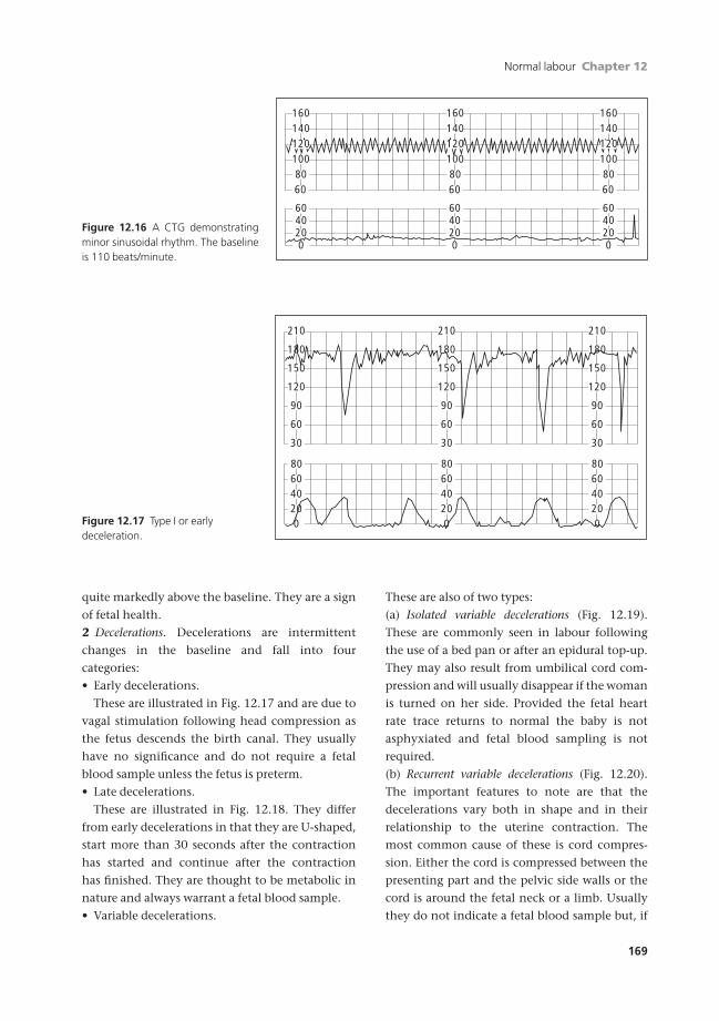
Normal labour Chapter 12
169
quite markedly above the baseline. They are a sign
of fetal health.
2 Decelerations. Decelerations are intermittent
changes in the baseline and fall into four
categories:
• Early decelerations.
These are illustrated in Fig. 12.17 and are due to
vagal stimulation following head compression as
the fetus descends the birth canal. They usually
have no significance and do not require a fetal
blood sample unless the fetus is preterm.
• Late decelerations.
These are illustrated in Fig. 12.18. They differ
from early decelerations in that they are U-shaped,
start more than 30 seconds after the contraction
has started and continue after the contraction
has finished. They are thought to be metabolic in
nature and always warrant a fetal blood sample.
• Variable decelerations.
These are also of two types:
(a) Isolated variable decelerations (Fig. 12.19).
These are commonly seen in labour following
the use of a bed pan or after an epidural top-up.
They may also result from umbilical cord com-
pression and will usually disappear if the woman
is turned on her side. Provided the fetal heart
rate trace returns to normal the baby is not
asphyxiated and fetal blood sampling is not
required.
(b) Recurrent variable decelerations (Fig. 12.20).
The important features to note are that the
decelerations vary both in shape and in their
relationship to the uterine contraction. The
most common cause of these is cord compres-
sion. Either the cord is compressed between the
presenting part and the pelvic side walls or the
cord is around the fetal neck or a limb. Usually
they do not indicate a fetal blood sample but, if
160140
1008060
1601401201008060
160140
100120 120
8060
6040
0
6040
020 20
6040
020
806040
0 0 020
80604020
80604020
210
150
120
90
60
30
210
60
30
210
150
120
90
150
180 180180
120
90
60
30
Figure 12.16 A CTG demonstratingminor sinusoidal rhythm. The baselineis 110 beats/minute.
Figure 12.17 Type I or early deceleration.
AMI12 6/9/04 5:00 PM Page 169
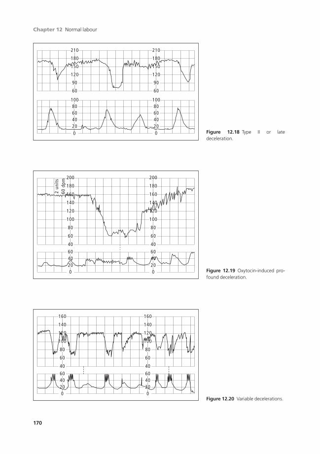
Chapter 12 Normal labour
170
60
020
6040 40
020
200
180
140
160
120
100
80
60
40
200
180
160
100
120
140
80
60
40
2 un
its
60 d
pm
160
140
120
100
60
80
40
160
140
120
100
60
80
40
6040
020
6040200
Figure 12.19 Oxytocin-induced pro-found deceleration.
Figure 12.20 Variable decelerations.
210
180
150
120
90
60
210
180
150
120
90
60
100806040
020
100806040
020
Figure 12.18 Type II or late deceleration.
AMI12 6/9/04 5:00 PM Page 170

Normal labour Chapter 12
171
associated with meconium or a change in the
baseline heart rate, one should be performed.
Passage of meconium
Stimulation of the vagus in utero causes the fetal
gut to contract and the anal sphincter to relax so
that meconium (fetal stool) is passed into the am-
niotic fluid. Meconium is made up of swallowed
cells in late pregnancy and alimentary tract cells,
all of which are stained with bile.
With a normal fetal heart rate trace, the fetus is
unlikely to be hypoxic, but if the fetal heart rate
trace is abnormal when meconium is passed, then
a fetal blood sample (FBS) should be performed.
Fetal blood sample
Fetal blood sampling is a diagnostic test for fetal
acidosis. A bead of blood is taken from the fetal
scalp and the pH and base deficit can be measured.
During a uterine contraction:
• Maternal blood flow to the intervillous space is
vastly reduced or may even cease.
• Passage of O2 from the mother to the fetus is
reduced and thus the fetus may become hypoxic.
• The fetus withstands these periods of hypoxia
by employing anaerobic metabolism. To do this,
the fetus must mobilize glycogen from liver and
muscle stores to produce glucose as an energy
source.
• Anaerobic metabolism results in the production
of large amounts of lactic acid and an increase
in the arterial CO2. In normal circumstances, the
rise in arterial CO2 is buffered, mostly by fetal
bicarbonate.
• Between contractions the lactic acid and the
buffered CO2 are passed back to the mother who
excretes them.
• If glycogen stores are poor (the preterm or those
with SGA), other sources of energy are required for
anaerobic metabolism. These produce more
CO2 and more lactic acid and thus fetal buffering
systems become overloaded. This results in a grad-
ual fall of pH; the fetus demonstrates a metabolic
acidosis.
If uterine activity is too frequent or sustained,
then blood flow to the fetus may be impaired for
a long space of time and this, again, will result
in a metabolic acidosis with an increasing base
deficit.
Figure 12.21 illustrates the mechanism by which
the fetal scalp sample is acquired. The fetal scalp is
punctured with a 2mm guarded blade and the
blood is aspirated into a capillary tube.
The pH results are interpreted as follows:
• pH >7.25: normal.
• pH 7.20–7.25: pre-asphyxia.
• pH <7.20: asphyxia.
• Base deficit <6.0mEq/l: normal.
• Base deficit 6.1–7.9mEq/l: pre-asphyxia.
• Base deficit >8.0mEq/l: asphyxia.
In obstetric practice it is common to use the term
asphyxia but what is truly meant is a metabolic
acidosis.
If a fetus has a pH of <7.20 and a base deficit
>8.0mEq/l he should be considered for delivery
by the most appropriate route. Fetuses that
demonstrate pre-asphyxia and are in the second
stage may be allowed normal delivery but only if
this is imminent.
The scalp pH only reflects the state of the fetus at
the time of the sample; the base deficit reflects a
slower change, and therefore is a longer predictor.
If the fetal heart rate trace continues to be ab-
normal, then the fetal blood sample should be
repeated hourly or the baby delivered.
Figure 12.21 Fetal blood samples can be taken from thescalp through an amnioscope.
AMI12 6/9/04 5:00 PM Page 171

Chapter 12 Normal labour
172
Home deliveries
Until this century, in the UK the place of birth was
most usually the home. Hospital deliveries started
in the mid-eighteenth century for charitable rea-
sons to help the single mothers and poor women
with unsuitable conditions at home. It has grown
gradually this century from 2% in 1900 to 98% in
1990.
Home deliveries had reduced to about 1% by
1986, but are slightly increasing to a figure of 2% in
2001 varying around the UK from 0–20% (see Fig.
12.22).
The drift to hospitalization occurred:
• As part of the fashion of using a hospital for more
medical managements.
• For safety. In the isolation of the home, it would
be difficult to care for an emergency:
(a) PPH.
(b) Delayed onset of baby breathing.
(c) Shoulder dystocia.
Against this, the advantages of home delivery are:
• Familiar surroundings for the woman.
• More relaxation because of confidence at home.
• Would probably know the midwife who was de-
livering her.
• More family members could attend at the birth
and afterwards.
The slight increase in home deliveries means
that a community service must be kept going. Two
midwives attend each home delivery and these are
usually more senior than those working in the hos-
pitals. A general practitioner can be called in an
emergency. If the woman has to be transferred to
hospital, the use of an ambulance with para-
medics, skilled in resuscitation, must be obtained.
The future of the hospital/domiciliary debate
could be helped by:
• The use of a birthroom in the hospital, away
from the main delivery suite. If delivery is normal
and all goes well, they can return home a few hours
later and so seem to have never really entered the
hospital.
• Use of formal DOMINO (Domestic In and Out)
services.
• Reduce the regimentation of hospital.
• Reduce the noise of the wards.
• Provide clean wards, enough linen and
lavatories.
• Get the woman back home early on day one or
two.
0
(%)
10
20
30
40
19601954 1970 1980 1990
Figure 12.22 Percentage of home deliveries (England andWales) 1954–1993.
Self-assessment
12.1 Put the following sentences into the correct order to describe the passage of the fetus through the birth canal.(a) The fetal head engages in a transverse position.(b) The fetal head extends round the symphysis pubis.(c) The fetal head flexes as it descends into the birth canal.(d) The fetal head restitutes to a transverse position.(e) The fetal head most commonly rotates through 90° so that the occiput becomes anterior as it reaches the
levator ani.12.2 Which of the following features of a cardiotocograph (CTG) would be considered non-reassuring in labour?
(a) Baseline variability of 5–15 beats per minute.(b) Early decelerations in the second stage of labour.(c) Late decelerations in the second stage of labour.(d) A fetal heart rate of 170 beats per minute.(e) Accelerations.
AMI12 6/9/04 5:00 PM Page 172

Normal labour Chapter 12
173
Self-assessment Continued
12.3 Which of the following are indications for continuous electronic fetal monitoring (EFM)?(a) A woman at term in spontaneous labour who ruptures her membranes and has meconium stained liquor.(b) A woman at term in spontaneous labour who ruptures her membranes and has clear liquor.(c) A woman at term in spontaneous labour who has had a deceleration detected using a sonicaid every
30 minutes.(d) A woman who ruptured her membranes 6 hours ago, clear liquor and is in spontaneous labour.(e) A woman at term in spontaneous labour who has had a previous Caesarean section.
12.4 In normal labour which of the following statements are true?(a) Uterine contractions are generated in the lower segment of the uterus.(b) The rate of cervical dilatation should be greater than 1cm/hour.(c) The fetal heart should be checked every 15 minutes in the first stage of labour with a sonicaid.(d) The second stage of labour should not exceed 2 hours in duration in a woman with an epidural.(e) An episiotomy should be performed in the majority of women.
AMI12 6/9/04 5:00 PM Page 173
174
Dysfunctional uterine action
Prolonged labour is more common in primigravi-
dae and may be due to primary or secondary
myometrial dysfunction or to malpresentation
of the fetus, e.g. occipitoposterior position. The
progress of labour should be monitored on a
partogram. Figure 12.4 (p. 159) illustrates the nor-
mal rate of cervical dilatation from the start of
labour. During the first 8 hours in primiparous
women there is minimal change in the cervical di-
latation but effacement (shortening and softening)
occurs —the latent phase. Effacement is the taking
up of the cervix, merging with the lower segment;
eventually there is no length to the cervical
canal.
Three abnormalities of labour may be
recognized.
Prolonged latent phase (PLP)
This is a rare abnormality and occurs almost exclu-
sively in primigravidae. Figure 13.1 illustrates this
together with the possible outcome. Aetiological
factors are:
• The wrong diagnosis of labour.
• An abnormal or high presenting part.
• Premature rupture of the membranes.
• Idiopathic: cervical dystocia.
(a) Primary. Failure of a ground substance of the
cervix to soften in late pregnancy.
(b) Secondary. Previous operations on the cervix
causing fibrosis.
Management1 Women who present with regular uterine
activity should be assessed by vaginal examination;
if the cervix is long and closed, they may be in early
labour or not in labour. A short trace cardiotoco-
graph (CTG) should be carried out to ensure fetal
well-being and the uterus should be carefully
palpated.
2 The woman should be allowed to walk around
or to sit comfortably. She should be re-examined
again 4 hours later if the contractions persist.
• If labour has ceased the woman should go home.
• If labour continues and pain relief is required,
then it should be given.
• If the cervix continues to efface but not dilate
and progress falls more than 2 hours to the right of
the partogram the membranes should be ruptured
(artificial rupture of membranes, ARM) and labour
stimulated by Syntocinon.
• In 85% of cases, labour will progress rapidly and
will reach a normal active phase.
• In 15% of cases, adequate uterine activity fails to
cause cervical dilatation. If, after 4–8 hours of Syn-
tocinon, the cervix is not further dilated, then a
Caesarean section should be performed.
Prolonged latent phase is primarily of primi-
gravidae as the multigravidous cervix tends to
efface and dilate at the same time.
Chapter 13
Abnormal labour
AMI13 6/9/04 5:02 PM Page 174
Abnormal labour Chapter 13
175
Cervix (cm)[Plot X]
Descent of head[Plot 0]
Mark fifths feltper abdomen
HoursTime
(24 hr clock)
109876543210
Alert line Action line
1 2 3 4 5 6 7 8 9 10 11 12 13 14 15 16 17 18 19 20 21 22 23 24
Aetive phase
Alert zone
Action zone
1
3
2
ARMSynto
03 04 05 06 07 08 09 10 11 12 13 14 15 16 17 18 19 21 22 23 24 01 0220
Latent phase
Cervix (cm)[Plot X]
Descent of head[Plot 0]
Mark fifths feltper abdomen
HoursTime
(24 hr clock)
109876543210
Alert line Action line
1 2 3 4 5 6 7 8 9 10 11 12 13 14 15 16 17 18 19 20 21 22 23 24
Aetive phase
Latent phase
Alert zone
Action zone
2
2
1
ARM Synto
03 04 05 06 07 08 09 10 11 12 13 14 15 16 17 18 19 21 22 23 24 01 0220
1
Figure 13.1 Prolonged latent phase in labour and possible outcomes: 1 and 2, vaginal delivery; 3, Caesarean section. ARM,artificial rupture of membranes; Synto, syntocinon infusion.
Figure 13.2 Secondary arrest of cervical dilatation and outcomes: 1, vaginal delivery; 2, Caesarean section. ARM, artificialrupture of membranes; Synto, syntocinon infusion.
Secondary arrest of cervical dilatation
The woman enters the active phase of labour,
reaches 5–7cm dilatation and then the cervix stops
dilating (Fig. 13.2). Uterine contractions have be-
come less frequent and may even stop.
• The fetal head engages in the occipitotransverse
position and, if it is well flexed and asynclitic, will
undergo rotation in the mid-cavity to the direct
occipitoanterior position. Poor flexion leads to
failure of rotation in the mid-cavity; this leads to
persistent occipitotransverse position.
• Syntocinon i.v. leads to regular, coordinated
uterine contractions that initially cause the fetal
head to flex. In most cases (85%) this allows the
head to rotate so that a spontaneous vaginal
delivery will occur.
• If Syntocinon administration over 4 hours
(multigravida) or 8 hours (primigravida) fails to
lead to further cervical dilatation, a Caesarean
section should be carried out for relative
cephalopelvic disproportion (CPD). This occurs in
about 15% of cases.
• This is a benign condition as far as the fetus is
concerned and very rarely leads to fetal distress.
Primary dysfunctional labour
It is defined as slow progress after the onset of
established labour and is the most worrying of
the abnormalities of labour for it can lead to:
• Fetal distress in a well-grown or a large baby.
• Prolonged labour leading to an increase in ma-
ternal fear and anxiety.
• Incoordinate uterine activity which increases
maternal pain.
• Maternal dehydration which leads to maternal
acidosis.
AMI13 6/9/04 5:02 PM Page 175

Chapter 13 Abnormal labour
176
The release of catecholamines stimulates uterine
activity to arise from the lower segment. This
means that the fundus and the lower uterine seg-
ment contract against each other and the cervix
fails to dilate or dilates very slowly.
Maternal dehydration and acidosis lead to hy-
drogen ions competing with calcium (the final
common pathway for smooth muscle contraction)
and further dysfunctional uterine activity occurs.
The causes are:
• A malpresentation such as a brow.
• Occipitoposterior position.
• Relative CPD: which means that the fetus is only
just small enough to pass through the pelvis but, if
all goes well, it will succeed. If there is poor flexion
or rotation, delay occurs.
• Macrosomia.
Once diagnosed, the dysfunction is treated with
Syntocinon. In very few cases the rate of cervical
dilatation returns to normal, but often the rate of
dilatation can be increased. The outcome is:
• Spontaneous vaginal delivery (15%).
• Caesarean section for fetal distress (50%).
• A vaginal instrumental delivery (35%). Care
should be taken because, even if full dilatation of
the cervix is obtained, the fetus may still be high in
mid-cavity. This could mean a difficult, rotation
forceps delivery.
Shoulder dystocia
Shoulder dystocia is an obstetric emergency. It oc-
curs when the shoulders do not spontaneously de-
liver after the head. The anterior shoulder becomes
trapped behind or above the symphysis pubis whilst
the posterior shoulder may be in the hollow of the
sacrum or above the sacral promontory.
Predisposing factors• Previous shoulder dystocia.
• Previous baby >4.5kg.
• Big baby clinically or on USS (AC >95th centile).
• Diabetic woman.
• Obese woman (BMI >30kg/m2).
• Secondary arrest in labour augmented by
Syntocinon.
• Prolonged second stage.
Signs• Fetal chin pulls back against the perineum.
• No external signs of restitution.
• Anterior shoulder fails to deliver with
contraction.
Management• Call for help
• Change the maternal position
1 McRoberts manoevre —flatten the bed, retract
the woman’s knees on to her chest as far as pos-
sible. This straightens the sacrum and maximizes
the pelvic diameter. Apply gentle traction. If not
delivered after 30 seconds try the following se-
quence with 30 seconds to try and deliver the
baby with each one.
2 Place woman on all fours (shoulders move to
oblique diameter).
3 Return to supine. External pressure —place
hands, held as for cardiopulmonary resuscita-
tion, on the mother’s abdomen just above the
symphysis pubis and apply pressure.
• Perform an episiotomy if not already done.
• Internal rotation. Place hand behind the anteri-
or shoulder and bring it forward, rotate the shoul-
ders using the posterior shoulder.
• Deliver the posterior arm.
• Symphysiotomy —division of the symphysis
pubis, supporting the hips so they do not separate
too quickly.
Risks
MATERNAL
• Vaginal trauma.
• Bladder/urethral damage particularly if a sym-
physiotomy is performed.
• Psychological trauma.
NEONATAL
• Erb’s palsy from brachial plexus injury.
• Cerebral palsy from hypoxia.
• Fractured humerus/clavicle.
• Neonatal death.
All parents require sensitive and careful debrief-
ing after shoulder dystocia. It is a frightening ex-
perience when a lot of doctors and midwives arrive
AMI13 6/9/04 5:02 PM Page 176
Abnormal labour Chapter 13
177
suddenly, being asked to change positions with
minimal explanation yet knowing there is some-
thing seriously wrong. They may also have to cope
with a baby with physical or mental disability.
Cephalopelvic disproportion
Classically, CPD is classified as follows:
1 Absolute. There is no possibility of a normal vagi-
nal delivery even if the mechanisms of labour are
completely correct. In the Western world, this
condition is extremely rare; it may be due to the
following:
• Fetal hydrocephalus.
• Congenitally abnormal pelvis (such as
Robert’s or Naegele’s pelvis) in which one or
both sacral ala are missing leading to a narrow-
ing of the pelvic inlet.
• A pelvis that has been damaged usually due to
a severe roll-over road traffic accident in youth.
• A pelvis that has been grossly distorted from
osteomalacia.
2 Relative CPD. This means that the baby is
large but would pass through the pelvis if the
mechanisms of labour function correctly. If,
however, the head is deflexed or fails to rotate in
the mid-cavity, then prolonged, abnormal labour
will occur.
The above definitions do not include estimates
of the weight of the baby or X-ray measurements of
the pelvis. CPD can only truly be diagnosed after a
trial of labour. This means awaiting the onset of
spontaneous labour and, if that labour becomes
prolonged and abnormal, stimulating with
Syntocinon as described above.
CPD may be suspected antenatally in women
who are less than 5¢2≤ (1.58m) in height. These
women tend to have a small gynaecoid pelvis but
they often also have small babies. In a cephalic
presentation there is now little evidence that
X-ray pelvimetry or a CT scan helps in manage-
ment. These women should have a trial of labour
and in many cases will deliver vaginally.
All women with a high head at term should have
an obvious cause excluded by an ultrasound exam-
ination. This will diagnose placenta praevia, uter-
ine fibroids, or an ovarian cyst as the cause. In the
absence of these findings, one should suspect that
the cause is CPD.
Head fitting tests and X-ray pelvimetry have
only a small role in the management of
women with a cephalic presentation because the
correct management is the proper use of a trial of
labour.
Malpresentations and malpositions
Breech presentation
IncidenceAt term 2–3%; more in preterm deliveries.
Aetiology• The ratio of amniotic fluid volume to fetal size
may be high, allowing freer movement (e.g. poly-
hydramnios and before 32 weeks).
• Extended legs of the fetus can splint and prevent
flexion of the fetal trunk so stopping further
turning and causing the fetus to stay as a breech
presentation.
• Fetuses in multiple pregnancies may interfere
with each other’s movements.
• Something might be filling the lower segment
(e.g. placenta praevia or fibroids).
• Fetal malformations may prevent cephalic pres-
entation (e.g. hydrocephaly).
Types (Fig. 13.3)
Flexed or extended knee joints.
1 Neither knee joint flexed so that both legs are ex-
tended: a frank breech or extended breech. This is
the commonest presentation.
2 Fully flexed fetus with both knees flexed: a
flexed breech.
3 One leg flexed and the other leg extended: an in-
complete breech.
4 Both hips extended; a footling. Often occurs
with very small babies.
On vaginal examination, the breech presenta-
tion in labour is described according to the relation
of the fetal sacrum to the maternal pelvis (Fig. 13.4).
AMI13 6/9/04 5:02 PM Page 177

Chapter 13 Abnormal labour
178
Diagnosis
ABDOMINAL EXAMINATION
No head is felt at the lower end and a hard, round-
ed knob is ballottable at the upper end of the
uterus.
VAGINAL EXAMINATION
Confirms there is no head in the pelvis.
INVESTIGATIONS
Ultrasound scan confirms the situation.
Management of a breech presentation in pregnancy1 From about 37 weeks onwards external cephalic
version (ECV) is worthwhile trying without
general anaesthesia, provided it is easy to perform.
If the mother is Rh-negative, anti-D immunoglob-
ulin should be given after the first attempt.
• Listen to the fetal heart immediately before
and after the procedure.
• If it works, the woman should be seen
weekly to ensure the fetus stays as a cephalic
presentation.
• If it fails, the woman should be counselled
about the route of delivery.
• Reasons for failure of ECV:
(a) Breech too deeply engaged in pelvis.
(b) Too tense a uterus.
(c) Too tense an abdominal wall.
(d) Fetal abnormality therefore do
(e) Undiagnosed twins an ultrasound.
• Contraindications to doing ECV.
(a) Previous uterine scar from Caesarean sec-
tion (relative).
(b) Hypertension in the mother.
(c) Planned delivery by Caesarean section
anyway.
(d) Ruptured membranes.
(e) Multiple pregnancy.
(f) Antepartum haemorrhage.
2 If ECV does not succeed then the woman should
be advised about the pros and cons of vaginal
breech delivery compared with Caesarean section
(Box 13.1).
3 A standing lateral CT scan of the pelvis should
be done for all primiparous patients and any mul-
tiparous women who have delivered a baby of
Flexedbreech
Extendedbreech
Incompletebreech
Footling
Figure 13.3 Types of breech presentation.
Anterior
Posterior
Left sacroanterior Left sacroposterior
Figure 13.4 Using the fetal sacrum asthe denominator, the position of thebreech presentation is described.
}
AMI13 6/9/04 5:02 PM Page 178
Abnormal labour Chapter 13
179
>3.5kg in the past. An ultrasound scan of the fetus
will establish its estimated weight. In a breech de-
livery the head (the largest and hardest part of the
fetus) is coming last and it is too late to wait and see
if this fits the pelvis. Therefore an estimate of the
chances of delivery has to be made on these CT and
ultrasound measurements. In a cephalic delivery,
the descending head acts as a pelvimeter whereas
in a breech it does not.
4 It is wise to deliver most breech presentations by
41 weeks. If the woman has not gone into spon-
taneous labour before this time then induce or do
an elective Caesarean section.
5 If there is any other variation from normal,
many obstetricians will deliver a breech-
presenting baby by elective Caesarean section at
38–39 weeks.
A recent randomized trial of vaginal versus elec-
tive Caesarean section for breech presentation has
suggested that the latter may be safer for the infant
if an ECV fails. This is truer for the developed world
than the developing world where the perinatal
morbidity and mortality for cephalic vaginal deliv-
eries is higher and the risks of Caesarean section for
the mother are also higher.
Management of a breech presentation in labour
FIRST STAGE
1 Increased risk of early rupture of the mem-
branes. When they do, a vaginal examination
should exclude a prolapse of the cord.
2 An epidural anaesthetic is a good method of
pain relief as the normal delivery can rapidly be
changed to an operative one if necessary (but is not
mandatory).
SECOND STAGE
1 Delivery is by the most senior obstetrician or
midwife available with an anaesthetist and a pae-
diatrician close to the labour ward.
2 A propped up dorsal position of the mother is
the easiest to manage. The labour bed should be ca-
pable of breaking in the middle for delivery of the
baby’s body, so that the mother can assume a litho-
tomy position.
3 The buttocks progress down the birth canal
and, when on the point of crowning, an episio-
tomy may be required. The baby is rotated to
sacroanterior.
4 The baby will often progress as far as the umbili-
cus with the mother’s own expulsive efforts. The
legs are assisted down, especially if extended.
5 Commonly, the arms are flexed across the
chest and so delivery occurs readily with the next
contraction.
6 If the arms are extended they have to be manipu-
lated down.
7 After delivery of the body, it is allowed to hang
and traction may be gently applied to the legs until
the suboccipital region appears under the maternal
pubis.
8 The head is delivered slowly by placing one fin-
ger in the baby’s mouth or gently flexing the head
with forceps, the blades applied to either side of the
fetal head from the front of the body which is held
up by an assistant. The face is delivered over the
mother’s perineum and the nose and mouth are
cleared of mucus and liquor, allowing the baby to
breathe. The rest of the head is slowly delivered,
not allowing any sudden decompression which
could result in pressure alterations inside the skull
and so cause intracerebral venous bleeding.
THIRD STAGE
1 Syntometrine is given with the delivery of the
head for there is an increased risk of PPH.
Box 13.1 The pros and cons of vaginal breech delivery
RequiresPelvic inlet >11cm (AP)Pelvic outlet >11cm (transverse)Well curved sacrumEstimated fetal weight <3.5kg
Pro ConØ Maternal morbidity ≠ Risk of fetal intrapartumØ Maternal mortality hypoxia ¥3
(rare) ≠ Risk of head entrapment≠ Risk of intracranial damageMay lead to emergency CS
done in less favourablecircumstances
AMI13 6/9/04 5:02 PM Page 179

Chapter 13 Abnormal labour
180
2 The placenta is delivered as described in normal
labour.
Caesarean sectionThis should be done if vaginal delivery is consid-
ered too hazardous because:
• Mild pelvis contraction.
• Fetus thought to be over 3.5kg.
• Fetus in unfavourable attitude.
• Multiple pregnancy if the first twin is a breech.
• Other complications, e.g. pre-eclampsia or
diabetes.
• Non-descent of buttocks in labour.
• Failure of progress in labour.
Risks to the fetus of breech deliveryPerinatal mortality in all breech deliveries is two
or three times that of cephalic presentations but
this is made up mainly of premature births (26–30
weeks). Mature breech deliveries (36+ weeks) in
reputable centres have no higher risk than mature
cephalic deliveries. Hence the reasons for
mortality are:
• Prematurity.
• Intracranial damage: subdural and intracranial
haemorrhage often after too rapid delivery of the
head.
• Rarely hypoxia. This may be:
(a) Before delivery (prolapsed cord).
(b) At the time of delivery (too slow delivery of
the head).
Shoulder presentation (transverse lie)
Incidence0.3% of all deliveries.
AetiologyAs for other malpresentation but most commonly:
1 Polyhydramnios causing an increased ratio of
fluid to fetus.
2 Something preventing the engagement of the
head in the pelvis:
• Placenta praevia.
• Fibroids.
• Contracted pelvis.
3 Abnormal shape of uterus (subseptate or arcuate
uterus).
4 Second twin.
5 Grand multiparity (5+).
Diagnosis1 Abdominal examination —the head is in one
flank and the buttocks in the other. Commonly,
the fetus can be rotated to a cephalic presentation
quite readily but reverts back to a transverse
position.
2 Vaginal examination —the pelvis is empty of
presenting parts.
3 Investigation: ultrasound scan confirms
diagnosis.
Management of transverse lie in pregnancy and labour1 Before 36 weeks, ECV may be attempted or the
woman referred back to the following week’s
clinic. The position is usually self-curing.
2 Past 37 weeks in a multiparous patient, and after
38 weeks in a primiparous one, admission to hos-
pital should be advised, where ECV is attempted
each day.
3 Should the woman go to term with the fetus still
in a transverse position, management may be by
either of the following:
• A stabilizing induction: ECV is done in the
labour ward. The fetal head is held over the brim
of the mother’s pelvis and high membrane rup-
ture is performed. Amniotic fluid escapes and the
head often sinks into the pelvis. Labour follows
in the normal fashion.
• An elective Caesarean section. In the Western
world this may be the safer line of treatment
for the fetus since it cuts down the risks of
prolapsed cord during labour, but it does leave
the mother with a scarred uterus for future preg-
nancies and an increased risk of postpartum
problems.
4 Occasionally a woman is admitted in mid or late
labour with a transverse lie. This would lead to an
impacted shoulder presentation, the folded fetus
having been driven a varying amount down the
pelvis, depending on how far labour has gone.
Treatment must be by immediate Caesarean
AMI13 6/9/04 5:02 PM Page 180

Abnormal labour Chapter 13
181
section even if the fetus is dead because of the risk
of uterine rupture.
Occipitoposterior positions
The fetal head usually engages in the pelvic brim in
the occipitotransverse position (long axis of head
fitting into maximum diameter of bean-shaped
pelvic brim). When labour starts, the head is driv-
en down the birth canal and rotates.
1 80% rotate forward through 90° to an occipi-
toanterior position.
2 15% undergo long internal rotation through
270° to become occipitoanterior having gone
through directly occipitoposterior on the way.
3 3% rotate back 90° to a directly occipitoposterior
position. These may deliver face to pubis.
4 2% stay in the transverse and descend in this
position. A minority of these might rotate on the
perineum but most end up in transverse arrest.
AetiologyPelvis. Flat sacrum with loss of pelvic curve and so
loss of room for rotation.
Uterus. Poor or disorganized uterine contractions
do not push the fetal head down and so there is no
impetus to rotate.
Head. Poor flexion so that larger diameters pres-
ent (suboccipitofrontal >10.5cm).
Analgesia. Epidural analgesia causes pelvic floor
relaxation. This allows the gutter of the levator ani
muscles to become lax so not directing the occiput
anteriorly. This is associated with lack of fetal head
rotation.
Diagnosis
PREGNANCY
Occasionally, by abdominal palpation when in a
cephalic presentation, the back cannot be felt in
the flank but fetal limbs can be felt all over the
front of the uterus. The head is often not engaged
after the time it would be expected to be.
LABOUR
• Abdominal palpation as above.
• Vaginal examination feeling the sutures and
fontanelles. Both anterior and posterior
fontanelles can be felt (deflexion) and the -
shaped posterior fontanelle is in the posterior
quadrant of the pelvis.
Management in pregnancyLeave alone.
Management in labourLaissez-faire. Await events for many will rotate
spontaneously. Prepare for a longer labour because:
• Pelvis may be minimally contracted or sacrum
slightly flattened.
• Incoordinate uterine contractions.
• Deflexed fetal head.
Therefore:
1 Watch progress by both:
• Abdominal assessment of engagement and
descent of fetal head.
• Vaginal assessment:
(a) Head in relation to ischial spines.
(b) Rotation of head.
(c) Dilatation of cervix which is often poorly
applied to the head.
(d) Check no prolapse of cord by vaginal
examination straight after membranes rupture.
2 Women often wish to push before the cervix is
fully dilated. If the occiput is posterior there is
extra pressure on the sacrum and rectum. Frequent
vaginal examinations are needed to make accurate
assessments of the real dilatation of the cervix and
the progress of labour.
3 Watch maternal condition. Especially
remember:
• Labour will be long, so maintain morale.
• Pain relief should be thorough—epidural
anaesthesia is good in this situation. If such re-
gional anaesthesia is unavailable many would
use morphine or diamorphine for this problem.
• No food and little fluids by mouth (general
anaesthetic may be wanted). Give i.v. fluids.
4 If head stays directly occipitoposterior, delivery
may occur spontaneously but, since larger diam-
eters are passing through the birth canal, the
mother will have to work harder and a gener-
ous episiotomy may be required. Face-to-pubis
delivery will occur.Y
AMI13 6/9/04 5:02 PM Page 181

Chapter 13 Abnormal labour
182
5 If head stays in occipitotransverse position it
will not deliver spontaneously. It must be rotated
to deliver and this will require good analgesia,
maybe epidural or general anaesthesia. A rotation-
al delivery is contraindicated if there is fetal dis-
tress and a Caesarean section should be performed.
Rotation and delivery may be by:
• Manual rotation to the occipitoanterior posi-
tion and subsequent forceps delivery.
• Kielland’s straight forceps rotation and subse-
quent delivery. These forceps have no pelvic
curve.
• Vacuum extraction. This applies only a linear
pull on the fetal head so that any rotation can
occur as determined by the pelvic muscles and
bones.
6 Give i.v. Syntometrine with the crowning of the
head for the risks of PPH are greatly increased. De-
liver the placenta promptly after the baby is born.
7 Be prepared to repair the rather large episiotomy
quickly.
8 If the head does not rotate then a Caesarean sec-
tion is indicated. This can be difficult as the head is
so low in the vagina.
Results
MOTHER
Following the operative delivery and the bigger
episiotomy, vaginal and vulval oedema and
haematomata are more frequent.
BABY
Because of the longer labour and high incidence
of operative delivery, perinatal mortality and
morbidity are increased. The mortality is due to
hypoxia and birth trauma. The morbidity is
from these and the results of intracranial
haemorrhage.
Face presentation
As the fetal head gets driven down the birth
canal, the front of the head can become ex-
tended (Fig. 13.5). Distinguish from face-to-pubis
delivery.
Incidence0.3% of all deliveries.
Aetiology1 Lax uterus, multiple pregnancy,
polyhydramnios.
2 Deflexed fetal head.
3 Shape of fetal head:
• Dolichocephalic (long head).
• Anencephalic (no cranium).
MechanismHead descends with face leading. Chin
(mentum) used as denominator to determine
rotation.
85% engage in the mentotransverse (submento-
bregmatic diameter —10cm). With descent, most
rotate to mentoanterior on the pelvic floor, the
fetal chin coming behind the maternal pubis. After
further descent, the chin can escape from under
the lower back of the pubis and the head is then de-
livered over the vulva by flexion.
Up to this point, the mechanisms of flexion/ex-
tension of the fetal head are the reverse of those
with a vertex presentation. After delivery of
the head, however, the external rotations are the
same allowing the fetal shoulders to negotiate the
pelvis.
A few face presentations rotate from the trans-
verse to mentoposterior, so that the fetal chin is in
Figure 13.5 Face presentation. Well engaged in the mentolateral position.
AMI13 6/9/04 5:02 PM Page 182

Abnormal labour Chapter 13
183
the curve of the mother’s sacrum; the fetal occiput
and back are crushed into each other behind the
pubic bone. Further descent is unlikely for the
head cannot extend further and so cannot negoti-
ate the forward curve of the birth canal and Cae-
sarean section is needed.
DiagnosisRarely made before labour and of little significance
if it is.
Abdomen• Longitudinal lie with body nearer to mid-axis of
uterus.
• More head felt on the same side as the back.
Vaginal examination• Do not expect the face to feel like the newborn
baby’s face. Oedema always obscures facial parts.
• Supra-orbital ridges lead to the bridge of the
nose.
• Mouth has hard gums in it and may suck on the
examining finger.
Management
IN PREGNANCY
• Await events.
• Membranes may rupture early (examine
vaginally to exclude prolapsed cord).
• Check that pelvis is adequate and that fetus is
not oversized. If either, consider Caesarean section
for face presentation in labour has a higher risk.
• Check with ultrasound that the fetus is not an
anencephalic for this might alter management.
IN LABOUR
• If anterior rotation to mentoanterior, a longer
labour but spontaneous delivery will probably
occur (90%).
• If head stays in mentotransverse, either manual
rotation to mentoanterior and forceps extraction,
or Kielland’s forceps rotation and extraction or de-
liver by Caesarean section.
• If face rotates posteriorly, this is impossible to
deliver vaginally. Hence, perform a Caesarean
section.
Results
MOTHER
Higher morbidity associated with operative
delivery.
BABY
Higher mortality:
• Abnormalities incompatible with life
(anencephaly).
• In the normal, hypoxia and cerebral congestion.
Brow presentation
A very poorly flexed head may present the largest
diameter of the skull: mentovertex (13cm) (Fig.
13.6).
Incidence0.1% of all deliveries.
DiagnosisRarely made before labour and of little significance
if it is.
Abdomen• Head feels big.
• Not well engaged.
• Groove between occiput and back. Head felt on
both sides of fetus.
Figure 13.6 Brow presentation with mentovertex diameterpresenting.
AMI13 6/9/04 5:02 PM Page 183

Chapter 13 Abnormal labour
184
Vaginal examination• Anterior fontanelle presents.
• Supraorbital ridges and base of nose can be felt at
edge of field.
Management
IN PREGNANCY
• Await events. No point in trying to convert to
more favourable presentation.
• Membranes may rupture early (examine
vaginally to exclude prolapsed cord).
IN LABOUR
• If diagnosed early, await events for some convert
spontaneously to face (by further extension) or
vertex (by flexion).
• If presentation persists, it will be impossible to
deliver vaginally. Hence deliver by Caesarean
section.
• If fetus is dead or there is hydrocephaly, the de-
structive operation of perforation of head and vagi-
nal extraction is possible provided the operator is
skilled in these arts, but in the Western world these
are a diminishing number.
Results
MOTHER
Higher morbidity associated with operative
delivery.
BABY
Because of wider use of Caesarean section, morbid-
ity and mortality rates are low.
Induction of labour
DefinitionA planned initiation of labour.
IncidenceVaries with the population and the type of obstet-
ric cases seen; in the UK between 5% and 25%.
Indications1 Maternal disease:
• Existing before pregnancy, e.g. diabetes.
• Occurring in pregnancy, e.g. pre-eclampsia.
2 Fetal disease, e.g. Rh disease.
3 Fetuses at risk from reduced placental perfusion,
being SGA.
In the UK the most common indications are:
• Post-maturity (or more strictly post-dates),
T+10–14.
• SGA.
• Maternal disease.
• Rh incompatibility.
• Fetal death or abnormality.
In addition, there are several softer indications
which obstetricians commonly employ for which
there is little or no scientific basis. These are:
• Poor past obstetric history.
• A pregnancy resulting from infertility treatment.
• Recurrent unexplained APH.
• At the woman’s, or her partner’s, wish (to be
avoided).
Prior to undertaking an induction the woman
should be examined vaginally to determine the
Bishops score (Table 13.1) and a membrane sweep
performed. This can be done if the cervix admits a
finger (>1cm dilated). A circular motion round the
edge of the internal os releases prostaglandins.
70% of women at term will go into spontaneous
labour within 48 hours so avoiding an induction.
The Bishop’s score is a weighted means of assessing
how likely it is that the woman will go into labour.
Women with a Bishop’s score of >6 are considered
favourable and failed induction rates are usually
less than 1%.
The commonest method of induction of labour
in the UK is a combination of medical and surgical
means. The usual system is:
Table 13.1 The Bishop’s score.
Cervix 0 1 2
Dilatation (cm) 0 1–2 3–4
Consistency Firm Medium Soft
Length (cm) >2 1–2 <1
Position Posterior Mid Anterior
Station of head 3 2 1
above ischial
spines (cm)
AMI13 6/9/04 5:02 PM Page 184
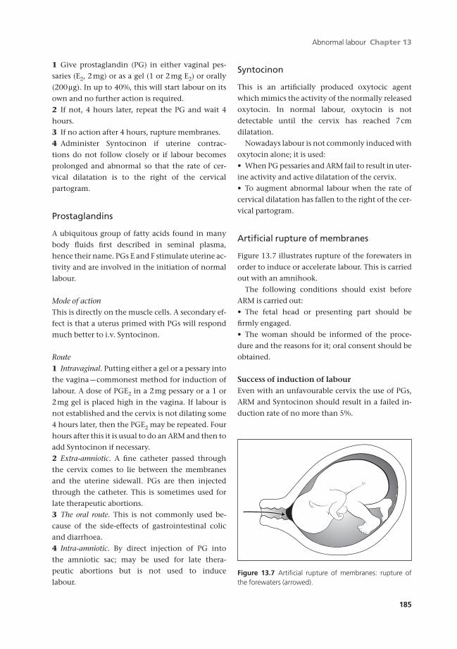
Abnormal labour Chapter 13
185
1 Give prostaglandin (PG) in either vaginal pes-
saries (E2, 2mg) or as a gel (1 or 2mg E2) or orally
(200mg). In up to 40%, this will start labour on its
own and no further action is required.
2 If not, 4 hours later, repeat the PG and wait 4
hours.
3 If no action after 4 hours, rupture membranes.
4 Administer Syntocinon if uterine contrac-
tions do not follow closely or if labour becomes
prolonged and abnormal so that the rate of cer-
vical dilatation is to the right of the cervical
partogram.
Prostaglandins
A ubiquitous group of fatty acids found in many
body fluids first described in seminal plasma,
hence their name. PGs E and F stimulate uterine ac-
tivity and are involved in the initiation of normal
labour.
Mode of actionThis is directly on the muscle cells. A secondary ef-
fect is that a uterus primed with PGs will respond
much better to i.v. Syntocinon.
Route1 Intravaginal. Putting either a gel or a pessary into
the vagina —commonest method for induction of
labour. A dose of PGE2 in a 2mg pessary or a 1 or
2mg gel is placed high in the vagina. If labour is
not established and the cervix is not dilating some
4 hours later, then the PGE2 may be repeated. Four
hours after this it is usual to do an ARM and then to
add Syntocinon if necessary.
2 Extra-amniotic. A fine catheter passed through
the cervix comes to lie between the membranes
and the uterine sidewall. PGs are then injected
through the catheter. This is sometimes used for
late therapeutic abortions.
3 The oral route. This is not commonly used be-
cause of the side-effects of gastrointestinal colic
and diarrhoea.
4 Intra-amniotic. By direct injection of PG into
the amniotic sac; may be used for late thera-
peutic abortions but is not used to induce
labour.
Syntocinon
This is an artificially produced oxytocic agent
which mimics the activity of the normally released
oxytocin. In normal labour, oxytocin is not
detectable until the cervix has reached 7cm
dilatation.
Nowadays labour is not commonly induced with
oxytocin alone; it is used:
• When PG pessaries and ARM fail to result in uter-
ine activity and active dilatation of the cervix.
• To augment abnormal labour when the rate of
cervical dilatation has fallen to the right of the cer-
vical partogram.
Artificial rupture of membranes
Figure 13.7 illustrates rupture of the forewaters in
order to induce or accelerate labour. This is carried
out with an amnihook.
The following conditions should exist before
ARM is carried out:
• The fetal head or presenting part should be
firmly engaged.
• The woman should be informed of the proce-
dure and the reasons for it; oral consent should be
obtained.
Success of induction of labourEven with an unfavourable cervix the use of PGs,
ARM and Syntocinon should result in a failed in-
duction rate of no more than 5%.
Figure 13.7 Artificial rupture of membranes: rupture ofthe forewaters (arrowed).
AMI13 6/9/04 5:02 PM Page 185

Chapter 13 Abnormal labour
186
Management of women who have a failed in-
duction depends upon the obstetrician’s opinion,
which may be either of the following:
1 The need for induction indicated a need for de-
livery and therefore failure of induction should
lead to a Caesarean section.
2 The indications for induction were border-
line and it is therefore reasonable to stop the in-
duction process and attempt it again the next day.
This can only be carried out if the membranes have
not been ruptured. It is not to be encouraged as it
leads to the woman’s loss of confidence in the
method.
Risks of induction• Uterine hyperstimulation may lead to fetal dis-
tress and so to a Caesarean section.
• Prolonged rupture of the membranes may in-
crease the risk of intrauterine infection.
• Prolonged labour may lead to a Caesarean
section.
• Women whose labours are induced have a high-
er incidence of Caesarean section (a risk factor of
times three). Often this is due to the reason for the
induction, e.g. an SGA fetus, but in many cases it is
due to prolonged labour.
Preterm labour
DefinitionLabour occurring at <37 completed weeks’
gestation.
Incidence• 6% of deliveries occur before 37 weeks’
gestation.
• 2% of deliveries occur before 34 weeks’
gestation.
PrognosisThis depends upon:
• The availability of a neonatal intensive care unit.
All infants born at <30 weeks’ gestation should be
transferred to a hospital that contains a neonatal
intensive care unit (level 3) if time and
maternal/fetal condition allow.
• The gestational age and birth weight. The peri-
natal team at a typical obstetric and neonatal com-
bined unit usually achieves a 50% survival rate at
26 weeks of gestation.
• The condition of the baby at birth. Asphyxiated
infants are more likely to die later from respiratory
distress syndrome (RDS).
• Immediate neonatal management.
• The use of antenatal steroids to improve the ma-
turity of the fetal lungs and reduce the risk of intra-
ventricular haemorrhage (IVH) in fetuses of less
than 37 weeks’ gestation.
DiagnosisHalf the women who present with painful contrac-
tions before 37 weeks’ gestation will stop spon-
taneously. Conversely preterm labour may be
insidious. The following plan is therefore
recommended:
1 Look for a cause for preterm labour (Box 13.2).
2 If membranes are intact, a vaginal examination
should be performed.
3 The fetal heart rate and uterine activity should
be electronically recorded continuously.
4 Repeat the vaginal examination 2 hours later if
there are more than two contractions every 10
minutes. Change in cervical effacement or dilata-
tion confirms preterm labour.
5 Check fibronectin levels in cervical fluid; eleva-
tion may indicate imminent labour.
Box 13.2 Causes of preterm labour
Previous pre-term labourPremature rupture of the membranesMultiple pregnancyPolyhydramniosAntepartum haemorrhageFetal deathBacterial vaginosisMaternal pyrexia (UTI and other infections)Uterine abnormalitiesCervical incompetence
AMI13 6/9/04 5:02 PM Page 186

Abnormal labour Chapter 13
187
Principles of management of ongoing labour (intact membranes)1 Full electronic monitoring is mandatory.
2 Arrange in utero transfer if neonatal intensive
care facilities are unavailable.
3 Give tocolytics for 48 hours to allow steroid
therapy to mature fetal lungs.
Tocolysis
There is no convincing statistical evidence that to-
colytic agents such as b-agonists or ritodrine use-
fully prolong pregnancy. However, at <4cm
dilatation they may delay delivery for 24–48 hours
in order to allow time for steroids to act or for the
woman to be transferred to a delivery site with a
neonatal intensive unit.
Contraindications
ABSOLUTE
• Thyroid disease.
• Cardiac disease.
• Severe hypertension (>160/110mmHg).
• Sickle cell disease.
• Chorioamnionitis.
• Intrauterine death.
RELATIVE
• Advanced labour, more than 4cm cervical dilata-
tion.
• APH.
• Maternal diabetes mellitus.
Side effectsThese include
• Tachycardia: treatment should be stopped if the
maternal pulse rate exceeds 120/minute.
• Hyperglycaemia: beta agonists are diabetogenic
as are steroids. Since steroids are usually given at the
same time as tocolytics the maternal blood glucose
should be checked 2 hourly and a sliding scale of in-
sulin started if the blood sugar exceeds 9mmol/l.
• Pulmonary oedema: this is due to fluid overload
and tachycardia. It can be avoided by giving the to-
colytic through a syringe pump to reduce the vol-
ume of colloid given as well as ensuring the woman
does not have a prolonged tachycardia of
>120/minute.
Recently a new tocolytic —Atosiban —has been
introduced. This is an oxytocin antagonist. Other
agents that have been used include NSAIDs (in-
domethacin) and Glyceryltrinitrite (GTN). NSAIDs
may cause oligohydramnios and closure of the
patent ductus arteriosus in the fetus if used for
more than 48 hours.
Steroid therapy
Maternal steroids have been shown to reduce the
incidence and severity of RDS between the gesta-
tions of 26 and 34 weeks.
Conduct of a preterm delivery
• The fetal heart should be electronically
monitored.
• A senior obstetrician should be present.
• A neonatal paediatrician should be present.
• Forceps may be used carefully.
• Ventouse delivery is contraindicated because of
the increased risk of bleeding under the scalp
(cephalhaematoma).
Preterm premature rupture ofmembranes (PPROM)
PROM refers to rupture of the membranes before
the onset of labour. At less than 37 weeks’ gestation
this is referred to as preterm premature rupture of
the membranes (PPROM).
ProblemsRisks of preterm delivery versus risk of intrauterine
infection.
Confirm the diagnosis• Avoid vaginal digital examinations.
• Perform a sterile speculum examination.
• If amniotic fluid is seen coming through the
cervix, membranes have ruptured.
• The smell of amniotic fluid is characteristic.
• A positive Nitrazine stick test (pH change) is of
imprecise help.
AMI13 6/9/04 5:02 PM Page 187

Chapter 13 Abnormal labour
188
Management
PPROM AFTER 34 WEEKS’ GESTATION
Management is controversial and follows one of
two lines:
• Immediate delivery to avoid intrauterine infec-
tion. Perinatal mortality because of immaturity is
almost identical to that at term. However, these ba-
bies do have an increased morbidity.
• Perform an amniocentesis to exclude an infec-
tion and if not present then manage the woman
conservatively.
• Give steroids.
• Give erythromycin until delivery.
PPROM AT LESS THAN 34 WEEKS’ GESTATION
Care must be individualized but the following lines
of management are reasonable:
• In utero transfer to a hospital with a neonatal in-
tensive care unit for all women <34 weeks’
gestation.
• Perform an amniocentesis. If the amniotic fluid
shows organisms, this suggests intrauterine infec-
tion and the woman should be delivered.
• Conservative management in the absence of an
amniocentesis. In this case, women are delivered
for the following reasons:
(a) Evidence of chorioamnionitis.
(b) Maturity.
(c) Spontaneous onset of labour.
• Give steroids.
• Give erythromycin until delivery.
Chorioamnionitis
This is usually diagnosed by one or more of the
following:
• Maternal temperature and tachycardia.
• Tender uterus.
• A foul-smelling vaginal discharge.
• Fetal tachycardia.
• Rise in maternal white cell count.
• Organisms in amniotic fluid.
Management1 Obtain high vaginal swab, a mid-stream urine
(MSU) sample and blood culture.
2 Induce labour. Caesarean section is preferably
avoided because of the risk of maternal infection,
but it is indicated in the following circumstances:
• Fetal distress.
• Preterm breech or other abnormal lie.
• A failed induction.
3 Start i.v. broad-spectrum antibiotics such as
erythromycin for both mother and baby.
Presentation and prolapse of the cord
Presentation of the cord
During labour, loops of cord may be felt ahead of
the presenting part; if the membranes are intact
this is not dangerous. The cord will probably slip to
one side when the presenting part comes down.
However, if, in a live fetus, cord presentation is felt
at a time of proposed artificial rupture of the mem-
branes, that procedure is better postponed for an
hour or so.
Prolapse of the cord
If the membranes rupture and the presenting part
does not fit the pelvis well, the umbilical cord can
be carried through the cervix by the flow of
amniotic fluid.
Associated factorsBadly fitting presenting part:
• Occipitoposterior position.
• Breech.
• Face and brow presentations.
• Transverse lie.
• High head:
(a) Preterm delivery.
(b) Small baby.
(c) Multiparity.
Incidence1:300 of all deliveries.
FindingsLoops of cord may:
AMI13 6/9/04 5:02 PM Page 188
Abnormal labour Chapter 13
189
• Pass through cervix and stay in vagina.
• Pass out of vulva.
DangersFetus is put at risk of cutting off blood supply.
• Spasm of umbilical arteries from:
(a) Cooling.
(b) Drying.
(c) Altered pH.
(d) Handling.
• Mechanical compression between presenting
part and maternal bony pelvis.
Diagnosis1 The fetal heart may show a sudden alteration in
rate or rhythm soon after membrane rupture.
2 Loops of cord appear at the vulva or are felt in
the vagina at examination. Do not handle cord too
much. Just determine if the vessels are pulsating.
Management
IF FETUS MATURE AND ALIVE
1 Deliver immediately:
• If cervix <9cm dilated —Caesarean section.
• If cervix >9cm and favourable cephalic presen-
tation in a multiparous patient —ventouse
delivery.
• If cervix fully dilated —ventouse or forceps
delivery.
2 If immediate delivery impossible (e.g. prolapsed
cord occurs outside a properly equipped obstetrical
unit):
• Keep cord moist, warm and do not handle. If
outside vulva, return cord to vagina.
• Prevent compression of cord between the pre-
senting part and the bony pelvis: put mother
in lateral position with pelvis raised on pillows;
press up presenting part with the fingers in the
vagina.
Keep these precautions until delivery about to
occur, i.e. if the mother must travel in an ambu-
lance, the doctor or midwife goes with her still
doing a vaginal examination to continue to hold
up presenting part.
Do not waste time trying to put the cord back
into the uterus above the presenting part. Each
attempt may allow more loops to come down and
the additional handling increases spasm.
IF FETUS DEAD
There is no urgency for there is no increased risk to
the mother. Allow events to proceed; the cord will
not obstruct labour.
Prognosis
MATERNAL
Increased morbidity risks of operative delivery.
FETAL
Depends where the woman is when the prolapse
occurs and at what stage of labour. If the mother is
in hospital and the prolapse is in the second stage,
the fetal loss is <3%. Should she be at home with a
first stage prolapse, figures as high as 70% loss
occur.
Postpartum haemorrhage (PPH)
Bleeding from the genital tract after delivery of the
fetus.
Primary PPH
DefinitionA blood loss in excess of 500ml from the vagina
within 24 hours of birth.
IncidenceVaries with use of oxytocic drugs. From 1% to 8%
of all deliveries.
Causes1 Uterus does not contract and so prevent bleed-
ing from placental site.
2 Partly separated placenta —uterus cannot con-
tract properly and so placental bed bleeds.
3 Retention of separated placenta—lower areas of
the uterus contract so that the placenta is trapped
and cannot be expelled.
4 Tears of the uterus, cervix, vagina or perineum.
5 A clotting defect of blood.
AMI13 6/9/04 5:02 PM Page 189
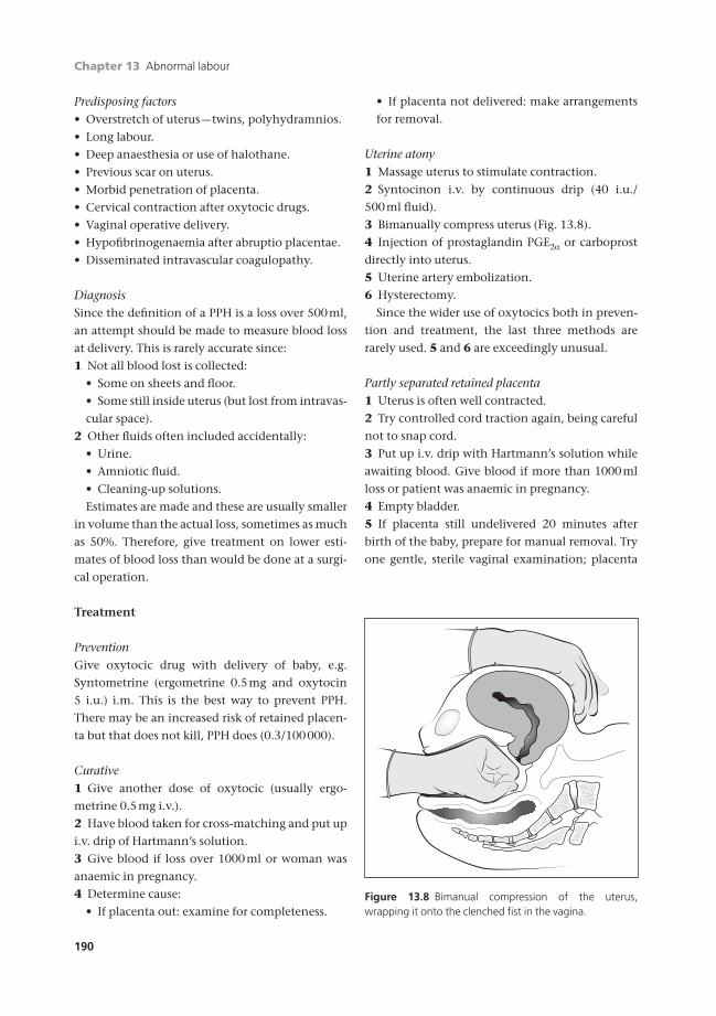
Chapter 13 Abnormal labour
190
Predisposing factors• Overstretch of uterus —twins, polyhydramnios.
• Long labour.
• Deep anaesthesia or use of halothane.
• Previous scar on uterus.
• Morbid penetration of placenta.
• Cervical contraction after oxytocic drugs.
• Vaginal operative delivery.
• Hypofibrinogenaemia after abruptio placentae.
• Disseminated intravascular coagulopathy.
DiagnosisSince the definition of a PPH is a loss over 500ml,
an attempt should be made to measure blood loss
at delivery. This is rarely accurate since:
1 Not all blood lost is collected:
• Some on sheets and floor.
• Some still inside uterus (but lost from intravas-
cular space).
2 Other fluids often included accidentally:
• Urine.
• Amniotic fluid.
• Cleaning-up solutions.
Estimates are made and these are usually smaller
in volume than the actual loss, sometimes as much
as 50%. Therefore, give treatment on lower esti-
mates of blood loss than would be done at a surgi-
cal operation.
Treatment
PreventionGive oxytocic drug with delivery of baby, e.g.
Syntometrine (ergometrine 0.5mg and oxytocin
5 i.u.) i.m. This is the best way to prevent PPH.
There may be an increased risk of retained placen-
ta but that does not kill, PPH does (0.3/100000).
Curative1 Give another dose of oxytocic (usually ergo-
metrine 0.5mg i.v.).
2 Have blood taken for cross-matching and put up
i.v. drip of Hartmann’s solution.
3 Give blood if loss over 1000ml or woman was
anaemic in pregnancy.
4 Determine cause:
• If placenta out: examine for completeness.
• If placenta not delivered: make arrangements
for removal.
Uterine atony1 Massage uterus to stimulate contraction.
2 Syntocinon i.v. by continuous drip (40 i.u./
500ml fluid).
3 Bimanually compress uterus (Fig. 13.8).
4 Injection of prostaglandin PGE2a or carboprost
directly into uterus.
5 Uterine artery embolization.
6 Hysterectomy.
Since the wider use of oxytocics both in preven-
tion and treatment, the last three methods are
rarely used. 5 and 6 are exceedingly unusual.
Partly separated retained placenta1 Uterus is often well contracted.
2 Try controlled cord traction again, being careful
not to snap cord.
3 Put up i.v. drip with Hartmann’s solution while
awaiting blood. Give blood if more than 1000ml
loss or patient was anaemic in pregnancy.
4 Empty bladder.
5 If placenta still undelivered 20 minutes after
birth of the baby, prepare for manual removal. Try
one gentle, sterile vaginal examination; placenta
Figure 13.8 Bimanual compression of the uterus, wrapping it onto the clenched fist in the vagina.
AMI13 6/9/04 5:02 PM Page 190

Abnormal labour Chapter 13
191
may be trapped by the closing cervix and an edge
can sometimes be hooked down and the placenta
gently eased out.
6 If an epidural is already acting, use this, other-
wise a general anaesthetic is needed.
• Try once more to remove placenta by con-
trolled cord traction just before the anaesthetist
induces sleep. Sometimes separation has occur-
red in the meantime.
7 Give a Syntocinon infusion and prophylactic
antibiotics afterwards.
8 Very rarely, the placenta may be abnormally
adherent:
• Placenta accreta: villi just penetrate into
myometrium.
• Placenta increta: villi penetrate deeply into
myometrium.
• Placenta percreta: villi penetrate through
myometrium to peritoneum.
Usually this is not possible to diagnose prospec-
tively. If no plane of separation exists (and often
there is little bleeding at the time) placenta accreta,
increta or percreta must be thought of. Should the
patient wish for no more children, the safest treat-
ment is hysterectomy. If this is not possible, piece-
meal removal is very dangerous and it is better
to leave the placenta to atrophy with antibiotic
control of infection.
Note: This is a very rare diagnosis made less often
as the observer becomes more experienced.
Tears of genital tractHeavy bleeding may occur from a tear of the cervix
despite a well-contracted uterus and a completely
expelled placenta.
1 It is difficult to diagnose certainly and
requires:
• Adequate general anaesthesia, unless epidural
already acting.
• The woman in lithotomy position on a firm
bed.
• Good lights and good assistance.
• Several sponge forceps and retractors (the
vagina and cervix are soft just after delivery and
tissues flop into the line of sight).
2 Search cervix systematically using three pairs of
ring forceps. If there is a tear:
• Check it does not run up into uterus,
especially if at 3 or 9 o’clock.
• Suture with polyglycol absorbable material.
3 Check lower uterine segment with fingers through
the cervix. If a tear is found, either repair through
abdominal incision or perform a hysterectomy.
4 Check top end of episiotomy or tear which may
go into posterior or lateral fornix.
• Repair systematically.
5 Check no actively bleeding vessels in episiotomy
or tear.
• Tie them off separately.
Blood clotting defect1 Check that blood taken from an arm vein clots
and stays clotted.
2 Check:
• Platelets.
• Coagulation studies.
3 Treat appropriately by fresh frozen plasma (FFP).
• Fibrinogen.
4 Remember such a state can allow secondary
atony of the myometrium, so watch for that too as
a cause of bleeding.
Effects of primary PPHRapid loss leads to hypovolaemic shock. If not
corrected, this can cause:
1 Death —about 8% of direct maternal deaths
follow PPH; half of these are avoidable.
2 Renal shutdown and consequent anuria.
3 Damage to pituitary portal circulation causing
necrosis and subsequent Sheehan’s syndrome.
4 Postpartum anaemia and chronic ill health.
Secondary PPH
This is abnormal vaginal bleeding that occurs after
24 hours following delivery. There is no volumetric
definition; women usually present:
• With the passage of clots.
• At 7–10 days with a resumption of fresh vaginal
bleeding.
Causes• Retained pieces of placenta.
• Retained pieces of membrane.
AMI13 6/9/04 5:02 PM Page 191

Chapter 13 Abnormal labour
192
• Retained blood clot.
• Infection of the residual decidua (endometritis).
Clinical findings• Fresh red vaginal bleeding and clots.
• A large uterus.
• A tender uterus.
• An open cervical internal os.
Risks• Substantial bleeding.
• Infection:
(a) Septicaemia.
(b) Blocked fallopian tubes.
Treatment• Admit to hospital.
• Give i.v. broad-spectrum antibiotic cover, 24
hours pre-operatively and continuing for a mini-
mum of 3 days. This combats the bacteraemia that
may occur and also reduces the risk of subsequent
tubal damage.
• Carry out an evacuation of retained products of
conception after 24 hours. This should be per-
formed gently by a senior obstetrician because of
the risk of perforation of the very soft uterus.
Massive blood loss
Rarely, a haemorrhage of 2–3 litres occurs sud-
denly at delivery. The woman’s life will depend on
a well-drilled team having a laid-down policy.
ManagementMassive loss from the woman’s circulating blood
volume of 2–3 litres in a few minutes. For practical
purposes consider it has happened if the woman
has required more than 2 units of blood quickly.
In anticipation of blood loss all women are
routinely grouped and screened for antibodies in
the antenatal clinics. Furthermore all at high risk
of haemorrhage during labour should be cross-
matched prospectively.
Action1 Put in two large-bore (at least 16 gauge) i.v.
cannulae.
2 Contact the duty obstetric registrar, if not
already present, and the duty anaesthetic registrar.
Call the obstetric consultant.
3 Use one or more of the following fluids.
• Up to 2 litres of Hartmann’s solution.
• Up to 1.5 litres of Gelofusin.
• Uncross-matched blood of the woman’s
group.
• Cross-matched blood as soon as available.
• Give O Rh-negative blood only as a last
resort.
• In an emergency situation the use of blood
filters and blood warming devices is not recom-
mended. Pressurized infusion bags should be
used.
• Give I unit of FFP for every 6 units of blood.
The haematologist will advise on platelet
replacement.
4 Stop the bleeding. If the bleeding is from the
uterus:
• Give 0.5mg of ergometrine i.v. and set up 40
units of Syntocinon in 500ml of Hartmann’s
solution to run at 40 drops/minute.
• Commence bimanual compression of the
uterus. If trauma, repair uterus/cervix/vagina.
• Intrauterine injection of PGE2 through
abdominal wall.
5 Contact the blood bank. Send at least 20ml of
blood for further cross-matching. Ask for at least 6
units of cross-matched blood.
6 Inform the duty haematologist of the clinical
situation, the rate of blood loss and the clotting
problems.
7 Insert a central venous line to monitor fluid
replacement.
8 One person should be assigned to record
keeping and should record the following:
• Pulse.
• Blood pressure.
• Maternal heart rate, preferably from an ECG.
• Central venous pressure.
• Urine output.
• Amount and type of fluids the woman is given.
• Drugs the woman has received.
9 Prepare for theatre if appropriate.
10 Before proceeding to hysterectomy, the follow-
ing should be considered:
AMI13 6/9/04 5:02 PM Page 192

Abnormal labour Chapter 13
193
(a) Direct i.m. injection of 0.5mg of PGE2 into
the exposed uterus.
(b) Open the broad ligament and ligate the uter-
ine artery on each side.
(c) Ligate internal iliac arteries.
Operative delivery
Before undertaking an operative vaginal delivery
the following five conditions should be met:
1 Adequate analgesia (epidural or pudendal block
with infiltration of the perineum with lignocaine).
2 The fetal head should not be palpable in the
abdomen.
3 Full dilatation.
4 Presenting part at the spines or below.
5 Empty bladder via catheter.
Vacuum extractor
This instrument is used to get a purchase on the
smooth fetal head, allowing traction to be applied
(Fig. 13.9). The suction raises an edged dome of the
soft tissues of the scalp and the pull is on the over-
hang of this edge.
UsageUsed widely in Europe and the UK, least in USA,
5–10% of UK deliveries.
Very useful in countries with developing health
services (e.g. Africa) for it can be used by less expe-
rienced operators than forceps.
Indications
FIRST STAGE
1 Fetal distress after cervix is 8cm dilated in a
multiparous women.
2 Lack of advance after 8cm dilated cervix in a
multiparous women.
SECOND STAGE
1 Lack of advance:
• Often with occipitoposterior or transverse
position; commonly in association with an
epidural.
• Mother is too tired.
2 Compound presentations —after replacing a pre-
senting hand.
3 High head of second twin.
(Less use in fetal distress for forceps are swifter
but, if the operator is inexperienced, the vacuum
extractor is safer.)
Contraindications• Disproportion.
• Malpresentations (face particularly).
• Very immature infants.
MethodsThis is best learned by watching and helping in the
labour ward. Here essentials only are given.
• Apply largest vacuum cap that slips through
cervix —60mm if possible.
• Hold cap flat against fetal head just anterior to
the occiput and start to build up vacuum.
• With just enough vacuum to hold cap on head,
check around the whole perimeter that maternal soft
tissues have not been sucked under the cap’s rim.
• Increase vacuum to 0.4kg/cm2 and then slowly
to 0.8kg/cm2; again check full circumference of
cap.
• Pull on handle, apply traction to fetal head in
line with the curve of the pelvis (Fig. 13.10).
• Press cap onto head at right angles to line of trac-
tion with fingers of other hand.
Line oftraction
Tube toapplyvacuum
Figure 13.9 Vacuum cap on fetal head sucks up a chignonof subcutaneous tissues to give a button on which to pull.The tractive efforts are mostly on the overhang (arrowed).
AMI13 6/9/04 5:02 PM Page 193

Chapter 13 Abnormal labour
194
• Remove cap by reducing vacuum suction.
• As well as the metal caps, there are softer rubber
and Silastic caps and hand-held disposable caps
which cause fewer fetal abrasions.
Complications
MATERNAL
• Cervical damage.
• Vaginal wall damage; reduced if application
of cap checked so as not to suck in walls when
vacuum is being established.
• Possible urinary retention later.
FETAL
• Cephalhaematoma.
• Skin abrasions.
Both are usually minor.
Forceps
The function of forceps is to get purchase on a
rounded object (the fetal head) and to apply trac-
tion. This is usually needed to hasten delivery, but
it can control the speed of descent, e.g. slow down
the aftercoming head in breech delivery.
UsageDepends on availability of obstetricians. In UK
5–10% of all deliveries.
MechanismThere are many types of forceps. Basically all have:
• Curved blades to fit around the head (Fig. 13.11).
• Handles to apply traction.
Some have also:
1 A curve to allow for the curve of the pelvis (Fig.
13.12).
2 If they do not have a pelvic curve, they may be
straight-handled to allow rotation manoeuvres.
3 Some have a sliding lock to allow for an asym-
metrically aligned head.
Line ofpull
at thislevel of
pelvis
Figure 13.10 Line of traction with vacuum extractor.
Figure 13.11 Cephalic curve of a pair of forceps to embrace a fetal head.
2 Delivery of head
1 Descent to perineum
Angles of traction
Figure 13.12 The pelvic curve of the forceps to fit thecurved line of advance of a fetus through the pelvis.
AMI13 6/9/04 5:02 PM Page 194

Abnormal labour Chapter 13
195
Thus there are two basic types. All have curved
blades and handles for traction. In addition:
• Traction forceps incorporate 1.
• Rotation and traction forceps incorporate 2 and
usually 3.
Indications for use1 Poor progress in the second stage. No exact time
limits but most would consider the longer limits as
2 hours in a primigravida and half that in a multi-
gravida. If an epidural anaesthetic is being used,
these time limits are usually extended.
2 Clinical fetal distress.
• Alterations of fetal heart rate and rhythm.
• Passage of meconium.
3 Biophysical or biochemical signs of fetal
hypoxia.
• On the fetal heart rate trace:
(a) Tachycardia or bradycardia.
(b) Loss of baseline variability.
(c) Late decelerations.
• On fetal blood sampling, scalp capillary blood:
base deficit greater than 10mEq/l; pH below 7.15
in second stage.
4 Maternal distress.
• A tired woman after a long first stage.
• One who is frightened or has not had proper
analgesia.
5 To prevent fetal morbidity.
• Very immature babies.
• Delivering the head of a breech presentation.
6 To prevent maternal morbidity.
• Women with cardiac or respiratory disease.
• Following a dural tap at attempted epidural
injection.
MethodThese methods are best learned by watching and
helping in the labour ward. Here essentials only are
given.
1 Each blade of the forceps is slipped in turn along
the obstetrician’s cupped hand holding back the
vaginal walls in turn.
2 The blades are locked together.
3 Traction is applied in a downwards and back-
wards direction to follow the line of the curve of
the birth canal (Fig. 13.13).
4 For a rotation, forceps are applied on the sides
of the baby’s face and then rotated through 90°
occipitoanterior and delivered by traction.
Complications
MATERNAL
• Perineal tear: avoid by a properly sited episioto-
my done at the right time; often an episiotomy will
extend at a forceps delivery. Check for this at the
apex of the episiotomy cut.
• Damage to vagina: occasional split of vagina
where caught between descending head and is-
chial spines.
• Retention of urine: possibly due to oedema at
bladder neck. Responds to continuous drainage.
FETAL
• Bruising to head: may go on to cephal-
haematoma.
• Facial palsy: facial nerve in front of ear unpro-
tected in fetus. May be compressed by forceps
blade. Usually only temporary effect.
• Intracranial haemorrhage: if blades incorrectly
applied or excessive traction used.
Push downwithleft hand
Pull onhandleswithrighthand
Resultant of two lines offorces draws fetal headdown in line with thispart of the birth canal
Figure 13.13 Correct line of pull is achieved by use of twohands working in cooperation, thus performing the taskmore simply than a pair of axis traction forceps.
AMI13 6/9/04 5:02 PM Page 195
Chapter 13 Abnormal labour
196
Failed forcepsAn attempt to deliver with forceps which is
unsuccessful.
REASONS
1 Cervix not fully dilated.
2 Misdiagnosis of position of head. Thought to be
occipitoanterior when it is occipitotransverse or
occipitoposterior.
3 Unsuspected disproportion.
TREATMENT
Get senior help in hospital. Then:
If 1, with mother and fetus well, await full
dilatation.
If 2, rotate head and deliver.
If 3, Caesarean section.
Caesarean section (CS)
Delivery of the fetus by surgical means through the
abdominal wall.
UsageDepends on availability of obstetricians and on the
population served. In UK 25% of all deliveries.
The Caesarean section rate has risen rapidly in
the last 25 years. There are several reasons for this:
• The introduction of epidural anaesthesia has re-
duced the anaesthetic risks of the procedure. This
has led to a lower threshold for doing a Caesarean
section in the second stage of labour rather than
performing rotational/high cavity forceps deliver-
ies which led to maternal and neonatal morbidity.
• The increased use of electronic fetal monitoring
has increased our awareness of fetal distress al-
though the majority of babies are born in good
condition despite an abnormal CTG and/or low pH
at fetal blood sampling.
• The reduction in the number of rotational for-
ceps deliveries has led to a deskilling of obstetri-
cians who do not feel confident to carry out these
procedures.
• The evidence that breech presentation babies
have a reduced morbidity and mortality if deliv-
ered by elective Caesarean section
• An increasing demand from women for elective
Caesarean sections with no medical reason.
The latter group are difficult to deal with, as a
first elective Caesarean section does have a lower
maternal morbidity than an emergency Caesarean
section but it is still higher than a normal vaginal
delivery. In first pregnancies we do not have a reli-
able indicator for the outcome of labour and so we
cannot guarantee that a woman will not end up
with a Caesarean section. Seventy per cent of
women who go into spontaneous labour at term
can expect to have a normal vaginal delivery. Many
of the women who request Caesarean section have
a genuine fear of labour and/or motherhood which
may be based on past experiences of sexual abuse,
poor mothering or other psychological problems.
It is wise to refer them to a psychologist or midwife
trained in these areas before agreeing to perform a
Caesarean section. All subsequent Caesarean sec-
tions carry an increased morbidity.
IndicationsFew are absolute, most are relative to the individ-
ual patient, the obstetrician and the obstetrical en-
vironment. Generally when the risks of vaginal
delivery to the mother or baby are greater than
those of abdominal delivery in any given circum-
stance, a CS should be done.
• CPD.
• Fetal distress in first stage of labour or a pro-
lapsed cord.
• Failure of labour to progress despite adequate
stimulation.
• To avoid fetal hypoxia of labour: pre-eclampsia;
intrauterine growth restriction.
• Antepartum bleeding: placenta praevia; abruptio
placentae.
• Poor past obstetric history.
• Malpresentations: brow.
• Malpositions: transverse lie, breech.
• Death of mother in late pregnancy, a live fetus
removed peri mortem.
The only absolute ones are gross disproportion,
the higher grades of placenta praevia.
In a UK population, a hospital in this decade
would have the following indications in differing
proportions for operations:
• Fetal distress.
• Pre-eclampsia.
AMI13 6/9/04 5:02 PM Page 196

Abnormal labour Chapter 13
197
• Poor fetal growth.
• Disorderly uterine action.
• Breech, face and brow.
• Previous CS.
Types of approach1 Lower segment operation: transverse approach
through lower segment. In the UK, over 99% of
operations are lower segment because:
• Wound is extraperitoneal so less risk of intra-
peritoneal infection.
• Fewer post-operative complications.
• Healing of scar is better for lower segment is
relatively at rest in puerperium.
• Risk of rupture less in subsequent pregnancies.
2 Classical operation: vertical approach through
upper segment, performed:
• If lower segment unapproachable, e.g. fibroids.
• If transverse lie.
• If very small baby expected (24–28 weeks), lower
vertical incision (De Lee’s incision).
AnaesthesiaThe operation requires an anaesthetic. The alterna-
tives are:
1 General anaesthetic.
2 Epidural block.
3 Spinal block.
4 Infiltration of local anaesthetic agents.
Surgical techniqueThese surgical operations are best learned by
watching and helping in theatre.
Complications1 Haemorrhage
• Worst if angles of transverse uterine incision
extend into uterine vessels.
• Always have 2 units of cross-matched blood
available.
2 Infection
• Watch asepsis and antisepsis.
• Give prophylactic antibiotics to all women.
3 Abdominal distension
• Common for a day or so.
• Await events.
4 Ileus
• Mild regional ileus may last 24 hours.
• Await events and avoid overloading the gut
(keep on i.v. fluid for 24 hours).
Longer ileus may follow if there was a lot of
handling and packing of the gut. Treat with
stomach aspiration and i.v. fluids.
5 Thromboembolism
• Much higher risk after CS than after vaginal
delivery.
• Avoid thrombosis by:
• TED stockings intraoperatively until woman
fully mobilized.
• Intraoperative compression stockings.
• Intraoperative subcutaneous heparin con-
tinued daily until woman fully mobilized.
• Early mobilization and leg exercises.
• Keep woman well hydrated post-operatively.
• Avoid embolism by taking leg and pelvic signs
seriously and anticoagulating early.
• Prevent thrombosis with prophylactic anticoag-
ulation (subcutaneous heparin) in all women and
particularly those women at higher risk:
(a) Aged over 35.
(b) Obese.
(c) Past history of thrombosis, particularly if
oestrogen associated (e.g. oral contraception).
(d) Anaemia.
Prognosis
MATERNAL
• Mortality. 1:3000 CS with causes mostly in the
above complications group.
• Morbidity. Subsequent pregnancies following a
CS may be affected.
1 If non-recurrent indication (e.g. fetal distress),
two-thirds deliver vaginally.
2 If recurrent indication (e.g. disproportion),
only one-eighth deliver vaginally.
Hence patients who have had a CS must have all
subsequent pregnancies properly conducted in a
hospital. Watch for dehiscence of uterine scar in
late pregnancy next time. Take extra care if giving
oxytocic stimulation.
FETAL
• Mortality. Depends on indication; CS reduces
risk, but, if done for progressive fetal condition, it
AMI13 6/9/04 5:02 PM Page 197

Chapter 13 Abnormal labour
198
may be indicated only in the women with worst
degrees of that condition and so fetal prognosis
is worse, not from the operative delivery but the
condition which indicated it.
• Morbidity. Higher risk of retained lung fluid if de-
livered by CS especially before 32 weeks’ gestation.
The flying squad
Every major hospital doing obstetrics in the UK
used to have available obstetricians and midwives
who could be taken urgently to any patient who
presented with an obstetric emergency outside the
hospital environment. In some teams, which cov-
ered a larger area, an anaesthetist was included.
The squad used to represent the patching up of a
domiciliary and GP unit service.
Much of the real service has become a resuscita-
tion one, the woman being brought to a state
suitable for transfer back to the hospital where
definitive treatment takes place. Problems at deliv-
ery have almost disappeared and the squad was
being called inappropriately. One-quarter of pa-
tients are given i.v. therapy but <1% had uncross-
matched blood. Many of the calls related to normal
women who had just delivered. The ambulance
service transports such women but no flying
squad is needed, rather a community midwife or a
paramedic.
The flying squad was introduced because private
transport was rare, telephones were unusual and
it was hard to get to the hospital. All that has
changed and now, every time obstetricians, mid-
wives and anaesthetists have to leave the centre
to look after an individual, it leaves the larger num-
ber of women at that hospital with a diminished
service.
Now paramedics in the ambulance services are
trained in advanced resuscitation. They go to the
home or GP unit and bring the woman safely into
hospital.
A few flying squads persist in rural areas, but by
2004 virtually all have been closed down for the
reasons discussed above.
Multiple pregnancies and deliveries
The commonest litter size in the human species is
one. Other multiples are rare (Table 13.2) and vary
with racial characteristics inside the species (e.g. there
is a higher incidence in West Africa than in Europe).
The differences between actual and theoretical
figures are probably due to the reducing birth rate,
more births to older women and fewer to those of
higher parity; (both having increased multiple
pregnancy rates). Higher rates in the UK are now
due to ovarian stimulation and assisted conception
with a trebling of triplets in IVF.
Table 13.2 Rates of multiple pregnancies per 1000
deliveries in England and Wales in 1995 compared with
the biologically expected rate.
Actual Theoretical
Twins 13.6 12.5 (1 :80)
Triplets 0.44 0.16 (1 :802)
Quadruplets and
higher orders 0.014 0.02 (1 :803)
BinovularMonovular
Two ovaTwo sperms
One ovumOne sperm
Fertilized
Figure 13.14 Biological differences in monovular and binovular twins.
AMI13 6/9/04 5:02 PM Page 198

Abnormal labour Chapter 13
199
Twins
Types (Fig. 13.14)
Monovular twinsMonovular twins are produced from one ovum
fertilized by one sperm. After the two-cell division
instead of going into the four-cell stage, the blas-
tomere divides into two separate cell bodies which
go on to two individuals. Thus there is common
chromatin material; sex and physical characteris-
tics will be the same, producing identical twins.
Binovular twinsBinovular twins are from two separate ova fertil-
ized by two different sperms. These ova are shed in
one menstrual cycle and most likely to be fertilized
after one intercourse although they can be at
separate times with different fathers. The two
blastomeres develop separately and have different
chromatin material. They can, therefore, be of
different sexes having no more in common than
any other members of the same family. They are
non-identical twins. Early ultrasound can help
differentiation.
IncidenceMonovular twins have an incidence of 3 or 4 :1000
worldwide and there is only a slight familial ten-
dency. Binovular twins may have a family history
on the maternal side. It is these that account for
racial and maternal age variations. Binovular twins
are more common than monovular ones (4 :1).
Binovular twins are commoner if:
• Maternal family history of non-identical twins.
• Over 35 years.
• After replacement of two, three or more fertilized
ova at in vitro fertilization.
Differentiation of twins1 Sex. If of different sexes, obviously binovular. If
of one sex, may be either.
2 Placenta. If two separated placentae, will be bi-
novular; if one placenta, may be monovular or bi-
novular (Fig. 13.15). Check septum between sacs
by peeling amnions from each other (Table 13.3).
3 Blood groups. If doubt in dichorionic types, check
the ABO, Rh, Duffy, Kell and MNS.
4 Fingerprints. If different, binovular.
5 DNA fingerprinting with probes identifying about
60 dispersed sequences of variable size.
Diagnosis of twins
History• Suspicion on family history especially maternal
non-identical twins.
• Suspicion on past obstetric history of twins.
• Suspicion from excessive vomiting in early
pregnancy.
ExaminationExamination from 20 weeks onwards shows uterus
bigger than expected. At first a lot of limbs are felt
ChorionAmnion
(1) Binovular (2) Probably binovular
(3) Monovular (4) Monovular
Figure 13.15 Placental types in twinning. Often the intervening membranes can be seen in early pregnancy onultrasound and the two types of twins differentiated.
Table 13.3 Differentiation of twins by checking placentae.
Septum Placenta type Twin type
(4) None Monoamniotic Monovular
Monochorionic
(3) Amnion only Diamniotic Monovular
Monochorionic
(2) Amnion and chorion Diamniotic Binovular or
Dichorionic monovular
(1) No common septum Diamniotic Binovular
Dichorionic
AMI13 6/9/04 5:02 PM Page 199

Chapter 13 Abnormal labour
200
and later, about 30–32 weeks, more than two
separate poles determined (e.g. two heads and
one breech).
InvestigationUltrasound at 6–7 weeks may show two or more
sacs. The embryos can be seen in these at 7–8
weeks. The differentiation of mono from binovular
can often be made by expert examination of the
dividing membranes.
Commonly one of a pair of twins diagnosed
early does not develop and is absorbed: the vanish-
ing twin syndrome.
Without ultrasound, twins may not be diag-
nosed until delivery on rare occasions. While
embarrassing to the attendants, this usually does
not affect the second twin unless Syntometrine
was given inadvertently at the birth of the first
baby. This could jeopardize the O2 supply to the
second twin and so his or her delivery should be
expedited.
Management of twins
Complications in pregnancy1 Miscarriage is more frequent.
2 Preterm labour commoner (50% before 37
weeks).
3 Pre-eclampsia commoner (¥3).
4 Risk of anaemia increased.
• Iron deficiency.
• Folic acid deficiency.
5 Polyhydramnios commoner (¥10 with monovu-
lar twins).
6 Risk of APH increased.
• Abruptio placentae.
• Placenta praevia.
Management in pregnancy1 Diagnose early by bearing it in mind (one only
diagnoses what one thinks about); often ultra-
sound will give the result before clinical suspicion.
2 Give extra iron and folic acid supplements and
see that the woman takes them.
• Check blood more often for haemoglobin
levels.
Complications in labour1 Delay in delivery of the second twin is
associated with a higher mortality.
2 PPH is more common.
3 Prolapse of umbilical cord is more common.
4 Mechanical collision of leading parts (or locking
of a breech–cephalic) as they both enter the pelvis.
This is very rare.
Management in labour1 Always plan for hospital delivery.
2 Ensure the first twin is longitudinal. Com-
monest combinations of presentations show that
both twins lie longitudinally 90% of the time
and the first twin is a cephalic presentation in 80%
(Fig. 13.16). Non-cephalic presentations are
common if early preterm labour.
If the first twin is transverse, do a Caesarean
section.
Cephalic – Cephalic50%
Cephalic – Breech25%
Breech – Cephalic10%
Breech – Breech10%
Cephalic – Transverseand all others5%
Figure 13.16 Presentation of twins at time of mature delivery (after 36 weeks).
AMI13 6/9/04 5:02 PM Page 200

Abnormal labour Chapter 13
201
3 Check for cord prolapse when membranes
rupture (often early in labour).
4 Progress is usually uneventful. Monitor both
fetal hearts and have an i.v. drip running.
5 An epidural anaesthetic is useful for it allows
rapid anaesthesia for any manoeuvres that may
be required for the second twin in the second
stage.
6 Deliver the first twin appropriately. Have an
anaesthetist and a paediatrician in the labour
ward. Make sure that nobody inadvertently gives
Syntometrine to the mother at this point.
7 Clamp cord of the first twin and divide. Hand
baby to competent assistant or paediatrician in
case resuscitation is required.
8 Immediately check the lie of the second twin. If
longitudinal, check presenting part. If oblique or
transverse, convert to longitudinal.
• External version —usually easy for uterus is
lax, or
• Combined external and internal version —
rupture membranes and bring a leg of the fetus
down through the cervix. This produces an
incomplete breech presentation but it is at
least longitudinal.
9 The second twin is best delivered within 20
minutes of the first. Usually uterus starts contract-
ing again about 5 minutes after first delivery. If it
does not do so spontaneously, use i.v. oxytocin
augmentation. Very little is needed. Rupture mem-
branes of second sac and deliver appropriately.
10 Give Syntometrine with delivery of the second
twin and continue oxytocin infusion for another
hour.
11 Deliver placentae as soon as uterus is con-
tracted after delivery of the second twin, for
retained placentae and PPH are common.
Outcome
MATERNAL
Higher risk:
• The complications of pregnancy, e.g. pre-
eclampsia.
• The complications of delivery, e.g. anaesthesia
and PPH.
FETAL
• Risks to the first twin are twice, and to the
second twin, about three times those of single
births.
Causes of death:
(a) Immaturity. Especially
(b) Hypoxia. the second
(c) Risks of operative delivery. twin.
• Neonatal risks are jaundice or anaemia following
intrauterine shunting of blood inside the placenta
leading to twin-to-twin transfusion in monovular
twins.
Triplets
Rarely due to tri-ovulation:
• Usually binovular twins with one fertilized egg di-
viding into two individuals or assisted conception.
• Usually born at an even more immature stage
than twins and have double the risks.
• The complications and management are as for
twins. Because of the immaturity of the fetuses,
delivery is commonly by Caesarean section.
}
Self-assessment
13.1 A woman in her first pregnancy is admitted in spontaneous labour at 39 weeks. On examination the presentationis cephalic, 2/5 palpable. The cervix is 1cm long and firm, 1cm dilated posterior to the head and the head is 2cmabove the spines. Which of the following statements are true?(a) She is in the active phase of the first stage of labour.(b) Her Bishop’s score is <4.(c) She is in the latent phase of the first phase of labour.(d) Her Bishop’s score is >4.(e) She should be encouraged to mobilize and re-examined in 4 hours time.
Continued on p. 202.
AMI13 6/9/04 5:02 PM Page 201

Chapter 13 Abnormal labour
202
Self-assessment Continued
13.2 Which of the following presenting positions can be delivered spontaneously vaginally?(a) Face presentation.(b) Brow presentation.(c) Occipitotransverse.(d) Occipitoposterior.(e) Vertex presentation.
13.3 Which of the following statements are true?(a) Breech presentation is more common in premature babies.(b) All women with a breech presentation should be offered external cephalic version at 37–38 weeks.(c) Breech presentation is more common at term than at 34 weeks.(d) Breech presentation is associated with a higher perinatal mortality regardless of the mode of delivery.(e) Caesarean section should be offered to all women with twins where the presentation is cephalic in the first
twin and breech in the second twin.13.4 List the five conditions that should be met before undertaking an operative vaginal delivery for a cephalic
presentation.13.5 Which of the following are true about primary postpartum haemorrhage (PPH)?
(a) It is defined as a loss of blood of >500ml.(b) It is more common following a spontaneous vaginal delivery.(c) The commonest cause is uterine atony.(d) It can be prevented by giving syntometrine with the birth of the anterior shoulder.(e) It is more common in women with pre-eclampsia.
AMI13 6/9/04 5:02 PM Page 202
In the puerperium the mother is returning to her
pre-pregnant state. The various organ systems take
different times but most physical restoration has
occurred by 6 weeks. During this time, one should
aim at:
• Restoring maternal health and preventing
illness.
• Maintaining infant health and preventing
illness.
• Establishing infant feeding.
• Educating the mother about her child’s and her
own future health.
Physiological changes in genital tract
Uterus
Uterine bulk reduces by involution following with-
drawal of oestrogens so that the organ is back in
the pelvis by 10–12 days.
• Thrombosis of blood vessels.
• Autolysis of cellular cytoplasm of myometrium.
• Regeneration of endometrium within 6 weeks.
• The uterus never returns quite to nulliparous size
but close to it.
• The cervix is normally stretched at delivery and
the external os is split after a delivery.
Vulva
After stretching, tissues revert to non-pregnant
state with the following differences:
• Less fatty tissue in the labia.
• Hymen is wrecked leaving skin tags only.
• Episiotomy or tear area leaves a scar which might
be tender.
Management of first week
Observations
• Temperature: watch for pyrexia.
• Pulse rate: watch for transient tachycardia asso-
ciated with thrombosis.
• Blood pressure: check especially if pre-eclampsia
occurred in pregnancy.
• Uterine size: check daily reduction by abdominal
palpation.
• Lochia: loss from vagina in the first days is the
shedding of the decidua. Starts red with fresh
blood but pales over 2 weeks to yellow-white. This
may last for weeks.
• Urine output: normal diuresis starts within 24
hours and lasts about 3 days.
• Bowels: often constipation. Avoid too long a gap
for a very hard stool is painful. Provide fibre and
extra fluids to give bulk to stools.
• Haemoglobin: check level about day two even if
203203
Chapter 14
Puerperium
AMI14 6/9/04 5:05 PM Page 203
Chapter 14 Puerperium
204
blood loss at delivery is recorded as minimal. Give
oral iron if below 11g/dl. Readvise on diet.
Pain relief
Uterine contractions• After-pains (contractions) are variable, painful
and need analgesia.
• Warn the woman that they are coming so that
she is prepared.
• Often felt more in multigravida, worse after suc-
cessive deliveries.
Perineal• After the local anaesthetic wears off, the epi-
siotomy will hurt. Further, oedema and burning
may make sitting uncomfortable. Give analgesics,
local heat and, if oedematous, ice packs.
• Check sutures are not cutting in.
Bed rest
A balance must be struck between resting in bed to
recover from the work of labour and resting so
much that stagnation of blood leads to deep vein
thrombosis.
Exercises
In association with bed rest, graduated exercises re-
store muscle tone to stretched areas and maintain
venous flow in limbs and pelvis. They are aimed at:
1 Breathing exercises.
2 Legs to prevent stagnation of blood in veins.
3 Abdominal wall to restore tone of rectus
muscles.
4 Pelvic floor to restore levator ani.
Exercises are best taught by a physiotherapist in
hospital teaching women what to do after leaving.
Fifteen minutes twice a day should be set aside for
these for some weeks.
Psychological support
Producing a baby is a psychological stress. Women
with different emotional backgrounds respond
variously. Most women find a balance between the
excitement and pleasure of the event on the one
hand and the worry and responsibility of looking
after the baby on the other. Watch for this and pre-
vent excessive worry by:
• Adequate explanation of any baby problems.
• Ensuring good sleep.
• Pain relief.
Discharge to home
The length of postnatal stay in hospital is deter-
mined by social and financial factors as well as by
medical ones. The length of stay is reducing and
the community services are supposed to take up
their share of looking after those who the hospital
discharges.
Many Trusts now run planned, very early
DOMINO discharge schemes (Domestic in and
out) where a woman comes into hospital with her
community midwife, delivers and goes home a few
hours later. In some instances a birthroom is used,
geographically apart but close to the labour suite;
here the woman can be delivered by her midwife in
less hospitalized surroundings. If an emergency
arises, obstetric, anaesthetic or paediatric care is
immediately available.
Fifty percent of women now leave the hospital
by day two under planned discharge schemes
evolved between the hospital and the local com-
munity midwives and general practitioners. Those
who had uncomplicated Caesarean sections go
home at 3–5 days.
Advantages of earlier discharge1 Satisfaction. By planning and letting the woman
know she will return home in a short time, she is
happier and more willing to come into hospital for
the time of maximum risk: labour.
2 Leaves more beds available for those with ante-
natal/pastnatal complications allowing intensive
care for the fetus or mother.
3 Reduces the risks of hospital cross-infection.
This is a rapidly diminishing factor in the UK
but outbreaks of gastroenteritis and haemolytic
streptococcal infection do still occasionally
happen.
AMI14 6/9/04 5:05 PM Page 204
Puerperium Chapter 14
205
Disadvantages of earlier discharge1 Lack of rest. Household duties soon demand the
woman’s attention.
2 Poor housing conditions. If the woman is unsup-
ported or from the lower income groups, she may
be better kept in hospital for a little longer. This is a
diminishing problem in the UK but a small propor-
tion of homes (5–10%) are still considered to be
unsuitable for early discharge.
3 Medical problems after delivery may be missed.
This is unlikely if proper community cover is given
so that women who go home are visited by mid-
wives and GPs by arrangement. In fact less than 2%
of early discharged women are re-admitted for
medical problems.
Problems in the puerperium
Over half of maternal deaths associated with
pregnancy and childbirth occur in the first few
days following delivery. Although rare, it is a
measure of the need for vigilance; postnatal
women should be seen each day for the first 10
days by a qualified doctor or midwife. Ideally a
postnatal visit should be at about 6 weeks; this
should be by the family practitioner if the delivery
was straightforward or at the hospital if there were
complications.
Psychological problems
The baby blues
Many women feel weepy and depressed 3–5 days
after delivery but this is usually short-lived. Factors
that prolong the baby blues are:
• Postpartum pyrexia.
• Anaemia of <8g/dl.
• Inadequate sleep.
• Delayed healing of the episiotomy or Caesarean
section wound.
• Delay in establishing breast feeding.
• Decline in sympathy, congratulations and atten-
tion of friends and family as the time from child-
birth increases.
Depression
Baby blues merge with serious depression. There is
not a specific form of depression that is related
solely to pregnancy and childbirth. The factors
that aggravate depression are:
• A background predisposition due to previous
history or family history.
• A conflict in the responsibilities of looking after
a new baby.
• Hormone changes acting on predisposition to
depression.
• Fantasies.
• Anxiety and guilt.
• Residual pain.
• Sleeplessness.
Treatment• Involve a psychiatrist.
• If no psychotic delusions, can be managed as an
outpatient.
• Oral antidepressants.
Postpartum psychosis
This is a rare condition which affects less than 1 in
500 women. It is potentially life-threatening to
both the mother and the baby.
Symptoms• Rejection of the baby.
• Delusion.
• Confusion.
• Agitation.
Management1 Admit the mother and baby to a special ward in
the psychiatric unit.
2 Ensure 24 hour supervision.
3 Give appropriate psychotherapy drugs. Some
use electroconvulsive therapy.
Postpartum psychosis is recurrent (about 20%)
but chances are decreased by a 2-year or more gap
between pregnancies.
Infections
The common infections of the puerperium are:
AMI14 6/9/04 5:05 PM Page 205
Chapter 14 Puerperium
206
1 Infections of the genital tract.
2 Urinary tract infection.
3 Breast infections.
Genital tract infection
An ascending infection which largely involves the
placental bed. Unlike pelvic inflammatory disease
(PID) it spreads directly through the uterus to the
parametrial tissues.
The commonest organisms are E. coli and Strepto-
coccus faecalis.
Diagnosis
HISTORY
• Puerperal pyrexia —temperature above 38°C
twice.
• Offensive discharge.
• Low abdominal pain.
• Passage of clots and a return of red lochia.
• Systemic symptoms associated with the pyrexia.
EXAMINATION
• Raised temperature and pulse.
• Bulky uterus.
• Tender uterus.
• Offensive lochia (often bleeding with clots).
• An open internal cervical os.
INVESTIGATIONS
• Hb —may be reduced.
• White cell count —raised with a neutrophilia.
• High vaginal or endocervical swab —may grow
the organisms.
Management1 Admit to hospital.
2 Evacuation of retained products of conception
under antibiotic cover.
Broad-spectrum antibiotics should be used to
cover the bacteraemia and subsequently for 5 days.
Complications
ACUTE
• Parametritis.
• Salpingitis.
• Broad ligament abscess.
• Peritonitis
• Septic thromboembolism.
LONGER-TERM
• Infertility.
• Menstrual irregularities and pelvic pain.
Urinary tract infections
These are common in the puerperium because of:
1 Bladder stasis.
2 Oedema of the bladder base.
3 Diminished bladder sensation.
4 Catheterization in labour.
The commonest organisms are E. coli and Proteus
spp.
Diagnosis
HISTORY
• Dysuria.
• Frequency of micturition.
• Loin pain if pyelonephritis supercedes.
• Systemic symptoms such as pyrexia and
tachycardia.
• May be asymptomatic and recognized on rou-
tine mid-stream urine (MSU) sample. This should
be performed on all patients who have been
catheterized in labour.
EXAMINATION
• Raised temperature.
• Tender suprapubically or in the renal angle.
INVESTIGATIONS
• MSU.
• White cell count.
• Nitrites and leucocytes on dipstick.
Management• Bed rest.
• High fluid, light solid diet.
• Broad-spectrum antibiotics until the results
of culture and sensitivity are known, then be
specific.
AMI14 6/9/04 5:05 PM Page 206

Puerperium Chapter 14
207
Complications• Pyelonephritis.
• Exacerbation of the baby blues.
Breast infection
This usually enters through a break in the skin
(cracked nipple). It is usually confined to one quad-
rant of the breast. Most commonly it is due to
Staphylococcus aureus.
Diagnosis
HISTORY
Painful area of one breast.
EXAMINATION
• Raised temperature.
• Erythematous segment of the breast.
• If an abscess, brawny swelling or even fluctua-
tion.
INVESTIGATION
Bacteriology of expressed breastmilk from the af-
fected side.
Management1 Without abscess
• Supportive brassiere.
• Continue breast feeding on other breast
and empty the affected breast with a breast
pump.
• Give broad-spectrum antibiotic such as flu-
cloxacillin orally.
2 With abscess
• Incise under a general anaesthetic.
• Circumareola incision.
• Break down septa and leave a drain through
dependent part.
• Adequate supportive brassiere.
• Appropriate i.v. antibiotics.
Thromboembolism
The postpartum period is the commonest
time in pregnancy for a thromboembolism
because:
1 Increased coagulation. The increase in clotting
factors from pregnancy remains although plasma
volume reduces to normal within a few hours of
delivery.
2 Stasis. Many women have been immobilized
in pregnancy, during labour or the immediate
puerperium.
3 Damage to venous endothelium.
• Uterine veins —following uterine sepsis.
• Deep leg veins —weight of legs compresses
veins if immobilized.
Diagnosis
HISTORY
• Calf pain.
• Unilateral leg oedema.
EXAMINATION
• A low-grade postpartum pyrexia.
• An unexplained maternal tachycardia.
• Tenderness over the deep veins of the calf.
• A positive Holman’s sign (calf tenderness on dor-
siflexion of the foot).
INVESTIGATIONS
1 Doppler ultrasound.
• Simple continuous wave Doppler ultrasound
will fail to show flow in the femoral vein.
• Colour flow Doppler may demonstrate the
clot in the veins.
2 Venography.
• This is the definitive test using image intensi-
fiers and low-viscosity contrast medium.
Management
PREVENTION
1 Early mobilization —all women are encouraged
to be up as soon as they wish. Non-weight-bearing
exercises used for postoperative mothers.
2 Prophylaxis is usually given to women who have
had a previous history of thromboembolism or are
at moderate to high risk.
• Compression (TED) stockings worn during
labour or Caesarean section.
• Subcutaneous low molecular weight heparin.
AMI14 6/9/04 5:05 PM Page 207

Chapter 14 Puerperium
208
TREATMENT
1 Anticoagulate immediately with full dose i.v.
heparin to prevent further extension of the
thrombosis with the risk of pulmonary embolism
and allows earlier recanalization of the clotted
veins.
2 Long-term anticoagulation with warfarin for 12
weeks.
Pulmonary embolism
The most dangerous destination of a clot embolus
from the leg or pelvic veins is in the pulmonary
circulation.
1 Mild cases follow microemboli. Dyspnoea and
slight, poorly defined pleural pain. The condition
resolves in a few days with no specific treatment
and may not even be diagnosed.
2 Severe cases arise from clot from the:
• Soleal veins —clot extends to popliteal vein
and breaks off (30%).
• Uterine and ovarian veins —a throm-
bophlebitis with a friable clot following mid-
pelvic sepsis (20%).
In 50%, no clinical signs of the origin exist
before the pulmonary embolism.
Diagnosis
HISTORY
Pre-existing deep vein thrombosis (in half the cases
only).
Early:
• Acute dyspnoea.
• Faintness.
Later:
• Chest pain.
• Haemoptysis.
EXAMINATION
Early:
• No physical signs beyond dyspnoea.
Later:
• May be cyanosis.
• Local signs of pulmonary underperfusion.
• Right heart failure.
Later still:
• Pleural signs.
• Collapse of a pulmonary lobe.
INVESTIGATIONS
Often no positive tests early. Next day:
1 X-ray:
• Raised diaphragm on affected side.
• Consolidation and infiltration of lung(s).
2 ECG —rhythm disturbance.
• Lead I —S wave inversion.
• Lead III —T wave inversion and deep Q wave.
• Leads VI, 2, 3, 4—T wave inversion.
• Excludes cardiac infarction.
3 Lung ventilation–perfusion studies with ra-
dioactive albumin to show ischaemic areas.
4 Pulmonary angiography to show clot.
5 Left and right heart catheterization to show re-
versal of pressures.
The last two are used in specialist thoracic units.
ManagementTwo-thirds of those dying do so within 2 hours, so
act quickly on suspicion, not awaiting the sophisti-
cated tests even if they are available.
IMMEDIATE TREATMENT
1 External cardiac massage if required.
2 Positive pressure O2 by intubation if necessary.
3 Heparin (see below).
4 Emergency embolectomy is only performed in
hospitals with their own thoracic units with bypass
facilities and rarely used.
DEFINITIVE TREATMENT
If resuscitation is successful, give:
1 Anticoagulants (more i.v. heparin):
• 25000 units immediately.
• 25000 units 6-hourly for 24 hours.
This is to prevent further emboli.
At the same time start warfarin oral therapy con-
trolled by prothrombin time.
2 Thrombolytics (streptokinase). Actively acceler-
ate lysis of existing clot:
• 500000 units immediately.
• 100000 units hourly for 72 hours.
AMI14 6/9/04 5:05 PM Page 208

Puerperium Chapter 14
209
3 Embolectomy (mortality rate 25%). Useful if:
• Thoracic unit in the same hospital.
• No response to streptokinase.
• Too ill for streptokinase.
• Contraindication to streptokinase, e.g. recent
surgery, peptic ulcer or hypertension.
Both high-dose heparin and streptokinase have
high risk of starting bleeding. Not indicated unless
embolus is thought to be life-threatening. If start-
ed, reduce to conservative dosage in 2–3 days and
continue for 4–6 weeks.
Secondary postpartum haemorrhage
Any considerable fresh bleeding occurring in the
puerperium after 24 hours. This is dealt with in
Chapter 13.
Previous diseases continuing into the puerperium
Pre-eclampsia and eclampsia
When these conditions occur before delivery, their
ultimate treatment is delivery of the baby. Usually
the woman gets better rapidly after the placenta is
delivered and the normal diuresis starts to remove
the water/salt overload. A few women stay as bad or
worsen in the first few days of the puerperium.
Management is the same as in pregnancy (see pp. 123–
6). Postpartum eclampsia has a high mortality rate.
Diabetes
Delivery may have been operative and for a day im-
mediately after the woman may be on i.v. therapy.
Remember that insulin requirements drop very
sharply in the puerperium so that many diabetics
are at their pre-pregnant levels of insulin by 2 days
into the puerperium. Infection of any wounds is
more likely and breast feeding may be an irregular
drain of carbohydrates, making insulin balance
more difficult.
Heart disease
The first 24 hours of the puerperium are dangerous,
for the post-delivery shunting of blood from the
uterine vessels may lead to pulmonary oedema and
subsequent right-sided cardiac failure. After that
the risk is reduced.
Puerperal infection could release bacteria that
might colonize on any damaged endothelium in
the heart (acute bacterial endocarditis). This is
more likely to follow a congenital heart disease
than rheumatic conditions, but antibiotic cover is
often provided for either into the puerperium.
Ovarian tumours
These are more likely to undergo torsion in the
puerperium because of the lax abdominal wall and
the diminishing uterus. Surgery is recommended
for any ovarian masses above 10cm diameter.
Self-assessment
14.1 A woman on the postnatal ward bursts into tears when you go and see her 48 hours after the birth of her baby.Which of the following might indicate that she is at high risk of developing postnatal depression?(a) She does not show any interest in her infant who is crying insistently.(b) She is bottle feeding the baby.(c) She is unable to sleep.(d) Her mother had severe postnatal depression.(e) Her partner has recently left her and was not present at the birth of the baby.
14.2 A woman presents 10 days after a normal vaginal delivery with a raised temperature of 38.4°C. Match the addi-tional information given in 1–5 to the five most likely of the diagnoses (a)–(j)1 She has passed several clots and is in pain. Her uterus is 16 weeks size and tender.2 She complains of pain on passing urine. Her left loin is tender.
Continued on p. 210.
AMI14 6/9/04 5:05 PM Page 209
Chapter 14 Puerperium
210
Self-assessment Continued
3 She complains of right breast tenderness and has a fluctuant mass in the upper quadrant.4 She complains of a productive cough with chest pain particularly on inspiration.5 Her left leg is swollen and tender with erythema particularly over the inner aspect of her calf where she has vari-
cose veins.(a) Breast engorgement.(b) Salpingitis.(c) Pyelonephritis.(d) Bronchitis.(e) Deep vein thrombosis.(f) Retained products of conception.(g) Pulmonary embolism.(h) Thrombophlebitis.(i) Cystitis.(j) Breast abscess.
14.3 Which of the following would be appropriate management/treatment of the conditions in question 14.2(1)–(5)?(a) Increased fluid intake and antibiotics.(b) Broad spectrum antibiotics with physiotherapy.(c) Heparin i.v. followed by 6 months anticoagulation.(d) Oral flucloxacillin with good breast support.(e) Broad spectrum antibiotics i.v. alone.(f) Flucloxacillin i.v. with support stockings.(g) Heparin i.v. followed by 3 months anticoagulation.(h) Broad spectrum antibiotics and surgical incision and drainage.(i) Broad spectrum antibiotics i.v. with evacuation of retained products.(j) Oral broad spectrum antibiotics alone.
AMI14 6/9/04 5:05 PM Page 210

Evaluation at birth
The most usual assessment of the newborn re-
corded is the Apgar score (Table 15.1) which mixes
two precise observations (heart rate and respira-
tion) with three more subjective ones.
It is not a guide to prognosis but a description of
the baby’s condition at birth and response to
resuscitation.
If there are any problems of respiration, a sample
of cord blood is taken for pH and base deficit
estimation.
Immediate management
TemperatureNewborn babies cool rapidly and need to be kept
warm.
• Dry and wrap in a warmed, sterile, cotton
blanket.
• Room temperature after birth should be
23–25°C.
InfectionAll babies are at a higher risk of infection.
• Wipe baby clean of vernix, blood, meconium
after birth.
• Wash eyes, face, skin, flexures and genitals at
each nappy change.
• Regular cleaning of umbilical cord stump until
separation.
ExaminationAll newborn babies should be examined soon after
delivery and a detailed examination repeated
within the first 24 hours of life. The first examina-
tion is often done by the doctor or midwife in
charge of the delivery, the second by a paediatri-
cian. See Box 15.1.
Cord care• Reclamp cord with plastic clip.
• Keep dry after washing.
• No dressings, for the stump dries quicker if ex-
posed to air.
• It should separate on about the 8th–10th day.
Blood glucoseSmall-for-dates, preterm and infants of diabetic
mothers are at risk of hypoglycaemia. BM Stix
should be checked regularly in these babies and
any others at risk. If 2mmol/l or less, a paediatri-
cian should be informed.
Screening tests• Two blood spots on 6th day:
(a) Guthrie test, to detect raised level of phenyl-
alanine indicating phenylketonuria;
(b) Thyroid stimulating hormone (TSH) assay.
Raised level indicates hypothyroidism.
If either test is positive, paediatric evaluation is
needed.
• Babies of Rh-negative mothers should have
211211
Chapter 15
The newborn
AMI15 6/9/04 5:05 PM Page 211
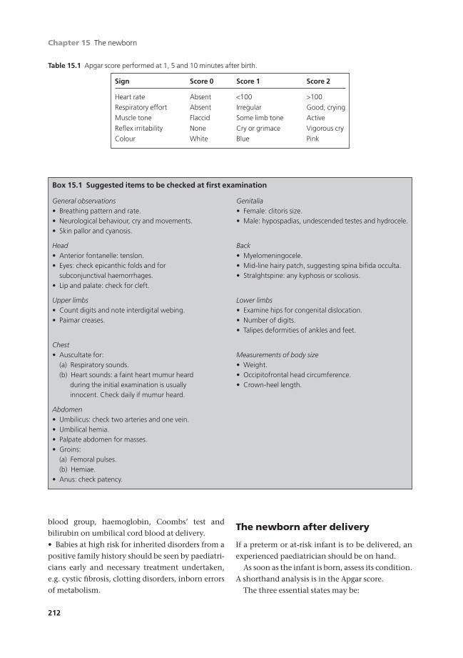
Chapter 15 The newborn
212
blood group, haemoglobin, Coombs’ test and
bilirubin on umbilical cord blood at delivery.
• Babies at high risk for inherited disorders from a
positive family history should be seen by paediatri-
cians early and necessary treatment undertaken,
e.g. cystic fibrosis, clotting disorders, inborn errors
of metabolism.
The newborn after delivery
If a preterm or at-risk infant is to be delivered, an
experienced paediatrician should be on hand.
As soon as the infant is born, assess its condition.
A shorthand analysis is in the Apgar score.
The three essential states may be:
Table 15.1 Apgar score performed at 1, 5 and 10 minutes after birth.
Sign Score 0 Score 1 Score 2
Heart rate Absent <100 >100
Respiratory effort Absent Irregular Good, crying
Muscle tone Flaccid Some limb tone Active
Reflex irritability None Cry or grimace Vigorous cry
Colour White Blue Pink
Box 15.1 Suggested items to be checked at first examination
General observations Genitalia• Breathing pattern and rate. • Female: clitoris size.• Neurological behaviour, cry and movements. • Male: hypospadias, undescended testes and hydrocele.• Skin pallor and cyanosis.
Head Back• Anterior fontanelle: tenslon. • Myelomeningocele.• Eyes: check epicanthic folds and for • Mid-line hairy patch, suggesting spina bifida occulta.
subconjunctival haemorrhages. • Stralghtspine: any kyphosis or scoliosis.• Lip and palate: check for cleft.
Upper limbs Lower limbs• Count digits and note interdigital webing. • Examine hips for congenital dislocation.• Paimar creases. • Number of digits.
• Talipes deformities of ankles and feet.
Chest• Auscultate for: Measurements of body size
(a) Respiratory sounds. • Weight.(b) Heart sounds: a faint heart mumur heard • Occipitofrontal head circumference.
during the initial examination is usually • Crown-heel length.innocent. Check daily if mumur heard.
Abdomen• Umbilicus: check two arteries and one vein.• Umbilical hemia.• Palpate abdomen for masses.• Groins:
(a) Femoral pulses.(b) Hemiae.
• Anus: check patency.
AMI15 6/9/04 5:05 PM Page 212

The newborn Chapter 15
213
• Healthy: pink; effective regular respiration.
• Inadequate breathing: irregular, shallow or gasp-
ing respiration.
• Terminal apnoea: white; floppy; no attempt to
breathe.
Care of the second and third stages is in the
hands of the paediatrician (see Meadow R. &
Newell S. (2001) Lecture Notes on Paediatrics,
Blackwell Publishing, Oxford.).
Healthy• If upper airway contains meconium, blood or
mucus, then laryngoscope and aspirate trachea
and larynx under direct vision.
• Wrap in a blanket and hand to mother.
Inadequate breathing1 If not already present, call paediatrician ur-
gently. Carry out the following:
• Face mask O2.
• Gentle peripheral stimulation.
• Oropharyngeal suction.
• Dry and cover the body with warm towels.
2 If the infant does not respond to this by 1–1.5
minutes (i.e. respiration not established and heart
rate falling below 80 beats/minute) extend neck,
hold jaw forward and apply face mask closely to
face to obtain a tight seal and give intermittent
positive-pressure ventilation (IPPV) through the
mask, at about 30 breaths/minute, 100% O2, 30cm
of water pressure.
3 If the infant still remains blue with inadequate
respiration and a falling heart rate at 2–2.5 min-
utes, laryngoscope, aspirate any mucus or me-
conium under direct vision and intubate. Give
IPPV at pressures of 20cmH2O (a little more if
lungs are stiff) and a rate of 30/minute.
4 As soon as the infant improves with any of the
above (i.e. heart rate 140 beats/minute, spon-
taneous respiration and pink in colour) remove
endotracheal tube, watch for respiration to be
established.
5 Analgesics such as pethidine or morphine
given to the mother late in labour depress
respiration in the infant. Naloxone can be given
(10mg/kg estimated weight i.v. or i.m.). Do not
give naloxone to the baby of a drug-dependent
mother as this may cause withdrawal, fits or
death.
6 There is no indication for analeptic drugs in the
management of birth asphyxia.
7 Preterm babies need intubation earlier than
term babies.
8 Do not perform IPPV with a mask on very small
babies.
Terminal apnoea1 Do not delay resuscitation if, at this stage, the
infant is pale, limp and apnoeic with a heart rate of
less than 100.
2 If not there, call paediatrician urgently.
3 Laryngoscope, aspirate under direct vision and
intubate. Give IPPV: rate 30/minute, 20–30cmH2O
with 100% O2.
• May require naloxone if mother given
pethidine.
4 If still apnoeic —assume birth asphyxia likely
and give sodium bicarbonate i.v. and adrenaline
via endotracheal tube under supervision of
paediatrician.
5 Cord blood at birth to estimate pH and base
deficit.
6 Admit to neonatal unit if:
• Poor response to resuscitation.
• Base deficit >15mEq/l.
7 Let mother hold baby before transfer to neonatal
unit, if possible.
Feeding
Babies are best breast fed; in the UK 60% of
mothers do at first and 30% are still breast feeding
at 16 weeks. Mothers should be given advice in the
antenatal period when breast feeding should be
encouraged.
Breast
Mothers who wish to breast feed put the baby to
the breast for a few minutes at each side in the
labour ward or certainly within 4 hours of birth.
1 The infant will initially obtain only a small
amount of colostrum when he sucks but this con-
tains anti-infective substances. By sucking, the
AMI15 6/9/04 5:05 PM Page 213

Chapter 15 The newborn
214
infant stimulates the production of more
colostrum and then milk.
2 Most of the feed is obtained within the first
3–5 minutes. The time the baby is on the breast
is not proportional to the amount of milk
received.
3 Baby takes the nipple towards the back of his
mouth, not just between the gums.
4 The most satisfactory method of breast feeding
is on demand. A rigid regimen of feeding is not to
be encouraged. Infants who are initially fed on de-
mand usually settle down within a few weeks to a
regular pattern of feeding every 3–4 hours. In gen-
eral, hungry babies cry and it is difficult to overfeed
a breast-fed baby.
5 If lactation is insufficient in the first few days,
breast feeding should not be abandoned. Help and
guidance should be given to the mother by one
midwife, as often breast feeding is not established
until mother and baby are at home and in the
second week.
Factors which help breast feeding• Motivation: encouragement at antenatal classes.
• Good midwife or health visitor with plenty of
time to advise.
• Adequate fluid in mother’s diet.
• Baby awake and mother comfortable.
• Change nappy before feeding to have a con-
tented baby.
• Proper brassiere to hold breasts correctly.
Factors which hinder breast feeding• Mother does not really wish to feed.
• Inverted, retracted nipples.
• Poor fixation on the nipple.
• Child with deformity of palate, tongue or lips.
Advantages of breast feeding1 Breastmilk protein, fat and solute content de-
signed for human babies.
2 Promotes infant–mother bonding.
3 Contains anti-infective agents: active white
cells, macrophages, IgA, IgG and lactoferrin.
4 Eliminates risk of infection from dirty
bottles.
5 Cheap and always available on demand.
Problems in first week1 Engorged breasts. Days 5–10 when breast feed-
ing is uncomfortable.
2 Cracked nipples.
3 Excessive air swallowing during first morning
feed:
• Too rapid flow of milk.
Formula or bottle milk
All cow’s milk preparations in the UK are low solute
milks. They have sodium and protein concentra-
tions similar to those in human milk and most
have added vitamins. Only these preparations
should be used in newborn infants.
1 A normal full-term infant receives about 60
ml/kg on the first day of bottle feeding. This should
then be increased to 150ml/kg/day by the end of
the first week. This is usually divided into 6
feeds/24 hours, e.g. 75ml in each feed for a 3kg
baby.
2 Make up feeds according to instructions. If feeds
are made up for 24 hours, keep in refrigerator.
3 Wash bottles and teats, then sterilize with dilute
hypochloride solution.
4 When feed is due, place bottle in pan of hot
water or microwave and warm milk to temperature
acceptable to back of hand. However, feeding at
room temperature does not cause any problems.
5 Solids should start around 4 months or earlier
if the baby can take them and weighs more than
6.5kg.
Problems1 Protein, fat and solute load not exactly the same
as human breastmilk.
2 Lacks anti-infective properties of breastmilk.
3 Teat hole is too large (too much milk) or too
small (too little milk).
4 Dirty bottles may lead to infections.
5 Cost of formula milks.
Neonatal jaundice
Nearly all babies become mildly jaundiced after
3–5 days. This is due to the breakdown of red blood
cells with haemoglobin F whilst they are replaced
AMI15 6/9/04 5:05 PM Page 214

The newborn Chapter 15
215
with red blood calls with haemoglobin A. For the
majority of babies this is a self-limiting condition
which requires no treatment. If the jaundice per-
sists or the baby becomes deeply jaundiced, sleepy
and refuses to feed then the bilirubin level should
be checked. Using a neonatal bilirubin chart the
value is plotted and the baby may require pho-
totherapy for a few days.
Rhesus negative babies or babies born with an
ABO/other RBC antibody incompatibility with
their mother may become severely jaundiced
within 24–48 hours of birth. If the bilirubin levels
reach very high levels then they may require an ex-
change transfusion to prevent bilirubin being de-
posited in the supraventricular nuclei of the brain
causing kernicterus. Kernicterus is associated with
deafness, delay in development and sometimes
fitting.
Statutory duties of the healthprofessionals after birth
As well as the moral obligations of doctors, mid-
wives and health visitors, the statutory duties are
laid down of each in relation to a woman who is
pregnant, in labour or recently delivered. The
woman may book for her delivery at a variety of
sites; basically they are in an institution or at
home. The woman has a legal right to choose ei-
ther and the midwifery services have to back this
up. The supervisor of midwives in the health dis-
trict has a duty to provide a midwife wheresoever a
woman wishes to deliver and however inadvisable
this might be on medical or midwifery grounds.
General practitioners have no such obligation.
They may not wish to cover the obstetrical adven-
tures of the woman who does not take advice and
they have no obligation to do so. If a midwife look-
ing after a pregnant or labouring woman runs into
difficulty, she calls upon the emergency duty gen-
eral practitioner who then has an obligation to at-
tend and help. Perhaps that help will be passed on
swiftly to the nearest hospital, but the doctor on
duty must advise if called, although he or she may
disapprove of the proceedings.
After the delivery there is an obligation for the
woman to be under the care of a midwife for ten
days although visiting may not be daily. At this
point there is a handover to the health visitor who
takes on the care of the mother and the child. The
health visitors are first rate community workers but
they have to look after the whole spectrum of life
and, with an increasing load of older people, it is
difficult for some health visitors to fit in all they
would wish to do for the recently delivered woman
and her baby.
Self-assessment
15.1 List the five elements of the Apgar score.15.2 A baby is born in the following condition at 1 minute. She is pink with a heart rate of 120bpm. She is not moving
or crying and has taken a few irregular gasps. Her limbs are flaccid but she is moving her facial muscles. What is herApgar score?(a) 9(b) 8(c) 7(d) 6(e) 5
15.3 OSCE question.Candidate’s instructions: During an antenatal visit at 32 weeks, Jane Wooller says that she is unsure whethershe wants to breastfeed her baby and wishes to discuss this with you. You are expected to discuss the advantagesand disadvantages of breastfeeding versus bottle feeding.
Continued on p. 216.
AMI15 6/9/04 5:05 PM Page 215
Chapter 15 The newborn
216
Self-assessment Continued
Role-player’s instructions: You are Mrs Jane Wooller, a 34-year-old woman in her first pregnancy. You are now32 weeks. The midwife discussed breastfeeding with you at the booking visit but you didn’t take much in becauseit seemed so far away then as an issue. You have been to two antenatal classes where breastfeeding has been discussed amongst the mother’s to be. You thought you would definitely breastfeed but found that during the discussion you felt rather revolted by the idea, particularly feeding in public. You are a very private person who,prior to pregnancy, took a great pride in your appearance and figure and you want to get back to your originalclothes size as soon as possible after the birth. Breastfeeding sounds rather invasive of your privacy and you do notrelish leaking breast milk onto your clothes. However, you do want to do the best for your baby. The rest is up tothe role-player’s imagination.
15.4 Which of the following babies are at increased risk of developing significant neonatal jaundice?(a) Group A negative baby born to a group A positive mother.(b) Group A negative baby born to a group A negative mother.(c) Group A positive baby born to a group A negative mother.(d) Group A positive baby born to a group B positive mother.(e) Group A positive baby born to a group A positive mother.
AMI15 6/9/04 5:05 PM Page 216

Part 4
The mature woman
AMI16 6/9/04 5:06 PM Page 217

AMI16 6/9/04 5:06 PM Page 218

Regular bleeding
There are many Latin words to describe abnormal
vaginal bleeding. It is better to use Anglo-Saxon
and describe the symptoms as heavy periods or pro-
longed periods but the classic terms are still in use
and need definition.
• Menorrhagia is an excessive loss of blood (>80ml)
with regular menstruation.
• Metrorrhagia is prolonged bleeding from the
uterus.
• Metro-menorrhagia is heavy and prolonged periods.
• Polymenorrhoea is frequent menstruation.
These may be associated with
1 Complications of early and undiagnosed pregnancy.
• Miscarriage.
• Ectopic pregnancy.
• Hydatidiform mole.
2 Foreign bodies in the uterus —intrauterine
contraceptives.
3 Treatment with hormones especially in
menopausal and postmenopausal women. Break-
through bleeding may occur with synthetic
progestogens given for oral contraception or for
treatment of pelvic disorders.
4 Psychosomatic causes, for example a severe emo-
tional shock, may induce irregular bleeding.
5 An abnormal bleeding tendency may be present
such as leukaemia or Hodgkin’s disease.
6 Hyper- or hypothyroidism may be associated with
menorrhagia or irregular bleeding.
Intermittent bleeding
The causes of non-cyclical abnormal vaginal bleed-
ing not associated with menstruation include:
• lesions of the cervix —polypi, carcinoma,
ectropion;
• lesions of the body of the uterus —endometritis,
fibroids, polypi, adenomyosis, carcinoma, sarcoma.
The level of haemoglobin is generally lower in
women between the ages of 15 and 50 than in
men; this difference is accounted for by blood loss
and the consequent iron deficiency associated
with menstruation and childbearing. Menorrhagia
or metrorrhagia can lead to anaemia but many
women with true heavy loss are able to respond to
the chronic repeated demand on their bone mar-
row, but may require iron therapy.
Menorrhagia
The range of menstrual loss is 10–80ml per cycle. If
above 80ml, this is considered excessive. More
commonly, diagnosis is made on the woman’s
history:
• increase in the number of pads or tampons used
to more than 10 per day;
• starting to pass clots in the menstrual flow;
• use of a pictorial blood loss assessment chart
which gives a semi-quantitative measure of loss.
Up to 30% of women in the later part of men-
strual life complain of heavy periods. Only a half of
219219
Chapter 16
Abnormal vaginal blood loss
AMI16 6/9/04 5:06 PM Page 219
Chapter 16 Abnormal vaginal blood loss
220
these actually lose more than 80ml of blood per pe-
riod. The diagnosis, however, rests on the woman’s
history rather than scientific measurement.
Aetiology
Menorrhagia commonly is a presenting symptom
in:
• fibroids if increase uterine mucosal area;
• adenomyosis (endometrium within the uterine
muscle wall);
• endometritis;
• incorrectly controlled hormone therapy (includ-
ing oral contraception).
Less commonly, it is associated with:
• endometrial polypi;
• an intrauterine device (IUD);
• recent tubal ligation (usually secondary to stop-
ping the contraceptive pill which keeps periods
artificially light);
• functional ovarian tumours;
• disorders of clotting (e.g. von Willebrand’s
disease).
Menorrhagia is not caused by idiopathic throm-
bocytopenic purpura.
After full examination and investigation (ultra-
sound scan, full blood count (FBC), endometrial
biopsy in women over 40) in about 50% of women
with true menorrhagia, no obvious pathology is
found. This is dysfunctional uterine bleeding (DUB)
implicating a malfunction of the endocrine con-
trolling systems. A better name would be menorrha-
gia of unknown origin.
Clinical course
While menorrhagia is most commonly found in
the over 35-year-old, it can occur at any stage in
female life up to the menopause. The causes and
management may be classified according to age
group.
Birth to 18 years of age
See Chapter 4.
Women aged 18–40 years
In this group true dysfunctional bleeding is un-
common and the most likely cause of abnormal
bleeding is some complication of pregnancy or
polycystic ovary syndrome (PCOS) (Chapter 4).
Occasionally heavier periods follow tubal ligation
for sterilization. Endometrial biopsy is rarely
needed in this group for the possibility of malig-
nant disease is unusual.
Women aged over 40All the organic causes of bleeding, including
malignant disease, may occur and it is essential to
exclude them.
Among women without organic disease, three
main patterns of the endometrium are seen on his-
tological examination.
• Atrophic (postmenopausal).
• A mixed pattern with proliferative and secretory
endometrium with possibly endometrial polypi.
This is associated with irregular shedding of the
endometrium for the normal mechanism of
ovulation is disordered so the endometrium is
irregularly or excessively shed.
• Hyperplasia of the endometrium may involve the
glands or the stroma or both. Cystic hyperplasia is
common in perimenopausal women. There is usu-
ally amenorrhoea followed by prolonged bleeding
from the hyper-oestrogenized endometrium. A
similar pattern is seen with an oestrogen secreting
tumour of the ovary. There is no great risk of
cystic hyperplasia going on to carcinoma of the
endometrium. Atypical hyperplasia, however, is
more likely to be associated with endometrial
carcinoma, the risk depending on the degree of
atypia.
Treatment of dysfunctional uterine bleeding
Treatment should never be given without accurate
diagnosis except perhaps in girls under 18 with
puberty bleeding. Blind hormone treatment is
particularly dangerous as malignant disease may
be masked.
AMI16 6/9/04 5:06 PM Page 220

Abnormal vaginal blood loss Chapter 16
221
Diagnosis
Ultrasound shows:
• Endometrial thickness should be less than
12mm in perimenopausal women.
• 5mm in postmenopausal women.
• Endometrial polyps.
• Fibroids.
• Ovarian pathology in women with PCOS. Inves-
tigations should include:
• Endometrial biopsy in:
(a) Women over 40.
(b) Those with abnormal endometrial thickness.
(c) Those with endometrial polyps.
This may be done in the out-patient department by
suction curettage. With appropriate skills, passing
these causes little discomfort, about the same as fit-
ting an IUCD. This avoids admission and the gen-
eral anaesthetic for dilation and curettage (D&C).
However, a fuller curettage is indicated if:
• it is impossible to pass the curette because of a
tight cervix;
• the woman is anxious and cannot relax;
• there is a high risk of malignancy of the
endometrium.
Hysteroscopy may accompany the curettage in
the out-patients department if the gynaecologist is
skilled and properly equipped. It is excellent at
showing endometrial polyps and submucous fi-
broids which could be missed by blind curettage.
In many cases an organic diagnosis will be made
and appropriate treatment be given.
• Cervical smear to exclude carcinoma, in situ
or cervical intraepithelial neoplasia (CIN) (see
Chapter 19).
Drug therapyThis is the first line of treatment because it:
• retains the uterus, a major factor for most women;
• avoids surgery with general anaesthesia;
• is more convenient, being administered in the
outpatients department or GP surgery;
• is much cheaper:
(a) to the Health Service;
(b) to the woman.
Drug treatment may be non-hormonal (first line)
or hormonal (second line) (see Boxes 16.1 and 16.2).
Surgical therapy
Based on removal of the endometrium or the
uterus with its endometrium.
Curettage• Removes outer layers of endometrium but leaves
the basal layers from which new tissue arises in a
month or so.
• Ineffective as a treatment —one or two menses
may be less heavy but are soon back to previous
levels or heavier.
Local ablation• Balloons, heated with 80°C water.
• Microwave local destruction.
• Free flowing heated water to 98°C.
Transcervical ablation• Under vision through a hysteroscope, en-
dometrium is destroyed:
(a) with electrocoagulating loops;
(b) with a laser.
• At present, uses general anaesthesia but needs
only one day in hospital:
(a) uterus retained although lining (mostly)
gone;
(b) after a year, 30% have amenorrhoea and an-
other 60% report lighter blood loss;
(c) if it fails (10–20%), can proceed to either
repeat ablation or to hysterectomy.
Box 16.1 Non-hormonal treatment
Taken during menses hence no problem if accidentalpregnancy is present
Prostaglandin synthetase inhibitorsReduce uterine prostaglandins
FenamatesMefenamic acid, 500mg six-hourly during menstruationAvoid if peptic ulceration
Antifibrinolytic agentsReduce abnormal fibrinolysisAvoid if history of thrombosisTranexamic acid, 1g six-hourly during menstruationEthamsylate, 500mg four-hourly during menstruation
AMI16 6/9/04 5:06 PM Page 221
Chapter 16 Abnormal vaginal blood loss
222
• It is not a minor operation:
(a) requires manipulative skills;
(b) requires expensive equipment;
(c) risks of perforating uterus (1%);
(d) risks of artery damage and bleeding (less
than 1%);
(e) risks of damaging bowel (less than 1%).
• However, saves time and hospital stay:
(a) early return to normal activity;
(b) keeping uterus makes it popular;
(c) contraception is still required unless there is
complete amenorrhoea.
Hysterectomy• Removal of the uterus stops periods.
Abdominal hysterectomy• Major procedure with general anaesthesia.
• Abdominal scar to heal.
• Hospital stay 4–6 days.
• Risks of damage to ureters <1%.
• Risks of damage to the bladder <1%.
• Risks of damage to the bowel <1%.
Vaginal hysterectomy• Needs to be a mobile, adhesion-free uterus.
• Uterus not greatly enlarged.
• Helps if there is some prolapse.
Laparoscopy assisted hysterectomy• Smaller abdominal incision.
• Needs skilled laparoscopic surgeon.
• Does the difficult part of a vaginal hysterectomy
from above ligating tubes, ovarian suspensory liga-
ment and round ligament via laparoscopy. Then
an easy vaginal removal.
• Has all the complications of laparoscopic
surgery.
Hysterectomy does not have to be accompanied by
removal of ovaries.
Indications for oophorectomy• If ovaries abnormal.
• If family history of ovarian cancer.
• Consider if over 45 years old and post-
menopausal. Full informed consent is
essential.
Retain:
• if menses cycling regularly;
• if ovaries healthy at surgery;
• if patient’s wish.
Atrophic vaginitis
Secondary to a lack of oestrogens postmenopaus-
ally, the epithelium becomes thin, smooth and
shiny with subepithelial haemorrhages. Low-grade
Box 16.2 Hormonal treatment
This aims at imitating or restoring the normal endocine cycle
Norethisterone • 5mg three times a day for six to nine monthsMedroxyprogesterone • Best for those with anovular cycles
acetate • Cheap and good for emergency treatment: 10 mg three times a day for very heavy bleeding
Danazol • 200mg daily continuously for three to six months• Derivative of testosterone so anti-oestrogenic at
hypothalamus and at endometrium• Expensive. May be weight gain and
androgenizationOral contraceptive • Dose according to brand but probably best to use
cyclical combined oestrogen (30mg) and progestogen (0.25mg)
• Praetical, cheap and easily availableMirena IUS • Progestogen IUS 75mg/day to uterine lining only
AMI16 6/9/04 5:06 PM Page 222

Abnormal vaginal blood loss Chapter 16
223
infection from pathogens, which can more easily
penetrate the surface, may occur.
Symptoms• Vulval soreness.
• Superficial dyspareunia.
• Pink discharge.
• Introital shrinking.
Physical signsRed shiny epithelium with skin cracking and sub-
cuticular haemorrhages. Occasionally causes post-
menopausal bleeding. It is important to exclude
other causes such as carcinoma.
TreatmentIf there is secondary infection, antibiotics to which
the organism is sensitive may be given. Topical
oestrogen creams or pessaries can be used, e.g.
dienoestrol or Premarin once or twice a week. This
is enough to thicken the epithelium and reduce the
pH, but insufficient to produce excessive endome-
trial stimulation or other causes of bleeding.
Endometrial polypi
If the endometrium hypertrophies under oestro-
gen stimulation, areas of it may protrude above the
surface producing a sessile or eventually a pedun-
culated polyp. This may not be shed at the time of
menstruation, but forms a site for persistent symp-
toms and signs.
SymptomsThere may be intermenstrual spotting or postmen-
strual spotting.
SignsThere would be no signs, but a hysteroscopy would
show the polyp.
ManagementEndometrial polypi should be removed. Those in-
side the uterus will require an anaesthetic and have
to be twisted off with a polypi forceps diathermy
loop. Should the endometrial polyp stalk be long
and the polyp present at the cervix, it is probably
wise not to remove it in the out-patients depart-
ment, because this could lead to heavy bleeding. A
D&C with hysteroscopy should be performed at
the same time to allow removal of any other polypi
that are higher up.
Pre- and postoperative care
Gynaecological operations are best learnt in the
operating theatre, preferably acting as a scrubbed
assistant, but the pre- and postoperative care must
be understood by a wider range of medical person-
nel than just the surgeon.
Preoperative care
HistoryA complete history is taken with special attention
to any recent illness. The date of the last period
should be noted, to ensure the patient is not preg-
nant. Drugs and medicines recently taken should
be noted —these may include hormones, especially
oral contraceptives, antibiotics and tranquillizers.
ExaminationA general examination is made. The heart and
lungs are examined and blood pressure measured.
Examination of the abdomen and pelvis follows.
The legs are inspected for varicosities.
InvestigationsInvestigations include an examination of haemo-
globin. Except in an emergency, it is unwise to
carry out major surgery with a haemoglobin level
below 10.5g/dl, or minor surgery below 8.5g/dl.
In women of Negro or Asian origin, a test for
haemoglobinopathies is essential.
The blood group and rhesus factor are deter-
mined and serum saved in the laboratory. If there is
a probability that transfusion will be needed during
the operation, blood is cross-matched in readiness.
The urine is tested for albumin, bacteria and
sugar. If there is a suspicion of a urinary infection,
a mid-stream sample is sent for bacteriological
examination. Renal function tests are advisable if
there is a suspicion of abnormal kidney function.
AMI16 6/9/04 5:06 PM Page 223

Chapter 16 Abnormal vaginal blood loss
224
An X-ray of the chest may be taken if indicated.
Urography and magnetic resonance imaging (MRI)
are done before operations when the ureters may
be involved, as in Wertheim’s hysterectomy. An
electrocardiogram is advisable in patients with any
suggestion of heart disease. Other investigations
may be suggested by the examination or the nature
of the case.
A pregnancy test should be performed prior to
surgery.
PreparationThe patient should be weighed on admission or
in the out-patient clinic. Shaving of the vulva has
now generally been abandoned for minor cases al-
though most surgeons prefer it for major vulval
and vaginal surgery. Postmenopausal women
with prolapse may be helped by preoperative
treatment with oestrogen, given before admission
in the form of oestrogen by mouth or vaginal
cream. Antibiotics, e.g. Flagyl or Augmentin, are
usually given before major elective surgery.
Thromboprophylaxis: TED stockings should be
worn intraoperatively and until the woman is fully
mobilized. During major surgery flowtron boots
which inflate and deflate regularly should be
used during the operation. Daily prophylactic
doses of low-molecular-weight heparin should be
considered for all women at moderate to high
risk undergoing major surgery, during and after
surgery.
The patient should have nothing by mouth for
at least 6 hours before the operation. If the opera-
tion may involve the intestines or rectum, the
bowel is emptied and prepared by the use of
succinyl sulphathiazole, neomycin or another
suitable preparation.
A physiotherapist should visit every patient ide-
ally before the operation and certainly everyone
for major surgery. He or she can teach breathing
and leg movements for the postoperative period.
Valid consent must be given in writing for the
operation by the patient herself. This must be
clearly and legibly countersigned by a doctor, who
should have explained the operation and its possi-
ble sequelae. Girls aged 16 or over sign consent for
the operation on their own behalf; for those under
that age, the consent of a parent or legal guardian
is usually necessary except in an emergency. There
may be difficulty in the case of a girl under 16 re-
questing a termination of pregnancy and insisting
that her parents are not to be informed. In such a
case, the doctor will have to exercise discretion and
act in what he or she considers to be the girl’s best
interest.
At the time that consent for the operation is
obtained, an explanation should be given of the
procedure which is contemplated, its nature and
effects, including possible complications. These
should be written on the consent form. In the case
of a married woman, her husband’s consent is not
legally necessary in the UK for operations which
lead to sterility such as hysterectomy or steriliza-
tion, but it is important that counselling of the
couple takes place. It should be made clear in the
case of operations for sterilization or vasectomy
that the operation should be considered irre-
versible; on the other hand it must be emphasized
that a few failures occur with sterilizations. This
advice should be recorded in the case notes.
Counselling in general is important since many
women do not understand the anatomy of their
pelvic organs and have unnecessary fears concern-
ing the effects of operations, in particular their ef-
fects on sexual function. Explanation, if necessary
illustrated by a model or diagram, will help to dis-
pel these fears.
Postoperative care
A period of recovery is required after any surgical
operation. After minor operations, such as hys-
teroscopy, the patient can go home on the same
day. She should not drive or operate machinery for
two days after general anaesthetic. She must be
warned to expect some bleeding for up to 14 days
after the operation. More profuse bleeding can fol-
low deep cauterization or conization of the cervix;
this may on some occasions be enough to require
readmission and possible suture of the cervix.
These women should be advised to refrain from in-
tercourse for 6 weeks.
After major surgery such as uncomplicated hys-
terectomy or prolapse repair, patients are encour-
AMI16 6/9/04 5:06 PM Page 224
Abnormal vaginal blood loss Chapter 16
225
aged to get up from bed and move about on the day
following the operation. Breathing exercises and
leg movements are begun from the day after the
operation. The length of stay in hospital varies but
many can leave after 3–6 days. Before departure a
clear explanation of the operation and the progno-
sis must be given by a doctor. An adequate period
of convalescence at home is necessary before re-
turning to work and normal activity.
The patient may be examined six weeks after
the operation. She must be encouraged to return to
normal life. Intercourse may be resumed as soon
as the vagina is healed. If the ovaries have been
removed premenopausally, the woman should be
offered oestrogens by tablet, patch or implant.
Patients treated for carcinoma must be followed up
carefully by gynaecological oncologists.
Postoperative complications
During the first 12 hours after an operation, the pa-
tient must be carefully observed for the following:
• respiratory failure or obstruction to the airways;
• shock;
• haemorrhage;
• cardiac failure.
She should be nursed in a recovery unit until she
has recovered consciousness and only then re-
turned to a general ward. The pulse rate and blood
pressure should be taken and charted every quarter
of an hour for the first two hours and thereafter
every few hours for the first 12 hours, longer if
there is any anxiety.
Pain must be relieved by adequate doses of anal-
gesics such as morphine or pethidine. Patient-
controlled analgesia, with the woman controlling
the flow of weak solutions of analgesia intra-
venously, is very useful for recovery from elective
gynaecological surgery. Addition of promazine or
chlorpromazine increases the effect of analgesics
and helps to prevent postoperative vomiting.
Haemorrhage1 Primary, occurring during the operation and
requiring immediate transfusion.
2 Delayed, occurring during the immediate post-
operative period, is generally due to a slipped liga-
ture or a bleeding vessel in the vagina or cervix.
Blood transfusion is given and the patient
returned to the operating theatre to deal with the
haemorrhage.
3 Secondary, occurring up to 14 days after opera-
tions and generally from the vagina or cervix,
but occasionally from the abdominal wound.
Infection is commonly associated, but suture of the
bleeding area and blood transfusion still may be
needed in all but the slightest cases. After cauteri-
zation of the cervix there is generally some bleed-
ing about the 10th and 12th day and patients
should be warned to expect this.
4 Anaemia is common after gynaecological opera-
tions and should be prevented with a correct diet
and iron therapy.
Respiratory tractComplications of a general anaesthetic include
sore throat, tracheitis, bronchitis, broncho-
pneumonia and massive collapse of the lungs.
Breathing exercises should be given after general
anaesthetics. Pulmonary infection should be
treated with antibiotics. Chest symptoms after
the first week sometimes indicate pulmonary
embolism.
Urinary tractRetention of urine is common after gynaecological
operations and it may be complete or partial.
Complete retention of urine often occurs after
hysterectomy or repair of prolapse. Rarely unex-
plained retention occurs after a minor operation
such as hysteroscopy.
Treatment is by immediate catheterization
usually emptying the bladder once. If it must be
repeated, an indwelling catheter has to be put in
for a few days. Some surgeons prefer suprapubic
catheterization.
Partial retention of urine is common after
operations for prolapse and a catheter should be
passed for residual urine five days after operation.
Catheter drainage should continue until the resid-
ual urine is below 200ml.
Cystitis and pyelonephritis are very common
after gynaecological operations. A catheter speci-
men or a mid-stream specimen should be exam-
AMI16 6/9/04 5:06 PM Page 225
Chapter 16 Abnormal vaginal blood loss
226
ined for evidence of infection after all major opera-
tions and suitable treatment given.
Poor or absent urine output may be due to ob-
struction to the ureters which may be accidentally
injured, ligated or obstructed by a haematoma; it
may also be reflex blockage. Warning is often given
by unilateral loin ache and a slight temperature. It
is a very serious complication and must be dealt
with urgently if necessary with relieving surgery by
a urologist.
Incontinence of urine through the urethra some-
times occurs after catheterization and in elderly
women; it is usually transient. Persistent incon-
tinence suggests a fistula. Urinary fistulae may be
vesico-vaginal or uretero-vaginal. These may be
caused by trauma at operation, by haematoma for-
mation or by difficult obstetric delivery. They also
result from malignant disease.
An uretero-vaginal fistula may follow opera-
tions, especially Wertheim’s hysterectomy if
the ureters are extensively dissected so that their
blood supply is imperilled and they become devas-
cularized.
Venous thrombosisTwo types can occur after surgery.
Phlebothrombosis is primary venous thrombosis
and generally begins in the deep veins of the calf;
predisposing causes are:
• trauma;
• anaemia;
• stasis;
• high oestrogen levels.
Thrombophlebitis is caused by infection, general-
ly in the pelvic veins initially and spreading to in-
volve the iliac veins.
Pulmonary embolism can follow thrombosis and
if massive is rapidly fatal. Small emboli cause pul-
monary infarction.
Prevention of thrombosis and embolism consists
of:
• the use of pneumatic boots and leggings during
the surgery;
• elasticated stockings worn before, during and
after operation;
• early movement;
• avoidance of anaemia;
• prompt treatment of infection;
• prophylactic low-dose heparin before and just
after surgery.
The contraceptive pill should ideally be stopped
four to six weeks before major elective surgery.
In established thrombosis, heparin is given
initially intravenously in a continuous dose of
1000–1500iu per hour or by separate intravenous
injections of 10000iu every six or eight hours. Cal-
cium heparin can be given subcutaneously. War-
farin is started with a loading dose of 10mg for two
days followed by a daily dose which is determined
by the clotting time. In proven DVT this should be
continued for three months and, if there was a pul-
monary embolism, for six months.
Self-assessment
16.1 OSCE question.
Candidate’s instructions: A 37-year-old woman, Elizabeth Parker, has come to see you in your GP surgery. She has only recently moved to your area. She is complaining of heavy periods. Please take a history. You have 7minutes.
Role-player’s instructions: You are Mrs Elizabeth Parker, a 37-year old. Over the last 2 years your periods have become increasingly heavy. They are regular every 28 days and last for 7 to 10 days. They used to last only 5 days. You have noticed that the first 4 days are particularly heavy with clots the size of 50p pieces. On the first day you can bleed so heavily that you have to change every 30 minutes to 1 hour. At night you wear two pads, a tampon and an incontinence pad and still have to change in the middle of the night. You have painful periods but this has not changed. You have three children and you were sterilized when you were 35. Prior to thisyou had been on the combined oral contraceptive pill. You smoke 25 cigarettes a day so you had stopped the pill.Your last smear test was 2 years ago and was normal. You are otherwise fit and well. All other details are up to therole-player.
AMI16 6/9/04 5:06 PM Page 226

Abnormal vaginal blood loss Chapter 16
227
Self-assessment Continued
16.2 From the list below select the three most relevant investigations for Mrs Parker.(a) Endometrial biopsy.(b) Transvaginal ultrasound scan.(c) Hormone profile.(d) High vaginal swab.(e) Cervical smear.(f) Hysteroscopy.(g) Full blood count.(h) Thyroid function tests.
16.3 The investigations are all normal. What is the most appropriate first line treatment?(a) Mirena IUS.(b) Norethisterone from day 15–26 of the cycle.(c) Antifibrinolytic therapy during menses.(d) Paracetamol during menses.(e) Endometrial ablation.
16.4 A woman is admitted for a total abdominal hysterectomy. Which of the following are routine prophylactic measures for all women undergoing this procedure?(a) Augmentin 1.2g i.v. intraoperatively.(b) Subcutaneous low-molecular-weight (LMW) heparin intraoperatively.(c) Flowtron boots intraoperatively.(d) TED stockings.(e) Penicillin V 1g i.v. intraoperatively.
16.5 When consenting a woman for a total abdominal hysterectomy which of the following should be included on theconsent form as possible complications?(a) Infection.(b) Haemorrhage.(c) Removal of the ovaries.(d) Damage to the ureters.(e) Damage to the bladder.
AMI16 6/9/04 5:06 PM Page 227

228
Many diseases causing lower abdominal symptoms
are gynaecological in origin. The general surgeon
and gynaecologist must both be trained to recog-
nize and deal with them. When available, consul-
tation will take place, but delay and mortality are
linked and acute abdominal conditions do not
leave time for leisurely consultations. If the condi-
tion is obviously gynaecological in origin, it is best
dealt with by gynaecologists as they are more used
to conservation of genital tract tissue, particularly
the ovaries.
Acute intra-abdominal emergencies present with
pain, shock, vomiting or abdominal distension.
The first of these is the most common in gynaeco-
logical conditions.
Pain
Pain in pelvic organs may arise from:
• inside the organ with irritation of its lining or
stretch of its walls;
• stretch of the visceral peritoneum over the
organ;
• involvement of the parietal peritoneum in
proximity.
For example, an ectopic pregnancy can cause
pain from:
• damage to the muscle of the oviduct by tro-
phoblast invasion and bleeding;
• stretch of the peritoneum from the broad ligament
over the fallopian tube at the site of the pregnancy;
• blood spilt onto the peritoneum by the rupture
of the tube (acute) or trickling from the outer end
of the tube (chronic).
The first two origins of stimuli are mediated
by the autonomic nervous system with poor
localization; hence non-specific pain in the pelvis
results.
The parietal peritoneum is innervated by the
somatic nervous system and localization may be
more specific. The peritoneum of the pouch of
Douglas, however, has a poor nerve supply and is
often undemonstrative. Since this is the area in
which the tubes and ovaries spend much of their
time, it makes pain localization in the abdominal
wall difficult in pelvic conditions in the earlier
stages, but signs are easier to detect on pelvic and
rectal examination.
Shock
A sudden deterioration in a woman’s vital state
may be characterized by:
• a rise followed by a fall in pulse rate;
• a fall in arterial blood pressure;
• pallor;
• faintness and later unconsciousness.
This may be due to either:
• true hypovolaemia, e.g. a ruptured ectopic preg-
nancy with two litres of blood in the peritoneal
cavity;
• relative hypovolaemia, e.g. excess of autonomic
Chapter 17
Pelvic pain
AMI17 6/9/04 5:08 PM Page 228
Pelvic pain Chapter 17
229
stimulation after peritoneal irritation with a sud-
den release of pus or blood.
Pallor, sweating, agitation and restlessness are
traditional indications of shock, of which pallor is
the most important for prognosis in the gynaeco-
logical field. Fainting and unconsciousness come
later in shock and may be considered as signs of
more extensive involvement.
Nausea and vomiting
These happen rarely in non-pregnant gynaecologi-
cal conditions but have a number of causes.
• Stimulation of a large number of nerve endings
of the peritoneum overlying an affected pelvic
organ, e.g. torted ovarian cyst.
• The direct action of toxins on the central nerv-
ous system from infective organisms. Pelvic in-
flammation often becomes localized early and
toxins are released intermittently into the general
bloodstream.
• Vomiting is a common accompaniment of early
pregnancy. Many gynaecological conditions hap-
pen in early gestation so vomiting might be noth-
ing to do with the acute condition in the pelvis but
more with the human chorionic gonadotrophin
(hCG) produced by the pregnancy trophoblast.
Distension
Distension is unusual in gynaecological conditions
but common in alimentary tract ones, particularly
those of the large bowel. In consequence, if disten-
sion is present, it is probably due to an associated
bowel problem rather than a gynaecological one.
Diagnosis
The triad of history, examination and investiga-
tions applies here as anywhere else in medicine.
This section is to be read in conjunction with the
account of gynaecological diagnosis making in
Chapter 2.
History
Women in severe acute abdominal pain do not like
having long histories taken. Hence, the questions
must be tailored to the situation.
Pain• Characteristics of the pain site, time and nature.
• Relationship of pain to various body functions,
e.g. vaginal bleeding or micturition.
• Past gynaecological or obstetrical events.
• Menstrual history, i.e. details of last menstrual
period.
• Symptoms of a possible pregnancy, e.g. breast
changes and the woman’s own impressions.
Examination
A general examination must cover a number of
points.
• Paleness (conjunctival assessment).
• Pulse rate.
• Arterial blood pressure.
• Temperature.
• Abdominal examination helps to localize pelvic
causes.
• Observation will show old scars and the degree of
distension; the site of the pain can be elicited from
the woman at this point.
• Gentle palpation of the abdomen leading to the
lower pelvic zones may help localization further.
• Firmer examination —if tenderness allows; this
will reveal guarding or any rebound tenderness.
• Many pelvic abdominal conditions do not have
specific localizing signs in the abdomen and so,
therefore, any opinion should await the perform-
ing of a bimanual vaginal examination.
• At bimanual vaginal assessment tenderness from a
pelvic organ will obviously limit the thoroughness
of the examination because the woman will guard
if there is pain on moving the cervix. This is called
cervical excitation (Fig. 17.1); moving the cervix to
the side of an ovarian or tubal mass will cause in-
tense pain by a further stretch of the overlying peri-
toneum. Moving the cervix towards the opposite
side will decrease the pain by releasing the tension
of the peritoneum.
• Rectal examination may be needed but usually
one can assess acute pouch of Douglas problems
at a vaginal assessment. If structural changes are
AMI17 6/9/04 5:08 PM Page 229
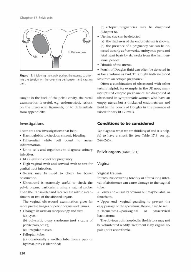
Chapter 17 Pelvic pain
230
sought in the back of the pelvic cavity, the rectal
examination is useful, e.g. endometriotic lesions
on the uterosacral ligaments, or to differentiate
from appendicitis.
Investigations
There are a few investigations that help.
• Haemoglobin to check on chronic bleeding.
• Differential white cell count to assess
inflammation.
• Urine cells and organisms to diagnose urinary
infection.
• hCG levels to check for pregnancy.
• High vaginal swab and cervical swab to test for
genital tract infection.
• X-rays may be used to check for bowel
obstruction.
• Ultrasound is extremely useful to check the
pelvic organs, particularly using a vaginal probe.
Then the transmitter and receiver are within a cen-
timetre or two of the affected organs.
The vaginal ultrasound examination gives far
more precise images of pelvic organs and tissues.
• Changes in ovarian morphology and size:
(a) cysts;
(b) polycystic ovary syndrome (not a cause of
pelvic pain per se);
(c) irregular masses.
• Fallopian tube:
(a) occasionally a swollen tube from a pyo- or
hydrosalpinx is identified;
(b) ectopic pregnancies may be diagnosed
(Chapter 8).
• Uterine size can be detected:
(a) the thickness of the endometrium is shown;
(b) the presence of a pregnancy sac can be de-
tected as early as five weeks, embryonic parts and
fetal heart beats by six weeks from the last men-
strual period.
• Fibroids of the uterus.
• Pouch of Douglas fluid can often be detected in
as low a volume as 7ml. This might indicate blood
loss from an ectopic pregnancy.
Often a combination of ultrasound with other
tests is helpful. For example, in the UK now, many
unruptured ectopic pregnancies are diagnosed at
ultrasound in symptomatic women who have an
empty uterus but a thickened endometrium and
fluid in the pouch of Douglas in the presence of
raised urinary hCG levels.
Conditions to be considered
We diagnose what we are thinking of and it is help-
ful to have a check list (see Table 17.3, on pp.
244–245).
Pelvic organs (Table 17.1)
Vagina
Vaginal traumaIntercourse occurring forcibly or after a long inter-
val of abstinence can cause damage to the vaginal
tube.
• Lower end —usually obvious but may be labial or
fourchette.
• Upper end —vaginal guarding to prevent the
easy passage of the speculum. Hence, hard to see.
• Haematoma —paravaginal or paracervical
haematoma.
The obvious point needed in the history may not
be volunteered readily. Treatment is by vaginal re-
pair under anaesthesia.
Remove pain
Pain
Figure 17.1 Moving the cervix pushes the uterus, so alter-ing the tension on the overlying peritoneum and causingpain.
AMI17 6/9/04 5:08 PM Page 230

Pelvic pain Chapter 17
231
sequently undergo calcification (visible on X-rays
and ultrasound).
• Red —necrobiosis, particularly encountered in
the mid-trimester of pregnancy or the early puer-
perium. This breakdown of blood supply by
thrombosis leads to necrosis and suffusion with
red blood cells.
• Sarcomatous —rare malignant change reported in
0.2–0.4% of fibroids examined in asymptomatic
older women at autopsy.
Symptoms• None —a pelvic swelling is found incidentally on
examination.
• Occasional tightness of waistband of clothes.
• Pressure —bladder compression causing daytime
frequency and occasionally impaired urinary
stream. In the supporting ligaments it causes back-
ache and overall sensation of pelvic heaviness.
• Pain —associated with red degeneration or tor-
sion of subserous pedicles. Dysmenorrhoea may
indicate the presence of a submucous fibroid.
• Menstrual disturbances:
(a) menorrhagia —heavy bleeding;
(b) metrorrhagia —prolonged menses;
(c) irregular, intermittent bleeding —often asso-
ciated with polyps and other surface lesions.
Investigations• Ultrasound —to define the location, the dimen-
sions and the consistency.
Uterus
Uterine fibroidsAlternatively named myomata or leiomyomata,
uterine fibroids are the commonest of all pelvic tu-
mours. They are benign fibromuscular swellings,
arising in the muscle wall of the uterus. Fibroids are
oestrogen sensitive.
• Submucous —lying immediately below the en-
dometrium and enlarging the surface of the uter-
ine cavity often leading to menorrhagia. Fibroids
may become pedunculated forming polypi which
can extrude through the cervix.
• Intramural —the commonest site for fibroids, sur-
rounded by smooth muscle, enlarging the uterine
wall and distorting venous drainage.
• Subserous —fibroids just beneath the peritoneum
on the outer uterine surface (Fig. 17.2). May be on
an elongated stalk (pedunculated) with a risk of
torsion; may grow into the broad ligament.
DegenerationUterine fibroids frequently outgrow their blood
supply and degenerate.
• Hyaline —an aseptic necrosis with loss of muscle
cell structure. This may lead to calcification.
• Cystic —a sequel to hyaline change with sub-
sequent breakdown and cyst formation giving a
honeycomb appearance.
• Fatty —in which partial necrosis results in the de-
velopment of fatty substances which may sub-
Broadligament
Pedunculated
Subserous
IntramuralSubmucous
Cervical
Polyp
Figure 17.2 Uterus with fibroids.
AMI17 6/9/04 5:08 PM Page 231
Chapter 17 Pelvic pain
232
DiagnosisBimanual palpation reveals hard rounded non-
tender often bosselated mass, moving when the
cervix is displaced.
• Grow fast in pregnancy.
• Shrink:
(a) at the menopause;
(b) with antigonadotrophic hormone therapy.
PathologyFibroids may be single or multiple (occasionally
numbering greater than 100) and vary in size from
a seedling to a football. They form a pseudocapsule
by compressing surrounding uterine muscle, a
process helpful to the surgeon at myomectomy.
AetiologyCause is unknown:
• rarely found under the age of 30 years;
• commoner in Afro-Caribbean populations;
• commoner in the nulliparous and women with
low fertility;
• often a family history.
Differential diagnosis• Pregnancy —particularly if fibroids have been
softened by cystic degeneration.
• Ovarian tumour —often cystic, unilateral and
does not move with cervical displacement.
• Adenomyosis —more commonly causes uniform
diffuse and tender uterine enlargement.
Treatment• If small and asymptomatic, conservative man-
agement with annual examination and ultrasound
monitoring of size is sufficient. This is especially
used in women over 40 because fibroids do not
grow after menopause and may shrink.
• Menstrual or pressure symptoms may dictate
surgery.
• Pain —requires analgesia.
• Heavier and longer periods with anaemia
are the commonest indication for proceeding to
surgery.
• Embolization under radiological control: a can-
nula is passed into the uterine arteries via the
femoral artery. The uterine arteries are embolized
by injecting tiny silicon particles causing the
fibroids to degenerate. Pain relief is essential for
48 hours. In women with large/multiple fibroids
there is an increased risk of hysterectomy com-
pared to surgical treatment. Pyrexia and abscess
formation can occur. It is not recommended for
women who wish to preserve their fertility, al-
though live births have been reported following
the procedure.
Surgery• Abdominal hysterectomy —suitable when fam-
Table 17.1 Pelvic diseases which may present as acute
abdominal emergencies.
Organ Condition
Non-pregnant Vagina Trauma
Uterus Fibroid
Torsion
Degeneration
Adenomyosis
Endometritis
Pyometra
Fallopian tubes Salpingitis
Pyosalpinx
Torsion
Ovaries Tumours
Simple cyst
Torsion
Bleeding
Rupture
Peritoneum Endometriosis
Early pregnancy Uterus Abortion
Impacted
retroversion
Red degeneration
of fibroids
Fallopian tubes Ectopic pregnancy
Torsion
Ligaments Round ligament
Stretch
Extra-pelvic Vomiting in
pregnancy
Pyelonephritis
Appendicitis
Rectus haematoma
AMI17 6/9/04 5:08 PM Page 232

Pelvic pain Chapter 17
233
ily is complete with women over the age of 40 or
when the uterus is grossly enlarged and distorted
by multiple fibroids.
• Vaginal hysterectomy —when fibroids are small
and few in number and there is an associated pro-
lapse of the uterus.
• Myomectomy —in young women whose families
are incomplete or when there is a personal desire to
retain the uterus. The procedure is often vascular
and may cause scarring, with adhesion formation
impairing fertility. If the fibroids are numerous it
may be impossible to remove them all, and growth
of the remainder may cause problems in the future.
Women undergoing this procedure should be
warned that the surgeon may have to proceed to a
hysterectomy and consent to the possibility of this
(1% risk).
• Submucous fibroids may be resected with laser or
diathermy through a hysteroscope.
Effects on childbearing• Implantation over a fibroid may lead to spon-
taneous miscarriage.
• Pain may develop from red degeneration.
• Premature labour may develop if the fibroids are
large, multiple and/or undergo red degeneration.
• Dysfunctional uterine contractions may follow
the interruption of smooth waves of electrical stim-
ulation by masses of inert, non-myometrial tissue.
• The pelvis may be obstructed causing malpresen-
tation or even obstructed labour. This is unusual
because most fibroids usually move up as the
uterus grows in pregnancy.
• Postpartum haemorrhage and retained placenta
are more common.
• Management in pregnancy is conservative.
The aim should be to secure a vaginal delivery. If a
Caesarean section becomes necessary, the incision
in the uterus should be manoeuvred around the
fibroids. They should not be removed or incised as
severe haemorrhage may develop leading to the
need for a hysterectomy.
Uterine adenomyosisAdenomyosis is a condition where endometrial
glands of stroma are found within the uterine mus-
culature. If localized to one site, it is called an ad-
enomyoma. It is like diffuse endometriosis in the
muscle of the uterus.
PathologyThe uterus is enlarged and thick-walled with no
pseudocapsule formation, as in fibroids. The endome-
trial glands sometimes do not all menstruate, as they
derive from the basal layer of the endometrium.
SymptomsThis condition is most frequent in women
aged 35–40, with reduced fertility. The symptoms
usually are:
• dysmenorrhoea;
• menorrhagia;
• dyspareunia.
SignsA uniformly enlarged uterus which is rarely larger
than 12cm. It is tender on palpation, particularly
premenstrually.
Differential diagnosis• Uterine fibroids.
• Early pregnancy.
• Uterine infection.
Treatment• Conservative use of progestational therapy, e.g.
norethisterone 10–20mg daily.
• Danazol 200–400mg daily.
• Gonadotrophin analogues: buserelin or
goserelin.
All hormone regimes aim to suppress menstrua-
tion, but each carries side-effects and usually can-
not be used for more than some months at a time.
SurgeryAbdominal hysterectomy is the treatment of
choice, although occasionally resection of the af-
fected area can be considered.
EndometritisThe condition is usually acute and associated with
ascending infection. The disease may result from:
• post-abortal infection;
• criminal abortion;
AMI17 6/9/04 5:08 PM Page 233

Chapter 17 Pelvic pain
234
• excessive curettage;
• intrauterine device infection;
• postpartum, particularly after Caesarean section.
It rarely results from blood-borne tuberculosis.
FindingsAcute infection:
• irregular bleeding;
• uterine tenderness.
Chronic infection:
• occasionally secondary amenorrhoea and sec-
ondary infertility, caused by the development of
intrauterine adhesions leading to partial or com-
plete occlusion of the uterine cavity (Asherman’s
syndrome).
Pyometra• Infection leading to pus formation may be asso-
ciated with blocking of the fallopian tubes and the
cervix.
• Pyometra are commoner in older women.
• A bag of pus builds up pressure and distends the
uterus causing:
(a) pain from the stretch of the muscle wall;
(b) occasional acute bursts of toxaemia as a
bolus of pus is forced into a vein and so to the
vascular network;
(c) chronic infection with low-grade tempera-
ture and malaise;
(d) occasionally pus is forced through the cervix
to produce a purulent or bloodstained discharge.
• Many pyometra are associated with cancer of the
endometrium (Chapter 20).
Any woman with such symptoms should have a
hysteroscopy and a D&C under appropriate anti-
biotic cover to exclude endometrial malignancy.
Fallopian tubes
Torsion of the fallopian tubeThis rare cause of lower abdominal pain is usually
associated with ovarian torsion on a long pedicle.
Treatment is by laparotomy or laparoscopy and de-
pends upon the degree of devascularization of the
tube and ovary:
• if on unwinding the tissues are healthy, they are
best conserved with a securing suture to the side
wall of the pelvis to prevent retorsion;
• if the tissues are devitalized, the ovary and tube
must be removed.
SalpingitisAn ascending infection from the vagina through
the cervix is the usual cause of salpingitis. It is
often associated with:
• intercourse;
• transcervical surgery (D&C or evacuation);
• intrauterine foreign bodies such as an intrauter-
ine device (IUCD);
• retained products of conception;
• very rarely, blood-borne infection.
Acute salpingitisThe fallopian tubes become red, swollen and dis-
torted, often obstructed at the abdominal end so
a pyosalpinx forms later becoming a hydrosalpinx.
Peritoneal inflammation with adhesions to the
serosal surface occurs, leading to pelvic abscess
and, if severe, to septicaemia. The condition is usu-
ally bilateral. The destruction of the cilia later leads
to infertility. Chronic hydrosalpinges may become
reinfected.
Clinical features• Pyrexia, often with a temperature higher than
39°C.
• Tachycardia.
• Dehydration.
Abdominal examination• Lower abdominal pain with guarding.
• If parietal peritoneum is involved, rebound
tenderness.
• Distension.
Vaginal examination• Cervical excitation pain (bilateral).
• Tender, normal sized uterus.
• Fullness in the fornices and tenderness over the
tubes.
• Vaginal discharge.
AMI17 6/9/04 5:08 PM Page 234

Pelvic pain Chapter 17
235
Investigations• Organisms may be isolated from cervical dis-
charge.
• Gonorrhoea (see Chapter 6).
• Escherichia coli, haemolytic streptococcus and
staphylococcus are often found in the puerperium
and post-abortion.
• Clostridium welchii may thrive on dead tissue, e.g.
placental products.
• Chlamydia is a common secondary organism.
• Leucocytosis (in excess of 20 ¥ 109/l).
• Laparoscopy is the only certain way of making a
true diagnosis. Remember to take serosal swabs.
Differential diagnosis• Appendicitis —pain is usually central then radiat-
ing to the right iliac fossa; the fever is lower.
• Ruptured ectopic pregnancy —if there is intraperi-
toneal bleeding, symptoms such as faintness and
shoulder tip pain. Tenderness tends to be unilater-
al and a pregnancy test is usually positive. There is
no pyrexia.
• Ovarian tumour torsion —the pain is localized and
unilateral. Pregnancy test is negative and there is
no pyrexia. Ultrasound will confirm.
• Pyelonephritis —the pain is usually associated
with loin tenderness and there are pus cells in the
urine.
• Intestinal obstruction —usually associated with
colicky pain and abdominal distension. X-rays
show fluid levels.
Treatment
CONSERVATIVE
• Sit the patient upright in bed.
• Set up intravenous infusion.
• Administer broad-spectrum antibiotics until the
high vaginal swab (HVS) microbiology and sensi-
tivity reports have been returned.
• Drugs such as Clindamycin, Augmentin, cephra-
doxyl or Flagyl are suitable. Continue the antibi-
otics after the acute phase for 2–3 weeks with
doxycycline.
• Provide analgesia and fluids.
• Refer to GU clinic with partner for treatment and
contact tracing (see Chapter 6).
RADICAL
• Exploratory surgery should be contemplated if
diagnosis is in doubt.
• Use only minimal interference, e.g. drainage
under antibiotic cover.
Chronic salpingitis
Chronic salpingitis is usually a sequel to acute
or subacute infections, but is often associated
with a lower grade purulent organism, e.g.
chlamydia.
Pathology• Thickened fallopian tubes.
• Fibrosis.
• Hydrosalpinges.
• Pelvic floor peritoneal adhesions.
Symptoms• Persistent recurrent episodes of low abdominal
pain.
• Deep dyspareunia.
• Congestive dysmenorrhoea.
• Heavy periods.
• Subfertility.
Investigations• Ultrasound scan of pelvis.
• Laparoscopy. If there is no recent acute episode,
dye installation with antibiotic cover.
Long-term sequelae• Subfertility.
• Ectopic pregnancy.
Ovaries
InfectionsPure oophoritis is rare. However, the ovary is often
involved in general pelvic infection. The condition
of salpingo-parametro-oophoritis is probably a
better description of what is usually called pelvic
abscess.
Certain viral conditions such as mumps can af-
fect the ovary, and can cause ovarian swelling and
AMI17 6/9/04 5:08 PM Page 235

Chapter 17 Pelvic pain
236
some upset in ovulation, although this is very rare.
Unlike such infections in the male, this is usually
temporary.
Tumours
HistoryEnlargement of the ovary often occurs without any
symptoms, for the ovaries are tucked away in the
pelvis and can expand without causing very much
pressure on surrounding organs until they get
quite large. Pain is not a usual association nor is
vaginal bleeding, except with the few hormone
manufacturing ovarian tumours.
SymptomsWhen ovarian tumours do produce symptoms
they are varied.
• Abdominal distension —this is usually noticed by
the woman herself with an increase of waist size or
when washing in the bath. Usually the tumour has
to be greater than 14cm in diameter before she no-
tices it and often, in the mildly plump, it could be
missed until 20cm.
• Pressure on the rectum, bladder or lymphatic
system producing appropriate pressure or back
symptoms.
• Pain —this is usually associated with complica-
tions of tumours:
(a) torsion;
(b) rupture;
(c) haemorrhage.
There is peritoneal irritation leading to a degree
of shock and a tendency to abdominal muscle
guarding. Torsion is usually accompanied by
vomiting.
After resuscitation, a laparoscopy is usually re-
quired. In skilled hands, minimal access surgery
through a laparoscope can deal with an obviously
simple ovarian cyst, which has either undergone
torsion or has bled. If there is doubt about whether
the tumour is malignant, most surgeons prefer to
open the abdomen at laparotomy and perform a
formal removal.
• Hormone secretion —this only happens with a few
rare tumours. They may synthesize either:
(a) oestrogens —leading to menstrual upset (e.g.
granulosa cell tumours);
(b) androgens —leading to masculinization in-
cluding amenorrhoea (e.g. arrhenoblastoma).
ExaminationThe woman may look cachectic and show signs
of weight loss if presenting with malignant
tumours of the ovary. Otherwise, there are few
general signs.
• The abdomen may appear enlarged. If there is
gross stretch of the abdominal wall, there may be
shiny skin with possible oedema and peau d’orange.
• Palpation may allow a firm ovarian cyst to be felt.
If there is ascites or the cyst is lax, it might be diffi-
cult to delineate it.
• Percussion may demonstrate central dullness
with resonance of the flanks. However, if ascites is
present, this sign is lost and shifting dullness may
replace it.
• Auscultation is usually not helpful.
• Pelvic examination may reveal a tumour or pain.
If benign, the mass can be felt separate from the
uterine body and may be freely mobile. If it is fixed,
infection, endometriosis or malignancy should be
suspected.
InvestigationsUltrasound of the abdomen can detect masses and
ascites; with smaller masses, a vaginal probe ap-
proach is even better at delineation. X-rays are not
very helpful unless calcification is present (e.g.
dermoids).
Some tumour marker blood concentrations are
raised, e.g. CA125. However, they are probably of
more use in screening tests than confirming a clin-
ical diagnosis which is best done by ultrasound.
Features of common tumoursIt is difficult to classify ovarian masses precisely, for
the ovary has several histological tissues in it and
each can contribute to ovarian tumours. Many are
simple variations of normal physiology.
Cysts
Follicular cystsThese consist of unruptured and enlarged Graafian
AMI17 6/9/04 5:08 PM Page 236
Pelvic pain Chapter 17
237
follicles and a normal ovary commonly contains
one or more small cysts (less than 5cm in diame-
ter). They are not neoplastic and tend to disappear
by resorption of fluid. These cysts rarely exceed
15cm in diameter and are lined with one or more
layers of granulosa cells which degenerate in long-
standing cysts. There may be difficulty clinically in
distinguishing a follicular cyst from a small serous
cystadenoma.
Corpus luteum cystsThese are lined with luteal cells derived from the
granulosa layer. The corpus luteum of pregnancy
may reach 3cm or more in diameter and appear
cystic. Sometimes, apart from pregnancy, the cor-
pus luteum persists, becoming cystic and causing
amenorrhoea followed by bleeding. Haemorrhage
into a corpus luteum can cause pain and the symp-
toms and signs may resemble those of ectopic
pregnancy.
Haemorrhagic cystsA haemorrhagic cyst may result from bleeding into
a Graafian follicle or corpus luteum. Sometimes
acute symptoms result, leading to laparotomy.
Removal of the ovary may be performed unneces-
sarily in young women. All that is required is
haemostasis of the affected area after shelling out
the haematoma.
Theca luteal cystsTheca luteal cysts are found in association with
raised hCG levels:
• hydatidiform mole;
• choriocarcinoma;
• gonadotrophin therapy.
Both ovaries are enlarged (10cm or more) with
multiple cysts lined by luteal cells. The ovaries re-
turn to normal when hCG levels reduce.
New growths
Serous cystadenomaThis benign tumour contains fluid which is rich in
protein, resembling blood serum. It often contains
papillary growths each with a connective tissue
core with a covering of cubical cells, similar to
those which line the cyst. In larger cysts, papillae
are always present and in some cases grow rapidly,
almost filling the cyst and giving the appearance of
a solid tumour.
Bilateral tumours are often seen and malignant
change is frequent. The histological diagnosis of
malignancy is occasionally not easy and may have
to be made on the clinical features.
Mucinous cystadenomaThe commonest of the benign new growths,
it contains viscous mucin, the secretion of the lin-
ing of the tumour. The cyst grows slowly and may
reach a very large size, so as to fill the abdominal
cavity. Tumours over 100kg have been reported
(i.e. heavier than the woman from whom they are
taken).
It is multilocular, each loculus being lined with
tall columnar epithelium which may be ciliated
and can proliferate to form papillary folds. Goblet
cells are found among the epithelial cells.
Malignant change occurs in about 5%.
Pseudomyxoma peritoneiThis is a rare condition whereas mucinous tumours
are common; it may occur if the contents of a cyst
leak or are spilled into the peritoneal cavity. Epi-
thelial cells lining the cyst proliferate and produce
a mucinous ascites, the whole peritoneal cavity
becoming filled with viscid mucinous material.
The condition arises also from a mucocoele of the
appendix and thus may be found in males as well
as females.
FibroadenomaA benign tumour that occurs in about 3% of
women with an ovarian tumour. It arises from con-
nective tissue as a solid non-encapsulated tumour
which may be bilateral and can grow to 20cm. The
normal ovary is compressed but not invaded. The
histological appearance is that of a benign tumour
composed of whorls of fibrous connective tissue
resembling the ovarian stroma. These tumours are
associated with:
• ascites;
• hydrothorax —only occasionally;
• hydropericardium.
AMI17 6/9/04 5:08 PM Page 237

Chapter 17 Pelvic pain
238
This association is Meigs’ syndrome and is also
found with a Brenner tumour, granulosa cell or
theca cell tumours. The effusions into the serous
cavities disappear when the tumour is removed.
Brenner tumourA rare tumour found mostly in postmenopausal
women, often discovered accidentally at autopsy
since it remains small and symptomless. It is a
mostly solid tumour with nests of epithelial cells
resembling transitional epithelium enclosed in
dense fibrous tissue. Cavities arising in the
epithelial nests contain mucin like mucinous cys-
tadenoma. A Brenner tumour is sometimes found
in a mucinous cystadenoma. Meigs’ syndrome
may occur.
Germ cell tumoursThis is a group of primitive germ cell tumours. The
best known is the dysgerminoma. This rare tumour
arises from primitive undifferentiated sex cells;
histologically identical tumours are found in the
ovary (dysgerminoma) and in the testicle (semin-
oma). It consists of epithelial cells arranged in
alveoli separated by septa of fibrous tissue infil-
trated with round cells, resembling lymphocytes.
The epithelial cells are large and round or polygon-
al, like spermatocytes. It may happen in young pa-
tients and is liable to malignant change in both
sexes. Dysgerminoma is more common in indi-
viduals with infantile genitalia and in males with
undescended testicles, but it also occurs in nor-
mal individuals. It secretes gonadotrophin so that
a positive pregnancy test may be obtained.
Endodermal sinus tumour or embryonal carcinoma
may occur with dysgerminoma.
Dermoid or benign teratomaThis common tumour of the ovary makes up 15%
of all ovarian tumours. It occurs mainly between
15 and 30 years and is the commonest tumour in
this age group because it develops from an unfertil-
ized ovum by parthenogenesis and thus occurs
mostly in the reproductive period. These tumours
are often multiple and bilateral (10%).
A dermoid is a thick-walled cyst with solid parts,
rarely exceeding 20cm in diameter. It does not
adhere to surrounding structures so torsion is
common. The cyst is lined by squamous epithe-
lium and contains:
• a fatty sebaceous secretion resembling sebum;
• hairs;
• sebaceous glands;
• hair follicles;
• teeth;
• cartilage;
• gastrointestinal epithelium;
• nervous tissue;
• thyroid tissue.
Malignant change sometimes occurs in the form
of squamous epithelioma or embryonal carcinoma
in one of the elements of the tumour.
Hyperthyroidism can follow in a benign tera-
toma consisting mainly of thyroid tissue.
Solid teratomaThis is a very rare tumour. It has a variety of primi-
tive tissues with ectoderm, mesoderm and endo-
derm all represented so that the tumour consists
of masses of embryonic cells of all varieties in a
bizarre histological pattern. It is highly malignant.
GonadoblastomaThis tumour occurs in abnormal gonads and in
individuals who are sex chromatin negative. It
consists of large germ cells like those of a
dysgerminoma and small cells like granulosa cells.
It may show hormonal activity and may become
malignant.
Granulosa cell tumourThe cells resemble granulosa cells, being polygonal
with deeply staining nuclei. They tend to be
arranged in rosettes; clear space may be seen be-
tween them and strands of connective tissue run
between the granulosa cells. Malignant change
may occur and they secrete oestrogens.
Granulosa cell tumours may occur at any age. In
infants and young children they are a rare cause of
precocious puberty with uterine bleeding. In adult
women, granulosa cell tumours cause profuse and
irregular uterine bleeding from a hyperoestrogen-
ized endometrium, often of metropathic type. An
ovarian tumour must not be overlooked when
AMI17 6/9/04 5:08 PM Page 238

Pelvic pain Chapter 17
239
considering these symptoms. In postmenopausal
women irregular uterine bleeding is caused, with
oestrogenization of the uterus, vulva and vagina. A
hyperoestrogenized endometrium is found and
there may be malignancy with associated carcin-
oma of the uterus.
ThecomaThis is a solid tumour which is usually 5cm in
diameter but may grow to 15cm although this is
rare. It resembles a fibroma and Meigs’ syndrome can
occur. Yellowish fatty areas which show up in
sections stained for fat are scattered among the
fibrous tissue cells. These are theca lutein cells.
A mixed granulosa cell tumour and thecoma also
occurs.
Thecoma occurs mainly in women over 30. It
may present with a pelvic mass or with uterine
haemorrhage or both; ascites and pleural effusions
may be seen. There is a high incidence of car-
cinoma of the endometrium in association with
thecoma.
Sertoli–Leydig cell tumourOften called androblastoma or arrhenoblastoma,
this tumour causes virilism from its testosterone
metabolism, but it is rare. The tumour may be cys-
tic or solid and is potentially malignant. The cells
consist of undifferentiated mesenchyme, and may
be arranged in tubules as in the testicle.
Dysmenorrhoea
Dysmenorrhoea or pain associated with menstrua-
tion occurs in two main forms:
• primary spasmodic;
• secondary, congestive or acquired.
Spasmodic dysmenorrhoea
This is very common; most normal women have
some discomfort at the onset of menstruation. In
dysmenorrhoea, pain is severe during the first
hours or days of the period. It may be:
• continuous or spasmodic like colic;
• accompanied by vomiting and fainting;
• felt in the pelvis and lower back;
• radiating into the legs;
• diarrhoea.
CauseThe pain is probably caused by excessive
prostaglandin production producing contrac-
tions of the uterine muscle in the first days of
menstruation. There is rarely any pathological
cause found.
It is associated with adenomyosis (p. 233).
Management• Simple analgesics. Aspirin, paracetamol and
codeine or a combination of these may be used.
Mefenamic acid (500mg three timesa day) gives
good relief of pain in many cases.
• Hormone treatment includes oral contraception
to inhibit ovulation and thus cause painless bleed-
ing. One of the low-dose oral contraceptives may
be preferred though women sometimes object to
contraceptives being given. The best effect follows
hormones that inhibit ovulation. Given for a few
months at a time they cause no ill effects and im-
provement may continue when treatment is
stopped.
Secondary, congestive or acquireddysmenorrhoea
This is rare before 25 years and uncommon before
30.
SymptomsPain begins before menstruation and may be re-
lieved when bleeding starts. It is felt in the pelvis
and back and made worse with exertion. Other
symptoms such as menorrhagia and dyspareunia
may be present.
This type of dysmenorrhoea usually occurs with
a physical cause.
• Chronic pelvic sepsis (p. 235).
• Endometriosis.
• Acquired fixed retroversion of the uterus.
• Fibroids.
AMI17 6/9/04 5:08 PM Page 239

Chapter 17 Pelvic pain
240
Endometriosis
Endometriosis is the presence of endometrium
outside the uterus. This tissue responds to the hor-
mone variations in the cycle as does normally sited
endometrium.
Endometriosis is most commonly a disease of
women in the second half of their reproductive
life, between 30 and 45 years, and tends to regress
at the menopause or even before. The greatest inci-
dence is in women who are childless or have few
children; full-term pregnancy leads to regression
of the growth, though abortion does not. En-
dometriosis is commonest in women of European
origin.
Pathology
Deposits of endometriosis consist of endometrial
glands and stroma. The tissue bleeds in response to
hormone cyclical changes, but there is no escape
for the blood, which becomes encysted; infiltra-
tion of surrounding structures such as bowel oc-
curs with subsequent fibrosis. These endometriotic
areas vary in size from a pinhead to a large cyst
with tarry material —the chocolate cyst. Perfora-
tion and leakage from chocolate cysts in the
ovaries is very irritant and leads to dense adhe-
sions.
The cause of endometriosis is unknown, but
hypotheses are:
• Retrograde spread of collections of endometrial
cells shed from the uterus at menstruation
passing along the fallopian tube to the peritoneal
cavity. This would account for by far the highest
incidence of endometriosis occurring in the pelvis.
• Blood or lymph borne embolization.
• Metaplasia of islands of totipotential coelomic
epithelium.
• Altered immunological recognition of endome-
trial tissue allowing acceptance of emboli of en-
dometrium in these.
Probably a combination of the first and last
theories is the most likely.
Sites
Endometriosis is commonest in the pelvis. It is
very occasionally found in bizarre sites such as
the pleura, umbilicus, Caesarean section scars,
diaphragm, arm, leg or kidney, but these cases are
rare.
OvariesThe ovaries are a very common site for the disease
which may take the form of:
• numerous endometrial cysts containing blood;
• a large chocolate or tarry cyst, densely adherent
to the surrounding tissues.
Histological examination does not always reveal
typical endometrial glands as these may have been
destroyed in large cysts.
Pelvic peritoneumThe pelvic peritoneum is very often affected over
the back of the uterus, the fallopian tubes, the
uterosacral ligaments and the pouch of Douglas.
Peritoneal deposits often present as widespread
black nodules with scarring and puckering of the
peritoneal surface. Adhesions may form between
these and the back of the uterus, causing fixed
retroversion.
Uterine ligamentsThe uterosacral ligaments and the rectovaginal
septum are commonly involved. Endometriosis
in the round ligament may be found inside the
abdominal cavity or may present as a tumour in
the groin if the inguinal end of the ligament is
involved.
BowelThe intestines and rectum may all become infil-
trated with endometriosis. The commonest result
is that fibrosis in the wall of the bowel leads to stric-
ture formation and thus to obstruction. Bleeding
into the bowel lumen is uncommon.
Urinary tractEndometriosis may occur in the bladder leading
to haematuria and painful micturition. Fibrosis
around the ureters can follow longstanding en-
dometriosis leading to obstruction of renal flow.
AMI17 6/9/04 5:08 PM Page 240
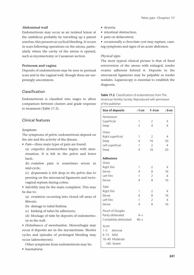
Pelvic pain Chapter 17
241
Abdominal wallEndometriosis may occur as an isolated lesion at
the umbilicus probably by travelling up a patent
urachus; this presents as cyclical bleeding. It occurs
in scars following operations on the uterus, partic-
ularly where the cavity of the uterus is opened,
such as myomectomy or Caesarean section.
Perineum and vaginaDeposits of endometriosis may be seen in perineal
scars and in the vaginal wall, though these are sur-
prisingly uncommon.
Classification
Endometriosis is classified into stages to allow
comparison between clusters and grade response
to treatment (Table 17.2).
Clinical features
SymptomsThe symptoms of pelvic endometriosis depend on
the site and the activity of the disease.
• Pain —three main types of pain are found:
(a) congestive dysmenorrhoea begins with men-
struation. It is felt in the pelvis and lower
back;
(b) ovulation pain is sometimes severe in
mid-cycle;
(c) dyspareunia is felt deep in the pelvis due to
pressing on the uterosacral ligaments and recto-
vaginal septum during coitus.
• Infertility may be the main complaint. This may
be due to:
(a) ovulation occurring into closed off areas of
fibrosis;
(b) damage to tubal fimbria;
(c) kinking of tubes by adhesions;
(d) blockage of tube by deposits of endometrio-
sis in the wall.
• Disturbances of menstruation. Menorrhagia may
occur if deposits are in the myometrium. Shorter
cycles and episodes of prolonged bleeding may
occur (adenomyosis).
Other symptoms from endometriosis may be:
• haematuria;
• dysuria;
• intestinal obstruction;
• pain on defaecation;
• occasionally a chocolate cyst may rupture, caus-
ing symptoms and signs of an acute abdomen.
Physical signsThe most typical clinical picture is that of fixed
retroversion of the uterus with enlarged, tender
ovaries adherent behind it. Deposits in the
uterosacral ligaments may be palpable as tender
nodules. Laparoscopy is essential to establish the
diagnosis.
Table 17.2 Classification of endometriosis from The
American Fertility Society. Reproduced with permission
of the publisher.
Size of deposits <1cm 1–3cm >3cm
Peritoneum
Superficial 1 2 4
Deep 2 4 6
Ovary
Right superficial 1 2 4
Deep 4 16 20
Left superficial 1 2 4
Deep 4 16 20
AdhesionsOvary
Right film 1 2 4
Dense 4 8 16
Left film 1 2 4
Dense 4 8 16
Tube
Right film 1 2 4
Dense 4 8 16
Left film 1 2 4
Dense 4 8 16
Pouch of Douglas
Partly obliterated 4ÆCompletely obliterated 40Æ
Score
1–5 Minimal
6–15 Mild
16–40 Moderate
>40 Severe
AMI17 6/9/04 5:08 PM Page 241
Chapter 17 Pelvic pain
242
Differential diagnosis• Chronic pelvic infection most closely resembles
pelvic endometriosis, with dysmenorrhoea, men-
orrhagia, sterility and dyspareunia being identical,
but the history and laparoscopy findings are differ-
ent and pelvic inflammatory disease (PID) pain is
rarely cyclical.
• Fibroids of the uterus are often associated with
endometriosis, and the differential diagnosis may
be difficult.
Treatment
Treatment of pelvic endometriosis is essentially
conservative because the condition:
• tends to occur during the reproductive period;
• does not become malignant;
• tends to regress at the menopause.
Hormone therapyThis is successful in many cases. The diagnosis
must first be made at laparoscopy and if there are
large chocolate cysts these should be drained; local
areas of endometriosis can be coagulated with
laser/diathermy through the laparoscope.
Danazol inhibits pituitary gonadotrophin secre-
tion and in adequate doses will suppress menstrua-
tion. The initial dose is 200mg daily in divided
doses, increasing up to 600mg daily as required.
Treatment should be given initially for six months.
There may be androgenic effects:
• acne;
• hirsutes;
• weight gain due to fluid retention.
It is important to be sure that the woman is not
pregnant and it must be stressed that danazol is not
a contraceptive.
Progesterone used to be the major therapy, using
norethisterone starting on the fifth day of men-
struation with 10mg daily and increasing up to
40mg daily. Nausea, vomiting, weight gain and
fluid retention occur, while a cure is unusual.
An alternative progestogen, medroxypro-
gesterone acetate (10–30mg daily by mouth), can
suppress menstruation for six months. If
breakthrough bleeding is troublesome, the dose
should be increased.
Side-effects are common with hormone
treatments, but in patients who persist, regression
of the endometriotic lesions occurs and preg-
nancy may become possible, though ovula-
tion may be delayed for several months after
treatment.
Synthetic substitutes of gonadotrophin releasing
hormone (GnRH) and their agonists are inhibitors
of ovarian function. Two weeks’ treatment (subcu-
taneously or nasal spray) reduces FSH and LH con-
centrations and leads to lower oestrogen levels.
Treatment for 3–6 months gives relief. Side-effects
may be:
• headaches;
• hot flushes;
• depression;
• loss of bone density if treatment is over 6
months.
SurgeryVery small lesions may be treated at laparoscopy by
diathermy or laser. Laparotomy is indicated in:
• pelvic masses over 5cm;
• acute rupture of a cyst;
• intestinal obstruction.
Conservative surgery is always performed if
possible, aiming to leave the uterus and normal
ovarian tissue.
In intractable cases, and especially among
women who do not want children, wider surgery
may be needed. Hysterectomy with bilateral
salpingo-oophorectomy may be performed for
intractable menorrhagia and dysmenorrhoea or
when there are fibroids or adenomyosis. Some
ovarian tissue may be left if not actually involved
in the destructive process.
Premenstrual tension
Premenstrual tension occurs most usually in the
second half of the menstrual cycle. It consists of a
cluster of behavioural symptoms and physical
signs which come in the second half of the cycle
with abolition immediately after menstruation.
Symptoms and signs• Irritability.
AMI17 6/9/04 5:08 PM Page 242

Pelvic pain Chapter 17
243
• Depression.
• Lassitude.
• Insomnia.
• Lack of concentration.
• Oedema with fluid retention.
• Abdominal swelling.
• Swollen fingers and ankles.
• Weight gain.
Migraine can occur during this phase or at the
onset of menstruation. The symptoms tend to
come on 7–10 days premenstrually, after the luteal
phase is established. Women may become more ac-
cident prone and there is an increased prevalence
of suicide. Some women who suffer from premen-
strual tension have endogenous depression exacer-
bated at this time.
Aetiology
HORMONAL
• Timing in cycle in luteal phase.
• Improved in absence of cyclical ovarian activity.
• Renin–angiotensin system.
• Reduction of endogenous opioids.
• Changes in mono-amine neurotransmitters.
Probably a combination of the second and third
factors.
Treatment• Explanation and reassurance.
• Progestogens —may be deficient in second half
of cycle so replace them.
• Oestrogens —to suppress ovulation.
• Oral contraceptive —combined preparation of
both of the above; works for some.
• Danazol —stops ovulation; beware unwanted
androgenic side-effects.
• Evening primrose oil —may effect essential fatty
acid metabolism.
• Pyridoxine (B6) —may affect dopamine and sero-
tonin metabolism. Benefit weak —beware over-
dosage and neuropathy with long-term treatment.
• Antidepressants —during the premenstrual
phase may help. More severe cases need psychiatric
treatment. Fluoxetine has been shown to be the
most effective.
The variety of medication emphasizes how little
the cause of this syndrome is understood. Treat-
ments may be a matter of trial and error.
Abdominal organs
Pyelonephritis
The woman presents with ill-defined abdominal
pain, pyrexia and shivering. The diagnosis differs
from an acute abdominal problem for the pain is
often round the side in the loin and tenderness is
then high up in the costal angles.
Examination of the urine for pus cells and
organisms reveals the diagnosis. Treatment is con-
servative with bedrest, fluids and appropriate
antibiotics.
Very rarely a ureteric stone may present in preg-
nancy. If this sticks in the ureter, it causes pain by
stretch of the ureter from dammed-back urine.
Pethidine is helpful both for its analgesic proper-
ties and because it is an antispasmodic.
Appendicitis
This occurs equally commonly in early pregnancy
as during any other nine months of a young
woman’s life. It is a young person’s disease.
Diagnosis can be difficult for the appendix rises
up from its usual position in the right iliac fossa.
The typical history of peri-umbilical pain moving
to the right iliac fossa may not be given in preg-
nancy; the signs are confusing as the caecum with
its attached appendix is pushed up the right para-
colic gutter by the enlarging uterus. Remembering
this, the examiner must seek the point of maxi-
mum tenderness higher in the abdomen.
Treatment should be by surgery and the surgeon
in later pregnancy would do well to mark the site of
maximum tenderness before the anaesthetic and
incise there rather than over McBurney’s point.
There used to be a higher mortality of appendi-
citis in pregnancy because of the reluctance of
people to operate for fear of miscarriage. However,
it must be realized that a progressing appendicitis
carries a much higher risk to the fetus and mother
than the problems of carrying out a surgical proce-
dure under controlled anaesthesia.
AMI17 6/9/04 5:08 PM Page 243
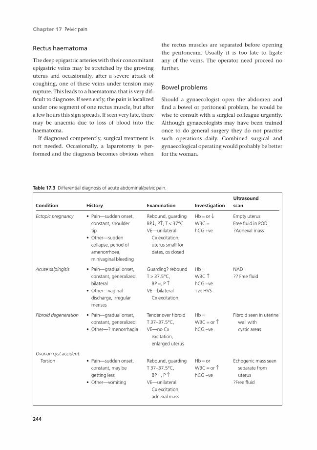
Chapter 17 Pelvic pain
244
Rectus haematoma
The deep epigastric arteries with their concomitant
epigastric veins may be stretched by the growing
uterus and occasionally, after a severe attack of
coughing, one of these veins under tension may
rupture. This leads to a haematoma that is very dif-
ficult to diagnose. If seen early, the pain is localized
under one segment of one rectus muscle, but after
a few hours this sign spreads. If seen very late, there
may be anaemia due to loss of blood into the
haematoma.
If diagnosed competently, surgical treatment is
not needed. Occasionally, a laparotomy is per-
formed and the diagnosis becomes obvious when
the rectus muscles are separated before opening
the peritoneum. Usually it is too late to ligate
any of the veins. The operator need proceed no
further.
Bowel problems
Should a gynaecologist open the abdomen and
find a bowel or peritoneal problem, he would be
wise to consult with a surgical colleague urgently.
Although gynaecologists may have been trained
once to do general surgery they do not practise
such operations daily. Combined surgical and
gynaecological operating would probably be better
for the woman.
Table 17.3 Differential diagnosis of acute abdominal/pelvic pain.
UltrasoundCondition History Examination Investigation scan
Ectopic pregnancy • Pain—sudden onset, Rebound, guarding Hb = or Ø Empty uterus
constant, shoulder BPØ, P≠, T < 37°C WBC = Free fluid in POD
tip VE—unilateral hCG +ve ?Adnexal mass
• Other—sudden Cx excitation,
collapse, period of uterus small for
amenorrhoea, dates, os closed
minivaginal bleeding
Acute salpingitis • Pain—gradual onset, Guarding? rebound Hb = NAD
constant, generalized, T > 37.5°C, WBC ≠ ?? Free fluid
bilateral BP =, P ≠ hCG -ve
• Other—vaginal VE—bilateral +ve HVS
discharge, irregular Cx excitation
menses
Fibroid degeneration • Pain—gradual onset, Tender over fibroid Hb = Fibroid seen in uterine
constant, generalized T 37–37.5°C, WBC = or ≠ wall with
• Other—? menorrhagia VE—no Cx hCG -ve cystic areas
excitation,
enlarged uterus
Ovarian cyst accident:
Torsion • Pain—sudden onset, Rebound, guarding Hb = or Echogenic mass seen
constant, may be T 37–37.5°C, WBC = or ≠ separate from
getting less BP =, P ≠ hCG -ve uterus
• Other—vomiting VE—unilateral ?Free fluid
Cx excitation,
adnexal mass
AMI17 6/9/04 5:08 PM Page 244

Pelvic pain Chapter 17
245
UltrasoundCondition History Examination Investigation scan
Rupture • Pain—sudden onset, Rebound, guarding Hb = Free fluid in POD
constant, getting less T 37°C, P, BP =, WBC = No cyst seen
• Other—? irregular VE—generalized hCG -ve
menses tenderness only
Haemorrhage • Pain—sudden onset, Rebound, guarding Hb = or ≠ Echogenic cyst
constant, becoming T 37°C, BP =, P ≠ WBC = Free fluid if ruptured
less VE—unilateral hCG -ve
Cx excitation,
adnexal mass
Appendicitis • Pain—gradual onset, Rebound, guarding, Hb = NAD
right-sided right-sided WBC ≠• Other—anorexia, no tenderness hCG -ve
BO, nausea/vomiting T 37–37.5°C,
BP =, P ≠VE—NAD
PR—empty rectum,
right-sided
tenderness
Pyelonephritis • Pain—loin pain, colicky Loin tenderness Hb = Renal pelvicalyceal
• Other—nausea, T > 37.5°C, WBC ≠ dilatation
vomiting, rigors, VE—NAD hCG -ve Pelvis—NAD
dysuria MSU +ve
Obstruction • Pain—intermittent, Rebound, guarding, Hb = Pelvis—NAD
generalized, colicky distension WBC = or ≠ AXR—dilated loops
• Other—nausea, No BS. T 37°C, hCG -ve with fluid levels
vomiting, anorexia, VE—NAD
bowels not open PR—empty
rectum
Table 17.1 Continued
BP = blood pressure; T = temperature; P = pulse; VE = vaginal examination; PR = rectal examination; NAD = no
abnormality detected; AXR = abdominal X-ray; Hb = haemoglobin; WBC = white blood count; hCG = pregnancy test;
MSU = mid-stream urine; HVS = high vaginal swab; POD = pouch of Douglas; Cx = cervical; BS = bowel sounds.
AMI17 6/9/04 5:08 PM Page 245

Chapter 17 Pelvic pain
246
Self-assessment
17.1 OSCE question.Candidate’s instructions: Rosemary Beckett, a woman of 32 is referred to the gynaecology clinic by her GP. Shegives a 2 year history of increasingly severe period pains. You are expected to take a history and outline your initialmanagement of Mrs Beckett.
Role-player’s instructions: You are Rosemary Beckett, a 32 year old primary school teacher. You have been mar-ried for 3 years. You have never been pregnant and use condoms for contraception. You tried the pill in your early20s but found that it made you feel depressed. Your periods are regular but are becoming increasingly painful overthe last 2 years. The pain starts as a low constant ache in your back radiating to the front. It starts 3–4 days beforeyour period, is worst on the first 2 days of your period and then subsides. Sex is uncomfortable in some positionsparticularly before your period. The pain can be acute and is deep inside especially when your husband is particu-larly vigorous. He is sympathetic but you feel that it is beginning to affect your relationship as your libido is less thanwhen you first married. You and your husband are planning to have a baby within the next 2 years and are wor-ried that the pain may prevent pregnancy. You have no past medical or surgical history of relevance, no allergiesand take Neurofen for the pain but no regular medication. All other details are up to the role-player.
17.2 Submucous fibroids are commonly associated with which of the following?(a) Constant lower abdominal pain.(b) Menorrhagia.(c) Vomiting.(d) Secondary dysmenorrhoea.(e) Ectopic pregnancy.
17.3 A 24-year-old woman presents with unilateral abdominal pain, vomiting, abdominal rebound and guarding. She isapyrexial and her LMP was one week ago. Which of the following is the most likely diagnosis?(a) Pelvic inflammatory disease.(b) Bleeding into a corpus luteal cyst.(c) Ruptured corpus luteal cyst.(d) Torted dermoid cyst.(e) Ruptured mucinous cystadenoma.
17.4 Which of the following is true of benign ovarian cysts?(a) May undergo degeneration.(b) May rupture causing acute abdominal pain.(c) May contain well differentiated tissues.(d) May contain altered blood.(e) May secrete testosterone.
17.5 A 19-year-old woman presents with right iliac fossa pain. The pain came on gradually and was associated with nau-sea and vomiting. She has not opened her bowels for 36 hours. On examination she has a temperature of 37.6°C.Abdominal examination reveals tenderness in the right iliac fossa (RIF) with guarding and rebound. Vaginal exam-ination reveals mild tenderness on the right with no cervical excitation. What is the most likely diagnosis?(a) Appendicitis.(b) Pyelonephritis.(c) Torted right ovarian cyst.(d) Small bowel obstruction.(e) Ruptured right ovarian cyst.
AMI17 6/9/04 5:08 PM Page 246

In the UK, gynaecologists do not usually deal with
breast problems. Breast disease is managed by
specialist breast surgeons or general surgeons with
an interest in breast surgery. This is partly a conse-
quence of the work of Sir Hedley Atkins who set up
the first specialist breast surgical unit in the world
at Guy’s Hospital.
Breast problems are extremely common com-
prising one in six of all general surgery referrals.
This is partly the result of a heightened awareness
of breast cancer, which is the commonest malig-
nancy in the UK among men and women.
Breast history
The most frequent breast symptom is a lump. The
particular features of a breast lump are usually
ascertained during clinical examination but it is
important to find out:
• Time —how long the lump has been there.
• Pain —whether it is painful. Breast pain is rarely
associated with malignancy but may be severe
and may interfere significantly with a patient’s
lifestyle. Its site, distribution, severity, radiation,
precipitating factors and relieving factors should
all be asked about, but the affect that it has on a
patient’s lifestyle is the most relevant question.
• Size —whether it has changed in size. In particu-
lar, areas of benign breast thickening and nodular-
ity may become more pronounced and more
tender prior to each menstrual period.
• Nipple discharge —a common symptom which
can be a sign of malignancy. It should be noted
whether this is unilateral or bilateral, occurs spon-
taneously or only on expression from a single or
multiple ducts, its colour and whether there is
blood staining.
• Nipple inversion —can also be a sign of malignan-
cy. The salient points are whether is it unilateral
or bilateral and how long it has been present.
• Nipple eczema —should be distinguished from
Paget’s disease of the nipple. This often requires a
skin biopsy.
Breast examination
Examination is carried out with the patient seated
in good light and in a warm room. She should be
stripped to the waist.
Inspection
Inspection involves comparing one breast with the
other, particularly with regard to:
• symmetry;
• preservation of the natural curved contour of the
breast;
• position and appearance of the nipples;
• scars;
• changes in the skin (erythema, peau d’orange,
ulceration) and skin dimpling. It is quite common
for one breast to be a little larger than the other but
247247
Chapter 18
Breast disease
AMI18 6/9/04 5:08 PM Page 247
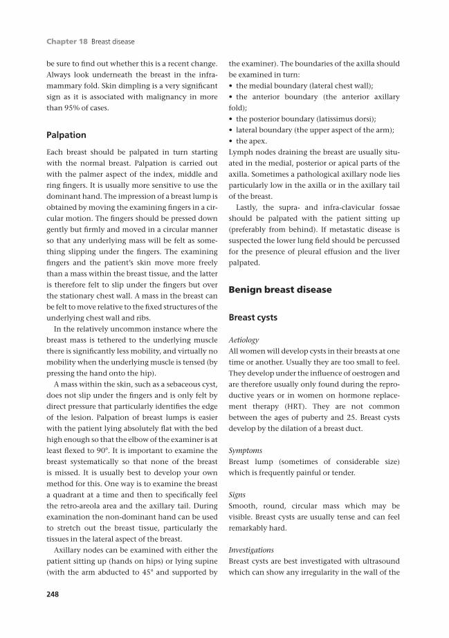
Chapter 18 Breast disease
248
be sure to find out whether this is a recent change.
Always look underneath the breast in the infra-
mammary fold. Skin dimpling is a very significant
sign as it is associated with malignancy in more
than 95% of cases.
Palpation
Each breast should be palpated in turn starting
with the normal breast. Palpation is carried out
with the palmer aspect of the index, middle and
ring fingers. It is usually more sensitive to use the
dominant hand. The impression of a breast lump is
obtained by moving the examining fingers in a cir-
cular motion. The fingers should be pressed down
gently but firmly and moved in a circular manner
so that any underlying mass will be felt as some-
thing slipping under the fingers. The examining
fingers and the patient’s skin move more freely
than a mass within the breast tissue, and the latter
is therefore felt to slip under the fingers but over
the stationary chest wall. A mass in the breast can
be felt to move relative to the fixed structures of the
underlying chest wall and ribs.
In the relatively uncommon instance where the
breast mass is tethered to the underlying muscle
there is significantly less mobility, and virtually no
mobility when the underlying muscle is tensed (by
pressing the hand onto the hip).
A mass within the skin, such as a sebaceous cyst,
does not slip under the fingers and is only felt by
direct pressure that particularly identifies the edge
of the lesion. Palpation of breast lumps is easier
with the patient lying absolutely flat with the bed
high enough so that the elbow of the examiner is at
least flexed to 90°. It is important to examine the
breast systematically so that none of the breast
is missed. It is usually best to develop your own
method for this. One way is to examine the breast
a quadrant at a time and then to specifically feel
the retro-areola area and the axillary tail. During
examination the non-dominant hand can be used
to stretch out the breast tissue, particularly the
tissues in the lateral aspect of the breast.
Axillary nodes can be examined with either the
patient sitting up (hands on hips) or lying supine
(with the arm abducted to 45° and supported by
the examiner). The boundaries of the axilla should
be examined in turn:
• the medial boundary (lateral chest wall);
• the anterior boundary (the anterior axillary
fold);
• the posterior boundary (latissimus dorsi);
• lateral boundary (the upper aspect of the arm);
• the apex.
Lymph nodes draining the breast are usually situ-
ated in the medial, posterior or apical parts of the
axilla. Sometimes a pathological axillary node lies
particularly low in the axilla or in the axillary tail
of the breast.
Lastly, the supra- and infra-clavicular fossae
should be palpated with the patient sitting up
(preferably from behind). If metastatic disease is
suspected the lower lung field should be percussed
for the presence of pleural effusion and the liver
palpated.
Benign breast disease
Breast cysts
AetiologyAll women will develop cysts in their breasts at one
time or another. Usually they are too small to feel.
They develop under the influence of oestrogen and
are therefore usually only found during the repro-
ductive years or in women on hormone replace-
ment therapy (HRT). They are not common
between the ages of puberty and 25. Breast cysts
develop by the dilation of a breast duct.
SymptomsBreast lump (sometimes of considerable size)
which is frequently painful or tender.
SignsSmooth, round, circular mass which may be
visible. Breast cysts are usually tense and can feel
remarkably hard.
InvestigationsBreast cysts are best investigated with ultrasound
which can show any irregularity in the wall of the
AMI18 6/9/04 5:08 PM Page 248

Breast disease Chapter 18
249
cyst. Mammography is generally not able to differ-
entiate between a solid and a cystic mass.
Natural historyBreast cysts often appear suddenly and equally
may disappear relatively quickly. Some cysts, how-
ever, remain for a number of years. Breast cysts
practically never become malignant. Rarely, they
can develop in-situ malignancy (intracystic papil-
lary carcinoma).
TreatmentBreast cysts do not need any treatment unless they
are uncomfortable in which case they can be aspi-
rated with a needle and syringe.
Fibrocystic change
AetiologyFibrocystic change, previously termed fibroadeno-
sis, is a normal way that the breast develops with
time and under the influence of the normal men-
strual cycle. It involves an increase in the fibrous
tissue of the breast stroma, together with the for-
mation of multiple small and large cysts. It occurs
to a greater or lesser extent in every woman and
may be responsible for breast lumps.
SymptomsTender breast lump that varies with the menstrual
cycle and becomes more prominent prior to each
period.
SignsA diffuse area of tender thickening or nodularity,
only rarely amounting to a discrete mass with
definable edges. The thickening may be approxi-
mately symmetrical with the same site in the
opposite breast.
InvestigationsFibrocystic change shows as a diffuse increase in
the density of the breast parenchyma on a mam-
mogram. If the associated cysts are large enough
these will appear as discrete, rounded masses. An
ultrasound shows small and large cysts.
Natural historySome women have more fibrocystic change than
others resulting in permanently lumpy breasts.
This is more apparent in slim women. The changes
generally resolve after the menopause.
TreatmentReassurance only is required.
Breast pain (mastalgia)
AetiologyThe vast majority of breast pain is caused by either
hormonal changes in the breast tissue or a chronic
inflammatory condition of the major breast ducts
close to the nipple —periductal mastitis. Breast
pain is only very rarely associated with malignancy
and then only when it is unilateral and localized to
a specific site.
SymptomsThe pain is usually described as an aching, particu-
larly in the lateral aspects of the breasts, radiating
into the axilla and sometimes into the upper arm.
The pain may be unilateral. It is often worse in the
second half of each menstrual cycle although
mastalgia relating to hormonal changes can also
frequently be non-cyclical. Hormonal breast pain
does not respond significantly to standard anal-
gesics. It is usually associated with marked tender-
ness and is helped by wearing a supportive
brassiere. The breast pain relating to periductal
mastitis is non-cyclical, sharper, more transient and
frequently radiates through the nipple.
SignsBreast tenderness. Patients with hormonal mastalgia
often have denser and more nodular breast tissue.
InvestigationsNo investigations are necessary except in the case
of unilateral and focal breast pain.
TreatmentSimple measures such as reducing the amount of
dietary caffeine and taking regular Starflower or
AMI18 6/9/04 5:08 PM Page 249
Chapter 18 Breast disease
250
Evening Primrose Oil. Drugs that are helpful
include Tamoxifen (10mgs, daily), Danazol
(100mgs, daily) and Bromocriptine (1mg, daily).
Nipple discharge
AetiologyNipple discharge may be caused by malignancy but
is more frequently the result of either periductal
mastitis (chronic inflammation involving a num-
ber of breast ducts) or a duct papilloma (usually in-
volving a single duct). Periductal mastitis is much
more common in smokers.
SymptomsPeriductal mastitis causes discharge of any colour
(brown, green, clear, milky). It is often bilateral. A
duct papilloma causes clear discharge which is
usually unilateral. Breast cancer causes unilateral
discharge. All three causes can be associated with
blood staining.
Signs• Periductal mastitis: multiduct, multicoloured dis-
charge, usually bilateral.
• Papilloma: single duct, crystal clear discharge.
• Carcinoma: unilateral discharge, often with
blood staining and associated underlying breast
lump and/or nipple distortion.
InvestigationsMammography and ultrasound should be carried
out to exclude malignancy. An ultrasound scan may
demonstrate a large papilloma. Nipple discharge
fluid can be sent for cytological examination.
Natural history• Periductal mastitis: this is a chronic condition.
• Duct papilloma: a duct papilloma can cause
profuse discharge which is inconvenient for the
patient.
Treatment• Periductal mastitis: no treatment is available
except for surgical total duct excision.
• Duct papilloma: breast duct microendoscopy and
microdochectomy.
Fibroadenoma
AetiologyA fibroadenoma is a solid mass arising in the breast
of young women, particularly from the age of
puberty to the mid 30s. It is also more common in
post menopausal women who take HRT. The
aetiology is unknown although there is undoubt-
edly a hormonal influence.
SymptomsPainless lump. Some patients have multiple
lesions.
SignsFirm to hard, rounded or elliptical mass which is
characterized by unusual mobility underneath the
palpating fingers (colloquially known as a breast
mouse).
InvestigationsA well defined mass with discrete clear margins on
both mammography and ultrasound.
Natural historyFibroadenomas do not become malignant. They
may gradually increase in size and in this case they
should be removed to definitely exclude a malig-
nant mass that may have the same clinical features
as a fibroadenoma.
TreatmentIt is important to differentiate this solid mass from
other solid masses in the breast such as a carcin-
oma. This should always involve at least needle as-
piration cytology and over the age of 30 preferably
a core needle biopsy. If there is any doubt about the
diagnosis the lesion should be removed surgically
by excision biopsy. This latter operation can often
be carried out under a local anaesthetic.
AMI18 6/9/04 5:08 PM Page 250
Breast disease Chapter 18
251
Breast abscess
AetiologyA breast abscess can either be associated with lacta-
tion or periductal mastitis.
SymptomsPainful, rapidly enlarging mass in the breast with
associated pyrexia.
SignsLactational abscess may be very large and the bor-
ders may be quite indistinct so that the mass forms
a prominent area of hardening of the breast tissue.
Abscesses associated with periductal mastitis are
found either in the retroareolar region or just
around the areola. Breast abscesses are always asso-
ciated with overlying breast erythema. There may
be peau d’orange. This latter scenario mimics
inflammatory breast cancer and this differential
diagnosis should always be entertained.
InvestigationsBreast abscesses are best shown by
ultrasonography.
TreatmentEarly in the development of a breast infection
(whatever the aetiology) treatment with antibi-
otics may be successful in averting a frank ab-
scess. Use either Augmentin or Flucloxacillin and
Metronidazole (anaerobes may be responsible).
Once the breast abscess is larger than 1cm needle
aspiration or incision drainage are usually re-
quired. Needle aspiration may need to be repeated.
Other benign breast lumps
• Lipoma: soft, rounded, quite difficult to feel, may
have a distinct edge.
• Hamartoma: fairly soft, discrete, no malignant
potential.
• Phylloides tumour: mimics a fibroadenoma and
may be quite similar histologically. Phylloides tu-
mours may grow to become very large. They may
recur following excision if a small amount of the
lesion is left behind and they should therefore be
removed with a clear histological margin. There
are malignant variants of phylloides tumours that
develop into fibrosarcomas.
Breast malignancy
Epidemiology
Rates
The incidence of breast cancer in the UK is still
increasing. Currently there are 40000 cases diag-
nosed each year (1999). The mortality from breast
cancer has been falling since the late 1980s. This is
thought to be due to the therapeutic effect of
Tamoxifen and more recently due to the beneficial
effects of the National Breast Screening Pro-
gramme (Figs 18.1 and 18.2).
Risk factors
GeneticMutations in either of the two recognized breast
cancer genes (BRCA1 and BRCA2) result in a very
high chance of developing breast or ovarian can-
cer. Other genetic abnormalities associated with a
higher than average breast cancer risk are Cow-
den’s syndrome and ataxia telangiectasia.
110
120
130
140
100
90
80
701980 2000
134.4
'85 '90
Mammographyscreeningintroduced
Rate
per
100
000
Figure 18.1 Age standardized incidence of breast cancerin England and Wales.
AMI18 6/9/04 5:08 PM Page 251

Chapter 18 Breast disease
252
HormonalThe following are associated with a higher than
average risk of breast cancer: early menarche, late
menopause, no full-term pregnancies, full term
pregnancy occurring after the age of 40. Longer
periods of breast feeding and multiple full-term
pregnancies are associated with a lower incidence
of breast cancer. The oral contraceptive pill has
practically no effect on breast cancer risk. HRT is
associated with a small increase in the risk of breast
cancer which amounts to around 35% increase
after 12 years of usage.
EnvironmentalRadiation (such as that associated with the fall out
from the atomic bombs in Japan) is associated with
all sub-types of breast cancer. Alcohol increases the
risk of breast cancer in a dose-dependent manner.
Diet has a large effect on breast cancer risk. Diets
rich in fresh fruit and vegetables are associated
with a much lower risk of breast cancer (50%
reduction). This risk reduction is probably partly
related to vitamin C intake. Smoking is not related
to breast cancer. The lowest incidence of breast
cancer occurs in Japan, the highest risk in Western
Europe and North America. Women migrating
from one geographic region to another slowly de-
velop the full breast cancer risk associated with
their new environment.
Presentation
Breast malignancy most commonly presents with a
painless lump. Skin tethering is highly suggestive
of malignancy. The characteristics of a malignant
lump compared with a benign lump are compared
in Table 18.1. For breast cancers around the nipple,
inversion or distortion of the nipple together with
nipple discharge may be presenting symptoms.
Infrequently patients present with an enlarged
axillary lymph node with no obvious palpable
mass in the breast. Other less common symptoms
and signs of breast cancer are peau d’orange,
distortion of the shape of the breast, generalized
enlargement of the breast, focal breast pain and
skin changes (erythema or ulceration). Paget’s
disease of the nipple is always associated with an
underlying malignancy. This may be invasive or
in situ disease. Paget’s disease may look similar to
nipple eczema being dry and scaly or may be beefy
red and weeping.
110
100
90
80
70
1950 '60 '70 '80 '90
Mammography
screening
introduced
Mor
talit
y pe
r 100
000
Figure 18.2 Age standardized mortality rates from breastcancer in women aged 55–69 years in England and Wales.
Table 18.1 Clinical features of some common breast lumps.
Fibrocystic Cyst Fibroadenoma Cancer
Tender Yes Yes No No
Discrete No Yes Yes Usually
Surface No edge/surface Smooth Smooth Irregular/smooth
Consistency Normal to firm Firm to hard Firm Firm to hard
Shape Nodularity/ridge Round Round/elipse/lobulated Rounded
Overlying skin Normal Normal Normal Tethering
Multiple lesions Yes Yes Yes No
AMI18 6/9/04 5:08 PM Page 252

Breast disease Chapter 18
253
Staging
• Stage 1: breast cancer confined to the breast.
• Stage 2: involvement of the breast and axillary
lymph nodes.
• Stage 3: locally advanced disease involving the
breast with muscle fixation or skin involvement.
• Stage 4: metastatic disease.
Breast cancer can be more accurately staged
according to the TNM classification.
• T relates to the size of primary tumour:
• T1 = 2 - 20mm,
• T2 = 21 - 50mm,
• T3 = 51 or more mm.
• N refers to the regional lymph node status:
• N0 = node negative,
• N1 = mobile ipsilateral nodes,
• N2 = immobile, fixed or matted nodes,
• N3 = internal mammary node involvement.
• The M classification whether there is metastatic
disease that:
• M0 = no detected metastases,
• M1 = metastatic disease.
Primary breast cancer is usually simply staged by
performing:
• chest X-ray to look for pleural or pulmonary
disease;
• liver function test or a liver ultrasound looks for
hepatic metastases;
• large tumours (over 4cm) or high-grade tumours
(3) would additionally have staging involving a
bone scan.
Investigation and diagnosis
Investigation of any breast lump is based on the
principle of triple diagnosis.
• Clinical assessment.
• Radiological imaging.
• Cytological or histological verification.
It is only if all these three types of assessment are
entirely benign that a lump can be left without
further investigation.
Invasive breast cancer shows up on a mammo-
gram by producing either:
• a dense (white) mass,
• an area of distortion of the breast parenchyma or
microcalcification.
On mammography, a malignant mass will appear
spiculate with ill-defined edges. Breast distortion
may occur without a mass, particularly in the case
of a lobular carcinoma. Malignant microcalcifica-
tion is focal, relatively fine and heterogeneous. The
sensitivity of mammography for detecting a malig-
nancy is around 85%.
An ultrasound scan will show a malignant mass as
an echo-poor focal area that interrupts the normal
transverse breast architecture. The lesion is ill-
defined and is usually taller rather than wider.
There may be a dense acoustic shadow. The sensi-
tivity of breast ultrasound for the diagnosis of
malignancy is between 75% and 80%.
Needle cytology is carried out without local anaes-
thetic. The specimen, which usually amounts to
just a drop or two of blood-stained-tissue fluid, is
spread onto glass microscope slides. These can be
air dried. No immediate staining is required
although this is carried out later in the laboratory.
The breast cytopathologist requires five clusters of
duct epithelial cells to be able to make a definitive
diagnosis. An alternative to fine needle aspiration
cytology (FNAC) is core-needle biopsy. This is now
being carried out much more frequently as it gives
additional information regarding the architecture
of the lesion. It is performed under local anaes-
thetic with a core-needle-biopsy gun and needle
varying between 14 and 11 gauge. It is possible to
perform cytological examination of the cells on
the surface of the core by simply rolling the tissue
core between two microscope slides. This gives
an accurate answer in less than an hour (touch
imprint cytology). A core biopsy will require at
least 48 hours for histological examination.
Magnetic resonance imaging (MRI) may be used to
diagnose breast malignancy. It is particularly sensi-
tive (95–100%) but the specificity is much lower
(65–70%). Examination needs to be carried out
with contrast during the first half of the menstrual
period. Benign breast lesions such as fibroadeno-
mas may enhance, but the enhancement charac-
teristics are generally different in benign compared
to malignant lesions.
AMI18 6/9/04 5:08 PM Page 253

Chapter 18 Breast disease
254
Treatment
General strategy
Breast cancer, when confined to the breast or the
axillary lymph nodes, is curable. Metastatic breast
cancer is not. More than 90% of patients present
with primary breast cancer (stage 1 or 2) with no
evidence of metastatic disease. However, at this
stage micro-metastases are probably present in
some patients and these are often the patients who
later relapse systemically. The treatment of
primary breast cancer is therefore to eradicate the
malignancy from the breast and axillary lymph nodes
and to provide some type of systemic (adjuvant) treat-
ment to try and eliminate any micro-metastases.
The presence or absence of micro-metastases is not
known in any one individual patient although the
likelihood of micro-metastases can be estimated by
taking into consideration the lymph node status
and the size and grade of the tumour.
When considering the treatment of primary
breast cancer think of three anatomical boxes, each
of which will receive its own treatment. The treat-
ment strategy for each box has either no or only
marginal impact on the other two anatomical areas
and each should therefore be formulated in-
dependently. The treatment of primary breast can-
cer should therefore involve adequate treatment of
the:
• breast itself (breast primary);
• axillary lymph nodes;
• micro-metastases.
The treatment of a breast primary
Surgical treatmentThe most effective modality utilized in the treat-
ment of breast cancer is the surgical treatment of
the primary. The aim of surgical treatment is to
achieve complete removal of the primary lesion to-
gether with any satellite lesions and any associated
ductal carcinoma in situ (DCIS). Mastectomy is one
effective way of removing a primary lesion
although it is now well established that a lump-
ectomy, if carried out in conjunction with breast
radiotherapy (breast conserving therapy), is as
effective as mastectomy both in terms of local
control and overall survival. Lumpectomy is not
appropriate for large tumours (generally over 4
cm), for multifocal tumours (unless the lesions are
small and closely applied to one another) or lesions
associated with very extensive ductal carcinoma in
situ. For these cases and for patients who cannot,
for one reason or another, undergo breast radio-
therapy (serious heart or lung disease) mastectomy
is the only safe option. In the case of large tumours
primary chemotherapy (neo-adjuvant chemother-
apy) can be employed at the outset to shrink the
tumour to a size that is amenable to a lumpectomy
and radiotherapy.
Opinion is still divided over the importance of
clear surgical margins following breast lumpec-
tomy. It is certain that widely involved margins
are associated with a much higher rate of local
recurrence. Focally involved margins or close
margins (1mm or less) are associated with a small
increase in local recurrence rates compared with
widely clear margins.
Mastectomy as performed by Halstead in the
early part of the twentieth century involved re-
moval of not only the breast but also the axillary
nodes and pectoralis minor and pectoralis major.
This was termed a radical mastectomy. It is now
known that it is not necessary to remove either of
the pectoral muscles since primary breast cancer
only infrequently infiltrates the skeletal muscle.
Removal of the breast and axillary lymph nodes is
termed a modified radical mastectomy.
Breast radiotherapyRadiotherapy to the breast following lumpec-
tomy is a critical and irreplaceable part of breast-
conserving therapy for primary breast cancer. If
breast radiotherapy is not possible then mastec-
tomy is the only safe alternative. The radiotherapy
course takes 6 weeks and involves daily fractions
(30 fractions). It is usually associated with a skin
reaction that comprises erythema and sometimes
peeling of the skin.
Breast radiotherapy can be given to the chest
wall following a mastectomy. It is shown to be
effective in reducing local recurrence rates in the
AMI18 6/9/04 5:08 PM Page 254
Breast disease Chapter 18
255
patients with positive axillary nodes, high grade
tumours and large tumours. There is some evi-
dence that irradiation of the supraclavicular
fossa and internal mammary nodes in addition to
the chest wall may produce a small improvement
in overall survival.
Treatment of the axilla
All patients with primary breast cancer (stage 1 and
2) undergo surgery to the axilla. In most instances
this involves axillary nodal clearance to level 2 or
3. At least 10 to 50 lymph nodes are removed. This
surgical procedure achieves two main functions.
First of all axillary node clearance is a very effective
means of controlling potential axillary disease. The
likelihood of axillary recurrence without any
axillary treatment is around 30–35%, whereas
following axillary node clearance the risks of
recurrence are only around 1%.
Axillary node clearance is also very important in
staging the axilla. The status of the axillary nodes is
the best prognostic indicator that is available in
patients with breast cancer, and other aspects of
the treatment such as adjuvant systemic treatment
and chest wall radiotherapy are decided based
largely on the axillary node status. Not only the
presence or absence of axillary disease but the
number of axillary nodes that are involved is
important. Axillary nodal clearance therefore
provides invaluable information.
Axillary node clearance is associated with sig-
nificant morbidity. The most serious problem
associated with this type of surgery is arm
lymphoedema. This occurs to a variable degree but
is serious enough to impact on a patient’s lifestyle
in around 15% of cases. Arm lymphoedema is
more common in older women and in women who
are overweight. Other problems following axillary
nodal clearance include an alteration in the sen-
sation of the skin of the upper arm due to inter-
ruption of the intercostobrachial nerve, pain in
the arm and an increased susceptibility to serious
infections in the ipsilateral upper limb.
Less radical axillary node dissection may be
undertaken (level 1) but it is not entirely
clear whether this makes very much impact on the
postoperative morbidity. A new alternative is sen-
tinel node biopsy. In this technique blue dye or an
isotope labelled colloid tracer are injected close to
the breast primary and are used to identify one or
two axillary lymph nodes that are termed sentinel
nodes. In theory, if the sentinel nodes are clear of
metastatic disease then all the other axillary nodes
are also negative and full axillary clearance is not
required. Sentinel node biopsy is associated with
much lesser morbidity since only one or two
lymph nodes are removed. Of course if the sentinel
node is affected by metastatic disease a second
operation is required to complete the axillary clear-
ance. Sentinel node biopsy has the potential for
avoiding axillary clearance in more than 50% of
patients with breast cancer but the accuracy of the
technique and the consequences of sentinel node
biopsy on overall survival have still to be assessed.
Adjuvant systemic treatment
Adjuvant systemic therapy aims to eliminate any
micro-metastatic disease that may be present but
undetectable at the time of primary diagnosis.
Adjuvant systemic treatment is given following
primary breast surgery and involves hormonal
treatment, chemotherapy or both.
Adjuvant chemotherapyAdjuvant chemotherapy is usually given as soon
after primary breast surgery as is safe (2–3 weeks).
New combinations of chemotherapy drugs that are
normally used are either CMF (Cyclophos-
phamide, Methotrexate or 5-Flourouracil) or FEC
(5-Flourouracil, Epirubicin and Cyclophos-
phamide). Usually six cycles of this treatment are
given. Depending on the regime employed there is
a 3 or 4 week gap between each cycle meaning that
chemotherapy continues for between 4.5 and 6
months. Common side effects include tiredness,
nausea, hair loss, stomatitis and infection. Adju-
vant chemotherapy is much more effective in
younger (pre-menopausal) women and in women
who are node positive. It is rarely used in women
over the age of 70 as there is little evidence of its
efficacy in this age group. In younger women
these drug combinations may cause infertility and
AMI18 6/9/04 5:08 PM Page 255

Chapter 18 Breast disease
256
this side effect needs to be very carefully explained
to patients.
Adjuvant hormone therapyHormone therapy is effective in breast cancer
because at least 75% of breast malignancies are
composed of cells which depend primarily on
oestrogen as a growth factor. These tumours are
identifiable by immunohistochemical staining
to reveal the oestrogen receptor. It is only breast
cancers that express the oestrogen receptor that
are amenable to adjuvant hormonal treatment.
The most common hormonal agent used in the
adjuvant setting is Tamoxifen. Tamoxifen is a
selective oestrogen receptor blocker which
fortunately effectively blocks the oestrogen recep-
tor in breast duct epithelial and breast cancer cells.
Tamoxifen is used in the adjuvant setting for a pe-
riod of 5 years following surgery. It has a generally
benign side effect profile and has only one signifi-
cant drug interaction (with Warfarin). Common
side effects are menopausal symptoms such as hot
flushes and night sweats. The two serious side
effects associated with Tamoxifen use are slightly
increased incidence of endometrial carcinoma and
a low incidence of deep venous thrombosis and
pulmonary embolism. Adjuvant Tamoxifen re-
duces the rate of breast cancer recurrence by
around a third and also reduces the chance of de-
veloping a primary breast cancer in the opposite
breast by 35–40%.
The aromatase inhibitor class of drugs are cur-
rently being advocated and trialled as adjuvant
treatments in primary breast cancer. It appears that
they are slightly more effective than Tamoxifen.
The drugs include Anastrozole, Letrozole and Ex-
emestane. They work by blocking the production of
oestradiol from adrenal precursors in peripheral
adipose tissue. The production of oestradiol in the
ovaries is not dependent on aromatase and the
aromatase inhibitors are therefore only effective
in postmenopausal women. They have a better side
effect profile than Tamoxifen and in particular do
not increase the incidence of endometrial carcinoma.
Measures that effectively reduce circulating
oestradiol in premenopausal women are also
effective as adjuvant treatments. These include
bilateral oophorectomy or Zoladex. Zoladex
is a GnRH agonist which suppresses oestrogen
secretion in the ovary. It is given by Depo injection
either every one or three months. For pre-
menopausal women it can be given in addition to
Tamoxifen with enhanced effect. Zoladex or
oophorectomy drastically reduce the circulating
oestradiol in young women and both are associat-
ed with significant bone loss.
Ductal carcinoma in situ
PathologyIt is now established that ductal carcinoma in situ
(DCIS) is the precursor lesion to invasive breast
cancer. The individual cells are derived from the
breast duct epithelium and are similar morpholog-
ically to those found in invasive breast cancer. The
critical fact that differentiates DCIS from invasive
breast cancer is the lack of invasion. The atypical
cells that characterize DCIS remain within the
breast ducts and as a consequence do not have the
ability to spread to either the regional lymph nodes
or to cause metastatic disease. As with invasive
breast cancer DCIS is divided into three histologi-
cal grades (low, intermediate and high grade). The
higher grade of DCIS particularly can be associated
with cell necrosis and subsequent secondary cal-
cium deposition. This ductal microcalcification is
detectable on mammography and is the most
common way for DCIS to be diagnosed.
Natural historyThe natural history of DCIS depends on the grade
of the malignant change. High-grade DCIS is very
likely to develop into invasive malignancy over a
fairly short term. When it does so it develops into
higher grade invasive carcinoma (usually grade III).
Low-grade DCIS (various types) may never turn
into invasive carcinoma, the progression towards
invasive disease is much slower, and if invasive
disease does occur it is usually of low grade (grade I).
Presentation and diagnosisIt is unusual for DCIS to form a palpable mass.
Most patients who are diagnosed with DCIS have
AMI18 6/9/04 5:08 PM Page 256
Breast disease Chapter 18
257
only a mammographic abnormality, usually con-
sisting of microcalcification of the ductal type.
This is typically focal, fine and heterogeneous with
branching forms. Suspicious microcalcification is
further investigated with stereotactic core needle
biopsy or mammotome biopsy.
TreatmentDuctal carcinoma in situ is confined to the breast. No
treatment to the axillary lymph nodes is required.
There is no chance of metastatic disease as long as
there is no invasive breast malignancy. For this
reason the treatment of DCIS does not involve
adjuvant systemic treatment such as chemotherapy.
The principle treatment modality for DCIS is
surgery. Complete excision with clear margins is
required. This may be achieved by mastectomy (for
extensive areas of DCIS) or by local excision (for
focal areas of DCIS measuring less than 4cm). Fol-
lowing local excision, breast radiotherapy further
reduces the chance of recurrence. For patients with
low-grade DCIS the chance of recurrence is anyway
very low and radiotherapy is not usually given. It
is, however, normally part of the treatment for
high grade DCIS. Patients with intermediate grade
DCIS may or may not have breast radiotherapy de-
pending on the size of the lesion and the margins.
The recurrence rate for DCIS following mastec-
tomy is 1%. The recurrence rate for high grade
DCIS following wide excision is between 25% and
30%. If radiotherapy is used in addition to local
surgery the recurrence rate is approximately
halved. Recurrence of DCIS is in the form of in situ
disease in 50% of cases and in the form of invasive
breast cancer in the other 50%.
Breast cancer screening
Breast cancer screening using mammography is a
good example of effective population screening. In
the UK it has been offered to women between the
age of 50 and 70. Screening involves mammo-
grams without clinical examination performed at
three yearly intervals. Breast screening using mam-
mography has proven to be effective between the
ages of 40 and 50 but more frequent mammogra-
phy (annual) is required. This is not currently part
of the NHS Screening Programme.
Breast cancer screening using mammography
can be made more sensitive in a number of ways.
The mammograms can be double reported (re-
viewed independently by two radiologists) and the
X-rays can be carried out in two perpendicular
views on each screening visit rather than as a single
view. These changes are expensive but are
gradually being introduced into the National
Breast Screening programme. In the UK women
over 70 can still elect to continue with mammo-
graphic screening but they have to request this by
contacting the screening unit themselves.
Web links
More information on benign and malignant breast
conditions can be obtained at www.breast-cancer-
information.com.
Self-assessment
18.1 What are the four stages of Breast Cancer Screening?18.2 List the risk factors for breast cancer.18.3 Which of the following agents are used in adjuvant therapy?
(a) Zoladex.(b) Anastrazole.(c) Cisplatin.(d) Provera.(e) Methotrexate.
18.4 List the three main areas that should be treated in a case of primary breast cancer.
AMI18 6/9/04 5:08 PM Page 257

258
Cervix
The best known screening service is for carcinoma
of the cervix; this should be a preventable disease
because:
• there is usually a phase of premalignancy, dys-
plasia or intraepithelial neoplasia;
• the cervix is a relatively accessible organ to
examine;
• cells can easily be obtained in the premalignant
phase.
The biggest problem is getting to women liable
to develop a carcinoma of the cervix to take part in
the screening.
Current position
In the UK screening is aimed at all women at risk
from within five years of starting sexual activity
(usually 15–20 years) to the age of 65 years. After this
the development of premalignant lesions is rare.
The screening service for cervical cancer aims to
recall women aged 20–64 years every 3 years. It is
possible that some carcinomas will grow very rapid-
ly, but for the majority a cervical smear performed
every three years will pick up the pathological
warning signs. Three-yearly smears detect 91% of
the premalignant conditions. Increasing the smear
frequency to yearly only improves the pick-up rate
to 93%; trebling the work load thus improves re-
sults by only 2% (see Box 19.1).
Smear tests are offered at most places where
women attend for obstetric or gynaecological pro-
cedures. Thus they may be done at:
• GP surgeries;
• antenatal clinics;
• family planning clinics;
• genitourinary medicine clinics;
• gynaecology out-patient clinics;
• well-woman clinics.
Since the average age group of women with pre-
malignant conditions is older than the reproduc-
tive age group, the most appropriate place for
smear tests is the GP surgery, using a well-
organized computer-generated record system
with an age–sex register (call–recall system from
Health Authority); those who do not accept their
invitation must be reinvited. There must also be
some system of ensuring that the results are
returned to the woman promptly. This is now
achieved through computer technology so that
each woman receives a letter saying either (a) that
her result is normal or (b) that she should contact
her doctor. All smear results are sent to the
woman’s GP, irrespective of where the smear is
taken.
This requires an enthusiastic GP service which is
appropriately funded. In the UK, the National
Health Service offers incentives to GPs to ensure
that a high percentage (>85%) of those in the ap-
propriate age groups have their smears at correct
intervals.
Chapter 19
Screening for gynaecological cancer
AMI19 6/9/04 5:09 PM Page 258

Screening for gynaecological cancer Chapter 19
259
Taking a smear
The smear can be taken by anyone competent to
perform a vaginal examination; thus it can be done
by a gynaecologist, a GP, a community clinic doc-
tor, a trained midwife or a nurse.
After discussion, the woman is positioned on
the couch and a warmed speculum is passed to
expose the cervix. A glass slide should be labelled
prior to undertaking the examination. The slide
is marked in pencil with the woman’s name, date-
of-birth, date-of-test and hospital number/NHS
number if known. A spatula is then used to scrape
the whole squamocolumnar junction (SCJ). If the
external os is regular, the pointed end of a spatula
can be passed into the canal and rotated by 360°
(Fig. 19.1). If the cervical os is stenosed, the brush
end of a cytobrush should be used (Fig. 19.2). This
is common after surgery to the cervix for cervical
abnormality or in postmenopausal women. The
most commonly used spatula is the Aylesbury
spatula with its elongated beak to go up the cervi-
cal canal.
The material removed on the tip of the spatula
should be smeared onto the glass slide and fixed
immediately to prevent the cells drying in air. The
slide can go straight into the fixative medium or
be sprayed with the fixative aerosol which dries
rapidly, fixing the cells and nuclei. The slide can
then go to the laboratory through the post or by
messenger.
The cytopathologist will need certain basic in-
formation about the woman in order to interpret
the findings carefully.
• The age of the woman.
• Menstrual cycle and date of last menstrual peri-
od.
• Any irregular vaginal bleeding.
• Is she pregnant?
• Any current hormone therapy (including oral
contraception)?
• Presence of intrauterine device.
• Clinical state of the cervix.
Box 19.1 The benefits and drawbacks to theindividual woman of cervical screening
Advantages1 Reassurance for most who have no premalignantchanges2 Reassurance to a few that any premalignant changesfound are at a very early stage3 Avoidance of radical treatments if the condition ispicked up early4 Produce an increased life expectancy
Disadvantages1 Fear of finding cancer. This may sound illogical but it istrue for most human beings2 The anxiety generated while waiting for the results3 The fear that comes from false positive results
Figure 19.1 Smear taken from a nulliparous cervix with theshaped end of an Aylesbury spatula.
Figure 19.2 Smear taken from a stenosed cervix with thebrush end of a cytobrush.
AMI19 6/9/04 5:09 PM Page 259

Chapter 19 Screening for gynaecological cancer
260
Examination of the smear
Do not take a smear during menstruation for red
cells obscure the epithelial cells at microscopic ex-
amination. However, if the woman has irregular
bleeding it is impossible to avoid this. A note
should be made to the pathologist if this occurs.
At the laboratory the smear is stained and exam-
ined by cytotechnicians. If any abnormality is
detected, the smear will be passed to a
cytopathologist.
False negative resultsThese occur when a woman has premalignant
changes in her cells but these are not reported. In-
cidence is unknown but from data on women who
do develop carcinoma of the cervix, it is probably
between 10 and 30%. Causes include:
• error in taking the smear:
(a) non-representative sample of cells;
(b) not from SCJ;
• a misinterpretation in the laboratory itself;
• incorrect typing of the report from the
laboratory.
False positive resultsThis happens when the smear is reported as having
a greater degree of malignant change than exists.
This is caused by:
• misinterpretation by the cytologist;
• infection;
• pregnancy;
• incorrect typing of results on the report;
• incorrect interpretation of the report by the
clinician.
Grading the smears
Originally the grading of smears was according to
the classification of Papanicolaou. This has
changed so that cytological grading is different
from histological grading (p. 261). Cytologists grade
the smear according to the appearances of the cells
they see. For a satisfactory smear they must receive
a sample with cells from the SCJ —in other words
cells that are undergoing metaplasia from colum-
nar to squamous cells since these are the cells most
likely to undergo neoplastic change. The features
assessed are:
• Nuclear/cytoplasmic ratio (amount of cytoplasm
should be twice that of the nucleus).
• Shape of the nucleus (poikilocytosis).
• Density of the nucleus (koilocytosis).
Other features that may be present:
• Inflammation —the presence of leucocytes.
• Infection —presence of Trichomonas, Candida,
diplococci.
• Evidence of mitosis.
The cytologist will classify the smear
accordingly:
• Insufficient for adequate assessment —no cells
seen from the SCJ.
• Inflammatory —excessive numbers of leuco-
cytes, Candida or Trichomonas seen.
• Borderline —some nuclear changes but
indeterminate.
• Mild dyskaryosis —cells have irregular enlarged
nuclei with a change in the chromatin pattern. The
nuclear membranes may be slightly irregular.
• Moderate dyskaryosis —as mild, but the nucleus
is enlarged to <50% of the cell size.
• Severe dyskaryosis —the nucleus is enlarged to
>50% of the cell size. The nuclei vary in size and
shape and the nuclear membranes are irregular.
• Possible invasive carcinoma —mitotic figures
seen.
The relationship of the degree of dyskaryosis to the
histological findings at biopsy or removal of the
affected area is not absolute.
All women with a smear showing mild/moder-
ate/severe dyskaryosis/invasion should be referred
to a colposcopy clinic where the cervix can be
examined under magnification. Inflammatory
smears should be treated with antibiotics (metro-
nidazole 400mg t.d.s. for 7 days) or antifungal
agents as appropriate and the smear repeated three
to six months later. All women with a smear show-
ing mild dyskaryosis or greater should be referred
to colposcopy. Women with a borderline smear
should have the smear repeated within 6 months.
If the woman has two consecutive borderline or in-
adequate smears she should be referred to col-
poscopy. In addition, a woman who has had three
abnormal smears in the preceding 5 years, that
AMI19 6/9/04 5:09 PM Page 260

Screening for gynaecological cancer Chapter 19
261
may not have been consecutive, should be referred
for colposcopy if she has not already been seen in a
clinic (Box 19.2). All women with moderate or
severe dysplasia or evidence of invasive carcinoma
should be seen in the colposcopy clinic within six
weeks of diagnosis.
It is very important when telling women the re-
sult of their smear test to emphasize that they do
not have cancer but if nothing was done they
could develop cancer in time. Treatment now will
cure the problem in 98% of cases.
Colposcopy
A speculum is passed and the cervix visualized as
for a smear. 4% Acetic acid is painted onto the sur-
face of the cervix with a cotton wool swab. Abnor-
mal cells at the SCJ will stain white with this liquid
because of their increased glycogen content
(Acetowhite). The speed and the density of the
white colour are proportional to the degree of
abnormality. The abnormal areas are noted and
drawn in diagrammatic form in the notes. Addi-
tional features that can be looked for that have
been associated with microinvasive disease are:
• mosaicism (tile like formation of the cells);
• punctation (small dots on the surface of the
cervix);
• new vessel formation (using a green light filter);
• the upper limit of the abnormality inside the
cervical canal must be seen.
Iodine solution is then painted onto the cervix.
Normal squamous epithelium will stain dark
brown with this solution while abnormal cells and
normal columnar epithelium will remain un-
stained. This delineates the area more accurately
but does not give an idea of how severe the lesion
may be so acetic acid should always be used first.
Once the lesion has been defined a biopsy is
taken and the treatments, with a local anaesthetic
cervical block, may be:
• Cryotherapy. The abnormal area is frozen with a
liquid nitrous oxide probe.
• Laser. Suitable for small mild/moderate lesions
where the limits are clearly visible on the surface of
the cervix.
• Loop excision of the transformation zone (LETZ).
The abnormal area is removed using a red-hot loop
(diathermy). This goes to a depth of 1cm down the
cervical canal ensuring that the whole of the trans-
formation zone is removed. A large loop excision
(LLETZ) is sometimes employed if the area is more
extensive.
Suitable for extensive, moderate or severe lesions
with no evidence of invasion.
• Formal cone biopsy. This has to be undertaken
under general anaesthetic. A specially shaped knife
is used bent inwards so that a cone of the cervix is
removed to a depth of 1.5–2cm. This is useful in
cases where microinvasion is suspected and is the
most likely procedure to cause later problems in
pregnancy: either cervical incompetence or cervi-
cal stenosis.
Cervical intraepithelial neoplasia (CIN)
This is a purely histological diagnosis. Biopsies or
samples from a LLETZ or cone biopsy are graded
into:
• CIN 1 —the outer third of the epidermis contains
cells with a reduced cytoplasmic/nuclear ratio and
increased nuclear density.
• CIN 2 —the outer two-thirds of the epidermis
contains abnormal cells.
• CIN 3 —the entire depth of the epidermis con-
tains abnormal cells but the basement membrane
is intact.
• Microinvasion —the entire depth of the epider-
mis contains abnormal cells and there are small
breaches in the basement membrane with abnor-
mal cells invading to a depth of <3mm.
Box 19.2 Indications for referral to colposcopy
Two consecutive borderline smearsMild dyskaryosisModerate dyskaryosisSevere dyskaryosisThree abnormal smears in the preceding 5 years that may
not have been consecutive if she has not already beenseen in a clinic
Two consecutive smears inadequate for assessment (par-ticularly in postmenopausal women)
(British Society for Cervical Cancer Prevention 2003)
AMI19 6/9/04 5:09 PM Page 261
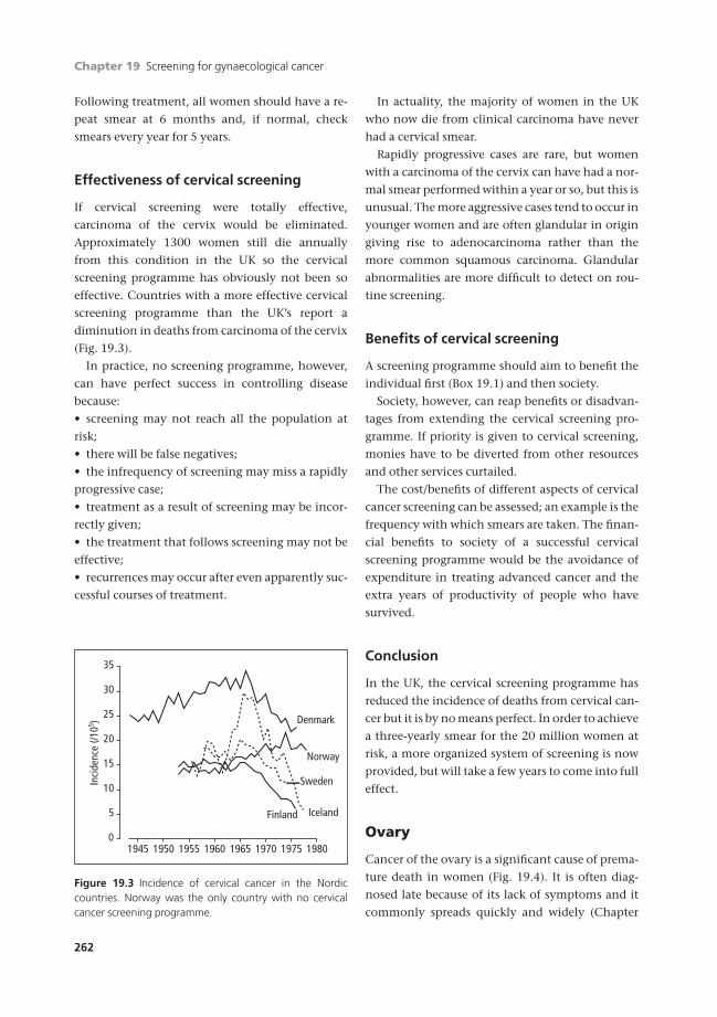
Chapter 19 Screening for gynaecological cancer
262
Following treatment, all women should have a re-
peat smear at 6 months and, if normal, check
smears every year for 5 years.
Effectiveness of cervical screening
If cervical screening were totally effective,
carcinoma of the cervix would be eliminated.
Approximately 1300 women still die annually
from this condition in the UK so the cervical
screening programme has obviously not been so
effective. Countries with a more effective cervical
screening programme than the UK’s report a
diminution in deaths from carcinoma of the cervix
(Fig. 19.3).
In practice, no screening programme, however,
can have perfect success in controlling disease
because:
• screening may not reach all the population at
risk;
• there will be false negatives;
• the infrequency of screening may miss a rapidly
progressive case;
• treatment as a result of screening may be incor-
rectly given;
• the treatment that follows screening may not be
effective;
• recurrences may occur after even apparently suc-
cessful courses of treatment.
In actuality, the majority of women in the UK
who now die from clinical carcinoma have never
had a cervical smear.
Rapidly progressive cases are rare, but women
with a carcinoma of the cervix can have had a nor-
mal smear performed within a year or so, but this is
unusual. The more aggressive cases tend to occur in
younger women and are often glandular in origin
giving rise to adenocarcinoma rather than the
more common squamous carcinoma. Glandular
abnormalities are more difficult to detect on rou-
tine screening.
Benefits of cervical screening
A screening programme should aim to benefit the
individual first (Box 19.1) and then society.
Society, however, can reap benefits or disadvan-
tages from extending the cervical screening pro-
gramme. If priority is given to cervical screening,
monies have to be diverted from other resources
and other services curtailed.
The cost/benefits of different aspects of cervical
cancer screening can be assessed; an example is the
frequency with which smears are taken. The finan-
cial benefits to society of a successful cervical
screening programme would be the avoidance of
expenditure in treating advanced cancer and the
extra years of productivity of people who have
survived.
Conclusion
In the UK, the cervical screening programme has
reduced the incidence of deaths from cervical can-
cer but it is by no means perfect. In order to achieve
a three-yearly smear for the 20 million women at
risk, a more organized system of screening is now
provided, but will take a few years to come into full
effect.
Ovary
Cancer of the ovary is a significant cause of prema-
ture death in women (Fig. 19.4). It is often diag-
nosed late because of its lack of symptoms and it
commonly spreads quickly and widely (Chapter
35
Norway
Denmark
IcelandFinland
Sweden
15
20
25
30
10
5
019801975197019651960195519501945
Inci
denc
e (/1
05 )
Figure 19.3 Incidence of cervical cancer in the Nordiccountries. Norway was the only country with no cervicalcancer screening programme.
AMI19 6/9/04 5:09 PM Page 262
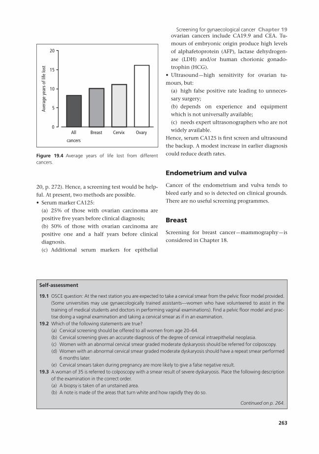
Screening for gynaecological cancer Chapter 19
263
20, p. 272). Hence, a screening test would be help-
ful. At present, two methods are possible.
• Serum marker CA125:
(a) 25% of those with ovarian carcinoma are
positive five years before clinical diagnosis;
(b) 50% of those with ovarian carcinoma are
positive one and a half years before clinical
diagnosis.
(c) Additional serum markers for epithelial
ovarian cancers include CA19.9 and CEA. Tu-
mours of embryonic origin produce high levels
of alphafetoprotein (AFP), lactase dehydrogen-
ase (LDH) and/or human chorionic gonado-
trophin (HCG).
• Ultrasound —high sensitivity for ovarian tu-
mours, but:
(a) high false positive rate leading to unneces-
sary surgery;
(b) depends on experience and equipment
which is not universally available;
(c) needs expert ultrasonographers who are not
widely available.
Hence, serum CA125 is first screen and ultrasound
the backup. A modest increase in earlier diagnosis
could reduce death rates.
Endometrium and vulva
Cancer of the endometrium and vulva tends to
bleed early and so is detected on clinical grounds.
There are no useful screening programmes.
Breast
Screening for breast cancer —mammography —is
considered in Chapter 18.
20
10
15
5
0All
cancers
Breast Cervix Ovary
Aver
age
year
s of
life
lost
Figure 19.4 Average years of life lost from different cancers.
Self-assessment
19.1 OSCE question: At the next station you are expected to take a cervical smear from the pelvic floor model provided.(Some universities may use gynaecologically trained assistants—women who have volunteered to assist in thetraining of medical students and doctors in performing vaginal examinations). Find a pelvic floor model and prac-tise doing a vaginal examination and taking a cervical smear as if in an examination.
19.2 Which of the following statements are true?(a) Cervical screening should be offered to all women from age 20–64.(b) Cervical screening gives an accurate diagnosis of the degree of cervical intraepithelial neoplasia.(c) Women with an abnormal cervical smear graded moderate dyskaryosis should be referred for colposcopy.(d) Women with an abnormal cervical smear graded moderate dyskaryosis should have a repeat smear performed
6 months later.(e) Cervical smears taken during pregnancy are more likely to give a false negative result.
19.3 A woman of 35 is referred to colposcopy with a smear result of severe dyskaryosis. Place the following descriptionof the examination in the correct order.(a) A biopsy is taken of an unstained area.(b) A note is made of the areas that turn white and how rapidly they do so.
Continued on p. 264.
AMI19 6/9/04 5:09 PM Page 263

Chapter 19 Screening for gynaecological cancer
264
Self-assessment Continued
(c) The area is excised using loop diathermy under local anaesthetic.(d) 4% acetic acid is painted onto the cervix.(e) Iodine solution is painted onto the cervix and the unstained areas noted.
19.4 List the six main reasons why the UK cervical screening programme is not 100% effective.19.5 List the five measures that have been put in place to try to improve the effectiveness of screening in the UK.
AMI19 6/9/04 5:09 PM Page 264

Part 5
The older woman
AMI20 6/9/04 5:11 PM Page 265

AMI20 6/9/04 5:11 PM Page 266

Cancer Registries collect data about every
new case of malignant disease diagnosed in that
catchment population. In the UK, data come
from:
• hospital in-patient statistics;
• pathology registers;
• radiotherapy registers;
• oncological out-patient clinics;
• colposcopy clinics.
Registries also get death certificates of all cancer
deaths in their population, so giving a measure of
the total incidence of gynaecological cancer. Mor-
tality rates of malignant disease come from death
certificates and are published by the Office of
Population Censuses and Surveys (OPCS). Thus,
there are two distinct sources of epidemiological
data about malignant disease:
• the living —prevalence and incidence rates;
• the dead —mortality rates.
In 2000, three gynaecological cancers featured
amongst the 10 most frequent cancers among
females. Some 5400 women were reported with
new cases of carcinoma of the ovary, 2424 with in-
vasive carcinoma of the cervix and 4730 with car-
cinoma of the body of the uterus. Thus, carcinoma
of the ovary has overtaken that of the cervix in the
last 20 years as the commonest gynaecological can-
cer. In 1973 registrations for cervix and ovary were
respectively 4065 and 3819.
In 2001, the deaths reported of the same three
cancers were 3807, 947 and 853. No precise math-
ematical ratios can be derived because the data
are of different populations in time —however,
this provides an indication of the poor prognosis of
cancer of the ovary compared with that of the
cervix/uterus; over the years the prognosis for
ovarian cancer has remained unchanged. The five-
year follow-up data are given in Table 20.1. These
differences may represent:
• a real change in the prevalence of a condition;
• a more complete reporting system;
• better diagnostic facilities for making an early
diagnosis.
There is a trend of increasing incidence of cervi-
cal cancer in younger women. There has been a 20-
fold increase in the number of women aged under
35 presenting with cervical carcinoma. Many of
these will have adenocarcinoma rather than squa-
mous cell carcinoma which is harder to detect on
routine screening and has a poorer prognosis. The
numbers remain small and there is no evidence
that the overall mortality for cervical cancer is
increasing.
The geographical incidence of carcinoma of the
cervix was highest in Central and Southern
America, decreasing as it crossed Europe and Africa
to Asia and the Far East. This apparent trend no
longer exists. There are many local variations; for
example, Portugal has a very high rate of carcin-
oma of the cervix while its close neighbour, Spain,
has a very low one, yet their economic and social
characteristics are very similar.
267267
Chapter 20
Malignant gynaecological conditions
AMI20 6/9/04 5:11 PM Page 267
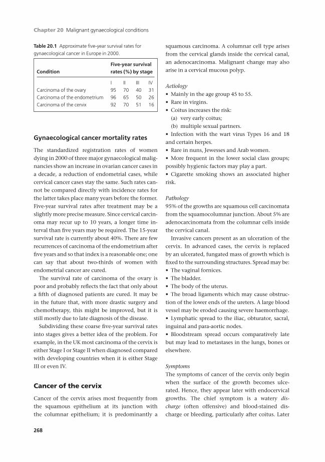
Chapter 20 Malignant gynaecological conditions
268
Gynaecological cancer mortality rates
The standardized registration rates of women
dying in 2000 of three major gynaecological malig-
nancies show an increase in ovarian cancer cases in
a decade, a reduction of endometrial cases, while
cervical cancer cases stay the same. Such rates can-
not be compared directly with incidence rates for
the latter takes place many years before the former.
Five-year survival rates after treatment may be a
slightly more precise measure. Since cervical carcin-
oma may recur up to 10 years, a longer time in-
terval than five years may be required. The 15-year
survival rate is currently about 40%. There are few
recurrences of carcinoma of the endometrium after
five years and so that index is a reasonable one; one
can say that about two-thirds of women with
endometrial cancer are cured.
The survival rate of carcinoma of the ovary is
poor and probably reflects the fact that only about
a fifth of diagnosed patients are cured. It may be
in the future that, with more drastic surgery and
chemotherapy, this might be improved, but it is
still mostly due to late diagnosis of the disease.
Subdividing these coarse five-year survival rates
into stages gives a better idea of the problem. For
example, in the UK most carcinoma of the cervix is
either Stage I or Stage II when diagnosed compared
with developing countries when it is either Stage
III or even IV.
Cancer of the cervix
Cancer of the cervix arises most frequently from
the squamous epithelium at its junction with
the columnar epithelium; it is predominantly a
squamous carcinoma. A columnar cell type arises
from the cervical glands inside the cervical canal,
an adenocarcinoma. Malignant change may also
arise in a cervical mucous polyp.
Aetiology• Mainly in the age group 45 to 55.
• Rare in virgins.
• Coitus increases the risk:
(a) very early coitus;
(b) multiple sexual partners.
• Infection with the wart virus Types 16 and 18
and certain herpes.
• Rare in nuns, Jewesses and Arab women.
• More frequent in the lower social class groups;
possibly hygienic factors may play a part.
• Cigarette smoking shows an associated higher
risk.
Pathology95% of the growths are squamous cell carcinomata
from the squamocolumnar junction. About 5% are
adenocarcinomata from the columnar cells inside
the cervical canal.
Invasive cancers present as an ulceration of the
cervix. In advanced cases, the cervix is replaced
by an ulcerated, fungated mass of growth which is
fixed to the surrounding structures. Spread may be:
• The vaginal fornices.
• The bladder.
• The body of the uterus.
• The broad ligaments which may cause obstruc-
tion of the lower ends of the ureters. A large blood
vessel may be eroded causing severe haemorrhage.
• Lymphatic spread to the iliac, obturator, sacral,
inguinal and para-aortic nodes.
• Bloodstream spread occurs comparatively late
but may lead to metastases in the lungs, bones or
elsewhere.
SymptomsThe symptoms of cancer of the cervix only begin
when the surface of the growth becomes ulce-
rated. Hence, they appear later with endocervical
growths. The chief symptom is a watery dis-
charge (often offensive) and blood-stained dis-
charge or bleeding, particularly after coitus. Later
Table 20.1 Approximate five-year survival rates for
gynaecological cancer in Europe in 2000.
Five-year survivalCondition rates (%) by stage
I II III IV
Carcinoma of the ovary 95 70 40 31
Carcinoma of the endometrium 96 65 50 26
Carcinoma of the cervix 92 70 51 16
AMI20 6/9/04 5:11 PM Page 268

Malignant gynaecological conditions Chapter 20
269
frank, sometimes severe and continuous bleeding
occurs, with the patient rapidly becoming anaemic.
Physical signsEarly, the cervix feels hard and bleeds on touch.
Later the cervix is ulcerated and friable. In
advanced cases the vaginal vault is filled with an
ulcerated mass and pieces of growth are detached
by the examining finger; examination may pro-
voke severe bleeding. In endocervical growths the
cervix feels barrel-shaped.
The cervical smear may contain frankly malig-
nant cells, but not always because the surface cells
are often dead and atypical.
DiagnosisThis depends on biopsy of the cervix. If the site and
size of the lesion allow, a cone biopsy should be
taken to include all the squamocolumnar junction
and most of the cervical canal.
Staging investigationsStaging for cervical carcinoma includes:
• Examination under anaesthetic, including
rectovaginal examination to assess the size of the
tumour, parametrial spread, extension into the
rectovagina; septum.
• Cystoscopy and sigmoidoscopy to assess bladder
and bowel involvement.
• Biopsy of the suspicious area.
• Chest X-ray.
• Intravenous urography (IVU).
Computerized tomography (CT) or magnetic reson-
ance imaging (MRI) may be offered if available to
give further information on tumour size and nodal
involvement, but does not alter the FIGO staging
which is determined by the above investigations.
Differential diagnosisMainly from:
• cervical ectropion;
• tuberculosis may present in a proliferative form;
• other chronic granulomatous infections.
StagingBox 20.1 shows the clinical staging of cancer of the
cervix.
This is a clinical classification; in fact 20% of
Stage 1 cases are found at subsequent operation to
have metastases in the lymph glands (i.e. would be
Stage 3).
Treatment of invasive carcinoma
The choice of treatment depends on many factors.
• The age and general condition of the patient.
• The extent and type of the lesion.
• Ideally all patients with cancer of the cervix
should be seen by a gynaecological oncologist and
a radiotherapist for consideration of all the factors
of the individual case.
• The experience, resources and personal prefer-
ence of the oncology team.
Renal function should be assessed by blood urea
and intravenous urography. Urinary infection is
often present and should be treated. Anaemia may
also need treatment.
Ultrasound and CT scan may help identify
spread in the pelvis or to lymph nodes.
Examination under anaesthesia is essential.
• The clinical extent of the growth is assessed.
• Hysteroscopy and curettage is performed and a
biopsy taken in all cases, even those which seem
the most obvious clinically.
• A rectal examination is important to exclude
invasion of the rectum itself. The clinical extent of
growth in the parametrium is also more easily felt
rectally.
Box 20.1 Staging of cancer of the cervix
Stage 0 Intraepithelial carcinoma (carcinoma in situ).The growth remains within the epitheliallayer of the cervix
Stage 1 Cancer clinically confined to the cervixStage 2 Growth has spread to the upper two-thirds
of the vagina or into the parametrium butnot as far as the pelvic wall
Stage 3 The growth has spread to the lower one-thirdof the vagina or into the parametrium as faras the lateral pelvic wall
Stage 4 Metastases have formed beyond the pelvis or growth has involved the bladder or therectum
AMI20 6/9/04 5:11 PM Page 269
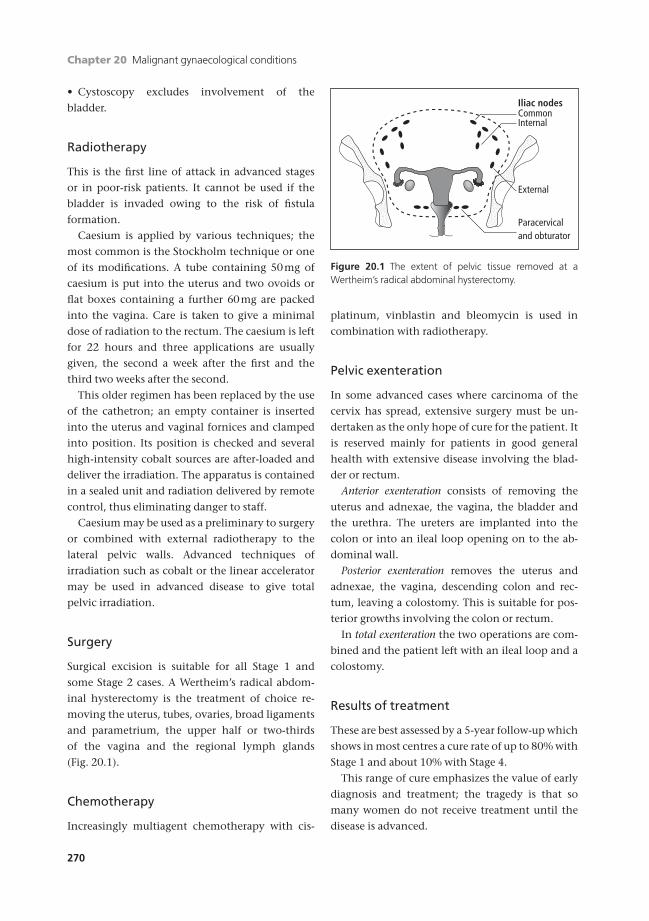
Chapter 20 Malignant gynaecological conditions
270
• Cystoscopy excludes involvement of the
bladder.
Radiotherapy
This is the first line of attack in advanced stages
or in poor-risk patients. It cannot be used if the
bladder is invaded owing to the risk of fistula
formation.
Caesium is applied by various techniques; the
most common is the Stockholm technique or one
of its modifications. A tube containing 50mg of
caesium is put into the uterus and two ovoids or
flat boxes containing a further 60mg are packed
into the vagina. Care is taken to give a minimal
dose of radiation to the rectum. The caesium is left
for 22 hours and three applications are usually
given, the second a week after the first and the
third two weeks after the second.
This older regimen has been replaced by the use
of the cathetron; an empty container is inserted
into the uterus and vaginal fornices and clamped
into position. Its position is checked and several
high-intensity cobalt sources are after-loaded and
deliver the irradiation. The apparatus is contained
in a sealed unit and radiation delivered by remote
control, thus eliminating danger to staff.
Caesium may be used as a preliminary to surgery
or combined with external radiotherapy to the
lateral pelvic walls. Advanced techniques of
irradiation such as cobalt or the linear accelerator
may be used in advanced disease to give total
pelvic irradiation.
Surgery
Surgical excision is suitable for all Stage 1 and
some Stage 2 cases. A Wertheim’s radical abdom-
inal hysterectomy is the treatment of choice re-
moving the uterus, tubes, ovaries, broad ligaments
and parametrium, the upper half or two-thirds
of the vagina and the regional lymph glands
(Fig. 20.1).
Chemotherapy
Increasingly multiagent chemotherapy with cis-
platinum, vinblastin and bleomycin is used in
combination with radiotherapy.
Pelvic exenteration
In some advanced cases where carcinoma of the
cervix has spread, extensive surgery must be un-
dertaken as the only hope of cure for the patient. It
is reserved mainly for patients in good general
health with extensive disease involving the blad-
der or rectum.
Anterior exenteration consists of removing the
uterus and adnexae, the vagina, the bladder and
the urethra. The ureters are implanted into the
colon or into an ileal loop opening on to the ab-
dominal wall.
Posterior exenteration removes the uterus and
adnexae, the vagina, descending colon and rec-
tum, leaving a colostomy. This is suitable for pos-
terior growths involving the colon or rectum.
In total exenteration the two operations are com-
bined and the patient left with an ileal loop and a
colostomy.
Results of treatment
These are best assessed by a 5-year follow-up which
shows in most centres a cure rate of up to 80% with
Stage 1 and about 10% with Stage 4.
This range of cure emphasizes the value of early
diagnosis and treatment; the tragedy is that so
many women do not receive treatment until the
disease is advanced.
Iliac nodesCommonInternal
External
Paracervicaland obturator
Figure 20.1 The extent of pelvic tissue removed at aWertheim’s radical abdominal hysterectomy.
AMI20 6/9/04 5:11 PM Page 270

Malignant gynaecological conditions Chapter 20
271
Complications of treatment
Complications may follow treatment with irradia-
tion or surgery. Radiotherapy treatment can flare
up infection in the renal tract, or exacerbate pelvic
abscess. Caesium proctitis may prove troublesome.
The mortality risk at the operation of Wertheim’s
hysterectomy is now only 1% in experienced
hands. In addition to any complications of a severe
abdominal operation, there is a risk of ureteric
fistula which has been reported as high as 8% of
patients submitted to Wertheim’s hysterectomy
after irradiation.
Palliation
When nothing can be done to cure the patient of
cancer, everyone concentrates on making her last
weeks or months as comfortable as possible. Death
may occur mercifully from uraemia or haemor-
rhage, but many women suffer severe and in-
tractable pain in the final stages of the disease.
Analgesics must be used liberally in sufficient
amounts to relieve pain. Epidural anaethesia
and/or nerve blocks may be helpful. Chordotomy
is sometimes used in intractable pain. If there is se-
vere rectal pain, colostomy may be necessary. Mor-
phia and heroin are of great help here and must be
retained in the profession’s therapeutic armamen-
tarium, prescribing and dispensing being under
strict control. Addiction is not a concern in those
with advanced pelvic cancer and dosage should be
liberal once started.
Malignant tumours of the uterus
Choriocarcinoma
A malignant tumour arising from chorionic tissue
following a hydatidiform mole, abortion or preg-
nancy and is considered in Chapter 7.
Endometrial carcinoma
Aetiology• Mean age of presentation is 56 years. Four-fifths
of the women are menopausal and it is rare under
the age of 40.
• Associated with hyperoestrogenic states:
(a) obesity;
(b) diabetes;
(c) late menopause;
(d) prolonged use of unopposed oestrogens;
(e) oestrogen secreting tumours;
(f) long-term tamoxifen usage.
• May be associated with:
(a) previous pelvic irradiation;
(b) lower parity.
Pathology• Usually an adenocarcinoma.
• More often well differentiated than
anaplastic.
• May be associated with squamous metaplasia
where, if excessive, becomes an adenocanthoma.
• May be associated with pyometra or
haematometra, secondary to cervical stenosis.
• Spreads by invasion through the myometrium
and by filling the uterine cavity.
• Spreads via cervical lymphatic drainage involv-
ing the iliac and para-aortic nodes.
• Tumours of upper uterus may spread along the
lymphatics in the round ligaments to the deep
inguinal nodes.
• In advanced cases, the bloodstream spread may
carry to the lungs, liver and to the bones.
Symptoms• Postmenopausal bleeding: this symptom should
be assumed to be caused by carcinoma of the en-
dometrium until proved otherwise.
• Bloodstained discharge.
• Irregular bleeding.
Signs• Less commonly, uterine enlargement.
• Bleeding through the cervix.
Investigations• Ultrasound to assess dimensions of any tumour
and to show endometrial thickness.
AMI20 6/9/04 5:11 PM Page 271

Chapter 20 Malignant gynaecological conditions
272
StagingBox 20.2 shows the clinical staging of endometrial
cancer.
TreatmentThe treatment of uterine carcinoma is usually
surgical.
• The surgical management of well differentiated
carcinoma involves a total abdominal hysterec-
tomy and bilateral salpingo-oophorectomy (TAH/
BSO). The role of pelvic lymphaedectomy is being
assessed in the MRC ASTEC trial.
• In women with high grade disease (grade 3 or
grade 2 with >2cm invasion), or in women with
>50% myometrial invasion or cervical involve-
ment, or in women with adverse pathological
subtypes (adenosquamous, clear cell or papillary
serous carcinoma), a full staging laparotomy in-
volving omental biopsy, lymph node sampling
and inspection of the upper abdomen is required
in addition to the TAH/BSO.
• Radical hysterectomy is indicated if there is
cervical involvement.
Radiotherapy is rarely employed alone unless
the patient is unable to withstand a surgical proce-
dure. It may be used as adjuvant treatment if ad-
verse factors are identified in the pathology review:
• >50% myometrial invasion;
• poorly differentiated high grade disease;
• adenosquamous, clear cell or papillary serous
carcinoma;
• positive pelvic lymph nodes.
The pelvic lymph node status determines whether
radiotherapy is given to the vault alone or to the
vault and the pelvic side wall.
Hormone therapy—progestogens inhibit the
rate of growth and spread of endometrial
carcinoma.
Rarer tumours
Sarcoma
Occurs in:
• childhood as sarcoma botryoides;
• postmenopausal women with a fibroid.
It is highly malignant, radioresistant, spreads by
the bloodstream and is diagnosed late. Treatment
is a total abdominal hysterectomy and bilateral
salpingo-oophorectomy. Recurrences are treated
with multiagent chemotherapy.
Mixed mesodermal tumours
These arise from mesodermal cells of the ovarian
ducts and may contain primitive muscle cells,
myxomatous tissue, cartilage and glands. They
present with abnormal bleeding and treatment is
by hysterectomy or occasionally by exenteration.
Vascular spread is common, prognosis poor.
Carcinoma of the ovary
Primary carcinoma of the ovary is now the com-
monest malignant tumour found in gynaecology
in the UK and an important cause of death in
women, accounting for some 4000 deaths annual-
ly in England and Wales. It is a disease of middle
and old age with 90% of cases in women above 45
years. It is often diagnosed late because of its lack of
symptoms and it commonly metastasizes quickly
and widely. Hence, a screening test would be
helpful (Chapter 19).
Risk factors relate to ovulatory history and the
past activity of the germinal epithelium:
• increased risk —no pregnancy;
• decreased risk —many pregnancies —use of oral
contraceptives.
Box 20.2 Staging of endometrial cancer
Stage 1 Carcinoma confined to the body of theuterus
Stage 2 Extension to the cervixStage 3 Extension outside the uterus but within the
true pelvisStage 4 Involvement of the:
(a) bladder(b) rectum(c) extension outside the true pelvis
AMI20 6/9/04 5:11 PM Page 272
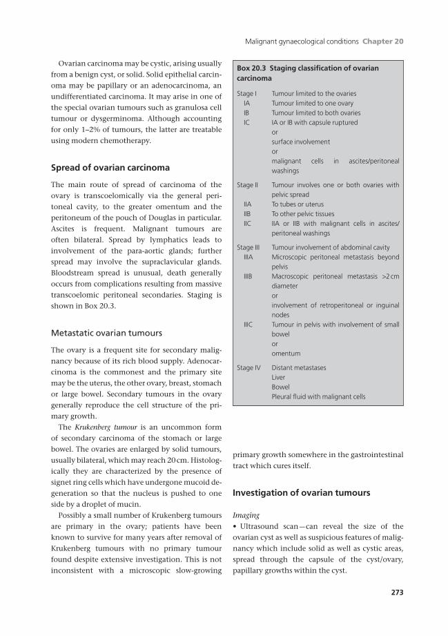
Malignant gynaecological conditions Chapter 20
273
Ovarian carcinoma may be cystic, arising usually
from a benign cyst, or solid. Solid epithelial carcin-
oma may be papillary or an adenocarcinoma, an
undifferentiated carcinoma. It may arise in one of
the special ovarian tumours such as granulosa cell
tumour or dysgerminoma. Although accounting
for only 1–2% of tumours, the latter are treatable
using modern chemotherapy.
Spread of ovarian carcinoma
The main route of spread of carcinoma of the
ovary is transcoelomically via the general peri-
toneal cavity, to the greater omentum and the
peritoneum of the pouch of Douglas in particular.
Ascites is frequent. Malignant tumours are
often bilateral. Spread by lymphatics leads to
involvement of the para-aortic glands; further
spread may involve the supraclavicular glands.
Bloodstream spread is unusual, death generally
occurs from complications resulting from massive
transcoelomic peritoneal secondaries. Staging is
shown in Box 20.3.
Metastatic ovarian tumours
The ovary is a frequent site for secondary malig-
nancy because of its rich blood supply. Adenocar-
cinoma is the commonest and the primary site
may be the uterus, the other ovary, breast, stomach
or large bowel. Secondary tumours in the ovary
generally reproduce the cell structure of the pri-
mary growth.
The Krukenberg tumour is an uncommon form
of secondary carcinoma of the stomach or large
bowel. The ovaries are enlarged by solid tumours,
usually bilateral, which may reach 20cm. Histolog-
ically they are characterized by the presence of
signet ring cells which have undergone mucoid de-
generation so that the nucleus is pushed to one
side by a droplet of mucin.
Possibly a small number of Krukenberg tumours
are primary in the ovary; patients have been
known to survive for many years after removal of
Krukenberg tumours with no primary tumour
found despite extensive investigation. This is not
inconsistent with a microscopic slow-growing
primary growth somewhere in the gastrointestinal
tract which cures itself.
Investigation of ovarian tumours
Imaging• Ultrasound scan —can reveal the size of the
ovarian cyst as well as suspicious features of malig-
nancy which include solid as well as cystic areas,
spread through the capsule of the cyst/ovary,
papillary growths within the cyst.
Box 20.3 Staging classification of ovariancarcinoma
Stage I Tumour limited to the ovariesIA Tumour limited to one ovaryIB Tumour limited to both ovariesIC IA or IB with capsule ruptured
orsurface involvementormalignant cells in ascites/peritoneal washings
Stage II Tumour involves one or both ovaries withpelvic spread
IIA To tubes or uterusIIB To other pelvic tissuesIIC IIA or IIB with malignant cells in ascites/
peritoneal washings
Stage III Tumour involvement of abdominal cavityIIIA Microscopic peritoneal metastasis beyond
pelvisIIIB Macroscopic peritoneal metastasis >2cm
diameterorinvolvement of retroperitoneal or inguinalnodes
IIIC Tumour in pelvis with involvement of smallboweloromentum
Stage IV Distant metastasesLiverBowelPleural fluid with malignant cells
AMI20 6/9/04 5:11 PM Page 273

Chapter 20 Malignant gynaecological conditions
274
• CT/MRI scan of the abdomen and pelvis —more
accurately delineates the spread of disease showing
peritoneal deposits, omental deposits, para-aortic
node involvement and/or liver metastases, ureteric
obstruction (rare in ovarian cancer).
• Chest X-ray —shows pleural effusions and/or
hilar lymphadenopathy.
Blood tests• Tumour markers —CA125, CA199, CEA. If any of
these are raised they are used postoperatively to
track the success of treatment and the onset of
recurrence.
• Liver function tests to detect spread to the liver.
• Urea and electrolytes.
• Full blood count.
The latter two do not help in staging the disease
but are important for preoperative assessment.
Treatment of ovarian tumours
The treatment of malignant ovarian tumours is
surgical removal as soon as the tumour is diag-
nosed. If the tumour is apparently malignant
and where ascites is present, laparotomy should
always be undertaken. Ascites may be associated
with a benign tumour such as a fibroma, and
even if there are metastases the prognosis is not
hopeless. It may be possible to remove secon-
dary masses in the omentum and, if the primary
tumour is removed, secondaries sometimes regress.
An ovarian tumour, even if very large, is best
removed intact. Tapping the fluid carries a risk of
spilling the contents and contaminating the
peritoneal cavity.
Carcinoma of the ovary should be treated initially
by surgery which should involve total hyster-
ectomy, bilateral salpingo-oophorectomy and
omentectomy, though in young women a normal,
uninvolved ovary might be left. In advanced cases,
as much tumour as possible should be removed at
a debulking operation and the greater omentum
should be excised. A search should be made for
peritoneal metastases, including those on the
upper and under surface of the liver. A CT or
MRI scan to check that the peritoneal cavity is
free from secondary deposits may be carried
out three to six months after the original
operation.
Even in apparently advanced disease the ulti-
mate prognosis appears to be improved by opera-
tive removal of the main tumour masses. The first
operation offers the best chance of cure. It should
be done by an experienced gynaecological oncolo-
gist, preferably working in a specialist centre of
gynaecologic oncology, who will do the widest
excision with the least damage to ureters, bladder
or intestines.
Radiotherapy is not much used in the man-
agement of ovarian cancer; the tumours are
rarely radiosensitive and radiation would have a
deleterious effect on the bone marrow in the
lumbar vertebrae.
Chemotherapy with cytotoxic agents gives more
hopeful results. Cisplatin, one of the platinum
compounds, given in intravenous infusion and
repeated every four weeks, shows good results.
Carboplatin is equally useful and has fewer
side-effects. In combination with other agents
such as Taxol, the platinum compounds give a
significant improvement in results (see Box
20.4).
Prognosis
The general prognosis for ovarian carcinoma is
poor, less than 20% surviving for five years. Factors
which worsen prognosis are:
• advanced stage of disease;
• poorly differentiated tumour;
• how much tumour remains after surgery.
Box 20.4 Chemotherapy with cytotoxic agents
Single platinum agents• Cisplatin• Carboplatin
Combinations• Platinum agents plus cyclophosphamide• Platinum agents plus cyclophosphamide plus doxoru-bicin• Platinum agents plus Taxol
AMI20 6/9/04 5:11 PM Page 274
Malignant gynaecological conditions Chapter 20
275
Carcinoma of the fallopian tube
Primary• A rare malignancy occurring in older women.
• Papillary carcinoma may be solid or alveolar.
• Sometimes there is a vaginal discharge of an or-
ange–yellow colour.
Treatment is as for ovarian carcinoma.
Secondary• Metastases in the tube most commonly come
from cancer of the ovary or uterus.
Malignant disease of the vulva
Squamous carcinoma
Vulval carcinoma is less common than other
gynaecological cancers. The squamous form is the
commonest malignant tumour of the vulva. It
occurs mainly in the older age group, with a peak
incidence at about 60 years of age. The condition
is associated with vulval dystrophy (Chapter 6).
PathologyThe primary growth is an ulcer with a raised,
everted edge and indurated base. Multiple primar-
ies may be found, sometimes the inner sides of
both labia minora are involved. The growth may
also arise on the clitoris.
Methods of spread• The growth may spread by direct extension and
contact to other parts of the vulva, vagina or anus.
Secondary spread is mainly by lymphatics.
Owing to the rich lymphatic drainage of the vulva,
the glands which tend to be involved are:
• superficial inguinal group of both sides;
• inguinal;
• femoral;
• iliac;
• aortic.
In untreated cases, the glands in the groin may
break down to form a fungating ulcerated mass of
growth.
Clinical featuresCarcinoma of the vulva commonly begins as a
small nodule, often unnoticed by the patient at
first. It grows in size and becomes ulcerated with
discharge and bleeding. It tends to grow on the
inner surface of the labia minora in elderly women
and may remain unnoticed except for slight
discomfort and soreness from the discharge until
an advanced stage.
Differential diagnosisTo differentiate malignancy from other causes of a
lump in the vulva or of ulceration is the main prob-
lem. All lumps or ulcers of the vulva must be fully
investigated including a biopsy.
TreatmentTreatment of carcinoma of the vulva is vulvectomy
and dissection of all the superficial and deep
inguinal glands and occasionally the iliac glands.
The vulva itself is widely excised with the glands
through separate incisions over each groin. Wide
excision in advanced growths may have to include
removal of the lower part of the urethra, vagina or
anal canal depending on site.
In operable cases, a 5-year cure rate of about 70%
is achieved. The prognosis depends mainly on
involvement of the lymphatic glands.
RADIOTHERAPY
Carcinoma of the vulva is relatively radiore-
sistant while the surrounding normal tissues are
radiosensitive. Hence, it is not employed usually,
but high-voltage treatment may be used for
recurrences.
CHEMOTHERAPY
This is not a primary treatment for squamous
epithelioma or cancer of the vulva, but as with
carcinomas of the anus it is being used more often
for recurrence.
Intraepithelial neoplasia
In vulval intraepithelial neoplasia (VIN), the ma-
lignant cells are limited to the outer layers of the
epidermis and there is no spread to the underlying
AMI20 6/9/04 5:11 PM Page 275

Chapter 20 Malignant gynaecological conditions
276
tissues and no metastases. The whole layer is infil-
trated with malignant cells.
Clinical featuresThe patient has irritation or soreness of the vulva.
The appearance may be that of vulval dystrophy or
there may be a red area with a serpiginous outline.
VIN may remain dormant for years or may assume
the characteristic of invasive carcinoma.
Diagnosis depends on biopsy.
Treatment is by a wide excision with a margin of
healthy skin and epithelium.
Basal cell carcinoma
This uncommon tumour presents as an indolent
ulcer without invasion of the underlying tissues.
Diagnosis is made by biopsy.
The treatment consists of local excision with a
margin of normal skin.
Malignant melanoma
Fortunately this highly malignant tumour is
rare. It may present as a melanotic nodule or
as a pedunculated tumour. The best treatment
is to perform a vulvectomy and if nodes are
involved, an en bloc dissection. Cases with
diffuse spread of melanoma may be treated by
radiotherapy.
Self-assessment
20.1 Which of the following statements are true?(a) Carcinoma of the cervix is the most common gynaecological malignancy.(b) More women die of ovarian cancer than any other gynaecological malignancy.(c) Cervical cancer most commonly presents with postmenopausal bleeding.(d) Endometrial cancer most commonly presents with postmenopausal bleeding.(e) Ovarian cancer most commonly presents with postcoital bleeding.
20.2 A woman of 48 presents with postcoital bleeding, her last smear was 8 years ago and was normal. She is other-wise well. On examination she has an ulcerated lesion on her cervix which bleeds on contact. The vulva, vagina anduterus all feel normal. Which of the following should be undertaken to investigate the cause of her postcoitalbleeding?(a) CT scan of abdomen and pelvis.(b) Liver function tests.(c) Biopsy of her cervix.(d) Chest X-ray.(e) Hysteroscopy and curettage.
20.3 A woman of 63 presents with a hard fixed abdominal mass noticed by her GP when she complained that herclothes were becoming tighter. Her menopause was 12 years ago and she has had no vaginal bleeding. She has noother abnormalities on examination. What is the most likely provisional diagnosis?(a) Stage 3 carcinoma of the ovary.(b) Stage 3 carcinoma of the cervix.(c) Stage 1 carcinoma of the ovary.(d) Stage 3 carcinoma of the uterus.(e) Stage 4 carcinoma of the ovary.
20.4 Which of the following are useful preoperative staging investigations for the woman in Question 20.3?(a) Hysteroscopy and curettage.(b) CT scan of abdomen and pelvis.(c) Serum tumour markers.(d) Chest X-ray.(e) Urea and electrolytes.
AMI20 6/9/04 5:11 PM Page 276

Malignant gynaecological conditions Chapter 20
277
Self-assessment Continued
20.5 A woman of 63 presents with an offensive discharge from her vagina. She admits that her vulva has been very itchyand sore for several years. Over the last few months she has noticed occasional blood stains on her pants and a lump on her left labia. Her GP has treated her with antibiotics to no effect. Which of the following are the mostappropriate investigations for this woman?(a) Examination and biopsy of her vulva.(b) High vaginal swab.(c) Abdominal palpation particularly in both groins.(d) Cervical smear.(e) Hysteroscopy and curettage.
AMI20 6/9/04 5:11 PM Page 277
278
The menopause is the cessation of normal men-
struation. The climacteric is a longer period during
which time the reproductive organs involute.
These time zones overlap each other in time just
as they do in youth with the two processes of
menarche and puberty.
The mean age of menopause in the UK is 51 with
a normal range from 45 to 56. Conventionally a
woman has to stop menstruating for 12 months
before she is considered to be postmenopausal.
Physiology
At the end of reproductive life, the ovaries become
less able to produce oocytes due to:
• a lack of primordial follicles, because all have
been used;
• more refractory receptor function in the granu-
losa and thecal cells.
The falling oestrogen levels result in a large
increase in follicular stimulating hormone (FSH).
The endometrium does not proliferate.
The ovarian stroma produces androstenedione
which converts in peripheral fat to oestrone, a
weaker oestrogen than oestradiol, the steroid on
which the woman has depended for much of her
reproductive life. Menstruation stops due to a lack
of cyclical oestrogen and progesterone.
Symptoms
At the menopause 60% of women are relatively
asymptomatic, 25% of women have mild symp-
toms and 15% have moderate to severe symptoms.
The two commonest symptoms are:
• hot flushes;
• dryness of the vagina.
There is often a loss of libido, part of which is
hormonal.
Mood swings, nervousness, anxiety, irritability
and depression are all measured in this group of
women. The decrease of oestrogens may reduce
their modulatory role on brain monoamine
synthesis.
Symptoms are found more commonly in those
who had premenstrual tension or dysmenorrhoea.
The symptoms are less frequent in Asian and Negro
women, possibly associated with better mainten-
ance of oestrogen levels by peripheral conversion
in these groups.
Loss of collagen leads to uterovaginal prolapse
and wrinkling of the skin.
Hot flushes
These are the feeling of heat over the face and
upper part of the body usually lasting for half to
one minute. They are followed by perspiration of
this area, which may render the woman wringing
wet. These flushes usually last for a year or so and
Chapter 21
The menopause
AMI21 6/9/04 5:11 PM Page 278
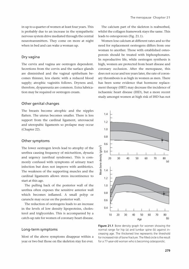
The menopause Chapter 21
279
in up to a quarter of women at least four years. This
is probably due to an increase in the sympathetic
nervous system drive mediated through the central
neurotransmitters. They come on more at night
when in bed and can wake a woman up.
Dry vagina
The cervix and vagina are oestrogen dependent.
Secretions from the cervix and the surface glands
are diminished and the vaginal epithelium be-
comes thinner, less elastic with a reduced blood
supply; atrophic vaginitis follows. Dryness and,
therefore, dyspareunia are common. Extra lubrica-
tion may be required or oestrogen cream.
Other genital changes
The breasts become atrophic and the nipples
flatten. The uterus becomes smaller. There is less
support from the cardinal ligament, uterosacral
and uteropubic ligaments so prolapse may occur
(Chapter 22).
Other symptoms
The lower oestrogen levels lead to atrophy of the
urethra causing frequency of micturition, dysuria
and urgency (urethral syndrome). This is com-
monly confused with symptoms of urinary tract
infection but does not improve with antibiotics.
The weakness of the supporting muscles and the
cardinal ligaments allows stress incontinence to
start at this age.
The pulling back of the posterior wall of the
urethra often exposes the sensitive anterior wall
which becomes inflamed. A small polyp or
caruncle may occur on the posterior wall.
The reduction of oestrogens leads to an increase
in the levels of low density lipoproteins, choles-
terol and triglycerides. This is accompanied by a
catch-up rate for women of coronary heart disease.
Long-term symptoms
Most of the above symptoms disappear within a
year or two but those on the skeleton stay for ever.
The calcium part of the skeleton is reabsorbed,
whilst the collagen framework stays the same. This
leads to osteoporosis (Fig. 21.1).
Women lose calcium at different rates and so the
need for replacement oestrogens differs from one
woman to another. Those with established osteo-
porosis should be treated with biphosphonates.
In reproductive life, while oestrogen synthesis is
high, women are protected from heart disease and
coronary occlusion. After the menopause, this
does not occur and ten years later, the rate of coron-
ary thrombosis is as high in women as men. There
has been some evidence that hormone replace-
ment therapy (HRT) may decrease the incidence of
ischaemic heart disease (IHD), but a more recent
study amongst women at high risk of IHD has not
20 30 40 50 60 70 80
1.4
1.2
1.0
0.8
0.6
0.4
2010 30 40 50 60 70 80
Age
1.4
1.2
1.0
0.8
0.6
0.4
Mea
n bo
ne d
ensi
ty (g
/cm
2 )
(a)
(b)
Figure 21.1 Bone density graph for women showing thenormal range for hip (a) and lumbar spine (b) against in-creasing age. The thickened line represents the thresholdfor increased risk of bone fracture. The filled circle is the resultfor a 77-year-old woman who is becoming osteoporotic.
AMI21 6/9/04 5:11 PM Page 279

Chapter 21 The menopause
280
demonstrated a protective effect. HRT does alter
the lipid profile with a higher level of high density
lipoproteins and lower cholesterol, but there is no
definitive evidence that HRT reduces the risk of
IHD in low-risk women.
Postmenopausal therapy
The treatment for postmenopausal symptoms is:
1 acute oestrogen replacement for women who have
symptoms, principally hot flushes and dry vagina;
2 more chronic replacement therapy for women
who are losing oestrogen in order to prevent osteo-
porosis. Oestrogen is a potent factor in the main-
tenance of bone mineralization. Low oestrogen
levels lead to a thinning of trabecular bone and
eventually osteoporosis. This leads to an increased
risk of fractures of the hip and wrist and com-
pression fractures of the vertebrae resulting in a
dowager hump.
The giving of symptomatic oestrogen replace-
ment is the more straightforward therapy.
The aim should be to use the lowest effective
dose of oestrogen for the shortest period of time.
It is usual to give it in a cyclical fashion of 28
days. This causes remission of symptoms in most
women, once the correct dose is achieved.
Progestogens are added in the second half of
the cycle in all women who have a uterus to
prevent a build up of endometrium with possible
hyperplasia, or atypical hyperplasia and then
malignancy.
Owing to the cyclical nature of the treatment,
the endometrium which develops during the oes-
trogen phase is shed after withdrawal and so there
appears to be a continuation of menstrual periods
(usually light).
Types of HRT
The hormone may be given in a number of ways, as
described below.
OrallyThis is the commonest and the most convenient.
Compliance may be patchy and patients may for-
get, rendering the therapy ineffective.
Transdermal patches and gelsOestrogen and progestogens are readily absorbed
through the skin. There is the advantage of the
oestrogen not having to pass through the portal
system after absorption, where much would be
destroyed. Hence, higher tissue levels of the oestro-
gens are achieved. The patches only need to be
changed every third/seventh day and so com-
pliance is higher. More recently, sprays have been
developed.
ImplantsOestrogens can be given in a retard preparation by
implantation under local anaesthesia. The pellets
can be inserted into the abdominal wall or the
thigh under the fascia lata. They last up to six
months and are easily replaced so compliance is
not relevant. Occasionally the oestrogens are given
with testosterone to provide some stimulus to the
libido but this reduces the cardioprotective effect
of oestrogen.
Repeated use of oestrogen implants can lead to
very high levels of oestrogen. As the implant wears
off the woman may experience menopausal symp-
toms even though the serum oestradiol levels are
still within or above physiological levels. This may
lead to women requesting their implants more and
more often leading to dangerous levels of oestradi-
ol with an increased risk of thrombosis. Early
replacement of implants should therefore be
avoided.
Progesterones should be taken by mouth during
the second half of each cycle in order to get a
withdrawal bleed and prevent build-up of the
endometrium in women with a uterus.
This method is most commonly used by women
who have had a hysterectomy.
VaginallySteroids are absorbed through the vaginal epithe-
lium, but a large dose is needed in the vagina to get
a reasonable dose inside the body. However, if
vaginal dryness is the main symptom, this is a
good route.
Preparations1 Orally —Progynova, oestradiol or Premarin
AMI21 6/9/04 5:11 PM Page 280

The menopause Chapter 21
281
(oestrone). Progestogen —norethisterone 1mg a
day for last 10 days.
2 Subcutaneous implant —50–100mg oestradiol
(with 100mg testosterone).
3 Patches of oestradiol 25 or 50mg with norethis-
terone acetate 1mg (12 days).
4 Vaginal application —oestriol or oestradiol as a
cream or pessary high in the vagina twice a week.
5 Non-bleed preparations. These can be either
oestrogen with continuous progestogen or non-
oestrogenic compounds (Tibolone) that mimic
oestrogen’s effect on menopausal symptoms and
bone.
All treatments should be given for two years or to
the age of 55. If the uterus has been removed previ-
ously, the supplementary progestogen is not re-
quired. Unless treatment is stopped for an interval,
the doctor and the patient will never know if the
treatment is still required.
Side effects of HRT
Hormone replacement therapy has minimal side
effects. A few women may experience abdominal
bloating and breast tenderness which usually re-
solves after 2 or 3 months.
Complications of HRT
Malignancy
There is no evidence of any increase in malignancy
of the cervix or ovary.
Neoplasia of the endometrium may follow
unopposed oestrogen; the risk increases with the
duration of use:
• 3–6-fold after five years of use.
• 10-fold after ten years.
Adding cyclical progestogens virtually elimin-
ates this risk.
Breast cancer is stimulated by higher oestrogen
levels. Meta-analysis indicates that the relative risk
of breast cancers is about ¥1.3 up to ten years and
exceeds this with longer-term therapy. Continuous
combined preparations have been shown to in-
crease the risk of breast cancer two-fold after 5
years and three-fold after 10 years of use. The in-
creased risk declines back to baseline within 5 years
of stopping treatment. Obviously, a woman with a
family history of breast cancer should be coun-
selled before starting HRT.
Continued periods
The risk rates of cancer of the ovary and cervix are
unaffected. Regular monthly bleeding going on
into the 60s is a nuisance. It often reduces in
amount but still occurs. In an attempt to prevent
this, progestogens may be given in a wider spread
but lower dose throughout the cycle.
Tibolone (2.5mg daily), a gonadomimetic,
possesses weak oestrogenic, progestational and
androgenic properties. It can be used to treat
flushes, psychological and libido problems
and is not accompanied by regular withdrawal
bleeding symptoms though it is not absolute
especially if used on women early in the
menopause.
Some women have a weight gain due to water
retention when they start the oestrogens but this
settles after a few months. Some women get a
depression like premenstrual tension during the
progestogen phase. Changing the dose of added
progestogens will help this.
Uterine enlargement
Hyperplasia of the uterus may lead to an increase
of bleeding. Any pre-existing fibroids may rarely
continue their growth, whereas normally after the
menopause their growth stops.
Postmenopausal bleeding
Postmenopausal bleeding is bleeding from the gen-
ital tract occurring six months or more after the
menopause. It is a serious symptom which may
indicate the presence of malignant disease in the
genital tract. Every woman with postmenopausal
bleeding should be assumed to have a carcinoma
until a full investigation has proved to the
contrary.
The chief causes are:
AMI21 6/9/04 5:11 PM Page 281

Chapter 21 The menopause
282
The vulva• Carcinoma.
• Urethral caruncle.
Rectal bleeding and haematuria must be
excluded.
The vagina• Carcinoma.
• Vaginitis, especially atrophic vaginitis.
• Foreign bodies, especially pessaries.
The cervix• Carcinoma of the ectocervix.
• Carcinoma of a cervical canal polyp.
• Benign cervical polyp.
The endometrium• Carcinoma.
• Sarcoma.
• Mixed mesodermal tumours.
• Polyp.
• Atrophic endometritis.
The fallopian tube• Carcinoma.
The ovary• Feminizing tumours.
• Granulosa cell tumour.
• Theca cell tumour.
Investigation of postmenopausalbleeding
• Inspection of vulva and urethra.
• Cervical smear.
• Bimanual vaginal examination.
• Transvaginal ultrasound scan.
• Hysteroscopy and endometrial biopsy.
Hormone treatment
Withdrawal bleeding may follow administration of
oestrogens for menopausal symptoms. This should
not be assumed to be the cause of any post-
menopausal bleeding until a full investigation in-
cluding cytology and curettage has excluded more
sinister causes.
Self-assessment
21.1 OSCE question.
Candidate’s instructions: You are the junior doctor seeing a Mrs Hilda Black, a 48 year-old for her preoperativeassessment. She is about to be admitted for a total abdominal hysterectomy and bilateral oophorectomy for men-orrhagia which has not responded to medical therapy. She wishes to discuss her options for hormone replacementtherapy (HRT). You are expected to answer her questions.
Role-player’s instructions: You are Mrs Hilda Black, a 48-year-old housewife. You have had very heavy, painful
periods for the last 5 years. You have tried all forms of medical therapy with minimal improvement and have fin-
ally decided to have a hysterectomy. The consultant has discussed removal of your ovaries and as your grand-
mother died of ovarian cancer you have decided to have them removed as well. The consultant gave you some
leaflets on hormone replacement therapy but they have left you rather confused. You have come to the hospital
for your preoperative check up and have a list of questions you wish to ask the doctor about your options for HRT.1 What sort of HRT will I need to take?2 How long should I take it for?3 What are the side effects?4 How will I take it?5 What are the benefits of taking HRT?6 What are the possible long term risks of HRT?Try not to allow the candidate to take a history from you and move on to the questions.
AMI21 6/9/04 5:11 PM Page 282

The menopause Chapter 21
283
Self-assessment Continued
21.2 A woman of 53 presents with vaginal bleeding. Her menopause was four years ago and she has been on oral cycli-cal HRT since then. She is very happy with her HRT but has noticed that she has been bleeding between her pack-ets. The bleeding lasts for a few days and can be quite heavy. Which of the following diagnoses are possible causesof her bleeding?(a) Endometrial polyp.(b) Subserosal fibroid.(c) Atrophic vaginitis.(d) Ovarian cancer.(e) Cervical cancer.
21.3 List the investigations which should be undertaken in this woman with reasons.
AMI21 6/9/04 5:11 PM Page 283
284
In the majority of adult women, when standing,
the uterus is anteverted, the fundus directed for-
wards, and anteflexed, the body of the uterus bent
forward on the cervix.
However, vaginal examinations always take
place with the woman lying down. Then the
body of the uterus often angles back to become
axial, i.e. in line with the long axis of the vagina.
Hence, while it is true that in the anatomical
position four-fifths of uteruses are anteverted and
one-fifth retroverted, at vaginal examination,
about 40% are anteverted, 40% axial and 20%
retroverted.
The structures which maintain the position of
the uterus are:
• the cardinal or transverse ligaments;
• the uterosacral ligaments.
These are attached to the sides of the supravagi-
nal cervix and lower uterus, leaving the body of the
uterus mobile in all directions and capable of
growth during pregnancy. The normal uterus is
mobile, altering its position when the bladder or
rectum becomes distended.
The secondary support of the uterus is the mus-
cular pelvic floor (see Chapter 1).
Prolapse
Prolapse is a downward descent of the female
pelvic organs due to weakness of the structures
which normally retain them in position. Both de-
scent and prolapse are relative terms and perceived
differently, but are more frequently encountered in
women who have borne children and rarely in nul-
liparous women. Prolapse does not usually become
apparent until after the menopause when there is
general shrinking and weakening of the supports
of the pelvic organs. It is less common in people of
African descent.
A prolapse resembles a hernia for there is protru-
sion of part of the abdominal contents through an
aperture in the supporting structures. Protrusion
takes place between the two levatores ani and,
in more severe cases, through the orifice of the
vagina.
Classification
Six components of a genital prolapse (Fig. 22.1) are
recognized.
1 Dislocation of the urethra —the urethra is dis-
placed downwards and backwards off the pubis. It
may be also dilated becoming an urethrocoele.
This arises from damage to or weakness of the tri-
angular ligament.
2 Cystocoele —hernia of the bladder trigone follow-
ing weakness of the vaginal and pubocervical fas-
cia. The bladder base descends and later a bladder
pouch is formed which may contain residual urine
increasing the risk of a urinary tract infection.
3 Uterine prolapse —descent of the uterus and
cervix.
Chapter 22
Pelvic floor disorders
AMI22 6/9/04 5:12 PM Page 284

Pelvic floor disorders Chapter 22
285
(a) First degree with a descent of the uterus, but
the cervix remains within the upper vagina.
(b) Second degree uterine descent when the cervix
reaches down to the vulva on straining, but does
not pass through it.
(c) Third degree or procidentia when the cervix
and some or all of the uterus is prolapsed outside
the vaginal orifice. In practice the fundus of the
uterus usually remains within the vagina, but
there is an associated inversion of the vagina.
4 Enterocoele or pouch of Douglas hernia —a pro-
lapse of the upper part of the posterior vaginal wall.
The hernia contains the peritoneum of the pouch
of Douglas often with a loop of bowel. Enterocoele
may occur concurrently with other types of genital
prolapse, especially procidentia. It is also seen in
prolapse following a hysterectomy.
5 Rectocoele —a prolapse of the lower part of the
posterior vaginal wall due to weakness or divarica-
tion of the levatores ani; the rectum bulges into the
vagina.
6 The perineal body —this may be deficient and part
of the anal canal may bulge into the vagina. It fol-
lows inadequately sutured tears after childbirth or
by failure of healing in such tears.
Symptoms
Symptoms of genital prolapse are variable and
do not bear much relation to the physical signs
found on examination but more to the degree of
traction on the pelvic ligaments. The symptoms
tend to worsen with the day’s activities and can
be relieved by lying down. The commonest
complaints are:
• A feeling of fullness of the vagina.
• A lump coming down.
• A dragging sensation or bearing down in the
back or lower abdomen.
• Vaginal discharge due to congestion of the
cervix, an ulcer of the ectocervix or cervical ectro-
pion. A bloodstained discharge may occur if there
is ulceration.
• Difficulty with coitus may be experienced if the
cervix protrudes or is greatly elongated.
• Urinary symptoms include:
(a) frequency of micturition is common and is
often daytime only;
(b) nocturnal frequency may be present if there is
added cystitis;
(c) urgency of micturition due to weakness
of the bladder sphincter mechanism and
urge incontinence may occur in some
cases;
(d) there may be difficulty in emptying the blad-
der completely and the woman may find she has
to push the prolapse up with a finger to complete
the act of micturition;
(e) complete retention of urine follows urethral
overstretch;
(f) stress incontinence when mild is common
in women even without prolapse. This is con-
sidered later in this chapter.
• Rectal symptoms: many women with prolapse
complain of constipation and this may be due
to difficulty in emptying the rectum completely
because it bulges into the vagina. Others notice
discomfort on sitting on a firm surface; the
vaginal wall over the rectocoele can bulge down
between the labia. With age, the labia become
atrophic and less protective and the prolapsed
vagina is exposed to trauma when sitting on hard
surfaces.
12
3 4 5 6
Figure 22.1 Analysis of the areas involved in a prolapse. 1. Dislocation of the urethra. 2. Cystocoele. 3. Descent ofcervix and uterus. 4. Enterocoele. 5. Rectocoele. 6. Defi-cient perineum.
AMI22 6/9/04 5:12 PM Page 285
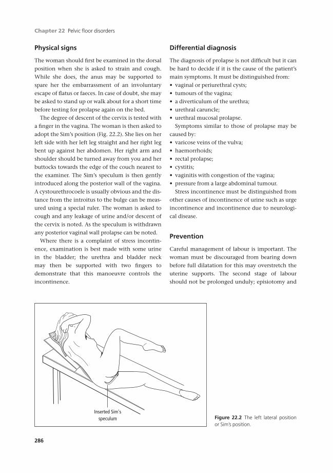
Chapter 22 Pelvic floor disorders
286
Physical signs
The woman should first be examined in the dorsal
position when she is asked to strain and cough.
While she does, the anus may be supported to
spare her the embarrassment of an involuntary
escape of flatus or faeces. In case of doubt, she may
be asked to stand up or walk about for a short time
before testing for prolapse again on the bed.
The degree of descent of the cervix is tested with
a finger in the vagina. The woman is then asked to
adopt the Sim’s position (Fig. 22.2). She lies on her
left side with her left leg straight and her right leg
bent up against her abdomen. Her right arm and
shoulder should be turned away from you and her
buttocks towards the edge of the couch nearest to
the examiner. The Sim’s speculum is then gently
introduced along the posterior wall of the vagina.
A cystourethrocoele is usually obvious and the dis-
tance from the introitus to the bulge can be meas-
ured using a special ruler. The woman is asked to
cough and any leakage of urine and/or descent of
the cervix is noted. As the speculum is withdrawn
any posterior vaginal wall prolapse can be noted.
Where there is a complaint of stress incontin-
ence, examination is best made with some urine
in the bladder; the urethra and bladder neck
may then be supported with two fingers to
demonstrate that this manoeuvre controls the
incontinence.
Differential diagnosis
The diagnosis of prolapse is not difficult but it can
be hard to decide if it is the cause of the patient’s
main symptoms. It must be distinguished from:
• vaginal or periurethral cysts;
• tumours of the vagina;
• a diverticulum of the urethra;
• urethral caruncle;
• urethral mucosal prolapse.
Symptoms similar to those of prolapse may be
caused by:
• varicose veins of the vulva;
• haemorrhoids;
• rectal prolapse;
• cystitis;
• vaginitis with congestion of the vagina;
• pressure from a large abdominal tumour.
Stress incontinence must be distinguished from
other causes of incontinence of urine such as urge
incontinence and incontinence due to neurologi-
cal disease.
Prevention
Careful management of labour is important. The
woman must be discouraged from bearing down
before full dilatation for this may overstretch the
uterine supports. The second stage of labour
should not be prolonged unduly; episiotomy and
Inserted Sim'sspeculum Figure 22.2 The left lateral position
or Sim’s position.
AMI22 6/9/04 5:12 PM Page 286

Pelvic floor disorders Chapter 22
287
low forceps extraction may reduce the risk of later
prolapse. Episiotomies and tears must be carefully
sutured in layers. Postnatal exercises should be en-
couraged after every labour. All women should see
a physiotherapist to help with this.
Prevention of vault prolapse after hysterectomy
is helped by suture of the cardinal and uterosacral
ligaments to the vaginal vault. Subtotal hysterec-
tomy is more likely to lead to vault prolapse than
total hysterectomy, even though many women ask
for the former.
Treatment
Treatment of prolapse may be palliative or surgical.
Physiotherapy
This can be successful, chiefly in young women
after recent childbirth where the vaginal walls and
pelvic floor are mainly affected. It is less effective in
vault prolapse. Exercises to strengthen the pelvic
floor muscles are carried out under the supervision
of a physiotherapist including the voluntary reten-
tion of weighted cones in the vagina to strengthen
the pelvic muscles. This may be combined with
electrotherapy to the pelvic floor muscles.
Palliative treatment
Many types of pessary and support have been de-
vised for prolapse. Their use is only temporary, the
better cure being a repair operation. With modern
techniques of surgery and anaesthesia, operation
can safely be undertaken in the majority of cases of
prolapse.
The indications for pessaries are:
• prolapse during pregnancy;
• prolapse immediately after delivery;
• when another pregnancy is desired within a
short time;
• in patients unfit for operation on medical
grounds;
• in patients who decline an operation.
Pessary treatment
A plastic ring pessary which fits well surrounds the
cervix, pointing slightly forward, and resting be-
tween the posterior fornix and the anterior vaginal
wall. It supports a vault prolapse by stretching the
vaginal wall while a cystocoele is directly sup-
ported by it. It is less successful in controlling a
rectocoele; if the perineum is deficient, the pessary
will tend to slip or even fall out.
Pessaries are made in 5mm sizes from 50 to
120mm.
There are a few cases where a ring pessary fails to
control prolapse and operation cannot be per-
formed. In these cases there are other appliances.
The cup and stem pessary consists of a sheet of vul-
canite or plastic with a stem to which are attached
tapes which are tied to a belt. It is removed at night
for cleaning and is thus less likely to cause ulcera-
tion. It is a useful long-term pelvic floor support,
but rarely used.
Disadvantages• Ulceration of the vagina and cervix.
• A neglected pessary may become embedded in
the vaginal wall and may only be removed with
great difficulty.
• A carcinoma of the vagina may develop.
Surgery
The best results from operations for repair depend
on the degree of descent of the various compon-
ents of the genital tract together with the judge-
ment and expertise of the surgeon.
Many surgeons perform vaginal hysterectomy
when operating for prolapse, an operation of
choice when prolapse is combined with menorrha-
gia or where there are small uterine fibroids. Va-
ginal hysterectomy is preferred in cases of uterine
procidentia.
Anterior colporrhaphy and posteriorcolpoperineorrhaphyThese operations are designed to restore the sup-
port to the vagina from the levator ani and muscles
of the perineum. Women with urinary symptoms
AMI22 6/9/04 5:12 PM Page 287

Chapter 22 Pelvic floor disorders
288
and vaginal prolapse should all undergo urody-
namic investigation prior to deciding on surgical
treatment (see p. 291). The reasons for doing uro-
dynamics include:
1 If genuine stress incontinence is demonstrated
vaginal surgery is not the treatment of choice.
2 If the patient has detrusor instability vaginal sur-
gery may make it worse and there is an increased
risk of postoperative urinary retention and/or
infection.
3 The first operation for urinary problems gives
the best chance of success and it is therefore essen-
tial to opt for the operation with the highest cure
rate.
New surgical techniques for uterovaginal
prolapse have been introduced. These include
sacrospinous fixation where the uterosacral liga-
ments are fixed to the sacrospinous ligament via
the vaginal or abdominal route.
Anterior colporrhaphy and posterior colpoperineor-
rhaphy may be combined with amputation of the
cervix and shortening and suture of the cardinal
ligaments; this is the Fothergill or Manchester
operation.
The reasons for amputating the cervix are:
• the supravaginal cervix may be elongated;
• after suture of the cervix, repair of the vaginal
vault is more satisfactory;
• the cervix is often unhealthy and infected;
• a possible site for future carcinoma has been
removed.
The cervix should not be amputated in young
women who may wish to bear children and in
cases where there is no vaginal vault prolapse.
Abdominal operations may be combined with
prolapse repair. Abdominal hysterectomy may be
required for large fibroids and the prolapse may be
repaired under the same anaesthetic or later.
Removal of a large tumour may itself lead to cure
or improvement of prolapse.
Stress incontinence presents surgical challenges.
It can occur with or without prolapse.
Preoperative care
Preparation for operation is most important. The
general condition of the patient is assessed and
treatment given for conditions such as obesity and
chronic cough. Cardiovascular disease and mild
diabetes are common in middle-aged women and
may need preoperative treatment.
Ulceration of the vagina can follow exposure
of the vaginal tissues outside the body in a
procidentia or from long-wearing of a ring pessary.
The risk of ulceration and infection are reduced by
regular changing of the pessary (6-monthly) and
using regular topical oestrogen cream. Elderly
women find the changing of the pessary uncom-
fortable because the introitus is commonly less
elastic and partially stenosed. It is important that
the change is performed by a well trained profes-
sional. The pessary can be made more flexible by
pre-soaking it in warm water. Regular changes over
a long period of time may be unacceptable to some
women and it is common for women to ask for
surgical intervention after a few years of using a
pessary.
Urinary tract infection is common and must be
treated. Urge incontinence and detrusor muscle in-
stability should be treated with antispasmodics
and surgery postponed until this urogynaecology
aspect is fully investigated and treated.
Postoperative care
Early movements and deep breathing are encour-
aged and the patient should get out of bed as soon
as possible. The use of a lavatory or commode in
private helps to overcome difficulties with mic-
turition and defaecation. Laxatives are given as
required.
Postoperative complications in the first two
weeks are:
• Chest complications associated with general
anaesthesia.
• Retention of urine and urinary tract infection. Re-
tention usually requires catheterization. If exten-
sive dissection, especially of the perineal tissues, is
carried out during the operation, an indwelling
Foley catheter should be inserted at the operation.
The bladder should be drained continuously for
three to five days and the catheter then clamped
intermittently for a day to reintroduce the sensa-
tion of bladder filling. An antibiotic agent should
AMI22 6/9/04 5:12 PM Page 288

Pelvic floor disorders Chapter 22
289
be given during continuous catheterization to pre-
vent urinary infection.
• Local sepsis is unusual with the use of antibiotic
prophylaxis.
• Haemorrhage may be primary, reactionary or
secondary. Blood transfusion may be required and
in secondary or reactionary haemorrhage, resutur-
ing of the vagina or cervix to arrest bleeding and
packing of the vagina may be needed under anaes-
thesia in the operating theatre.
• Pelvic vein thrombosis and pulmonary embolism
may occur.
There are remote complications.
• Vaginal discharge may persist for some weeks. In
some cases it is due to granulation tissue in the
scars which are best treated with silver nitrate
sticks. Sutures used in the repair may not be ab-
sorbed. They can be nicked and any excess suture
material removed at two weeks.
• Urinary complications include frequency due
to irritable bladder or chronic infection. Rarely a
vesicovaginal or urethrovaginal fistula develops.
• Dyspareunia is common and may be caused by
leaving the vagina too small; care must be taken
not to reduce the vaginal circumference, especially
if a posterior repair follows an anterior wall opera-
tion. Dyspareunia may also result from disuse of
the vagina due to fear. Senile atrophy may also
be seen in the age group of those having repair
operations.
Future pregnancies after repair
Successful pregnancy can be achieved after pro-
lapse repair though if the cervix has been ampu-
tated there may be an increased tendency to
miscarry. Caesarean section is advisable in most
cases especially if there is fibrosis of the remaining
cervix, if there has been an extensive vault repair
or where operation has been done for severe stress
incontinence.
Vaginal delivery may rarely be allowed.
Urogynaecology
Physiology
The bladder has two main functions in the human.
• To act as a reservoir for the storage of urine.
• To empty this reservoir away from the skin of the
body at an appropriate time and appropriate place.
Acting as a reservoir, the normal bladder:
• is lined with waterproof transitional epithelium
which does not allow diffusion of the urinary elec-
trolytes across its wall;
• has a high compliance, accommodating a large
volume of urine (300–500ml) with a rise of intra-
vesical pressure to only 15cmH2O;
• is able to expand suprapubically and extraperi-
toneally without hindrance or constraint by bone
or pelvic viscera;
• maintains the pressure in its outflow tract along
the urethra at a higher level than intravesical pres-
sure thus preventing leakage of urine.
The bladder is an efficient expulsive organ:
• The smooth detrusor muscle is richly innervated
by the parasympathetic nervous system outflow of
sacral roots 2, 3 and 4.
• At the onset of micturition, the pelvic floor stri-
ated muscle is voluntarily relaxed, reducing the in-
traurethral pressure. The background inhibition of
the sacral reflex arc is suppressed. Efferent im-
pulses pass to the detrusor muscle causing a rise
in intravesical pressure. This then exceeds the
intraurethral pressure and leads to the passage of
urine down the urethra.
Urinary incontinence
The involuntary loss of urine may be due to:
• true incontinence from a urinary fistula;
• genuine stress incontinence;
• detrusor instability (urge incontinence);
• overflow incontinence;
• reflex incontinence.
Urinary fistula
A pathological tract may open between a part of
the urinary system and the epithelial surface of the
AMI22 6/9/04 5:12 PM Page 289

Chapter 22 Pelvic floor disorders
290
vagina or occasionally the skin. These tracts bypass
the normal controlling mechanisms causing devel-
opment of continuous (true) incontinence. They
can be congenital or follow major surgery, opera-
tive delivery or disease such as cancer of the cervix.
• Congenital (rare); the ureter draining the upper
pole of one kidney opens on the anterior wall of
the vagina. It presents in children with continuous
urinary loss. This may result in poorly function-
ing renal polar hydronephrosis or an ectopic
hydroureter.
• Caused by surgery —avascular necrosis leads to
weakening of the wall of the ureter or bladder and
the development of a ureteric or vesicovaginal
fistula.
(a) Gynaecological surgery, particularly if there
has been anatomical distortion by infection, en-
dometriosis, carcinoma or by preceding small
blood vessel damage (endarteritis) caused by
irradiation.
(b) Obstetric trauma, in association with ob-
structed labour where the presenting part causes
avascular necrosis of the bladder base or some-
times the rectum, causing the development of a
vesicovaginal or rectovaginal fistula respectively.
Genuine stress incontinence
An involuntary loss of urine occurs from the
urethra, when the transmitted intra-abdominal
pressure causes a rise of the intravesical pressure
which exceeds the intraurethral pressure in the ab-
sence of a detrusor contraction. Approximately
25% of older women have mild problems and
5–10% severe.
SymptomsInvoluntary urine loss associated with a sudden,
usually unexpected, rise of abdominal pressure
such as coughing, sneezing, laughing or lifting.
Physical signsThe coincidental downswing of the bladder neck
leads to urinary leakage from the urethra; often
only a few ml are passed.
About 50% are associated with prolapse of the
vagina.
Detrusor instability
Incidence —8–10% increasing with age.
AetiologyIncompletely understood but may be due to:
• an abnormality in the central nervous system
when anxiety and stress result in the loss of ability
to inhibit the detrusor reflex and, therefore, the
development of detrusor contraction;
• a recognized neurological defect, such as spinal
trauma, demyelinating disorders or epilepsy;
• intense bladder inflammation, particularly in
the elderly.
Symptoms• Urgency of micturition leading to urge inconti-
nence —an inability to hold on.
• Frequency of micturition both by day and night.
• Often associated with stress incontinence.
DiagnosisThe demonstration of detrusor contraction (more
than 15cmH2O) on cystometry, provoked by blad-
der filling or straining and the movement or sound
of running water.
Overflow incontinence
Loss of urine when the bladder has become filled,
usually associated with either:
• obstructive surgery to the bladder neck;
• denervation of the detrusor muscle (usually by
extensive pelvic surgery, neurological defects,
diabetes).
Symptoms• The frequent passage of small volumes of urine.
• Hesitation of micturition.
• A slow stream.
• A sensation of incomplete emptying.
• Involuntary leakage when bending or getting
out of a chair.
Physical signs• A palpable bladder.
• Leakage on elevation of bladder base.
AMI22 6/9/04 5:12 PM Page 290

Pelvic floor disorders Chapter 22
291
• On cystometry, slow urine flow rate.
• High residual urine.
• Risk of back pressure to upper urinary tract if
chronic.
Reflex incontinence
Reflex involuntary voiding is associated with
sensory stimulation of the sacral 2, 3 and 4
segments. This develops when the higher centres
are cut off from the sacral reflex arc and thus
micturition ceases to be centrally suppressed. It is
triggered when there is a significant increase of
the afferent impulses to the sacral segments 2, 3
and 4 either from the bladder or the somatic
nerves.
Detrusor contractions are often associated with a
simultaneous contraction of the pelvic floor (de-
trusor dyssynergia) causing partial obstruction of
urine flow, unlike the relaxation in centrally or-
ganized normal micturition.
Urodynamic investigation ofincontinence
• Bladder behaviour can be assessed by keeping
fluid output charts to measure the frequency of the
volumes of urine passed during the day, with a
diary of fluid intake.
• Cystometry (with subtracted abdominal
pressure) determines the presence or absence
of involuntary detrusor contractions thereby
differentiating the stable from the unstable
bladder and these pressure measurements may be
coupled with video radiological screening (Figs
22.3 and 22.4).
• The residual urine can be measured by catheter
or pelvic ultrasound.
30
20
10
00 100 200 300 400 500
Bladder filling volume (ml)
Blad
der p
ress
ure
(cm
H2O
)
CoughCough
0 100 200 300
Bladder filling volume (ml)
Voiding Cough Cough
80
60
40
20
0
Blad
der p
ress
ure
(cm
H2O
)
Figure 22.4 Cystometry of filling ofan unstable bladder.
Figure 22.3 Cystometry of a normalbladder filling with rise in intravesicalpressure.
AMI22 6/9/04 5:12 PM Page 291

Chapter 22 Pelvic floor disorders
292
• Measurement of urinary flow rate with pressure
measurements differentiates the obstructed ure-
thra from the poorly functioning detrusor.
• Ultrasound urograms outline defects of the blad-
der and upper urinary tract and retrograde urogra-
phy to demonstrate ureteric and vesicovaginal
fistulae.
Treatment
Stress incontinence
Conservative• Mild urinary leakage (particularly postnatally) —
with a good physiotherapist and patient motiva-
tion, pelvic floor exercises usually result in
improvement.
• Reduction of weight, excessive physical exertion
and the treatment of coughing also help.
Surgical• Vaginal approach —anterior colporrhaphy with
bladder neck buttress is a simple operation and per-
mits repair of other prolapses at the same time.
Long-term success rate in curing incontinence is
approximately 40%.
• Sling operation —multiple varieties —using syn-
thetic substances (nylon, prolene, Teflon, mersi-
lene mesh, tension-free vaginal tapes) or natural
tissues (e.g. round ligament or external oblique
aponeurosis). Insertion is by open surgery or blind
by directed needles through the retropubic space
(e.g. Stamey procedure).
• Retropubic bladder neck suspension operations —
suturing the vaginal wall to the pectineal ligament
(Burch) or periosteum over the back of the
pubic bone (Marshall–Marchetti–Krantz). These
procedures are associated with an 85–90% cure
rate of the incontinence but do not remedy much
prolapse apart from that of the anterior vaginal
wall.
• Paraurethral injection of collagens to stimulate
fibrin formation.
The unstable bladder
There is often a psychosomatic element (Fig. 22.5).
The symptoms are improved by several means.
• Enthusiastic encouragement, with the help of
urinary output volume chart, particularly by in-
continence advisers, district nurses and doctors. If
necessary, admission to hospital for intensive blad-
der training under close supervision.
• The use of anticholinergic drugs, e.g. oxybu-
tynin, Pro-Banthine, imipramine.
• The use of vaginal oestrogen cream to reduce
bladder irritability and the tendency of recurrent
urinary infection.
Overflow incontinence
• If obstructed, urethral dilatation or urethrotomy
results in improvement.
• If due to weakened detrusor muscle —continu-
ous drainage with a suprapubic catheter for two to
three weeks to improve the tone of the detrusor
often helps with subsequent bladder training. Oc-
casionally intermittent self-catheterization can be
of assistance although infection is common.
• a-agonists —bethanocol.
Urinary tract infection
Almost all urinary tract infections develop from
the upward spread of the bacteria along the 5cm of
urethra. Infections are associated with:
80
60
40
20
0
Blad
der p
ress
ure
(cm
H2O
)
Detrusor contraction and voiding
Figure 22.5 Cystometry with detrusor muscle contractionand voiding.
AMI22 6/9/04 5:12 PM Page 292

Pelvic floor disorders Chapter 22
293
• the upward passage of organisms during
intercourse;
• catheterization;
• the incomplete emptying of the bladder leading
to stagnant urine;
• atrophic urethritis from a lack of oestrogens;
• poor hygiene following defaecation or
intercourse.
Symptoms• Burning dysuria.
• Severe urinary frequency.
• Urgency of micturition.
• Suprapubic discomfort.
• Urine odour.
If the infection has spread beyond the bladder to
the upper part of the urinary tract then:
• loin pain;
• vomiting;
• rigors.
Investigations• Dipstick impregnated with nitrite sensitive
amine to screen for bacteria.
• Mid-stream urine culture.
Treatment• Encouragement of high fluid intake.
• Rest in bed if temperature is raised.
• Rapid use of appropriate antibiotics.
• Topical oestrogens in postmenopausal women.
• Educational hygiene encouraging postcoital
micturition and correct wiping after defaecation.
• Bladder training with suprapubic catheter if the
residual urine volumes are high.
Urinary frequency
Is associated with:
• bladder irritants, e.g. coffee, cola and fortified
wines;
• the presence of calculi;
• high fluid intake resulting in an increased urin-
ary output;
• diabetes mellitus may present with polydypsia
and polyuria;
• the use of diuretics;
• anxiety, tension and stress, e.g. outside the
examination hall;
• development of habits and rituals associated
with particular voiding patterns;
• insomnia leading to nocturia;
• the reduction of any peripheral oedema
overnight resulting in increased kidney excretion,
bladder filling and nocturia.
Nocturnal enuresis
The involuntary voiding of urine into the bed-
clothes whilst asleep.
AetiologyNot fully known, but the following associations
have been noted:
• deep sleep leading to the loss of suppression of
the voiding reflex;
• impairment of the kidneys to concentrate urine
whilst asleep, for example by the persistence of
daytime renal excretion pattern;
• psychological disturbances such as great
unhappiness;
• bladder instability in later life.
Treatment• Frequent waking overnight to ensure regular
voiding.
• The use of desmopressin and nasal sprays to sup-
press urine formation whilst asleep.
• Mattress alarms.
• Imipramine.
AMI22 6/9/04 5:12 PM Page 293

Chapter 22 Pelvic floor disorders
294
Self-assessment
22.1 A woman of 52 is referred to the gynaecology outpatients by her GP. She is complaining of urinary incontinence.You are expected to take a history from her.
Role-player’s instructions: You are Mrs Sarah Ball, a 52-year-old primary school teacher. Over the last five yearsyou have been increasingly troubled by suddenly leaking small amounts of urine when you cough, run or do anyform of exercise. You used to go to aerobic classes but have given them up. You are also finding it difficult atschool—several of the children have commented on a smell of urine in the classroom and you feel very embar-rassed. You empty your bladder between each class but still notice some leaking as you move round the classroom.You wear a panty-liner all the time which you find irritating. you have to get up to the toilet twice every night. Theamount you pass is normal; occasionally you have to go again 5–10 minutes after emptying your bladder. You havenoticed some backache and a feeling of something coming down. You have had three children all born vaginal-ly—the first baby was delivered by forceps and you had a lot of stitches. Your last period was three years ago. Yourlast smear was two years ago and they have always been normal. You are not taking HRT because your motherdied of breast cancer. You smoked 20 cigarettes a day until four years ago. Your weight has gradually increasedsince you stopped smoking. You are otherwise fit and well.
22.2 Describe how you would examine and manage this patient.22.3 List five principal complications of vaginal repair operations (colporrhaphies).22.4 Define genuine stress incontinence.22.5 Which of the following are common symptoms of detrusor instability?
(a) Urgency.(b) Slow urinary stream.(c) Incomplete emptying.(d) Nocturia.(e) Frequency.
AMI22 6/9/04 5:12 PM Page 294

Part 6
Audit of obstetrics and gynaecology
AMI23 6/9/04 5:14 PM Page 295

AMI23 6/9/04 5:14 PM Page 296

The population of the world is increasing, al-
though indications are that the boom of the earlier
part of the last century is flattening off. In the UK
the levels have been fairly steady since the Second
World War.
Birth rates are measured in all countries that col-
lect sufficient data. This means that developing
countries with poor data sources are not very reli-
able in measuring birth rates.
The total birth rate is simplest:
This requires a knowledge of not just all the births
but a proper census of the population to derive the
denominator (Fig. 23.1). However, it does not re-
late to the process of birth; no men have babies and
few young women or those over 45 do; hence a
more sophisticated measure is the general fertility
rate:
This rate requires a more detailed data analysis of the
censuses and is used in Western countries. The data
for the last years in the UK are shown in Fig. 23.2.
A more readily understandable set of ratios is the
completed family size but this can only be done
retrospectively (Fig. 23.3).
In the UK the total birth rate is 14 per 1000 and
the general fertility rate is 64 per 1000. The com-
General fertility rate =Births per year
Women aged 15–45 years¥ 1000
Total birth rate =Births per yearMidyear population
¥ 1000
pleted family size is 1.8, just below replacement
level.
There are variations in the monthly birth rate
which is highest from October to March. The rates
by days of the week are highest on weekdays (see
Fig. 23.4a).
As can be seen, the birth rate for babies under
2500g provides a mirror image to the incidence of
births and accounts for the higher perinatal mor-
tality rates at the weekend (see Fig. 23.4b).
Maternal mortality
Definitions
Deaths of women while pregnant or within 42 days
of delivery, miscarriage, termination of pregnancy,
from any cause related to or aggravated by the
pregnancy or its management but not from acci-
dental or incidental causes. These deaths are fur-
ther subdivided into direct and indirect causes;
direct causes are those that result from obstetric
complications, whilst indirect deaths are those
resulting from pre-existing disease or disease
that develops during pregnancy secondary to
physiological changes of pregnancy.
Major causes of maternal mortality
Maternal mortality has declined dramatically in
the last 50 years in the UK (Fig. 23.5), but there are
297297
Chapter 23
Statistics of reproductive medicine
AMI23 6/9/04 5:14 PM Page 297
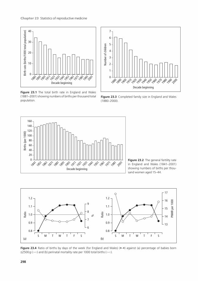
Chapter 23 Statistics of reproductive medicine
298
40
30
20
10
0
Decade beginning
Birt
h ra
te (b
irths
/100
0 to
tal p
opul
atio
n)
1881
1891
1901
1911
1921
1931
1941
1951
1961
1971
1981
1991
2001
Figure 23.1 The total birth rate in England and Wales(1881–2001) showing numbers of births per thousand totalpopulation.
160
120
80
40
140
100
60
20
0
Decade beginning
Birt
hs (p
er 1
000)
1841
1851
1861
1871
1881
1891
1901
1911
1921
1931
1941
1951
1961
1971
1981
2001
7
6
3
5
4
2
1
0
Decade beginning
Num
ber o
f chi
ldre
n
1880
1890
1900
1910
1920
1930
1940
1950
1960
1970
1980
1990
2000
1.2
(a)
1.1
1.0
0.9
9
6
7
8
0.8
S M T W T F S
Ratio
%
(b)
1.2
1.1
1.0
0.9
16
17
13
14
15
0.8
S M T W T F S
Ratio
PNM
R pe
r 100
0
Figure 23.3 Completed family size in England and Wales(1880–2000).
Figure 23.2 The general fertility ratein England and Wales (1841–2001)showing numbers of births per thou-sand women aged 15–44.
Figure 23.4 Ratio of births by days of the week (for England and Wales) (•–•) against (a) percentage of babies born £2500g (�–�) and (b) perinatal mortality rate per 1000 total births (�–�).
AMI23 6/9/04 5:14 PM Page 298

Statistics of reproductive medicine Chapter 23
299
still women who die as a consequence of pregnan-
cy or labour. The major causes are secondary to
hypertensive disease of pregnancy, thrombosis and
haemorrhage which are discussed in more detail.
Maternal mortality is expressed as deaths/1000
births or deaths/100000 maternities. In the three-
year period 1997–1999 the maternal mortality rate
(MMR) for direct and indirect deaths was 11.4/
100000 maternities. In other words there was one
death for each 9000 deliveries. The advances in
medical care prior to pregnancy have led to more
women with pre-existing medical conditions be-
coming pregnant. This has led to indirect maternal
deaths making a larger contribution to the mater-
nal mortality rate. In 1997–1999 the MMR for
direct deaths was 5.0/100000 compared with
6.4/100000 for indirect deaths. In 1985–87 the
rates were 6.1 and 3.8 respectively. However, the
combined direct and indirect MMR has hardly
changed over the past 15 years.
Pregnancy-induced hypertension, pre-eclampsia and eclampsia (MMR:0.7/100000)
Eclampsia is getting rarer. When it comes earlier in
pregnancy (before 28 weeks) it has a worse effect.
Death is from intracranial haemorrhage or renal
failure.
To reduce deaths:
• Identify high-risk women.
• Check blood pressure frequently in pregnancy.
• Admit those with signs of pre-eclampsia.
This may be at home for lesser degrees rather
than in hospital.
• Recognize biochemical and haematological
aspects of HELLP syndrome (haemolysis, elevated
liver enzymes, low platelets) (Chapter 10).
Thromboembolism (MMR: 1.7/100000)
1 A third are antenatal and two-thirds after deliv-
ery. A third of the latter follow Caesarean section.
2 High-risk patients:
• Over 35 years.
• Obese.
• Operative delivery.
• Previous thrombosis.
3 Half the deaths are with little warning of previ-
ous thrombotic episodes.
To reduce deaths:
• Prophylactic anticoagulation of high-risk
patients.
• Avoid risk factors.
• Prompt effective treatment on suspicion.
Abortion
1 Usually after procured and illegal interferences.
2 Patients die from haemorrhage, sepsis or renal
failure.
To reduce deaths:
• Wider use of legal therapeutic abortion.
• Better contraception.
Haemorrhage (MMR: 0.3/100000)
Abruptio placentaeSevere hypovolaemia leads to shock and later renal
shutdown.
To reduce deaths:
• Central venous pressure monitoring.
• Adequate and quick blood replacement.
Placenta praeviaRepeated and increasing haemorrhage in last
trimester of pregnancy. The severe degrees must be
0Mat
erna
l dea
ths/
1000
bir
ths
1
2
4
3
1900 1920 1960Years
20001940 1980
5
Figure 23.5 Maternal mortality in England and Wales,1900–1997.
AMI23 6/9/04 5:14 PM Page 299

Chapter 23 Statistics of reproductive medicine
300
treated by Caesarean section, which may be a tech-
nically difficult operation.
To reduce deaths:
• Pay more attention to warning bleeds in
pregnancy.
• Have consultant in theatre for Caesarean
section.
Postpartum haemorrhageUsually from an atonic uterus but can follow cervi-
cal trauma.
To reduce deaths:
• Give oxytocic drug routinely at delivery.
• Deliver patients at risk in hospital where blood is
available (see Chapter 13).
• Act promptly using a planned protocol.
Ectopic pregnancyWith reduction of deaths from other forms of
haemorrhage this is becoming relatively more
important.
To reduce deaths:
• Admit patients with suspicious symptoms.
• Act promptly on patients with actual symptoms.
• Be prepared to laparoscope on suspicion and do
not rely on ultrasound and b hCG findings alone.
Anaesthesia
Deaths associated with general anaesthesia are
reducing greatly in the UK. Inhalation of acid
stomach contents in labour under general anaes-
thetic leads to Mendelson’s syndrome.
To reduce deaths:
• Wider use of regional anaesthetics (e.g. spinal
epidural).
• If general anaesthetic essential, a senior anaes-
thetist involved and intubation with a cuffed
tube.
Other causes
All the other causes produce few deaths. Infection,
once the killer of 1 :5 women in childbirth, is much
reduced (although still causing some deaths each
year, mostly after Caesarean section in labour with
prolonged ruptured membranes). Heart disease as a
cause is diminishing, as rheumatic fever is better
avoided or diagnosed and treated in childhood.
Substandard care
In the UK every maternal death is reported to a
central committee which publishes its confidential
findings at three-yearly intervals. This is not a judi-
cial enquiry and no blame is apportioned to any in-
dividual. It is a medical audit where the profession
looks closely at its own work and tries to learn from
mistakes. The committee tries to assess in each case
if an avoidable factor was present: if there was
‘some departure from the acceptable standards of
satisfactory care’.
In a recent report, 50% of the deaths directly due
to pregnancy and delivery were considered to
have been avoidable by this definition. It was the
patient who made the largest single contribution
to this in the antenatal period by either not com-
ing for care or else ignoring advice given. In labour,
the hospital obstetricians and anaesthetists were
associated with the highest incidence of substan-
dard care incidents. This was mostly from not pay-
ing sufficient heed to warning signs and not
having senior-enough doctors in the delivery suite.
Shortage of staff and facilities is beginning to be re-
ported in this category. Substandard care from
general practitioners and midwives in these cases
was rare.
The most important ways of reducing maternal
deaths are:
1 Improved access to antenatal care.
2 Improved education in the population of the
importance of antenatal care.
3 More consultant obstetric and anaesthetic in-
volvement on the delivery suite.
4 Introduction of evidence-based guidelines for all
areas of maternity care.
5 Regular training of all staff including emergency
drills for postpartum haemorrhage and shoulder
dystocia.
Near misses
Because maternal deaths are so few, even after
national analysis, one cannot draw many statistical
AMI23 6/9/04 5:14 PM Page 300

Statistics of reproductive medicine Chapter 23
301
conclusions. However, an extended method can be
used to increase the database. Here, one looks at
women who have a firm diagnosis of a given, pre-
cisely diagnosed condition and examines the back-
ground even though they did not die. For example,
one could look at those who have lost over two
litres of blood at a primary postpartum haemor-
rhage. This would be a fairly well-defined group for
the loss is so great and one would be able to com-
pare the aetiology and management with that of
women who died from postpartum haemorrhage.
By so doing one would examine about six times as
many cases. Similarly, one could look at the rea-
sonably firm diagnosis of pulmonary embolism
proven by a scan and, again, examine them as
a larger group than those who died from the
condition.
The idea of near misses as a method of examina-
tion is excellent for an individual hospital or group
of hospitals that work as one, but is more difficult
to apply regionally or nationally unless the defini-
tions are firmly established.
Risk management
Routine reporting of all incidents that affect the
quality of care for the patient are reported to a risk
management manager and lead clinician. These
may appear minor (past notes unavailable in
clinic) or major (delay in obtaining cross-matched
blood in a case of major obstetric haemorrhage),
however they can both have a major effect on
the standard of patient care and the outcome for
the mother and her child. All reports should be
a purely factual account and not apportion blame
to any individual. The manager and the lead
clinician collect all the reports and investigate the
problem. This process may highlight a problem in
the system of care which needs to be rectified
or a need for further staff education, either individ-
ually or as a group. Serious adverse incidents are
usually investigated locally and then reported
to the Strategic Health Authority together with
recommendations for preventing such incidents
reoccurring.
Patient complaints are investigated in a similar
way and responses containing the facts of the case,
the findings of the investigation and the recom-
mendations are sent to the patient. Patients have
the right to request a formal meeting with the hos-
pital after the investigation to express their views
and make their own recommendations. Hospitals
now employ patient advocates to help patients
through this process. The direct communication to
the patient reduces the number of cases that pro-
ceed to litigation.
Audit is used as a risk management tool. All
maternity units in the UK are expected to
follow evidence based multidisciplinary guide-
lines. These may be developed locally, by the
Royal College of Obstetricians and Gynaecologists
or the National Institute of Clinical Excellence
(NICE). Each unit is expected to audit their care
against these guidelines at regular intervals. Each
audit should identify areas where the guidelines
are not being followed or are inadequate. Recom-
mendations are made to improve the standard of
care and the audit repeated to ensure that the
changes have improved care. This is called the
audit cycle.
Perinatal mortality
The perinatal mortality rate (PMR) is the total of
stillbirths and first-week neonatal deaths occurring
in every 1000 total births. In 1997 it was 7.9/1000
total births in England and Wales.
Factors influencing PMR
1 The mother’s:
• Place of residence.
• Past nutrition and diseases.
• Education.
• Social class.
2 The age and parity of the mother.
3 An efficient health service.
4 The definition of stillbirth and neonatal death.
In the UK this was changed in 1991 to deaths after
24 completed weeks of gestation from a previous
28 weeks’ limit. Hence, a small apparent increase
for a short time appears.
Figure 23.6 shows the progressive reduction of
PMR in 70 years.
AMI23 6/9/04 5:14 PM Page 301

Chapter 23 Statistics of reproductive medicine
302
Causes of perinatal mortality
Precise causes of perinatal death are often confused
by a lack of autopsy information and an insistence
on a single or primary cause of death on the
certificate.
Classification of causes of perinatal death1 Macerated stillbirths without malformation.
2 Congenital malformation in either stillbirths or
neonatal deaths.
3 Intrapartum perinatal deaths secondary to as-
phyxia or trauma or both.
4 Neonatal deaths as a result of immaturity.
5 Other specific causes, e.g. Rh haemolytic disease.
Small for gestational age (SGA)Two-thirds of neonatal deaths are associated
with SGA. A high incidence of hyaline membrane
disease and intraventricular haemorrhage is
found.
Congenital malformationsA tenth of stillbirths and a quarter of neonatal
deaths have a congenital anomaly. Malformations
of the CNS and cardiovascular system are the most
common.
AsphyxiaThere is post-mortem evidence of asphyxia in a
third of stillbirths and a tenth of neonatal deaths.
This is due to:
BEFORE LABOUR
1 Abruptio placentae.
2 Placental failure:
• Pre-eclampsia.
• Hypertension.
• Postmaturity.
• Diabetes.
IN LABOUR
1 Prolonged labour.
2 Cord prolapse.
AT DELIVERY
• Impacted shoulder.
• Delayed onset of respiration.
Birth injuryLess than a tenth of neonatal deaths.
ASSOCIATED WITH
1 Too fast a delivery:
• Precipitate delivery with immature fetus.
• Breech presentation with insufficient time for
moulding of head.
2 Too difficult a delivery:
• Disproportion.
• Badly performed operative delivery, particu-
larly forceps.
Infection1 Intrauterine.
2 Neonatal.
0
Rate
/100
0 bi
rths
10
30
40
50
1930 1940 1960Years
Perinatal deaths
Stillbirths
Change of
definition
First week deaths
20001950 1980
70
19901970
60
20
Figure 23.6 Perinatal mortality, still-births and 1st-week neonatal deathsin England and Wales from 1930.
AMI23 6/9/04 5:14 PM Page 302
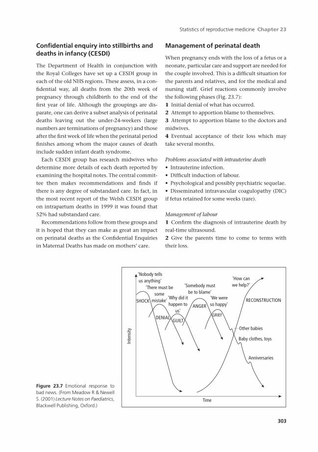
Statistics of reproductive medicine Chapter 23
303
Confidential enquiry into stillbirths anddeaths in infancy (CESDI)
The Department of Health in conjunction with
the Royal Colleges have set up a CESDI group in
each of the old NHS regions. These assess, in a con-
fidential way, all deaths from the 20th week of
pregnancy through childbirth to the end of the
first year of life. Although the groupings are dis-
parate, one can derive a subset analysis of perinatal
deaths leaving out the under-24-weekers (large
numbers are terminations of pregnancy) and those
after the first week of life when the perinatal period
finishes among whom the major causes of death
include sudden infant death syndrome.
Each CESDI group has research midwives who
determine more details of each death reported by
examining the hospital notes. The central commit-
tee then makes recommendations and finds if
there is any degree of substandard care. In fact, in
the most recent report of the Welsh CESDI group
on intrapartum deaths in 1999 it was found that
52% had substandard care.
Recommendations follow from these groups and
it is hoped that they can make as great an impact
on perinatal deaths as the Confidential Enquiries
in Maternal Deaths has made on mothers’ care.
Management of perinatal death
When pregnancy ends with the loss of a fetus or a
neonate, particular care and support are needed for
the couple involved. This is a difficult situation for
the parents and relatives, and for the medical and
nursing staff. Grief reactions commonly involve
the following phases (Fig. 23.7):
1 Initial denial of what has occurred.
2 Attempt to apportion blame to themselves.
3 Attempt to apportion blame to the doctors and
midwives.
4 Eventual acceptance of their loss which may
take several months.
Problems associated with intrauterine death• Intrauterine infection.
• Difficult induction of labour.
• Psychological and possibly psychiatric sequelae.
• Disseminated intravascular coagulopathy (DIC)
if fetus retained for some weeks (rare).
Management of labour1 Confirm the diagnosis of intrauterine death by
real-time ultrasound.
2 Give the parents time to come to terms with
their loss.
Time
'Nobody tellsus anything'
'There must besome
mistake' 'Why did ithappen to
us'
'Somebody mustbe to blame'
'We wereso happy'
Other babies
Baby clothes, toys
Anniversaries
'How canwe help?'
Inte
nsity
SHOCK
DENIALGUILT
ANGER
GRIEF
RECONSTRUCTION
Figure 23.7 Emotional response tobad news. (From Meadow R & NewellS. (2001) Lecture Notes on Paediatrics,Blackwell Publishing, Oxford.)
AMI23 6/9/04 5:14 PM Page 303

Chapter 23 Statistics of reproductive medicine
304
3 Plan induction of labour at a time which is suit-
able for the parents but ensuring that they have a
midwife to look after them throughout the period
of induction and labour.
4 Arrange facilities for the partner to stay through-
out the procedure.
Investigations1 Hb (including HbA1C).
2 Cross-match and save serum.
3 Kleihauer test.
4 Clotting studies.
5 Lupus anticoagulant and anticardiolipin anti-
bodies (for SLE/APS).
6 Fasting blood sugar and HbA (for diabetes).
7 Liver function tests.
Management1 Induce labour with prostaglandin pessaries
given every 3 hours.
2 Do not rupture the membranes until the woman
is in labour and the cervix is more than 4cm
dilated.
3 Keep an accurate fluid balance chart.
4 Give liberal analgesia. If the woman wishes
an epidural ensure her blood clotting values are
normal.
5 Discuss whether the couple wish to see the baby
after delivery. Parents should be encouraged but
not pressed to view their babies.
• The babies should be photographed, clothed
and looking as natural as possible. These photo-
graphs should be filed in case parents wish to see
them much later —sometimes they ask a year or
so later.
Postnatal careThe woman should be looked after by midwifery
staff known to her in the antenatal period. The
length of hospital stay is not determined by med-
ical events but more by what the couple want. She
may go home as soon as she wishes but should not
have the feeling of being sent away. Her general
practitioner and community midwife must be in-
formed by one of the medical staff of the loss by
telephone.
1 Arrange for the mother to be seen by a specialist
midwife who is skilled in counselling patients who
have lost their babies.
2 Ask if she wishes her baby to be baptized and
buried; if so, arrange the procedure with the hos-
pital chaplain or other religious leader according
to the parent’s request.
3 Consent for post-mortem should be obtained
from the parents.
4 The following procedures should be undertaken:
(a) Heart blood should be sent for karyotyping
and viral studies.
(b) Two polaroid photographs of the baby
should be taken. One should be a general photo-
graph and the other should be a close-up of the
baby’s face. In addition, foot and hand prints
and a lock of hair are retained.
(c) X-ray the baby.
(d) If available, MRI of baby.
5 The consultant should interview the parents be-
fore discharge from hospital and explain as far as
possible the circumstances surrounding the death.
6 The couple are met 4–6 weeks later with all the
autopsy evidence to hand.
7 The couple should be put in touch with a
society, e.g. Stillbirth, Abortion and Neonatal
Death Society (SANDS), or people who have ex-
perienced a similar problem.
8 Lactation should be suppressed by means of a
firm supporting brassiere. Bromocriptine or cab-
ergoline should be offered.
9 Women who had to have a hysterectomy or
have one surviving twin may need professional
psychotherapeutic help.
AMI23 6/9/04 5:14 PM Page 304

Statistics of reproductive medicine Chapter 23
305
Self-assessment
23.1 Give the definitions for the following.1 Total birth rate.2 General fertility rate.3 Maternal mortality.4 Maternal mortality rate.5 Perinatal mortality rate.
23.2 Give the three most common direct causes of maternal mortality.23.3 Give three measures that can be undertaken to improve standards of care.23.4 Give three maternal factors that affect the perinatal mortality rate.23.5 Following the death of a baby what investigations should be performed?
AMI23 6/9/04 5:14 PM Page 305

306
Chapter 1
Answers
1.1 (1) h, (2) g, (3) d, (4) b or j, (5) b or j
The uterine artery is a branch of the (1), (h) Inter-
nal iliac artery. The uterus is a hollow, muscle-
walled organ in direct communication with the
(2), (g) fallopian tubes and the vagina. Inferior to
the uterine artery lies the (3), (d) ureter. The liga-
ments that support the uterus include the (4), (j)
uterosacral and (5), (b) transverse cervical.
1.2 b, e
See pp. 9–10. The granulosa cells secrete
oestradiol. Once the oocyte is released they be-
come luteal cells and secrete oestradiol and
progesterone. At menarche there are around
500000 oocytes in the ovary.
1.3 a, d, e
See pp. 4, 10, 13. At the time of the luteinizing
hormone (LH) surge the oocyte undergoes
meiosis but with an unequal distribution of the
cellular cytoplasm forming an oocyte ready for
fertilization and the first polar body with 23
chromosomes in each. Glycogen is secreted in
the luteal (secretory) phase of the cycle from the
effects of progesterone on the endometrial
glands. The endometrium is shed because the
spiral arterioles go into spasm causing hypoxia
and death of the endometrium. The follicular
phase (from day one of the cycle to ovulation)
can be very variable (cf. polycystic ovaries)
whilst the luteal phase is of a fixed duration.
1.4 a, c, d
Oestradiol exerts a negative feedback on folli-
cle stimulating hormone (FSH) (i.e. when
Answers to self-assessment questions
Round ligament (j)
Ampulla of fallopian tube (e)
Uterine corpus (b)
Infundibulopelvic ligament (f)
Internal os of cervix (g)
1.5
AMIAN 6/9/04 5:29 PM Page 306
Answers to self-assessment questions
307
oestradiol levels are low FSH levels rise). FSH
and LH are controlled by the hypothalamus
which secretes gonadotrophin releasing hor-
mone (GnRH). Testosterone is the major pre-
cursor of oestradiol and is secreted by the theca
cells under the influence of LH, the testos-
terone passes to the granulosa cells and is
converted to estradiol catalysed by FSH.
Progesterone only starts to be secreted at the
time of ovulation and reaches its peak seven
days after ovulation in a non-conception cycle.
Chapter 2
Marking scheme
The marking schemes vary from medical school to
medical school. Some mix marks for communica-
tion and details of the history together into one
question. Some may group aspects of the question
together and allow examiners to give a mark of 0, 1
or 2 depending on how well the candidate does in
each section. The scheme outlined below gives
a list of the things that may give you a mark
(shown in parentheses) or that may be included in
a group and is therefore only a guide to possible
marks. It is wise to ask your medical school how
their marking scheme is structured. You can then
devise your own marking sheets and use the cases
for practice.
Communication
• Introduces themselves. (1)
• Eye contact. (1)
• Picks up on verbal clues. Patients often do not
remember all the relevant details of their history
immediately but may say something that implies
you have an opportunity to ask another question
to help them remember or feel comfortable an-
swering the question. For example, the first patient
may not be in a relationship because of dyspareu-
nia and she may give a clue when answering the
question about whether or not she has a boyfriend;
the second patient may not remember the episode
of being asked to take some tablets, or may know
that she had had an infection causing her ectopic
pregnancy, but feels a bit stupid for not having fol-
lowed medical advice. (1)
• Picks up on non-verbal clues. Non-verbal clues
include patients not looking at you, wriggling
on their chair, not answering the question. You
should notice this and re-ask the question in a dif-
ferent way or challenge the patient gently if she
is looking very diffident or refusing to answer the
question. (1)
• Use of non-medical language. (1)
• Listens. (1)
• Allows questions. (1)
• Structured history. This is marks for a logical
order to the history which leads to a full assess-
ment of the patient’s problem. Do not get bogged
down in irrelevant detail such as a long surgical
history or detailed system questions as you will run
out of time. Learn the list of headings and go
through them quickly ignoring negative answers
and going into further detail for a positive answer.
(1/2)
• Accurate summary. (1)
History taking
• Identifies main complaint (1)
• Explores main complaint appropriately (1)
• Ascertains all details of main complaint (1)
• Last menstrual period (LMP)/menstrual
history (1)
• Past gynaecological/sexual health
history (1)
• Past obstetric history (1)
• Past medical and surgical history (1)
• Family history (1)
• Social history (1)
• Medication/allergies (1)
• Summary (1)
The first three of these may be grouped together
to give a score of 0–2.
In some medical schools the role-player may
give marks for communication —did you trust this
doctor, would you see him/her again, did you
understand what they told you? —are questions
that may be asked. Role-players have been shown
to be very accurate in picking out the students who
will do well and those that will not.
AMIAN 6/9/04 5:29 PM Page 307

Answers to self-assessment questions
308
Chapter 3
The relevant information for each scenario is given
below.
1 Turner’s syndrome is associated with short
stature in 100% of cases. Her ovaries are dysgenetic
so they do not ovulate giving her a low oestrogen
with a high LH and FSH. They should receive oe-
strogen replacement in the form of the pill or hor-
mone replacement therapy (HRT). Egg donation is
their only means of becoming pregnant.
2 This young girl has a low body mass index (BMI)
and does a lot of exercise. Both of these predispose
young women to hypogonadotrophic hypogo-
nadism. They have a low oestrogen and low LH
and FSH. The most appropriate treatment is weight
gain and a reduction in exercise (unlikely to be
accepted in a talented athlete, but they can still
gain weight). Treatment for fertility should not be
started until weight gain has been achieved.
3 This young girl has the stigmata of polycystic
ovary syndrome —secondary oligoamenorrhoea,
acne and hirsutism. Her family history of type 2
diabetes makes her more likely to be insulin resist-
ant leading to anovulation and hirsutism. Her LH
may be raised, FSH and oestradiol will be normal
whilst her testosterone will be raised. First line
treatment should be the pill with a non-androgenic
progestogen (cyproterone acetate, desorgestrol,
gestinone). To achieve fertility clomiphene is the
first line of treatment.
4 Galactorrhoea is diagnostic of hyperprolactin-
aemia. Her LH, FSH and oestradiol will be low and
her prolactin raised. First line treatment is with a
dopamine agonist. This is usually all that is re-
quired to restore menses and fertility.
5 This is a classic presentation of cryptomeno-
rhoea —imperforate hymen. All her hormone profile
will be normal. First line of treatment is surgery —a
cruciate incision in the hymen. Menses will con-
tinue normally and fertility will be unaffected.
Answers
3.1 (1) f, (2) j, (3) d, (4) h, (5) b
3.2 (1) d, (2) f, (3) b, (4) c, (5) a
3.3 (1) h, (2) a, (3) f, (4) e, (5) c
Chapter 4
Answers
4.1 a, d, f, i, j
Women with premature ovarian failure (POF)
do not respond to FSH, their only recourse to
achieve pregnancy is through egg donation.
The risk of multiple pregnancy is 10%. A sperm
count of >20 million per ml with >50% motility
and >10% normal forms is considered normal.
4.2 b, c, e
4.3 (a) 5, (b) 8, (c) 2, (d) 10, (e) 1
Tubal surgery is less successful than in vitro
fertilization (IVF) in achieving an intrauterine
pregnancy in women with tubal damage. The
first line treatment for ovulation induction in
women with polycystic ovary syndrome (PCOS)
is clomiphene. FSH is successful for women with
hypogonadotrophic hypogonadism. Klinefel-
ter’s syndrome men should not be offered intra-
cytoplasmic sperm injection (ICSI) because of
the high risk of triploidy in the offspring.
Chapter 5
Answers
5.1 Marking scheme as for communication in
Chapter 2 and:
• History of menarche and cycle length (1)
• Nature of relationship with boyfriend
including his age (1)
• Implications of starting sex at this age (1)
• Relationships within the family (1)
• Telling her that she is below the legal
age for sex (1)
• Trying to persuade her to tell her
parents (1)
• Agreeing that this consultation is
confidential (1)
• Discussing the pros and cons of the oral
contraceptive pill (1)
• Explaining how to take the pill and the
7 day rule (1)
• Bringing the consultation to a close (1)
AMIAN 6/9/04 5:29 PM Page 308

Answers to self-assessment questions
309
Following the Gillick case doctors are able to
prescribe contraception for under-age girls and
are bound to keep it confidential from her par-
ents if the girl so wishes. This does not, how-
ever, remove the responsibility of the doctor to
act in the best interests of the girl. She should
be encouraged to tell her parents/guardian.
She should be aware of the risks of entering
into a sexual relationship —increased risk of
PID, finding it unsatisfactory/ painful, need
for cervical smears and of pregnancy even if on
the pill. When you have agreed to give her the
pill it is important to give her the advice on
how to take it (see pp. 49–50).
5.2 Marking scheme:
• Establishes age and size of family (1)
• Establishes stability of relationship (1)
• Discusses male sterilization (1)
• Discusses alternative methods
including Mirena (1)
• Agrees to sterilization (1)
• Explains operation/day case (1)
• Explains failure rate (1:300) (1)
• Explains that sterilization is irreversible
and permanent (1)
• Explains need for mini-lap if necessary
(unlikely) (1)
• Reassures about weight gain and periods (1)
Vasectomy (male sterilization) should
always be discussed as there are fewer risks of
the operation compared to laparoscopy. The
stability of the relationship is very important
although difficult to assess as divorce followed
by a new partnership is a common reason for
women to request reversal of sterilization.
Couples sometimes feel that removing the
threat of further pregnancies will solve their
relationship difficulties —seldom the case.
There is no evidence that contraceptive pills in
any form increase weight gain but it is a com-
monly held belief amongst women.
5.3 b, d
The combined oral contraceptive pill (COCP)
should be started on the first day of a period.
By missing the first two pills in a packet the pill
free interval has been lengthened beyond 7
days significantly increasing the risk that ovu-
lation will occur. The COCP does not need to
be taken at the same time each day, although
the progestogen only pill does because the
mechanism for pregnancy prevention is not
by suppression of ovulation. Antiepileptic
medication interferes with the metabolism of
the COCP so a higher dose of oestrogen is re-
quired to suppress ovulation (50mg vs 35mg).
5.4 a, e
Intrauterine contraceptive devices (IUCDs) do
not increase the risk of a sickle cell crisis but a
careful sexual history should be taken. An
IUCD does not increase the risk of cervical
cancer.
5.5 b, c, e
Oestrogen alone does not reduce the risk of
implantation. High dose progestogen alone is
now the recommended form of oral emer-
gency contraception. Oral emergency contra-
ception is only effective up to 72 hours after
unprotected intercourse whilst an IUCD can
be used up to 5 days after intercourse. En-
dometrial curettage does not prevent
implantation.
Chapter 6
Answers
6.1 None of these statements is correct.
Gonorrhoea is best diagnosed by endocervical
culture on specialized media. High diagnostic
yields occur if multiple sites are cultured, i.e.
endocervix, urethra and anorectum. Ery-
thromycin is used to treat penicillin-allergic
pregnant women but has poor transplacental
distribution. In addition, it is not as reliable as
penicillin or doxycycline in the treatment of
syphilis. Therefore, it is recommended that the
mother is re-treated with doxycycline after
breastfeeding has stopped. Consideration
should be given to treating the baby with pro-
caine penicillin at birth. First attack genital
herpes can occur months or years after initial
infection with the herpes virus, so contact
tracing has limited value. Bacterial vaginosis is
best diagnosed by Gram stain of high vaginal
AMIAN 6/9/04 5:29 PM Page 309
Answers to self-assessment questions
310
swab (HVS) material and culture for Garnerella
vaginalis is no longer recommended. Endocer-
vical specimens should be used to test for
Chlamydia trachomatis but this is usually done
by DNA amplification or enzyme linked im-
munoassay (ELISA) techniques, as culture re-
quires specialist laboratories.
6.2 c, e
Human immunodeficiency virus (HIV) is a
retrovirus and contains RNA and the reverse
transcriptase enzyme which allows a DNA
copy of the single RNA strand to be made. HIV
uses the CD4 receptor to enter cells. Most sero-
conversions are asymptomatic. Patients with
CD4 counts between 200 and 350 ¥ 106/L will
also be encouraged to start anti-HIV drugs if
they have severe HIV-related symptoms, have
had an opportunistic infection or have a rap-
idly falling CD4 count in the presence of a
high viral load. Antiretroviral resistance test-
ing may give false reassurance about suscepti-
bility to anti-HIV drugs used in the past as
resistant virus may be archived in the body
and re-emerge under selective pressure should
the drug be introduced. Tenofovir belongs to a
new class of nucleotide reverse transcriptase
inhibitors.
6.3 Marking scheme:
• Communication
• Introduces themselves (1)
• Eye contact (1)
• Pick up anxiety over sexually
transmitted infection (STI) (1)
• History-taking
• Asked about extramarital partners (1)
• Asked about condoms (1)
• Took a drug history (1)
• Asked about nature of discharge (1)
• Asked about past STIs (1)
• Investigations
• Offered full STI screen* (2)
• Offered HIV test (1)
• Discussed 3 month window period
for HIV (1)
*Half mark for each of chlamydial
infection, trichomoniasis, gonorrhoea
and syphilis.
6.4 c
Vulvovaginal candidiasis is not usually associ-
ated with an offensive smell, which suggests
either bacterial vaginosis or trichomoniasis.
Condylomata lata are lesions seen in secondary
syphilis and do not respond to podophyllot-
oxin although condylomata acuminata (warts)
respond well to this therapy. Women who have
been raped before are more often victims of
rape again. For this reason, it is important to
offer hepatitis B vaccination to all women who
have been raped. The forensic examination
should be done as soon as possible after the
rape to collect evidence for the police. The STI
screen is best done at 7–10 days after the as-
sault, when STI pathogens are more likely to be
detected. The sexual history should not be
taken by the same doctor who did the forensic
examination (who will likely attend court) as
juries and judges sometimes view women with
a past history of STIs in ‘bad light’. It is best for
this ‘sensitive’ information to be given to an-
other doctor unlikely to be called to court. The
best diagnostic method for trichomoniasis uses
DNA amplification technology, although this
is not widely available at the present time.
6.5 a, c
Ejaculation is under sympathetic control me-
diated by adrenoreceptors. Psychosexual prob-
lems usually involve conflicts, often between
couples. Psychosexual medicine works best
when both members of the couple participate
in the therapeutic intervention and under-
stand the rationale. Vaginismus can be ob-
served during the clinical examination on
insertion of a speculum or fingers during a
bimanual examination. Anorgasmia occurs
more commonly in women than men and has
both psychological and physical causes.
Chapter 7
Answers
7.1 (1) c, (2) f, (3) h, (4) e, (5) a
In pregnancy the maternal cardiac output in-
AMIAN 6/9/04 5:29 PM Page 310
Answers to self-assessment questions
311
creases principally because of a greater (1), (c)
stroke volume. Haemoglobin concentrations de-
crease because of an increased (2), (f) plasma vol-
ume despite an increased (3), (h) red cell mass.
The uterus grows by (4), (e) hypertrophy with a
blood flow at term of (5), (a) 100–150ml/kg/min.
If you got any of these wrong please reread
the text pp. 83–85.
7.2 b, e
Fetal haemoglobin has a higher oxygen affinity
than haemoglobin A and shifts the dissociation
curve to the left as it becomes saturated with
oxygen at lower oxygen concentrations. In fetal
life oxygenated blood flows in the umbilical
vein and deoxygenated blood in the umbilical
arteries. In fetal life the lungs are filled with
fluid produced by the pneumocytes which has
a different chemical composition to amniotic
fluid. This is confusing as the lungs do not de-
velop properly in the absence of amniotic fluid
(anhydramnios) presumably because growth
factors in amniotic fluid are absorbed from the
fetal mouth and gut as the fetus drinks the am-
niotic fluid although we do not have direct evi-
dence for this. Fetal organogenesis is complete
by 12 weeks of gestation although growth and
maturation of all tissues continue throughout
pregnancy. Exposure to infections in the
second trimester (e.g. rubella, toxoplasmosis,
cytomegalovirus (CMV), listeriosis) can lead to
mental retardation, blindness, deafness, etc.,
because of failure of maturation rather than
congenital abnormality.
7.3 a, c, e
The placenta acts as a filter so that nutrients,
oxygen and hormones pass from the mother
to the fetus whilst waste products pass from
the fetus to the mother. Human placental lac-
togen regulates the metabolism of insulin and
glucose. Progesterone from the placenta rel-
axes the uterine muscle helping to maintain
the pregnancy.
7.4 b, c, d
• b = face presentation
• c = vertex presentation
• d = dimension used in ultrasound and is one
of the diameters of a vertex presentation
All of these have an average diameter of
10cm. The largest diameter is the mentoverti-
cal with an average diameter of 13cm.
Chapter 8
Answers
8.1 a, c, d, e
After two miscarriages the chances of a suc-
cessful pregnancy next time are 75%. The ma-
jority of miscarriages are due to chromosomal
abnormalities in the baby which are not inher-
ited and occur by chance. In giving this piece
of information many parents assume that
there is something wrong with them and it is
important to present this with sensitivity and
emphasise that it is a chance occurrence in the
majority of miscarriages. Women should also
be advised to take folic acid to reduce the risk
of neural tube defects.
8.2 b, d
Subserosal fibroids are not associated with an
increased risk of ectopic pregnancy. A corpus
luteal cyst is a normal finding in pregnancy.
For other risk factors see Box 8.2, p. 100.
8.3 a, d, e
Hydatidiform mole is commoner in the Far
East than in the UK. It is associated with hy-
peremesis and early onset pre-eclamptic tox-
aemia (PET). Because of the risk of developing
choriocarcinoma, regular follow-up with serial
human chorionic gonadatrophin (hCG) esti-
mations is essential. Surgical evacuation is the
treatment of choice with chemotherapy used
for those with persistently raised hCG
concentrations.
8.4 c, e
Choriocarcinoma spreads to the lungs. 40% of
cases of choriocarcinoma follow a hydatidi-
form mole whilst the risk of developing chori-
ocarcinoma after a hydatidiform mole is 4%.
AMIAN 6/9/04 5:29 PM Page 311

Answers to self-assessment questions
312
Chapter 9
Answers
9.1 Checklist (see pp. 109–110).
• Introduction and verbal consent to examine.
• Check with woman for areas of tenderness.
• Inspection: the abdomen is distended com-
patible with pregnancy. Check for scars, rashes,
linea nigra, (pigmented midline), anaemia,
fetal movements.
• Palpation: measure symphysiofundal height
(SFH), lie, presentation, engagement, fetal
movements, liquor volume.
• Auscultation of fetal heart with a Pinard or
Doppler.
• Some stations also require you to take the
blood pressure and check the urine. It is common
in exams for albumin or glucose to be added to
the urine to catch out the unwary student.
9.2 b, d
Toxoplasmosis and CMV are not routinely
screened in the UK although in regions with a
high prevalence they may be. Syphilis is easily
treated with penicillin and prevents transmis-
sion to the fetus. For rubella, whilst vaccina-
tion cannot be given in pregnancy, knowing
the immunity status allows the non-immune
woman to avoid contact with children or
adults with rubella and to be immunized in
the postnatal period. Hepatitis B is screened
but not Hepatitis A, as Hepatitis A usually re-
solves with no risk of transmission to the baby
whilst Hepatitis B may affect the baby postna-
tally and immunization can be offered at birth
to the neonate.
9.3 a, c, e
A reduced liquor volume has been shown to
be one of the most sensitive indicators for fetal
distress. Polyhydramnios may be associated
with gestational diabetes or fetal abnormality,
e.g. tracheoesophageal fistula. Fetal move-
ments are a good indicator of well-being. Um-
bilical artery dopplers are reassuring if the
pressure index and flow are normal. Uterine ar-
tery dopplers are used as identifiers of women at
increased risk of developing PET if notches are
present at 20 and 24 weeks. An abnormal fetal
lie may be associated with congenital abnor-
malities but is not a predictor of fetal distress.
9.4 b
This woman is in her sixth pregnancy so she is
Gravida 6. She has had three babies born after
24 weeks of pregnancy although one died —
Para 3. She has had two pregnancy losses
below 24 weeks —Para 3 + 2. Even though the
21 week gestation baby was born alive it is still
classified as a miscarriage because the gesta-
tion is less than the legal definition of viabil-
ity —24 completed weeks of pregnancy.
Chapter 10
Answers
10.1 c, d
Raised blood pressure without proteinuria is
called pregnancy-induced hypertension and
is rarely associated with increased risks for
the mother or fetus. Women with gestational
diabetes are at increased risk of developing
PET rather than the other way round. An
eclamptic fit secondary to cerebral oedema
can be fatal to both the mother and baby
whilst HELLP syndrome is usually more in-
sidious in onset and resolves spontaneously
after delivery of the baby.
10.2 b
This woman is at high risk of developing
eclampsia and requires immediate treatment
for her blood pressure and cerebral irritation
(fulminating pre-eclampsia). Magnesium
sulphate has been shown to be the most
effective prophylactic treatment for
pre-eclampsia. It does have an effect on hy-
pertension but is quite slow-acting. This
woman’s blood pressure is at a level which
predisposes her to cerebral haemorrhage.
Hydrallazine given in bolus doses every 15
minutes until the diastolic blood pressure is
between 90–100 acts more rapidly. Once the
blood pressure is controlled, the baby should
AMIAN 6/9/04 5:29 PM Page 312

Answers to self-assessment questions
313
be delivered by whichever method is most
appropriate (see p. 125).
10.3 a, c, d, e
All pregnant women should have their blood
group checked regardless of gestation or site
of the pregnancy. Anti-D should be given
when there is any bleeding in pregnancy. The
commonest cause of rhesus iso-immuniza-
tion is now the failure to give anti-D follow-
ing a miscarriage, therapeutic abortion or
ectopic pregnancy.
10.4 d
Placenta praevia, cervical cancer and cervical
polyp/ectropion can present with an antepar-
tum haemorrhage but are usually painless.
Von-Willebrand’s disease predisposes a
woman to bleeding but the diagnosis is usual-
ly known prior to pregnancy. Placental abrup-
tion can be concealed so the uterus may be
large for dates due to haemorrhage behind the
placenta. The uterus is classically tender and
hard (woody). The maternal tachycardia sug-
gests haemorrhage. Young women can main-
tain their blood pressure despite a large loss of
blood so it is easy to underestimate the degree
of haemorrhage.
10.5 a, e
Renal agenesis is associated with anhydram-
nios because the fetus does not excrete any
urine. PET is associated with oligohydram-
nios particularly if there is intrauterine
growth restriction. Obstetric cholestasis does
not alter the liquor volume. In a fetus with
tracheoesophageal fistula, the fetus is unable
to ingest the amniotic fluid and so liquor vol-
ume increases.
Chapter 11
Answers
11.1 c, d
Ketoacidosis is never seen in gestational dia-
betes and is very rare even in women with
type 1 diabetes. Women with a first degree
relative with type 2 diabetes (non-insulin de-
pendent/ late onset) are at increased risk of
developing gestational diabetes. It is most
commonly controlled by diet alone. All
women with diabetes whether pre-existing or
gestational are at increased risk of developing
PET.
11.2 a, b, c, e
Congenital abnormalities are rare in gesta-
tional diabetes but three times more com-
mon in poorly controlled pre-existing
diabetes.
11.3 c, e
Iron and folate deficiency are the two most
important common causes of anaemia in
pregnancy. Supplements should be given
particularly if the diet is not balanced (e.g.
strict vegetarians).
11.4 a, c, d
b-haemolytic streptococcus is a vaginal com-
mensal that cannot be eradicated with peni-
cillin as it will return, so there is no point in
treating it prior to labour. The main risk to
the neonate is of pneumonia and/or septi-
caemia which is associated with a high peri-
natal mortality. Treatment of the mother
with penicillin during labour reduces the risk
to the baby.
Chapter 12
Answers
12.1 c, a, e, b, d
See pp. 165–166. If possible find a doll and a
pelvis and rehearse the stages of rotation and
delivery of the fetal head.
12.2 c, d
See pp. 115, 167–70.
12.3 a, c, e
Meconium may be a sign of fetal hypoxia and
so all fetuses should be electronically moni-
tored. Electronic fetal heart rate monitoring
(EFM) is indicated if a deceleration is heard
on intermittent monitoring. An alteration in
the fetal heart rate is often the first sign of
impending scar dehiscence in labour after a
Caesarean section. Spontaneous rupture of
membranes with clear liquor is not an indica-
AMIAN 6/9/04 5:29 PM Page 313

Answers to self-assessment questions
314
tion for EFM provided the onset of labour is
spontaneous.
12.4 b, c, d
Uterine contractions are generated in the
fundus of the uterus and pass down the
uterus to the lower segment. In a woman
using epidural anaesthesia who is fully dilat-
ed the normal practice is to allow the head to
descend for 1 hour and for the woman to
push for 1 hour. If the baby is not delivered
then she should be assessed by an obstetri-
cian and decision made with regard to deliv-
ery. Women with an epidural should be
continuously electronically monitored.
Chapter 13
Answers
13.1 b, c, e
The WHO definition of the active phase of
labour is when the cervix is 3cm or more di-
lated. To revise the Bishop’s score see p. 184.
In early labour women should be encouraged
to mobilize as this encourages the fetal head
to descend and put pressure on the cervix,
helping it to dilate.
13.2 a, d, e
Brow presentation cannot deliver sponta-
neously as the presenting diameter is the
largest (13cm). Occipitotransverse diameter is
also large and the baby requires rotation to oc-
cipitoanterior (vertex position) before deliv-
ery. Occipitoposterior positions can deliver
spontaneously although they commonly re-
quire operative intervention with forceps.
13.3 a, b, d
Most babies that are breech before 34 weeks
turn to become cephalic so that the incidence
at term is 3% compared to 10% in preterm ba-
bies. The more preterm the infant the higher
the likelihood of a breech presentation. Of-
fering external cephalic version (ECV) prior
to 37 weeks is often a waste of time, firstly the
baby may spontaneously turn to cephalic
and secondly the baby is more likely to revert
to breech if turned before 37 weeks but
highly unlikely to do so beyond 37 weeks.
Breech presentation is associated with an in-
creased prevalence of congenital abnormalities
and at term the perinatal morbidity and mor-
tality are higher even if the baby is delivered by
Caesarean section. There is no evidence that
the outcome for the second twin is improved
by elective Caesarean section if the first twin is
cephalic, since the first twin will have dilated
up the vaginal passage and the second twin is
usually smaller than the first twin.
13.4 1 Adequate analgesia
2 Head not palpable per abdomen
3 Empty bladder
4 Full dilatation
5 Head at spines or below
13.5 a, c, d
Syntometrine is a combination of syntoci-
non and ergometrine, both effective utero-
tonics which reduce the incidence of
postpartum haemorrhage (PPH) significant-
ly. Neither spontaneous vaginal delivery nor
pre-eclampsia (in the absence of HELLP syn-
drome) increase the risk of PPH.
Chapter 14
Answers
14.1 c, d, e
Bottle feeding does not predispose a woman
to develop postnatal depression (PND) unless
she wanted to breastfeed and is unable to —
this would be rare at only 48 hours after
birth. Sleeplessness, a strong family history
and a recent life event all predispose a
woman to PND. Rejection of the infant is
more likely to be an early sign of postnatal
psychosis.
14.2 (1) f, (2) c, (3) j, (4) d, (5) h
A high temperature in a woman passing clots
is most likely to indicate retained products of
conception with endometritis. Salpingitis is
usually a late sequelae. The left loin tender-
ness would indicate pyelonephritis rather
than cystitis. The fluctuant mass in her breast
AMIAN 6/9/04 5:29 PM Page 314

Answers to self-assessment questions
315
makes the diagnosis one of breast abscess
rather than simple engorgement. Pulmonary
emboli rarely present with a temperature
over 38°C or a productive cough although
pleuritic pain is common. Deep vein throm-
bosis usually gives a low grade pyrexia or
none at all with deep pain in the muscle
of the calf rather than tenderness and
erythema.
14.3 (1) i, (2) a, (3) h, (4) b, (5) f
Flucloxacillin is a broad spectrum antibiotic
which is particularly effective against Staphy-
lococcus aureus and is used for any infection
near the skin. A breast abscess will not settle
without surgical incision and drainage. All
urinary tract infections require increased
fluid intake and antibiotics. Bronchitis with a
temperature as high as this may go on to de-
velop pneumonia and so physiotherapy is
very helpful to clear the infected tissue from
the lungs. Retained products of conception
are a focus for infection and should be re-
moved after 24 hours of broad spectrum
antibiotics to reduce the risk of uterine
perforation and bleeding.
Chapter 15
Answers
15.1 1 Heart rate
2 Respiratory effort
3 Muscle tone
4 Reflex irritability
5 Colour
15.2 d
• Heart rate >100 = 2
• Respiratory effort, irregular = 1
• Muscle tone, flaccid = 0
• Reflex irritability, grimace = 1
• Colour, pink = 2
15.3 Marking scheme:
Breastfeeding
For (5 marks)
• Breast milk is designed for babies
• Contains right balance of nutrients
• Contains immunoglobulins
• Reduces infections in the baby particularly
GI upset
• Cheap and always available on demand
• No bottles to make up or heat up
• No sterilising kit needed
Against (5 marks)
• Engorged breasts
• Cracked nipples
• May not be enough milk
• May leak but can get pads
• Exposed in public but more acceptable
now, with special bras and clothes can be
very discrete.
• Have to take baby with you
• Only one that can feed the baby
15.4 c, d
If the baby is rhesus negative and the mother
positive fetal blood cells can not sensitize the
mother. If the baby is positive and the mother
negative then the fetal cells can sensitize the
mother who makes anti-D antibodies which
then cross the placenta and haemolyse the
fetal red blood cells causing jaundice. At birth
the antibodies persist in the babies circulation
and combined with the normal breakdown of
blood cells can lead to significant jaundice. A
baby of blood group A born to a mother with
blood group B may suffer from an ABO in-
compatibility because the antigens and anti-
bodies on the surface of the baby’s red blood
cells are different from that of its mother.
They commonly become severely jaundiced
in the first 24 hours of life. It is rare if the
mother is blood group O.
Chapter 16
Answers
16.1 Marking scheme:
• Introduction and getting basic
details (1)
• Menstrual history (0–4)
• Previous cycle
• Change in nature of period
AMIAN 6/9/04 5:29 PM Page 315

Answers to self-assessment questions
316
• Protection used
• Impact on life
• Past contraceptive history (1)
• Last smear test (1)
• Past obstetric history (1)
• Social history (1)
• Medication and allergies (1)
16.2 b, e, g
Hysteroscopy and endometrial biopsy are
not routine in women under the age of 40 as
endometrial cancer is rare in this age group.
They are indicated if the ultrasound scan is
abnormal (see p. 221). Cervical pathology is
common and may not be visible to the naked
eye so a smear is always indicated even if it
has been done quite recently. A full blood
count is a reasonably objective measure of
blood loss since a Hb of less than 11g/dl with
a microcytic picture indicates anaemia sec-
ondary to menstrual blood loss. Thyroid
function tests are rarely informative in an
asymptomatic woman. It is rare for women to
become menopausal below the age of 40 (3%)
so a hormone profile is likely to be normal.
PCOS usually presents much earlier
16.3 c
First line treatment with non-hormonal
medication has been shown to be the most
effective first-line therapy with the least side-
effects. Antifibrinolytics, antiprostaglandins
and fenamates are the most effective. Parac-
etamol is ineffective. Norethisterone is only
effective if taken continuously. Mirena IUS is
a second or third line therapy whilst en-
dometrial ablation is only indicated if all
other treatments have failed.
16.4 a, c, d
Penicillin V does not affect anaerobic
organisms and so is not suitable for infection
prophylaxis in gynaecology. Subcutaneous
heparin is reserved for women at high risk of
thrombosis. TED stockings and flowtron
boots are recommended internationally for
all women undergoing pelvic surgery.
16.5 All are correct.
Although damage to the bladder and ureters
are rare complications they are serious and so
women should be warned. The ovaries may
need removal if they are very adherent to the
uterus or are abnormal. Infection and haem-
orrhage are a risk for all operations however
small.
Chapter 17
Answers
17.1 Marking scheme:
History
• Nature of pain (0–2)
• Site, onset, cyclicity, radiation
• Dyspareunia (0–2)
• Nature, deep, relationship,
sensitivity
• Menstrual history (0–2)
• Regular, normal flow,
contraception, desire for fertility,
• Logical sequence (0–2)
• Past medical, surgical,
gynaecological and obstetric
history, SH, FH, medication, allergies
• Management (0–2)
• Endometriosis, pelvic
examination, USS, laparoscopy
17.2 b, d
Submucous fibroids protrude into the uterine
cavity and are lined by endometrium. The in-
creased surface area and the increased vascu-
larity of the fibroid commonly leads to
menorrhagia. The uterus recognizes the fi-
broid as being abnormal and tries to extrude
it through the cervix commonly causing se-
vere secondary dysmenorrhoea. Constant
lower abdominal pain is usually associated
with fibroid degeneration. Vomiting and
ectopic pregnancy are rare associations.
17.3 d
Pelvic inflammatory disease (PID) presents
with a pyrexia of >38°C, bilateral pain and no
vomiting. Bleeding into or rupture of a cyst
can cause localized peritonism but rarely
vomiting. A torted ovarian cyst is commonly
associated with vomiting.
AMIAN 6/9/04 5:29 PM Page 316

Answers to self-assessment questions
317
17.4 b, c, d, e
Fibroids degenerate whilst ovarian cysts can
rupture causing acute abdominal pain. Der-
moid cysts contain well differentiated tissues
such as teeth, hair, sebaceous glands, renal,
neural and other tissues. Endometriomas or
luteal cysts which have bled internally con-
tain altered blood (chocolate cysts). Ovarian
cysts can secrete androgens (sertoli-leydig
cell tumours/androblastomas) or oestrogen
(granulosa cell tumours).
17.5 a
This is a classic presentation of appendicitis.
Pyelonephritis usually presents with loin
pain, a swinging pyrexia of >38°C, vomiting,
frequency and dysuria. A torted ovarian cyst
is not usually associated with pyrexia nor
absence of bowel movements. Bowel obstruc-
tion presents with generalized abdominal
pain and apyrexia and a ruptured ovarian
cyst rarely causes GI symptoms.
Chapter 18
Answers
18.1 1 Mammography for all women aged 50–65
2 Clinical examination
3 Ultrasound +/- MRI
4 Needle cytology
18.2 1 Genetic: Mutations in either of the two rec-
ognized breast cancer genes (BRCA1 and
BRCA2) Cowden’s Syndrome and ataxia
telangiectasia.
2 Hormonal factors: early menarche, late
menopause, no full-term pregnancies, full-
term pregnancy occurring after the age of 40.
HRT of all types but particularly continuous
combined HRT (see Chapter 21).
3 Environmental factors:
• Radiation.
• Alcohol increases the risk of breast cancer
in a dose-dependent manner.
• Diet has a large effect on breast cancer risk.
Diets rich in fresh fruit and vegetables are as-
sociated with a much lower risk of breast can-
cer (50% reduction).
18.3 a, b, e
Cisplatin is commonly used in ovarian cancer
but is not effective for breast cancer. Provera
is used in endometrial cancer. Other adju-
vant therapies include tamoxifen, cyclophos-
phamide, 5-fluorouracil and epirubicin.
18.4 1 Breast itself (breast primary)
2 Axillary lymph nodes
3 Micrometastases
Chapter 19
Answers
19.1 Marking scheme (potential 15 marks)
• Introduces him/herself (1)
• Fills in the form correctly* (0–3)
• Name and address of woman
• Date of birth
• Hospital/NHS number (if known)
• GP’s name and address
• Clinical details
• Last menstrual period
• Hormone treatment/IUCD
• Fills in slide correctly (0–3)
• Name of woman
• Date of birth
• Date of test
• Hospital/NHS number (if known)
• Explains procedure and obtains
verbal consent (0–2)
• Assembles and inserts speculum
correctly (1)
• Uses Aylesbury spatula (not blunt
ended) (0–2)
• Rotates spatula through 360°
• Removes speculum correctly (1)
• Wipes both sides of spatula onto
slide (1)
• Fixes slide with fixative (1)
*not always expected under exam
conditions but vital in clinic.
It is difficult to remember that a model
should be spoken to as if she were a real
woman. Most OSCE questions award marks
for the communication part of the vaginal
AMIAN 6/9/04 5:29 PM Page 317
Answers to self-assessment questions
318
examination. It is therefore vital that the
student practices this acting technique with
a model and a critical observer prior to the
examination.
19.2 a, c
All women with a single cervical smear show-
ing mild/moderate/severe dyskaryosis should
be referred to colposcopy. Colposcopy with a
cervical biopsy is essential for an accurate di-
agnosis of the degree of cervical intraepithe-
lial neoplasia (CIN). The correlation between
the degree of dyskaryosis and CIN is not
strong and so cervical screening does not give
an accurate diagnosis. The guideline for the
referral of mild dyskaryosis has recently been
updated. The previous practice of repeating
the smear after 6 months and referring to col-
poscopy if the smear is abnormal again has
been changed such that all these women
should be referred for colposcopy. Addition-
ally if a woman has three abnormal smears in
the preceding 5 years that may not have been
consecutive then she should be referred for
colposcopy if she has not already been seen.
In pregnancy the cervical smear is more
likely to give a false positive result rather than
a negative result.
19.3 d, b, e, a, c
It is important that 4% acetic acid is painted
onto the cervix prior to staining with iodine
since the density of the white staining and
the rapidity of change give an idea of the de-
gree of abnormality whilst iodine only shows
the area of abnormality. A biopsy should be
taken prior to undertaking treatment to give
an accurate histological diagnosis. The loop
diathermy can distort the histology result
due to heat artefact.
19.4 1 Not all women attend for cervical screening.
2 There will be false negatives (inadequate
sampling/misinterpretation of the slide by a
cytopathologist/glandular abnormality).
3 The infrequency of screening may miss a
rapidly progressive case.
4 Treatment may be incorrectly given.
5 Treatment may not be adequate (full excision
may not be achieved/reported on histology).
6 Recurrences may occur even if treatment
was initially effective (see p. 262).
19.5 1 GPs keep a computerized register of all
patients by age and sex.
2 The computer generates automatic letters
of recall every 3 years for all women between
the ages of 20–64.
3 Results are sent to the woman and her GP
regardless of where the test was taken.
4 GPs are rewarded financially for achieving
a >85% uptake of the programme.
5 Smear tests are offered in a variety of com-
munity and hospital based clinics.
Chapter 20
Answers
20.1 b, d
Ovarian cancer is now the most common gy-
naecological malignancy (breast cancer is not
classified as a gynaecological malignancy)
and carries the poorest prognosis because the
majority of cases present with Stage 3. Cervi-
cal carcinoma most commonly presents with
postcoital bleeding whilst endometrial can-
cer presents with postmenopausal bleeding.
Ovarian cancer remains asymptomatic until
late in the disease when women usually pres-
ent with a pelvic mass and/or ascites.
20.2 a, c, d
This woman has carcinoma of the cervix
until proved otherwise. Staging for cervical
carcinoma includes:
• Examination under anaesthetic (including
rectovaginal examination to assess the size of
the tumour, parametrial spread, extension
into the rectovagina; septum).
• Cystoscopy and sigmoidoscopy to assess
bladder and bowel involvement.
• Biopsy of the suspicious area.
• Chest X-ray.
• IVU.
CT or MRI may be offered if available to
give further information on tumour size,
nodal involvement but does not alter the
AMIAN 6/9/04 5:29 PM Page 318

Answers to self-assessment questions
319
FIGO staging which is determined by the
above investigations.
20.3 a
Carcinoma of the uterus and cervix nearly al-
ways present with vaginal bleeding. Stage 1
cancer of the ovary usually presents with a
mobile mass which is rarely palpable per ab-
domen since ovarian cancer often spreads
beyond the ovary at diameters of less than
10cm. Stage 4 ovarian cancer can only be
diagnosed clinically if there are palpable
supraclavicular glands, a pleural effusion or a
palpable liver edge —all of these are rare. The
most common stage of ovarian carcinoma at
presentation is stage 3.
20.4 b, c, d
A hysteroscopy and curettage is not helpful
in ovarian carcinoma but essential in the
staging and diagnosis of uterine and cervical
carcinoma. Whilst urea and electrolytes are
essential pre-operative tests they are not use-
ful in the staging of ovarian cancer. Imaging
of the chest abdomen and pelvis should con-
firm the stage of the disease prior to opera-
tion giving the surgeon and the patient
useful information about the nature of the
proposed operation and its likely success. To-
gether with the medical oncologists a plan
for postoperative chemotherapy can be
drawn up and the patient made fully aware of
what is in store prior to major surgery.
20.5 a, c
This woman has vulval carcinoma until
proven otherwise. A careful examination of
her vulva will reveal an ulcerated area which
is probably secondarily infected. A swab and
a biopsy should be taken from the ulcerated
area. A high vaginal swab, cervical smear and
hysteroscopy are unlikely to be informative.
Abdominal palpation is indicated to detect
enlarged inguinal nodes.
Chapter 21
Answers
21.1 Marking scheme:
1 Oestrogen only (1)
2 Ideally till the age of 55 or a
minimum of two years (1)
3 Minimal side effects, transient breast
tenderness and abdominal bloating (1)
4 Can take orally, transdermally or by
implant every 6 months (1)
5 Main benefit is longterm reduction
in risk of osteoporosis (1)
6 Longterm increased risk of breast
cancer particularly after 5 years (1)
General ability to discuss with the
patient, ability to give accurate
information, reassures the patient,
appreciates her concerns and helps
her make a decision (0–4)
21.2 a, e
Endometrial polyps are a common cause of
postmenopausal vaginal bleeding. Atrophic
vaginitis may spontaneously bleed but this is
more common following sexual intercourse.
It is also rare in women who are on HRT as
they do not get vaginal dryness. Subserosal fi-
broids usually become inactive following the
menopause even in women on HRT. Ovarian
cancer virtually never presents with vaginal
bleeding whilst cervical cancer often does.
21.3
• Inspection of vulva and vagina to check for
vulval ulceration. Urethral caruncle, atrophic
vaginitis (unlikely in this woman).
• Speculum examination of the cervix; to
exclude cervical polyp, ectropion or frank
carcinoma.
• Cervical smear; to detect carcinoma in situ
or cervical dysplasia.
• Bimanual vaginal examination; to exclude
uterine enlargement.
• Transvaginal ultrasound scan; to check
for endometrial abnormalities including
polyps.
AMIAN 6/9/04 5:29 PM Page 319

Answers to self-assessment questions
320
• Endometrial biopsy; to ensure that the his-
tology of the endometrium is normal.
• Hysteroscopy and biopsy if transvaginal
scan is abnormal or equivocal.
Chapter 22
Answers
22.1 Marking scheme:
• Introduction, name and age of patient (1)
• Identifying main complaint
accurately (1)
• History taking
• Leaking when coughing/exercising
• Nocturia(0–4)
• Symptoms of prolapse
• Disruption to life
• Past obstetric history
• Past gynae history (0–4)• Family history
• Social history
22.2
• Abdominal palpation to check for masses.
• Vaginal examination —dorsal initially to
check for descent and the size of the uterus
and presence of any pelvic masses.
• Place her in the Sims position. Ask her to
cough. Note presence or absence of cys-
tourethrocoele and uterine descent. Note any
urinary leaking.
• Management —her symptoms are sugges-
tive of genuine stress incontinence. She re-
quires urodynamic assessment with video
cystourethrography before deciding on treat-
ment. Whilst awaiting this she should be re-
ferred to the physiotherapists for pelvic floor
exercises including voluntary retention of
vaginal cones. If urodynamics confirm a di-
agnosis of GSI then a retropubic or vaginal
sling operation will give her the best chance
of cure rather than a vaginal colporrhaphy.
22.3 Any five of the following:
1 Urinary tract infection.
2 Local infection.
3 Primary or secondary haemorrhage.
4 Urinary retention.
5 Dyspareunia/vaginal stenosis.
6 Venous thrombosis.
7 Vaginal discharge.
8 Chest infection.
9 Fistula formation.
22.4 An involuntary loss of urine from the urethra
when the transmitted intra-abdominal pres-
sure causes a rise in the intravesical pressure
which exceeds the intraurethral pressure in
the absence of a detrusor contraction.
22.5 a, d, e
Incomplete emptying and a slow urinary
stream are more often associated with over-
flow incontinence. Frequency and urgency
are the commonest symptoms of detrusor
instability but are not specific enough to
establish the diagnosis without urodynamic
investigation.
Chapter 23
Answers
23.1 1 Total birth rate = births per year ¥1000/midyear population.
2 General fertility rate = births per year ¥1000/ women aged 15–45.
3 Maternal mortality: deaths of women
while pregnant or within 42 days of delivery,
miscarriage, termination of pregnancy, from
any cause related to or aggravated by the
pregnancy or its management but not from
accidental or incidental causes.
4 Maternal mortality rate = no. of maternal
deaths/ 1000 births or 100000 maternities.
5 Perinatal mortality rate = total of stillbirths
+ deaths in the first 7 days of life over 24
weeks of gestation/ 1000 births.
23.2 Hypertensive disease of pregnancy, throm-
boembolism, haemorrhage.
23.3 Introduction of evidence based guidelines,
incident reporting and investigation, regular
audit.
23.4 Place of residence, past nutrition and dis-
eases, education, social class, age and parity.
}}
AMIAN 6/9/04 5:29 PM Page 320

Answers to self-assessment questions
321
23.5 Full blood count, fasting blood sugar and
HbA1C, Kleihauer test, clotting studies, lupus
anticoagulant and anticardiolipin antibod-
ies, Liver function tests, post-mortem exami-
nation, fetal karyotyping, X-ray or MRI of the
baby if post-mortem declined.
Additional reading
Meadow R. & Newell S. (2001) Lecture Notes on
Paediatrics, 6th edn. Blackwell Publishing, Oxford.
Confidential enquiry into Maternal deaths
1997–1999 —Why Mothers die
Confidential enquiry into Stillbirth and Death in
Infancy (CESDI)
Web sites and relevant published guidelines
NICE guidelines www.nice.org.uk
Caesarean section
Osteoporosis
Assessment and treatment of fertility
Electronic fetal monitoring
Induction of labour
Anti-D immunoglobulin for Rh prophylaxis
Confidential enquiry into maternal and child
health
RCOG guidelines 1999–2002 www.rcog.org/guide-
lines
Male and female sterilization
Antenatal corticosteroids
Gestational trophoblastic disease
Alcohol consumption in pregnancy
Amniocentesis
Anti-D immunoglobulin for Rh prophylaxis
Breast cancer (pregnancy after)
Breech presentation (management)
Chickenpox in pregnancy
Early pregnancy loss —management
Eclampsia (management)
Endometriosis (investigation and management)
Genital herpes in pregnancy
HRT and venous thromboembolism
Instrumental vaginal delivery
Pelvimetry —clinical indications
Perineal repair
Peritoneal closure
Placenta praevia: diagnosis and management
Recurrent miscarriage —management
Small for gestational age fetus —investigation
and management
Third and fourth degree perineal tears following
vaginal delivery —management
Thromboembolic disease in pregnancy and the
puerperium
Tocolytic drugs for women in pre-term labour.
Tubal pregnancies
AMIAN 6/9/04 5:29 PM Page 321

AMIAN 6/9/04 5:29 PM Page 322

anorgasmia 78anovulation 38antenatal classes 112antenatal period 105–21
advice to mothers 110–11antenatal visits 20, 106,
106–10diabetes mellitus 148examination 107–10fetal position 110history 107investigations 108
definitions of terms 117–18expected date of delivery (EDD)
106fetal well-being assessment
112screening 112–13third trimester assessment
114–17ultrasound scan 113, 113–14
health advice 108–9psychological preparation
111–12antibiotics
amoxycillin 70azithromycin, chlamydial 69benzathine penicillin 72cefotaxime 70ceftriaxone 70ciprofloxacin 70clindamycin 66criminal abortion 79postpartum haemorrhage 192
anticoagulants 208anticonvulsant medications,
pregnancy effects 150anti-D immunoglobulin, Rh
disease 128antiretrovirals 74Apgar score, newborn baby 212apnoea, terminal, newborn baby
213appendicitis 151, 243–4artificial insemination 43artificial rupture of membranes
(ARM) 160, 185, 185asphyxia, perinatal mortality 302
323
differential diagnosis 35ectopic pregnancy 101pregnancy 105primary 31–3
chromosomal causes 31–2congenital causes 32investigation 33
secondary 33–6investigations 34–5treatment 35–6
American Fertility Society,endometriosis classification241
amniotic fluid 93–4asymmetrical SGA 120average values 134diagnostic tests 93excess 134–5lack 135
amoxycillin 70anaemia
fetus 129pregnancy 140–4
folic acid deficiency 142haemolytic 142–3haemorrhagic 143–4iron deficiency 141, 141–2megaloblastic 141
anaesthesiacaesarean section 197labour 164–5
general 164local 165regional 164–5
maternal mortality 300analgesia, labour 161, 162–3anastrozole 256anatomy, female 1–8
bony pelvis 7, 7–8, 8fallopian tubes 4–5ovary 3–4perineum 7uterus 1–8vagina 5–6vulva 6, 6–7
androgen insensitivity syndrome(AIS) 31–2, 33
anorexia nervosa 33–4, 35
Index
abdominal distension 229, 236abdominal examination 22, 180abdominal hysterectomy 222abdominal pain 243–4
pelvic causes 151pelvic pain vs 244–5in pregnancy 151–2
abdominal wall, endometriosis241
ablation, endometrial, inabnormal vaginal bleeding221
abortion 56–9, 57, 95classification 96criminal 79, 99early 58future pregnancies 58, 59late terminations 59maternal mortality 299medical 58methods 57–9mid-trimester 58–9
complications 59rubella infection 154septic 99–100UK position 56–7
Abortion Act (1967) 56Abortion Act Certificate A 56abscess
Bartholin’s 64breast 251
acceleration (A), incardiotocograph 115, 116
aciclovir 70acidosis 176acupuncture 163–4adenomyosis 233adoption 44adrenarche 29adult haemoglobin (HbA) 88, 90alcohol, during pregnancy 111alimentary tract, pregnancy
changes 85allergy, penicillin 72a-fetoprotein (AFP), fetal
screening 112–13alveoli, fetal physiology 89amenorrhoea 31–6
Page numbers in italics represent figures, those in bold represent tables
AMI Index 6/9/04 5:31 PM Page 323

Index
324
aspiration pneumonia(Mendelson’s syndrome)164, 300
asthma 145asymptomatic bacteriuria 138Atosiban 187atrophic vaginitis 222–3attitudes of female patient 19axillary nodes
clearance 255examination 248
azithromycin, chlamydial 69azoospermia 40
baby blues 205bacterial vaginosis 66–7, 67bacteriuria, asymptomatic 138barium studies 26barrier contraception 47, 52–3Bartholin glands 7
abscess 64cysts 64swellings 64
basal cell carcinoma 276basal temperature charts 40
family planning 53–4baseline rate (BR),
cardiotocograph 115, 116variability 168
bathing, during pregnancy 111bed rest, puerperium 204benzathine penicillin 72bilirubin levels, newborn baby
215bimanual examination 23, 23,
24avoidance in placenta praevia
132pelvic pain 229
bimanual uterus compression190, 190
binovular twins 198, 199biochemical tests, fetal
monitoring 115biophysical profile, fetal
monitoring 115–17biopsy
cervical cancer 261endometrial 221laser 261sentinel node 255
biparietal diameter, determinegestational age 113, 113
birth injury 302birth plans 112birth rates 297, 298
days of the week 298Bishops score 184, 184bivalve speculum (Cusco’s) 23, 23
bladderbehaviour assessment 291physiology 289unstable 292
blastocyst 10bleeding
dysfunctional uterine,treatment 220–2
fetal, late pregnancy 134in infancy 36late pregnancy, cervical cancer
134placenta praevia with 132–3postmenopausal 281–2vaginal see vaginal bleeding see also haemorrhage
bloodflow, placental 93group cross-match 223investigations 24, 108massive loss 192–3maternal/fetal,
oxyhaemoglobindissociation curves 90
tests, ovarian cancer 273volume, pregnancy changes 85
blood clotting defect, postpartumhaemorrhage 191
blood dyscrasias 134blood loss, massive 192–3blood pressure
antenatal visits 109elevated see pre-eclampsia;
pregnancy-inducedhypertension
measurementlabour 160maternal 124–5
blood spots test, newborn baby211–12
blood transfusionmassive blood loss 192placenta praevia with bleeding
132–3postpartum haemorrhage
190–1Rh disease 129–30
bloody/mucous show, labouronset 111
bonesdensity 279menopause 279–80
bony pelvis 7, 7–8, 8booking clinic 20borderline smear 260bowel, endometriosis 240bowel function
gynaecological patient 22during pregnancy 111
bowel problems 244bradycardia 168, 168Braxton Hicks contractions 158breast(s)
cancer see breast cancer disease see breast disease inspection 247–8menopause 279palpation 248pregnancy diagnosis 105puerperium infection 207
breast abscess 251breast cancer 251–6
environmental factors 252epidemiology 251genetic factors 251–2hormonal factors 252hormone replacement therapy
281incidence 251, 251investigation/diagnosis 253mortality rates 252presentation 252, 252risk factors 251–2screening 257, 263staging 253treatment 254–6
axilla 255primary breast 254–5systemic 255–6
breast cysts 248–9breast disease 247–57
benign 248–51abscess 251cysts 248–9fibroadenoma 250fibrocystic change 249lumps 251nipple discharge 250pain (mastalgia) 247,
249–50examination 247–8history 247malignancy see breast cancer web links 257
breast feeding 213–14breathing, inadequate, newborn
baby 213breech position 177–80, 178
management 178–80caesarean section 180labour 179–80pregnancy 178–9
types 177–8, 178vaginal delivery vs caesarean
section 178–9Brenner tumour 238brow presentation 183, 183–4bupivacaine 164
AMI Index 6/9/04 5:31 PM Page 324

Index
325
caesarean section (CS) 196–8breech delivery 180complications 197indications 196–7occipitoposterior position 182prognosis 197–8shoulder presentation
(transverse lie) 180usage 196vaginal delivery vs in breech
position 178–9caesium 270calcium intake, pregnancy
changes 84calendar method of contraception
53, 53cancer 267–77
average years of life lost 263five-year survival rates 268hormone replacement therapy
281mortality rates 267, 268screening 258–64see also individual cancers
Cancer Registries, data collection267
candidiasis (thrush) 67Canestan HC 67carbohydrate intake, pregnancy
changes 84cardiac complications, in
pregnancy 144cardiac output
labour 84pregnancy changes 84–5, 85
Cardiff count-to-ten fetal activitychart 114
cardiotocograph (CTG), fetalmonitoring 115–16,166–71
see also electronic fetal heartrate monitor (EFM)
cardiovascular systemfetal 88pregnancy changes 84
catecholamines 176catheterization
postoperative complications225
uterine prolapse repair 288–9caudal block 165cefotaxime 70ceftriaxone 70cephalopelvic disproportion 177cervical cancer 258–62, 268–71
aetiology 268bleeding in late pregnancy 134diagnosis 269differential diagnosis 269
incidence 267mortality rates 268pathology 268physical signs 269screening
benefits 262benefits/drawbacks 259cervical intraepithelial
neoplasia (CIN) 261–2colposcopy 261current position 258effectiveness 262incidence 262smear test 259–61see also smear tests
staging 269symptoms 268–9treatment 269–71
complications 271palliation 271results 270
cervical cap 52cervical dilatation 159, 160–1
secondary arrest 175cervical dystocia 174cervical ectropion 133cervical excitation 230cervical incompetence, recurrent
miscarriage 98cervical intraepithelial neoplasia
(CIN) 261–2cervical polyp, bleeding in late
pregnancy 133cervical smears 71
see also smear testscervicitis 65
bleeding in late pregnancy 133cervix 1, 2, 3
amputation 288cancer see cervical cancer conditions of 65–6examination 65, 110labour 157
see also cervical dilatation postmenopausal bleeding 282
chemotherapy 103breast cancer 255–6cervical cancer 270ovarian cancer 274vulval squamous carcinoma
275chlamydia 68, 68–9
ectopic pregnancy 100Chlamydia trachomatis 62, 65chorioamnionitis 188choriocarcinoma 103, 271chorionic gonadotrophin see
human chorionicgonadotrophin (hCG)
chorionic villus sampling 113rubella infection 154
chromosome abnormalitiesamenorrhoea 31miscarriage 95–6
ciprofloxacin 70clindamycin 66clinical approach, to women 19clitoris 6clomiphene citrate 42clonus, pre-eclampsia 125Clostridium welchii 99clothes, during pregnancy 111clotrimazole 67clotting defect, postpartum
haemorrhage 191clue cells 67coagulopathy, placenta abruption
133coccyx 8coital difficulties, correction 41coitus interruptus 53colpoperineorrhaphy, posterior
287–8colporrhaphy, anterior 287–8colposcopy 24computerized tomography (CT)
scans 25ovarian cancer 273
condoms 53female 52–3HIV prevention 74
cone biopsy 261Confidential Enquiry into
Stillbirths and Deaths inInfancy (CESDI) 303
congenital adrenal hyperplasia(CAH) 32
congenital malformations/abnormalities
maternal diabetes 147perinatal mortality 302respiratory system 89
congenital syphilis 71consent 224contact tracing 62
HIV infection 74contraception 46–54
barrier 47, 52–3both partners 53–4counselling 46–7, 47female methods 47–53injectable contraception 50intrauterine contraceptive
devices (IUCD) 50, 50–1see also intrauterine
contraceptive device(IUCD)
male 53
AMI Index 6/9/04 5:31 PM Page 325

Index
326
contraception (cont.)oral contraception see oral
contraceptive safe period/natural family
planning 53, 53–4spermicides 47, 53trends 46world wide usage 47
contractions see uterinecontractions
corpus luteum 4cysts 237
counsellingcontraception 46–7, 47miscarriage 99preoperative 224sterilization 54
cow’s milk preparations 214criminal abortion 79, 99cryotherapy biopsy 261cryptomenorrhoea 32curettage 221Cusco’s speculum (bivalve) 23, 23cystadenoma 237cystitis 225–6cystocoele 284cystometry 291, 291, 292cystourethrocoele 286cysts
Bartholin glands 64breast 248–9corpus luteum 237follicular 236–7ovarian 156, 236–7theca luteal 237
cytology 24cytomegalovirus (CMV) 154cytotoxic agents 274
Daktacort 67danazol 242decelerations (D), in
cardiotocograph 115–16dehydration, maternal 176dehydroepiandrosterone sulphate
(DHEAS) 158delivery
diabetes mellitus 148–9expected date (EDD) 106home 172, 172obstetric cholestasis 136perinatal mortality rate 302Rh disease 129vaginal see vaginal delivery see also caesarean section (CS)
dentist visits, antenatal period108
Depo-Provera (depo-medroxy-progesterone acetate) 50
depression, puerperium 205detrusor muscle 289
instability 290treatment 292
diabetes mellitusantenatal visits 148gestational 149–50pregnancy 147–50
effects on 147fetal effects 147–8management 148poorly controlled 147pre-pregnancy care 148
puerperium 209diamorphine (heroin) 163diastolic pressure, pregnancy
changes 85dietary advice, antenatal period
108discharge to home 204–5distension, abdominal 229, 236DNA amplification techniques,
chlamydial investigation68
DOMINO (Domestic In and Out)services 172
discharge schemes 204donor insemination 44Doppler waveform
asymmetrical SGA 120fetal circulation 116–17, 117uteroplacental circulation 117
douching 53Down’s syndrome, screening
112–13doxycycline
chlamydial 69syphilis 72
drugsabnormal vaginal bleeding 221erectile dysfunction 76reactions, pruritus vulvae 63
ductal carcinoma in situ 256–7natural history 256presentation/diagnosis 256–7treatment 257
ductus arteriosus 88Dutch cap (vaginal diaphragm)
52, 53dysfunctional uterine bleeding
treatment 220–2see also menorrhagia
dysgerminoma 238dyskaryosis, smear 260dysmenorrhoea 239
secondary/congestive/acquired239
spasmodic 239dyspareunia 77
ECG, pulmonary embolism 208eclampsia 126–7
maternal mortality 299puerperium 209
ectopic pregnancy 100–2causes/risks 100chlamydia 68, 100clinical features 100–1history 106incidence 100intrauterine devices 51investigations 101maternal mortality 300pathology 100ruptured 101treatment 101–2ultrasound 25unruptured 101
ejaculatory failure 75–6electronic fetal heart rate monitor
(EFM) 160, 166–71accelerations 168–9baseline variability 168, 168,
169decelerations 169, 169, 170
isolated variable 169recurrent variable 169–71
heart rate speed 167–8intermittent variations 168–71
embolectomy 209endocrine disease
erectile dysfunction 76pregnancy 146–50see also diabetes mellitus
endometrial ablation, in abnormal vaginal bleeding221
endometrial biopsy 221endometrial polypi 223endometriosis 240–2
causes 240classification 241, 241clinical features 241–2differential diagnosis 241sites 240treatment 242
endometritis 233–4endometrium 12, 13
amenorrhoea 34cancer 271–2
aetiology 271hormone replacement
therapy 281mortality rates 268pathology 271screening 263staging 272treatment 272
changes 13
AMI Index 6/9/04 5:31 PM Page 326

Index
327
dysfunctional bleeding,treatment 220–2
hyperplasia 220postmenopausal bleeding 282
enterocoele 284environmental factors, breast
cancer 252epidural block 164, 164–5
breech delivery 179complications 165
epigastric pain, pre-eclampsia125
epilepsy 150–1epileptiform fit 126episiotomy 161, 162erectile dysfunction 76–7
treatment 77Escherichia coli 138ethics 20examination 19, 33
abdominal 22amenorrhoea 33, 34bimanual 23, 23, 24newborn baby 211, 212pelvic 22–3, 33pelvic pain 229–30rectal 24speculum 23–4
exemestane 256exercise
during pregnancy 111puerperium 204
expected date of delivery (EDD)106
external cephalic version (ECV)178
shoulder presentation(transverse lie) 180
face presentation 182, 182–3fallopian tubes 4–5, 13–14
abortion 100carcinoma 275ectopic pregnancy 100functions 13infertility 38
tubal patency tests 40investigations 230lesions 41–2postmenopausal bleeding
282salpingitis 234–5sterilization 54–6structure 5torsion 234tuboplasty 41–2
Falope ring 55family history, gynaecological
patient 21
family planning 46natural 53, 53–4see also contraception
family sizes 298fat intake, pregnancy changes 84fears, during pregnancy 111female anatomy see anatomyfemale patient 19–26
attitudes 19choice 19–20clinical approach 19ethics 20gynaecological patient 20–6
see also gynaecological patient pregnant 20young patient 29–37
fertility, problems see infertilityfertility rate 297, 298fertilization 10
assisted see infertility, assistedfertilization
fetal blood sample 171, 171rubella infection 154
fetal haemoglobin (HbF) 88proportion at different stages
90fetal heart rate 160fetal scalp electrodes 167a-fetoprotein (AFP) see a-
fetoprotein (AFP)fetus
abdominal circumferencefetal growth 90, 90, 117small for gestational age 118,
119abnormalities
late terminations 59ultrasound detection 114
anaemia 129asphyxia, perinatal mortality
302breech delivery risks 180caesarean section 197–8development 85–6
critical periods 91metabolism 88placenta formation 87, 87–8stages of growth 86, 87see also placenta
fetal environment 91forceps delivery 195–6full term
body 92cerebral veins 92head 92, 92
head circumferencecephalopelvic disproportion
177fetal growth 117
small for gestational age 118,119
HIV infection 73, 73hypoxia, forceps delivery 195during labour 165–71
see also labour, fetus during macrosomia, diabetes mellitus
147, 149movement, maternal
assessment 114, 114occipitoposterior position 182pH 171physiology 88
cardiovascular system 88respiratory system 88–91, 89
positioning, antenatal visits110
shoulder dystocia 176ultrasound scan 113, 113–14well-being assessment see
antenatal period, fetal well-being assessment
fibroadenoma 237–8breast 250
fibrocystic change, breast 249fibroids 242
in pregnancy 155–6uterus 231, 231–3
field block 165Filshie clip 55, 55fimbria 13fine needle aspiration cytology
(FNAC), breast cancer 253fluconazole 67flying squad 198folic acid deficiency anaemia
142folic acid supplements 142
antenatal period 109follicles 9
maturation 4follicular cysts 236–7follicular stimulating hormones
(FSH) 8–9, 41–2foramen ovale 88forceps delivery 194, 194–6
complications 195–6failed 196indications 195method 195
foreign bodies, vagina 66formula feeding 214fungal infections 63, 64
gamete intra-fallopian tubetransfer (GIFT) 43
Gardnerella vaginalis 66gas gangrene, criminal abortion
79
AMI Index 6/9/04 5:31 PM Page 327

Index
328
general practitioners, newbornbaby 215
genetic factors, breast cancer251–2
genetic malformations,miscarriage 95–6
genital tractbleeding, late pregnancy 130–4puerperium 203tears, postpartum haemorrhage
191genital tract infections
microbiology/therapy 66–7in pregnancy 152puerperium 206
genital warts 70–1genitourinary medicine (GUM)
62germ cells 10, 29, 31
tumours 238gestational diabetes 149–50glucose homeostasis
newborn baby 211in pregnancy 147
glucose tolerance test (GTT) 150glycosuria 63gonadoblastoma 238gonadotrophic releasing
hormones (GnRH) 8–9endometriosis 242
gonadotrophins, infertility 42gonorrhoea 69, 69–70gram negative shock 140granulosa cell tumour 238–9grief reactions 303, 303growth 29gynaecological patient 20–6
examination 22–4history 21–2investigations 24–6
haematocolpos 32haematological values, in
pregnancy 141haemoglobin levels
adult (HbA) 88proportion at different stages
90fetal (HbF) 88
proportion at different stages90
preoperative care 223haemolytic anaemia 142–3haemorrhage
abortion complication 58, 59maternal mortality 299–300postoperative 225postpartum see postpartum
haemorrhage (PPH)
see also bleeding; vaginalbleeding
haemorrhagic anaemia 143–4haemorrhagic cysts 237hamartoma 251Hartmann’s solution 192headache, pre-eclampsia 125health advice, antenatal period
108–9health care workers
delivery in HIV positive mother155
statutory duties with newbornbaby 215
heart, pregnancy changes 84, 85heart disease
pregnancy 144–5puerperium 209
heart rate monitoring seeelectronic fetal heart ratemonitor (EFM)
HELLP syndrome 125heparin 226hepatitis B immunization 63hepatitis B infection 155herpes genitalis 152–3herpes simplex infection 70
in pregnancy 152–3highly active antiretroviral
therapy (HAART) 74history 21–2
amenorrhoea 34infertility 39pelvic pain 229
home deliveries 172, 172hormonal therapy
abnormal vaginal bleeding 222breast cancer 256endometrial cancer 272endometriosis 242postmenopausal bleeding 282
hormone contraception 47see also oral contraceptive
hormone replacement therapy(HRT) 280–1
complications 281preparations 280–1side-effects 281types 280–1
hormonesbreast cancer 252deficiency, miscarriage 95menopause 278ovarian tumours 236
hormone tests, infertilityinvestigations 40
hospitalization, labour 172hot flushes 278–9Hulka–Clemens clip 55
human chorionic gonadotrophin(hCG) 42, 91
hydatidiform mole 102pregnancy test 106
human immunodeficiency virus(HIV) 72
test in pregnancy 108human immunodeficiency virus
(HIV) infection 72–4,154–5
donor insemination 44investigations 73mother-to-child transmission
73, 73prevalence 72prevention/partner notification
74risk assessment 63signs/symptoms 73status in pregnant women 20treatment 73–4
resistance 74viral load 74
human papillomavirus (HPV)70–1
human placental lactogen 92hydatidiform mole 102–3
invasive 103hymen 7hyperbilirubinaemia
fetal 148rhesus (Rh) incompatibility 128
hyperemesis gravidarum 122hyperprolactinaemia 33hyper-reflexia, pre-eclampsia 125hypertension, in pregnancy
122–7see also pregnancy-induced
hypertensionhyperthyroidism 146hypnosis 163hypoglycaemia
fetal 147preterm labour 187
hypopituitarism 146hypothermia, fetal 147–8hypothyroidism 146hypovolaemia 140hysterectomy
abdominal 222abnormal vaginal bleeding 222cervical cancer 270uterine fibroids 232–3uterine prolapse 287vaginal 222
hysterosalpingography 56tubal patency test 40
hysteroscopy 25, 221hysterotomy 59
AMI Index 6/9/04 5:31 PM Page 328

Index
329
ileus, caesarean section 197iliac artery (internal) 1imidazole 67imiquimod topical therapy 71implants, hormone replacement
therapy 280incontinence see urinary
incontinenceinfections
abortion complication 58, 59genital tract see genital tract
infections perinatal mortality rate 302pregnancy 152–5
cross the placenta 153–5puerperium 205–7see also specific infections
infertility 38–45assisted fertilization 43–4
artificial insemination 44conception rates 43direct injection of sperm into
oocyte (intracytoplasmicsperm injection-I CSI) 44
gamete intra-fallopian tubetransfer (GIFT) 43
surrogate mother 44–5in utero insemination (IUI) 44in vitro fertilization 44zygote intra-fallopian transfer
(ZIFT) 43–4causes 38–9endometriosis 241examination 39history 39investigations 39–41
basal temperature charts 40hormone tests 40ovulation predictor tests 40seminal analysis 39–40tubal patency tests 40ultrasound 41
treatment 41–3coital differences 41male infertility 42–3tubal lesions 41–2uterine body lesions 41
inflammatory smear 260insulin, pregnancy requirements
147, 148intracytoplasmic sperm injection-
I CSI (direct injection ofsperm into oocyte) 44
intrauterine contraceptive device(IUCD) 47, 50, 50–1
complications 50–1contraindications 51copper devices 50insertion 52, 52
post-coital 52pregnancy rate 52
intrauterine growth restriction(IUGR) 118
intrauterine transfusion, Rhdisease 129
intravenous urography (IVU) 26in utero insemination (IUI) 44investigations 24–6in vitro fertilization (IVF) 43iron
pregnancy demands 84, 141rich foods 141
iron deficiency anaemia 141,141–2
iron dextran 142iron supplementation 141
antenatal period 109Irving method 54ischaemic heart disease (IHD),
menopause 279–80
jaundice, neonatal 214–15
karyotyping patients, recurrentmiscarriage 98
Kjelland’s forceps,occipitoposterior position182
Krukenberg tumour 273
labia major 6labia minor 6labour 157–73
abnormal see labour, abnormal(below)
asthma in 145contraction pressure 162fetal blood sample 171, 171fetal head descent 160fetus during 165–71
delivery of body 166descent 165extension 166flexion 165, 166internal rotation 165–6monitoring 166–71, 167restitution/internal rotation
166, 166first stage 158
management 159–61heart disease 145home deliveries 112, 172, 172induction 184–6
methods 184–5risks 186success 185–6
intrauterine pressure patterns158
management 159–65, 160admission 159diagnosis 159first stage 159–61occipitoposterior position
181–2second stage 161third stage 162–3
mechanisms 157–9meconium passage 171onset advice 111pain relief 112, 162–5
anesthesia 164–5drug 162–3non-drug 163–4
pelvic organ changes 157perinatal mortality rate 302pulmonary tuberculosis 145second stage 158
management 161stages 157, 158third stage 158–9
management 161–2uterine action 157
see also uterus, labour labour, abnormal 174–202
cephalopelvic disproportion177
cord presentation/prolapse188–9
see also umbilical cord dysfunctional uterine action
174malpresentations/malpositions
177–98multiple pregnancies/delivery
198, 198–201operative delivery 193–8postpartum haemorrhage (PPH)
189–93premature/preterm 186–8
causes 186conduct of delivery 187contraindications 187premature rupture of
membranes 187–8prognosis 186
primary dysfunctional 175–6prolonged 174–6, 186prolonged latent phase 174secondary arrest of cervical
dilatation 175shoulder dystocia 176–7
laparoscopic sterilization 55laparoscopy 25
assisted hysterectomy 222ectopic pregnancy 101
laparotomy, ovarian cancer 274laparotomy sterilization 54
AMI Index 6/9/04 5:31 PM Page 329

Index
330
laser biopsy 261last normal menstrual period
(LNMP) 106dating pregnancy 85–6, 86
legal issuescriminal abortion 79rape 78surrogacy 45
letrozole 256levonorgestrel implants
(Norplant) 50lichen sclerosus at atrophicus 64lipoma 251Listeria monocytogenes 153listeriosis 153loop excision of the
transformation zone (LETZ)261
low birth weight (LBW) 117–18lumpectomy 254lung(s), fetal physiology 89luteinizing hormone (LH) 9, 42
puberty 29lymph nodes, examination 248
MacDonald sutures 98magnetic resonance imaging
(MRI) 25, 25breast cancer 253ovarian cancer 273
malesinfertility problems 38sexual problems 75–7
malignant melanoma 276mammography 253, 257Marcain 164mastalgia 249–50mastectomy 254maternal mortality/morbidity
297, 299, 299–300abortion 299anesthesia 300caesarean section 196, 197definitions 297eclampsia 127, 299ectopic pregnancy 300forceps delivery 195haemorrhage 299–300heart disease 145mortality causes 297, 299–300near misses 300–1occipitoposterior position 182placenta praevia 132postpartum haemorrhage 300pre-eclampsia 126, 299pregnancy-induced
hypertension 299pulmonary tuberculosis 145
risk management 300shoulder dystocia 176substandard care 300thromboembolism 299
McRoberts manoeuvre 176meconium passage 171medical history, gynaecological
patient 21medical termination 58megaloblastic anemia 141meiosis, non-disjunction 32melanoma, malignant 276membrane rupture, labour onset
111Mendelson’s syndrome
(aspiration pneumonia)164, 300
menopause 278–83physiology 278postmenopausal bleeding
281–2investigations 282
symptoms 278–80treatment 280–1
menorrhagia 219–22adolescent 36aetiology 220clinical course 220women aged 18–40, 220women over 40, 220
menstrual cycle 12, 12–13, 31–6amenorrhoea 31–6
see also amenorrhoea changes 13
fertilized ovum 13unfertilized ovum 13
disturbances, endometriosis241
last normal menstrual period(LNMP) 106
dating pregnancy 85–6, 86problems, young women 31–7see also vaginal bleeding
menstruation history,gynaecological patient 21
mesodermal tumours, mixed272
metabolism, pregnancy changes83–4
methotrexate, ectopic pregnancy101
metro-menorrhagia/metrorrhagia219
metronidazole 66, 68micturition, gynaecological
patient 22mid-stream specimen of urine
(MSU) 139
midwives 20newborn baby 215
mifepristone, early abortion 58miscarriage 95–100
chances of live birth insubsequent pregnancy 99
classification 96complete 97counselling after 99delayed/missed 97faulty implantation 96fetal causes 95–6history 106incomplete 97inevitable 96–7maternal causes 95recurrent 97–8spontaneous
clinical features 96–8incidence 96
threatened 96Mobiluncus species 66monovular twins 198, 199mons 6morcellation/extraction 59morphine 163mucosa 5Mycoplasma species 66myomectomy, uterine fibroids
233
National Maternity Record 20National Strategy for Sexual
Health and HIV 62nausea 229
pregnancy 105Neisseria gonorrhoeae 65, 69neonatal intensive care unit 186neonatal jaundice 214–15neural tube defects (NTDs),
screening 112–13newborn baby 211–16
Apgar score 212breathing
after labour 161inadequate 213
care after delivery from diabeticmother 149
evaluation 211feeding 213–14healthy 213management 211–12mortality see perinatal mortality pulmonary tuberculosis 145screening tests 211statutory duties of health care
workers 215terminal apnoea 213
AMI Index 6/9/04 5:31 PM Page 330

Index
331
nil-by-mouth, preoperative care224
nipple discharge 247, 250nipple eczema 247nipple inversion 247Nitrazine stick test 187nitrous oxide 162nocturnal enuresis 293norethisterone 36norethisterone acetate 281norethisterone enanthate
(Noristerat) 50Norplant (levonorgestrel
implants) 50nuchal translucency 113
obesity 35amenorrhoea 34
obstetric cholestasis 135–6obstetric history, gynaecological
patient 21obstetric trauma, urinary
incontinence 290occipitoposterior position 181–2oestradiol 281oestriol 281oestrogens 11, 41–2
breast cancer 256labour 158metabolism pathways 11placental 92replacement 280
Offences Against the Person Act1861 99
oligohydramnios 135oligospermia 40oliguria 140oocytes 4, 9
sperm penetration 10, 10oophorectomy 222oral contraceptive 47–50
combined pill 47–50, 48contraindications 49drug interactions 49forgotten pills 50metabolic effects 48–9prescribing 49–50side effects 49
emergency (post-coital) pill 50progestogen-only pill 50
osteoporosis 279ovarian cysts 236–7
in pregnancy 156ovarian hormones 11ovaries 3–4, 41–2
amenorrhoea 34cancer 272–4
imaging 273–4
investigations 273–4metastatic 273mortality rates 268prognosis 274screening 262–3spread 273staging 273treatment 274see also ovaries, tumours
endometriosis 240infections 235–6infertility 38–9investigation 230physiology 9–10postmenopausal bleeding
282structure 3–4tumours 236, 237–9
metastatic 273puerperium 209ultrasound scan 25, 25see also ovaries, cancer
overall impression (O),cardiotocograph 116
overflow incontinence 290–1treatment 292
ovulation 10failure 41–2hormone levels 9
ovulation predictor tests 40family planning 54
oxyhaemoglobin dissociationcurves, maternal/fetalblood 90
oxytocinlabour 158postpartum haemorrhage 190
painabdominal see abdominal pain breast disease 247, 249–50ectopic pregnancy 100endometriosis 241ovarian tumours 236pelvic see pelvic pain perineal, puerperium 204peritoneal 10postoperative 225
pain relieflabour see labour puerperium 204
palliative treatment, uterineprolapse 287
papaverine injections 77partogram, labour management
160pearl index (PI) 46pelvic diaphragm 8
pelvic disease, acute abdominalemergency 232
pelvic examination 22–3amenorrhoea 33
pelvic exenteration, cervicalcancer 270, 270
pelvic floor disorders 284–94pelvic floor exercise 204pelvic infection, endometriosis vs
242pelvic inflammatory disease (PID)
68pelvic lymphangiography 26pelvic muscles 8pelvic organs, changes in labour
157pelvic pain 228–46
abdominal organs 243–4abdominal vs pelvic 244–5diagnosis 229distention 229dysmenorrhoea 239dyspareunia 77endometriosis see endometriosis nausea/vomiting 229pelvic organs 230–9premenstrual tension (PMS)
242–3self assessment 246shock 228–9sources 228specific conditions 230–44
see also specific conditionspelvic peritoneum, endometriosis
240pelvic tumour
MRI scan 25, 25in pregnancy 155–6
penicillinallergy 72resistance 70
perinatal mortality 301–4, 302causes 302classification 302emotional response 303investigations 304management 303–4
labour 303–4postnatal 304problems associated 303
perinatal mortality rate (PMR),factors influencing 301
perineal body 285perineal pain, puerperium 204perineum 7
endometriosis 241peritoneal pain 10peritoneum, peritoneal folds 5
AMI Index 6/9/04 5:31 PM Page 331

Index
332
personal information,gynaecological patient 21
pessary treatment 288uterine prolapse 287
pethidine 163pH, fetal 171phlebothrombosis 226phototherapy 130phylloides tumour 251physical examination see
examinationphysiology 8–17
fallopian tubes 13–14female 8–17
fertilization 10ovarian hormones 11ovary 9–10pituitary hormones 8–9
menstrual cycle 12–13vulva/vagina 13
physiotherapy, uterine prolapse287
piriformis muscles 8pituitary disease, pregnancy 146pituitary gland, amenorrhoea
33–4pituitary hormones 8–9
puberty 29placenta
abnormal implantation 93delivery 161–2exchange 91fetal growth 90formation 87, 87–8full term 92–3
fetal surface 92–3maternal surface 92–3
hormones 91–2infections that cross 153–5labour, separation 158–9, 159maternal circulation 93physiology 91–2postpartum haemorrhage 190–1transfer 90twins 199, 199
placenta abruption 132–3, 299placenta accreta 93placenta increta 93placenta percreta 93placenta praevia 93, 130–2,
299–300classification 130management 132prognosis 132severity grades 131
podophyllotoxin 71polycystic ovary syndrome (PCOS)
34, 35polyhydramnios 134–5
polymenorrhoea 219polyuria 140Pomeroy operation 54population control 46postmenopausal bleeding 281–2postmenopausal therapy see
hormone replacementtherapy (HRT)
postnatal depression 205postpartum haemorrhage (PPH)
189–93diagnosis 190effects 191massive blood loss 192–3maternal mortality 300placenta praevia 132predisposing factors 190secondary 191–2, 209treatment 190–1
postpartum psychosis 205pouch of Douglas, hernia 285precocious puberty 36pre-eclampsia 125–6
maternal mortality 299prognosis 126puerperium 209signs/symptoms 125treatment 125–6
pre-eclampsia toxaemia (PET) 139pregnancy
asthma 145bleeding in 95–104consultation with GP 20diagnosis 105–6
investigations 106signs 106symptoms 105–6
diseases in 138–56see also individual diseases
diseases of 122–37see also individual diseases
ectopic see ectopic pregnancy fetal environment 91following
ectopic pregnancy 100termination 58, 59uterine prolapse repair 289
genital tract bleeding, latepregnancy 130–4
maternal changes 83–5multiple 198, 198–201prevention 46–61
contraception 46–54sterilization 54–6termination 56–9see also contraception
uterine fibroids effect on 233pregnancy-induced hypertension
122–7, 124
aetiology 123clinical course 123–4definitions 123maternal mortality 299mild disease 124moderate disease 124–5
fetal 124–5maternal 124–5
prevalence 123risks 122–3severe see pre-eclampsia see also eclampsia; pre-eclampsia
pregnancy test 106Premarin 280–1premature ejaculation 76premature ovarian failure (POF)
34, 35premenstrual tension/
premenstrual syndrome(PMT/PMS) 242–3
preterm labour see labour, abnormal, premature/preterm
preterm premature rupture ofmembranes (PPROM)187–8
progesterone 11, 92endometriosis 242miscarriage 95replacement 280
Progynova 280–1prolactin 41
excess 35–6prolactinoma 146prolonged latent phase (PLP)
174–5, 175prostaglandin (PDE2a) 59
labour 158labour induction 185
prostaglandin E1, erectiledysfunction 77
prostatitis 42protein intake, pregnancy changes
84Proteus mirabilis 138pruritus vulvae 63–4pseudomyxoma peritonei 237psychological problems 205
abortion 58, 59erectile dysfunction 76
psychological support,puerperium 204
puberty 29–37bleeding in infancy 36germ cells 29, 31menorrhagia under 18 36menstrual cycle see menstrual
cycle precocious 36
AMI Index 6/9/04 5:31 PM Page 332

Index
333
pudendal nerve block 165puerperium 203–10
disease continuing 209heart disease 145management 203–5
first week 203–4physiological changes 203problems 205–9
pulmonary embolism 208–9postoperative complication
226pulmonary oedema, preterm
labour 187pulmonary tuberculosis 145–6pyelonephritis 225–6, 243pyometra 234pyriformis muscles 8
radiological embolization, uterinefibroids 232
radiotherapybreast cancer 254–5cervical cancer 270ductal carcinoma in situ 257endometrial cancer 272ovarian cancer 274vulva squamous carcinoma
275rape 78rectal examination 24
pelvic pain 229–30rectal symptoms, uterine prolapse
285rectocoele 285rectus haematoma 244relaxation, in labour 163renal disease 139–40renal failure, acute in pregnancy
139–40aetiology 140management 140
renal systemfunction assessment 125pregnancy changes 85, 139
respiratory disease, pregnancy145–6
respiratory systemfetal 88–91, 89postoperative complications
225pregnancy changes 85
rest, during pregnancy 111resuscitation, Rh disease 129retroversion 155rhesus (Rh) incompatibility
127–30clinical picture 128detection 129immunization 128
management 128–9Rh genes 127–8risks 127
rheumatic fever, pregnancy144
Rh genes 127–8Rh immunization 128rubella 153–4
safe period 53, 53–4salpingitis
acute 234–5chronic 235
salpingolysis 41, 100salpingostomy 41sarcoma 272screening tests, newborn baby
211self-assessment questions
anatomy 14–15answers 306–21antenatal classes 120–1bleeding in pregnancy 104breast disease 257–8cancer 263–4, 276–7female patient 26infertility 45labour 172–3
abnormal 201–2menopause 282–3newborn baby 215–16pelvic floor disorders 294physiology 14–15pregnancy 93
bleeding in 104diseases in 156diseases of 136–7prevention 60–1
puberty 36–7puerperium 209–10statistics of reproductive
medicine 305seminal analysis 39–40sentinel node biopsy 255septic abortion 99–100Sertoli–Leydig tumour 239sexual activity
physiological changes 14timing, infertility treatment 42
sexual characteristics 29sexual contact history 63sexual history 21, 63sexual intercourse, during
pregnancy 110sexually transmitted disease (STD)
62, 67–74chlamydial infection 68, 68–9genital warts 70–1gonorrhoea 69, 69–70
herpes simplex infection 70syphilis 71–2trichomoniasis 67–8
sexual problems 75–9female 77–8history/examination 75male problems 75–7
sheath see condomsSheehan’s syndrome 33Shirodkar sutures 98shock 228–9shoulder dystocia 176–7shoulder presentation (transverse
lie) 180–1sickle cell disease 143silastic ring 55Sim’s position 286, 286Sim’s speculum 23 23skeleton, menopause effects
279Skene’s ducts 7small for gestational age (SGA)
118, 118asymmetrical 118–19, 119
management 120causes 119perinatal mortality 302symmetrical 118
management 120smear tests 71
examination 260false negative 260false positive 260findings 259grading 260–1taking a smear 259, 259
social historyantenatal period 106gynaecological patient 21
speculum, examination with 23,23–4
sperm donors 44disclosure 44
spermicides 47, 53sperm penetration 10, 10sperm washing 42spinal block 164squamous carcinoma, vulva 275Staphylococcus aureus 66statistics of reproductive medicine
297–305birth rates 297, 298
days of the week 298family sizes 298fertility rate 297, 298maternal mortality see maternal
mortality/morbidity perinatal mortality 301–4
see also perinatal mortality
AMI Index 6/9/04 5:31 PM Page 333

Index
334
sterilization 54–6counselling 54female 54–6
failure 55–6male 56, 56
steroids 186, 187strawberry cervix 67streptococcal infection, vagina
153streptokinase 208stress incontinence 290
treatment 292suction evacuation 102surgical termination 58surgical therapy
abnormal vaginal bleeding221–2
see also vaginal bleeding,abnormal
breast cancer 254cervical cancer 270endometrial cancer 272endometriosis 242ovarian cancer 274postoperative care 288–9preoperative care 288urinary incontinence following
290uterine fibroids 232–3uterine prolapse 287–9
surrogate mothers 44–5sympathectomy 75–6symphysiofundal height (SFH),
measurement 109, 109symphysiotomy 176symptoms, gynaecological patient
21Syntocinon
labour induction 185postpartum haemorrhage
191secondary arrest of cervical
dilatation 175syntometrine 58
labour 161syphilis 71–2
in pregnancy 152primary 71secondary 71treatment 72
systolic pressure, pregnancychanges 85
tachycardia 167, 167preterm labour 187
tamoxifen 256Tanner’s classification, sexual
maturity in girls 30
temperature management,newborn baby 211
TENS (transcutaneous nervestimulation) 164
teratoma 238termination of pregnancy see
abortiontesticular size, models 39tetanus 99thalassaemia 143theca luteal cysts 237thecoma 239thromboembolism
caesarean section 197maternal mortality 299oral contraceptive 49puerperium 207–8
thrombolytics 208thrombophlebitis 226thrombosis 208
postoperative complications 226prophylaxis 224
thyroid disease 34pregnancy 146
tibolone 281tinidazole 68tocolysis 187toxic shock syndrome 66Toxoplasma gondii 154toxoplasmosis 154transcervical ablation,
endometrium 221transcutaneous nerve stimulation
(TENS) 164transverse lie (shoulder
presentation) 180–1travel, during pregnancy 111Treponema pallidum 71Trichomonas vaginalis 63, 65trichomoniasis 67–8triple test 112–13triplets 201tumour markers 236, 263
ovarian cancer 273Turner’s syndrome 31, 33twins 198, 199–201
binovular 198, 199diagnosis 199–200management 200–1
labour 200–1pregnancy 200
monovular 198, 199outcomes 201placenta 199, 199presentation 200
UK position, termination ofpregnancy 56–7
ulceration, vaginal/cervix 65ultrasound 25, 25
abnormal vaginal bleeding221
amenorrhoea 33breast cancer 253breech delivery 179diabetes mellitus 148ectopic pregnancy 101hydatidiform mole 102infertility investigations 41ovarian cancer 263, 273ovarian tumours 236placenta praevia 131, 131pregnancy diagnosis 106routine anomaly scanning
(18–20 weeks) 113, 113–14twin diagnosis 200urograms 292
umbilical cord 93, 93, 161blood flow 88care 211prolapse/presentation 188–9
management 189uretero-vaginal fistula 226urethra
caruncle 64conditions of 64–5dislocation 284examination 23menopause 279prolapse 64
urethral meatus (external) 7urethral syndrome 279urinary fistula 289–90urinary flow measurement 292urinary frequency 293urinary incontinence 289–92
overflow see overflowincontinence
postoperative complications226
reflex 291stress see stress incontinence treatment 292urodynamic investigations 291,
291–2, 292urinary symptoms
pregnancy diagnosis 105–6uterine prolapse 285
urinary tractendometriosis 240postoperative complications
225–6pregnancy changes 85
urinary tract infections (UTIs)292–3
pregnancy 138–9
AMI Index 6/9/04 5:31 PM Page 334

Index
335
puerperium 206–7uterine prolapse 288–9
urineinvestigations 24, 108sample 139tests 223volume 140
urine retention, uterine prolapse288–9
urodynamics 288urinary incontinence 291,
291–2, 292urogynaecology 289–94ursodeoxycholic acid, obstetric
cholestasis 136uterine artery 1uterine atony 190uterine cavity, septic abortion
99uterine contractions
Braxton Hicks 158contraction pressure 162labour onset 111puerperium 204
uterine muscular 12, 13uterus 1–3, 2, 2
adenomyosis 233bimanual compression 190,
190blood supply 1cervix 1, 2, 3conditions of, in pregnancy
155endometritis 233–4fibroids 231, 231–3hormone replacement therapy
281investigations 230labour 157
dysfunctional 174–6first stage 158hyperstimulation 186second stage 158third stage 158–9
lesions 41ligaments 1, 3, 3
endometriosis 240menopause 279perforation 51
abortion complication 58pregnancy changes 83prolapse 284–9
classification 284–5differential diagnosis 286physical signs 286, 286pregnancies following repair
289prevention 286–7
surgery see surgical therapy,uterine prolapse
symptoms 285treatment 287–9
puerperium 203pyometra 234retroversion 155structure 1tumours 271–2
choriocarcinoma 271endometrial carcinoma
271–2mixed mesodermal 272sarcoma 272see also endometrium,
cancer
vacuum extraction 193, 193–4occipitoposterior position 182
vagina 5–6benign conditions of 63–4conditions of 65–6dryness 279endometriosis 241examination 65foreign bodies in 66physiology 14streptococcal infection 153trauma 230ulceration 288walls 6
vaginal bleeding 219abnormal see vaginal bleeding,
abnormalcontinued, hormone
replacement therapy 281ectopic pregnancy 100infancy 36intermittent 219postmenopausal 281–2, 282in pregnancy 95–104
abortion see abortion choriocarcinoma 103ectopic pregnancy 100–2hydatidiform mole 102–3,
103miscarriage see miscarriage see also ectopic pregnancy
regular 219vaginal bleeding, abnormal
219–27atrophic vaginitis 222–3diagnosis 221drug therapy 221endometrial polypi 223endometriosis 241hormonal treatment 222intermittent 219
menorrhagia see menorrhagianon-hormonal treatment 221self assessment 226surgical therapy 221–2
complications 225–6postoperative care 224–6preoperative care 223–4
treatment 220–2vaginal delivery
breech position 179caesarean section vs in breech
position 178–9instrumental 176
vaginal diaphragm (Dutch cap)52, 53
vaginal discharge 65gynaecological patient 21–2uterine prolapse repair 289
vaginal examination 23shoulder presentation
(transverse lie) 180vaginal fluid 6vaginal hysterectomy 222vaginal sheath/condom 52–3vaginal streptococcal infections
153vagina varicosities, bleeding in
late pregnancy 133–4vaginismus 77–8vaginitis
atrophic 222–3bleeding in late pregnancy 133
variability (VA), incardiotocograph 115
vasa praevia rupture 134vasovagal attack, intrauterine
devices insertion 52vegans, antenatal period 108–9venous thrombosis, postoperative
complications 226ventilation (v)/perfusion scan 26vestibule 7Viagra 77villi 87, 87, 88visual disturbances, pre-eclampsia
125vitamin supplementation,
antenatal period 109vomiting 229
hyperemesis gravidarum 122pregnancy 105
vulva 6, 6–7benign conditions of 63–4cancer 275
screening 263examination 23, 65infections 64physiology 14
AMI Index 6/9/04 5:31 PM Page 335

Index
336
vulva (cont.)postmenopausal bleeding 282puerperium 203
vulva intraepithelial neoplasia(VIN) 275–6
vulval dystrophy 63vulvectomy 275
warfarin 208water immersion, labour 163
weightgain
fetus and newborn 90hormone replacement
therapy 281in pregnancy 84, 84
puberty 29Wertheim’s radical abdominal
hysterectomy 270, 270woman police constable (WPC) 78
X-rays 26pelvis measurements 177pulmonary embolism 208
zoladex 256zygote intra-fallopian transfer
(ZIFT) 43–4
AMI Index 6/9/04 5:31 PM Page 336
Related Documents