Welcome message from author
This document is posted to help you gain knowledge. Please leave a comment to let me know what you think about it! Share it to your friends and learn new things together.
Transcript

Pondaag.indd 88 28-12-2011 11:41:35
Part 3
Electrophysiological support for prognosis and diagnosis
Duchenne described in great detail four patients with an obstetric brachial plexus palsy, recognizing that their shoulder joint deformity was the result of a paralysis of the shoulder muscles. The reaction of muscles to direct application of electrical current was investigated thoroughly, making Duchenne one of the first electromyographers.
G.-B. Duchenne de Boulogne. De l’Électrisation localisée et de son application al la Pathologie et la Thérapeutique. 1855
Pondaag.indd 88 28-12-2011 11:41:35


Chapter 6
A review of electromyography in OBPL
J. Gert van DijkWillem PondaagMartijn J. A. Malessy
Muscle & Nerve2001, 24: 1451-1461

Chapter 692
Abstract The few studies on prognosis of obstetric brachial plexus lesions that are not hampered by selection bias or a short follow-up suggest that functional impair-ment persists in 20-25% of cases, more than commonly thought. Electromyography (EMG), potentially useful for prognosis, is often considered of little value. Denerva-tion in the first week of life has been interpreted as evidence of an antenatal lesion, but is the logical result of the short axonal length affected. EMG performed at close to the time of possible intervention (3 months) usually shows a discrepancy: motor unit potentials are seen in clinically paralyzed muscles. This can be explained in five ways: an overly pessimistic clinical examination; overestimation of EMG recruitment due to small muscle fibers; persistent fetal innervation; developmental apraxia; or misdi-rection, in which axons reach inappropriate muscles. Further research into the patho-physiology of obstetric brachial plexus lesions is needed to improve prognostication.
Obstetric brachial plexus lesions (OBPLs) are usually caused by traction to one brachial plexus.18,46 As severity varies between neurapraxia, axonotmesis, neurotme-sis, and root avulsion,93 and the extent of injury varies between damage to one nerve or all roots, the impact of OBPL ranges from temporary functional impairment to a lifelong total paralysis of one arm.
OBPL is rare,5 which may have contributed to a considerable uncertainty concern-ing key issues, treatment in particular.46 Some investigators refrain from surgery completely, in view of the purportedly good chances of spontaneous recovery. Others advocate a “wait-and-see” approach,63 reserving surgery for infants in whom sponta-neous repair is deemed insufficient after a certain period, the length of which is itself a matter of debate.15,26,50 This uncertainty is due to a lack of appropriately designed out-come studies, deplored in various editorials. 10,46,81 Another major difficulty is the lack of reliable indicators of prognosis. Electromyography (EMG) would seem an ideal tool for this purpose, but again reports are contradictory: some positive views have been presented,20,90,91 but many investigators have expressed serious doubts about the value of EMG.18,33,43,50,87,89 This controversy regarding the role of EMG in OBPL con-trasts strongly with its clear-cut role in adult plexus lesions.
There appears to be only one study comparing the prognostic accuracy of clinical, radiologic, and EMG findings, suggesting the utility of the EMG, but the limited size and follow-up duration prohibit generalization of the conclusion.105 There is often a striking discrepancy in OBPL between a clinical paralysis, on the one hand, and EMG findings suggesting functional innervation, on the other hand, by the presence of mo-tor unit potentials (MUPs) in the absence of “denervation activity” (fibrillation po-tentials and/or positive sharp waves).100,102,106
This discrepancy probably underlies the perceived lack of utility of the EMG in OBPL. This review outlines major clinical features of OBPL and considerations re-garding surgery, and presents a conventional interpretation of the EMG findings in OBPL. This is contrasted with other possible interpretations of the findings, which together urge a re-examination of the role of the EMG.

93A review of EMG
Clinical features of OBPL
Incidence and risk factorsBased on eight studies, OBPL has shown a mean incidence of 0.12% of births, with a range of 0.04-0.20%.47 The most common delivery pattern concerns children pre-senting in the vertex position, where progression of the shoulder is blocked by the symphysis, causing traction of the brachial plexus (shoulder dystocia). Compared to routine delivery, dystocia doubles delivery forces.3 The main risk factor for shoulder dystocia is macrosomia, which occurs in maternal diabetes. 51,104 Additional factors for the occurrence of OBPL include method of delivery,25,61 multiparity,104 and ethnic background.30,104
A less common delivery pattern concerns children, usually with low birthweight, born in a breech position.23 This pattern carries a high risk of root avulsion.23,98 In fact, it has been argued that breech presentation should always be followed by caesarean section,36 although OBPL has been described after caesarean section.44
Patterns of damageFour major patterns of injury can be distinguished, based on a craniocaudal spread of the lesions.65 In group I, the Erb/ Duchenne pattern, C5 and C6 roots are affected ex-clusively; this pattern comprises about one half of patients.11,92 In group II, comprising one third of cases, C5-C7 functions are impaired.11,92 This type results in the typical “waiter’s tip” posture: the shoulder is adducted and internally rotated; the elbow is ex-tended; and the wrist and fingers are flexed (Fig. 1). In group III (15% of cases), some finger flexion remains present, but the arm is otherwise paralysed. 11,92 The group IV pattern involves a flail arm with Horner’s sign due to severe damage to all roots.
The rare (Déjèrine-)Klumpke type of OBPL falls outside this pattern and involves isolated paralysis of the hand with Horner’s sign.2 This type is often associated with
Figure 1: Three-month-old infant with OBPL
Note the abnormal posture of the left arm. The shoulder is adducted and internally rotated, pointing to weakness of the deltoid and infraspinatus muscles (largely C5). The elbow is extended as a result of biceps weakness (largely C6). The wrist and fingers are flexed due to weakness of extensors (largely C7).

Chapter 694
avulsion of the C8 and T1 roots. Associated injuries include phrenic nerve palsy, clav-icular fracture, dislocation of the humerus, fractures about the shoulder girdle, facial nerve palsy, and torticollis.
Clinical examinationExamination is difficult and imprecise because of the lack of cooperation. Careful ob-servation is a prerequisite. The observer should be aware of “trick” movements. For instance, the elbow may be flexed through action of the wrist extensors, mimicking a functional biceps. Another is seen when a child, lying on his back, flings an arm up-ward with the pectoral muscles; the elbow is then flexed through the effect of grav-ity, imitating a functional biceps muscle. Systematic examination starts with the passive range of joint motion, allowing contractures to be recognized. Second, active joint excursions should be examined in different positions to assess muscle strength, taking effects of gravity into account.15 As in adults, strength can be quantified with the Medical Research Council (MRC) grading system,86 but differentiation between grades 3 (motion against gravity), 4 (weakness), and 5 (normal strength) is often dif-ficult. Some investigators have proposed combining all levels, with overcoming the effects of gravity as grade 3.95 At the other end of the spectrum, it is conceivable that limited movements, corresponding to MRC grades 1 and 2, may be missed entirely. Although such levels are functionally irrelevant, they are important, as their presence argues against a total loss of neural continuity and thus against immediate surgery.
Clarke introduced a scoring system in which functional movements of the hand and arm are given points, resulting in a sum score, which serves as a predictor for re-covery and as an indicator for nerve surgery.15
PrognosisMany reviews have indicated that the general prognosis of OBPL is good, meaning that spontaneous recovery will occur in over 90% of cases.30,32,37,50,67,96 However, we be-lieve that prognosis may not be as good as believed for the following three reasons.
The first reason lies in assessing the functional end stage. As noted earlier, the neu-rological examination is imprecise. Besides a lack of cooperation, immature motor control may be to blame: less severe lesions may only become apparent when motor control becomes sufficiently subtle to require the full range of movement. Sjöberg et al. stressed that children may feel more inadequate as demands increase with age.88 In adults, spontaneous recovery of nerve injuries is usually thought to reach an end stage 2 years after injury; as a similar period may be applicable in children, earlier as-sessments are probably inadequate.
A unique problem in OBPL is that the affected arm may grow less than the normal one. This contributes to the functional handicap, but cannot be assessed at an early age. Furthermore, persistent palsy is also associated with psychosocial problems, not yet present in infants.7 Finally, co-contraction (or “synkinesis”) has been shown to af-fect the functional end stage in older children and adults with OBPL. This refers to activation of antagonistic muscles or muscles not normally involved in the intended
95A review of EMG
movement, along with contraction of an agonist.16,35,39,73,75 Recurring patterns of co-contraction in OBPL include: activation of the triceps when elbow flexion is intend-ed 8,16,39,73; activation of shoulder adductors when abduction is intended16,35; and activa-tion of the “trumpet sign,” which consists of activation of shoulder abductors when elbow flexion is intended, causing a posture resembling that of holding a trumpet before the mouth.16,75 Co-contraction can seriously affect arm function; for example, in one study of 25 cases, shoulder abduction was impaired because of weakness only in 5 cases, adduction co-contraction in 15, and combined effects in the remaining 5.35 In other words, co-contraction was more important than simple weakness. (Recent therapeutic interventions aim to abolish this counterproductive effect of nerve re-generation with botulinum toxin73 or muscle transposition.16) Together, these factors suggest that a functional end stage in OBPL can only be assessed reliably in children of about 4 years or older. Even then, specialized predefined assessment protocols are needed, but these have been used only rarely.6,58,94 An example of a protocol emphasiz-ing functioning in daily life is that described by Sundholm et al.94 The absence of such defined test protocols in many earlier studies casts doubts on their reliability. Studies in which severity of OBPL was assessed at an early age are likely to have erred on the optimistic side.
The second reason to doubt estimates of prognosis lies in case ascertainment; that is, studies based on referral to specialized centres or on hospital records may be biased in unknown directions. Only population-based studies with complete case as-certainment are likely to be able to avoid this error, and there are only few such stud-ies.5,30,42,61,88
The third reason is that surgery was used in many studies. As the decision to op-erate was usually based on an early assessment of severity, such studies can clearly not be used to disentangle prognosis and the reliability of its assessment. In view of these considerations, an ideal study on the natural history of OBPL would consist of complete ascertainment of all cases in a population over a certain period; there would be no surgical intervention, and severity would be assessed at an age of at least 4 years using a specific, standardized protocol. Ideally, children should also be assessed after birth and at about 3 months of age, to determine how well the functional end stage can be predicted at an early age. There are no studies that fulfil all these criteria, but two Swedish studies come close. The first was based on complete case ascertainment in the city of Malmö over a 10-year period; surgery was not performed, and outcome was assessed at ages of 3-11 years.88 The investigators reported persistent palsy in 12 of 48 (25%) cases, with “considerable reduction in arm function.” The second concerned complete ascertainment in the county of Skaraborg, and assessment with a prede-fined program at the ages of 4-14 years.5 Of 52 cases, 11 (22%) had severe stationary impairment of arm-hand function.
We conclude that estimates of prognosis of OBPL are based largely on insufficient assessment, and that one fifth to one quarter of cases may result in significant impair-ment.
Chapter 696
Decisions regarding surgery
Discussions regarding surgery currently focus on whether it is justified at all, and, if so, when it should be performed. The first question depends, as in adults, on the pres-ence of neurotmesis or root avulsion, as spontaneous recovery is then impossible. As this is most difficult to establish in OBPL of groups I and II, discussions regarding sur-gery are most relevant in these types. If the presence of such lesions could be assessed unequivocally, the problem would largely be solved. Unfortunately, neither clinical examination nor any additional test, including ultrasound or radiological imaging methods, can presently answer this question with sufficient reliability. It is gener-ally agreed upon that results of nerve surgery are better in the first year of life than later, so unequivocal neurotmesis or root avulsion presents a strong case for surgery in the first months of life. As more roots are avulsed, fewer functions can be regained through surgery. Nerve transfers can then be applied in which nerves from outside the plexus are connected to elements within the plexus (e.g., intercostal-to-muscu-locutaneous nerve transfer).57
Unfortunately, accurate assessment of severity at 1-2 months of age is not possible, so decisions are often postponed in the hope of spontaneous recovery. If this occurs, surgery with its associated risks is justifiably avoided, but if it does not the results of surgery may be worse than if it had been carried out earlier. Some investigators ad-vocate palliative surgery, such as tendon transfer,92 as a useful alternative to primary nerve surgery. This opinion is not generally accepted, as others state that the results of tendon transfers in OBPL are inferior to those of good nerve regeneration.9
There is also no consensus regarding the guidelines for timing decisions. Two main points of view can be distinguished.33 One favours nerve surgery at about 3 months of age, based on the absence of biceps contraction; the other calls for surgery at 9 months of age, based on the score of the Toronto Functional Assessment scale.15,95 Although both methods focus on function, there is an important difference in the way infants may achieve function.
In the “3 months/biceps” approach, elbow flexion must be achieved through the biceps muscle, whereas it may be based on “trick” movements in the other approach. Although some infants develop such tricks, others with comparable lesions do not. Al-though elbow flexion may be present in both mechanisms, there are consequences for arm motility as a whole. There is hardly any information on long-term effects, so there is no answer to the question of whether both approaches are equally valid.
In short, there are insufficient data to feel confident when deciding on whom to operate and when. Comprehensive outcome studies on prognosis are needed before these questions can be answered.
EMG findings in OBPL: the problem
EMG in OBPL presents two problems, both of which are related to the age of the in-fant.

97A review of EMG
EMG in the first week of lifeSeveral investigators have noticed early denervation activity in the first few weeks of life in OBPL, and concluded that the lesion must have occurred before birth,19,49,69-71 although some did attribute it to birth trauma.62 This conclusion was explicitly or implicitly based on the interval of 10-14 days that usually elapses between nerve in-jury and the appearance of denervation activity. Similar conclusions were drawn to explain denervation activity in cases of peroneal neuropathy80 or radial nerve palsies in newborn infants.74 If a lesion occurs well before birth, obstetric procedures cannot be held responsible, with legal and financial consequences. Some investigators have advocated EMG in the first week of life for this particular reason.68
EMG at 3 months of ageDecisions regarding surgery are often made at around 3 months of age, as the need for diagnostic procedures is greatest at that time. A common finding is that EMG con-tradicts the clinical examination.99,103,106 Clinically, there may be complete paralysis of shoulder abduction and elbow flexion. In an adult, one would then expect EMG to show profuse denervation activity and absence of motor unit potentials (MUPs) in the biceps and deltoid muscles. In contrast, in a child with OBPL, MUPs are present, and denervation activity is absent. Recruitment patterns usually cannot be ascer-tained with confidence, but observation suggests the firing pattern to be sufficiently “full” to allow at least some visible movement. Similar findings were reported as early as 1965.106 This discrepancy does not occur in all cases. In infants with paresis instead of paralysis, MUPs without denervation activity are expected. In some cases with pa-ralysis, the expected picture of denervation activity without MUPs does occur, but these patterns occur less often than the contradictory findings discussed previously.
Paradiso et al. described 100 EMG studies in 78 children with OBPL of Erb’s type, aged between 5 days and 15 months.70 Denervation activity was invariably present from day 10 through day 60, and was last seen during week 19. Abnormal MUPs (high-amplitude, long-duration, polyphasic or small polyphasic MUPs) were first seen at week 9, and were present in all patients from week 13 on.
These data show that EMG develops in OBPL as it does in adults; that is, dener-vation appears, and later disappears, to make way for altered MUPs. But the resem-blance with adults ends there: How can denervation activity appear in the first week of life, too early to attribute it to birth trauma? Why is it so conspicuously absent in clinically paralysed muscles at 3 months of age? How can MUPs be present in these same muscles at that age? How can the presence of MUPs be reconciled with the ab-sence of any recognizable movement?
EMG findings: possible answers
Quick appearance of denervation activityMany textbooks indicate that denervation activity starts 10-14 days after axonotme-sis, but they fail to stress that the timing of onset of denervation depends on both the

Chapter 698
severity of the nerve lesion and the length of the nerve between muscle and the site of injury. In small animals, denervation activity starts earlier. In rats, denervation activ-ity has been shown to begin 42 hours83 or 90 hours54 after injury. Other rat studies have shown that denervation begins a day earlier after complete than after partial lesions.4
Short nerve lengths probably cause rapid denervation because the remaining nerve segment is quickly emptied of trophic factors. As the nerve length from the plexus to the biceps is about 300-400 mm for an adult, and 100 mm for an infant, emp-tying may occur three or four times earlier. The diameter of motor axons of infants is about 5 µm in infants and 8 µm in adults,85 making the cross-section in infants about 2.5 times smaller. Together, these two factors might cause draining of the axon to be complete 7.5-10 times sooner than in adults. Compared with the 10-14-day adult pe-riod until appearance of denervation activity, a period of 1-2 days, or even less, is to be expected in infants. One study in pigs was designed to investigate the time of onset of denervation in OBPL, and showed that denervation after nerve section in the deltoid muscle occurred after 24 h in 2-day-old piglets (this was the first EMG after trauma) and after 5 or 8 days in two adult pigs.28
The 10-14-day interval between lesion and onset of denervation is thus wholly inappropriate in infants. Serial data on very early EMGs are lacking in OBPL, so it is not known when denervation activity due to a lesion at birth may first become vis-ible. Because a period of 1-2 days is highly probable, early denervation activity may not be used to prove putative intrauterine damage. In fact, the vast majority of cases with early denervation are probably simply due to an injury during birth. An earlier lesion would also require the presence of devastating forces in utero before the on-set of labour. Although we are not qualified to address this problem, we tend to agree with Royden Jones that it is hard to think of a mechanism with enough force to avulse nerve roots in utero.79
This does not mean that intrauterine plexus palsy is wholly impossible, but other evidence should be sought to prove its presence or absence. Antenatal plexus injury has been implicated in an extraordinary case of a child born with a smaller, atrophied, flaccid arm due to a bicornuate uterus.19 In very rare cases, plexus lesions in infants may be due to tumors. 1 The presence of clearly altered MUP morphology early in life may also be seen as evidence of a prenatal lesion,70 although the unknown sensitivity and specificity of this finding in infants cast doubts on its utility. Note that we do not state that intrauterine plexus or nerve lesions 80 are impossible, but that the presence of denervation activity indicates that the lesion occurred about 1 day prior to investi-gation, or earlier.
Discussions in the obstetric literature appear to revolve around the issue whether the forces acting on the brachial plexus were exerted by obstetricians (creating an opening for possible lawsuits) or the result of unavoidable pressures during birth.24,44,45 Mathematical models 29 or the presence of OBPL after caesarean section and in births without obstetric help 44 have shown that the excess force need not be applied by ob-stetricians. Whatever the mechanism, the presence of early denervation is of no value to either the plaintiff or the defence in a court of law.

99A review of EMG
Disappearance of denervation activityA disappearance of denervation activity between days 10 and 60 of age70 shows that muscle fibres are literally no longer denervated. Still, the process of muscle denerva-tion may differ from that in adult muscles. Muscle biopsies in two cases of denerva-tion in infants showed small round fibres, similar to those seen in Werdnig-Hoffmann disease.27 One biopsy was taken at 8 weeks in a case of OBPL, and the other taken in the third month of life following an iatrogenic lesion during the first week of life. The investigators concluded that the process of muscle denervation was age-dependent.27
Presence of apparently inactive MUPsThe problem can be rephrased as follows: If the presence of MUPs at 3 months of age indicates a functional contact with axons, why does this not result in observable movement? Five different reasons may be given, as discussed in what follows (Fig. 2). Co-contraction can also be explained through several of the mechanisms involved.
Figure 2: Explanations for apparently inactive MUPs in OBPL
C5
C6
C7
Deltoid
Biceps
a
b
c
d
Clinical Examination
1
Motor program4
3 Fetal innervation
5 Misdirection
2 Fiber size & EMG
This schematic representation of biceps and deltoid innervation shows various explanations for apparently inactive MUPs in the EMG of the biceps muscle. Full explanations are given in the text. Severe paresis may be mistaken for a complete paralysis due to lack of cooperation during clinical examination (1). The EMG may be interpreted as “too optimistic” (2); as muscle fibre di-ameter is 3.3 times smaller in infants, 11 times more fibres are present in the uptake area of the needle (left panel) than in adults (right panel). Persistent foetal “luxury” innervation of the biceps muscle through the “wrong” root (C7) may persist after birth (3). Abnormal development of motor programs may cause an improper development of agonist/ antagonist firing patterns (4). Finally, incorrect outgrowth may cause inappropriate firing (5). In complex misdirection (a), a nerve fibre from C5 meant for the deltoid also gives off a branch to the biceps. The two parts of this motor unit in the two muscles will respond to abduction commands. In simple misdirection (b), a C5 axon meant for the deltoid ends up in the biceps without branching. Fibre (c) runs from C6 to the biceps and represents normal innervation.
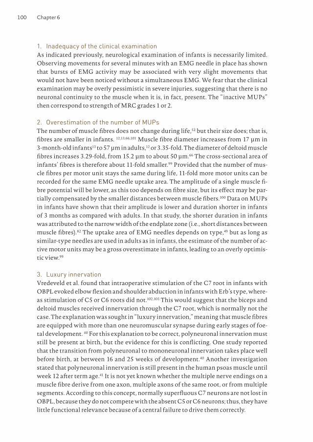
Chapter 6100
1. Inadequacy of the clinical examinationAs indicated previously, neurological examination of infants is necessarily limited. Observing movements for several minutes with an EMG needle in place has shown that bursts of EMG activity may be associated with very slight movements that would not have been noticed without a simultaneous EMG. We fear that the clinical examination may be overly pessimistic in severe injuries, suggesting that there is no neuronal continuity to the muscle when it is, in fact, present. The “inactive MUPs” then correspond to strength of MRC grades 1 or 2.
2. Overestimation of the number of MUPsThe number of muscle fibres does not change during life,52 but their size does; that is, fibres are smaller in infants. 12,13,66,101 Muscle fibre diameter increases from 17 µm in 3-month-old infants13 to 57 µm in adults,12 or 3.35-fold. The diameter of deltoid muscle fibres increases 3.29-fold, from 15.2 µm to about 50 µm.66 The cross-sectional area of infants’ fibres is therefore about 11-fold smaller.99 Provided that the number of mus-cle fibres per motor unit stays the same during life, 11-fold more motor units can be recorded for the same EMG needle uptake area. The amplitude of a single muscle fi-bre potential will be lower, as this too depends on fibre size, but its effect may be par-tially compensated by the smaller distances between muscle fibers.100 Data on MUPs in infants have shown that their amplitude is lower and duration shorter in infants of 3 months as compared with adults. In that study, the shorter duration in infants was attributed to the narrow width of the endplate zone (i.e., short distances between muscle fibres).82 The uptake area of EMG needles depends on type,49 but as long as similar-type needles are used in adults as in infants, the estimate of the number of ac-tive motor units may be a gross overestimate in infants, leading to an overly optimis-tic view.99
3. Luxury innervationVredeveld et al. found that intraoperative stimulation of the C7 root in infants with OBPL evoked elbow flexion and shoulder abduction in infants with Erb’s type, where-as stimulation of C5 or C6 roots did not.102,103 This would suggest that the biceps and deltoid muscles received innervation through the C7 root, which is normally not the case. The explanation was sought in “luxury innervation,” meaning that muscle fibres are equipped with more than one neuromuscular synapse during early stages of foe-tal development. 40 For this explanation to be correct, polyneuronal innervation must still be present at birth, but the evidence for this is conflicting. One study reported that the transition from polyneuronal to mononeuronal innervation takes place well before birth, at between 16 and 25 weeks of development.40 Another investigation stated that polyneuronal innervation is still present in the human psoas muscle until week 12 after term age.41 It is not yet known whether the multiple nerve endings on a muscle fibre derive from one axon, multiple axons of the same root, or from multiple segments. According to this concept, normally superfluous C7 neurons are not lost in OBPL, because they do not compete with the absent C5 or C6 neurons; thus, they have little functional relevance because of a central failure to drive them correctly.
101A review of EMG
4. Central motor disordersOBPL not only affects motor output, but also sensory input. Many neurologic sys-tems exhibit a critical period for formation, dependent on afferent impulses. The deaf-ferentation of OBPL may inhibit the development of normal motor control, which can remain abnormal even if later peripheral nerve repair partially ameliorates the periph-eral part of the problem.59,99,106 In a rabbit study designed to address this problem, func-tional impairment was larger for lesions sustained at birth than for lesions later in life, although peripheral regeneration was shown to occur at all ages.106 The mean number of motor units in biceps and thenar muscles was approximately 50% that of control arms in a study in children with OBPL with a minimum age of 4 years.84 MUP ampli-tudes were increased; the pattern was therefore that of severe loss of motor units of long standing. Eight children had normal numbers of motor units in the biceps mus-cle, in contrast to the significant upper arm and shoulder problems. Agnosia or apraxia was hypothesized.84 A follow-up study revealed that voluntary elbow flexion force in the affected arms was considerably lower than the force exerted by electrical stimula-tion of the muscles,14 whereas forces were similar in control subjects. In short, subjects with OBPL had fewer biceps motor units than control subjects, and they were unable to recruit all of those they did have.14 “Developmental apraxia”14 was suggested to ex-plain why some motor units were not put to work through voluntary commands. We have repeatedly observed that children with OBPL will flex their elbows while picking up a ball, but will “forget the arm” during running or when they otherwise neglect to focus on the use of the arm. This has also been mentioned by others.94 This suggests that use of the arm depends on the requested movement pattern (i.e., on central pat-terns).
It is conceivable that disordered motor programs do not merely result in a lack of firing at the right moment, but also in firing when this is not required. This mechanism has been implicated in co-contraction in OBPL,75 and may explain “inactive MUPs.”
5. Abnormal nerve branchingMotor axons growing toward the periphery after lesion need not regain their original target. In fact, there appears to be no guiding principle in outgrowth after axonal sec-tion, and thus reinnervation appears to be an essentially random process. Axons may reach agonistic, antagonistic, or unrelated muscles.31 Because motor neurons still fire in response to their original driving patterns, flexors may be activated together with extensors, resulting in severe movement disturbances. 31 In “complex misdirection,” axons split into various branches that may end up in various muscles or even in cuta-neous nerves (Fig. 3).8,17,75-78 Roth stimulated ulnar and median nerves, motor points of various muscles, and sensory nerves in the fingers, and recorded resultant MUPs (“heterogeneous axon reflexes”) from a large number of muscles in 16 cases of OBPL, aged 1-47 years.75-78 These were found in all 16 cases (up to 100 in one patient). Out of 618 searches for a communication between two muscles, or between a muscle and an inappropriate nerve, no less than 38% resulted in such heterogeneous axon reflexes. Abnormal sensory-muscular communications were found in 10 of 14 subjects.75 Simi-lar sensory-motor communications were, almost without exception, also found in

Chapter 6102
adults with previously sutured peripheral nerves.64 In “simple misdirection” the axon does not split, so the motor unit forms in its entirety in the wrong muscle.21
Both types of misdirection can result in co-contraction of various muscles, and can explain “inactive MUPs”; instead these belong in another muscle. In adults, mis-direction can be tested by asking subjects to contract muscles other than the one the needle is in, but this cannot be done in infants. Routine experience in adult plexus lesions has shown that misdirection is a regular occurrence. Still, co-contraction in adult plexus lesions is never as severe a problem as it is in older children with OBPL. One possible explanation is that axons in infants are more likely to split than adult axons. There is animal evidence that nerve regeneration differs between adults and infants, as shown by the longer persistence of unmyelinated axons in rats,97 increased axonal branching in neonatal rats,38 earlier axonal degeneration and recovery in foetal sheep,53 or increased regeneration capability in young sheep.22 It is therefore conceiv-able that the tendency for axonal branching differs between infants and adults.
EMG findings: what is the solution?
The quick appearance of denervation activity is simply a consequence of infants’ ax-ons being shorter and thinner. Its quick disappearance and the emergence of MUPs point to regeneration. Nevertheless, it remains unknown as to why these MUPs, at around 3 months, appear to have no function. The five possible explanations given previously are not mutually exclusive, as none renders any of the others impossible.
Figure 3: Nerve outgrowth in OBPL
C5
C6
C7
C8
T1
LTN
SSN
Flexion
Extension
Abduction
AxN/Deltoid
MCN/Biceps
MedN
RadN/Triceps
UlnN
C5
C6
C7
C8
T1
LTN
SSN
AxN/Deltoid
MCN/Biceps
MedN
RadN/Triceps
UlnN
a) Normal innervation pattern. Motor commands for shoulder abduction project on C5 motor neurons that innervate the deltoid muscle; likewise, commands for elbow flexion reach the biceps through the C6 root, and elbow extension is effected through the C7 root and the triceps muscle (in reality, muscles are innervated through more than one root, and more than one muscle contributes to each function). b) The situation after severe proximal axonotmesis in the superior trunk (circle). Outgrowing axons may or may not split, and can grow into all available pathways downstream from the superior trunk. Motor commands may then reach unintended muscles. For instance, ax-ons destined to effect elbow flexion may now innervate the biceps, but also its antagonist (triceps) or the deltoid muscle. SSN, suprascapular nerve; LTN, long thoracic nerve; MCN, musculocuta-neous nerve; AxN, axillary nerve; MedN, median nerve; RadN, radial nerve; UlnN, ulnar nerve; Biceps, biceps muscle; Deltoid, deltoid muscle; Triceps, triceps muscle
103A review of EMG
It would be surprising if the central nervous system were not involved at all, although this might take two completely different forms. The first is a permanent loss of func-tion due to a closed window of opportunity to develop a motor program, as suggested earlier.14,84 The other rests on the finding that central nervous system plasticity occurs in adult plexus lesions 55-57; the young nervous system appears capable of surprising amounts of plasticity and functional repair, so a central “solution,” involving recruit-ing muscles for trick movements, is not unreasonable.
Axonal misdirection has been shown to exist in older children with OBPL, and, as branching is part of nerve regeneration, it is highly likely that branching is present at 3 months of age. Axonal branching can explain why direct muscle stimulation was shown to result in more force than voluntary activation.14 In that study, voluntary ac-tivation of the biceps did not excite all motor units in it, as some “belonged” to other motor programs. However, in response to criticism along these lines,72 the existence of “heterogeneous axon reflexes” was doubted, because sensory-motor connections could have been due to normal cutaneous reflexes.59 There is no need, however, to pre-sent these theories as conflicting; extensive peripheral “cross-wiring” would present the central nervous system with conflicting feedback, as a command for elbow flexion would be followed by feedback from flexors, abductors, or extensors, or some combi-nation of these muscles. The inability to learn to activate muscles selectively might seriously disrupt normal agonist-antagonist programming. As noted earlier, the other explanations may also be true. In particular, it would be very surprising if the 11-fold tighter packing of muscle fibres in a muscle does not affect the interpretation of the EMG recruitment pattern to some degree.
Conclusion and prospects
Treatment and assessment of prognosis in OBPL suffer from a lack of methodologi-cally sound evidence. OBPL presents a number of specific problems that affect the assessment of prognosis; we are currently performing a systematic review of progno-sis that takes these factors into account. The EMG might be very useful for prognosis, but pathophysiological and longitudinal studies are needed to redefine when it should be performed and which parameters are useful for prognosis. Electromyographers and surgeons should realize that the presence of MUPs in a clinically paralytic mus-cle indeed indicates that there is continuity from spinal cord to muscle, and should be aware that factors other than mere continuity can also be relevant to regain useful function of the arm. This is not an academic question, as surgical efforts are largely aimed at restoring neural continuity. We therefore urge researchers not to restrict their focus to the restoration of continuity, but rather to widen their attention to en-compass all factors affecting restoration of function. Focus points for such research should be the extent of axonal branching and the selectivity of the central drive to var-ious muscles in obstetric and adult plexus lesions. Such knowledge will improve not just the utility of the EMG, but also in understanding the nature of nerve repair and motor control in infants. Ultimately, this may improve the treatment of this complex disorder, which will affect these individuals for their entire lives.

Chapter 6104
1 Alfonso I, Papazian O, Prieto G, Alfonso DT, Melnick SJ. Neoplasm as a cause of brachial plexus palsy in neonates. Pediatr Neurol 2000; 22:309-311.
2 Al Qattan MM, Clarke HM, Curtis CG. Klumpke’s birth palsy. Does it really exist? J Hand Surg Br 1995; 20:19-23.
3 Allen A, Sorab J, Gonik B. Risk factors for shoulder dystocia: an engineering study of clinician-applied forces. Obstet Gynecol 1991; 77:352-355.
4 Arancio O, Cangiano A, de Grandis D. Fibril-latory activity and other membrane changes in partially denervated muscles. Muscle Nerve 1989; 12:149-153.
5 Bager B. Perinatally acquired brachial plexus palsy – a persisting challenge. Acta Paediatr 1997; 86:1214-1219.
6 Basheer H, Zelic V, Rabia F. Functional scoring system for obstetric brachial plexus palsy. J Hand Surg Br 2000; 25B: 41-45.
7 Bellew M, Kay SPJ, Webb F, Ward A. Devel-opmental and behavioural outcome in obstetric brachial plexus palsy. J Hand Surg Br 2000; 25B:49-51.
8 Benaim JL, Jouve JL, Bardot J, Casanova D, Magalon G, Bollini G. Pseudo-paralysie du biceps brachial dans les paralysies obste tricales du plexus brachial (POPB) – a propos de l’ “optimisme” de l’EMG. Neuro-physiol Clin 1999; 29:490-494.
9 Birch R, Bonney G, Wynn Parry CB. Surgical disorders of the peripheral nerves. Edin-burgh: Churchill Livingstone; 1998.
10 Bodensteiner JB, Rich KM, Landau WM. Early infantile surgery for birth-related brachial plexus injuries: justification requires a prospective controlled study. J Child Neurol 1994; 9:109-110.
11 Boome RS, Kaye CJ. Obstetric traction inju-ries of the brachial plexus. Natural history, indications for surgical repair and results. J Bone Joint Surg Br 1988; 70:571-576.
12 Brooke MH, Engel WK. The histographic analysis of human muscle biopsies with regard to fiber types. 1. Adult male and female. Neurology 1969; 19:221-233.
13 Brooke MH, Engel WK. The histographic analysis of human muscle biopsies with regard to fiber types. 4. Children’s biopsies. Neurology 1969; 19:591-605.
14 Brown T, Cupido C, Scarfone H, Pape K, Galea V, McComas A. Developmental aprax-ia arising from neonatal brachial plexus palsy. Neurology 2000; 55:24-30.
15 Clarke HM, Curtis CG. An approach to obstetrical brachial plexus injuries. Hand Clin 1995; 11:563-580.
16 Chuang DC-C, Ma H-S, Wei F-C. A new strategy of muscle transposition for treat-ment of shoulder deformity caused by obstetric brachial plexus palsy. Plast Recon-str Surg 1998; 101: 686-694.
17 De Grandis D, Fiaschi A, Michieli G, Mezzi-na C. Anomalous reinnervation as a sequel to obstetric brachial plexus palsy. J Neurol Sci 1979; 43:127-132.
18 Dodds SD, Wolfe SW. Perinatal brachial plexus palsy. Curr Opinion Pediatr 2000; 12:40-47.
19 Dunn DW, Engle WA. Brachial plexus palsy: intrauterine onset. Pediatr Neurol 1985; 1:367-369.
20 Eng GD, Binder H, Getson P, O’Donnell R. Obstetrical brachial plexus palsy (OBPP) outcome with conservative management. Muscle Nerve 1996; 19:884-891.
21 Esslen E. Electromyographic findings on two types of misdirection of regenerating axons. Electroencephalogr Clin Neurophys-iol 1960; 12:738-741.
22 Fullarton AC, Lenihan DV, Myles LM, Glasby MA. Obstetric brachial plexus palsy: a large animal model for traction injury and its repair. J Hand Surg Br 2000; 25B:52-57.
23 Geutjens G, Gilbert A, Helsen K. Obstetric brachial plexus palsy associated with breech delivery. A different pattern of injury. J Bone Joint Surg Br 1996; 78:303-306.
24 Gherman RB, Ouzounian JG, Goodwin TM. Brachial plexus palsy: an in utero injury? Am J Obstet Gynecol 1999; 180: 1303-1307.
25 Gilbert WM, Nesbitt TS, Danielsen B. Asso-ciated factors in 1611 cases of brachial plexus injury. Obstet Gynecol 1999; 93: 536-540.
26 Gilbert A, Tassin JL. Reparation chirurgicale du plexus brachial dans la paralysie obstetri-cale. Chirurgie 1984; 110:70-75.
27 Goebel HH, Muller J. The unusual features of traumatic neurogenic muscular atrophy in the infant: an anatomic study. Neuropädi-atrie 1977; 8:274-285.

105A review of EMG
28 Gonik B, McCormick EM, Verweij BH, Rossman KM, Nigro MA. The timing of congenital brachial plexus injury: a study of electromyographic findings in the newborn piglet. Am J Obstet Gynecol 1998; 178:688-695.
29 Gonik B, Walker A, Grimm M. Mathematic modeling of forces associated with shoulder dystocia: a comparison of endogenous and exogenous sources. Am J Obstet Gynecol 2000; 182:689-691.
30 Gordon M, Rich H, Deutschberger J, Green M. The immediate and long-term outcome of obstetric birth trauma. I. Brachial plexus paralysis. Am J Obstet Gynecol 1973; 117: 51-56.
31 Gramsbergen A, IJkema-Paassen J, Meek MF. Sciatic nerve transection in the adult rat: abnormal EMG patterns during loco-motion by aberrant innervation of hindleg muscles. Exp Neurol 2000; 161:183-193.
32 Greenwald AG, Schute PC, Shiveley JL. Brachial plexus birth palsy: a 10-year report on the incidence and prognosis. J Pediatr Orthop 1984; 4:689-692.
33 Grossman JAI. Early operative intervention for birth injuries to the brachial plexus. Sem Pediatr Neurol 2000; 7:36-43.
34 Grossman JAI, Ramos LE, Shumway S, Alfonso I. Management strategies for chil-dren with obstetrical plexus injuries. Int Pediatr 1997; 12:82-86.
35 Gu Y-D, Chen L, Shen L-Y. Classification of impairment of shoulder abduction in obstet-ric brachial plexus palsy and its clinical significance. J Hand Surg Br 2000; 25B:46-48.
36 Hannah ME, Hannah WJ, Hewson SE, Hodnett ED, Saigal S, Willan AR. Planned caesarean section versus planned vaginal birth for breech presentation at term: a randomised multicentre trial. Lancet 2000; 356:1375-1383.
37 Hardy AE. Birth injuries of the brachial plexus: incidence and prognosis. J Bone Joint Surg Br 1981; 63:98-101.
38 Heath DD, Coggeshall RE, Hulsebosch C. Axon and neuron numbers after forelimb amputation in neonatal rats. Exp Neurol 1986; 92:220-233.
39 Held JP, Pierrot Deseilligny, Meillet J. Essai d’analyse des syncinésies dans les paralysies obstétricales du plexus brachial. Ann Méd Phys 1969; 12:3-8.
40 Hesselmans LFGM, Jennekens FGI, Van den Oord CJM, Veldman H, Vincent A. Develop-ment of innervation of skeletal muscle fibers in man: relation to acetylcholine receptors. Anat Rec 1993; 236:553-562.
41 Ijkema-Paassen J, Gramsbergen A. Polyneu-ral innervation in the psoas muscle of the developing rat. Muscle Nerve 1998; 21:1058-1063.
42 Jacobsen S. Forekomsten af obstetrisk plexus brachialis laesion pa Lollnd-Falster 1960-1970. Nord Med 1971; 86:1200-1201.
43 Jahnke AH, Bovill DF, McCarroll HR, James P, Ashley RK. Persistent brachial plexus birth palsies. J Pediatr Orthop 1991; 11:533-537.
44 Jennett RJ, Tarby TJ. Brachial plexus palsy: an old problem revisited again. Am J Obstet Gynecol 1997; 176:1354-1357.
45 Jennett RJ, Tarby TJ, Kreinick CJ. Brachial plexus palsy: an old problem revisited. Am J Obstet Gynecol 1992; 166: 1673-1677.
46 Kay S. Brachial palsies. Lancet 1999; 354:614-615.
47 Kay SPJ. Obstetrical brachial palsy. Br J Plast Surg 1998; 51: 43-50.
48 King JC, Dumitru D, Nandedkar S. Concen-tric and single fiber electrode spatial recording characteristics. Muscle Nerve 1997; 20:1525-1533.
49 Koenigsberger MR. Brachial palsy at birth: intrauterine or due to delivery trauma? Ann Neurol 1980; 8:228.
50 Laurent JP, Lee RT. Birth-related upper brachial plexus injuries in infants: operative and nonoperative approaches. Child Neurol 1994; 9:111-117.
51 Levine MG, Holroyde J, Woods JRJ, Siddiqi JA, Scott M, Miodovnik M. Birth trauma: incidence and predisposing factors. Obstet Gynecol 1984; 63:792-795.
52 Lexell J, Sjöström M, Nordlund A-S, Taylor CC. Growth and development of human muscle: a quantitative morphological study of whole vastus lateralis from childhood to adult age. Muscle Nerve 1992; 15:404-409.

Chapter 6106
53 Lin KY, Posnick JC, Al-Qattan MM, Vajsar J, Becker LE. Fetal nerve healing: an experi-mental study. Plast Reconstr Surg 1994; 93:1323-1333.
54 Luco JV, Eyzaguirre C. Fibrillation and hypersensitivity to ACh in denervated muscle: effect of length of degenerating nerve fibers. J Neurophysiol 1955; 18:65-73.
55 Malessy M, van der Kamp W, Thomeer R, van Dijk JG. Cortical excitability of the biceps muscle after intercostal-tomuscu-locutaneous nerve transfer. Neurosurgery 1998; 42: 787-795.
56 Malessy MJA, Thomeer RTWM. Evaluation of intercostal to musculocutaneous nerve transfer in reconstructive brachial plexus surgery. J Neurosurg 1998; 88:266-271.
57 Malessy MJA, Thomeer RTWM, van Dijk JG. Changing central nervous system control following intercostal nerve transfer. J Neurosurg 1998; 89:568-574.
58 Mallet J. Paralysie obstétricale du plexus brachial. Rev Chir Orthop Reparatrice Appar Mot 1972; 58:115-204.
59 McComas A, Galea V. Developmental aprax-ia arising from neonatal brachial plexus palsy. A reply from the authors. Neurology 2000; 55:1761.
60 McComas AJ, Pape K, Kirsch S. Apraxia in congenital brachial palsy. Can J Neurol Sci 1993; 20:362.
61 McFarland LV, Raskin M, Daling JR, Benedetti TJ. Erb/ Duchenne’s palsy: a consequence of fetal macrosomia and method of delivery. Obstet Gynecol 1986; 68:784-788.
62 Mancias P, Slopis JM, Yeakley JW, Vriesen-dorp FJ. Combined brachial plexus injury and root avulsion after complicated delivery. Muscle Nerve 1994; 17:1237-1238.
63 Michelow BJ, Clarke HM, Curtis CG, Zuker RM, Seifu Y, Andrews DF. The natural histo-ry of obstetrical brachial plexus palsy. Plast Reconstr Surg 1994; 93:675-681.
64 Montserrat L, Benito M. Motor reflex responses elicited by cutaneous stimulation in the regenerating nerve of man: axon reflex or ephaptic response? Muscle Nerve 1990; 13: 501-507.
65 Narakas AO. Obstetrical brachial plexus injuries. In: Lamb DW, editor. The paralysed
hand. Edinburgh: Churchill Livingstone; 1987. p 116-135.
66 Oertel G. Morphometric analysis of normal skeletal muscles in infancy, childhood and adolescence. J Neurol Sci 1988; 88:303-313.
67 Painter MJ, Bergman I. Obstetrical trauma to the neonatal central and peripheral nerv-ous system. Semin Perinatol 1982; 6:89-104.
68 Papazian O, Alfonso I, Grossman JAI. Evaluación neurofisilógica de los niños con parálisis del plexo braquial obstétrica. Rev Neurol 1998; 27:263-270.
69 Papazian O, Alfonso I, Yaylali I, Velez I, Jayakar P. Neurophysiological evaluation of children with traumatic radiculopathy, plex-opathy, and peripheral neuropathy. Semin Pediatr Neurol 2000; 7:26-35.
70 Paradiso G, Granana N, Maza E. Prenatal brachial plexus paralysis. Neurology 1997; 49:261-262.
71 Peterson GW, Bohr TW. Neonatal “obstet-ric” palsy: a “preexisting condition”? Two case reports. Muscle Nerve 1995; 18:1031.
72 Rapalino OA, Levine DN. Developmental apraxia arising from neonatal brachial plex-us palsy. Neurology 2000; 55: 1761.
73 Rollnik JD, Hierner R, Schubert M, Shen ZL, Johannes S, Tro¨ger M, Wohlfarth K, Berger AC, Dengler R. Botulinum toxin treatment of cocontractions after birth-related brachial plexus lesions. Neurology 2000; 55:112-114.
74 Ross D, Royden Jones H, Fisher J, Konkol RJ. Isolated radial nerve lesion in the newborn. Neurology 1983; 33:1354-1356.
75 Roth G. Reinnervation dans la paralysie plexulaire brachiale obstétricale. J Neurol Sci 1983; 58:103-115.
76 Roth G. Intranervous regeneration. J Neurol Sci 1979; 41: 139-148.
77 Roth G. Intranervous regeneration of lower motor neuron – I. Electromyogr Clin Neuro-physiol 1978; 18:225-288.
78 Roth G. Intranervous regeneration of lower motor neuron – II. Electromyogr Clin Neurophysiol 1978; 18:311-351.
79 Royden Jones H. Plexus and nerve root lesions. In: Royden Jones H, Bolton CF, Harper CM, editors. Pediatric clinical elec-tromyography. Philadelphia: Lippincott Raven; 1996. p 123-169.

107A review of EMG
80 Royden Jones H, Herbison GJ, Jacobs SR, Kollros PR, Macones GA. Intrauterine onset of a mononeuropathy: peroneal neuropathy in a newborn with electromyographic find-ings at age one day compatible with prenatal onset. Muscle Nerve 1996; 19:88-91.
81 Rust RS. Congenital brachial plexus palsy: where have we been and where are we now? Semin Pediatr Neurol 2000; 7: 58-63.
82 Sacco G, Buchthal F, Rosenfalck P. Motor unit potentials at different ages. Arch Neurol 1962; 6:366-373.
83 Salafsky B, Bell J, Prewitt MA. Development of fibrillation potentials in denervated fast and slow skeletal muscle. Am J Physiol 1968; 215:637-643.
84 Scarfone H, McComas AJ, Pape K, Newber-ry R. Denervation and reinnervation in congenital brachial palsy. Muscle Nerve 1999; 22:600-607.
85 Schröder JM, Bohl, von Bardeleben U. Changes of the ratio between myelin hickness and axon diameter in human developing sural, femoral, ulnar, facial, and trochlear nerves. Acta Neuropathol 1988; 76:471-483.
86 Seddon HJ. Peripheral nerve injuries. Medi-cal Research Council special report series. London: HMSO; 1954.
87 Shenaq SM, Berzin E, Lee R, Laurent JP, Nath R, Nelson MR. Brachial plexus birth injuries and current mamagement. Clin Plast Surg 1998; 25:527-536.
88 Sjöberg I, Erichs K, Bjerre I. Cause and effect of obstetric (neonatal) brachial plexus palsy. Acta Paediatr Scand 1988; 77:357-364.
89 Sloof ACJ. Obstetric brachial plexus lesions and their neurosurgical treatment. Clin Neurol Neurosurg 1993; 95(suppl):S73-S77.
90 Smith SJM. The role of neurophysiological investigation in traumatic brachial plexus lesions in adults and children. J Hand Surg 1996; 21B:145-147.
91 Spinner RJ, Kline DG. Surgery for peripheral nerve and brachial plexus injuries or other nerve lesions. Muscle Nerve 2000; 23:680-695.
92 Strombeck C, Krumlinde-Sundholm L, Forssberg H. Functional outcome at 5 years in children with obstetrical brachial plexus palsy with and without microsurgical recon-
struction. Dev Med Child Neurol 2000; 42:148-157.
93 Sunderland S. Nerve injuries and their repair: a critical appraisal. Edinburgh: Churchill Livingstone; 1991.
94 Sundholm LK, Eliasson AC, Forssberg H. Obstetric brachial plexus injuries: assess-ment protocol and functional outcome at age 5 years. Dev Med Child Neurol 1998; 40:4-11.
95 Tassin JL. Paralysies obstetricales du plexus brachial. Evolution spontanée; Résultats des interventions reparatrices prococes. Thèse, Université de Paris; 1983.
96 Terzis JK, Papakonstantinou KC. Manage-ment of obstetric brachial plexus palsy. Hand Clin 1999; 15:717-736.
97 Toft PB, Fugleholm K, Schmalbruch H. Axonal branching following crush lesions of peripheral nerves of rat. Muscle Nerve 1988; 11:880-889.
98 Ubachs JM, Slooff AC, Peeters LL. Obstetric antecedents of surgically treated obstetric brachial plexus injuries. Br J Obstet Gynae-col 1995; 102:813-817.
99 Van Dijk JG, Malessy MJA, Stegeman DF. Why is the EMG in obstetric brachial plexus lesions overly optimistic? Muscle Nerve 1998; 21:260-261.
100 Van Dijk JG, Malessy MJA, Stegeman DF. The electromyogram in obstetric brachial palsy is too optimistic: fiber size or another explanation? Reply. Muscle Nerve 1999; 22:428-428.
101 Vogler C, Bove KE. Morphology of skeletal muscle in children. Arch Pathol Lab Med 1985; 109:238-242.
102 Vredeveld JW, Blaauw H, Sloof BACJ, Rozeman CAM. The findings in paediat-ric obstetric brachial plexus palsy differ from those in older patients: a suggested explanation. Dev Med Child Neurol 2000; 42:158-161.
103 Vredeveld JW, Richards R, Rozeman CAM, Blaauw G, Slooff ACJ. The electromyogram in obstetric brachial palsy is too optimistic: fiber size or another explanation? Muscle Nerve 1999; 22:427-428.
104 Wolf H, Hoeksma AF, Oei SL, Bleker OP. Obstetric brachial plexus injury: risk factors

Chapter 6108
related to recovery. Eur J Obstet Gynecol Reprod Biol 2000; 88:133-138.
105 Yilmaz K, Caliskan M, Öge E, Aydinli N, Tunaci M, Özmen M. Clinical assessment, MRI, and EMG in congenital brachial plex-us palsy. Pediatr Neurol 1999; 21:705-710.
106 Zalis OS, Zalis AW, Barron KD, Oester YT. Motor patterning following transitory sensory-motor deprivations. Arch Neurol 1965; 13:487-494.
Related Documents