OBJECTIVES Describe the Anatomy of the Parathyroid Gland. Describe the Microscopic Features of the Parathyroid Gland.

OBJECTIVES Describe the Anatomy of the Parathyroid Gland. Describe the Microscopic Features of the Parathyroid Gland.
Dec 28, 2015
Welcome message from author
This document is posted to help you gain knowledge. Please leave a comment to let me know what you think about it! Share it to your friends and learn new things together.
Transcript

OBJECTIVES
Describe the Anatomy of the Parathyroid Gland.
Describe the Microscopic Features of the Parathyroid Gland.

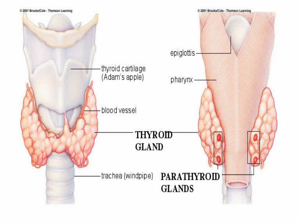
Anatomy Of The Parathyroid Gland


Parathyroid glands
There are 4 small parathyroid gland ,two embedded in the posterior surface of each lobe of the thyroid gland
The glands are small, ovoid, in shape, yellowish in color, usually lie external to the fibrous thyroid capsule .

Comparing between superior and inferior parathyroid gland
Superior parathyroid gland
Inferior parathyroid gland
Location Lie at the level of the middle of the posterior surface of the thyroid gland.
Usually lie close to the inferior poles of the thyroid gland.
Position More constant in its position
Variable in its position :
Size Small Large

Blood supply


Blood Supply Arterial supply:• Inferior thyroid artery which branched
from the thyro-cervical trunk.
Venous drainage:• The Inferior thyroid veins

Lymph Drainage• The lymph of gland
drains into the lower Deep cervical LN. and paratracheal LN.
Nerve Supply • It’s mostly superior
or middle cervical sympathetic ganglia.

Microscopic Features Of The Parathyroid
Gland

The Parathyroid Gland
PT: Parathyroid
GlandT :Thyroid
GlandS :Fibrous
SeptaL : Lymphocyte
s

Cont.
• This micrograph shows a parathyroid gland characteristically embedded in the thyroid gland or within its capsule.
• The thin fibrous capsule of the parathyroid gland gives rise to delicate septa which divide the parenchyma into nodules of secretory cells .Also the septa carry blood vessels, lymphatics and nerves.
• Note when you see some infiltration of the thyroid by lymphocytes that lead to common feature of the ageing thyroid gland.

The Parathyroid Gland
AAdipose
tissue
PChief cells
CCapillarie
s
OOxyphil
cells
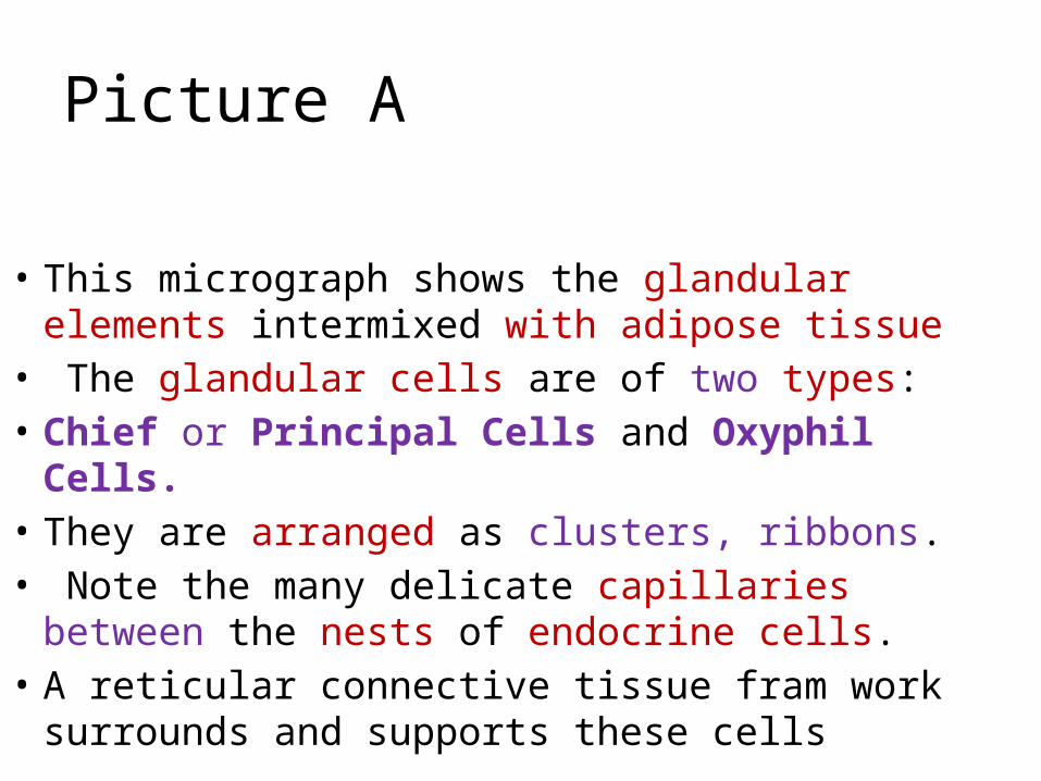
Picture A
• This micrograph shows the glandular elements intermixed with adipose tissue
• The glandular cells are of two types: • Chief or Principal Cells and Oxyphil Cells.• They are arranged as clusters, ribbons. • Note the many delicate capillaries between
the nests of endocrine cells.• A reticular connective tissue fram work
surrounds and supports these cells

Picture B
• This micrograph show the chief cells which small with round central nuclei and pale eosinophilic or clear cytoplasm. These are the cells which synthesise and secrete Parathyroid Hormone ( PTH ).
• Oxyphil cells which big cells that tend to occur in nodules, have copious eosinophilic cytoplasm that ultrastructurally is seen to be packed with mitochondria. These cells do not secrete PTH and increase in number with age. Their function is not know.
• Note the many delicate capillaries between the nests of endocrine cells.
Related Documents