REVIEW Obesity and the gut microbiota: does up-regulating colonic fermentation protect against obesity and metabolic disease? Lorenza Conterno • Francesca Fava • Roberto Viola • Kieran M. Tuohy Received: 16 March 2011 / Accepted: 20 April 2011 / Published online: 11 May 2011 Ó Springer-Verlag 2011 Abstract Obesity is now considered a major public health concern globally as it predisposes to a number of chronic human diseases. Most developed countries have experienced a dramatic and significant rise in obesity since the 1980s, with obesity apparently accompanying, hand in hand, the adoption of ‘‘Western’’-style diets and low- energy expenditure lifestyles around the world. Recent studies report an aberrant gut microbiota in obese subjects and that gut microbial metabolic activities, especially carbohydrate fermentation and bile acid metabolism, can impact on a number of mammalian physiological functions linked to obesity. The aim of this review is to present the evidence for a characteristic ‘‘obese-type’’ gut microbiota and to discuss studies linking microbial metabolic activities with mammalian regulation of lipid and glucose metabo- lism, thermogenesis, satiety, and chronic systemic inflam- mation. We focus in particular on short-chain fatty acids (SCFA) produced upon fiber fermentation in the colon. Although SCFA are reported to be elevated in the feces of obese individuals, they are also, in contradiction, identified as key metabolic regulators of the physiological checks and controls mammals rely upon to regulate energy metabo- lism. Most studies suggest that the gut microbiota differs in composition between lean and obese individuals and that diet, especially the high-fat low-fiber Western-style diet, dramatically impacts on the gut microbiota. There is cur- rently no consensus as to whether the gut microbiota plays a causative role in obesity or is modulated in response to the obese state itself or the diet in obesity. Further studies, especially on the regulatory role of SCFA in human energy homeostasis, are needed to clarify the physiological con- sequences of an ‘‘obese-style’’ microbiota and any putative dietary modulation of associated disease risk. Keywords Obesity Microbiota SCFA Fiber Prebiotics Probiotics Introduction Obesity is now considered among the top public health issues worldwide. In many countries, obesity rates reported before 1980 were below 10%, whereas nearly half of the Organization for Economic Co-operation and Development (OECD) countries now report 50% or more of the popu- lation as being overweight, with the percentage obese reaching 20 to 30% (OECD 2010). Obesity has a dramatic impact on the body, with major changes in energy metabolism and regulatory mechanisms leading to type 2 diabetes, cardiovascular disease (CVD), hormone-linked cancers, and gastrointestinal diseases including inflamma- tory bowel disease and colon cancer. Characteristic phys- iological perturbation in terms of hormonal imbalances (e.g., elevated leptin and insulin) and chronically elevated glucose and blood lipids (TAG, cholesterol) occur in con- gruence with oxidative stress and chronic systemic inflammation, which itself leads to cellular damage in diverse body tissues including the liver, pancreas, vascular system, and the intestinal mucosa. Obesity therefore is a key risk factor for numerous chronic diseases including CVD, the metabolic syndrome, type 2 diabetes, and certain cancers (Pi-Sunyer 2009). L. Conterno (&) F. Fava K. M. Tuohy Nutrition and Nutrigenomics Group, Research and Innovation Centre, FEM-IASMA, 38010 S. Michele a.A, Trento, Italy e-mail: [email protected] R. Viola Research and Innovation Centre, FEM-IASMA, 38010 S. Michele a.A, Trento, Italy 123 Genes Nutr (2011) 6:241–260 DOI 10.1007/s12263-011-0230-1

Welcome message from author
This document is posted to help you gain knowledge. Please leave a comment to let me know what you think about it! Share it to your friends and learn new things together.
Transcript

REVIEW
Obesity and the gut microbiota: does up-regulating colonicfermentation protect against obesity and metabolic disease?
Lorenza Conterno • Francesca Fava •
Roberto Viola • Kieran M. Tuohy
Received: 16 March 2011 / Accepted: 20 April 2011 / Published online: 11 May 2011
� Springer-Verlag 2011
Abstract Obesity is now considered a major public
health concern globally as it predisposes to a number of
chronic human diseases. Most developed countries have
experienced a dramatic and significant rise in obesity since
the 1980s, with obesity apparently accompanying, hand in
hand, the adoption of ‘‘Western’’-style diets and low-
energy expenditure lifestyles around the world. Recent
studies report an aberrant gut microbiota in obese subjects
and that gut microbial metabolic activities, especially
carbohydrate fermentation and bile acid metabolism, can
impact on a number of mammalian physiological functions
linked to obesity. The aim of this review is to present the
evidence for a characteristic ‘‘obese-type’’ gut microbiota
and to discuss studies linking microbial metabolic activities
with mammalian regulation of lipid and glucose metabo-
lism, thermogenesis, satiety, and chronic systemic inflam-
mation. We focus in particular on short-chain fatty acids
(SCFA) produced upon fiber fermentation in the colon.
Although SCFA are reported to be elevated in the feces of
obese individuals, they are also, in contradiction, identified
as key metabolic regulators of the physiological checks and
controls mammals rely upon to regulate energy metabo-
lism. Most studies suggest that the gut microbiota differs in
composition between lean and obese individuals and that
diet, especially the high-fat low-fiber Western-style diet,
dramatically impacts on the gut microbiota. There is cur-
rently no consensus as to whether the gut microbiota plays
a causative role in obesity or is modulated in response to
the obese state itself or the diet in obesity. Further studies,
especially on the regulatory role of SCFA in human energy
homeostasis, are needed to clarify the physiological con-
sequences of an ‘‘obese-style’’ microbiota and any putative
dietary modulation of associated disease risk.
Keywords Obesity � Microbiota � SCFA � Fiber �Prebiotics � Probiotics
Introduction
Obesity is now considered among the top public health
issues worldwide. In many countries, obesity rates reported
before 1980 were below 10%, whereas nearly half of the
Organization for Economic Co-operation and Development
(OECD) countries now report 50% or more of the popu-
lation as being overweight, with the percentage obese
reaching 20 to 30% (OECD 2010). Obesity has a dramatic
impact on the body, with major changes in energy
metabolism and regulatory mechanisms leading to type 2
diabetes, cardiovascular disease (CVD), hormone-linked
cancers, and gastrointestinal diseases including inflamma-
tory bowel disease and colon cancer. Characteristic phys-
iological perturbation in terms of hormonal imbalances
(e.g., elevated leptin and insulin) and chronically elevated
glucose and blood lipids (TAG, cholesterol) occur in con-
gruence with oxidative stress and chronic systemic
inflammation, which itself leads to cellular damage in
diverse body tissues including the liver, pancreas, vascular
system, and the intestinal mucosa. Obesity therefore is a
key risk factor for numerous chronic diseases including
CVD, the metabolic syndrome, type 2 diabetes, and certain
cancers (Pi-Sunyer 2009).
L. Conterno (&) � F. Fava � K. M. Tuohy
Nutrition and Nutrigenomics Group, Research and Innovation
Centre, FEM-IASMA, 38010 S. Michele a.A, Trento, Italy
e-mail: [email protected]
R. Viola
Research and Innovation Centre, FEM-IASMA,
38010 S. Michele a.A, Trento, Italy
123
Genes Nutr (2011) 6:241–260
DOI 10.1007/s12263-011-0230-1
Data collected about this new epidemic of obesity are
revealing a complicated network of contributory factors
including genetics, age, diet, and nutritional environment.
However, the rapid increase in obesity over such a short
time frame makes a novel genetic cause per se unlikely and
strongly favors modified environmental factors over the
past 30 years. Such environmental factors include dietary
habit, exercise or energy expenditure, and lifestyle. Indeed,
there appears to be a strong correlation between Western-
ization in terms of diet and lifestyle and obesity. Reduced
energy expenditure of modern lifestyles and a Western-
style diet prevalent in developed nations are both impli-
cated as causative factors in obesity and are likely to work
in synergy to increase obesity rates at the population level
(Lieberman 2003; Keim et al. 2004). A shift from more
traditional diets, rich in whole-plant foods like whole-grain
cereals, fruits, and vegetables, e.g., those of traditional
Chinese, Japanese, rural African, and hunter-gatherer
populations or aboriginal peoples in Australia and south
America, to modern Western-style diets rich in refined
carbohydrates, fat, and red/processed meats and low in
fiber and whole-plant foods, is strongly correlated with
increased body weight, obesity, and the diseases of obesity
(O’Dea 1991; Ravussin et al. 1994; Williams et al. 2001;
Novotny et al. 2009; Willcox et al. 2009). The same is seen
in Europe, where poor adherence to a ‘‘Mediterranean style
diet’’ and reduced intake of fiber, fruit, and vegetables is
presaging increased body weight and obesity even in
countries that traditionally have lower rates of obesity such
as Italy (Celi et al. 2003; di Giuseppe et al. 2008; Baldini
et al. 2009; De Filippo et al. 2010). This divergence from
traditional whole-plant food-based diets and concomitant
increases in body weight and diabetes in particular has
been reported in genetically diverse populations from dif-
ferent corners of the world again strongly discounting
genetics as the major cause for the current wave of obesity.
A more recently appreciated characteristic of obesity is
an aberrant intestinal microbiota composition in obese
individuals, which appears to be linked to the obese state
itself and yet susceptible to dietary modulation (for recent
reviews, see Backhed 2010; Ley 2010; Tuohy et al. 2010).
Whether this aberrant microbiota composition plays an
etiological role in obesity or is a consequence of the diet in
obesity remains to be determined with evidence from dif-
ferent laboratories, models systems, and human studies
supporting either hypothesis (Ley et al. 2006; Duncan et al.
2007). However, this in itself is not surprising considering
the complex interplay between the resident human intes-
tinal microbiota and diverse mammalian physiological
systems including the immune system, endocrine system,
and importantly, energy homeostasis and lipid metabolism.
Also the fact that diet, especially dietary fiber, is already
known to modify microbial profiles and fermentative
output of the gut microbiota would make a low-fiber
Western-style obesogenic diet a likely candidate for
impacting on gut microbiota composition and activity
(Tuohy et al. 2009a, b; De Filippo et al. 2010). Further,
recent metagenomic studies are suggesting that many
metabolic functions within the gut microbiota are shared
between diverse species, suggesting that ecological func-
tion may not be as closely linked to bacterial phylogenetics
as we have assumed in the past and that although certain
groups of indicator organisms may predict either a healthy
microbiota (e.g., the bifidobacteria and lactobacilli) or one
more likely to be associated with disease (e.g., elevated
numbers of enterobacteria), when trying to correlate a
phenotypic trait as globally pervasive as energy metabo-
lism, it may not be surprising that different studies show
different results in terms of microbial populations corre-
lating with obesity (Cummings et al. 2004).
In the following text, we present a review of studies
showing that the gut microbiota, both in composition and
metabolic activity, appears to be different in obese com-
pared with lean individuals and discuss the different
mechanisms suggested to link aberrant intestinal microbiota
profiles with obesity and the diseases of obesity (Table 1).
We discuss these recent observations with respect to
mechanistic nutritional studies linking colonic fermentation
and microbial metabolites, in particular the short-chain fatty
acids, acetate, propionate, and butyrate to the regulation of
mammalian energy metabolism and body composition.
Is the gut microbiota altered in obesity?
Evidence from germ-free animals
Some of the earliest data linking the mammalian gut mic-
robiota with obesity and particularly, energy homeostasis
and fat storage come from studies conducted in germ-free
animals. These are animals raised and maintained in the
complete absence of contact with living microorganisms
and as such do not undergo natural physiological succes-
sional development in terms of immune education, mucosal
architecture, mammalian–microbiota co-metabolic path-
ways (e.g., bile acid turnover or xenobiotic metabolism),
and as we are finding out, energy metabolism and storage,
which occur in free-living vertebrates concomitantly with
gut microbiota successional development. However, they
do serve as a very useful tool to elucidate fundamental
mechanisms underpinning microbial communication and
interactions with mammalian physiology at the systems
level under controlled conditions not attainable using con-
ventional models or in human studies. Backhed et al. (2004)
found that transplant of gut microbiota from conventional
animals (animals raised under normal, microbiota
242 Genes Nutr (2011) 6:241–260
123
associated conditions) into germ-free mice (C57BL/6) led
to 60% increased body fat and insulin resistance within
14 days despite reduced food intake and a 40% reduction in
muscle mass. Colonization with the common mammalian
anaerobic commensal Bacteroides thetaiotaomicron alone
also led to a 23% increase in the total body fat. This bac-
terial species has a very high capacity of degrading plant
polysaccharides, the major constituent of dietary fiber,
which are not broken down by host-encoded enzymes. The
study showed that conventionalization promoted increased
monosaccharide uptake from the gut, increased delivery of
monosaccharides to the liver, increased transactivation of
lipogenic enzymes, and increased LPL activity leading to
higher uptake of fatty acids and triglyceride accumulation.
These authors were the first to show that acquisition of a gut
microbiota impacts on fat deposition, clearly linking
microbial activities in the intestine with mammalian energy
homeostasis and obesity.
Turnbaugh et al. in 2006 showed that germ-free animals
colonized with the gut microbiota from obese animals
showed greater body weight and fat mass than germ-free
animals colonized with the microbiota derived from lean
animals. The obese donor was found to be populated by a
microbiota with a higher proportion of Firmicutes than the
lean donor. The recipients showed a gut microbiota profile
similar to their obese donor 2 weeks after colonization.
However, even if the initial body mass of the recipient and
the chow consumption during the 2 weeks was not statis-
tically different, mice colonized with the obese microbiota
showed a higher increase (47%) in body fat, than their lean
recipient littermates (27%). Furthermore, using massive
metagenomic sequencing, these authors showed that the
gut microbiota of obese animals had a greater capacity to
extract energy from the diet than the microbiota of lean
animals. In fact, the obese microbiome showed an enzyme
profile high in glycoside hydrolases and other enzymes
responsible for transport and metabolism of the glycosides
involved in the generation of fermentation end products
like butyrate and acetate (Turnbaugh et al. 2006). In con-
firmation, cecal concentrations of short-chain fatty acids
(SCFA), important energy sources absorbed by the host,
accounting for about 10% of daily energy intake (Mac-
farlane and Gibson 1997), were higher in obese animals
compared to lean (Turnbaugh et al. 2006). In a study by
Samuel and Gordon (2006), gnotobiotic wild-type mice
colonized with Methanobrevibacter smithii and/or B.
thetaiotaomicron showed that co-colonization with these
two bacterial species increases the feed conversion effi-
ciency and changes the specificity of bacterial polysac-
charide fermentation, driving the host to a significant
increase in body fat compared with mice colonized with
either one of the bacterial species alone. This study showed
the important role cross-feeding plays in the energy
economy of the colonic microbiota, and the consequences
of microbiota modulation or changes in microbiota com-
position within the gut may wrought on mammalian
physiology at the whole organism level.
Evidence from genetic models of obesity
Genetic predisposition to obesity in ob/ob mice, due to
mutation of the gene coding for leptin, the obesity hormone
responsible for regulating food intake and appetite in the
hypothalamus, appears to shape a peculiar gut microbiota
specialized for enhanced dietary energy recovery. This
microbiota was characterized by an up-regulated metabolic
machinery with an enhanced capacity for energy extraction
from food (Ley et al. 2005; Turnbaugh et al. 2006). Ley
et al. (2005) found that these obese ob/ob mice possessed a
gut microbiota distinct from their lean ob/? and ?/?
siblings at the phylum level with a 50% reduction in the
abundance of Bacteroidetes and a concomitant increase in
Firmicutes. This groundbreaking study was one of the first
to show that a single genetic mutation in a mammalian
gene responsible for regulating food intake could impact on
the composition of the mammalian gut microbiota. A later
study using the Zucker fa/fa rat model of obesity, which
more closely mimics common diet-induced obesity in
humans where obesity is induced by resistance to leptin
rather than lack of functioning leptin protein, found that in
rats too, obesity was characterized by a gut microbiota
profile distinct from lean phenotype littermates. Using a
combination of FISH and DGGE for a broad picture of
microbiota composition and relative abundance, Waldram
et al. (2009) found that total bacterial numbers and num-
bers of the Actinobacteria, Bifidobacterium, and Atopobi-
um/Coriobacterium were significantly lower in the obese
fa/fa rats and that obese animals displayed elevated num-
bers of the Firmicutes groups Eubacterium rectale/Blautia
coccoides and lactobacilli/enterococci compared with lean
counterparts. The lean fa/- were characterized by a higher
number of Eubacterium rectale/Blautia coccoides and
lactobacilli/enterococci than the -/- rats. Moreover, these
authors using an NMR-based metabolomics approach
showed that many of the metabolites responsible for dif-
ferentiating obese from lean animals derived from com-
bined host–gut microbiota metabolic pathways. In fact, the
obese phenotype was characterized by higher amount of
urine acetate but lower hippurate, creatinine, and also
trimetylammine-N-oxide (TMAO). Bifidobacterial num-
bers in the cecum were positively correlated with hippurate
and dimethylglycine in the urine. The plasma analysis
discriminated the obese rats as having higher concentra-
tions of acetoacetate, LDL, and VLDL and lower concen-
trations of glycine and glutamate than lean rats. Murphy
et al. (2010) confirmed that the gut microbiota of ob/ob
Genes Nutr (2011) 6:241–260 243
123
mice and high-fat-fed wild-type animals, as discussed in
more detail later, displayed a microbiota with elevated
abundance of Firmicutes and reduced abundance of Bac-
teroidetes compared with lean animals. Although they also
confirmed that Actinobacteria were dominant members of
the gut microbiota in these animals, changes wrought by
diet or obesity itself in this bacterial phylum varied greatly
between individuals masking any group effects. However,
they also found that the energy-harvesting potential of the
microbiota appeared to be dissociated from changes within
the gut microbiota at the phylum level, contrary to the
Turnbaugh findings, and suggested that the energy-har-
vesting potential of the microbiota was more related to diet
and that it also changed over time, with fecal acetate, the
main SCFA fermentation end product, decreasing over the
15-week experimental period.
Evidence from diet-induced models of obesity
Recent studies have also shown that diets designed to bring
on obesity and the diseases of obesity in animal models can
also impact on the composition and activity of the gut
microbiota. These studies are shedding new light on the
complex interaction between nutrient intake (both quantity
and quality), the gut microbiota, and host energy metabo-
lism in regulating susceptibility to metabolic disease and
excess body weight gain. Diet-induced obesity more clo-
sely resembles the situation in humans, where diet under
the prevailing genetic constraints of a given individual
drives energy storage, body fat accumulation on the one
hand and thermogenesis and energy expenditure on the
other. Dumas et al. (2006) studied the crosstalk between
mammalian and microbiotal metabolism in dietary-induced
impaired glucose homeostasis and non-alcoholic fatty liver
disease (NAFLD) in the 129S6 mouse model. Using an
NMR-based metabolomics approach, they observed chan-
ges in plasma and urine metabolic profiles, which differ-
entiated animals that develop non-alcoholic fatty liver
disease, insulin resistance, and later obesity on high-fat
diets from lean and healthy animals. Many of the metab-
olites associated with disease derived from microbiota–
host co-metabolism of choline, including a reduction in
plasma concentrations of phosphatidylcholine and elevated
urinary excretion of methylamines (dimethylamine, tri-
methylamine, and trimethylamine-N oxide). The authors
suggested that on high-fat diets, this mouse model, 129S6,
developed NAFLD and insulin resistance due to conversion
of choline into methylamines by their intestinal microbiota,
leading to choline deficiency, mimicking the disease-
inducing effects of low-choline diets.
Cani et al. (2007a) found that mice on a high-fat (10%
w/w), low-fiber diet developed obesity with a significant
concomitant ‘‘die-off’’ in saccharolytic bacteria within the
gut microbiota. Moreover, diet-induced aberrant gut mic-
robiota could be related to increased intestinal permeability
and uptake of the inflammatory bacterial cell wall fragment
lipopolysaccharide (LPS), also called endotoxin, which
induced a state of chronic systemic inflammation typified
by elevated TNF-alpha, IL-1, IL-6, and PAI-1 in the blood,
and fat deposition in the liver, which contributed to the
development of insulin resistance and subsequent obesity
and type 2 diabetes. They later found that this situation
could be reversed using the prebiotic fiber oligofructose,
via upregulation of the gut hormone glucagon-like peptide-
2 (GLP-2), an important regulator of intestinal permeabil-
ity, and improved intestinal function, and which in rodents
at least, can impact on satiety (discussed later in more
detail) (Cani et al. 2009).
Turnbaugh et al. (2008) found that a high-sugar, high-fat
Western-style diet leads to a ‘‘bloom’’ in Mollicutes class
of the Firmicutes, reducing the Firmicutes community
species richness, together with concomitant reduction in
other bacterial groups including the Bacteroidetes. KEGG
pathway metabolic reconstruction, using metagenomic
sequencing of the whole cecal microbiota, revealed that the
Mollicutes have the ability to import the refined sugars
characterizing the Western diet, such as glucose, fructose,
and sucrose and to use them to produce SCFA.
Recently, Murphy et al. (2010) observed that feeding
mice a high-fat diet also causes the increased Firmicutes
and reduced Bacteroidetes obese-type microbiota described
previously by Turnbaugh, Ley, and Gordon in ob/ob mice
and in obese humans. In both wild-type and ob/ob mice,
Murphy found that high-fat feeding induced obesity and an
obese-type microbiota. Surprisingly, despite an initial
increased dietary energy conversion by the microbiota in
obese animals, in both ob/ob and high-fat fed wild-type
animals, energy harvesting as measured by SCFA produc-
tion in the cecum and energy content of feces appeared to be
divorced from microbiota profile after prolonged exposure
to the experimental diet. Fecal energy (as measured by
bomb calorimetry) and concentrations of fecal acetate,
quantitatively the main short-chain fatty acid produced by
the gut microbiota, decreased in both ob/ob animals and
obese high-fat fed animals between weeks 7 and 15 of the
experiment, while in lean animals, fecal energy content and
concentrations of SCFA remained stable.
Evidence from human studies
The human gut microbiota also appears modified in obesity.
A group of 12 obese human subjects showed a gut micro-
biota highly populated by Firmicutes accompanied by lower
abundance of Bacteroidetes and a diet-induced modulation
of this obese-type microbiota profile shifting toward higher
relative abundance of Bacteroides and decreased abundance
244 Genes Nutr (2011) 6:241–260
123

of Firmicutes upon weight loss with low-calorie diets (either
because of fat reduction or carbohydrate reduction) (Ley
et al. 2006). Bacteroidetes abundance was in direct corre-
lation with the body weight loss percentage and not with the
change of calorie content of the diets. Although each indi-
vidual was unique in terms of bacterial species populating
their intestine, the dominance of Firmicutes and Bacteroi-
detes was maintained. In agreement with this study,
Turnbaugh et al. (2009a, b) and later Nadal et al. (2009) also
observed the relatively lower abundance of Bacteroidetes
accompanied by a greater relative abundance of Firmicutes
in the gut microbiota of obese humans. In addition, the
Bacteroidetes populations showed a reduced heterogeneity
in the obese gut microbiota (Turnbaugh et al. 2009a). The
obese microbiota was enriched for genes encoding energy
harvesting–related enzymes. Dominance of Firmicutes was
observed in both obese and normal weight groups in a recent
human study by Zhang et al. (2009). The obese gut micro-
biota had a relatively increased abundance of the Bacteroi-
detes family Prevotellacee, which are important H2
producers. The Archae, mostly from the Methanobacteriales
order, were also significantly more abundant in these obese
subjects compared with lean. Other than showing a clearly
distinct gut microbiota, the obese subjects were populated
by a H2-producing–H2-consuming consortium that was
hypothesized to contribute significantly in the obese process
through increased energy yield from non-digestible dietary
components. Armougom et al. (2009) found that a group of
obese patients had lower Bacteroidetes, but also Firmicutes
compared with the lean controls, and that elevated abun-
dance of Lactobacillus species within the Firmicutes was
characteristic of obesity. In adolescents (average age 15), the
Firmicutes Clostridium histolyticum, C. lituseburense, and
E. rectale-C. coccoides diminished, while Bacterioides/
Prevotella increased after a weight loss of more than 4 kg
(Nadal et al. 2009). Also in obese adolescents, Santacruz
et al. (2009) revealed that following a low-calorie diet
designed for weight loss, those individuals (n = 23) who
lost the most weight had a distinct gut microbiota compared
with those who were less successful in weight loss. Subjects
in the high weight loss group (weight loss higher than 4 kg)
had a higher average count of total fecal bacteria before
dietary intervention compared with the low-weight loss
group, characterized by higher numbers of Bacterioides
fragilis, Clostridium leptum, and Bifidobacterium catenul-
atum, together with a lower numbers of C. coccoides, Lac-
tobacillus, Bifidobacterium, B. breve, and B. bifidum as
determined by qPCR. Thus, it appears that the composition
of the gut microbiota in obesity can impact on weight loss in
humans following low-calorie diets and contribute to
‘‘success’’ rates on an individual basis, where success rates
for weight-reducing diets are typically low, about 15%
(Ayyad and Andersen 2000).
Other studies have failed to record an association
between low abundance of Bacteriodetes within the gut
microbiota and obesity in humans. In fact, a higher count of
Bacterioides, together with Clostridium and Staphylococ-
cus, was recorded in overweight women, in comparison
with the lean women during pregnancy (Collado et al.
2008) or in a group of overweight/obese humans compared
with the lean subjects who were populated by Methano-
brevibacter (Schwiertz et al. 2010). The same overweight/
obese group also showed higher fecal concentrations of
total SCFA, in particular propionate, compared with the
lean control group. A link between obesity and carriage of
Staphylococcus aureus in feces was noted in a group of
obese children (Kalliomaki et al. 2008) whose infant feces
contained higher numbers of S. aureus than that of lean
children. The same study also revealed higher bifidobac-
terial counts in the infant feces of those childen who upon
reaching the age of 7 were within the normal weight range.
Bifidobacteria appeared to be sensitive to carbohydrate
dietary intake since their count decreased in obese patients
after either high-protein, medium carbohydrate or high
protein, low carbohydrate (Duncan et al. 2007). Together
with reduced bifidobacterial counts, a significant decrease
in total SCFA, particularly acetate, butyrate, and valerate,
concentrations in feces was measured after decreasing the
carbohydrate intake in these obese subjects. The same
dietary intervention showed that Roseburia and Eubacte-
rium numbers decreased in feces and this too was correlated
with the deceased dietary carbohydrate even if the popu-
lation level of the clostridial cluster XIVa group did not
appear to be affected. Roseburia and Eubacterium group
were also shown to be positively correlated with weight loss
(Sotos et al. 2008). Both these bacterial groups play
important roles in the production and interspecies cross-
feeding interactions, which determine final concentrations
and relative proportions of the different SCFA in feces.
As mentioned earlier, Murphy et al. (2010) studied the
relative impact of genetically induced obesity (in the ob/ob
mouse model) and diet-induced obesity (wild-type mice fed
a high-fat diet) on the gut microbiota and its metabolic
output. They confirmed a progressive increase in the rela-
tive abundance of Firmicutes in both ob/ob and high-fat-fed
wild-type animals observed previously by the Gordon
group. In genetically obese animals, the phylum Bacteroi-
detes abundance decreased over time. In terms of ‘‘energy
harvesting’’ by the gut microbiota, ob/ob animals after
7 weeks (but not 11 or 15 weeks) showed lower fecal
energy content than the other groups but also produced
more feces. No difference in fecal energy output as deter-
mined by bomb calorimetry was observed between the
wild-type animals on different diets. SCFA production
decreased over time in all groups, with higher cecal con-
centrations generally observed in the ob/ob and high-fat-fed
Genes Nutr (2011) 6:241–260 245
123

animals. Although propionate and butyrate concentrations
were not determined in feces due to assay sensitivity, ace-
tate was always higher in the genetically obese animals.
This in part agrees with Turnbaugh’s observation that ob/ob
animals display reduced fecal energy content but elevated
cecal SCFA concentrations as a result of enhanced energy-
harvesting potential of the obese gut microbiota (Turnbaugh
et al. 2006) and observations in other human obese/lean
comparison studies. Schwiertz et al. (2010) reported that in
a comparative study of 30 lean, 35 overweight, and 33 obese
human subjects, fecal SCFA concentrations were elevated
in the obese in agreement with the observations in ob/ob
mice, but that the established obese-type microbiota Fir-
micutes/Bacteroidetes ratio was reversed in obese humans
with Bacteroidetes being more abundant in the obese
compared with lean subjects. Brinkworth et al. (2009)
investigated the impact of 8 weeks dietary intervention with
two very different energy-restrictive diets designed to
induce weight loss (a low-carbohydrate, high-fat diet and a
low-fat, high-carbohydrate, high-fiber diet) on the gut
microbiota and their metabolic activity in overweight and
obese individuals. Subjects on the low-carbohydrate high-
fat diet had lower fecal output, lower fecal concentrations of
total SCFA and butyrate, and reduced numbers of fecal
bifidobacteria compared with subjects on the high-carbo-
hydrate and fiber, low-fat diet. The authors considered this
low-carbohydrate, high-fat microbiota profile a detrimental
modulation of the gut microbiota, and one possible con-
tributory to gastrointestinal disease. However, these diet-
induced changes within the gut microbiome of obese indi-
viduals occurred either under uncontrolled dietary envi-
ronments (Schwiertz et al. 2010) or energy-restrictive
conditions over a relatively short period of time (Brink-
worth et al. 2009). It would be interesting to determine
whether a similar modulation of fecal SCFA excretion
occurs in overweight individuals on high-calorie or calorie-
sufficient controlled diets of varying macronutrient com-
position over a longer period of time.
In summation, even if a consistent specific pattern in the
bacterial populations has not been found in all obese versus
lean human studies, in most studies, bacterial profiles of the
obese gut microbiota were different to those found in the
lean individuals, and differences were observed in bacterial
populations or microbial metabolites upon dietary inter-
vention with diets designed to modify body weight. This
may not be surprising considering that many metabolic
activities are shared between diverse bacterial species (e.g.,
many different groups of bacteria are involved in both
carbohydrate fermentation and the deconjugation of bile
acids and the enterohepatic circulation of bile acids). For a
given individual, what is important is that the gut micro-
biota appears to be altered in obesity or on obese-type
diets, that this aberrant microbiota can impact on different
physiological mechanisms regulating body energy metab-
olism, lipid homeostasis, and immune function, and that
dietary components can be used to modulate this aberrant
microbiota and their interactions with the host. However,
despite strong data from animal studies, the ability of diet
to modulate gut microbial activities for improved human
energy homeostasis remains to be confirmed in well-pow-
ered human intervention studies.
Cellular mechanisms linking the colonic microbiota,
fermentation, and mammalian energy metabolism
On analysis of data from animal experiments, germ-free,
genetic models of obesity and nutritional models of obesity
alike, and from the limited number of human studies, it is
difficult to get a coherent picture of whether the gut mic-
robiota plays an etiological role in the epidemic of new
obesity sweeping the developed world or whether it is a
consequence of diet in obesity, or both. However, these
studies clearly illustrate the importance of the microbiotal
metabolic output and direct physiological interactions at
the cellular level between microorganisms and mammals in
global energy metabolism. Animal studies in particular
have shown that the gut microbiota impacts on a number of
important physiological processes and metabolic pathways
responsible for regulating mammalian energy homeostasis
that either alone or more likely in combination contribute
to regulating body composition and human obesity.
Increased glucose uptake from the small intestine
Germ-free animal studies have shown that intestinal colo-
nization with the gut microbiota or the common anaerobic
human commensal, B. thetaiotaomicron, induces the
expression of sodium/glucose transporter-1 (SGLT1) in the
small intestine (Hooper et al. 2001). This results in a
doubling of glucose absorption from the intestine of these
ex-germ-free animals. In addition, these studies also
showed an increase in vasculature and blood supply to the
intestine in ex-germ-free animals, showing that coloniza-
tion of the intestine with commensal bacteria is an
important step in mucosal maturation (Stappenbeck et al.
2002). Increased glucose absorption as a result of intestinal
colonization and mucosal maturation may therefore be
particularly important in infants, where microbial coloni-
zation of the sterile gut occurs shortly after birth.
Contribution of SCFA directly to energy metabolism
It has been estimated that SCFA produced in the colon
principally upon fermentation of non-digestible carbohy-
drates by the resident microbiota contributes about 10% of
246 Genes Nutr (2011) 6:241–260
123
daily energy requirements in man (Macfarlane and Gibson
1997). The main SCFA are acetate, propionate, and buty-
rate, in that order. Butyrate is an important energy source
for the colonic mucosa and plays a role in epigenetic control
of gene expression through the inhibition of histone
deacetylase, thus modifying DNA methylation (Meijer et al.
2010). As described below, acetate acts as a substrate for
hepatic de novo lipogenesis via acetyl-coA and fatty acid
synthase (FAS), while propionate down-regulates lipogen-
esis. Additionally, acetate also acts as a substrate for hepatic
cholesterol biosynthesis, a process that in rats at least is
blocked by inhibition of the cholesterol biosynthesis rate-
limiting enzyme, 3-hydroxy-3-methylglutaryl coenzyme A
(HMG-CoA) by propionate. Thus, the ratio of acetate/pro-
pionate produced, mainly from carbohydrate fermentation
in the colon, plays a critical role in regulating lipid and
cholesterol metabolism in our bodies (Favier et al. 1995).
De novo lipogenesis in the liver
Hepatic de novo lipogenesis is the process whereby the
body converts excess glucose into lipids for storage.
Increased glucose absorption from the intestine as a result
of conventionalization of germ-free animals leads to
increased hepatic lipogenesis through the activation of
carbohydrate response element–binding protein (ChRE-
BP)-activated genes or by increased insulin concentrations
activating sterol response element–binding protein-1
(SREBP-1). Colonization also leads to an up-regulation of
acetyl-CoA carboxylase (Acc1) and fatty acid synthase
(FAS) enzymes involved in lipogenesis (Backhed et al.
2004). In conventional animals and in human hepatocytes,
SCFA have been shown to impact on lipogenesis, with
acetate as lipogenic substrate and propionate inhibiting
lipogenesis through reduced expression of FAS in partic-
ular. A number of factors impact on the ratio of acetate to
propionate and subsequent regulation of de novo lipogen-
esis including better absorption of propionate across the
colonic mucosa compared with acetate, and the fact that the
liver appears to preferentially clear propionate from the
portal vein compared with acetate (90% of propionate in
the portal vein is removed following one pass through the
liver compared with 75% of portal acetate) and that the
fermentation of different fibers or non-digestible carbohy-
drates by the colonic microbiota have been shown to give
different concentrations of SCFA in vitro and in acute
feeding studies in healthy individuals (Dankert et al. 1981;
Peters et al. 1992; Vogt et al. 2004). Thus, high concen-
trations of propionate regulate both acetate uptake from the
colon and fas gene expression in the liver, controlling de
novo lipogenesis at both substrate supply and enzyme
activity levels (Agheli et al. 1998; Daubioul et al. 2002;
Fava et al. 2006).
Fat storage and serum triglycerides
The gut microbiota has been suggested to alter fat storage
through the regulation of FIAF (fasting-induced adipose
factor, also known as angiopoietin-like 4 protein, ANG-
PTL4, or PPARc angiopoietin-related PGAR), an inhibitor
of lipoprotein lipase (LPL). FIAF, produced by brown and
white fat, liver and intestine, inhibits LPL, regulating fatty
acid oxidation in both muscle and adipose tissue. LPL
promotes release of fatty acids from circulating chylomi-
crons and VLDL, which results in their storage as tri-
glycerides in the adipose tissue. FIAF inhibition of LPL
therefore reduces fat storage. FIAF is suppressed in germ-
free animals colonized with either a conventional mouse
gut microbiota or B. thetaiotaomicron (Backhed et al.
2004). Thus, it has been proposed that suppression of FIAF
in conventionalized germ-free animals may be a mecha-
nism by which conventionalization leads to increased fat
deposition and obesity. Fleissner et al. (2010) on the con-
trary found that intestinal production of Fiaf/Angptl4 did
not play a role in gut microbiota–mediated effects on fat
storage. Intestinal expression of Fiaf/Angptl4 was elevated
in both germ-free and conventional C3H mice on either
high-fat or Western-style diets, without effecting circulat-
ing levels of the protein. In this report, germ-free mice on a
high-fat diet had higher body weight than conventional
animals on the same diet, in contrast to earlier reports of
obesity and increased body fat induced by conventionali-
zation. However, the authors did note dramatic effects of
diet on the gut microbiota, with the microbiota of con-
ventional mice fed either high-fat or Western-style diets
showing a relative enrichment for Firmicutes, mainly due
to high abundance of a single family, the Erysipelotrich-
aceae, and lower relative abundance of Bacteroidetes, as
was observed by Ley, Backhed, Turnbaugh in the Gordon
group (Ley et al. 2005, 2006; Turnbaugh et al. 2006, 2008,
2009a; Turnbaugh and Gordon 2009). Recently, the pro-
biotic strain Lactobacillus paracasei ssp. paracasei F19, a
Firmicutes originally isolated from the human small
intestine, was shown to reduce fat storage in mice fed a
high-fat diet (20%) through increased circulating levels of
FIAF. Mice supplemented with L. paracasei F19 showed a
significant increase in lipoprotein VLDL triglyceride load
but no change in cholesterol profiles (TC, VLDL, LDL,
HDL). However, total body fat was significantly reduced in
probiotic-supplemented animals as measured by MRI, and
circulating levels of FIAF were significantly higher in this
group. Co-culture of the intestinal cell line HCT116 with
selected gut bacteria showed that the probiotic lactobacilli,
L. rhamnosus GG and L. paracasei F19 and to a lesser
extent the Bifidobacterium animalis subsp. lactis Bb12,
stimulated FIAF gene expression after 6 h unlike the
commensal Bacteroides thetaiotaomicron. Bacteroides and
Genes Nutr (2011) 6:241–260 247
123
bifidobacteria are anaerobic bacteria and their relative
failure to induce FIAF may indicate a necessity for active
growth and metabolic activity. F19 also stimulated FIAF
production in cell lines LoVo, HT29, and SW480. The
active factor appeared to be present in cell-free superna-
tants of F19 and to be resistant to heat inactivation.
Expression of FIAF appeared to be regulated by PPARcand PPARa, as siRNA to these transcription factors
markedly reduced F19-induced gene expression. Coloni-
zation of germ-free animals with F19 also resulted in
increased circulating ANGPTL4. Suppression of these
receptors did not always compromise FIAF expression as
also observed previously by Backhed et al. (2004).
Enterohepatic circulation of bile acids
Bile acids are released into the small intestine upon
ingestion of fatty meals to aid lipid uptake via emulsifi-
cation and micelle formation. These cholesterol derivatives
are reabsorbed in the ileum but if deconjugated or
hydroxylated by the gut microbiota are rendered less
hydrophobic limiting their absorption across the mucosa
and driving their excretion in feces. Bile acids excreted in
feces must be replaced by new bile acids synthesized from
cholesterol in the liver and can thus impact on blood
cholesterol levels. Certain diets, including those supple-
mented or naturally high in fiber or polyphenols, can
increase excretion of bile acids, although the relative
contribution of direct bile acid binding in the small intes-
tine and subsequent protection from re-absorption or
up-regulation of the deconjugative activities of the gut
microbota remains to be determined. Fukasawa et al.
(2010) investigated the mechanisms underpinning the
observed hypolipidemic effect of the prebiotic, short-chain
fructooligosaccharides or FOS. Using a nutrigenomics-
based approach, these authors examined hepatic gene
expression in rats fed a diet supplemented with the FOS for
2 weeks compared with an isoenergetic diet. DNA micro-
array analysis of hepatic gene expression revealed modified
regulation of genes involved in lipid metabolism, organic
acid metabolism, amino acid and derivative metabolic
processes, and genes related to proliferation, differentia-
tion, and programmed cell death. Hepatic expression of
proliferator-activated receptor-a (PPAR-a) and farnesoid X
receptor (FXR) ligand-activated transcription factors was
activated. These transcription factors are involved in fatty
acid oxidation, lipoprotein, bile acid and amino acid
metabolism, glucose homeostasis, and bile acid homeo-
stasis, lipoprotein and glucose metabolism, respectively.
PPAR-a is thought to be activated by endogenous long-
chain unsaturated fatty acids, eicosanoids and prostaglan-
dins (themselves regulated by SCFA), and dietary fatty
acids, including conjugated linoleic acid (CLA), and FXR
is a bile acid receptor. Thus, prebiotic modulation of the
gut microbiota may have wrought these changes in hepatic
gene expression and therefore modified lipid, glucose, and
bile acid homeostasis by the production of particular pro-
files of bile acids and their interaction with the transcription
factor FXR or upon SCFA production via PPAR-a. FOS
and other fructans, specifically oligofructose and inulin,
have been confirmed to improve blood lipid profiles in
hyperlipidemic subjects, in a recent metanalysis of human
studies (Brighenti 2007).
Modulating mammalian lipid and energy metabolism
using probiotic microorganisms
Although yoghurt and milk drinks fermented with lactic
acid bacteria have long been studied for their ability to
regulate human body weight among other health effects,
the scientific evidence of efficacy is equivocal. One recent
human intervention in 87 overweight individuals (24.2–
30.7 kg/m2, abdominal visceral fat area (81.2–178.5 cm2)
found that 200 g/day of a probiotic (Lactobacillus gasseri
SBT2055) fermented milk significantly reduced body
weight, abdominal fat area, and subcutaneous fat area,
while no change in these parameters was observed with a
control non-probiotic fermented milk over a 12-week per-
iod. High molecular weight adiponectin was elevated in
the serum of both groups after fermented milk intervention
(Kadooka et al. 2010). Similarly, in a 10-year follow-up
study, Luoto et al. (2010) found that perinatal probi-
otic intervention with Lactobacillus rhamnsus GG was
associated with restraining of excessive infant weight
gain during the first years of life in 159 mother/child
pairings.
Yin et al. (2010) examined the impact of four bifido-
bacterial strains of human origin in a rat model of diet-
induced obesity. Compared with rats on a high-fat diet
without probiotic supplementation, dietary supplementa-
tion with one of the bifidobacterial strains reduced body
weight, another increased body weight, while two other
bifidobacterial strains had no effect on body weight.
Interestingly, all bifidobacterial strains showed improve-
ments in lipid and cholesterol markers, with reduced serum
and hepatic triglycerides and at least a trend toward
reduced serum and liver cholesterol. Adding bifidobacteria
to the diet did not effect blood glucose or insulin. Impor-
tantly, this study shows that the anti-obesity effects of
probiotics may be strain-specific (going some way to
explain lack of effect in body weight management in
certain studies).
Wall et al. (2009) found that enteric microbiota or
bacteria of enteric origin could alter fatty acid composition
in murine and porcine liver and adipose tissue when added
as feed along with dietary fat. Oral administration of a
248 Genes Nutr (2011) 6:241–260
123
probiotic Bifidobacterium breve strain capable of produc-
ing conjugated linoleic acid (CLA) from dietary linoleic
acid could influence fat composition in the different
mammalian tissues. BALB/c mice, immunodeficient mice
(SCID), and weanling pigs were fed linoleic acid–supple-
mented diets with or without B. breve NCIMB 702258. The
combination of dietary linoleic acid and probiotic micro-
organism led to increased cis-9, trans-11 CLA in the livers
of mice and pigs, and higher concentrations of PUFA
omega-3 (n-3) fatty acids eicosapentaenoic acid and
docosahexaenoic acids were found in adipose tissue.
Mucosal inflammatory markers (including TNF-alpha, IL-6,
and INF-gamma) were reduced in pigs upon probiotic/
linoleic acid feeding compared with control linoleic acid
diet, which may be of relevance in obesity and the diseases
of obesity. CLA has also been shown to alleviate non-
alcoholic fatty liver disease (Nagao et al. 2005). In an
earlier study, Lee et al. (2006), with the human-derived
probiotic Lactobacillus rhamnosus PL60 strain that pro-
duces t10, c12-CLA, reduced body weight without reduc-
ing energy intake in high fat–induced obese mice. Probiotic
dietary supplementation reduced white fat mass and there
appeared to be a normalization of hepatic steatosis. These
studies show that the most likely explanation for probiotic-
induced regulation of body fat involves modulation of bile
acid and cholesterol metabolism under a prevailing high-fat
dietary environment possibly through the up-regulation of
PPAR and FXR transcription factors in the liver. CLA is an
known ligand of PPAR transcription factors involved in the
regulation of nutritional-induced inflammatory processes,
and probiotic modulation of the enterohepatic circulation
of bile acids or binding of cholesterol in the intestine and
subsequent increased fecal excretion may impact on acti-
vation of the bile acid–induced FXR transcription factor.
This in turn would impact on downstream gene expression
under FXR regulation involved in lipid absorption and de
novo lipogenesis. However, these studies need to be con-
firmed, and few data in humans exists.
Muscle fatty acid oxidation and thermogenesis
Conventionalization of germ-free animals shows that the
intestinal microbiota reduces the expression of adenosine
monophosphate–activated protein kinase (AMPK) in the
liver and muscle, which plays a key role in fatty acid beta-
oxidation (Backhed et al. 2007). Muscle tissue in particular
exhibits increased rates of fatty acid oxidation in germ-free
animals fed a Western-style diet compared with conven-
tional counterparts, with elevated AMP, AMPK, and
phosphorylated acetyl-CoA carboxylase leading to
increased carnitine palmitoyl transferase activity (Backhed
et al. 2007). This leads to increased fatty acid oxidation in
muscle and may help maintain lean phenotype in germ-free
animals exposed to a high-fat/Western-style diet. Under
natural ecological conditions, where the intestine is
colonized by a gut microbiota from birth, oral acetate,
or vinegar, ingestion has long been associated with
improvements in blood lipid, cholesterol, and glucose
levels and in the regulation of satiety. Recently, Kondo
et al. (2009) confirmed that acetate delivered by oral
gavage to high-fat fed mice inhibited accumulation of body
fat and hepatic fat deposition without changing food intake.
They found that acetate induced hepatic gene expression of
PPARa and of fatty acid oxidation and thermogenesis-
related proteins, acetyl-CoA oxidase, carnitine palmitoyl
transferase-I (CPT-1), and uncoupling protein-2 (UCP-2)
via a a2-50 AMP-activated protein kinase mediated mech-
anism. As acetate is the main SCFA produced from
fermentation of carbohydrate in the colon, it would be
interesting to see whether similar results are mediated upon
fiber or prebiotic up-regulation of colonic fermentation.
Gao et al. (2009) found that supplementing the diet of high-
fat fed mice with sodium butyrate (5% w/w) reduced body
weight in obese animals, maintained weight in lean animals
and protected against insulin resistance. Butyrate enhanced
adaptive thermogenesis, a key regulator of energy
homeostasis and fatty acid oxidation. Brown adipose tissue
(BAT) is responsible for adaptive thermogenesis in
response to diet. Adipocyte size was smaller in BAT in the
butyrate-fed group, while gene expression and protein
levels of two key genes involved in thermogenesis, PGC-
1a and UCP-1, were upregulated. Butyrate supplementa-
tion also increased the proportion of type I oxidative fibers
in muscle tissue, which are relatively rich in mitochondria,
store energy as triglycerides and are more resistant to
fatigue. AMPK and p38 were activated in the liver of
butyrate-fed animals suggesting that these two kinases may
contribute to the increased PGC-1a activity induced by
butyrate since they are known to extend PGC-1a half-life
through phosphorylation and enhance its transcription
activity. Fatty acid oxidation in muscle mitochondria
increased in the butyrate-fed animals as measured by the
oxidation of 14C-labeled palmitic acid with concomitant
increased expression of PPAR-d, a promoter of fatty acid
oxidation in muscle, and PGC-1a controlled genes CPT1b
and COX-1 (cytochrome c oxidase I). In a second experi-
ment, butyrate was also found to reduce body weight and
body fat percentage, and to improve markers of insulin
resistance when fed to obese animals. Together, these data
suggest a possible role for butyrate in controlling body
weight and markers of the metabolic syndrome, mainly
through increased energy expenditure and thermogenesis.
Butyrate in this case was delivered directly in the diet
at high levels and it remains to be seen whether similar
up-regulation of thermogenesis is achievable through
increased colonic fermentation of fiber and indeed in
Genes Nutr (2011) 6:241–260 249
123

humans. However, recent microbial ecology studies com-
paring the gut microbiota and colonic fermentation
between groups of individuals following high-fiber ‘‘tra-
ditional’’ whole-plant food diets compared with Western-
style low-fiber diets are showing that high dietary fiber
diets are associated with considerably higher fecal SCFA
concentrations. For example, De Filippo et al. (2010) found
significantly lower concentrations of total SCFA in chil-
dren in Italy following a Western-style low-fiber diet
compared with age-matched children in Burkina Faso fol-
lowing a more traditional high-fiber African diet. Con-
centrations of fecal propionate and butyrate were nearly
four times higher in the Burkina Faso children. Interest-
ingly, fecal microbiota 16S rRNA community sequencing
analysis showed that the Enterobacteriaceae appeared to
be in significantly higher abundance in the European
children, while the African children had higher abundance
of Actinobacteria (mainly bifidobacteria) and Bacteroide-
tes and a relative depletion in Firmicutes. These studies
also support epidemiological data showing an inverse
association between fiber intake and obesity.
SCFA regulate satiety and thus food intake, through
control of gut hormone expression
SCFA produced upon carbohydrate fermentation in the
colon regulate gut hormones including peptide YY (PYY)
and glucagon-like peptide (GLP), which in turn regulate
production and release of digestive enzymes and satiety,
our feeling of fullness. The human brain and gut are con-
nected via an endocrine network of signaling hormones
that oversees energy homeostasis, regulating feelings of
hunger and satiety, regulating food intake and transit times
through the different sections of the gastrointestinal tract.
The pancreas secretes insulin in reply to GLP-1, promoting
satiety and slowing gastric emptying. GLP-1 and PYY are
expressed mainly in intestinal L cells and are released
systemically in response to G-coupled receptors (Darzi
et al. 2011). Two G-coupled receptors have been identified,
which have SCFA as ligands, FFA2 and FFA3 (formally
GPR43 and GPR41, respectively) (Stoddart et al. 2008).
Although both are activated by all three major SCFA,
FFA3 is preferentially activated by propionate and buty-
rate, and the receptors are expressed in a range of human
tissues including the intestinal epithelium, immune cells
including neutrophils and adipocytes (Darzi et al. 2011).
Propionate has been shown to induce circulating leptin in
mice via activation of FFA3 in adipocytes (Xiong et al.
2004). In the colon, FFA2 and FFA3 are found in L cells
together with the anorexigenic gut hormones, PYY, which
regulates intestinal motility and thus the availability of
food for digestion and nutrient absorption from the gut, and
GLP-1 excreted by L cells, which regulates satiety. In rats
and pigs, luminal administration of SCFA solutions induce
PYY and reduce upper gut motility (Cherbut et al. 1998;
Cuche et al. 2000) implicating FFA3 in gut hormone reg-
ulated intestinal motility and satiety control.
The activity of the endocrine cells of the intestine also
appears to be under the influence of the microbiota resident
in the intestine of zebrafish (Bates et al. 2006) and rats
(Uribe et al. 1994; Cani et al. 2007b). When oligofructose
has been introduced with the diet, a contemporary increase
in bifidobacteria and L cells has been observed in the rat
colon. Similarly, prebiotic carbohydrates have been shown
to modify the gut microbiota and increase GLP-1 and 2
production in ob/ob mice (Cani et al. 2009). In addition,
gut hormone PYY has been shown to be released in
response to gut microbiota metabolic stimulus. Intestinal
permeability is underregulated by GLP-2, which also
upregulates glucose transport from the intestine (Drucker
1999).
Parnell and Reimer (2009) showed that high-level
(21 g/day) oligofructose intake could reduce body weight
over a 12-week period in overweight healthy adults (n = 24)
compared with a placebo group. This weight reduction was
accompanied by reduced ghrelin and increased production
of PYY but not GLP-1, in the oligofructose group com-
pared with control, consistent with reduced food intake in
the prebiotic-supplemented group. Prebiotic intervention
also improved fasting glucose and insulin levels. The same
authors later showed that oligofructose at 10 and 20% diet
reduces blood cholesterol and triglycerides via upregula-
tion of cholesterol excretion via bile and inhibited TAG
accumulation in the liver in a FAS-independent manner
(Parnell and Reimer 2010). So et al. (2007) found that
resistant starch (RS), starch that resists digestion in the
upper gut but acts as a main carbohydrate source for
colonic fermentation and SCFA production, could impact
on satiety and body composition in rats compared with
non-RS, which is readily digested and absorbed in the
upper gut and therefore does not usually contribute greatly
to colonic fermentation. The Authors used 1H-magnetic
resonance imaging (MRI) to measure whole body compo-
sition and fat deposition in the liver and manganese-
enhanced MRI to investigate hypothalamic neural activity
involved in appetite control in response to up-regulation of
colonic fermentation by dietary RS. Mice on either diet had
similar body weights after the 8-week intervention but
significantly different fat distribution, with the RS-fed
animals having lower total body adiposity, subcutaneous
and visceral fat, and intrahepatocellular lipids than the
animals fed the RS diet. Similarly, plasma leptin, adipo-
nectin (an inflammatory molecule produced by adipocytes),
and blood insulin/glucose ratio were all significantly lower
in the RS fed animals. RS–fed animals had larger adipo-
cytes with lower insulin-stimulated glucose uptake than
250 Genes Nutr (2011) 6:241–260
123

adipocytes from the RS-fed animals, both indicative of
obesity and metabolic disease. Manganese-enhanced MRI
of the hypothalamus appetite centers of these animals
showed that ventromedial hypothalamic nucleus and
paraventricular hypothalamic nucleus had significantly
greater uptake of Mn2? in the RS–fed animals compared
with the RS-fed animals, indicating that RS feeding
decreased neuronal activity responsible for appetite con-
trol, enhancing satiety in these animals. Thus, SCFA pro-
duced by the colonic microbiota appear to be both energy
source and signaling molecules important for regulating
food intake and gastrointestinal transit times.
Gut microbiota, ‘‘leaky-gut’’, and inflammation
Intestinal mucosal permeability is in large part governed by
the extent to which epithelial cells adhere to each other.
This occurs through a complex system of junction proteins
(tight junction, adherens junction, gap junction, and des-
mosomes). In particular, the tight junctions are made of a
heteropolymer membrane integral proteins including
occludine and claudin (Tsukita 2001), ZO-1 (Stevenson
et al. 1986). The junction, in particular the tight junction
barrier, is responsible for charge (cation) and size selec-
tivity paracellular mechanisms modulating the passage of
the intestinal contents (microbes and metabolites) into the
blood stream. If the barrier mechanism malfunctions, the
gut contents ‘‘leak’’ into the circulatory system. This leads
to the passage of pathogens, but also toxins and allergens
including LPS (Barbara 2006; Guttman et al. 2006) together
with other metabolic products, which can affect other dis-
tant organ functions (Maes 2008; Maes and Leunis 2008;
Sandek et al. 2008; Vaarala et al. 2008). Butyrate has been
shown to reduce mucosal permeability, increasing trans-
epithelial electrical resistance, and impeding PEG translo-
cation in heat-damaged rat colon (Venkatraman et al. 1999);
therefore, colonic bacterial fermentation leading to high
production of butyrate has been suggested to exert a posi-
tive effect on restoring mucosal barrier function. In addi-
tion, certain members of the gut microbiota, including
strains of Lactobacillus plantarum (Anderson et al. 2010),
Escherichia coli (Ukena et al. 2007), and Bifidobacterium
lactis (Putaala et al. 2008), are capable of directly enhanc-
ing the expression of tight junction proteins occludine and
ZO-1, leading to fortification of the intestinal barrier.
Diabetes, the metabolic syndrome, and obesity are met-
abolic diseases characterized by low-grade systemic
inflammation (Hotamisligil and Erbay 2008). Immune
responses are part of the complex interplay between dif-
ferent host physiological processes that respond to host
nutritional stimuli. Cytokines like tumor necrosis factor-a(TNF-a), IL-1, and IL-6 are associated with the inflamma-
tory processes that occur in obesity and lead to the
development of insulin resistance (Hotamisligil et al. 1996).
Bacterial LPS is an important structural component of
Gram-negative bacterial cell walls, such as those of Bac-
teroidetes and the Enterobacteriaceae, and is highly
inflammatory, being a pathogen-associated molecular pat-
tern recognized by the innate immune system. Cani et al.
(2007a) found that mice injected with LPS showed
increased weight gain and insulin resistance without
effecting the energy intake. They also found that animals
fed a high-fat diet showed a similar physiological and
inflammatory response and had elevated plasma LPS. The
same study showed that mice deficient for the Toll-like
receptor 4 (TLR-4) co-receptor CD14 responsible for innate
immune system recognition of LPS were protected from
LPS, and high-fat diet-induced inflammation, weight gain,
and insulin resistance. Serum amyloid A (SAA) proteins
have been proposed as mediators of inflammation and
metabolism. SAA are elevated in obesity and there is a
suggestion that this is in response to LPS (Yang et al. 2006).
This protein has been proposed as link between chronic
inflammation and obesity. In mice, SAA3 is elevated in
both the adipocytes and intestinal cells if bacteria are
present. Based on observations in germ-free, conventionally
raised, and Myd88 -/- mice, LPS from bacterial cell wall
may (Reigstad et al. 2009) activate SAA (and TNF-a)
production from the colonic cells through the signal cascade
(Kaway and Akira 2006) TLR4–Myd88–NF-jB, with NF-
jB finally regulating the expression of SAA in the nucleus.
LPS stimulus could arise from either direct contact with the
epithelium cells or by leakage of LPS across the intestinal
mucosa. Cani et al. (2007c) demonstrated that mice fed a
high-fat diet supplemented with the prebiotic oligofructose
had reduced plasma concentrations of the cytokines TNF-a,
IL-1, and INF-c recognized as tight junction disruption
promoters, compared with control animals on high-fat
diet alone via up-regulation of GLP-2 production from
intestinal L cells as discussed previously. GLP-2 is known
to be up-regulated by SCFA. In addition, after observing
higher plasma LPS content in humans on energy-rich diets,
it has been suggested that a diet rich in fat may cause
metabolic inflammation by contributing to uptake of LPS
from the intestinal lumen (Amar et al. 2008; Cani et al.
2007c). This mechanism was clarified by Ghoshal et al.
(2009) showing that dietary fat translocated from the
intestine to the blood stream as triglycerides by the chylo-
microns also carries LPS from the intestine.
The high-fat diets have also been shown to influence the
composition of the gut microbiota (as discussed previ-
ously). de La Serre et al. (2010) recently determined that it
is the appearance of inflammation that leads to with
hyperphagia and obesity in rats on high-fat diets rather than
changes within the composition of the gut microbiota per se.
Using the Spargue-Dawley outbred rats that display
Genes Nutr (2011) 6:241–260 251
123

heterogeneity in obese phenotype on high-fat diets, with
some individuals being prone to obesity (DIO-P) and others
resistant to obesity (DIO-R), the authors found that high-fat
feeding had a dramatic and similar impact on the compo-
sition of the gut microbiota in both rat phenotypes com-
pared with rats on a low-fat diet. High-fat feeding increased
the relative proportions of both Bacteroidiales and Clos-
tridiales irrespective of obesity and mucosal inflammation
(de La Serre et al. 2010). Obese animals did, however, show
elevated Enterobacteriaceae (important producers of
inflammatory LPS) compared with both obesity-resistant
high-fat-fed animals and low-fat-fed animals. Hyperphagia,
significantly higher body weight, and adiposity index were
observed in the DIO-P animals compared with DIO-R and
control low-fat-fed animals. Obesity only occurred upon
up-regulation of mucosal inflammatory markers, myelo-
peroxidase activity as a measure of inflammation and neu-
trophil infiltration of ileal mucosa and TLR4 activation as
measured by immunolocalization of the TLR4/MD2 com-
plex within and along the basolateral region of enterocytes
indicating bacterial translocation and plasma LPS elevation.
Intestinal alkaline phosphatase (IAP) dephosphorylates LPS
reducing the toxicity of the lipid A region of the LPS
molecule and is recognized as a local mucosal defence
factor possibly acting through regulation of TLR4 recog-
nition of LPS from the microbiota (Chen et al. 2011). In the
DIO-P animals, IAP activity was reduced in the duodenal
mucosa after 8 weeks of high-fat feeding, while both ileal
p-MLC expression and cytoplasmic occludin immunore-
activity were significantly increased in DIO-P after
12 weeks high fat compared with the other groups.
Although not measured in this present study, IAP expres-
sion is inhibited by inflammatory cytokines IL-1b and
TNF-a (known to be elevated in metabolic endotoxemia
and obesity) and is induced by the SCFA butyrate (Malo
et al. 2006), likely to be in short supply in high-fat low-
fermentable fiber diets, such as the one used by de La Serre
et al. (2010) Gut permeability as measured by FITC-labeled
dextran appearance in plasma was observed in DIO-P high-
fat-fed animals after 10 weeks. Together, these data point
toward a high-fat-induced modulation of the gut microbiota,
which in obesity prone animals, leads to mucosal inflam-
mation, compromises mucosal defences and increases gut
permeability and elevates plasma LPS, which then goes on
to trigger body weight gain and obesity. The trigger of
mucosal inflammation in these animals, which then com-
promises the mucosal barrier, remains to be determined,
although modified IAP expression in response to diet or
microbiota modulation (e.g., SCFA concentrations) and
increased inflammatory response to LPS either at the
mucosal surface or upon carriage across the gut wall by
dietary fat are promising candidates.
Conclusions
Although most studies show differences in the composition
of the gut microbiota between lean and obese individuals, a
clear ‘‘obese-type’’ microbiota fingerprint or profile does
not appear to be defined at the phylogenetic level.
Although some of this discrepancy may be accounted for
by methodological differences, for example, deep 16S
rRNA gene community sequencing as opposed to more
direct enumeration methods like qPCR and FISH, it is
possible that this heterogeneity may also reflect the fact
that metabolic function within a given gut microbiome may
be commonly shared between different bacterial species,
even distantly related bacterial species.
The interactions between the gut microbiota, nutrient
intake, energy harvesting, weight gain, and host metabo-
lism appear to be quite intricate. Animal studies have
shown that the gut microbiota can increase glucose uptake
from the small intestine, produce SCFA directly contribute
to energy metabolism, control lipid generation in the liver,
control fat storage, and serum triglycerides concentration.
The gut microbiota link with the liver metabolism is well
established through the enterohepatic circulation of bile
acids and their deconjugation in the colon and also mod-
ulation of liver metabolic activities and lipid handling by
SCFA produced by the gut microbiota upon carbohydrate
fermentation in the colon. SCFA also play a role in satiety
and control of food intake by regulating the gut hormone
expression. Probiotics, in particular the bifidobacteria, and
especially Bifidobacterium breve, may influence liver lipid
and cholesterol markers and modify the bodies handling of
dietary lipids such as linoleic acid. A number of studies
also show that this important group of gut bacteria, often
considered a marker of a healthy gut microbiota, are
inversely associated with obesity. SCFA, pre-, and probi-
otics are also linked to the regulation and reduction of
inflammation in obesity, while high-fat diets have been
linked to inflammation through increased LPS leakage
from the gut.
Apparent contradiction between germ-free studies
(showing that the gut microbiota increases energy recovery
from diet and thus contributes to obesity) and studies in
conventional animals and humans, which show that up-
regulation of colonic fiber fermentation can lead to reduced
energy intake, body weight control, and protection from the
diseases of obesity, may not on second glance be in fact
contradictory. The germ-free models describe a xenobiotic
situation more akin to early successional development of
the gut micobiota and intestinal colonization in infants,
where appropriate successional development of the gut
microbiota is linked to increased body weight, health in the
infant and later in life, especially in breast fed, term infants,
252 Genes Nutr (2011) 6:241–260
123
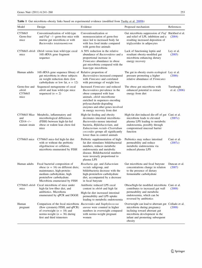
Table 1 Gut microbiota–obesity links based on experimental evidence (modified from Tuohy et al. 2009b)
Model Design Evidence Proposed mechanism References
C57bl6/J
Germ-free
mice
Conventionalization of wild-type
and Fiaf -/-germ-free mice with
murine gut microbiota or with
Bacteroides tetaiotaomicron
Conventionalization or
monoassociation of germ-free
mice led to increased body fat
with less food intake compared
with germ-free animals
Gut microbiota suppression of Fiafand relief of LPL inhibition and a
resulting increased deposition of
triglycerides in adipocytes
Backhed et al.
(2004)
C57bl6/J ob/obmice
Ob/ob versus lean wild-type cecal
16S rRNA gene fragment
sequence
A 50% reduction in the relative
abundance of Bacteroidetes and a
proportional increase in
Firmicutes abundance in obese
gut microbiota compared with the
lean-type microbiota
Lack of functioning leptin and
resultant obesity-modified gut
microbiota enhancing dietary
energy recovery
Ley et al.
(2005)
Human adults 16S rRNA gene sequence library of
gut microbiota in obese subjects
on weight reduction diets (low
carbohydrate or low fat, n = 12)
Relative proportion of
Bacteroidetes increased compared
with Fimicutes and correlated
with percentage of weight loss
The gut in obesity exerts ecological
pressure promoting a higher
relative abundance of Firmicutes
Ley et al.
(2006)
Germ-free and
ob/obC57bl6/J
mice
Sequenced metagenome of cecal
ob/ob and lean wild-type mice
sequenced (n = 2)
Increased Firmicutes and reduced
Bacteroidetes prevalence in the
obese compared with lean
animals. ob/ob microbiome
enriched in sequences encoding
polysaccharide-degrading
enzymes and other genes involved
in energy recovery from diet
The obese gut microbiota with
enhanced potential to extract
energy from diet
Turnbaugh
et al. (2006)
C57bl6/J Mice
and
CD14-/-
mutant strain
Metabolic, inflammatory and
microbiological differences
(FISH) between high-fat-fed
obese or rodent lean chow-fed
mice
High-fat feeding and obesity
decimates intestinal microbiota–
Bacteroides-mouse intestinal
bacteria, Bifidobacterium, and
Eubacterium rectale–Clostridiumcoccoides groups all significantly
lower than in control animals
High-fat diet-induced die-off of gut
microbiota leads to elevated
plasma LPS leading to metabolic
endotoxemia, possibly through
compromised mucosal barrier
function
Cani et al.
(2007c)
C57bl6/J mice C57bl6/J mice-fed high-fat diet
with or without the prebiotic
oligofructose or cellulose,
microbiota enumerated by FISH
Prebiotic supplementation of high-
fat diet stimulates bifidobacterial
numbers, reduces metabolic
endotoxemia and metabolic
disease. Bifidobacterial numbers
were inversely proportional to
plasma LPS
Prebiotics may reduce intestinal
permeability and reduce
metabolic endotoxemia via
reduced plasma LPS
Cani et al.
(2007c)
Human adults Fecal bacterial composition of
obese (n = 16) on different diets;
maintenance, high-protein–
medium carbohydrate, high
protein/low carbohydrate.
Microbiota enumerated by FISH
Roseburia spp. and Eubacteiumrectale subgroup, and
bifidobacteria decrease with the
high-protein/low-carbohydrate
diet, accompanied by a decrease
in fecal butyrate
Gut microbiota and fecal butyrate
concentrations change in relation
to the presence of dietary
fermentable carbohydrate
Duncan et al.
(2007)
C57bl6/J ob/obmice
Cecal microbiota of mice under
high-fat low-fiber diet, and
antibiotics. Microbiota
enumerated by qPCR and DGGE
Antibiotic reduced LPS cecal
content in ob/ob and high fat
High-fat diet increased intestinal
permeability and LPS uptake
leading to metabolic endotoxemia
Obese/high-fat modified microbiota
contributes to increased gut wall
permeability and metabolic
endotoxemia, which can be
reversed by antibiotics
Cani et al.
(2008)
Human
pregnant
Comparison of the fecal microbiota
(flow cytometry FISH, and qPCR)
of overweight (n = 18) and
normo-weight (n = 36) during
first and third trimesters
Bacteroides and Staphylococcusaureus were counted in higher
numbers in overweight compared
with normo-weight pregnant
women
Overweight can lead to aberrant gut
microbiota during pregnancy
inclining toward aberrant gut
microbiota development in the
infant and promoting subsequent
obesity
Collado et al.
(2008)
Genes Nutr (2011) 6:241–260 253
123
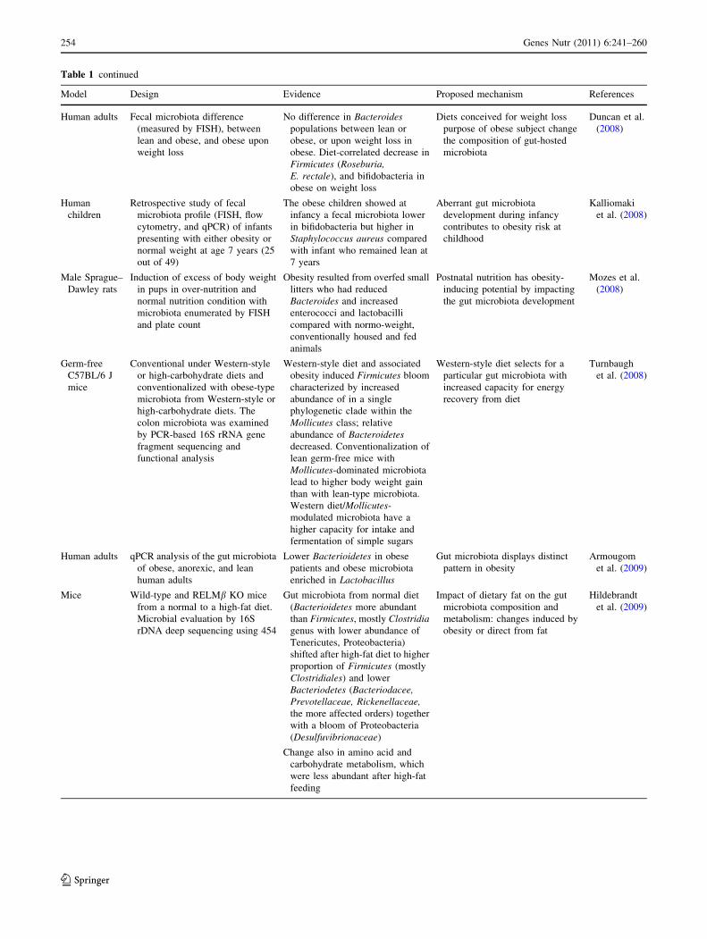
Table 1 continued
Model Design Evidence Proposed mechanism References
Human adults Fecal microbiota difference
(measured by FISH), between
lean and obese, and obese upon
weight loss
No difference in Bacteroidespopulations between lean or
obese, or upon weight loss in
obese. Diet-correlated decrease in
Firmicutes (Roseburia,E. rectale), and bifidobacteria in
obese on weight loss
Diets conceived for weight loss
purpose of obese subject change
the composition of gut-hosted
microbiota
Duncan et al.
(2008)
Human
children
Retrospective study of fecal
microbiota profile (FISH, flow
cytometry, and qPCR) of infants
presenting with either obesity or
normal weight at age 7 years (25
out of 49)
The obese children showed at
infancy a fecal microbiota lower
in bifidobacteria but higher in
Staphylococcus aureus compared
with infant who remained lean at
7 years
Aberrant gut microbiota
development during infancy
contributes to obesity risk at
childhood
Kalliomaki
et al. (2008)
Male Sprague–
Dawley rats
Induction of excess of body weight
in pups in over-nutrition and
normal nutrition condition with
microbiota enumerated by FISH
and plate count
Obesity resulted from overfed small
litters who had reduced
Bacteroides and increased
enterococci and lactobacilli
compared with normo-weight,
conventionally housed and fed
animals
Postnatal nutrition has obesity-
inducing potential by impacting
the gut microbiota development
Mozes et al.
(2008)
Germ-free
C57BL/6 J
mice
Conventional under Western-style
or high-carbohydrate diets and
conventionalized with obese-type
microbiota from Western-style or
high-carbohydrate diets. The
colon microbiota was examined
by PCR-based 16S rRNA gene
fragment sequencing and
functional analysis
Western-style diet and associated
obesity induced Firmicutes bloom
characterized by increased
abundance of in a single
phylogenetic clade within the
Mollicutes class; relative
abundance of Bacteroidetesdecreased. Conventionalization of
lean germ-free mice with
Mollicutes-dominated microbiota
lead to higher body weight gain
than with lean-type microbiota.
Western diet/Mollicutes-
modulated microbiota have a
higher capacity for intake and
fermentation of simple sugars
Western-style diet selects for a
particular gut microbiota with
increased capacity for energy
recovery from diet
Turnbaugh
et al. (2008)
Human adults qPCR analysis of the gut microbiota
of obese, anorexic, and lean
human adults
Lower Bacterioidetes in obese
patients and obese microbiota
enriched in Lactobacillus
Gut microbiota displays distinct
pattern in obesity
Armougom
et al. (2009)
Mice Wild-type and RELMb KO mice
from a normal to a high-fat diet.
Microbial evaluation by 16S
rDNA deep sequencing using 454
Gut microbiota from normal diet
(Bacterioidetes more abundant
than Firmicutes, mostly Clostridiagenus with lower abundance of
Tenericutes, Proteobacteria)
shifted after high-fat diet to higher
proportion of Firmicutes (mostly
Clostridiales) and lower
Bacteriodetes (Bacteriodacee,Prevotellaceae, Rickenellaceae,the more affected orders) together
with a bloom of Proteobacteria
(Desulfuvibrionaceae)
Change also in amino acid and
carbohydrate metabolism, which
were less abundant after high-fat
feeding
Impact of dietary fat on the gut
microbiota composition and
metabolism: changes induced by
obesity or direct from fat
Hildebrandt
et al. (2009)
254 Genes Nutr (2011) 6:241–260
123

Table 1 continued
Model Design Evidence Proposed mechanism References
Human
adolescents
Fecal microbiota (FISH and IgA-
coating bacteria) of obese
adolescents before and after
restricted calorie diet and physical
activity regime (n = 39,
10 weeks)
C. histolyticum, E. rectale-C.coccoides groups decreased count
with weight loss; Bacteroides-
Prevotella increased upon weight
loss of [4 kg;
IgA-coating bacteria decreased in
those who lost [6 kg
Potential link between diet, gut
microbiota, immunity, and host
metabolic processes involved in
obesity
Nadal et al.
(2009)
Human
adolescents
Fecal microbiota (qPCR) of
overweight adolescents (n = 36)
under calorie-restricted diet and
physical activity
Total bacterial were higher, as were
Bacterioides fragilis and
Clostridium leptum and
Bifidobacterium catenulatum in
the obese population in which
intervention was more effective.
C. coccoides, Lactobacillus,
Bifidobacterium, B. bifidus, and
B. breve were lower. The weight
loss dietary intervention affected
also B. longum
Differences in the gut microbiota
are correlated with a high
effective response to weight loss
inducing intervention
Santacruz
et al. (2009)
Humans adults Intestinal microbiota (qPCR) and
feces SCFA of lean (n = 30),
overweight (n = 35), and obese
(n = 33) humans
Higher proportion of Bacterioidetes
in overweight and obese.
Ruminococcus flavefacienssubgroup reduced in overweight
and obese Clostridium leptumgroup, Methanobrevibacter and
Bifidobacterium all reduced in
overweight and obese
Higher amount of SCFA in obese,
more propionate in overweight
and obese
SCFA concentration elevated in
obese feces with significant
differences in the composition of
gut microbiota between lean and
obese at phylum and sub-phylum
levels
Schwiertz
et al. (2010)
Human female
twins and
their mothers
Adult female monozygotic and
dizygotic twin pairs concordant
for obesity and their mothers
(n = 31, 23 and 46, respectively)
Gut microbiota described by 16s
rRNA gene sequencing
Lower abundance of Bacteroidetes,
but higher Actinobacteria (no
change in Firmicutes) in obese.
Reduced species diversity in
obese. Metabolic pathways and
functional genes altered in
obesity. Functional heterogeneity
associated with the relative
amount of Bacteroidetes.
Microbiota enriched in
Firmicutes/Actinobacteriaexhibited more less diverse
functions
Modulated functional microbiome
with metagenomic differences in
carbohydrate, lipid, and amino
acid metabolism
Turnbaugh
et al.
(2009a)
Zucker fa/fa,
fa/? and ?/
? rats
Cecal microbiota composition
(FISH and DGGE) in Zucker
genotypes on normal diet
correlated with 1H-NMR
metabolomics of urine and blood
Microbiota of obese fa/fa animals
distinct from non-obese
genotypes. Total bacteria,
bifidobacteria, lactobacilli, and
Atopobium species, all
significantly lower in obese ceca.
Distinct urine and plasma
metabolite profiles associated
with obesity and obese-type
microbiota
Gut microbiota of the Zucker
genetic model of obesity linked to
energy metabolism and obesity in
these animals
Waldram
et al. (2009)
Humans post-
gastric
bypass
(PGB)
surgery
Fecal microbiota from 3 lean, 3
morbidly obese, and 3 PGB
surgery patients upon weight loss
describe after PCR-based 16S
rRNA gene fragment sequencing
and qPCR for methanogens
Bacterioidetes (Prevotellaceae)more abundant in obese
Firmicutes dominant in lean and
obese but reduced in PGB, PGB
had increased
Gammaproteobacteria
Methanogenic functional group
elevated in obese
Increased methanogenesis,
enhancing fermentation through
relief of end-product inhibition
increases production of acetate
absorbed at the human gut
contributing to enhanced energy
recovery
Zhang et al.
(2009)
Genes Nutr (2011) 6:241–260 255
123

Table 1 continued
Model Design Evidence Proposed mechanism References
Human
children
Obese and non-obese Indian
children (11–14 years: n: 28)
fecal microbiota enumerated by
16S rRNA target by qPCR
Bacterioides-Prevotella,
Lactobacillus acidophilus,Eubacterium rectale were not
significantly different in obese
and non-obese. High number of
Faecalibacterium prausnitziicharacterized the obese children
Evident alteration of gut microbiota
in obese children
Balamurugan
et al. (2010)
Sprague–
Dawley rat
Obesity-prone (DIO-P) and
resistant (DIO-R) rats under high-
fat diet were examined for 16S
rRNA qPCR microbiota, TLR4,
and LPS
Reduced total bacterial count with
higher relative proportions of
Bacterioidales and Clostridialesafter high-fat feeding. Greater
abundance of Enterobacteriales in
DIO-P
Increased intestinal permeability
and plasma LPS upon high-fat
feeding
Change in microbiota induced by
high-fat diet and inflammation
development associated with
obesity
de La Serre
et al. (2010)
Germ-free
(GF) and
conventional
(CV) mice
Fecal microbiota and Fiaf/Angptl4in GF and CV male adult mice on
low-fat diet (LFD), a high-fat diet
(HFD), or a commercial Western
diet (WD)
Bacteroidetes relative amount was
lower in on both HFD and WD in
favor of Firmicutes. One species
of Firmicutes was predominated:
the Erysipelotrichaceae
Higher expression of Fiaf/Angptl4at intestinal level in HFD and WD
Concluded that the absence of gut
microbiota does not provide a
general protection from diet-
induced obesity, that intestinal
production of intestinal Fiaf/
Angptl4 and gut microbiota are
not linked together in fat storage.
Diet composition highly affects
gut microbiota
Fleissner
et al. (2010)
C57bl6/J ob/obmice
Feces of ob/ob (low-fat diet)
compared to wild type (low-fat
and high-fat diet HF) for their
metagenomic profile (16S rRNA
tags pyrosequencing) in relation
to low-fat and high-fat diet. SCFA
analysis
Firmicutes more abundant in ob/oband HF wild mice. Bacteriodessignificantly decrease in ob/ob.
Actinobacteria increased in ob/oband HF mice from 7 to 11 weeks.
Protobacteria decline in HF mice.
Bifidobacteria decline in ob/oband HF at 11 weeks
SCFA concentrations and fecal
energy content higher in obese at
week 7 but lower upon adaptation
to diet
High-fat diet may have more
influence gut microbiota
composition than host genotype.
Diet and obesity-induced changes
in microbiota energy harvesting
change upon adaptation to high-
fat diet
Murphy et al.
(2010)
Human
pregnant
Feces from overweight (n 16) and
normal weight (n 34) pregnant
women analyzed by qPCR,
together with monitoring of body
weight and biochemical
parameters from plasma
Overweight women had more
abundant fecal Enterobacteriacee,E. coli, and Staphylococcus and
lower counts of Bifidobacteriumand Bacterioidetes in comparison
with normal weight subjects. A.muciniphila and Bifidobacteriumwere higher in subjects with
normal weight gain, compared
with those with excessive weight
gain. C. leptum and
Staphylococcus counts correlated
with excessive weight gain.
Higher amount of folic acid and
Fe in normal weight subject
Gut microbiota associated with
body weight and body weight
gain, pregnancy important
metabolites, beneficial health
effect on woman and infant
Santacruz
et al. (2010)
Mice Wild-type and Apoa-I knocked out
mice intolerant to glucose, under
fat or normal diet, DGGE-DNA
fingerprint and 16S rRNA
pyrosequencing
Bifidobacteriacea disappeared after
high-fat diet and
Desulfovibrionacea prevailed in
the glucose impaired/obese group
High impact of diet on the
microbiota composition,
potentially capable to induce
metabolic syndrome
Zhang et al.
(2010)
256 Genes Nutr (2011) 6:241–260
123
whereas low body weight, preterm infants are more likely
to have higher body weight in later life.
However, the discussed mechanisms and their involve-
ment with the gut microbiota and obesity axis have to be
further explored, in particular to establish the role of
colonic fermentation in human energy homeostasis. The
combination of the metagenomic and metabolomic
approaches appears to offer a very powerful tool for the
elucidation of underlying metabolic interactions between
the gut microbiota and its host and ultimately to elucidate
the cause or effect relationship between the aberrant gut
microbiota in obesity and the diseases of obesity.
References
Agheli N, Kabir M, Berni-Canani S, Petitjean E, Boussairi A, Luo J,
Bornet F, Slama G, Rizkalla SW (1998) Plasma lipids and fatty
acid synthase activity are regulated by short-chain fructo-
oligosaccharides in sucrose-fed insulin-resistant rats. J Nutr
128(8):1283–1288
Amar J, Burcelin R, Ruidavets JB, Cani PD, Fauvel J, Alessi MC,
Chamontin B, Ferrieres J (2008) Energy intake is associated with
endotoxemia in apparently healthy men. Am J Clin Nut
87(5):1219–1223
Anderson RC, Cookson AL, McNabb WC, Kelly WJ, Roy NC (2010)
Lactobacillus plantarum DSM 2648 is a potential probiotic that
enhances intestinal barrier function. FEMS Microbiol Lett
309:184–192. doi:10.1111/j.1574-6968.2010.02038.x
Armougom F, Henry M, Vialettes B, Raccah D, Raoult D (2009)
Monitoring bacterial community of human gut microbiota
reveals an increase in Lactobacillus in obese patients and
methanogens in anorexic patients. PLOS ONE 4(9):art. no.
e7125
Ayyad C, Andersen T (2000) Long-term efficacy of dietary treatment
of obesity: a systematic review of studies published between
1931 and 1999. Obes Rev 1(2):113–119. doi:10.1046/j.1467-
789x.2000.00019.x
Backhed F (2010) Addressing the gut microbiome and implications
for obesity. Int Dairy J 20(4): 259–261. Sp. Iss. SI. doi:
10.1016/j.idairyj.2009.11.012
Backhed F, Ding H, Wang T, Hooper LV, Koh GY, Nagy A,
Semenkovich CF, Gordon JI (2004) The gut microbiota as an
environmental factor that regulates fat storage. Proc Natl Acad
Sci USA 101(44):15718–15723. doi:10.1073/pnas.0407076101
Backhed F, Manchester JK, Semenkovich CF, Gordon JI (2007)
Mechanisms underlying the resistance to diet-induced obesity in
germ-free mice. Proc Natl Acad Sci USA 104(3):979–984. doi:
10.1073/pnas.0605374104
Balamurugan R, George G, Kabeerdoss J, Hepsiba J, Chandragun-
asekaran AMS, Ramakrishna BS (2010) Quantitative differences
in intestinal Faecalibacterium prausnitzii in obese Indian
children. Br J Nutr 103(3):335–338. doi:10.1017/S000711450
9992182
Baldini M, Pasqui F, Bordoni A, Maranesi M (2009) Is the
Mediterranean lifestyle still a reality? Evaluation of food
consumption and energy expenditure in Italian and Spanish
university students. Public Health Nutr 12(2):148–155 [Epub
2008 May 27]
Barbara G (2006) Mucosal barrier defects in irritable bowel
syndrome. Who left the door open? Am J Gastroenterol
101(6):1295–1298. doi:10.1111/j.1572-0241.2006.00667.x
Bates JM, Mittge E, Kuhlman J, Baden KN, Cheesman SE, Guillemin
K (2006) Distinct signals from the microbiota promote different
aspects of zebrafish gut differentiation. Dev Biol 297(2):
374–386. doi:10.1016/j.ydbio.2006.05.006
Brighenti F (2007) Dietary fructans and serum triacylglycerols: a meta-
analysis of randomized controlled trials. J Nutr 137(11 Suppl):
2552S–2556S
Brinkworth GD, Noakes M, Clifton PM, Bird AR (2009) Comparative
effects of very low-carbohydrate, high-fat and high-carbohy-
drate, low-fat weight-loss diets on bowel habit and faecal
short-chain fatty acids and bacterial populations. Br J Nutr
101(10):1493–1502. doi:10.1017/S0007114508094658
Cani PD, Amar J, Iglesias MA, Poggi M, Knauf C, Bastelica D,
Neyrinck AM, Fava F, Tuohy KM, Chabo C, Waget A, Delmee
E, Cousin B, Sulpice T, Chamontin B, Ferrieres J, Tanti JF,
Gibson GR, Casteilla L, Delzenne NM, Alessi MC, Burcelin R
(2007a) Metabolic endotoxemia initiates obesity and insulin
resistance. Diabetes 56(7):1761–1772. doi:10.2337/db06-1491
Cani PD, Hoste S, Guiot Y, Delzenne NM (2007b) Dietary non-
digestible carbohydrates promote L-cell differentiation in the
proximal colon of rats. Br J Nutr 98(1):32–37. doi:10.1017/
S0007114507691648
Cani PD, Neyrinck AM, Fava F, Knauf C, Burcelin RG, Tuohy KM,
Gibson GR, Delzenne NM (2007c) Selective increases of
bifidobacteria in gut microflora improve high-fat-diet-induced
diabetes in mice through a mechanism associated with endotox-
aemia. Diabetologia 50(11):2374–2383. doi:10.1007/s00125-
007-0791-0
Cani PD, Bibiloni R, Knauf C, Neyrinck AM, Neyrinck AM,
Delzenne NM, Burcelin R (2008) Changes in gut microbiota
control metabolic endotoxemia-induced inflammation in high-fat
diet-induced obesity and diabetes in mice. Diabetes 57(6):1470–
1481. doi:10.2337/db07-1403
Cani PD, Possemiers S, Van de Wiele T, Guiot Y, Everard A, Rottier
O, Geurts L, Naslain D, Neyrinck A, Lambert DM, Muccioli
GG, Delzenne NM (2009) Changes in gut microbiota control
inflammation in obese mice through a mechanism involving
GLP-2-driven improvement of gut permeability. Gut 58(8):
1091–1103. doi:10.1136/gut.2008.165886
Celi F, Bini V, De Giorgi G, Molinari D, Faraoni F, Di Stefano G,
Bacosi ML, Berioli MG, Contessa G, Falorni A (2003)
Epidemiology of overweight and obesity among school children
and adolescents in three provinces of central Italy, 1993–2001:
study of potential influencing variables. Eur J Clin Nutr
57(9):1045–1051. doi:10.1038/sj.ejcn.1601640
Chen KT, Malo MS, Beasley-Topliffe LK, Poelstra K, Millan JL,
Mostafa G, Alam SN, Ramasamy S, Warren HS, Hohmann EL,
Hodin RA (2011) A role for intestinal alkaline phosphatase in
the maintenance of local gut immunity. Dig Dis Sci
56:1020–1027. doi:10.1007/s10620-010-1396-x
Cherbut C, Ferrier L, Roze C et al (1998) Short-chain fatty acids
modify colonic motility through nerves and polypeptide YY
release in the rat. Am J Physiol 275:G1415–G1422
Collado MC, Isolauri E, Laitinen K, Salminen S (2008) Distinct
composition of gut microbiota during pregnancy in overweight
and normal-weight women. Am J Clin Nut 88:894–899
Cuche G, Cuber JC, Malbert CH (2000) Ileal short-chain fatty acids
inhibit gastric motility by a humoral pathway. Am J Physiol
Gastrointest Liver Physiol 279:G925–G930
Cummings JH, Antoine JM, Azpiroz F, Bourdet-Sicard R, Brandtzaeg
P, Calder PC, Gibson GR, Guarner F, Isolauri E, Pannemans D,
Shortt C, Tuijtelaars S, Watzl B (2004) PASSCLAIM—gut
health and immunity. Eur J Nutr 43(Suppl 2):II118–II173. doi:
10.1007/s00394-004-1205-4
Dankert J, Zijlstra J, Wolthers B (1981) Volatile fatty acids in human
peripheral and portal blood: quantitative determination, vacuum
Genes Nutr (2011) 6:241–260 257
123

distillation, and gas chromatography. Clin Chim Acta
110:301–307. doi:10.1016/0009-8981(81)90359-4
Darzi J, Frost GS, Robertson MD (2011) Do SCFA have a role in
appetite regulation? Proc Nutr Soc 70:119–128. doi:10.1017/
S0029665110004039
Daubioul C, Rousseau N, Demeure R, Gallez B, Taper H, Declerck B,
Delzenne N (2002) Dietary fructans, but not cellulose, decrease
triglyceride accumulation in the liver of obese Zucker fa/fa rats.
J Nutr 132:967–973
De Filippo C, Cavalieri D, Di Paola M, Ramazzotti M, Poullet JB,
Massart S, Collini S, Pieraccini G, Lionetti P (2010) Impact off
diet in shaping gut microbiota revealed by a comparative study
in children from Europe and rural Africa. Proc Natl Acad Sci
USA 17:14691–14696. doi:10.1073/pnas.1005963107
de La Serre CB, Ellis CL, Lee J, Hartman AL, Rutledge JC, Raybould
HE (2010) Propensity to high-fat diet-induced obesity in rats is
associated with changes in the gut microbiota and gut inflam-
mation. Am J Physiol Gastrointest Liver Physiol 299:G440–
G448. doi:10.1152/ajpgi.00098.2010
di Giuseppe R, Bonanni A, Olivieri M, Di Castelnuovo A, Donati
MB, de Gaetano G, Cerletti C, Iacoviello L (2008) Adherence to
Mediterranean diet and anthropometric and metabolic parame-
ters in an observational study in the ‘Alto Molise’ region: the
MOLI-SAL project. Nutr Metab Cardiovasc Dis 18:415–421.
doi:10.1016/j.numecd.2007.05.010
Drucker DJ (1999) Glucagon-like peptide 2. Trends Endocrinol
Metab 10:153–156. doi:10.1016/S1043-2760(98)00136-2
Dumas ME, Barton RH, Toye A, Cloarec O, Blancher C, Rothwell A,
Fearnside J, Tatoud R, Blanc V, Lindon JC, Mitchell SC,
Holmes E, McCarthy MI, Scott J, Gauguier D, Nicholson JK
(2006) Metabolic profiling reveals a contribution of gut micro-
biota to fatty liver phenotype in insulin-resistant mice. Proc Natl
Acad Sci USA 103:12511–12516. doi:10.1073/pnas.0601056103
Duncan SH, Belenguer A, Holtrop G, Johnstone AM, Flint HJ, Lobley
GE (2007) Reduced dietary intake of carbohydrates by obese
subjects results in decreased concentrations of butyrate and
butyrate-producing bacteria in feces. Appl Environ Microbiol
73:1073–1078. doi:10.1128/AEM.02340-06
Duncan SH, Lobley GE, Holtrop G, Ince J, Johnstone AM, Louis P,
Flint HJ (2008) Human colonic microbiota associated with diet,
obesity and weight loss. Int J Obes 32:1720–1724. doi:10.1038/
ijo.2008.155
Fava F, Lovegrove JA, Gitau R, Jackson KG, Tuohy KM (2006) The
gut microbiota and lipid metabolism: Implications for human
health and coronary heart disease. Curr Med Chem 13:3005–
3021. doi:10.1021/jf101475m
Favier ML, Moundras C, Demigne C, Remesy C (1995) Fermentable
carbohydrates exert a more potent cholesterol-lowering effect
than cholestyramine. Biochim Biophys Acta 14;1258(2):115–
121
Fleissner CK, Huebel N, El-Bary MMA, Loh G, Klaus S, Blaut M
(2010) Absence of intestinal microbiota does not protect mice
from diet-induced obesity. Br J Nutr 104:919–929. doi:
10.1017/S0007114510001303
Fukasawa T, Kamei A, Watanabe Y, Koga J, Abe K (2010) Short-
chain fructooligosaccharide regulates hepatic peroxisome pro-
liferator-activated receptor alpha and farnesoid 9 receptor target
gene expression in rats. J Agric Food Chem 58:7007–7012. doi:
10.1021/jf1006616
Gao ZG, Yin J, Zhang J, Ward RE, Martin RJ, Lefevre M, Cefalu
WT, Ye JP (2009) Butyrate improves insulin sensitivity and
increases energy expenditure in mice. Diabetes 58:1509–1517.
doi:10.2337/db08-1637
Ghoshal S, Witta J, Zhong J, de Villiers W, Eckhardt E (2009)
Chylomicrons promote intestinal absorption of lipopolysaccha-
rides. J Lipid Res 50:90–97. doi:10.1194/jlr.M800156-JLR200
Guttman JA, Samji FN, Li Y, Vogl AW, Finlay BB (2006) Evidence
that tight junctions are disrupted due to intimate bacterial contact
and not inflammation during attaching and effacing pathogen
infection in vivo. Infect Immun 74:6075–6084. doi:10.1128/
IAI.00721-06
Hildebrandt MA, Hoffmann C, Hamady M, Chen YY, Knight R,
Bushman FD, Ahima RS, Wu GD (2009) High fat diet
determines the composition of the gut microbiome independent
of host genotype and phenotype. Gastroenterology 136:A102–
A102, Suppl. 1
Hooper LV, Wong MH, Thelin A, Hansson L, Falk PG, Gordon JI
(2001) Molecular analysis of commensal host-microbial rela-
tionships in the intestine. Science 291(5505):881–884. doi:
10.1126/science.291.5505.881
Hotamisligil GS, Erbay E (2008) Nutrient sensing and inflammation
in metabolic diseases. Nat Rev Immunol 8:923–934. doi:
10.1038/nri2449
Hotamisligil GS, Peraldi P, Budavari A, Ellis R, White MF,
Spiegelman BM (1996) IRS-1-mediated inhibition of insulin
receptor tyrosine kinase activity in TNF-alpha- and obesity-
induced insulin resistance. Science 271(5249):665–668. doi:
10.1126/science.271.5249.665
Kadooka Y, Sato M, Imaizumi K, Ogawa A, Ikuyama K, Akai Y,
Okano M, Kagoshima M, Tsuchida T (2010) Regulation of
abdominal adiposity by probiotics (Lactobacillus gasseriSBT2055) in adults with obese tendencies in a randomized
controlled trial. Eur J Clin Nutr 64:636–643. doi:10.1038/
ejcn.2010.19
Kalliomaki M, Collado MC, Salminen S, Isolauri E (2008) Early
differences in fecal microbiota composition in children may
predict overweight. Am J Clin Nutr 87:534–538
Kaway T, Akira S (2006) TLR signaling. Cell Death Differ
13:816–825. doi:10.1038/sj.cdd.4401850
Keim NL, Blanton CA, Kretsch MJ (2004) America’s obesity
epidemic: measuring physical activity to promote an active
lifestyle. J Am Diet Assoc 104:1398–1409. doi:10.1016/j.jada.
2004.06.005
Kondo T, Kishi M, Fushimi T, Kaga T (2009) Acetic acid upregulates
the expression of genes for fatty acid oxidation enzymes in liver
to suppress body fat accumulation. J Agric Food Chem
57:5982–5986. doi:10.1021/jf900470c
Lee HY, Park JH, Seok SH, Baek MW, Kim DJ, Lee KE, Paek KS,
Lee Y, Park JH (2006) Human originated bacteria, Lactobacillus
rhamnosus PL60, produce conjugated linoleic acid and show
anti-obesity effects in diet-induced obese mice. Biochim Bio-
phys Acta 1761:736–744. doi:10.1016/j.bbalip.2006.05.007
Ley RE (2010) Obesity and the human microbiome. Curr Opin
Gastroenterol 26:5–11. doi:10.1097/MOG.0b013e328333d751
Ley RE, Backhed F, Turnbaugh P, Lozupone CA, Knight RD, Gordon
JI (2005) Obesity alters gut microbial ecology. Proc Natl Acad
Sci USA 102(31):11070–11075. doi:10.1073/pnas.0504978102
Ley RE, Turnbaugh PJ, Klein S, Gordon JI (2006) Microbial
ecology—human gut microbes associated with obesity. Nature
444(7122):1022–1023. doi:10.1038/nature4441022a
Lieberman LS (2003) Dietary, evolutionary, and modernizing influ-
ences on the prevalence of type 2 diabetes. Annu Rev Nutr
23:345–377. doi:10.1146/annurev.nutr.23.011702.073212
Luoto R, Kalliomaki M, Laitinen K, Isolauri E (2010) The impact of
perinatal probiotic intervention on the development of over-
weight and obesity: follow-up study from birth to 10 years. Int J
Obes 34:1531–1537. doi:10.1038/ijo.2010.50
Macfarlane GT, Gibson GR (1997) Carbohydrate fermentation,
energy transduction and gas metabolism in the human large
intestine. In: Mackie RI, White BA (eds) Gastrointestinal
microbiology, vol 1: gastrointestinal ecosystems and fermenta-
tions. Chapman and Hall, New York, pp 269–318
258 Genes Nutr (2011) 6:241–260
123
Maes M (2008) The cytokine hypothesis of depression: inflammation,
oxidative & nitrosative stress (IO&NS) and leaky gut as new
targets for adjunctive treatments in depression. Neuro Endocri-
nol Lett 29:287–291
Maes M, Leunis JC (2008) Normalization of leaky gut in chronic
fatigue syndrome (CFS) is accompanied by a clinical improve-
ment: effects of age, duration of illness and the translocation of
LPS from gram-negative bacteria. Neuro Endocrinol Lett
29:902–910
Malo MS, Biswas S, Abedrapo MA, Yeh L, Chen A, Hodin RA
(2006) The pro-inflammatory cytokines, IL-1beta and TNF-
alpha, inhibit intestinal alkaline phosphatase gene expression.
DNA Cell Biol 25:684–695
Meijer K, de Vos P, Priebe MG (2010) Butyrate and other short-chain
fatty acids as modulators of immunity: what relevance for
health? Curr Opin Clin Nutr Metab Care 13:715–721. doi:
10.1097/MCO.0b013e32833eebe5
Mozes S, Bujnakova D, Sefcikova Z, Kmet V (2008) Intestinal
microflora and obesity in rats. Folia Microbiol 53:225–228
Murphy EF, Cotter PD, Healy S, Marques TM, O’Sullivan O, Fouhy
F, Clarke SF, O’Toole PW, Quigley EM, Stanton C, Ross PR,
O’Doherty RM, Shanahan F (2010) Composition and energy
harvesting capacity of the gut microbiota: relationship to diet,
obesity and time in mouse models. Gut 59:1635–1642. doi:
10.1136/gut.2010.215665
Nadal I, Santacruz A, Marcos A, Warnberg J, Garagorri M, Moreno
LA, Martin-Matillas M, Campoy C, Marti A, Moleres A,
Delgado M, Veiga OL, Garcia-Fuentes M, Redondo CG, Sanz Y
(2009) Shifts in clostridia, bacteroides and immunoglobulin-
coating fecal bacteria associated with weight loss in obese
adolescents. Int J Obes 33:758–767. doi:10.1038/ijo.2008.260
Nagao K, Inoue N, Wang YM, Shirouchi B, Yanagita T (2005)
Dietary conjugated linoleic acid alleviates nonalcoholic fatty
liver disease in Zucker (fa/fa) rats. J Nutr 135:9–13
Novotny R, Williams AE, Vinoya AC, Oshiro CE, Vogt TM (2009)
US acculturation, food intake, and obesity among Asian–Pacific
hotel workers. J Am Diet Assoc 109:1712–1718. doi:10.1016/
j.jada.2009.07.013
O’Dea K (1991) Westernisation, insulin resistance and diabetes in
Australian aborigines. Med J Aust 155:258–264. doi:10.2337/
diacare.24.5.811
OECD (2010) Obesity and the economics of prevention: fit not fat.
OECD Publishing. doi: 10.1787/9789264084865-en
Parnell JA, Reimer RA (2009) Weight loss during oligofructose
supplementation is associated with decreased ghrelin and
increased peptide YY in overweight and obese adults. Am J
Clin Nut 89:1751–1759. doi:10.3945/ajcn.2009.27465
Parnell JA, Reimer RA (2010) Effect of prebiotic fibre supplemen-
tation on hepatic gene expression and serum lipids: a dose-
response study in JCR: LA-cp rats. Br J Nutr 103:1577–1584.
doi:10.1017/S0007114509993539
Peters S, Pomare E, Fisher C (1992) Portal and peripheral blood short-
chain fatty acid concentrations after caecal lactulose instillation
at surgery. Gut 33:1249–1252. doi:10.1136/gut.33.9.1249
Pi-Sunyer X (2009) The medical risks of obesity. Postgrad Med
121:21–33. doi:10.3810/pgm.2009.11.2074
Putaala H, Salusjarvi T, Nordstrom M, Saarinen M, Ouwehand AC,
Hansen EB, Rautonen N (2008) Effect of four probiotic strains
and Escherichia coli O157:H7 on tight junction integrity and
cyclo-oxygenase expression. Res Microbiol 159:692–698. doi:
10.1016/j.resmic.2008.08.002
Ravussin E, Valencia ME, Esparza J, Bennett PH, Schulz LO (1994)
Effects of a traditional lifestyle on obesity in Pima Indians.
Diabetes Care 17:1067–1074
Reigstad CS, Lunden GO, Felin J, Backhed F (2009) Regulation of
serum amyloid A3 (SAA3) in mouse colonic epithelium and
adipose tissue by the intestinal microbiota. PLos One 4:art. no.
e5842. doi: 10.1371/journal.pone.0005842
Samuel BS, Gordon JI (2006) A humanized gnotobiotic mouse model
of host-archaeal-bacterial mutualism. Proc Natl Acad Sci USA
103:10011–10016. doi:10.1073/pnas.0602187103
Sandek A, Rauchhaus M, Anker SD, von Haehling S (2008) The
emerging role of the gut in chronic heart failure. Curr Opin Clin
Nutr Metab Care 11:632–639
Santacruz A, Marcos A, Warnberg J, Marti A, Martin-Matillas M,
Campoy C, Moreno LA, Veiga O, Redondo-Figuero C, Garag-
orri JM, Azcona C, Delgado M, Garcia-Fuentes M, Collado MC,
Sanz Y (2009) EVASYON study Grp interplay between weight
loss and gut microbiota composition in overweight adolescents.
Obesity 17:1906–1915. doi:10.1038/oby.2009.112
Santacruz A, Collado MC, Garcia-Valdes L, Segura MT, Martin-
Lagos JA, Anjos T, Marti-Romero M, Lopez RM, Florido J,
Campoy C, Sanz Y (2010) Gut microbiota composition is
associated with body weight, weight gain and biochemical
parameters in pregnant women. Br J Nutr 104:83–92. doi:
10.1017/S0007114510000176
Schwiertz A, Taras D, Schafer K et al (2010) Microbiota and SCFA in
lean and overweight healthy subjects. Obesity 18:190–195. doi:
10.1038/oby.2009.167
So PW, Yu WS, Kuo YT, Wasserfall C, Goldstone AP, Bell JD, Frost
G (2007) Impact of resistant starch on body fat patterning and
central appetite regulation. PLoS One 2:e1309. doi:10.1371/
journal.pone.0001309
Sotos M, Nadal I, Marti A et al (2008) Gut microbes and obesity in
adolescents. Proc Nutr Soc 67:E20
Spalding KL, Arner E, Westmark PO et al (2008) Dynamics of fat cell
turnover in humans. Nature 453:783–787. doi:10.1038/nature
06902
Stappenbeck TS, Hooper LV, Gordon JI (2002) Developmental
regulation of intestinal angiogenesis by indigenous microbes via
Paneth cells. Proc Natl Acad Sci USA 99:15451–15455. doi:
10.1073/pnas.202604299
Stevenson BR, Siliciano JD, Mooseker MS, Goodenough DA (1986)
Identification of ZO-1: a high molecular weight polypeptide
associated with the tight junction (zonula occludens) in a variety
of epithelia. J Cell Biol 103:755–766
Stoddart LA, Smith NJ, Milligan G (2008) International Union of
Pharmacology. LXXI. Free fatty acid receptors FFA-1, -2, and -3:
pharmacology and pathophysiological functions. Pharmacol Rev
60:405–417. doi:10.1124/pr.108.00802
Tsukita S (2001) Claudins and the tight junction barrier. Kidney Int
60:407
Tuohy KM, Gougoulias C, Shen Q, Walton G, Fava F, Ramnani P
(2009a) Studying the human gut microbiota in the trans-omics
era—focus on metagenomics and metabonomics. Curr Pharm
Des 15:1415–1427. doi:10.2174/138161209788168182
Tuohy KM, Costabile A, Fava F (2009b) The gut microbiota in
obesity and metabolic disease—a novel therapeutic target. Nutr
Ther Metab 27:113–133
Tuohy KM, Brown DT, Klinder A, Costabile A (2010) Shaping the
human microbiome with prebiotic foods—current perspectives
for continued development. Food Sci Tech Bull Funct Foods
7:49–64. doi:10.1616/1476-2137.15989
Turnbaugh PJ, Gordon JI (2009) The core gut microbiome, energy
balance and obesity. J Physiol Lond 587:4153–4158. doi:
10.1113/jphysiol.2009.174136
Turnbaugh PJ, Ley RE, Mahowald MA, Magrini V, Mardis ER,
Gordon JI (2006) An obesity-associated gut microbiome with
increased capacity for energy harvest. Nature 444(7122):1027–
1031. doi:10.1038/nature05414
Turnbaugh PJ, Baeckhed F, Fulton L, Gordon JI (2008) Diet-induced
obesity is linked to marked but reversible alterations in the
Genes Nutr (2011) 6:241–260 259
123
mouse distal gut microbiome. Cell Host Microbe 3:213–223. doi:
10.1016/j.chom.2008.02.015
Turnbaugh PJ, Hamady M, Yatsunenko T, Cantarel BL, Duncan A,
Ley RE, Sogin ML, Jones WJ, Roe BA, Affourtit JP, Egholm M,
Henrissat B, Heath AC, Knight R, Gordon JI (2009a) A core gut
microbiome in obese and lean twins. Nature 457(7228):480–484.
doi:10.1038/nature07540
Turnbaugh PJ, Ridaura VK, Faith JJ, Rey FE, Knight R, Gordon JI
(2009b) The effect of diet on the human gut microbiome: a
metagenomic analysis in humanized gnotobiotic mice. Sci Transl
Med 1(6): art. no. 6ra14. doi: 10.1126/scitranslmed.3000322
Ukena SN, Singh A, Dringenberg U, Engelhardt R, Seidler U, Hansen
W et al (2007) Probiotic Escherichia coli Nissle 1917 inhibits
leaky gut by enhancing mucosal integrity. PLoS ONE 2:e1308.
doi:10.1074/jbc.M202849200
Uribe A, Alam M, Johansson O et al (1994) Microflora modulates
endocrine cells in the gastrointestinal mucosa of the rat.
Gastroenterology 107:1259–1269
Vaarala O, Atkinson MA, Neu J (2008) The ‘‘perfect storm’’ for type
1 diabetes: the complex interplay between intestinal microbiota,
gut permeability, and mucosal immunity. Diabetes 57:2555–
2562. doi:10.2337/db08-0331
Venkatraman A, Ramakrishna BS, Pulimood AB (1999) Butyrate
hastens restoration of barrier function after thermal and detergent
injury to rat distal colon in vitro. Scand J Gastroenterol
34:1087–1092
Vogt JA, Ishii-Schrade KB, Pencharz PB, Wolever TMS (2004)
L-Rhamnose increases serum propionate after long-term supple-
mentation, but lactulose does not raise serum acetate. Am J Clin
Nut 80:1254–1261
Waldram A, Holmes E, Wang YL, Rantalainen M, Wilson ID, Tuohy
KM, McCartney AL, Gibson GR, Nicholson JK (2009) Top-
down systems biology modeling of host metabotype-microbiome
associations in obese rodents. J Proteome Res 8:2361–2375. doi:
10.1021/pr8009885
Wall R, Ross RP, Shanahan F, O’Mahony L, O’Mahony C, Coakley
M, Hart O, Lawlor P, Quigley EM, Kiely B, Fitzgerald GF,
Stanton C (2009) Metabolic activity of the enteric microbiota
influences the fatty acid composition of murine and porcine liver
and adipose tissues. Am J Clin Nut 89:1393–1401. doi:10.3945/
ajcn.2008.27023
Willcox DC, Willcox BJ, Todoriki H, Suzuki M (2009) The
Okinawan diet: health implications of a low-calorie, nutrient-
dense, antioxidant-rich dietary pattern low in glycemic load.
J Am Coll Nutr 28(Suppl):500S–516S
Williams DE, Knowler WC, Smith CJ, Hanson RL, Roumain J,
Saremi A, Kriska AM, Bennett PH, Nelson RG (2001) The effect
of Indian or Anglo dietary preference on the incidence of
diabetes in Pima Indians. Diabetes Care 24:811–816
Xiong Y, Miyamoto N, Shibata K et al (2004) Short-chain fatty acids
stimulate leptin production in adipocytes through the G protein-
coupled receptor GPR41. Proc Natl Acad Sci USA 101:1045–
1050. doi:10.1073/pnas.2637002100
Yang RZ, Lee MJ, Hu H, Pollin TI, Ryan AS, Nicklas BJ, Snitker S,
Horenstein RB, Hull K, Goldberg NH, Goldberg AP, Shuldiner
AR, Fried SK, Gong DW (2006) Acute-phase serum amyloid A:
an inflammatory adipokine and potential link between obesity
and its metabolic complications. PLos Med 3:884–894. doi:
10.1371/journal.pmed.0030287
Yin YN, Yu QF, Fu N, Liu XW, Lu FG (2010) Effects of four
Bifidobacteria on obesity in high-fat diet induced rats. World J
Gastroenterol 16:3394–3401. doi:10.3748/wjg.v16.i27.3394
Zhang HS, DiBaise JK, Zuccolo A, Kudrna D, Braidotti M, Yu YS,
Parameswaran P, Crowell MD, Wing R, Rittmann BE,
Krajmalnik-Brown R (2009) Human gut microbiota in obesity
and after gastric bypass. Proc Natl Acad Sci USA 106:2365–
2370. doi:10.1073/pnas.0812600106
Zhang CH, Zhang MH, Wang SY, Han RJ, Cao YF, Hua WY, Mao
YJ, Zhang XJ, Pang XY, Wei CC, Zhao GP, Chen Y, Zhao LP
(2010) Interactions between gut microbiota, host genetics and
diet relevant to development of metabolic syndromes in mice.
ISME J 4:232–241. doi:10.1038/ismej.2009.112
260 Genes Nutr (2011) 6:241–260
123
Related Documents

![Profile of the gut microbiota of adults with obesity: a ... · obesity can maximize the weight loss produced by a per-sonalized diet [58]. It is undeniable that the gut microbiota](https://static.cupdf.com/doc/110x72/5fcdfd7ea7419c7d56295d97/profile-of-the-gut-microbiota-of-adults-with-obesity-a-obesity-can-maximize.jpg)