CLINICAL STUDY – PATIENT STUDY O 6 -methylguanine DNA methyltransferase status determined by promoter methylation and immunohistochemistry in gliosarcoma and their clinical implications Shin-Hyuk Kang • Kyung-Jae Park • Chae-Yong Kim • Mi Ok Yu • Chul-Kee Park • Sung-Hye Park • Yong-Gu Chung Received: 5 January 2010 / Accepted: 30 May 2010 Ó Springer Science+Business Media, LLC. 2010 Abstract O 6 -methylguanine-DNA methyltransferase (MGMT) is known as a DNA repair protein, and loss of function in MGMT is related to an increase in survival in patients with malignant gliomas treated with alkylating agents. In the present study, we determined the status of MGMT using methylation-specific polymerase chain reac- tion (PCR) and immunohistochemistry on paraffin-embed- ded specimens in 12 human gliosarcomas, and these results were then related to overall survival (OS) and response to alkylating agents. The MGMT promoter was methylated in six patients. Immunostaining of MGMT was positive in 58.3% of patients. MGMT methylation status was correlated with immunostaining results in five patients (41.7%). The median OS and progression-free survival (PFS) of the whole population were 13.4 months [95% confidence interval (CI), 12.3–14.5 months] and 8.3 months (95% CI, 7.4–9.2 months), respectively. In patients with methylated MGMT promoter, median OS was 15.0 months, compared with 11.3 months in the unmethy- lated group. Median PFS of gliosarcoma patients was 10.3 months for the methylated group, whereas it was 7.3 months for the unmethylated group. On multivariate analysis, patients with methylated MGMT promoter had better prognosis than patients with unmethylated MGMT promoter with respect to OS and PFS (P = 0.045 and 0.034, respectively). However, there was no statistical significance between MGMT protein expression and survival. The results show that a significant fraction of gliosarcomas have MGMT promoter methylation and protein expression, and suggest that patient survival is associated with MGMT methylation status. Keywords Gliosarcoma Immunohistochemistry Methylation-specific PCR O 6 -methylguanine-DNA methyltransferase Introduction Gliosarcomas are rare primary brain tumors characterized by a biphasic pattern with glial and mesenchymal compo- nents [1]. Known as a variant of glioblastoma multiforme (GBM) [2], gliosarcomas have often been treated in the same manner as GBM, which consists of radical resection, followed by radiotherapy and adjuvant chemotherapy [3–5]. Alkylating agents are the most effective cytotoxic agents in malignant gliomas. The cytotoxic mechanisms of S.-H. Kang and K.-J. Park contributed equally to this work. S.-H. Kang K.-J. Park M. O. Yu Y.-G. Chung Department of Neurosurgery, Korea University Anam Hospital, Korea University College of Medicine, #126-1, 5-Ga, Anam-dong, Seongbuk-gu, Seoul 136-705, Korea C.-Y. Kim (&) Department of Neurosurgery, Seoul National University Bundang Hospital, Seoul National University College of Medicine, 166 Gumi-ro, Bundang-gu, Seongnam-si, Gyeonggi-do 463-707, Korea e-mail: [email protected] C.-K. Park Department of Neurosurgery, Seoul National University Hospital, Seoul National University College of Medicine, Seoul, Korea M. O. Yu School of Life Sciences and Biotechnology, Korea University, Seoul, Korea S.-H. Park Department of Pathology, Seoul National University College of Medicine, Seoul, Korea 123 J Neurooncol DOI 10.1007/s11060-010-0267-9

Welcome message from author
This document is posted to help you gain knowledge. Please leave a comment to let me know what you think about it! Share it to your friends and learn new things together.
Transcript
CLINICAL STUDY – PATIENT STUDY
O6-methylguanine DNA methyltransferase status determinedby promoter methylation and immunohistochemistryin gliosarcoma and their clinical implications
Shin-Hyuk Kang • Kyung-Jae Park •
Chae-Yong Kim • Mi Ok Yu • Chul-Kee Park •
Sung-Hye Park • Yong-Gu Chung
Received: 5 January 2010 / Accepted: 30 May 2010
� Springer Science+Business Media, LLC. 2010
Abstract O6-methylguanine-DNA methyltransferase
(MGMT) is known as a DNA repair protein, and loss of
function in MGMT is related to an increase in survival in
patients with malignant gliomas treated with alkylating
agents. In the present study, we determined the status of
MGMT using methylation-specific polymerase chain reac-
tion (PCR) and immunohistochemistry on paraffin-embed-
ded specimens in 12 human gliosarcomas, and these results
were then related to overall survival (OS) and response to
alkylating agents. The MGMT promoter was methylated in
six patients. Immunostaining of MGMT was positive in
58.3% of patients. MGMT methylation status was
correlated with immunostaining results in five patients
(41.7%). The median OS and progression-free survival
(PFS) of the whole population were 13.4 months [95%
confidence interval (CI), 12.3–14.5 months] and
8.3 months (95% CI, 7.4–9.2 months), respectively. In
patients with methylated MGMT promoter, median OS was
15.0 months, compared with 11.3 months in the unmethy-
lated group. Median PFS of gliosarcoma patients was
10.3 months for the methylated group, whereas it was
7.3 months for the unmethylated group. On multivariate
analysis, patients with methylated MGMT promoter had
better prognosis than patients with unmethylated MGMT
promoter with respect to OS and PFS (P = 0.045 and 0.034,
respectively). However, there was no statistical significance
between MGMT protein expression and survival. The
results show that a significant fraction of gliosarcomas have
MGMT promoter methylation and protein expression, and
suggest that patient survival is associated with MGMT
methylation status.
Keywords Gliosarcoma � Immunohistochemistry �Methylation-specific PCR �O6-methylguanine-DNA methyltransferase
Introduction
Gliosarcomas are rare primary brain tumors characterized
by a biphasic pattern with glial and mesenchymal compo-
nents [1]. Known as a variant of glioblastoma multiforme
(GBM) [2], gliosarcomas have often been treated in the
same manner as GBM, which consists of radical resection,
followed by radiotherapy and adjuvant chemotherapy [3–5].
Alkylating agents are the most effective cytotoxic agents
in malignant gliomas. The cytotoxic mechanisms of
S.-H. Kang and K.-J. Park contributed equally to this work.
S.-H. Kang � K.-J. Park � M. O. Yu � Y.-G. Chung
Department of Neurosurgery, Korea University Anam Hospital,
Korea University College of Medicine, #126-1, 5-Ga,
Anam-dong, Seongbuk-gu, Seoul 136-705, Korea
C.-Y. Kim (&)
Department of Neurosurgery, Seoul National University
Bundang Hospital, Seoul National University College
of Medicine, 166 Gumi-ro, Bundang-gu, Seongnam-si,
Gyeonggi-do 463-707, Korea
e-mail: [email protected]
C.-K. Park
Department of Neurosurgery, Seoul National University
Hospital, Seoul National University College of Medicine,
Seoul, Korea
M. O. Yu
School of Life Sciences and Biotechnology, Korea University,
Seoul, Korea
S.-H. Park
Department of Pathology, Seoul National University College
of Medicine, Seoul, Korea
123
J Neurooncol
DOI 10.1007/s11060-010-0267-9
alkylating agents may be linked to alkylation of DNA
bases, formation of cross-bridges, and induction of nucle-
otide mispair, thus leading to cell death [6, 7]. For example,
1,3-bis(2-chloroethyl)-1-nitrosourea (BCNU) generates
several DNA adducts, including O6-methylguanine, which
preferentially pairs with thymine and leads to GC-to-AT
transitions [8]. In addition, BCNU induces chloroethyl
modifications at O6, which can lead to interstrand cross-
linking [9]. However, alkylating drugs provide no benefit to
approximately 50% of patients and do not produce long-
term remission [10, 11]. A possible reason for this is the
development of drug resistance, one of the major deter-
minants of which is associated with intracellular expression
of O6-methylguanine-DNA methyltransferase (MGMT), a
protein that mediates repair of O6-guanine DNA adducts
[12]. Several studies have shown that silencing of the gene
encoding MGMT by promoter methylation leads to
improved survival in patients with glioblastomas receiving
alkylating agents such as BCNU and temozolomide [13–
16]. In addition, lack of MGMT protein expression based
on immunohistochemical assessment has been noted in
malignant glioma drug responses [7, 17].
To date, a few studies have reported the relationship
between MGMT promoter methylation and survival of
patients with gliosarcomas [18]. Of note, these studies
included patients with GBM as well as gliosarcomas, and
the analysis was not conducted separately according to
these two different diseases. Therefore, evidence for the
impact of MGMT status on prognosis of gliosarcomas
remains inconclusive. In this study, we examined the status
of MGMT promoter methylation and protein expression,
and assessed their prognostic role in primary gliosarcoma
patients who received alkylating agent chemotherapy.
Patients and methods
Patients and samples
Between 1996 and 2008, a total of 18 patients with glio-
sarcomas were treated in the Department of Neurosurgery
of Korea University Anam Hospital and Seoul National
University Hospital. Among the 18 patients, 5 patients who
had poorly preserved paraffin blocks and 1 patient with
secondary gliosarcoma were excluded. Therefore, 12
patients were enrolled in the present study. The clinical
information of the patients was collected from their med-
ical records. All patients underwent surgical resection, the
degree of which was categorized as partial, subtotal ([90%
of tumor removal), or gross total resection (no distinct
residual tumor) based on comparison of pre- and postop-
erative magnetic resonance (MR) images obtained \72 h
after surgery [19]. All patients received postoperative
radiotherapy within 4 weeks of surgery. A combination
protocol consisting of procarbazine, lomustine, and vin-
cristine (PCV regimen) [20], or concomitant–adjuvant
temozolomide chemotherapy in accordance with the pro-
tocol of Stupp et al. [5], was administered to the patients as
first-line chemotherapy. After tumor progression, reopera-
tion with or without additional chemotherapy, additional
chemotherapy alone, or gamma knife surgery (GKS) was
given to the patients.
All tumor samples were obtained from surgical resection
and were fixed with formalin and embedded in paraffin.
Diagnosis of gliosarcoma was confirmed upon neuropa-
thologic re-examination in all specimens according to the
histologic criteria of Meis et al. [21], i.e., the tumor was
bimorphic, composed of an astrocytic malignant cell pop-
ulation with necrosis and secondary concomitant sarcom-
atous spindle cells, and confluent in at least one medium-
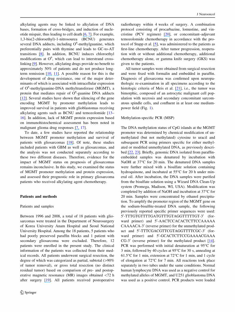
power field (Fig. 1).
Methylation-specific PCR (MSP)
The DNA methylation status of CpG islands at the MGMT
promoter was determined by chemical modification of un-
methylated (but not methylated) cytosine to uracil and
subsequent PCR using primers specific for either methyl-
ated or modified unmethylated DNA, as previously descri-
bed [22, 23]. Briefly, genomic DNA isolated from paraffin-
embedded samples was denatured by incubation with
NaOH at 37�C for 20 min. The denatured DNA samples
were further mixed with a bisulfate solution containing
hydroquinone, and incubated at 55�C for 20 h under min-
eral oil. After incubation, the DNA samples were purified
from the bisulfate solution using a Wizard DNA Clean-Up
system (Promega, Madison, WI, USA). Modification was
completed by addition of NaOH and incubation at 37�C for
20 min. Samples were concentrated by ethanol precipita-
tion. To amplify the promoter region of the MGMT gene on
the sodium-bisulfite-treated DNA sample, the following
previously reported specific primer sequences were used:
50-TTTGTGTTTTGATGTTTGTAGGTTTTTGT-30 (for-
ward primer) and 50-AACTCCACACTCTTCCAAAAA
CAAAACA-30 (reverse primer) for the unmethylated prod-
uct and 50-TTTCGACGTTCGTAGGTTTTCGC-30 (for-
ward primer) and 50-GCACTCTTCCGAAAACGAAA
CG-30 (reverse primer) for the methylated product [14].
PCR was performed with initial denaturation at 95�C for
3 min, followed by 40 cycles at 95�C for 30 s, annealing at
61.5�C for 1 min, extension at 72�C for 1 min, and 1 cycle
of elongation at 72�C for 7 min. All reactions took place
separately in two tubes under the same conditions. Normal
human lymphocyte DNA was used as a negative control for
methylated alleles of MGMT, and U251 glioblastoma DNA
was used as a positive control. PCR products were loaded
J Neurooncol
123
on a 12% polyacrylamide gel, stained with ethidium bro-
mide, and examined under ultraviolet (UV) illumination.
Immunohistochemistry for MGMT
Formalin-fixed paraffin-embedded specimens were cut to
thickness of 5 lm. In the deparaffinized sections, the
endogenous peroxidase was blocked with 3% H2O2 in
methanol. Antigen retrieval was performed in an autoclave
at 120�C for 10 min in 50 mmol/l citrate buffer (pH 6.0)
for MGMT immunohistochemistry in the same buffer.
Blocking of nonspecific binding was accomplished with
5% skim milk. After washing in phosphate-buffered saline
(PBS), the sections were incubated with anti-MGMT
antibody (1:20, clone MT3.1; Neomarkers Inc., Fremont,
CA, USA) overnight at 4�C, followed by staining with a
streptavidin–biotin–peroxidase kit (Nichirei, Tokyo,
Japan). The sections were reacted in a diaminobenzidine
peroxytrichloride substrate solution, and counterstained
with hematoxylin. As a negative control, the primary
antibody was omitted. Human liver was used as a positive
control. The immunoreactivity of MGMT protein was
evaluated semiquantitatively by counting the stained cells
in [1,000 cells. A tumor with [10% stained cells was
considered to be MGMT positive, according to a previous
study [19, 24]. Each slide was individually reviewed by a
neuropathologist, and all immunohistochemical analyses
were carried out in a blind manner to prevent potential bias
from knowledge of the clinical data.
Statistical analysis
Overall survival was calculated from date of surgery to
date of death, and progression-free survival (PFS) was
counted from date of surgery to date of increase in tumor
size [25% or presence of a new lesion on imaging. The
effect of MGMT status on survival was determined by
Kaplan–Meier method. Survival curves for the groups
according to MGMT status were compared using the log-
rank test. Correlation between the status of MGMT pro-
moter methylation and MGMT protein expression was
analyzed by Fisher’s exact test. Other prognostic factors
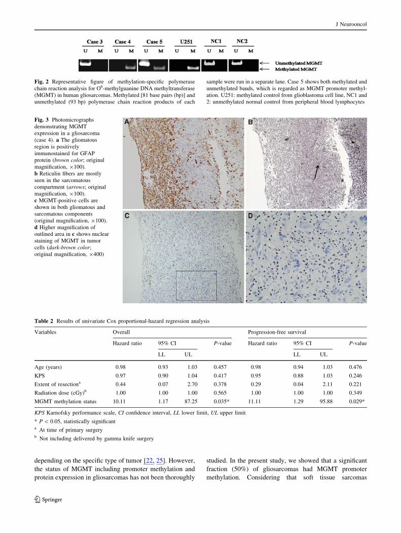
Fig. 1 Photomicrograph showing typical features of a gliosarcoma
(case 3). a The tumor exhibits a biphasic pattern with distinct glial
and sarcomatous components [hematoxylin and eosin (H&E), original
magnification, 9100]. b The gliomatous component is immunoreac-
tive with glial fibrillary acidic protein (GFAP) (original magnifica-
tion, 9100). c The sarcomatous component is positive for reticulin
(original magnification, 9100). d Tumor cells show immunoreactivity
against Ki-67, representing highly proliferative activity (original
magnification, 9200)
b
J Neurooncol
123
including age, Karnofsky performance scale (KPS), radia-
tion dose, and extent of resection according to MGMT
promoter methylation were evaluated by Mann–Whitney
test and Fisher’s exact test. Cox proportional-hazards
regression analysis was used for hazard ratios and associ-
ated 95% confidence intervals (CI). Multivariate models
were fitted by use of a Cox proportional-hazards regression
model. Statistical analysis was performed using commer-
cially available statistical software (SPSS version 11.0;
SPSS Inc., Chicago, IL, USA), and probability values
\0.05 were considered statistically significant.
Results
Patient characteristics
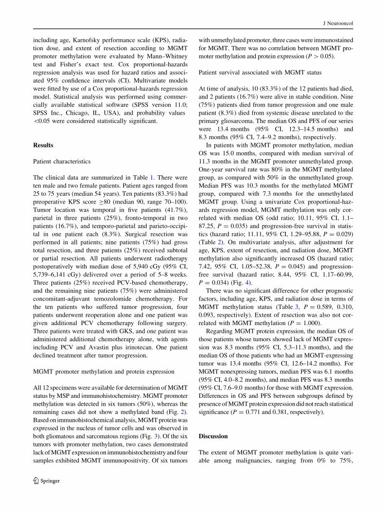
The clinical data are summarized in Table 1. There were
ten male and two female patients. Patient ages ranged from
25 to 75 years (median 54 years). Ten patients (83.3%) had
preoperative KPS score C80 (median 90, range 70–100).
Tumor location was temporal in five patients (41.7%),
parietal in three patients (25%), fronto-temporal in two
patients (16.7%), and temporo-parietal and parieto-occipi-
tal in one patient each (8.3%). Surgical resection was
performed in all patients; nine patients (75%) had gross
total resection, and three patients (25%) received subtotal
or partial resection. All patients underwent radiotherapy
postoperatively with median dose of 5,940 cGy (95% CI,
5,739–6,141 cGy) delivered over a period of 5–8 weeks.
Three patients (25%) received PCV-based chemotherapy,
and the remaining nine patients (75%) were administered
concomitant-adjuvant temozolomide chemotherapy. For
the ten patients who suffered tumor progression, four
patients underwent reoperation alone and one patient was
given additional PCV chemotherapy following surgery.
Three patients were treated with GKS, and one patient was
administered additional chemotherapy alone, with agents
including PCV and Avastin plus irinotecan. One patient
declined treatment after tumor progression.
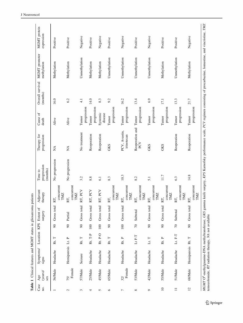
MGMT promoter methylation and protein expression
All 12 specimens were available for determination of MGMT
status by MSP and immunohistochemistry. MGMT promoter
methylation was detected in six tumors (50%), whereas the
remaining cases did not show a methylated band (Fig. 2).
Based on immunohistochemical analysis, MGMT protein was
expressed in the nucleus of tumor cells and was observed in
both gliomatous and sarcomatous regions (Fig. 3). Of the six
tumors with promoter methylation, two cases demonstrated
lack of MGMT expression on immunohistochemistry and four
samples exhibited MGMT immunopositivity. Of six tumors
with unmethylated promoter, three cases were immunostained
for MGMT. There was no correlation between MGMT pro-
moter methylation and protein expression (P [ 0.05).
Patient survival associated with MGMT status
At time of analysis, 10 (83.3%) of the 12 patients had died,
and 2 patients (16.7%) were alive in stable condition. Nine
(75%) patients died from tumor progression and one male
patient (8.3%) died from systemic disease unrelated to the
primary gliosarcoma. The median OS and PFS of our series
were 13.4 months (95% CI, 12.3–14.5 months) and
8.3 months (95% CI, 7.4–9.2 months), respectively.
In patients with MGMT promoter methylation, median
OS was 15.0 months, compared with median survival of
11.3 months in the MGMT promoter unmethylated group.
One-year survival rate was 80% in the MGMT methylated
group, as compared with 50% in the unmethylated group.
Median PFS was 10.3 months for the methylated MGMT
group, compared with 7.3 months for the unmethylated
MGMT group. Using a univariate Cox proportional-haz-
ards regression model, MGMT methylation was only cor-
related with median OS (odd ratio; 10.11, 95% CI, 1.1–
87.25, P = 0.035) and progression-free survival in statis-
tics (hazard ratio; 11.11, 95% CI, 1.29–95.88, P = 0.029)
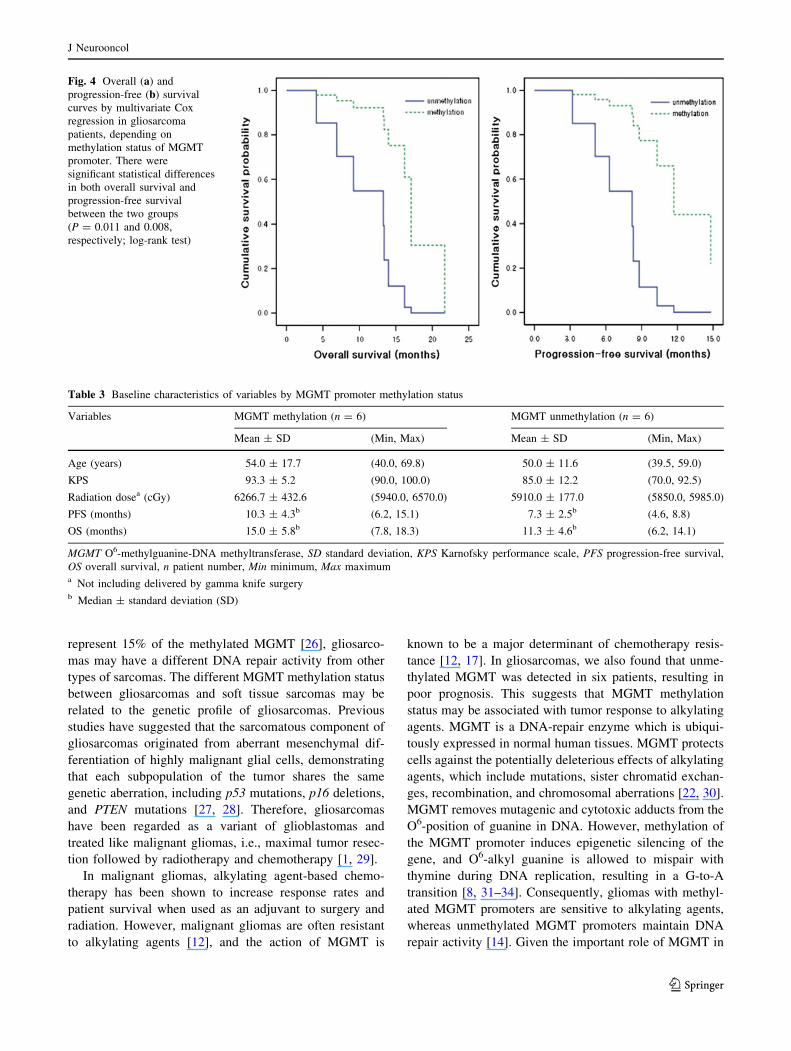
(Table 2). On multivariate analysis, after adjustment for
age, KPS, extent of resection, and radiation dose, MGMT
methylation also significantly increased OS (hazard ratio;
7.42, 95% CI, 1.05–52.38, P = 0.045) and progression-
free survival (hazard ratio; 8.44, 95% CI, 1.17–60.99,
P = 0.034) (Fig. 4).
There was no significant difference for other prognostic
factors, including age, KPS, and radiation dose in terms of
MGMT methylation status (Table 3, P = 0.589, 0.310,
0.093, respectively). Extent of resection was also not cor-
related with MGMT methylation (P = 1.000).
Regarding MGMT protein expression, the median OS of
those patients whose tumors showed lack of MGMT expres-
sion was 8.3 months (95% CI, 5.3–11.3 months), and the
median OS of those patients who had an MGMT-expressing
tumor was 13.4 months (95% CI, 12.6–14.2 months). For
MGMT nonexpressing tumors, median PFS was 6.1 months
(95% CI, 4.0–8.2 months), and median PFS was 8.3 months
(95% CI, 7.6–9.0 months) for those with MGMT expression.
Differences in OS and PFS between subgroups defined by
presence of MGMT protein expression did not reach statistical
significance (P = 0.771 and 0.381, respectively).
Discussion
The extent of MGMT promoter methylation is quite vari-
able among malignancies, ranging from 0% to 75%,
J Neurooncol
123
Ta
ble
1C
lin
ical
feat
ure
san
dM
GM
Tst
atu
sin
gli
osa
rco
ma
pat
ien
ts
Cas
e
no
.
Ag
e
(yea
rs)/
sex
Sy
mp
tom
s/
sig
ns
Lo
cati
on
KP
SE
xte
nt
of
surg
ery
Ad
juv
ant
ther
apy
Tim
eto
pro
gre
ssio
n
(mo
nth
s)
Th
erap
yfo
r
pro
gre
ssio
n
Cau
seo
f
dea
th
Ov
eral
lsu
rviv
al
(mo
nth
s)
MG
MT
pro
mo
ter
met
hy
lati
on
MG
MT
pro
tein
exp
ress
ion
15
6/M
ale
Hea
dac
he
Rt.
T9
0G
ross
tota
lR
T,
con
curr
ent
TM
Z
No
pro
gre
ssio
nN
AA
liv
e1
6.0
Met
hy
lati
on
Po
siti
ve
27
5/ Fem
ale
Hem
ipar
esis
Lt.
P9
0P
arti
alR
T,
con
curr
ent
TM
Z
No
pro
gre
ssio
nN
AA
liv
e6
.2M
eth
yla
tio
nP
osi
tiv
e
35
7/M
ale
Sei
zure
Rt.
T9
0G
ross
tota
lR
T,
PC
V3
.2N
otr
eatm
ent
Tu
mo
r
pro
gre
ssio
n
4.1
Un
met
hy
lati
on
Neg
ativ
e
42
5/M
ale
Hea
dac
he
Rt.
T-P
10
0G
ross
tota
lR
T,
PC
V8
.8R
eop
erat
ion
Tu
mo
r
pro
gre
ssio
n
14
.0M
eth
yla
tio
nP
osi
tiv
e
54
5/M
ale
Hea
dac
he
Rt.
P-O
10
0G
ross
tota
lR
T,
PC
V6
.1R
eop
erat
ion
Sy
stem
ic
dis
ease
8.3
Met
hy
lati
on
Neg
ativ
e
66
5/M
ale
Hea
dac
he
Rt.
T9
0G
ross
tota
lR
T,
con
curr
ent
TM
Z
8.3
GK
ST
um
or
pro
gre
ssio
n
9.2
Un
met
hy
lati
on
Po
siti
ve
73
2/ Fem
ale
Hea
dac
he
Rt.
P1
00
Gro
ssto
tal
RT
,
con
curr
ent
TM
Z
10
.3P
CV
,A
vas
tin
,
irin
ote
can
Tu
mo
r
pro
gre
ssio
n
16
.2U
nm
eth
yla
tio
nN
egat
ive
85
3/M
ale
Hea
dac
he
Lt
F-T
70
Su
bto
tal
RT
,
con
curr
ent
TM
Z
8.2
Reo
per
atio
nan
d
PC
V
Tu
mo
r
pro
gre
ssio
n
13
.4U
nm
eth
yla
tio
nP
osi
tiv
e
94
2/M
ale
Hea
dac
he
Lt.
T9
0G
ross
tota
lR
T,
con
curr
ent
TM
Z
5.1
GK
ST
um
or
pro
gre
ssio
n
6.9
Un
met
hy
lati
on
Neg
ativ
e
10
55
/Mal
eH
ead
ach
eR
t.P
90
Gro
ssto
tal
RT
,
con
curr
ent
TM
Z
11
.7G
KS
Tu
mo
r
pro
gre
ssio
n
17
.1M
eth
yla
tio
nP
osi
tiv
e
11
51
/Mal
eH
ead
ach
eL
t.F
-T7
0S
ub
tota
lR
T,
con
curr
ent
TM
Z
6.3
Reo
per
atio
nT
um
or
pro
gre
ssio
n
13
.3U
nm
eth
yla
tio
nP
osi
tiv
e
12
68
/Mal
eH
emip
ares
isR
t.T
90
Gro
ssto
tal
RT
,
con
curr
ent
TM
Z
14
.8R
eop
erat
ion
Tu
mo
r
pro
gre
ssio
n
21
.7M
eth
yla
tio
nN
egat
ive
MG
MT
O6-m
eth
ylg
uan
ine-
DN
Am
eth
ylt
ran
sfer
ase,
GK
Sg
amm
ak
nif
esu
rger
y,
KP
SK
arn
ofs
ky
per
form
ance
scal
e,P
CV
reg
imen
con
sist
ing
of
pro
carb
azin
e,lo
mu
stin
e,an
dv
incr
isti
ne,
TM
Zte
mo
zolo
mid
e,R
Tra
dia
tio
nth
erap
y,
NA
no
tav
aila
ble
J Neurooncol
123
depending on the specific type of tumor [22, 25]. However,
the status of MGMT including promoter methylation and
protein expression in gliosarcomas has not been thoroughly
studied. In the present study, we showed that a significant
fraction (50%) of gliosarcomas had MGMT promoter
methylation. Considering that soft tissue sarcomas
Fig. 2 Representative figure of methylation-specific polymerase
chain reaction analysis for O6-methylguanine DNA methyltransferase
(MGMT) in human gliosarcomas. Methylated [81 base pairs (bp)] and
unmethylated (93 bp) polymerase chain reaction products of each
sample were run in a separate lane. Case 5 shows both methylated and
unmethylated bands, which is regarded as MGMT promoter methyl-
ation. U251: methylated control from glioblastoma cell line, NC1 and
2: unmethylated normal control from peripheral blood lymphocytes
Fig. 3 Photomicrographs
demonstrating MGMT
expression in a gliosarcoma
(case 4). a The gliomatous
region is positively
immunostained for GFAP
protein (brown color; original
magnification, 9100).
b Reticulin fibers are mostly
seen in the sarcomatous
compartment (arrows; original
magnification, 9100).
c MGMT-positive cells are
shown in both gliomatous and
sarcomatous components
(original magnification, 9100).
d Higher magnification of
outlined area in c shows nuclear
staining of MGMT in tumor
cells (dark-brown color;
original magnification, 9400)
Table 2 Results of univariate Cox proportional-hazard regression analysis
Variables Overall Progression-free survival
Hazard ratio 95% CI P-value Hazard ratio 95% CI P-value
LL UL LL UL
Age (years) 0.98 0.93 1.03 0.457 0.98 0.94 1.03 0.476
KPS 0.97 0.90 1.04 0.417 0.95 0.88 1.03 0.246
Extent of resectiona 0.44 0.07 2.70 0.378 0.29 0.04 2.11 0.221
Radiation dose (cGy)b 1.00 1.00 1.00 0.565 1.00 1.00 1.00 0.349
MGMT methylation status 10.11 1.17 87.25 0.035* 11.11 1.29 95.88 0.029*
KPS Karnofsky performance scale, CI confidence interval, LL lower limit, UL upper limit
* P \ 0.05, statistically significanta At time of primary surgeryb Not including delivered by gamma knife surgery
J Neurooncol
123
represent 15% of the methylated MGMT [26], gliosarco-
mas may have a different DNA repair activity from other
types of sarcomas. The different MGMT methylation status
between gliosarcomas and soft tissue sarcomas may be
related to the genetic profile of gliosarcomas. Previous
studies have suggested that the sarcomatous component of
gliosarcomas originated from aberrant mesenchymal dif-
ferentiation of highly malignant glial cells, demonstrating
that each subpopulation of the tumor shares the same
genetic aberration, including p53 mutations, p16 deletions,
and PTEN mutations [27, 28]. Therefore, gliosarcomas
have been regarded as a variant of glioblastomas and
treated like malignant gliomas, i.e., maximal tumor resec-
tion followed by radiotherapy and chemotherapy [1, 29].
In malignant gliomas, alkylating agent-based chemo-
therapy has been shown to increase response rates and
patient survival when used as an adjuvant to surgery and
radiation. However, malignant gliomas are often resistant
to alkylating agents [12], and the action of MGMT is
known to be a major determinant of chemotherapy resis-
tance [12, 17]. In gliosarcomas, we also found that unme-
thylated MGMT was detected in six patients, resulting in
poor prognosis. This suggests that MGMT methylation
status may be associated with tumor response to alkylating
agents. MGMT is a DNA-repair enzyme which is ubiqui-
tously expressed in normal human tissues. MGMT protects
cells against the potentially deleterious effects of alkylating
agents, which include mutations, sister chromatid exchan-
ges, recombination, and chromosomal aberrations [22, 30].
MGMT removes mutagenic and cytotoxic adducts from the
O6-position of guanine in DNA. However, methylation of
the MGMT promoter induces epigenetic silencing of the
gene, and O6-alkyl guanine is allowed to mispair with
thymine during DNA replication, resulting in a G-to-A
transition [8, 31–34]. Consequently, gliomas with methyl-
ated MGMT promoters are sensitive to alkylating agents,
whereas unmethylated MGMT promoters maintain DNA
repair activity [14]. Given the important role of MGMT in
Fig. 4 Overall (a) and
progression-free (b) survival
curves by multivariate Cox
regression in gliosarcoma
patients, depending on
methylation status of MGMT
promoter. There were
significant statistical differences
in both overall survival and
progression-free survival
between the two groups
(P = 0.011 and 0.008,
respectively; log-rank test)
Table 3 Baseline characteristics of variables by MGMT promoter methylation status
Variables MGMT methylation (n = 6) MGMT unmethylation (n = 6)
Mean ± SD (Min, Max) Mean ± SD (Min, Max)
Age (years) 54.0 ± 17.7 (40.0, 69.8) 50.0 ± 11.6 (39.5, 59.0)
KPS 93.3 ± 5.2 (90.0, 100.0) 85.0 ± 12.2 (70.0, 92.5)
Radiation dosea (cGy) 6266.7 ± 432.6 (5940.0, 6570.0) 5910.0 ± 177.0 (5850.0, 5985.0)
PFS (months) 10.3 ± 4.3b (6.2, 15.1) 7.3 ± 2.5b (4.6, 8.8)
OS (months) 15.0 ± 5.8b (7.8, 18.3) 11.3 ± 4.6b (6.2, 14.1)
MGMT O6-methylguanine-DNA methyltransferase, SD standard deviation, KPS Karnofsky performance scale, PFS progression-free survival,
OS overall survival, n patient number, Min minimum, Max maximuma Not including delivered by gamma knife surgeryb Median ± standard deviation (SD)
J Neurooncol
123
tumor resistance to alkylating agents, O6-benzylguanine or
a poly(ADP-ribose) polymerase inhibitor can be helpful in
combination treatment for malignant gliomas [35–37].
In the present study, MGMT promoter methylation was
observed in 50% of the gliosarcomas, and lack of MGMT
expression was detected in 41.7% of the cases. However,
there was not a consistent correlation between promoter
methylation and MGMT protein expression. Several stud-
ies have suggested that methylation status may not be
correlated with MGMT protein expression [38, 39].
MGMT expression can be detected from nonneoplastic
components of the glioma [22, 40]. Alternatively, MGMT
promoter methylation occurs in only one allele, and an
unmethylated allele can produce MGMT expression in a
subset of the tumor [19]. Furthermore, MSP is a highly
sensitive technique in which a methylated band may be
observed even if tumor cells carry MGMT promoter
methylation in a minor portion. In the case of epigenetic
silencing, dimethylation of histone H3 lysine 9 and methyl-
CpG binding proteins can be determining factors for
MGMT expression, as well as DNA hypermethylation [41].
Recent studies have documented that methylation status is
homogeneous in the same tumor, but MGMT expression
can be heterogeneous regionally [42]. Taken together,
MGMT protein can be expressed in gliosarcomas even
though the MGMT promoter is methylated.
Although a number of studies have documented that
MGMT promoter methylation is related with survival in
malignant gliomas treated with alkylating agents [13, 32,
43], only one report, to the best of our knowledge, has
discussed the association between MGMT status and
prognosis, in which MGMT promoter methylation had a
positive effect on survival for gliosarcomas [18]. However,
this study included both gliosarcomas and GBM, and the
survival analysis was not performed separately according
to these two different entities. This led us to investigate
MGMT status associated with prognosis in a group of
patients with gliosarcomas who received chemotherapy
based on alkylating agents. In our study, there was longer
OS in gliosarcoma patients with methylated MGMT
(median OS, 15.0 months) compared with unmethylated
MGMT tumors (median OS, 11.3 months). With respect to
PFS, there was a similar pattern between methylated and
unmethylated MGMT in human gliosarcomas (median
PFS, 10.3 months versus 7.3 months). However, the dif-
ferences in OS and PFS between subgroups defined by
presence of MGMT protein expression was not statistically
significant. Recent work has shown that MGMT protein
expression is not a reliable biomarker for diagnosis and
patient outcome, although it examined various cutoff val-
ues of MGMT expression, inter- or intraobserver agree-
ment, two anti-MGMT antibodies, and endothelial or
hematogenous cells showing MGMT immunoreactivity. It
is suggested that novel anti-MGMT antibodies should be
directed against other epitopes for better clinical markers
[39]. In addition, we may consider that irrelevant mAb of
the same isotype is more appropriate for negative control in
MGMT immunostaining studies. Therefore, MGMT
immunostaining should be investigated further for clinical
correlation in malignant gliomas, although immunostaining
is a more convenient procedure for clinical application.
Together with these findings, we suggest that MGMT
promoter methylation may serve as a good prognostic
factor in gliosarcoma patients treated with alkylating agent
chemotherapy.
Our current study has limitations, including the retro-
spective method of analysis as well as the heterogeneity of
treatment modalities. In addition, various prognostic fac-
tors should also be considered in detail, although most
cases had KPS [70 and gross total resection in this study.
Therefore, large prospective studies will be necessary to
determine the impact of MGMT status on gliosarcoma
prognosis, despite the rarity of these tumors.
Conclusions
We examined MGMT status in gliosarcomas and found
that a significant fraction of the tumors showed MGMT
promoter methylation and protein expression. Our findings
suggest that MGMT promoter methylation is associated
with good prognosis in patients with gliosarcomas.
References
1. Salvati M, Caroli E, Raco A, Giangaspero F, Delfini R, Ferrante
L (2005) Gliosarcomas: analysis of 11 cases do two subtypes
exist? J Neurooncol 74:59–63
2. Louis DN, Ohgaki H, Wiestler OD, Cavenee WK, Burger PC,
Jouvet A, Scheithauer BW, Kleihues P (2007) The 2007 WHO
classification of tumours of the central nervous system. Acta
Neuropathol 114:97–109
3. Galanis E, Buckner JC, Dinapoli RP, Scheithauer BW, Jenkins
RB, Wang CH, O’Fallon JR, Farr G Jr (1998) Clinical outcome of
gliosarcoma compared with glioblastoma multiforme: North
Central Cancer Treatment Group results. J Neurosurg 89:425–430
4. Lutterbach J, Guttenberger R, Pagenstecher A (2001) Gliosar-
coma: a clinical study. Radiother Oncol 61:57–64
5. Stupp R, Mason WP, van den Bent MJ, Weller M, Fisher B,
Taphoorn MJ, Belanger K, Brandes AA, Marosi C, Bogdahn U,
Curschmann J, Janzer RC, Ludwin SK, Gorlia T, Allgeier A,
Lacombe D, Cairncross JG, Eisenhauer E, Mirimanoff RO (2005)
Radiotherapy plus concomitant and adjuvant temozolomide for
glioblastoma. N Engl J Med 352:987–996
6. Natsume A, Ishii D, Wakabayashi T, Tsuno T, Hatano H, Mizuno
M, Yoshida J (2005) IFN-beta down-regulates the expression of
DNA repair gene MGMT and sensitizes resistant glioma cells to
temozolomide. Cancer Res 65:7573–7579
J Neurooncol
123
7. Brell M, Tortosa A, Verger E, Gil JM, Vinolas N, Villa S, Acebes
JJ, Caral L, Pujol T, Ferrer I, Ribalta T, Graus F (2005) Prog-
nostic significance of O6-methylguanine-DNA methyltransferase
determined by promoter hypermethylation and immunohisto-
chemical expression in anaplastic gliomas. Clin Cancer Res
11:5167–5174
8. Gerson SL (2002) Clinical relevance of MGMT in the treatment
of cancer. J Clin Oncol 20:2388–2399
9. Gonzaga PE, Potter PM, Niu TQ, Yu D, Ludlum DB, Rafferty
JA, Margison GP, Brent TP (1992) Identification of the cross-link
between human O6-methylguanine-DNA methyltransferase and
chloroethylnitrosourea-treated DNA. Cancer Res 52:6052–6058
10. Prados MD, Russo C (1998) Chemotherapy of brain tumors.
Semin Surg Oncol 14:88–95
11. Silber JR, Bobola MS, Blank A, Schoeler KD, Haroldson PD,
Huynh MB, Kolstoe DD (2002) The apurinic/apyrimidinic
endonuclease activity of Ape1/Ref-1 contributes to human glioma
cell resistance to alkylating agents and is elevated by oxidative
stress. Clin Cancer Res 8:3008–3018
12. Sarkaria JN, Kitange GJ, James CD, Plummer R, Calvert H,
Weller M, Wick W (2008) Mechanisms of chemoresistance to
alkylating agents in malignant glioma. Clin Cancer Res 14:2900–
2908
13. Hegi ME, Diserens AC, Gorlia T, Hamou MF, de Tribolet N,
Weller M, Kros JM, Hainfellner JA, Mason W, Mariani L,
Bromberg JE, Hau P, Mirimanoff RO, Cairncross JG, Janzer RC,
Stupp R (2005) MGMT gene silencing and benefit from tem-
ozolomide in glioblastoma. N Engl J Med 352:997–1003
14. Esteller M, Garcia-Foncillas J, Andion E, Goodman SN, Hidalgo
OF, Vanaclocha V, Baylin SB, Herman JG (2000) Inactivation of
the DNA-repair gene MGMT and the clinical response of gliomas
to alkylating agents. N Engl J Med 343:1350–1354
15. Paz MF, Yaya-Tur R, Rojas-Marcos I, Reynes G, Pollan M,
Aguirre-Cruz L, Garcia-Lopez JL, Piquer J, Safont MJ, Balana C,
Sanchez-Cespedes M, Garcia-Villanueva M, Arribas L, Esteller
M (2004) CpG island hypermethylation of the DNA repair
enzyme methyltransferase predicts response to temozolomide in
primary gliomas. Clin Cancer Res 10:4933–4938
16. Kamiryo T, Tada K, Shiraishi S, Shinojima N, Kochi M, Ushio Y
(2004) Correlation between promoter hypermethylation of the
O6-methylguanine-deoxyribonucleic acid methyltransferase gene
and prognosis in patients with high-grade astrocytic tumors
treated with surgery, radiotherapy, and 1-(4-amino-2-methyl-5-
pyrimidinyl)methyl-3-(2-chloroethyl)-3-nitrosourea-based che-
motherapy. Neurosurgery 54:349–357; discussion 357
17. Friedman HS, McLendon RE, Kerby T, Dugan M, Bigner SH,
Henry AJ, Ashley DM, Krischer J, Lovell S, Rasheed K, Marchev
F, Seman AJ, Cokgor I, Rich J, Stewart E, Colvin OM, Prov-
enzale JM, Bigner DD, Haglund MM, Friedman AH, Modrich PL
(1998) DNA mismatch repair and O6-alkylguanine-DNA alkyl-
transferase analysis and response to Temodal in newly diagnosed
malignant glioma. J Clin Oncol 16:3851–3857
18. Prados MD, Chang SM, Butowski N, DeBoer R, Parvataneni R,
Carliner H, Kabuubi P, Ayers-Ringler J, Rabbitt J, Page M, Fe-
doroff A, Sneed PK, Berger MS, McDermott MW, Parsa AT,
Vandenberg S, James CD, Lamborn KR, Stokoe D, Haas-Kogan
DA (2009) Phase II study of erlotinib plus temozolomide during
and after radiation therapy in patients with newly diagnosed glio-
blastoma multiforme or gliosarcoma. J Clin Oncol 27:579–584
19. Nakasu S, Fukami T, Baba K, Matsuda M (2004) Immunohis-
tochemical study for O6-methylguanine-DNA methyltransferase
in the non-neoplastic and neoplastic components of gliomas. J
Neurooncol 70:333–340
20. Levin VA, Silver P, Hannigan J, Wara WM, Gutin PH, Davis RL,
Wilson CB (1990) Superiority of post-radiotherapy adjuvant
chemotherapy with CCNU, procarbazine, and vincristine (PCV)
over BCNU for anaplastic gliomas: NCOG 6G61 final report. Int
J Radiat Oncol Biol Phys 18:321–324
21. Meis JM, Martz KL, Nelson JS (1991) Mixed glioblastoma
multiforme and sarcoma. A clinicopathologic study of 26 radia-
tion therapy oncology group cases. Cancer 67:2342–2349
22. Esteller M, Hamilton SR, Burger PC, Baylin SB, Herman JG
(1999) Inactivation of the DNA repair gene O6-methylguanine-
DNA methyltransferase by promoter hypermethylation is a com-
mon event in primary human neoplasia. Cancer Res 59:793–797
23. Herman JG, Graff JR, Myohanen S, Nelkin BD, Baylin SB
(1996) Methylation-specific PCR: a novel PCR assay for meth-
ylation status of CpG islands. Proc Natl Acad Sci U S A 93:9821–
9826
24. Matsukura S, Miyazaki K, Yakushiji H, Ogawa A, Chen Y,
Sekiguchi M (2003) Combined loss of expression of O6-meth-
ylguanine-DNA methyltransferase and hMLH1 accelerates pro-
gression of hepatocellular carcinoma. J Surg Oncol 82:194–200
25. Nakamura M, Watanabe T, Yonekawa Y, Kleihues P, Ohgaki H
(2001) Promoter methylation of the DNA repair gene MGMT in
astrocytomas is frequently associated with G:C ? A:T mutations
of the TP53 tumor suppressor gene. Carcinogenesis 22:1715–1719
26. Kawaguchi K, Oda Y, Saito T, Yamamoto H, Takahira T, Ko-
bayashi C, Tamiya S, Tateishi N, Iwamoto Y, Tsuneyoshi M
(2006) DNA hypermethylation status of multiple genes in soft
tissue sarcomas. Mod Pathol 19:106–114
27. Reis RM, Konu-Lebleblicioglu D, Lopes JM, Kleihues P, Ohgaki H
(2000) Genetic profile of gliosarcomas. Am J Pathol 156:425–432
28. Boerman RH, Anderl K, Herath J, Borell T, Johnson N, Scha-
effer-Klein J, Kirchhof A, Raap AK, Scheithauer BW, Jenkins
RB (1996) The glial and mesenchymal elements of gliosarcomas
share similar genetic alterations. J Neuropathol Exp Neurol
55:973–981
29. di Norcia V, Piccirilli M, Giangaspero F, Salvati M (2008)
Gliosarcomas in the elderly: analysis of 7 cases and clinico-
pathological remarks. Tumori 94:493–496
30. Kim JI, Suh JT, Choi KU, Kang HJ, Shin DH, Lee IS, Moon TY,
Kim WT (2009) Inactivation of O6-methylguanine-DNA meth-
yltransferase in soft tissue sarcomas: association with K-ras
mutations. Hum Pathol 40:934–941
31. Baylin SB, Herman JG (2000) DNA hypermethylation in tumor-
igenesis: epigenetics joins genetics. Trends Genet 16:168–174
32. Jaeckle KA, Eyre HJ, Townsend JJ, Schulman S, Knudson HM,
Belanich M, Yarosh DB, Bearman SI, Giroux DJ, Schold SC
(1998) Correlation of tumor O6 methylguanine-DNA methyl-
transferase levels with survival of malignant astrocytoma patients
treated with bis-chloroethylnitrosourea: a Southwest Oncology
Group study. J Clin Oncol 16:3310–3315
33. Silber JR, Mueller BA, Ewers TG, Berger MS (1993) Comparison
of O6-methylguanine-DNA methyltransferase activity in brain
tumors and adjacent normal brain. Cancer Res 53:3416–3420
34. Tanaka S, Kobayashi I, Utsuki S, Oka H, Fujii K, Watanabe T,
Nagashima T, Hori T (2003) O6-methylguanine-DNA methyl-
transpherase gene expression in gliomas by means of real-time
quantitative RT-PCR and clinical response to nitrosoureas. Int J
Cancer 103:67–72
35. Quinn JA, Jiang SX, Reardon DA, Desjardins A, Vredenburgh JJ,
Rich JN, Gururangan S, Friedman AH, Bigner DD, Sampson JH,
McLendon RE, Herndon JE II, Walker A, Friedman HS (2009)
Phase II trial of temozolomide plus O6-benzylguanine in adults
with recurrent, temozolomide-resistant malignant glioma. J Clin
Oncol 27:1262–1267
36. Quinn JA, Pluda J, Dolan ME, Delaney S, Kaplan R, Rich JN,
Friedman AH, Reardon DA, Sampson JH, Colvin OM, Haglund
MM, Pegg AE, Moschel RC, McLendon RE, Provenzale JM,
Gururangan S, Tourt-Uhlig S, Herndon JE II, Bigner DD,
Friedman HS (2002) Phase II trial of carmustine plus O(6)-
J Neurooncol
123
benzylguanine for patients with nitrosourea-resistant recurrent or
progressive malignant glioma. J Clin Oncol 20:2277–2283
37. Donawho CK, Luo Y, Penning TD, Bauch JL, Bouska JJ,
Bontcheva-Diaz VD, Cox BF, DeWeese TL, Dillehay LE, Fer-
guson DC, Ghoreishi-Haack NS, Grimm DR, Guan R, Han EK,
Holley-Shanks RR, Hristov B, Idler KB, Jarvis K, Johnson EF,
Kleinberg LR, Klinghofer V, Lasko LM, Liu X, Marsh KC,
McGonigal TP, Meulbroek JA, Olson AM, Palma JP, Rodriguez
LE, Shi Y, Stavropoulos JA, Tsurutani AC, Zhu GD, Rosenberg
SH, Giranda VL, Frost DJ (2007) ABT-888, an orally active
poly(ADP-ribose) polymerase inhibitor that potentiates DNA-
damaging agents in preclinical tumor models. Clin Cancer Res
13:2728–2737
38. Grasbon-Frodl EM, Kreth FW, Ruiter M, Schnell O, Bise K,
Felsberg J, Reifenberger G, Tonn JC, Kretzschmar HA (2007)
Intratumoral homogeneity of MGMT promoter hypermethylation
as demonstrated in serial stereotactic specimens from anaplastic
astrocytomas and glioblastomas. Int J Cancer 121:2458–2464
39. Preusser M, Charles Janzer R, Felsberg J, Reifenberger G, Ha-
mou MF, Diserens AC, Stupp R, Gorlia T, Marosi C, Heinzl H,
Hainfellner JA, Hegi M (2008) Anti-O6-methylguanine-methyl-
transferase (MGMT) immunohistochemistry in glioblastoma
multiforme: observer variability and lack of association with
patient survival impede its use as clinical biomarker. Brain Pathol
18:520–532
40. Blanc JL, Wager M, Guilhot J, Kusy S, Bataille B, Chantereau T,
Lapierre F, Larsen CJ, Karayan-Tapon L (2004) Correlation of
clinical features and methylation status of MGMT gene promoter
in glioblastomas. J Neurooncol 68:275–283
41. Soejima H, Zhao W, Mukai T (2005) Epigenetic silencing of the
MGMT gene in cancer. Biochem Cell Biol 83:429–437
42. Cao VT, Jung TY, Jung S, Jin SG, Moon KS, Kim IY, Kang SS,
Park CS, Lee KH, Chae HJ (2009) The correlation and prognostic
significance of MGMT promoter methylation and MGMT protein
in glioblastomas. Neurosurgery 65:866–875; discussion 875
43. Chen ZP, Yarosh D, Garcia Y, Tampieri D, Mohr G, Malapetsa
A, Langleben A, Panasci LC (1999) Relationship between O6-
methylguanine-DNA methyltransferase levels and clinical
response induced by chloroethylnitrosourea therapy in glioma
patients. Can J Neurol Sci 26:104–109
J Neurooncol
123
Related Documents