P.1134 Editors: Miller, Neil R.; Newman, Nancy J. Title: Walsh & Hoyt's Clinical Neuro-Ophthalmology, 6th Edition Copyright ©2005 Lippincott Williams & Wilkins > Table of Contents > Volume I > Section III - The Ocular Motor System > 23 - Nystagmus and Related Ocular Motility Disorders 23 Nystagmus and Related Ocular Motility Disorders R. John Leigh Janet C. Rucker General Concepts and Clinical Approach This chapter concerns abnormal eye movements that disrupt steady fixation and thereby degrade vision. We now know a good deal about the normal anatomy, physiology, and pharmacology of ocular motor control (1). Our approach is to apply this knowledge to nystagmus and other ocular oscillations, since pathophysiology provides a sounder conceptual framework than a system based solely on phenomenology. We first summarize the mechanisms by which gaze is normally held steady to achieve clear and stable vision (2). We then discuss the pathogenesis and clinical features of each of the disorders that disrupt steady gaze, including the various forms of pathologic nystagmus and saccadic intrusions. Finally, we summarize currently available treatments for these abnormal eye movements and their visual consequences. Normal Mechanisms for Gaze Stability In order for us to see an object best, its image must be held steady over the foveal region of the retina. Although the visual system can tolerate some motion of images on the retina (3), if this motion becomes excessive (more than about 5°/second for Snellen optotypes), vision declines. Furthermore, if the image is moved from the fovea to peripheral retina, it will be seen less clearly. In healthy persons, three separate mechanisms work together to prevent deviation of the line of sight from the object of regard. The first is fixation, which has two distinct components: (a) the visual system's ability to detect retinal image drift and program corrective eye movements; and (b) the suppression of unwanted saccades that would take the eye off target. The second mechanism is the vestibulo-ocular reflex, by which eye movements compensate for head perturbations at short latency and thus maintain clear vision during natural activities, especially locomotion. The third mechanism is the ability of the brain to hold the eye at an eccentric position in the orbit against the elastic pull of the suspensory ligaments and extraocular muscles, which tend to return it toward central position. For all three gaze-holding mechanisms to work effectively, their performance must be tuned by adaptive mechanisms that monitor the visual consequences of eye movements. Types of Abnormal Eye Movements that Disrupt Steady Fixation: Nystagmus and Saccadic Intrusions The essential difference between nystagmus and saccadic intrusions lies in the initial eye movement that takes the line of sight off the object of regard. For nystagmus, it is a slow drift (or “slow phase”), as opposed to an inappropriate saccadic movement that intrudes on steady fixation. After the initial movement, corrective or other abnormal eye movements may follow. Thus, nystagmus may be defined as a repetitive, to-and-fro movement of the eyes that is initiated by a slow phase (drift). Saccadic intrusions, on the other hand, are rapid eye movements that take the eye off target. They include a spectrum of abnormal movements, ranging from single saccades to sustained saccadic oscillations. Ovid: Walsh & Hoyt's Clinical Neuro-Ophthalmolog y http://ovidsp.tx.ovid.com.ezproxy.fiu.edu/spa/ovidweb.cgi 1 of 89 6/28/2009 8:53 AM

Welcome message from author
This document is posted to help you gain knowledge. Please leave a comment to let me know what you think about it! Share it to your friends and learn new things together.
Transcript

P.1134
Editors: Miller, Neil R.; Newman, Nancy J.
Title: Walsh & Hoyt's Clinical Neuro-Ophthalmology, 6th Edition
Copyright ©2005 Lippincott Williams & Wilkins
> Table of Contents > Volume I > Section III - The Ocular Motor System > 23 - Nystagmus and Related Ocular Motility Disorders
23
Nystagmus and Related Ocular Motility Disorders
R. John Leigh
Janet C. Rucker
General Concepts and Clinical ApproachThis chapter concerns abnormal eye movements that disrupt steady fixation and thereby degrade vision. We now
know a good deal about the normal anatomy, physiology, and pharmacology of ocular motor control (1). Our
approach is to apply this knowledge to nystagmus and other ocular oscillations, since pathophysiology provides a
sounder conceptual framework than a system based solely on phenomenology. We first summarize the mechanisms
by which gaze is normally held steady to achieve clear and stable vision (2). We then discuss the pathogenesis and
clinical features of each of the disorders that disrupt steady gaze, including the various forms of pathologic
nystagmus and saccadic intrusions. Finally, we summarize currently available treatments for these abnormal eye
movements and their visual consequences.
Normal Mechanisms for Gaze StabilityIn order for us to see an object best, its image must be held steady over the foveal region of the retina. Although
the visual system can tolerate some motion of images on the retina (3), if this motion becomes excessive (more than
about 5°/second for Snellen optotypes), vision declines. Furthermore, if the image is moved from the fovea to
peripheral retina, it will be seen less clearly.
In healthy persons, three separate mechanisms work together to prevent deviation of the line of sight from the
object of regard. The first is fixation, which has two distinct components: (a) the visual system's ability to detect
retinal image drift and program corrective eye movements; and (b) the suppression of unwanted saccades that
would take the eye off target. The second mechanism is the vestibulo-ocular reflex, by which eye movements
compensate for head perturbations at short latency and thus maintain clear vision during natural activities,
especially locomotion. The third mechanism is the ability of the brain to hold the eye at an eccentric position in the
orbit against the elastic pull of the suspensory ligaments and extraocular muscles, which tend to return it toward
central position. For all three gaze-holding mechanisms to work effectively, their performance must be tuned by
adaptive mechanisms that monitor the visual consequences of eye movements.
Types of Abnormal Eye Movements that Disrupt Steady Fixation:Nystagmus and Saccadic IntrusionsThe essential difference between nystagmus and saccadic intrusions lies in the initial eye movement that takes the
line of sight off the object of regard. For nystagmus, it is a slow drift (or “slow phase”), as opposed to an
inappropriate saccadic movement that intrudes on steady fixation. After the initial movement, corrective or other
abnormal eye movements may follow. Thus, nystagmus may be defined as a repetitive, to-and-fro movement of the
eyes that is initiated by a slow phase (drift). Saccadic intrusions, on the other hand, are rapid eye movements that
take the eye off target. They include a spectrum of abnormal movements, ranging from single saccades to sustained
saccadic oscillations.
Ovid: Walsh & Hoyt's Clinical Neuro-Ophthalmology http://ovidsp.tx.ovid.com.ezproxy.fiu.edu/spa/ovidweb.cgi
1 of 89 6/28/2009 8:53 AM

P.1135
Differences Between Physiologic and Pathologic NystagmusIt is important to realize that not all nystagmus is pathologic. Physiologic nystagmus preserves clear vision during
self-rotation. Under most circumstances, for example during locomotion, head movements are small and the
vestibulo-ocular reflex is able to generate eye movements that compensate for them. Consequently, the line of
sight remains pointed at the object of regard. In response to large head or body rotations, however, the vestibulo-
ocular reflex alone cannot preserve clear vision because the eyes are limited in their range of rotation. Thus, during
sustained rotations, quick phases occur to reset the eyes into their working range: vestibular nystagmus. If
rotation is sustained for several seconds, the vestibular afferents no longer accurately signal head rotation, and
visually driven or optokinetic nystagmus takes over to stop excessive slip of stationary retinal images. Additional
examples of physiologic nystagmus are arthrokinetic and audiokinetic nystagmus (discussion following). In contrast
to vestibular and optokinetic nystagmus, pathologic nystagmus causes excessive drift of stationary retinal images
that degrades vision and may produce illusory motion of the seen world: oscillopsia (4,5,6,7). An exception is
congenital nystagmus, which may be associated with normal visual acuity and which seldom causes oscillopsia (8).
Nystagmus, both physiologic and pathologic, may consist of alternating slow drifts (slow phases) in one direction and
corrective, resetting saccades (quick phases) in the other: jerk nystagmus (Fig. 23.1A). Pathologic nystagmus may,
however, also consist of smooth to-and-fro oscillations: pendular nystagmus (Fig. 23.1D). Conventionally, jerk
nystagmus is described according to the direction of the quick phase. Thus, if the slow movement is drifting up, the
nystagmus is called “downbeating”; if the slow movement is to the right, the nystagmus is “left-beating.” Although
it is convenient to describe the frequency, amplitude, and direction of the quick phases of the nystagmus, it should
be remembered that it is the slow phase that reflects the underlying abnormality.
Ovid: Walsh & Hoyt's Clinical Neuro-Ophthalmology http://ovidsp.tx.ovid.com.ezproxy.fiu.edu/spa/ovidweb.cgi
2 of 89 6/28/2009 8:53 AM

Figure 23.1. Four common slow-phase waveforms of nystagmus. A, Constant velocity drift of the eyes. This
occurs in nystagmus caused by peripheral or central vestibular disease and also with lesions of the cerebral
hemisphere. The added quick-phases give a “saw-toothed” appearance. B, Drift of the eyes back from an
eccentric orbital position toward the midline (gaze-evoked nystagmus). The drift shows a negative
exponential time course, with decreasing velocity. This waveform reflects an unsustained eye position signal
caused by a “leaky” neural integrator. C, Drift of the eyes away from the central position with a positive
exponential time course (increasing velocity). This waveform suggests an unstable neural integrator and is
usually encountered in congenital nystagmus. D, Pendular nystagmus, which is encountered as a type of
congenital nystagmus and with acquired brainstem disease. (From Leigh RJ, Zee DS. The Neurology of Eye
Movements. Ed 3. New York, Oxford University Press, 1999.)
Nystagmus may occur in any plane, although it is often predominantly horizontal, vertical, or torsional. Physiologic
nystagmus is essentially conjugate. Pathologic nystagmus, on the other hand, may have different amplitudes in the
two eyes (dissociated nystagmus); it may go in different directions leading to different trajectories of nystagmus in
the two eyes; or may have different temporal properties, i.e., phase shift between the two eyes, leading to
movements that are sometimes in opposite directions (disconjugate nystagmus).
Ovid: Walsh & Hoyt's Clinical Neuro-Ophthalmology http://ovidsp.tx.ovid.com.ezproxy.fiu.edu/spa/ovidweb.cgi
3 of 89 6/28/2009 8:53 AM

Methods of Observing, Eliciting, and Recording NystagmusIt is often possible to diagnose the cause of nystagmus through careful history and systematic examination of the
patient (9,10). History should include duration of nystagmus, whether it interferes with vision and causes
oscillopsia, and accompanying neurological symptoms. The physician should also determine if nystagmus and
attendant visual symptoms are worse with viewing far or near objects, with patient motion, or with different gaze
angles (e.g., worse on right gaze). If the patient habitually tilts or turns the head, the physician should determine
whether or not these features are evident on old photographs.
Before assessing eye movements, the physician must examine the visual system, looking for signs of optic nerve
demyelination or malformation, or ocular albinism which often suggests the diagnosis. The stability of fixation
should be assessed with the eyes close to central position, viewing near and far targets, and at eccentric gaze
angles. It is often useful to record the direction and amplitude of nystagmus for each of the cardinal gaze positions.
If the patient has a head turn or tilt, the eyes should be observed in various directions of gaze when the head is in
that position as well as when the head is held straight. During fixation, each eye should be occluded in turn to check
for latent nystagmus. The presence of pseudonystagmus and oscillopsia in patients with head tremor who have lost
their vestibulo-ocular reflex must be differentiated from true nystagmus.
Subtle forms of nystagmus, due to low amplitude or inconstant presence, require prolonged observation over 2–3
minutes. Low amplitude nystagmus may be detected only by viewing the patient's retina with an ophthalmoscope
(11). (Note, however, that the direction of horizontal or vertical nystagmus is inverted when viewed through the
ophthalmoscope.) The effect of removal of fixation should always be determined. Nystagmus caused by peripheral
vestibular imbalance may be apparent only under these circumstances. Removal of fixation is often achieved by
eyelid closure; nystagmus is then evaluated by recording eye movements, by palpating the globes, or by
auscultation with a stethoscope. Lid closure itself may affect nystagmus, however, and it is better to evaluate the
effects of removing fixation with the eyelids open. Several clinical methods are available, such as Frenzel goggles
which consist of 10- to 20-diopter spherical convex lenses placed in a frame that has its own light source. The
goggles defocus the patient's vision, thus preventing fixation of objects, and also provide the examiner with a
magnified, illuminated view of the patient's eyes. An alternative is to use two high-plus spherical lenses from a trial
case, or to determine the effect of transiently covering the viewing eye during ophthalmoscopy in an otherwise
dark room.
Evaluation of nystagmus is incomplete without a systematic examination of each functional class of eye movements
(vestibular, optokinetic, smooth-pursuit, saccades, vergence) and their effect on the nystagmus, since different
forms of nystagmus can be directly attributed to abnormalities of some of these movements. Physiological
optokinetic nystagmus occurs during self-rotation, but it can be elicited at the bedside using a small drum or tape
with alternating black and white lines, although larger displays are more effective in patients with voluntary gaze
palsies. The slow phases represent visual tracking, including smooth pursuit; the resetting quick phases are saccadic
in origin (12). In children and patients with impaired voluntary gaze, an optokinetic stimulus often provides useful
information about both pursuit and saccadic systems (13,14,15,16,17). Vestibular nystagmus can be conveniently
induced by rotating the patient in a swivel office chair for 30 seconds and then stopping: postrotational nystagmus
and vertigo are induced, which may help patients identify the nature of any paroxysmal attacks of dizziness. Caloric
and other forms of induced vestibular nystagmus are described below.
It is often helpful to measure the nystagmus waveform because the shape of the slow phase often provides a
pathophysiological signature of the underlying disorder (18,19). To properly characterize nystagmus, it is important
to measure eye position and velocity, as well as target position, during attempted fixation at different gaze angles,
in darkness, and during vestibular, optokinetic, saccadic, pursuit, and vergence movements. Common slow-phase
waveforms of nystagmus are shown in Figure 23.1.
Conventionally, nystagmus is measured in terms of its amplitude, frequency, and their product: intensity. However,
visual symptoms caused by nystagmus usually correlate best with the speed of the slow phase and displacement of
the image of the object of regard from the fovea (7).
There are many different methods now available for recording eye movements, and these are discussed more fully
Ovid: Walsh & Hoyt's Clinical Neuro-Ophthalmology http://ovidsp.tx.ovid.com.ezproxy.fiu.edu/spa/ovidweb.cgi
4 of 89 6/28/2009 8:53 AM

P.1136
elsewhere (20,21). Because many patients with nystagmus cannot accurately point their eyes at visual targets,
precise measurement is best achieved with the magnetic search coil technique (Fig. 23.2), since the contact lens
that the patient wears can be precalibrated on a protractor-gimbal device. In addition, this is the only technique
that permits precise measurement of horizontal, vertical, and torsional oscillations
over an extended range of amplitudes and frequencies. Although originally introduced as a research tool, the
technique is now widely used to evaluate clinical disorders of eye movements, and is well tolerated (22). We have
studied over 500 patients with this method.
Figure 23.2. A method for precise measurement of horizontal, vertical, and torsional eye rotations. The
subject is wearing a Silastic © annulus embedded in which are two coils of wire, one wound in the frontal
plane (to sense horizontal and vertical movements) and the other effectively in the sagittal plane (to sense
torsional eye movements). When the subject sits in a magnetic field, voltages are induced in these search
coils that can be used to measure eye position. (From Leigh RJ, Zee DS. The Neurology of Eye Movements. Ed
3. New York, Oxford University Press, 1999.)
Classification of Nystagmus Based on PathogenesisOur classification of nystagmus starts by relating the various forms of nystagmus to disorders of visual fixation, the
vestibulo-ocular reflex, or the mechanism for eccentric gaze-holding. In addition, the adaptive processes that
optimize these eye movements may be affected by disease, and we discuss these recalibration mechanisms as we
deal with each class of nystagmus. Some forms of nystagmus can be better explained than others by this scheme.
Nonetheless, our goal is to provide current hypotheses for nystagmus and saccadic intrusions whenever possible.
Some hypotheses are backed by substantial evidence, whereas others are more tentative. The justification for this
approach is that it provides explanations for clinical findings when knowledge allows, but also provides provisional
hypotheses for other disorders that can be tested in future studies.
Nystagmus Associated with Disease of the Visual System and ItsProjections to Brainstem and Cerebellum
Origin and Nature of Nystagmus Associated With Disease of the Visual
Ovid: Walsh & Hoyt's Clinical Neuro-Ophthalmology http://ovidsp.tx.ovid.com.ezproxy.fiu.edu/spa/ovidweb.cgi
5 of 89 6/28/2009 8:53 AM

P.1137
PathwaysDisorders of the visual pathways are often associated with nystagmus. The most obvious example is the nystagmus
that invariably accompanies blindness (23,24). How does this arise? At least two separate mechanisms can be
identified: the visual fixation mechanism itself and the visually mediated calibration mechanism that optimizes its
action.
The smooth visual fixation mechanism stops the eyes from drifting away from a stationary object of regard (25,26).
For example, if a normal subject attempts to fixate on the remembered location of a target while in darkness, the
eye drifts off target several times faster than if the subject fixates the visible target (27). Uncorrected drifts are
eventually remedied by a saccade that places the image back on the fovea. This fixation mechanism depends upon
the motion detection (magnocellular) portion of the visual system (28) which is inherently slow, with a response
time of about 100 milliseconds that encumbers all visually mediated eye movements, including fixation, smooth
pursuit, and optokinetic responses. If the response time is delayed further by disease of the visual system, then the
attempts by the brain to correct eye drifts may actually add to the retinal error rather than reduce it, and may lead
to ocular oscillations (29).
Vision is also needed for recalibrating and optimizing all types of eye movements. These functions depend on visual
projections to the cerebellum, the structure called by Robinson “the ocular motor repair shop” (30). Thus, signals
from secondary visual areas concerned with motion-vision project to the cerebellum via the pontine nuclei and
middle cerebellar peduncle (see Chapter 17). For example, neurons in the dorsolateral pontine nuclei and Purkinje
cells in the cerebellar flocculus both encode visual-motion signals (31,32). Visual signals for recalibration may also
pass via the inferior olive, which sends climbing fibers to the cerebellum (33,34). If the ocular motor system is to be
recalibrated, visual signals need to be compared with eye movement commands. At present, it is not certain how or
where this function is performed. One possibility is a group of cells in the paramedian tracts (PMT) in the lower
pons, which receive inputs from almost all ocular motor structures and which project to the cerebellar flocculus
(35). Lesions at any part of this visual-motor recalibration pathway can deprive the brain of signals that hold each of
the eyes on the object of regard, the result being drifts of the eyes off target, leading to nystagmus.
Disease affecting any part of the visual system, from retina to cortical visual areas, or interrupting visual projections
to pons and cerebellum, may be associated with nystagmus. In this section, we first catalogue the features of
nystagmus reported with disease localized to the different sites in this pathway. We then describe features of
acquired pendular nystagmus, one of the most common forms of nystagmus associated with disease affecting the
visual system or its brainstem-cerebellar projections.
Clinical Features of Nystagmus with Lesions Affecting the VisualPathways
Disease of the RetinaCongenital or acquired retinal disorders causing blindness, such as Leber's congenital amaurosis, lead to continuous
jerk nystagmus with components in all three planes, which changes direction over the course of seconds or minutes
(Fig. 23.3A). The drifting “null point”—the eye position at which nystagmus changes direction—probably reflects
inability to calibrate the ocular motor system, and it has also been reported after experimental cerebellectomy
(36). This nystagmus often shows the increasing-velocity waveform (Fig. 23.1C) that was once thought to be specific
for congenital nystagmus (discussion following). Recent developments in gene therapy for retinal disorders suggest
that if vision can be restored, nystagmus will be suppressed (37,38).
Disease Affecting the Optic NervesOptic nerve disease is commonly associated with pendular nystagmus. With unilateral disease of the optic nerve,
nystagmus largely affects the abnormal eye (monocular nystagmus), with prominent, vertical, low-frequency,
bidirectional drifts (Fig. 23.3B); horizontal drifts are generally unidirectional with corrective quick-phases (24,39).
When disease affects both optic nerves, the amplitude of nystagmus is often greater in the eye with poorer vision
Ovid: Walsh & Hoyt's Clinical Neuro-Ophthalmology http://ovidsp.tx.ovid.com.ezproxy.fiu.edu/spa/ovidweb.cgi
6 of 89 6/28/2009 8:53 AM

(the Heimann-Bielschowsky phenomenon) (40). This phenomenon is not confined to primary optic nerve disease,
however, and also occurs in patients with profound amblyopia, dense cataract, and high myopia (24,39,41).
Oscillations may disappear when vision is restored or they may persist (42), leading to oscillopsia. The former
findings support the contention that these ocular oscillations are primarily caused by loss of vision rather than by
any primary disorder of the ocular motor system. The origin of vertical drifts that occur in a blind eye is unknown
but has been attributed to disturbance of either the vertical vergence mechanism (41), or to a monocular visual
stabilization system (24).
Ovid: Walsh & Hoyt's Clinical Neuro-Ophthalmology http://ovidsp.tx.ovid.com.ezproxy.fiu.edu/spa/ovidweb.cgi
7 of 89 6/28/2009 8:53 AM

P.1138
Figure 23.3. Nystagmus and gaze instability associated with visual loss. A, Binocular blindness since birth due
to Leber's congenital amaurosis. In the horizontal plane, nystagmus changes direction (evident in velocity
channels) and there is a “wandering null point.” Slow-phase waveforms are variably linear decreasing velocity
or, especially in the vertical plane, increasing velocity. B, Patient who had defocused vision since childhood
following eye trauma and removal of his left lens. Following implantation of an artificial lens at age 35 years,
his corrected visual acuity was 20/20 OD and 20/25 OS, but he was unable to maintain steady fixation with the
left eye and suffered from variable diplopia and abnormal motion of vision in his left eye that he could not
control. His left eye shows the Heimann-Bielschowsky phenomenon, vertical instability of fixation with slow
drifts (17).
In infants, the appearance of monocular, vertical pendular nystagmus raises the possibility of optic nerve tumor and
neuroimaging studies are indicated (43,44). However, monocular oscillations in children are sometimes due to
spasmus nutans (45,46); this condition is discussed below. Monocular visual impairment, such as amblyopia, also
leads to horizontal nystagmus and, if present from birth, the features are those of latent nystagmus, which is
discussed in a later section.
Disease Affecting the Optic ChiasmParasellar lesions such as pituitary tumors have traditionally, albeit rarely, been associated with seesaw nystagmus,
which is discussed in detail in a later section. Seesaw nystagmus also occurs in patients and in a mutant strain of
dogs that lack an optic chiasm (47,48,49). It remains possible that visual inputs, especially crossed inputs, are
important for optimizing vertical-torsional eye movements and if interrupted, might lead to seesaw oscillations
(50,51).
Disease Affecting the Postchiasmal Visual SystemHorizontal nystagmus is a documented finding in patients with unilateral disease of the cerebral hemispheres,
especially when the lesion is large and posterior (52). Such patients show a constant-velocity drift of the eyes
toward the intact hemisphere (i.e., quick phases directed toward the side of the lesion, which are often low
amplitude). Such patients usually also show asymmetry of horizontal smooth pursuit, brought out at the bedside
using an optokinetic tape or drum (53,54); the response is reduced when the stripes move, or the drum is rotated,
toward the side of the lesion. This asymmetry of visual tracking has led to the suggestion that nystagmus in such
patients reflects an imbalance of pursuit tone as the cause (52). Whether this asymmetry occurs primarily from
impairment of parietal cortex necessary for directing visual attention (55), or from disruption of cortical areas
important for processing motion-vision (28,56,57), remains unclear.
Acquired Pendular Nystagmus and Its Relationship to Disease of theVisual PathwaysAcquired pendular nystagmus (Fig. 23.4) is one of the more common types of nystagmus and is associated with the
most distressing visual symptoms. Its pathogenesis remains undefined, and more than one mechanism may be
responsible. It is encountered in a variety of conditions (Table 23.1).
Acquired pendular nystagmus usually has horizontal, vertical, and torsional components with the same frequency,
although one component may predominate. (In congenital pendular nystagmus, however, the oscillation usually is
predominantly horizontal, with a small torsional and negligible vertical component.) If the horizontal and vertical
oscillatory components are in phase, the trajectory of the nystagmus is oblique. If the horizontal and vertical
oscillatory components are out of phase, the trajectory is elliptical (Fig. 23.4B). A special case is a phase difference
of 90° and equal amplitude of the horizontal and vertical components, when the trajectory is circular. When the
oscillations of each eye are compared, the nystagmus may be conjugate, but often the trajectories are dissimilar,
Ovid: Walsh & Hoyt's Clinical Neuro-Ophthalmology http://ovidsp.tx.ovid.com.ezproxy.fiu.edu/spa/ovidweb.cgi
8 of 89 6/28/2009 8:53 AM
P.1139
and the size of oscillations is different (sometimes appearing monocular), and there may be an asynchrony of timing
(phase shift). The latter may reach 180°, in which case the oscillations are convergent-divergent (29).
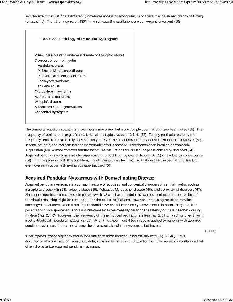
Table 23.1 Etiology of Pendular Nystagmus
Visual loss (including unilateral disease of the optic nerve)
Disorders of central myelin
Multiple sclerosis
Pelizaeus-Merzbacher disease
Peroxisomal assembly disorders
Cockayne's syndrome
Toluene abuse
Oculopalatal myoclonus
Acute brainstem stroke
Whipple's disease
Spinocerebellar degenerations
Congenital nystagmus
The temporal waveform usually approximates a sine wave, but more complex oscillations have been noted (29). The
frequency of oscillations ranges from 1–8 Hz, with a typical value of 3.5 Hz (58). For any particular patient, the
frequency tends to remain fairly constant; only rarely is the frequency of oscillations different in the two eyes (59).
In some patients, the nystagmus stops momentarily after a saccade. This phenomenon is called postsaccadic
suppression (60). A more common feature is that the oscillations are “reset” or phase-shifted by saccades (61).
Acquired pendular nystagmus may be suppressed or brought out by eyelid closure (62,63) or evoked by convergence
(64). In some patients with this condition, smooth pursuit may be intact, so that despite the oscillations, tracking
eye movements occur with nystagmus superimposed (58).
Acquired Pendular Nystagmus with Demyelinating DiseaseAcquired pendular nystagmus is a common feature of acquired and congenital disorders of central myelin, such as
multiple sclerosis (MS) (64), toluene abuse (65), Pelizaeus-Merzbacher disease (66), and peroxisomal disorders (67).
Since optic neuritis often coexists in patients with MS who have pendular nystagmus, prolonged response time of
the visual processing might be responsible for the ocular oscillations. However, the nystagmus often remains
unchanged in darkness, when visual inputs should have no influence on eye movements. In normal subjects, it is
possible to induce spontaneous ocular oscillations by experimentally delaying the latency of visual feedback during
fixation (Fig. 23.4C); however, the frequency of these induced oscillations is less than 2.5 Hz, which is lower than in
most patients with pendular nystagmus (29). When this experimental technique is applied to patients with acquired
pendular nystagmus, it does not change the characteristics of the nystagmus, but instead
superimposes lower-frequency oscillations similar to those induced in normal subjects (Fig. 23.4D). Thus,
disturbance of visual fixation from visual delays can not be held accountable for the high-frequency oscillations that
often characterize acquired pendular nystagmus.
Ovid: Walsh & Hoyt's Clinical Neuro-Ophthalmology http://ovidsp.tx.ovid.com.ezproxy.fiu.edu/spa/ovidweb.cgi
9 of 89 6/28/2009 8:53 AM

Figure 23.4. Acquired pendular nystagmus. A, Time plots of nystagmus of the patient's right eye while fixating
a central visual target. The horizontal and vertical records have been offset from zero eye position for
convenience of display. Upward deflections correspond to rightward or upward eye rotations. B, Nystagmus
trajectory seen as a scan path of eye movements in the horizontal and vertical planes corresponding to the
time plot in A. The scan path corresponded to the direction of oscillopsia that the patient reported. C and D,
Examples of ocular oscillations induced by imposing an effective delay in visual feedback. C, Normal subject.
An effective delay in visual feedback of 480 msec was imposed at time zero. The subject developed ocular
oscillations at about 1.0 Hz. D, Patient with multiple sclerosis and acquired pendular nystagmus. The effects of
imposing an electronic delay of 480 msec. The oscillations of her nystagmus (unchanged at 6.5 Hz) were
superimposed upon growing 0.67 Hz oscillations induced by the electronic manipulation. (Panels C and D
adapted from Averbuch-Heller L, Zivotofsky AZ, Das VE, et al. Investigations of the pathogenesis of acquired
pendular nystagmus. Brain 1995;188:369–378.)
A more likely possibility is that visual projections to the cerebellum are impaired, leading to instability in the
reciprocal connections between brainstem nuclei and cerebellum that are important for recalibration. The high
prevalence of internuclear ophthalmoplegia (INO) in these patients, suggests involvement of paramedian brainstem
regions, including the cell groups of the paramedian tracts (PMT) (35,68,69,70). PMT cell groups send a neural copy
of ocular motor signals to the cerebellum, and seem important for the integration and calibration of eye movement
commands. The observation that acquired pendular nystagmus is “reset” or phase-shifted after saccades (more so
with large saccades) suggested that the oscillations arise in the brainstem-cerebellar gaze-holding network (the
neural integrator for eye movements—which is discussed in the section “Nystagmus Due to Abnormalities of the
Mechanism for Holding Eccentric Gaze”) (61).
Ovid: Walsh & Hoyt's Clinical Neuro-Ophthalmology http://ovidsp.tx.ovid.com.ezproxy.fiu.edu/spa/ovidweb.cgi
10 of 89 6/28/2009 8:53 AM

P.1140
Oculopalatal Myoclonus (Oculopalatal Tremor)Acquired pendular nystagmus may be one component of the syndrome of oculopalatal (pharyngo-laryngo-
diaphragmatic) myoclonus (71,72,73). This condition usually develops several months after brainstem or cerebellar
infarction, although it may not be recognized until years later. Oculopalatal myoclonus also occurs with
degenerative conditions (74). The term “myoclonus” is misleading, since the movements of affected muscles are to
and fro and are approximately synchronized, typically at a rate of about 2 cycles per second. The palatal movements
may be termed “tremor,” rather than myoclonus, and the eye movements are really a form of pendular nystagmus
(75). Although the palate is most often affected, movements of the eyes, facial muscles, pharynx, tongue, larynx,
diaphragm, mouth of the eustachian tube, neck, trunk, and extremities may occur.
The ocular movements typically consist of to-and-fro oscillations, less sinusoidal than with demyelinating disease,
and often with a large vertical component, although they may also have small horizontal or torsional components.
The movements may be somewhat disconjugate (both horizontally
and vertically) (58), with some orbital position dependency (72), and some patients show cyclovergence (torsional
vergence) oscillations. Occasionally, patients develop the eye oscillations without movements of the palate,
especially following brainstem infarction. Eyelid closure may bring out the vertical ocular oscillations (62). The
nystagmus sometimes disappears with sleep, but the palatal movements usually persist. The condition is usually
intractable, and spontaneous remission is uncommon (76). Vertical pendular oscillations sometimes occur in the
acute period following pontine infarction (77), but the pathogenesis of these movements is probably different than
that of oculopalatal myoclonus, since they often resolve spontaneously.
Figure 23.5. Pathology of oculopalatal myoclonus. A section through the cerebellum and medulla shows
marked demyelination of the right dentate nucleus and restiform body (double arrows). The left inferior
olive is hypertrophic and shows mild demyelination (arrow). (From Nathanson M. Arch Neurol Psychiatr
1956;75:285–296.)
The main pathologic finding with palatal myoclonus is hypertrophy of the inferior olivary nucleus (Fig. 23.5), which
may be seen during life using magnetic resonance (MR) imaging (74). There may also be destruction of the
contralateral dentate nucleus (71). Histologically, the olivary nucleus has enlarged, vacuolated neurons with
enlarged astrocytes. Functional scanning demonstrates increased glucose metabolism (78). Guillain and Mollaret
Ovid: Walsh & Hoyt's Clinical Neuro-Ophthalmology http://ovidsp.tx.ovid.com.ezproxy.fiu.edu/spa/ovidweb.cgi
11 of 89 6/28/2009 8:53 AM

P.1141
proposed that disruption of connections between the dentate nucleus and the contralateral inferior olivary nucleus,
which run via the red nucleus and central tegmental tract, is responsible for the syndrome (71). However, neither
the dentate nucleus nor the red nucleus has been shown to have a specific role in ocular motor control. Thus, it has
thus been postulated that the nystagmus results from instability in the projection from the inferior olive to the
cerebellar flocculus, a structure thought to be important in the adaptive control of the vestibulo-ocular reflex
(69,72). It is also possible that disruption of projections from the cell groups of the paramedian tracts (PMT) (35) to
the cerebellum leads to the ocular oscillations.
Whipple's Disease and Other Predominantly Convergent-Divergent PendularOscillationsComparatively little has been written about vergence pendular oscillations, which are often small in amplitude, and
there is some evidence that they are often overlooked by clinicians. More widespread use of the magnetic search
coil technique has made it easier to identify the convergent-divergent components of this form of nystagmus.
Averbuch-Heller and colleagues reported three patients with pendular oscillations that were about 180° out of
phase in the horizontal and torsional planes but had conjugate vertical components (29). In one of these patients,
the torsional component of the oscillations had the largest amplitude. Thus, the patient actually had a
cyclovergence nystagmus.
Vergence pendular oscillations occur in patients with MS (79), brainstem stroke (58), and cerebral Whipple's disease
(80). In Whipple's disease, the oscillations typically have a frequency of about 1.0 Hz and are accompanied by
concurrent contractions of the masticatory muscles, a phenomenon called oculomasticatory myorhythmia.
Supranuclear paralysis of vertical gaze also occurs in this setting and is similar to that encountered in progressive
supranuclear palsy (81).
At least two possible explanations have been offered to account for the convergent-divergent nature of vergence
pendular oscillations: a phase shift between the eyes, produced by dysfunction in the normal yoking mechanisms, or
an oscillation affecting the vergence system itself (79). The latter explanation is more likely, because patients who
have been studied show no phase shift (i.e., are conjugate) vertically, and because the relationship between the
horizontal and torsional components is similar to that occurring during normal vergence movements
(excyclovergence with horizontal convergence) (29). Under experimental conditions, the vergence system can be
made to oscillate at frequencies up to 2.5 Hz—lower than that reported in patients with conditions other than
Whipple's disease (30,80). To account for these higher-frequency oscillations, it seems necessary to postulate
instability within the brainstem-cerebellar connections of the vergence system, for example, between the nucleus
reticularis tegmenti pontis and cerebellar nucleus interpositus, which may help hold vergence angle steady (29,82).
Nystagmus Caused by Vestibular ImbalanceNystagmus related to imbalance in the vestibular pathway can be caused by damage to peripheral or central
structures. Because the nystagmus varies, it usually is possible to distinguish nystagmus caused by peripheral
vestibular imbalance from nystagmus caused by central vestibular imbalance.
Nystagmus Caused by Peripheral Vestibular Imbalance
Clinical Features of Peripheral Vestibular NystagmusDisease affecting the peripheral vestibular pathway (i.e., the labyrinth, vestibular nerve, and its root entry zone)
causes
nystagmus with linear slow phases (Fig. 23.1A). Such unidirectional slow-phase drifts reflect an imbalance in the
level of tonic neural activity in the vestibular nuclei. If disease leads to reduced activity, for example, in the
vestibular nuclei on the left side, then the vestibular nuclei on the right side will drive the eyes in a slow phase to
the left. In this example, quick phases will be directed to the right—away from the side of the lesion. Paradoxically,
some patients show nystagmus with a horizontal component that beats toward the side of the lesion. Such cases may
Ovid: Walsh & Hoyt's Clinical Neuro-Ophthalmology http://ovidsp.tx.ovid.com.ezproxy.fiu.edu/spa/ovidweb.cgi
12 of 89 6/28/2009 8:53 AM
be “recovery nystagmus” (83), which represents the effects of a central adaptation process. An imbalance of
vestibular tone usually also causes vertigo and a tendency to fall toward the side of the lesion. Apart from these
attendant symptoms, two features of the nystagmus itself are useful in identifying the vestibular periphery as the
culprit: its trajectory (direction) and whether it is suppressed by visual fixation.
The trajectory of nystagmus can often be related to the geometric relationships of the semicircular canals and to
the finding that experimental stimulation of an individual canal produces nystagmus in the plane of that canal. Thus,
complete unilateral labyrinthine destruction leads to a mixed horizontal-torsional nystagmus (the sum of canal
directions from one ear), whereas in benign paroxysmal positional vertigo (BPPV), a mixed upbeat-torsional
nystagmus reflects posterior semicircular canal stimulation. Pure vertical or pure torsional nystagmus almost never
occurs with peripheral vestibular disease, because this would require selective lesions of individual canals from one
or both ears, an unlikely event.
Nystagmus caused by disease of the vestibular periphery often is more prominent, or may only become apparent,
when visual fixation is prevented. The reason for this is that when visually generated eye movements are working
normally, as they usually are in patients with peripheral vestibular disease, they will slow or stop the eyes from
drifting.
Another common, but not specific, feature of nystagmus caused by peripheral vestibular disease is that its intensity
increases when the eyes are turned in the direction of the quick phase—Alexander's law (84). This probably reflects
an adaptive strategy developed to counteract the drift of the vestibular nystagmus and so establish an orbital
position (i.e., in the direction of the slow phases) in which the eyes are quiet and vision is clear. This phenomenon
forms the basis for a common classification of unidirectional nystagmus. Nystagmus is called “first degree” if it is
present only on looking in the direction of the quick phases, “second degree” if it is also present in the central
position, and “third degree” if it is present on looking in all directions of gaze.
Although these clinical features help make the diagnosis of peripheral vestibular disease, it is important to realize
that brainstem and cerebellar disorders may sometimes mimic peripheral disease and, especially in elderly patients
or those with risk factors for vascular disease, careful observation is the prudent course.
Nystagmus Induced by Change of Head PositionVestibular nystagmus is often influenced by changes in head position. This feature can be used to aid in diagnosis,
especially of benign paroxysmal positional vertigo (BPPV). Patients with BPPV complain of brief episodes of vertigo
precipitated by change of head position, such as when they turn over in bed or look up to a high shelf. The
condition may follow head injury or viral neurolabyrinthitis (85).
To test for nystagmus and vertigo in a patient with possible BPPV, the examiner should turn the patient's head
toward one shoulder and then quickly move the head and neck together into a head-hanging (down 30–45°)
position. About 2–5 seconds after the affected ear is moved to this dependent position, a patient with BPPV will
report the onset of vertigo, and a mixed upbeat-torsional nystagmus, best viewed with Frenzel goggles, will
develop. The direction of the nystagmus changes with the direction of gaze. Upon looking toward the dependent
ear, it becomes more torsional; on looking toward the higher ear, it becomes more vertical. This pattern of
nystagmus corresponds closely to stimulation of the posterior semicircular canal of the dependent ear (which causes
slow phases mainly by activating the ipsilateral superior oblique and contralateral inferior rectus muscles). The
nystagmus increases for up to 10 seconds, but it then fatigues and is usually gone by 40 seconds. When the patient
sits back up, a similar but milder recurrence of these symptoms occurs, with the nystagmus being directed opposite
to the initial nystagmus. Repeating this procedure several times will decrease the symptoms and make the signs
more difficult to elicit. This habituation of the response is of diagnostic value, since a clinical picture similar to that
of BPPV can be caused by cerebellar tumors, MS, or posterior circulation infarction. With such central processes,
however, there is no latency to onset of nystagmus and no habituation of the response with repetitive testing. Some
patients present with the lateral canal variant of BPPV (86,87); sudden horizontal head turns as the patient lies
supine may induce a paroxysm of horizontal nystagmus beating toward the ground and vertigo.
Studies show that otolithic debris in the respective canals (canalolithiasis) interferes with the flow of endolymph or
Ovid: Walsh & Hoyt's Clinical Neuro-Ophthalmology http://ovidsp.tx.ovid.com.ezproxy.fiu.edu/spa/ovidweb.cgi
13 of 89 6/28/2009 8:53 AM

P.1142
movement of the cupula and is probably responsible for BPPV and its variants (88,89). Neck movement causing
vertebrobasilar kinking and vertigo as an isolated manifestation of transient brainstem ischemia is an uncommon
mechanism; in such cases, associated neurologic symptoms are usually present (90).
Nystagmus that persists after a horizontal change in head position (e.g., with the subject supine and the head
turned to the right or left) is less specific than transient nystagmus induced by changes in head position. Indeed,
some otherwise normal subjects develop nystagmus that is horizontal with respect to the head and becomes evident
behind Frenzel goggles during static, horizontal positional testing. Such positional nystagmus may remain beating in
the same direction whether the head is turned to the right or left, or it may change direction with lateral head turn
such that it is either always beating toward the earth (geotropic) or away from the earth (ageotropic or
apogeotropic). Sustained geotropic and ageotropic nystagmus probably reflect the effects of changing otolithic
influences and may be encountered with
either peripheral, or central vestibular lesions (90,91). Only if such nystagmus is present during visual fixation does
it suggest the possibility of central disease. Occasionally, disease affecting central vestibular connections, such as a
cerebellar tumor (92), infarction (93,94), or MS may produce nystagmus associated with postural vertigo and severe
nausea with vomiting. These manifestations may suggest a peripheral lesion; however, the characteristics of the
nystagmus are usually central, rather than peripheral. Alcohol is well know to cause positional nystagmus, and both
central and peripheral mechanisms contribute (95).
In patients who have symptomatically recovered from a unilateral, peripheral vestibulopathy, nystagmus can often
be induced following vigorous head shaking in the horizontal or the vertical plane for 10–15 seconds (96,97,98).
After horizontal head shaking, patients may show horizontal nystagmus with quick phases directed away from the
side of the lesion. Vertical nystagmus following horizontal head shaking (an example of “perverted nystagmus”)
often implies central vestibular disease (99,100). After vertical head shaking, patients with unilateral peripheral
vestibular lesions may show less prominent nystagmus with horizontal quick phases directed toward the side of the
lesion. Hyperventilation-induced nystagmus occurs in patients with schwannoma and other tumors of the 8th cranial
nerve (9,101). Indeed, hyperventilating 25 deep breaths is useful in the evaluation of the dizzy patient. Patients
with cerebellar disease may show transient downbeating nystagmus after horizontal head shaking or
hyperventilation (102).
Nystagmus Induced by Proprioceptive and Auditory StimuliIt is uncertain whether or not an imbalance of cervical inputs can produce a nystagmus similar to that caused by
peripheral vestibular disease. In normal human subjects, eye movements generated from cervical
proprioception—the cervico-ocular reflex (COR)—play little role in the stabilization of gaze (103), although the COR
does increase in responsiveness in individuals who have lost vestibular function (104,105), and in certain patients
with cerebellar disease (106).
The perception of passive body motion relies primarily on vestibular and visual information. However, an illusion of
body rotation accompanied by a conjugate, horizontal, jerk nystagmus—arthrokinetic nystagmus—can be induced
when the horizontally extended arm of a normal, stationary subject is passively rotated about a vertical axis in the
shoulder joint (107). The slow phase of the nystagmus is in a direction opposite to that of the arm movement. The
mean slow-phase velocity increases with increasing arm velocity, and the nystagmus continues for a short time
following cessation of arm movement (arthrokinetic after-nystagmus). The existence of arthrokinetic
circularvection and nystagmus suggests that there exists in normal humans a functionally significant somatosensory-
vestibular interaction within the central vestibular system, at least for afferent pathways carrying position and
kinesthetic information from the joints.
Normal stationary subjects in darkness may experience illusory self-rotation when exposed to a rotating sound field
(108,109). This illusion is generally accompanied by audiokinetic nystagmus, which is conjugate and horizontal,
with the slow phase in the direction opposite to that of the experienced self-rotation (110). This nystagmus
indicates that apparent, as well as actual, body orientation can influence ocular motor control. Neither the illusory
self-rotation nor the nystagmus occurs when the subject is exposed to a rotating sound field in the light, i.e., when
Ovid: Walsh & Hoyt's Clinical Neuro-Ophthalmology http://ovidsp.tx.ovid.com.ezproxy.fiu.edu/spa/ovidweb.cgi
14 of 89 6/28/2009 8:53 AM

P.1143
a stable visual environment is present, suggesting that visual information must dominate auditory information in
determining apparent body orientation and sensory localization (110). Patients who develop vestibular symptoms
and nystagmus when exposed to certain sounds—Tullio's phenomenon—often have dehiscence of the superior
semicircular canal or pathologic stimulation of otolithic organs (111,112,113,114,115,116).
Peripheral Vestibular Nystagmus Induced by Caloric or Galvanic StimulationNystagmus induced by caloric stimulation of one ear has all the features of that caused by unilateral or asymmetric
peripheral vestibular disease. During caloric stimulation, a temperature gradient across the temporal bone induces a
convection current in the endolymph of a semicircular canal if it is orientated vertical to the earth (117). A second
mechanism, which probably involves the effects of cooling the vestibular nerve, is less important (118,119). Before
attempting to induce caloric nystagmus, the physician must first check that the tympanic membrane is visible and
intact. The subject is then placed supine and the neck is flexed 30°. A cold stimulus (30°C) induces horizontal
slow-phase components directed toward the stimulated ear (quick phases in the opposite direction). With a warm
stimulus (44°C) and the same head orientation, quick phases are toward the stimulated ear (hence the mnemonic,
COWS: cold-opposite, warm-same).
Caloric stimulation is an important way to test each peripheral labyrinth; details of quantitative testing are
summarized elsewhere (91). Bedside testing with ice-cold water is especially useful in the evaluation of the
unconscious patient (120,121). In this setting, tonic eye deviation indicates preservation of pontine function.
Induction of caloric nystagmus is also a useful way to confirm preservation of consciousness in patients feigning
coma. Suppression of caloric nystagmus by visual fixation depends on pathways important for visually mediated eye
movements. For example, caloric nystagmus is impaired in patients with lesions of the cerebellar flocculus (122).
Galvanic stimulation of the peripheral labyrinth also induces nystagmus but, at present, this is largely used as a
research tool (91,123,124).
Nystagmus Caused by Central Vestibular Imbalance
Clinical Features of Central Vestibular NystagmusIn this section, we describe the clinical features of three common forms of nystagmus thought to be caused by
imbalance
of central vestibular connections: downbeat, upbeat, and torsional nystagmus. We also discuss the less common
phenomenon of horizontal nystagmus caused by central vestibular imbalance. Finally, we offer a pathophysiologic
scheme to account for these forms of central vestibular nystagmus.
Table 23.2 Etiology of Downbeat Nystagmus
Cerebellar degeneration, including familial episodic ataxia, and paraneoplastic degeneration
Craniocervical anomalies, including Arnold-Chiari malformation
Infarction of brainstem or cerebellum
Dolichoectasia of the vertebrobasilar artery
Multiple sclerosis
Cerebellar tumor, including hemangioblastoma
Syringobulbia
Encephalitis
Head trauma
Toxic-metabolic
Ovid: Walsh & Hoyt's Clinical Neuro-Ophthalmology http://ovidsp.tx.ovid.com.ezproxy.fiu.edu/spa/ovidweb.cgi
15 of 89 6/28/2009 8:53 AM
P.1144
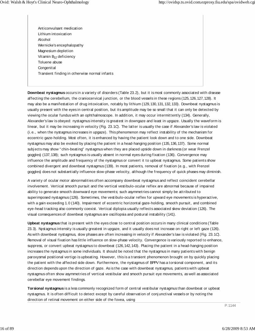
Anticonvulsant medication
Lithium intoxication
Alcohol
Wernicke's encephalopathy
Magnesium depletion
Vitamin B12 deficiency
Toluene abuse
Congenital
Transient finding in otherwise normal infants
Downbeat nystagmus occurs in a variety of disorders (Table 23.2), but it is most commonly associated with disease
affecting the cerebellum, the craniocervical junction, or the blood vessels in these regions (125,126,127,128). It
may also be a manifestation of drug intoxication, notably by lithium (129,130,131,132,133). Downbeat nystagmus is
usually present with the eyes in central position, but its amplitude may be so small that it can only be detected by
viewing the ocular fundus with an ophthalmoscope. In addition, it may occur intermittently (134). Generally,
Alexander's law is obeyed: nystagmus intensity is greatest in downgaze and least in upgaze. Usually the waveform is
linear, but it may be increasing in velocity (Fig. 23.1C). The latter is usually the case if Alexander's law is violated
(i.e., when the nystagmus increases in upgaze). This phenomenon may reflect instability of the mechanism for
eccentric gaze-holding. Most often, it is enhanced by having the patient look down and to one side. Downbeat
nystagmus may also be evoked by placing the patient in a head-hanging position (135,136,137). Some normal
subjects may show “chin-beating” nystagmus when they are placed upside down in darkness (or wear Frenzel
goggles) (137,138); such nystagmus is usually absent in normal eyes during fixation (136). Convergence may
influence the amplitude and frequency of the nystagmus or convert it to upbeat nystagmus. Some patients show
combined divergent and downbeat nystagmus (139). In most patients, removal of fixation (e.g., with Frenzel
goggles) does not substantially influence slow-phase velocity, although the frequency of quick phases may diminish.
A variety of ocular motor abnormalities often accompany downbeat nystagmus and reflect coincident cerebellar
involvement. Vertical smooth pursuit and the vertical vestibulo-ocular reflex are abnormal because of impaired
ability to generate smooth downward eye movements; such asymmetries cannot simply be attributed to
superimposed nystagmus (126). Sometimes, the vestibulo-ocular reflex for upward eye movements is hyperactive,
with a gain exceeding 1.0 (140). Impairment of eccentric horizontal gaze-holding, smooth pursuit, and combined
eye-head tracking also commonly coexist. Vertical diplopia usually reflects associated skew deviation (126). The
visual consequences of downbeat nystagmus are oscillopsia and postural instability (141).
Upbeat nystagmus that is present with the eyes close to central position occurs in many clinical conditions (Table
23.3). Nystagmus intensity is usually greatest in upgaze, and it usually does not increase on right or left gaze (126).
As with downbeat nystagmus, slow phases are often increasing in velocity if Alexander's law is violated (Fig. 23.1C).
Removal of visual fixation has little influence on slow-phase velocity. Convergence is variously reported to enhance,
suppress, or convert upbeat nystagmus to downbeat (126,142,143). Placing the patient in a head-hanging position
increases the nystagmus in some individuals. It should be noted that the nystagmus in many patients with benign
paroxysmal positional vertigo is upbeating. However, this is a transient phenomenon brought on by quickly placing
the patient with the affected side down. Furthermore, the nystagmus of BPPV has a torsional component, and its
direction depends upon the direction of gaze. As is the case with downbeat nystagmus, patients with upbeat
nystagmus often show asymmetries of vertical vestibular and smooth pursuit eye movements, as well as associated
cerebellar eye movement findings.
Torsional nystagmus is a less commonly recognized form of central vestibular nystagmus than downbeat or upbeat
nystagmus. It is often difficult to detect except by careful observation of conjunctival vessels or by noting the
direction of retinal movement on either side of the fovea, using
Ovid: Walsh & Hoyt's Clinical Neuro-Ophthalmology http://ovidsp.tx.ovid.com.ezproxy.fiu.edu/spa/ovidweb.cgi
16 of 89 6/28/2009 8:53 AM

an ophthalmoscope or contact lens. Although both peripheral vestibular and congenital nystagmus may have
torsional components, purely torsional nystagmus, like purely vertical nystagmus, indicates disease affecting central
vestibular connections (see Table 23.4) (144,145,146,147). Torsional nystagmus shares many of the features of
downbeat and upbeat nystagmus, including modulation by head rotations, variable slow-phase waveforms, and
suppression by convergence (146). It is also probably a common finding in patients with the ocular tilt reaction
(148). Nonrhythmic but continuous torsional eye movements may be a feature of paraneoplastic encephalopathy
(149).
Table 23.3 Etiology of Upbeat Nystagmus
Cerebellar degenerations, including familial episodic ataxia
Multiple sclerosis
Infarction of medulla, midbrain, or cerebellum
Tumors of the medulla, midbrain, or cerebellum
Wernicke's encephalopathy
Brainstem encephalitis
Behçet's syndrome
Meningitis
Leber's congenital amaurosis or other congenital disorder of the anterior visual pathways
Thalamic arteriovenous malformation
Organophosphate poisoning
Tobacco
Associated with middle ear disease
Congenital
Transient finding in otherwise normal infants
Table 23.4 Etiology of Torsional Nystagmus
Syringobulbia, with or without syringomyelia and Chiari malformation
Brainstem stroke (Wallenberg's syndrome) or arteriovenous malformation
Brainstem tumor
Multiple sclerosis
Oculopalatal myoclonus
Head trauma
Congenital
Associated with the ocular tilt reaction
Horizontal nystagmus in central position from central vestibular imbalance is an uncommon but well-documented
phenomenon. The underlying disorder usually is an Arnold-Chiari malformation (9,150). The slow-phase waveform in
this form of nystagmus may be of the increasing-velocity type, making distinction from congenital nystagmus
potentially difficult. However, patients with acquired central vestibular horizontal nystagmus typically report
Ovid: Walsh & Hoyt's Clinical Neuro-Ophthalmology http://ovidsp.tx.ovid.com.ezproxy.fiu.edu/spa/ovidweb.cgi
17 of 89 6/28/2009 8:53 AM
recent onset of visual symptoms, such as oscillopsia, and measurements usually demonstrate an associated vertical
component that is absent in congenital nystagmus. Patients with horizontal nystagmus that is present in the central
position always should be observed continuously for 2–3 minutes to exclude the possibility that the nystagmus is
actually periodic alternating nystagmus (PAN, discussion following).
Pathogenesis of Central Vestibular NystagmusOur understanding of the pathogenesis of central forms of vestibular nystagmus has increased because of more cases
with clinicopathological correlation, the development of animal and mathematic models, and the application of
modern anatomy and physiology. Downbeat nystagmus is usually associated with lesions of the vestibulocerebellum
flocculus, paraflocculus, nodulus, and uvula and the underlying medulla (126,151). Upbeat nystagmus is most
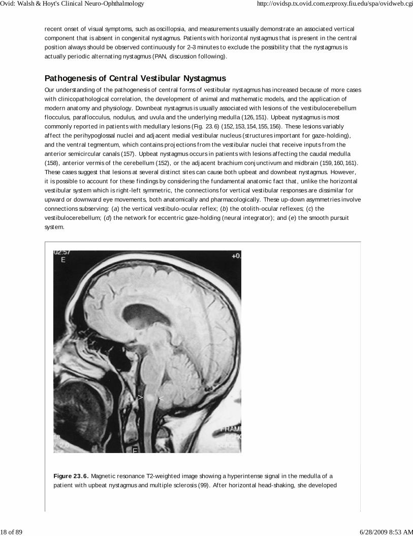
commonly reported in patients with medullary lesions (Fig. 23.6) (152,153,154,155,156). These lesions variably
affect the perihypoglossal nuclei and adjacent medial vestibular nucleus (structures important for gaze-holding),
and the ventral tegmentum, which contains projections from the vestibular nuclei that receive inputs from the
anterior semicircular canals (157). Upbeat nystagmus occurs in patients with lesions affecting the caudal medulla
(158), anterior vermis of the cerebellum (152), or the adjacent brachium conjunctivum and midbrain (159,160,161).
These cases suggest that lesions at several distinct sites can cause both upbeat and downbeat nystagmus. However,
it is possible to account for these findings by considering the fundamental anatomic fact that, unlike the horizontal
vestibular system which is right-left symmetric, the connections for vertical vestibular responses are dissimilar for
upward or downward eye movements, both anatomically and pharmacologically. These up-down asymmetries involve
connections subserving: (a) the vertical vestibulo-ocular reflex; (b) the otolith-ocular reflexes; (c) the
vestibulocerebellum; (d) the network for eccentric gaze-holding (neural integrator); and (e) the smooth pursuit
system.
Figure 23.6. Magnetic resonance T2-weighted image showing a hyperintense signal in the medulla of a
patient with upbeat nystagmus and multiple sclerosis (99). After horizontal head-shaking, she developed
Ovid: Walsh & Hoyt's Clinical Neuro-Ophthalmology http://ovidsp.tx.ovid.com.ezproxy.fiu.edu/spa/ovidweb.cgi
18 of 89 6/28/2009 8:53 AM
P.1145
P.1146
downbeating nystagmus (perverted head-shaking nystagmus) and tumbling vertigo.
Excitatory projections for the vertical vestibulo-ocular reflex from the posterior semicircular canals, which mediate
downward eye movements, synapse in the medial vestibular nucleus and then cross dorsally in the medulla beneath
the nucleus prepositus hypoglossi to reach the contralateral medial longitudinal fasciculus (MLF). Experimental
lesions that damage this pathway cause upward eye drifts and downbeat nystagmus (162). On the other hand, it
appears that excitatory connections from the anterior semicircular canals, which mediate upward eye movements,
take different routes; more than one pathway may contribute (163). In addition, a central imbalance of otolithic
inputs may contribute to vertical nystagmus (164), and account for “chin-beating” nystagmus when normal subjects
are positioned upside down in darkness (137,138). The case for the cerebellar flocculus being an important structure
in the production of downbeat nystagmus rests on the finding that Purkinje cells send inhibitory
projections to the central connections of the anterior semicircular canal but not to the posterior canal (Fig. 23.7)
(165,166,167). This asymmetry of inhibitory projections accounts for the finding that experimental flocculectomy
causes downbeat nystagmus (168). This lesion disinhibits the projections to the anterior canal but not to the
posterior canal, causing the eyes to drift up and producing downbeat nystagmus (167). A neural network that
includes the vestibulocerebellum and the nucleus prepositus hypoglossi and adjacent medial vestibular nucleus is
also thought to be important for the eccentric gaze-holding mechanism. Consistent with this hypothesis is the
report of a patient with lithium intoxication, who had downbeat nystagmus and a complete failure of gaze-holding,
and showed lesions in the nucleus prepositus hypoglossi (130). (However, a range of disorders of eye movements are
reported with lithium [131,169,170,171,172], so more than one mechanism may be disrupted by it.) Lesions of the
vestibulocerebellum may cause instability of this network, making the eyes drift at increasing velocity away from
central position in the vertical or horizontal planes (150,173,174,175). It has also been suggested that the
characteristics of downbeat nystagmus could be explained by a central imbalance in smooth pursuit with cerebellar
lesions (140). Resolution of upbeat or downbeat nystagmus after the first few months of life in otherwise normal
infants (176,177) may reflect calibration of pursuit or gaze-holding mechanisms as the visual system becomes fully
myelinated.
Periodic Alternating NystagmusPeriodic alternating nystagmus is a spontaneous horizontal nystagmus, present in central gaze, that reverses
direction approximately every 90–120 seconds (Fig. 23.8). Because the period of oscillation is about 4 minutes, the
disorder may be missed unless the examiner observes the nystagmus for several minutes. As the nystagmus finishes
one half-cycle (e.g., of right-beating nystagmus), a brief transition period occurs during which there may be
upbeating or downbeating nystagmus or saccadic movements before the next half-cycle (e.g., of left-beating
nystagmus) starts. A congenital form of PAN also exists (discussed in the section “Congenital Nystagmus”), but this is
usually much less regular in the timing of reversal of direction and shows slow-phase waveforms typical of congenital
nystagmus. PAN must also be differentiated from “ping-pong gaze,” an ocular deviation that reverses direction not
over several minutes but every few seconds and that is encountered in unconscious patients with large
bihemispheric lesions (178).
In most patients with acquired PAN, the nystagmus has the same characteristics in light or in darkness. Smooth
pursuit and optokinetic nystagmus are usually impaired (179). Vestibular stimuli are able to reset the oscillations,
and critically timed rotational stimuli can stop PAN for several minutes (179,180).
Acquired PAN occurs in association with a number of
conditions (Table 23.5), many of which affect the cerebellum. Experimental ablation of the nodulus and uvula of
the cerebellum in monkeys causes PAN when the animals are placed in a dark room. Baclofen abolishes this
nystagmus (181). One function of the nodulus and uvula is to control the time course of rotationally induced
nystagmus—so-called “velocity storage” (182). Following ablation of the nodulus and uvula, the duration (velocity
Ovid: Walsh & Hoyt's Clinical Neuro-Ophthalmology http://ovidsp.tx.ovid.com.ezproxy.fiu.edu/spa/ovidweb.cgi
19 of 89 6/28/2009 8:53 AM

storage) of rotationally induced nystagmus is prolonged excessively. It is postulated that normal vestibular repair
mechanisms reverse the direction of this nystagmus, thus producing the oscillations of PAN (179,181,182). These
oscillations would ordinarily be blocked by visual stabilization mechanisms that tend to suppress nystagmus, but
disease of the cerebellum that causes PAN usually also impairs these mechanisms.
Figure 23.7. Schematic hypothesis for downbeat nystagmus. Inputs from the anterior semicircular canals of
the vestibular labyrinth evoke upward eye movements via projections through the superior vestibular nuclei
to motoneurons supplying elevator muscles, including the superior rectus (CN III is oculomotor nucleus).
Inputs from the posterior semicircular canals evoke downward eye movements via projections through the
medial vestibular nuclei to motoneurons supplying depressor muscles, including the inferior rectus. The
flocculus of the cerebellum inhibits anterior but not posterior canal projections in the vestibular nuclei. If
inhibition from the flocculus is impaired, the eyes will drift upward causing downbeat nystagmus. (Schematic
based on Ito M, Nisimaru N, Yamamoto M. Specific patterns of neuronal connexions involved in the control of
the rabbit's vestibulo-ocular reflexes by the cerebellar flocculus. J Physiol (Lond) 1977;265:833–854; Baloh
RW, Spooner JW. Downbeat nystagmus: A type of central vestibular nystagmus. Neurology 1981;31:304–310.)
Ovid: Walsh & Hoyt's Clinical Neuro-Ophthalmology http://ovidsp.tx.ovid.com.ezproxy.fiu.edu/spa/ovidweb.cgi
20 of 89 6/28/2009 8:53 AM

Figure 23.8. Periodic alternating nystagmus in a 24-year-old woman with multiple sclerosis prior to (A) and
during (B) treatment with baclofen. Before treatment, PAN reverses direction approximately every 90
seconds; there is an associated downbeat nystagmus (evident in the inset at right, which has a magnified time
scale). During treatment with baclofen, PAN is essentially abolished, even when the room was switched to
complete darkness (indicated by horizontal bars in B, and shown in more detail in inset at right). Upward
deflections indicate rightward or upward eye movements. (From Garbutt S, Thakore N, Rucker JC, Han Y,
Kumar AN, Leigh RJ. Effects of visual fixation and convergence on periodic alternating nystagmus due to
multiple sclerosis. Neuro-Ophthalmology, in press.)
Ovid: Walsh & Hoyt's Clinical Neuro-Ophthalmology http://ovidsp.tx.ovid.com.ezproxy.fiu.edu/spa/ovidweb.cgi
21 of 89 6/28/2009 8:53 AM

P.1147
Two other unusual disorders may be related to PAN. The first is a variation of PAN, in which oscillations occurred in
both the horizontal and vertical planes, 90° out of phase,
and has been called periodic alternating windmill nystagmus (183). This phenomenon occurred in a blind patient.
The second is the report of a patient with paroxysms of mixed torsional-horizontal-vertical nystagmus that occurred
every 2 minutes in association with nausea (184). In this patient, the initial mechanism was probably caused by
paroxysmal hyperactivity in one vestibular nucleus complex, unlike PAN, in which prolongation of the vestibular
response is the initial mechanism. However, in both entities, an adaptive mechanism appears to influence the
nystagmus every 2 minutes. This is perhaps the most direct evidence that dysfunction of an ocular motor
recalibration mechanism can lead to nystagmus.
Table 23.5 Etiology of Periodic Alternating Nystagmus
Chiari malformations and other hindbrain anomalies
Multiple sclerosis
Cerebellar degenerations
Cerebellar tumor, abscess, cyst, and other mass lesion
Creutzfeldt-Jakob disease
Ataxia telangiectasia
Brainstem infarction
Anticonvulsant medications
Lithium intoxication
Infections affecting cerebellum, including syphilis
Hepatic encephalopathy
Trauma
Following visual loss (from vitreous hemorrhage or cataract)
Congenital nystagmus
Seesaw and Hemi-Seesaw NystagmusIn seesaw and hemi-seesaw nystagmus, one half-cycle consists of elevation and intorsion of one eye and
synchronous depression and extorsion of the other eye; during the next half-cycle, the vertical and torsional
movements reverse. The waveform may be pendular (185,186,187,188) or jerk. In the latter case, the slow phase
corresponds to one half-cycle (189). A seesaw component is present in many central forms of nystagmus. Seesaw
nystagmus may be congenital (185,190), or acquired (Table 23.6). Quantitative studies have done much to clarify
the characteristics and pathogenesis of seesaw nystagmus. It has been proposed that jerk seesaw nystagmus (hemi-
seesaw nystagmus) occurs in patients with lesions in the region of the interstitial nucleus of Cajal (INC) (189),
although experimental inactivation of this structure has not produced this nystagmus (191). Such patients often
have a contralateral ocular tilt reaction. With a right INC lesion, the reaction consists of a left head tilt, a skew
deviation with a right hypertopia, tonic intorsion of the right eye and extorsion of the left eye, and misperception
that earth-vertical is tilted to the left (189,192). Rarely, the ocular tilt reaction is paroxysmal in form, in which case
it is ipsilateral to the INC lesion; however paroxysmal skew deviation is also reported with lesions of the cerebellar
uvula (193). Some patients with this condition also show corresponding paroxysms of jerk seesaw nystagmus (189).
The ocular tilt reaction is believed to be caused by an imbalance of central otolithic projections from vestibular
nuclei to the INC. Stimulation in the region of INC in monkeys, for example, produces an ocular tilt reaction
consisting of extorsion and depression of the eye on the stimulated side and intorsion and elevation of the other
eye (194); somewhat similar results have been reported in humans (195,196). Thus, the various forms of the ocular
tilt reaction are similar to the slow phase of jerk seesaw nystagmus. Isolated INC lesions may be characterized by
Ovid: Walsh & Hoyt's Clinical Neuro-Ophthalmology http://ovidsp.tx.ovid.com.ezproxy.fiu.edu/spa/ovidweb.cgi
22 of 89 6/28/2009 8:53 AM

P.1148
ipsilesional torsional nystagmus and a restricted range of vertical saccades that are not slowed (197). If the adjacent
rostral interstitial nucleus of the MLF (riMLF) is also damaged, however, either no quick phases (189), or
contralesional quick phases (198), may be observed. (As discussed in Chapter 17, each riMLF contributes to upward
and downward saccades but only to ipsilaterally directed torsional quick phases.)
Table 23.6 Etiology of Seesaw Nystagmus
Meso-diencephalic diseasea
Parasellar masses
Brainstem stroke
Septo-optic dysplasia
Chiari malformation
Syringobulbia
Retinitis pigmentosa
Head trauma
Congenital form, including agenesis of optic chiasm, and as a transient finding in albinism
a Includes hemi-seesaw nystagmus.
Pendular seesaw nystagmus has most often been reported in patients with large tumors in the region of the optic
chiasm and diencephalon (Fig. 23.9), and thus these oscillations
have been attributed to either compression of the diencephalon or to the effects of chiasmal visual field defects.
One aspect of the vestibular responses concerns movements to compensate for head roll motion if the subject looks
at an object located off the midsagittal plane; in this case a seesaw rotation of the eyes is the geometrically
appropriate compensation (199). Normal calibration of this response, which would require that motion-visual
information be sent to the cerebellum could be impaired with large suprasellar lesions, leading to the pendular
variant of seesaw nystagmus (188). Thus, both the jerk and pendular variants of seesaw nystagmus probably arise
from imbalance or miscalibration of vestibular responses that normally function to optimize gaze during head
rotations in roll.
Ovid: Walsh & Hoyt's Clinical Neuro-Ophthalmology http://ovidsp.tx.ovid.com.ezproxy.fiu.edu/spa/ovidweb.cgi
23 of 89 6/28/2009 8:53 AM

Figure 23.9. Pathology of seesaw nystagmus. A 47-year-old woman developed progressive visual loss and was
noted to have bitemporal hemianopia and seesaw nystagmus. The patient died 8 months later, following
subtotal removal of a craniopharyngioma. Note that the diencephalon and rostral mesencephalon are
compressed and partially destroyed by the tumor that did not, however, obstruct the foramina of Monro
(arrow).
Nystagmus Due to Abnormalities of the Mechanism for Holding EccentricGaze
Gaze-Evoked NystagmusNystagmus that is induced by turning the eye to an eccentric position in the orbit is called gaze-evoked nystagmus.
It is the most common form of nystagmus encountered in clinical practice. Although the terms gaze-evoked
nystagmus, end-point nystagmus, and gaze-paretic nystagmus are often used synonymously, gaze-evoked nystagmus
is a general term that includes both physiologic and pathologic nystagmus. When the nystagmus is physiologic, the
term end-point nystagmus is appropriate (see below). When the nystagmus is associated with a paresis of gaze, as in
patients with ocular motor nerve palsies or weakness of the extraocular muscles, the term gaze-paretic nystagmus is
appropriate.
Gaze-evoked nystagmus usually occurs on lateral or upward gaze, seldom on looking down. If fixation is impaired or
prevented (e.g., in darkness), the slow phases consist of centripetal drifts that may have an exponentially decaying
waveform (Fig. 23.1B). If visual fixation is possible, however, the slow phases have a more linear profile.
In order to understand how gaze-evoked nystagmus arises, one must consider the neural command required to hold
the eye steadily at an eccentric position in the orbit. When the eye is turned toward a corner of the orbit, the
fascia and ligaments that suspend the eye exert an elastic force to return toward central position. Overcoming this
elastic restoring force requires a tonic contraction of the extraocular muscles. This is achieved by an eye position
signal to the ocular motoneurons, called a step, that is generated by the gaze-holding network, also called the
neural integrator. This network includes the vestibulocerebellum, the medial vestibular nucleus and adjacent
nucleus prepositus hypoglossi in the medulla, and the interstitial nucleus of Cajal (INC) in the midbrain.
Gaze-evoked nystagmus is caused by a deficient step, such that the eyes cannot be maintained at an eccentric
Ovid: Walsh & Hoyt's Clinical Neuro-Ophthalmology http://ovidsp.tx.ovid.com.ezproxy.fiu.edu/spa/ovidweb.cgi
24 of 89 6/28/2009 8:53 AM
srose
Highlight
srose
Highlight

P.1149
orbital position and are pulled back toward central position by the elastic forces of the orbital fascia. Corrective
quick phases then move the eyes back toward the desired position in the orbit. Frequently, lesions that produce
gaze-evoked nystagmus also impair visual fixation and smooth pursuit.
Gaze-evoked nystagmus may be caused by a variety of medications, including alcohol, anticonvulsants, and
sedatives. Gaze-evoked nystagmus may also be caused by structural lesions that damage the gaze-holding neural
network. Experimental lesions of the nucleus prepositus hypoglossi/medial vestibular nucleus region effectively
abolish horizontal gaze-holding function (200,201,202), and also partially impair vertical gaze-holding. Inactivation
of INC abolishes vertical gaze-holding function (203). Experimental flocculectomy greatly, but not completely,
impairs horizontal gaze holding (168) in addition to causing downbeat nystagmus.
Rarely, cerebellar lesions cause the gaze-holding mechanism to become unstable, so that the eyes drift with
increasing velocity away from central position in either the vertical (173) or the horizontal plane (150). This “gaze-
instability nystagmus” often violates Alexander's law.
Another cause of gaze-evoked nystagmus is familial episodic ataxia type 2 (EA-2), which is characterized by attacks
of ataxia and vertigo lasting hours, with interictal nystagmus. They nystagmus is typically gaze-evoked with a
vertical component that can be downbeat or upbeat. Pursuit and optokinetic responses may be impaired, whereas
vestibular responses may be normal or increased (204).
Differences between Physiologic End-Point Nystagmus and PathologicGaze-Evoked NystagmusGaze-evoked nystagmus is commonly encountered in normal subjects, in which cases it is often called end-point
nystagmus (205,206,207). It typically occurs on looking far laterally and is poorly sustained. The nystagmus is
primarily horizontal. It is usually symmetric, but it may be asymmetric, being more prominent on looking to one side
than to the other (207). In some normal persons, the nystagmus is sustained, occurs with less than full deviations of
the eye, and may be slightly dissociated or have a torsional component. In addition, some normals show pendular
oscillations in far eccentric gaze (208). In such individuals, this physiologic form of gaze-evoked nystagmus can
usually be differentiated from that caused by disease, since the former has lower intensity (i.e., slower drift) and,
most importantly, is not accompanied by other ocular motor abnormalities. Pathologic gaze-evoked nystagmus, in
contrast, is accompanied by other defects of eye movements, such as impaired smooth pursuit (209).
Dissociated NystagmusA special type of pathologic gaze-evoked nystagmus is dissociated or “ataxic” nystagmus. This type of nystagmus is
most commonly encountered with an internuclear ophthalmoplegia (INO). Dissociated nystagmus is, in fact, a
series of saccades followed by postsaccadic drift that occurs when the patient attempts to look laterally away from
the side of the lesion. Since the saccades initiate the oscillations, this ocular motor abnormality is not a true
nystagmus, but rather a series of saccadic pulses. Consider, for example, a
patient with a right-sided INO (Fig. 23.10, top). When the patient attempts to look to the left, the adducting
saccades of the right eye are slow and hypometric. Each consists of a hypometric pulse, followed by a glissadic drift
of the eye toward the target. Abducting saccades in the left eye are hypermetric, overshooting the target, and are
followed by a glissadic backward drift of the eye. A series of such small saccades and drifts gives the appearance of
dissociated nystagmus. Because of the difference in the velocity of the adducting saccades in the eye on the side of
the lesion and the abducting saccades in the contralateral eye, comparison of horizontal saccades made by each eye
is most useful in making the diagnosis of an INO. When the INO is subtle, moving an optokinetic tape or rotating an
optokinetic drum toward the side of the affected medial rectus muscle induces asymmetric quick phases, with
smaller-sized movements in the affected eye. Some caution is required in interpreting this sign, however, since
abducting saccades are normally slightly faster than adducting saccades (210). In addition, there are settings other
than an INO in which patients may show an asymmetry of saccadic velocities, with the adducting eye moving more
slowly than the abducting eye (discussion following). Nevertheless, when one observes slowed adducting saccades in
one eye and normal abducting saccades in the opposite eye in a patient with no previous history of strabismus
Ovid: Walsh & Hoyt's Clinical Neuro-Ophthalmology http://ovidsp.tx.ovid.com.ezproxy.fiu.edu/spa/ovidweb.cgi
25 of 89 6/28/2009 8:53 AM
srose
Highlight
srose
Highlight
srose
Highlight
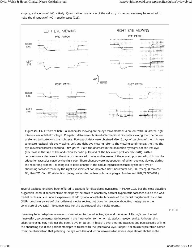
P.1150
surgery, a diagnosis of INO is likely. Quantitative comparison of the velocity of the two eyes may be required to
make the diagnosis of INO in subtle cases (211).
Figure 23.10. Effects of habitual monocular viewing on the eye movements of a patient with unilateral, right
internuclear ophthalmoplegia. Pre-patch data were obtained after habitual binocular viewing, but the patient
preferred to fixate with the right eye. Post-patch data were obtained after 5 days of patching of the right eye
to ensure habitual left eye viewing. Left and right eye viewing refer to the viewing conditions at the time the
eye movements were recorded. Post-patch: Note the decrease in the abduction nystagmus of the left eye
(decrease in the size of the abduction saccadic pulse and of the backward postsaccadic drift), with a
commensurate decrease in the size of the saccadic pulse and increase of the onward postsaccadic drift for the
adduction saccades made by the right eye. These changes were independent of which eye was viewing during
the recording session. Patching led to little change in the adducting saccades made by the left eye or
abducting saccades made by the right eye (vertical bar indicates +20°; horizontal bar, 500 msec). (From Zee
DS, Hain TC, Carl JR. Abduction nystagmus in internuclear ophthalmoplegia. Ann Neurol 1987;21:383–388.)
Several explanations have been offered to account for dissociated nystagmus in INO (9,212), but the most plausible
suggestion is that it represents an attempt by the brain to adaptively correct hypometric saccades due to the weak
medial rectus muscle. Acute experimental INO by local anesthetic blockade of the medial longitudinal fasciculus
(MLF), produces paresis of the ipsilateral medial rectus, but does not produce abducting nystagmus in the
contralateral eye (213). To compensate for the weakness of the medial rectus,
there may be an adaptive increase in innervation to the adducting eye and, because of Hering's law of equal
innervation, a commensurate increase in the innervation to the normal, abducting eye results. Although this
adaptive change may help get the paretic eye on target, it leads to overshooting saccades and postsaccadic drift of
the abducting eye if the patient attempts to fixate with the ipsilesional eye. Support for this interpretation comes
from the observation that patching the eye with the adduction weakness for several days almost abolishes the
Ovid: Walsh & Hoyt's Clinical Neuro-Ophthalmology http://ovidsp.tx.ovid.com.ezproxy.fiu.edu/spa/ovidweb.cgi
26 of 89 6/28/2009 8:53 AM
overshoot and pulse-step mismatch of the abducting eye when the latter eye fixates (Fig. 23.10, bottom left) (212).
Additional support for the view that the abduction nystagmus of an INO is a compensatory response to a
“peripheral” weakness comes from the observation that surgically caused medial rectus weakness leads to a similar
nystagmus, which resolves if the eye with the weak medial rectus is patched for several days (214). In addition to
previous extraocular muscle surgery, both myasthenia gravis (215) and the Miller Fisher syndrome (216) may produce
a dissociated nystagmus similar to that seen in an INO.
Dissociated nystagmus characterized by larger movements in the adducting eye occurs when some patients with
abducens nerve palsy look into the paretic field (9). Indeed, whenever a patient habitually prefers to fixate with a
paretic eye, the normal eye will show a dissociated nystagmus while looking in the direction of action of the paretic
muscle, regardless of the pathogenesis of the weakness.
Bruns' NystagmusTumors in the cerebellopontine angle, such as meningiomas or schwannomas of the vestibulocochlear nerve, may
produce a low-frequency, large-amplitude nystagmus when the patient looks toward the side of the lesion, and a
high-frequency, small-amplitude nystagmus when the patient looks toward the side opposite the lesion. The
nystagmus that occurs on gaze toward the side of the lesion is gaze-evoked nystagmus caused by defective gaze
holding, whereas the nystagmus that occurs during gaze toward the side opposite the lesion is caused by vestibular
imbalance. This special nystagmus is called Bruns' nystagmus (217,218).
Convergence-Retraction NystagmusSo-called convergence-retraction nystagmus is characterized by quick phases that converge or retract the eyes on
attempts to look up. It is elicited either by asking the patient to make an upward saccade or by using a handheld
optokinetic drum or tape and moving the stripes or figures down. This maneuver produces slow, downward, pursuit
eye movements, but upward quick phases are replaced by rapid convergent movements, retractory movements, or
both. Affected patients usually have impaired or absent upward gaze for both pursuit and saccadic eye movements;
however, in some cases upward pursuit appears normal, whereas upward saccades are obviously abnormal.
Convergence-retraction nystagmus is commonly produced by lesions of the mesencephalon that damage the
posterior commissure, such as pineal tumors (219,220,221); an example is shown in Figure 23.11. It has been
proposed that convergence-retraction nystagmus is, in fact, a saccadic disorder rather than nystagmus because the
primary adductive movements are asynchronous adducting saccades (222). However, other studies have indicated
that the movements may be vergence in origin (223); more studies are needed to resolve this issue. During
horizontal saccades, the abnormal pattern of convergent innervation manifests itself as slowing of the abducting
eye: “pseudo-abducens palsy” (219,221). Convergence-retraction nystagmus may also occur with a Chiari
malformation or epileptic seizures (224,225). Convergence-retraction nystagmus is usually intermittent, being
determined by saccadic activity, and it thus can be differentiated from other more continuous forms of disjunctive
nystagmus, such as convergent-divergent pendular nystagmus (29,79) and the oculomasticatory myorhythmia
characteristic of Whipple's disease (80).
Ovid: Walsh & Hoyt's Clinical Neuro-Ophthalmology http://ovidsp.tx.ovid.com.ezproxy.fiu.edu/spa/ovidweb.cgi
27 of 89 6/28/2009 8:53 AM

P.1151
Figure 23.11. Pathology of convergence-retraction nystagmus. A pinealoma is compressing the dorsal
midbrain of a 13-year-old boy who also had inability to elevate the eyes above the horizontal midline,
limitation of downward gaze, and anisocoria.
Jerk-waveform divergence nystagmus is diagnosed infrequently, but it may occur in patients with cerebellar
disease, such as the Chiari malformation. In such cases, combined divergent and downbeat nystagmus produces slow
phases that are directed upward and inward (139).
Centripetal and Rebound NystagmusIf a patient with gaze-evoked nystagmus attempts to look eccentrically for a sustained period, the nystagmus may
begin to decrease in amplitude and may even reverse direction, so that the eye begins to drift centrifugally
(“centripetal nystagmus”) (226). If the eyes are then returned to the central position, a short-lived nystagmus with
slow drifts in the direction of the prior eccentric gaze occurs. This is called rebound nystagmus (227,228). Both
centripetal and rebound nystagmus may reflect an attempt by brainstem or cerebellar mechanisms to correct for the
centripetal drift of
gaze-evoked nystagmus. Rebound nystagmus typically occurs in patients with cerebellar disease, but it has been
reported following experimental lesions in the region of the nucleus prepositus hypoglossi and medial vestibular
nucleus (200), and in normal subjects with typical gaze-evoked nystagmus (207). Extreme gaze deviation away from
the side of the lesion in one patient with a lateral medullary infarction was reported to cause this type of
paroxysmal nystagmus and vertigo lasting about 1 minute (229). Such a phenomenon could be explained by a
sustained eye position signal causing an imbalance of central vestibular mechanisms. Rebound nystagmus has been
reported in a patient with a tumor confined to the flocculus (230); other ocular motor abnormalities, including
impaired smooth pursuit and gaze-evoked nystagmus persisted after the tumor invaded the vestibular nuclei, but
the rebound nystagmus disappeared. Such findings have suggested that rebound is due to a the development of a
bias within the vestibular nuclei.
Congenital Forms of Nystagmus
Nature and Forms of Congenital Ocular Oscillations
Ovid: Walsh & Hoyt's Clinical Neuro-Ophthalmology http://ovidsp.tx.ovid.com.ezproxy.fiu.edu/spa/ovidweb.cgi
28 of 89 6/28/2009 8:53 AM

In this section, we review those forms of nystagmus that develop during infancy (231). Advances have been made
with the identification of congenital forms of nystagmus in mutant dogs with abnormal anatomy of the visual system
(47), and in normal monkeys that are deprived of binocular vision during early life (232). However, although some
patients with congenital nystagmus show visual abnormalities, others with similar ocular oscillations do not.
Furthermore, the characteristic waveforms, e.g., pendular (Fig. 23.1D) or jerk (Fig. 23.1A), do not specify
pathogenesis or indicate whether the congenital nystagmus is associated with visual system anomalies (18). Thus,
the underlying mechanisms are not fully understood. Three distinct syndromes are currently recognized: congenital
nystagmus, latent nystagmus, and spasmus nutans.
Although there is no widely accepted classification of nystagmus in infancy, the Classification of Eye Movement
Abnormalities and Strabismus (CEMAS) Working Group (233) has proposed that the term infantile nystagmus
syndrome (INS) be used to encompass what has previously been called congenital nystagmus, including motor or
sensory varieties. The main criteria for defining INS are infantile onset and accelerating slow-phase waveforms (Fig.
23.1C). However, a potential problem posed by the CEMAS classification for some ophthalmologists is that
measurement of eye movements, which is necessary to characterize the waveforms, may not be available. In
additional, CEMAS has called latent nystagmus Fusional Maldevelopment Nystagmus Syndrome (FMNS). In this
chapter, we will continue to use the terms “congenital nystagmus” and “latent nystagmus” but the reader should
note that a transition of terminology may occur in the neuro-ophthalmological community over the next few years.
Congenital Nystagmus
Clinical FeaturesCongenital nystagmus is usually diagnosed during infancy, but occasionally presents during adult life (234,235) when
it may create a diagnostic problem, especially if the patient has other symptoms, such as headaches or dizziness.
Certain clinical features usually differentiate congenital nystagmus from other ocular oscillations. It is almost always
conjugate and horizontal, even on up or down gaze. A small torsional component to the nystagmus is probably
common (236), but is difficult to identify clinically. Only rarely is congenital nystagmus purely vertical
(237,238,239). Congenital nystagmus is usually accentuated by the attempt to visually fixate an object and by
attention or anxiety. Eyelid closure (240) and convergence usually suppress it (241,242,243), but occasionally
congenital nystagmus is evoked by viewing a near target (79,244). Its intensity may also be influenced by viewing
the vertical lines of an optokinetic tape (245). Congenital nystagmus often decreases when the eyes are moved into
a particular position in the orbit, called the “null” region. In some patients, the direction of the nystagmus
spontaneously reverses direction during sustained viewing of a fixation point (18,246,247). The direction of the
nystagmus may also be influenced by which eye is viewing, with the nystagmus beating away from the covered eye;
this is similar to latent nystagmus (see below).
The most distinctive feature of congenital nystagmus is its waveforms, the most common of which are increasing-
velocity (Fig. 23.1C) and pendular (Fig. 23.1D). Frequently superimposed on these waveforms, which may be
combined, are foveation periods—the hallmark of congenital nystagmus (18,248,249). During each cycle, usually
after a quick phase, there is a brief period when the eye is still and is pointed at the object of regard. With jerk
waveforms, the quick phases (saccades) may brake the oscillation (250) or bring the eye to the target. With
pendular waveforms, the oscillation is flattened by a foveation period when the eye is closest to the target (Fig.
23.12). The particular waveform that is seen in a patient with congenital nystagmus depends in part upon the
patient's age. Most infants show largeamplitude “triangular” waveforms. Shortly thereafter, the waveform becomes
pendular, but it changes again to a jerk type as the patient reaches about one year of age (251,252). These
waveforms are so characteristic of congenital nystagmus that reliable records of eye position and velocity will often
secure the diagnosis.
In those individuals with congenital nystagmus that spontaneously changes direction, the timings of reversal tend to
be this irregular. This aperiodic alternating congenital nystagmus appears to have a different pathogenesis than
acquired periodic alternating nystagmus (247,253,254).
Foveation periods are only rarely reported in acquired forms of nystagmus (255). They are probably one reason
Ovid: Walsh & Hoyt's Clinical Neuro-Ophthalmology http://ovidsp.tx.ovid.com.ezproxy.fiu.edu/spa/ovidweb.cgi
29 of 89 6/28/2009 8:53 AM

P.1152
P.1153
(along with elevated thresholds for motion detection) why most patients with congenital nystagmus do not complain
of oscillopsia, despite otherwise nearly continuous movement
of their eyes (8,256,257,258,259,260,261,262), and why many have normal visual acuity (263,264). However,
foveation periods are not invariably present in congenital nystagmus. When they are absent or poorly developed,
vision is usually impaired (265).
Figure 23.12. A pendular type of congenital nystagmus waveform with superimposed quick phases, showing
the large horizontal component, smaller torsional component, and almost absent vertical component. Note
that vertical and torsional channels have been offset to aid clarity of display. Foveation periods follow quick
phases (when eye position is close to zero) and are indicated by horizontal bars. Upward deflections indicate
rightward (horizontal), upward (vertical), or clockwise (torsional) eye rotations, with respect to the patient.
Up to 30% of patients with congenital nystagmus also have strabismus (266). A commonly described associated
finding in such patients is “inverted pursuit” or “reversed optokinetic nystagmus” (267). With a handheld
optokinetic drum or tape, quick phases are directed in the same direction as the drum rotates or the tape moves. In
fact, based on measurements made of tracking during the foveation period, it has been shown that both smooth
pursuit and optokinetic eye movements are preserved in at least some individuals with congenital nystagmus
(268,269). Similarly, vestibular responses are generally normal in patients with congenital nystagmus if judged by
retinal image stability during foveation periods (270,271). Thus, their view of the world is similar whether they are
stationary or in motion (272). In those patients with associated visual disorders such as albinism, however, vestibular
responses to lower frequencies of head rotations and optokinetic responses may be impaired (273,274).
Occasional patients exhibit congenital nystagmus only during attempted smooth tracking (275), and others can
voluntarily release or inhibit their congenital nystagmus, suggesting that fixation plays some role in the oscillations
(276). Congenital nystagmus associated with congenital gaze-holding failure (i.e., leaky neural integrator) has been
reported
in a kindred (277) suggesting that, at least in this family, instability of the gaze-holding mechanism (neural
Ovid: Walsh & Hoyt's Clinical Neuro-Ophthalmology http://ovidsp.tx.ovid.com.ezproxy.fiu.edu/spa/ovidweb.cgi
30 of 89 6/28/2009 8:53 AM

integrator) is unlikely to have been the primary cause of the nystagmus.
Head turns are common in patients with congenital nystagmus and are an adaptive strategy to bring the eyes close
to the null position in the orbit, where nystagmus is reduced (278). The observation of such a head turn in childhood
photographs is often helpful in diagnosing congenital nystagmus. Another strategy used by patients with either
congenital or latent nystagmus (discussion following) is to purposely induce an esotropia to suppress the nystagmus.
Such an esotropia requires a head turn to direct the viewing eye at the object of interest. This phenomenon is
called the nystagmus blockage syndrome (279,280).
Some patients with congenital nystagmus also show head oscillations (271,281,282). Such head movements cannot
act as an adaptive strategy to improve vision, however, unless the vestibulo-ocular reflex is negated. In fact, head
movements are not compensatory in most patients with congenital nystagmus and tend to increase when the
individual attends to an object, an effort that also increases the nystagmus. It seems possible, therefore, that the
head tremor and ocular oscillations represent the output of a common disordered neural mechanism (282).
Pathogenesis of Congenital NystagmusAs noted earlier, nystagmus developing early in life and showing some of the waveform characteristics of congenital
nystagmus in humans also occurs in mutant dogs who lack the normal hemidecussation of fibers in the optic chiasm
(47), and in normal monkeys who are subjected to monocular visual deprivation in infancy (232). It is also associated
with a variety of visual system disorders, including ocular and oculocutaneous albinism (283,284,285,286),
achromatopsia, retinal cone dystrophy, optic nerve hypoplasia, Leber's congenital amaurosis, retinal coloboma,
aniridia, corectopia, congenital stationary night blindness, Chédiak-Higashi syndrome, Joubert syndrome, and
peroxisomal disorders (287). Because of the many diagnostic possibilities, a complete ophthalmologic evaluation and
an electroretinogram must be performed in patients with congenital nystagmus associated with decreased visual
acuity or visual dysfunction (287,288,289). Congenital nystagmus, both with and without associated visual system
abnormalities, may be familial (290). Several modes of inheritance have been reported (231,291,292,293); in
X-linked forms (292), the mothers may show subtle ocular motor abnormalities. Congenital nystagmus, with some
similarities of waveform, has been reported in monozygotic twins (294). However, there may be considerable
differences in waveforms among members of a single family with hereditary congenital nystagmus (290).
The known anatomic variations of the anterior visual system in individuals with congenital nystagmus, such as
excessive crossing at the chiasm in association with albinism (295,296,297), or absent crossing of nasal fibers in
achiasmatic subjects with congenital seesaw nystagmus (47,48,49), have led to the development of models for
congenital nystagmus based on “miswiring” of visual pathways (276,298). A model postulating that maladaptation to
early visual deprivation leads to instability of the gaze-holding mechanisms has also been proposed (299). Further
work is required to validate these models.
Latent (Occlusion) Nystagmus
Clinical FeaturesTrue latent nystagmus is a horizontal jerk nystagmus that is absent when both eyes are viewing but appears when
one eye is covered. This conjugate nystagmus is characterized by quick phases of both eyes that beat toward the
side of the fixating eye. In most patients, a low-amplitude nystagmus is also present when both eyes are viewing,
and is called “manifest latent nystagmus” (300). It apparently occurs because only one eye is fixating, and vision
from the other deviated eye is suppressed (301,302). Latent nystagmus usually reverses direction upon covering of
either eye. In some patients, however, it is present when one eye is covered but is absent when the other is
covered. Occasional patients can control their latent nystagmus at will (303). Latent nystagmus is usually associated
with strabismus, typically esotropia (304). Amblyopia is frequent, whereas binocular vision with normal stereopsis is
rare. Latent nystagmus has been described in identical twins (305).
The slow phase of latent nystagmus usually shows a decaying velocity waveform when the eyes are close to central
position (Fig. 23.1B), in contrast to the increasing velocity waveform of congenital nystagmus. A number of studies
suggest that foveation may occur during the slowest part of the drift if the amplitude of the nystagmus is large, and
Ovid: Walsh & Hoyt's Clinical Neuro-Ophthalmology http://ovidsp.tx.ovid.com.ezproxy.fiu.edu/spa/ovidweb.cgi
31 of 89 6/28/2009 8:53 AM

P.1154
immediately after the quick phase if the amplitude is small (306). Latent nystagmus usually follows Alexander's law,
with the nystagmus being greatest on looking in the direction of the quick phases, away from the covered eye.
Some patients turn the head to keep the viewing eye in an adducted position, where nystagmus is minimal.
Occasionally, congenital and latent nystagmus coexist, in which case the waveforms may be quite complex. Rarely,
in such patients, if vision is clearer with the latent nystagmus waveforms than with the congenital nystagmus
waveforms, the patients switch from congenital to latent nystagmus as one eye becomes esotropic and the other
takes up fixation (279). In addition to strabismus, patients with latent nystagmus frequently show an upward
deviation of the covered eye (alternating sursumduction or dissociated vertical deviation) (307). In such patients,
the nystagmus often has a torsional component (307). Latent nystagmus is quite common, and accurate diagnosis is
important to avoid inappropriate investigations. It should be differentiated from gaze-evoked nystagmus in
association with strabismus, especially abducting nystagmus occurring with INO, in which an exotropia may be
present.
Pathogenesis of Latent NystagmusLatent nystagmus is thought to be caused by a defect in cortical motion processing that results from lack of
development of binocular vision (308). It is a common feature of Down's syndrome (309). Individuals with latent
nystagmus often show impaired initiation of smooth pursuit in a specific pattern: nasal target motion evokes more
vigorous pursuit than temporal motion, and vertical optokinetic motion evokes responses with horizontal
components (17). A related theory is that latent nystagmus is caused by an imbalance in the subcortical optokinetic
system (308), perhaps secondary to a loss of cortical motion detectors. This would account for the temporal-nasal
directional predominance of monocular optokinetic responses shown by some patients with latent nystagmus and of
normal infants prior to maturation of cortical vision (310). Animal models of monocular deprivation support this view
(311). Another proposal is that latent nystagmus is caused by a defect in the influence of the internal
representation of egocentric coordinates upon the direction of gaze. Support for this hypothesis comes from the
observation that a patient with lifelong monocular blindness was able to reverse the direction of his latent
nystagmus by attempting to view from his blind eye (312). It is also possible that an abnormality of extraocular
proprioception predisposes to latent nystagmus (313), since extraocular proprioception is important for the normal
development of binocularity (314). These proposed mechanisms may not be mutually exclusive, and their relative
importance is unclear.
Spasmus NutansSpasmus nutans is characterized by the triad of nystagmus, head nodding, and an anomalous head position, such as
torticollis (315). It usually begins in the first year of life, although it may not be detected until the child is 3 or 4
years old. Neurologic abnormalities are absent, although strabismus or amblyopia may coexist. The syndrome is
sometimes familial and has been reported in monozygotic twins (316). Spasmus nutans spontaneously remits, usually
within 1 to 2 years after onset, although it may last for over 8 years (317).
The most consistent feature of spasmus nutans is the nystagmus, although head nodding may be the first abnormality
to be noticed (45,317,318,319). Because the nystagmus is usually intermittent and has a small-amplitude,
high-frequency (3–11 Hz) pendular waveform, it can easily be overlooked. When recognized, however, it has a
“shimmering” quality.
The nystagmus of spasmus nutans almost always differs in the two eyes, and it may even be uniocular (Fig. 23.13).
Other distinguishing features are the variability of the amplitude of nystagmus in each eye and the difference in the
phase relationship between the two eyes. Even over the course of a few seconds or minutes, the oscillations may
variably be conjugate, disconjugate, dissociated, or purely monocular (Fig. 23.13). The plane of the nystagmus is
predominantly
horizontal, but it may have vertical or torsional components. It may sometimes be brought out by evoking the near
response (320).
Ovid: Walsh & Hoyt's Clinical Neuro-Ophthalmology http://ovidsp.tx.ovid.com.ezproxy.fiu.edu/spa/ovidweb.cgi
32 of 89 6/28/2009 8:53 AM

Figure 23.13. Examples of nystagmus of spasmus nutans from one child during a single recording session. A,
There are binocular oscillations with no phase difference between the eyes. B, There are binocular
oscillations with an approximately 180° phase difference between the eyes. C, There are uniocular
oscillations of the left eye. LE, left eye; RE, right eye; POS, position; VEL, velocity; timing marks at top of
records indicate 1-second intervals. (From Weissman BM, Dell'Osso LF, Abel LA et al. Spasmus nutans: A
quantitative prospective study. Arch Ophthalmol 1987;105:525–528.)
The head nodding of spasmus nutans is irregular, with horizontal or vertical components. It is usually more
prominent when the child attempts to inspect something of interest. About two-thirds of the patients have an
additional head tilt or turn. In some patients, the head nodding appears to turn off the nystagmus (318,319);
however, it is unclear whether head nodding, turning, or tilting are always adaptive strategies adopted to reduce
the nystagmus or if they are simply another manifestation of the underlying abnormality in the central nervous
system.
Two main clinical decisions must be made by the physician who sees a child with eye and head oscillations. The first
is to determine if the nystagmus is associated with retinal disease or a tumor of the visual pathway, particularly an
optic chiasmal glioma (43,44,321). A careful ophthalmologic evaluation must be performed in all children, with
particular emphasis on the anterior visual system. If there is any suggestion that the child has optic nerve or
chiasmal dysfunction, neuroimaging studies should be performed. Some experts recommend that neuroimaging be
performed on all children, given the difficulties inherent in eliciting a history of visual loss, and ophthalmoscopy, in
small children. The second decision is to determine whether the ocular motor disturbance is actually spasmus
nutans, which will resolve over time, or congenital nystagmus, which probably will not. Spasmus nutans usually can
be differentiated from congenital and latent nystagmus by its intermittency, high frequency, and dissociated
characteristics. If the child will cooperate, eye movement recordings often help differentiate between these two
entities (45).
Eye Movements During Epileptic SeizuresSeveral forms of nystagmus occur as manifestations of epileptic seizures (322,323,324,325,326). Although such
Ovid: Walsh & Hoyt's Clinical Neuro-Ophthalmology http://ovidsp.tx.ovid.com.ezproxy.fiu.edu/spa/ovidweb.cgi
33 of 89 6/28/2009 8:53 AM

P.1155
epileptic nystagmus is probably quite common, only rarely have eye movements been reliably measured during
seizures.
Patients with epileptic foci affecting portions of the cortex concerned with the programming of smooth pursuit and
saccades (see Chapter 17) may show either ipsiversive or contraversive eye deviation and nystagmus. In patients
with epileptic saccadic activity, the underlying focus usually affects the occipito-temporo-parietal junction (322).
In one such patient who had a right temporo-occipital focus, the seizure began with a contraversive (leftward) gaze
deviation due to a staircase of small saccades (324). After a few seconds, left-beating nystagmus commenced, with
slow phases that showed a decreasing-velocity waveform. The nystagmus was accompanied by high-voltage spike
activity of 11–14 Hz that did not spread to the frontal cortex. At the end of the seizure, the eyes returned to
central position. This patient was unresponsive, partly from a metabolic encephalopathy. Her gaze deviation was
initiated by saccades, and the subsequent nystagmus was probably caused by an impaired gaze-holding mechanism
(i.e., a deficient neural integrator).
In another patient, who also had a seizure focus in the right temporo-occipital cortex, the initial eye movement was
an ipsiversive gaze deviation, followed by a left-beating nystagmus that had linear slow phases (326). During the
nystagmus, sharp wave activity of 12–14 Hz occurred. The patient with the ipsiversive deviation was conscious
throughout the entire attack. His gaze deviation may have been a smooth pursuit movement, with the resetting
leftward-beating quick phases of nystagmus triggered by the eccentric eye position and ipsilateral rightward drift of
the eyes.
Thus, contraversive quick phases in epileptic patients may result from two different mechanisms. First, they may be
primary, contraversive saccades that are caused by epileptic activity in the saccadic regions, and that are followed
by centripetal drift from impaired gaze-holding. Such drifts are often seen in patients taking antiepileptic
medications. Second,
they may be secondary, reflexive contraversive saccades that are correcting a slow ipsiversive deviation across the
midline that is caused by epileptic activation of either the smooth pursuit or optokinetic regions. The second
mechanism produces true nystagmus, whereas the first is actually a saccadic disorder. Experimental studies in awake
monkeys indicate that the threshold for stimulating pursuit eye movements is lower than that for stimulating
saccades (327).
To induce epileptic nystagmus in awake patients, the frequency of discharge must be high (above 10 spikes per
second) and must affect the temporo-parieto-occipital junction area (322). In patients with coexistent brainstem
lesions, the only manifestation of such activity may be rapid, small-amplitude, vertical eye movements (328). The
absence of horizontal movements in such patients reflects dysfunction of the paramedian pontine reticular
formation (PPRF).
Eyelid NystagmusSeveral studies have defined the anatomic and physiologic links between eye and eyelid movements
(329,330,331,332), and have evaluated the common effects of disease on both (330). Upward movements of the
eyelids frequently accompany upward movements of vertical nystagmus. In fact, the absence of lid nystagmus in a
patient with upbeat nystagmus may suggest disconnection between the premotor signals for the superior rectus and
levator palpebrae superioris, implicating the region between the riMLF and the oculomotor nucleus. For the same
reasons, lid nystagmus unaccompanied by vertical eye nystagmus may reflect midbrain lesions (333,334). In patients
with long-standing compression of the central caudal nucleus, “midbrain ptosis” may occur (see Chapter 24), and
this may lead to lid nystagmus (334).
Occasionally, twitches of the eyelid accompany horizontal nystagmus. This phenomenon was described in a patient
with Wallenberg's lateral medullary syndrome (335), in whom lid nystagmus was inhibited by convergence. In other
patients, eyelid nystagmus may be induced by convergence. This is called Pick's sign (336,337,338). In both cases,
lesions are often present in the medulla, cerebellum, or both structures (336,337). The curious association of eyelid
nystagmus with convergence may occur because the eyelid normally retracts with near effort. Therefore, any
compromise of lid function will become more evident on attempt to converge. Eyelid nystagmus has been likened to
Ovid: Walsh & Hoyt's Clinical Neuro-Ophthalmology http://ovidsp.tx.ovid.com.ezproxy.fiu.edu/spa/ovidweb.cgi
34 of 89 6/28/2009 8:53 AM

P.1156
the pathologic form of gaze-evoked nystagmus that occurs in patients with cerebellar disease and that is often
associated with downward drifts of the eyelids, followed by corrective rapid upward movements (337).
Saccadic Intrusions
Common Features of Saccadic IntrusionsSeveral types of inappropriate saccadic eye movements may intrude upon steady fixation. These are schematized in
Figure 23.14, and actual recorded examples are shown in Figure 23.15. Saccadic intrusions must be differentiated
from nystagmus, in which a drift of the eyes from the desired position of gaze is the primary abnormality, and from
saccadic dysmetria (Fig. 23.14A), in which the eye over- or under-shoots a target, sometimes several times, before
achieving stable fixation (339,340). Because all of these movements are often rapid and brief, it may be necessary
to measure eye and target position, as well as eye velocity, in order to identify accurately the saccadic abnormality.
In this section, we first describe the characteristics of each type of saccadic intrusion and then review possible
mechanisms of pathogenesis.
Square-Wave JerksSquare-wave jerks, also called Gegenrucke, are a common finding in healthy persons, particularly the elderly
(341,342,343,344). They have a typical profile on eye movement records, and it is this profile from which their name
is derived. They are small, conjugate saccades, ranging from 0.5 to 5.0° in size, that take the eye away from the
fixation position and return it after about 200 milliseconds (Figs. 23.14C and 23.15A). They are often more
prominent during smooth pursuit, are most easily detected during ophthalmoscopy, and are also present in
darkness.
Square-wave jerks with an increased frequency (up to 2 Hz) occur in certain cerebellar syndromes (345,346), in
progressive supranuclear palsy (347,348), and in cerebral hemispheric disease (349). When very frequent, they are
called square-wave oscillations (350). These movements may be mistaken for nystagmus. Cigarette smoking
increases the frequency of square-wave jerks (351,352).
Macrosquare-Wave Jerks (Square-Wave Pulses)Macrosquare-wave jerks are large eye movements, typically greater than 5°, that occur at a frequency of about
2–3 Hz. After taking the eye off the target, they return it after a latency of about 80 milliseconds (Fig. 23.14D)
(353). These eye movements occur in light or darkness, and they occasionally are suppressed during monocular
fixation (354). Macrosquare-wave jerks occur in bursts and vary in amplitude. They are encountered in disease
states that disrupt cerebellar outflow, such as MS.
Macrosaccadic OscillationsMacrosaccadic oscillations usually consist of horizontal saccades that occur in bursts, initially building up and then
decreasing in amplitude, with intersaccadic intervals of about 200 millisecomds (Figs. 23.14B and 23.15C). Described
originally in cerebellar patients, macrosaccadic oscillations are thought to be an extreme form of saccadic
dysmetria, in which the patient's saccades are so hypermetric that they overshoot the target continuously in both
directions and thus oscillate around the fixation point (355,356). They
are usually induced by a gaze shift, but they may also occur during attempted fixation or even in darkness (357).
They are often visually disabling (358). They may have vertical or torsional components and, occasionally, the
former may be quite prominent clinically (359). Macrosaccadic oscillations are occasionally encountered in patients
with myasthenia gravis after administration of edrophonium (360). In such patients who have severe
ophthalmoparesis, suddenly reversing the neuromuscular block with edrophonium reveals the adaptive efforts that
the brain has been making–increasing innervation (gain) especially for saccades. Consequently, for a short period,
saccadic gain is too high and the eyes oscillate either side of a visual target.
Ovid: Walsh & Hoyt's Clinical Neuro-Ophthalmology http://ovidsp.tx.ovid.com.ezproxy.fiu.edu/spa/ovidweb.cgi
35 of 89 6/28/2009 8:53 AM

Figure 23.14. Schematic drawings of various saccadic oscillations. A, Saccadic dysmetria: Saccades with
inappropriate amplitudes that occur in response to target jumps. B, Macrosaccadic oscillations: Hypermetric
saccades about the position of the target. C, Square-wave jerks: Small, inappropriately occurring saccades
away from and back to the position of the target. D, Macrosquare-wave jerks: Large, uncalled-for saccades
away from and back to the position of the target. E, Ocular flutter: To-and-fro back-to-back saccades
without an intersaccadic interval. (From Leigh RJ, Zee DS. The Neurology of Eye Movements. Ed 3.
Philadelphia, FA Davis, 1991.)
Saccadic Pulses, Ocular Flutter, and OpsoclonusSaccadic pulses are brief intrusions upon steady fixation. They are produced when a saccadic pulse is
unaccompanied by a step command. The eye movement thus consists of a saccade away from the fixation position,
with a rapid drift back. Saccadic pulses may occur in series or as doublets. They are encountered in some normal
subjects and in patients with MS (361). Saccadic pulses may occur in other disorders (362). Their pathophysiology is
not well understood.
Ovid: Walsh & Hoyt's Clinical Neuro-Ophthalmology http://ovidsp.tx.ovid.com.ezproxy.fiu.edu/spa/ovidweb.cgi
36 of 89 6/28/2009 8:53 AM

Figure 23.15. Actual recordings of saccadic oscillations. A, Horizontal saccadic intrusions (square-wave
jerks) that repeatedly move the image of regard off the fovea. The patient had progressive supranuclear
palsy. B, Diagonal microsaccadic flutter that was detectable only with an ophthalmoscope, but because of its
high frequency it caused oscillopsia and impaired vision in this patient, who was otherwise well. C,
Macrosaccadic oscillations from the right eye of a patient with a pontine infarction (357). Fixation is
interrupted by bursts of saccadic intrusions that are time-locked in the horizontal, vertical, and torsional
planes. The return saccade usually overshoots the central fixation point. Torsional and vertical tracings have
been offset for convenience of display. Upward deflections correspond to rightward, upward, or clockwise
eye rotations, with respect to the patient.
There is a continuum between saccadic pulses and saccadic oscillations without an intersaccadic interval (363,364).
Ovid: Walsh & Hoyt's Clinical Neuro-Ophthalmology http://ovidsp.tx.ovid.com.ezproxy.fiu.edu/spa/ovidweb.cgi
37 of 89 6/28/2009 8:53 AM

P.1157
The latter may occur in one direction, usually the horizontal plane, in which case
they are called ocular flutter (Fig. 23.14E), or they may be multivectorial, in which case they are termed
opsoclonus or saccadomania. The frequency of oscillations is usually high, typically 10–15 cycles per second, being
higher with smaller-sized movements. Ocular flutter may be intermittent and mainly associated with voluntary
saccades (flutter dysmetria) or convergence movements (365). Occasionally, the amplitude of the oscillations is very
small (“microflutter”) (366). In such cases, the movements may be detected only with a slit lamp or an
ophthalmoscope or by using eye movement recordings, even though they are producing oscillopsia or other visual
symptoms. Sometimes such microflutter has components in all three planes (Fig. 23.15B).
Sustained opsoclonus is a striking finding, in which multidirectional conjugate saccades, usually of large amplitude,
interfere with steady fixation, smooth pursuit, or convergence. These movements may persist during sleep.
Opsoclonus is often accompanied by myoclonus—brief jerky involuntary limb movements—hence the term
“opsoclonus-myoclonus.” In children, this syndrome is called “dancing eyes and dancing feet” (367). Ataxia and
encephalopathy may also accompany opsoclonus.
The reported causes of ocular flutter and opsoclonus are summarized in Table 23.7. In about 50% of cases, the
etiology remains obscure. In children, about half the cases of opsoclonus are associated with tumors of neural crest
origin, such as neuroblastoma. In adults, opsoclonus occurs most often in association with small-cell lung, breast,
and other cancers (368). Low CSF concentrations of 5-hydroxyindolacetic acid (5-HIAA) and homovanillic acid (HVA)
can often be demonstrated in children with the opsoclonus-myoclonus syndrome (369). However, CSF abnormalities
may occur in opsoclonus associated with both tumor and encephalitis (370) and, therefore, do not help to
distinguish between the infectious and paraneoplastic etiologies.
Various autoantibodies can be detected in sera of some patients with opsoclonus. Of these, anti-Ri antibody is the
most common (371,372,373), but various other antibodies have been demonstrated (374). Anti-Ri is reported in
association with cancer of the breast or pelvic organs and, less commonly, in patients with small-cell lung or bladder
cancer. Sometimes, however, no tumor can be found (375,376,377). A second antibody, anti-Hu, has been reported
with opsoclonus in two children with neuroblastoma and in an adult with small-cell lung cancer (372,378). This is an
antineuronal antibody that binds nuclear RNA and is usually associated with paraneoplastic sensory neuronopathy,
cerebellar degeneration, and limbic encephalitis. A third type of antibody, directed against neurofilaments, was
found in a child with paraneoplastic opsoclonus (379). Recent studies have emphasized the diversity of immunity to
neuronal autoantigens (374), with only a minority of patients showing immunoreactivity to specific antineuronal
antibodies (368).
Table 23.7 Etiology of Ocular Flutter and Opsoclonusa
Viral encephalitis
As a component of the syndrome of myoclonic encephalopathy of infants (“dancing eyes and dancing
feet”)
Paraneoplastic (occult tumor, especially small-cell lung and breast cancers)
Neuroblastoma
Other tumors
Trauma (in association with hypoxia and sepsis)
Meningitis
Intracranial tumors
Hydrocephalus
Thalamic hemorrhage
Multiple sclerosis
Ovid: Walsh & Hoyt's Clinical Neuro-Ophthalmology http://ovidsp.tx.ovid.com.ezproxy.fiu.edu/spa/ovidweb.cgi
38 of 89 6/28/2009 8:53 AM

P.1158
Hyperosmolar coma
Associated with systemic disease; e.g., viral hepatitis, sarcoid, AIDS
Side effects of drugs: lithium, amitriptyline, phenytoin, and diazepam
Toxins: chlordecone, thallium, strychnine, toluene, and organophosphates
Transient phenomenon of healthy neonates
Voluntary “nystagmus” or psychogenic flutter
a Not all case reports have documented the abnormality with eye movement recordings.
The prognosis of idiopathic opsoclonus (including patients with manifestations of brainstem encephalitis) is
generally good (368). Some patients with paraneoplastic opsoclonus myoclonus show spontaneous remissions,
irrespective of the underlying tumor (372). Patients whose tumor can be identified and treated may recover
neurologically; those who are not treated have a more severe course (368).
Voluntary Saccadic Oscillations or Voluntary NystagmusSome normal subjects can voluntarily induce saccadic oscillations, usually by converging; this party trick has been
called voluntary nystagmus but is really psychogenic flutter (380,381,382). Voluntary nystagmus is found in about
5–8% of the population and may occur as a familial trait (381). The oscillations are conjugate, with frequency and
amplitude similar to those encountered in ocular flutter and opsoclonus. Although usually confined to the horizontal
plane, voluntary nystagmus can occasionally be vertical or torsional (383), and may be accompanied by a head
tremor (384). Voluntary nystagmus can be produced in the light or dark and with the eyes open or closed. It causes
oscillopsia and reduced visual acuity and is often accompanied by eyelid flutter, a strained facial expression, and
convergence. Individuals who are able to produce voluntary nystagmus may also be able to superimpose voluntary
saccades on smooth movements during tracking of a target (385). The clinical challenge is to be able to distinguish
voluntary forms of saccadic oscillations, which have no pathologic significance, from disorders such as ocular flutter
and opsoclonus, which require a complete evaluation.
Pathogenesis of Saccadic Intrusions
Neural SubstrateAs discussed in Chapter 17, the brainstem circuits responsible for generating saccades are well worked out and
provide a means for testing brainstem and cerebellar function (386). Thus, the saccadic command is generated by
burst neurons of the brainstem reticular formation that project
monosynaptically to ocular motoneurons The burst neurons for horizontal saccades are located in the PPRF (387),
whereas the burst neurons for vertical and torsional saccades are located in the riMLF (388). Burst neurons discharge
only during saccadic eye movements. The activity of all saccadic burst neurons is gated by omnipause neurons,
which are crucial for suppressing unwanted saccades during fixation and slow eye movements. The omnipause
neurons are located in the caudal pons within the raphe interpositus nucleus (RIP), adjacent to the abducens
nucleus (389,390). Inputs into omnipause neurons arise in the superior colliculus, frontal eye fields, and
mesencephalic reticular formation.
Square-Wave JerksDuring steady fixation, the threshold for electrical stimulation of saccades in either the frontal eye fields or the
superior colliculus is elevated (391,392), which is probably mediated through the projections of these structures to
the omnipause neurons. In the rostral superior colliculus, a distinct population of “fixation neurons” has been
Ovid: Walsh & Hoyt's Clinical Neuro-Ophthalmology http://ovidsp.tx.ovid.com.ezproxy.fiu.edu/spa/ovidweb.cgi
39 of 89 6/28/2009 8:53 AM

identified (393), whereas in the frontal eye fields, neurons active during suppression of saccades have been
identified (391). Pharmacologic inactivation at both sites leads to disruption of fixation by saccadic intrusions, but
not by flutter or opsoclonus (394,395). Furthermore, inactivation of the mesencephalic reticular formation may
cause saccadic intrusions (396). Thus, impairment of any of these projections to the omnipause neurons can lead to
saccadic intrusions, possibly explaining the increased incidence of square-wave jerks with cerebral hemispheric
disease (349), and in progressive supranuclear palsy, in which the mesencephalic reticular formation and superior
colliculus are both affected (397). We have also noted the development, and subsequent disappearance, of saccadic
intrusions during recovery from peripheral extraocular muscle weakness, e.g., in the Miller Fisher syndrome or
bilateral abducens palsy. It seems possible that this reflects central adaptation of saccadic commands in response to
peripheral weakness.
Ocular Flutter and OpsoclonusThe pathogenesis of saccadic oscillations without an intersaccadic interval, e.g., ocular flutter and opsoclonus,
seems closely related to the properties of the burst neurons themselves. Burst neurons have very high discharge
rates (up to 1,000 spikes per second), and they discharge vigorously even for small saccades (398). The anatomical
connections between burst neurons in the brainstem and their high discharge rates (“gain”) predisposes to
oscillations if the omnipause neurons are not actively inhibiting them and no specific saccadic command has been
issued. Thus, normal subjects may show transient saccadic oscillations when, for example, they make a saccade in
combination with a vergence movements (399). In this case, the saccade shifts the direction of gaze quickly, but the
vergence movement takes much longer to align both eyes. While the vergence component is being completed, small
conjugate horizontal oscillations are evident, indicating that the omnipause neurons are unable to selectively shut
off the saccadic burst neurons when their job is done (399). Theoretically, therefore, disease affecting omnipause
neurons, or their afferents, might be expected to lead to saccadic oscillations such as ocular flutter and opsoclonus
(363), especially if the membrane properties of the burst neurons became more excitable. Some experimental data
are in conflict with this hypothesis, however, since chemical lesions of the omnipause neurons are reported to
cause slowing of both horizontal and vertical saccades (400). Attempts to demonstrate histopathologic changes in
omnipause neurons in some patients with paraneoplastic saccadic oscillations have usually failed to show any
changes (401,402), although one study demonstrated complete absence of cells in the omnipause region of a patient
with paraneoplastic saccadic oscillations (377). In this patient, intracellular deposits of anti-Ri IgG were discovered
predominantly in the brainstem, especially in the reticular formation of pons and midbrain. Posner has suggested
that, in paraneoplastic opsoclonus, the tumor and certain CNS structures share an epitope (372). This common
epitope elicits an efficient immune response against the tumor, thus conferring a more indolent oncologic course,
but it also elicits an immune response against normal neural tissue, causing the neurologic syndrome (see Chapter
36). One patient developed ocular flutter in association with an MS lesion in the PPRF region; as the lesion resolved,
so did the oscillations (403). Furthermore, in two patients with presumed viral opsoclonus-myoclonus, MR imaging
demonstrated lesions involving the pontine tegmentum (404).
There is some evidence that impaired glycinergic transmission may play a role in the pathogenesis of both ocular
flutter and opsoclonus (405). First, poisoning with a glycinergic antagonist, strychnine, can produce both myoclonus
and opsoclonus (406). Second, in generalized myoclonic conditions, such as hyperekplexia, abnormal receptors to
glycine are found (407). The concurrent appearance of opsoclonus with myoclonus may suggest a similar mechanism.
Third, glycine is identified as the neurotransmitter of the omnipause neurons (390). Since the latter are often
implicated in the pathogenesis of saccadic intrusions, glycinergic dysfunction, presumably caused by
autoantibodies, might be responsible for the opsoclonus-myoclonus syndrome.
Cerebellum and Saccadic OscillationsCerebellar dysfunction has traditionally been blamed for ocular flutter and opsoclonus (402,408,409). Functional
imaging in patients with opsoclonus has shown activation of deep cerebellar nuclei (410). However, experimental
lesions of the cerebellum do not produce these oscillations, even though striking saccadic dysmetria can be
produced, especially when the caudal fastigial nuclei of the cerebellum are inactivated (411,412). Reliable
measurements of eye movements are often necessary to make the distinction between severe forms of saccadic
Ovid: Walsh & Hoyt's Clinical Neuro-Ophthalmology http://ovidsp.tx.ovid.com.ezproxy.fiu.edu/spa/ovidweb.cgi
40 of 89 6/28/2009 8:53 AM
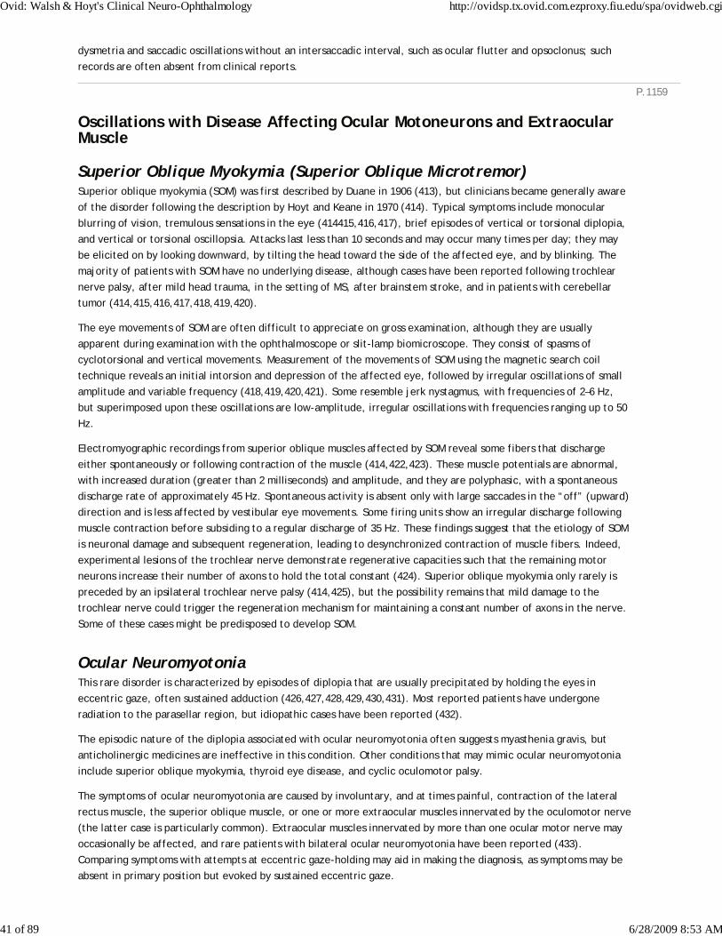
P.1159
dysmetria and saccadic oscillations without an intersaccadic interval, such as ocular flutter and opsoclonus; such
records are often absent from clinical reports.
Oscillations with Disease Affecting Ocular Motoneurons and ExtraocularMuscle
Superior Oblique Myokymia (Superior Oblique Microtremor)Superior oblique myokymia (SOM) was first described by Duane in 1906 (413), but clinicians became generally aware
of the disorder following the description by Hoyt and Keane in 1970 (414). Typical symptoms include monocular
blurring of vision, tremulous sensations in the eye (414415,416,417), brief episodes of vertical or torsional diplopia,
and vertical or torsional oscillopsia. Attacks last less than 10 seconds and may occur many times per day; they may
be elicited on by looking downward, by tilting the head toward the side of the affected eye, and by blinking. The
majority of patients with SOM have no underlying disease, although cases have been reported following trochlear
nerve palsy, after mild head trauma, in the setting of MS, after brainstem stroke, and in patients with cerebellar
tumor (414,415,416,417,418,419,420).
The eye movements of SOM are often difficult to appreciate on gross examination, although they are usually
apparent during examination with the ophthalmoscope or slit-lamp biomicroscope. They consist of spasms of
cyclotorsional and vertical movements. Measurement of the movements of SOM using the magnetic search coil
technique reveals an initial intorsion and depression of the affected eye, followed by irregular oscillations of small
amplitude and variable frequency (418,419,420,421). Some resemble jerk nystagmus, with frequencies of 2–6 Hz,
but superimposed upon these oscillations are low-amplitude, irregular oscillations with frequencies ranging up to 50
Hz.
Electromyographic recordings from superior oblique muscles affected by SOM reveal some fibers that discharge
either spontaneously or following contraction of the muscle (414,422,423). These muscle potentials are abnormal,
with increased duration (greater than 2 milliseconds) and amplitude, and they are polyphasic, with a spontaneous
discharge rate of approximately 45 Hz. Spontaneous activity is absent only with large saccades in the “off” (upward)
direction and is less affected by vestibular eye movements. Some firing units show an irregular discharge following
muscle contraction before subsiding to a regular discharge of 35 Hz. These findings suggest that the etiology of SOM
is neuronal damage and subsequent regeneration, leading to desynchronized contraction of muscle fibers. Indeed,
experimental lesions of the trochlear nerve demonstrate regenerative capacities such that the remaining motor
neurons increase their number of axons to hold the total constant (424). Superior oblique myokymia only rarely is
preceded by an ipsilateral trochlear nerve palsy (414,425), but the possibility remains that mild damage to the
trochlear nerve could trigger the regeneration mechanism for maintaining a constant number of axons in the nerve.
Some of these cases might be predisposed to develop SOM.
Ocular NeuromyotoniaThis rare disorder is characterized by episodes of diplopia that are usually precipitated by holding the eyes in
eccentric gaze, often sustained adduction (426,427,428,429,430,431). Most reported patients have undergone
radiation to the parasellar region, but idiopathic cases have been reported (432).
The episodic nature of the diplopia associated with ocular neuromyotonia often suggests myasthenia gravis, but
anticholinergic medicines are ineffective in this condition. Other conditions that may mimic ocular neuromyotonia
include superior oblique myokymia, thyroid eye disease, and cyclic oculomotor palsy.
The symptoms of ocular neuromyotonia are caused by involuntary, and at times painful, contraction of the lateral
rectus muscle, the superior oblique muscle, or one or more extraocular muscles innervated by the oculomotor nerve
(the latter case is particularly common). Extraocular muscles innervated by more than one ocular motor nerve may
occasionally be affected, and rare patients with bilateral ocular neuromyotonia have been reported (433).
Comparing symptoms with attempts at eccentric gaze-holding may aid in making the diagnosis, as symptoms may be
absent in primary position but evoked by sustained eccentric gaze.
Ovid: Walsh & Hoyt's Clinical Neuro-Ophthalmology http://ovidsp.tx.ovid.com.ezproxy.fiu.edu/spa/ovidweb.cgi
41 of 89 6/28/2009 8:53 AM

P.1160
The mechanism responsible for ocular neuromyotonia is unknown, although both ephaptic neural transmission and
changes in the pattern of neuronal transmission following denervation have been suggested, since spontaneous
activity is seen in the ocular electromyogram of some affected patients (427,428). Axonal hyperexcitability caused
by dysfunction of potassium channels has also been implicated in the production of neuromyotonia by analogy with
systemic neuromyotonia (434).
Spontaneous Eye Movements in Unconscious PatientsThe examination of eye movements often provides important diagnostic information in unconscious patients
(121,435,436). Deviations of one or both eyes and vestibular eye movements (induced by head rotation or caloric
stimulation) are discussed in Chapters 17 and 18. In this section, we summarize spontaneous eye movements.
Although some of these eye movements have been regarded as saccadic intrusions, this issue remains unclear
because, for example, it is not possible to elicit quick phases of nystagmus with caloric stimulation in unconscious
patients. There is a great need for investigation of the pathogenesis of these disorders.
Slow conjugate or disconjugate roving eye movements are similar to the eye movements of light sleep, but they are
slower than the rapid movements of paradoxic or rapid eye movement (REM) sleep. The presence of these eye
movements suggests that the brainstem gaze mechanisms are intact (435).
Ocular bobbing consists of intermittent, usually conjugate, rapid downward movement of the eyes followed by a
slower return to central position. Reflex-induced horizontal eye movements are usually absent. Classic ocular
bobbing is a sign of intrinsic pontine lesions, usually hemorrhage (Fig. 23.16), but it has also been reported in
patients with cerebellar lesions that compress the pons.(437,438,439,440,441,442,443,444,445,446) and in patients
with metabolic and toxic encephalopathies (447,448). An inverse form of ocular bobbing is characterized by a slow
downward movement and rapid return to midposition: “ocular dipping” (449,450,451,452,453,454,455). “Reverse
bobbing” is characterized by a rapid deviation of the eyes upward and a slow return to the horizontal (456),
whereas the terms “reverse dipping” or “converse bobbing” describe a slow upward drift of the eyes followed by a
rapid return to central position (457,458). In general, the variants of ocular bobbing are less reliable for localization
than is the classic form. Nevertheless, because some patients show several types of bobbing, a common underlying
pathophysiology is possibly responsible (459,460). Since the pathways that mediate upward and downward eye
movements differ anatomically and pharmacologically, it seems possible that these movements represent a varying
imbalance of mechanisms for vertical gaze. Repetitive vertical eye movements, including variants of ocular bobbing,
that contain convergent-divergent components may indicate disease affecting the dorsal midbrain (461,462).
Ovid: Walsh & Hoyt's Clinical Neuro-Ophthalmology http://ovidsp.tx.ovid.com.ezproxy.fiu.edu/spa/ovidweb.cgi
42 of 89 6/28/2009 8:53 AM
P.1161
Figure 23.16. Pathology of ocular bobbing. A, Section through the midpons demonstrating a massive area of
hemorrhage and necrosis in a 2-year-old girl with a pontine glioblastoma who developed a left hemiparesis,
obtundation, seizures, and ocular bobbing. B, Organizing hematoma in the caudal portion of the basis pontis
on the right side in a 57-year-old woman who developed a right gaze palsy, right facial palsy, and obtundation.
(A, From Daroff RB, Waldman J. J Neurol Neurosurg Psychiatry 1965;28:375–377. B, From Katz B, Hoyt WF,
Townsend J. J Clin Neuroophthalmol 1982;2:193–195.)
Ping-pong gaze consists of slow, horizontal, conjugate deviations of the eyes alternating every few seconds (178).
Ping-pong gaze usually occurs with bilateral infarction of the cerebral hemispheres or of the cerebral peduncles
(463). Transient oscillations with a similar periodicity to ping-pong gaze can sometimes be induced by a rapid
horizontal head rotation in patients with bilateral hemispheric disease (464).
Periodic alternating gaze deviation, in which conjugate gaze deviations change direction every 2 minutes, occurs
in some patients with hepatic encephalopathy (465). This phenomenon is related to PAN, which is discussed above.
Rapid, small-amplitude, vertical eye movements may be the only manifestation of epileptic seizures in patients with
coexistent brainstem injury (328). Rapid monocular eye movements with horizontal, vertical, or torsional
components may also indicate brainstem dysfunction.
Treatments for Nystagmus and Saccadic Intrusions
Rational Basis for TreatmentIdeally, knowledge of the pathogenesis of a form of nystagmus should suggest the treatment. Perhaps the best
example for this is the acquired form of PAN, for which an animal model exists, pharmacologic mechanisms have
been established, and drug treatment (baclofen) is usually effective. Although such knowledge is still lacking for
many forms of nystagmus and saccadic intrusions, headway has been made. In this section, we summarize current
concepts of relevant pharmacology and the best therapies for the various disorders. It should be noted that
although a number of drugs have been reported to improve nystagmus in individual patients,
few have been subjected to controlled clinical trials. When drug treatments fail or effective drugs cannot be
tolerated by the patient, certain optical devices may be used to either suppress the nystagmus or negate its visual
consequences. Finally, surgery can be performed to either weaken the extraocular muscles or reattach them to the
globe in such a way that the resting position of the eyes is at the null position.
It is essential to carefully evaluate patients before and during therapy for abnormal eye movements. In the case of
new treatments, it is best to carry out a controlled, masked evaluation. Careful measurements of visual acuity using
near and far test charts and systematic eye movement examination are essential. It is also very useful to record the
oscillations. This can be achieved semiquantitatively by video, but it is best performed using one of several
recording techniques, ideally the magnetic search coil method (Fig. 23.2).
Pharmacologic Treatments (466,467)
Nystagmus from Vestibular Imbalance
Peripheral Vestibular ImbalanceNystagmus caused by peripheral vestibular lesions usually resolves spontaneously over the course of a few days.
Present approaches use vestibular suppressants for 24–48 hours, primarily for severe vertigo and nausea. If the
nystagmus persists after this time, exercises are used to accelerate the brain's ability to redress the imbalance (468).
In the case of BPPV, maneuvers to displace otolithic debris from the affected semicircular canal and exercises to
Ovid: Walsh & Hoyt's Clinical Neuro-Ophthalmology http://ovidsp.tx.ovid.com.ezproxy.fiu.edu/spa/ovidweb.cgi
43 of 89 6/28/2009 8:53 AM

sustain recovery are usually effective (90,469,470,471,472).
Central Vestibular Mechanisms—Pharmacologic BasisThe technique of pharmacologic inactivation by microinjection of drugs into the brainstem and cerebellum has
provided some useful insights and clues to clinicians interested in treating abnormal eye movements. Thus,
unilateral microinjection of the GABAA agonist muscimol into the medial vestibular nucleus and nucleus prepositus
hypoglossi of the monkey and cat produces bilateral gaze-evoked nystagmus (201,473,474). In addition, there is
evidence that prolongation of the peripheral vestibular signal by central mechanisms, called velocity storage, by the
nodulus and uvula of the cerebellum is regulated by inhibitory pathways that use GABAB (475). The velocity-storage
phenomenon in normal monkeys is suppressed by the GABAB agonist baclofen (475), and mildly enhanced by both
diazepam (476) and picrotoxin (477). Acquired PAN, which is thought to be partly caused by abnormal prolongation
of velocity storage, is abolished by baclofen, and this response occurs with both experimental and clinical lesions of
the nodulus and uvula (181,478). Microinjection of drugs with effects on glutamate receptors into medial vestibular
nucleus/nucleus prepositus hypoglossi also effects gaze-holding ability (474). There is also evidence that nicotinic
acetylcholinergic mechanisms play a role in vestibular-mediated vertical eye movements. Nicotine can produce
upbeat nystagmus in normal subjects in darkness (479,480) and intravenous physostigmine may increase the intensity
of downbeat nystagmus (481) In addition, intravenous opioids have been reported to induce downbeat nystagmus
(482,483).
Downbeat and Upbeat NystagmusThe GABAA agonist clonazepam is reported to be effective in reducing downbeat nystagmus (484). A single dose of
1–2 mg of clonazepam was administered to determine whether long-term therapy was likely to be effective.
Baclofen may reduce the velocity of both upbeat and downbeat nystagmus and reduce associated oscillopsia (481).
Barton and colleagues performed a double-blind study in which two patients with downbeat nystagmus experienced
reduction in nystagmus after intravenous scopolamine (485). Improvement of occasional patients with downbeat
nystagmus with trihexyphenidyl has been reported (486). However, all of these therapies are ineffective in many
patients with downbeat nystagmus.
A new approach to the treatment of downbeat nystagmus arose out of the seminal observation by Griggs that
nystagmus occurring in episodic ataxia type 2 responds to acetazolamide (487); this disorder is now known to be a
calcium channelopathy (488,489). This led to a study of 3,4-diaminopyridine, a potassium channel blocker, as a
treatment for downbeat nystagmus. Strupp and colleagues studied 17 patients with downbeat nystagmus due to a
range of disorders (490). Ten of the patients showed a decrease of more than 50% in their nystagmus 30 minutes
after ingesting 20 mg of 3,4-diaminopyridine. This medication was generally well tolerated, although it is known to
induce seizures in some subjects. The possible mechanism of action of 3,4-diaminopyridine relates to one
hypothesis for downbeat nystagmus described earlier in this chapter (167).
The cerebellum inhibits vestibular circuits mediating upward, but not downward, eye movements (Fig. 23.7)
(165,166). Consequently, impaired cerebellar inhibition could cause uninhibited upward drifts of the eyes, which
evoke corrective rapid downward movements: downbeat nystagmus. Potassium channels are abundant on cerebellar
Purkinje cells—the output neurons from cerebellar cortex—and a related agent, 4-aminopyridine, is reported to
increase the discharge of these neurons by affecting the slowly depolarizing potential. Enhancement of Purkinje
cell activity due to 3,4-diaminopyridine could restore to normal levels the inhibitory influence of the cerebellar
cortex upon vertical vestibular eye movements. Studies are under way to determine the long-term effects of these
drugs on nystagmus and its visual consequences, and to compare 3,4-diaminopyridine and 4-aminopyridine; the
latter penetrates the blood-brain barrier better, has a longer half-life, and is generally better tolerated.
The recent demonstration that a patient with downbeat nystagmus showed antiglutamic acid decarboxylase
antibodies in the vestibular complex raises the possibility of evaluating drugs with glutamate effects in patients with
downbeat nystagmus (491).
Periodic Alternating Nystagmus
Ovid: Walsh & Hoyt's Clinical Neuro-Ophthalmology http://ovidsp.tx.ovid.com.ezproxy.fiu.edu/spa/ovidweb.cgi
44 of 89 6/28/2009 8:53 AM

P.1162
P.1163
As noted before, this is the best example of a form of nystagmus for which drug treatment is based on known
pathophysiology and pharmacology. Most cases of acquired PAN respond to baclofen (180,478). The congenital form
of PAN, which probably has a different pathogenesis than the acquired form, only occasionally responds to baclofen
(492), but one child did show a response to dextroamphetamine (493).
Acquired Pendular NystagmusThe neuropharmacology of acquired pendular nystagmus is unknown, and more than one mechanism may be
involved. This type of nystagmus used to be treated with barbiturates (73), but sedative side effects limit the use of
this class of drugs.
In the case of oculopalatal myoclonus, increased acetylcholine esterase activity and cholinergic denervation
supersensitivity has been reported in hypertrophied inferior olivary nucleus (494,495), prompting trials of
anticholinergic agents for acquired pendular nystagmus. Initial studies showed that individual patients may be
helped by trihexyphenidyl (496,497). One double-blind crossover trial compared trihexyphenidyl with tridihexethyl
chloride (a quaternary anticholinergic that does not cross the blood-brain barrier) in 10 patients, 5 of whom had
acquired pendular nystagmus. Of these five, only two showed a decrease in nystagmus and an improvement of visual
acuity when given tridihexethyl chloride. Trihexyphenidyl had no effect on the nystagmus in any patient.
Significantly, neither of the two patients whose nystagmus improved with medication elected to continue the drug
because of anticholinergic side effects. In another masked study, scopolamine, benztropine, and glycopyrrolate (a
quaternary agent devoid of central nervous activity) were administered intravenously to five patients with acquired
pendular nystagmus (485). The results, confirming an earlier report (58), demonstrated a single dose of scopolamine
effectively reduced nystagmus and improved vision in all five patients, whereas benztropine was less effective, and
glycopyrrolate had no significant effect.
Why did intravenous scopolamine suppress acquired pendular nystagmus, but oral trihexyphenidyl did not? One
possibility is that trihexyphenidyl selectively antagonizes only the m1 muscarinic receptors, whereas scopolamine
probably affects all five subtypes of muscarinic receptors (498). However, this is not the whole story, because in an
open trial of transdermal scopolamine, which included four patients with acquired pendular nystagmus, two showed
a decrease, one showed no effect, and one showed an increase of nystagmus (499). Thus, scopolamine does not
appear to be a promising treatment for acquired pendular nystagmus. Intravenous administration is not a practical
therapy, and clouds the sensorium. Occasional patients show some improvement with transdermal scopolamine, but
this is often not sustained (499), and prolonged use of this mode of therapy is also not without risk.
Reports that the neurotransmitter GABA contributed to the normal function of the gaze-holding mechanism
(201,473,474) focused attention on drugs with GABAergic effects. Valproate is reported to help some patients with
acquired pendular nystagmus (500). Isoniazid has also been studied as treatment for acquired pendular nystagmus in
three patients with MS, and reduced nystagmus and relieved oscillopsia in two (501). However, isoniazid has
potential side effects, and better-tolerated drugs have become available, notably gabapentin, which has been
evaluated in controlled trials (75). Of 15 patients with acquired pendular nystagmus studied in a double-blind
comparison with baclofen, visual acuity improved with gabapentin, but not with baclofen; an example is shown in
Figure 23.17. Gabapentin significantly reduced nystagmus in all three planes, but baclofen did so only in the vertical
plane. In 10 of the 15 patients, reduction of nystagmus was substantial, and 8 elected to continue taking the
medication. The main side effect of gabapentin was increased ataxia and unsteadiness. However, the demonstration
that the more purely GABAergic agent vigabatrin failed to suppress nystagmus led to the
suggestion that gabapentin was probably working through a different transmitter mechanism, such as glutamate
(502).
Ovid: Walsh & Hoyt's Clinical Neuro-Ophthalmology http://ovidsp.tx.ovid.com.ezproxy.fiu.edu/spa/ovidweb.cgi
45 of 89 6/28/2009 8:53 AM

Figure 23.17. Effects of gabapentin on the horizontal component of pendular nystagmus in a 41-year-old
woman with multiple sclerosis. After the initial recording (top), the patient was given 300 mg of gabapentin,
which reduced her oscillations and improved her vision (middle). The effect was sustained two months later,
when she was taking a dose of 300 mg three times per day. Visual acuity measurements of the recorded eye
are shown at the right in each panel.
One agent with effects on gluatamate receptors is memantine, which has recently received approval from the U.S.
Food and Drug Administration for treatment of Alzheimer's disease. Memantine has been in use in Germany for over
20 years, and is known to be a safe and well-tolerated drug. It is a low-to-moderate uncompetitive (open channel)
N-methyl-D-aspartate (NMDA) receptor antagonist. A study from the University of Munich reported that it
suppressed acquired pendular nystagmus in patients with multiple sclerosis (503). There is need for a controlled
study comparing memantine with gabapentin. At present, gabapentin has been shown to be effective in some
patients with pendular nystagmus as part of the syndrome of oculopalatal tremor (75), but memantine has not been
Ovid: Walsh & Hoyt's Clinical Neuro-Ophthalmology http://ovidsp.tx.ovid.com.ezproxy.fiu.edu/spa/ovidweb.cgi
46 of 89 6/28/2009 8:53 AM
studied in this disorder. Palatal tremor may respond to carbamazepine in some patients (504,505). Occasional
patients with acquired pendular nystagmus benefit from both gabapentin and surgical recession of muscles (506);
the effects of surgery are described below.
Alcohol has been reported to suppress acquired pendular nystagmus (507). Smoking cannabis has been reported to
suppress both acquired pendular nystagmus (508), and congenital nystagmus (509), but formal evaluations of this
potential therapy are hindered at present by current social views on cannabis.
Seesaw NystagmusImprovement in seesaw nystagmus has been reported in some patients treated with alcohol (510,511). Clonazepam
has reduced the nystagmus and associated oscillopsia in occasional patients (512). Two patients with pendular
seesaw nystagmus showed reduction of their nystagmus with gabapentin, but more studies are needed to confirm
this (75).
Familial Episodic Ataxia with NystagmusThe attacks of episodic ataxia type 2 (EA-2), which is a calcium channelopathy, respond well to treatment with
acetazolamide (489). However, the interictal nystagmus and ataxia (which in some patients is progressive) usually do
respond. The briefer attacks of episodic ataxia type 1 (EA-1), which is a potassium channelopathy, respond to
acetazolamide in some patients. The attacks of EA-1 spontaneously improve with time in some patients (489).
Saccadic Intrusions: Square-Wave JerksBoth the frontal eye fields and the superior colliculus send brainstem projections that influence the omnipause
neurons that gate saccades (discussed earlier). Experimental studies have elucidated the pharmacology of one part
of this descending pathway (513). The nondopaminergic portion of the substantia nigra, the pars reticulata (SNpr),
receives inputs from the caudate nucleus and selectively gates reflexive or voluntary saccades via the superior
colliculus. This is accomplished, in part, by a phasic modulation of tonic inhibitory influence of the SNpr upon the
superior colliculus. The caudate nucleus appears to facilitate the initiation of voluntary self-generated types of
saccades and to aid steady fixation by preventing unwanted reflexive saccades to stimuli. The nigrotectal pathway is
GABAergic, and injection of bicuculline into the superior colliculus increases the frequency and amplitude of the
saccades, which take the form of square-wave intrusions (514). Furthermore, pharmacologic inactivation of the
frontal eye fields with bicuculline causes saccadic intrusions (395). This experimental evidence suggests that
treatment with GABA agonists might prevent inappropriate saccades. In fact, several benzodiazepines (diazepam,
clonazepam) and the barbiturate phenobarbital were effective in abolishing high-amplitude square-wave jerks and
macrosaccadic oscillations in one patient (515). There is also some evidence that amphetamines can suppress
square-wave jerks in some patients (516). On the other hand, both nicotine (479) and opiates (483) are reported to
induce square-wave jerks. Thus, more work is needed to better understand the pharmacology of saccadic intrusions
and how they can be treated.
Ocular Flutter and OpsoclonusPatients with parainfectious opsoclonus-myoclonus often improve spontaneously, but intravenous immunoglobulin
may speed recovery (517). Similarly, although propranolol, verapamil, clonazepam, gabapentin, and thiamine have
all been reported to diminish microsaccadic ocular flutter in individual patients (366,518,519), the effect may have
been due to spontaneous remission. Opsoclonus associated with neural crest tumors in children usually responds to
corticosteroid treatment (520); however, up to 50% of such children have persistent neurological disabilities,
including ataxia, poor speech, and cognitive problems (372,518). Similar responses to steroids may occur in children
with parainfectious or idiopathic opsoclonus (518). Treatment with steroids has not been uniformly successful in
such cases, although plasmapheresis, intravenous immunoglobulin, and immunoadsorption therapy have occasionally
proved effective (370,521,522).
Superior Oblique Myokymia and Ocular Neuromyotonia
Ovid: Walsh & Hoyt's Clinical Neuro-Ophthalmology http://ovidsp.tx.ovid.com.ezproxy.fiu.edu/spa/ovidweb.cgi
47 of 89 6/28/2009 8:53 AM

P.1164
Superior oblique myokymia spontaneously resolves in some patients (417), and others are not sufficiently bothered
by their symptoms that they request treatment. Individual patients have responded to carbamazepine, baclofen,
b-adrenergic blocking agents, or gabapentin given systemically or topically (415,416,419). Patients who do not
respond to drug therapy, who develop side effects from the drugs, or who do not wish to take drugs for their
condition, may experience complete relief of symptoms after extraocular muscle surgery (discussion following).
Ocular neuromyotonia is usually responsive to carbamazepine (432,433).
Optical TreatmentsConvergence prisms provide one optical approach for patients with congenital or acquired nystagmus whose
nystagmus
dampens when they view a near target (242,523); a useful starting point is 7.00-diopter base-out prisms combined
with -1.00-diopter spheres to compensate for accommodation (although the spherical correction may not be needed
in presbyopic individuals). In some patients with congenital nystagmus, the resultant improvement of vision is
sufficient for them to qualify for a driver's license. Patients whose nystagmus is worse during near viewing may
benefit from wearing base-in (divergence) prisms (64).
Theoretically, it should be possible to use prisms to help patients whose nystagmus is reduced or absent when the
eyes are moved into a particular position in the orbit: the null region. For patients with congenital nystagmus, there
is usually some horizontal eye position in which the nystagmus is minimized, whereas downbeat nystagmus may
decrease or disappear in upgaze. In practice, patients use head turns to bring their eyes to the optimum position,
and only rarely are prisms that produce a conjugate shift helpful.
A different approach to the treatment of nystagmus has been the use of an optical system that stabilizes images on
the retina (524). This system consists of a high-plus spectacle lens worn in combination with a high-minus contact
lens. The system is designed on the principle that stabilization of images on the retina could be achieved if the
power of the spectacle lens focused the primary image close to the center of rotation of the eye. However, such
images are then defocused, and a contact lens is required to extend the clear image back onto the retina. Since the
contact lens moves with the eye, it does not negate the effect of retinal image stabilization produced by the
spectacle lens. With such a system, it is possible to achieve up to about 90% stabilization of images upon the retina.
There are several limitations to the system, however. One is that it disables all eye movements (including the
vestibulo-ocular reflex and vergence) and thus is useful only when the patient is stationary and is viewing
monocularly. Another limitation is that with the highest-power components (contact lens of -58.00 diopters and
spectacle lens of +32diopters), the field of view is limited. Some patients with ataxia or tremor (such as those with
MS) have difficulty inserting the contact lens. However, initial problems posed by rigid polymethyl methacrylate
contact lenses can be overcome by using gas-permeable, or even soft contact lenses (525). Most patients do not
need the highest power components for oscillopsia to be abolished, and vision to be improved. We have found that
in selected patients the device may prove useful for limited periods of time, for example, if the patient wishes to
watch a television program (526).
Contact lenses alone sometimes suppress congenital nystagmus (527). This effect is not from the mass of the lenses
but is probably mediated via trigeminal afferents (528); this issue is discussed further below.
A more recent innovation has been to use an electronic circuit to distinguish between the nystagmus oscillations and
normal eye movements (529). This approach is most applicable in patients with pendular nystagmus. Eye movements
are measured using an infrared sensor and, after filtering, fed to a phase-locked loop that generates a signal similar
to the nystagmus but is insensitive to other eye movements, such as saccades. This electronic signal is then used to
rotate Risley prisms, through which the patient views the environment. When the Risley prisms rotate in synchrony
with the patients nystagmus, they negate the visual effects of the ocular oscillations. Improvement and
miniaturization of a prototype device may eventually lead to a spectacle-mounted device that selectively cancels
out the visual effects of pathological nystagmus (529,530).
The main therapy for latent nystagmus consists of measures to improve vision, particularly patching for amblyopia in
Ovid: Walsh & Hoyt's Clinical Neuro-Ophthalmology http://ovidsp.tx.ovid.com.ezproxy.fiu.edu/spa/ovidweb.cgi
48 of 89 6/28/2009 8:53 AM

P.1165
children (531).
Botulinum Toxin Treatment of NystagmusAn approach to treatment of nystagmus that has gained some popularity is injection of botulinum toxin into either
the extraocular muscles or the retrobulbar space (532,533). Using both techniques, Ruben and colleagues reported
improvement of vision in most of their 12 patients with a variety of diagnoses (534). The major side effect was
ptosis. However, eye movements were not systematically measured and compared before and after injection. Repka
and colleagues also described improvement of vision following retrobulbar injection of botulinum toxin in six
patients and documented the effects on eye movements (535). The main reservation expressed by these authors was
the temporary nature of the treatment and the necessity for repeated injections, with their attendant risks. We
measured binocular eye rotations in three planes before and after monocular injection of botulinum toxin either
into the horizontal recti (536), or into the retrobulbar space (537). Nystagmus was abolished or reduced in the
treated eye for about 2–3 months, but no patient was pleased with the results because of ptosis, diplopia, increase
of nystagmus in the noninjected eye or, in one patient, filamentary keratitis. No patient that we studied elected to
repeat the procedure.
Surgical Procedures for NystagmusTwo surgical procedures may be effective for certain patients with congenital nystagmus. One is the Anderson-
Kestenbaum operation (538,539,540). This procedure is designed to move the attachments of the extraocular
muscles so that the new central position of the eyes is at the null position. It is performed after first making careful
eye movement measurements of nystagmus intensity with the eyes in various positions of gaze and determining the
approximate null position. The appropriate extraocular muscles are then weakened or strengthened as necessary to
achieve the required shift in the position of the null (541,542,543). The Anderson-Kestenbaum procedure not only
shifts and broadens the null region, but also results in decreased nystagmus outside the region. It is of uncertain
value in the treatment of acquired forms of nystagmus.
The second procedure is an artificial divergence operation (544,545). It may be helpful in patients with congenital
nystagmus that dampens or is suppressed during near viewing and who have stereopsis. Studies comparing these two
methods indicate that the artificial divergence operation generally
results in a better visual outcome than the Anderson-Kestenbaum procedure alone (543,545,546,547).
Several authors have recommended performing large recessions of all of the horizontal rectus muscles for treatment
of patients with congenital nystagmus (548,549). Based on a long experience, Dell'Osso noted that any surgical
procedure that detached and reattached the extraocular muscles tended to suppress congenital nystagmus. This led
him to suggest that simply dissecting the perimuscular fascia and then reattaching the muscles at the same site on
the globe might prove effective, especially in cases when convergence does not dampen the nystagmus. Results of
this procedure on a canine model for congenital nystagmus supported this hypothesis (550). Reported lack of effect
in monkeys concerns latent nystagmus, not typical congenital nystagmus (551,552). Preliminary results of a large,
controlled clinical trial suggest that the operation is effective in some patients (242,553).
How could such a procedure damp congenital nystagmus? Recent studies by Büttner-Ennever and colleagues have
indicated that the terminal portion of the extraocular muscles, near their site of their attachment, contains
multiply-innervated muscle fibers (554). Using rabies toxin as an anatomic tracer, it has been possible to show that a
separate group of ocular motor neurons (distinct from the classic oculomotor, trochlear, and abducens nuclei, and
surrounding each of them) innervates these multiply-innervated fibers.
Finally, it is known that ocular proprioceptors (the pallisade organs) lie at the insertion site of the extraocular
muscles (554). Thus, procedures similar to those proposed by Dell'Osso may work by disrupting a proprioceptive
feedback pathway that normally sets the tone of the extraocular muscles.
There is also some evidence that the tendino-scleral junction may contain neurovascular abnormalities in the eyes
of patients with congenital nystagmus (555). This suggestion, and the “orbital revolution” set in motion by the
Ovid: Walsh & Hoyt's Clinical Neuro-Ophthalmology http://ovidsp.tx.ovid.com.ezproxy.fiu.edu/spa/ovidweb.cgi
49 of 89 6/28/2009 8:53 AM
discovery of pulleys for the extraocular muscles by Miller and Demer, promise the development of new therapies for
congenital nystagmus (556).
The role of surgery in the treatment of acquired nystagmus is not well established, although individual patients may
benefit from recession operations (506,557). However, it is clear that suboccipital decompression improves
downbeat nystagmus in Chiari syndromes and also prevents progression of other neurologic deficits (558,559,560).
As noted above, SOM that does not respond to treatment with medication may respond to extraocular muscle
surgery. The procedure used by most surgeons is a superior oblique tenectomy combined with myectomy of the
ipsilateral inferior oblique muscle (414,415,417,422,561). However, Kosmorsky and colleagues reported successful
treatment of a patient with SOM by performing a nasal transposition of the anterior portion of the affected superior
oblique tendon, thereby weakening cyclorotation (562).
Other Forms of TreatmentA variety of methods other than those described above have been used to treat nystagmus, principally the
congenital variety. Electrical stimulation or vibration over the forehead may suppress congenital nystagmus (264).
The mechanism of vibration on eye movements is uncertain, since vibration over the mastoid in patients who have
lost vestibular function induces ocular torsion (563). However, it is postulated that the suppressive effect on
congenital nystagmus, as well as suppression induced by wearing contact lenses (528), may be exerted via the
trigeminal system, which receives extraocular proprioception (264,554).
Acupuncture administered to the neck muscles may suppress congenital nystagmus in some patients via a similar
mechanism (564,565). Biofeedback has also been reported to help some patients with congenital nystagmus
(566,567). The role of any of these treatments in clinical practice has yet to be demonstrated.
References
1. Kaminski HJ, Leigh RJ. Neurobiology of eye movements: From molecules to behavior. Ann New York Acad
Sci 2002;1:619.
2. Leigh RJ, Das VE, Seidman SH. A neurobiological approach to acquired nystagmus. Ann NY Acad Sci
2002;956:380–390.
3. Carpenter RHS. The visual origins of ocular motility. In Carpenter RHS, ed. Eye Movements. Vol 8. London:
MacMillan Press, 1991:1–10.
4. Bender MB. Oscillopsia. Arch Neurol 1965;13:204–213.
5. Wist ER, Brandt T, Krafczyk S. Oscillopsia and retinal slip: Evidence supporting a clinical test. Brain
1983;106:153–168.
6. Stahl JS, Averbuch-Heller L, Leigh RJ. Acquired nystagmus. Arch Ophthalmol 2000;118:544–549.
7. Leigh RJ, Averbuch-Heller L, Tomsak RL, Remler BF, Yaniglos SS, Dell'Osso LF. Treatment of abnormal eye
movements that impair vision: Strategies based on current concepts of physiology and pharmacology. Ann
Neurol 1994;36:129–141.
8. Abadi RV, Whittle JP, Worfolk R. Oscillopsia and tolerance to retinal image movement in congenital
Ovid: Walsh & Hoyt's Clinical Neuro-Ophthalmology http://ovidsp.tx.ovid.com.ezproxy.fiu.edu/spa/ovidweb.cgi
50 of 89 6/28/2009 8:53 AM

nystagmus. Invest Ophthalmol Vis Sci 1999;40:339–345.
9. Leigh R, Zee D. The Neurology of Eye Movements. Ed 3. New York, Oxford University Press, 1999.
10. Serra A, Leigh RJ. Diagnostic value of nystagmus: Spontaneous and induced ocular oscillations. J Neurol
Neurosurg Psychiatry 2002;73:615–618.
11. Zee DS. Ophthalmoscopy in examination of patients with vestibular disorders. Ann Neurol 1978;3:373–374.
12. Trillenberg P, Zee DS, Shelhamer M. On the distribution of fast-phase intervals in optokinetic and
vestibular nystagmus. Biol Cybern 2002;87:68–78.
13. Garbutt S, Harris CM. Abnormal vertical optokinetic nystagmus in infants and children. Br J Ophthalmol
2000;84:451–455.
14. Garbutt S, Harwood MR, Harris CM. Comparison of the main sequence of reflexive saccades and the quick
phases of optokinetic nystagmus. Br J Ophthalmol 2001;85:1477–1483.
15. Garbutt S, Harwood MR, Harris CM. Anticompensatory eye position (“contraversion”) in optokinetic
nystagmus. Ann NY Acad Sci 2002;956:445–448.
16. Garbutt S, Han Y, Kumar AN, et al. Vertical optokinetic nystagmus and saccades in normal human subjects.
Invest Ophthalmol Vis Sci 2003;44:3833–3841.
17. Garbutt S, Han Y, Kumar AN, et al. Disorders of vertical optokinetic nystagmus in patients with ocular
misalignment. Vision Res 2003;43:347–357.
18. Dell'Osso LF, Daroff RB. Congenital nystagmus waveforms and foveation strategy. Documenta
Ophthalmologica 1975;39:155–182.
19. Abadi RV, Dickinson CM. Waveform characteristics in congenital nystagmus. Documenta Ophthalmologica
1986;64:153–167.
20. Carpenter RHS. Movements of the Eyes. Ed 2. London: Pion, 1988.
21. DiScenna AO, Das VE, Zivotofsky AZ, Seidman SH, Leigh RJ. Evaluation of a video tracking device for
measurement of horizontal and vertical eye rotations during locomotion. J Neurosci Methods 1995;58:89–94.
22. Murphy P, Duncan A, Glennie A, Knox P. The effect of sceral search coil lens wear on the eye. Br J
Ophthalmol 2001;85:332–335.
Ovid: Walsh & Hoyt's Clinical Neuro-Ophthalmology http://ovidsp.tx.ovid.com.ezproxy.fiu.edu/spa/ovidweb.cgi
51 of 89 6/28/2009 8:53 AM

P.1166
23. Leigh RJ, Zee DS. Eye movements of the blind. Invest Ophthalmol Vis Sci 1980;19:328–331.
24. Leigh RJ, Thurston SE, Tomsak RL, Grossman GE, Lanska DJ. Effect of monocular visual loss upon stability
of gaze. Invest Ophthalmol Vis Sci 1989;30:288–292.
25. Luebke AE, Robinson DA. Transition dynamics between pursuit and fixation suggest different systems.
Vision Res 1988;28:941–946.
26. Leigh RJ, Huebner WP, Gordon JL. Supplementation of the human vestibulococular reflex by visual
fixation and smooth pursuit. J Vestibular Res 1994;4:347–353.
27. Steinman RM, Haddad GM, Skavenski AA, Wyman D. Miniature eye movement. Science 1973;181:810–819.
28. Zeki S. A Vision of the Brain. London: Blackwell Scientific, 1993.
29. Averbuch-Heller L, Zivotofsky AZ, Remler BF, Das VE, Dell'Osso LF, Leigh RJ. Convergent-divergent
pendular nystagmus: possible role of the vergence system. Neurology 1995;45:509–515.
30. Robinson DA. The control of eye movements. In Handbook of Physiology: The Nervous System. Vol 2, Part
2. Bethesda, American Physiological Society, 1981.
31. Belton T, McCrea RA. Role of the cerebellar flocculus region in the coordination of eye and head
movements during gaze pursuit. J Neurophysiol 2000;84:1614–1626.
32. Suzuki DA, May JG, Keller EL, Yee RD. Visual motion response properties of neurons in dorsolateral pontine
nucleus of alert monkey. J Neurophysiol 1990;63:37–59.
33. Ito M. Cerebellar flocculus hypothesis. Nature 1993;363:24–25.
34. Luebke AE, Robinson DA. Gain changes of the cat's vestibulo-ocular reflex after flocculus deactivation. Exp
Brain Res 1994;98:379–390.
35. Büttner-Ennever JA, Horn AK. Pathways from cell groups of the paramedian tracts to the floccular region.
Ann NY Acad Sci 1996;781:532–540.
36. Robinson DA. The effect of cerebellectomy on the cat's vestibulo-ocular integrator. Brain Res
1974;71:195–207.
37. Narfstrom K, Katz ML, Bragadottir R, et al. Functional and structural recovery of the retina after gene
therapy in the RPE65 null mutation dog. Invest Ophthalmol Vis Sci 2003;44:1663–1672.
Ovid: Walsh & Hoyt's Clinical Neuro-Ophthalmology http://ovidsp.tx.ovid.com.ezproxy.fiu.edu/spa/ovidweb.cgi
52 of 89 6/28/2009 8:53 AM

38. Acland G, Aguirre G, Ray J, et al. Gene therapy restores vision in a canine model of childhood blindness.
Nature Genetics 2001;28:92–95.
39. Pritchard C, Flynn JT, Smith JL. Wave form characteristics of vertical oscillations in long-standing vision
loss. J Pediatr Ophthalmol Strabismus 1988;25:233–236.
40. Barton JJS, Cox TA. Acquired pendular nystagmus in multiple sclerosis: Clinical observations and the role
of optic neuropathy. J Neurol Neurosurg Psychiatry 1993;56:262–267.
41. Yee RD, Jelks GW, Baloh RW, Honrubia V. Uniocular nystagmus in monocular visual loss. Ophthalmology
1979;86:511–518.
42. Pratt-Johnson JA, Tillson G. Intractable diplopia after vision restoration in unilateral cataract. Am J
Ophthalmol 1989;107:23–26.
43. Farmer J, Hoyt CS. Monocular nystagmus in infancy and early childhood. Am J Ophthalmol
1984;98:504–509.
44. Lavery MA, O'Neill JF, Chu FC, Martyn LJ. Acquired nystagmus in early childhood: A presenting sign of
intracranial tumor. Ophthalmology 1984;91:425–435.
45. Weissman BM, Dell'Osso LF, Abel LA, Leigh RJ. Spasmus nutans: A quantitative prospective study. Arch
Ophthalmol 1987;105:525–528.
46. Gottlob I, Zubcov AA, Catalano RA, et al. Signs distinguishing spasmus nutans (with and without central
nervous system lesions) from infantile nystagmus. Ophthalmology 1990;97:1166–1175.
47. Dell'Osso LF, Williams RW. Ocular motor abnormalities in achiasmatic mutant Belgian sheepdogs: Unyoked
eye movements in a mammal. Vision Res 1995;35:109–116.
48. Apkarian P, Bour LJ, Barth PG, et al. Non-decussating retinal-fugal fibre syndrome: An inborn achiasmatic
malformation associated with visuotopic misrouting, visual evoked potential ipsilateral asymmetry and
nystagmus. Brain 1995;118:1195–1216.
49. Apkarian P, Bour LJ. See-saw nystagmus and congenital nystagmus identified in the non-decussating
retinal-fugal fiber syndrome. Strabismus 2001;9:143–163.
50. Barton JJS. Blink- and saccade-induced seesaw nystagmus. Neurology 1995;45:831–833.
51. Averbuch-Heller L, Leigh RJ. Saccade-induced nystagmus. Neurology 1996;46:289.
52. Sharpe JA, Lo AW, Rabinovitch HE. Control of the saccadic and smooth pursuit systems after cerebral
Ovid: Walsh & Hoyt's Clinical Neuro-Ophthalmology http://ovidsp.tx.ovid.com.ezproxy.fiu.edu/spa/ovidweb.cgi
53 of 89 6/28/2009 8:53 AM

hemidecortication. Brain 1979;102:387–403.
53. Cogan DG, Loeb DR. Optokinetic response and intracranial lesions. Arch Neurol Psychiatry 1949;61:183–187.
54. Kömpf D. The significance of optokinetic nystagmus asymmetry in hemispheric lesions.
Neuroophthalmology 1986;6:61–64.
55. Morrow MJ. Craniotopic defects of smooth pursuit and saccadic eye movement. Neurology
1996;46:514–521.
56. Thurston SE, Leigh RJ, Crawford TJ, et al. Two distinct deficits of visual tracking caused by unilateral
lesions of cerebral cortex in humans. Ann Neurol 1988;23:266–273.
57. Dieterich M, Bense S, Stephan T, et al. fMRI signal increases and decreases in cortical areas during
small-field optokinetic stimulation and central fixation. Exp Brain Res 2003;148:117–127.
58. Gresty MA, Ell JJ, Findley LJ. Acquired pendular nystagmus: Its characteristics, localising value and
pathophysiology. J Neurol Neurosurg Psychiatry 1982;45:431–439.
59. Barton JJS. Is acquired pendular nystagmus always phase locked? J Neurol Neurosurg Psychiatry
1994;57:1263–1264.
60. Aschoff JC, Conrad B, Kornhuber HH. Acquired pendular nystagmus with oscillopsia in multiple sclerosis: A
sign of cerebellar nuclei disease. J Neurol Neurosurg Psychiatry 1974;37:570–577.
61. Das VE, Oruganti P, Kramer PD, Leigh RJ. Experimental tests of a neural-network model for ocular
oscillations caused by disease of central myelin. Exp Brain Res 2000;133:189–197.
62. Jacobs L, Bender MB. Palato-ocular synchrony during eyelid closure. Arch Neurol 1976;33:289–291.
63. Fukushima M, Enomoto H, Tsutsui J. Visually suppressed acquired pendular nystagmus in four cases with
spinocerebellar degeneration. Neuroophthalmol Jpn 1984;1:54–59.
64. Barton JJ, Cox TA, Digre KB. Acquired convergence-evoked pendular nystagmus in multiple sclerosis. J
Neuroophthalmol 1999;19:34–38.
65. Maas EF, Ashe J, Spiegel P, Zee DS, Leigh RJ. Acquired pendular nystagmus in toluene addiction.
Neurology 1991;41:282–285.
66. Trobe JD, Sharpe JA, Hirsh DK, Gebarski SS. Nystagmus of Pelizaeus-Merzbacher disease. Arch Neurol
1991;48:87–91.
Ovid: Walsh & Hoyt's Clinical Neuro-Ophthalmology http://ovidsp.tx.ovid.com.ezproxy.fiu.edu/spa/ovidweb.cgi
54 of 89 6/28/2009 8:53 AM

67. Kori AA, Robin NH, Jacobs JB, et al. Pendular nystagmus in patients with peroxisomal assembly disorder.
Arch Neurol 1998;55:554–558.
68. Büttner U, Helmchen C, Büttner-Ennever JA. The localizing value of nystagmus in brainstem disorders.
Neuroophthalmology 1995;15:283–290.
69. Lopez LI, Bronstein AM, Gresty MA, DuBoulay EP, Rudge P. Clinical and MRI correlates in 27 patients with
acquired pendular nystagmus. Brain 1996;119:465–472.
70. Nakamagoe K, Iwamoto Y, Yoshida K. Evidence for brainstem structures participating in oculomotor
integration. Science 2000;288:857–859.
71. Guillain G, Mollaret P. Deux cas myoclonies synchrones et rhythmées vélo-pharyngo-laryngo-
oculodiaphragmatiques: Le problèm anatomique et physiolopathologique de ce syndrome. Rev Neurol (Paris)
1931;2:545–566.
72. Nakada T, Kwee IL. Oculopalatal myoclonus. Brain 1986;109:431–441.
73. Nathanson M. Palatal myoclonus: Further clinical and pathophysiological observations. Arch Neurol
Psychiatr 1956;75:285–296.
74. Sperling MR, Herrmann J. Syndrome of palatal myoclonus and progressive ataxia: Two cases with magnetic
resonance imaging. Neurology 1985;35:1212–1214.
75. Averbuch-Heller L, Tusa RJ, Fuhry L, Rottach KG, Ganser GL, Heide W et al. A double-blind controlled
study of gabapentin and baclofen as treatment for acquired nystagmus. Ann Neurol 1997;41:818–825.
76. Jacobs L, Newman RP, Bozian D. Disappearing palatal myoclonus. Neurology 1981;31:748–751.
77. Keane JR. Acute vertical ocular myoclonus. Neurology 1986;36:86–89.
78. Dubinsky RM, Hallet M, DiChiro G, Fulham M, Schwankhaus J. Increased glucose metabolism in the medulla
of patients with palatal myoclonus. Neurology 1991;41:557–562.
79. Sharpe JA, Hoyt WF, Rosenberg MA. Convergence-evoked nystagmus: Congenital and acquired forms. Arch
Neurol 1975;32:191–194.
80. Schwartz MA, Selhorst JB, Ochs AL, et al. Oculomasticatory myorhythmia: A unique movement disorder
occurring in Whipple's disease. Ann Neurol 1986;20:677–683.
81. Averbuch-Heller L, Paulson G, Daroff R, Leigh R. Whipple's disease mimicking progressive supranuclear
Ovid: Walsh & Hoyt's Clinical Neuro-Ophthalmology http://ovidsp.tx.ovid.com.ezproxy.fiu.edu/spa/ovidweb.cgi
55 of 89 6/28/2009 8:53 AM
P.1167
palsy: The diagnostic value of eye movement recording. J Neurol Neurosurg Psychiatry 1999;66:532–535.
82. Gamlin PD, Clarke RJ. Single-unit activity in the primate nucleus reticularis tegmenti pontis related to
vergence and ocular accommodation. J Neurophysiol 1995;73:2115–2119.
83. McClure JA, Copp CC, Lycett P. Recovery nystagmus in Meniere's disease. Laryngoscope
1981;91:1727–1737.
84. Robinson DA, Zee DS, Hain TC, et al. Alexander's law: Its behavior and origin in the human vestibulo-ocular
reflex. Ann Neurol 1984;16:714–722.
85. Baloh RW. Clinical practice. Vestibular neuritis. N Engl J Med 2003;348:1027–1032.
86. Baloh RW, Jacobson K, Honrubia V. Horizontal semicircular canal variant of benign positional vertigo.
Neurology 1993;43:2542–2549.
87. Bisdorff AR, Debatisse D. Localizing signs in positional vertigo due to lateral canal cupulolithiasis.
Neurology 2001;57:1085–1088.
88. Parnes LS, McClure JA. Free floating endolymph particles: A new operative finding during posterior
semicircular canal occlusion. Laryngoscope 1992;102:988–992.
89. Brandt T, Steddin S. Current view of the mechanism of benign paroxysmal positioning vertigo:
Cupulolithiasis or canalolithiasis. J Vestibular Res 1993;3:373–382.
90. Brandt T. Vertigo. Its Multisensory Syndromes. Ed 2. London, Springer-Verlag, 1999.
91. Baloh RW, Halmagyi GM. Disorders of the Vestibular System. Oxford, Oxford University Press, 1996.
92. Drachman DA, Diamond ER, Hart CW. Postural-evoked vomiting associated with posterior fossa lesions. Ann
Otol Rhinol Laryngol 1977;86:97–101.
93. Duncan GW, Parker SW, Fisher CM. Acute cerebellar infarction in the PICA territory. Arch Neurol
1975;32:364–368.
94. Lee H, Yi HA, Cho YW, et al. Nodulus infarction mimicking acute peripheral vestibulopathy. Neurology
2003;60:1700–1702.
95. Fetter M, Haslwanter T, Bork M, Dichgans J. New insights into positional alcohol nystagmus using three-
dimensional eye-movement analysis. Ann Neurol 1999;45:216–223.
Ovid: Walsh & Hoyt's Clinical Neuro-Ophthalmology http://ovidsp.tx.ovid.com.ezproxy.fiu.edu/spa/ovidweb.cgi
56 of 89 6/28/2009 8:53 AM
96. Hain TC, Spindler J. Head-shaking nystagmus. In: Sharpe JA, Barber HO, eds. The Vestibulo-Ocular Reflex
and Vertigo. New York: Raven Press, 1993:217–228.
97. Zee DS. Afternystagmus and headshaking nystagmus. Equilibrium Res 1993;52:442–447.
98. Katsarkas A, Smith H, Galiana H. Head-shaking nystagmus (HSN): Theoretical explanation and the
experimental proof. Acta Otolaryngol 2000;120:177–181.
99. Minagar A, Sheremata WA, Tusa RJ. Perverted head-shaking nystagmus: A possible mechanism. Neurology
2001;57:887–889.
100. Strupp M. Perverted head-shaking nystagmus: Two possible mechanisms. J Neurol 2002;249:118–119.
101. Minor LB, Haslwanter T, Straumann D, Zee DS. Hyperventilation-induced nystagmus in patients with
vestibular schwannoma. Neurology 1999;53:2158–2168.
102. Walker MF, Zee DS. The effect of hyperventilation on downbeat nystagmus in cerebellar disorders.
Neurology 1999;53:1576–1579.
103. Sawyer RN, Thurston SE, Becker KR, et al. The cervico-ocular reflex of normal human subjects in
response to transient and sinusoidal trunk rotations. J Vestibular Res 1994;4:245–249.
104. Kasai T, Zee DS. Eye-head coordination in labyrinthine-defective human beings. Brain Research
1978;144:123–141.
105. Bronstein AM, Hood JD. Oscillopsia of peripheral vestibular origin. Central and cervical compensatory
mechanisms. Acta Otolaryngol (Stockh) 1987;104:1987.
106. Bronstein AM, Hood JD. Cervical nystagmus due to loss of cerebellar inhibition on the cervico-ocular
reflex: A case report. J Neurol Neurosurg Psychiatry 1985;48:128–131.
107. Brandt T, Büchele W, Arnold F. Arthrokinetic nystagmus and egomotion sensation. Exp Brain Res
1977;30:331–338.
108. Dodge R. Thresholds of rotation. J Exp Psychol 1923;6:107–137.
109. Graybiel A. The oculogravic illusion. Arch Ophthalmol 1952;48:605–615.
110. Lackner J. Induction of illusory self-rotation and nystagmus by a rotating soundfield. Aviat Space Environ
Med 1977;48:129–131.
Ovid: Walsh & Hoyt's Clinical Neuro-Ophthalmology http://ovidsp.tx.ovid.com.ezproxy.fiu.edu/spa/ovidweb.cgi
57 of 89 6/28/2009 8:53 AM

111. Dieterich M, Brandt T, Fries W. Otolith function in man: results from a case of otolith Tullio phenomenon.
Brain 1989;112:1377–1392.
112. Rottach KG, von Maydell RD, DiScenna AO, Zivotofsky AZ, Averbuch-Heller L, Leigh RJ. Quantitative
measurements of eye movements in a patient with Tullio phenomenon. J Vestibular Res 1996;6:255–259.
113. Halmagyi GM, Aw ST, McGarvie LA, et al. Superior semicircular canal dehiscence simulating otosclerosis. J
Laryngol Otol 2003;117:553–557.
114. Halmagyi GM, McGarvie LA, Aw ST, Yavor RA, Todd MJ. The click-evoked vestibulo-ocular reflex in
superior semicircular canal dehiscence. Neurology 2003;60:1172–1175.
115. Ostrowski VB, Byskosh A, Hain TC. Tullio phenomenon with dehiscence of the superior semicircular canal.
Otol Neurotol 2001;22:61–65.
116. Cremer PD, Minor LB, Carey JP, Della Santina CC. Eye movements in patients with superior canal
dehiscence syndrome align with the abnormal canal. Neurology 2000;55:1833–1841.
117. Baloh RW. Robert Barany and the controversy surrounding his discovery of the caloric reaction. Neurology
2002;58:1094–1099.
118. Paige GD. Caloric responses after horizontal canal inactivation. Acta Otolaryngol (Stockh)
1981;100:321–327.
119. Arai Y, Yakushin SB, Cohen B, Suzuki J, Raphan T. Spatial orientation of caloric nystagmus in semicircular
canal-plugged monkeys. J Neurophysiol 2002;88:914–928.
120. Nathanson M, Bergman PS, Anderson PJ. Significance of oculocephalic and caloric responses in the
unconscious patient. Neurology 1957;7:829–832.
121. Plum F, Posner JB. The Diagnosis of Stupor and Coma. Ed 3. Philadelphia: F.A. Davis, 1981.
122. Takemori S, Cohen B. Loss of visual suppression of vestibular nystagmus after flocculus lesions. Brain Res
1974;72:213–224.
123. Schneider E, Glasauer S, Dieterich M. Comparison of human ocular torsion patterns during natural and
galvanic vestibular stimulation. J Neurophysiol 2002;87:2064–2073.
124. Jahn K, Naessl A, Schneider E, et al. Inverse U-shaped curve for age dependency of torsional eye
movement responses to galvanic vestibular stimulation. Brain 2003;126:1579–1589.
125. Halmagyi GM, Rudge P, Gresty MA, Sanders MD. Downbeating nystagmus: A review of 62 cases. Arch
Ovid: Walsh & Hoyt's Clinical Neuro-Ophthalmology http://ovidsp.tx.ovid.com.ezproxy.fiu.edu/spa/ovidweb.cgi
58 of 89 6/28/2009 8:53 AM

Neurol 1983;40:777–784.
126. Baloh RW, Yee RD. Spontaneous vertical nystagmus. Rev Neurol (Paris) 1989;145:527–532.
127. Jacobson DM, Corbett JJ. Downbeat nystagmus and dolichoectasia of the vertebrobasilar artery. J
Neuroophthalmol 2002;22:150–151.
128. Lee AG. Downbeat nystagmus associated with caudal brainstem compression by the vertebral artery. J
Neuroophthalmol 2001;21:219–220.
129. Apte SN, Langston JW. Permanent neurological deficits due to lithium toxicity. Ann Neurol
1983;13:453–455.
130. Corbett JJ, Jacobson DM, Thompson HS, et al. Downbeating nystagmus and other ocular motor defects
caused by lithium toxicity. Neurology 1989;39:481–487.
131. Halmagyi GM, Lessell I, Curthoys IS, et al. Lithium-induced downbeat nystagmus. Am J Ophthalmol
1989;107:664–670.
132. Williams DP, Troost BT, Rogers J. Lithium-induced downbeat nystagmus. Arch Neurol 1988;45:1022–1023.
133. Remler BF, Leigh RJ, Osorio I, Tomsak RL. The characteristics and mechanisms of visual disturbance
associated with anticonvulsant therapy. Neurology 1990;40:791–796.
134. Yee RD, Baloh RW, Honrubia V. Episodic vertical oscillopsia and downbeat nystagmus in a Chiari
malformation. Arch Ophthalmol 1984;102:723–725.
135. Bertholon P, Bronstein AM, Davies RA, et al. Positional down beating nystagmus in 50 patients: cerebellar
disorders and possible anterior semicircular canalithiasis. J Neurol Neurosurg Psychiatry 2002;72:366–372.
136. Leigh RJ. Clinical significance of positionally induced downbeat nystagmus. Ann Neurol 2003;53:688.
137. Marti S, Palla A, Straumann D. Gravity dependence of ocular drift in patients with cerebellar downbeat
nystagmus. Ann Neurol 2002;52:712–721.
138. Kim JI, Somers JT, Stahl JS, et al. Vertical nystagmus in normal subjects: Effects of head position,
nicotine and scopolamine. J Vestib Res 2000;10:291–300.
139. Yee RD, Baloh RW, Honrubia V, et al. Slow build-up of optokinetic nystagmus associated with downbeat
nystagmus. Invest Ophthalmol Vis Sci 1979;18:622–629.
140. Zee DS, Friendlich AR, Robinson DA. The mechanism of downbeat nystagmus. Arch Neurol
Ovid: Walsh & Hoyt's Clinical Neuro-Ophthalmology http://ovidsp.tx.ovid.com.ezproxy.fiu.edu/spa/ovidweb.cgi
59 of 89 6/28/2009 8:53 AM

1974;30:227–237.
141. Büchele W, Brandt T, Degner D. Ataxia and oscillopsia in downbeat-nystagmus vertigo syndrome. Adv
Oto-Rhino-Laryngol 1983;30:291–297.
142. Cox TA, Corbett JJ, Thompson HS, Lennarson L. Upbeat nystagmus changing to downbeat nystagmus with
convergence. Neurology 1981;31:891–892.
143. Fetter M, Dichgans J. Upbeat nystagmus changing to downbeat nystagmus with convergence in a patient
with a lower medullary lesion. Neuroophthalmology 1990;10:89–95.
144. Morrow MJ, Sharpe JA. Torsional nystagmus in the lateral medullary syndrome. Ann Neurol
1988;24:390–398.
145. Weissman JD, Seidman SH, Dell'Osso LF, et al. Torsional, see-saw, “bow-tie” nystagmus in association with
brain stem anomalies. Neuro-ophthalmology 1990;10:315–318.
146. Noseworthy JH, Ebers GC, Leigh RJ, Dell'Osso LF. Torsional nystagmus: Quantitative features and possible
pathogenesis. Neurology 1988;38:992–994.
147. Lopez L, Bronstein AM, Gresty MA, Rudge P, DuBoulay EP. Torsional nystagmus. A neuro-otological and
MRI study of 35 cases. Brain 1992;115:1107–1124.
148. Averbuch-Heller L, Rottach KG, Zivotofsky AZ, et al. Torsional eye movements in patients with skew
deviation and spasmodic torticollis: Responses to static and dynamic head roll. Neurology 1997;48:506–514.
149. Rosenthal JG, Selhorst JB. Continuous non-rhythmic cycloversion: A possible paraneoplastic disorder.
Neuroophthalmology 1987;7:291–295.
150. Barton JJS, Sharpe JA. Oscillopsia and horizontal nystagmus with accelerating slow phases following
lumbar puncture in the Arnold-Chiari malformation. Ann Neurol 1993;33:418–421.
151. Durig JS, Jen JC, Demer JL. Ocular motility in genetically defined autosomal dominant cerebellar ataxia.
Am J Ophthalmol 2002;133:718–721.
152. Daroff RB, Troost BT. Upbeat nystagmus. JAMA 1973;225:312.
153. Gilman N, Baloh RW. Primary position upbeat nystagmus. Neurology 1977;27:294–297.
154. Fisher A, Gresty M, Chambers B, Rudge P. Primary position upbeating nystagmus: A variety of central
positional nystagmus. Brain 1983;106:949–964.
Ovid: Walsh & Hoyt's Clinical Neuro-Ophthalmology http://ovidsp.tx.ovid.com.ezproxy.fiu.edu/spa/ovidweb.cgi
60 of 89 6/28/2009 8:53 AM
P.1168
155. Keane JR, Itabashi HH. Upbeat nystagmus: Clinicopathologic study of two patients. Neurology
1987;37:491–494.
156. Munro NA, Gaymard B, Rivaud S, et al. Upbeat nystagmus in a patient with a small medullary infarct. J
Neurol Neurosurg Psychiatry 1993;56:1126–1128.
157. Ranalli PJ, Sharpe JA. Upbeat nystagmus and the ventral tegmental pathway of the upward vestibulo-
ocular reflex. Neurology 1988;38:1329–1330.
158. Tilikete C, Hermier M, Pelisson D, Vighetto A. Saccadic lateropulsion and upbeat nystagmus: Disorders of
caudal medulla. Ann Neurol 2002;52:658–662.
159. Nakada T, Remler MP. Primary position upbeat nystagmus: Another central vestibular nystagmus? J Clin
Neuroophthalmol 1981;1:181–185.
160. Kattah JC, Dagli TF. Compensatory head tilt in upbeating nystagmus. J Clin Neuro-ophthalmol
1990;10:27–31.
161. Benjamin EE, Zimmerman CF, Troost BT. Lateropulsion and upbeat nystagmus are manifestations of
central vestibular dysfunction. Arch Neurol 1986;43:962–964.
162. de Jong JM, Cohen B, Matsuo V, Uemura T. Midsagittal pontomedullary brainstem section: Effects on
ocular adduction and nystagmus. Exp Neurol 1980;68:420–428.
163. McCrea R, Strassman A, Highstein S. Anatomical and physiological characteristics of vestibular neurons
mediating the vertical vestibulo-ocular reflexes of the squirrel monkey. J Comp Neurol 1987;264:571–594.
164. Gresty MA, Barratt HJ, Rudge P, Page N. Analysis of downbeat nystagmus. Otolithic vs semicircular canal
influences. Arch Neurol 1986;43:52–55.
165. Ito M, Nisimaru N, Yamamoto M. Specific patterns of neuronal connexions involved in the control of the
rabbit's vestibulo-ocular reflexes by the cerebellar flocculus. J Physiol (Lond) 1977;265:833–854.
166. Baloh RW, Spooner JW. Downbeat nystagmus: A type of central vestibular nystagmus. Neurology
1981;31:304–310.
167. Leigh RJ. Potassium channels, the cerebellum, and treatment for downbeat nystagmus. Neurology
2003;61:158–159.
168. Zee DS, Yamazaki A, Butler PH, Gücer G. Effects of ablation of flocculus and paraflocculus on eye
movements in primate. J Neurophysiol 1981;46:878–899.
Ovid: Walsh & Hoyt's Clinical Neuro-Ophthalmology http://ovidsp.tx.ovid.com.ezproxy.fiu.edu/spa/ovidweb.cgi
61 of 89 6/28/2009 8:53 AM

169. Deleu D, Ebinger G. Lithium-induced internuclear ophthalmoplegia. Clinical Neuropharmacology
1989;12:244–226.
170. Flechtner K-M, Mackert A, Thies K, et al. Lithium effect on smooth pursuit eye movements of healthy
volunteers. Biol Psychiatr 1992;32:932–938.
171. Lee MS, Lessell S. Lithium-induced periodic alternating nystagmus. Neurology 2003;60:344.
172. Sandyk R. Oculogyric crisis induced by lithium carbonate. Eur Neurol 1984;23:92–94.
173. Zee DS, Leigh RJ, Mathieu-Millaire F. Cerebellar control of ocular gaze stability. Ann Neurol 1980;7:37–40.
174. Abel LA, Traccis S, Dell'Osso LF, Ansevin CF. Variable waveforms in downbeat nystagmus imply short-term
gain changes. Ann Neurol 1983;13:616–620.
175. Glasauer S, Hoshi M, Kempermann U, Eggert T, Buttner U. Three-dimensional eye position and slow phase
velocity in humans with downbeat nystagmus. J Neurophysiol 2003;89:338–354.
176. Hoyt CS. Nystagmus and other abnormal ocular movements in children. Ped Clin North Amer
1987;34:1415–1423.
177. Good WV, Hou C, Carden SM. Transient, idiopathic nystagmus in infants. Dev Med Child Neurol
2003;45:304–307.
178. Ishikawa H, Ishikawa S, Mukuno K. Short-cycle periodic (ping-pong) gaze. Neurology 1993;43:1067–1070.
179. Leigh RJ, Robinson DA, Zee DS. A hypothetical explanation for periodic alternating nystagmus: Instability
in the optokinetic-vestibular system. Ann NY Acad Sci 1981;374:619–635.
180. Furman JM, Wall C III, Pang D. Vestibular function in periodic alternating nystagmus. Brain
1990;113:1425–1439.
181. Waespe W, Cohen B, Raphan T. Dynamic modification of the vestibulo-ocular reflex by the nodulus and
uvula. Science 1985;228:199–202.
182. Cohen B, Henn V, Raphan T, Dennett D. Velocity storage, nystagmus, and visual-vestibular interactions in
humans. Ann NY Acad Sci 1981;374:421–433.
183. Sanders MD, Glaser JS. Alternating windmill nystagmus. In: Smith JL, ed. Neuro-Ophthalmology. Vol 7. St.
Louis: CV Mosby, 1973:133–136.
Ovid: Walsh & Hoyt's Clinical Neuro-Ophthalmology http://ovidsp.tx.ovid.com.ezproxy.fiu.edu/spa/ovidweb.cgi
62 of 89 6/28/2009 8:53 AM

184. Lawden MC, Bronstein AM, Kennard C. Repetitive paroxysmal nystagmus and vertigo. Neurology
1995;45:276–280.
185. Daroff RB. See-saw nystagmus. Neurology 1965;15:874–877.
186. Drachman DA. See-saw nystagmus. J Neurol Neurosurg Psychiatry 1966;29:356–361.
187. Druckman R, Ellis P, Kleinfeld J, Waldman M. See-saw nystagmus. Arch Ophthalmol 1966;76:668–675.
188. Nakada T, Kwee IL. Seesaw nystagmus. Role of visuovestibular interaction in its pathogenesis. J Clin
Neuroophthalmol 1988;8:171–177.
189. Halmagyi GM, Aw ST, Dehaene I, et al. Jerk-waveform see-saw nystagmus due to unilateral
meso-diencephalic lesion. Brain 1994;117:775–788.
190. Schmidt D, Kommerell G. Congenitaler Schaukel-nystagmus (seesaw nystagmus): Ein fall mit erhaltenen
gesichtsfeldern, vertikaler blickparese und kopftremor. Graefes Arch Klin Exp Ophthalmol 1974;191:265–272.
191. Rambold H, Helmchen C, Buttner U. Unilateral muscimol inactivations of the interstitial nucleus of Cajal
in the alert rhesus monkey do not elicit seesaw nystagmus. Neurosci Lett 1999;272:75–78.
192. Brandt T, Dieterich M. Vestibular syndromes in the roll plane: topographic diagnosis from brain stem to
cortex. Ann Neurol 1994;36:337–347.
193. Radtke A, Bronstein AM, Gresty MA, et al. Paroxysmal alternating skew deviation and nystagmus after
partial destruction of the uvula. J Neurol Neurosurg Psychiatry 2001;70:790–793.
194. Westheimer G, Blair SM. The ocular tilt reaction: A brainstem oculomotor routine. Invest Ophthalmol
1975;14:833–839.
195. Sano K, Sekino H, Tsukamoto N, Yoshimasu N, Ishijima B. Stimulation and destruction of the region of the
interstitial nucleus in cases of torticollis and see-saw nystagmus. Confin Neurol 1972;34:331–338.
196. Lueck CJ, Hamlyn P, Crawford TJ, et al. A case of ocular tilt reaction and torsional nystagmus due to
direct stimulation of the midbrain in man. Brain 1991;114:2069–2079.
197. Helmchen C, Rambold H, Kempermann U, et al. Localizing value of torsional nystagmus in small midbrain
lesions. Neurology 2002;59:1956–1964.
198. Helmchen C, Glasauer S, Bartl K. Contralesionally beating torsional nystagmus in a unilateral rostral
midbrain lesion. Neurology 1996;47:482–486.
Ovid: Walsh & Hoyt's Clinical Neuro-Ophthalmology http://ovidsp.tx.ovid.com.ezproxy.fiu.edu/spa/ovidweb.cgi
63 of 89 6/28/2009 8:53 AM
199. Seidman SH, Telford L, Paige GD. Vertical, torsional and horizontal eye movement responses to head roll
in the squirrel monkey. Exp Brain Res 1995;104:218–226.
200. Cannon SC, Robinson DA. Loss of the neural integrator of the oculomotor system from brain stem lesions
in monkey. J Neurophysiol 1987;57:1383–1409.
201. Mettens P, Godaux E, Cheron G, Galiana HL. Effect of muscimol microinjections into the prepositus
hypoglossi and the medial vestibular nuclei on cat eye movements. J Neurophysiol 1994;72:785–802.
202. Kaneko CR. Eye movement deficits following ibotenic acid lesions of the nucleus prepositus hypoglossi in
monkeys. II. Pursuit, vestibular, and optokinetic responses. J Neurophysiol 1999;81:668–681.
203. Crawford JD, Cadera W, Vilis T. Generation of torsional and vertical eye position signals by the interstitial
nucleus of Cajal. Science 1991;252:1551–1553.
204. Baloh RW, Yue Q, Furman JM, Nelson SF. Familial episodic ataxia: Clinical heterogeneity in four families
linked to chromosome 19p. Ann Neurol 1997;41:8–16.
205. Abel LA, Parker L, Daroff RB, Dell'Osso LF. End-point nystagmus. Invest Ophthalmol Vis Sci
1978;17:539–544.
206. Eizenman M, Cheng P, Sharpe JA, Frecker RC. End-point nystagmus and ocular drift: An experimental and
theoretical study. Vision Res 1990;30:863–877.
207. Shallo-Hoffmann J, Schwarze H, Simonsz H, Muhlendyck H. A reexamination of end-point and rebound
nystagmus in normals. Invest Ophthalmol Vis Sci 1990;31:388–392.
208. Abadi RV, Scallan CJ. Ocular oscillations on eccentric gaze. Vision Res 2001;41:2895–2907.
209. Büttner U, Grundei T. Gaze-evoked nystagmus and smooth pursuit deficits: their relationship studied in
52 patients. J Neurol 1995;242:384–389.
210. Collewijn H, Erkelens CJ, Steinman RM. Binocular coordination of human horizontal saccadic eye
movements. J Physiol (Lond) 1988;40:157–182.
211. Frohman TC, Frohman EM, O'Suilleabhain P, et al. Accuracy of clinical detection of INO in MS:
Corroboration with quantitative infrared oculography. Neurology 2003;61:848–850.
212. Zee DS, Hain TC, Carl JR. Abduction nystagmus in internuclear ophthalmoplegia. Ann Neurol
1987;21:383–388.
213. Gamlin PD, Gnadt JW, Mays LE. Lidocaine-induced unilateral internuclear ophthalmoplegia: Effects on
Ovid: Walsh & Hoyt's Clinical Neuro-Ophthalmology http://ovidsp.tx.ovid.com.ezproxy.fiu.edu/spa/ovidweb.cgi
64 of 89 6/28/2009 8:53 AM

convergence and conjugate eye movements. J Neurophysiol 1989;62:82–95.
214. von Noorden GK, Tredici TD, Ruttum M. Pseudo-internuclear ophthalmoplegia after surgical paresis of the
medial rectus muscle. Am J Ophthalmol 1984;98:602–608.
215. Glaser JS. Myasthenic pseudo-internuclear ophthalmoplegia. Arch Ophthalmol 1966;75:363–366.
216. Swick HM. Pseudointernuclear ophthalmoplegia in acute idiopathic polyneuritis (Fisher's syndrome). Am J
Ophthalmol 1974;77:725–728.
217. Bruns L. Die Geschwulste des Nervensystems. Berlin: S. Karger, 1908.
218. Nedzelski JM. Cerebellopontine angle tumors: bilateral flocculus compression as a cause of associated
oculomotor abnormalities. Laryngoscope 1983;93:1251–1260.
219. Daroff RB, Hoyt WF. Supranuclear disorders of ocular control systems in man: Clinical, anatomical and
physiological correlations 1969. In: Bach-y-Rita P, Collins CC, Hyde JE, eds. The Control of Eye Movements.
New York: Academic Press, 1971:175–235.
220. Keane JR. The pretectal syndrome. Neurology 1990;40:684–690.
221. Pullicino P, Lincoff N, Truax BT. Abnormal vergence with upper brainstem infarcts: pseudoabducens
palsy. Neurology 2000;55:352–358.
222. Ochs AL, Stark L, Hoyt WF, D'Amico D. Opposed adducting saccades in convergence-retraction nystagmus.
A patient with sylvian aqueduct syndrome. Brain 1979;102:479–508.
223. Rambold H, Kompf D, Helmchen C. Convergence retraction nystagmus: A disorder of vergence? Ann
Neurol 2001;50:677–681.
224. Young GB, Brown JD, Boltin CF, Sibbald WM. Periodic lateralized epileptiform discharges (PLEDs) and
nystagmus retractorius. Ann Neurol 1977;2:61–62.
225. Brenner RP, Carlow TJ. PLEDs and nystagmus retractorius. Ann Neurol 1979;5:403.
226. Leech J, Gresty M, Hess K, Rudge P. Gaze failure, drifting eye movements, and centripetal nystagmus in
cerebellar disease. Brit J Ophthalmol 1977;61:774–781.
227. Hood JD, Kayan A, Leech J. Rebound nystagmus. Brain 1973;96:507–526.
228. Bondar RL, Sharpe JA, Lewis AJ. Rebound nystagmus in olivocerebellar atrophy: A clinicopathological
Ovid: Walsh & Hoyt's Clinical Neuro-Ophthalmology http://ovidsp.tx.ovid.com.ezproxy.fiu.edu/spa/ovidweb.cgi
65 of 89 6/28/2009 8:53 AM

P.1169
correlation. Ann Neurol 1984;15:474–477.
229. Büttner U, Straube A, Brandt T. Paroxysmal spontaneous nystagmus and vertigo evoked by lateral eye
position. Neurology 1987;37:1553–1555.
230. Yamazaki A, Zee DS. Rebound nystagmus: EOG analysis of a case with a floccular tumor. Brit J Ophthalmol
1979;63:782–786.
231. Abadi RV, Bjerre A. Motor and sensory characteristics of infantile nystagmus. Br J Ophthalmol
2002;86:1152–1160.
232. Tusa RJ, Mustari MJ, Das VE, Boothe RG. Animal models for visual deprivation-induced strabismus and
nystagmus. Ann NY Acad Sci 2002;956:346–360.
233. Classification of Eye Movement Abnormalities and Strabismus (CEMAS) Working Group.
http://www.nei.nih.gov/news/statements/cemas 2003, Ref Type: Electronic Citation.
234. Gresty MA, Bronstein AM, Page NG, Rudge P. Congenital-type nystagmus emerging in later life. Neurology
1991;41:653–656.
235. Friedman DI, Dell'Osso LF. “Reappearance” of congenital nystagmus after minor head trauma. Neurology
1993;43:2414–2416.
236. Averbuch-Heller L, Dell'Osso LF, Leigh RJ, Jacobs JB, Stahl JS. The torsional component of “horizontal”
congenital nystagmus. J Neuroophthalmol 2002;22:22–32.
237. Sogg RL, Hoyt WF. Intermittent vertical nystagmus in a father and son. Arch Ophthalmol 1962;68:515–517.
238. Marmor MF. Hereditary vertical nystagmus. Arch Ophthalmol 1973;90:107–111.
239. Bixenman WW. Congenital hereditary downbeat nystagmus. Can J Ophthalmol 1983;18:344–348.
240. Shibasaki H, Motomura S. Suppression of congenital nystagmus. J Neurol Neurosurg Psychiatry
1978;41:1078–1083.
241. Dickinson CM. The elucidation and use of the effect of near fixation in congenital nystagmus. Ophthal
Physiol Opt 1986;6:303–311.
242. Dell'Osso LF. Development of new treatments for congenital nystagmus. Ann NY Acad Sci
2002;956:361–379.
Ovid: Walsh & Hoyt's Clinical Neuro-Ophthalmology http://ovidsp.tx.ovid.com.ezproxy.fiu.edu/spa/ovidweb.cgi
66 of 89 6/28/2009 8:53 AM

243. Shawkat FS, Kriss A, Thompson D, et al. Vertical or asymmetric nystagmus need not imply neurological
disease. Br J Ophthalmol 2000;84:175–180.
244. Zak TA. Infantile convergence-evoked nystagmus. Ann Ophthalmol 1983;15:368–369.
245. Daroff RB, Hoyt WF, Bettman JW Jr, Lessell S. Suppression and facilitation of congenital nystagmus by
vertical lines. Neurology 1973;23:530–533.
246. Abadi RV, Pascal E. Periodic alternating nystagmus in humans with albinism. Invest Ophthalmol Vis Sci
1994;35:4080–4086.
247. Shallo-Hoffmann J, Riordan-Eva P. Recognizing periodic alternating nystagmus. Strabismus
2001;9:203–215.
248. Bedell HE, White JW, Abplanalp PL. Variability of foveations in congenital nystagmus. Clinical Vision
Science 1989;4:247–252.
249. Dell'Osso LF, Van der Steen J, Steinman RM, Collewijn H. Foveation dynamics in congenital nystagmus. I.
Fixation. Documenta Ophthlamologica 1992;79:1–23.
250. Dell'Osso LF, Daroff RB. Braking saccade: A new fast eye movement. Aviat Space Environ Med
1976;47:435–437.
251. Reinecke RD, Guo S, Goldstein HP. Waveform evolution in infantile nystagmus: An electro-oculo-graphic
study of 35 cases. Binocular Vis 1988;31:191–202.
252. Hertle RW, Maldanado VK, Maybodi M, Yang D. Clinical and ocular motor analysis of the infantile
nystagmus syndrome in the first 6 months of life. Br J Ophthalmol 2002;86:670–675.
253. Shallo-Hoffmann J, Faldon M, Tusa RJ. The incidence and waveform characteristics of periodic
alternating nystagmus in congenital nystagmus. Invest Ophthalmol Vis Sci 1999;40:2546–2553.
254. Gradstein L, Reinecke RD, Wizov SS, Goldstein HP. Congenital periodic alternating nystagmus. Diagnosis
and management. Ophthalmology 1997;104:918–928.
255. Stahl JS, Rottach KG, Averbuch-Heller L, et al. A pilot study of gabapentin as treatment for acquired
nystagmus. Neuroophthalmology 1996;16:107–113.
256. Dell'Osso LF, Averbuch-Heller L, Leigh RJ. Oscillopsia suppression and foveation-period variation in
congenital, latent, and acquired nystagmus. Neuroophthalmology 1997;18:163–183.
257. Goldstein HP, Gottlob I, Fendick MG. Visual remapping in infantile nystagmus. Vision Res
Ovid: Walsh & Hoyt's Clinical Neuro-Ophthalmology http://ovidsp.tx.ovid.com.ezproxy.fiu.edu/spa/ovidweb.cgi
67 of 89 6/28/2009 8:53 AM

1992;32:1115–1124.
258. Hertle RW, FitzGibbon EJ, Avallone JM, Cheeseman E, Tsilou EK. Onset of oscillopsia after visual
maturation in patients with congenital nystagmus. Ophthalmology 2001;108:2301–2307.
259. Tkalcevic LA, Abel LA. Effects of stimulus size and luminance on oscillopsia in congenital nystagmus.
Vision Res 2003;43:2697–2705.
260. Dell'Osso LF, Jacobs JB. An expanded nystagmus acuity function: Intra- and intersubject prediction of
best-corrected visual acuity. Doc Ophthalmol 2002;104:249–276.
261. Cesarelli M, Bifulco P, Loffredo L, Bracale M. Relationship between visual acuity and eye position
variability during foveations in congenital nystagmus. Doc Ophthalmol 2000;101:59–72.
262. Ukwade MT, Bedell HE. Stereothresholds in persons wtih congenital nystagmus and in normal observers
during comparable retinal image motion. Vision Res 1999;39:2963–2973.
263. Dell'Osso LF. Congenital and latent/manifest latent nystagmus: Diagnosis, treatment, foveation,
oscillopsia and acuity. Jpn J Ophthalmol 1994;38:329–336.
264. Sheth NV, Dell'Osso LF, Leigh RJ, Peckham HP. The effects of afferent stimulation on congenital
nystagmus foveation periods. Vision Res 1995;35:2371–2382.
265. Bedell HE, Bollenbacher MA. Perception of motion smear in normal observers and in persons with
congenital nystagmus. Invest Ophthalmol Vis Sci 1996;37:188–195.
266. Hertle RW, Dell'Osso LF. Clinical and ocular motor analysis of congenital nystagmus in infancy. J AAPOS
1999;3:70–79.
267. Halmagyi GM, Gresty MA, Leech J. Reversed optokinetic nystagmus (OKN): Mechanism and clinical
significance. Ann Neurol 1980;7:429–435.
268. Kurzan R, Büttner U. Smooth pursuit mechanisms in congenital nystagmus. Neuroophthalmology
1989;9:313–325.
269. Dell'Osso LF, Van der Steen J, Steinman RM, Collewijn H. Foveation dynamics in congenital nystagmus. II.
Smooth pursuit. Documenta Ophthlamologica 1992;79:25–49.
270. Yee RD, Baloh RW, Honrubia V, Kim YS. A study of congenital nystagmus: Vestibular nystagmus. J
Otolaryngol 1981;10:89–98.
271. Carl JR, Optican LM, Chu FC, Zee DS. Head shaking and vestibulo-ocular reflex in congenital nystagmus.
Ovid: Walsh & Hoyt's Clinical Neuro-Ophthalmology http://ovidsp.tx.ovid.com.ezproxy.fiu.edu/spa/ovidweb.cgi
68 of 89 6/28/2009 8:53 AM

Invest Ophthalmol Vis Sci 1985;26:1043–1050.
272. Dell'Osso LF, Van der Steen J, Steinman RM, Collewijn H. Foveation dynamics in congenital nystagmus. III.
Vestibulo-ocular reflex. Documenta Ophthlamologica 1992;79:51–70.
273. Demer JL, Zee DS. Vestibulo-ocular and optokinetic deficits in albinos with congenital nystagmus. Invest
Ophthalmol Vis Sci 1984;25:74–78.
274. Gresty MA, Barratt HJ, Page NG, Ell JJ. Assessment of vestibulo-ocular reflexes in congenital nystagmus.
Ann Neurol 1985;17:129–136.
275. Kelly BJ, Rosenberg ML, Zee DS, Optican LM. Unilateral pursuit-induced congenital nystagmus. Neurology
1989;39:414–416.
276. Tusa RJ, Zee DS, Hain TC, Simonsz HJ. Voluntary control of congenital nystagmus. Clinical Vision Sci
1992;7:195–210.
277. Dell'Osso LF, Weisman BM, Leigh RJ, Abel RJ, Sheth NV. Hereditary congenital nystagmus and
gaze-holding failure: The role of the neural integrator. Neurology 1993;43:1741–1749.
278. Stevens DJ, Hertle RW. Relationships between visual acuity and anomalous head posture in patients with
congenital nystagmus. J Pediatr Ophthalmol Strabismus 2003;40:259–264.
279. Dell'Osso LF, Ellenberger C Jr, Abel LA, Flynn JT. The nystagmus blockage syndrome: Congenital
nystagmus, manifest latent nystagmus, or both? lnvest Ophthalmol Vis Sci 1983;24:1580–1587.
280. von Noorden GK, Avilla C, Sidokaro Y, LaRoche R. Latent nystagmus and strabismic amblyopia. Am J
Ophthalmol 1987;103:87–89.
281. Gresty MA, Halmagyi GM. Head nodding associated with idiopathic childhood nystagmus. Ann NY Acad Sci
1981;374:614–618.
282. Dell'Osso LF, Daroff RB. Abnormal head positions and head motion associated with nystagmus. In Keller EL,
Zee DS, eds. Adaptive Processes in Visual and Oculomotor Systems. Oxford, Pergamon Press, 1986:473–478.
283. Simon JW, Kandel GL, Krohel GB, Nelsen PT. Albinotic characteristics in congenital nystagmus. Am J
Ophthalmol 1984;97:320–327.
284. Collewijn H, Apkarian P, Spekreijse H. The oculomotor behavior of human albinos. Brain 1985;108:1–28.
285. Abadi RV, Pascal E. The recognition and management of albinism. Ophthal Physiol Opt 1989;9:3–15.
Ovid: Walsh & Hoyt's Clinical Neuro-Ophthalmology http://ovidsp.tx.ovid.com.ezproxy.fiu.edu/spa/ovidweb.cgi
69 of 89 6/28/2009 8:53 AM

286. Faugere V, Tuffery-Giraud S, Hamel C, Claustres M. Identification of three novel OA1 gene mutations
identified in three families misdiagnosed with congenital nystagmus and carrier status determination by
real-time quantitative PCR assay. BMC Genet 2003;4:1.
287. Weiss AH, Biersdorf WR. Visual sensory disorders in congenital nystagmus. Ophthalomology
1989;96:517–523.
288. Good PA, Searle AE, Campbell S, Crews SJ. Value of the ERG in congenital nystagmus. Br J Ophthalmol
1989;73:512–515.
289. Lambert SR, Taylor D, Kriss A. The infant with nystagmus: Normal appearing fundi, but an abnormal ERG.
Ophthalmology 1989;34:173–186.
290. Dell'Osso LF, Flynn JT, Daroff RB. Hereditary congenital nystagmus: An intrafamilial study. Arch
Ophthalmol 1974;92:366–374.
291. Oetting WS, Armstrong CM, Holleschau AM, et al. Evidence for genetic heterogeneity in families with
congenital motor nystagmus (CN). Ophthalmic Genet 2000;21:227–233.
292. Kerrison JB, Vagefi MR, Barmada MM, Maumenee IH. Congenital motor nystagmus linked to Xq26-q27. Am
J Hum Genet 1999;64:600–607.
293. Kerrison JB, Koenekoop RK, Arnould VJ, et al. Clinical features of autosomal dominant congenital
nystagmus linked to chromosome 6P12. Am J Ophthalmol 1998;125:64–70.
294. Abadi RV, Dickinson CM, Lomas MS, Ackerley R. Congenital idiopathic nystagmus in identical twins. Br J
Ophthalmol 1983;67:693–695.
295. Guillery RW, Okoro AN, Witkop CJ Jr. Abnormal visual pathways in the brain of a human albino. Brain Res
1975;96:373–377.
296. Creel D, O'Donnell FE Jr, Witkop CJ. Visual system anomalies in human ocular albinos. Science
1978;201:931–933.
297. McCarty JW, Demer JL, Hovis LA, Nuwer MR. Ocular motility anomalies in developmental misdirection of
the optic chiasm. Am J Ophthalmol 1992;113:86–95.
298. Optican LM, Zee DS. A hypothetical explanation of congenital nystagmus. Biol Cyber 1984;50:119–134.
299. Harris C. Problems in modelling congenital nystagmus: Towards a new model. In: Findlay JM, Walker R,
Kentridge RW, eds. Eye Movement Research: Mechanisms, Processes and Applications. Amsterdam: Elsevier;
1995:239–253.
Ovid: Walsh & Hoyt's Clinical Neuro-Ophthalmology http://ovidsp.tx.ovid.com.ezproxy.fiu.edu/spa/ovidweb.cgi
70 of 89 6/28/2009 8:53 AM

P.1170
300. Abadi RV, Scallan CJ. Waveform characteristics of manifest latent nystagmus. Invest Ophthalmol Vis Sci
2000;41:3805–3817.
301. Dell'Osso LF, Schmidt D, Daroff RB. Latent, manifest latent, and congenital nystagmus. Arch Ophthalmol
1979;97:1877–1885.
302. Gresty MA, Metcalfe T, Timms C, et al. Neurology of latent nystagmus. Brain 1992;115:1303–1321.
303. Kommerell G, Zee DS. Latent nystagmus. Release and suppression at will. Invest Ophthalmol Vis Sci
1993;34:1785–1792.
304. Dell'Osso LF, Traccis S, Abel LA. Strabismus: A necessary condition for latent and manifest latent
nystagmus. Neuroophthalmology 1983;3:247–257.
305. Abramson D. Latent nystagmus in identical twins. Arch Ophthalmol 1973;89:256–257.
306. Dell'Osso LF, Leigh RJ, Sheth NV, Daroff RB. Two types of foveation strategy in “latent” nystagmus: Visual
acuity and stability. Neuroophthalmology 1995;15:1–20.
307. Anderson JR. Latent nystagmus and alternating hyperphoria. Br J Ophthalmol 1954;38:217–231.
308. Tychsen L, Lisberger SG. Maldevelopment of visual motion processing in humans who had strabismus with
onset in infancy. J Neurosci 1986;6:2495–2508.
309. Averbuch-Heller L, Dell'Osso LF, Remler BF, et al. Latent nystagmus in Down's syndrome. Neurology
1997;48(Suppl 2):A133.
310. Hainline L, Abramov I. Saccades and small-field optokinetic nystagmus in infants. J Am Optom Assoc
1985;56:620–626.
311. Tusa RJ, Mustari MJ, Burrows AF, Fuchs AF. Gaze-stabilizing deficits and latent nystagmus in monkeys with
brief, early-onset visual deprivation: Eye movement recordings. J Neurophysiol 2001;86:651–661.
312. Dell'Osso LF, Abel LA, Daroff RB. Latent/manifest latent nystagmus reversal using an ocular prosthesis:
Implications for vision and ocular dominance. Invest Ophthalmol Vis Sci 1987;28:1873–1876.
313. Ishikawa S. Latent nystagmus and its etiology. In: Reinecke RD, ed. Third Meeting of the International
Strabismological Association, Kyoto. New York: Grune & Stratton, 1979:203–214.
314. Buisseret P. Influence of extraocular muscle proprioception on vision. Physiol Rev 1995;75:323.
Ovid: Walsh & Hoyt's Clinical Neuro-Ophthalmology http://ovidsp.tx.ovid.com.ezproxy.fiu.edu/spa/ovidweb.cgi
71 of 89 6/28/2009 8:53 AM

315. Norton EW, Cogan DG. Spasmus nutans: A clinical study of 20 cases followed 2 years or more since onset.
Arch Ophthalmol 1954;52:442–446.
316. Hoyt CS, Aicardi E. Acquired monocular nystagmus in monozygous twins. J Pediatr Ophthalmol Strabismus
1979;16:115–118.
317. Gottlob I, Wizov SS, Reinecke RD. Spasmus nutans:A long-term follow-up. Invest Ophthalmol Vis Sci
1995;36:2768–2771.
318. Gresty M, Leech J, Sanders M, Eggars H. A study of head and eye movement in spasmus nutans. Br J
Ophthalmol 1976;60:652–654.
319. Gresty MA, Ell JJ. Spasmus nutans or congenital nystagmus? Classification according to objective criteria.
Br J Ophthalmol 1981;65:510–511.
320. Chrousos GA, Ballen AE, Matsuo V, Cogan DG. Near-evoked nystagmus in spasmus nutans. J Pediatr
Ophthalmol Strabismus 1986;23:141–143.
321. Lambert SR, Newman NJ. Retinal disease masquerading as spasmus nutans. Neurology 1993;43:1607–1609.
322. Kaplan PW, Tusa RJ. Neurophysiologic and clinical correlations of epileptic nystagmus. Neurology
1993;43:2508–2514.
323. Jacome DE, FitzGerald R. Monocular ictal nystagmus. Arch Neurol 1982;39:653–656.
324. Thurston SE, Leigh RJ, Osorio I. Epileptic gaze deviation and nystagmus. Neurology 1985;35:1518–1521.
325. Kaplan PW, Lesser RP. Vertical and horizontal epileptic gaze deviation and nystagmus. Neurology
1989;39:1391–1393.
326. Tusa RJ, Kaplan PW, Hain TC, Naidu S. Ipsiversive eye deviation and epileptic nystagmus. Neurology
1990;40:662–665.
327. Komatsu H, Wurtz RH. Modulation of pursuit eye movements by stimulation of cortical areas MT and MST.
J Neurophysiol 1989;62:31–47.
328. Simon RP, Aminoff MJ. Electrographic status epilepticus in fatal anoxic coma. Ann Neurol
1986;20:351–355.
329. Schmidtke K, Büttner-Ennever JA. Nervous control of eyelid function: A review of clinical, experimental
and pathological data. Brain 1992;115:227–247.
Ovid: Walsh & Hoyt's Clinical Neuro-Ophthalmology http://ovidsp.tx.ovid.com.ezproxy.fiu.edu/spa/ovidweb.cgi
72 of 89 6/28/2009 8:53 AM

330. Averbuch-Heller L. Neurology of the eyelids. Curr Opin Ophthalmol 1997;8:27–34.
331. Guitton D, Simard R, Codère F. Upper eyelid movements measured with a search coil during blinks and
vertical saccades. Invest Ophthalmol Vis Sci 1991;32:3298–3305.
332. Evinger LC, Manning KA, Sibony PA. Eyelid movements: Mechanisms and normal data. Invest Ophthalmol
Vis Sci 1991;32:387–400.
333. Brusa A, Massa S, Piccardo A, Stoehr R, Bronzini E. Le nystagmus palpebral. Rev Neurol (Paris)
1984;140:288–292.
334. Brodsky MC, Boop FA. Lid nystagmus as a sign of intrinsic midbrain disease. J Neuroophthalmol
1995;15:236–240.
335. Daroff RB, Hoyt WF, Sanders MD, Nelson LR. Gaze-evoked eyelid and ocular nystagmus inhibited by the
near reflex: Unusual ocular motor phenomena in a lateral medullary syndrome. J Neurol Neurosurg Psychiatry
1968;31:362–367.
336. Sanders MD, Hoyt WF, Daroff RB. Lid nystagmus evoked by ocular convergence: An ocular
electromyographic study. J Neurol Neurosurg Psychiatry 1968;31:368–371.
337. Safran AB, Berney J, Safran E. Convergence-evoked eyelid nystagmus. Am J Ophthalmol 1982;93:48.
338. Howard RS. A case of convergence evoked eyelid nystagmus. J Clin Neuro-ophthalmol 1986;6:169–171.
339. Selhorst JB, Stark L, Ochs AL, Hoyt WF. Disorders in cerebellar ocular motor control. I. Saccadic
overshoot dysmetria, an oculographic, control system and clinico-anatomic analysis. Brain 1976;99:497–508.
340. Bötzel K, Rottach K, Buttner U. Normal and pathological saccadic dysmetria. Brain 1993;116:337–353.
341. Herishanu Y, Sharpe JA. Normal square wave jerks. Invest Ophthalmol Vis Sci 1981;20:268–272.
342. Elidan J, Gay I, Lev S. Square wave jerks: Incidence, characteristic, and significance. J Otolaryngol
1984;13:375–381.
343. Shallo-Hoffmann J, Petersen J, Mühlendyck H. How normal are “normal” square wave jerks? Invest
Ophthalmol Vis Sci 1989;30:1009–1011.
344. Shallo-Hoffmann J, Sendler B, Mühlendyck H. Normal square wave jerks in differing age groups. Invest
Ophthalmol Vis Sci 1990;31:1649–1652.
Ovid: Walsh & Hoyt's Clinical Neuro-Ophthalmology http://ovidsp.tx.ovid.com.ezproxy.fiu.edu/spa/ovidweb.cgi
73 of 89 6/28/2009 8:53 AM

345. Dale R, Kirby A, Jampel RS. Square wave jerks in Friedreich's ataxia. Am J Ophthalmol 1978;85:400–406.
346. Sharpe JA, Fletcher WA. Saccadic intrusions and oscillations. Can J Neurol Sci 1984;11:426–433.
347. Troost BT, Daroff RB. The ocular motor defects in progressive supranuclear palsy. Ann Neurol
1977;2:397–403.
348. Rascol O, Sabatini U, Simonetta-Moreau M, et al. Square wave jerks in Parkinsonian syndromes. J Neurol
Neurosurg Psychiatry 1991;54:599–602.
349. Sharpe JA, Herishanu YO, White OB. Cerebral square wave jerks. Neurology 1982;37:1389–1392.
350. Abel LA, Traccis S, Dell'Osso LF, et al. Square wave oscillation: The relationship of saccadic intrusions and
oscillations. Neuroophthalmology 1984;4:21–25.
351. Sibony PA, Evinger C, Manning K. Tobacco-induced primary-position upbeat nystagmus. Ann Neurol
1987;21:53–58.
352. Sibony PA, Evinger C, Manning KA. The effects of tobacco smoking on smooth pursuit eye movements.
Ann Neurol 1988;23:238–241.
353. Dell'Osso LF, Troost BT, Daroff RB. Macro square wave jerks. Neurology 1975;25:975–979.
354. Dell'Osso LF, Abel LA, Daroff RB. “Inverse latent” macro square-wave jerks and macro saccadic
oscillations. Ann Neurol 1977;2:57–60.
355. Selhorst JB, Stark L, Ochs AL, Hoyt WF. Disorders in cerebellar ocular motor control. II. Macrosaccadic
oscillation: An oculographic, control system and clinico-anatomical analysis. Brain 1976;99:509–522.
356. Kase M, Nagata R, Arikado T. Macrosaccadic oscillation, saccadic dysmetria and motor error in
spinocerebellar degeneration. Jpn J Ophthalmol 1985;29:369–377.
357. Averbuch-Heller L, Kori AA, Rottach K, Dell'Osso LF, Remler BF, Leigh RJ. Dysfunction of pontine
omnipause neurons causes impaired fixation: macrosaccadic oscillations with a unilateral pontine lesion.
Neuroophthalmology 1996;16:99–106.
358. Swartz B, Li S, Bespalova I, et al. Pathogenesis of clincial signs in recessive cerebellar ataxia with saccadic
intrusions and neuropathy. Ann Neurol 2003;54:xx.
359. Fukazawa T, Tashiro K, Hamada T, Kase M. Multisystem degeneration: drugs and square wave jerks.
Neurology 1986;36:1230–1233.
Ovid: Walsh & Hoyt's Clinical Neuro-Ophthalmology http://ovidsp.tx.ovid.com.ezproxy.fiu.edu/spa/ovidweb.cgi
74 of 89 6/28/2009 8:53 AM

P.1171
360. Komiyama A, Toda H, Johkura K. Edrophonium-induced macrosaccadic oscillations in myasthenia gravis.
Ann Neurol 1999;45:522–525.
361. Herishanu YO, Sharpe JA. Saccadic intrusions in internuclear ophthalmoplegia. Ann Neurol 1983;14:67–72.
362. Selhorst JB. Myoclonic ocular jerks. Ann Neurol 1983;13:212–213.
363. Zee DS, Robinson DA. A hypothetical explanation of saccadic oscillations. Ann Neurol 1979;5:405–414.
364. Bergenius J. Saccade abnormalities in patients with ocular flutter. Acta Otolaryngol 1986;102:228–233.
365. Bhidayasiri R, Somers JT, Kim JI, et al. Ocular oscillations induced by shifts of the direction and depth of
visual fixation. Ann Neurol 2001;49:24–28.
366. Ashe J, Hain TC, Zee DS, Schatz NJ. Microsaccadic flutter. Brain 1991;114:461–472.
367. Dyken P, Kolar O. Dancing eye, dancing feet: Infantile polymyoclonia. Brain 1968;91:305–320.
368. Bataller L, Graus F, Saiz A, Vilchez J. Clinical outcome in adult onset idiopathic or paraneoplastic
opsoclonus-myoclonus. Brain 2001;124:437–443.
369. Pranzatelli MR, Huang Y, Tate E, et al. Cerebrospinal fluid 5-hydroxyindoleacetic acid and homovanillic
acid in the pediatric opsoclonus-myoclonus syndrome. Ann Neurol 1995;37:189–197.
370. Engle EC, Hedley-White T. A 29-month-old girl with worsening ataxia, nystagmus, and subsequent
opsoclonus and myoclonus. Case records of the Massachusetts General Hospital. N Engl J Med 1995;333:1995.
371. Luque FA, Furneaux HM, Ferziger R, et al. Anti-Ri: An antibody associated with paraneoplastic opsoclonus
and breast cancer. Ann Neurol 1991;29:241–251.
372. Posner JB. Neurological Complications of Cancer. Philadelphia: F.A. Davis, 1995.
373. Shams'ili S, Grefkens J, de Leeuw B, et al. Paraneoplastic cerebellar degeneration associated with
antineuronal antibodies: Analysis of 50 patients. Brain 2003;126:1409–1418.
374. Bataller L, Rosenfeld MR, Graus F, et al. Autoantigen diversity in the opsoclonus-myoclonus syndrome.
Ann Neurol 2003;53:347–353.
375. Dropcho EJ, Kline LB, Riser J. Antineuronal (anti-Ri) antibodies in a patient with steroid-responsive
opsoclonus-myoclonus. Neurology 1993;43:207–211.
Ovid: Walsh & Hoyt's Clinical Neuro-Ophthalmology http://ovidsp.tx.ovid.com.ezproxy.fiu.edu/spa/ovidweb.cgi
75 of 89 6/28/2009 8:53 AM

376. Casado JL, Gil-Peralta A, Graus F, et al. Anti-ri antibodies associated with opsoclonus and progressive
encephalomyelitis with rigidity. Neurology 1994;44:1521–1522.
377. Hormigo A, Rosenblum MK, River ME, Posner JB. Immunological and pathological study of anti-Ri-
associated encephalopathy. Ann Neurol 1994;36:896–902.
378. Hersh B, Dalmau J, Dangond F, et al. Paraneoplastic opsoclonus-myoclonus associated with anti-Hu
antibody. Neurology 1994;44:1754–1755.
379. Noetzel MJ, Cawley LP, James VL, et al. Anti-neurofilament protein antibodies in opsoclonus-myoclonus.
J Neuroimmunol 1987;15:137–145.
380. Shults W, Stark L, Hoyt W, Ochs A. Normal saccadic structure of voluntary nystagmus. Arch Ophthalmol
1977;95:1399–1404.
381. Zahn JR. Incidence and characteristics of voluntary nystagmus. J Neurol Neurosurg Psychiatry
1978;41:617–623.
382. Hotson JR. Convergence-initiated voluntary flutter: A normal intrinsic capability in man. Brain Res
1984;294:299–304.
383. Krohel G, Griffin JF. Voluntary vertical nystagmus. Neurology 1979;29:1153–1154.
384. Lee J, Gresty M. A case of voluntary nystagmus and head tremor. J Neurol Neurosurg Psychiatry
1993;56:1321–1322.
385. Ciuffreda KJ. Voluntary nystagmus: New findings and clinical implications. Am J Optom Physiol Opt
1980;57:795–800.
386. Leigh RJ, Kennard C. Using saccades as a research tool in the clinical neurosciences. Brain 2003;127:1–18.
387. Horn AKE, Büttner-Ennever JA, Suzuki Y, Henn V. Histological identification of premotor neurons for
horizontal saccades in monkey and man by parvalbumin immunostaining. J Comp Neurol 1997;359:350–363.
388. Horn AKE, Büttner-Ennever JA. Premotor neurons for vertical eye-movements in the rostral
mesencephalon of monkey and man: Histological idnetification by parvalbumin immunostaining. J Compar
Neurol 1998 (In press).
389. Büttner-Ennever JA, Cohen B, Pause M, Fries W. Raphe nucleus of pons containing omnipause neurons of
the oculomotor system in the monkey, and its homologue in man. J Compar Neurol 1988;267:307–321.
390. Horn AKE, Büttner-Ennever JA, Wahle P, Reichenberger I. Neurotransmitter profile of saccadic
Ovid: Walsh & Hoyt's Clinical Neuro-Ophthalmology http://ovidsp.tx.ovid.com.ezproxy.fiu.edu/spa/ovidweb.cgi
76 of 89 6/28/2009 8:53 AM

omnipause neurons in nucleus raphe interpositus. J Neuroscience 1994;14:2032–2046.
391. Goldberg ME, Bushnell MC, Bruce CJ. The effect of attentive fixation on eye movements evoked by
electrical stimulation of the frontal eye fields. Exp Brain Res 1986;61:579–584.
392. Sparks DL, Mays LE. Spatial localization of saccade targets. I. Compensation for stimulation-induced
perturbations in eye position. J Neurophysiol 1983;49:45–63.
393. Munoz DP, Wurtz RH. Fixation cells in monkey superior colliculus. I. Characteristics of cell discharge. J
Neurophysiol 1993;70:559–575.
394. Munoz DP, Wurtz RH. Fixation cells in monkey superior colliculus. II. Reversible activation and
deactivation. J Neurophysiol 1993;70:576–589.
395. Dias EC, Kiesau M, Segraves MA. Acute activation and inactivation of macaque frontal eye field with
GABA-related drugs. J Neurophysiol 1995;74:2744–2748.
396. Waitzman DM, Silakov VL. Effects of reversible lesions of the central mesencephalic reticular formation
(cMRF) on primate saccadic eye movements. Soc Neurosci Abstr 1994;20:1399.
397. Steele JC, Richardson JC, Olszewski J. Progressive supranuclear palsy: A heterogeneous degeneration
involving the brain stem, basal ganglia and cerebellum with vertical gaze and pseudobulbar palsy, nuchal
dystonia and dementia. Arch Neurol 1964;10:333–359.
398. van Gisbergen JAM, Robinson DA, Gielen S. A quantitative analysis of generation of saccadic eye
movements by burst neurons. J Neurophysiol 1981;45:417–442.
399. Ramat S, Somers JT, Das VE, Leigh RJ. Conjugate ocular oscillations during shifts of the direction and
depth of visual fixation. Invest Ophthalmol Vis Sci 1999;40:1681–1686.
400. Kaneko CRS. Effect of ibotenic acid lesions of the omnipause neurons on saccadic eye movements in
Rhesus macaques. J Neurophysiol 1996;75:2229–2242.
401. Ridley A, Kennard C, Scholtz CL, et al. Omnipause neurons in two cases of opsoclonus associated with oat
cell carcinoma of the lung. Brain 1987;110:1699–1709.
402. Wong AM, Musallam S, Tomlinson RD, Shannon P, Sharpe JA. Opsoclonus in three dimensions:
oculographic, neuropathologic and modelling correlates. J Neurol Sci 2001;189:71–81.
403. Schon F, Hodgson TL, Mort D, Kennard C. Ocular flutter associated with a localized lesion in the
paramedian pontine reticular formation. Ann Neurol 2001;50:413–416.
404. Hattori T, Hirayama K, Imai T, et al. Pontine lesions in opsoclonus-myoclonus syndrome shown by MRI. J
Ovid: Walsh & Hoyt's Clinical Neuro-Ophthalmology http://ovidsp.tx.ovid.com.ezproxy.fiu.edu/spa/ovidweb.cgi
77 of 89 6/28/2009 8:53 AM

Neurol Neurosurg Psychiatry 1988;51:1572–1575.
405. Averbuch-Heller L, Remler B. Opsoclonus. Semin Neurol 1996;16:21–26.
406. Blain PG, Nightingale S, Stoddart JC. Strychnine poisoning: Abnormal eye movements. J Toxicol Clin
Toxicol 1982;19:215–217.
407. Rajendra S, Schofield P. Molecular mechanisms of inherited startle syndromes. Trends Neurosci
1995;18:80–82.
408. Cogan DG. Opsoclonus, body tremulousness, and benign encephalitis. Arch Ophthalmol 1968;79:545–551.
409. Ellenberger C Jr, Campa JF, Netsky MG. Opsoclonus and parenchymatous degeneration of the cerebellum:
Cerebellar origin of an abnormal eye movement. Neurology 1968;18:1041–1046.
410. Helmchen C, Rambold H, Sprenger A, et al. Cerebellar activation in opsoclonus: An fMRI study. Neurology
2003;61:412–415.
411. Optican LM, Robinson DA. Cerebellar-dependent adaptive control of primate saccadic system. J
Neurophysiol 1980;44:1058–1076.
412. Robinson FR, Straube A, Fuchs AF. Role of the caudal fastigial nucleus in saccade generation. II. Effects of
muscimol inactivation. J Neurophysiol 1993;70:1741–1758.
413. Duane A. Unilateral rotary nystagmus. Trans Am Ophthalmol Soc 1906;11:63–67.
414. Hoyt WF, Keane JR. Superior oblique myokymia: Report and discussion on five cases of benign
intermittent uniocular microtremor. Arch Ophthal 1970;84:461–467.
415. Susac J, Smith J, Schatz N. Superior oblique myokymia. Arch Neurol 1973;29:432–434.
416. Rosenberg M, Glaser J. Superior oblique myokymia. Ann Neurol 1983;13:667–669.
417. Brazis PW, Miller NR, Henderer JD, Lee AG. The natural history and results of treatment of superior
oblique myokymia. Neuroophthalmology 1994;12:1063–1067.
418. Morrow MJ, Sharpe JA, Ranalli PJ. Superior oblique myokymia associated with a posterior fossa tumor:
oculographic correlation with an idiopathic case. Neurology 1990;40:367–370.
419. Leigh RJ, Tomsak RL, Seidman SH, Dell'Osso LF. Superior oblique myokymia: Quantitative characteristics
of the eye movements in three patients. Arch Ophthalmol 1991;109:1710–1713.
Ovid: Walsh & Hoyt's Clinical Neuro-Ophthalmology http://ovidsp.tx.ovid.com.ezproxy.fiu.edu/spa/ovidweb.cgi
78 of 89 6/28/2009 8:53 AM

420. Thurston SE, Saul RF. Superior oblique myokymia: Quantitative description of the eye movement.
Neurology 1991;35:1518–1521.
421. Tomsak RL, Kosmorsky GA, Leigh RJ. Gabapentin attenuates superior oblique myokymia. Am J Ophthalmol
2002;133:721–723.
422. Kommerell G, Schaubele G. Superior oblique myokymia. An electromyographical analysis. Trans Ophthal
Soc UK 1980;100:504–506.
423. Komai K, Mimura O, Uyama J, et al. Neuro-ophthalmological evaluation of superior oblique myokymia.
Neuroophthalmology 1992;12:135–140.
424. Murphy E, Brown J, Iannuzzelli PG, Baker RS. Regeneration and soma size changes following axotomy of
the trochlear nerve. J Comp Neurol 1990;292:524–536.
425. Lee JP. Superior oblique myokymia: A possible etiologic factor. Arch Ophthalmol 1984;102:1178–1179.
426. Clark E. A case of apparent intermittent overaction of the left superior oblique. Br Orthop J
1966;23:116–117.
427. Ricker VK, Mertens HG. Okuläre Neuromyotonie. Klin Monatsbl Augenheilkd 1970;156:837–842.
428. Papst W. Zur Differentialdiagnose der okulären Neuromyotonie. Ophthalmologica 1972;164:252–263.
429. Lessell S, Lessell IM, Rizzo JFI. Ocular neuromyotonia after radiation therapy. Am J Ophthalmol
1986;102:766–770.
430. Shults W, Hoyt W, Behrens M, et al. Ocular neuromyotonia: A clinical description of six patients. Arch
Ophthalmol 1986;104:1028–1034.
431. Newman SA. Gaze-induced strabismus. Surv Ophthalmol 1993;38:303–309.
432. Frohman EM, Zee DS. Ocular neuromyotonia: Clinical features, physiological mechanisms, and response to
therapy. Ann Neurol 1995;37:620–626.
433. Morrow MJ, Kao GW, Arnold AC. Bilateral ocular neuromyotonia: Oculographic correlations. Neurology
1996;46:264–266.
434. Newsom-Davis J, Mills KR. Immunological associations of acquired neuromyotonia (Isaac's syndrome). Brain
1993;116:453–469.
Ovid: Walsh & Hoyt's Clinical Neuro-Ophthalmology http://ovidsp.tx.ovid.com.ezproxy.fiu.edu/spa/ovidweb.cgi
79 of 89 6/28/2009 8:53 AM

P.1172
435. Fisher CM. Neurological examination of the comatose patient. Acta Neurol Scand 1969;45(Suppl 36):1–56.
436. Buettner UW, Zee DS. Vestibular testing in comatose patients. Arch Neurol 1989;46:561–563.
437. Fisher CM. Ocular bobbing. Arch Neurol 1964;11:543–546.
438. Daroff RB, Waldman AL. Ocular bobbing. J Neurol Neurosurg Psychiatry 1965;28:375–377.
439. Susac JO, Hoyt WF, Daroff RB, Lawrence W. Clinical spectrum of ocular bobbing. J Neurol Neurosurg
Psychiatry 1970;33:771–775.
440. Bosch EP, Kennedy SS, Aschenbrener CA. Ocular bobbing: Myth of its localizing value. Neurology
1975;25:949–953.
441. Katz B, Hoyt WF, Townsend J. Ocular bobbing and unilateral pontine hemorrhage. J Clin Neuroophthalmol
1982;2:193–195.
442. Larmande P, Limodin J, Henin D, Lapierre F. Ocular bobbing: Abnormal eye movement or eye movement's
abnormality? Ophthalmologica 1983;187:161–165.
443. Zegers de Beyl D, Flament-Durand J, Borenstein S, Brunko E. Ocular bobbing and myoclonus in central
pontine myelinolysis. J Neurol Neurosurg Psychiatry 1983;46:564–565.
444. Tijssen CC, Ter Bruggen JP. Locked-in syndrome associated with ocular bobbing. Acta Neurol Scand
1986;73:444–446.
445. Rosa A, Masmoudi K, Mizon JP. Typical and atypical ocular bobbing: Pathology through five case reports.
Neuroophthalmology 1987;7:285–290.
446. Gaymard B. Disconjugate ocular bobbing. Neurology 1993;43:2151.
447. Drake ME Jr, Erwin CW, Massey EW. Ocular bobbing in metabolic encephalopathy: Clinical, pathologic,
and electrophysiologic study. Neurology 1982;32:1029–1031.
448. Hata S, Bernstein E, Davis LE. Atypical ocular bobbing in acute organophosphate poisoning. Arch Neurol
1986;43:185–186.
449. Knobler RL, Somasundaram M, Schutta HS. Inverse ocular bobbing. Ann Neurol 1981;9:194–197.
450. Ropper AH. Ocular dipping in anoxic coma. Arch Neurol 1981;38:297–299.
Ovid: Walsh & Hoyt's Clinical Neuro-Ophthalmology http://ovidsp.tx.ovid.com.ezproxy.fiu.edu/spa/ovidweb.cgi
80 of 89 6/28/2009 8:53 AM

451. Ropper AH. Atypical ocular bobbing. Ann Neurol 1981;10:400.
452. Luda E. Ocular dipping. Arch Neurol 1982;39:67.
453. van Weerden TW, van Woerkom TC. Ocular dipping. Neurology 1985;35:135.
454. Safran AB, Berney J. Synchronism of reverse ocular bobbing and blinking. Am J Ophthalmol
1983;95:863–864.
455. Stark SR, Masucci EF, Kurtzke JF. Ocular dipping. Neurology 1984;34:391–393.
456. Titer EM, Laureno R. Inverse/reverse ocular bobbing. Ann Neurol 1988;23:103–104.
457. Goldschmidt TJ, Wall M. Slow-upward ocular bobbing. J Clin Neuroophthalmol 1987;7:241–243.
458. Mehler MF. The clinical spectrum of ocular bobbing and ocular dipping. J Neurol Neurosurg Psychiatry
1988;51:725–727.
459. Brusa A, Firpo MP, Massa S, Piccardo A, Bronzini E. Typical and reverse bobbing: a case with localizing
value. European Neurol 1984;23:151–155.
460. Rosenberg ML. Spontaneous vertical eye movements in coma. Ann Neurol 1986;20:635–637.
461. Keane JR. Pretectal pseudobobbing. Five patients with ‘V’-pattern convergence nystagmus. Arch Neurol
1985;42:592–594.
462. Rosenberg ML, Calvert PC. Ocular bobbing in association with other signs of midbrain dysfunction. Arch
Neurol 1986;43:314–315.
463. Larmande P, Dongmo L, Limodin J, Ruchoux M. Periodic alternating gaze: A case without any hemispheric
lesion. Neurosurgery 1987;20:481–483.
464. Leigh RJ, Hanley DF, Munschauer FEI, Lasker AG. Eye movements induced by head rotations in
unresponsive patients. Ann Neurol 1983;15:465–473.
465. Averbuch-Heller L, Meiner Z. Reversible periodic alternating gaze deviation in hepatic encephalopathy.
Neurology 1995;45:191–192.
466. Buttner U, Fuhry L. Drug therapy of nystagmus and saccadic intrusions. Adv Otorhinolaryngol
1999;55:195–227.
Ovid: Walsh & Hoyt's Clinical Neuro-Ophthalmology http://ovidsp.tx.ovid.com.ezproxy.fiu.edu/spa/ovidweb.cgi
81 of 89 6/28/2009 8:53 AM

467. Leigh RJ, Tomsak R. Drug treatments for eye movement disorders. J Neurol Neurosurg Psychiatry
2003;74:1–4.
468. Herdman SJ. Vestibular Rehabilitation. Ed 2. Philadelphia, F.A. Davis, 1998.
469. Brandt T, Steddin S, Daroff RB. Therapy for benign paroxysmal positioning vertigo, revisited. Neurology
1994;44:796–800.
470. Epley JM. Human experience with canalith repositioning maneuvers. Ann NY Acad Sci 2001;942:179–191.
471. Asprella LG, Gagliardi G, Cifarelli D, Larotonda G. “Step by step” treatment of lateral semicircular canal
canalolithiasis under videonystagmoscopic examination. Acta Otorhinolaryngol Ital 2003;23:10–15.
472. Froehling DA, Bowen JM, Mohr DN, et al. The canalith repositioning procedure for the treatment of
benign paroxysmal positional vertigo: Randomized controlled trial. Mayo Clin Proc 2000;75:695–700.
473. Straube A, Kurzan R, Büttner U. Differential effects of bicuculline and muscimol microinjections into the
vestibular nuclei on simian eye movements. Exp Brain Res 1991;86:347–358.
474. Arnold DB, Robinson DA, Leigh RJ. Nystagmus induced by pharmacological inactivation of the brainstem
ocular motor integrator in monkey. Vision Res 1999;39:4286–4295.
475. Cohen B, Helwig D, Raphan T. Baclofen and velocity storage: A model of the effects of the drug on the
vestibulo-ocular reflex in the rhesus monkey. J Physiol (Lond) 1987;393:703–725.
476. Blair SM, Gavin M. Modifications of vestibulo-ocular reflex induced by diazepam. Arch Otolaryngol
1979;105:698–701.
477. Gavin M, Blair S. Modification of the Macaque's vestibulo-ocular reflex by picrotoxin. Arch Otolaryngol
1981;107:372–376.
478. Halmagyi GM, Rudge P, Gresty MA, Leigh RJ, Zee DS. Treatment of periodic alternating nystagmus. Ann
Neurol 1980;8:609–611.
479. Sibony PA, Evinger C, Manning K, Pellegrini JJ. Nicotine and tobacco-induced nystagmus. Ann Neurol
1990;28:198.
480. Pereira CB, Strupp M, Eggert T, Straube A, Brandt T. Nicotine-induced nystagmus: three-dimensional
analysis and dependence on head position. Neurology 2000;55:1563–1566.
481. Dieterich M, Straube A, Brandt T, Paulus W, Büttner U. The effects of baclofen and cholinergic drugs on
Ovid: Walsh & Hoyt's Clinical Neuro-Ophthalmology http://ovidsp.tx.ovid.com.ezproxy.fiu.edu/spa/ovidweb.cgi
82 of 89 6/28/2009 8:53 AM

upbeat and downbeat nystagmus. J Neurol Neurosurg Psychiatry 1991;54:627–632.
482. Henderson RD, Wijdicks EF. Downbeat nystagmus associated with intravenous patient-controlled
administration of morphine. Anesth Analg 2000;91:691–692.
483. Rottach KG, Wohlgemuth WA, Dzaja AE, Eggert T, Straube A. Effects of intravenous opioids on eye
movements in humans: possible mechanisms. J Neurol 2002;249:1200–1205.
484. Currie JN, Matsuo V. The use of clonazepam in the treatment of nystagmus-induced oscillopsia.
Ophthalmology 1986;93:924–932.
485. Barton JJS, Huaman AG, Sharpe JA. Muscarinic antagonists in the treatment of acquired pendular and
downbeat nystagmus: Double-blind, randomized trial of three intravenous drugs. Ann Neurol 1994;35:319–325.
486. Leigh RJ, Burnstine TH, Ruff RL, Kasmer RJ. The effect of anticholinergic agents upon acquired
nystagmus: A double-blind study of trihexyphenidyl and tridihexethyl chloride. Neurology 1991;41:1737–1741.
487. Griggs RC, Moxley R, Lafrance R, McQuillen J. Hereditary paroxysmal ataxia: response to acetazolamide.
Neurology 1978;28:1259–1264.
488. Griggs RC, Nutt JG. Episodic ataxias as channelopathies. Ann Neurology 1995;37:285–286.
489. Baloh RW, Jen JC. Genetics of familial episodic vertigo and ataxia. Ann NY Acad Sci 2002;956:338–345.
490. Strupp M, Schuler O, Krafczyk S, et al. Treatment of downbeat nystagmus with 3,4-diaminopyridine: A
placebo-controlled study. Neurology 2003;61:165–170.
491. Antonini G, Nemni R, Giubilei F, et al. Autoantibodies to glutamic acid decarboxylase in downbeat
nystagmus. J Neurol Neurosurg Psychiatry 2003;74:998–999.
492. Solomon D, Shepard N, Mishra A. Congenital periodic alternating nystagmus: response to baclofen. Ann
NY Acad Sci 2002;956:611–615.
493. Hertle RW, Maybodi M, Bauer RM, Walker K. Clinical and oculographic response to Dexedrine in a patient
with rod-cone dystrophy, exotropia, and congenital aperiodic alternating nystagmus. Binocul Vis Strabismus Q
2001;16:259–264.
494. Matsuo F, Ajax E. Palatal myoclonus and denervation supersensitivity in the central nervous system. Ann
Neurol 1979;5:72–78.
495. Koeppen AH. Olivary hypertrophy: Histochemical demonstration of hydrolytic enzymes. Neurology
1980;30:471–480.
Ovid: Walsh & Hoyt's Clinical Neuro-Ophthalmology http://ovidsp.tx.ovid.com.ezproxy.fiu.edu/spa/ovidweb.cgi
83 of 89 6/28/2009 8:53 AM
496. Herishanu Y, Louzoun Z. Trihexyphenidyl treatment of vertical pendular nystagmus. Neurology
1986;36:82–84.
497. Jabbari B, Rosenberg M, Scherokman B, et al. Effectiveness of trihexyphenidyl against pendular
nystagmus and palatal myoclonus: Evidence of cholinergic dysfunction. Movement Disorders 1987;2:93–98.
498. Buckley NJ, Bonner TI, Buckley CM, Brann MR. Antagonist binding properties of five cloned muscarinic
receptors expressed in CHO-K1 cells. Mol Pharmacol 1989;35:469–476.
499. Kim JI, Averbuch-Heller L, Leigh RJ. Evaluation of transdermal scopolamine as treatment for acquired
nystagmus. J Neuroophthalmol 2001;21:188–192.
500. Lefkowitz D, Harpold G. Treatment of ocular myoclonus with valproic acid. Ann Neurol 1985;17:103–104.
501. Traccis S, Rosati G, Monaco MF, Aiello I, Agnetti V. Successful treatment of acquired pendular elliptical
nystagmus in multiple sclerosis with isoniazid and base-out prisms. Neurology 1990;40:492–494.
502. Bandini F, Castello E, Mazzella L, Mancardi GL, Solaro C. Gabapentin but not vigabatrin is effective in the
treatment of acquired nystagmus in multiple sclerosis: How valid is the GABAergic hypothesis? J Neurol
Neurosurg Psychiatry 2001;71:107–110.
503. Starck M, Albrecht H, Straube A, Dieterich M. Drug therapy for acquired pendular nystagmus in multiple
sclerosis. J Neurology 1997;244:9–16.
504. Ferro JM, Castro-Caldas A. Palatal myoclonus and carbamazepine. Ann Neurol 1981;10:402–403.
505. Sakai T, Shiraishi S, Murakami S. Palatal myoclonus responding to carbamazepine. Ann Neurol
1981;9:199–200.
506. Jain S, Proudlock F, Constantinescu CS, Gottlob I. Combined pharmacologic and surgical approach to
acquired nystagmus due to multiple sclerosis. Am J Ophthalmol 2002;134:780–782.
507. Mossman S, Bronstein A, Rudge P, Gresty MA. Acquired pendular nystagmus suppressed by alcohol.
Neuroophthalmology 1993;13:99–106.
508. Schon F, Hart PE, Hodgson TL, et al. Suppression of pendular nystagmus by smoking cannabis in a patient
with multiple sclerosis. Neurology 1999;53:2209–2210.
509. Dell'Osso LF. Suppression of pendular nystagmus by smoking cannabis in a patient with multiple sclerosis.
Neurology 2000;54:2190–2191.
510. Frisén L, Wikkelso C. Posttraumatic seesaw nystagmus abolished by ethanol ingestion. Neurology
Ovid: Walsh & Hoyt's Clinical Neuro-Ophthalmology http://ovidsp.tx.ovid.com.ezproxy.fiu.edu/spa/ovidweb.cgi
84 of 89 6/28/2009 8:53 AM

P.1173
1986;136:841–844.
511. Lepore FE. Ethanol-induced reduction of pathological nystagmus. Neurology 1987;37:887.
512. Cochin JP, Hannequin D, Domarcolino C, Didier T, Augustin P. Intermittent see-saw nystagmus abolished
by clonazepam. Rev Neurol (Paris) 1995;151:60–62.
513. Hikosaka O, Takikawa Y, Kawagoe R. Role of the basal ganglia in the control of purposive saccadic eye
movements. Physiol Rev 2000;80:953–978.
514. Hikosaka O, Wurtz RH. Modification of saccadic eye movements by GABA-related substances. I. Effect of
muscimol and bicuculline in monkey superior colliculus. J Neurophysiol 1985;53:266–291.
515. Tychsen L, Sitaram N. Catecholamine depletion produces irrepressible saccadic eye movements in normal
humans. Ann Neurol 1989;25:444–449.
516. Currie JN, Goldberg ME, Matsuo V, FitzGibbon EJ. Dyslexia with saccadic intrusions: A treatable reading
disorder with a characteristic oculomotor sign. Neurology 1986;36(Suppl 10):xx.
517. Pless M, Ronthal M. Treatment of opsoclonus-myoclonus with high-dose intravenous immunoglobulin.
Neurology 1996;46:583–584.
518. Pranzatelli MR. The neurobiology of the opsoclonus-myoclonus syndrome. Clin Neuropharmacol
1992;15:186–228.
519. Moretti R, Torre P, Antonello D, Nasuelli D, Cazzato G. Opsoclonus-myoclonus syndrome: Gabapentin as a
new therapeutic proposal. Eur J Neurology 2000;7:455–456.
520. Koh PS, Raffensperger JG, Berry S, et al. Long-term outcome in children with opsoclonus-myoclonus and
ataxia and coincident neuroblastoma. J Pediatrics 1994;125:712–716.
521. Nitschke M, Hochberg F, Dropcho E. Improvement of paraneoplastic opsoclonus-myoclonus after protein
A column therapy. N Engl J Med 1995;332:192.
522. Cher LM, Hochberg FH, Teruya J, et al. Therapy for paraneoplastic neurologic syndromes in six patients
with protein A column immunoadsorption. Cancer 1995;75:1678–1683.
523. Hertle RW. Examination and refractive management of patients with nystagmus. Surv Ophthalmol
2000;45:215–222.
524. Rushton D, Cox N. A new optical treatment for oscillopsia. J Neurol Neurosurg Psychiatry
Ovid: Walsh & Hoyt's Clinical Neuro-Ophthalmology http://ovidsp.tx.ovid.com.ezproxy.fiu.edu/spa/ovidweb.cgi
85 of 89 6/28/2009 8:53 AM

1987;50:411–415.
525. Yaniglos SS, Leigh RJ. Refinement of an optical device that stabilizes vision in patients with nystagmus.
Optometry Vis Sci 1992;69:447–450.
526. Yaniglos SS, Stahl JS, Leigh RJ. Evaluation of current optical methods for treating the visual
consequences of nystagmus. Ann NY Acad Sci 2002;956:598–600.
527. Biousse V, Tusa R, Russell B, et al. The use of contact lenses to treat visually symptomatic congenital
nystagmus. J Neurol Neurosurg Psychiatry 2003.
528. Dell'Osso LF, Traccis S, Erzurum SI. Contact lenses and congenital nystagmus. Clin Vis Sci 1988;3:229–232.
529. Stahl JS, Lehmkuhle M, Wu K, et al. Prospects for treating acquired pendular nystagmus with servo-
controlled optics. Invest Ophthalmol Vis Sci 2000;41:1084–1090.
530. Stahl JS, Plant GT, Leigh RJ. Medical treatment of nystagmus and its visual consequences. J R Soc Med
2002;95:235–237.
531. von Noorden GK. Binocular Vision and Ocular Motility. Ed 5. St Louis, CV Mosby; 1996.
532. Crone RA, de Jong PT, Notermans G. Behandlung des Nystagmus durch Injektion von Botulinustoxin in die
Augenmuskeln. Klin Mbl Augenheilk 1984;184:216–217.
533. Helveston EM, Pogrebniak AE. Treatment of acquired nystagmus with botulinum A toxin. Am J Ophthalmol
1988;106:584–586.
534. Ruben ST, Lee JP, Oneil D, Dunlop I, Elston JS. The use of botulinum toxin for treatment of acquired
nystagmus and oscillopsia. Ophthalmology 1994;101:783–787.
535. Repka MX, Savino PJ, Reinecke RD. Treatment of acquired nystagmus with botulinum neurotoxin A. Arch
Ophthalmol 1994;112:1320–1324.
536. Leigh RJ, Tomsak RL, Grant MP, et al. Effectiveness of botulinum toxin administered to abolish acquired
nystagmus. Ann Neurol 1992;32:633–642.
537. Tomsak RL, Remler BF, Averbuch-Heller L, Chandran M, Leigh RJ. Unsatisfactory treatment of acquired
nystagmus with retrobulbar botulinum toxin. Am J Ophthalmol 1995;119:489–496.
538. Anderson JR. Causes and treatment of congenital eccentric nystagmus. Br J Ophthalmol 1953;37:267–281.
Ovid: Walsh & Hoyt's Clinical Neuro-Ophthalmology http://ovidsp.tx.ovid.com.ezproxy.fiu.edu/spa/ovidweb.cgi
86 of 89 6/28/2009 8:53 AM

539. Kestenbaum A. Nouvelle operation de nystagmus. Bull Soc Ophtalmol Fr 1953;6:599–602.
540. Lee IS, Lee JB, Kim HS, et al. Modified Kestenbaum surgery for correction of abnormal head posture in
infantile nystagmus: Outcome in 63 patients with graded augmentation. Binocul Vis Strabismus Q
2000;15:53–58.
541. Dell'Osso LF, Flynn JT. Congenital nystagmus surgery: A quantitative evaluation of the effects. Arch
Ophthalmol 1979;97:462–469.
542. D'Esposito M, Reccia R, Roberti G, Russo P. Amount of surgery in congenital nystagmus. Ophthalmologica
1989;198:145–151.
543. Zubcov AA, Stark N, Weber A, Wizov SS, Reinecke RD. Improvement of visual acuity after surgery for
nystagmus. Ophthalmology 1993;100:1488–1497.
544. Cüppers C. Probleme der operativen Therapie des okularen Nystagmus. Klin Mbl Augenheilk
1971;159:145–157.
545. Sendler S, Shallo-Hoffmann J, Mühlendyck H. Die Artifizielle-Divergenz-Operation beim kongenitale
Nystagmus. Fortschr Ophthalmol 1990;87:85–89.
546. Kaufmann H, Kolling G. Therapie bei Nystagmuspatienten mit Binokularfunktionen mit und ohne
Kopfzwangshaltung. Ber Dtsch Ophthalmol Ges 1981;78:815–819.
547. Graf M. Kestenbaum and artificial divergence surgery for abnormal head turn secondary to nystagmus:
Specific and nonspecific effects of artificial divergence. Strabismus 2002;10:69–74.
548. Helveston EM, Ellis FD, Plager DA. Large recession of the horizontal recti for treatment of nystagmus.
Ophthalmology 1991;98:1302–1305.
549. von Noorden GK, Sprunger DT. Large rectus muscle recession for the treatment of congenital nystagmus.
Arch Ophthalmol 1991;109:221–224.
550. Dell'Osso LF, Hertle RW, Williams RW, Jacobs JB. A new surgery for congenital nystagmus: Effects of
tenotomy on an achiasmatic canine and the role of extraocular proprioception. J AAPOS 1999;3:166–182.
551. Wong AM, Tychsen L. Effects of extraocular muscle tenotomy on congenital nystagmus in macaque
monkeys. J AAPOS 2002;6:100–107.
552. Dell'Osso LF, Hertle RW. Effects of extraocular muscle tenotomy on congenital nystagmus in macaque
monkeys. J AAPOS 2002;6:334–336.
Ovid: Walsh & Hoyt's Clinical Neuro-Ophthalmology http://ovidsp.tx.ovid.com.ezproxy.fiu.edu/spa/ovidweb.cgi
87 of 89 6/28/2009 8:53 AM

553. Hertle RW, Dell'Osso LF, FitzGibbon EJ, et al. Horizontal rectus tenotomy in patients with congenital
nystagmus. Ophthalmology 2003;110:2097–2105.
554. Büttner-Ennever JA, Horn AK, Graf W, Ugolini G. Modern concepts of brainstem anatomy: From
extraocular motoneurons to proprioceptive pathways. Ann NY Acad Sci 2002;956:75–84.
555. Hertle RW, Chan CC, Galita DA, Maybodi M, Crawford MA. Neuroanatomy of the extraocular muscle
tendon enthesis in macaque, normal human, and patients with congenital nystagmus. J AAPOS 2002;6:319–327.
556. Demer JL. Pivotal role of orbital connective tissues in binocular alignment and strabismus. Invest
Ophthalmol Vis Sci 2003.
557. Depalo C, Hertle RW, Yang D. Eight eye muscle surgical treatment in a patient with acquired nystagmus
and strabismus: a case report. Binocul Vis Strabismus Q 2003;18:151–158.
558. Pedersen RA, Troost BT, Abel LA, Zorub D. Intermittent downbeat nystagmus and oscillopsia reversed by
suboccipital craniectomy. Neurology 1980;30:1239–1242.
559. Dones J, De Jesus O, Colen CB, Toledo MM, Delgado M. Clinical outcomes in patients with Chiari I
malformation: A review of 27 cases. Surg Neurol 2003;60:142–147.
560. Spooner JW, Baloh RW. Arnold-Chiari malformation: Improvement in eye movements after surgical
treatment. Brain 1981;104:51–60.
561. Palmer EA, Shults WT. Superior oblique myokymia: Preliminary results of surgical treatment. J Pediatric
Ophthalmol Strabismus 1984;21:96–101.
562. Kosmorsky GS, Ellis BD, Fogt N, Leigh RJ. The treatment of superior oblique myokymia utilizing the
Harada-Ito procedure. J Neuroophthalmol 1995;15:142–146.
563. Karlberg M, Aw ST, Black RA, et al. Vibration-induced ocular torsion and nystagmus after unilateral
vestibular deafferentation. Brain 2003;126:956–964.
564. Ishikawa S, Ozawa H, Fujiyama Y. Treatment of nystagmus by acupuncture. Highlights in Neuro-
Ophthalmology: Proceedings of the Sixth Meeting of the International Neuro-Ophthalmology Society (INOS).
Amsterdam, Aeolus Press, 1987:227–232.
565. Blekher T, Yamada T, Yee RD, Abel LA. Effects of acupunture on foveation characteristics in congenital
nystagmus. Br J Ophthalmol 1998;82:115–120.
566. Abadi RV, Carden D, Simpson J. A new treatment for congenital nystagmus. Br J Ophthalmol 1980;64:2–6.
Ovid: Walsh & Hoyt's Clinical Neuro-Ophthalmology http://ovidsp.tx.ovid.com.ezproxy.fiu.edu/spa/ovidweb.cgi
88 of 89 6/28/2009 8:53 AM

567. Ciuffreda KJ, Goldrich SG, Neary C. Use of eye movement auditory biofeedback in the control of
nystagmus. Am J Optom Physiol Optics 1982:396–409.
568. Averbuch-Heller L, Zivotofsky AZ, Das VE, DiScenna AO, Leigh RJ. Investigations of the pathogenesis of
acquired pendular nystagmus. Brain 1995;188:369–378.
Ovid: Walsh & Hoyt's Clinical Neuro-Ophthalmology http://ovidsp.tx.ovid.com.ezproxy.fiu.edu/spa/ovidweb.cgi
89 of 89 6/28/2009 8:53 AM
Related Documents